technical note: shape variability induced by reassembly of ...christine-tardieu.fr/am. j. phys....
TRANSCRIPT

Technical Note: Shape Variability Induced byReassembly of Human Pelvic Bones
Noemie Bonneau,1* July Bouhallier,2 Caroline Simonis,3 Michel Baylac,4 Olivier Gagey,5,6
and Christine Tardieu1
1UMR 7179 CNRS-Museum National d’Histoire Naturelle, 75005 Paris, France2UMR 7206 CNRS-Museum National d’Histoire Naturelle, 75005 Paris, France3Ministere de l’Education Nationale, DEPP, 75015 Paris, France4UMR 7205 CNRS-Museum National d’Histoire Naturelle, 75005 Paris, France5Bicetre University Hospital, AP-HP Paris F-94270, France6JE 2494 University Paris-Sud Orsay, Paris F-91405, France
KEY WORDS measurement precision; morphometrics; hip bone; nutation
ABSTRACT In traditional as well as in geometricmorphometric studies, the shape of the pelvis is oftenquantified after the reassembly of the two hip bones andthe sacrum. However, on dry bones, the morphology ofthe cartilaginous tissues that form the two sacroiliacjoints and the pubic symphysis before death remainsunknown, leading to potential inaccuracies and errorsduring the reassembly process. A protocol was estab-lished to investigate the effects of reassembly on thequantification of pelvis shape. The shape of fresh pelvesobtained after dissection, in which the three bones are inan anatomically relevant position, was compared withthe shape of different reassemblies based on the individ-
ual dry bones of the same individuals. Our results dem-onstrated a significant effect of the reassembly. Variationin the reassembly process is likely related, first, to thecomplete absence of cartilaginous tissues on dry bonesand, second, to the morphology of the sacroiliac jointwhich, in vivo, allows physiological movements, resultingin different potential positions of the two sacroiliacsurfaces relative to one another. However, the artificialvariation introduced by the reassembly process appearssmall compared with the biological variation betweenthe different individuals. Am J Phys Anthropol 000:000–000, 2012. VVC 2012 Wiley Periodicals, Inc.
At the end of growth, the adult pelvis is composed ofthree bones, two hip bones, and one sacrum, which areconnected through three articulations: two sacroiliacjoints and the pubic symphysis. During taphonomicalprocesses, soft tissues present at the level of the articula-tions are destroyed and the three pelvic bones becomeseparated. In morphometric studies, the shape of thepelvis is often quantified after reassembly of these threebones. However, during reassembly of the morphology ofthe cartilaginous tissues, which forms the two sacroiliacjoints and the pubic symphysis before death remainsunknown and its reconstruction may result in potentialinaccuracies and errors. Morphometric studies are basedon quantitative analyses of morphological variation (e.g.,Bookstein, 1991; Dryden and Mardia, 1998; Slice, 2005).In traditional as well as in geometric morphometric stud-ies, it has long been recognized that the methods used tocollect data produce artifacts, which introduces additionalsources of variation (Davenport et al., 1935; Jamison andZegura, 1974; von Cramon-Taubadel et al., 2007). In theliterature, several methods have been proposed to assessthe observer-induced measurement error (see von Cra-mon-Taubadel et al., 2007, for a recent review) and differ-ent studies quantified variation induced by landmarkplacement (Kohn et al., 1995; Richtsmeier et al., 1995). Inthe case of shape analysis of the pelvis, although a fewauthors have attempted to incorporate soft tissue correc-tions when quantifying the shape of reassembled pelves,either before (Brown et al., 2011) or after taking measure-ments (Ruff, 1991; Ruff et al., 1997), the magnitude andpatterns of shape variability induced by pelvic bone reas-
sembly remain untested. A protocol was established to an-alyze the effects of reassembly on the quantitative analy-sis of pelvis shape. The shape of fresh pelves obtainedafter dissection, in which the three bones are in an ana-tomically relevant position, was compared with the shapeof different reassemblies based on the individual drybones of the same individuals. The quantification of theshape variation induced by reassembly has applicationsfor anthropologists who quantify morphological variationbetween different populations and/or different species,and forensic scientists who need to determine the sex,age, stature, and ethnic origin of human skeletal remains(Patriquin et al., 2002; Dedouit et al., 2007).
MATERIAL AND METHODS
After approval by the scientific comity of the ‘‘Centredu Don des Corps’’ (University of Paris Descartes,
Grant sponsor: the Societe d’Anatomie de Paris and the ActionTransversale ‘‘Formes possibles, Formes realisees’’ of the MuseumNational d’Histoire Naturelle (Paris, France).
*Correspondence to: Noemie Bonneau, Museum National d’His-toire Naturelle, 55 rue Buffon, 75005 Paris, France.E-mail: [email protected]
Received 8 April 2011; accepted 20 January 2012
DOI 10.1002/ajpa.22040Published online in Wiley Online Library
(wileyonlinelibrary.com).
VVC 2012 WILEY PERIODICALS, INC.
AMERICAN JOURNAL OF PHYSICAL ANTHROPOLOGY 000:000–000 (2012)

France), dissections were performed on 20 nonembalmedcadavers (11 men and 9 women) with a mean age of 76.3years (SD 5 12.4; range 5 56–96 years).The pelvis was removed from all corpses and soft tis-
sues were cut away. Stainless steel nails with a diameterof 1 mm were implanted in each bone of the pelvis togenerate a three-dimensional system of reference land-marks. Four nails were implanted in each of the hipbones and four nails in the sacrum, totaling 12 nails perpelvis (see arrows in Fig. 2). These 12 nails wereimplanted, on the one hand, with a maximal dispersionrelative to the overall pelvic volume to produce an accu-rate three-dimensional system and, on the other hand,in the thickest parts of the bones, e.g., the iliac tubercleand the pelvic brim, to avoid their disassembly duringthe study period.
Data acquisition
Digitalizations were performed using aMicroScribe1G2(Immersion) with a precision of60.38 mm according to themanufacturer. Three-dimensional coordinates (x, y, z) wererecorded in amillimetric orthonormal reference system.Two kinds of datasets were acquired (Table 1). Dataset
A corresponds to acquisitions on entire pelves, i.e., freshpelves conserving cartilaginous articulations, and drypelves after reassembly. Dataset B corresponds to anacquisition of homologous landmark and outline coordi-nates of the three dry bones of each pelvis separately.
Dataset A. Each fresh pelvis was immobilized in aclamp and the coordinates of the 12 nails were digitizedat the center of the nail head. This digitization protocolwas applied once by two observers.After dissection, pelves were cleaned and treated at
the Service de Preparations Osteologiques et Taxidermi-ques (SPOT) of the National Museum of Natural History(Paris, France). The osteological preparation consisted ofdifferent baths of alcohols, enzymatic digestion and dry-
ing. During osteological treatments, cartilaginous tissuesat the level of the sacroiliac joints and pubic symphysiswere removed and the three bones were separated (Fig.1a). In five individuals, the right and/or the left sacroil-iac joints were fused or presented osteoarthritis. Thesesubjects were eliminated and thus only 15 individuals,with a mean age of 75.3 years (SD 5 14.3; range 5 56–96 years) were used in this study. This sample includedeight men, with a mean age of 75.8 years (SD 5 16.0;range 5 57–96 years) and seven women, with a meanage of 74.9 years (SD 5 13.3; range 5 56–93 years). Totest for potential bone deformation induced by osteologi-cal preparation, the six Euclidian distances between thefour nails of each isolated bone were computed and com-pared before and after preparation. Given a precision ofthe Microscribe G2 at 0.38 mm and a nail diameter of 1mm, we fixed the threshold for error due to protocol-induced measurement error at twice the instrument pre-cision, i.e., 0.76 mm. Differences between distancesobtained on isolated bones before and after preparationnever reached 0.76 mm, implying no bone deformationduring osteological preparations.For the reassembly of the pelvis, a nonpermanent
method was selected that used rubber bands and did notdamage the osteological material in contrast to screwsand glue. With rubber bands placed in appropriate posi-tions (Fig. 1b), the biomechanical proprieties of the pelviscould be exploited and the reassembly remained stable.Moreover, it was possible to make precise adjustments toobtain accurate reassembly. This method utilizes three tofour rubber bands and can be used in any collection.Next, the pelvis was fixed on a table using modeling clayand the coordinates of the 12 nails were digitized. Thereassembly was performed three times, one differentdays, by two observers on each pelvis.
Dataset B. Homologous landmark and outline coordi-nates were acquired on each dry bone separately. Foreach hip bone, the coordinates of the four nails, 14
Fig. 1. (a) The three bones of the pelvis after osteological treatment. (b) Each pelvis was reassembled using rubber bands placedin strategic positions that exploit the biomechanical properties of the pelvis, resulting in a stable reassembly.
TABLE 1. Two kinds of datasets were acquired: dataset A corresponds to acquisitions on the entire pelves, i.e., the fresh pelvesconserving their cartilaginous articulations and the dry pelves after reassembly; dataset B corresponds to acquisitions of homologous
landmark and outline coordinates on the three isolated pelvic bones
Acquisitions for one pelvis
Dataset A: Entire pelvis Two acquisitions on the fresh pelvisThree reassemblies by observer n81 Eight entire pelvesThree reassemblies by observer n82
Dataset B: Isolated bones Homologous landmark and outline coordinate acquisition by observer n81
2 N. BONNEAU ET AL.
American Journal of Physical Anthropology

homologous landmarks (Table 2 and Fig. 2) and outlineof the facies lunata were digitized. For each sacrum thecoordinates of the four nails, three homologous land-marks (Table 2 and Fig. 2) and outline of the superiorarticular plane of the first sacral vertebra were digitized.Outlines were acquired using the MicroScribe, pro-grammed to take coordinates one millimeter apart. Theywere used afterwards to compute the center and the ori-entation of the plane of the first sacral vertebra.The homologous landmarks (Table 2 and Fig. 2) corre-
spond to landmarks classically used in the literature
(Weidenreich, 1913; Rickenmann, 1957; Zuckerman etal., 1973; McHenry, 1978; Steudel, 1981; Berge et al.,1984; Berge, 1996, 1998; Bouhallier et al., 2004; Boulayet al., 2006a; Tardieu et al., 2006).
Data processing
The first step of data processing consisted of a reorien-tation of the homologous landmarks and outlinesacquired on the isolated dry bones (dataset B) in thethree-dimensional space defined by each of the eight
Fig. 2. Homologous landmarks used in the study and further described in Table 2. The scans of the three bones were performedusing a Breuckmann1 surface scanner. Arrows correspond to the localization of the visible nails. Left: lateral view of the right hipbone; middle: 3=4 view of the sacrum; right: medial view of the left hip bone.
TABLE 2. List of homologous landmarks used in the study
No. Definition
Coxal1 Superior point of the pubic symphysis2 Junction between the iliopubic (superior public) ramus and the acetabular rim3 Point in the acetabular rim crossing the continuation of the inferior iliac spine, at the level of the insertion of the iliofemoral
ligament4 Anterior superior iliac spine5 Extreme of curvature of the iliac (cristal) tubercle at the level of the lateral border of the iliac crest6 Spinea limitans: tubercle forms by the posteroinferior border of the insertion of the muscle quadratum lumborum, at the level
of the lateral border of the iliac crest7 Posteroinferior point of the inferior ramus of the sacroiliac joint8 Extreme of curvature of the greater sciatic notch9 Ischial spine10 Most posterosuperior point of the ischial tuberosity, i.e., superior border of the insertion of the muscle semi-membranosus11 At the level of the transversal crest of the ischial tuberosity, junction between the insertion of the muscle semi-tendinosus and
the muscle adductor magnus12 Inferior point of the pubic symphysis13 Scalenion: tangent point of the line between the acetabular center and the ventral border of the sacroiliac joint (Rickenmann,
1957)14 Posterior point of the junction between the sacrum and the superior ramus of the sacroiliac jointSacrum1s Middle of the right lateral border of the upper plate of the first sacral vertebra2s Middle of the left lateral border of the upper plate of the first sacral vertebra3s Middle of the junction between the second and third sacral vertebra
3REASSEMBLY OF HUMAN PELVIC BONES
American Journal of Physical Anthropology

entire pelves per individual, i.e., two fresh pelves and sixreassembled pelves (dataset A). The superimposition pro-cess based on a generalized procrustes analysis (GPA;Gower, 1975; Rohlf and Slice, 1990) corresponds to ascaling step followed by translations and rigid three-dimensional rotations of the bones, using the landmarksdefined by the 12 nails. Homologous landmarks and out-lines were only passively superimposed, i.e., the parame-ters of scaling, translation, and rotation computedduring the superimposition process based on nails wereapplied to the homologous landmarks and outlines. Thecentroid size of the combined nails coordinates used dur-ing the scaling step was restored before further mathe-matical and statistical treatments because it has no bio-logical meaning. A custom-designed function of theRmorph library (Baylac, 2010) was used for this super-imposition process. All analyses were performed usingthe R graphical and statistical package v.2.9.0 (R Devel-opment Core Team, 2009).To test for potential movements of bone during coordi-
nate acquisition, the distance between the nails based onthe digitization of individual bones versus that of theentire pelvis was calculated. None of the 15 pelves exhib-ited a mean distance greater than 0.76 mm and allpelves were therefore included in the analyses.The use of nails as a reference system provided an
accurate method to distinguish variation caused by reas-sembly and observer-induced measurement error. In con-trast to biological landmarks the position of the nails isunambiguous. Thus, observer-induced measurementerror was eliminated from the data and the effect of thereassembly can be analyzed. However, it should be notedthat this approach still confounds variation introducedby reassembly with variation due to measurementinstrument error.The three-dimensional coordinates of the homologous
landmarks obtained after the superimposition based onthe nail coordinates were used for geometric morphomet-ric analyses. Following a GPA (Gower, 1975; Rohlf andSlice, 1990) based on the homologous landmarks coordi-nates, tangent space projections were used to compute aprincipal component analysis (PCA). A multivariateanalysis of variance (MANOVA) was performed on thenon-null principal components to test the effect of fouridentified factors: observer (two observers), type of pelvicpreparation (fresh and reassembled), repetition (1–3),and sex. The F statistics were estimated from the Pillai’strace as recommended by Hand and Taylor (1987). A ca-nonical variate analysis (CVA) was also performed andvisualizations of shape variation along the first and sec-ond axis were done using multivariate regressions (Mon-teiro, 1999).Based on the three-dimensional coordinates of homolo-
gous landmarks and outlines, four linear measurementsand two angles were calculated on each pelvis (Fig. 3).Linear breadth measurements included the bi-iliacbreadth (Fig. 3a), computed based on the distancebetween right and left landmark n85, and the sacroiliacbreadth (Fig. 3b), computed based on the distancebetween the right and left landmark n813. The distancebetween the center of the superior articular plane of thefirst sacral vertebra and the middle of the right and leftlandmark n81 of the hip bones was calculated to approxi-mate the traditional measurement of the anteroposteriordiameter of inlet (Fig. 3c). The anteroposterior diameterof inlet has traditionally been measured using the ante-rior border of the sacral promontory rather than its cen-
ter. The distance between the landmark n83 of the sac-rum and the middle of the right and left landmark n812of the hip bones was calculated to approximate the tradi-tional measurement of the anteroposterior diameter of
Fig. 3. Four linear measurements and two angles were cal-culated using the entire pelvis: (a) Bi-iliac breadth; (b) sacroil-iac breadth; (c) approximation of the traditional measurementof the anteroposterior diameter of the inlet; (d) approximationof the traditional measurement of the anteroposterior diameterof the outlet. (A) The angle describing the orientation of theischiopubic rami. (B) The ‘‘angle of sacral incidence’’ describingthe three-dimensional orientation of the lumbo-sacral joint.
4 N. BONNEAU ET AL.
American Journal of Physical Anthropology

outlet (Fig. 3d). The anteroposterior diameter of outlethas traditionally been measured using the apex of thesacrum rather than the junction between the second andthird sacral vertebra. The first angle concerned the orien-tation of the ischiopubic (inferior pubic) rami. To do so,two vectors were computer based on the homologous land-marks n811 and 12 of each hip bone to obtain the three-dimensional orientation of the right and left ischiopubicrami. The angle between these two vectors forms theangle of the ischiopubic rami (Fig. 3A). Second, todescribe the three-dimensional orientation of the lumbo-sacral joint, the ‘‘angle of sacral incidence’’ was computed(Fig. 3B). This parameter is defined as the angle betweenthe line perpendicular to the superior articular plane ofthe first sacral vertebra at its center and the line connect-ing this point to the middle of the interacetabular dis-tance (distance between the centers of the two acetabula)(Duval-Beaupere et al., 1992; Legaye et al., 1993, 1998;Duval-Beaupere and Legaye, 2004; Tardieu, 2006; Boulayet al., 2006b). This angle was computed based on the out-line of the superior articular plane of the first sacral ver-tebra and the outlines acquired along the edge of the
right and left facies lunata. The outlines acquired alongthe edge of the facies lunata were used to compute aregression based on the equation of a sphere providingthe three-dimensional centers of the two acetabula.For each of these parameters, the differences between
the real value, computed using fresh pelvis, and eachvalue obtained using the six reassemblies were calcu-lated. The mean of the absolute difference and the maxi-mal difference were recorded.Finally, the mediolateral thickness of the pubic sym-
physis was calculated based on the distance between theright and left superior points of the pubic symphysis(right and left landmark n81) and the distance betweenthe right and left inferior points of the pubic symphysis(right and left landmark n812). The mean value of themediolateral thickness of the sacroiliac joint can beapproximated based on the sacroiliac breadth measure-ments, the variation in the measurement of the sacroil-iac breadth between reassembled pelvis and fresh pelvisbeing considered to be entirely due to the absence of thecartilaginous tissues at the level of the sacroiliac joints.ANOVAs were performed to test the sex effect on thepubic symphysis and sacroiliac joint thicknesses.
RESULTS AND DISCUSSION
Results obtained based on the PCA are represented inFigure 4 illustrating the distribution of the differentpelves along PC1 (29.7% of variance) and PC2 (15.0% ofvariance). The eight points per polygon, corresponding tothe two fresh pelves and the six reassembled pelves perindividual, are not identical, illustrating the artificialvariation introduced by the reassembly process. How-ever, the polygons representing the different individualsnever overlap. Consequently, the variation introduced bythe reassembly remains smaller than the biological dis-tance between the different individuals. According to theMANOVA, the factors corresponding to observer, thetype of pelvic preparation and sex showed a strong andsignificant effect (Table 3). However, there is no signifi-cant difference between the different reassemblies by the
Fig. 4. (a) Dispersion along PC1 (29.7% of the variance) and PC2 (15.0% of the variance) of the PCA computed based on thethree-dimensional coordinates of the homologous landmarks. Convex polygons enclose each of the 15 individuals: the solid line cor-responds to males and the dotted line to females. (b) Detail of the four individuals contained by the box in Figure 3a. For each indi-vidual, fresh pelvis (circles) and reassemblies performed by the first and second observer (triangles and squares, respectively) arerepresented.
TABLE 3. Results of the MANOVA
dfPillai’strace F df1 df2 P
Observer (obs) 1 1 3 86 19 0.0021 **
Pelvic preparation (pp) 1 1 17 86 19 \0.001 ***
Repetition (rep) 2 2 1 172 40 0.155 NSSex 1 1 889,258 86 19 \0.001 ***
Obs 3 rep 1 1 1 86 19 0.203 NSObs 3 sex 1 1 2 86 19 0.108 NSPp 3 sex 1 1 16 86 19 \0.001 ***
Obs 3 rep 2 2 1 172 40 0.165 NSSex 3 rep 2 2 1 172 40 0.664 NSObs 3 pp 3 sex 1 1 1 86 19 0.864 NSObs 3 sex 3 rep 2 2 1 172 40 0.639 NSResidual d.f. 104
NS, not significant; **, significant at 0.01; ***, significant at0.001.
5REASSEMBLY OF HUMAN PELVIC BONES
American Journal of Physical Anthropology
same observer. The inclusion of size in the analysis didnot modify these results. Only one interaction term wassignificant (Table 3), indicating an effect of the pelviccondition according to sex. There is no significant effectof sex in the mediolateral thickness of both the sacroiliacjoint estimated based on the sacroiliac breadth and onthe pubic symphysis (P [ 0.05). Thus, the interactiondetected in the MANOVA between sex and pelvic prepa-ration (Table 3) cannot be explained by a significant dif-ference in the thickness of the cartilaginous partsbetween males and females. Bone condition may, in con-trast, explain part of the variation. A visual inspection ofthe bones showed that females are more affected byosteological treatment due to a more pronounced osteo-porosis in older females than males. Bones of olderfemales tend to be thinner and more porous and, thus,were more damaged than those of male subjects duringthe chemical treatment of the bone used to remove softtissues. Eroded joint surfaces induce variability of thereassembly, and, interestingly, females tended to presentmore variable reassemblies (Fig. 4). Because of the oldage of the female individuals included in our sample, theosteological treatments may have induced some bonedamage. However, this erosion was limited and appearsminimal compared with the damage resulting from ordi-nary taphonomical processes.Shape variations introduced by the reassembly process
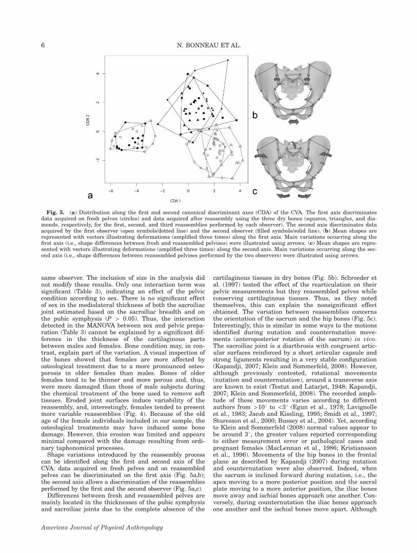
can be identified along the first and second axis of theCVA: data acquired on fresh pelves and on reassembledpelves can be discriminated on the first axis (Fig. 5a,b);the second axis allows a discrimination of the reassembliesperformed by the first and the second observer (Fig. 5a,c).Differences between fresh and reassembled pelves are
mainly located in the thicknesses of the pubic symphysisand sacroiliac joints due to the complete absence of the
cartilaginous tissues in dry bones (Fig. 5b). Schroeder etal. (1997) tested the effect of the rearticulation on theirpelvic measurements but they reassembled pelves whileconserving cartilaginous tissues. Thus, as they notedthemselves, this can explain the nonsignificant effectobtained. The variation between reassemblies concernsthe orientation of the sacrum and the hip bones (Fig. 5c).Interestingly, this is similar in some ways to the motionsidentified during nutation and counternutation move-ments (anteroposterior rotation of the sacrum) in vivo.The sacroiliac joint is a diarthrosis with congruent artic-ular surfaces reinforced by a short articular capsule andstrong ligaments resulting in a very stable configuration(Kapandji, 2007; Klein and Sommerfeld, 2008). However,although previously contested, rotational movements(nutation and counternutation), around a transverse axisare known to exist (Testut and Latarjet, 1948; Kapandji,2007; Klein and Sommerfeld, 2008). The recorded ampli-tude of these movements varies according to differentauthors from [108 to \38 (Egun et al., 1978; Lavignolleet al., 1983; Jacob and Kissling, 1995; Smidt et al., 1997;Sturesson et al., 2000; Bussey et al., 2004). Yet, accordingto Klein and Sommerfeld (2008) normal values appear tobe around 38, the greater values reported correspondingto either measurement error or pathological cases andpregnant females (MacLennan et al., 1986; Kristianssonet al., 1996). Movements of the hip bones in the frontalplane as described by Kapandji (2007) during nutationand counternutation were also observed. Indeed, whenthe sacrum is inclined forward during nutation, i.e., theapex moving to a more posterior position and the sacralplate moving to a more anterior position, the iliac bonesmove away and ischial bones approach one another. Con-versely, during counternutation the iliac bones approachone another and the ischial bones move apart. Although
Fig. 5. (a) Distribution along the first and second canonical discriminant axes (CDA) of the CVA. The first axis discriminatesdata acquired on fresh pelves (circles) and data acquired after reassembly using the three dry bones (squares, triangles, and dia-monds, respectively, for the first, second, and third reassemblies performed by each observer). The second axis discriminates dataacquired by the first observer (open symbols/dotted line) and the second observer (filled symbols/solid line). (b) Mean shapes arerepresented with vectors illustrating deformations (amplified three times) along the first axis. Main variations occurring along thefirst axis (i.e., shape differences between fresh and reassembled pelvises) were illustrated using arrows. (c) Mean shapes are repre-sented with vectors illustrating deformations (amplified three times) along the second axis. Main variations occurring along the sec-ond axis (i.e., shape differences between reassembled pelvises performed by the two observers) were illustrated using arrows.
6 N. BONNEAU ET AL.
American Journal of Physical Anthropology
the sacroiliac joints surfaces generally match well, slightdifferences in reassembly and positioning of the sacrummay result in corresponding deviation of the hip bones(Fig. 5c).In summary, according to our geometric morphometric
results, a significant effect on shape analyses is intro-duced by removal of soft tissue and reassembly of thepelvic bones using rubber bands (Table 3). Yet, variationintroduced by the reassembly remains small comparedwith the biological variation observed between the 15pelves examined (Fig. 4). Variation in the reassemblyprocess is likely related, first, to the complete absence ofcartilaginous tissues on dry bones and, second, to themorphology of the sacroiliac joint which, in vivo, allowsphysiological movements, resulting in different potentialpositions of the two sacroiliac surfaces relative to oneanother. Using the results obtained in this study, meas-urements based on rearticulated pelvis can be improved,either by improving the reassembly process performedbefore measurements or by using corrections which couldbe performed after measurements.First, the reassembly process can be improved by plac-
ing buffers or clay with a width corresponding to themean mediolateral thickness of the pubic symphysis andsacroiliac joints. Concerning the pubic symphysis, themean value calculated in the sample of the 15 individu-als equaled 6.84 mm (SD 5 2.48; range 5 2.90–11.20mm), a result close to the value of 5.9 mm in males and4.9 mm in females proposed by Vix and Ryu (1971). Thedifference between the thickest symphysis and the nar-rowest symphysis is 8.30 mm, i.e., \1 cm. No significanteffect of sex was detected (P [ 0.05) but no definitiveconclusion can be drawn due to the small size and rela-tively old age of the individuals included in this study.Concerning the mediolateral thickness of the sacroiliac
joints, a mean value can be estimated based on the sac-roiliac breadth measurements. The mean differencebetween reassemblies and fresh pelvis of 2.5 mm sug-gests an approximate thickness of 1.3 mm for each sacro-iliac joint. This value is low compared with the meanthickness of 3.5–4.5 mm obtained on middle-age subjectsby MacDonald and Hunt (1952) but is in accordancewith the great decrease of the thickness of the cartilageduring aging. The advanced age of the subjects used inthe study may thus explain the low value of the meanthickness obtained for the sacroiliac joint. Consequently,it would be interesting to analyze younger adults. Itshould be noted that recommendations proposed toimproved reassembly process pertain only to adultpelves. In juvenile pelves, cartilaginous parts remainand the hip bones and the sacrum are composed of manypieces of bone, which remain unfused and are conservedas separated bones in anthropological collections. Conse-quently, the effect of the reassembly may be very impor-tant for young children because not only orientation of
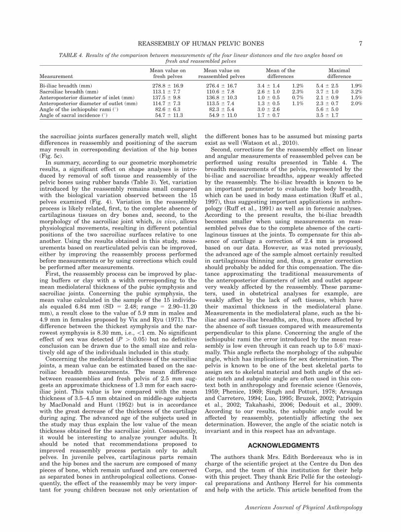
the different bones has to be assumed but missing partsexist as well (Watson et al., 2010).Second, corrections for the reassembly effect on linear
and angular measurements of reassembled pelves can beperformed using results presented in Table 4. Thebreadth measurements of the pelvis, represented by thebi-iliac and sacroiliac breadths, appear weakly affectedby the reassembly. The bi-iliac breadth is known to bean important parameter to evaluate the body breadth,which can be used in body mass estimation (Ruff et al.,1997), thus suggesting important applications in anthro-pology (Ruff et al., 1991) as well as in forensic analyses.According to the present results, the bi-iliac breadthbecomes smaller when using measurements on reas-sembled pelves due to the complete absence of the carti-laginous tissues at the joints. To compensate for this ab-sence of cartilage a correction of 2.4 mm is proposedbased on our data. However, as was noted previously,the advanced age of the sample almost certainly resultedin cartilaginous thinning and, thus, a greater correctionshould probably be added for this compensation. The dis-tance approximating the traditional measurements ofthe anteroposterior diameters of inlet and outlet appearvery weakly affected by the reassembly. These parame-ters, used in obstetrical analyses for example, areweakly affect by the lack of soft tissues, which havetheir maximal thickness in the mediolateral plane.Measurements in the mediolateral plane, such as the bi-iliac and sacro-iliac breadths, are, thus, more affected bythe absence of soft tissues compared with measurementsperpendicular to this plane. Concerning the angle of theischiopubic rami the error introduced by the mean reas-sembly is low even through it can reach up to 5.68 maxi-mally. This angle reflects the morphology of the subpubicangle, which has implications for sex determination. Thepelvis is known to be one of the best skeletal parts toassign sex to skeletal material and both angle of the sci-atic notch and subpubic angle are often used in this con-text both in anthropology and forensic science (Genoves,1959; Phenice, 1969; Singh and Potturi, 1978; Arsuagaand Carretero, 1994; Luo, 1995; Bruzek, 2002; Patriquinet al., 2002; Takahashi, 2006; Dedouit et al., 2009).According to our results, the subpubic angle could beaffected by reassembly, potentially affecting the sexdetermination. However, the angle of the sciatic notch isinvariant and in this respect has an advantage.
ACKNOWLEDGMENTS
The authors thank Mrs. Edith Bordereaux who is incharge of the scientific project at the Centre du Don desCorps, and the team of this institution for their helpwith this project. They thank Eric Pelle for the osteologi-cal preparations and Anthony Herrel for his commentsand help with the article. This article benefited from the
TABLE 4. Results of the comparison between measurements of the four linear distances and the two angles based onfresh and reassembled pelves
MeasurementMean value onfresh pelves
Mean value onreassembled pelves
Mean of thedifferences
Maximaldifference
Bi-iliac breadth (mm) 278.8 6 16.9 276.4 6 16.7 3.4 6 1.4 1.2% 5.4 6 2.5 1.9%Sacroiliac breadth (mm) 113.1 6 7.7 110.6 6 7.8 2.6 6 1.0 2.3% 3.7 6 1.0 3.2%Anteroposterior diameter of inlet (mm) 137.5 6 9.8 136.8 6 10.3 1.0 6 0.5 0.7% 2.1 6 0.9 1.5%Anteroposterior diameter of outlet (mm) 114.7 6 7.3 113.5 6 7.4 1.3 6 0.5 1.1% 2.3 6 0.7 2.0%Angle of the ischiopubic rami (8) 82.6 6 6.3 82.3 6 5.4 3.0 6 2.6 5.6 6 5.0Angle of sacral incidence (8) 54.7 6 11.3 54.9 6 11.0 1.7 6 0.7 3.5 6 1.7
7REASSEMBLY OF HUMAN PELVIC BONES
American Journal of Physical Anthropology

morphometric facility of the Paris Museum (UMR 2700CNRS – MNHN: ‘‘Plateforme de Morphometrie’’), includ-ing Anne-Claire Fabre and Raphael Cornette who havecontributed to the illustrations. Finally, they are gratefulto the anonymous reviewers, the associate editor, andChristopher Ruff who provided interesting and helpfulcomments on this work.
LITERATURE CITED
Arsuaga JL, Carretero JM. 1994. Multivariate analysis of the sex-ual dimorphism of the hip bone in a modern human populationand in early hominids. Am J Phys Anthropol 93:241–257.
Baylac M. 2010. Rmorph a morphometric library for [email protected].
Berge C. 1996. The evolution and growth of the hominid pelvis.A preliminary thin-plate spline study of ilium shape. In: Mar-cus LF, Corti M, Loy A, Naylor GJP, Slice DE, editors. Advan-ces in morphometrics. New York: Plenum Press. p 441–448.
Berge C. 1998. Heterochronic processes in human evolution: anontogenetic analysis of the hominid pelvis. Am J Phys Anthro-pol 105:441–459.
Berge C, Orban-Segebarth R, Schmid P. 1984. Obstetrical inter-pretation of the australopithecine pelvic cavity. J Hum Evol13:573–587.
Bookstein FL. 1991. Morphometric tools for landmark data (ge-ometry and biology). Cambridge: Cambridge University Press.
Bouhallier J, Berge C, Penin X. 2004. Analyse Procruste de lacavite pelvienne des australopitheques (AL 288, STS 14), deshumains et des chimpanzes: consequences obstetricales. C RPalevol 3:295–304.
Boulay C, Tardieu C, Benaim C, Hecquet J, Marty C, Prat-Pra-dal D, Legaye J, Duval-Beaupere G, Pelissier J. 2006a. Three-dimensional study of pelvic asymmetry on anatomical speci-mens and its clinical perspectives. J Anat 208:21–33.
Boulay C, Tardieu C, Hecquet J, Benaim C, Mouilleseaux B,Marty C, Prat-Pradal D, Leagaye J, Duval-Beaupere G, Pel-issier J. 2006b. Sagittal alignment of spine and pelvis regu-lated by pelvic incidence: standard value and prediction oflordosis. Eur Spine J 15:415–422.
Brown KB, DeLeon VB, Ruff CB. 2011. Going to extremes: bodysize and obstetrical adaptation. Am J Phys Anthropol 144(Suppl 52):98.
Bruzek J. 2002. A method for visual determination of sex, usingthe human hip bone. Am J Phys Anthropol 117:157–168.
Bussey MD, Yanai T, Milburn P. 2004. A non-invasive techniquefor assessing innominate bone motion. Clin Biomech 19:85–90.
Davenport CB, Steggerda M, Drager W. 1935. Critical examina-tion of physical anthropology on the living. Proc Am AcadArts Sci 69:265–285.
Dedouit F, Telmon N, Costagliola R, Otal P, Joffre F, Rouge D.2007. Virtual anthropology and forensic identification: reportof one case. Forensic Sci Int 173:182–187.
Dryden IL, Mardia KV. 1998. Statistical shape analysis. NewYork: Wiley.
Duval-Beaupere G, Legaye J. 2004. Composante sagittale de lastatique rachidienne. Rev Rhum 71:105–119.
Duval-Beaupere G, Schmidt C, Cosson P. 1992. A barycentre-metric study of the sagittal shape of spine and pelvis: the con-ditions required for an economic standing position. AnnBiomed Eng 20:451–462.
Egun DN, Olsson TH, Schmid H. 1978. Movements in the sacro-iliac joint demonstrated with roentgenstereophotogrammetry.Acta Radiol Diagn 19:833–944.
Genoves S. 1959. L’estimation des differences sexuelles dans l’oscoxal: differences metriques et differences morphologiques.Bull Mem Soc Anthropol Paris 10:3–95.
Gower JC. 1975. Generalized procrustes analysis. Psychome-trika 40:33–50.
Hand DJ, Taylor CC. 1987. Multivariate analysis of varianceand repeated measures: a practical approach for behaviouralscientists. London: Chapman & Hall.
Jacob HAC, Kissling RO. 1995. The mobility of the sacroiliacjoints in healthy volunteers between 20 and 50 years of age.Clin Biomech 10:352–361.
Jamison PL, Zegura SL. 1974. A univariate and multivariate ex-amination of measurement error in anthropology. Am J PhysAnthropol 40:197–204.
Kapandji AI. 2007. Physiologie articulaire Tome 3. Rachis –Ceinture pelvienne – Rachis lombal – Rachis dorsal – Rachiscervical – Tete. Maloine 6eme edition.
Klein P, Sommerfeld P. 2008. Biomecanique des membres inferi-eures. Bases et concepts, bassin, membres inferieurs. Elseviered. Masson.
Kohn LAP, Cheverud JM, Bhatia G, Commean P, Smith K, Van-nier MW. 1995. Anthropometric optical surface imaging sys-tem repeatability, precision and validation. Ann Plast Surg34:362–370.
Kristiansson P, Svardsudd K, von Schoultz B. 1996. Serumrelaxin, symphyseal pain, and back pain during pregnancy.Am J Obstet Gynecol 175:1342–1347.
Lavignolle B, Vital JM, Senegas J, Destandau J, Toson B, BouyxP, Morlier P, Delorme G, Calabet A. 1983. An approach to thefunctional anatomy of the sacroiliac joints in vivo. Anat Clin5:169–176.
Legaye J, Duval-Beaupere G, Hecquet J, Marty C. 1998. Pelvicincidence: a fundamental pelvic parameter for three-dimen-sional regulation of spinal sagittal curves. Eur Spine J 7:99–103.
Legaye J, Hecquet J, Marty C, Duval-Beaupere G. 1993. Equili-bre sagittal du rachis. Relations entre bassin et courburesrachidiennes sagittales en position debout. Rachis 5:215–226.
Luo YC. 1995. Sex determination from the pubis by discrimi-nant function analysis. Forensic Sci Int 74:89–98.
MacDonald GR, Hunt TE. 1952. Sacro-iliac joints: observationson the gross and histological changes in the various agegroups. Canad M A J 66:157–163.
McHenry HM. 1978. Analysis of the hominoid os coxae by Car-tesian coordinates. Am J Phys Anthropol 48:215–226.
McLennan AH, Nicolson R, Green RC, Bath M. 1986. Serumrelaxin and pelvic pain of pregnancy. Lancet 243:244.
Monteiro LR. 1999. Multivariate regression models and geomet-ric morphometrics: the search for causal factors in the analy-sis of shape. System Biol 48:192–199.
Patriquin ML, Steyn M, Loth SR. 2002. Metric assessment ofrace from the pelvis in South Africans. Forensic Sci Int127:104–113.
Phenice TW. 1969. A newly developed visual method of sexingthe os pubis. Am J Phys Anthropol 30:297–302.
R Development Core Team. 2009. R: a language and environmentfor statistical computing. R Foundation for Statistical Comput-ing, Vienna, Austria. Available at: http://www.R-project.org.
Rickenmann E. 1957. Beitrage zur vergleichenden anatomieinsbesondere des beckens bei catarrhinen. Basel: Karger.
Richtsmeier JT, Paik CH, Elfert PC, Cole TM, Dahlman HR.1995. Precision, repeatability and validation of the localiza-tion of cranial landmarks using computed tomography scans.Cleft Palate-Craniofacial J 32:217–227.
Rohlf FJ, Slice DE. 1990. Extensions of the procrustes methodfor the optimal superimposition of landmarks. Syst Zool39:40–59.
Ruff CB. 1991. Climate, body size and body shape in hominidevolution. J Hum Evol 21:81–105.
Ruff CB, Trinkaus E, Holliday TW. 1997. Body mass andencephalization in Pleistocene Homo. Nature 387:173–176.
Schroeder CF, Schmidtke SZ, Bidez MW. 1997. Measuring thehuman pelvis: a comparison of direct and radiographic techni-ques using a modern United States-based sample. Am J PhysAnthropol 103:471–479.
Singh S, Potturi BR. 1978. Greater sciatic notch in sex determi-nation. J Anat 125:619–624.
Slice DE. 2005. Modern morphometrics in physical anthropol-ogy. New York: Kluwer Academic/Plenum.
Smidt GL, Wei SH, McQuade K, Barakatt EMA, Sun T, Stan-ford W. 1997. Sacroiliac motion for extreme hip positions: afresh cadaver study. Spine 22:2073–2082.
8 N. BONNEAU ET AL.
American Journal of Physical Anthropology

Steudel K. 1981. Functional aspects of primate pelvic structure:a multivariate approach. Am J Phys Anthropol 55:399–410.
Sturesson B, Uden A, Vleeming A. 2000. A radiostereometricanalysis of the movements of the sacroiliac joints in the recip-rocal straddle position. Spine 25:214–217.
Takahashi H. 2006. Curvature of the greater sciatic notch insexing the human pelvis. Anthropol Sci 114:187–191.
Tardieu C, Hecquet J, Barrau A, Loridon P, Boulay C, Legaye J,Carlier R, Marty C, Duval-Beaupere G. 2006. Le bassin, inter-face articulaire entre rachis et membres inferieurs: analysepar le logiciel De-Visu. C R Palevol 5:583–595.
Testut L, Latarjet A. 1948. Traite d’anatomie humaine.Tome premier: osteologie, arthrologie, myologie. 9e ed. Doin etCie.
Vix VA, Ryu CY. 1971. The adult symphysis pubis: normal andabnormal. Am J Radiol 112:517–525.
von Cramon-Taubadel N, Frazier BC, Lahr MM. 2007. The problemof assessing landmark error in geometric morphometrics: theory,methods, and modifications. Am J Phys Anthropol 134:24–35.
Watson PJ, O’Higgins P, Fagan MJ, Dobson CA. 2010. Valida-tion of a morphometric reconstruction technique applied to ajuvenile pelvis. Proc Inst Mech Eng H 225:48–57.
Weidenreich M. 1913. Ueber das huftbein und das becken derprimaten und ihre umformung durch den aufrechten gang.Anat Anz 44:497–513.
Zuckerman S, Ashton EH, Flinn RM, Oxnard CE, Spence TF.1973. Some locomotor features in the pelvic girdle in prima-tes. Symp Zool Soc Lond 33:71–165.
9REASSEMBLY OF HUMAN PELVIC BONES
American Journal of Physical Anthropology