technical series - tulipgroup.com · techniques permitting the detection of incomplete antibodies,...
TRANSCRIPT

TEC
HN
ICA
L SE
RIES
TEC
HN
ICA
L SE
RIES
...Setting trends
Anti Human GlobulinReagentBasic Concepts and Practices
TULIP DIAGNOSTICS (P) LTD.Gitanjali, Tulip Block, Dr. Antonio Do Rego Bagh, Alto Santacruz, Bambolim Complex ,
Post Office, Goa - 403 202. INDIA. Telephone Nos.: (0832) 2458546-51 Fax:(0832) 2458544E-mail: [email protected] Website:http://www.tulipgroup.com
For the use of Registered Medical Practioners and Laboratories only

Tulip Diagnostics (P) Ltd is a part of
the innovative Tulip group of companies based
at Goa, India.
The Groups commitment to building
products to international standards, through
indigineous R&D has accorded the company
virtual leadership in most product segments
domestically. International recognition to these
efforts has led to exports to about fortyfive
countries globally with an ever increasing user
base. Tulip strongly believes that knowledge
upgradation remains the fundamental basis
for better diagnosis and patient care.
Publishing of technical series is one such
initiative to make available to the Laboratory
professionals and clinicians updated knowledge
that is vital for them to set trends in their day to
day practice.
1
Anti Human Globulin Test – Historical PerspectiveThe first step towards a rational approach to blood transfusion became possiblewith Landsteiners discovery in 1901 of the ABO blood group system. Nearlyhalf a century later, the next advance occurred with the introduction of serologicaltechniques permitting the detection of incomplete antibodies, which arenonagglutinating immunoglobulin G (IgG) antibodies that sensitize red bloodcells. In 1945, Coombs et al. described the use of anti human globulin test forthe detection of weak and non-agglutinating antibodies in serum. Further in1946, Coombs et al. described the use of anti human globulin test to detect invivo sensitization of red blood cells in babies suffering from haemolytic diseaseof the newborn. Although the test initially was of great value in investigatingRho (D) haemolytic disease of the newborn, it was not long before its versatilityfor the detection of other IgG blood group antibodies became evident.
Although Coombs and his coworkers were instrumental in introducing theanti human globulin test to blood group serology, the principle of the test hadinfact been described by Moreschi in 1908. Moreschis studies involved theuse of rabbit anti goat antiserum to agglutinate rabbit red blood cells that weresensitized with low non-agglutinating dose of goat anti rabbit red cell serum.
Later in 1957, Dacie et al. published data regarding the importance of antihuman complement activity. Further in 1960 many reports were publishedindicating the need for anti human complement activity in anti human globulinserum to allow the detection of antibodies in the indirect test. Though many ofthe antibody specificities mentioned in these reports are now considered oflittle clinical significance, however one specificity that was consistently mentionedand is still considered to be clinically significant was Anti-Jka. Further it wasalso shown that the presence of anti human complement activity would enhancethe reaction of some antibodies namely Anti-Fya and Anti-K.
Preliminary Notes-Antigen and antibody related to bloodgroup serology
Antigens are foreign substances, which initiate an immune response in humanbody and react specifically with antibodies. Blood group antigens that arelocated on the surface of red blood cells when transferred to another individualare almost certain to carry antigens not possessed by the recipient. Theseforeign antigens then initiate an immune response and production of antibodies
in vivo.Antibodies are counter agents produced as a defense mechanism whenforeign antigen enters human body. With respect to blood group serology itmeans that blood group antibodies are specific and are formed against redblood cell antigens not possessed by the recipient.
Antibody nomenclature with respect to blood group serologyRed blood cell antibodies may be naturally occurring or immune in nature.Naturally occurring antibodies are produced because of the exposure of humanbody to certain bacteria and plant substances whose antigenic structureresembles blood group antigens. Naturally occurring antibodies are most oftenIgM class of antibodies.
Immune antibodies (alloantibodies) are antibodies present in an individual dueto transfusion or pregnancy. Immune antibodies most often are IgG class ofantibodies, sometimes IgM or a mixture of IgM and IgG class of antibodies.
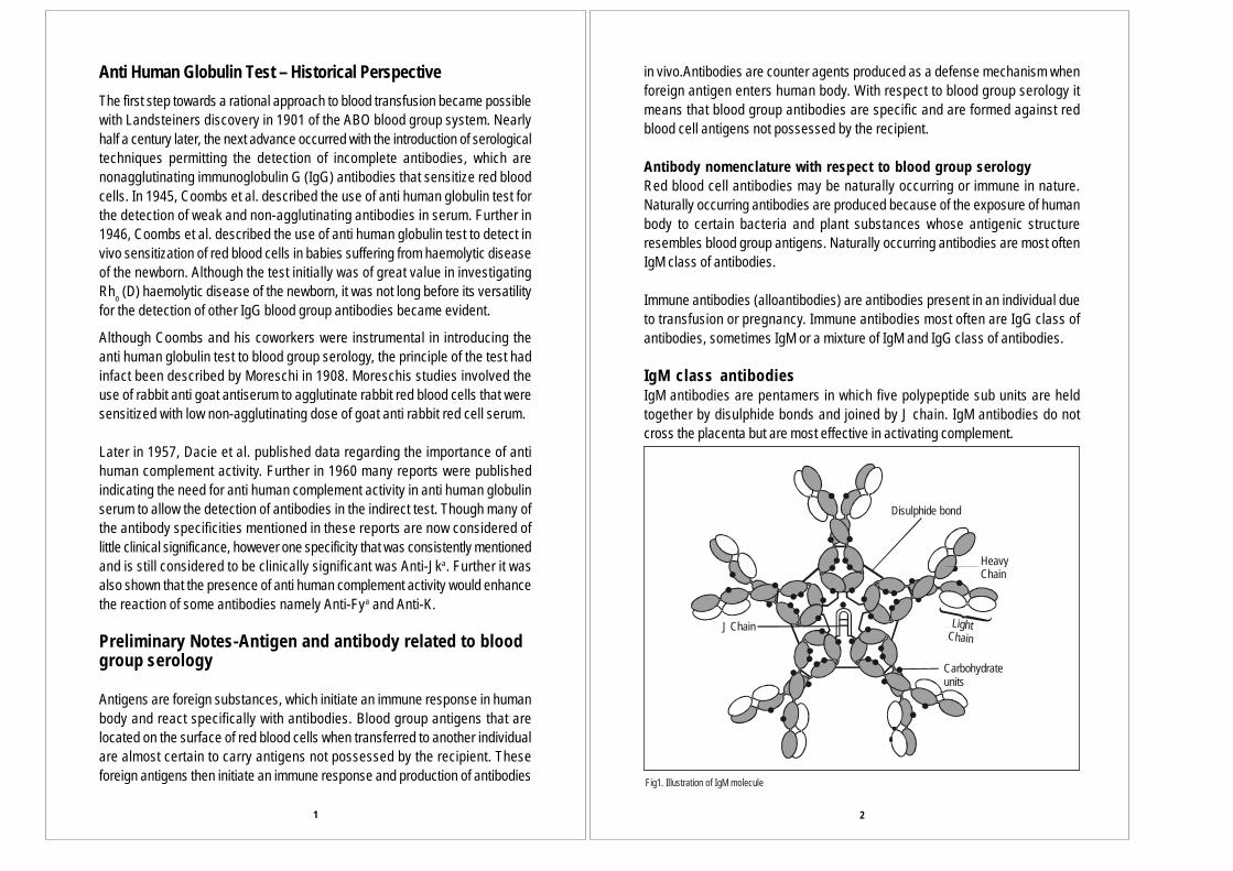
IgM class antibodiesIgM antibodies are pentamers in which five polypeptide sub units are heldtogether by disulphide bonds and joined by J chain. IgM antibodies do notcross the placenta but are most effective in activating complement.
Disulphide bond
Heavy hainC
LighthainC
Carbohydrate units
J Chain
Fig1. Illustration of IgM molecule
2

Sialic acid present on the red cell membranes impart to the cells, a net negativecharge. As a result mutual repulsive force exists between red blood cellsknown as zeta potential and the red blood cells are separated by a distance. Inblood group serology IgM antibodies are termed as complete antibodiesbecause of their ability to directly bridge the distance between two red bloodcells inspite of the zeta potential and bring about visible agglutination. IgMantibodies are able to bridge this distance and bring about visible agglutination.
IgG class antibodies:IgG antibodies are monomers with two heavy chains and two light chains. IgGis the only immunoglobulin that can cross the placental barrier, a fact whichexplains the role of IgG antibodies in haemolytic disease of the newborn. Foursubclasses of IgG are present and they vary widely in their ability to activate
Fig.2 Illustration of complete antibodies
IgG2 IgG3 IgG4
C terminalHeavy Chain
Carbohydrate unitInter chain disulphide bond
Light Chain
Hinge region
N terminalAntigen binding site
Fab
Fc
IgG1
Fig. 3 Structure of four subclasses of IgG
complement. IgG3 molecules are highly effective in activating complement, IgG1moderately activate complement, IgG2 slightly activate complement and IgG4do not activate complement.
IgG antibodies are also known as incomplete antibodies because they cannotbridge the distance between two red blood cells and bring about directagglutination like the IgM immunoglobulins. Antibodies that do not agglutinatered blood cells directly are termed as incomplete antibodies or sensitizingantibodies and the red blood cells that have such antibodies on their surfaceare known as sensitized cells. Incomplete antibodies sensitized onto red bloodcells can be agglutinated by anti human globulin reagent through the usage ofenhancement medium to visualize agglutination.
Fig . 4 Illustration of red blood cells sensitized with incomplete antibodies
Fig. 5 Illustration of IgG sensitized red blood cells agglutinated by AHG reagent
Role of enhancement mediumAn enhancement medium can either be a protein (e.g. Bovine serum albumin) orenzymes (e.g. papain, bromelin, ficin) or low ionic salt solution (LISS) or chargedmolecules (polybrene) that reduce zeta potential, enhance antibody uptake, enhancementin agglutination reactions.
In order to clearly understand antibody enhancement methods, it is important to considerthe basic aspect of haemagglutination reactions. Agglutination is a reversible chemicalreaction and occurs probably in two stages:
Stage 1: Sensitization i.e. attachment of antibody to red blood cell membrane antigen.Stage 2: Formation of bridges between the sensitized red blood cells to form lattice(crosslinking) to visualize agglutination.
Thus various factors that affect these two stages can be manipulated to enhanceagglutination.
3 4

65
Enhancement techniques
! Albumin additivesBovine serum albumin (BSA), a dipolar protein has been used routinely formany years as an enhancement medium. The enhancement effect of albumin isattributed to its influence on the second stage of agglutination by reducing thenet negative charge on the red blood cell membranes thereby potentiatingagglutination. Bovine serum albumin is available as solutions of 22% or 30%concentration.
! EnzymesTreating red blood cells with certain enzymes enhances agglutination ofantibodies with red blood cell antigens. Most commonly used enzymes inimmunohaematological procedures are papain, bromelin and ficin belong tothiol protease class of enzymes.
Proteolytic enzymes reduce the red blood cell surface charge by cleavingsialic acid molecules on red blood cell membrane, lowering the zeta potentialand enhance agglutination. Enzyme treatment also causes spicule formationon the red blood cell membrane. This greatly increases the potential number ofcontact points for antibodies to react with corresponding antigens on red bloodcell membrane.
The reaction of Anti-P, Lewis and Kidd blood group antibodies are strongerwith enzyme treated red blood cells. Another important observation is thatLewis and Kidd blood group antibodies may lyse enzyme treated red bloodcells.
! Positively charged moleculesIn the presence of positively charged polymers such as hexadimethrine bromide(Polybrene ), protamine sulphate and poly-L-lysine, normal red blood cells exhibitspontaneous aggregation. Thus any red blood cells sensitized with antibody will exhibitenhanced agglutination.
The action of these charged molecules may be due to neutralization of negative chargecontributed by sialic acid and release of water molecules that cover red blood cellmembrane thereby enhancing agglutination.
Polybrene generally is added to red blood cells that have been incubated withantibody at low ionic strength and low pH.
! Polyethylene Glycol (PEG)PEG is a water-soluble polymer used as an additive to increase antibodyuptake. Its action is to remove water molecule that takes up more space aroundthe red blood cell membrane, thereby enhancing antibody uptake and reactionstrength.
! LISS (Low Ionic Salt Solution) and LISS additivesLISS (approximately 0.03 M) greatly increases stage 1 uptake of antibody tored blood cells, as compared to normal saline (approximately 0.17M). Toprevent lysis of red blood cells at such a low ionic strength, a non-ionicsubstance such as glycine is incorporated in LISS.
LISS additives may sometimes contain macromolecules (e.g. BSA) in additionto ionic salts and buffers. Addition of macromolecules to LISS reduces ionicstrength with shortened incubation times.
EnzymesReduce negative charge
AlbuminReduce negative charge
Polybrene, ProtamineSupply cations
Fig. 6 Illustration of Antibody Enhancement
ComplementAn observable phenomenon that may result from red blood cell antigen-antibodyinteraction is haemolysis. This requires co-operation of another defense mechanism ofbody known as the complement system. Complement is the name given to a set ofapproximately twenty serum proteins, which in response to antigenic stimulus reactwith one another in a sequential cascade.
The most important complement component is C3b, which can be, activated either bythe classical or the alternate pathway. The antigen-antibody interactions activate theclassical pathway that is of great significance in blood group serology.

7 8
C1qC1r2C1s2 C1qr2s2
C4 C4bC1s2 + Ca++
C4b + C2 C4bC2 C4b2bC1s2
C4a
C2a
C3 C3b
C3a
C4b2b
C5 C5b
C5a
C4b2b3b
C5b + C6-9 C5b-C9
Ag-Ab complex
Phase 1
Phase 2
Phase 3
Phase 4
Fig. 7 Activation of Classical complement pathway
Immunoglobulin requirements for activation of classical pathwayThe mode of activation of classical pathway is through binding of C1q to CH2domain of IgM or to the CH2 domain of IgG1 or IgG3 antibodies. The C1qmolecule has six Fc binding sites and inorder to make a firm bond with antibodymolecule, atleast two of these sites must bind to antibody.
Fig.8 Illustration of immunoglobulin molecule
VL
CL
VH
C 1H
Antigenbindingsites
Hinge region
Disulphide bonds
Light chain
C 2H C 3H
C terminus
Heavy chain
Pepsin cleavage
Papain cleavage
Carbohydrate
Fab
Fc
Fc
Fab Antigen Surface Fig 9. Illustration of staple form-IgM molecule
In case of IgM antibodies, when the molecule is in planar or star form, there isonly a single binding site for C1q, as a result it cannot bind firmly and activateC1q. However when combined with antigen, IgM molecule usually assumesstaple form with the F(ab) at right angle with Fc. The distortion produced bythis movement exposes an additional C1q binding site. As a result IgM antibodycombined with its corresponding antigen bind to two C1q sites on single IgMmolecule.
In case of IgG antibodies, two IgG antibodies must be present on antigensurface at a distance of 20-30 nm apart from each other. This is the maximumspan of the C1q molecule. It is for this reason IgM is considered more effectivein activating C1q than IgG molecule.
An important point to note is that all examples of immune Anti-D fail to activatecomplement. The reason being that the D antigenic sites are too far apart fromeach other on the red blood cell surface. As discussed two IgG molecules mustbe at a distance of 20-30 nm apart from each other so as to activate C1q.Since D antigenic sites are too far apart from each other, the bound IgGmolecules cannot activate C1q.
Complement binding antibodiesComplement components may attach to red blood cells in vivo or in vitro viaone of the probable mechanisms!"Complement binding alloantibody (blood group antibodies present inindividual due to transfusion or pregnancy) specif ic for red blood cel lantigen may cause sensitization of complement component on red bloodcell surface due to transfusion.
Complement binding alloantibodies when present in low concentrationsbring about the attachment of complement component upto the C3 stage,

9 10
Complement binding by human red cell Antibodies
System Antibody Readily detectablehaemolysis
Positive antiglobulintest (anti-complement)
ABO anti-Aanti-B many manyant-A,Banti-A1 none very fewanti-HI none fewanti-Ht all all
Lewis anti-Lea some allanti-Leb few many
P anti-P1 few fewant-PP1 Pk all allanti-P (auto-) all allanti-P (allo-) many all
Ii anti-Ianti-i
Sd anti-Sda none some
Rh anti-D none noneanti-c, E, etc. none none
K anti-K none many
Fy anti-Fya none many
J k ant-Jka few all
MNSs anti-M, -N none noneanti-S, -s none some
Lu anti-Lua, -Lub none none
Di anti-Dia few few
Xg anti-Xga none many
}auto- many many
}
but produce little or no haemolysis. Further activation of C3 occurs only whencomplement is powerfully activated due to high concentration of antibodies,leading to the generation of large amount of C3b. Certain antibodies such asAnti-Jka are seldom lytic but invariably activate complement thereby leading tothe deposition of complement components on the red blood cell surface.
! About 10-20% of patients with warm AIHA (Autoimmune haemolytic anaemia)demonstrate in vivo sensitization with complement component C3.
!"In cold haemagglutinin disease, the cold reactive autoantibody can reactwith red blood cell antigens at temperature upto 320C range (thermal amplitude10-320C). When the patient is exposed to cold temperature, red blood cell issensitized with autoantibody, which activates complement. If the red blood cellsescape haemolysis, they return to the central circulation where the temperatureis 370C and the autoantibody dissociates from the cells, leaving complementcomponent C3d firmly bound to the red blood cell membrane.
Basic concepts of Anti human Globulin testing
Two major class of antibodies react with red blood cell antigens namely thecomplete antibodies and the incomplete antibodies.
Complete antibodies, or saline agglutinins will agglutinate red blood cellssuspended in saline, which are usually IgM class of antibodies.
Incomplete antibodies do not react in saline but require special techniques toagglutinate red blood cells; which are IgG class of antibodies.
After combining with their corresponding antigen, blood group antibodies havethe ability to activate complement. Thus sensitization of incomplete antibodiesor complement component can occur either in vivo or in vitro. Anti humanglobulin reagent is used to detect IgG or complement sensitized red bloodcells.
For detecting in vivo antibody or complement sensitization DAT (DirectAntiglobulin Test) is used whereas for detecting in vitro antibody or complementsensitization IAT (Indirect Antiglobulin Test) is used.
Anti Human Globulin ReagentsPolyspecific Anti human globulin (Coombs) Reagent:The term ‘polyspecific’ reagent refers to reagent system containing blend ofantibodies for more than one antigen i.e. blend of Anti human IgG, Anti humanC3b, Anti human C3d. Polyspecific reagent may be a blend of:

11 12
! Polyclonal Anti human IgG, polyclonal Anti human C3b, polyclonal Anti human C3d.! Polyclonal Anti human IgG, monoclonal Anti human C3b, monoclonal Anti human C3d.! Polyclonal Anti human IgG, monoclonal Anti human C3d.
ICSH Recommendations for Polyspecific Anti Human GlobulinreagentsICSH has recommended certain specifications, which a commercially available Antihuman globulin reagent should comply:! The reagent should only agglutinate red blood cells sensitized with antibodies and
or coated with significant levels of complement components.! The potency of Anti human IgG component should be evaluated by serological
titration (chequer board titration).! A polyspecific reagent should contain Anti human C3c and Anti human C3d
at controlled levels to avoid false positive reactions or a suitable potent monoclonalAnti human C3d. It should contain little or no Anti human C4 activity. The assessmentof these qualities requires red cells specifically coated with complement componentsfor evaluating the serological potency.
! All the evaluations should be done in parallel with international reference reagents.
Complement (C3b, C3d)IgG
Polyclonal antisera Monoclonal antisera
Anti human C3b, Anti human C3d
Anti human IgG Anti human C3b,Anti human C3d, Anti human C3g
Anti Human GlobulinPolyspecific
Anti Human GlobulinPolyspecific
Fig. 10 Anti Human Globulin Manufacture
Anti Human GlobulinMonospecific
Anti Human GlobulinMonospecific
Anti Human GlobulinMonospecific
Polyclonal Anti human IgG is usually prepared in rabbits, though when largevolumes of antibody are required sheep or goats may be used. In productionof polyclonal Anti human IgG, animals are hyperimmunised to produce hightiter, high avidity Anti human IgG class antibodies. After achieving the desiredspecificity and titer, the animals are bled for the production batch of reagent.
With monoclonal antibodies, the hybrid clones are screened for antibodieswith the required specificity and affinity. The antibody secreting clones maythen be propagated in culture or by innoculation into mice, in which case theantibody is collected in ascites. Monoclonal anti human complement may beeither IgM or IgG class of antibodies.
Thus separate blends of Anti human IgG and Anti human complement antibodiesare made, and each pool is then adsorbed with A1, B and O red blood cells toremove heteropsecific antibodies. The total antibody content of each pool isdetermined and then pools are analysed by titration method to calculate theoptimum antibody dilution for Anti human IgG component and Anti humancomplement component.
Optimum concentration of Anti human IgG component is ascertained by chequerboard titration method. In chequer board titration dilutions of Anti human IgGare reacted with various dilutions of Anti-D IgG sensitized cells. This is acritical step because excess Anti human IgG may lead to prozoning during testreturning false negative results, correspondingly if Anti human IgGconcentration is less than optimum it may lead to false negative results with weaklysensitized cells. The evaluation procedure should also involve parallel testing withinternational reference reagent (e.g. W.H.O. RIIIM – Polyspecific Anti human globulinserum, available from Central Laboratory of Netherlands Red Cross TransfusionService, Amsterdam).
Similarly optimum concentration of Anti human Complement is ascertained bysensitizing complement to O Rho (D) positive red blood cells by the sucrosemethod, preparing various dilutions of complement sensitized cells. The optimumconcentration of Anti human complement component is evaluated by reactingdilutions of Anti human complement with dilutions of cells sensitized with differentlevels of serially diluted complement in parallel with international referencereagent.

13 14
Fig. 11 Chequer board titration results
Once the required optimum performance characteristic for each componentis ascertained, reagent blend of Anti human IgG and Anti human Complementantibodies is prepared and run as a system in parallel with the international referencereagent. Thus the final product is one that contains optimum concentration of Antihuman IgG and Anti human Complement antibodies for broad spectrum reactivity.
N 16 32 64 128 256 512 1024 2048 4096
N
1:2
1:4
1:8
1:16
1:32
1:64
3.5 3.5 3.5 3.5 3.5 2.5 1.0 1.5 1.0 1.0 Gr
2.53.5 3.5 2.5 1.0 1.5 1.5 1.5 2.0
3.5 3.5 3.5 3.5 2.0 2.0 1.5 1.5 2.0 1.03.5 3.5 3.5 3.5 2.0 2.0S 1.0S 1.0S 2.0 1.0
3.5 3.5 3.5 2.5 1.5 1.5 1.5 1.0S 1.0S 1.0W3.5 3.5 3.5 3.0 2.0 1.5 1.0S 1.0 1.0 1.0W
3.5 3.5 3.0S 2.0W 1.5 1.0S 1.0S 1.0S 1.0W 1.0W3.5 3.5 3.0S 3.0 1.5 1.5 1.5 1.0 1.0S 1.0
3.5 3.5 3.0S 2.0 1.5 1.5 1.0S 1.0S 1.0W 1.0W3.5 3.0S 2.5 2.0 1.5 1.5 1.0S 1.0S 1.0 1.0
3.5 2.0 3.0S 2.0 2.0W 1.5 1.0S 1.0 1.0W 1.0W3.5 3.0S 2.5 2.0 1.0S 1.0S 1.0 1.0 0.5 Gr
3.5 2.0S 2.0 2.0 1.0S 1.0S 1.0S Gr 0.5 Gr3.5 2.0 2.0 1.5 1.0S 1.0S 1.0W 0.5 0.5 ---
Dilutions of IgG Sensitized Cells
Dilut
ions o
f AHG
W.H.O. R III M Commercially available Polyspecific AHG
Monospecific Coombs Reagent:Monospecific reagents for use in anti human globulin test are prepared by similar procedure,however the reagent contains antibody of only one specificity.! Monospecific Anti human IgG reagent contains only polyclonal Anti human IgG! Monospecific Anti human C3b reagent contains only polyclonal Anti human C3b or
monoclonal Anti human C3b.! Monospecific Anti human C3d reagent contains only polyclonal Anti human C3d or
monoclonal Anti human C3d.
Direct Anti Human Globulin Test (DAT)DAT is used to detect in vivo sensitization of red blood cells with immunoglobulin, complementor both. A positive DAT, with or without shortened red blood cell survival, may result from:
! Autoantibodies to intrinsic red blood cell antigens.
! Alloantibodies in recipients circulation reacting with antigens on recentlytransfused donor red blood cells.
! Alloantibodies in donor plasma, plasma derivatives or blood fractions, which reactwith antigens on red blood cells of transfusion recipients.
! Alloantibodies in maternal circulation, which cross placenta and sensitize foetal redblood cells (HDN).
! Antibodies directed against certain drugs, which bind to red blood cell membranes(e.g. Penicillin)
! Adsorbed proteins, including immunoglobulins, which attach to abnormal membranesor red blood cells modified by therapy with certain drugs, notably those ofcephalosporin group.
! Complement components or rarely IgG bound to red blood cells afteradministration of drugs such as quinidine and phenacetin may induce drug/antidrug interaction.
! "Non red blood cell immunoglobulins associated with red blood cells inpatients with hypergammaglobulinaemia or recipients with high dose ofintravenous gammaglobulin.
! In-patients with organ transplantation, passenger lymphocytes of donororigin produce antibodies directed against ABO or other antigens on the recipient’scells, causing a positive DAT.
! Patients receiving ALG (antilymphocyte globulin) or ATG (antithymocyte globulin)of animal origin may develop a positive DAT within a few days, apparently relatedto high titer heterophile antibodies in these products and the presence ofcorresponding antibodies in animal derived AHG sera.
15 16
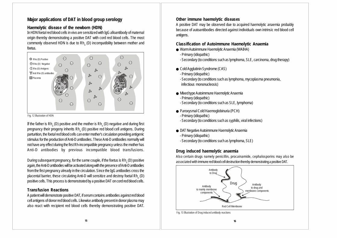
Major applications of DAT in blood group serologyHaemolytic disease of the newborn (HDN)In HDN foetal red blood cells in vivo are sensitized with IgG alloantibody of maternalorigin thereby demonstrating a positive DAT with cord red blood cells. The mostcommonly observed HDN is due to Rho (D) incompatibility between mother andfoetus.
If the father is Rho (D) positive and the mother is Rho (D) negative and during firstpregnancy their progeny inherits Rho (D) positive red blood cell antigens. Duringparturition, the foetal red blood cells can enter mother’s circulation providing antigenicstimulus for the production of Anti-D antibodies. These Anti-D antibodies normally willnot have any effect during the first Rh-incompatible pregnancy unless the mother hasAnti-D antibodies by previous incompatible blood transfusions.
During subsequent pregnancy, for the same couple, if the foetus is Rho (D) positiveagain, the Anti-D antibodies will be activated along with the presence of Anti-D antibodiesfrom the first pregnancy already in the circulation. Since the IgG antibodies cross theplacental barrier, these circulating Anti-D will sensitize and destroy foetal Rho (D)positive cells. This process is demonstrated by a positive DAT on cord red blood cells.
Transfusion ReactionsA patient will demonstrate positive DAT, if serum contains antibodies against red bloodcell antigens of donor red blood cells. Likewise antibody present in donor plasma mayalso react with recipient red blood cells thereby demonstrating positive DAT.
Fig. 12 Illustration of HDN
Rho (D) Positive
Rho (D) Negative
Rho (D) Antigens
Anti antibodiesRho (D)
Placenta
Other immune haemolytic diseasesA positive DAT may be observed due to acquired haemolytic anaemia probablybecause of autoantibodies directed against individuals own intrinsic red blood cellantigens.
Classification of Autoimmune Haemolytic Anaemia! Warm Autoimmune Haemolytic Anaemia (WAIHA)
- Primary (idiopathic)- Secondary (to conditions such as lymphoma, SLE, carcinoma, drug therapy)
! Cold Agglutinin Syndrome (CAS)- Primary (idiopathic)- Secondary (to conditions such as lymphoma, mycoplasma pneumonia, infectious mononucleosis)
! Mixed type Autoimmune Haemolytic Anaemia- Primary (idiopathic)- Secondary (to conditions such as SLE, lymphoma)
! Paroxysmal Cold Haemoglobinuria (PCH)- Primary (idiopathic)- Secondary (to conditions such as syphilis, viral infections)
! DAT Negative Autoimmune Haemolytic Anaemia- Primary (idiopathic)- Secondary (to conditions such as lymphoma, SLE)
Drug induced haemolytic anaemiaAlso certain drugs namely penicillin, procainamide, cephalosporins may also beassociated with immune red blood cell destruction thereby demonstrating a positive DAT.
Drug
Antibodyto Drug
Antibodyto mainly membrane
components
Antibodyto drug and
membrane components
Red Cell Membrane
Fig. 13 Illustration of Drug induced antibody reactions

17 18
Importance of serological studies in DAT positive results
As per AABB technical manual, three investigation approaches are helpfulin evaluation of positive DAT:! Test the DAT positive red blood cells with Monospecific Anti human IgG and
Monospecific Anti human C3d reagents to characterize type of proteins sensitizedwith red blood cell membrane.
! Test serum/plasma to detect and identify clinically significant antibodies to red bloodcell antigens.
!"Test eluate prepared from sensitized red blood cells with a panel of reagent red bloodcells to define whether the sensitized protein is immunoglobulin or complementcomponent. Elution frees antibody from sensitized red blood cellsand recovers antibody in usable form. When only complement is sensitized, eluatesare frequently non-reactive.
Probable serological findings with DAT positive-AIHA /Drug induced Haemolytic Anaemia
Parameter WAIHA CAS Mixedtype AIHA PCH Drug induced
AIHADAT positiveresult
IgG/ IgG + C3/C3
Mostly C3 IgG +C3 Mostly C3 IgG/IgG + C3
ImmunoglobulinType
IgM IgG, IgM IgG IgG
Eluate IgG Non reactive IgG Non reactive IgGSerum May React
by IATMayhaemloyzeenzymetreated redcells at 37 CMostlyagglutinateenzyme treated redcells at 37 C
Mayagglutinateuntreatedred cells at20 CRarelyagglutinateuntreated Cells at 37 C
0
0
0
0
IgMhaem-agglutinatingantibodyreactive at4 Cusuallyreact at 30 C inalbumin
0
0
IgG IATreactiveantibodyIgMhaem agglutinatingantibodyusually reactat 30 - 37 Cin saline,also may react at 4 Csaline
0
0
IgG biphasichaemolysin(DonathLandsteinerantibody)
IgG antibodysimilar to WAIHA
Specificity Usually RhSpecificity
Usually Anti-Ibut can beAnti-i, rarelyAnti-Pr
Usually specificityunclear, can be Anti-I, Anti-i or other cold agglutinin specificities
Anti-P (non-reactive withp and P red cells
k
Specificityoften Rhrelated
●
●
●
●
●
● ●
●
● ●
Indirect Anti human Globulin Test (IAT)In IAT procedures, serum or plasma is incubated with red blood cells, washed toremove unbound globulins. Agglutination that occurs after addition of Anti human Globulinreagent indicates reaction between antibody in the serum and antigen present on thered blood cell membrane.
Applications of IATIAT determines in vitro sensitization of red blood cells and is used in following situations:
! Detection of incomplete antibodies to potential donor red blood cells, pregnantwomen, blood donors.
! Identification of antibody specificity using a panel of red blood cells with knownantigenic profile.
! Determination of red blood cell phenotype using known antisera (e.g. Du testing)! Titration of incomplete antibodies.
Probable sources of Error in Antihuman Globulin Testing
False Negative Results! Neutralization of Antihuman Globulin Reagent! Failure to wash cells adequately to remove all serum/plasma. Fill tube atleast
three fourthfull of saline for each wash.-If increased serum volumes are used, routine wash may be inadequate. Wash additional times more than three or four wash phases.-Contamination of Anti human globulin reagent by extraneous protein. Do not use finger or hand to cover tube. Contaminated droppers or wrong reagent dropper can neutralize entire vial of Anti human globulin reagent.-High concentration of IgG paraproteins in test serum (cryoglobulin) . Wash additional times more than three or four wash phases.
!"Interruption in testing- Bound IgG may dissociate from red blood cells or leave too little IgG to detect or may neutralize Anti human globulin reagent. Perform the test immediately.- Agglutination of IgG coated cells will weaken. Centrifuge and read immediately.
!"""""Improper reagent storage- Anti human globulin reagent may loose reactivity if frozen. Reagent may become
bacterially contaminated. Store at the recommended storage condition.
- Excess heat or repeated freeze/thaw cycles may cause loss of reactivity of Anti human globulin reagent. Replace the reagent back to the recommended storage condition.
!"Improper procedure- Overcentrifugation may pack cells so tightly that agitation required to resuspend cells breaks up agglutinates. Undercentrifugation may not be optimal for agglutination. The optimum centrifugation speed should be ascertained for each centrifuge.- Failure to add test serum, enhancement medium or Anti human globulin reagent may lead to negative test result. Follow the manufacturers instructions meticulously.- Too heavy red cell concentration may mask weak agglutination. Too light suspension may be difficult to read.- Improper/insufficient serum:cell ratio
!"Complement- Rare antibodies, notably Anti-Jka and Anti-Jkb may only be detected when polyspecific Anti human globulin reagent is used and active complement is present.
!" Saline- Low pH of saline solution can decrease sensitivity of Anti human globulin test.Optimal wash solution for most antibodies is pH 7.0-7.2. It has been observed that commercially available infusion saline/ saline stored in plastic containers can seriously compromise the sensitivity of anti human globulin test. Saline s tored in plastic containers and further autoclaved leads to leaching of certain chemicals which shifts the pH to the acidic side and impacts the sensitivity of anti human globulin test. Preferably use phosphate buffered saline as wash solution or suspending medium.- Some antibodies may require saline to be at specific temperature to retain antibody on red blood cell. Use 370C or 40C saline.
False Positive Results
!"""Particles or Contaminants- Dust or dirt in glassware may cause clumping of cells. Fibrin or precipitates in test serum may similarly produce cell clumps that mimic agglutination
!"Improper procedure- Overcentrifugation may pack cells so tightly that they do not easily disperse and appear positive- Centrifugation of test with polyethylene glycol or positively charged polymers prior to washing may create clumps that do not disperse.
!"Cells with positive DAT result- Cells that are positive by DAT will also be positive in any indirect antiglobulin test. In such case antibodies should be eluted from the sensitized cells.
!"Complement- Complement components, primarily C4, may bind to cells from clots or from CPDA-1 donor segments during storage at 40C and occasionally at higher temperature . For DATs, use red b lood ce l ls ant icoagu la tedwi th EDTA, ACD or CPD- Samples collected in scratched glass tubes can lead to spurious activation of complement.- Complement may attach to cells in specimens collected from infusion lines used to administer dextrose-containing solutions. Strongest reactions are seen when large bore needles are used or when sample volume is less than 0.5 ml.
Coombs Control Cells/Complement Coated Cells
Coombs control cells should be used routinely in direct and indirect Anti HumanGlobulin test. Coombs control reagent is Anti-D IgG sensitized, washed and madeupto a 5% suspension. Coombs control cells are used for:
!"Procedural validation of tests employing Coombs reagent. Coombs control cells are added after performing Anti Human Globulin test. To a negative result after addition of Coombs control cells, agglutination indicates that AHG was indeed added and that it has not been neutralized.!"Functional validation of Coombs reagent. The performance of Coombs reagent can be validated as a quality control measure on routine basis.
Similarly complement coated cells can also be prepared in vitro. Thus complementcoated cells can also be used for functional validation of Coombs reagent.
Now with commercially available red blood cell stabilizing solution, the Coombs controlcells and complement coated cells can be prepared in situ and stored in cell stabilizingsolution for long term storage and use.
19 20

AppendixIndirect Anti human Globulin Test for the detection of Red bloodcell antibodies
!"Saline Phase Indirect Anti Human Globulin TestSpecimen:Serum or plasma may be used. Preferably freshly collected serum should be used.Reagents:1. Normal saline2. Polyspecific AHG or Monospecific Anti human IgG reagent.3. Coombs control cells.4. Donor cells/ reagent red blood cellsProcedure:1. To properly labeled test tubes add two drops of serum.2. Add one drop of reagent red blood cells or donor red blood cells as a 2-5%
saline suspension to each tube and mix well.3. Centrifuge for 15-20 seconds at approximately 900-1000g. Observe for
haemolysis and or agglutination. Grade and record the results.4. Incubate at 370C for 30-60 minutes.5. Centrifuge for 15-20 seconds at approximately 900-1000 g and observe for
haemolysis and or agglutination. Grade and record the results.6. Wash the red blood cells three or four times with saline and completely decant
after the final wash.7. Add AHG reagent to the cell button according to the manufacturers instructions.
Mix well.8. Centrifuge for 15-20 seconds at approximately 900-1000 g and observe for
reaction. Grade and record the results.9. Confirm the validity of negative tests by adding Coombs control cells.
!"" Albumin Phase Indirect Anti Human Globulin TestSpecimen:Serum or plasma may be used. Preferably freshly collected serum should be used.Reagents:1. Normal saline2. Bovine albumin (22% or 30%).3. Polyspecific AHG or Monospecific Anti human IgG reagent.4. Coombs control cells.5. Donor cells/ reagent red blood cells
Procedure:1. To properly labeled test tubes add two drops of serum.2. Add an equivalent volume of 22% or 30% bovine albumin (unless manufacturers
directions state otherwise).3. Add one drop of 2-5% saline suspended reagent or donor red blood cells to each
tube and mix.4. Incubate at 370C for 15-30 minutes.5. Centrifuge for 15-20 seconds at 900-1000g . Observe for haemolysis and or
agglutination. Grade and record the results.6. Wash the cells three or four times with saline and completely decant after final wash.7. Add AHG to cell button according to the manufacturers instruction. Mix well.8. Centrifuge and observe for reaction. Grade and record the results.9. Confirm the validity of negative tests by adding Coombs control cells.
!""LISS Phase Indirect Anti Human Globulin TestSpecimen:Serum or plasma may be used. Preferably freshly collected serum should be used.Reagents:1. Normal saline2. LISS3. Polyspecific AHG or Monospecific Anti human IgG reagent.4. Coombs control cells.5. Donor cells/ reagent red blood cellsProcedure:1. Wash reagent or donor red blood cells three times in normal saline and
completely decant saline after last wash.2. Resuspend the cells to a 2-5% suspension in LISS.3. To properly labeled test tube add two drops of serum.4. Add two drops of LISS suspended red blood cell suspension and incubate
according to manufacturers direction. Typically this is 10-15 minutes at 370C.5. Centrifuge according to manufacturers direction.Typically this is 15-30 seconds at
900-1000g and observe for haemolysis and agglutination by gentlyresuspending the cell button. Grade and record results.
6. Wash the cells three or four times with saline and completely decant after final wash.7. Add AHG to cell button according to the manufacturers instruction. Mix well.8. Centrifuge for 15-20 seconds at 900-1000g and observe for reaction. Grade and
record the results.9. Confirm the validity of negative tests by adding Coombs control cells.
21 22
!" "PEG Enhanced Indirect Anti Human Globulin TestSpecimen:Serum or plasma may be used. Preferably freshly collected serum should be used.Reagents:1. Normal saline2. PEG (20% in PBS)3. Polyspecific AHG or Monospecific Anti human IgG reagent.4. Coombs control cells.5. Donor cells/ reagent red blood cellsProcedure:1. For each sample to be tested, mix 2 drops of test serum, 4 drops of 20% PEG in PBS, and 1 drop of 2-5 % red blood cell suspension.2. Incubate according to manufacturers directions. Typically this is 15 minutes at 370C.3. Do not centrifuge.4. Wash the cells four times with saline and completely decant after the final wash.5. Add AHG to cell button according to the manufacturers instruction. Mix well.6. Centrifuge for 15-20 seconds at 900-1000g and observe for reaction. Grade and
record the results.7. Confirm the validity of negative tests by adding Coombs control cells.
!"LIM (Low Ionic Medium-Polybrene) Indirect Anti Human Globulin TestSpecimen:Serum or plasma may be used. Preferably freshly collected serum should be used.Reagents:1. Normal saline2. Low ionic Medium (LIM): To a 500 ml volumetric flask add 25 g of dextrose and 1g of Na2EDTA 2H2O. Fill flask to 500 ml mark with distilled water.3. Polybrene (Commercially available)4. Resuspending medium: 0.2 M trisodium Citrate, 5% dextrose; Working solution made by mixing 60 ml of 0.2 M trisodium citrate with 40 ml of 5% dextrose.5. Washing solution (for Antihuman Globulin Testing): 0.01M trisodium citrate6. Polyspecific AHG or Monospecific Anti human IgG reagent.7. Coombs control cells.8. Donor cells/ reagent red blood cellsProcedure:1. Prepare 1% suspension of donor or reagent red blood cells in the serum used for testing.2. Add 1.0 ml of LIM solution. Mix and incubate for 1 minute at room temperature.
3. Add 0.1 ml of 0.05% Polybrene to each tube and mix.4. Centrifuge according to manufacturers directions. Typically this is 10 seconds at
900-1000 g and decant the supernatant fluid. Do not resuspend cell button.5. Add 0.1 ml of resuspending solution. Shake tube gently and observe for persistent
agglutination. If strength of agglutination is weak, examine the test and a knownnegative control macroscopically. Do not recentrifuge.
6. If desired, the Anti Human Globulin test may be performed as follows:a) Add 0.05 ml of resuspending solution to each tube and mix well.b) Wash the cells three times with 0.01 M trisodium citrate solution.c) Add two drops of AHG reagent to the cell button and mix.d) Centrifuge for 15 seconds at 900-1000 g. Read and record the results.e) Add Coombs control cells to each negative tube.
Interpretation of results for Anti Human Globulin Tests:1. Agglutination/haemolysis after incubation at 370C constitutes a positive test.2. The presence of agglutination after addition of AHG reagent constitutes a positive
test.3. Anti Human Globulin tests are considered negative when no agglutination is observed after initial centrifugation and positive result with Coombs control cells.
If after addition of Coombs control cells a negative result is observed then the testis invalid and must be repeated.
4. For the LIM (Low Ionic Medium Polybrene) procedure, agglutination that persistsafter addition of resuspending solution indicates a positive result.
Controls:1. The procedure used for the detection of unexpected antibodies in pretransfusion
testing should be checked daily with weak antibodies.2. When LIM technique is used test an unknown serum against reagent red blood
cells, an inert serum should be tested against a random cell sample forcomparative purposes.
Notes:1. The incubation time and volume and concentration of red cells incubated are
those given in literature. In all cases, the manufacturers package insert should bestrictly adhered to.
2. For the PEG procedure:a) Omit centrifugation after 370C incubation, as red blood cells will not resuspend
readily.b) Use Monospecific Anti human IgG rather than polyspecific AHG to avoid
unwanted positive reactions due to C3-binding antibodies.
23 24

3. LISS additive and PEG solutions are available from various commercial sources.Manufacturers instruction should be followed when using these reagents.
!"Papain - One stage Enzyme technique/ Two stage Enzymetechnique
Specimen:Serum to be tested. Preferably freshly collected serum should be used.Reagent:1. Reagent red blood cells2. Polyspecific AHG or Monospecific Anti human IgG reagent.3. Coombs control cells.4. Donor cells / reagent red blood cellsProcedure for One stage enzyme technique1. To an appropriately labeled test tube add two drops of serum.2. Add two drops of 2-5% saline suspension of reagent red blood cells.3. Add two drops of papain solution and mix well.4. Incubate at 370C for 30 minutes.5. Centrifuge for 15-20 seconds at 900-1000g and gently resuspend the cells,
observe for agglutination. Grade and record the results.6. Wash the cells four times with saline and completely decant after the final wash.7. Add AHG to cell button according to the manufacturers instruction. Mix well.8. Centrifuge for 15-20 seconds at 900-1000g and observe for reaction. Grade and
record the results.9. Confirm the validity of negative tests by adding Coombs control cells.Procedure for Two stage enzyme technique1. Add one drop of washed packed cells and one drop of papain reagent to
appropriately labeled tube2. Incubate at 370C for 30 minutes.3. Wash the papain treated three times with isotonic saline and prepare 2-5% cell
suspension.4. To an appropriately labeled tube add one drop of papain treated red blood cell
suspension and two drops of serum under test.5. Mix well and incubate at 370C for 30 minutes.6. Centrifuge for 15-20 seconds at 900-1000g and gently resuspend the cells,
observe for agglutination. Grade and record the results.7. Wash the cells four times with saline and completely decant after the final wash.8. Add AHG to cell button according to the manufacturers instruction. Mix well.
9. Centrifuge for 15-20 seconds at 900-1000g and observe for reaction. Grade andrecord the results.
10.Confirm the validity of negative tests by adding Coombs control cells.
Antibody Titration studies
!" "Antibody Titration for characterizing type of antibody in serumSpecimen:Serum (antibody) to be titrated.Reagents:1. Red blood cells that express the antigen(s) corresponding to the antibody
specificity(ies), in a 2-5% saline suspension. Uniformity of cell suspensions isvery important to ensure comparability of results.
2. Normal saline. (Dilutions may be made with 6% albumin if desired).Procedure:The master dilution technique for titration studies is as follows:1. Label ten test tubes according to the serum dilution (e.g. 1 in 1, 1 in 2, etc.).2. Deliver one volume of saline to all test tubes except the first.3. Add an equal volume of serum to each of the first two tubes (undiluted and 1 in 2).4. Using a clean pipette, mix the contents of the 1 in 2 dilution several times, and
transfer one volume into the next tube (1 in 4 dilution).5. Continue the same process for all dilutions, using a clean pipette to mix and
transfer each dilution. Remove one volume of diluted serum from the final tubeand save it for use if further dilutions are required.
6. Label ten 10 x 75 mm or 12 x 75 mm tubes for the appropriate dilutions.7. Using separate pipettes for each dilution, transfer two drops of each diluted serum
into the appropriately labeled tubes, and add one drop of red blood cell suspension.8. Mix well and test by serologic technique appropriate to the antibody.9. Examine test results macroscopically, grade and record the reactions. The prozone
phenomenon may cause reactions to be weaker in the more concentratedserum preparations than in higher dilutions; to avoid misinterpretation of results, itmay be preferable to examine first the tube containing the most dilute serum andproceed through the more concentrated samples to the undiluted specimen.
Interpretation:1. Observe the highest dilution that produces 1+ macroscopic agglutination. The titer is
the reciprocal of the dilution. If there is agglutination in the tube containing the mostdilute serum, the endpoint has not been reached, and additional dilutions should beprepared and tested.
25 26

2. In comparative studies, a significant difference in titer is three or more dilutions.Variations in technique and inherent biologic variability can cause duplicate teststo give results that differ by one dilution in either direction.
3. Titer values alone can be misleading, without additional evaluation of strength ofagglutination.
The observed strength of agglutination can be assigned a number and the sumof these numbers for all tubes in a titration study represents the score. Thearbitrarily assigned threshold for significance in comparing scores is a differenceof 10 or more between different test samples.
Notes:Titration is a semiquantitative technique and technical variables greatly affect theresults. Hence care should be taken to achieve the most uniform possible practices.1. Careful pipetting is essential. Pipettes with disposable tips that can be changed
after each dilution is recommended.2. Optimal time and temperature of incubation, time and force of centrifugation must
be used consistently.3. The age, phenotype and concentration of test red blood cells influence the
results. When the titers of several antibody containing sera are to be compared,all should be tested against red blood cells (preferably freshly collected) fromthe same donor. If this is not possible, the tests should use a pool of reagentred blood cells from donors of the same phenotype. When a single serum isto be tested against different red blood cell samples, all samples shouldbe collected and preserved in the same manner, and diluted to thesame concentration before use.
4. Completely reproducible results are virtually impossible to achieve. Comparisonsare valid only when specimens are tested concurrently. In prenatal testingof sequential serum samples to detect changing antibody activity, samplesshould be frozen for comparison with subsequent samples. Each new sampleshould be tested in parallel with the immediately preceding sample. In testswith a single serum against different red blood cell samples, material fromthe master dilution must be used for all tests.
5. Measurements are more accurate with large volumes than with small volumes, amaster dilution technique gives more reliable results than individual dilutions for asingle test. The volume needed for all planned tests should be calculated and anadequate quantity of each dilution prepared.
!" Antibody Titration studies for early detection of Haemolytic disease of the newborn
Specimen:Serum for titration (containing potentially significant unexpected antibodies tored blood cell antigens, 1 ml). If possible, test the current sample in parallelwith the most recent previously submitted (preceding) sample from the currentpregnancy.Materials:1. Anti Human IgG reagent2. Dilute bovine albumin (approximately 6% w/v), optional: 22% (w/v) bovine
albumin, 1 ml; isotonic saline, 3 ml.3. Micropipettes or equivalent: 0.1-0.5 ml delivery, with disposable tips.4. Red blood cells: Group O reagent red blood cells with double dose expression ofantigen to which the serum contains antibody (use R2R2 RBCs when titrating AntiD);wash three times and dilute to a 2% red blood cell suspension with isotonic saline.Quality Control:1. Test the preceding sample in parallel with the current sample.2. Prepare dilutions using separate pipette for each tube. Failure to do so will result
in falsely high titers due to carry-over.3. Confirm all negative reactions with Coombs control cells.Procedure:1. Using 0.5 ml volumes, prepare serial dilutions of serum in saline or 6% albumin.
The initial tube should contain undiluted serum and the doubling dilution rangeshould be from 1 in 2 to 1 in 2048 (total of 12 tubes).
2. Place 0.1 ml of each dilution into appropriately labeled 10 or 12 x 75 mm test tubes.3. Add 0.1 ml of red blood cell suspension to each dilution.
27 28
Examples of Antibodies Titers, Endpoint and Scores
Reciprocal of Serum Dilution1 2 4 8 16 32 64 128 256 512 Titer Score
Strength: 3+ 3+ 3+ 2+ 2+ 2+ 1+ + + 0 64(256)
Score: 10 10 10 8 8 8 5 3 2 0 64 Sample 1
Strength: 4+ 4+ 4+ 3+ 3+ 2+ 2+ 1+ + 0 128(256)
Score: 12 12 12 10 10 8 8 5 3 0 80
Strength: 1+ 1+ 1+ 1+ + + + + + 0 8(256)
Score: 5 5 5 5 3 3 3 2 2 0 33
Sample 2
Sample 3

4. Gently agitate the contents of each tube; incubate at 370C for 1 hour.5. Wash the tubes four times with saline; completely decant the final wash supernatant.6. To the cell buttons thus obtained, add Anti human IgG according to the
manufacturers direction.7. Centrifuge as for haemagglutination tests.8. Examine the results macroscopically; grade and record the reactions.9. Add one drop of Coombs control cells to all negative tests; recentrifuge and examine
the tests macroscopically for mixed field agglutination; repeat antibody detection testswhen tests with Coombs control cells are nonreactive.
Results:The titer is reported as the reciprocal of the highest dilution of serum at which 1+agglutination is observed. A titer greater than or equal to 16 is consideredsignificant and may warrant monitoring for HDN by cordocentesis, high resolutionultrasound, or examination of the amniotic fluid for bilirubin pigmentation.
Notes:1. Titration studies should be performed upon initial detection of the antibody;
save an aliquot of the serum (frozen at –200C or colder) for comparativestudies with the next submitted sample.
2. When the titer is less than 16 and the antibody specificity has been associated withHDN, it is recommended that repeat titration studies be performed every2-4 weeks, beginning at 18 weeks gestation; save an aliquot of the serum (frozenat –200C or colder) for comparative studies with the next submitted sample.
3. When the decision has been made to monitor the pregnancy by an invasiveprocedure such as amniocentesis, no further titrations are warranted.
4. Each institution should develop a policy to ensure some degree of uniformity inreporting and interpreting antibody titers.
5. For antibodies to low incidence antigens consider using paternal red blood cells.6. Do not use enhancement techniques (albumin, PEG, LISS) or enzyme treated
red blood cells, because elevated titers may be obtained.7. LISS should not be used as diluent in titration studies; nonspecific uptake of
globulins may occur in serum-LISS dilutions.8. Failure to obtain the correct results may be caused by incorrect technique,
notably; failure to use separate pipette tips for each dilution or failure to mixthawed frozen serum.
Use of Sulfhydryl Reagents to distinguish between IgM andIgG antibodiesSpecimen:2 ml of serum to be testedReagents:1. Phosphate buffered saline at pH 7.32. 0.01 M dithiothreitol (DTT) prepared by dissolving 0.154 g of DTT in 100 ml of
pH 7.3 PBS. Store at 2-80C.Procedure:1. Dispense 1 ml of serum into each of two test tubes.2. To one tube, labeled as control, add 1 ml of pH 7.3 PBS.3. To the other tube, labeled as test, add 1 ml of 0.01 M DTT.4. Mix and incubate at 370C for 30 -60 minutes.5. Test the antibody activity in each sample by titration against red blood cells of
appropriate phenotype.
Notes:1. Sulfhydryl reagents used at low concentration may weaken antigens of Kell
system. For investigation of antibodies in Kell system, it may be necessary to usealkylation with iodoacetic acid, followed by dialysis.
2. Gelling of serum or plasma sample may be observed during treatmentwith DTT. This can occur if the DTT has been prepared incorrectly, andhas a concentration above 0.01 M. Gelling may also occur if serum and
Effect of Dithiothreitol on Blood Group Antibodies
Dilution1/2 1/4 1/8 1/16 1/32 Test Sample Interpretation
Serum + DTT Serum + PBS
Serum + DTT Serum + PBS
Serum + DTT Serum + PBS
IgG
IgM
IgG+IgM*
* May also indicate only partial inactivation of IgM
3+ 2+ 2+ 1+ 03+ 2+ 2+ 1+ 0
0 0 0 0 03+ 2+ 2+ 1+ 0
2+ 1+ 0 0 03+ 2+ 2+ 1+ 0
29 30

DTT are incubated too long. An aliquot of the sample undergoing treatmentcan be tested after 30 minutes of incubation, if the activity thought to be dueto IgM has disappeared, there is no need to incubate further. Gelledsamples cannot be tested for antibody activity because overtreatmentwith DTT causes denaturation of all serum proteins.
Elution Techniques!"Citric Acid Elution MethodSpecimen:Packed DAT positive red blood cells washed six times with salineReagents:1. Elution solution: citric acid (monohydrate), 1.3 g, KH2PO4 0.65 g saline to 100ml,
store at 40C.2. Neutralizing solution: Na3PO4, 13.0 g; distilled water to 100 ml; store at 40C.3. Supernatant saline from final wash of the red blood cells to be tested.Procedure:1. Chill all reagents to 40C in ice bath before use.2. Place 1 ml of packed red blood cells in a 13 x 100 mm test tube.3. Add 1 ml of eluting solution and note the time.4. Stopper the tube and mix by inversion for 90 seconds.5. Remove the stopper and promptly centrifuge the tube at 900-1000g for 45
seconds.6. Transfer supernatant fluid to a clean test tube and add 5-6 drops of neutralizing
solution; save red blood cells for use in adsorption studies if needed.7. Check pH; adjust it, if necessary, to pH 7.0 by adding more neutralizing
solution.8. Centrifuge at 900-1000 g for 2-3 minutes to remove precipitate that
forms after neutralization. Harvest the supernatant eluate and test it inparallel with supernatant saline from final wash.
Notes:1. Once the red blood cells have been rendered DAT-negative, they may be
tested for the presence of blood group antigens, except those of the Kellblood group system. Expression of antigens in the Kell system is markedlyweakened after citric acid treatment.
2. Citric acid modified red blood cells may also be treated with protease and used inautologous adsorption studies.
!"""""" Cold Acid ElutionSpecimen:Packed DAT positive red blood cells washed six times with salineReagents:1. Glycine-HCl (0.1M, pH 3.0), prepared by dissolving 3.75 g of glycine and 2.922 g of
sodium chloride in 500 ml of distilled water. Adjust pH to 3.0 with 12N HCl. Store at 40C.2. Phosphate buffer (0.8 M, pH 8.2), prepared by dissolving 109.6 g of Na2HPO4
and 3.8 g of KH2PO4 in approximately 600 ml of distilled water. Adjust pH, ifnecessary, with either 1N NaOH or 1N HCl. Dilute to a final volume of 1 litre withdistilled water. Store at 40C.
3. Normal saline, at 40C.4. Supernatant saline from final wash of red blood cells to be tested.Procedure:1. Place the red blood cells in 13 x 100 mm test tube and chill them in an ice bath for
5 minutes before adding glycine-HCl.2. Add 1 ml of chilled saline and 2 ml of chilled glycine-HCl to 1 ml of washed red
blood cells.3. Mix and incubate the tube in an ice bath for 1 minute.4. Quickly centrifuge the tube at 900-1000 g for 2-3 minutes.5. Transfer the supernatant eluate into a clean test tube, and add 0.1 ml of pH 8.2
phosphate buffer for each 1 ml of eluate.6. Mix and centrifuge at 900-1000 g for 2-3 minutes.7. Transfer the supernatant eluate into a clean test tube, and add test in parallel with
the supernatant saline from the final wash.Notes:1. Keep glycine in ice bath during use, to maintain correct pH.2. Phosphate buffer will crystallize during storage at 40C. Redissolve it at 370C
before use.3. Addition of phosphate buffer restores neutrality to the acidic eluate. Unneutralized
acidity may cause haemolysis of the reagent red blood cells used in testing theeluate. The addition of 22% bovine albumin (one part to four parts of eluate) mayreduce such haemolysis.
!" "Glycine-HCl/EDTA ElutionSpecimen:Packed DAT positive red blood cells washed six times with salineReagents:1. Disodium EDTA (10% w/v): Na2EDTA, 2 H2O, 10 g , distilled water 100 ml.
31 32

2. Glycine-HCl (0.1 M at pH 1.5): Glycine 3.754 g; NaCl 2.922 g; distilled water500 ml; adjust to pH 1.5 with 12 N HCl; store at 40C.
3. TRIS base: Hydroxymethyl aminomethane, 12.1g; distilled water 100 ml.4. Supernatant saline from final wash of the red blood cells to be tested.Procedure:1. Mix 4 ml of glycine-HCl and 1 ml of EDTA in 16 x 100 mm test tube.2. Immediately add 1 ml of washed red blood cells and mix well.3. Incubate at room temperature for 1-2 minutes.4. Centrifuge the tube at 900-1000 g for 2-3 minutes.5. Transfer the supernatant eluate into a clean test tube and adjust to pH 7.5 with
1M TRIS base.6. Mix and centrifuge at 900-1000 g for 2-3 minutes.7. Transfer the supernatant eluate into a clean test tube, and test it in parallel with the
supernatant saline from the final wash.Notes:1. Once the red blood cells have been rendered DAT-negative, they may be tested
for the presence of blood group antigens, except those in the Kell system.Treatment with glycine-HCl/EDTA denatures Kell system antigens.
2. Red blood cells modified with glycine-HCl/EDTA may be treated with proteaseand used in autologous adsorption studies.
!"Heat ElutionSpecimen:Packed DAT positive red blood cells washed six times with salineReagents:1. 6% bovine albumin, prepared by diluting 22% or 30% bovine albumin with
saline.2. Supernatant saline from final wash of the red blood cells to be tested.Procedure:1. Mix equal volumes of washed packed cells and 6% bovine albumin in 13 x 100
mm test tube.2. Place the tube at 560C for 10 minutes. Agitate the tube periodically during the
incubation period.3. Centrifuge the tube at 900-1000 g for 2-3 minutes, preferably in a heated
centrifuge.4. Immediately transfer the supernatant eluate into a clean test tube, and test in
parallel with supernatant saline from final wash.
!"The Donath-Landsteiner TestSpecimen:Serum separated from freshly collected blood sample maintained at 370C.Reagents:1. Freshly collected normal serum, to use as a source of complement2. 50% suspension of washed group O red blood cells that express the P antigen.Procedure:1. Label three sets of three 10 x 75 mm test tubes as follows: A1-A2-A3; B1-B2-
B3;C1-C2-C3.2. To tubes 1 and 2 of each set, add 10 volumes of the patient’s serum.3. To tubes 2 and 3 of each set, add 10 volumes of fresh normal serum.4. To all tubes, add one volume of 50% suspension of washed P-positive red blood
cells and mix well.5. Place the three ‘A’ tubes in a bath of melting ice for 30 minutes, and then at 370C
for 1 hour.6. Place the three ‘B’ tubes in a bath of melting ice, and keep them in melting ice for
9 minutes.7. Place the three ‘C’ tubes at 370C, and keep them at 370C for 90 minutes.8. Centrifuge all tubes, and examine the supernatant fluid for haemolysis.Interpretation:The Donath Landsteiner test is considered positive when the patients serum, with orwithout added complement, causes haemolysis in the tubes that were incubated first inmelting ice and then at 370C (i.e. tubes A1 and A2), and there is no haemolysis in anyof the tubes maintained throughout at 370C or in melting ice. The A3, B3 and C3 tubesserve as a control for complement activity and should not manifest haemolysis.Notes:1. The biphasic nature of the haemolysin associated with PCH requires that serum be
incubated with cells at cold temperature and then at 370C.2. Active complement is essential for demonstration of the antibody. Because patients
with PCH may have low levels of serum complement, fresh normal serum should beincluded in the reaction medium as a source of complement.
3. To avoid loss of antibody by cold autoadsorption before testing, the patient’s bloodshould be allowed to clot at 370C, and the serum separated from the clot at thistemperature.
33 34

Chequer Board titration for quality control of Anti-IgG potency inpolyspecific AHG reagent and evaluation of complement potencywith complement coated cells
Reagents and materials required for chequer board titration:1. Anti-D IgG reagent with albumin titer 256-512.2. Polyspecific AHG reagent3. Freshly collected O Rho D positive cells4. Normal saline5. 12 x 100 mm and 12 x 75 mm test tubes6. Pipettes 1 ml and 5 ml7. Table centrifuge8. Timer9. Water bath or laboratory incubatorReagent Preparation Procedure:Preparation of 3% cell suspension1. Collect 2 ml of freshly drawn venous blood in a clean 12 x 100 mm test tube with
suitable anticoagulant.2. Centrifuge at 3000 rpm for 2-3 minutes to form a cell button.3. Discard the supernatant.4. Resuspend the cell button in 5 ml of normal saline.5. Centrifuge at 3000 rpm for 2-3 minutes.6. Repeat the washing of cells (Step 4 and 5) twice more so that the cells are washed
three times.7. After the final centrifugation, remove the supernatant without disturbing the cell button.8. Take 0.75 ml of packed cells and resuspend them in 24.25 ml of normal saline to
get a 3% cell suspension.
Dilutions of Anti-D (IgG) reagent1. Take a set of ten, 12 x 100 mm test tubes and number them from 1 to 10.2. Add 2 ml of normal saline to each of the tubes from tube number 2 to 10.3. Add 2 ml each of Anti-D (IgG) reagent to tube number 1 and 2.4. Mix the content of tube number 2 and transfer 2 ml of diluted reagent to tube
number 3 with the help of pipette.5. Continue this serial dilution till tube number 10.6. Discard 2 ml of diluted reagent from tube number 10.
Cell Sensitization1. To each of the above dilutions of Anti-D (IgG) add 2 ml of well mixed freshly
prepared 3% cell suspension.2. Mix well all the tubes and cover them with aluminium foil.3. Incubate the tubes at 370C for 30 minutes, with periodic mixing.4. Centrifuge the tubes at 3000 rpm for 2-3 minutes.5. Remove the supernatant and resuspend the cell button in 5 ml of normal saline.6. Centrifuge at 3000 rpm for 2-3 minutes.7. Repeat the washing (Step 4 and 5) atleast four times.8. Resuspend the cell button from each tube in 2 ml of normal saline to get a 3%
suspension of sensitized cells.Note:Thorough washing of sensitized cells (after incubation) is very important as evenslight traces of free Anti-D IgG can lead to false negative results.
Dilutions of Anti Human Globulin Reagent:1. Take a set of six, 12 x 100 mm test tubes and number them from 1 to 6.2. Add 2.5 ml of normal saline to each of the tubes from tube number 2 to 6.3. Add 2.5 ml of Polyspecific AHG reagent to tube number 1 and 2.4. Mix the content of tube number 2 and transfer 2.5 ml of the diluted reagent to tube
number 3.5. Continue this serial dilution till tube number 6.6. Discard 2.5 ml of diluted reagent from tube number 6.
Preparation of Complement coated cells:Reagents and material required:1. LISS solution2. Buffered saline3. O group red blood cells 50% suspension4. Inert O group serum5. Test tubes 12 x 100 mm6. Table centrifuge7. Water bath or laboratory incubator
Preparation of 50% cell suspension of O group red blood cells1. Collect 1 ml of freshly drawn venous blood in a clean 12 x 100 mm test tube
containing suitable anticoagulant.2. Centrifuge the tube at 3000 rpm for 2-3 minutes to form cell button.3. Discard the supernatant.
35 36

4. Resuspend the cell button in 5 ml of buffered saline.5. Centrifuge at 3000 rpm for 2-3 minutes.6. Repeat the washing of cells (Step 4 and 5) twice more so that the cells are
washed three times.7. After the final centrifugation, remove the supernatant without disturbing the cell
button.8. Add 1 ml of buffered saline to the packed red blood cells to get a 50% O group
red cellsuspension.Collection of inert O group serum1. Collect 2 ml of freshly drawn venous blood in a clean 12 x 100 mm test tube.2. Immediately centrifuge at 3000 rpm for 2-3 minutes.3. Collect 0.5 ml of serum in a clean test tube.Sensitization of O group red blood cells1. Place 8.5 ml of LISS into a 20-25 ml container.2. Add 0.5 ml of fresh inert O group serum to it.3. Mix well and add 1 ml of 50% O group red cell suspension.4. Mix thoroughly and incubate at 370C for 30 minutes with occasional further mixing.5. Centrifuge at 3000 rpm for 2-3 minutes to form a cell button.6. Discard the supernatant and resuspend the cell button in 20 ml buffered saline.7. Centrifuge at 3000 rpm for 2-3 minutes.8. Repeat the washing of cells (Step 6 and 7) three more times so that cells are
washed four times.9. After the final centrifugation, remove the supernatant without disturbing the cell
button.10.Add 14.5 ml of buffered saline to packed red blood cells to obtain 2-3%
suspension of complement coated cells.Chequer board titration:1. Take a set of sixty, 12 x 75 mm test tubes, number and arrange them as shown
below in the table:
2. Add 0.2 ml each of N (neat) AHG in the respective tubes.3. Similarly add dilutions of AHG in their respective tubes (horizontal rows).4. Referring to the above mentioned table add 0.2 ml each of 2% suspension of
sensitized cells with Anti-D (IgG) dilutions in their respective tubes (vertical rows).5. Mix well all the tubes.6. Centrifuge the tubes at 3000 rpm for 20 seconds.7. Gently dislodge the cell button and observe for agglutination.8. Chart the results in the above given table.Complement potency titration:
1. Add 0.2 ml neat AHG reagent.2. Similarly add dilutions of AHG in respective tubes.3. Referring to the above mentioned table add 0.2 ml each of 2% suspension of
complement coated cells to all the tubes containing AHG reagent with dilutions.4. Mix well all the tubes.5. Centrifuge the tubes at 3000 rpm for 20 seconds.6. Gently dislodge the cell button and observe for agglutination.
Preparation of Coombs Control cellsReagents and Materials required:1. Anti-D (IgG)2. Freshly collected O Rho (D) positive cells3. Normal saline4. 12 x 100 mm test tubes5. Pipettes 1ml and 5 ml6. Table Centrifuge, timer.7. Water bath or laboratory incubator.Procedure:Since commercially available Anti-D (IgG) reagents have albumin titer of 256-512,diluting Anti-D (IgG) reagent 1:40 to 1:50 in normal saline is enough to achievesensitization with O Rho(D) positive cells.1. Take equal volume of 1:40 to 1:50 diluted Anti-D (IgG) in a 12 x 75 mm test tube
and 3% cell suspension of O Rho (D) positive cells.
DILUTION OF AHG
N1:21:41:81:161:32
2% CELL SUSPENSION OFCOMPLIMENT COATED CELLS
Dilution of AHG 3% Rho (D) + ve senstitized cells
N 1:2 1:4 1:8 1:16 1:32 1:64 1:128 1:256 1:512N
1:2
1:4
1:8
1:16
1:32
37 37

2. Mix well and incubate at 370C for 30 minutes. Periodic mixing during 30 minutes interval ensures thorough sensitization.
3. Remove the supernatant and resuspend the cell button in 5 ml of normal saline.4. Centrifuge at 3000 rpm for 2-3 minutes.5. Repeat the washing(Step 3 and 4) atleast four times.6. Resuspend the cell button in normal saline to obtain a 3% suspension of Coombs
control cells.
References and Suggested readings:1. AABB Technical Manual, 13th Edition, 1999.2. Blood Transfusion in Clinical Medicine, P.L.Mollison, C.P.Engelfriet, Marcela Contreras,
9th Edition, 1993, Blackwell Scientific Publications.3. Guidelines for Blood Transfusion Sevice-United Kingdom, 2nd Edition, 1994, Printed by
HMSO in United Kingdom. 4. Modern Blood Banking and Transfusion Practices, Denise M. Harmening, 3rd Edition,
1998, FA Davis Company.5. Practical Haematology- Dacie and Lewis, Edited by S.M. Lewis, Barbara J Bain, Imelda
Bates, 9th Edition, 2001, Churchill Livingstone Publications.6. Clinical Diagnosis and Management by Laboratory Methods - Todd, Sanford and
Davidsohn, John Bernard Henry, 17th Edition, 1998, W.B.Saunders Company.
38 39
REACTIONS OF COMMON ERYTHROCYTE ANTIBODIES*
Usua
lly Ig
G U
suall
y IgM
H, I M S F S F M S Fi M S F S S M S FA, B, A, B M F F M S M S FLua M S F F N M S MLub S S M F N R S MM, N M S S F N M M FP1 M S S S F M S FPP1Pk M M M M M S M MLea, Leb M S S M S M S FS, s S S M S N F S MK, k, Jsa, Jsb F S M F N F S MC, D, E, c, e, S S M M N F S MFya, Fyb F F M F N N F MJka, Jkb F S M M F N S M
*M = Most (>20%), S = Some (5-20%), F = Few (1-5%), R = Rare (<1%), N = Not reported.
AntibodyFor
SalineMedium
AlbuminMedium
AHGTest
EnzymeTest
In VitroHemolysis
Optimal C.º
4 24 37