technology assessment program · draft technical brief project id: 039-015-334 . january 28, 2019 ....
TRANSCRIPT

Skin Substitutes for Treating Chronic Wounds
Draft Technical Brief Project ID: 039-015-334
January 28, 2019
Technology Assessment Program

ii
Key Messages Purpose of Review To describe skin substitute products commercially available in the United States used to treat chronic wounds, examine systems used to classify skin substitutes, identify and assess randomized controlled trials (RCTs), and suggest best practices for future studies. Key Messages
• We identified 74 commercially available skin substitutes to treat chronic wounds. The majority of these do not contain cells and are derived from human amniotic membrane (the inner layer of the placenta), animal tissue, or human cadaver skin.
• Included studies (17 randomized controlled trials and 3 systematic reviews) and experimental ongoing clinical trials will have examined only 25 (34%) of these skin substitutes by early 2019.
• Available published studies rarely reported whether wounds recurred after initial healing. Studies rarely reported outcomes important to patients, such as return of function and pain relief.
• Future studies may be improved by using a 4-week run-in period before study enrollment and at least a 12-week study period. They should also report whether wounds recur during 6-month followup.

iii
This report is based on research conducted by the XXXXX Evidence-based Practice Center (EPC) under contract to the Agency for Healthcare Research and Quality (AHRQ), Rockville, MD (Contract No. XXX-20XX-XXXXX). The findings and conclusions in this document are those of the authors, who are responsible for its contents; the findings and conclusions do not necessarily represent the views of AHRQ. No statement in this article should be construed as an official position of Agency for Healthcare Research and Quality or of the U.S. Department of Health and Human Services. None of the investigators have any affiliations or financial involvement that conflicts with the material presented in this report. The information in this report is intended to help health care decisionmakers—patients and clinicians, health system leaders, and policymakers, among others—make well-informed decisions and thereby improve the quality of health care services. This report is not intended to be a substitute for the application of clinical judgment. Anyone who makes decisions concerning the provision of clinical care should consider this report in the same way as any medical reference and in conjunction with all other pertinent information, i.e., in the context of available resources and circumstances presented by individual patients. This report is made available to the public under the terms of a licensing agreement between the author and the Agency for Healthcare Research and Quality. This report may be used and reprinted without permission except those copyrighted materials that are clearly noted in the report. Further reproduction of those copyrighted materials is prohibited without the express permission of copyright holders. AHRQ or U.S. Department of Health and Human Services endorsement of any derivative products that may be developed from this report, such as clinical practice guidelines, other quality enhancement tools, or reimbursement or coverage policies may not be stated or implied. Persons using assistive technology may not be able to fully access information in this report. For assistance contact [email protected]. Suggested citation: To be added to final report

iv
Acknowledgments To be added to final report
Key Informants In designing the study questions, the EPC consulted a panel of Key Informants who represent subject experts and end-users of research. Key Informant input can inform key issues related to the topic of the technical brief. Key Informants are not involved in the analysis of the evidence or the writing of the report. Therefore, in the end, study questions, design, methodological approaches and/or conclusions do not necessarily represent the views of individual Key Informants. Key Informants must disclose any financial conflicts of interest greater than $5,000 and any other relevant business or professional conflicts of interest. Because of their role as end-users, individuals with potential conflicts may be retained. The TOO and the EPC work to balance, manage, or mitigate any conflicts of interest. The list of Key Informants who provided input to this report follows: To be added to final report
Peer Reviewers To be added to final report

v
Skin Substitutes for Treating Chronic Wounds Structured Abstract Background. Normal healthy skin provides a protective barrier against microbes, water loss, and ultraviolet light damage; helps with thermoregulation; and provides tactile sensations. Wounds are disruptions of the skin’s structural and functional integrity and normally transition through distinct phases until the skin’s structure and function are restored. Chronic wounds have failed to pass through the normal healing process. Patients with chronic wounds, such as diabetic foot ulcers and venous leg ulcers, experience loss of function, pain, wound recurrence, and significant morbidity. Usual care for chronic wounds involves removing necrotic tissue, applying dressings that maintain a moist wound environment, treating wound infections, and restoring blood flow to the wound site. If these procedures fail to restore the healing process, additional therapies may be considered. Purpose. This Technical Brief describes the various products commercially available in the United States that may be considered skin substitutes, examines systems used to classify skin substitutes, identifies and assesses the clinical literaure evaluating skin substitutes published since the 2012 AHRQ report Skin Substitutes for Treating Chronic Wounds, and suggests the best practices that may be part of any future studies evaluating skin substitutes. Methods. We performed a systematic search of the published literature (EMBASE, MEDLINE, PubMed, CINAHL) and grey literature since 2012. We searched for systematic reviews/meta-analyses, randomized controlled trials (RCTs), and prospective nonrandomized comparative studies examining commercially available skin substitutes in individuals with diabetic foot ulcers, venous leg ulcers, pressure ulcers, and arterial leg ulcers. We extracted data on clinical outcomes, such as complete wound healing, healing rate, and recurrence. We compared study eligibility criteria and outcomes measured between included studies and ongoing clinical trials registered in ClinicalTrials.gov to identify trends in the field. We interviewed Key Informants with expertise in chronic wound care to help select a classification system to categorize the skin substitutes, guide study eligibility criteria, describe limitations in the current field, and recommend best practices for designing future studies. Findings. We identified 74 commercially available skin substitutes and categorized them based on the Davison-Kolter classification system. Sixty-eight (92%) were categorized as acellular dermal substitutes, mostly replacements from human amniotic membranes and animal tissue sources. Three systematic reviews and 17 RCTs examined use of 13 distinct skin substitutes, including acellular dermal substitutes, cellular dermal substitutes, and cellular epidermal and dermal substitutes in diabetic foot ulcers and venous leg ulcers. Twenty-seven experimental ongoing clinical trials examined an additional 12 skin substitutes with similar classifications. Studies rarely reported clinical outcomes such as amputation, wound recurrence at least 2 weeks after treatment ended, and patient-related outcomes such as return to function, pain, exudate, and odor. The lack of studies examining the efficacy of most skin substitute products and the need for better-designed and -reported studies providing more clinically relevant data in this field is this Technical Brief’s clearest implication.
vi
Contents Background ......................................................................................................................................1
Normal Skin .............................................................................................................................. 1
Chronic Wounds ....................................................................................................................... 1
Current Treatments for Chronic Wounds ................................................................................. 3
Standard of Care ..................................................................................................................3
Advanced Therapies.............................................................................................................3
Skin Substitutes ....................................................................................................................3
Guiding Questions ...........................................................................................................................4
Methods............................................................................................................................................5
Findings............................................................................................................................................8
Guiding Question 1 ................................................................................................................... 8
Key Points ............................................................................................................................8
FDA Regulations for Skin Substitute Products ...................................................................9
Guiding Question 1 Overview ...........................................................................................13
Guiding Question 2 ................................................................................................................. 13
Key Points ..........................................................................................................................13
Acellular Skin Substitutes ..................................................................................................15
Cellular Skin Substitutes ....................................................................................................19
Guiding Question 2 Overview ...........................................................................................20
Guiding Question 3 ................................................................................................................. 22
Key Points ..........................................................................................................................24
Systematic Reviews ...........................................................................................................24
Primary Studies ..................................................................................................................26
Successfully Healed Wound ..............................................................................................30
Failure to Heal....................................................................................................................30
Assessor Blinding ..............................................................................................................30
Standard of Care ................................................................................................................30
Guiding Question 3 Overview ...........................................................................................30
Guiding Question 4 ................................................................................................................. 31
Key Points ..........................................................................................................................31
Systematic Reviews ...........................................................................................................32

vii
Primary Studies ..................................................................................................................33
Risk of Bias ........................................................................................................................39
Guiding Question 4 Overview ...........................................................................................39
Guiding Question 5
Guiding Question 5 Overview ...........................................................................................40
Guiding Question 6 ................................................................................................................. 40
Key Points ..........................................................................................................................40
Variations in Study Design ................................................................................................41
Patient Inclusion.................................................................................................................42
Study Design ......................................................................................................................43
Outcomes ...........................................................................................................................43
Summary and Implications ............................................................................................................44
Skin Substitutes Being Examined in Clinical Trials ............................................................... 44
Findings................................................................................................................................... 45
Evidence Gaps ........................................................................................................................ 46
Next Steps ......................................................................................................................................46
1. What Studies Should Be Conducted in the Future? ............................................................ 46
2. What Should Future Study Designs Have in Common? ..................................................... 47
References ......................................................................................................................................48
Abbreviations and Acronyms ........................................................................................................52
Appendix A. Search Strategies ................................................................................................... A-1
Appendix B. Excluded Studies Based on Review of Full-Length Articles ................................ B-1
Appendix C. Clinical Evidence................................................................................................... C-1
Appendix D. Commercially Available Skin Substitutes Products.............................................. D-1
Appendix E. Ongoing Clinical Trials .......................................................................................... E-1
Table of Tables Table 1. Inclusion and exclusion criteria ................................................................................... 6
Table 2. Products regulated by FDA through the premarket approval process ......................... 9
Table 3. Products regulated by FDA through the 510(k) marketing clearance process ............ 9
Table 4. Products regulated by FDA as human tissue for transplantation in accordance with FDA’s requirements for banked human tissue .................................................. 11
Table 5. Acellular/Dermal replacement from human cadaver dermis ..................................... 16
Table 6. Acellular/Dermal replacement from human amniotic membrane ............................. 16

viii
Table 7. Acellular/Dermal replacement from animal tissue source ........................................ 17
Table 8. Acellular/Dermal replacement from synthetic materials ........................................... 19
Table 9. Acellular/Dermal replacement from combined natural and synthetic materials ....... 19
Table 10. Acellular/Epidermal and Dermal replacement from human amniotic membrane ..... 19
Table 11. Cellular/Dermal replacement from human amniotic membrane ............................... 20
Table 12. Cellular/Dermal replacement from combined natural and synthetic materials ......... 20
Table 13. Cellular/Epidermal and Dermal replacement from human cadaver skin .................. 20
Table 14. Cellular/Epidermal and Dermal replacement from combined human and animal sources ....................................................................................................................... 20
Table 15. Primary studies included in systematic reviews ........................................................ 25
Table 16. Skin substitutes compared with standard of care in 11 RCTs ................................... 26
Table 17. Skin substitutes examined in 6 head-to-head comparative studies ........................... 27
Table 18. Overview of 11 RCTs comparing skin substitutes with standard of care ................. 35
Table 19. Overview of 6 head-to-head comparative studies ..................................................... 38
Table of Figures Figure 1. Acellular portion of algorithm adapted from Davison-Kolter et al. Skin Substitute
Classification System ................................................................................................ 21
Figure 2. PRISMA flow diagram of study screening ............................................................... 23
Figure 3. Wound Size Criterion: 17 included RCTs and 27 ongoing clinical trials ................. 41
Figure 4. Wound Duration Criterion: 17 included RCTs and 27 ongoing clinical trials .......... 42
Figure 5. Wound Recurrence: 17 included RCTs and 27 ongoing clinical trials ..................... 42
Figure 6. Skin substitutes examined in 17 included RCTs and 27 ongoing clinical trials ....... 45

1
Background
Normal Skin Normal healthy skin has several distinct functions. It protects the underlying tissues from
abrasions, entry of microbes, unwanted water loss, and ultraviolet light damage. Tactile sensations of touch, pressure, and vibration; thermal sensations of heat and cold; and pain sensations all originate in the skin’s nervous system. The body’s thermoregulation relies on the skin’s ability to sweat and control blood flow to the skin to increase or decrease heat loss. The skin’s functions are performed by three distinct tissue layers: a thin outer layer of cells called the epidermis, a thicker middle layer of connective tissue called the dermis, and an inner, subcutaneous layer. The outer layers of the epidermis are composed of flattened, cornified, dead keratinocytes that form a barrier to water loss and microbe entry. These cells are derived from keratinocytes in the basal layer, which lies above the dermis, and are responsible for skin reepithelization. The epidermis does not contain nerves or blood vessels and obtains water and nutrients through diffusion from the dermis. The dermis is composed mostly of collagen fibers and some elastic fibers both produced by fibroblasts and, along with water and large proteoglycan molecules, makes up the extracellular matrix (ECM). This layer of the skin provides mechanical strength and a substrate for water and nutrient diffusion; it contains blood vessels, nerves, and cells involved in immune function, growth, and repair. The subcutaneous layer is composed of adipocytes that form a thick layer of adipose tissue.1,2
Chronic Wounds Wounds are disruptions of the skin’s structural and functional integrity. Wounds normally
transition through four distinct phases: hemostasis, inflammation, cellular migration and proliferation, and remodeling, until the skin’s structure and function are restored. Chronic wounds have failed to pass through the normal healing process in an orderly and timely manner and often remain in the inflammation phase.3,4 A wound may be considered chronic if it has not entered the cellular migration and proliferation phase after 4 weeks. Repeated tissue injury, microorganisms, and ECM fragments attract inflammatory immune cells and prolong the inflammatory phase. Elevated matrix metalloproteases (MMP) in chronic wounds may break down growth factors and other agents responsible for stimulating native fibroblasts to produce granulation tissue in the wound bed, a key step in wound healing. MMPs include collagenase and gelatinase. In addition, the fibroblasts in chronic wounds appear senescent and unresponsive to growth factor signals. The increased MMP levels result in ECM breakdown that prevents the wound from moving into the proliferative phase.
Patients with chronic wounds experience loss of function, wound recurrence, and significant morbidity, and care of these patients is a major challenge in the United States.3 The majority of chronic wounds are pressure ulcers, diabetic foot ulcers, and venous leg ulcers, each of which may need specific interventions to restart the healing process. Complete healing of chronic wounds is marked by reepithelization of epidermis and repair of the dermis. Successful healing of chronic wounds depends on critical factors, such as proper blood flow and nutrition to ensure tissue growth, infection control, maintenance of a moist environment, and removal of dead tissue to allow space for new cells and tissue to fill the wound void.3

2
According to the International Diabetes Federation Diabetes Atlas 8th edition, about 30.2 million people had diabetes in the United States in 2017.5 Annually, between 1 to 4 percent of individuals with diabetes will develop a foot ulcer. Among Medicare Parts A and B fee-for-service beneficiaries with diabetes, the annual incidence of diabetic foot ulcer is about 6 percent and of lower-extremity amputation about 0.5 percent. In the United States, the lifetime incidence of foot ulcers has been estimated at between 19 percent and 34 percent of those with diabetes.6 Recurrence of diabetic foot ulcers is high: about 40 percent of patients at 1 year and almost 60 percent within 3 years.6 Diabetic foot ulcers are particularly burdensome and associated with markedly increased morbidity and mortality.7 These wounds are associated with a high risk of limb amputation, with about 20 percent of moderate to severe diabetic foot ulcer infections leading to amputation.6 Mortality after amputation exceeds 70 percent at 5 years.
Active or healed venous leg ulcers occur in about 1 percent of the general population;8,9 however, the burden is greater in the elderly. Using data from the General Practice Research Database, Margolis et al. (2002) estimated the annual prevalence of venous leg ulcers among the elderly (aged 65 years or older) was 1.69 (95% CI, 1.65, 1.74), and the overall incidence rate was 0.76 (95% CI, 0.71, 0.83) per 100 person-years for men and 1.42 (95% CI, 1.35, 1.48) for women.10 Individuals with venous leg ulcers have a reduced quality of life due to pain, which in turn affects sleep and overall well-being. They also experience impairments in physical function and reduced mobility, which often lead to loss of work and isolation. Rice et al. (2014) investigated the financial burden of venous leg ulcers in the United States using two insurance claims databases, a random sample of Medicare beneficiaries aged 65 or older, and a privately insured population aged 18 to 65.11 The average annual incidence rate of venous leg ulcers was 2.2 percent in Medicare patients and 0.5 percent in those with private insurance. Patients with venous leg ulcers used more medical resources and had more days missed from work, resulting in higher work-loss costs compared with patients who did not have venous leg ulcers. Using these data, the estimated annual U.S. payer burden is $14.9 billion.11
The incidence of pressure ulcers is increasing due to an aging population with decreased mobility and increases in morbidity associated with obesity and cardiovascular disease.12 Each year, more than 2.5 million people in the United States develop pressure ulcers.13 Two percent to 28 percent of nursing home residents have pressure ulcers.14 Special wound care is needed in 35 percent of nursing home residents with stage 2 or higher pressure ulcers. Once developed, pressure ulcers typically need a lengthy course of treatment, with an annual cost in the United States near $11 billion, based on data from the Healthcare Cost and Utilization Project for adult hospital stays in 2006.15
An analysis of the Medicare 5% Limited Data Set for calendar year 2014 reported on the cost of care for chronic wounds, including diabetic foot ulcers, venous leg ulcers, and pressure ulcers.16 In this dataset, the prevalence of infected diabetic foot ulcers was 3.4 percent, infected venous leg ulcers was 2.3 percent, and pressure ulcers was 1.8 percent. The estimated cost of care for diabetic foot ulcers ranged from $6.2 billion to $18.7 billion, for venous leg ulcers the range was $0.7 billion to $1.5 billion, and for pressure ulcers the range was $3.9 billion to $22 billion. The low-range estimate counted only Medicare provider payments when a wound was the primary diagnosis on the claim. The high-range estimate counted Medicare provider payments when a wound was either the primary or secondary diagnosis.

3
Current Treatments for Chronic Wounds
Standard of Care Usual care or standard of care for established chronic wounds incorporates common
principles that apply to managing all wound types: • Remove necrotic tissue through debridement (typically sharp debridement). • Maintain moisture balance by selecting the proper wound dressing to control exudate. • Take measures to prevent or treat wound infections. • Correct ischemia in the wound area. • For venous leg ulcers, apply some form of compression. • For diabetic foot ulcers, apply some form of offloading.
However, the methods for achieving each of these wound management principles varies among clinical practice guidelines and clinical studies.1 Using saline wet-to-dry gauze on any chronic wound is no longer considered part of standard wound care.17 We excluded any studies that used saline wet-to-dry gauze.
Advanced Therapies Once the basic procedures for chronic wound care have been provided, more advanced
therapies may be considered and applied along with standard of care. An initial period of 4 weeks of standard of care without achieving a 50 percent reduction in wound size may signala need for a change or additional therapies.3 An RCT in patients with diabetic foot ulcers demonstrated that a 50 percent reduction in wound area at 4 weeks was a strong predictor of wound healing by 12 weeks when standard of care was used.18 Only 9 percent of patients that did not meet the 50 percent reduction at 4 weeks threshold healed by 12 weeks. The positive predictive value was 58 percent, and the negative predictive value was 91 percent. For venous leg ulcers, Kantor and Margolis (2000) also showed that percent change in wound area after 4 weeks is predictive of complete wound healing by 24 weeks.19 The positive predictive value was 68 percent, and the negative predictive value was 75 percent.
Skin Substitutes Skin substitutes are used as an adjunct to established chronic wound care methods to increase
the likelihood of complete healing.20 According to Ferreira et al.,21 “skin substitutes are a heterogeneous group of biological and/or synthetic elements that enable the temporary or permanent occlusion of wounds. Although dermal substitutes can vary from skin xenografts or allografts to a combination of autologous keratinocytes over the dermal matrix, their common objective is to achieve the greatest possible similarity with the patient’s skin.” Skin substitutes should have functional and structural characteristics that closely match autologous skin. The ideal skin substitute would be durable, completely autologous, and endothelialized and contain adnexal structures and adult stem cells, but such a construct does not yet exist.20 Commercially manufactured skin substitutes should protect the integument from water loss and infection; provide a stable, biodegradable scaffold to promote the synthesis of new dermal tissue; allow host or other cells to proliferate within the scaffold that will act as functional dermal cells rather than scar tissue; and resist tearing forces while being easy to handle.22-24 Growth factors and other components of the skin substitute may promote cell proliferation, reduce wound degradation caused by matrix metalloproteinases within the wound, and promote wound

4
vascularization. These properties may enhance the wound healing potential of skin substitutes beyond that of wound dressings.
Guiding Questions 1. What skin substitutes currently used to treat chronic wounds are being regulated by the U.S.
Food and Drug Administration (FDA) under the following pathways: PMA, 510(k), PHS 361[21 CFR 1270 and 1271]?
2. What classification systems have been developed to categorize skin substitutes? a. What are important skin substitute parameters and active components currently being
used when classifying skin substitutes? 3. What are the study design characteristics (such as those listed below) in each included
investigation for each chronic wound type? a. Comparator to skin substitute b. Inclusion/exclusion criteria of patients including at least age, gender, and general health
requirements (e.g., status of HbA1c, diabetes, peripheral vascular disease, obesity, smoking, renal)
c. Inclusion/exclusion criteria of wounds including at least wound type, wound size/depth/duration/severity, vascular status, infection status, and prior treatment requirements (e.g., no treatment with growth factors or negative pressure wound therapy)
d. Patient characteristics of enrollees including at least age, gender, general health (e.g., status of HbA1c, diabetes, peripheral vascular disease, obesity, smoking, renal), and prior and concurrent wound treatments
e. Wound characteristics of enrollees including at least wound type, wound size/depth/duration/severity, vascular status, and infection status
f. Basic study design and conduct information including at least method of patient enrollment, care setting, and use of run-in period
g. Definition of wound characteristics: definition of “failure to heal”, and definition of a successfully healed wound
h. Method of applying skin substitutes including provider, frequency of application, definition of standard of care, and handling of infections
i. Measurement and assessment methods including method of assessment(s); frequency and time points for assessment(s); and blinding of assessors
j. Statistical methods including power calculations, intent-to-treat analysis for studies designed to test superiority, and handling of drop-outs
4. What are the outcomes of treatment strategies including skin substitutes alone and/or in addition to other wound care modalities compared to other wound care modalities in patients with different types of chronic wounds, for patient oriented outcomes such as the following? Consider at least: a. Number/percentage of completely closed/healed wounds (skin closure with complete
reepithelialization without drainage or dressing requirements versus failure to heal) b. Time to complete wound closure

5
c. Wound recurrence (reoccurrence) (include time when initial wound healing was measured, and followup to assess durability of healed wounds)
d. Wound infection e. Need for amputation f. Need for hospitalization (frequency and duration) g. Return to baseline activities of daily living and function h. Pain reduction i. Exudate and odor reduction j. Adverse effects ( besides those above)
5. What skin substitutes are currently being investigated in ongoing trials? 6. What best practices in study design could be used to produce high quality evidence on skin
substitutes?
Methods 1. Data Collection
a. Discussions with Key Informants (KIs) We selected KIs with expertise in chronic wound care, including wound assessment technologies, wound care research, tissue engineering, and dermatology. We interviewed either individually or collectively six KIs located in the United States and the United Kingdom. We asked KIs about the advantages and disadvantages of currently regulated skin substitutes and if any products should not be classified as skin substitutes. We asked in what unique situations should skin substitutes not be applied, and what basic treatments should be used for standard of care for chronic wounds of interest to the report. We asked how they would define “failure to heal,” how the clinical effectiveness of skin substitutes should be measured, and what outcomes are important to patients. We also asked how studies can be designed to minimize confounding factors such as ancillary treatments and patient adherence that pose a challenge to interpreting research. We used KI input to confirm the selection of the classification system used to organize skin substitutes, refine the systematic literature search, provide information about ongoing research, discuss evidence limitations, and recommend approaches to help fill these evidence gaps. KI input helped inform Guiding Questions 2, 3, 4, and 6.
b. Grey Literature Search ECRI followed the draft grey literature protocol developed by the EPC Librarian Working Group. This includes review organizations, clinical trial registries, regulatory agencies, and Google. We also included secondary sources, such as Epistemonikos, TRIP, and the Cochrane Library, in the search. Since this project’s scope included evaluating the classification of skin substitutes as well as evidence, ECRI’s searches included the classifications used by FDA, Health Canada, and other controlled vocabularies used to index biomedical literature. Date limits and platforms for these sources are listed in Appendix A. For this technical brief, grey literature was most helpful for addressing Guiding Questions 1, 2, 5, and 6.

6
c. Published Literature Search Evidence from the published literature search helped inform Guiding Questions 3, 4, and 6. For this project, ECRI searched the bibliographic databases listed in Appendix A. Searches were initially limited to RCTs, systematic reviews, and meta-analyses published since 2012, the publication date of the evidence report “Skin Substitutes for Treating Chronic Wounds.”1 Literature searches were expanded to include additional study designs (e.g., prospective nonrandomized comparative studies) after preliminary searches did not identify sufficient evidence for pressure ulcers and arterial leg ulcers. Literature searches will be updated during the peer-review process before finalization of the review. We performed literature screening by a single reviewer using the database Distiller SR (Evidence Partners, Ottawa, Canada). We initially screened the results for relevancy based on predetermined eligibility criteria (see Table 1) and requested full text for relevant abstracts.
Table 1. Inclusion and exclusion criteria PICOTs and Other Criterion
Inclusion Criteria Exclusion Criteria
Population Human subjects in whom a chronic wound (pressure ulcer, diabetic foot ulcer, venous leg ulcer, or arterial leg ulcer) lasting more than 30 days without healing has been diagnosed
Animal subjects Humans subjects with acute wounds (lasting fewer than 30 days), surgical wounds, or burns
Intervention Commercially available skin substitute products regulated by the FDA (Premarket Approval, 510(k) marketing clearance, and Human cells, tissues, and cellular and tissue-based products)
Non FDA-regulated skin substitutes
Comparator Other FDA-regulated skin substitute product Standard of care Standard of care plus synthetic dressings, growth factors, skin grafts Other acceptable treatments used as a comparison
Inadequate standard of care (based on clinical practice guidelines, literature searches, and opinion of Key Informants)
Ancillary treatments Studies administering similar standard of care Studies not administering similar standard of care or not describing standard of care
Study design Systematic review of randomized controlled trials (RCTs) or individual RCTs. If <5 RCTs are identified for each wound type, prospective non-randomized comparative studies enrolling a minimum of 5 patients per arm will be included
Any study design in which patients are not randomly allocated to treatment except for wound types where insufficient evidence (<5 RCTs) has been identified
Study enrollment Minimum of 5 patients per arm for RCTs and prospective non-randomized comparative studies
<5 patients per study arm for RCTs and prospective non-randomized comparative studies
Publication type Peer-reviewed articles available in full text Conference abstracts Outcomes Reports at least 1 outcome of interest listed
under Guiding Question 4 Does not report any outcome of interest listed under Guiding Question 4
Timing Any NA Setting Any NA
FDA = U.S. Food and Drug Administration; RCT = randomized controlled trial
Studies were included if they addressed a guiding question, presented data on patients with chronic wounds being treated with a skin substitute commercially available in the United States, and administered similar standard of care to all individuals enrolled in the study. The principal investigator resolved questions regarding inclusion. Questions regarding

7
adequate standard of care were discussed with the KIs. This process was repeated due to additional evidence identified in an expanded literature search.
d. Risk-of-Bias Assessment Risk of bias for systematic reviews was based on the review author’s risk-of-bias assessment. Risk of bias for individual studies was conducted in duplicate using risk-of-bias criteria based on Viswanathan et al. 201825 and emphasizing criteria important to chronic wound care management. We used a 10-item risk-of-bias tool consisting of questions that address various areas of study design and conduct that influence the potential for bias in individual studies. We modified the questions to reflect important study design and conduct issues in wound care (e.g., wound recurrence reported). We made our assessments based on complete wound healing as the primary outcome of interest. Each question was answered as “Yes” or “No.” A “Yes” answer means the study reported using this aspect of study design or conduct. A “No” answer means the study reported that this aspect of study design or conduct was not used or was not reported. The questions are phrased so that a “Yes” answer reflects a lower risk of bias and a “No” reflects a higher risk of bias.
Risk-of-Bias Questions Selection Bias Question 1: Did the study use appropriate randomization methods? Question 2: Was there concealment of treatment-group allocation? Question 3: Were the numbers of comorbidities similar (no more than a 15 percent difference) at the start of treatment between groups? Question 4: Were the mean wound sizes at the start of treatment similar (no more than a 15 percent difference) between groups? Question 5: Were the mean wound durations at the start of treatment similar (no more than a 15 percent difference) between groups? Question 6: Was the method of measuring wound condition at enrollment reported? Detection Bias Question 7: Was the wound assessor blinded to the patient’s treatment group?
Reporting Bias Question 8: Did the study report wound recurrence as an outcome, and was it assessed at least 2 weeks after treatment ended?
Attrition Bias Question 9: Did 85 percent or more of enrolled patients provide data at the time point of interest? Question 10: Was there a 15 percent or less difference in completion rates in the study arms? We categorized the risk of bias for complete wound healing in each study as “Low,” “Medium,” or “High” using the following method:
• Low potential for risk: No more than three “No” answers.

8
• Moderate potential for risk: four to seven “No” answers. • High potential for risk: 8 to 10 “No” answers.
2. Data Organization and Presentation a. Information Management For Guiding Question 1, we categorized skin substitutes by FDA regulatory classifications identified in the grey literature. We extracted information on product descriptions to determine distinguishing features of these products. For Guiding Question 2, we selected the Davison-Kolter classification system22 as the basis for organizing the skin substitutes identified in Guiding Question 1. We used only the sections of the system appropriate for skin substitutes for chronic wounds since the original system also includes products solely intended for burns. Results from the screening of clinical evidence from the published literature helped inform Guiding Questions 3, 4, and 6. Information on patient characteristics, wound treatments, and outcomes assessed are stratified by wound type. When available, results stratified by baseline wound size and duration are presented. Studies are grouped by the Davison-Kolter classification (e.g., acellular dermal substitute), and a summary sentence for each included investigation was provided. Ongoing clinical trials sourced from the grey literature and KI input on best practices helped inform Guiding Questions 5 and 6.
b. Data Presentation A list of FDA-regulated skin substitutes and ongoing trials, as well as data abstracted from clinical studies, are presented in evidence tables. Distinguishing features of skin substitute classifications and a summary of published evidence are displayed graphically in evidence maps.
Findings
Guiding Question 1: What skin substitutes currently used to treat chronic wounds are being regulated by the U.S. Food and Drug Administration (FDA) under the following pathways: PMA, 510(k), PHS 361[21 CFR 1270 and 1271]?
Key Points • Commercially manufactured skin substitutes should protect the integument from water
loss and infection; provide a stable, biodegradable scaffold to promote the synthesis of new dermal tissue; allow host or other cells to proliferate within the scaffold that will act as functional dermal cells rather than scar tissue; and resist tearing forces while being easy to handle.22-24
• Our searches identified 74 skin substitute products regulated by FDA and sold in the United States. Three products have gone through the premarket approval (PMA) process, 26 products have gone through the 510(k) premarket submission process, and 45 products are regulated as human cells, tissues, and cellular and tissue-based products (HCT/Ps) derived from human cadaver skin and human placental membranes.

9
FDA Regulations for Skin Substitute Products PMA is FDA’s required process of scientific review to ensure the safety and effectiveness of
Class III devices. Class III devices support or sustain human life, are of substantial importance in preventing impairment of human health, or present a potential, unreasonable risk of illness or injury. FDA used the PMA process to approve three products: Integra Omnigraft Dermal Regeneration Matrix (now called the Integra Dermal Regeneration Template), Dermagraft®, and Apligraf® (see Table 2). Apligraf also meets applicable requirements for a HCT/P in accordance with 21 Code of Federal Regulations (CFR) Parts 1270 and 1271 (see prescribing information).
A 510(k) is a premarketing submission made to FDA to demonstrate the device to be marketed is as safe and effective (i.e., substantially equivalent) to a legally marketed device that is not subject to PMA. Skin substitutes regulated through premarket submission are primarily combination products containing animal or synthetic sources. We identified 26 products cleared for marketing through the 510(k) process (see Table 3).
Public Health Service (PHS) 361 [21 CFR 1270 & 1271] created a unified registration and listing system for establishments that manufacture HCT/Ps and established donor eligibility, current good tissue practice, and other procedures to prevent the introduction, transmission, and spread of communicable diseases by HCT/Ps. FDA recently (December 2017) issued a Guidance for Industry titled “Regulatory Considerations for Human Cells, Tissues, and Cellular and Tissue-Based Products: Minimal Manipulation and Homologous Use.” The document provides FDA’s “current thinking on the criteria under Title 21 of the [CFR] Part 1271, specifically the 21 CFR 1271.10(a)(1) criterion of minimal manipulation and the 21 CFR 1271.10(a)(2) criterion of homologous use.” We identified 45 products regulated as HCT/Ps (see Table 4).
Appendix D provides detailed product information.
Table 2. Products regulated by FDA through the premarket approval process Device Manufacturer Product Description Apligraf Organogenesis,
Inc., Canton, MA, USA
Apligraf is a living, bilayered skin substitute. The lower dermal layer combines bovine type 1 collagen and human fibroblasts (dermal cells). The upper epidermal layer is formed by human keratinocytes (epidermal cells).
Dermagraft Organogenesis Dermagraft is a cryopreserved human fibroblast derived dermal substitute; composed of fibroblasts, extracellular matrix, and a bioabsorbable scaffold.
Integra Dermal Regeneration Template and Integra Omnigraft Regeneration Template
Integra LifeSciences Corp., Plainsboro, NJ, USA
Integra Dermal Regeneration Template has 2 layers: a thin outer layer of silicone and a thick inner matrix layer of pure bovine collagen and glycosaminoglycan.
Table 3. Products regulated by FDA through the 510(k) marketing clearance process Device Manufacturer Product Description Architect® stabilized collagen matrix
Harbor MedTech, Inc., Irvine, CA, USA
Architect is made from decellularized equine pericardial tissue.
Bio-ConneKt® Wound Matrix
MLM Biologics, Inc., Alachua, FL, USA
The bio-ConneKt Wound Matrix is comprised of reconstituted type I collagen derived from equine tendon.
Colla-pad CoreLeader Biotech, New Taipei City, Taiwan
Colla-pad is made from lyophilization with bovine- sourced collagen
CollaSorb® collagen dressing
Hartmann USA, Rock Hill, SC, USA
CollaSorb is composed of 90% native collagen and 10% calcium alginate.

10
Device Manufacturer Product Description CollaWound collagen sponge
Collamatrix Co., Ltd., Miaoli County, Taiwan
CollaWound wound dressing is comprised of cross-linked porous collagen matrix.
Collexa® Innocoll Pharmaceuticals, Ireland
Collexa is a collagen (derived from bovine and equine Achilles tendons) matrix sponge with a polyurethane foam backing.
Cytal® wound matrix Acell, Inc., Columbia, MD, USA
Cytal is comprised of porcine urinary bladder matrix with an intact epithelial basement membrane.
Endoform™ dermal template
Hollister Wound Care, Libertyville, IL, USA
Endoform Dermal Template contains a naturally derived ovine collagen ECM that is terminally sterilized.
Excellagen® Taxus Cardium Pharmaceuticals Group, San Diego, CA, USA
Excellagen is collagen gel composed of formulated, 2.6% (26 mg/mL) fibrillar bovine dermal collagen (Type 1) that is topically applied directly to the wound surface.
EZ Derm® Mölnlycke Health Care, Norcross, GA, USA
EZ Derm is a porcine xenograft for partial skin loss injuries or as temporary cover.
Helicoll™ EnColl Corp., Fremont, CA, USA
Helicoll is an acellular collagen matrix derived from bovine sources.
Hyalomatrix® tissue reconstruction matrix
Anika Therapeutics, Bedford, MA, USA
Hyalomatrix is a nonwoven pad composed of a wound contact layer made of a derivative of hyaluronic acid in fibrous form with an outer layer composed of a semipermeable silicone membrane.
Integra® Bilayer Matrix Wound Dressing
Integra LifeSciences Corp., Plainsboro, NJ, USA
Integra Bilayer Wound Matrix is composed of a porous matrix of cross-linked bovine tendon collagen and glycosaminoglycan and a semi-permeable polysiloxane (silicone layer).
Integra® Flowable Wound Matrix
Integra LifeSciences Integra Flowable Wound Matrix is composed of granulated cross-linked bovine tendon collagen and glycosaminoglycan.
Integra® Matrix Wound Dressing; originally Avagen wound dressing.
Integra LifeSciences Integra Wound Matrix is composed of a porous matrix of cross-linked bovine tendon collagen and glycosaminoglycan.
MicroMatrix® Acell MicroMatrix is composed of a porcine-derived extracellular urinary bladder matrix.
Miroderm® Miromatrix Medical, Inc., Eden Prairie, MN, USA
Miroderm is a noncross-linked acellular wound matrix derived from porcine liver
Ologen™ Collagen Matrix Aeon Astron Europe B.V.
Ologen Collagen Matrix is made of cross-linked lyophilized porcine type I atelocollagen (≥90%) and glycosaminoglycans (≤10%).
Omega3 Wound (originally Merigen wound dressing)
Kerecis, Arlington, VA, USA
The Kerecis MariGen Wound Dressing is processed fish dermal matrix composed of fish collagen and is supplied as a sterile intact or meshed sheet.
Oasis® Wound Matrix Smith & Nephew, Inc., Fort Worth, TX, USA
Oasis Matrix products are naturally derived scaffolds of ECM, composed of porcine small intestinal submucosa.
PriMatrix® Dermal Repair Scaffold
Integra LifeSciences PriMatrix Dermal Repair Scaffold is derived from fetal bovine dermis.
Puracol® and Puracol® Plus Collagen Wound Dressings
Medline Industries, Northfield, IL, USA
Composed of 100% bovine collagen.
PuraPly® Antimicrobial (PuraPly AM) Wound Matrix (formally called FortaDerm)
Organogenesis, Inc., Canton, MA, USA
PuraPly Antimicrobial Wound Matrix consists of a collagen sheet coated with 0.1% polyhex-methylenebiguanide hydrochloride.
Restrata™ Acera Surgical, Inc., St. Louis, MO, USA
Restrata is a fully synthetic electrospun wound dressing composed of randomly oriented nanofibers
Talymed® Marine Polymer Technologies, Inc., Burlington, MA, USA
Talymed advanced matrix is composed of shortened fibers of poly‑N‑acetyl glucosamine isolated from microalgae.

11
Device Manufacturer Product Description TheraForm™ Standard/Sheet Absorbable Collagen Membrane
Sewon Cellontech Co., Seoul, Korea
TheraForm is a sterile, pliable, porous scaffold made of biocollagen
Table 4. Products regulated by FDA as human tissue for transplantation in accordance with FDA’s requirements for banked human tissue*
Device Manufacturer Product Description Affinity® Human Amniotic Allograft
Organogenesis, Inc., Canton, MA, USA
Affinity is a fresh amniotic membrane aseptically processed and hypothermically preserved.
AlloPatch HD® Acellular Dermal Matrix
Musculoskeletal Transplant Foundation - MTF Biologics Edison, NJ, USA
AlloPatch HD is human allograft skin.
AlloPatch® Pliable Musculoskeletal Transplant Foundation - MTF Biologics
AlloPatch Pliable is human reticular dermal tissue.
AlloSkin™ AC Acellular Dermal Matrix
AlloSource, Centennial, CO, USA
AlloSkin AC is a meshed dermis-only human skin graft.
AlloSkin™ RT AlloSource AlloSkin RT is a meshed human dermal graft. AlloWrap® AlloSource AlloWrap is a human amniotic membrane. AltiPlast® Aziyo Biologics,
Silver Spring, MD, USA AltiPlast is a cryopreserved placental matrix derived from human amniotic and chorionic membranes.
AltiPly® Aziyo Biologics Lyophilized placental membrane. AmnioBand® Allograft Placental Matrix
MTF Biologics, Edison, NJ, USA
AmnioBand is a minimally processed dehydrated human allograft, which retains the structural properties of the extracellular matrix.
Amnioexcel® Integra LifeSciences Corp. acquired Derma Sciences, Plainsboro, NJ, USA
Amnioexcel is dehydrated human amnion-derived tissue allograft with intact extracellular matrix.
AmnioFill® Human Placental Tissue Allograft
MiMedx Group, Inc., Marietta, GA, USA
AmnioFill is a minimally manipulated, nonviable cellular tissue matrix allograft derived from human placental tissue.
AmnioFix® Amnion/Chorion Membrane Allograft
MiMedx Group AmnioFix is an allograft composed of dehydrated human amnion/chorion membrane.
Amniomatrix® Human Amniotic Suspension Allograft
Integra LifeSciences acquired Derma Sciences, Plainsboro, NJ, USA
Amniomatrix is a cryopreserved suspension allograft derived from the amniotic membrane and components of the amniotic fluid. According to Integra Lifesciences, FDA regulates Amniomatrix human amniotic suspension allograft as an HCT/P. However, this product is highly processed to yield a liquid suspension containing amniotic membrane and amniotic fluid components. An FDA letter dated June 22, 2015, to BioDlogics, LLC, which produces Amniomatrix for Integra Lifesciences, states that Amniomatrix and other morcellated amniotic membrane products (including BioDFactor® Viable Tissue Matrix form BioDlogics) do not meet the minimal manipulation criterion requirements for HCT/P and instead should be considered drugs. We have not identified any reply from BioDlogics.
Artacent® Wound Tides Medical, Lafayette, LA, USA
Wound-specific, dual-layer amniotic tissue graft designed for enhanced efficacy and ease of use. Intended for chronic wounds.
BioDFactor® Viable Tissue Matrix
Integra LifeSciences, originally BioD, LLC Plainsboro, NJ, USA
BioDFactor Viable Tissue Matrix is a flowable tissue allograft derived from morselized amniotic tissue and components of the amniotic fluid.
BioDFence® Integra LifeSciences, originally BioD, LLC
BioDFence G3 and BioDDryFlex are membrane allografts derived from the human placental tissues.

12
Device Manufacturer Product Description Biovance® Amniotic Membrane Allograft
Alliqua Biomedical, Langhorne, PA, USA
Biovance is a decellularized, dehydrated human amniotic membrane with a preserved natural epithelial basement membrane and an intact extracellular matrix structure.
Cellesta Amniotic Membrane
Ventris Medical, Newport Beach, CA, USA
Cellesta Amniotic Membrane is a minimally manipulated, placental allograft product. The single-layered allografts are affixed to a poly mesh backing and can be sutured, glued, or laid over the desired tissue.
Cygnus® Amnion Patch Allografts
Vivex Biomedical, Atlanta, GA, USA
Cygnus is derived from human amniotic membrane.
Dermacell® Human Acellular Dermal Matrix. Dermacell AWM is intended for chronic wounds.
LifeNet Health, Virginia Beach, VA, USA
Dermacell is a human acellular dermal matrix.
Dermapure® Tissue Regenix Group, San Antonio, TX, USA
DermaPure is a decellurized human dermis product.
DermaSpan™ Acellular Dermal Matrix
Zimmer Biomet. (manufactured by Biomet Orthopedics, Warsaw, IN, USA)
DermaSpan Acellular Dermal Matrix is derived from allograft human skin.
Dermavest® and Plurivest® Human Placental Connective Tissue Matrix
Aedicell, Inc., Honeoye Falls, NY, USA
Dermavest Human Placental Tissue Matrix is composed of human placental tissue.
Epicord® MiMedx, Marietta, GA, USA
Epicord is a minimally manipulated, dehydrated, nonviable cellular umbilical cord allograft.
Epifix® MiMedx Epifix is a dehydrated human amnion/chorion membrane allograft.
FlōGraft® Amniotic Fluid-Derived Allograft
Applied Biologics, Scottsdale, AZ, USA
FlōGraft is chorion-free allograft composed of amnion and amniotic fluid derived from prescreened, live, healthy donors.
FlowerAmnioPatch™ and FlowerAmnioFlo™
Flower Orthopedics, Horsham, PA, USA
FlowerAmnioPatch is a dual-layer amniotic membrane allograft. FlowerAmnioFlo is a flowable amnion tissue allograft.
FlowerDerm™ Flower Orthopedics FlowerDerm is a meshed dermis-only decellularized human skin graft.
GammaGraft™ Promethean LifeSciences, Inc., Pittsburgh, PA, USA
GammaGraft is an irradiated human skin allograft.
Grafix® Osiris Therapeutics, Inc., Columbia, MD, USA
Grafix is a cryopreserved placental membrane.
GrafixPL Prime Osiris Therapeutics GrafixPL Prime is a lyopreserved placental amniotic membrane.
GraftJacket™ RTM Wright Medical Group N.V., Memphis, TN, USA
GraftJacket Matrix is a human dermal collagen matrix
hMatrix® ADM Bacterin International, Inc., Belgrade, MT, USA
hMatrix ADM is an allograft derived from donated human skin.
Integra® BioFix® Amniotic Membrane Allograft
Integra LifeSciences Integra BioFix and Integra BioFix Plus are human tissue allografts derived from allogeneic dehydrated and decellularized amniotic membrane.
Integra® BioFix® Flow Placental Tissue Matrix Allograft
Integra LifeSciences Integra BioFix Flow is derived from decellularized particulate human placental connective tissue matrix.
InteguPly® Aziyo Biologics, Silver Spring, MD, USA
InteguPly is human acellular dermis.
Interfyl™ Human Connective Tissue Matrix
Alliqua Biomedical, Langhorne, PA, USA
Interfyl is connective tissue matrix filler derived from human placenta.
Matrix HD® Allograft RTI Surgical, Alachua, FL, USA
Matrix HD allograft is an acellular human dermis allograft.

13
Device Manufacturer Product Description Neox® Wound Allografts Amniox Medical, Inc.,
Miami, FL, USA Neox Wound Matrix is preserved human umbilical cord and amniotic membrane.
NuShield® Organogenesis, Inc., Canton, MA, USA
NuShield is a dehydrated placental allograft.
PalinGen® Membrane and Hydromembrane
Amnio Technology LLC, Phoenix, AZ, USA
PalinGen Membrane and Hydromembrane are human allografts processed from healthy placental tissue.
Revita® StimLabs, LLC, Roswell, GA, USA
Revita is an intact human placental membrane allograft.
TheraSkin® LifeNet Health (procurement and processing) Solsys Medical, Newport News, VA, USA (distribution)
TheraSkin is a human, living, split-thickness allograft.
WoundEx® Membrane and WoundEx Flow
Skye Biologics, Inc., El Segundo, CA, USA
WoundEx Membrane is a dehydrated amniotic membrane. WoundEx Flow is a flowable human placental connective tissue matrix.
Xwrap® Amniotic Membrane-Derived Allograft
Applied Biologics, Scottsdale, AZ, USA
Xwrap is a chorion-free amniotic membrane wrap, cover, or patch.
*CFR Title 21, Part 1271 Human Cells, Tissues, and Cellular and Tissue-Based Products
Guiding Question 1 Overview Skin substitutes are used together with established chronic wound care methods (debridement
of necrotic tissue, maintaining a moist wound environment, preventing and treating infections, and correcting ischemia in the wound area) when the normal wound healing process has stalled. These products should protect the wound and provide a stable, biodegradable scaffold that promotes wound healing. Our searches identified 74 skin substitute products regulated by FDA and sold in the United States. The largest category—45 products derived from human cadaver skin and placental membranes—are regulated as HCT/Ps. Twenty-six products, derived from animal tissue sources or synthetic sources, are regulated through the 510(k) premarket submission process. Three products have gone through FDA’s PMA process.
Guiding Question 2: What classification systems have been developed to categorize skin substitutes? What are important skin substitute parameters and active components currently being used when classifying skin substitutes?
Key Points • Some classification systems were based on the skin layers to be replaced and the source
of material used in the product (human versus animal or synthetic) but did not distinguish between cellular and acellular products.
• Davison-Kolter et al.22 proposed a system organized according to cellularity, layering, replaced region, material used, and permanence (see Figure 1). Cellularity is considered the most important discriminator among skin substitutes since the presence of cells increases the rejection risk and increases manufacturing complexity. In this system, skin substitute products are divided first into acellular and cellular groups.
• Acellular dermal substitutes made from natural biological materials are the most common commercially available skin substitute product for the treatment or management of

14
chronic wounds. This category includes decellularized human cadaver dermis (13 products identified, see Table 5), human amniotic membranes (26 products identified, see Table 6), and animal tissue (22 products identified, see Table 7). Fewer products are made from synthetic materials (2 products identified, see Table 8) or a combination of natural and synthetic materials (3 products identified, see Table 9). A few skin substitute products are acellular replacements for both the epidermis and dermis (1 product identified, see Table 10).
• We identified only seven products that contain cells and would be classified in the cellular grouping (see Table 11, Table 12, Table 13, and Table 14).
The earliest classification systems used to categorize skin substitutes were based on the skin layers to be replaced. For example, in 2001, Balasubramani et al.26 proposed a classification system with three categories or classes based on the skin’s layers. Class I consisted of cultured epidermal equivalent only. Class II included dermal components from processed skin or fabricated with collagen and other extracellular matrix proteins. Class III included products with distinct epidermal and dermal components. This system does not distinguish between cellular and acellular products or the source of material used in the product (human versus animal or synthetic).
Kumar proposed a three-category system in 2008 based on whether the skin substitute was temporary or durable.27 Class I included temporary impervious dressing material, Class II included single-layer durable skin substitutes, and Class III included composite skin substitutes that replaced both dermal and epidermal layers.
Ferreria et al. proposed a more comprehensive classification system in 2011 based on three criteria: the skin layer to be replaced, the durability in the wound bed, and the origin of the grafting material.21 Skin layer was divided into epidermal (E), dermal (D), and dermal/epidermal composites (C). Durability was divided into temporary (T) and permanent (P). Origin of grafting material was divided into biological (b) which includes human and animal, biosynthetic (bs), and synthetic (s).
In 2014, Nathoo et al. categorized skin substitutes based on their origin: xenografts (ECM material derived from an animal source), synthetic bilayers (collagen matrix with a layer of silicone), acellular allografts (decellularized human cadaver dermis), allogeneic living epidermal substitutes (neonatal keratinocytes are used to generate a living epidermis), allogeneic dermal substitutes (cell-based dermal substitute derived from newborn foreskin), composite allografts (collagen scaffold with cultured fibroblasts and a layer of human keratinocytes), and autologous cultured skin grafts (cultured autologous epithelial substitute).20
In 2018, Davison-Kolter et al. proposed a new skin substitute classification system that built on the older systems and corrected their shortcomings, particularly some confusing and nonintuitive categories (acellular and cellular products could be placed in the same category).22 The new system developed by Davison-Kolter et al. organized skin substitutes according to cellularity, layering, replaced region, materials used, and permanence. The authors considered cellularity the most important discriminator among skin substitutes since the presence of cells increases the rejection risk and increases manufacturing complexity. Layering is either single or bilayer, with bilayer generally replacing both dermis and epidermis. Replaced region refers to whether the product is intended to replace dermis, epidermis, or both. Materials used to produce the skin substitute are either natural (sourced from human or animal), synthetic, or both. Permanence is described as biodegradable (temporary) and nonbiodegradable (permanent). These parameters are used in a factorial design to produce a classification system that can be

15
used for any new or old skin substitute. Figure 1 displays the classification pathway for acellular products. The pathway for cellular products is identical.
We have organized the 74 skin substitute products by the classification principles described by Davison-Kotler et al.22 and present them in this section. We used Acellular/Cellular, followed by Dermal and Epidermal/Dermal, and Source material (natural human, natural animal, and synthetic) in our organization scheme. We did not consider permanence since all the skin substitute products are biodegradable/temporary and contain no permanent nonbiodegradable components. For detailed information on each product, see Appendix D in Table D-1 to Table D-10.
Acellular Skin Substitutes Acellular dermal substitutes made from natural biological materials are the most common
commercially available skin substitute products for the treatment or management of chronic wounds.3 This category includes decellularized human cadaver dermis (Table 5), human amniotic membranes (Table 6.), and animal tissue (Table 7.). Fewer products are made from synthetic materials (Table 8) or a combination of natural and synthetic materials (Table 9). A few skin substitute products are acellular replacements for both the epidermis and dermis (Table 10). Natural sources have the advantage of having a scaffold that is similar in composition and organization to native dermis.24 While composed mostly of collagen, these natural materials contain glycosaminoglycans, proteoglycans, and glycoproteins to produce a scaffold similar to native dermal tissue. Amniotic membranes contain large amounts of cytokines and growth factors, which may enhance chronic wound healing.
The major disadvantage of natural products is the rejection risk if cell remnants are not removed during processing.24 Processing must be sufficient to remove immunogenic components without destroying the ECM’s native structure. Different processing methods lead to different means of preserving the tissues. Some products must be stored frozen and then thawed before use, while other products can be stored at room temperature. Shelf life also varies across products. Tissues obtained from human donors also have the risk of infectious disease transmission.
The human dermis is composed mostly of collagen fibers along with elastic fibers secreted by fibroblasts. Together with water and large proteoglycan molecules, these proteins make up the ECM. Human dermal skin substitutes obtained from cadavers provide a structurally intact natural three-dimensional ECM.23,24 The natural structure provides the right pore size for host cell recruitment, vascularization, and the formation of a new dermis. Bioactive compounds, including collagen and various growth factors, are also contained within the ECM.28 Dermal substitutes are prone to degradation by MMP secreted by fibroblasts in the wounds. Some dermal substitutes are chemically cross-linked to decrease degradation, but this may have detrimental effects on wound healing. Cells within the transplanted dermis would typically lead to rejection within 10 to 15 days; therefore, the donated skin tissue must be processed to remove the cells. Harsh processing will remove cell remnants but also damage or destroy the extracellular structure. Natural human dermis must be sterilized to prevent potential disease transmission. Sterilization with ethylene oxide or gamma-irradiation may induce structural changes as well. Various manufacturers of acellular dermal skin substitutes compete based on their proprietary processing technique and maintenance of the ECM and its growth factors. Products derived from human cadaver dermis are presented in Table 5.

16
Table 5. Acellular/Dermal replacement from human cadaver dermis Device Manufacturer Regulatory
Information AlloPatch HD® Acellular Dermal Matrix Musculoskeletal Transplant Foundation - MTF
Biologics Edison, NJ, USA
HCT/P
AlloPatch® Pliable Musculoskeletal Transplant Foundation - MTF Biologics
HCT/P
Alloskin™ AC Acellular Dermal Matrix AlloSource, Centennial, CO, USA HCT/P AlloSkin RT AlloSource HCT/P Dermacell® Human Acellular Dermal Matrix and Dermacell AWM
LifeNet Health, Virginia Beach, VA, USA HCT/P
Dermapure® Tissue Regenix Group, San Antonio, TX, USA HCT/P DermaSpan™ Acellular Dermal Matrix Zimmer Biomet. (manufactured by Biomet Orthopedics,
Warsaw, IN, USA) HCT/P
FlowerDerm™ Flower Orthopedics, Horsham, PA, USA HCT/P GammaGraft™ Promethean LifeSciences, Inc., Pittsburgh, PA, USA HCT/P GraftJacket™ RTM Wright Medical Group N.V., Memphis, TN, USA HCT/P hMatrix® ADM Bacterin International, Inc., Belgrade, MT, USA HCT/P InteguPly® Aziyo Biologics, Silver Spring, MD, USA HCT/P Matrix HD® Allograft RTI Surgical, Alachua, FL, USA HCT/P
Commercially available human placental membranes are a relatively new treatment for chronic wounds. An earlier AHRQ evidence report on skin substitutes did not consider amniotic membrane products.1 The amnion/chorion membranes or separate amnion are obtained from the placenta of screened donors after caesarean delivery. The membranes have an ECM rich in collagen as well as growth factors and lack immunologic markers.2 Antibacterial and pain- reduction properties have also been reported. Processing of these tissues is necessary to remove bloodborne pathogens and stabilize the membranes for storage and off-the-shelf use. Harsh processing as with human cadaver dermis may damage the biological activity of placental membranes.29 Placental membranes are now available in dehydrated or cryopreserved states for application to chronic wounds.3 Products derived from human amniotic membrane are presented in Table 6.
Table 6. Acellular/Dermal replacement from human amniotic membrane Device Manufacturer Regulatory Information AlloWrap® AlloSource, Centennial, CO, USA HCT/P AltiPlast® Aziyo Biologics, Silver Spring, MD, USA HCT/P AmnioBand® Allograft Placental Matrix
MTF Biologics , Edison, NJ, USA HCT/P
Amnioexcel® Integra LifeSciences Corp. acquired Derma Sciences, Plainsboro, NJ, USA
HCT/P
AmnioFill® Human Placental Tissue Allograft
MiMedx Group, Inc., Marietta, GA, USA HCT/P
AmnioFix® Amnion/Chorion Membrane Allograft
MiMedx Group HCT/P
Amniomatrix® Human Amniotic Suspension Allograft
Integra LifeSciences acquired Derma Sciences HCT/P
Artacent® Wound Tides Medical, Lafayette, LA, USA HCT/P BioDFactor® Viable Tissue Matrix
Integra LifeSciences, originally BioD, LLC HCT/P
Biodfence® Integra LifeSciences, originally BioD, LLC HCT/P Biovance® Amniotic Membrane Allograft
Alliqua Biomedical, Langhorne, PA, USA HCT/P
Cellasta Amniotic Membrane Ventris Medical, Newport Beach, CA, USA HCT/P

17
Device Manufacturer Regulatory Information Cygnus® Amnion Patch Allografts
Vivex Biomedical, Atlanta, GA, USA HCT/P
Dermavest® and Plurivest® Human Placental Connective Tissue Matrix
Aedicell, Inc., Honeoye Falls, NY, USA HCT/P
Epicord® MiMedx, Marietta, GA, USA HCT/P Epifix® MiMedx HCT/P Floweramniopatch™ and Floweramnioflo™
Flower Orthopedics, Horsham, PA, USA HCT/P
Integra® BioFix® Amniotic Membrane Allograft
Integra LifeSciences HCT/P
Integra® BioFix® Flow Placental Tissue Matrix Allograft
Integra LifeSciences HCT/P
Interfyl™ Human Connective Tissue Matrix
Alliqua Biomedical HCT/P
Neox® Wound Allografts Amniox Medical, Inc., Miami, FL, USA HCT/P NuShield® Organogenesis, Inc., Canton, MA, USA HCT/P PalinGen® Membrane & Hydromembrane
Amnio Technology LLC, Phoenix, AZ, USA HCT/P
Revita® StimLabs, LLC, Roswell, GA, USA HCT/P WoundEx® Membrane and WoundEx Flow
Skye Biologics, Inc., El Segundo, CA, USA HCT/P
Xwrap® Amniotic Membrane-Derived Allograft
Applied Biologics, Scottsdale, AZ, USA HCT/P
Several skin substitute products are derived from animal sources. Porcine-derived small intestinal submucosa, porcine urinary bladder matrix, bovine dermis, equine pericardium, and sheep bladder are processed for use as skin substitutes because of their type 1 collagen content. Type 1 collagen is the primary collagen found in skin and provides tensile strength and support. It stretches without breaking. Integra bilayer wound matrix (Integra LifeSciences, Plainsboro, NJ, USA) contains cross-linked bovine collagen, glycosaminoglycans, and a synthetic silicone layer. Oasis® wound matrix (Smith & Nephew, Inc., Fort Worth, TX, USA) is derived from porcine small intestinal submucosa. Primatrix® (Integra LifeSciences) uses fetal bovine dermis as a source of type III collagen. Type III collagen forms reticular fibers, which make a fine mesh network in organs such as the liver. Some patients may have an allergic reaction to animal- sourced products. Products derived from animal tissue are presented in Table 7.
Table 7. Acellular/Dermal replacement from animal tissue source Device Manufacturer Regulatory
Information Source
Architect® stabilized collagen matrix
Harbor MedTech, Inc., Irvine, CA, USA 510(k) clearance Decellularized equine pericardial tissue
Bio-ConneKt® Wound Matrix
MLM Biologics, Inc., Alachua, FL, USA 510(k) clearance Reconstituted collagen derived from equine tendon.
Colla-pad CoreLeader Biotech, New Taipei City, Taiwan
510(k) clearance Bovine sourced collagen
CollaSorb® collagen dressing
Hartmann USA, Rock Hill, SC, USA
510(k) clearance Bovine-derived collagen
CollaWound collagen sponge
Collamatrix Co., Ltd., Miaoli County, Taiwan
510(k) clearance Porcine collagen
Collexa® Innocoll Pharmaceuticals, Ireland 510(k) clearance Collagen derived from bovine and equine Achilles tendons
18
Device Manufacturer Regulatory Information
Source
Cytal® wound matrix Acell, Inc., Columbia, MD, USA
510(k) clearance Porcine urinary bladder matrix
Endoform™ dermal template
Hollister Wound Care, Libertyville, IL, USA
510(k) clearance Ovine collagen
Excellagen® Taxus Cardium Pharmaceuticals Group, San Diego, CA, USA
510(k) clearance Bovine dermal collagen
EZ Derm® Mölnlycke Health Care, Norcross, GA, USA
510(k) clearance Porcine dermis
Helicoll™ EnColl Corp., Fremont, CA, USA 510(k) clearance Bovine collagen Integra® Matrix Wound Dressing; originally Avagen wound dressing.
Integra LifeSciences Corp., Plainsboro, NJ, USA
510(k) clearance Bovine tendon collagen and glycosaminoglycan
MicroMatrix® ACell 510(k) clearance Porcine urinary bladder matrix
Miroderm® Miromatrix Medical, Inc., Eden Prairie, MN, USA
510(k) clearance Porcine liver
ologen™ Collagen Matrix
Aeon Astron Europe B.V.
510(k) clearance Porcine type I atelocollagen and glycosaminoglycans
Kerecis ™ Omega3 Wound (originally Merigen wound dressing)
Kerecis, Arlington, VA, USA
510(k) clearance Fish dermal matrix composed of fish collagen
Oasis® Wound Matrix Smith & Nephew, Inc., Fort Worth, TX, USA
510(k) clearance Porcine small intestinal submucosa
PriMatrix® Dermal Repair Scaffold
Integra LifeSciences Corp., Plainsboro, NJ, USA
510(k) clearance Fetal bovine dermis
Puracol® and Puracol® Plus Collagen Wound Dressings
Medline Industries, Northfield, IL, USA 510(k) clearance Bovine collagen
PuraPly® Antimicrobial (PuraPly® AM) Wound Matrix (formally called FortaDerm)
Organogenesis Inc., Canton, MA, USA 510(k) clearance Porcine intestinal collagen
Talymed® Marine Polymer Technologies, Inc., Burlington, MA, USA
510(k) clearance Fibers of poly‑N‑acetyl glucosamine isolated from microalgae
TheraForm™ Standard/Sheet Absorbable Collagen Membrane
Sewon Cellontech Co., Seoul, Korea
510(k) clearance Porcine collagen
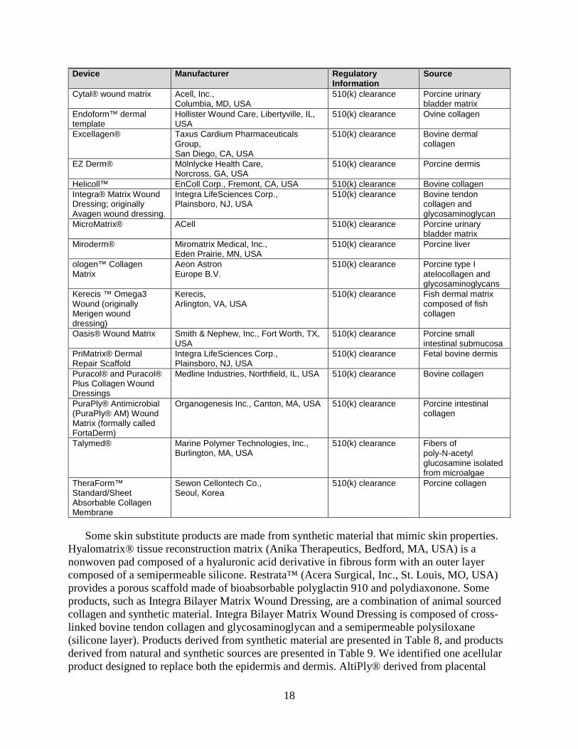
Some skin substitute products are made from synthetic material that mimic skin properties. Hyalomatrix® tissue reconstruction matrix (Anika Therapeutics, Bedford, MA, USA) is a nonwoven pad composed of a hyaluronic acid derivative in fibrous form with an outer layer composed of a semipermeable silicone. Restrata™ (Acera Surgical, Inc., St. Louis, MO, USA) provides a porous scaffold made of bioabsorbable polyglactin 910 and polydiaxonone. Some products, such as Integra Bilayer Matrix Wound Dressing, are a combination of animal sourced collagen and synthetic material. Integra Bilayer Matrix Wound Dressing is composed of cross-linked bovine tendon collagen and glycosaminoglycan and a semipermeable polysiloxane (silicone layer). Products derived from synthetic material are presented in Table 8, and products derived from natural and synthetic sources are presented in Table 9. We identified one acellular product designed to replace both the epidermis and dermis. AltiPly® derived from placental

19
membranes maintains the outer basement membrane and an epithelial layer scaffold to promote reepithelialization (see Table 10).
Table 8. Acellular/Dermal replacement from synthetic materials Device Manufacturer Regulatory Information Hyalomatrix® tissue reconstruction matrix
Anika Therapeutics, Bedford, MA, USA 510(k) clearance
Restrata™ Acera Surgical Inc., St. Louis, MO, USA 510(k) clearance
Table 9. Acellular/Dermal replacement from combined natural and synthetic materials Device Manufacturer Regulatory
Information Source
Integra® Bilayer Matrix Wound Dressing
Integra LifeSciences Corp., Plainsboro, NJ, USA
510(k) clearance Cross-linked bovine tendon collagen and glycosaminoglycan and a semi-permeable polysiloxane (silicone layer).
Integra Dermal Regeneration Template and Integra Omnigraft Regeneration Template
Integra LifeSciences Corp., Plainsboro, NJ, USA
Premarket approval process
Cross-linked bovine tendon collagen and glycosaminoglycan and a semi-permeable polysiloxane (silicone layer).
Integra Flowable Wound Matrix
Integra LifeSciences Corp., Plainsboro, NJ, USA
510(k) clearance Granulated cross-linked bovine tendon collagen and glycosaminoglycan and a semi-permeable polysiloxane (silicone layer).
Table 10. Acellular/Epidermal and Dermal replacement from human amniotic membrane Device Manufacturer Regulatory Information AltiPly® Aziyo Biologics, Silver Spring, MD, USA HCT/P
Cellular Skin Substitutes Our examination of the commercially available skin substitute products found only seven
contain cells. Four amniotic membrane-derived products claim to have viable cells: Affinity human amniotic allograft, FlōGraft amniotic fluid-derived allograft, Grafix, and GrafixPL Prime (Table 11). Dermagraft (Table 12) is a human fibroblast-derived dermal substitute. Fibroblast cells from human foreskin are seeded onto a bioabsorbable polyglactin mesh scaffold. They proliferate and secrete cytokines to form a metabolically active dermal substitute. Dermagraft has been approved for treating diabetic foot ulcers greater than 6-weeks duration.3,20 Theraskin (Table 13) is a cryopreserved human, living, split-thickness allograft that contains fibroblasts and keratinocytes. The tissue is harvested within 24-hours postmortem from an organ donor. When harvested, the allograft is washed with antibiotics and cryopreserved. According to the manufacturer, living cells survive through harvesting, cryopreservation, and thawing.30 FDA regulates Theraskin as human tissue for transplantation. Apligraf (Table 14) is a bioengineered skin substitute with two layers.3,20 The dermal layer is type I bovine collagen gel seeded with living human neonatal fibroblasts. The epidermis is neonatal keratinocytes. The cells actively

20
secrete growth factors, cytokines, and ECM proteins. Apligraf is approved for treating diabetic foot ulcers and venous leg ulcers.
Table 11. Cellular/Dermal replacement from human amniotic membrane Device Manufacturer Regulatory Information Affinity® Human Amniotic Allograft
Organogenesis, Inc., Canton, MA, USA HCT/P
FlōGraft® Amniotic Fluid-Derived Allograft
Applied Biologics, Scottsdale, AZ, USA HCT/P
Grafix® Osiris Therapeutics, Inc., Columbia, MD, USA HCT/P GrafixPL Prime Osiris Therapeutics HCT/P
Table 12. Cellular/Dermal replacement from combined natural and synthetic materials Device Manufacturer Regulatory Information Dermagraft® Organogenesis, Inc., Canton, MA, USA Premarket approval process
Table 13. Cellular/Epidermal and Dermal replacement from human cadaver skin Device Manufacturer Regulatory Information Theraskin® LifeNet Health, Virginia Beach, VA, USA (procurement and
processing) Solsys Medical, Newport News, VA, USA (distribution)
HCT/P
Table 14. Cellular/Epidermal and Dermal replacement from combined human and animal sources Device Manufacturer Regulatory Information Apligraf® Organogenesis, Inc., Canton, MA, USA Premarket approval process
Guiding Question 2 Overview The skin substitute classification system proposed by Davison-Kolter et al.22 emphasizes
cellularity as the primary discriminator to group these products. Products are divided into groups that contain cells (cellular) and those that do not (acellular) followed by the region of skin being replaced (epidermal, dermal, or both) and the source of the material used to create the product (natural, synthetic, or both). Figure 1 depicts the acellular portion of the Davison-Kolter et al. classification pathway. The cellular pathway is identical. We divided acellular and cellular skin substitute products according to whether they replaced just the dermis or the dermis and epidermis. No skin substitute products replace only the epidermis. We then grouped products according to their source (natural human, natural animal, and synthetic). We split Davison-Kolter’s natural source group into natural human and natural animal. Using this modification to the Davison-Kotler et al. classification scheme, we identified human cadaver dermis (13 products), human amniotic membranes (26 products), animal tissue sources (22 products), synthetic sources (2 products), and a combination of natural and synthetic materials (3 products) as acellular dermal substitutes. One product was an acellular replacement for both epidermis and dermis.
Only seven products contained cells and would be considered in the cellular pathway of the Davison-Kotler et al. classification. Four amniotic membrane-derived products claim to have viable cells. The other three are Dermagraft (four amniotic membrane-derived products claim to have viable cells), Theraskin (cryopreserved human, living, split-thickness allograft), and Apligraf (bioengineered skin substitute with neonatal keratinocyte epidermis and a type I bovine collagen dermis).

21
Figure 1. Acellular portion of algorithm adapted from Davison-Kolter et al. Skin Substitute Classification System*
DERMALEPIDERMAL EPIDERMAL AND DERMAL
NATURAL SYNTHETIC BOTH
TEMPORARYTEMPORARY
OR PERMANENT
TEMPORARYOR
PERMANENT
NATURAL SYNTHETIC BOTH
TEMPORARYTEMPORARY
OR PERMANENT
TEMPORARYOR
PERMANENTTEMPORARY
TEMPORARYOR
PERMANENT
TEMPORARYOR
PERMANENT
NATURAL SYNTHETIC BOTH
ACELLULAR PRODUCTS
SINGLE LAYER BILAYER
* The pathway for cellular products is identical. Adapted from a figure from Davison-Kotler E, Sharma V, Kang NV, et al. A universal classification system of skin substitutes inspired by factorial design. Tissue Eng Part B Rev. 2018.22 Permission to use this copyrighted material was granted by Mary Ann Liebert, Inc. publishers.

22
Guiding Question 3: What are the study design characteristics (such as those listed below) in each included investigation for each chronic wound type?)
a. Comparator to skin substitute b. Inclusion/exclusion criteria of patients, including at least age, gender, and general health
requirements (e.g., status of HbA1c, diabetes, peripheral vascular disease, obesity, smoking, renal)
c. Inclusion/exclusion criteria of wounds including at least wound type, wound size/depth/duration/severity, vascular status, infection status, and prior treatment requirements (e.g., no treatment with growth factors or negative pressure wound therapy)
d. Patient characteristics of enrollees including at least age, gender, general health (e.g., status of HbA1c, diabetes, peripheral vascular disease, obesity, smoking, renal), and prior and concurrent wound treatments
e. Wound characteristics of enrollees including at least wound type, wound size/depth/duration/severity, vascular status, and infection status
f. Basic study design and conduct information including at least method of patient enrollment, care setting, and use of run-in period
g. Definition of wound characteristics: definition of “failure to heal”, and definition of a successfully healed wound
h. Method of applying skin substitutes including provider, frequency of application, definition of standard of care, and handling of infections
i. Measurement and assessment methods including method of assessment(s); frequency and time points for assessment(s); and blinding of assessors
j. Statistical methods including power calculations, intent-to-treat analysis for studies designed to test superiority, and handling of drop-outs
Our search of the published literature identified 148 potentially relevant studies. We excluded 22 articles at title screening for not being relevant to skin substitutes or chronic wound healing. Of the 126 remaining articles, we excluded 73 articles at the abstract level for reasons including not addressing a guiding question, not a study design of interest (e.g., retrospective comparative), and study protocol. Of the 53 remaining articles, we excluded 33 studies at the full-text level. Studies were excluded for including products not regulated by FDA, not being available in the United States, and duplicate studies or duplicate reporting of patients. See Appendix B for a list of studies organized by reason for exclusion. See Figure 2 for a PRISMA flow diagram of our study screening.

23
Figure 2. PRISMA flow diagram of study screening
Studies identified through database searches(n=148)
Studies screened at abstract level(n=126)
Full-text studies assessed(n=53)
20 clinical studies (3 systematic reviews,
17 randomized controlled trials)
Studies excluded at title level (n=22)
Studies excluded at abstract level(n=73)
Studies excluded at full-text level (n=33)
• Not an intervention of interest (n=9)• Primary studies published before 2012 (n=8)• Duplicate study or duplicate reporting of
patients (n=5)• Not a study of interest (n=5)• Not a study design of interest (n=2)• Not a comparator of interest (n=2)• No outcomes of interest (n=1)• Included in 2012 AHRQ report Skin Substitutes
for Treating Chronic Wounds (n=1)

24
Key Points • Of the 74 commercially available skin substitutes, three systematic reviews and 17 RCTs
examined use of 13 distinct skin substitutes, including acellular dermal substitutes, cellular dermal substitutes, and cellular epidermal and dermal substitutes in diabetic foot ulcers and venous leg ulcers.
• Three systematic reviews examined the use of amniotic membranes and acellular dermal matrices (ADMs) in diabetic foot ulcers. Thirteen primary studies examined nine distinct skin substitutes. Most studies enrolled fewer than 25 patients per arm and measured outcomes up to 16 weeks.
• Seventeen RCTs examined 13 distinct skin substitutes (4 skin substitutes not examined in the systematic reviews) in diabetic foot ulcers (12 studies) and venous leg ulcers (5 studies). Comparators were standard of care (11 studies) and another FDA-regulated skin substitute (6 studies).
• Of the 13 distinct skin substitutes examined in 17 RCTs, six skin substitutes were examined in more than one study. Two skin substitutes (Dermagraft,31-34 EpiFix35-38) were examined in four studies each. Four skin substitutes (Grafix/GrafixPrime,32,39 MatriStem Wound Matrix/MatriStem Micromatrix,34,40 Apligraf,30,38 Theraskin30,33) were examined in two studies each.
• Eligibility criteria in 17 RCTs was most commonly reported as a noninfected debrided wound of at least 4 weeks, with a wound size of 1 cm2 to 25 cm2. Conditions such as uncontrolled diabetes (HbA1c >12%), morbid obesity, peripheral vascular disease, severe malnutrition, severe liver, and severe renal disease were excluded.
• Eighty-two percent of studies enrolled fewer than 60 patients per arm. All studies were manufacturer-funded, and most studies were conducted in U.S. wound care centers.
• Our risk-of-bias analysis indicated that 47 percent and 64 percent of included studies had more than a 15 percent difference between study arms in baseline mean wound size (range up to 53.5 cm2) and baseline mean wound duration (range up to 479 weeks).
• Successful wound closure was mostly described as 100 percent reepithelialization without drainage or dressing. KIs suggested that 40 percent to 50 percent wound closure in 4 weeks was a good predictor of successful wound closure.
We identified three systematic reviews41-43 and 17 RCTs30-40,44-49 that addressed Guiding Question 3. Diabetic foot ulcers were examined in all 3 reviews and 12 RCTs,32-34,36,38-40,44-48 while venous leg ulcers were examined in 5 RCTs.30,31,35,37,49 We did not identify any relevant studies examining skin substitutes in pressure ulcers or arterial leg ulcers. We also did not identify any studies examining skin substitutes classified under the modified Davison-Kolter system22 as acellular epidermal, acellular epidermal and dermal, and cellular epidermal. We present below study design characteristics of all included studies.
Systematic Reviews Three systematic reviews examined the use of skin substitutes in diabetic foot ulcers.41-43
Two reviews examined amniotic membranes,41,43 while one examined ADM.42 Ten studies examined acellular dermal substitutes versus standard of care, one study examined two acellular dermal substitutes, and two studies examined an acellular dermal substitute versus a cellular epidermal and dermal substitute. Skin substitutes examined in these reviews included AlloPatch Pliable, Amnioband®, AmnioExcel®, Apligraf®, DermACELL®, EpiFix®, Grafix®,

25
GraftJacket®, and Integra® Dermal Regeneration Template. See Table 15 for additional details on the primary studies included in these reviews.
Paggiaro et al. 201841 included six RCTs published from 2013 to 2017 and conducted in the United States. One RCT each evaluated Grafix, Amnioband, EpiFix, and AmnioExcel. One study examined weekly versus biweekly EpiFix, while one three-arm RCT examined EpiFix, Apligraf, and standard of care. Enrollment ranged from 25 to 100 patients; 66 percent of studies enrolled fewer than 25 patients per arm. Followup was 6 weeks and 12 weeks. Standard of care was described as alginate or collagen alginate.
Haugh et al. 201743 meta-analyzed studies comparing commercially available amniotic tissue products with standard wound care in RCTs. Five RCTs analyzed results from 259 patients after excluding 52 patients also treated with a bioengineered skin substitute (Apligraf). Four studies analyzed dehydrated amniotic products (EpiFix and AmnioExcel), while one study analyzed a cryopreserved amniotic product (Grafix). Standard of care described for three studies included debridement and moist wound therapy or nonadherent dressings. Two studies used offloading, and only one study included infection surveillance or compression dressings. Enrollment and followup were similar to those in the Paggiaro et al. 2018 review.41
Guo et al. 201742 included six RCTs published from 2004 to 2015 that compared ADM with standard of care in 632 patients. ADMs were human-derived in five studies and animal-derived in one study. One study each evaluated AlloPatch Pliable and Integra Dermal Regeneration Template. Three studies examined GraftJacket, while one study examined GraftJacket and DermACELL. Standard of care was described as including sharp debridement, glucose control, infection control, offloading, and daily dressing change. Dressings were described as alginate, advanced moist therapy, 0.9 percent sodium chloride/gel/foam/gauze, alginate/hydrocolloids/ hydrogel/foam, and wound gel with gauze dressings (2 studies). Enrollment ranged from 28 to 307 patients; 50 percent of studies enrolled fewer than 25 patients per arm. Followup ranged from 4 weeks to 16 weeks. Additional information on study design characteristics is provided in Table C-1 in Appendix C.
Table 15. Primary studies included in systematic reviews Randomized Controlled Trial
Skin Substitutes Examined
Category Systematic Review
Paggiaro et al. 201841
Systematic Review
Guo et al. 201742
Systematic Review
Haugh et al. 201743
DiDomenico et al. 2016
Amnioband vs. SOC Acellular dermal X
Snyder et al. 2016 AmnioExcel® vs. SOC Acellular dermal X X Walters et al. 2016 DermACELL® vs.
GraftJacket® Regenerative Tissue Matrix vs. SOC
Acellular dermal vs. Acellular dermal
X
Zelen et al. 2016 EpiFix vs. Apligraf vs. SOC
Acellular dermal vs. Cellular epidermal and dermal
X X
Zelen et al. 2016 AlloPatch Pliable vs. SOC Acellular dermal X Lavery et al. 2014 Grafix vs. SOC Acellular dermal X X Driver et al. 2015 Integra® Dermal
Regeneration Template vs. SOC
Acellular dermal X
Zelen et al. 2015 EpiFix vs. Apligraf vs. SOC
Acellular dermal vs. Cellular epidermal and dermal
X

26
Randomized Controlled Trial
Skin Substitutes Examined
Category Systematic Review
Paggiaro et al. 201841
Systematic Review
Guo et al. 201742
Systematic Review
Haugh et al. 201743
Zelen et al. 2014 EpiFix (biweekly vs. weekly)
Acellular dermal X
Zelen et al. 2013 EpiFix vs. SOC Acellular dermal X X Reyzelman et al. 2009
GraftJacket vs. SOC Acellular dermal X
Brigido et al. 2006 GraftJacket vs. SOC Acellular dermal X Brigido et al. 2004 GraftJacket vs. SOC Acellular dermal X
SOC = standard of care
Primary Studies Study design characteristics for the 17 primary studies were grouped by the modified
Davison-Kolter classification system. Patient enrollment criteria, patient characteristics, and basic study design characteristics are summarized in Table C-3 to Table C-17 in Appendix C. Details on wound closure assessments, definitions of failure to heal, and details on all wound treatments (including standard of care) are described in Table C-18 to Table C-20 in Appendix C. Standard of care in these studies was described as including sharp debridement, glucose control, compression bandages for venous leg ulcers, infection control, offloading, and daily dressing changes with a moisture-retentive dressing such as an alginate or hydrocolloid. Further information on the skin substitutes is presented in Appendix D.
Of the 17 RCTs, 11 studies compared standard of care with 9 unique FDA-regulated skin substitutes (see Table 16) plus similar standard of care.31,35-37,39,40,44-47,49 Ten studies examined acellular dermal substitutes including Allopatch HD® Acellular Dermal Matrix, Amnioband, AmnioExcel, EpiFix (3 studies), Grafix, Hyalomatrix® Wound Matrix, Integra Dermal Regeneration Template, and MatriStem® Wound Matrix.35-37,39,40,44-47,49 One study examined a cellular dermal substitute (Dermagraft).31
Table 16. Skin substitutes compared with standard of care in 11 RCTs Skin Substitute Category Study Study
Comparator(s) Wound Type
Allopatch HD® Acellular Dermal Matrix
Acellular dermal Zelen et al. 201844 SOC DFU
AmnioBand® Allograft Placental Matrix
Acellular dermal DiDomenico et al. 201645 SOC DFU
AmnioExcel® Acellular dermal Snyder et al. 201646 SOC DFU Dermagraft® Cellular dermal Harding et al. 201331 SOC VLU EpiFix® Acellular dermal Bianchi et al. 201835 SOC VLU EpiFix Acellular dermal Zelen et al. 201336 SOC DFU EpiFix Acellular dermal Serena et al. 201437 SOC VLU Grafix® Acellular dermal Lavery et al. 201439 SOC DFU Hyalomatrix® Wound Matrix Acellular dermal Alvarez et al. 201749 SOC VLU Integra® Dermal Regeneration Template
Acellular dermal Driver et al. 201547 SOC DFU
MatriStem® Wound Matrix* Acellular dermal Alvarez et al. 201740 SOC DFU * Now marketed as Cytal® Wound Matrix DFU = diabetic foot ulcer; SOC = standard of care; VLU = venous leg ulcer
The remaining six RCTs compared one FDA-regulated skin substitute with another FDA-regulated skin substitute.30,32-34,38,48 Five additional unique skin substitutes (Apligraf, DermACELL, GrafixPrime®, GraftJacket Regenerative Tissue Matrix, and Theraskin) were examined.

27
One three-arm study compared standard of care with two acellular dermal substitutes (DermACELL, GraftJacket).48 Two studies compared an acellular dermal substitute (MatriStem Wound Matrix and MatriStem Micromatrix, GrafixPrime) with a cellular dermal substitute (Dermagraft).32,34 One study compared an acellular dermal substitute with a cellular epidermal and dermal substitute (EpiFix versus Apligraf).38 One study compared a cellular dermal substitute with a cellular epidermal and dermal substitute (Dermagraft versus Theraskin).33 Lastly, one study compared two cellular epidermal and dermal substitutes (Apligraf versus Theraskin).30 See Table 17 for a list of head-to-head comparative studies.
Table 17. Skin substitutes examined in 6 head-to-head comparative studies Skin Substitutes Category Study Wound Type GrafixPrime® vs. Dermagraft® Acellular dermal vs. Cellular dermal Ananian et al. 201832 DFU Apligraf® vs. Theraskin® Cellular epidermal and dermal vs.
Cellular epidermal and dermal Towler et al. 201830 VLU
DermACELL® vs. GraftJacket® Regenerative Tissue Matrix* vs. SOC
Acellular dermal vs. Acellular dermal Cazzell et al. 201748 DFU
Dermagraft vs. Theraskin Cellular dermal vs. Cellular epidermal and dermal
Sanders et al. 201433 DFU
MatriStem® Micromatrix and MatriStem Wound Matrix** vs. Dermagraft
Acellular dermal vs. Cellular dermal Frykberg et al. 201634 DFU
EpiFix vs. Apligraf Acellular dermal vs. Cellular epidermal and dermal
Zelen et al. 201638 DFU
*Now GraftJacket™ RTM ** Now marketed as Cytal® Wound Matrix DFU = diabetic foot ulcer; SOC = standard of care; VLU = venous leg ulcer
Acellular Dermal Substitutes versus Standard of Care Ten studies compared standard of care with acellular dermal substitutes, including
Allopatch HD Acellular Dermal Matrix, Amnioband, AmnioExcel, EpiFix (3 studies), Grafix, Hyalomatrix Wound Matrix, Integra Dermal Regeneration Template, and MatriStem Wound Matrix.35-37,39,40,44-47,49 Patient enrollment criteria, patient characteristics, and basic study design characteristics are summarized in Table C-3 to Table C-5 in Appendix C.
Eligible patients in these studies were required to have adequate circulation to the wound (6 studies) and no infection (9 studies). Age eligibility was ≥18 years (7 studies), 18 to 80 years (1 study), and 18 to 85 years (1 study). HbA1c was required to be less than 12 percent (5 studies) and less than 10 percent (1 study) in studies reporting. Eligible wounds were classified as Wagner 1 or 246,47 or Grade I-A Texas40 (based on the University of Texas Wound Classification System)40 (for more information on these classification systems, see the article by Clayton and Elasy50). Six different criteria were used for wound surface requirements. The most commonly reported was >1 cm2 to <25 cm2. Minimum wound duration was 4 weeks (6 studies), 8 weeks (1 study), and 4 to 52 weeks (1 study).
Studies excluded patients with New York Heart Association Class III and IV chronic heart failure,37 active or unstable Charcot foot,46,47 and wounds that decreased by more than 20 percent or 30 percent in area during the screening period.44-47 One study did not report inclusion/exclusion criteria.
Enrollment in each study arm was fewer than 60 patients (9 studies) or greater than 150 patients (1 study). Mean age was 58 years in both arms (range, 55 to 62 years). Percent male ranged from 45 to 80 in the acellular dermal arm and 37 to 92 in the standard of care arm. Mean wound size ranged from 2 cm2 to 48 cm2 in the acellular dermal arm and 2.7 cm2 to 53.5 cm2 in

28
the standard of care arm. Four (40%) studies had more than a 15 percent difference in mean wound size between arms. Mean wound duration ranged from 6.5 weeks to 41.0 weeks in the acellular dermal arm and 4.8 weeks to 58 weeks in the standard of care arm. Six (60%) studies had more than a 15 percent difference in mean wound duration between arms. Wound severity was rated as Grade I-A (University of Texas Wound Classification System),40 Wagner 1 or 2,46 and 70 percent to 75 percent Wagner 247 in 3 studies. Enrolled patients were described as having type 1 or type 2 diabetes (8 studies), obesity (7 studies), and as tobacco and alcohol users (3 studies). One study enrolled individuals with Charcot foot and partial amputation.40 Five (50%) studies had more than a 15 percent difference in number of comorbidities reported at the start of treatment. All studies were conducted in the United States; 60 percent were conducted in outpatient wound care centers. Research institutes and academic and private practices were other care settings. Seven (70%) studies used a run-in period (6 used a 2-week run-in period; 1 study used a 1-week run-in period). Most common study lengths were 12 weeks and 16 weeks. All studies were manufacturer-funded.
Cellular Dermal Substitutes versus Standard of Care One study addressed this comparison. Harding et al. 201331 randomly allocated patients with
venous leg ulcers to Dermagraft plus four-layer compression therapy (n=186) or four-layer compression therapy (n=180). Patients were required to have sufficient circulation to the study leg to make wound healing possible. Ulcers that reduced in size (cm2) by less than 50 percent while under compression therapy during the study’s 2-week screening period were eligible. Patients with morbid obesity, severe peripheral vascular disease/renal disease, or uncontrolled diabetes were excluded. Mean age was approximately 68 years; 46 percent were male. Median wound size was over 7 cm2 (range 2.3 to 28.2) with median wound duration 45 to 50 weeks (range 8.9 to 470.4). The study was conducted in 25 hospital and community-based venous leg ulcer clinics in the United Kingdom (19 centers), Canada (1 center), and United States (1 center) and had a 2-week screening period. This study was manufacturer-funded. See additional information on patient enrollment criteria, patient characteristics, and basic study design characteristics in Table C-6 to Table C-8 in Appendix C.
Acellular Dermal Substitutes versus Acellular Dermal Substitutes One 3-arm study addressing this comparison (Cazzell et al. 201748) randomly allocated
patients to DermACELL (n=71), GraftJacket (n=28), or standard of care (n=69). Patients were required to have adequate circulation to the affected area and a noninfected single-target diabetic foot ulcer with a Wagner Ulcer Classification of 1 or 2. Patients with presence of peripheral vascular disease, Charcot’s disease, or HbA1c >12 percent within 90 days of screening were excluded. Age was limited to individuals between 20 years and 80 years. Mean age was mid-50s, and the majority were males. Mean wound duration was 35 to 40 weeks, but ranged as high as 479 weeks. Wound severity was mostly Grade 2 Wagner. Besides having type 1 and type 2 diabetes mellitus, some individuals were also current smokers. The study was conducted in 13 outpatient wound care centers in 9 U.S. states and had a 30-day run-in period. This study was manufacturer-funded. See Table C-9 to Table C-11 in Appendix C.

29
Acellular Dermal Substitutes versus Cellular Dermal Substitutes and Cellular Epidermal and Dermal Substitutes
Three studies compared acellular dermal substitutes with a cellular dermal substitute32,34 or a cellular epidermal and dermal substitute38 in diabetic foot ulcers. Individuals in these studies were required to have clean, noninfected wounds with adequate circulation and HbA1c below 12 percent. Individuals with index ulcers that improved over 20 percent to 30 percent during the run-in period were excluded from all three studies. One study excluded severely malnourished patients. Age eligibility in one study was 18 years to 80 years.
Studies randomly allocated fewer than 40 patients to each study arm. Males accounted for more than 70 percent in 2 studies and less than 20 percent in 1 study. Mean wound size ranged from 1.7 cm2 to 7.15 cm2 in the intervention arm and from 1.7 cm2 to 5.7 cm2 in the standard of care arm. Two (66%) studies had more than a 15 percent difference in mean wound size. Mean wound duration was over 140 days in each arm32 and 263 days overall34 in studies reporting this outcome. Two (66%) studies had more than a 15 percent difference in mean wound duration; one study was undeterminable. Wound severity was mostly Grade A1 Texas in one study.34 Comorbidities included diabetes, obesity, smoking use, mild peripheral arterial disease, and heart disease, including chronic heart failure.
Studies were conducted in wound clinics, medical centers, Veterans Affairs medical facilities, research clinics, private practices, and hospital-based outpatient clinics in the United States. Run-in periods were 1 week, 2 weeks, and 4 weeks, respectively. Study lengths were 9 weeks, 12 weeks, and 6 months, respectively. Two studies reported manufacturer funding; one study did not report funding. See Table C-12 to Table C-14 in Appendix C.
Cellular Dermal Substitutes versus Cellular Epidermal and Dermal Substitutes
One study compared a cellular epidermal and dermal substitute with a cellular dermal substitute in diabetic foot ulcers.33 Eligible patients had noninfected ulcers, with HbA1c <12 percent, and wounds greater than 30-days duration.33 (See Table C-15 to Table C-17). Twelve patients was the maximum enrollment in any study arm. Mean age was 58 years. Mean wound size was 4.7 cm2 in the intervention arm and 5.4 cm2 in the standard of care arm. Mean wound duration was more than a 15 percent difference (43 weeks vs. 11 weeks). Comorbidities included diabetes, peripheral arterial disease, smoking use, and neuropathy. This 20-week study was conducted in a U.S. wound care center and was manufacturer-funded.
Cellular Epidermal and Dermal Substitutes versus Cellular Epidermal and Dermal Substitutes
One study compared two cellular epidermal and dermal substitutes in venous leg ulcers.30 Eligible patients had wounds greater than 30-days duration and area less than 40 cm2. Individuals with end-stage renal disease, severe malnutrition, or severe liver disease were excluded. Fifteen patients was the maximum enrollment in any study arm. Mean age was early 60s, with mostly males enrolled. Mean wound size was 6.3 cm2 in the intervention arm and 4.9 cm2 in the standard of care arm. Mean wound duration was not reported. Comorbidities included diabetes, obesity, peripheral vascular disease, smoking use, lymphedema, and neuropathy. This 20-week study used a 30-day run-in period, was conducted in a U.S. wound care center, and reported “no funding.” For additional details, see Table C-15 to Table C-17 in Appendix C.

30
Successfully Healed Wound Studies defined a successfully healed wound as 100 percent reepithelialization without
drainage or dressing required (8 studies),34,38,40,44-48 100 percent reepithelialization without drainage (6 studies),30,31,33,35,37,39 and 100 percent reepithelialization (2 studies).32,36 One study did not define a healed wound.49 KIs agreed that a completely healed wound must include no drainage or required dressing and 100 percent reepithelialization. Some KIs suggested that 40 percent to 50 percent wound closure in 4 weeks was a good predictor of a successful wound closure. See Table C-18 for details on assessing wound closure and primary outcomes.
Failure to Heal Failure to heal during the treatment phase was described as not achieving a reduction in area
by at least 40 percent35 and at least 50 percent.36,38,44,45 Failure to heal was not defined in 12 (70%) studies. See Table C-19.
Assessor Blinding Of the 17 primary studies, 8 reported blinding of outcome assessors, with 9 (53%) studies not
reporting assessor blinding.
Standard of Care Debridement was a component of standard of care for 16 (94%) primary studies (1 not
reporting). Offloading was an additional component in 11 (92%) studies examining diabetic foot ulcers, while multilayer compression was added to standard of care for all studies examining venous leg ulcers. Moist wound therapy was applied using alginate, foam, or hydrogel dressings. Four (23%) studies reported treatments for comorbidities and included infection management37,44,46 and infection and diabetes management.45 For details on all wound treatments, see Table C-20 in Appendix C.
Guiding Question 3 Overview Of the 74 commercially available skin substitutes, three systematic reviews and 17 RCTs
examined use of 13 distinct skin substitutes, including acellular dermal substitutes, cellular dermal substitutes, and cellular epidermal and dermal substitutes in diabetic foot ulcers and venous leg ulcers. Of these 13 distinct skin substitutes, six were examined in more than one study. Two skin substitutes (Dermagraft,31-34 EpiFix35-38) were examined in four studies each. Four skin substitutes (Grafix/GrafixPrime,32,39 MatriStem Wound Matrix/MatriStem Micromatrix,34,40 Apligraf,30,38 Theraskin30,33) were examined in two studies each. Standard of care was the most common comparator in the included studies, with few studies reporting infection surveillance and diabetic control as key components. Six RCTs compared an FDA-regulated skin substitute with another FDA-regulated skin substitute. Most studies enrolled fewer than 60 patients per arm, were manufacturer-funded, and conducted in U.S. wound care centers. Enrollees were required to have adequate circulation to noninfected debrided wounds and controlled diabetes if enrolled in a study examining diabetic foot ulcers. Successful wound closure was mostly described as 100 percent reepithelialization without drainage or dressing. KIs suggested that 40 percent to 50 percent wound closure in 4 weeks was a good predictor of successful wound closure.

31
Guiding Question 4: What are the outcomes of treatment strategies, including skin substitutes alone and/or in addition to other wound care modalities compared to other wound care modalities in patients with different types of chronic wounds, for patient oriented outcomes such as the following? Consider at least:
a. Number/percentage of completely closed/healed wounds (skin closure with complete reepithelialization without drainage or dressing requirements versus failure to heal)
b. Time to complete wound closure c. Wound recurrence (reoccurrence) (include time when initial wound healing was
measured, and followup to assess durability of healed wounds) d. Wound infection e. Need for amputation f. Need for hospitalization (frequency and duration) g. Return to baseline activities of daily living and function h. Pain reduction i. Exudate and odor reduction j. Adverse effects (besides those above)
Key Points • Acellular dermal substitutes versus standard of care:
o Three systematic reviews reported more than a 2-fold increased risk for complete healing of diabetic foot ulcers with AlloPatch Pliable, Amnioband, AmnioExcel, DermACELL, EpiFix, Grafix GraftJacket, and Integra Dermal Regeneration Template versus standard of care. Two reviews also reported a shorter time to heal favoring AlloPatch Pliable, Amnioband, Grafix, and GraftJacket over standard of care.41,42 None of the reviews reported an overall risk-of-bias rating for included studies.
o Nine (90%) RCTs comparing acellular dermal substitutes with standard of care reported statistically significant findings up to 16 weeks favoring the interventions for complete wound closure35,36,39,40,44-47 and shorter time to heal35,36,39,40,44-46,49 in diabetic foot ulcers36,39,40,44-47 and venous leg ulcers.35,49 Three studies rated wound severity as Grade I-A (University of Texas Wound Classification System),40 Wagner 1 or 2,46 and mostly Wagner 2.47 The most commonly reported enrollment criteria included >1 cm2 to <25 cm2 wound surface, >4-weeks duration, ankle brachial index (ABI) 0.7 to ≤1.2, and HbA1c <12 percent. Severe adverse events occurring with acellular dermal substitutes included diabetic foot infections, cellulitis, and osteomyelitis. Four (40%) studies reported less-frequent recurrence with a skin substitute.
• Cellular dermal substitutes versus standard of care: o Significant differences were reported for Dermagraft plus Profore™ four-layer
compression over four-layer compression in venous leg ulcers closed at 12 weeks in a subgroup of patients with ulcer duration ≤12 months. Recurrence was lower

32
with Dermagraft. Enrollment criteria included a wound surface <25 cm2, wound duration <5 years, ABI 0.8 to 1.2, and no morbid obesity.31
• Acellular dermal substitutes versus acellular dermal substitutes: o Results comparing DermACell versus GraftJacket in diabetic foot ulcers were not
provided after a three-arm study intentionally underpowered the GraftJacket arm. Individuals had mostly Wagner Grade 2 ulcers, with ABI ranging from 0.8 to 1.2, and HbA1c <12 percent.48
• Acellular dermal substitutes versus cellular dermal substitutes and cellular epidermal and dermal substitutes:
o GrafixPrime provided significant benefit over Dermagraft for diabetic foot wounds ≤5 cm2 healed at 8 weeks. Enrollees had a wound area <15 cm2, a wound duration <52 weeks, and an ABI between 0.7 and 1.3.32
o MatriStem and Dermagraft provided similar benefit for diabetic foot ulcers healed up to 10 weeks, time to closure, change in wound size, and 6-month recurrence. Ulcers were mostly Grade A1 (University of Texas Wound Classification System), and enrollees were required to have wounds ≥4-weeks duration, with an ABI ≥0.7.34
o EpiFix provided significant benefit over Apligraf in number of diabetic foot ulcers healed and time to heal at 12 weeks. Individuals had wounds <25 cm2, and ≥4 weeks duration, an ABI between 0.7 and 1.2, and HbA1c <12 percent.38
• Cellular dermal substitutes versus cellular epidermal and dermal substitutes: o Statistically significant benefits to Theraskin over Dermagraft in diabetic foot
ulcers at 12 weeks included more wounds healed in a shorter time with fewer grafts. No difference in wound healing was reported at 20 weeks. Patients had wounds <10 cm2, >30 days duration, with HbA1c <12 percent.33
• Cellular epidermal and dermal substitutes versus cellular epidermal and dermal substitutes:
o No statistically significant difference was reported between Apligraf and Theraskin for venous leg ulcer healing (at 12 and 20 weeks) and number of grafts per subject. Recurrence did not occur at 26 weeks. Eligible patients had wounds greater than 30-days duration and area less than 40 cm2.30
We now present an overview of the findings and a risk-of-bias assessment of the three systematic reviews and 17 primary studies included in the report. These studies examined use of 13 distinct skin substitutes (17% of 74 commercially available skin substitutes), including acellular dermal substitutes, cellular dermal substitutes, and cellular epidermal and dermal substitutes in diabetic foot ulcers and venous leg ulcers. We provide details on all the clinical evidence in Appendix C and summarize findings for each primary study in Table 18 and Table 19.
Systematic Reviews The two systematic reviews on amniotic membranes (Paggiaro et al. 201841 and Haugh et al.
201743) reported complete wound healing41,43 and mean time to complete wound healing41 in 11 RCTs. Four studies were included in both reviews.
Paggiaro et al. 201841 reported treatment with amniotic membranes versus standard of care resulted in a significant increase in wound healing (risk ratio 2.77, 95% CI: 1.76 to 4.36) in a
33
significantly shorter time (mean difference -32.28 days, 95% CI: -41.05 to -23.71). Statistical heterogeneity was low to moderate, and not further explored given the small number of studies. The authors noted that use of amniotic membranes resulted in more diabetic foot ulcers being healed at a quicker rate.The authors noted that use of amniotic membranes resulted in more diabetic foot ulcers being healed at a quicker rate. Haugh et al. 201743 reported a similar difference in complete wound healing favoring the intervention (risk ratio 2.75, 95% CI: 2.06 to 3.66; p<0.001). Statistical heterogeneity was moderate and not explored further. The authors noted that despite results indicating the treatment of diabetic foot ulcers with amniotic membrane improves healing rates, further studies are needed to determine whether these products also decrease the incidence of subsequent complications, such as amputations or death.
Findings in Guo et al. 201742 indicated a 2.31 and 1.57 significant increased relative risk of complete wound healing at 12 weeks and 16 weeks, respectively, favoring ADM versus standard of care. Mean time to complete wound healing was significantly shorter with ADM (mean difference -2.98 weeks, 95% CI: -5.15 to -0.82; p=0.007). Statistical heterogeneity was significant for the outcomes complete wound healing at 12 weeks (6 studies) and time to heal (4 studies). For complete wound healing, the authors noted that moderate heterogeneity remained after removing one study measuring the healing rate in the first four weeks. For time to heal, one study was noted as having overly influenced the heterogeneity. Risk of adverse events was not significantly different. The authors concluded that “compared with standard of care, acellular dermal matrix may accelerate the healing velocity of uninfected, non-ischemic, full-thickness diabetic foot ulcer… while generating no more complications.” For additional data for these reviews, see Table C-1 in Appendix C.
None of the reviews reported an overall risk-of-bias rating for included studies. Two reviews described lack of allocation concealment (selection bias), lack of blinding assessors (detection bias), incomplete outcome data (attrition bias), and other bias (not described) as study limitations.41,42 These reviews used the Cochrane Handbook for systematic reviews of interventions for their risk-of-bias assessment while our risk-of-bias assessment tool used for individual studies (see Methods) mostly focused on wound-related outcomes (e.g., reporting of recurrence, similar wound size and duration in study arms). Haugh et al. 201743 assessed risk of bias based on guidelines proposed by the Meta-Analysis of Observational Studies in Epidemiology Collaboration51 but did not report findings. For details of the risk-of-bias assessments, see Table C-2. in Appendix C.
Primary Studies We briefly summarize below the findings for the 17 RCTs addressing this guiding question.
Summaries are categorized by the modified Davison-Kolter classification system22 as in Guiding Question 3. See Table 18 for an overview of findings and risk-of-bias rating for 11 studies addressing standard of care versus an acellular dermal substitute or cellular dermal substitute. See Table 19 for an overview of findings and risk-of-bias rating for six head-to-head comparisons comparing acellular dermal substitutes, cellular dermal substitutes, and cellular epidermal and dermal substitutes.
For further information on the clinical results, see Table C-21 to Table C-30 in Appendix C. For details on all wound treatments (including standard of care), see Table C-20 in Appendix C. Table C-31 summarizes risk-of-bias assessments.

34
Acellular Dermal Substitutes versus Standard of Care Of the 10 studies addressing this comparison, 7 studies reported statistically significant
differences in number of wounds healed and time to heal favoring the intervention over standard of care.35,36,39,40,44-46 Latest followup was 6 weeks (2 studies),36,46 12 weeks (4 studies),39,40,44,45 and 16 weeks (1 study).35 Skin substitutes examined in these studies included AlloPatch Pliable,44 Amnioband,45 AmnioExcel,46 EpiFix (2 studies),35,36 Grafix,39 and Matristem Wound Matrix.40 Six studies evaluated effectiveness in diabetic foot ulcers,36,39,40,44-46 while one study evaluated venous leg ulcers.35 Standard of care included debridement and offloading for all studies evaluating diabetic foot ulcers. Standard of care for one study evaluating venous leg ulcers included a standard moist wound dressing and multilayer compression therapy.35 Three studies included infection management44,46 and infection and diabetes management as standard of care.45
One study reported statistically significant differences in complete wound closure for diabetic foot ulcers (at 16 weeks) and body pain favoring Integra Dermal Regeneration Template over standard of care.47 Moist wound therapy consisting of 0.9 percent sodium chloride gel plus a secondary dressing, including an offloading/protective device, was reported as standard of care.
Two additional studies examining venous leg ulcers37,49 reported a significantly shorter time to heal with Hyalomatrix Wound Matrix plus compression49 and more frequent closure at 4 weeks with EpiFix plus multilayer compression therapy.37 Standard of care included multilayer compression therapy in both studies. Additional treatments included a nonadherent silicone foam dressing but no debridement49 and infection management.37
Four studies reported that recurrence occurred less frequently with application of a skin substitute than with standard of care.39,40,45,47
The most commonly reported enrollment criteria in these studied included >1 cm2 to <25 cm2 wound surface, >4-weeks duration, ABI 0.7 to ≤1.2, and HbA1c <12 percent.
One study reported nine severe adverse events occurred with EpiFix.35 Two studies reported patients receiving AlloPatch Pliable and Grafix were hospitalized with diabetic foot infections.39,44 Two studies reported cellulitis occurring with EpiFix and AmnioExcel.37,46 One study also reported wound infection and osteomyelitis with AmnioExcel.46 Two studies reported similar low adverse event rates.45,47 One study reported only overall adverse events (including cellulitis),40 while one study did not report adverse events.49 For additional information on clinical outcomes, see Table C-21 and Table C-22.
Cellular Dermal Substitutes versus Standard of Care One 24-week study (Harding et al. 2013)31 reported no significant differences between Dermagraft plus Profore four-layer compression therapy versus four-layer compression therapy except for venous leg ulcers healed at 12 weeks in a subgroup of patients with ulcer duration ≤12 months. Standard of care included a nonadherent dressing, with deeper ulcers also receiving gauze and heavily exuding ulcers receiving additional absorbent dressings. Recurrence was lower with Dermagraft (15% versus 23%), but venous ulcer pain was slightly higher (5.3% versus 5.0%). Safety was reported as comparable. Enrollment criteria included a wound surface <25 cm2, wound duration <5 years, ABI 0.8 to 1.2, and no morbid obesity. For additional information on clinical outcomes, see Table C-23 and Table C-24.

35
Table 18. Overview of 11 RCTs comparing skin substitutes with standard of care Skin Substitute
Category Study Wound Type
Overview Risk-of-bias Assessment
Allopatch HD® Acellular Dermal Matrix
Acellular dermal
Zelen et al. 201844
DFU Significant differences in DFUs closed (at 6 and 12 weeks) and time to wound closure at 12 weeks favored AlloPatch Pliable (n=40) over SOC (n=40). 8 (10%) patients were hospitalized with diabetic foot infections; 2 (2%) were treated with AlloPatch Pliable.
Moderate
AmnioBand® Allograft Placental Matrix
Acellular dermal
DiDomenico et al. 201645
DFU Statistically significant differences were reported in wound healing at 6 weeks (70% Amnioband, 15% SOC; p=0.001) and mean time to closure (at 6 and 12 weeks) favoring Amnioband (n=20) vs. SOC (n=20). 1 serious AE occurred in each arm; 2 (10%) DFUs reoccurred in SOC arm.
Moderate
AmnioExcel® Acellular dermal
Snyder et al. 201646
DFU Findings indicated a significant difference in wound closure of DFUs at 6 weeks with AmnioExcel (n=15) over SOC (n=14) (35% AmnioExcel, 0% SOC; p=0.0170) and a significantly shorter time to closure with AmnioExcel (p<0.0001). AmnioExcel-treated AEs included wound infection, osteomyelitis, and cellulitis in 1 patient each.
Moderate
Dermagraft® Cellular dermal
Harding et al. 201331
VLU No significant findings were reported between Dermagraft plus Profore™ compression therapy (n=186) vs, Profore compression therapy (n=180) for time to wound closure and recurrence. A subgroup analysis of patients with ulcer duration ≤12 months indicated a statistically significant benefit with Dermagraft plus Profore compression therapy for wounds healed at 12 weeks (p=0.029). Safety was reported as comparable.
Low
EpiFix® Acellular dermal
Bianchi et al. 201835
VLU Significant findings included a benefit to wound closure (at 12 and 16 weeks) and time to heal (log-rank P=0.011) using EpiFix plus compression (n=52) over standard of care (n=57). 9 severe adverse events were reported in the EpiFix arm.
Low
EpiFix Acellular dermal
Zelen et al. 201336
DFU Findings suggest a biweekly application of EpiFix (n=13) results in significantly more DFU healing at 6 weeks and at a 50% faster healing rate than SOC (n=12). Cellulitis occurred in 2 (16%) patients receiving SOC.
Moderate
EpiFix Acellular dermal
Serena et al. 201437
VLU Serena et al. 201437 reported more wound closure at 4 weeks with a human amnion/chorion membrane allograft (11.3% EpiFix plus MLCT (n=53) vs. 7.8% MLCT (n=51). 2 cases of cellulitis occurred in the EpiFix plus MLCT arm.
Low

36
Skin Substitute
Category Study Wound Type
Overview Risk-of-bias Assessment
Grafix® Acellular dermal
Lavery et al. 201439
DFU DFUs were 6 times more likely to completely heal with Grafix (n=50) vs. SOC (n=47) (OR 6.037, 95% CI: 2.449 to 14.882). Grafix arm had a significantly higher probability of complete wound healing (67.1% vs. 27.1%; Log-Rank, p<0.0001), faster median time to complete wound closure (42 days vs. 69.5 days; p=0.019) and fewer wound-related infections (18% vs. 36.25; p=0.044). No significant difference was reported for wound recurrence (17.8% vs. 30%; p=0.42) or hospitalizations related to infections (6% vs. 15%; p=0.15).
Low
Hyalomatrix® Wound Matrix
Acellular dermal
Alvarez et al. 201749
VLU No statistically significant differences were reported between Hyalomatrix Wound Matrix plus compression (n=9) vs. standard of care (n=7) for wound healing. Time to heal was significantly shorter with Hyalomatrix Wound Matrix plus compression (41 days vs. 104 days; p=0.029). AEs were not reported.
Moderate
Integra® Dermal Regeneration Template
Acellular dermal
Driver et al. 201547
DFU Significant findings were reported for complete wound closure (at 12 and 16 weeks) and body pain favoring Integra Dermal Regeneration Template (n=154) over SOC (n=153). No statistically significant differences were reported for median time to wound closure (43 days vs. 78 days) and wound recurrence at 28 weeks (19% IDRT vs. 26% SOC; p=0.32). AEs potentially study-related were “similar” (4.5% IDRT vs. 5.2% SOC).
Low
MatriStem® Wound Matrix*
Acellular dermal
Alvarez et al. 201740
DFU Significant differences were reported in wounds closed at 12 weeks (91% vs. 33%; p=0.041) and mean days to wound closure (62.4 vs. 92.8) favoring a urinary bladder matrix (n=11) over SOC (n=6). Recurrence was less frequent at 1 year with MatriStem Wound Matrix (10% vs. 50%). Overall AEs included local wound infection (n=6), dermatitis (n=4), and cellulitis (n=1).
Moderate
AE = adverse event; CI = confidence interval; DFU = diabetic foot ulcer; IDRT = Integra dermal regeneration template; OR = odds ratio; SOC = standard of care; VLU = venous leg ulcer
Acellular Dermal Substitutes versus Acellular Dermal Substitutes One three-arm study comparing two acellular dermal substitutes with standard of care
reported a significant difference in diabetic foot ulcers healed at 24 weeks favoring DermACELL over standard of care. The GraftJacket arm was intentionally underpowered since statistical significance was not sought or expected for this study arm. We did not include recurrence rates since data were missing for 48.5 percent of patients in the “per protocol population.” Serious treatment-related adverse events were reported as comparable.48 Individuals had mostly Wagner Grade 2 ulcers, with ABI ranging from 0.8 to 1.2 and HbA1c <12 percent. For additional information on clinical outcomes, see Table C-25 and Table C-26.

37
Acellular Dermal Substitutes versus Cellular Dermal Substitutes and Cellular Epidermal and Dermal Substitutes
Three studies compared acellular dermal substitutes with a cellular dermal substitute32,34or a cellular epidermal and dermal substitute38 in diabetic foot ulcers.
One study reported significant findings for GrafixPrime over Dermagraft for wounds ≤5 cm2 healed at 8 weeks (81.3% vs. 37.5%; p=0.0118). Enrollees had a wound area <15 cm2, a wound duration <52 weeks, and an ABI between 0.7 and 1.3.32
One study reported no statistically significant differences for all outcomes (including wounds healed up to 10 weeks, time to closure, change in wound size) between MatriStem and Dermagraft with similar 6-month recurrence. Ulcers were mostly Grade A1 University of Texas Wound Classification System, and enrollees were required to have wounds ≥4 weeks duration, with an ABI ≥0.7.34
Lastly, authors reported EpiFix was significantly favored over Apligraf for complete wounds healed and time to heal at 12 weeks. Individuals had wounds <25 cm2, ≥ 4-weeks duration, an ABI between 0.7 and 1.2, and HbA1c <12 percent.38
Overall adverse events were similar between MatriStem and Dermagraft.34 Osteomyelitis and cellulitis occurred in more patients receiving GrafixPrime than Dermagraft (13.1% versus 5.4%),32 and five wound/foot infections were reported using EpiFix or Apligraf.38 For additional information on clinical outcomes, see Table C-27 and Table C-28.
Cellular Dermal Substitutes versus Cellular Epidermal and Dermal Substitutes
Statistically significant benefits to Theraskin over Dermagraft at 12 weeks included more diabetic foot ulcers healed, a shorter time to wound closure, and fewer grafts needed. At 20 weeks, however, no significant difference in wound healing was indicated (90.91% Theraskin, 66.67% Apligraf; p=0.4282). Patients had wounds <10 cm2, >30-days duration, and HbA1c <12 percent.33 For additional information on clinical outcomes, see Table C-29 and Table C-30.
Cellular Epidermal and Dermal Substitutes versus Cellular Epidermal and Dermal Substitutes
One study reported no statistically significant difference between Apligraf and Theraskin for venous leg ulcer healing (at 12 and 20 weeks) and number of grafts per subject. Wounds remained healed at week 26. Eligible patients had wounds greater than 30-days duration and area less than 40 cm2.30 For additional information on clinical outcomes, see Table C-29 and Table C-30.

38
Table 19. Overview of 6 head-to-head comparative studies Skin Substitutes
Category Study Wound Type
Overview Risk-of-bias Assessment
GrafixPrime® vs. Dermagraft®
Acellular dermal vs. Cellular dermal
Ananian et al. 201832
DFU Authors reported GrafixPrime (n=31) was not inferior to Dermagraft (n=31) for the percent of patients achieving complete closure of DFUs (9.68%, 90% CI: -10.67% to 28.94%). Significant findings for GrafixPrime over Dermagraft included wounds ≤5 cm2 healed at 8 weeks (81.3% vs. 37.5%; p=0.0118). Osteomyelitis and cellulitis occurred in more patients receiving GrafixPrime (13.1% vs. 5.4%).
Moderate
Apligraf® vs. Theraskin®
Cellular epidermal and dermal vs. Cellular epidermal and dermal
Towler et al. 201830
VLU No statistically significant differences were reported between Apligraf (n=12) and Theraskin (n=15) for VLU healing (at 12 and 20 weeks) and number of grafts per subject. Wounds remained healed through week 26.
Moderate
DermACELL® vs. GraftJacket® Regenerative Tissue Matrix* vs. SOC
Acellular dermal vs. Acellular dermal
Cazzell et al. 201748
DFU Significant findings were reported favoring DermACELL (n=71) over SOC (n=69) for wounds healed at 16 weeks (66% vs. 37.7%; p=0.009) and 24 weeks (70% vs. 49.3%; p=0.044). The GraftJacket arm (n=28) was intentionally underpowered in this study. Serious treatment-related adverse events were comparable between arms (28.2% DermACELL, 28.6% GraftJacket, 27.9% SOC).
Low
Dermagraft vs. Theraskin
Cellular dermal vs. Cellular epidermal and dermal
Sanders et al. 201433
DFU Statistically significant benefits to Theraskin (n=11) over Dermagraft (n=12) included more DFUs healed, a shorter time to wound closure, and fewer number of grafts needed at 12 weeks. At 20 weeks, no statistically significant difference in wound healing was indicated (90.91% Theraskin, 66.67% Apligraf; p=0.4282).
Moderate
MatriStem® Micromatrix and MatriStem Wound Matrix** vs. Dermagraft
Acellular dermal vs. Cellular dermal
Frykberg et al. 201634
DFU No statistically significant differences were reported for all outcomes (including wounds healed and time to closure) between MatriStem (n=27) and Dermagraft (n=29). 6-month recurrence and overall adverse events were similar.
Moderate

39
Skin Substitutes
Category Study Wound Type
Overview Risk-of-bias Assessment
EpiFix vs. Apligraf
Acellular dermal vs. Cellular epidermal and dermal
Zelen et al. 201638
DFU Findings included a significantly shorter time to heal DFUs with EpiFix (n=32) vs. Apligraf (n=33) or (n=35), and significantly fewer grafts used during 12-week study period with EpiFix (mean ±SD: 3.4±2.9 EpiFix, 5.9±3.6 Apligraf; p=0.003). Complete healing at 12 weeks was higher with EpiFix (97% EpiFix, 73% Apligraf, 51% SOC; adjusted p=0.00019). 7 wound/foot infections were reported; 2 were in SOC arm.
Low
CI = confidence interval; DFU = diabetic foot ulcer; SD = standard deviation; SOC = standard of care; VLU = venous leg ulcer
Risk of Bias We assessed risk of bias of primary studies using a 10-item risk-of-bias tool (see Methods
section). Ten studies were rated moderate risk of bias, while seven studies were rated low risk of bias. No studies were rated high risk of bias.
The most common reasons for moderate risk of bias were selection bias, detection bias, and reporting bias. Most studies were at low risk of attrition bias due to use of intent-to-treat analysis. The most common causes of selection bias were greater than 15 percent differences between groups in number of baseline comorbidities, wound size, and wound duration, as well as failure to report adequate randomization methods. Problems with detection bias and reporting bias included failure to blind wound assessors and failure to measure or report wound recurrence. For additional details of the risk-of-bias assessment, see Table C-31.
Guiding Question 4 Overview Three systematic reviews and 17 RCTs examined use of 13 distinct skin substitutes (17% of
74 commercially available skin substitutes), including acellular dermal substitutes, cellular dermal substitutes, and cellular epidermal and dermal substitutes. Studies examining acellular dermal substitutes versus standard of care indicated more effective complete wound healing and a shorter time to heal with acellular skin substitutes for diabetic foot ulcers and venous leg ulcers. Additional evidence from studies examining other skin substitute classifications versus standard of care and head-to-head comparisons of FDA-regulated skin substitutes are necessary to establish whether any one FDA-regulated substitute product is superior to another FDA-regulated skin substitute.
Studies rarely reported clinical outcomes such as hospitalization due to infection and amputations. Patient-related outcomes, such as functional capacity, pain, exudate, and odor control, were also under-reported. Need for hospitalization and pain reduction was reported in 18 percent of included studies (3 of 17); need for amputation, exudate and odor control were reported in a single study (6%, 1 of 17); return to baseline activities of daily living and functional capacity were not reported in any study.
Guiding Question 5: What skin substitutes are currently being investigated in ongoing trials?
Our search of ClinicalTrials.gov identified 29 ongoing clinical trials examining skin substitutes in chronic wounds of interest. We provide information below on 27 experimental

40
trials and 2 patient registries collecting secondary data on relevant patient populations. For additional information on all ongoing trials, see Table E-1 in Appendix E.
The 27 experimental trials are examining 19 skin substitutes. In addition to the 13 skin substitutes examined in Guiding Questions 3 and 4, these ongoing clinical trials are examining an additional 12 skin substitutes, including Absolve Biologic Wound Matrix, Affinity, Artacent™ Human Amniotic Membrane, Biovance®, DermGEN, EpiCord, ExpressGraft C9T1 skin tissue, MiroDerm Fenestrated Biological Wound Matrix, Neox®Cord 1K, PriMatrix Dermal Repair Scaffold, PuraPly™ Antimicrobial Wound Matrix, and Restrata™. Based on the modified Davison-Kolter classification system, these 19 skin substitutes can be classified as acellular dermal, cellular dermal, and cellular epidermal and dermal substitutes.
The 27 experimental studies are examining diabetic foot ulcers (20 studies), pressure ulcers (3 studies), venous leg ulcers (2 studies), diabetic foot ulcers and venous leg ulcers (1 study), and chronic wounds (1 study). The two patient registries are collecting secondary data on skin substitutes examining diabetic foot ulcers, pressure ulcers, and venous leg ulcers.
Trial status includes recruiting (14 studies); active, not recruiting (7 studies); completed (3 studies); enrolling by invitation (3 studies); and unknown (2 studies). Study designs include RCTs (18 studies), single-arm (e.g., case series; 7 studies), patient registries (2 studies), and cohort (2 studies). Most RCTs are comparing skin substitutes with standard of care; four RCTs are comparing two skin substitutes.
Guiding Question 5 Overview Twenty-nine ongoing clinical trials are examining skin substitutes in chronic wounds of
interest. Twenty-seven experimental trials are examining 19 skin substitutes with similar classifications as included studies; most studies are examining diabetic foot ulcers. Six experimental trials and two patient registries will provide additional published data on treatment of venous leg ulcers and pressure ulcers.
Guiding Question 6: What best practices in study design could be used to produce high quality evidence on skin substitutes?
Key Points • Variation in study design reduces the ability to compare outcomes across studies. • Comparisons across studies may be enhanced by standardizing approaches for inclusion
criteria (wound size, wound duration before study inclusion, wound severity) by using a 4-week run-in period before study enrollment and a 12-week study period, by reporting wound recurrence up to 6 months as well as wounds healed during the study, and by blinded wound assessment.
• KIs suggested that patient inclusion criteria could be expanded to include patients more representative of clinical practice and of poorer health than typical patients included in RCTs.
• KIs suggested that failure to heal after 6 weeks of treatment with a skin substitute may be an appropriate criteria for discontinuing use of a skin substitute and switching to another advanced therapy option.

41
Variations in Study Design Our examination of the studies included in Questions 3 and 4 indicates that variation in study
designs reduces the ability to compare outcomes across studies. For example, we identified 19 different criteria in 36 (published and ongoing) studies reporting wound size inclusion criterion (Figure 3). Sizes ranged from as small as 0.5 cm2 to 100 cm2. One to 25 cm2 was the most common range used as a wound size inclusion criterion. More than 4 weeks was the most common wound duration inclusion criterion (21 studies) (Figure 4), while a few studies allowed up to 52 weeks. Seven ongoing studies did not report wound duration as an inclusion/exclusion criterion. Only five published studies reported on wound recurrence after 12 weeks (Figure 5). Seven of the published studies and 19 of the ongoing studies did not report recurrence as a primary or secondary outcome. Wound severity using classification systems (e.g., Texas Wound Classification System) at enrollment was reported in 29 percent of studies. The run-in period using standard of care before patients were randomly assigned to treatment was either 2 weeks or 4 weeks, and the percent wound healing used to determine eligibility for the trial varied from 20 percent to 50 percent. Given the variation in these and other study design features, we suggest that research in this field may benefit from a more standardized study design.
Figure 3. Wound Size Criterion: 17 included RCTs and 27 ongoing clinical trials
0
1
2
3
4
5
6
7
8
9
10
Tota
l num
ber o
f stu
dies
Size in cm2
Wound size criterion
Ongoing clinical trials
Included studies

42
Figure 4. Wound Duration Criterion: 17 included RCTs and 27 ongoing clinical trials
Figure 5. Wound Recurrence: 17 included RCTs and 27 ongoing clinical trials
Based on input from the KIs and our examination of the published and ongoing trials, we suggest the following design and conduct features for future studies of skin substitutes.
Patient Inclusion Several KIs suggested that studies could include a broader selection of patients with
comorbidities and poorer health that are more representative of the patient population seen in clinical practice. Most published studies identified for this report included patients without
0
5
10
15
20
25
NR >2 >4 ≥6 >8 >13 <52 4 to52
4 to104
8 to260
Tota
l num
ber o
f stu
dies
Weeks
Wound duration criterion
Ongoing clinical trials
Included studies
0
5
10
15
20
25
30
Tota
l num
ber o
f stu
dies
Weeks
Wound recurrence
Ongoing clinical trials
Included studies

43
cardiovascular disease and kidney disease. Investigations of diabetic foot ulcers typically included only patients with good control of their diabetes (HbA1c <12 percent). Some studies included smokers but did not assess healing rates within this population. Some expansion of patient inclusion criteria, such as including patients with HbA1c >12 percent, may provide information needed to better judge the effectiveness of skin substitutes in clinical practice. Larger trials would allow subgroup analysis according to initial wound size and duration and according to comorbidities and HbA1c levels; only two of the included studies reported a subgroup analysis by wound size and/or wound duration. With expanded inclusion criteria, a broader range of wound sizes and durations could be included.
The ongoing trials include a registry study that may provide more data on patients outside the typical RCT (Table E-1). Data collected by the U.S. Wound Registry is intended to provide comparative-effectiveness data for patients with chronic wounds and ulcers being treated with cellular and/or tissue-based products that will include skin substitutes.
Study Design Wound therapy for experimental and standard of care should be clearly described with all
materials used on the wound attributed by product name and manufacturer. Unsuccessful therapies used before enrollment need to be described to distinguish patients who have received only standard of care from patients who may have received another advanced therapy.
KIs recommended that studies include a 4-week run-in period before study enrollment and randomization. Patients achieving 50 percent or better wound reduction during this period would continue with standard of care and would not be enrolled in the study. One KI indicated that a product that could accelerate healing with one application might still be appropriate to study in patients achieving 50 percent healing during a 4-week run-in period, given the potential for cost savings.
In addition, KIs suggested that studies should treat patients for a minimum of 12 weeks to determine healing and then follow them until 6 months to determine wound recurrence. Skin substitutes would be applied as recommended by the product labeling and by a trained healthcare provider. Failure to heal after 6 weeks of treatment with a skin substitute may be an appropriate criterion for discontinuing use of a skin substitute and switching to another advanced therapy option.
Some KIs opined that studies of skin substitutes should be conducted in specialized wound centers with expertise in the use of wound care products. They felt such centers could determine whether proper standard of care had been used before the patient entered a trial. However, this would potentially limit the applicability of such studies to other clinical settings.
Blinding of patients and clinicians is difficult because skin substitutes are distinctly different from other products used to treat chronic wounds. However, allocation of treatment during randomization should always be blinded, and independent individuals blinded to wound treatment should assess wound healing.
Additional studies not sponsored by industry would provide greater balance in this field.
Outcomes Complete wound healing defined as complete reepithelization with no drainage or need for a
dressing and confirmation at 2-weeks followup should be the primary outcome. This is the criteria FDA suggests in “Guidance for Industry: Chronic Cutaneous Ulcer and Burn Wounds — Developing Products for Treatment.”52 Rate of wound closure should also be reported.
44
Published studies seldom reported wound recurrence. In addition to reassessing healed wounds at 2 weeks, KIs suggested that wound recurrence be reported at 6-month followup after the wound has been designated healed. One KI mentioned use of teledermatology to track healing of chronic wounds. While we found no mention of measuring recurrence using teledermatology, perhaps future trials could incorporate this method of followup.
KIs suggested that patients be evaluated for pain using a visual analog scale (1–10), for wound odor and exudate, and for activities of daily living using a standardized validated assessment tool. As noted earlier, need for hospitalization and pain reduction were reported in only 18 percent of included studies; need for amputation, exudate and odor control were reported in a single study (6%, 1/17); return to baseline activities of daily living and functional capacity were not reported in any study. Quality-of-life scales used in included studies or ongoing clinical trials included wound-related quality-of-life scales (Cardiff Wound Impact Schedule, W-QoL) quality-of-life scales specific to diabetic wounds (Diabetic Foot Ulcer Scale), and general quality-of-life scales (Short Form [SF]-36, SF-12v2). One ongoing clinical trial is measuring patient experience and perception of comfort and pain, as well as cost of treatment, including patient out-of-pocket payments (e.g., transport, medication for pain management, sleep) and patient/carer lost work time.
Lastly, reporting adverse events such as wound infection during the study, allergic reactions to skin substitutes and wound therapy components, cellulitis, amputation, hospitalization due to infections, and deaths related to wounds, would benefit clinicians using these treatments in their practices. Documenting reasons for dropping out of a trial would also be helpful.
Summary and Implications
Skin Substitutes Being Examined in Clinical Trials Of the 74 commercially available skin substitutes relevant to this report, included studies and experimental ongoing clinical trials will have examined only 25 (34%) of these skin substitutes by early 2019. Using the modified Davison-Kolter classification system, studies will have examined acellular dermal substitutes, cellular dermal substitutes, and cellular epidermal and dermal substitutes. Ongoing studies continue the trend of examining acellular dermal substitutes, mostly replacements from human amniotic membranes. Figure 6 displays the skin substitutes that published and experimental ongoing clinical trials are examining.

45
Figure 6. Skin substitutes examined in 17 included RCTs and 27 ongoing clinical trials
The lack of studies examining the effectiveness of most skin substitute products and the need for better-designed and -reported studies providing more clinically relevant data are the clearest implications of this Technical Brief. Given that companies producing skin substitutes are promoting their products based on proprietary processing methods and claims of superior and more effective skin substitute composition as a result of these processes, each of these products needs to be examined in a properly designed and conducted clinical trial, as suggested above. Trial outcome information may then inform product labeling and assist clinicians using these products. Trials can be standardized in design to make comparisons across studies easier.
While the bulk of evidence continues to focus on use in diabetic foot ulcers, ongoing trials will provide additional published data on treating venous leg ulcers and pressure ulcers. Two registry trials may provide additional effectiveness and harm data on use of skin substitutes for diabetic foot ulcers, venous leg ulcers, and pressure ulcers.
Findings Of the 17 included RCTs, 11 studies compared a skin substitute with standard of care.
Standard of care in these studies was reasonable for each wound type and described as including sharp debridement, glucose control, compression bandages for venous leg ulcers, infection control, offloading, and daily dressing changes with a moisture-retentive dressing, such as an
15
3 4
26
22
0
5
10
15
20
25
30
35
40
45
Acellular - Dermal Cellular - Dermal Cellular - Epidermal andDermal
Tota
l num
ber o
f stu
dies
Modified Davison-Kolter classification
Skin substitutes examined
Ongoing clinicaltrials
Included studies

46
alginate or hydrocolloid. While 90 percent of studies examining acellular dermal substitutes favored the experimental intervention over standard of care for complete wound healing and shorter time to heal, insufficient data are available to determine whether recurrence is less frequent with acellular dermal substitutes. Only one study compared cellular dermal substitutes with standard of care. Clinical evidence for cellular dermal substitutes may be limited by the lack of products in this category.
Findings from six head-to-head comparative studies did not indicate significant differences between skin substitutes in outcomes measured at the latest followup except for one study for wounds ≤5 cm2 healed at 8 weeks. Of the two studies reporting on recurrence, one study indicated similar recurrence,34 while another study reported no recurrence at 26 weeks. Three studies compared acellular dermal substitutes with a cellular dermal substitute or a cellular epidermal and dermal substitute in diabetic foot ulcers. One study comparing two acellular dermal substitutes intentionally underpowered one arm of the study since statistical significance was not sought or expected for this study arm.
Evidence Gaps The majority of studies examined diabetic foot ulcers. More studies are needed on venous leg
ulcers and other chronic wounds to determine whether skin substitutes are an effective and practical therapy for these wounds. RCTs are also needed comparing the different types of product categories as well as studies within categories. Because the acellular products use human dermis, placental membranes, or animal-sourced material, these products should be compared with standard of care and with each other. Results from an acellular dermal product created from human skin cannot be extrapolated to similar products or to acellular placental membrane and acellular animal products. Processing methods differ between manufacturers, and each claims that its process is superior and preserves more of the factors that encourage wound healing, creating a need for more comparison studies between products.
Industry funds the large majority of published studies, which raises concern about publication bias or selective outcome reporting in that poor results may not be published. Independent funding of skin substitute research would reduce potential for bias and make comparisons of products more likely. The evidence gaps will be only partially addressed by currently registered ongoing trials, which are largely funded by industry. Only four of the ongoing RCTs are comparing two skin substitutes.
We have little information on the long-term effects of using skin substitutes. Wound recurrence was seldom reported, and potential toxic or carcinogenic effects are not known. Information on amputations and hospitalizations due to infections are also missing. More data are needed on hospitalization, pain reduction, need for amputation, exudate and odor control, and return to baseline activities of daily living and function.
Next Steps
1. What Studies Should Be Conducted in the Future? The current evidence base lacks studies comparing many of the skin substitutes to standard of
care and to each other. These types of studies should be encouraged. Many clinicians lack access to information on these products specific to the course of healing and adverse events. The processing procedures used to create skin substitutes vary in terms of how they remove cells and

47
DNA, preserve ECM structure, use or do not use cross-linking to reduce degradation, and how the product is eventually stored (frozen or room temperature). Studies could be conducted comparing similar products, such as acellular human dermis or placental membranes, processed by different methods.
2. What Should Future Study Designs Have in Common? Variation in study designs reduces the ability to compare outcomes across studies.
Researchers should be encouraged to use a more standardized study design approach when assessing skin substitutes and report on wound recurrence, patient pain, and activities of daily living as well as wound healing. Studies could use a standard 4-week run-in period and enroll only patients who had not achieved 50 percent wound reduction during this period. Studies should last a minimum of 12 weeks and then follow patients an additional 6 months to monitor wound recurrence. Allocation of treatment during randomization should always be blinded, and wound healing should be assessed by independent individuals blinded to wound treatment. Trials might also use a standard method of measuring wound size and healing rate. Adverse events (infections, amputations, allergic reactions, and deaths related to wounds) should be reported or stated as having not occurred, whichever is the case.
Clinicians would benefit from having additional clinical evidence of effectiveness in patients resembling those in clinical practice. Patients with cardiovascular disease, kidney disease, and poor glucose control or those who smoke could be included in studies large enough to allow subgroup analysis of these patient populations. Long-term followup of patients may be particularly important to judge not only recurrence but also potential toxic or other harmful effects.

48
References 1. Snyder DL, Sullivan N, Schoelles KM. Skin
substitutes for treating chronic wounds. Final report. Rockville (MD): Agency for Healthcare Research and Quality (AHRQ); 2012 Dec 18. 203 p. (Technology assessment report; Also available: http://www.ahrq.gov/research/findings/ta/skinsubs/HCPR0610_skinsubst-final.pdf.
2. Vig K, Chaudhari A, Tripathi S, et al. Advances in skin regeneration using tissue engineering. Int J Mol Sci. 2017 Apr 7;18(4):789. Also available: http://dx.doi.org/10.3390/ijms18040789. PMID: 28387714.
3. Frykberg RG, Banks J. Challenges in the treatment of chronic wounds. Adv Wound Care (New Rochelle). 2015 Sep 1;4(9):560-82. Also available: http://dx.doi.org/10.1089/wound.2015.0635. PMID: 26339534.
4. Martin P, Nunan R. Cellular and molecular mechanisms of repair in acute and chronic wound healing. Br J Dermatol. 2015 Aug;173(2):370-8. Epub 2015 Jul 14. Also available: http://dx.doi.org/10.1111/bjd.13954. PMID: 26175283.
5. IDF diabetes atlas. 8th ed. Brussels (Belgium): International Diabetes Federation; 2017. 147 p. Also available: http://www.diabetesatlas.org.
6. Armstrong DG, Boulton AJM, Bus SA. Diabetic foot ulcers and their recurrence. N Engl J Med. 2017 Jun 15;376(24):2367-75. Also available: http://dx.doi.org/10.1056/NEJMra1615439. PMID: 28614678.
7. Jeffcoate WJ, Vileikyte L, Boyko EJ, et al. Current challenges and opportunities in the prevention and management of diabetic foot ulcers. Diabetes Care. 2018 Apr;41(4):645-52. Also available: http://dx.doi.org/10.2337/dc17-1836. PMID: 29559450.
8. Bergan JJ, Schmid-Schönbein GW, Smith PD, et al. Chronic venous disease. N Engl J Med. 2006 Aug 3;355(5):488-98. Also available: http://dx.doi.org/10.1056/NEJMra055289. PMID: 16885552.
9. White-Chu EF, Conner-Kerr TA. Overview of guidelines for the prevention and treatment of venous leg ulcers: a US perspective. J Multidiscip Healthc. 2014 Feb 11;7:111-7. Also available: http://dx.doi.org/10.2147/JMDH.S38616. PMID: 24596466.
10. Margolis DJ, Bilker W, Santanna J, et al. Venous leg ulcer: incidence and prevalence in the elderly. J Am Acad Dermatol. 2002 Mar;46(3):381-6. PMID: 11862173.
11. Rice JB, Desai U, Cummings AK, et al. Burden of venous leg ulcers in the United States. J Med Econ. 2014 May;17(5):347-56. Also available: http://dx.doi.org/10.3111/13696998.2014.903258. PMID: 24625244.
12. Boyko TV, Longaker MT, Yang GP. Review of the current management of pressure ulcers. Adv Wound Care (New Rochelle). 2018 Feb 1;7(2):57-67. Also available: http://dx.doi.org/10.1089/wound.2016.0697. PMID: 29392094.
13. Berlowitz D, VanDeusen Lukas C, Parker V, et al. Preventing pressure ulcers in hospitals: a toolkit for improving quality of care. Rockville (MD): Agency for Healthcare Research and Quality (AHRQ); 2014. 156 p. Also available: http://www.ahrq.gov/research/ltc/pressureulcertoolkit/putoolkit.pdf.
14. Park-Lee E, Caffrey C. Pressure ulcers among nursing home residents: United States, 2004. NCHS Data Brief. 2009 Feb;(14):1-8. Also available: http://www.cdc.gov/nchs/data/databriefs/db14.pdf. PMID: 19389326.
15. Hospitalization related to pressure ulcers among adults 18 years and older, 2006. Rockville (MD): Agency for Healthcare Research and Quality; 2008 Dec. 9 p. (HCUP Statistical Brief; no.64). Also available: https://www.hcup-us.ahrq.gov/reports/statbriefs/sb64.pdf.
16. Nussbaum SR, Carter MJ, Fife CE, et al. An economic evaluation of the impact, cost, and medicare policy implications of chronic nonhealing wounds. Value Health. 2018 Jan;21(1):27-32. Epub 2017 Sep 19. Also available: http://dx.doi.org/10.1016/j.jval.2017.07.007. PMID: 29304937.

49
17. Ovington LG. Hanging wet-to-dry dressings out to dry. Adv Wound Care. 2002 Mar-Apr;15(2):79-86.
18. Sheehan P, Jones P, Caselli A, et al. Percent change in wound area of diabetic foot ulcers over a 4-week period is a robust predictor of complete healing in a 12-week prospective trial. Diabetes Care. 2003 Jun;26(6):1879-82. PMID: 12766127.
19. Kantor J, Margolis DJ. A multicentre study of percentage change in venous leg ulcer area as a prognostic index of healing at 24 weeks. Br J Dermatol. 2000 May;142(5):960-4. PMID: 10809855.
20. Nathoo R, Howe N, Cohen G. Skin substitutes: an overview of the key players in wound management. J Clin Aesthet Dermatol. 2014 Oct;7(10):44-8. PMID: 25371771.
21. Ferreira MC, Paggiaro AO, Isaac C, et al. Skin substitutes: current concepts and a new classification system. Rev Bras Cir Plást. 2011 Oct-Dec;26(4):696. Also available: http://dx.doi.org/10.1590/S1983-51752011000400028.
22. Davison-Kotler E, Sharma V, Kang NV, et al. A universal classification system of skin substitutes inspired by factorial design. Tissue Eng Part B Rev. 2018 Aug;24(4):279-88. Epub 2018 Feb 12. Also available: http://dx.doi.org/10.1089/ten.TEB.2017.0477. PMID: 29336231.
23. van der Veen VC, Boekema BK, Ulrich MM, et al. New dermal substitutes. Wound Repair Regen. 2011 Sep;19 Suppl 1:s59-65. Also available: http://dx.doi.org/10.1111/j.1524-475X.2011.00713.x. PMID: 21793967.
24. van der Veen VC, van der Wal MB, van Leeuwen MC, et al. Biological background of dermal substitutes. Burns. 2010 May;36(3):305-21. Epub 2009 Nov 7. Also available: http://dx.doi.org/10.1016/j.burns.2009.07.012. PMID: 19897310.
25. Viswanathan M, Patnode CD, Berkman ND, et al. Recommendations for assessing the risk of bias in systematic reviews of health-care interventions. J Clin Epidemiol. 2018 May;97:26-34. Epub 2017 Dec 14. Also available: http://dx.doi.org/10.1016/j.jclinepi.2017.12.004. PMID: 29248724.
26. Balasubramani M, Kumar TR, Babu M. Skin substitutes: a review. Burns. 2001 Aug;27(5):534-44. PMID: 11451612.
27. Kumar P. Classification of skin substitutes. Burns. 2008 Feb;34(1):148-9. Epub 2007 Sep 14. Also available: http://dx.doi.org/10.1016/j.burns.2007.04.016. PMID: 17869426.
28. Eweida AM, Marei MK. Naturally occurring extracellular matrix scaffolds for dermal regeneration: do they really need cells? Biomed Res Int. 2015;2015:839694. Epub 2015 Oct 5. Also available: http://dx.doi.org/10.1155/2015/839694. PMID: 26509165.
29. Koob TJ, Rennert R, Zabek N, et al. Biological properties of dehydrated human amnion/chorion composite graft: implications for chronic wound healing. Int Wound J. 2013 Oct;10(5):493-500. Epub 2013 Aug 1. Also available: http://dx.doi.org/10.1111/iwj.12140. PMID: 23902526.
30. Towler MA, Rush EW, Richardson MK, et al. Randomized, prospective, blinded-enrollment, head-to-head venous leg ulcer healing trial comparing living, bioengineered skin graft substitute (Apligraf) with living, cryopreserved, human skin allograft (TheraSkin). Clin Podiatr Med Surg. 2018 Jul;35(3):357-65. Epub 2018 Apr 14. Also available: http://dx.doi.org/10.1016/j.cpm.2018.02.006. PMID: 29861018.
31. Harding K, Sumner M, Cardinal M. A prospective, multicentre, randomised controlled study of human fibroblast-derived dermal substitute (Dermagraft) in patients with venous leg ulcers. Int Wound J. 2013 Apr;10(2):132-7. Also available: http://dx.doi.org/10.1111/iwj.12053. PMID: 23506344.
32. Ananian CE, Dhillon YS, Van Gils CC, et al. A multicenter, randomized, single-blind trial comparing the efficacy of viable cryopreserved placental membrane to human fibroblast-derived dermal substitute for the treatment of chronic diabetic foot ulcers. Wound Repair Regen. 2018 Aug 11;Epub ahead of print. Also available: http://dx.doi.org/10.1111/wrr.12645. PMID: 30098272.

50
33. Sanders L, Landsman AS, Landsman A, et al. A prospective, multicenter, randomized, controlled clinical trial comparing a bioengineered skin substitute to a human skin allograft. Ostomy Wound Manage. 2014 Sep;60(9):26-38. PMID: 25211605.
34. Frykberg RG, Cazzell SM, Arroyo-Rivera J, et al. Evaluation of tissue engineering products for the management of neuropathic diabetic foot ulcers: an interim analysis. J Wound Care. 2016 Jul 1;25(Sup7):S18-S25. Also available: http://dx.doi.org/10.12968/jowc.2016.25.7.S18. PMID: 29027848.
35. Bianchi C, Cazzell S, Vayser D, et al. A multicentre randomised controlled trial evaluating the efficacy of dehydrated human amnion/chorion membrane (EpiFix®) allograft for the treatment of venous leg ulcers. Int Wound J. 2018 Feb;15(1):114-22. Epub 2017 Oct 11. Also available: http://dx.doi.org/10.1111/iwj.12843. PMID: 29024419.
36. Zelen CM, Serena TE, Denoziere G, et al. A prospective randomised comparative parallel study of amniotic membrane wound graft in the management of diabetic foot ulcers. Int Wound J. 2013 Oct;10(5):502-7. Also available: http://dx.doi.org/10.1111/iwj.12097. PMID: 23742102.
37. Serena TE, Carter MJ, Le LT, et al. A multicenter, randomized, controlled clinical trial evaluating the use of dehydrated human amnion/chorion membrane allografts and multilayer compression therapy vs. multilayer compression therapy alone in the treatment of venous leg ulcers. Wound Repair Regen. 2014 Nov-Dec;22(6):688-93. Also available: http://dx.doi.org/10.1111/wrr.12227. PMID: 25224019.
38. Zelen CM, Serena TE, Gould L, et al. Treatment of chronic diabetic lower extremity ulcers with advanced therapies: a prospective, randomised, controlled, multi-centre comparative study examining clinical efficacy and cost. Int Wound J. 2016 Apr;13(2):272-82. Also available: http://dx.doi.org/10.1111/iwj.12566. PMID: 26695998.
39. Lavery LA, Fulmer J, Shebetka KA, et al. The efficacy and safety of Grafix(®) for the treatment of chronic diabetic foot ulcers: results of a multi-centre, controlled, randomised, blinded, clinical trial. Int Wound J. 2014 Oct;11(5):554-60. Epub 2014 Jul 21. Also available: http://dx.doi.org/10.1111/iwj.12329. PMID: 25048468.
40. Alvarez OM, Smith T, Gilbert TW, et al. Diabetic foot ulcers treated with porcine urinary bladder extracellular matrix and total contact cast: interim analysis of a randomized, controlled trial. Wounds. 2017 May;29(5):140-6. Epub 2017 Feb 27. PMID: 28267677.
41. Paggiaro AO, Menezes AG, Ferrassi AD, et al. Biological effects of amniotic membrane on diabetic foot wounds: a systematic review. J Wound Care. 2018 Feb 1;27(Sup2):S19-S25. Also available: http://dx.doi.org/10.12968/jowc.2018.27.Sup2.S19. PMID: 29419367.
42. Guo X, Mu D, Gao F. Efficacy and safety of acellular dermal matrix in diabetic foot ulcer treatment: a systematic review and meta-analysis. Int J Surg. 2017 Apr;40:1-7. Epub 2017 Feb 14. Also available: http://dx.doi.org/10.1016/j.ijsu.2017.02.008. PMID: 28232031.
43. Haugh AM, Witt JG, Hauch A, et al. Amnion membrane in diabetic foot wounds: a meta-analysis. Plast Reconstr Surg Glob Open. 2017 Apr;5(4):e1302. Also available: http://dx.doi.org/10.1097/GOX.0000000000001302. PMID: 28507863.
44. Zelen CM, Orgill DP, Serena TE, et al. An aseptically processed, acellular, reticular, allogenic human dermis improves healing in diabetic foot ulcers: a prospective, randomised, controlled, multicentre follow-up trial. Int Wound J. 2018 Oct;15(5):731-9. Epub 2018 Apr 22. Also available: http://dx.doi.org/10.1111/iwj.12920. PMID: 29682897.
45. DiDomenico LA, Orgill DP, Galiano RD, et al. Aseptically processed placental membrane improves healing of diabetic foot ulcerations: prospective, randomized clinical trial. Plast Reconstr Surg Glob Open. 2016 Oct;4(10):e1095. Also available: http://dx.doi.org/10.1097/GOX.0000000000001095. PMID: 27826487.

51
46. Snyder RJ, Shimozaki K, Tallis A, et al. A prospective, randomized, multicenter, controlled evaluation of the use of dehydrated amniotic membrane allograft compared to standard of care for the closure of chronic diabetic foot ulcer. Wounds. 2016 Mar;28(3):70-7. PMID: 26978860.
47. Driver VR, Lavery LA, Reyzelman AM, et al. A clinical trial of Integra Template for diabetic foot ulcer treatment. Wound Repair Regen. 2015 Nov-Dec;23(6):891-900. Epub 2015 Oct 19. Also available: http://dx.doi.org/10.1111/wrr.12357. PMID: 26297933.
48. Cazzell S, Vayser D, Pham H, et al. A randomized clinical trial of a human acellular dermal matrix demonstrated superior healing rates for chronic diabetic foot ulcers over conventional care and an active acellular dermal matrix comparator. Wound Repair Regen. 2017 May;25(3):483-97. Also available: http://dx.doi.org/10.1111/wrr.12551. PMID: 28544150.
49. Alvarez OM, Makowitz L, Patel M. Venous ulcers treated with a hyaluronic acid extracellular matrix and compression therapy: interim analysis of a randomized controlled trial. Wounds. 2017 Jul;29(7):E51-E54. PMID: 28759431.
50. Clayton, Jr W, Elasy, TA. A review of the pathophysiology, classification, and treatment of foot ulcers in diabetic patients. Clin Diabetes. 2009;27(2):52-8.
51. Stroup DF, Berlin JA, Morton SC, et al. Meta-analysis of observational studies in epidemiology: a proposal for reporting. Meta-analysis Of Observational Studies in Epidemiology (MOOSE) group. JAMA. 2000 Apr 19;283(15):2008-12. PMID: 10789670.
52. Center for Biologics Evaluation and Research (CBER). Guidance for industry: chronic cutaneous ulcer and burn wounds -- developing products for treatment. Rockville (MD): U.S. Food and Drug Administration, Center for Drug Evaluation and Research; 2006 Jun. 22 p. Also available: http://www.fda.gov/cder/guidance/index.htm#clinical%20medicine.

52
Abbreviations and Acronyms ABI: ankle brachial index ACD: acellular dermal matrix ADA: American Diabetes Association ADM: acellular dermal matrix AE: adverse event AHRQ: Agency for Healthcare Research
and Quality AIDS: acquired immune deficiency
syndrome BMI: body mass index CAD: coronary artery disease CFR: Code of Federal Regulations CI: confidence interval CINAHL: Cumulative Index to Nursing and
Allied Health cm: centimeter CMS: U.S. Centers for Medicare &
Medicaid Services DAMA: dehydrated amniotic membrane
allograft DFU: diabetic foot ulcer dHACA: dehydrated human amnion and
chorion allograft DM: diabetes mellitus DPb: composite dermal/epidermal,
permanent, biological ECM: extracellular matrix EPb: epidermis, permanent, biological EPC: Evidence-based Practice Center FDA: U.S. Food and Drug Administration HbA1c: Hemoglobin A1c test HBOT: hyperbaric oxygen therapy HCT/P: human cell, tissue, and cellular and
tissue-based product HFDS: human fibroblast-derived dermal
substitute HR: hazard ratio HR-ADM: human reticular acellular dermis
matrix HRQoL: health-related quality of life
IDRT: Integra dermal regeneration template
ITT: intention-to-treat KI: Key Informant LOCF: last observation carried forward MD: mean difference MLCT: multi-layer compression therapy MMP: matrix metalloproteases MRI: magnetic resonance imaging NA: not applicable NHS: National Health Service NLM: National Library of Medicine NPWT: negative pressure wound
therapy NR: not reported NYHA: New York Heart Association OR: odds ratio PAD: peripheral arterial disease PHS: public health service PICOTS: population, intervention,
comparators, outcomes, timing, and setting
PMA: premarket approval PU: pressure ulcer PVD: peripheral vascular disease RCT: randomized controlled trial RR: risk ratio SAE: serious adverse event SAL: sterility assurance level SD: standard deviation SE: standard error SOC: standard of care TCOM: transcutaneous oximetry TRIP: Turning Research Into Practice
(database) UK: United Kingdom vCPM: viable cryopreserved placental
membrane VLU: venous leg ulcer