temporal and spatial patterns in tumor prevalence in … · temporal and spatial patterns in tumor...
TRANSCRIPT

U.S. Fish & Wildlife Service
Temporal and Spatial Patterns inTumor Prevalence in Brown Bullhead( ) in theTidal Potomac River WatershedAmeiurus nebulosus
CBFO-C13-02April 2013


Temporal and Spatial Patterns in Tumor Prevalence in Brown Bullhead (Ameiurus nebulosus) in the Tidal Potomac River Watershed
CBFO-C13-02
Alfred E. Pinkneya John C. Harshbargerb
Michael A. Rutterc
a U.S. Fish and Wildlife Service, Chesapeake Bay Field Office, 177 Admiral Cochrane Drive, Annapolis, Maryland 21401
b Department of Pathology, George Washington University Medical Center, 2300 I Street, NW,
Washington, DC 20037
c Department of Mathematics, Penn State Erie, The Behrend College, 5091 Station Road, Erie, PA 16563
Prepared under supervision of: Sherry Krest, Program Leader
U.S. Fish and Wildlife Service Chesapeake Bay Field Office 177 Admiral Cochrane Drive Annapolis, Maryland 21401
April 2013

ii

iii
Abstract
Liver tumors in bottom-dwelling fish are caused by exposure to polynuclear aromatic hydrocarbons (PAHs) in sediment. Tumor surveys are used to monitor the status of freshwater, estuarine, and marine habitats, often tracking improvement after cleanup actions. Here we describe five brown bullhead (Ameiurus nebulosus) tumor surveys conducted in 2009–2011 in the tidal Potomac River watershed. The focus was on the Anacostia River (Washington, DC), a Chesapeake Bay Region of Concern for contamination. Logistic regression, used to analyze the Chesapeake Bay bullhead tumor database from 1992–2011 (n=1404), showed that both liver and skin tumor prevalence were higher in females and increased with total length. We report statistically significant decreases in the probabilities of a 280 mm bullhead from the Anacostia River having a liver tumor between the 1996 and 2001 samplings (female: 78.2%, male: 42.5%) and the 2009–2011 samplings (female: 42.6%, male: 13.3%). Liver and skin tumor prevalence were similar in collections from the Anacostia River (2009–2011), Potomac River in Washington, DC (2009) and Piscataway Creek (2011) which suggests a regional problem rather than one restricted to the Anacostia. Despite the improvement, bullheads from the Anacostia River and several Potomac watershed locations still have liver tumor prevalence significantly higher than estimated Bay-wide background (280 mm bullheads: liver—9.7% female, 2.3% male; skin—3.7% female, 2.4% male). There are inadequate data to establish a cause-effect relationship between the reduced liver tumor prevalence in Anacostia River bullheads and actions that may have reduced PAH exposure. A new Anacostia sediment chemistry survey is needed to determine if PAH concentrations have decreased since the last survey in 2000.
We recommend monitoring tumor prevalence in the Anacostia and Potomac on a
5-year cycle, along with sediment chemistry analyses from the collection locations, so that this indicator can continue to track changes in habitat quality. The present study which sampled the Anacostia in 2009, 2010, and 2011, provided confidence in the status circa 2010 by sampling three years. Surveys in 2014, 2015, and 2016 would provide a similar level of confidence for the next five year interval. Further evidence for causation could be provided by coupling the tumor survey and sediment sampling with measurement of polycyclic aromatic compound (PAC)-DNA adducts in the liver of a subset of fish. We recommend further research into the causes of bullhead skin tumors, by applying state-of-the-art techniques to identify possible viruses and evaluating the immune status of affected and unaffected fish. Additional sampling of Chesapeake Bay Reference Group locations would be useful to update and further refine the estimates of regional background.

iv

v
Table of Contents
Abstract ......................................................................................................................................... iii Table of Contents .......................................................................................................................... v
Introduction ................................................................................................................................... 1
Methods .......................................................................................................................................... 2
Results ............................................................................................................................................ 5
Discussion ...................................................................................................................................... 7
Acknowledgements ..................................................................................................................... 12
References .................................................................................................................................... 13
TABLES ....................................................................................................................................... 21
FIGURES ..................................................................................................................................... 29
Appendix A: Glossary of pathological terms ........................................................................... 37

vi

1
Introduction
Tumor prevalence in bottom dwelling fish has been used to monitor the quality of
freshwater habitats such as the Laurentian Great Lakes (Baumann and Harshbarger 1998; Rafferty et al. 2009); estuarine habitats such as Chesapeake Bay (Pinkney et al. 2009; Vogelbein and Unger 2006) and Puget Sound (Myers et al. 2003, 2008); and marine habitats along the Pacific coast of the United States (Myers et al. 1994). The brown bullhead (Ameiurus nebulosus) has been the most frequently used freshwater species in North American tumor surveys. This is attributable to its benthic life history, propensity to develop liver tumors and highly visible orocutaneous tumors, and small home range (Millard et al. 2009; Sakaris et al. 2005). Because the brown bullhead is frequently found in tidal waters with salinities up to about 8 parts per thousand (Jenkins and Burkhead 1993), the species has been used for tumor surveys in Chesapeake (Pinkney et al. 2001; 2004a, b; 2009; 2011) and Delaware (Pinkney et al. 2004c; Steyermark et al. 1999) Bay tributaries.
The strongest evidence for chemical etiology of liver tumors in wild fish exists for polynuclear aromatic hydrocarbons (PAHs) (e.g., Baumann and Harshbarger 1998; Myers et al. 2003, 2008; Vogelbein and Unger 2006). Experimental studies have demonstrated a cause and effect relationship between PAHs and liver tumors or preneoplastic lesions (e.g., Hawkins et al. 1990; Schiewe et al. 1991). A linkage between PAHs and liver tumors in brown bullheads was established by Baumann and Harshbarger (1998) from surveys conducted in the 1980s and 1990s in the Black River, Ohio, USA. They observed that tumor prevalence increased and decreased consistently with similar changes in sediment PAH concentrations. Further evidence of an association between sediment PAHs and bullhead liver tumors was demonstrated by Pinkney et al. (2004a) in the Anacostia River (Washington, DC), using polycyclic aromatic compound (PAC)-DNA adducts as a biomarker of response to PAH exposure (Reichert et al. 1998). The 1996, 2000, and 2001 samplings from three locations in the Anacostia River (Fig. 1, total of six collections of 175 bullheads) resulted in a liver tumor prevalence of 50% to 68%, equivalent to the highest reported for this species in North America. Sediment total PAH concentrations averaged 15.2 ppm to 30.9 ppm at the sampling sites and mean liver DNA adduct concentrations in Anacostia bullheads were 16 to 28 times greater than in those from the Tuckahoe River (Maryland) which flows through an agricultural watershed. There was a strong diagonal radioactive zone in chromatograms of the Anacostia bullhead livers (Pinkney et al. 2004a), which is diagnostic for PAC-DNA adducts (Reichert et al. 1998).
Skin tumors in brown bullheads have been induced by repeatedly painting the skin with
sediment extracts containing high PAH concentrations (Black et al. 1985). Baumann et al. (1996) reported that higher oral and cutaneous tumor prevalence occurred in PAH-contaminated Great Lakes tributaries compared with reference sites. Grizzle et al. (1984) observed an increased prevalence of papillomas in black bullhead (Ameiurus melas) exposed to chlorinated wastewater effluent, and prevalence decreased when chlorination was lowered. Poulet et al. (1994), however, noted the occurrence of orocutaneous tumors in 94 brown bullheads collected from 17 locations (both contaminated and uncontaminated) in New York state. They found that the distribution of lesions did not suggest a strong correlation with exposure to known chemical carcinogens. Pinkney et al. (2011) did not find any relationship between brown bullhead skin tumors and

2
exposure to PAHs or alkylating agents in surveys of several Chesapeake Bay tributaries using DNA adducts as biomarkers. Bunton (2000) concluded that, although skin tumors in brown bullhead are associated with bottom dwelling, feeding and contact with contaminated sediments, other (unknown) factors may be involved. Oncogenic viruses, which induce skin tumors in other fish species (Getchell et al. 1998), have never been identified in brown bullhead (Poulet et al. 1993; Poulet and Spitsbergen 1996).
Since 1992, the U.S. Fish and Wildlife Service, Chesapeake Bay Field Office (CBFO) has conducted brown bullhead tumor surveys in Chesapeake Bay tributaries (Pinkney et al. 1995; 2001; 2004a, b; 2009; 2011; Pinkney and Harshbarger 2005, 2008; Fig. 1). The goal of these surveys has been to assess the utility of liver and skin tumor prevalence as an environmental indicator. Pinkney et al. (2009) evaluated the database through 2006 and concluded that liver tumor prevalence met all six U.S. Environmental Protection Agency (2006) criteria for an environmental indicator with a clear association with sediment PAH contamination. Skin tumor prevalence did not meet the criteria because the linkages with contaminants are uncertain. Pinkney et al. (2009) recommended conducting bullhead tumor surveys within the Chesapeake watershed on a regular basis to evaluate habitat status and the success of cleanup actions.
Many of the Chesapeake Bay surveys were conducted within the tidal Potomac River watershed along tidal creeks and tributaries that drain subwatersheds with varying landuse. At one extreme is the highly urban Anacostia River, Washington, DC, one of three Chesapeake Bay Regions of Concern for chemical contamination. Other surveys were conducted in suburban tributaries, some of which flow through or adjacent to National Wildlife Refuge lands (Table 1).
This paper reports and interprets the results of five brown bullhead tumor surveys
conducted between 2009 and 2011 in the tidal Potomac River watershed. The objectives were to 1) compare tumor prevalence in Anacostia River bullheads collected in 2009, 2010, and 2011 with those from 1996, 2000, and 2001; 2) compare tumor prevalence at various locations within the Potomac River watershed; and 3) statistically analyze the 1992-2011 Chesapeake Bay bullhead tumor database to identify reference locations and covariates.
Methods
Potomac River watershed survey locations
In 2009–2011 boat electroshocking was used to obtain five collections of brown bullheads within the Potomac River watershed (Fig. 1, Table 1). Some collections were compiled from more than one day of electroshocking often spread over several months. At least 30 brown bullheads (> 250 mm total length) were targeted for each collection; sample numbers are shown in Table 2. Fish were collected randomly (i.e., no selections were based on external appearance), transported in aerated site water to CBFO, and held without feeding for up to two days until necropsy.
Three collections were obtained from the Anacostia River near the CSX Railroad Bridge
in April and May 2009, May 2010, and May and June 2011. This location had been sampled in 1996 and 2001 (Tables 1, 2). We sampled the Potomac River in Washington, DC near Theodore

3
Roosevelt Island (PRTRI) in September 2009 (approximately 5 km upstream of the mouth of the Anacostia). This urban area of the Potomac receives inputs from several nearby combined sewer outfalls, stormwater outfalls, and discharge from Rock Creek. Piscataway Creek, a 29.9 km tributary of the Potomac in Prince Georges County, MD, enters the Potomac about 17 km downriver from the confluence of the Anacostia and Potomac. In July and September 2011the tidal reach of Piscataway Creek was sampled, with fish collected approximately1.5 km from its confluence with the Potomac River and within 0.5 km of the Fort Washington Marina (see Table 1 for location descriptions).
Laboratory procedures
Bullheads were measured for total length (mm), weighed (g), euthanized in tricaine methanesulfonate (99.5% pure, Argent Chemical Laboratories, Inc., Redmond, WA, USA), and necropsied. Condition factor (K) was determined ([weight X 105]/ length3; Carlander 1969). Histopathology was performed on all livers and all external, raised orocutaneous lesions. These were usually associated with the mouth (Fig. 2a), often on the dental ridge, and less frequently on the ventral portion of the operculum. Internal organs were exposed by a longitudinal, ventral abdominal incision, and the liver was excised. Sex was determined and recorded. Livers were weighed and the hepatosomatic index (HSI) was calculated as liver weight divided by body weight. Fish were aged with pectoral spines according to Baumann et al. (1990).
Four blocks of hepatic tissue cut from each liver by scalpel were placed in a numbered
cassette and submerged in a dedicated bottle containing 10% buffered neutral formalin. Intact lesions with adjacent tissue were removed with a bone cutter as most external lesions were too hard to cut by scalpel due to underlying bone. After fixation, bone was decalcified for 3-5 days in a solution of 10% citric acid trisodium salt dehydrate (99% Sigma Chemical, St. Louis, MO) (w/v) dissolved in 22.5% formic acid (90% grade (Fisher Scientific, Pittsburgh, PA, USA) diluted in deionized water). The softened tissues were cut, and tissue blocks placed in cassettes. Cassettes containing the fixed tissues were rinsed 8-24 hours in running tap water, and shipped to Mass Histology Services, Inc. (Worcester, MA, USA) where they were dehydrated, infiltrated with paraffin, and embedded in paraffin blocks. Paraffin blocks were sectioned with a microtome at 4–5 microns. Tissue sections were mounted on glass microscope slides, deparaffinized, stained with hematoxylin and eosin, coverslipped, cleaned, and labeled (Luna 1968).
Finished slides were evaluated using the diagnostic terminology recommended by Blazer
et al. (2006, 2007). Hepatocellular neoplasms were classified as hepatocellular adenomas (non-invasive) and hepatocellular carcinomas (invasive). Bile duct neoplasms were classified as cholangiomas (non-invasive) and cholangiocarcinomas (invasive). Raised pink areas on the skin were diagnosed as papillomas (non-invasive) or squamous cell carcinomas (invasive).
Data analysis
Raw data from the 2009-2011 tidal Potomac watershed collections were tallied and summarized for each type of lesion (Table 2). We integrated the results of these new collections into the database containing all Chesapeake Bay watershed bullhead surveys conducted from 1992 through 2011 (Fig. 1). Brief descriptions of the tidal Potomac watershed locations are in

4
Table 1, with data on other Chesapeake locations in Pinkney and Harshbarger (2008) and Pinkney et al. (2009, 2011). Two additional sites (outside of the Potomac watershed) were sampled recently and included in the logistic regression analysis of the Chesapeake Bay database. The Chester River (n=42) collection was obtained in September and October 2010 by trawling a reach from the Crumpton Bridge to about 3 km downstream. The Upper Chester watershed is largely agricultural (69% in Kent County, 63% in Queen Anne’s County) and forested (29% Kent, 33% Queen Anne’s), with only about 2-4% developed (Maryland Department of Natural Resources 2005). The Middle River collection (n=45) was obtained by electroshocking and trawling in an area within two km of Martin Airport in October and November 2011. In the Middle River watershed, about 62% of the land has been developed for residential, commercial, or industrial purposes and 27% is forested (Versar 2001).
Logistic regression (Kutner et al. 2004) was used to determine which covariates best
describe the prevalence of skin or liver tumors. We tested models that included age, length, sex, weight, and all possible combinations of two or more of these covariates without interaction, as well as a no-covariate model. Biomarkers were not considered in the analysis because these data were only available for a small number of fish in the database. For each model, the effect of each location was included as a constant term, as was sex. The remaining covariates were included as linear terms when needed. In order to fit the model to year and location combinations in which none of the observed fish had tumors, we used a Bayesian approach that involved standardizing the data and assuming weak, non-informative priors for all parameters (Gelman et al. 2008). By using weakly informative priors on the parameter describing tumor rates, reasonable estimates are obtained when no tumors are observed. Data were standardized by taking each observation and subtracting the mean of the dataset and dividing by a constant such that the resulting standardized data had a standard deviation of 0.5. Given that the data were standardized, prior distributions on the parameters were chosen to be Cauchy distributions with a mean of zero and standard deviation of 2.5 (Gelman et al. 2008). Model parameters were estimated using the R statistical software package (R Core Team 2012) and the "arm" package (Gelman et al. 2012). Model fit was evaluated using the Bayes Information Criterion (BIC; Schwartz, 1978), defined as: )ln(ln2 nkLBIC +−= , where ln L is the log-likelihood of the model, or how well the model
fits the data. For logistic regression, this is ( )∑=
−n
iprediobsi PP
1
2,, where P is the tumor prevalence for
the ith fish, k is the number of parameters in the model, and n is the number observations used to fit the model.
The model with the lowest BIC can be considered that which best fits the data with the least complexity compared to the other tested models. Models were fit to the liver tumor data and skin tumor data separately. Initially, only bullheads with age, length, weight, and sex recorded were considered for each model. If, for example, age was not included among the best set of covariates, then individuals lacking age data could be added to the data set. Once the models were established, regression curves showing the influence of the covariates were constructed.
For every location with multiple collections, BIC values were compared for models with
and without the collections combined over years. When combining years resulted in lower BIC values, the tumor prevalence was considered to be similar across years and the collections combined. To compare locations, the liver and skin tumor prevalence were estimated separately

5
for the average values of the important covariates using a Bayesian hierarchical logistic regression model. The hierarchical modeling framework was used to account for the consequences of combining collections over years by fitting a second level of intercept parameters for each year in a combined collection (Gelman and Hill 2006). As before, the data was centered and weak, non-informative (Cauchy distributions) priors were used for all estimated parameters. Model parameters were estimated with the Gibbs sampler JAGS (Just Another Gibbs Sampler; Plumer 2012). The lengths of the Gibbs sampler chains were chosen such that R̂ (an estimate of the factor by which the variance would be reduced if the Gibbs sampler chain was infinitely long) for all parameters approached one, indicating convergence (Gelman et al. 2004). Collections or locations (multiple collections at the same location) for skin and liver tumor prevalence were grouped together as not being different if the 95% highest probability density interval (HPDI, a Bayesian analogue of a 95% confidence interval) for the difference in tumor prevalence contained zero, indicating no difference.
To group locations into a "Chesapeake Bay Reference Group (CBRG)," a location with
the lowest combined rank in liver and skin tumor probabilities was selected as the initial location. Then each location was tested against the initial location using logistic regression with the same hierarchical modeling framework as described above. Locations that were not significantly different (using the 95% HPDI) from the initial location for both liver and skin tumors were added to the CBRG. This approach was modeled after a similar effort in Lake Erie (Rutter 2010).
Results
Pathology of bullheads in the 2009–2011 tidal Potomac collections
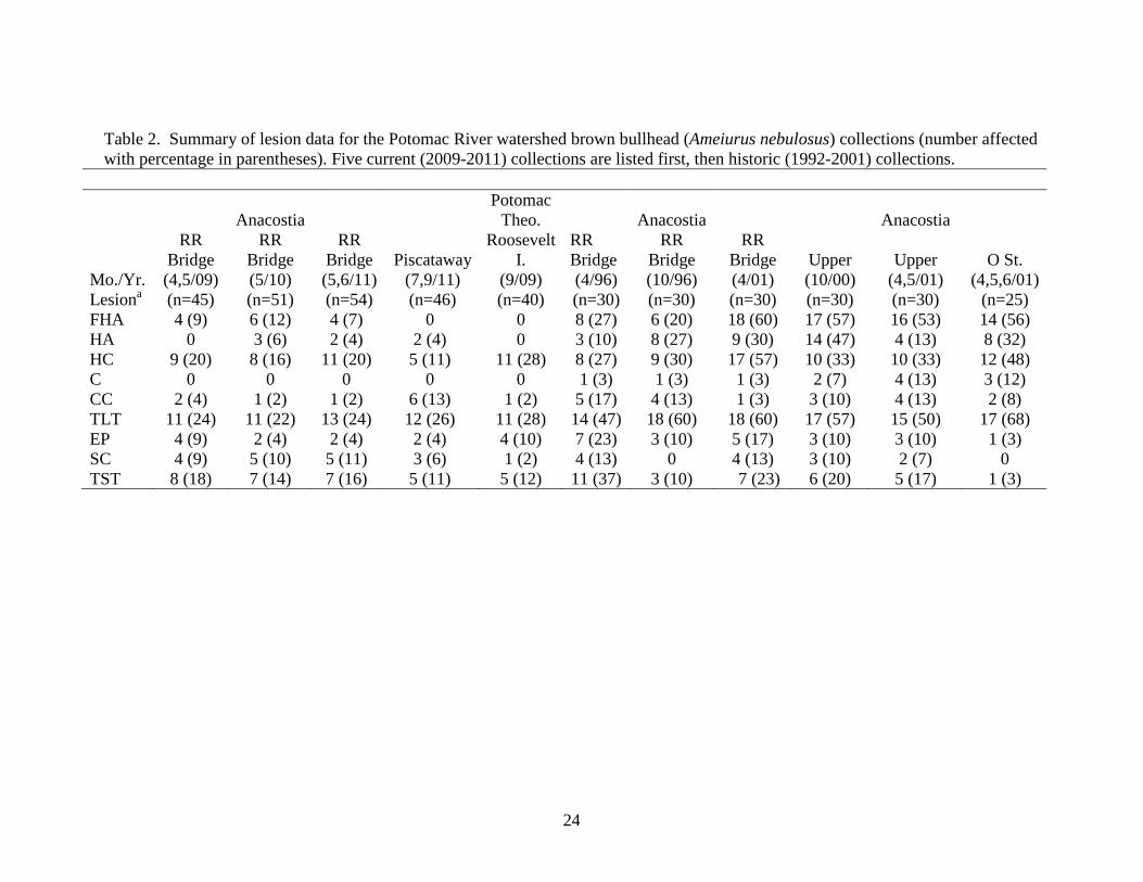
Gross examination revealed the presence of raised pink, oval or round, solid (in texture) lesions in the mouth area (Fig. 2a) of bullheads from all five collections (Table 2). After histopathological examination, nearly all of these visible lesions were diagnosed as either non-invasive epidermal papillomas or invasive squamous cell carcinomas (Fig. 2b). Skin tumor prevalence was highest in the Anacostia River collections ranging from 13.0% to 17.8% , slightly lower in the PRTRI collection (12.5%), and lowest in the Piscataway Creek collection (8.7%; Table 2). Only one liver tumor was suspected based on the gross appearance of the organ (Fig. 3a). The raised white portion was confirmed histologically as a cholangiocarcinoma (Fig. 3b). This liver was also diagnosed with a hepatocellular carcinoma (Fig. 3c). Liver tumor prevalence was similar among the five Potomac watershed collections: PRTRI (2009)—30.0%, Anacostia River (2009-2011)—21.6% to 26.7%, and Piscataway Creek (2011)—26.1%. Logistic regression analysis
The entire Chesapeake Bay tumor database of 1451 fish from 39 collections obtained from 1992 through 2011 was screened to retain only fish that were aged (n = 1193, based on 34 collections). For liver tumors, the set of covariates with the lowest BIC was the model that used length and sex. Since age was not included in the set of best covariates, additional fish for which

6
age was not determined were added to the data set. Hence, the data set included a total of 1404 fish from all 39 collections (those not identified as male or female were excluded) and again the best model (with the lowest BIC) used length and sex as covariates. In all cases for liver tumors, we were able to pool multiple collections from a location based on BIC. For skin tumors, the results were similar with the best model including only length and sex. For this analysis, multiple collections from a location were pooled except for collections from Neabsco Creek and the Severn River. For both skin and liver tumors, increased length and being female resulted in higher probabilities.
The Choptank River was the first location assigned to the CBRG, based on having the
lowest combined rank in skin and liver tumor probabilities. Based on the HPDI analyses, fish from the Furnace Creek, Rhode River, Tuckahoe Creek, Chester River, Quantico Embayment, Farm Creek, and Marumsco Creek locations were added to the CBRG. Fish from the Back River and South River were not added despite their low liver tumor prevalence because their skin tumor prevalence was high. The Middle River location was excluded due to high liver tumor prevalence. Fish from the Severn River location were not included because for one year (2004), skin tumor prevalence was high.
Combining fish from the eight locations listed above resulted in 390 fish being included
in the CBRG (Table 3). The mean estimates for female and male liver tumor prevalence for the CBRG were 9.7% and 2.3%, respectively. The mean estimates for female and male skin tumor prevalence were 3.7% and 2.4%, respectively (Table 3). Combining fish from different sites and increasing sample size resulted in narrower HPDIs for the CBRG compared with those for the individual locations (Tables 3–5).
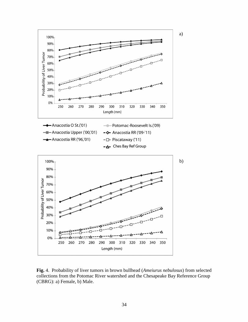
For the Potomac River watershed locations, there were several distinct statistical
groupings for liver tumor probabilities (Table 4, with length–prevalence curves in Figure 4). The highest probability group (group f) was the 1996, 2000, and 2001 collections from the three Anacostia River locations (six collections). Mean probabilities of a 280 mm bullhead having a liver tumor were 77.5% to 89.0% in females and 43.0% to 63.4% in males. At the Anacostia CSX Railroad Bridge location, sampled twice in 1996, once in 2001, and once in 2009, 2010, and 2011, the probability of a 280 mm female having a liver tumor went from 77.5% in the 1996-2001 grouping (n=90) to 42.2% in 2009-2011 (n=150), a 46% decrease. In males, the change was from 43.0% to 13.6%, a 68% decrease. The 2009-2011 Anacostia River collections, PRTRI location and Neabsco Creek formed a separate statistical group (group e) with mean probabilities ranging from 34.2% to 44.5% in females and 10.0% to 14.8% in males. The liver tumor probabilities for the PRTRI location (44.5% in females, 14.8% in males) were nearly identical with the pooled Anacostia 2009-2011 collections (42.2% in females, 13.6% in males). Within the Potomac watershed, bullheads from the Piscataway, Neabsco, Anacostia, and PRTRI locations had significantly greater liver tumor probabilities than those from the CBRG (Table 4).
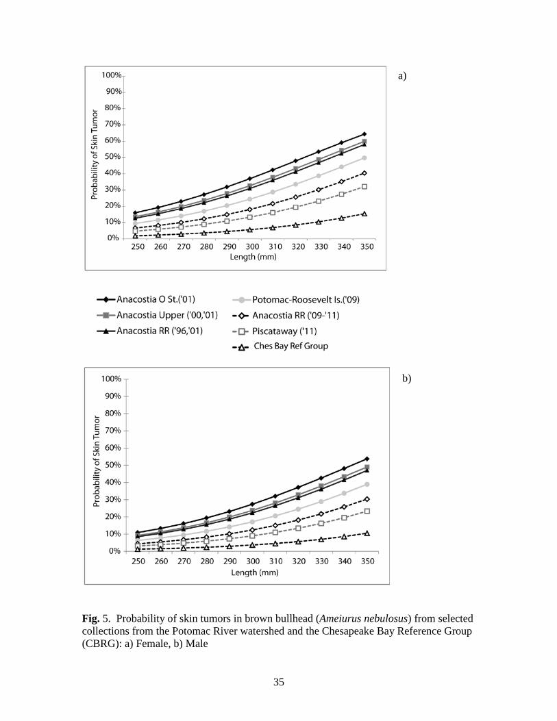
For skin tumors, there were several overlapping statistical groupings for the Potomac
watershed locations (Table 5, with length–prevalence curves in Figure 5). The probabilities of skin tumors in 280 mm bullheads for Anacostia 2009-2011 (17.2% for females, 11.8% for males) while lower, were not statistically different from the 1996, 2000, 2001 Anacostia collections (27.5% for females, 19.6% for males). At the Anacostia CSX Railroad Bridge location, the

7
probability of a 280 mm female brown bullhead having a skin tumor went from 27.5% in the 1996, 2001 grouping (n=90) to 17.2% in 2009-2011 (n=150), a 37% decrease. In males, the change was from 19.6% to 11.8%, a 40% decrease. These changes in the skin tumor prevalence of the Anacostia bullheads over time were not statistically significant. Within the Potomac River watershed, bullheads from all Anacostia locations and from the 1992 Neabsco collection had significantly greater skin tumor probabilities than those from the CBRG (Table 5).
Discussion PAHs and decreased tumor prevalence in the Anacostia River bullheads
The findings of statistically significant and substantial (46% in females, 68% in males) decreases in liver tumor prevalence at the Anacostia CSX location over time, coupled with the literature linking these tumors with PAHs, led us to analyze for causation. According to Myers et al. (2003), the classic epidemiological criteria for causation include 1) strength of the association between the chemical exposure and effect, 2) consistency of the association, 3) toxicological and biological plausibility, 4) temporal sequence (exposure precedes effect, and effect decreases after exposure decreases), 5) biological gradient, 6) specificity of the association, and 7) experimental evidence supporting the cause-effect relationship. These criteria were met in field studies in Black River, Ohio (Baumann and Harshbarger 1998), Puget Sound (Myers et al. 2003, 2008), and Elizabeth River, Virginia (Vogelbein and Unger 2006, 2011) that linked improvements in liver tumor prevalence in resident fish to decreased PAH exposure after the completion of remedial actions.
In the Black River, Baumann and Harshbarger (1998) tracked changes in liver tumor
prevalence in 3-year old brown bullhead. They reported 60% liver tumor prevalence in 1980, with a total PAH sediment concentration of 1096 ppm near the collection site. In 1983, a coking plant that was the primary source of contamination closed. Sediment total PAH concentrations decreased to 4.3 ppm in 1987, when the tumor prevalence was 30%. Remedial dredging in 1990 resulted in the presence of a plume of resuspended contaminated sediments. The increase in tumor prevalence in 1992 (60%, 16.6 ppm total PAH) and 1993 (62%, no sediment data) was attributed to the 1990 exposure of age-one and young-of-the-year bullheads, respectively. A subsequent drop in tumor prevalence in 3-year old bullhead in 1994 to 20% (total PAH of 9.8 ppm) was attributed to their lack of exposure to the plume. In a 1998 collection, Blazer et al. (2009a) reported an 8.9% prevalence of liver tumors.
At the Eagle Harbor Superfund Site, Puget Sound, Washington, Myers et al. (2008)
reported that liver tumor and preneoplastic lesion prevalence in resident English sole (Parophrys vetulus) decreased dramatically after placement of a 22-hectare and later a 6-hectare sand cap over creosote-contaminated sediments. Decreased sediment PAH concentrations and decreased concentrations of hepatic PAC-DNA adducts were documented. After dredging and capping of creosote-contaminated sediments in the Elizabeth River, Virginia, Vogelbein and Unger (2011) reported a decrease in the prevalence of liver tumors and preneoplastic lesions in mummichog (Fundulus heteroclitus).
8
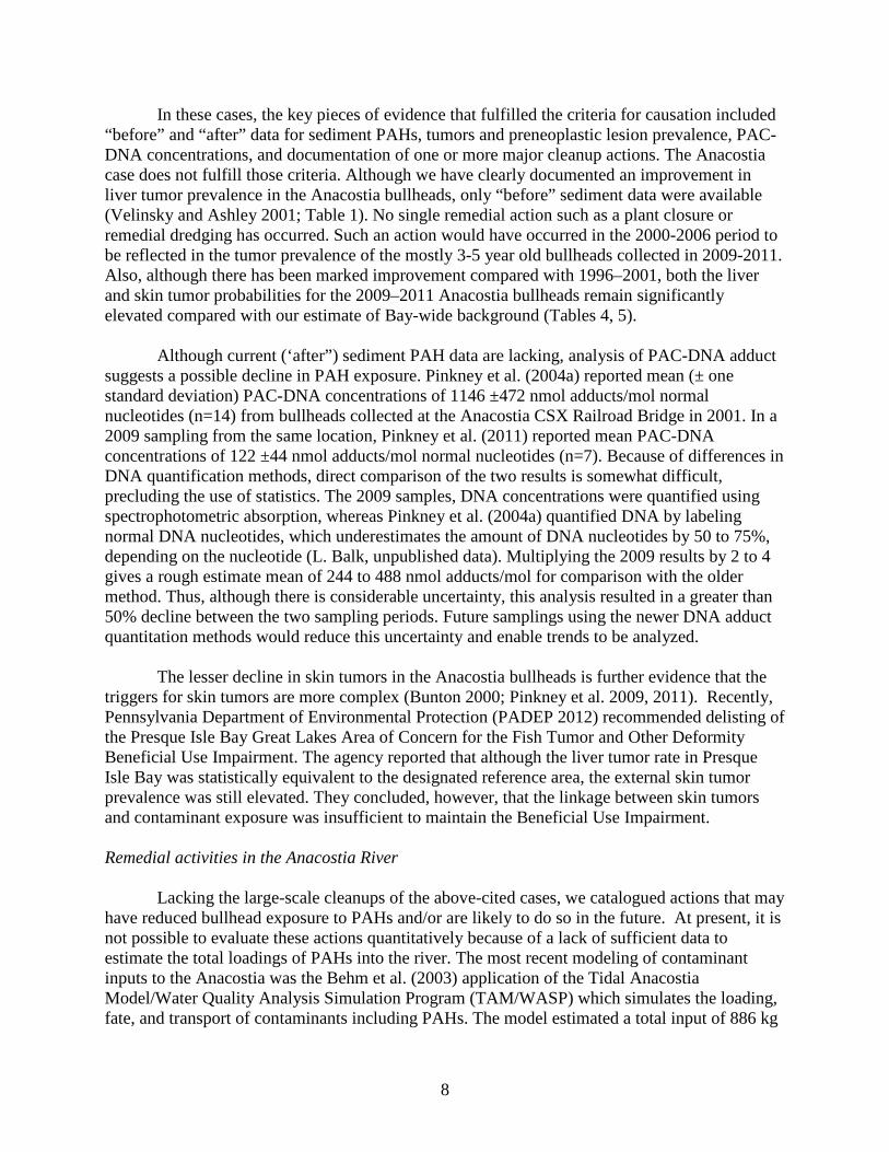
In these cases, the key pieces of evidence that fulfilled the criteria for causation included “before” and “after” data for sediment PAHs, tumors and preneoplastic lesion prevalence, PAC-DNA concentrations, and documentation of one or more major cleanup actions. The Anacostia case does not fulfill those criteria. Although we have clearly documented an improvement in liver tumor prevalence in the Anacostia bullheads, only “before” sediment data were available (Velinsky and Ashley 2001; Table 1). No single remedial action such as a plant closure or remedial dredging has occurred. Such an action would have occurred in the 2000-2006 period to be reflected in the tumor prevalence of the mostly 3-5 year old bullheads collected in 2009-2011. Also, although there has been marked improvement compared with 1996–2001, both the liver and skin tumor probabilities for the 2009–2011 Anacostia bullheads remain significantly elevated compared with our estimate of Bay-wide background (Tables 4, 5).
Although current (‘after”) sediment PAH data are lacking, analysis of PAC-DNA adduct
suggests a possible decline in PAH exposure. Pinkney et al. (2004a) reported mean (± one standard deviation) PAC-DNA concentrations of 1146 ±472 nmol adducts/mol normal nucleotides (n=14) from bullheads collected at the Anacostia CSX Railroad Bridge in 2001. In a 2009 sampling from the same location, Pinkney et al. (2011) reported mean PAC-DNA concentrations of 122 ±44 nmol adducts/mol normal nucleotides (n=7). Because of differences in DNA quantification methods, direct comparison of the two results is somewhat difficult, precluding the use of statistics. The 2009 samples, DNA concentrations were quantified using spectrophotometric absorption, whereas Pinkney et al. (2004a) quantified DNA by labeling normal DNA nucleotides, which underestimates the amount of DNA nucleotides by 50 to 75%, depending on the nucleotide (L. Balk, unpublished data). Multiplying the 2009 results by 2 to 4 gives a rough estimate mean of 244 to 488 nmol adducts/mol for comparison with the older method. Thus, although there is considerable uncertainty, this analysis resulted in a greater than 50% decline between the two sampling periods. Future samplings using the newer DNA adduct quantitation methods would reduce this uncertainty and enable trends to be analyzed.
The lesser decline in skin tumors in the Anacostia bullheads is further evidence that the
triggers for skin tumors are more complex (Bunton 2000; Pinkney et al. 2009, 2011). Recently, Pennsylvania Department of Environmental Protection (PADEP 2012) recommended delisting of the Presque Isle Bay Great Lakes Area of Concern for the Fish Tumor and Other Deformity Beneficial Use Impairment. The agency reported that although the liver tumor rate in Presque Isle Bay was statistically equivalent to the designated reference area, the external skin tumor prevalence was still elevated. They concluded, however, that the linkage between skin tumors and contaminant exposure was insufficient to maintain the Beneficial Use Impairment. Remedial activities in the Anacostia River
Lacking the large-scale cleanups of the above-cited cases, we catalogued actions that may have reduced bullhead exposure to PAHs and/or are likely to do so in the future. At present, it is not possible to evaluate these actions quantitatively because of a lack of sufficient data to estimate the total loadings of PAHs into the river. The most recent modeling of contaminant inputs to the Anacostia was the Behm et al. (2003) application of the Tidal Anacostia Model/Water Quality Analysis Simulation Program (TAM/WASP) which simulates the loading, fate, and transport of contaminants including PAHs. The model estimated a total input of 886 kg

9
per year for total PAHs, with 549 kg/year (62%) from the Northeast and Northwest Branch tributaries, 155 kg/year (17%) from the Lower Beaverdam Creek tributary, 91 kg/year (10%) from separate stormwater outfalls, 36 kg/year (4%) from combined sewer outfalls, 34 kg/year (4%) from groundwater contamination from the Washington Gas site, and 22 kg/year (2%) from Watts Branch tributary. Behm et al. (2003) estimated losses of 246 kg/year (28%) to the Potomac River and 218 kg/year (25%) to volatilization and decay.
Behm et al. (2003) acknowledged the uncertainties in the model and stated that the load
estimates may be inaccurate from -50% to +300%. Water column load data are based largely on a one-year study conducted in 1995–1996 (Foster et al. 2000) that measured chemical concentrations during four storms and six base flow periods. Behm et al. (2003) recommended obtaining additional and updated storm water monitoring data for the upstream tributaries, Lower Beaverdam Creek, and the separate and combined sewer systems.
Foster et al. (2000) determined that 95% of the Northeast Branch and 99% of the
Northwest Branch loads are from particle-bound rather than dissolved PAHs. Thus, any actions that reduced the volume of runoff and the amount of suspended sediment flowing into the tributaries and sewers likely reduced PAH loading into the Anacostia. The sediment Total Maximum Daily Load (TMDL) prepared by MDE and DC Department of Health (DOH) (2007) calls for an 85% overall reduction of sediment/TSS from the baseline loads determined for 1995-1997 of (46,906 tons/year) [4.25 x 10⁷ kg/year] to 7097.6 tons/year [6.44 x 10⁶ kg/year]. Strategies to achieve these reductions include the implementation of Environmental Site Design (ESD), i.e., the use of small-scale stormwater management practices, nonstructural techniques, and better site planning to mimic natural hydrologic cycling of rainwater and minimize the impact of land development on water resources (Biohabitats, Inc. 2012). Actions aimed at reducing point and non-point source pollution within the District of Columbia were documented in its 2012 Water Quality Assessment (District Department of the Environment (DDOE) 2012). It states that since the promulgation of stormwater management regulations in 1998, over 2000 stormwater Best Management Practices (BMPs) have been installed throughout Washington, DC at new development and redevelopment projects for nonpoint source pollution control. Control of stormwater has been improved by DDOE’s aggressive maintenance and inspection program to ensure that BMPs are operating efficiently. Street sweeping, one of the activities promoted by the District to meet stormwater goals and reduce sediment loading, also reduces PAH loading (Biohabitats Inc. 2012).
Stream restoration projects are also an important component of the load reduction, because, according to MDE and DOH (2007), stream channel erosion contributes 67% of the total annual sediment load to the Anacostia River, followed by urban land at 23%. For example, the 2.9 km restoration of the Watts Branch tributary completed in 2011 was estimated to reduce erosion of the reach by 75% (M. Secrist, U.S. Fish and Wildlife Service, personal communication).
The District of Columbia issued a TMDL addressing the loading of organics including
PAHs into the Anacostia (DC DOH 2003), based on the TAM/WASP model (Behm et al. 2003). The document calls for 98% to 99.6% reductions in the loadings from the tributaries and combined sewer outfalls and a complete cessation of loadings from the Washington Gas

10
hazardous waste site. This site was a coal-gas manufacturing plant from 1888 until 1948. Contamination of oil and tar was discovered when non-aqueous phase liquid was discovered in the river in 1976. Capture of groundwater contamination from the waste site was achieved through a pump and treat system started in 1996, with additional wells added in 2003 (Hydro-Terra Inc. 2005). An estimated 41 kg per year of PAH were captured in the treatment system and prevented from migrating to the river (M.J. Brady, Washington Gas, personal communication).
Source control actions in the 446 hectare Hickey Run subwatershed have also reduced
PAH loadings to the Anacostia River. Hickey Run has a long history of oil pollution; in the 1930s it caught fire (US ACE 2010). The chronic oil pollution likely results from illegal dumping and the upstream loadings from automotive repair facilities (US EPA 2005). In 1998, a TMDL was developed by DOH for oil and grease that called for a reduction in point source loads by 89% and for nonpoint source loads by 30%. Part of the TMDL implementation in 2002-2003 included block by block surveys and site inspections of automobile repair shops within the Hickey Run watershed by DOH (Ngbatana 2004). The surveys identified the deficiencies in operations and waste handling that led to spills and illegal dumping. DOH initiated the Environmental Education Compliance of Auto Repair Shops (EE-CARS) Program in which automotive shops were instructed on how to decrease loadings. US EPA (2005) reported that the 10 mg/L water quality standard for oil and grease was achieved and Hickey Run was removed from the list of impaired waters. A 77% reduction in daily loadings was calculated (US EPA 2005), which amounted to an annual decrease of 4470 kg of oil and grease. Using the average of four estimates of the PAH/Oil and Grease Ratio (Washington Department of Ecology 2008), the estimated decreased annual PAH loading in Hickey Run amounted to 1.2 kg per year.
Velinsky et al. (2011) concluded that coal tar- and asphalt-based pavement sealants were
one source of PAHs to the Anacostia River. A recent ban on the use of coal tar pavement sealants, which yield higher amounts of PAHs, was enacted in 2009. Since the ban, eight sites that were illegally coated (a total area of 0.06 square kilometers) were remediated (K. Judson, DDOE, personal communication). Using an annual release rate of 0.51 grams total PAH/square meter (Scoggins et al. 2009), the total amount of PAHs prevented from entering the river is estimated at 30.5 kg per year. Note that the timing of this action was too late to have affected the tumor prevalence data for 2009-2011.
Spatial comparisons within the Potomac River watershed
A major finding of this study was the similarity in liver and skin tumor prevalence in the 2009–2011 Anacostia bullhead collections, PRTRI, and Piscataway Creek (Tables 2, 4-5; Figures 4, 5). Unfortunately, it is difficult to characterize the extent of sediment contamination at the latter two locations. We identified only three sediment samples collected within 1.5 km of PRTRI. Foster and Cui (2008) collected these samples in 2000 and reported a mean total PAH concentration of 4.4 ppm (Table 1). No sediment PAH data have been identified from areas near the Piscataway Creek fish collection.
The similar tumor prevalence at all locations suggests that the tumor issue may be a more
regional problem than one restricted to the Anacostia River. Thus, possibly due to the cleanup activities described above, the current liver tumor prevalence in the Anacostia now reflects the

11
overall urban loading in the Washington, DC area rather than a special case. A repeat sampling of the PRTRI and Piscataway Creek locations as well as new locations extending downriver from Washington, DC, supported by sediment PAH analyses would be needed to verify this suggestion. Logistic regression analysis of the Chesapeake Bay tumor database
The logistic regression analysis of liver tumors was consistent with the previous Chesapeake Bay data base analyses (Pinkney et al. 2009, 2011), which also identified length and sex as covariates. The higher prevalence in longer fish is most likely related to age. As discussed in Pinkney et al. (2009), the ages in the database were based on spines, which are less precise than otoliths and tend to underestimate the ages of larger fish (Maceina and Sammons 2006). This would result in a narrower age spread, which is better captured by length as a covariate. The increased prevalence in females is consistent with Cooke and Hinton (1999) who reported greater liver tumor prevalence in females of six fish species and theorized that increased endogenous estrogens serve as tumor promoters. Nunez et al. (1989) first demonstrated that 17-β-estradiol promotes hepatocellular carcinoma in fish. Interestingly, Rutter (2010) in an analysis of 459 bullheads from Lake Erie did not identify sex as a significant covariate for liver tumors.
In contrast to Pinkney et al. (2009), which did not identify any covariates as predictors
for skin tumors and Pinkney et al. (2011) which identified only length, the present analysis identified both length and sex as covariates, with females having a higher probability. Both Blazer et al. (2009b) and Rutter (2010) reported significant positive relationships between otolith age and orocutaneous tumors in brown bullheads from Lake Erie locations. A powerful age effect was reported by Blazer et al. (2009b) in bullheads from the Presque Isle Bay area of Lake Erie, in which the skin tumor prevalence increased steadily from 5.9% in age-4 bullheads to 100% at age-12. There was a similar pattern at the Long Point Inner Bay (reference) site, on the northern shore of the lake. No skin tumors were noted until age-7 (7.1%) and the prevalence increased to 36.4% in age-10 bullhead. Age and length were also reported to be significant covariates by Rutter (2010) who used Bayesian methods to analyze 459 brown bullheads from five Lake Erie sites. These analyses were based largely on otoliths rather than spines. As mentioned above, we suspect that our covariate, length, is a surrogate for otolith age.
In contrast to the current study, Rutter (2010) did not identify sex as a significant
covariate in his analysis of Lake Erie bullheads. We cannot identify a toxicological mechanism for increased skin tumor prevalence in female bullheads and the differing results further highlight the uncertainty associated with skin tumors. We recommend further research into bullhead skin tumor etiology. Such studies should utilize molecular biology techniques (e.g., Nakamura et al. 2009) that facilitate the discovery of previously unidentified viruses and should determine the immune status of bullheads (e.g., Iwanowicz et al. 2012) with and without skin tumors.
Conclusions and recommendations
The key finding was a statistically significant decrease of roughly 50% in the probability
of liver tumors in the Anacostia River bullheads (standardized to 280 mm length) between 2001

12
and 2009–2011. Although these tumors are clearly linked with exposure to PAHs, there were inadequate data to establish a cause-effect relationship between a theorized decrease in exposure and the lower prevalence. An updated sediment chemistry survey of the river is needed to establish whether a reduction in sediment PAH exposure has occurred. A second important finding was the similar tumor prevalence in the Anacostia, Potomac River in Washington, DC and Piscataway Creek. There were few sediment data available for the latter two locations to characterize the extent of PAH exposure. A repeat survey of those and other Potomac River locations supported by sediment PAH measurements is needed to verify the suggestion that the elevated tumor prevalence is a regional rather than Anacostia issue. By analyzing the Chesapeake Bay database, we provided an estimate of “regional background” and showed that, while improved, the tumor prevalence in Anacostia bullheads and those from other Potomac watershed locations remain elevated.
We recommend monitoring of tumor prevalence at the Anacostia CSX Railroad Bridge
location on a 5-year cycle, along with an updated sediment survey, so that this indicator can continue to track changes in habitat quality. The present study which sampled the Anacostia in 2009, 2010, and 2011, provided confidence in the status circa 2010 by sampling three years. Surveys in 2014, 2015, and 2016 would provide a similar level of confidence for the next five year interval. Further evidence for causation could be provided by coupling the tumor survey and sediment sampling with measurement of PAC–DNA adducts using the methods described in Pinkney et al. (2011). We recommend further research into bullhead skin tumor etiology, by applying state-of-the-art techniques to identify possible viruses and evaluating the immune status of affected and unaffected fish. Additional sampling of CBRG locations would be useful to update and further refine the estimates of regional background.
Acknowledgements
We appreciate the assistance of Josh Newhard, Mike Mangold, Ian Park, John Gill, and Steve Minkkinen (USFWS) in the field and Susan Frey (USFWS) in the CBFO laboratory. Collections within the District of Columbia were made by Danny Ryan, Luke Lyon, Joe Swann, and Eric Thadey of the District of Columbia Department of the Environment, Fisheries Research Branch. Kyle Hartman (West Virginia University) aged the spines. Leslie Gerlich, Ebony Davis, and Laurie Hewitt (USFWS) produced the graphics. The comments of Luke Iwanowicz (USGS), Nicoline Shulterbrandt (DDOE), and Jonathan Essoka (US EPA) are appreciated. Funding for the Chesapeake Bay tumor surveys was provided by the USFWS Maryland Fisheries Resource Office, USFWS Division of Environmental Contaminants, and the South River Federation.
13
References
Baumann, P.C., & Harshbarger, J.C. (1998). Long term trends in liver neoplasm epizootics of brown bullhead in the Black River, Ohio. Environmental Monitoring and Assessment 53, 213-223.
Baumann, P.C., Harshbarger, J.C., & Hartman, K.J. (1990). Relationship between liver tumors
and age in brown bullhead populations from two Lake Erie tributaries. Science of the Total Environment 94, 71-87.
Baumann, P.C., Smith, I.R., & Metcalfe, C.D. (1996). Linkages between chemical contaminants
and tumors in benthic Great Lakes fish. Journal of Great Lakes Research 22, 131-152. Behm, P., Buckley, A., & Schultz, C.L. (2003). TAM/WASP toxics screening level model
for the tidal portion of the Anacostia River. Final Report. Rockville, MD: Interstate Commission on the Potomac River Basin.
Biohabitats, Inc. (2012). Anacostia Watershed Implementation Plan. Prepared for Montgomery County Department of Environmental Protection. Baltimore, MD: Biohabitats, Inc.
Black, J., Fox, H., Black, P., & Bock, F. (1985). Carcinogenic effects of river sediment extracts
in fish and mice. In R.L. Jolley, R.J. Bull, W.P. Davis, S. Katz, M.H. Roberts Jr., & V.A. Jacobs (Eds.), Water chlorination. Chemistry, environmental impact and health effects, volume 5 (pp. 415-428). Chelsea, MI: Lewis Publishers.
Blazer, V.S., Fournie, J.W., Wolf, J.C., & Wolfe, M.J. (2006). Diagnostic criteria for
proliferative hepatic lesions in brown bullhead Ameiurus nebulosus. Diseases of Aquatic Organisms 72, 19-30.
Blazer, V.S., Fournie, J.W., Wolf, J.C., & Wolfe, M.J. (2007). Manual for the microscopic
diagnosis of proliferative liver and skin lesions in the brown bullhead (Ameiurus nebulosus). Erie, PA: Pennsylvania Sea Grant. http://pserie.psu.edu/seagrant/publications/technicaldocs/HistoFieldManual.pdf
Blazer, V.S., Rafferty, S.D., Baumann, P.C., Smith, S.B., & Obert, E.C. (2009a). Assessment of
the “fish tumors or other deformities” beneficial use impairment in brown bullhead (Ameiurus nebulosus): II. Liver neoplasia. Journal of Great Lakes Research 35, 527-537.
Blazer V.S., Rafferty, S.D., Baumann, P.C., Smith, S.B., & Obert, E.C. (2009b). Assessment of
the “fish tumors or other deformities” beneficial use impairment in brown bullhead (Ameiurus nebulosus): I. Orocutaneous tumors. Journal of Great Lakes Research 35, 517-526.
Bunton, T.E. (2000). Brown bullhead (Ameiurus nebulosus) skin carcinogenesis. Experimental
Toxicologic Pathology 52, 209-220.

14
Carlander, K.D. (1969). Handbook of freshwater fishery biology. volume 1. Ames, IA: Iowa State University Press.
Cooke, J.B., & Hinton D.E. (1999). Promotion by 17-β estradiol and β hexachlorocyclohexane of
hepatocellular tumors in medaka, Oryzias latipes. Aquatic Toxicology 45, 127-145. District of Columbia Department of the Environment (DDOE) (2012). The District of Columbia
Water Quality Assessment. 2012 Integrated report to the U.S. Environmental Protectiona Agency and Congress pursuant to sections 305(b) and 303(d) Clean Water Act (P.L. 97-117). Washington, DC: DDOE.
District of Columbia Department of Health (DC DOH) (2003). District of Columbia final Total
Maximum Daily Loads for organics and metals in the Anacostia River, Fort Chaplin Tributary, Fort Davis Tributary, Fort Dupont Creek, Fort Stanton Tributary, Hickey Run, Nash Run, Popes Branch, Texas Avenue Tributary, and Watts Branch. Washington, DC: DC DOH.
Foster, G.D., & Cui, V. (2008). PAHs and PCBs deposited in surficial sediments along a rural to
urban transect in a Mid-Atlantic coastal river basin (USA). Journal of Environmental Science and Health Part A 43, 1333–1345.
Foster, G.D., Roberts, E. Jr., Gruessner, B., & Velinsky, D. (2000). Hydrogeochemistry and
transport of organic contaminants in an urban watershed of Chesapeake Bay (USA). Applied Geochemistry 15, 901-915.
Gelman, A., & Hill, J. (2006). Data analysis using regression and multilevel/hierarchical
models. New York: Cambridge University Press. Gelman, A., Carlin, J.B., Stern, H.S., & Rubin, D.B. (2004). Bayesian data analysis, second
edition. Boca Raton, FL: Chapman and Hall/CRC. Gelman, A., Jakulin, A., Pittau, M.G., & Su, Y.S. (2008). A weakly informative default prior
distribution for logistic and other regression models. Annals of Applied Statistics 2, 1360-1383.
Gelman A., Su, Y-S., Yajima, M., Hill, J., Pittau, M.G., Kerman, J., et al. (2012). arm: Data
analysis using regression and multilevel/hierarchical models. R package version 1.5-07. 2012. http://CRAN.R-project.org/package=arm.
Getchell, R.G., Casey, J.W., & Bowser, P.R. (1998). Seasonal occurrence of virally induced skin
tumors in wild fishes. Journal of Aquatic Animal Health 10, 191-201. Grizzle, J.M., Melius, P., & Strength, D.R. (1984). Papillomas on fish exposed to chlorinated
wastewater effluent. Journal of the National Cancer Institute 73, 1133-1142.

15
Hawkins, W.E., Walker, W.W., Overstreet, R.M., Lytle, J.S., & Lytle, T.F. (1990). Carcinogenic effects of some polycyclic aromatic hydrocarbons on the Japanese medaka and guppy in waterborne exposures. Science of the Total Environment 94, 155-167.
Hydro-Terra, Inc. (2005). Five-year review of remedial actions at East Station Site in
Washington, DC. Columbia, MD: Hydro-Terra, Inc. Iwanowicz, L.R., Blazer, V.S., Hitt, N.P., McCormick, S.D., DeVault, D.S., & Ottinger, C.A.
(2012). Histologic, immunologic and endocrine biomarkers indicate contaminant effects in fishes of the Ashtabula River. Ecotoxicology 21, 165–182.
Jenkins, R.E., & Burkhead, N.M. (1994). Freshwater fishes of Virginia. Bethesda, MD:
American Fisheries Society. Kutner, M.H., Nachtsheim, C.J., Neter, J., & Li, W. (2004). Applied linear statistical models. 5th
ed. New York: McGraw-Hill. Luna L, editor. (1968). Manual of histologic staining methods of the Armed Forces Institute of
Pathology. New York: McGraw-Hill. Maceina, M.J., & Sammons, S.M. (2006). An evaluation of different structures to age freshwater
fish from a northeastern U.S. river. Fisheries Management Ecology 13, 237-242. Maryland Department of the Environment (MDE) and District of Columbia Department of
Health (DC DOH). (2007). Total maximum daily loads of sediment/total suspended soilds for the Anacostia River basin, Montgomery and Prince George’s Counties, Maryland and the District of Columbia. Baltimore, MD: MDE.
Maryland Department of Natural Resources (MD DNR). (2005). Characterization of the Upper
Chester River watershed in Kent County and Queen Anne’s County, Maryland. Annapolis, MD: MD DNR.
Millard, M.R., Smith, D.R., Obert, E., Bartron, M.L., Wellington, C., Grise, S., et al. (2009).
Movements of brown bullheads in Presque Isle Bay, Lake Erie, Pennsylvania. Journal of Great Lakes Research 35, 613-619.
Myers, M.S., Stehr, C.M., Olson, O.P., Johnson, L.L., McCain, B.B., Chan, S.L., et al. (1994).
Relationships between toxicopathic hepatic lesions and exposure to chemical contaminants in English sole (Pleuoronectes vetulus), starry flounder (Platichthys stellatus), and white croaker (Genyonemus lineatus) from selected marine sites on the Pacific coast, USA. Environmental Health Perspectives 102, 2-17.
Myers, M.S., Johnson, L.L., & Collier, T.K. (2003). Establishing the causal relationship
between polycyclic aromatic hydrocarbon (PAH) exposure and hepatic neoplasms and neoplasia-related liver lesions in English sole (Pleuronectes vetulus). Human and Ecological Risk Assessment 9, 67-94.
16
Myers, M.S., Anulacion, B.F., French, B.L., Reichert, W.L., Laetz, C.A., Buzitis, J., et al. (2008). Improved flatfish health following remediation of a PAH-contaminated site in Eagle Harbor, Washington. Aquatic Toxicology 88, 277-288.
Nakamura, S., Yang, C.S., Sakon, N., Ueda, M., Tougan, T., Yamashita, A., et al. (2009). Direct
metagenomic detection of viral pathogens in nasal and fecal specimens using an unbiased high-throughput sequencing approach. PLoS ONE 4, e4219, 8 p.
Ngbatana, T. (2004). Hickey Run oil and grease mitigation efforts. A draft report. Washington, DC: District of Columbia Department of Health.
Nunez, O., Hendricks, J.D., Arbogast, D.N., Fong, A.T., Lee, B.C., & Bailey, G.S. (1989).
Promotion of aflatoxin B1 hepatocarcinogenesis in rainbow trout by 17ß estradiol. Aquatic Toxicology 15, 289-302.
Pennsylvania Department of Environmental Protection (PA DEP). (2012). Presque Isle Bay Area
of Concern. Final Stage 3 Remedial Action Plan. Delisting. Harrisburg, PA: PA DEP. Pinkney, A.E., & Harshbarger, J.C. (2005). Tumor prevalence in brown bullheads (Ameiurus
nebulosus) from the South River, Anne Arundel County, Maryland. Annapolis, MD: U.S. Fish and Wildlife Service.
Pinkney, A.E., & Harshbarger, J.C. (2008). Tumor prevalence in brown bullheads (Ameiurus
nebulosus) from the Little Blackwater River, Maryland. Annapolis, MD: U.S. Fish and Wildlife Service.
Pinkney, A.E., Sutherland, D.W., Foley, R.E., & Harshbarger, J.C. (1995). Investigation of
sediment contamination and fish pathology at Featherstone National Wildlife Refuge, Virginia. Annapolis, MD: U.S. Fish and Wildlife Service.
Pinkney, A.E., Harshbarger, J.C., May, E.B., & Melancon, M.J. (2001). Tumor prevalence and
biomarkers of exposure in brown bullheads (Ameiurus nebulosus) from the tidal Potomac River watershed. Environmental Toxicology and Chemistry, 20, 1196-1205.
Pinkney, A.E., Harshbarger, J.C., May, E.B. & Reichert, W.L (2004a). Tumor prevalence and
biomarkers of exposure and response in brown bullhead (Ameiurus nebulosus) from the Anacostia River, Washington, DC and Tuckahoe River, Maryland. Environmental Toxicology and Chemistry 24, 638-647.
Pinkney, A.E., Harshbarger, J.C., May, E.B., & Melancon, M.J. (2004b). Tumor prevalence and
biomarkers of exposure in brown bullheads (Ameiurus nebulosus) from Back River, Furnace Creek, and Tuckahoe River, Maryland. Archives of Environmental Contamination and Toxicology 46, 492-501.

17
Pinkney, A.E., Harshbarger, J.C., & Roberts, M.R. (2004c). Tumor prevalence in brown bullheads from Darby Creek, John Heinz National Wildlife Refuge at Tinicum, Philadelphia, PA. Annapolis, MD: U.S. Fish and Wildlife Service.
Pinkney, A.E., Harshbarger, J.C., & Rutter, M.A. (2009). Tumors in brown bullheads (Ameiurus
nebulosus) in the Chesapeake Bay watershed: Analysis of survey data 1992 through 2006. Journal of Aquatic Animal Health 21, 71-81.
Pinkney, A.E., Harshbarger, J.C., Karouna-Renier, N.K., Jenko, K., Balk, L., Skarphéðinsdóttir,
H., et al. (2011). Tumor prevalence and biomarkers of genotoxicity in brown bullhead (Ameiurus nebulosus) in Chesapeake Bay tributaries. Science of the Total Environment 410, 248-257.
Plumer, M. (2012). JAGS: just another Gibbs sampler. Version 3.2.0. Available at:
http://www-fis.iarc.fr/~martyn/software/jags Poulet, F.M., & Spitsbergen, J.M. (1996). Ultrastructural study of spontaneous orocutaneous
papillomas of brown bullheads Ictalurus nebulosus. Diseases of Aquatic Organisms 24, 93-98.
Poulet, F.M., Casey, J.W., & Spitsbergen, J.M. (1993). Studies on the transmissibility and
etiology of orocutaneous tumors of brown bullheads Ictalurus nebulosus. Diseases of Aquatic Organisms 16, 97-104.
Poulet, F.M., Wolfe, M.J., & Spitsbergen, J.M. (1994). Naturally occurring orocutaneous
papillomas and carcinomas of brown bullheads (Ictalurus nebulosus) in New York State. Veterinary Pathology 31, 8-18.
R Core Team. (2012). R: A language and environment for statistical computing. R Foundation
for Statistical Computing, Vienna, Austria. ISBN3-900051-07-0, 2012; http://www.R-project.org/.
Rafferty, S.D., Blazer, V.S., Pinkney, A.E., Grazio, J.L., Obert, E.C., & Boughton, L. (2009). A
historical perspective on the “fish tumors or other deformities” beneficial use impairment at Great Lakes Areas of Concern. Journal of Great Lakes Research 35, 496-506.
Reichert,W.L., Myers, M.S., Peck-Miller, K., French, B., Anulacion, B.F., Collier, T.K., et al.
(1998). Molecular epizootiology of genotoxic events in marine fish: linking contaminant exposure, DNA damage, and tissue-level alterations. Mutation Research 411, 215-225.
Rutter, M.A. (2010). A statistical approach for establishing tumor incidence delisting criteria in
areas of concern: A case study. Journal of Great Lakes Research 36, 646-655. Sakaris, P.C., Jesien, R.V., & Pinkney, A.E. (2005). Brown bullhead, Ameiurus nebulosus, as an
indicator species: seasonal movement patterns and home ranges within the Anacostia River, Washington, D.C. Transactions of the American Fisheries Society 134, 1262-1270.

18
Schiewe, M.S., Weber, D.D., Myers, M.S., Jacques, F.J., Reichert, W.L., Krone, C.A., et al.
(1991). Induction of foci of cellular alteration and other hepatic lesions in English sole (Parophrys vetulus) exposed to an extract of an urban marine sediment. Canadian Journal of Fisheries and Aquatic Sciences 48, 1750-1760.
Schwartz, G. (1978). Estimating the dimension of a model. Annals of Statistics 6, 461-464. Scoggins, M., Ennis, T., Parker, N., & Herrington, C. (2009). A photographic method for
estimating wear of coal tar sealcoat from parking lots. Environmental Science and Technology 43, 4909-4914.
Steyermark, A.C., Spotila, J.R., Gillette, D., & Isseroff, H. (1999). Biomarkers indicate health
problems in brown bullheads from the industrialized Schuylkill River, Philadelphia. Transactions of the American Fisheries Society 128, 328-338.
U.S. Army Corps of Engineers (US ACE). (2010) Anacostia River watershed restoration plan
and report. Baltimore, MD: US ACE. U.S. Environmental Protection Agency (US EPA). (2005). District of Columbia: Hickey Run oil
& grease water quality goals achieved. http://water.epa.gov/polwaste/nps/success319/state_dc.cfm
U.S. Environmental Protection Agency (US EPA). (2006). Charge to the peer reviewers:
ecological condition and other relevant indicators for the U.S. Environmental Protection Agency’s 2007 Report on the Environment. Washington, DC: US EPA.
Velinsky, D.J., & Ashley, J.T.F. (2001). Sediment transport: Additional chemical analysis study,
phase II. Philadelphia, PA: Academy of Natural Sciences. Velinksy, D.J., Riedel, G.F., Ashley, J.T.F., & Cornwall, J.C. (2011). Historical contamination of
the Anacostia River, Washington, D.C. Environmental Monitoring and Assessment 183, 307-328.
Versar, Inc. (2001). Water quality management plan for Middle River watershed. Submitted to
Baltimore County Department of Environmental Protection and Resource Management. Columbia, MD: Versar, Inc.
Vogelbein, W.G., & Unger, M.A. (2006). Liver carcinogenesis in a non-migratory fish: The
association with polycyclic aromatic hydrocarbon exposure. Bulletin of the European Association of Fish Pathologists 26, 11-20.
Vogelbein, W.G., & Unger, M.A. (2011). Elizabeth River biological effects monitoring 2010:
Tracking success of sediment remediation at Money Point. Gloucester Point, VA: Virginia Institute of Marine Science.

19
Washington Department of Ecology (2008). Comparison of loading estimates to Puget Sound for oil and petroleum products by Ecology and the National Research Council. Pub. # 08-10-084 Addendum 1. Olympia, WA: Washington Department of Ecology.

20

21
TABLES

22
Table 1. Descriptions of the Potomac River watershed sampling locations including sediment polynuclear aromatic hydrocarbon (PAH) concentration data. Locations sampled in 2009–2011 are in bold. Surface sediment sampling data are within 1-1.5 km of the bullhead collection locations. Location Description/type of watershed Sediment PAH concentrations
(mean ± SD, ppm dry weight) Anacostia R. near O St. Combined Sewer Outfall
2.5 km from mouth, sampled adjacent to combined sewer outfalls and Washington Navy Yard Superfund site (primarily PCBs), 1.4 km downriver from Washington Gas site (PAHs); urban
2000: n= 31a Carcinogenic PAHsb: 16.4 ± 8.9 Total PAHs: 30.9 ± 18.2
Anacostia R. at CSX RR Bridge
5.8 km from river mouth, sampling range extended upriver 1.2 km to the East Capital Street Bridge and downriver 0.6 km to the Pennsylvania Avenue Bridge; near combined sewer outfalls; and 1.7 km upriver from the Washington Gas site; urban
1996: n=3c Carcinogenic PAHsb: 9.0 ± 0.4 Total PAHs: 26.8 ± 2.9 2000: n=21a Carcinogenic PAHsb: 14.0 ± 7.3 Total PAHs: 28.1 ± 17.3
Anacostia R. just below Route 50 Bridge
10 km from river mouth, near Kenilworth Landfill waste site (elevated PAHs in soil) and mouth of Little Beaverdam Creek with small dump sites; urban
2000: n=16a Carcinogenic PAHsb: 7.9 ± 5.0 Total PAHs: 15.3 ± 9.2
Farm Creek 1.6 km tidal creek flows through Featherstone National Wildlife Refuge (NWR); suburban
1991: n= 6d Carcinogenic PAHsb: 0.030 ± 0.010 Total PAHs: 0.10 ± 0.05 1992: n= 3d Carcinogenic PAHs: 0.34 ± 0.04 Total PAHs: 12.0 ± 1.7
Marumsco Creek
0.7 km tidal creek bordering Occoquan Bay NWR; suburban
1992: n=3 d Carcinogenic PAHs: 0.63 ± 0.34 Total PAHs: 10.5 ± 1.3
Neabsco Creek
0.5 km navigable tidal creek adjacent to two marinas and bordering Featherstone NWR; suburban
1991: n= 6d Carcinogenic PAHsb: 0.21 ± 0.08 Total PAH: 0.65 ± 0.23 1992: n= 3d Carcinogenic PAHsb: 1.4 ± 1.2 Total PAHs: 14.9 ± 9.3 1996: n= 3c

23
Carcinogenic PAHsb: 1.4 ± 2.2 Total PAHs: 4.8 ± 6.8
Piscataway Creek
0.5 km from creek mouth, near Fort Washington marina, suburban
No data available
Potomac R. at Theodore Roosevelt Island
Sampling circumnavigated this 36 ha island in the Potomac River between the Kennedy Center/Georgetown area of Washington, DC and northern Virginia; urban
2000: n=3e Carcinogenic PAHsb: 2.1 ±1.1 Total PAHs: 4.4 ± 2.0
Quantico embayment
78-ha embayment of the Potomac River adjacent to the Marine Corps Base Quantico and the Old Landfill Superfund site (PCBs and DDT compounds)
1996: n=3c
Carcinogenic PAHsb: 5.1 ± 4.7 Total PAHs: 1.2 ± 1.0
a Velinsky and Ashley (2001) b Sum of the following PAHs: benzo(a)pyrene, benz(z)anthracene, benzo(b)fluoranthene, benzo(k)fluoranthene, benzo(ghi)fluoranthene, chrysene, dibenzo(ah)anthracene, and indeno(1,2,3-c,d)pyrene c Pinkney et al. (2001) d Pinkney et al. (1995) e Foster and Cui (2008)
24
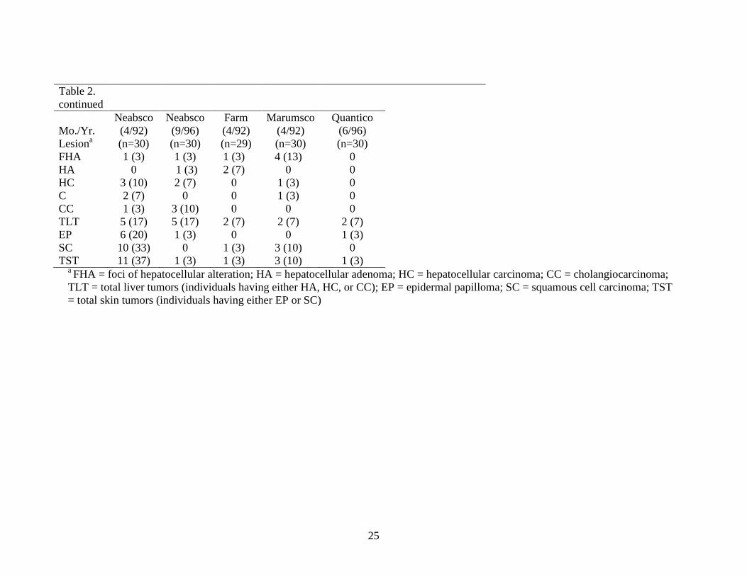
Table 2. Summary of lesion data for the Potomac River watershed brown bullhead (Ameiurus nebulosus) collections (number affected with percentage in parentheses). Five current (2009-2011) collections are listed first, then historic (1992-2001) collections.
Mo./Yr. Lesiona
RR Bridge (4,5/09) (n=45)
Anacostia RR
Bridge (5/10) (n=51)
RR Bridge (5,6/11) (n=54)
Piscataway (7,9/11) (n=46)
Potomac Theo.
Roosevelt I.
(9/09) (n=40)
RR Bridge (4/96) (n=30)
Anacostia RR
Bridge (10/96) (n=30)
RR
Bridge (4/01) (n=30)
Upper (10/00) (n=30)
Anacostia
Upper (4,5/01) (n=30)
O St.
(4,5,6/01) (n=25)
FHA 4 (9) 6 (12) 4 (7) 0 0 8 (27) 6 (20) 18 (60) 17 (57) 16 (53) 14 (56) HA 0 3 (6) 2 (4) 2 (4) 0 3 (10) 8 (27) 9 (30) 14 (47) 4 (13) 8 (32) HC 9 (20) 8 (16) 11 (20) 5 (11) 11 (28) 8 (27) 9 (30) 17 (57) 10 (33) 10 (33) 12 (48) C 0 0 0 0 0 1 (3) 1 (3) 1 (3) 2 (7) 4 (13) 3 (12) CC 2 (4) 1 (2) 1 (2) 6 (13) 1 (2) 5 (17) 4 (13) 1 (3) 3 (10) 4 (13) 2 (8) TLT 11 (24) 11 (22) 13 (24) 12 (26) 11 (28) 14 (47) 18 (60) 18 (60) 17 (57) 15 (50) 17 (68) EP 4 (9) 2 (4) 2 (4) 2 (4) 4 (10) 7 (23) 3 (10) 5 (17) 3 (10) 3 (10) 1 (3) SC 4 (9) 5 (10) 5 (11) 3 (6) 1 (2) 4 (13) 0 4 (13) 3 (10) 2 (7) 0 TST 8 (18) 7 (14) 7 (16) 5 (11) 5 (12) 11 (37) 3 (10) 7 (23) 6 (20) 5 (17) 1 (3)
25
Table 2. continued
Mo./Yr. Lesiona
Neabsco (4/92) (n=30)
Neabsco (9/96) (n=30)
Farm (4/92) (n=29)
Marumsco (4/92) (n=30)
Quantico (6/96) (n=30)
FHA 1 (3) 1 (3) 1 (3) 4 (13) 0 HA 0 1 (3) 2 (7) 0 0 HC 3 (10) 2 (7) 0 1 (3) 0 C 2 (7) 0 0 1 (3) 0 CC 1 (3) 3 (10) 0 0 0 TLT 5 (17) 5 (17) 2 (7) 2 (7) 2 (7) EP 6 (20) 1 (3) 0 0 1 (3) SC 10 (33) 0 1 (3) 3 (10) 0 TST 11 (37) 1 (3) 1 (3) 3 (10) 1 (3)
a FHA = foci of hepatocellular alteration; HA = hepatocellular adenoma; HC = hepatocellular carcinoma; CC = cholangiocarcinoma; TLT = total liver tumors (individuals having either HA, HC, or CC); EP = epidermal papilloma; SC = squamous cell carcinoma; TST = total skin tumors (individuals having either EP or SC)

26
Table 3. Tumor prevalence probabilities of a 280 mm brown bullhead (Ameiurus nebulosus) from locations within the Chesapeake Bay watershed included in the Chesapeake Bay Reference Group (CBRG) (mean percent with 95 percent Highest Posterior Density Interval (HPDI). Loc.a n= Liver Skin Female Male Female Male CHOP 91 7.0 (0.4–19.1) 1.6 (0.1–4.6) 2.0 (0.0–7.5) 1.3 (0.0–5.1) FURN 44 0.1 (0.0–5.6) 0.0 (0.0–1.2) 8.4 (1.2–23.6) 5.7 (0.7–16.6) RHOD 18 7.8 (0.0–26.2) 1.7 (0.0–7.0) 5.0 (0.0–19.9) 3.3 (0.0–14.1) TUCK 108 12.2 (3.3–24.4) 2.8 (0.7–6.2) 1.4 (0.0–5.8) 0.9 (0.0–3.8) CHES 40 12.2 (3.3–24.4) 3.2 (0.1–10.1) 0.3 (0.0–7.4) 0.2 (0.0–5.0) QUAN 30 13.1 (1.0–35.4) 3.0 (0.2–10.0) 4.6 (0.0–17.2) 3.0 (0.0–12.0) FARM 29 14.1 (0.5–37.2) 3.3 (0.1–10.7) 3.5 (0.0–14.9) 2.3 (0.0–10.0) MARU 30 17.5 (1.2–43.5) 4.1 (0.2–12.8) 10.4 (1.1–29.3) 7.0 (0.6–20.4) CBRG 390 9.7 (5.4–15.6) 2.3 (1.1–3.8) 3.7 (1.3–6.6) 2.4 (1.0–4.3) aCHOP: Choptank R. (2008), FURN: Furnace Creek (1998), RHOD: Rhode R. (2007), TUCK: Tuckahoe R. (1996, 1998, 2000, 2001), CHES: Chester R. (2010), QUAN: Quantico Embayment (1996), FARM: Farm Creek (1992), MARU: Marumsco Creek (1992)

27
Table 4. Comparison of liver tumor probabilities of a 280 mm brown bullhead (Ameiurus nebulosus) from locations within the Potomac River watershed (in bold), other Chesapeake Bay locations, and the Chesapeake Bay Reference Group (CBRG) (mean percent with 95 percent Highest Posterior Density (HPD) confidence interval). Female Male Stat. groupa Little Blackwater R. (up) 0.6 (0.0–13.4) 0.1 (0.0–3.1) abcde Severn R. 2.6 (0.1–7.3) 0.6 (0.0–1.7) a Little Blackwater R. (down) 6.3 (0.0–21.3) 1.4 (0.0–5.6) abcd South R. 8.4 (3.9–13.9) 1.9 (0.7–3.4) ab CBRG 9.7 (5.4–15.6) 2.3 (1.1–3.8) abc Middle R. 23.3 (6.8–44.0) 6.1 (1.4–14.2) bcde Back R. 24.8(8.6–44.7) 6.7 (1.5–13.9) cde Piscataway Crk. 32.3 (11.5–54.6) 9.2 (1.9–19.6) de Neabsco Crk. 34.2 (14.0–53.8) 10.0 (3.0–19.6) e Anacostia CSX (2009-2011) 42.2 (27.2–57.2) 13.6 (7.2–21.7) e Potomac R. Theo. Roosevelt I. 44.5 (20.3–68.7) 14.8 (3.1–29.2) e Anacostia CSX (1996, 2001) 77.5 (65.2–88.6) 43.0 (26.4–58.4) f Anacostia Upriver 82.0 (68.1–92.9) 49.2 (30.0–67.7) f Anacostia O Street 89.0 (72.0–98.1) 63.4 (33.7–86.3) f a Locations with different letters are significantly different at p<0.05based on 95% HPDI based on average of male and female probabilities. b CBRG (Chesapeake Bay Reference Group) consists of bullheads from the following locations: Chester R., Choptank R., Farm Creek, Furnace Creek, Marumsco Creek, Quantico Embayment, Rhode R., Tuckahoe Creek

28
Table 5. Comparison of skin tumor probabilities of a 280 mm brown bullhead (Ameiurus nebulosus) from locations within the Potomac River watershed (in bold), other Chesapeake Bay locations, and the Chesapeake Bay Reference Group (CBRG) (mean percent with 95 percent Highest Posterior Density (HPD) confidence interval). Female Male Stat. group a Neabsco Crk. (1996) 0.2 (0.0–9.1) 0.1 (0.0–6.1) abcd CBRG 3.7 (1.3–6.6) 2.4 (1.0–4.3) a Middle R. 4.7 (0.2–16.1) 3.1 (0.1–10.7) abc Severn R. (2007, 2008) 5.6 (1.1–13.0) 3.7 (0.6–8.6) ab Piscataway Crk. 9.0 (1.1–24.9) 6.0 (0.5–17.6) abcd Potomac R. Theo. Roosevelt I. 12.4 (2.1–31.3) 8.3 (0.9–21.9) abcd Back R. 14.3 (4.5–27.5) 9.7 (3.1–19.3) bcd Anacostia CSX (2009-2011) 17.2 (7.2–30.5) 11.8 (4.5–20.7) bcd Little Blackwater R. (down) 18.4 (2.7–42.6) 12.7 (1.6–31.9) bcde Anacostia O Street 22.7 (3.1–50.9) 15.7 (2.2–40.1) bcde Anacostia Upriver 23.8 (7.0–43.3) 16.6 (5.2–32.9) cde Anacostia CSX (1996, 2001) 27.5 (13.1–45.2) 19.6 (8.1–33.4) de Little Blackwater R. (up) 31.7 (8.3–62.4) 23.1 (3.6–48.8) cde Neabsco Crk. (1992) 33.7 (9.4–62.4) 24.7 (5.7–50.0) de Severn R. (2004) 35.9 (11.3–66.0) 26.2 (6.1–53.4) de South R. 42.5 (29.9–55.7) 32.2 (21.5–43.9) e
a Locations with different letters are significantly different at p<0.05based on 95% HPDI based on average of male and female probabilities. b CBRG (Chesapeake Bay Reference Group) consists of bullheads from the following locations: Chester R., Choptank R., Farm Creek, Furnace Creek, Marumsco Creek, Quantico Embayment, Rhode R., Tuckahoe Creek

29
FIGURES

30

31
Fig. 1. Brown bullhead (Ameiurus nebulosus) collection locations in the Chesapeake Bay
watershed.

32
Fig. 2. a) Brown bullhead (217 mm total length) from the Anacostia River with lip lesions (lower: 1.5 x 0.8 cm, upper: 1.0 x 0.5 cm) later diagnosed as squamous cell carcinomas. b) Squamous cell carcinoma: a peg of neoplastic epithelium (A) from the lip breached the basal cell layer (B) and invaded the connective tissue (C).
a)
b)

33
Fig. 3. Brown bullhead (mm total length) collected from the Potomac River Theodore Roosevelt Island location. a) Photograph showing granular white lesion 8 mm diameter and 3-4 mm thick; b) Cholangiocarcinoma: Arrow passing between two macrophage aggregates identifies unencapsulated, poorly-differentiated, cancer of bile duct origin. The tumor consists of poorly formed ducts with irregular lumens and thickened walls. Bar = 300 µm. c) Hepatocellular carcinoma: Top arrow points southeast to an unencapsulated, highly cellular, homogeneous, acidophilic tumor mass. The growing tumor has marginated macrophage aggregates indicated by the lower arrow pointing northeast. Bar = 300 µm.
a)
b)
c)
34
Fig. 4. Probability of liver tumors in brown bullhead (Ameiurus nebulosus) from selected collections from the Potomac River watershed and the Chesapeake Bay Reference Group (CBRG): a) Female, b) Male.
a)
b)
35
Fig. 5. Probability of skin tumors in brown bullhead (Ameiurus nebulosus) from selected collections from the Potomac River watershed and the Chesapeake Bay Reference Group (CBRG): a) Female, b) Male
a)
b)

36

37
Appendix A: Glossary of pathological terms
Abbreviation Definition FHA Foci of hepatocellular alteration (often a precancerous condition) HA Hepatocellular adenoma (a non-invasive liver tumor) HC Hepatocellular carcinoma (an invasive liver tumor) C Cholangioma (a non-invasive bile duct tumor) CC Cholangiocarcinoma (an invasive bile duct tumor) TLT Total liver tumors (all fish with either HA, HC, C, or CC) EP Epidermal papilloma (a non-invasive skin tumor) SC Squamous cell carcinoma (an invasive skin tumor) TST Total skin tumors (all fish with either EP or SC) H.S.I. Hepatosomatic index (liver wt/body wt) K Condition factor (wt x 100,000 divided by length cubed)
Squamous: Scaly or platelike Neoplasm: tumor; any new and abnormal growth, specifically one in which cell multiplication is uncontrolled and progressive. Neoplasms may be benign or malignant (invasive).