temporal lobe necrosis: a dwindling entity in a patient with
TRANSCRIPT
CASE REPORT Open Access
Temporal lobe necrosis: a dwindling entity in apatient with nasopharyngeal cancer afterradiation therapyMeera Dassarath1,2†, Zhongyuan Yin1†, Jing Chen1, Hongli Liu1, Kunyu Yang1*, Gang Wu1
Abstract
Introduction: Our objective was to report a case of misdiagnosed temporal lobe necrosis (TLN) in a patient withnasopharyngeal cancer (NPC) after radiation therapy.
Case Presentation: We report a case of a 45 years old Chinese woman who developed moderate to severeheadache and dizziness 1 year after 2D radiation therapy for NPC. Subsequent MRI scanning revealed a bigenhancing mass in the right temporal lobe. The initial diagnosis was metastatic or intracranial extension of NPC, ora primary intracranial malignancy. She was referred to the neurosurgery department where a maximal surgicalresection of the lesion was performed. A diagnosis of TLN was made according to the final histology.
Conclusion: TLN still matters in the IMRT era. The diagnostic quagmire of TLN lies in its close resemblance toneoplasm on clinical presentation and imaging. Reviewing the patient’s treatment plan to scrutinize the dose tothe temporal lobes is an important prerequisite for diagnosis.
IntroductionWorldwide, nasopharyngeal cancer (NPC) is most com-mon in South China in the provinces of Guangdong,Guangxi, Fujian, Hunan, and Hong Kong. Its incidencein these regions is 30 per 100,000 compared to just 1per 100,000 in Europe [1]. Radiation therapy remainsthe main modality of treatment in NPC patients becauseof anatomic constraints and a high degree of radiosensi-tivity of this tumor [2-4]. However, temporal lobe necro-sis (TLN) is one of the late side effects of radiationtherapy that may be encountered in these patients.The first case of radiation necrosis was described by
Fischer and Holfelder in 1930 in a 45 year old patienttreated with radiotherapy for basal cell epithelioma ofthe temporal region with a total dose of 6840 cGy [5].Lee et al. reported an incidence of up to 3% of TLN inpatients after radiation therapy [6]. This entity was oncecommonly encountered in the era when 2-D techniquewas widely used for the treatment of NPC. A significant
decline of this complication has been observed since theintroduction of Intensity-modulated Radiation Therapy(IMRT). Consequently, more and more physicians areoblivious of this complication due to its present rarity.However, knowledge about the possibility of its occur-rence, as a delayed side effect of radiation therapy, isvital as it is associated with high morbidity and mortal-ity. It has been seen to account for 65% of radiationrelated deaths from NPC in Hong Kong [7,8] and the5 year survival with temporal lobe necrosis, with orwithout treatment, has been reported to be around 59%there [6].Owing to its close proximity to the skull base, the
medial parts of bilateral temporal lobes are inevitablyincluded in the target volume. Since the inferior por-tions of the temporal lobes lie within the portals ofradiation therapy, TLN is normally seen to occur inbilateral inferomedial parts of the temporal lobes.Differentiating this condition from metastatic or pri-
mary brain tumor remains a clinical and radiologicalchallenge due to their close resemblance. We herebyreport the case of a 45 year old Chinese woman withNPC who received radiation therapy at a local hospitalusing 2D technique and developed severe radiation
* Correspondence: [email protected]† Contributed equally1Cancer Center, Union Hospital, Tongji Medical College, Huazhong Universityof Science and Technology, Wuhan, Hubei, 430022, PR ChinaFull list of author information is available at the end of the article
Dassarath et al. Head & Neck Oncology 2011, 3:8http://www.headandneckoncology.org/content/3/1/8
© 2011 Dassarath et al; licensee BioMed Central Ltd. This is an Open Access article distributed under the terms of the CreativeCommons Attribution License (http://creativecommons.org/licenses/by/2.0), which permits unrestricted use, distribution, andreproduction in any medium, provided the original work is properly cited.

induced TLN. The diagnosis of TLN, in her case, wasmissed twice but was finally established by maximal sur-gical resection and histological examination, hencefurther laying emphasis on the reduced awareness anddiagnostic dilemma of this entity.
Case presentationA 45 year old Chinese lady with a six months history ofbilateral nasal obstruction, tinnitus, associated headacheand dizziness presented to the local hospital on Novem-ber 3rd, 2009. Nasoendoscopy revealed a large mass inthe nasopharynx, a biopsy of which confirmed non-keratinizing squamous cell carcinoma, WHO type 2.2[9]. Staging MRI and CT scans showed a large tumormeasuring 5 × 3 cm occupying the roof and the poster-ior wall of the nasopharynx which invaded the inferiorpart of the clivus. Cross sectional images also revealedbilaterally enlarged lymph nodes in the upper neck.According to the 2002 AJCC staging system [10], shewas diagnosed as a NPC patient of stage cT3N2M0. She
was treated with 2-phase lateral opposed facial-cervicalfields at a 2 Gy daily fraction (Figure 1), 5 times a weekto a total dose of 70 Gy with concurrent cisplatin fromNovember 10th to December 31st, 2010. Completeresponse was achieved after concurrent chemo-radio-therapy. 3 cycles of adjuvant chemotherapy consisting ofCisplatin and 5-fluorouracil was given following concur-rent chemo-radiotherapy.However, 1 year after radiation therapy the patient
developed moderate to severe headache and dizziness.She returned to the local hospital to seek medical inter-vention for her symptoms on March 2nd, 2010. Therewas no associated history of fever, blurring of vision,nausea or vomiting. Physical examination was unre-markable. No abnormal neurological signs were noted.Her hematological and biochemical profile as well asher liver function test results were normal. However,Brain MRI scan showed abnormalities in the white mat-ter of bilateral inferior portions of the temporal lobes.A large heterogeneous lesion with circumferential rim
Figure 1 2D Treatment plan showed that the inferior parts of bilateral temporal lobe received high dose irradiation.
Dassarath et al. Head & Neck Oncology 2011, 3:8http://www.headandneckoncology.org/content/3/1/8
Page 2 of 6
enhancement surrounded by extensive edema was foundin the inferior part of the right temporal lobe. Smallhomogeneous lesions with limited edema could also beseen in the left temporal lobe (Figure 2, 3). However,these findings in the contralateral temporal lobe failedto hint the correct diagnosis at the attending physician.Moreover, the MRI also showed associated mild whitematter demyelination and cerebral atrophy. Uponassumption that the patient was either a case of primarybrain tumor or intracranial extension of NPC, she wasreferred to the neurosurgery department at local hospi-tal where she was advised to undergo surgery.The patient refused surgery or any further treatment.
Subsequently, her clinical condition worsened with time.2 months later she complained of more intense head-ache especially in the morning with accompanying dizzi-ness, nausea and blurring of vision, which prompted herto return to the neurosurgery department at our hospi-tal in May, 2010. Physical examination showed presenceof papilloedema. There was absence of neck rigidity andother focal neurological deficits. Brain MRI re-scanningshowed no other new lesions. In keeping with the initialdiagnosis of temporal lobe malignancy, the attendingneurosurgeon performed a maximal surgical resection ofthe lesion in the right temporal lobe on May 18th, 2010.However, histological examination confirmed theabsence of neoplastic cells in the resected brain tissues.Instead, partial liquefactive necrotic tissue with lympho-cytic infiltration was noted. Additionally, there was asso-ciated dilatation, congestion and hemorrhage ofsurrounding blood vessels which corroborated with adiagnosis of radiation induced TLN (Figure 4).
Fortunately, her symptoms relieved significantly afterresection of the right lesion. Postoperative MRI revealeda big cavity in the lower portion of right temporal lobewith surrounding edema, and several minor enhancedlesions in the counterpart of the left temporal lobe (Fig-ure 5). She presented to our department to seek medicalopinion about further treatment for the minor lesions inthe left temporal lobe. She was prescribed anticoagulantsand high dose vitamins. At present the patient remainswell and is in close follow-up 3 months after the TLNresection.
DiscussionTLN, as a late potential sequalae of radiation treatmentin patients with NPC, has experienced a down slopeover the past decade. This has occurred with the grow-ing worldwide use of IMRT for treatment of this
Figure 2 Axial gadolinium-enhanced T1-weighted imagesrevealed a large heterogeneous mass with circumferential rimenhancement surrounded by extensive edema was found inthe inferior part of the right temporal lobe. Small homogeneouslesions with limited edema could also be seen in the left temporallobe.
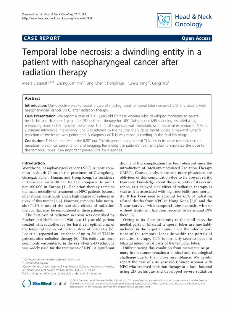
Figure 3 Coronal images also showed the same findings.
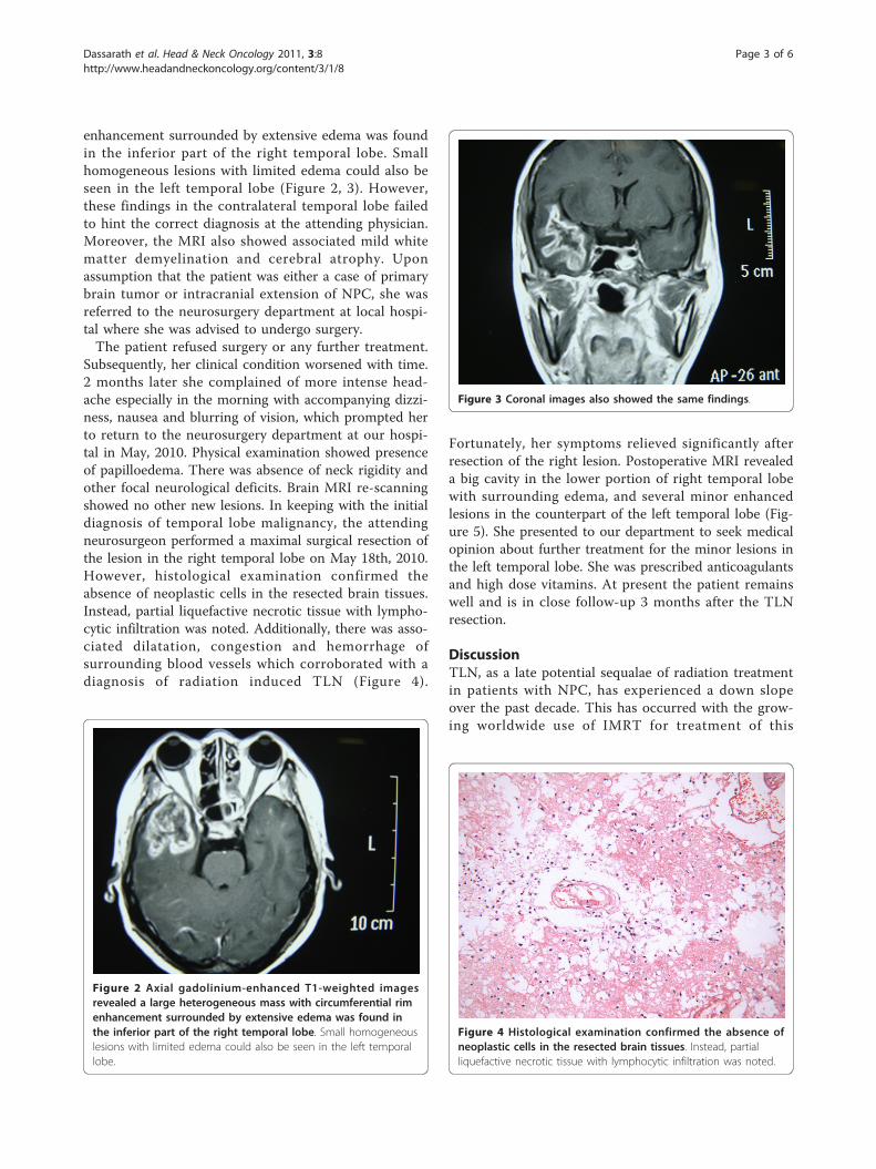
Figure 4 Histological examination confirmed the absence ofneoplastic cells in the resected brain tissues. Instead, partialliquefactive necrotic tissue with lymphocytic infiltration was noted.
Dassarath et al. Head & Neck Oncology 2011, 3:8http://www.headandneckoncology.org/content/3/1/8
Page 3 of 6

carcinoma which has provided a potentially therapeuticbenefit of dose escalation with reduced toxicity to nor-mal tissues [11]. Symptoms of TLN usually appear oneto three years after the last dose of radiation treatmentwith a silent latent interval noted between the end oftreatment and development of radiation-induced necro-sis [12]. Four main types of clinical presentations weredescribed in a study by Lee et al. They ranged fromcomplete absence of symptoms to vague features oftemporal lobe damage, symptoms of temporal lobe
epilepsy or nonspecific characteristics of intracraniallesions. The asymptomatic patients were incidentallydiagnosed during follow up with CT or MRI [7]. Theclinical progression of our patient was that of non-speci-fic symptoms to overt symptoms of raised intracranialpressure.Using 2D radiotherapy technique, inferior parts of
bilateral temporal lobes almost always receive the samedose as the tumor, especially for patients with locallyadvanced disease infiltrating the skull base or cavernous
Figure 5 Postoperative axial gadolinium-enhanced T1-weighted images a big cavity in the lower portion of right temporal lobe withsurrounding edema, and several minor enhanced lesions in the counterpart of the left temporal lobe.
Dassarath et al. Head & Neck Oncology 2011, 3:8http://www.headandneckoncology.org/content/3/1/8
Page 4 of 6

sinus. It has been reported that there is a 25% chance ofdeveloping TLN within 5 years following use of a totaldose of equal to or more than 62.5 Gy [13]. The patientreceived 70 Gy to her skull base using 2D techniquewhich put her at a high risk of developing radiationnecrosis. IMRT, on the other hand, has the advantage ofgenerating complicated 3D dose distributions to con-form closely to the target volume and the beam inten-sity can be optimized using computer algorithms. It hasbeen reported that IMRT provided excellent tumor tar-get coverage and allowed the delivery of a high dose tothe target with significant sparing of nearby critical nor-mal tissues [14]. Hence, it plays an important role indecreasing radiation-induced injuries in patients withNPC [15].The differential diagnoses of TLN include intracranial
extension of NPC, second primary intracranial neo-plasm, cerebral metastasis, meningeal spread and brainabscess [16]. Differentiation of tumor progression andradiation injury after radiation therapy is indispensablefor appropriate treatment [17]. CT, MRI and PET-CTare all useful tools for TLN diagnosis, but none is speci-fic. The characteristic features on MRI include masseffect, vasogenic edema and contrast enhancement.However, MRI alone cannot reliably discriminate tumorfrom radiation induced necrosis [18], even though thelatter can be associated with specific patterns ofenhancement such as “soap bubble” or “Swiss cheese”enhancement patterns [19]. The use of functional ima-ging like the FDG-PET and the proton MR spectroscopyhas shown some advantages in differentiating the enti-ties. But presently, they have not been widely used dueto their high cost or complicated techniques.A definitive diagnosis is obtained from surgical explora-tion and biopsy or removal of the necrotic mass [16],but this is not routinely justified. It may not strike thedoctor to include TLN as one of the differential diag-noses if he does not review the history and treatmentplan of the patient. By carefully correlating the history,the findings on physical examination, the laboratoryinvestigations with the features on imaging scans likebrain MRI and CT, a correct working diagnosis can beconfidently reached without resorting to biopsy [20-22].In our case report, a brain abscess can be excluded as
our patient had few symptoms and signs to support aninfectious disease. Ruling out hematogenous spread ofNPC is easy, as it is very rare and would unlikely be pre-sent bilaterally [23], while tumor extension is usuallyassociated with erosion of the skull base. Hence, radia-tion induced necrosis as diagnosis for our patient is sup-ported by the fact that the symptoms developed6 months after treatment, there was no history of asso-ciated fever and the physical findings were consistentwith mass effect of the lesion, and last but not the least,
the lesions on MRI were bilateral, involving the inferiorportions of the temporal lobes, which are usuallyincluded in the portals of radiation. However, in ourcase, this diagnosis may have evaded the attending doc-tor due to its dwindling existence and close similarity tobrain tumor and failure to correlate the bilateralism ofthe lesions with the treatment plan.Traditional treatment for TLN includes the use of
steroids, hyperbaric oxygen, antiplatelets, anticoagulants,high dose vitamins and surgery, but all have shown alimited efficacy [12,24]. Steroids have been used to pro-vide prompt symptomatic relief and help in retardingthe pathologic process [25]. Surgery is used as the lastresort palliative measure in patients with significantincrease in intracranial pressure or in those who haveprogressive neurologic deficits despite steroids or othermedical therapy. However, in NPC patients who usuallyhave bilateral temporal lobe involvement, surgery can behazardous. The possible risk of Kluver Bucy syndrome isof concern when considering bilateral temporal lobect-omy [7]. The advent of bevacizumab, a humanizedmonoclonal antibody that inhibits vascular endothelialgrowth factor (VEGF) has paved the way to possibilitiesof reversing the pathogenesis of TLN and providing per-manent results. Evidence is now available that justifiesthe consideration of its use in the treatment of radiationnecrosis secondary to treatment of head and neckcancers [26-28].
ConclusionAlthough TLN has been dwindling since the introduc-tion of IMRT, it should always be kept in mind whenencountering temporal lobe lesions in NPC after radia-tion therapy. The diagnostic quagmire of TLN lies in itsclose resemblance to neoplasm on clinical presentationand imaging appearance. This can be achieved by com-bining the history, physical examination findings, withabnormalities seen on imaging tools like MRI.
ConsentWritten informed consent was obtained from the patientfor publication of this case report and accompanyingimages. A copy of the written consent is available forreview by the Editor-in-Chief of this journal.
Author details1Cancer Center, Union Hospital, Tongji Medical College, Huazhong Universityof Science and Technology, Wuhan, Hubei, 430022, PR China. 2Departmentof Oncology, Queen Victoria Hospital, Candos, Quatre-Bornes, Mauritius.
Authors’ contributionsKY and GW have made substantial contributions to conception and design,and acquisition, analysis and interpretation of data. MD, ZY, HL, KY havebeen involved in drafting the manuscript or revising it critically for importantintellectual content. All authors read and approved the final manuscript.
Dassarath et al. Head & Neck Oncology 2011, 3:8http://www.headandneckoncology.org/content/3/1/8
Page 5 of 6

Competing interestsThe authors declare that they have no competing interests.
Received: 22 November 2010 Accepted: 10 February 2011Published: 10 February 2011
References1. Hildesheim A, Levine PH: Etiology of nasopharyngeal carcinoma: a review.
Epidem Rev 1993, 15:466-485.2. Marcial VA, Hanley JA, Chang C, Davis LW, Moscol JA: Split-course
radiation therapy of carcinoma of the nasopharynx: results of a nationalcollaborative clinical trial of the Radiation Therapy Oncology Group. Int JRadiat Oncol Biol Phys 1980, 6:409-414.
3. Al-Sarraf M, Pajak TF, Cooper JS, Mohiuddin M, Herskovic A, Ager PJ:Chemo-radiotherapy in patients with locally advanced nasopharyngealcarcinoma: a Radiation Therapy Oncology Group study. J Clin Oncol 1990,8:1342-1351.
4. Al-Sarraf M, LeBlanc M, Giri PG, et al: Chemoradiotherapy versusradiotherapy in patients with advanced nasopharyngeal cancer: phase IIIrandomized Intergroupstudy 0099. J Clin Oncol 1998, 16:1310-1317.
5. Fischer AW, Holfelder H: lokales amyloid in Gehirn. Dtsch Z Chir 1930,227:475-483.
6. Lee AWM, Law SCK, Ng SH, et al: Retrospective analysis ofnasopharyngeal carcinoma treated during 1976-1985:late complicationsfollowing megavoltage irradiation. Br J Radiol 1992, 65:918-928.
7. Lee AWM, Ng SH, Ho JHC, et al: Clinical diagnosis of late temporal lobenecrosis following radiation therapy for nasopharyngeal carcinoma.Cancer 1988, 61:1535-1542.
8. Lee AWM, Cheng LOC, Ng SH, et al: Magnetic resonance imaging in theclinical diagnosis of late temporal lobe necrosis following radiotherapyfor nasopharyngeal carcinoma. Clin Radiol 1990, 42:24-31.
9. Chan JK, Bray F, McCarron P, et al: Nasopharyngeal carcinoma. InPathology and genetics of head and neck tumors. World Health Organizationclassification of tumors. Edited by: Barnes EL, Eveson JW, Reichart P,Sidransky D. Lyon, France: IARC Press; 2005:85-97, Feb.4th, 2010.
10. Greene FL, Page DL, Fleming ID, Editors, et al: AJCC Cancer StagingManual. New York: Springer;, 6 2002, 47.
11. Teo PML, Ma BBY, Chan ATC: Radiotherapy for nasopharyngealcarcinoma–transition from two-dimensional to three-dimensionalmethods. Radiother Oncol 2004, 73:163-172.
12. Giglio P, Gilbert MR: Cerebral radiation necrosis. Neurology 2003, 9:180-188.13. Marks JE, Wong J: The risk of cerebral radiation necrosis in relation to
dose, time and fractionation. Prog Exp Tumor Res 1985, 29:210-218.14. Lee N, Xia P, Quivey JM, et al: Intensity-modulated radiotherapy in the
treatment of nasopharyngeal carcinoma: an update of the UCSFexperience. Int J Radiat Oncol Biol Phys 2002, 53:12-22.
15. Cheng JCH, Chao KSC, Low D: Comparison of Intensity ModulatedRadiationTherapy (IMRT) Treatment Techniques for NasopharyngealCarcinoma. Int J Cancer (Radiat Oncol Invest) 2001, 96:126-131.
16. Glass JP, Hwang TL, Leavens ME, Libshitz HI: Cerebral radiation necrosisfollowing treatment of extracranial malignancies. Cancer 1984,54:1966-1972.
17. Schlemmera HP, Bacherta P, Herfartha KK, Zunaa I, Debusa J, van Kaicka G,Proton MR: Spectroscopic Evaluation of Suspicious Brain Lesions AfterStereotactic Radiotherapy. Amer J of Neuroradiol 2001, 22:1316-1324.
18. Bonavita S, Di Salle F, Tedeschi G: Proton MRS in neurological disorders.Eur J Radiol 1999, 30:125-131.
19. Kumar AJ, Leeds NE, Fuller GN, et al: Malignant gliomas: MR imagingspectrum of radiation therapy- and chemotherapy-induced necrosis ofthe brain after treatment. Radiology 2000, 217:377-384.
20. Martins AN, Johnson JS, Henry JM, Stoffel TJ, Di Chiro G: Delayed radiationnecrosis of the brain. J Neurosurg 1977, 47:336-345.
21. Shaw PJ, Bates D: Conservative treatment of delayed cerebral radiationnecrosis. J Neurol Neurosurg Psychiatry 1984, 47:1338-1341.
22. Safdari H, Castan P, Dubois JB, et al: Lesions cerebralespostradiotherapiques. Rev Neurol (Paris) 1985, 141:553-561.
23. Ho J: Treatment of cancer. London: Chapman & Hall; 1982, 249.24. Glantz MJ, Burger PC, Friedman AH, et al: Treatment of radiation induced
nervous system injury with heparin and warfarin. Neurology 1994,44:2020-2027.
25. Eyster EF, Nielsen SL, Scheline GE, Wilson CB: Cerebral radiation necrosissimulating a brain tumor. J Neurosurg 1974, 39:267-271.
26. Gonzales J, Kumar AJ, Conrad CA, Levine VA: Effect of bevacizumab onradiation necrosis of the brain. Int J Radiat Oncol Biol Phys 2007,67:323-326.
27. Tocurator R, Zuniga R, Mohan YS, et al: Initial experince with bevacizumabtreatment for biopsy confirmed cerebral radiation necrosis. J Neurooncol2009, 94:63-68.
28. Levin VA, Bidaut L, Hou P, et al: Randomized Double-Blind Placebo-Controlled Trial of Bevacizumab Therapy for Radiation Necrosis of theCentral Nervous System. Int J Radiat Oncol Biol Phys .
doi:10.1186/1758-3284-3-8Cite this article as: Dassarath et al.: Temporal lobe necrosis: a dwindlingentity in a patient with nasopharyngeal cancer after radiation therapy.Head & Neck Oncology 2011 3:8.
Submit your next manuscript to BioMed Centraland take full advantage of:
• Convenient online submission
• Thorough peer review
• No space constraints or color figure charges
• Immediate publication on acceptance
• Inclusion in PubMed, CAS, Scopus and Google Scholar
• Research which is freely available for redistribution
Submit your manuscript at www.biomedcentral.com/submit
Dassarath et al. Head & Neck Oncology 2011, 3:8http://www.headandneckoncology.org/content/3/1/8
Page 6 of 6