temporomandibular joint in health and disease a …mandibular notch, separating the two processes,...
TRANSCRIPT

UNIVERSITA’ DEGLI STUDI DI MILANO
SCUOLA DI DOTTORATO IN SCIENZE MORFOLOGICHE, FISIOLOGICHE E DELLO SPORT
DIPARTIMENTO DI MORFOLOGIA UMANA E SCIENZE BIOMEDICHE – CITTA’ STUDI
DOTTORATO DI RICERCA IN SCIENZE MORFOLOGICHE – XXIV CICLO
BIO16
TEMPOROMANDIBULAR JOINT IN HEALTH AND DISEASE A 3D MORPHOFUNCTIONAL ANALYSIS
Tesi di Dottorato di
ANDREA MAPELLI
MATRICOLA: R08124
Tutor: prof.ssa Chiarella Sforza
Coordinatore: prof.ssa Laura Vizzotto
Anno Accademico 2010/2011

TABLE OF CONTENTS
ACKNOWLEDGEMENTS.................................................................................... 1
ABSTRACT............................................................................................................ 2
OVERVIEW............................................................................................................ 3
ANATOMY & FUNCTION................................................................................... 6
THE TEMPOROMANDIBULAR JOINT....................................................................... 6
THE MASTICATORY MUSCLES................................................................................. 12
MANDIBULAR KINEMATICS..................................................................................... 16
MATERIAL & METHODS................................................................................... 20
INSTRUMENTATIONS................................................................................................. 20
ANALYZED STOMATOGNATHIC FUNCTIONS...................................................... 23
RECORDING PROTOCOL............................................................................................ 26
MEASUREMENT PROTOCOL..................................................................................... 29
STUDY I – HEALTHY SUBJECTS..................................................................... 38
INTRODUCTION........................................................................................................... 38
METHODS...................................................................................................................... 38
RESULTS........................................................................................................................ 40
DISCUSSION................................................................................................................. 44
STUDY II – PATIENTS WITH MODERATE TMD........................................... 50
INTRODUCTION........................................................................................................... 50
METHODS...................................................................................................................... 51
RESULTS........................................................................................................................ 53
DISCUSSION.................................................................................................................. 57
STUDY III – PATIENTS WITH DIAGNOSIS OF CLASS III
DENTOSKELETAL DEFORMITY, BEFORE AND AFTER
ORTHOGNATHIC SURGERY.............................................................. .............. 60
INTRODUCTION........................................................................................................... 60
METHODS...................................................................................................................... 61
RESULTS........................................................................................................................ 62
DISCUSSION.................................................................................................................. 66
GENERAL CONCLUSIONS................................................................................. 69

LIST OF ABBREVIATIONS................................................................................ 70
REFERENCES....................................................................................................... 72

1
ACKNOWLEDGEMENTS
Per la realizzazione di questo lavoro devo ringraziare in primis la professoressa Chiarella, per la
sua costante SUPERvisione, e con lei la professora Claudia e i dottori/dottoresse Fernanda,
Marzia, Alessandro, Claudinha, Marcio, Andrè, Emanuele, Giorgio e Riccardo per il prezioso
contributo.

2
ABSTRACT
Quantitative, objective and accurate evaluation of masticatory muscle activity and jaw
movement is mandatory for a better understanding of the normal function and dysfunction of the
stomatognathic apparatus.
A non-invasive recording protocol, integrating an electromyographic system and an
optoelectronic 3D-motion analyzer, has been developed and used to perform multifactorial
estimations of TMJ functioning. The masticatory system has been objectively quantified,
assessing bite stability, mandibular border movements and chewing performance, in both healthy
and pathologic individuals. Three separate investigations have been made.
In the first study, functional symmetries of the craniofacial complex involving the patterns of
jaw movements and the activities of masticatory muscles were assessed in a control group of
clinically healthy subjects. Data were evaluated separately for men and women, and a gender-
related effect was tested, together with the potential influence of mandibular dimensions.
In the second study, the same complete protocol was employed to analyze the masticatory
function in patients with mild-moderate temporomandibular disorders, in order to provide new
insight concerning a still controversial pathology.
The aim of the third study was to assess the recovery of mandibular range of motion in border
movements, focusing on the potential changes in mandibular condylar motion, analyzed in
untreated patients with skeletal Class III malocclusions, and patients who had received
orthognathic surgery for the correction of this dentoskeletal deformity.
The outcomes suggest that the proposed method could be a useful tool to evaluate the
neuromuscular coordination during the performance of static and dynamic masticatory activities,
and to detect functionally altered stomatognathic conditions.
Diagnosis of alterations of the stomatognathic apparatus, and assessment of the effects of
therapy, would both profit from this quantitative approach, thus reducing the discordance among
several clinical examinations.
Key words
Human temporomandibular joint; 3D motion analysis; electromyography; health; disease.
3
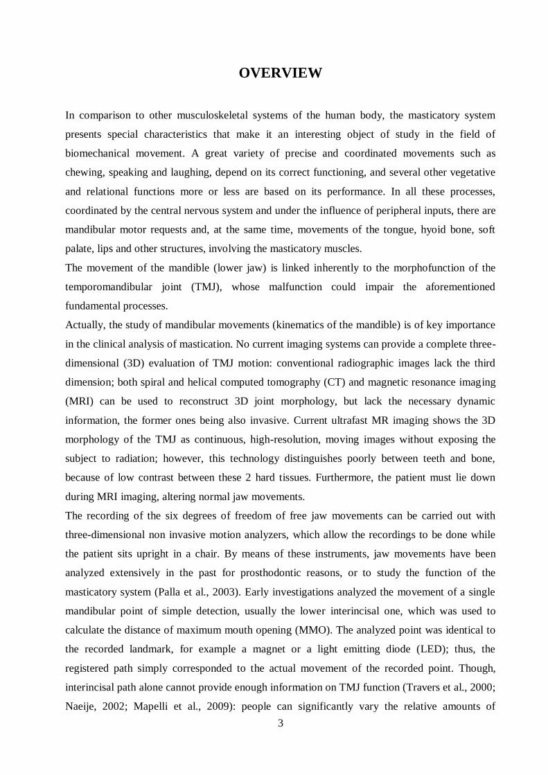
OVERVIEW
In comparison to other musculoskeletal systems of the human body, the masticatory system
presents special characteristics that make it an interesting object of study in the field of
biomechanical movement. A great variety of precise and coordinated movements such as
chewing, speaking and laughing, depend on its correct functioning, and several other vegetative
and relational functions more or less are based on its performance. In all these processes,
coordinated by the central nervous system and under the influence of peripheral inputs, there are
mandibular motor requests and, at the same time, movements of the tongue, hyoid bone, soft
palate, lips and other structures, involving the masticatory muscles.
The movement of the mandible (lower jaw) is linked inherently to the morphofunction of the
temporomandibular joint (TMJ), whose malfunction could impair the aforementioned
fundamental processes.
Actually, the study of mandibular movements (kinematics of the mandible) is of key importance
in the clinical analysis of mastication. No current imaging systems can provide a complete three-
dimensional (3D) evaluation of TMJ motion: conventional radiographic images lack the third
dimension; both spiral and helical computed tomography (CT) and magnetic resonance imaging
(MRI) can be used to reconstruct 3D joint morphology, but lack the necessary dynamic
information, the former ones being also invasive. Current ultrafast MR imaging shows the 3D
morphology of the TMJ as continuous, high-resolution, moving images without exposing the
subject to radiation; however, this technology distinguishes poorly between teeth and bone,
because of low contrast between these 2 hard tissues. Furthermore, the patient must lie down
during MRI imaging, altering normal jaw movements.
The recording of the six degrees of freedom of free jaw movements can be carried out with
three-dimensional non invasive motion analyzers, which allow the recordings to be done while
the patient sits upright in a chair. By means of these instruments, jaw movements have been
analyzed extensively in the past for prosthodontic reasons, or to study the function of the
masticatory system (Palla et al., 2003). Early investigations analyzed the movement of a single
mandibular point of simple detection, usually the lower interincisal one, which was used to
calculate the distance of maximum mouth opening (MMO). The analyzed point was identical to
the recorded landmark, for example a magnet or a light emitting diode (LED); thus, the
registered path simply corresponded to the actual movement of the recorded point. Though,
interincisal path alone cannot provide enough information on TMJ function (Travers et al., 2000;
Naeije, 2002; Mapelli et al., 2009): people can significantly vary the relative amounts of

4
condylar translation and rotation and still have similar amounts of opening at the incisors
(Salaorni and Palla, 1994; Monteverdi et al., 2006).
Nowadays, TMJ kinematic behaviour can be efficiently and accurately detected by
optoelectronic tracking systems, which allow the non invasive direct/ indirect recording of
multiple mandibular points in all six degrees of freedom (Piehslinger et al., 1993; Salaorni and
Palla, 1994; Koolstra and van Eijden, 1995; Yatabe et al., 1995, 1997; Lotters et al., 1996;
Zwijnenburg et al., 1996; Gallo et al., 1997; Merlini and Palla, 1988; Catic and Naeije, 1999;
Lobbezoo et al., 2000; Travers et al., 2000; Lewis et al., 2001; Naeije, 2002; Ferrario et al.,
2005; Mapelli et al., 2009). In particular, the three-dimensional condylar motion can be picked
out, thus offering more insight for TMJ functional evaluation. Though, the analysis of the
movement of a condylar point is more complex, as it is impossible to record its trajectory
directly; consequently, this must be geometrically reconstructed on the basis of biomechanical
models and mathematical calculations. Accordingly, it is also possible to investigate the relative
contribution of rotation (condyle-disc compartment) and translation (mandibular fossa-disc
compartment) along all condylar paths in both mouth opening and closing, allowing a deeper
understanding of the normal joint motion (Mapelli et al., 2009).
From a clinical point of view, it would be interesting to perform such a detailed assessment of
condyle-disc motion in patients with joint alterations (Merlini and Palla, 1988; Catic and Naeije,
1999), quantifying the different performance of the mandibular fossa-disc and condyle-disc TMJ
compartments from normal individuals. For instance, Sforza et al. (2009) observed that patients
rehabilitated after a condylar fracture showed modification of the rotation/translation
components of mouth opening despite a good recovery of total mandibular movement. Findings
like that can be of help in redirecting treatment plans.
Beside multiple investigations which have evaluated the diagnostic potential of the mandibular
motion analyzers (Karlsson and Carlsson, 1990; Travers et al., 2000; Wintergerst et al., 2004;
Hansdottir and Bakke, 2004; Miyawaki et al., 2004; Bianchini et al., 2008; Rilo et al., 2009;
Wang et al., 2009; Sforza et al., 2009, 2010b), other researchers have proved the reliability of
surface electromyography in the stomatognathic functional analysis (Pinho et al., 2000; Ferrario
et al., 2007; Ries et al., 2008; Tartaglia et al., 2008a, 2008b; Ardizone et al., 2010; Forrester et
al., 2010).
Surface electromyography (EMG) has been used since the early 1950s for studying the action of
the superficial masseter and temporal muscles during mastication. Currently it is a part of patient
assessment in dentistry (Ferrario et al., 2006b), providing quantitative data on the function of
superficial muscles with minimal discomfort to the patient and without invasive or dangerous

5
procedures. Indeed, when well-standardized protocols are used (in order to solve problems like
the wrong positioning of the electrodes, the difference of impedance of the patient’s skin, the
muscle cross-talk, etc.), surface EMG of the head muscles has been reported to be an effective
method for the functional assessment of the stomatognathic apparatus (Farella et al., 2003;
Garcia-Morales et al., 2003; Ciuffolo et al., 2005), with a good repeatability (Kogawa et al.,
2006; Ferrario et al., 2006b; De Felicio et al., 2009b).
Diagnosis of alterations of the stomatognathic apparatus, and assessment of the effects of
therapy, would both profit from a quantitative approach, thus reducing the discordance among
several clinical examinations (Schmitter et al., 2005; Manfredini et al., 2006). Objective
measurements are also needed by insurances and forensic medicine (Tartaglia et al., 2008a).
Over the last 20 years, the Functional Anatomy Research Centre (FARC) of the Dipartimento di
Morfologia Umana e Scienze Biomediche “Città Studi”, Università degli Studi di Milano (Italy),
has devised and developed, in parallel, a protocol for TMJ kinematic analysis (latest version in
Mapelli et al., 2009) and a protocol for the characterization of masticatory muscles activity
(latest version in Tartaglia et al., 2008b). The former is currently applied to both mandibular
border movements (Ferrario et al., 2005; Mapelli et al., 2009; Sforza et al., 2009, 2010b) and
gum chewing; the latter is used for both jaw clenching (Ferrario et al., 2004b, 2006b, 2007;
Tartaglia et al., 2008b, 2011; De Felicio et al., 2009b) and chewing (Ferrario and Sforza 1996;
Ferrario et al., 1999, 2004b; Dellavia et al., 2007; Tartaglia et al., 2008a).
In the present investigation, the two protocols have been applied to analyse both the mandibular
kinematics and the electromyographic characterization of the masticatory muscles in:
- healthy subjects (Study I)
- patients with moderate temporomandibular disorders (Study II)
- patients with diagnosis of class III dentoskeletal deformity, before and after orthognathic
surgery (Study III).
The general purpose was to investigate the stomatognathic morphology and function during the
performance of standardized static (clenching) and dynamic (border movements and chewing,
neuromuscular coordination) tasks.

6
ANATOMY and FUNCTION
THE TEMPOROMANDIBULAR JOINT
The temporomandibular joint (TMJ), one of the most complex joints in the body, is the bilateral
synovial articulation between the mandible and the skull, composed of the condylar heads of the
mandible and the articular eminence of the right and left temporal bones, covered by dense,
fibrous connective tissue and surrounded by several ligaments. Interposed between the
incongruent articulating surfaces is an articular disc, contoured to fit over the head of the condyle
and into the concavity of the mandibular fossa, which compartmentalizes the joint into two
separate synovial-lined cavities (fig.1).
Figure 1. Sagittal section through the left temporomandibular joint (Williams et al., 1989).
The mandible (or lower jaw), the unpaired bone of the stomatognathic apparatus, is the largest,
strongest and lowest bone in the face. It has a horizontally U-shaped body, convex forward, and
two broad rami, ascending posteriorly. The mandibular ramus is quadrilateral, with two surfaces,
four borders and two processes: the coronoid process and the condyloid process (fig. 2). The
mandibular notch, separating the two processes, is a deep semilunar depression, and is crossed
by the masseteric vessels and nerve.
7
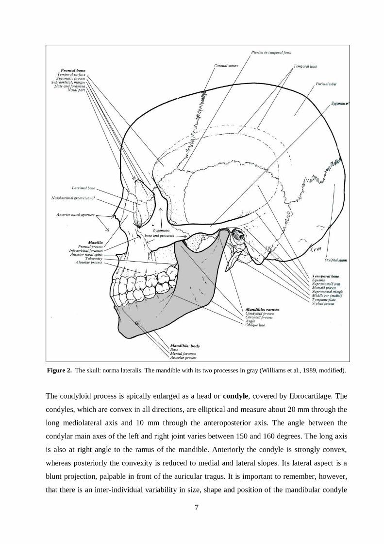
Figure 2. The skull: norma lateralis. The mandible with its two processes in gray (Williams et al., 1989, modified).
The condyloid process is apically enlarged as a head or condyle, covered by fibrocartilage. The
condyles, which are convex in all directions, are elliptical and measure about 20 mm through the
long mediolateral axis and 10 mm through the anteroposterior axis. The angle between the
condylar main axes of the left and right joint varies between 150 and 160 degrees. The long axis
is also at right angle to the ramus of the mandible. Anteriorly the condyle is strongly convex,
whereas posteriorly the convexity is reduced to medial and lateral slopes. Its lateral aspect is a
blunt projection, palpable in front of the auricular tragus. It is important to remember, however,
that there is an inter-individual variability in size, shape and position of the mandibular condyle

8
that may be caused by any one or a combination of factors, including heredity and functional
adaptation.
On the temporal bone, the TMJ occupies the inferior surface of the zygomatic process, on the
posterior surface of the articular eminence. The articular eminence is the strongly convex bony
elevation on the root of the zygomatic process representing the anterior-most boundary of the
articular or mandibular fossa (also referred to as the glenoid fossa). In particular, the articular
tubercle is the bony knob on the lateral aspect of the articular eminence, where the fibrous
capsule and the temporomandibular ligament attach.
The articular surfaces of the TMJ are covered by dense, collagenous connective tissue overlying
a thin proliferative layer of cells associated with the underlying hyaline cartilage. It is reported
that the hyaline cartilage of the condyle is present while the individual is still growing, until
about 20 years of age, whereas the cartilage covering the articular eminence has a shorter life
span. At the termination of growth, this cartilage layer is replaced by compact bone. In the adult,
the compact bone of the condyle is covered by a layer of fibrocartilage that, in turn, is covered
by a thin layer of proliferative tissue. Cells of the proliferative layer may become activated to
function in remodeling of the joint as a result of changes in function, wear, and tooth movement.
Superficial to the proliferative layer is a relatively thick layer of dense, irregular collagenous
connective tissue whose deeper layers house fibroblasts. Although the articular structures are
avascular, they are bathed in synovial fluid, which provides lubrication and nourishment for the
cellular coverings.
The articular disc is the primary mechanism of stress distribution and lubrification within the
TMJ. It is a deformable, dense, and fibrous connective tissue plate that is oval and contoured to
fit between the mandibular condyle and the articular eminence of the temporal bone. The inferior
surface of the disc is concavely contoured to fit the convex condyle of the mandible. Superiorly,
its surface is sagittally concavo-convex. The convex posterior portion conforms to the concave
mandibular fossa, whereas anteriorly, the disc becomes concave to fit the convex posterior
aspect of the articular eminence. The disc is thicker at its periphery and thinner at the load-
bearing area of the joint (that occasionally, especially in older individuals, becomes perforated).
Peripherally, the disc becomes less dense as it merges into the surrounding fibrous capsule and,
in front, it blends with the tendon of lateral pterygoid muscle (fig. 3).

9
Figure 3. Form, subdivisions and thickness variations of the intra-articular disc in the TMJ. Lateral aspect (a) and
sagittal section (b) (Williams et al., 1989).
The peripheral regions of the disc are very vascular, whereas the central, stress-bearing portion is
devoid of blood vessels. Posteriorly, the disc is attached to a highly vascular connective tissue
known as the retrodiscal tissue: a venous plexus separates upper and lower layers, the upper band
of fibro-elastic tissue is attached to the fossa posterior margin, while the lower band (non-elastic
fibrous tissue) is attached to the posterior surface of the condyle. During mandibular movements,
the geometric relationships of the TMJ articular surfaces vary, so that the disc undergoes stress
concentrations that change with time and location. The primary function of the deformable
visco-elastic articular disc is to permit these activities while reducing the risk of trauma.
The TMJ capsule, composed of dense, irregular collagenous connective tissue, encloses the
entire articulating region of the temporal bone, disc, and mandibular condyle, sealing the joint
space. The joint capsule is evident only laterally (fig. 4), while medially, anteriorly and
posteriorly is not a recognizable entity independent from the disc connections with the temporal
and condylar surface (Sforza et al., 2010a).
a b

10
Figure 4. Lateral aspect of a right human TMJ capsule (courtesy of prof. Simone C.H. Regalo, University of Sao Paulo, Brasil).
It attaches laterally to the longitudinal root of zygomatic process, medially to the sphenoid spine,
inferiorly to the neck of condyle, anteriorly to the front edge of articular tubercle (the limit is not
clear and seems to continue with the lateral pterygoid muscle), posteriorly to the front edge of
the petrosquamous suture. Above the articular disc the capsule is loose and it is taut below it.
The placement of the disc between the two articulating bones and its peripheral attachments to
the walls of the capsule causes the capsular space to be divided into two separate superior and
inferior synovial compartments. The larger, superior compartment between the disc and temporal
bone permits some freedom of movement between the disc and articular eminence. Anteriorly,
the capsule and disc are tightly fused, permitting the insertion of some fibers of the lateral
pterygoid muscle into the disc. Medially and laterally, the capsule and disc are attached to the
condyle margins, thus necessitating associated simultaneous movement of the condyle and disc.
The inferior compartment encloses the entire neck of the mandible and is more firmly attached to
the disc. This attachment prohibits excessive movement between the disc and condyle. The joint
capsule is richly endowed with sensory endings from the mandibular division of the trigeminal
nerve, most of which are supplied from its auriculo-temporal and masseteric branches. Vascular
supply to the joint is provided by branches of the superficial temporal and maxillary arteries as
they approximate the joint.
Several ligaments strengthen the joint and limit the movements.
Two short, strong collateral ligaments (discal ligaments) serve to anchor the medial and lateral
borders of the articular disc to the poles of the condyle, ensuring that disc and condyle move
together in protraction and retraction.
Reinforcements of the joint capsule along its lateral margin by obliquely oriented bundles of
collagenous fibers are responsible for naming this pronounced lateral portion of the capsule, the
lateral ligament or temporomandibular ligament (fig. 5).

11
Figure 5. The left temporomandibular joint: lateral aspect (Williams et al., 1989).
The temporomandibular ligament possesses two separate bands of fibers, whose directions are
oblique to each other. The superficial layer, which is more extensive, arises as a broad band from
the lateral surface of the articular eminence at the articular tubercle. The ligament narrows as it
passes obliquely inferior and posterior to be inserted on the posterolateral aspect of the
mandibular neck just inferior to the lateral pole of the condyle. The smaller, medially situated
portion of the lateral ligament arises from the crest of the eminence to pass almost horizontally to
insert into the lateral aspect of the condyle. The lateral ligament permits free movement in the
anteroinferior direction, but its superficial portion prevents lateral movement, whereas the deeper
horizontal portion prevents posterior displacement of the condyle.
Two additional ligaments are considered accessory to the TMJ. The sphenomandibular ligament
(fig. 6), medial to and separate from the capsule, is a flat, thin band descending from the
sphenoidal spine and widening to reach the lingula of the mandibular foramen. Superolateral to it
there are the lateral pterygoid muscle and auriculo-temporal nerve; inferior to this it is separated
from the mandibular neck by the maxillary vessels, below which the inferior alveolar vessels and
nerve and a parotid gland separate it from the mandibular ramus.

12
Figure 6. The left temporomandibular joint: medial aspect (Williams et al., 1989).
The stylomandibular ligament, the other accessory ligament, is a specialization of the deep
cervical fascia. This medial ligament extends as a thin band from the apex of the styloid process
of the temporal bone to the posterior border of the angle and ramus of the mandible.
Although the precise functions of these two accessory ligaments are not fully understood as they
relate to the TMJ, it has been suggested that the sphenomandibular ligament assists in limiting
lateral mandibular movement, whereas the stylomandibular ligament apparently assists in
limiting the anterior extent of protrusion of the mandible (Williams et al., 1989; Hiatt and
Gartner, 2010).
THE MASTICATORY MUSCLES
The muscles of mastication are a set of four bilateral muscles (the temporalis, medial pterygoid,
lateral pterygoid, and masseter) whose function is to move the mandible about the
temporomandibular joint as it occurs in phonation, chewing (mastication), and swallowing. All
of these muscles, excepting the masseter muscle, originate from either the temporal or
infratemporal fossae and insert upon the medial aspect of the mandible. The masseter muscle, in
contrast, originates on the zygomatic arch and inserts upon the lateral aspect of the mandible.
The epimysia that cover these muscles become the fascia encircling the masticator compartment,
which contains the four muscles of mastication and the ramus of the mandible. Prolongations of
the buccal fat pad fill in the spaces between the muscles of mastication deep to the mandible.

13
The temporalis muscle is a fan-shaped muscle originating on the bones of the broad temporal
fossa (fig. 7). Specifically, the site of origin extends inferiorly from the inferior temporal line
over the entire temporal fossa, including parts of the parietal and most of the squama of the
temporal bones, and the greater wing of the sphenoid, including its infratemporal crest and the
temporal surface of the frontal bone. Occasionally, some fibers arise from the posterior temporal
surface of the frontal process of the zygoma. The muscle bundles converge to insert as a tendon
on the coronoid process of the mandible and down along its anterior surface and the anterior
border of the ramus as far anteriorly as the third molar. The anterior fibers of this muscle are
vertically directed from origin to insertion, whereas the middle fibers are oblique and the
posterior fibers are almost horizontal.
The muscle is primarily an elevator of the mandible; however, because of the directional
alignments of the muscle fibers, the posterior and middle portions of the muscle are reported to
act also in retracting the mandible.
The temporalis muscle is innervated by anterior and posterior deep temporal nerves from the
mandibular division of the trigeminal nerve. The nerves enter the muscle from its deep aspect in
the temporal fossa. Vascularization is supplied via branches of the superficial temporal and
maxillary arteries. Arising from the former is the middle temporal artery, which enters the
muscle on its superficial aspect. Anterior and posterior deep temporal arteries, arising from the
maxillary artery, accompany the like-named nerves and enter the deep aspect of the muscle,
where they anastomose with the middle temporal artery.
Figure 7. Model of the skull with left temporalis and masseter muscles.
Temporalis
Masseter
14
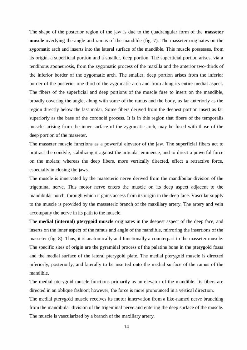
The shape of the posterior region of the jaw is due to the quadrangular form of the masseter
muscle overlying the angle and ramus of the mandible (fig. 7). The masseter originates on the
zygomatic arch and inserts into the lateral surface of the mandible. This muscle possesses, from
its origin, a superficial portion and a smaller, deep portion. The superficial portion arises, via a
tendinous aponeurosis, from the zygomatic process of the maxilla and the anterior two-thirds of
the inferior border of the zygomatic arch. The smaller, deep portion arises from the inferior
border of the posterior one third of the zygomatic arch and from along its entire medial aspect.
The fibers of the superficial and deep portions of the muscle fuse to insert on the mandible,
broadly covering the angle, along with some of the ramus and the body, as far anteriorly as the
region directly below the last molar. Some fibers derived from the deepest portion insert as far
superiorly as the base of the coronoid process. It is in this region that fibers of the temporalis
muscle, arising from the inner surface of the zygomatic arch, may be fused with those of the
deep portion of the masseter.
The masseter muscle functions as a powerful elevator of the jaw. The superficial fibers act to
protract the condyle, stabilizing it against the articular eminence, and to direct a powerful force
on the molars; whereas the deep fibers, more vertically directed, effect a retractive force,
especially in closing the jaws.
The muscle is innervated by the masseteric nerve derived from the mandibular division of the
trigeminal nerve. This motor nerve enters the muscle on its deep aspect adjacent to the
mandibular notch, through which it gains access from its origin in the deep face. Vascular supply
to the muscle is provided by the masseteric branch of the maxillary artery. The artery and vein
accompany the nerve in its path to the muscle.
The medial (internal) pterygoid muscle originates in the deepest aspect of the deep face, and
inserts on the inner aspect of the ramus and angle of the mandible, mirroring the insertions of the
masseter (fig. 8). Thus, it is anatomically and functionally a counterpart to the masseter muscle.
The specific sites of origin are the pyramidal process of the palatine bone in the pterygoid fossa
and the medial surface of the lateral pterygoid plate. The medial pterygoid muscle is directed
inferiorly, posteriorly, and laterally to be inserted onto the medial surface of the ramus of the
mandible.
The medial pterygoid muscle functions primarily as an elevator of the mandible. Its fibers are
directed in an oblique fashion; however, the force is more pronounced in a vertical direction.
The medial pterygoid muscle receives its motor innervation from a like-named nerve branching
from the mandibular division of the trigeminal nerve and entering the deep surface of the muscle.
The muscle is vascularized by a branch of the maxillary artery.

15
Figure 8. Left pterygoid muscles: the zygomatic arch and part of the ramus of the mandible have been removed
(Williams et al., 1989).
The lateral (external) pterygoid muscle is a short muscle, filling the remainder of the
infratemporal fossa and covering much of the medial pterygoid muscle. This muscle possesses
two heads of origin (fig. 8). The smaller, superior head originates from the infratemporal region
of the greater wing of the sphenoid bone as far laterally as the infratemporal crest. The larger,
inferior head originates from the lateral surface of the lateral pterygoid plate. The fibers of the
superior head course posteriorly and laterally in an almost horizontal direction from the
infratemporal crest. Fibers of the inferior head are directed posteriorly, laterally, and slightly
superiorly on their way to the mandible. Though the two heads of origin are separated from each
other, their fibers converge as they approach the site of insertion on and about the mandible. The
superior head inserts into the articular capsule of the TMJ, the anterior border of the articular
disc, and the superior part of the mandibular neck. The inferior head inserts along the anterior
surface of the mandibular neck.
The lateral pterygoid muscle is described classically as the jaw opener, which protrudes the
mandible and moves the mandible from side to side when functioning unilaterally. In particular,
the superior head, attached to the articular capsule and disc, functions in stabilizing the
mandibular condyle, whereas the inferior head is reported to function in pulling the mandible
and disc forward and down, effecting jaw opening (Ferrario and Sforza, 1992).

16
The lateral pterygoid muscle is innervated by a branch entering its deep surface from either the
anterior division separately or as a branch of the buccal nerve from the mandibular division of
the trigeminal nerve. Vascular supply is provided by a branch from the maxillary artery as it
passes either superficial or deep to the muscle.
The origins and insertion sites of these muscles on the mandible dictate the joint function.
Generally, the functions are for opening or closing the jaw; however, subtle variations exist
when muscles are acting antagonistically or synergistically with other muscles on one side or the
other or on both sides (Williams et al., 1989; Hiatt and Gartner, 2010).
MANDIBULAR KINEMATICS
The bilateral TMJs are connected through the mandible and, therefore, the articulations function
as a single unit rather than independently. The TMJ is composed essentially of two convex
structures opposed to each other, with an intermediate articular disc placed between them.
Considering the anatomy of the disc, it becomes clear that movement within the TMJ is basically
of two types. Ginglymus (hinge) movement is possible between the condyles of the mandible
and the inferior surface of the discs. The other permitted movement within the joint is an
arthrodial (gliding) motion (Ferrario et al., 2005). This is possible as the superior surface of the
articular disc slides down at the articular eminence. Therefore, the TMJ is considered a
ginglymo-arthrodial joint: the condyle/disc movement is rotatory, whereas the disc/temporal
bone motion is translational.
Functionally, the mandible can be depressed or elevated, protruded or retracted and, since both
joints always act together but may differ in actual movement, some lateral rotation may occur.
The resting position is defined as having the patient’s head in the anatomic position (in an
upright posture). This places the masticatory musculature at rest, permitting the teeth of the
upper and lower jaws to be slightly apart (2-5 mm between incisors), but having the upper and
lower lips touching. It is in this attitude that the mandibular condyle is positioned so that its
anterosuperior articulating surfaces is opposite the posterior slope of the articular eminence of
the temporal bone (fig. 9), with the disc between the two bones.

17
Figure 9. Right TMJ bones at resting position (Rampello, 2004).
It is apparent that the resting position is independent from shape, number, position and even
presence or absence of the teeth; indeed, it depends a lot on the muscular tone of the elevator
muscles, as well as the gravity force to counteract.
The position of centric occlusion is obtained when the cusps of the mandibular and maxillary
teeth are in contact and interdigitate maximally; indeed, it is also referred to as intercuspal
position (ICP). The condyles are slightly rotated backwards and retracted with respect to the rest
position.
Mouth opening involves the translational (gliding) movement of the disc and condyle down the
slope of the articular eminence coupled with rotatory (hinge) movement of the mandibular
condyles against the disc (Mapelli et al., 2009). This results in a rototranslation, which is due to
the attachments that link the disc to the condylar head. Thus, the posterior portion of the angle of
the mandible moves slightly backwards, and the mandibular body moves inferiorly, steered by
its own weight (fig. 10).
Figure 10. Right TMJ bones at maximum mouth opening (Rampello, 2004).

18
The lateral pterygoid muscles initiate the depression, drawing heads and discs onto the articular
tubercles, aided, when the mouth is widely open or against resistance, by the digastric,
geniohyoid, and mylohyoid muscles. This assumes that the hyoid bone has been fixed by the
stylohyoid muscle. Discal sliding ceases when its posterior fibro-elastic attachments to the
temporal bones are stretched to their limit. Further hinging and gliding of the condyles bring
them into articulation with the most anterior parts of the discs as the mouth opens fully. The
maximum mouth opening (MMO) is achieved when the elevator muscles cannot be stretched
anymore.
In mouth closure movements are reversed: each head glides back and hinges on its disc, still
held by the lateral pterygoid, which relaxes to allow the disc to slide back and up into the
mandibular fossa, assisted by the masseter, temporalis and medial pterygoid muscles which raise
the mandible.
Mandibular protrusion is accomplished by contracting the lateral and medial pterygoid
muscles together with the superficial fibers of the masseter muscles and the anterior portion of
the temporalis muscles, which draw the condyle-disc complex forward and down the articular
eminence, whereas the inferior incisors project in front of the upper ones.
Mandibular retraction, in contrast, returns the mandible to a position posterior to the resting
position. This action is accomplished by the medial and posterior fibers of the temporalis
muscles, assisted by middle and deep parts of the masseters, digastric and geniohyoid muscles.
Mandibular lateral rotation (i.e. a lateral deviation on one side) is achieved by the condyle-
disc complex of the controlateral side (working condyle), which slides inferiorly and anteriorly
on the articular eminence while moving medially. The result of this active process effects a
passive lateral rotation of the condyle head on the ipsilateral side (balancing condyle). The
lateral pterygoid muscle of the side opposite the lateral rotation effects the movement, acting
together with its ipsilateral medial pterygoid muscle.
The chewing or grinding movement is produced by one condyle-disc complex, which glides
alternately forward and backward, while the other one moves simultaneously in the opposite
directions; at the same time the condyles undergo a vertical rotation on the discs. The grinding
movement is caused by the alternate action of the pterygoid muscles of either side.
Such a list, though useful, obscures the complex integrations of simultaneous contraction and
lengthening of many muscles. In fact, it must be pointed out that the entire masticatory and
accessory muscles are involved in producing any one or combinations of these movements.
Furthermore, condylar movements are controlled not only by the shape of the articulating
surfaces and the contraction patterns of the muscles, but also by the dentition. This, indeed,

19
determines the end position as well as the movement of the condyle-disc complex when jaw
movements are performed with the teeth in contact (Hiatt and Gartner, 2010).

20
MATERIAL and METHODS
INSTRUMENTATIONS
Optoelectronic Motion Analyzer
Mandibular kinematics was recorded using an optoelectronic three-dimensional motion analyzer,
the SMART-E system (BTS S.p.a, Garbagnate Milanese, Italy), one of the most advanced
optoelectronic motion capture systems currently available.
High-precision infrared sensitive CCD video cameras (fig. 11) are coupled with the video
processor with up to 120 Hz sampling ratio. The 3D positions of lightweight, passive and retro-
reflective markers are instantly recorded with a spatial accuracy of up to 0.1 mm.
Figure 11. Detail of a camera.
In brief, stroboscopic infrared light (wavelength, 880 nm) is emitted by an array of LED (light
emitting diodes) mounted around the lens of each camera, and the CCD sensor detects the
reflection from the markers placed on the body.
The process of recognizing passive markers in the 2D video frames is performed via enhanced
blob analysis. The 3D coordinates of each marker are finally computed based upon the 2D data
of at least two cameras. This process, called spatial triangulation, needs the system to be
previously calibrated.
Indeed, calibration allows the system to estimate the capture volume, the relative position and
orientation of the cameras (external parameters), their geometric and optical characteristics
(internal parameters).
BTS SMART system requires two calibration phases. The “static calibration” sets the position

21
and the orientation of the global reference system: all cameras simultaneously record a still,
special reference device (fig. 12), whose marker reciprocal distances are known to the system.
The “dynamic” calibration exploits the epipolar constraint between a 3D point and its 2D
projections on the sensor of two cameras: all cameras simultaneously record a rigid bar (Y axis)
in motion throughout the working volume.
Figure 12. Global reference system.
At the end of the metric calibration and correction of optical and electronic distortions, the
system provides the current accuracy level, which will characterize the following acquisition
sessions.
Once a movement has been recorded, special software provides the spatial configuration of the
marker set (fig. 13).
Figure 13. 3D graphic representation of the marker set at issue.
X
Y
Z
22
The operator has to label the markers of interest in one frame, opening the corresponding model
previously created; afterward, the system should be able to recognize all moving markers,
tracking their pathways.
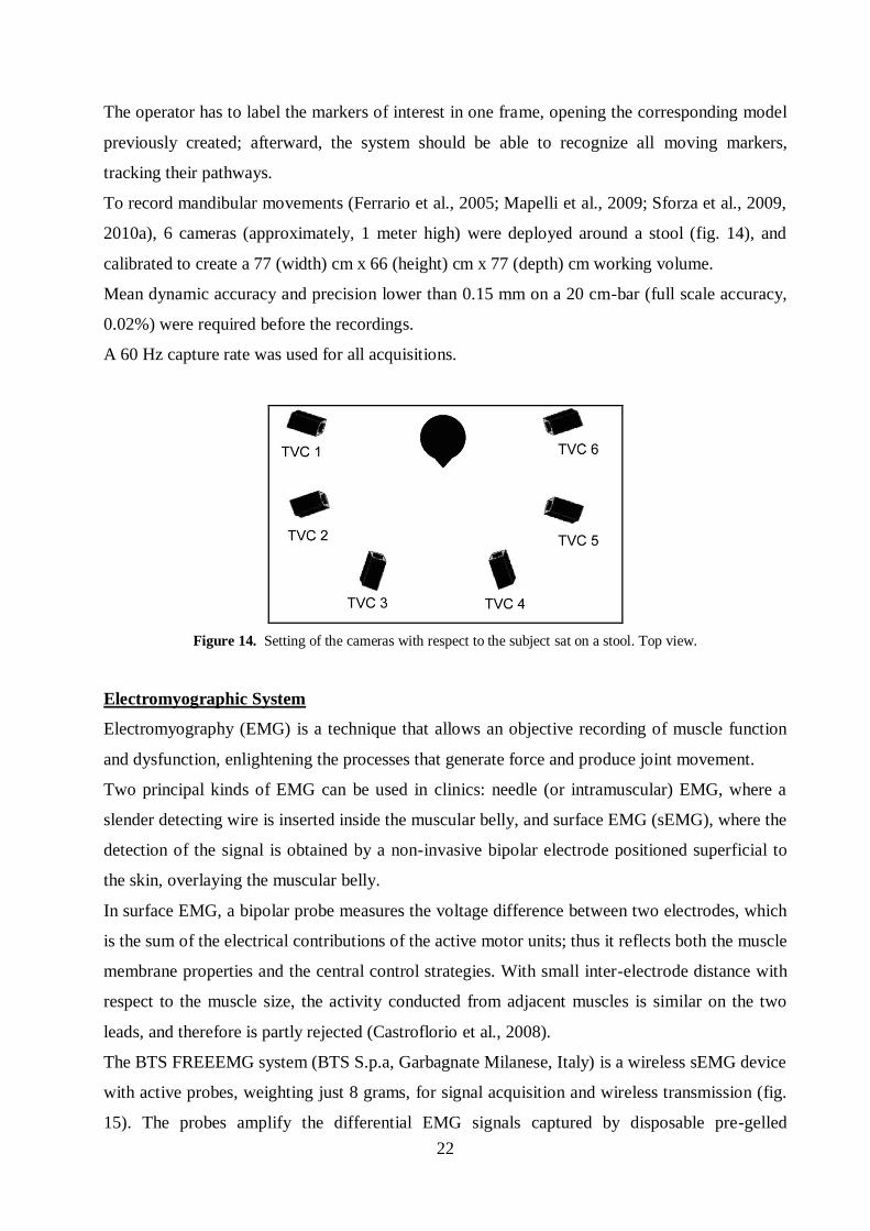
To record mandibular movements (Ferrario et al., 2005; Mapelli et al., 2009; Sforza et al., 2009,
2010a), 6 cameras (approximately, 1 meter high) were deployed around a stool (fig. 14), and
calibrated to create a 77 (width) cm x 66 (height) cm x 77 (depth) cm working volume.
Mean dynamic accuracy and precision lower than 0.15 mm on a 20 cm-bar (full scale accuracy,
0.02%) were required before the recordings.
A 60 Hz capture rate was used for all acquisitions.
Figure 14. Setting of the cameras with respect to the subject sat on a stool. Top view.
Electromyographic System
Electromyography (EMG) is a technique that allows an objective recording of muscle function
and dysfunction, enlightening the processes that generate force and produce joint movement.
Two principal kinds of EMG can be used in clinics: needle (or intramuscular) EMG, where a
slender detecting wire is inserted inside the muscular belly, and surface EMG (sEMG), where the
detection of the signal is obtained by a non-invasive bipolar electrode positioned superficial to
the skin, overlaying the muscular belly.
In surface EMG, a bipolar probe measures the voltage difference between two electrodes, which
is the sum of the electrical contributions of the active motor units; thus it reflects both the muscle
membrane properties and the central control strategies. With small inter-electrode distance with
respect to the muscle size, the activity conducted from adjacent muscles is similar on the two
leads, and therefore is partly rejected (Castroflorio et al., 2008).
The BTS FREEEMG system (BTS S.p.a, Garbagnate Milanese, Italy) is a wireless sEMG device
with active probes, weighting just 8 grams, for signal acquisition and wireless transmission (fig.
15). The probes amplify the differential EMG signals captured by disposable pre-gelled
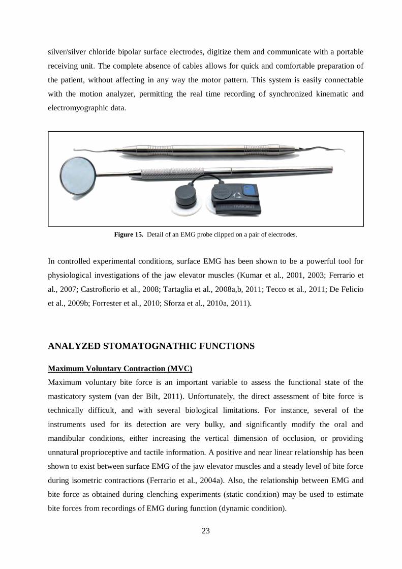
23
silver/silver chloride bipolar surface electrodes, digitize them and communicate with a portable
receiving unit. The complete absence of cables allows for quick and comfortable preparation of
the patient, without affecting in any way the motor pattern. This system is easily connectable
with the motion analyzer, permitting the real time recording of synchronized kinematic and
electromyographic data.
Figure 15. Detail of an EMG probe clipped on a pair of electrodes.
In controlled experimental conditions, surface EMG has been shown to be a powerful tool for
physiological investigations of the jaw elevator muscles (Kumar et al., 2001, 2003; Ferrario et
al., 2007; Castroflorio et al., 2008; Tartaglia et al., 2008a,b, 2011; Tecco et al., 2011; De Felicio
et al., 2009b; Forrester et al., 2010; Sforza et al., 2010a, 2011).
ANALYZED STOMATOGNATHIC FUNCTIONS
Maximum Voluntary Contraction (MVC)
Maximum voluntary bite force is an important variable to assess the functional state of the
masticatory system (van der Bilt, 2011). Unfortunately, the direct assessment of bite force is
technically difficult, and with several biological limitations. For instance, several of the
instruments used for its detection are very bulky, and significantly modify the oral and
mandibular conditions, either increasing the vertical dimension of occlusion, or providing
unnatural proprioceptive and tactile information. A positive and near linear relationship has been
shown to exist between surface EMG of the jaw elevator muscles and a steady level of bite force
during isometric contractions (Ferrario et al., 2004a). Also, the relationship between EMG and
bite force as obtained during clenching experiments (static condition) may be used to estimate
bite forces from recordings of EMG during function (dynamic condition).

24
One of the principal problems that limit a widespread use of sEMG in clinics is the necessary
normalization of its recordings (DeLuca, 1997). Indeed, to compare EMG recordings among
different subjects it is mandatory to relate all measurements to the electrical muscle activity
detected during some standardization recording, like a maximum voluntary contraction
(Castroflorio et al., 2005). In the dental field, sEMG potentials collected during an MVC on both
cotton rolls and in intercuspal position have been reported to have the best inter- and intra-
individual repeatability (Ferrario et al., 2006b; De Felicio et al., 2009b; Suvinen et al., 2009;
Forrester et al., 2010; Hellmann et al., 2011), and diagnostic tests based on MVC standardization
have been in use for the last 10 years (Ferrario et al., 2004b, 2006b, 2007; Tartaglia et al., 2008b,
2011; De Felicio et al., 2009b; Tecco et al., 2011).
Mastication
Mastication is a complex task that mixes voluntary and automatic motor pathways controlled by
central nervous system pattern generators, located in the brainstem, and is regulated by the
feedback from several receptors (extero-, proprio- and viscero-receptors) (Rilo et al., 2007).
Mastication requires well-controlled repeated separation and contacts of the maxillary and
mandibular teeth, characterized by rhythmic up and down motion, protrusive-retrusive
movement, rotation in the horizontal plane, and lateral shifting of the mandible.
The concomitant assessment of chewing kinematics, morphology and EMG activity of the
masticatory muscles provides important information on the chewing function (Kohyama et al.,
2008; Piancino et al., 2008). The exact process varies between the individuals but, once the
pattern is established, it remains fairly constant for that particular person. This is not to imply
that the process is static; indeed it is continually altered, since changes within the stomatognathic
system are constant and dynamic throughout life (Hiatt and Gartner, 2010).
Among the others, the chewing pattern is generally thought to be one of several useful
parameters for objectively evaluating chewing function, and unilateral gum chewing is the test
most commonly used for obtaining standardized data (Yamashita et al., 1999). The typical adult
chewing pattern, as represented in the frontal plane, is a teardrop shape with the opening phase
medial to a more lateral closing phase. Akiyama et al. (1991) analysed the masticatory
movements of the mandible in the frontal plane and classified the chewing patterns into eight
different types according to the opening and closing paths of the interincisal point (fig. 16).

25
Figure 16. Schematic illustration of typical chewing patterns of the interincisal point in the frontal view (Takeda et
al., 2009).
Pattern I was defined as smooth tracings in both directions, often teardrop shaped, or lenticular.
Pattern II was defined as opening patterns that were inclined to the chewing (or working) side,
combined with the characteristic closing movements resembling a mirrored “s”. Pattern III was
defined as opening patterns that were inclined to the nonchewing (or balancing) side, combined
with closing movements resembling convexity. Pattern IV was defined as opening patterns that
were inclined to the nonchewing side, combined with characteristic closing movements
resembling a mirrored “s”. Pattern V was defined as opening and closing patterns that were
drawn from the same chewing stroke. Pattern VI was defined as having both sections inverted
(reversed pattern). Pattern VII was defined as crossed opening and closing patterns. Finally,
pattern VIII was defined as a linear opening and closing pattern. Patterns I, II, and III are the
grinding patterns and are usually observed in subjects with normal occlusion. Patterns IV and VI
are characteristically observed in subjects with a posterior cross-bite. Patterns V, VII, and VIII
are observed in subjects with mandibular prognathism and temporomandibular disorders, and in
particular, pattern VIII is referred to as a chopping pattern (Takeda et al., 2009).
Modifications in the masticatory movements can point to alterations in several structures: the
masticatory muscles, the temporomandibular joints, the teeth and parodontium, the nervous
afferent and efferent pathways (Buschang et al., 2000). Direct in-vivo observations of chewing
movements are therefore mandatory for a better understanding of the normal function and
dysfunction of the stomathognathic apparatus.

26
Border movements
The border excursions of the human lower jaw with respect to the skull are limited by constraints
within the masticatory system. These limitations are considered to be a fundamental portion of
the functional performance of the masticatory system. The constraints that determine the
envelope of border movements may be passive (TMJ surfaces, ligaments, passive tensions of the
muscles) and/or active (reflexes of muscles to protect the articular capsule). The surrounding soft
structures, such as skin and glands, also may limit mandible excursions (Koolstra et al., 2001).
Aside from maximum mouth opening (MMO), lateral and forward excursions with occlusal
contacts (contacts between upper and lower teeth) are a fundamental part of the mandibular
border movements used clinically to evaluate function, and they have received considerable
study (Peck et al., 1999). The condylar pathways during lateral excursions have also been given
special attention because abnormalities in these pathways may be related to temporomandibular
disorders (Hayasaki et al., 2008).
RECORDING PROTOCOL
For each subject, the recordings took approximately 30 minutes (considering also the time
needed for subject’s preparation). The protocol did not involve dangerous or painful procedures,
and it was preventively approved by the ethics committee of the Department of Human
Morphology, University of Milan.
After the methods and aims of the investigation had been completely described, written informed
consent was obtained from each participant.
The subject sat on a stool in the middle of the working volume, with his/her head unsupported,
and was asked to maintain a natural erect position.
Electromyographic assessment of Maximum Voluntary Contraction
To reduce skin impedance, facial epidermis was carefully cleaned with alcohol prior to the 10
mm-electrodes placement (like Kendall Arbo; Tyco Healthcare, Neustadt, Germany). Five
minutes later, when the conductive paste had moistened the skin surface adequately, the left and
right masseter and temporalis anterior muscles were examined with the aforementioned
electromyographic system, positioning four probes on their muscular bellies parallel to muscular
fibres: vertically along the anterior muscular margin, about on the coronal suture, for the
temporalis anterior; with the upper pole of the electrode at the intersection between the tragus-
labial commissura and the exocanthion-gonion lines for the masseter (fig. 17).
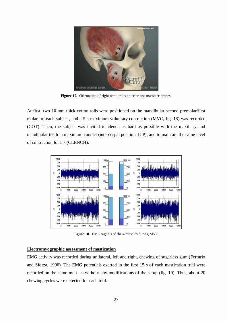
27
Figure 17. Orientation of right temporalis anterior and masseter probes.
At first, two 10 mm-thick cotton rolls were positioned on the mandibular second premolar/first
molars of each subject, and a 5 s-maximum voluntary contraction (MVC, fig. 18) was recorded
(COT). Then, the subject was invited to clench as hard as possible with the maxillary and
mandibular teeth in maximum contact (intercuspal position, ICP), and to maintain the same level
of contraction for 5 s (CLENCH).
Figure 18. EMG signals of the 4 muscles during MVC.
Electromyographic assessment of mastication
EMG activity was recorded during unilateral, left and right, chewing of sugarless gum (Ferrario
and Sforza, 1996). The EMG potentials exerted in the first 15 s of each mastication trial were
recorded on the same muscles without any modifications of the setup (fig. 19). Thus, about 20
chewing cycles were detected for each trial.

28
Figure 19. EMG signal of the right masseter muscle during 4 right chewing strokes.
Kinematic and electromyographic assessment of mastication
Maintaining electrodes and wireless probes on the skin, other two unilateral chewing cycle
sequences were recorded using the optoelectronic motion analyzer, together with the
electromyographic system. To this scope, three head passive markers (diameter: 6 mm) were
added on the nasion and the left and right frontotemporali, defining a cranial reference plane, by
means of biadhesive tape. These three markers were insensitive to skin motion artefacts during
jaw movement. Other three passive markers (diameter: 6 mm) were positioned on the three
corners of an equilateral triangular stainless steel extra oral device (side 40 mm; weight 2 g); this
tool was fixed on the mandibular anterior gingiva just out of dental contact using a surgical
adhesive (Stomahesive; Convetec Inc, Deeside, United Kingdom), and provided a mandibular
reference system. This rigid body was positioned to be as unobtrusive as possible and to allow
each participant to move in and out of maximum intercuspation freely (fig. 20).

29
Figure 20. Complete measurement setup of a subject.
In a single reference frame, a further passive mandibular marker (diameter: 3 mm) was located
on the midline incisor edge (inter-incisor point, IP); it identified a dental (occlusal) landmark,
relative to the extraoral system (Ferrario et al., 2005). Similarly, two condylar reference points
(CRPs) were firstly individuated by palpation and secondly detected by means of a marked
pointer while the subject was keeping his mouth closed in ICP.
Kinematic assessment of mandibular border movements
With the same configuration of markers, a border movement sequence of free maximum mouth
opening (MMO) and closing, followed by mandible maximum unilateral laterotrusions and
protrusion was performed three times by each subject. Each trial had to be started and concluded
with the jaws in ICP. In particular, subjects were instructed to naturally open their mouth and to
slowly move the mandible to the right/left side and forward from ICP as far as comfortable, with
sliding tooth contacts.
MEASUREMENT PROTOCOL
Both raw EMG signals and marker coordinates constituted the input data for the protocol
calculations, which were implemented on Microsoft Excel, Matlab or C-code. Descriptive and
inferential statistics were evaluated by means of SPSS Statistics.

30
EMG signals - MVC
For each of the four analyzed muscles, the mean EMG potential evaluated on the most constant 3
s-interval of COT trial (mean of the root mean squared, RMS, calculated in 25 ms-temporal
windows) were set at 100%, and all EMG potentials obtained during both MVC directly
performed on the occlusal surfaces (CLENCH) and mastication (see below) were expressed as a
percentage of this value (unit: mV/mV*100). According to this protocol, normalized EMG data
may inform on the influence of occlusion (teeth contact) on the neuromuscular activity, avoiding
individual variability (anatomical variations, relative muscular hypo- or hypertrophy,
physiological and psychological status, etc.) and technical variations (muscle cross-talk,
electrode position, variability due to skin and electrode impedance, etc.).
The EMG waves of paired muscles were compared by computing a percentage overlapping
coefficient (POC, unit: %). POC is an index of the symmetric distribution of the muscular
activity as determined by occlusion. The index (fig. 21) ranges between 0% (no symmetry) and
100% (perfect symmetry).
Figure 21. Graphic representation of POC calculation for a pair of muscles. The sum of non-overlapped areas is
divided by the total area under the two curves.
Masseter and temporalis POCs were obtained for each subject. To individuate the most prevalent
side of masticatory muscles, the asymmetry index (ASIM, unit: %) was also computed as the
percentage ratio of the difference between the mean right and left standardized potentials, and
the sum of the same standardized potentials. This index is positive (up to +100%) when the right
muscles standardized potentials are larger than the left ones, negative (down to -100%) when the
left muscles potentials are larger, and null when they are equal.

31
Because an unbalanced contractile activity of contralateral masseter and temporalis muscles, for
instance, right temporalis and left masseter, might prompt a potential lateral displacing
component, the torsion coefficient (TORS, unit: %) was calculated by superimposing the right
temporalis plus left masseter normalized EMG amplitudes over the left temporalis plus right
masseter normalized EMG amplitudes: the area of the superimposition was assessed as a
percentage of the total EMG amplitudes. TORS ranges between 0% (complete presence of lateral
displacing force) and 100% (no lateral displacing force). The add-on TORQUE index was
calculated to express with a positive or negative sign the respectively prevalence of right or left
displacing component, similarly to what ASIM index did for POC index.
To individuate the most prevalent pair of masticatory muscles, the activity index (ATTIV, unit:
%) was also computed as the percentage ratio of the difference between the mean masseter and
temporalis standardized potentials, and the sum of the same standardized potentials. This index
is positive (up to +100%) when the masseter muscles standardized potentials are larger than the
temporalis muscles ones, negative (down to -100%) when the temporalis muscles potentials are
larger, and null when they are equal. When standardized muscular potentials are not balanced
between the two analyzed masticatory muscles, the occlusal centre of gravity might be displaced
onwards (temporalis prevalent) or backwards (masseter prevalent).
Finally, the mean (masseter and temporalis) total standardized muscle activities was calculated
as the integrated area of the EMG potential over time (std. IMPACT, unit: mV/mV %).
EMG signals - Mastication
EMG signals of gum chewing were normalized on COT trial in the same way explained for
MVC. From the 600 RMS potentials recorded from the four tested muscles during each 15 s
chewing test, two main parameters were computed: the masticatory frequency and the
confidence ellipse (Hotelling’s 95%) of the simultaneous maximum differential right-left
masseter and temporalis standardized activity extracted from each cycle (Lissajous’s plot).
The confidence ellipse is a statistical tool to assess the repeatability of the masticatory muscle
pattern of contraction during the execution of a standardized movement (e.g. unilateral gum
chewing). The differential right-left masseter activity serves as the x-coordinate, and the
differential temporal activity as the y-coordinate, in a Cartesian graph representation (fig. 22).

32
Figure 22. Right and left side gum chewing in a normal subject. Surface EMG data are plotted according to a
Lissajous’s plot.
From the pairs of coordinates, the position of the unknown population centre is estimated by the
sample centre. The phase angle gives the inclination of the ellipse relative to the coordinate axes,
whereas the amplitude gives the distance of the centre of the ellipse from the centre of the
coordinate axes. To assess if the left- and the right-side chewing tests were performed with
symmetrical muscular patterns, using the centres of the two confidence ellipses (left and right-
side chewing) calculated in each subject, a further index, the symmetric mastication index (SMI,
unit: %), was computed. In subjects with a normal neuromuscular coordination, the centres of
the ellipses describing unilateral chewing plotted as a Lissajous’s figure should be located in the
first (right side) and third (left side) quadrants (Kumai, 1993), with about the same amplitude
and a 180° difference between the phases. A symmetrical muscular pattern, provided that the
ellipses are statistically significant, would then produce a SMI equal (or very close) to 100%.
Conversely, an asymmetrical pattern would produce a SMI close to 0%. To directly compare
right- and left-side chewing, then, this latter’s phase was mirrored, subtracting 180° to its value.
Furthermore, the mean (masseter and temporalis) total muscle activities during chewing was
assessed as the integrated areas of the standardized EMG potentials over time (IMPACT, unit:
%*s). For each patient, both the activity normalized on the number of performed cycles, and its
percentage referred to the working side, were also computed.
Kinematics
The extraoral mandibular markers (Mk1, Mk2, Mk3) individuated the plane of mandibular
motion, given that both mandibular dynamic deformations (Yatabe et al., 1997; Catic and Naeije,
1999; Chen et al., 2000; Naeije, 2003; Ferrario et al., 2005) and instability at the device-gingiva
interface were negligible (Ferrario et al., 2005; Mapelli et al., 2009). The relative motion

33
between the head reference system (Mk4, Mk5, Mk6) and the mandibular one was computed by
means of mapping operators, which allow analysing mandibular pathway relative to the head
(fig. 23).
Figure 23. Global view of the marker set (a) and the two reference systems (b).
Hence, neck and trunk movements were subtracted from the raw motion of the mandible.
Subsequently, the displacements of the dental (Mk0) and condylar points (Mk7, Mk8) were
reported, frame by frame, in the global reference system (head system), with their paths being
evaluated in the three anatomical planes (horizontal, frontal, sagittal planes).
The right-left coordinates of the condylar points were further arbitrarily corrected of 15 mm in
the medial direction (Merlini and Palla, 1988; Salaorni and Palla, 1994; Gallo et al., 1997) to
better represent the head of the condyles (condylar reference points, CRPs) (fig. 24).
Mk 2
Mk 5 Mk 4 Mk 6
Mk 1
Mk 3
Mk 7 Mk 8
Mk 0
a b

34
Figure 24. Spatial coordinates of a subject’s right CRP during a sequence of mandibular border movements.
Positive/negative values.
The data were mathematically smoothed with the use of a second-order Butterworth’s low-pass
filter (cut-off frequency of 8 Hz). Indeed, according to Miles (2007), the mandible voluntary
movements together with its continuous tremors can reach a peak frequency of 7 Hz. In fact,
voluntary movements of the mandible are interspersed with small accelerations and decelerations
of 6-7 Hz, which are the result of alternating activity in antagonistic masticatory muscles
superimposed onto the muscle activity that is responsible for the voluntary movements.
A mandibular radius (r) was estimated as the distance between the dental marker (IP) and the
midpoint of the intercondylar axis, while the mandibular width (w) was estimated as the distance
between the right and left condylar markers (fig. 25).
Figure 25. Mandibular anthropometric parameters.
CRPL
w
IP
CRPR
r
opening/closing
right laterotrusion
left laterotrusion protrusion

35
In each motion frame, the rotational angles made by the extraoral device (i.e. the mandible)
around the three global axes were calculated using Cardan angles; this method provided a
description of joint movements nearer to the common concepts of flexion/ extension, abduction/
adduction, and internal/ external rotation used in clinical practice.
The sagittal mandibular movement during mouth opening and closing was further divided into
its rotation and translation components; in each frame of motion, the relative percentage
contribution of the two components to the total movement was calculated. In order to compare
different patients, the mandibular movement was normalized on MMO distance (sagittal
projection): mouth opening and closing were sampled in 10% steps, and in each step the rotation
and translation components were further considered. To find the rotational component of the
mandibular movement, the mandibular radius (r) was used together with the sagittal plane
mandibular angle of rotation: the circumference arc (s) that r described turning around the CRP
was the rotational component, whereas CRP pathway was the gliding component (fig. 26).
Therefore, both components were expressed with the same unit (length), allowing their
comparison (Mapelli et al., 2009). Another advantage of this approach is that mandibular
dimension does not affect the relative contribution of condylar rotation and translation: the
normalization, in fact, is included in the calculations.
Figure 26. Sagittal view of the rotation and translation components of a mandibular motion step.
To assess mastication kinematics, each individual's cycles were detected by a specific algorithm
code written in Matlab. In particular, the three-dimensional coordinates of the first frame for
each sequence defined the starting point of the cycle sequence. The program did not include the
first cycle; it searched along the trace of the IP until it identified the starting frame of each

36
subsequent cycle. The starting frame was defined as the frame at which the three-dimensional
distance from the initial point of the cycle attained a minimum (i.e., stopped decreasing and
started increasing). Having identified the starting frame, the program continued along the trace
until it identified the next, which marked the end of the cycle. The following frame was the
starting frame of the next cycle. To be included as a valid cycle, each cycle had to last more than
250 msec in duration and to be more than 3.0 mm long vertically. Then, each chewing cycle
detected at the IP was broken down into two phases (open-close), and each path length, time
duration and velocity were extracted. Then, the total area delimited by the IP in the frontal plane
was evaluated, together with its percentage subdivision in the working and balancing sides.
Moreover, the morphology of the chewing cycles (fig. 27) was assessed classifying the pathway
of each stroke into 1 of 8 standardized categories (Akiyama et al., 1991): pattern I, II and III
were pooled together and referred to as “ideal” cycle shapes, whereas the other 5 (pattern IV, V,
VI, VII, VIII) were considered “anomalous” (Takeda et al., 2009).
Figure 27. Frontal view of a typical Pattern I chewing cycle.
Method error
Measurement variability in EMG data was tested by repeated analyses of seven subjects chosen
at random; for all MVC variables the intraclass correlation coefficient ranged between 0.629 and
0.977, without significant differences among the measurement sessions (Ferrario et al., 2006a).
A good reproducibility of the same indexes was reported also in another test-retest examination
(De Felicio et al., 2009b).
The method error of mandibular movements was assessed in the reference subjects, and has been
previously reported (Ferrario et al., 2005): the intraclass correlation coefficients (five subjects,
three independent sessions) ranged between 0.571 and 0.760, without significant differences
among repeated sessions.

37
Furthermore, to check the stability of the extraoral framework, we assessed the difference in the
position of the two lateral cranial markers relative to the framework reference system between
the initial intercuspal position and the final one that was reached after a wide range of
mandibular movements. For this experiment, two additional patients (not included in the current
study) who had a fixed orthodontic appliance were enrolled. Three tests with the framework
fixed to the fixed orthodontic appliance were compared to other three tests with the standard
positioning (framework attached to the anterior gingiva using surgical adhesive). The framework
fixed to the orthodontic appliance gave a mean difference of 0.35 mm (SD, 0.04), whereas in the
second configuration the mean difference was 0.49 mm (SD, 0.02). Overall, the differences in
the two configurations were similar, showing that the adhesive interface can be considered
satisfactory for the current measurements.

38
Study I – HEALTHY SUBJECTS
INTRODUCTION
A detailed knowledge of normal jaw function is necessary for a better understanding of TMJ
disorders, dento-facial malocclusions, and for their treatment.
The aim of the current study was to quantitatively assess, in healthy subjects, the range of motion
of mandibular border movements, the morphology and kinematics of chewing cycles, and the
electromyographic activity of masticatory muscles during MVC and chewing. The relative
contribution of rotation and translation of the condyle-disc assembly during both opening and
closing movements was also assessed.
Data were evaluated separately for men and women, and a gender-related influence was tested,
considering also a potential effect of mandibular dimension. The comparison of male and female
data may offer a further contribution to the assessment of functional and anatomical sexual
dimorphism in the human stomatognathic apparatus, a still debated question (Lewis et al., 2001;
Naeije, 2002).
Furthermore, asymmetry is a common finding in humans. Apart from unpaired and asymmetric
organs, both morphology and function of paired structures differ in the left and right sides of the
body. Morphological evaluations of craniofacial asymmetry is a usual part of the
characterization of both normal subjects and patients (Naeije et al., 1989; Ferrario et al., 2000).
In the present study, functional symmetries of the craniofacial complex involving the patterns of
jaw movements and the activities of masticatory muscles were also tested.
METHODS
Subjects and data collection
Nineteen volunteer healthy subjects (9 men and 10 women), aged 21–49 years, were analyzed in
this study. They were recruited from the students and staff attending the Dipartimento di
Morfologia Umana e Scienze Biomediche “Città Studi”, Università degli Studi di Milano (Italy).
History and clinical examination were used to select the subjects. The inclusion criteria to be
recruited in the control group were: a sound, complete, permanent dentition with bilateral canine
and molar Angle Class I jaw relationships; anterior teeth with vertical and horizontal overlap
between 0 and 3 mm; maxillary and mandibular interincisal lines without lateral deviations

39
larger than 2 mm; no cast restorations or cuspal coverages, no anterior or lateral reverse
occlusion; no previous history of craniofacial trauma or congenital anomalies; no TMJ or
craniocervical disorders, based on the RDC/TMD and ProTMDmulti questionnaire (see Study
II).
Data of one man was lost for technical reasons.
Statistical analysis
Descriptive statistics of subjects’ age, mandibular radius and width were calculated separately
for men and women. The same was done for the 3 Cardan angles and the 3D pathways of IP and
CRPs during MMO, maximum right and left laterotrusions and protrusion of the mandible; for
the kinematic parameters of gum chewing; for all the EMG indices of MVC and gum chewing.
The Chi-squared test was used to test the homogeneity of chewing pattern distributions both
between men and women and between right and left working sides.
The normal distribution of data was checked with the Kolmogorov-Smirnov test, and Levene’s
test was used to test for homogeneity of variances.
Then, male and female age, mandibular dimensions and electromyographic indices in MVC were
compared by Student’s t-test for independent samples. One-way analysis of covariance
(ANCOVA) was used to compare unilateral data (between-subject fixed factor: gender), and 2-
way mixed-model ANCOVA (between-subject fixed factor: gender; within-subject fixed factor:
side) for bilateral data (right and left condyles, laterotrusions, chewing cycles). 3-way ANCOVA
was adopted to test both condylar translation and its percentage relative to mandible rotation
during mouth opening, and also for length, velocity and duration of chewing cycles (between-
subject fixed factor: gender; within-subject fixed factors: side and phase).
For kinematic evaluations, mandibular radius was included in the fixed part of the model as a
covariate, to evaluate the dependence of sex differences on mandibular size. The first order
interactions between the factors were also computed.
Since main effects of repeated-measures factors are independent of the between-participant
covariate of mandible size, pure repeated-measures effects were reported from an analysis that
excluded the covariate.
The significance level was set at 5% for all statistical analyses (p < 0.05).
40
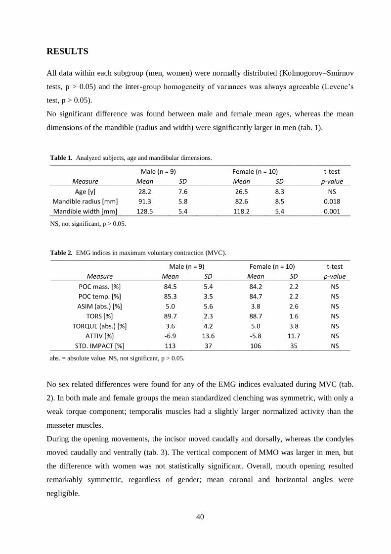
RESULTS
All data within each subgroup (men, women) were normally distributed (Kolmogorov–Smirnov
tests, p > 0.05) and the inter-group homogeneity of variances was always agreeable (Levene’s
test, p > 0.05).
No significant difference was found between male and female mean ages, whereas the mean
dimensions of the mandible (radius and width) were significantly larger in men (tab. 1).
Table 1. Analyzed subjects, age and mandibular dimensions.
Male (n = 9) Female (n = 10) t-test Measure Mean SD Mean SD p-value
Age [y] 28.2 7.6 26.5 8.3 NS Mandible radius [mm] 91.3 5.8 82.6 8.5 0.018
Mandible width [mm] 128.5 5.4 118.2 5.4 0.001
NS, not significant, p > 0.05.
Table 2. EMG indices in maximum voluntary contraction (MVC).
Male (n = 9) Female (n = 10) t-test Measure Mean SD Mean SD p-value
POC mass. [%] 84.5 5.4 84.2 2.2 NS POC temp. [%] 85.3 3.5 84.7 2.2 NS
ASIM (abs.) [%] 5.0 5.6 3.8 2.6 NS TORS [%] 89.7 2.3 88.7 1.6 NS
TORQUE (abs.) [%] 3.6 4.2 5.0 3.8 NS ATTIV [%] -6.9 13.6 -5.8 11.7 NS
STD. IMPACT [%] 113 37 106 35 NS
abs. = absolute value. NS, not significant, p > 0.05.
No sex related differences were found for any of the EMG indices evaluated during MVC (tab.
2). In both male and female groups the mean standardized clenching was symmetric, with only a
weak torque component; temporalis muscles had a slightly larger normalized activity than the
masseter muscles.
During the opening movements, the incisor moved caudally and dorsally, whereas the condyles
moved caudally and ventrally (tab. 3). The vertical component of MMO was larger in men, but
the difference with women was not statistically significant. Overall, mouth opening resulted
remarkably symmetric, regardless of gender; mean coronal and horizontal angles were
negligible.

41
Table 3. Kinematic range of motion of the mandible at Maximum Mouth Opening (MMO).
Male (n = 9) Female (n = 10) ANCOVA Measure Mean SD Mean SD p-value
IP MMO [mm] 50.8 5.7 47.8 4.4 NS #
IP caudal component [mm] 43 5.5 39.2 4.2 NS # IP dorsal component [mm] 26.4 6.4 26.6 6.7 NS #
IP lateral component [mm] -1.2 1.3 -1.4 1.9 NS # Sagittal angle [°] 33.5 5.1 34.6 2.3 NS #
Coronal angle [°] 0.7 3.8 -0.5 3.1 NS # Horizontal angle [°] 1.3 1.4 2.3 1.6 NS #
CRP caudal component [mm] 9.8 4.1 8.9 3.9 NS & CRP ventral component [mm] 14.5 6.8 11.8 6.6 NS &
CRP lateral component [mm] -0.2 1.1 -0.2 1.5 NS &
#, 1-way ANCOVA. &, 2-way ANCOVA, since the effect of side was not significant, right and left values were pooled; positive lateral components correspond to right displacements. NS, not significant, p > 0.05,
refers to main factors and interactions.
During mandibular lateral displacement (tab. 4), the balancing condyle moved significantly more
than the working condyle. The former showed greater vertical (downward) and sagittal (onward)
ranges of motion (orbitant condyle), while the latter was almost still (pivoting condyle).
Table 4. Kinematic range of motion of the mandible at maximum laterotrusion.
Male (n = 9) Female (n = 10) ANCOVA
Measure Mean SD Mean SD p-value
IP laterotrusion [mm] 9.9 2.1 8.9 1.9 NS &
wCRP caudal component [mm] 0.2 1.9 0.7 1.9 NS & wCRP dorsal component [mm] 0.8 1.9 0.4 1.5 NS &
wCRP lateral component [mm] 1.1 1.6 1.3 1.1 NS & bCRP caudal component [mm] 8.3 2.0 7.0 2.3 NS &
bCRP ventral component [mm] 8.1 3.0 6.9 2.2 NS & bCRP medial component [mm] 1.8 1.4 1.8 0.8 NS &
&, 2-way ANCOVA. Since the effect of side was not significant, right and left values were pooled. NS, not
significant, p > 0.05, refers to main factors and interactions. w = working, b = balancing.
Similarly to mouth opening, mandibular protrusion was quite symmetric and a little larger in
men than in women, even not significantly (tab. 5).

42
Table 5. Kinematic range of motion of the mandible at maximum protrusion.
Male (n = 9) Female (n = 10) ANCOVA Measure Mean SD Mean SD p-value
IP protrusion [mm] 9.1 1.5 8.0 2.3 NS #
CRP caudal component [mm] 6.6 2.3 5.3 1.9 NS & CRP ventral component [mm] 8.6 1.9 7.6 3.2 NS &
CRP lateral component [mm] 0.1 0.7 -0.5 1.4 NS &
#, 1-way ANCOVA. &, 2-way ANCOVA, since the effect of side was not significant, right and left values were pooled. NS, not significant, p > 0.05, refers to main factors and interactions.
Tables 3-5 describe the border movements of the mandible, reporting the ranges of motion of its
reference points (lower interincisal point and condyles). No sex and side significant effects were
found.
During mouth opening and closing standardized as a percentage of MMO distance (fig. 28), the
relative contributions of mandibular rotation and translation were almost always similar in men
and women, with differences ranging between 2 and 3% (2-way ANCOVA, p > 0.05).
Figure 28. Translation component of the condylar movement in mouth opening and closing as a percentage of the
total mandibular movement. Since there was no significant sex effect at any step, male and female values were
pooled.
The rotation component was prevalent during all the movement of mouth opening and closing, in
particular at MMO; the peaks of relative condylar translation were reached near ICP.
At the maximum displacement of the interincisal point, the overall percentage of mandibular
movement explained by pure condylar translation was similar between men (mean, 29%; SD,

43
5%) and women (mean, 28%; SD, 7%); also their mean absolute values were close to each other
(men, 21.8±6 mm; women, 20±6.5 mm). The effect of gender was not statistically significant, as
well as side (right/left condyle), phase (opening/closing) and covariate (mandible radius) effects
(3-way ANCOVA: p > 0.05).
During mastication, on average, chewing cycles were characterized by similar parameters of
frequency and area between men and women, regardless of the working side (2-way ANCOVA).
As shown in table 6, opening and closing had the same length, but the latter was significantly
faster (3-way ANCOVA, phase factor: p < 0.0001 for both duration and velocity dependent
variables). No significant sex, inter-side difference or covariate effects were found.
Table 6. Kinematic parameters of unilateral gum chewing.
Male (n = 9) Female (n = 10) ANCOVA Measure Mean SD Mean SD p-value
Frequency [Hz] 1.23 0.34 1.26 0.29 NS &
IP area [mm2] 26 13 17 8 NS & IP ipsilateral area [%] 73 13 77 20 NS &
Opening length [mm] 14.1 2.5 11.8 2.6 NS $
Closing length [mm] 14.4 2.7 11.9 2.4
Opening duration [ms] 375 74 380 75 0.00 $
Closing duration [ms] 291 80 310 43
Opening velocity [m/s] 0.04 0.011 0.033 0.008 0.00 $
Closing velocity [m/s] 0.07 0.045 0.048 0.017
&, 2-way ANCOVA; $, 3-way ANCOVA. Since the effect of side was not significant, right and left values were
pooled. NS, not significant, p > 0.05, refers to main factors and interactions.
Table 7. Electromyographic parameters of unilateral gum chewing.
Male (n = 9) Female (n = 10) ANOVA
Measure Mean SD Mean SD p-value
amplitude [%] 99 55 95 69 NS &
Hotelling's ellipse area [%2] 946 545 1069 1075 NS &
Impact [%s] 632 308 688 380 NS & Impact/cycle [%s] 36 17 37 21 NS &
%w Impact [%] 67 9 69 11 NS & SMI [%] 67 21 67 17 NS &
&, 2-way ANOVA. Since the effect of side was not significant, right and left values were pooled. NS, not
significant, p > 0.05, refers to main factors and interactions. %w = percentage of the working side.
Table 7 shows that male and female subjects had also similar EMG parameters of mastication,
regardless of the working side (2-way ANOVA, p > 0.05 for both the main effects and
interaction). Overall, a large inter-subject variability was apparent.

44
Although the centres of the ellipses describing unilateral chewing were located in the first
quadrants of the Lissajous diagram (left side chewing data were previously mirrored), thus
indicating a prevalent activity of the working-side muscles, right working side chewing was
characterized by a statistically significant lower phase than left working side chewing (fig. 29).
Figure 29. Lissajous phases of unilateral chewing, mean±SD. Two-way ANOVA: side, p = 0.005; sex and
interaction, p > 0.05. The horizontal dashed line indicates the bisector of the first quadrant.
The qualitative analysis of chewing cycle morphology found that the percentage distribution of
“ideal” (patterns I, II, III) and “anomalous” (patterns IV, V, VI, VII, VIII) frontal shapes (fig.
16), either between men and women or between right and left working sides, was similar (Chi-
squared test: p > 0.05).
DISCUSSION
In the present control group, mandibular dimensions resulted on average larger in men than in
women, in agreement with previously literature findings (Naeije, 2002; Ferrario et al., 2005;
Mapelli et al., 2009).
The ranges of mandibular motion well compare with distances previously published for
maximum mouth opening, laterotrusions and protrusion in healthy adults (Piehslinger et al.,
1993; Travers et al., 2000; Buschang et al., 2001; Lewis et al., 2001; Ferrario et al., 2005).
Nevertheless, MMO distance was in disaccord with other investigations, being both superior

45
(Gallo et al., 1997; Gallagher et al., 2004) and inferior (Merlini and Palla, 1988; Zwijnenburg et
al., 1996) to what obtained by other investigators. Differences in sample composition (age, sex,
ethnic origin, Gallagher et al., 2004), and in the measuring device, may explain the
discrepancies. For instance, electromagnetic trackers were reported to create distortions for
MMO larger than 40 mm (Lewis et al., 2001).
Saitoh et al. (2007) and Hayasaki et al. (2008) reported a less caudal movement of the balancing
condyle during unilateral laterotrusion, but their subjects were instructed to achieve habitual
lateral excursion.
Accordingly to previous investigations (Zwijnenburg et al., 1996; Naeije, 2002), but in contrast
with others (Salaorni and Palla, 1994; Gallo et al., 1997; Lewis et al., 2001; Ferrario et al.,
2005), no sex-related differences in mandibular kinematics were found. Our use of a condylar
reference point located 15 mm medial to the digitized point previously palpated may partially
explain some of these differences.
In the current investigation, no right-left side differences were found, in accord with other
studies (Travers et al., 2000; Hayasaki et al., 2008). In contrast, Buschang et al. (2001) reported
significantly larger movements of the left than of the right condyle during protrusion and
laterotrusion in healthy adults. Similar amounts of left-side dominance in condylar protrusive
movements have been already reported (Harper, 1990; Piehslinger et al., 1993; Theusner et al.,
1993; Gsellmann et al., 1998). The authors related these findings to normal morphological
asymmetry or asymmetrical constraints, thus, not necessarily diagnostic of craniomandibular
disorders (Kenworthy et al., 1997).
In mouth opening and closing, mandibular translation was present during the whole motion
(Lotters et al., 1996; Lewis et al., 2001; Mapelli et al., 2009), it was always smaller than rotation,
and decreased during mouth opening. A similar finding was reported by Lotters et al. (1996),
although they also showed a relatively reduced translation near ICP. In contrast, a radiographic
study found a rotating component of about 14–30% in the initial steps of jaw motion (Wu et al.,
1988). Leader et al. (2003) found a larger translation than rotation in the first phases of mouth
opening. Anatomically, movement decomposition reported in figure 28 during mouth opening
could be due to the progressive passive block provoked on the head of mandible by the ligament
tension (Matsumoto et al., 1995; Lotters et al., 1996), which impedes further antero–inferior
translation. During mouth closing, after the first steps in which the blockage remains, the
translation component progressively increases: the elastic recall of the ligaments outclasses the
active blocking system (Lotters et al., 1996; Yatabe et al., 1997).

46
The present total opening translation did not differ from the closing one, in accord with some
previous studies (Travers et al., 2000; Lewis et al., 2001; Mapelli et al., 2009), but in contrast
with other ones (Lobbezoo et al., 2000); our findings do not support the notion that the condylar
opening pathway is shorter than the closing pathway (Yatabe et al., 1997).
Again, no sex-related differences in condylar path lengths were found, according to previous
reports (Salaorni and Palla, 1994; Yatabe et al., 1995; Lotters et al., 1996; Zwijnenburg et al.,
1996; Naeije, 2002). The current results do not support the theory of a more mobile TMJ in
women than in men (Gallagher et al., 2004). According to that theory, the longer component of
condylar translation in women is due to the greater laxity of the sphenomandibular ligament,
which would be stressed later in motion (Lotters et al., 1996).
A pre-softened gum was chosen to obtain a standardized and constant (volume and weight) bolus
all over the chewing test. This food should maintain its characteristics during the complete
chewing trial. Moreover, no significant modifications of its texture are to be expected in this
short duration (15 s for each trial).
The form or shape of the human masticatory cycle has been of interest for many years (Buschang
et al., 2000). Several morphological classifications have been proposed for the analysis of
masticatory pattern. Proschel and Hofman (1988) analyzed the masticatory movement in detail
and classified it into 196 patterns. It would have been difficult, however, to classify less than 50
strokes counted per each subject into 196 patterns. In contrast, Akiyama et al. (1991) proposed a
more simple and practical classification system, which consists of 8 patterns (see Materials and
Methods section). This latter classification, which was used in the present study, contains typical
masticatory patterns, such as grinding (patterns I, II, and III), reverse (pattern VI), and chopping
(pattern VIII). Pattern I, II, III were deemed “ideal”, whereas the other 5 were referred to as
“anomalous”. In the current study, no difference was found between the distribution frequencies
of “ideal” and “anomalous” patterns in healthy subjects. Actually even “normal” subjects show
some anomalous strokes between the ideal teardrop-shaped patterns to accomplish several tasks.
And, of course, controversy remains regarding what constitutes an “ideal” chewing pattern
(Yamashita et al., 1999).
It has been indicated that the masticatory pattern movement should be examined not only
morphologically, but also in terms of masticatory rhythm, because this last is often influenced by
malocclusion (Youssef et al., 1997). Hence, cycle frequency, opening and closing phase duration
and velocity were examined. Neither left-right, nor sex differences were found on mean values
of these kinematic parameters. The only significant difference was between phase velocities,

47
being closure faster than mouth opening, probably due to the elastic recall of the ligaments and
the concentric contraction of the elevator muscles of the mandible.
The mean frontal area of chewing cycles was similar to that found by Evans and Lewin (1986),
who reported an average value of 20.6 mm2. By contrast, the present values are notably lower
than the ones obtained by Chew et al. in 1988 (33.0 mm2). However, it should be outlined that all
these studies have in common the detection of a great inter- and intra-subject variability
(Throckmorton et al., 2001; Hayasaki et al., 2003) even in healthy subjects, which makes the
simple consideration of the mean value unreliable. In 2006, Ferrario and co-workers
quantitatively analyzed the variability of unilateral chewing movements in young adults. The
highest between subjects/ between sessions variance ratios were found for cycle duration and
shape, whereas the spatial characteristics of gum chewing cycles had a large within-subject
variability (Ferrario et al., 2006a).
Among the jaw elevator muscles, the masseter and temporalis muscles are those most often
assessed in clinical evaluations because they are the most superficial, and they are the only
accessible to surface EMG examination. In contrast, the medial and lateral pterygoid muscles
can be evaluated only with needle EMG. Indeed, in the assessment of stomatognathic
dysfunction and several head disorders, the analysis of masseter and temporalis muscles can
provide quantitative functional data with minimal discomfort to the patient and without invasive
or dangerous procedures (Visser et al., 1995; Sato et al., 1998; Liu et al., 1999; Burnett et al.,
2000; Pinho et al., 2000; Ferrario et al., 2002, 2004b; Suvinen et al, 2003; Landulpho et al.,
2004).
Unfortunately, as underlined by several researchers, this simple, low cost, and fast exam also has
many limitations that must be carefully considered and eventually removed (DeLuca, 1997). For
instance, technical artifacts (the instrumental noise), the thickness of the skin fat layer, crosstalk
from different muscles. Therefore, a correct EMG assessment should be performed only with
standardized (normalized) potentials, thus removing most of biological and technical noise
(DeLuca, 1997).
In the current study, to reduce patient variability, the EMG protocol comprised a normalization
record (a MVC on cotton rolls performed just before the recording of the actual tests, i.e. with
the same electrodes, cables, and EMG apparatus, and on the same cutaneous area) that should
limit biologic and technical noise (DeLuca, 1997; Burnett et al., 2000).
Standardized EMG potentials can allow one to verify and quantify the muscular equilibrium,
both between the muscles of the two sides of the body (symmetry: POC and ASIM indices for

48
MVC; SMI index for mastication) and between couples of muscles with a possible laterodeviant
effect on the mandible (TORS and TORQUE indices for MVC).
Also, EMG can allow the measurement of the actual impact of morphology on stomatognathic
function (Visser et al., 1995; Sato et al., 1998; Liu et al., 1999; Burnett et al., 2000; Pinho et al.,
2000; Ferrario et al., 2002, 2006b; Landulpho et al., 2004). From the standardized electric
potentials produced by the single masticatory muscles, the muscular activity (Impact, integrated
value in time) can be calculated to assess the actual effort made by the muscles (Sato et al., 1998;
Burnett et al., 2000; Ferrario et al., 2004b, 2006b; Tartaglia et al., 2008a). Quantitative analyses
of the patterns of muscular contraction during standardized dynamic activities allow one to
assess neuromuscular coordination (Ferrario and Sforza, 1996).
In the current study, sex did not seem to influence patterns of contraction of the analysed
muscles, either during MVC or mastication, as previously reported (Ferrario et al., 2006b; De
Felicio et al., 2009b; Tartaglia et al., 2011). Indeed, this aspect has been scanty analyzed in
previous studies; in some occasions, only one sex was assessed (Kroon and Naeje, 1992; Gay et
al., 1994; Farella et al., 2002), no information about sex was given (Bazzotti, 1999), or no
comparisons were made (Koyano et al., 1995). A possible explanation for the lack of sex
specificity may have to do with the use of gum (Gerstner and Parekh, 1997); previous studies
reported that gum reduces the level of sex specificity in mastication (Neill and Howell, 1986;
Howell, 1987).
Current mean values of MVC parameters were in agreement with data obtained in previously
analysed control groups (De Felicio et al., 2009b; Tartaglia et al., 2011), indicating low
asymmetry between right and left muscle pairs and balanced contractile activities of contralateral
masseter and temporalis muscles (low lateral displacing force). The activity value was low,
indicating a good antero-posterior charge distribution at the mandible. The standard deviation for
each index was limited. In addition, the Impact value was compatible with that observed for
control subjects (De Felicio et al., 2009b; Tartaglia et al., 2011).
During mastication, EMG activity of the masseter and anterior temporalis muscles of the
chewing side was higher than that of the other side, as previously reported (Miyawaki et al.,
2000, 2001; Piancino et al., 2008). The right and left Lissajous’s figures, as reported by SMI
index, were overall symmetrical, with similar position within the Cartesian axes, and similar size
and shape, indicating comparable patterns of right- and left-sided mastication with regard to the
chewing forces and cooperation between muscles. The lack of significant right-left side
differences is in good accord with other literature data (Kumar et al., 2001, 2003; Farella et al.,
2002), even if some investigations reported asymmetric findings (Gay et al., 1994).

49
However, right working side chewing was characterized by statistically significant lower phase
than left working side chewing. The right Lissajous’s figure closer to the masseter axis indicates
that, on average, the temporal and masseter muscles of the working (right) side contracted more
strongly in gum mastication, but the difference in masseter activity between the working and the
balancing sides was larger than the difference in the temporal activity.
Even in the present highly selected subjects whose occlusion was good from a morphofunctional
point of view, the presence of this asymmetric behaviour seems to be an intrinsic characteristic
of occlusion, independently from biological noise.
Overall, even when standardized, coherent boluses were used in well standardized conditions,
chewing in normal subjects seems to be highly variable (Hayasaki et al., 2003), as it is reported
in the current study. A large variety of movement patterns can be found even within a single
individual chewing the same coherent bolus (Proschel, 1987; Ferrario et al., 2006a). The findings
of the present investigation are obviously strictly inherent to the extremely standardized protocol
and cannot be directly extended to natural chewing (free movements of bolus in both sides of
mouth) or to other foods with different mechanical characteristics.
The principal limitation of this study is the number of analysed subjects, which was under the
proper sample size to avoid type II errors. Indeed, from a starting group of more than a 50 young
adults, less than half of them fulfilled the inclusion criteria. In particular, a large number of men
and women had received or were receiving orthodontic treatment.
These normal data provide a first reference for the assessment of patients with alterations in the
cranio-mandibular system (Study II and III).

50
Study II – PATIENTS WITH MODERATE TMD
INTRODUCTION
Temporomandibular disorders (TMD) consist of a number of clinical problems that involve the
masticatory musculature, the temporomandibular joint and associated structures or both.
Approximately 7–15% of the adult population is affected, with higher prevalence in women at
reproductive ages. The most frequent complaints reported by subjects with TMD are pain in the
TMJ and/or masticatory muscles, TMJ sound and difficulty to chew (Michelotti and Iodice,
2010). Additionally, some patients do not come to observation with acute problems, and chronic
disabilities increase the diagnostic problems (Epker et al., 1999).
In an effort to make diagnosis as objective as possible, several protocols for history taking and
clinical assessment have been proposed (De Felicio et al., 2009a); among the most used, there
are the Research Diagnostic Criteria for temporomandibular disorders (RDC/TMD).
Multiple treatments have been employed, although the pathophysiology is not fully understood
and the mechanisms of action of the treatments may be not clear (Cairns, 2010; Michelotti and
Iodice, 2010). According to literature, in certain situations, an adequate muscular training may
allow a better function, reducing pain and disability (Sforza et al., 2010a). The orofacial
myofunctional therapy has demonstrated positive effects on the reduction of symptoms and
clinical signs of TMD, as well as improved swallowing and chewing functions (De Felicio et al.,
2010). The classic dental and skeletal etiologic theories of TMD have been challenged and
refuted by recent studies conducted around the world (Klasser and Greene, 2009). The new
approaches are focusing on the relationship between oral motor function and TMD (Lobbezoo et
al., 2006; Ardizone et al., 2010; Douglas et al., 2010). Mastication may be hampered by TMD
(Bakke and Hansdottir, 2008); as a result, limited masticatory function is one of the problems
that patients with TMD encounter. Rehabilitation to improve masticatory function is therefore
one of the goals in the treatment of TMD. Thus, it seems important to understand the link
between pain-related injury and the stomatognathic status. Furthermore, studying coordination
patterns under normal and pain circumstances may be possible to unfold “efficient” versus
“inefficient” movement strategies, which knowledge can in turn be integrated into training and
rehabilitation strategies (Cote and Hoeger Bement, 2010).
Most of the TMD studies rely on self-assessed masticatory function obtained from
questionnaires, whereas only a limited number of studies has measured the masticatory
performance objectively.

51
The current gold standard to identify the presence or absence of TMD still remains mainly based
on clinical examination supplemented, when deemed appropriate, with imaging (Klasser and
Okeson, 2006). Therefore, other objective, quantitative methods are needed to supplement the
diagnosis of TMD, and to monitor the effectiveness of the relevant treatments.
Indeed, the mandibular movement changes are often perceptible in the presence of TMJ
disorders (Merlini and Palla, 1988); thus, the detection of movement changes could be used as
both a TMD diagnostic tool and a success criterion for treatment (Lemoine et al., 2007).
Currently, also surface electromyography (EMG) can make an objective recording of the
masticatory muscle function and dysfunction (Gay et al., 1994; Suvinen et al., 2003; Castroflorio
et al., 2005, 2008; Ferrario et al., 2007; Tartaglia et al., 2008a, 2011; De Felicio et al., 2009b).
Most of the previous EMG investigations made on symptomatic TMD patients analyzed severe
pain conditions, finding that their masticatory muscles were more asymmetric and more easily
fatigued, less efficient and coordinated, and produced reduced electric potentials and bite forces
when compared to those of healthy subjects (Suvinen et al., 2003; Ferrario et al., 2007; Tartaglia
et al., 2008a).
In the current study, masticatory function of patients with mild-moderate TMD, who did not seek
treatment, has been objectively determined, assessing bite stability, mandibular border
movements and masticatory performance. Both EMG and jaw kinematic parameters were
evaluated and compared to those of the normal subjects collected in Study I.
METHODS
Subjects and data collection
Twenty patients with mild-moderate TMD (5 men and 15 women), aged 22–56 years, were
analyzed in this study, and compared to the control group previously assessed (Study I). To be
recruited in the pathologic group, patients had to present TMD according to the Research
Diagnostic Criteria for TMD (RDC/TMD, Dworkin and LeResche, 1992), short lasting (at most
6-month duration) mild-moderate severity, according to ProTMDmulti protocol (De Felicio et al,
2009a), and they should not have been seeking treatment.
All subjects were evaluated by the same experienced examiner, specialist in TMD and orofacial
pain, according to the RDC/TMD; the clinical assessment featured auscultation for joint noise
and inspection of tenderness to palpation in the masseter, temporal, supra-hyoid muscles and by
the TMJ.

52
The ProTMDmulti questionnaire was used to determine the subjective perception (presence and
severity) of TMD signs and symptoms. The questionnaire is divided in two parts. The first part
checks the presence of TMD signs and symptoms with a series of 12 questions requiring a
positive or negative reply. In the second part, the subjects are asked to indicate the severity of 9
signs and symptoms (muscular pain, TMJ pain, neck pain, otalgia, tinnitus, ear fullness, tooth
sensitivity, joint noise, difficulty to swallow) felt (or not) according to the situation (when
waking up, during mastication, when speaking, at rest). Severity is indicated on a printed 11
point numerical scale, where zero corresponds to the complete absence of the symptom, and 10
corresponds to the highest possible severity. The total severity score is obtained by summing all
the single severity scores (total range, 0-360).
According to RDC/TMD (axis I), the patients with TMD were classified as follows: 4 with
miofascial pain and arthralgia, 16 manifesting disc displacement with reduction (14, bilateral
DDR; 2, unilateral DDR) together with miofascial pain and/or arthralgia.
According to ProMultiTMD protocol, all subjetcs reported joint noise and one or more
symptoms like headache (14), difficulty in mandibular movements (12), muscle pain (10), ear
fulness (6) and tinnitus (4). The mean total severity score was 33.3 for the TMD group and 3 for
the control group.
Furthermore, all the TMD patients had a permanent dentition, with at least one molar maxillary-
mandibular contact per dental hemiarch, no dental pain or periodontal problems, no previous or
current orthodontic, tumors or traumas in the head and neck region, no neurological or cognitive
deficit.
Statistical analysis
Descriptive statistics were calculated for all the parameters of the complete protocol, as already
done in Study I.
The normal distribution of data was checked with the Kolmogorov-Smirnov test.
Then, all the parameters were compared between control (Ctrl) and TMD groups by means of
Student’s t-test for independent samples. The Fisher’s exact test and the Chi-squared test were
used, respectively, to test the homogeneity of sex and chewing pattern distributions in the two
groups.
The significance level was set at 5% for all statistical analyses (p < 0.05).

53
RESULTS
All data were normally distributed within each group (Kolmogorov–Smirnov tests, p > 0.05).
Patients and healthy subjects had similar estimated age and mandibular dimensions (tab. 8).
The sex distribution was not significantly different in the two analyzed groups (Fisher’s exact
test: p = 0.19).
Table 8. Analyzed subjects, age and mandible dimensions.
Healthy (n = 9 M + 10 F) TMD (n = 5 M + 15 F) t-test
Measure Mean SD Mean SD p-value
Age [y] 27.3 7.8 30.0 10.4 NS Mandible radius [mm] 86.7 8.4 83.5 6.8 NS
Mandible width [mm] 123.1 7.5 121.2 8.3 NS
NS, not significant, p > 0.05.
Table 9. EMG indices in maximum voluntary contraction (MVC).
Healthy (n = 9 M + 10 F) TMD (n = 5 M + 15 F) t-test
Measure Mean SD Mean SD p-value
POC mass. [%] 84.4 3.9 78.8 11.6 0.052
POC temp. [%] 85.0 2.8 81.0 8.4 0.055 ASIM (abs.) [%] 4.4 4.2 10.4 9.1 0.013
TORS [%] 89.2 2.0 85.3 8.9 0.075 TORQUE (abs.) [%] 4.4 4.0 9.8 10.9 0.047
ATTIV [%] -6.4 12.3 -5.1 21.5 NS STD. IMPACT [%] 109 35 108 39 NS
abs. = absolute value. NS, not significant, p > 0.08.
Seemingly, TMD patients were less symmetric (smaller POC of masseter and temporalis
muscles) and had a larger torque component than healthy subjects (p-value close to the
significance threshold). The TMD group had an overall standardized activity (STD. IMPACT)
nearly identical to that measured in the control group.
The inter-individual variability was always greater in the TMD group.
For all variables of interincisal point and condylar reference points, at MMO, protrusion and
lateral excursions, there were no differences between the groups (fig. 30 and 31, t-test: p > 0.05).
In particular, at MMO, the absolute values of IP lateral displacement (mean±SD, C: 1.7±1.0 mm;
TMD: 1.5±1.1 mm), coronal angle (C: 2.7±2.0°; TMD: 2.4±2.3°) and horizontal angle (C:
1.9±1.4°; TMD: 2.5±2.0°) were not different between the two groups.

54
Figure 30. IP displacement and sagittal angle at MMO, mean+SD. t-test: p > 0.05.
Figure 31. IP maximum lateral and ventral displacements, mean+SD. t-test: p > 0.05.

55
During mouth opening and closing, standardized as a percentage of MMO distance, the relative
contributions of mandibular rotation and translation were not significantly different between
healthy subjects and patients (t-test: p > 0.05).
Figure 32. Translation component of the right condyle in opening and closing. Case report of a 32-years-old TMD
female patient.
Figure 32 illustrates a case report of a female patient with DDR; the reduction of the disc is
recognizable as the peak of almost pure translation that occurred during closing.
During mastication, the kinematic characteristics obtained in patients were well comparable to
those of healthy individuals (tab. 10). Again, the closing phase was faster than the opening
phase. EMG parameters, instead, revealed some differences between the two groups (tab. 11):
TMD patients had significantly larger standard activity of the 4 analyzed muscles and intra-
subject variability (the mean Hotelling’s ellipse had a doubled area). Overall, also the inter-
subject variability was increased relative to the control group.

56
Table 10. Kinematic parameters of unilateral gum chewing.
Healthy (n = 9 M + 10 F) TMD (n = 5 M + 15 F) t-test Measure Mean SD Mean SD p-value
Frequency [Hz] 1.25 0.30 1.27 0.23 NS
IP area [mm2] 21 11 19 13 NS IP ipsilateral area [%] 75 17 73 20 NS
Opening length [mm] 12.9 2.7 13.1 2.5 NS
Closing length [mm] 13.1 2.8 13.2 2.5 NS
Opening duration [ms] 378 73 381 66 NS
Closing duration [ms] 301 62 298 59 NS
Opening velocity [m/s] 0.036 0.010 0.036 0.009 NS
Closing velocity [m/s] 0.058 0.034 0.052 0.014 NS
Right and left values are averaged. NS, not significant, p > 0.05.
Table 11. Electromyographic parameters of unilateral gum chewing.
Healthy (n = 9 M + 10 F) TMD (n = 5 M + 15 F) t-test
Measure Mean SD Mean SD p-value
amplitude [%] 97 61 110 50 NS
Right phase [°] 27 22 20 41 NS
Left phase [°] 50 20 39 37 NS
Hotelling's ellipse area [%2] 1010 845 2275 2734 0.061 Impact [%s] 662 340 1099 526 0.004
Impact/cycle [%s] 37 19 57 28 0.013 %w Impact [%] 68 10 64 13 NS
SMI [%] 67 18 60 30 NS
Right and left values are pooled (except for the phase). %w = working side %. NS, not significant, p > 0.07.
In the control group, all subjects had the centres of the ellipses describing unilateral chewing
located within the first (right-side chewing) and third (left-side chewing) quadrants of the
Cartesian coordinate system, thus indicating a prevalent activity of the working-side muscles.
Among TMD patients, instead, 9 subjects had at least one of their unilateral chewing cycles with
the centre of the ellipse positioned outside the correct quadrant.
For what concern the chewing cycle morphology (tab. 12), the patients performed significantly
more anomalous patterns than ideal ones (Chi-squared test: p = 0.000).
Table 12. Chewing pattern distribution frequency.
ideal anomalous
Healthy 281 311
TMD 141 421
Right and left side cycles are pooled. Chi-squared test: p = 0.000.

57
DISCUSSION
In the present study, signs and symptoms were classified according to the RDC/TMD criteria
(Dworkin and LeResche, 1992), the reliability of which has been demonstrated in a multicentre
international study (John et al., 2005).
Even not significantly heterogeneous, sex distribution was not the same in the two analyzed
groups; female patients were remarkably more than men in TMD group. The larger percentage
of women experiencing TMD is well-known (Epstein and Klasser, 2011; Mobilio et al., 2011),
and may be somehow related to inherent sex-related differences in jaw and cervical muscular
composition, activity, contraction and recovery (Mobilio et al., 2011). Another theory asserts
that women probably adapt to orofacial pain differently than men do, and this could be one
reason why TMD appears to be more common in women (Gerstner and Parekh, 1997).
However, in the current study, the effect of sex was assumed negligible, since it has never
resulted significant in the control group (Study I). Age and mandibular dimensions were well
comparable between the two groups.
Moreover, since signs and symptoms were overall bilateral for all patients, no “healthy” or
“pathologic” sides were separately evaluated for patients.
Kinematic data of mastication and mandibular border movements resulted quite the same in both
TMD and control subjects; patients’ movements were also as symmetric as in healthy
individuals. Admittedly, mastication was done with a chewing gum, which is easy to masticate
and performed without crushing, breakdown, or selection of food particles; such a simple pattern
of jaw closing for compression of the bolus might remain unaffected by the TMD in some
patients (Yashiro and Takada, 2005).
Notwithstanding, the patients performed significantly more anomalous unilateral chewing
patterns than ideal ones. In general, the most frequent chewing pattern in healthy subjects is a
smooth, uncrossed, teardrop-shaped pattern (Yamashita et al., 1999); TMD anomalous cycle
shapes were three times more frequent than ideal ones, whereas in the control group there was an
almost balanced distribution.
During mouth opening and closing, the relative contributions of mandibular rotation and
translation were not significantly different between healthy subjects and patients. However, it
should be noticed that this kind of assessment, when different subjects are pooled, mostly in
pathologic group, might conceal highly relevant individual features, as shown in figure 32.
The quantitative EMG characteristics of the masticatory muscles of TMD patients during
standardized teeth clenching were found to differ from those recorded in healthy control subjects

58
without TMJ alterations. Overall, TMD patients showed more asymmetry between the
standardized activity of muscle pairs (POC temporal and masseter, ASIM) and larger unbalanced
contractile activities of controlateral masseter and temporal muscles (TORS and TORQUE).
Both findings are in accord with the data previously collected on patients with moderate to
severe TMD by Ferrario et al. (2007). Asymmetry of muscle activities have already been
reported also in long lasting (Santana-Mora et al., 2009; Tartaglia et al., 2011) and acute TMD
patients (Tartaglia et al. 2008a). While the current findings on POC indices well parallel the
previous data, the lack of significant differences on the total muscle activity (standardized
IMPACT) is in accord with some (Tartaglia et al., 2011), but not all investigations (Ferrario et
al., 2007; Tartaglia et al., 2008a), and may be related to the different TMD level of the patient
groups.
Consequently, according to the current and previous experimental findings, the temporalis and
masseter muscle asymmetries seem to be useful parameters in differentiating between patients
and healthy people when MVC is performed.
In the chewing test recorded in the current study, both larger Hotelling’s ellipses (lower
coordination) and larger standardized muscular activities (likely higher energy) were found in
patients with TMD. Overall, when a larger energy is spent for the same effort, efficiency
decreases, and muscles may become fatigued earlier. Several reports found out that mean
contractile not-standardized activity at rest was higher in TMD patients than healthy subjects,
indicating basal hypertonia; during clenching, by contrast, it was about half that observed in
healthy subjects (Visser et al., 1995; Sato et al., 1998; Pinho et al., 2000). Since our impact index
is normalized on cotton rolls recordings, it may conceal this occurrence. Accordingly, the larger
Impact found during mastication of TMD patients may not mean necessarily higher energy cost.
The right and left Lissajous figures of gum-chewing were not symmetrical in many TMD
patients, and their total pattern varied greatly (high inter-subject variability); this aspect was also
outlined by the wide range of the SDs. The asymmetrical figures indicate a difference of
chewing patterns between right- and left-sided mastication. Deformation of the normal figure,
the extent of which is represented by the deviation of phase angle from 45°, is a reflection of
either imbalance in basic activity between the paired muscles or great differences between the
activity of the temporalis and masseter muscles, indicating jaw movement in a biased direction.
Ramfjord and Ash (1983) reported that most patients with unilateral pain in the TMJ try to chew
on the involved side, and conjectured that this was due to less pressure on the working condyle
than on the balancing condyle. Biomechanical calculations confirmed that altered muscular
activities, with asymmetric contractions and increased torque, could increase TMJ load (Ferrario

59
and Sforza, 1994). However, the continuous unilateral chewing could result in abnormally high
levels of stress on the joint apparatus or on the muscles on the working side, which in turn could
lead to dysfunction on the habitual working side (Kumai, 1993).
From all these findings, it could be surmised that in cases of mild-moderate TMD, the change in
muscle recruitment may be a compensatory mechanism for pain relief, and asymmetrical muscle
recruitment may precede the muscle pain symptoms (Lobbezoo et al., 2006). The strategy of
differential activation, which is related to the functional complexity of the sensory–motor system
and to the multidimensional nature of pain (Peck et al., 2008), protects the injured muscle/joint
while simultaneously maintaining optimal function (i.e. no apparent kinematic differences with
respect to the control group). In case of depletion or insufficiency of such adaptive capacity, the
system will produce a failure in some point, which will be manifested as a sign or a symptom
(Douglas et al., 2010). The sequence events may vary but, once the TMD springs up, the
stomatognathic system is no longer able to withstand the functional loads without the occurrence
of some discomfort, pain and/or compensation.

60
Study III – PATIENTS WITH DIAGNOSIS OF CLASS III
DENTOSKELETAL DEFORMITY, BEFORE AND AFTER
ORTHOGNATHIC SURGERY
INTRODUCTION
Patients with diagnosis of Class III dentoskeletal deformity are characterized by mandibular
prognathism and/or maxillary deficiency, with a more anterior mandible (or lower jaw) relative
to the maxilla (or upper jaw). Before treatment (orthognathic surgery), which consists in setting
back the mandible and/or advancing the maxilla (figure 33), these patients suffer not only from
morphological abnormalities but also from a number of functional deficits such as reduced
maximum bite force, altered occlusal contact areas, reduced ability to break down food, altered
mandibular kinematics and masticatory muscles deficit (Throckmorton, 2006). Indeed,
dysfunction and aesthetics are the two major indications for orthognathic surgery.
While aesthetic problems are widely investigated, the literature on dysfunctional deficits is poor
because mainly consists of retrospective uncontrolled studies. In the last 25 years several authors
investigated different aspects of jaw function recovery after orthognathic surgery: occlusal
contact areas, muscle activity, masticatory efficiency, mandibular range of motion, maximum
bite force, neurosensory function, Helkimo Index and questionnaires on patients’ perceived
function (Melsen et al., 2010). These studies have shown that the prevalence of functional
disorders is larger among patients submitted to orthognathic surgery than in the normal
population.
While overall function may appear clinically normal (for instance, a normal amount of mouth
opening), on a detailed examination the pattern of motion is often altered (Sforza et al., 2010b).
Unfortunately, most of the examinations that have been proposed in literature cannot provide a
three-dimensional reconstruction of the motion of the mandibular (occlusal) plane. This
information may provide useful insight about temporomandibular joint (TMJ) function in III
class patients, and the changes following surgery, complementing the clinical examination.
In a previous pilot study (Sforza et al., 2010b), the rotation and translation component of the
mandible at maximum mouth opening (MMO) were assessed in a group of patients successfully
rehabilitated after orthognathic surgery. Notwithstanding mandibular motion was clinically well
restored after orthognathic surgery, the kinematics of the joint was modified. Unfortunately, data
of pre-surgery patients were not available, and no information about condylar movements were
provided.

61
The aim of the current study was to assess the recovery of mandibular range of motion in border
movements, its rotational and gliding components during mouth opening and closing, and to
provide information on the changes in mandibular condylar motion, analyzed before (Pre-
surgery group) and after (Post-surgery group) orthognathic surgery for the correction of skeletal
Class III malocclusions.
Figure 33. Graphic representation of maxillary advancement coupled with bilateral sagittal split mandibular
osteotomy (website [1], modified).
METHODS
Subjects and data collection
The “Pre-surgery” group was composed of 10 patients (3 men, 7 women; age range 17-27 years)
with a skeletal Class III malocclusion, all scheduled for orthognathic surgery (Le Fort I
maxillary advancement coupled with bilateral sagittal split mandibular osteotomy, BSSO). All
subjects were recruited by a private practice dentist from Pavia (Italy). The inclusion criteria
were Skeletal Class III malocclusion, no history of surgery on both jaws, complete set of
dentition, no syndromic or medically compromised patients.
The “Post-surgery” group was composed of 9 patients (3 men, 6 women; age range 22-48 years)
surgically treated for skeletal Class III malocclusion. All subjects were recruited by the same
operator. The inclusion criteria included Skeletal Class III malocclusion prior to surgery, no
other history of surgery on both jaws and complete set of dentition, no syndromic or medically
compromised patients. Preoperatively no patient had TMJ disorders. Preoperative orthodontic
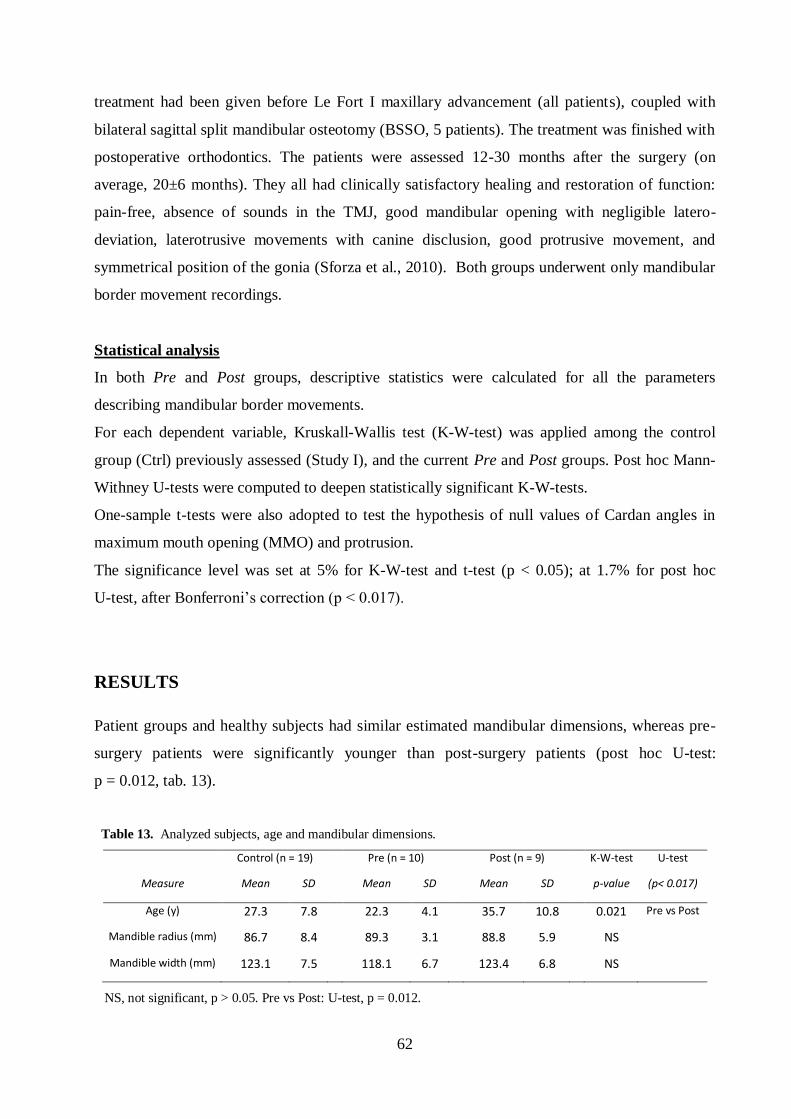
62
treatment had been given before Le Fort I maxillary advancement (all patients), coupled with
bilateral sagittal split mandibular osteotomy (BSSO, 5 patients). The treatment was finished with
postoperative orthodontics. The patients were assessed 12-30 months after the surgery (on
average, 20±6 months). They all had clinically satisfactory healing and restoration of function:
pain-free, absence of sounds in the TMJ, good mandibular opening with negligible latero-
deviation, laterotrusive movements with canine disclusion, good protrusive movement, and
symmetrical position of the gonia (Sforza et al., 2010). Both groups underwent only mandibular
border movement recordings.
Statistical analysis
In both Pre and Post groups, descriptive statistics were calculated for all the parameters
describing mandibular border movements.
For each dependent variable, Kruskall-Wallis test (K-W-test) was applied among the control
group (Ctrl) previously assessed (Study I), and the current Pre and Post groups. Post hoc Mann-
Withney U-tests were computed to deepen statistically significant K-W-tests.
One-sample t-tests were also adopted to test the hypothesis of null values of Cardan angles in
maximum mouth opening (MMO) and protrusion.
The significance level was set at 5% for K-W-test and t-test (p < 0.05); at 1.7% for post hoc
U-test, after Bonferroni’s correction (p < 0.017).
RESULTS
Patient groups and healthy subjects had similar estimated mandibular dimensions, whereas pre-
surgery patients were significantly younger than post-surgery patients (post hoc U-test:
p = 0.012, tab. 13).
Table 13. Analyzed subjects, age and mandibular dimensions.
Control (n = 19) Pre (n = 10) Post (n = 9) K-W-test U-test
Measure Mean SD Mean SD Mean SD p-value (p< 0.017)
Age (y) 27.3 7.8 22.3 4.1 35.7 10.8 0.021 Pre vs Post
Mandible radius (mm) 86.7 8.4 89.3 3.1 88.8 5.9 NS
Mandible width (mm) 123.1 7.5 118.1 6.7 123.4 6.8 NS
NS, not significant, p > 0.05. Pre vs Post: U-test, p = 0.012.

63
During laterotrusions and protrusion, the ranges of motion evaluated by the lower interincisor
and the two condylar reference points were not significantly different among the three groups
(K-W-test: p > 0.05).
Several differences were found at maximum mouth opening for both general mandibular motion
(tab.14, figure 34) and condylar pathways (tab. 15, figure 35).
Table 14. Kinematic range of motion of the mandible at Maximum Mouth Opening (MMO).
MMO IP caudal IP dorsal Sagittal
Measure [mm] component
[mm] component
[mm] angle
[°]
Control (n = 19) Mean 49.2 41.0 26.5 34.1
SD 5.1 5.1 6.4 3.8
Pre (n = 10) Mean 42.8 32.8 26.9 28.8
SD 8.7 6.0 8.6 7.5
Post (n = 9) Mean 49.2 40.0 28.2 33.8
SD 7.5 5.0 8.1 6.6
K-W-test p-value 0.022 0.004 NS NS
Ctrl vs Pre 0.007 0.002 Post hoc U-test Pre vs Post 0.050 0.013 Ctrl vs Post 0.961 0.446
NS, not significant, p > 0.05.
Figure 34. IP displacement and sagittal angle at MMO, mean+1SD.

64
Pre-surgery III class patients opened their mouth significantly less than the control group,
whereas post-surgery patients regained the normal range of motion. In particular, this difference
was completely due to the significant reduced caudal component, which was in turn partially
explained by a rather limited sagittal angle (tab. 14, figure 34).
Overall, however, mouth opening was symmetric in each group, being the horizontal and coronal
angles not significantly different from 0° (t-test, p > 0.05).
Table 15. Condylar linear displacements at MMO.
R_CRP caudal L_CRP caudal R_CRP ventral L_CRP ventral
Measure component
[mm] component
[mm]
component [mm]
component [mm]
Control (n = 19) Mean 9.3 8.8 13.1 12.0
SD 3.9 2.7 6.7 5.7
Pre (n = 10) Mean 4.3 4.6 6.5 7.3
SD 3.5 3.5 5.2 5.9
Post (n = 9) Mean 5.7 5.7 9.6 10.9
SD 3.8 3.1 2.4 3.9
K-W-test p-value 0.006 0.004 0.041 0.175
Ctrl vs Pre 0.003 0.004 0.016 Post hoc U-test Pre vs Post 0.165 0.327 0.165
Ctrl vs Post 0.052 0.016 0.248
R, right; L, left.
In the pre-surgery group both condyles moved downward significantly less than in healthy
subjects, and this limitation was not recovered in post-treatment subjects. Also, III class patients,
on average, had a right condyle that moved forward significantly less than the control group,
with only a partial improvement of the performance in post-surgery patients; a similar behaviour
was observed for the left condyle, even if it was not statistically significant (tab. 15).
Subsequently, the global path of the condyle resulted significantly reduced in pre-surgery
patients, and the treatment did not help in reaching normal gliding condylar movements (fig. 35).
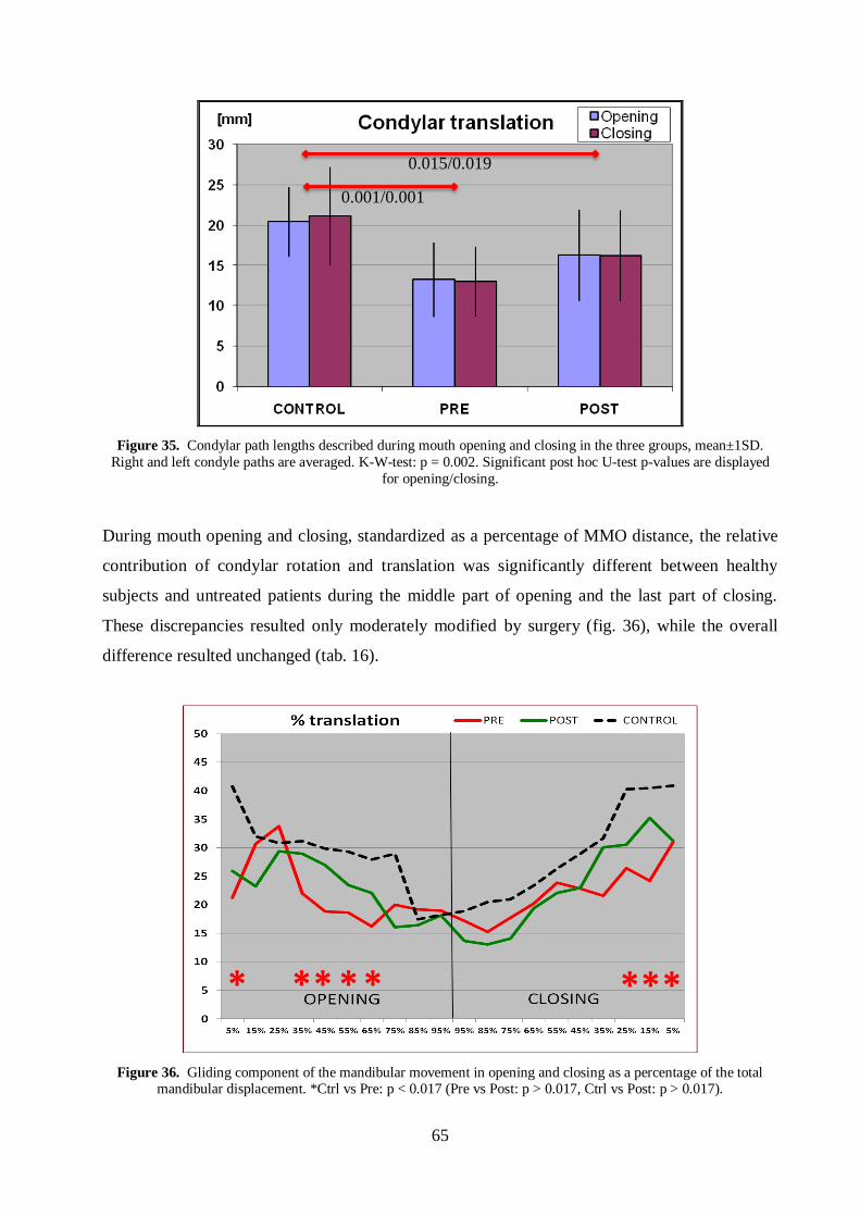
65
Figure 35. Condylar path lengths described during mouth opening and closing in the three groups, mean±1SD.
Right and left condyle paths are averaged. K-W-test: p = 0.002. Significant post hoc U-test p-values are displayed
for opening/closing.
During mouth opening and closing, standardized as a percentage of MMO distance, the relative
contribution of condylar rotation and translation was significantly different between healthy
subjects and untreated patients during the middle part of opening and the last part of closing.
These discrepancies resulted only moderately modified by surgery (fig. 36), while the overall
difference resulted unchanged (tab. 16).
Figure 36. Gliding component of the mandibular movement in opening and closing as a percentage of the total mandibular displacement. *Ctrl vs Pre: p < 0.017 (Pre vs Post: p > 0.017, Ctrl vs Post: p > 0.017).
0.001/0.001
0.015/0.019
* * * * * * * *

66
Table 16. Global translation contribution on mouth opening and closing.
Opening Translation Closing Translation Measure component [%] component [%]
Control (n = 19) Mean 28.3 28.7
SD 4.4 5.9
Pre (n = 10) Mean 22.5 22.4
SD 5.9 6.3
Post (n = 9) Mean 23.4 23.4
SD 3.4 3.8
K-W-test p-value 0.003 0.014
Ctrl vs Pre 0.005 0.015 Post hoc U-test Pre vs Post 0.623 0.120
Ctrl vs Post 0.007 0.022
DISCUSSION
Literature showed that one of the most common short and medium-term complications during
function recovery after orthognathic surgery is MMO reduction. Different causes have been
considered, such as the duration of the post-surgery intermaxillary fixation, surgical trauma,
changes in soft-tissue and muscular traction, amount of mandibular setback and surgeon’s
experience. A minimum of 6 months has been deemed necessary for recovery from passivity and
minor muscular trauma (Song et al., 1997).
In the past decades, the clinical measure of MMO was one of the most important indices of a
good post-surgery recovery. Usually, the MMO was directly measured with a ruler or a gauge in
the patient’s mouth (Zimmer et al., 1992; Nagamine et al., 1993; Ueki et al., 2008; Yazdani et
al., 2010) or recorded by means of kinesiography or axiography (Zarrinkelk et al., 1995, 1996).
However, standard clinical outcomes or the use of diagnostic instruments without an ad hoc
biomechanical model cannot be sensitive enough to detect oral dysfunction, like the change in
the percentage contribution of translation and rotation of condylar movements. The widely use of
the lower interincisor path alone for TMJ kinematic assessment is inadequate (Mapelli et al.,
2009); in the current study a three-dimensional optoelectronic motion analyzer was used together
with ad-hoc biomechanical model (see Materials and Methods for details) to detect all the 6
spatial degrees of freedom of mandibular kinematics in patients with skeletal Class III
malocclusion.
67
Ten individuals affected by Class III dentoskeletal deformity, and who had been received no
treatment yet, showed a strongly limited condylar translation in almost all steps of mouth
opening and closing. When compared to the normal values (as detailed in Study I), their
condylar path was almost halved, being so both its ventral and caudal displacements. Together
with a limited amount of sagittal opening angle, this latter restriction determined a remarkable
lower distance of maximum mouth opening, which was mainly characterized by an important
reduction of its caudal component.
Nine patients with the same dentoskeletal dysfunction who were recorded, on average, 20
months after having received orthognathic surgery (together with proper pre- and post-
orthodontic treatments), showed no notable gain in condylar movement, during mouth opening.
Even if a slight increment in condylar advancement was found (whereas the downward one was
negligible), overall, condylar rototranslation did not change after surgery. This may be explained
because, at the same time, the amount of mandibular rotation increased, up to physiologic values.
In patients who had received orthognathic surgery, also Sforza et al. (2008) measured a lower
percentage of condylar translation than in healthy subjects, even not statistically significant.
Anyway, the increased amount of the sagittal angle at MMO determined, in turn, the recovery of
the normal caudal component of maximum mouth opening and consequently the normal MMO
linear displacement at the interincisor point.
Furthermore, both mouth opening symmetry, and laterotrusions and protrusion ranges of motion
did not differ significantly from what was found in healthy subjects, either in pre-surgery or
post-surgery patients. In particular, the normal amount of condylar displacement during
protrusion should not appear astonishing, since it was quite the same as in mouth opening.
Indeed, in healthy subjects, condylar paths during jaw protrusion are usually shorter than during
mouth opening (Study I).
Overall, then, the present investigation only partially upholds previous findings (Throckmorton
et al., 1995; Sforza et al., 2010b) reporting functional recovery referable to orthognathic surgery.
In fact, kinematic parameters which were not dissimilar between the post-surgery patients and
the control group (protrusion and laterotrusion ranges of motion, symmetric border movements)
were also similar to the corresponding data of the untreated III class patients. On the other side,
what actually resulted enhanced in post-surgery patients with respect to pre-surgery ones, as
MMO, came out together with other unrecovered dysfunction, as condylar hypomobility. Only
longitudinal investigations may help understanding the full amount of impairment given by this
characteristic. However, condylar hypomobility has already been hypothesized in post-surgery
Class III patients: it could be explained by modifications in condylar position related to the

68
glenoid fossa (Kim et al., 2010) or remodelling of the mandibular condyle after surgical setback
(Gill et al., 2008). Also anatomical and functional alterations in the muscular control must be
considered: Katsumata et al. (2004) suggested that the masseter muscle may undergo reversible
atrophy after mandibular setback osteotomy and there may be alterations in the other masticatory
muscles and temporomandibular ligaments. Notwithstanding, the present findings suggest that
condylar hypomobility might be an intrinsic feature of the temporomandibular joint in skeletal
Class III.
The current study has two main drawbacks: both pre- and post-surgery groups were independent
to each other and were both composed of a limited number of patients. For this reason, a new
project has just begun with the purpose of recording a larger number of patients, with
longitudinal assessments during the subsequent phases of healing.
69
GENERAL CONCLUSIONS
Current clinical assessments and medical treatments are increasingly evidence-based, relying on
a widespread diffusion of diagnostic tools and treatment protocols that should make scientific-
based options available to the largest number of health professionals.
Indeed, the quantitative and accurate evaluation of masticatory muscle activity and jaw
movement is mandatory for a better understanding of the normal function and dysfunction of the
stomatognathic apparatus, and should assist conventional clinical assessment.
In an effort to make diagnosis as objective as possible, in the current thesis, a non-invasive and
short-lasting protocol integrating EMG signals and kinematic data has been proposed, in order to
develop multifactorial estimations of TMJ functioning. In particular, masticatory function has
been objectively determined, assessing bite stability, mandibular border movements and chewing
performance, in both clinically healthy and pathologic individuals.
The outcomes suggest that the proposed method could be a useful tool to evaluate the
neuromuscular coordination during the performance of static and dynamic masticatory activities,
and to detect functionally altered stomatognathic conditions. Diagnosis of alterations of the
stomatognathic apparatus, and assessment of the effects of therapy, would both profit from this
quantitative approach, thus reducing the discordance among several clinical examinations.
However, the findings of the present investigation are obviously strictly inherent to the
standardized protocol, and cannot be directly extended to natural, habitual function.
Future investigations will focus on the following main developments:
- to expand the protocol to more natural examined conditions;
- to identify the interrelationship between the findings of clinical examination with the
quantitative results provided by the current protocol;
- to propose new diagnostic/predictive indices, after testing their reliability, in order to
increase the precision of the clinical diagnosis and the choice of treatment.

70
LIST OF ABBREVIATIONS
2D: bi-dimensional
3D: three-dimensional
ANCOVA: analysis of covariance
ANOVA: analysis of variance
ASIM: asymmetry index
ATTIV: activity index
BSSO: bilateral sagittal split mandibular osteotomy
CCD: charge coupled device
CLENCH: maximum voluntary contraction without cotton rolls
COT: maximum voluntary contraction on cotton rolls
CRP: condylar reference point
CT: computed tomography
Ctrl: control group
DDR: disc displacement with reduction
EMG: electromyography
FARC: Functional Anatomy Research Centre
ICP: intercuspal position
IMPACT: integrated area of the standardized EMG potential over time
IP: inter-incisor point
K-W: Kruskal Wallis
LED: light emitting diode
Mk: marker
MMO: maximum mouth opening
MRI: magnetic resonance imaging
MVC: standardized maximum voluntary contraction
NS: not significant
POC: percentage overlapping coefficient
POST: after surgery group
PRE: before surgery group
r: mandibular radius

71
RDC: research diagnostic criteria
RMS: root mean square
s: circumference arc of the interincisor point
SD: standard deviation
sEMG: surface electromyography
SMI: symmetric mastication index
TMD: temporomandibular disorder
TMJ: temporomandibular joint
TORQUE: torque coefficient
TORS: torsion coefficient
TVC: television camera
w: mandibular width

72
REFERENCES
1. Akiyama H, Shiga H, Kobayashi Y. The analysis of masticatory movements. Frontal patterns
of chewing path of incisor point in normal subjects. J Jpn Prosthodont Soc, 1991; 35: 609-21.
2. Ardizone I, Celemin A, Aneiros F, del Rio J, Sanchez T, Moreno I. Electromyographic study
of activity of the masseter and anterior temporalis muscles in patients with
temporomandibular joint (TMJ) dysfunction: comparison with the clinical dysfunction index.
Med Oral Patol Oral Cir Bucal, 2010; 15: 14-9.
3. Bakke M, Hansdottir R. Mandibular function in patients with temporomandibular joint pain:
a 3-year follow-up. Oral Surg Oral Med Oral Pathol Oral Radiol Endod, 2008; 106: 227-34.
4. Bazzotti L. Electromyography tension and frequency spectrum analysis of some masticatory
muscles at rest, isotonic and isometric contractions. Electromyogr Clin Neurophysiol, 1999;
39: 421-34.
5. Bianchini EM, Paiva G, de Andrade CR. Mandibular movement patterns during speech in
subjects with temporomandibular disorders and in asymptomatic individuals. Cranio, 2008;
26: 50-8.
6. Burnett CA, Fartash L, Murray B, Lamey PJ. Masseter and temporalis muscle EMG levels
and bite force in migraineurs. Headache, 2000; 40: 813-7.
7. Buschang PH, Hayasaki H, Throckmorton GS. Quantification of human chewing-cycle
kinematics. Arch Oral Biol, 2000; 45: 461-74.
8. Buschang PH, Throckmorton GS, Travers KH, Hayasaki H. Incisor and mandibular condylar
movements of young adult females during maximum protrusion and lateratrusion of the jaw.
Arch Oral Biol, 2001; 46: 39-48.
9. Cairns BE. Pathophysiology of TMD pain--basic mechanisms and their implications for
pharmacotherapy. J Oral Rehabil, 2010; 37: 391-410.
10. Castroflorio T, Bracco P, Farina D. Surface electromyography in the assessment of jaw
elevator muscles. J Oral Rehabil, 2008; 35: 638-45.
11. Castroflorio T, Farina D, Bottin A, Piancino MG, Bracco P, Merletti R. Surface EMG of jaw
elevator muscles: effect of electrode location and inter-electrode distance. J Oral Rehabil,
2005; 32: 411-7.
12. Catic A, Naeije M. Location of the hinge axis and the kinematic centre in asymptomatic and
clicking temporomandibular joints. J Oral Rehabil, 1999; 26: 661-5.
13. Chen DC, Lai YL, Chi LY, Lee SY. Contributing factors of mandibular deformation during
mouth opening. J Dent, 2000; 28: 583-8.
73
14. Chew CL, Lucas PW, Tay DKL, Keng SB, Ow RKK. The effect of food texture on the
replication in mastication. J Dent, 1988;16: 210-4.
15. Ciuffolo F, Manzoli L, Ferritto AL, Tecco S, D’Attilio M, Festa F. Surface
electromyographic response of the neck muscles to maximal voluntary clenching of the teeth.
J Oral Rehabil, 2005; 32: 79-84.
16. Cote JN, Hoeger Bement MK. Update on the relation between pain and movement:
consequences for clinical practice. Clin J Pain, 2010; 26: 754-62.
17. De Felicio CM, De Oliveira MM, Da Silva MA. Effects of orofacial myofunctional therapy
on temporomandibular disorders. Cranio, 2010; 28: 249-59.
18. De Felicio CM, Melchior Mde O, Da Silva MA. Clinical validity of the protocol for multi-
professional centers for the determination of signs and syntomps of temporomandibular
disorders. Part II. Cranio, 2009a; 27: 62-7.
19. De Felicio CM, Sidequersky FV, Tartaglia GM, Sforza C. Electromyographic standardized
indices in healthy Brazilian young adults and data reproducibility. J Oral Rehabil, 2009b; 36:
577-83.
20. DeLuca C. The use of surface electromyography in biomechanics. J Appl Biomech, 1997;
13: 135-63.
21. Douglas CR, Avoglio JL, de Oliveira H. Stomatognathic adaptive motor syndrome is the
correct diagnosis for temporomandibular disorders. Med Hypo, 2010; 74: 710-8.
22. Dworkin SF, LeResche L. Research diagnostic criteria for temporomandibular disorders:
review, criteria, examinations and specifications, critique. J Craniomand Dis Facial Oral
Pain, 1992; 6: 301-55.
23. Epker J, Gatchel R, Ellis III E. A model for predicting chronic TMD. JADA, 1999; 130:
1470-5.
24. Epstein JB, Klasser GD. Whiplash-associated disorders and temporomandibular symptoms
following motor-vehicle collisions. Quintessence Int, 2011; 42: e1-e14.
25. Evans WG and Lewin A. Some characteristics of mandibular movements in a group of San. J
Dent Assoc S Afr, 1986; 41: 543-8.
26. Farella M, Bakke M, Michelotti A, Rapuano A, Martina R. Masseter thickness, endurance
and exercise-induced pain in subjects with different vertical craniofacial morphology. Eur J
Oral Sci, 2003; 111: 183-8.
27. Farella M, Van Eijden T, Baccini M, Michelotti A. Task-related electromyographic spectral
changes in the human masseter and temporalis muscles. Eur J Oral Sci, 2002; 110: 8-12.

74
28. Ferrario VF, Piancino MG, Dellavia C, Castroflorio T, Sforza C, Bracco P. Quantitative
analysis of the variability of unilateral chewing movements in young adults. Cranio, 2006a;
24: 274-82.
29. Ferrario VF, Sforza C, Colombo A, Ciusa V. An electromyographic investigation of
masticatory muscles symmetry in normo-occlusion subjects. J Oral Rehabil, 2000; 27: 33-40.
30. Ferrario VF, Sforza C, Lovecchio N, Mian F. Quantification of translational and gliding
components in human temporomandibular joint during mouth opening. Arch Oral Biol,
2005; 50: 507-15.
31. Ferrario VF, Sforza C, Serrao G, Grassi G, Mossi E. Active range of motion of the head and
cervical spine: a three-dimensional investigation in healthy young adults. J Orthop Res,
2002; 20:122-9.
32. Ferrario VF, Sforza C, Serrao G. The influence of crossbite on the coordinated
electromyographic activity of human masticatory muscles during mastication. J Oral Rehabil,
1999; 26: 575-81.
33. Ferrario VF, Sforza C, Zanotti G, Tartaglia GM. Maximal bite forces in healthy young adults
as predicted by surface electromyography. J Dent, 2004a; 32: 451-7.
34. Ferrario VF, Sforza C. Biomechanical model of the human mandible in unilateral clench:
distribution of temporomandibular joint reaction forces between working and balancing
sides. J Prosthet Dent, 1994; 72: 169-76.
35. Ferrario VF, Sforza C. Biomechanical model of the human mandible: a hypothesis involving
stabilizing activity of the superior belly of lateral pterygoid muscle. J Prosthet Dent, 1992;
68: 829-35.
36. Ferrario VF, Sforza C. Coordinated electromyographic activity of the human masseter and
temporalis anterior muscles during mastication. Eur J Oral Sci, 1996; 104: 511-7.
37. Ferrario VF, Tartaglia GM, Galletta A, Grassi GP, Sforza C. The influence of occlusion on
jaw and neck muscle activity: a surface EMG study in healthy young adults. J Oral Rehabil,
2006b; 33: 341-8.
38. Ferrario VF, Tartaglia GM, Luraghi FE, Sforza C. The use of electromyography as a tool in
differentiating temporomandibular disorders from neck disorders. Man Ther, 2007; 12: 372-
9.
39. Ferrario VF, Tartaglia GM, Maglione M, Simion M, Sforza C. Neuromuscular coordination
of masticatory muscles in subjects with two types of implant-supported prostheses. Clin Oral
Implants Res, 2004b; 15: 219-25.

75
40. Forrester SE, Allen SJ, Presswood RG, Toy AC, Pain MTG. Neuromuscular function in
healthy occlusion. J Oral Rehabil, 2010; 37: 663-9.
41. Gallagher C, Gallagher V, Whelton H, Cronin M. The normal range of mouth opening in an
Irish population. J Oral Rehabil, 2004; 31: 110-6.
42. Gallo LM, Airoldi GB, Airoldi RL, Palla S. Description of mandibular finite helical axis
pathways in asymptomatic subjects. J Dent Res, 1997; 76: 704-13.
43. Garcia-Morales P, Buschang PH, Throckmorton GS, English JD. Maximum bite force,
muscle efficiency and mechanical advantage in children with vertical growth patterns. Eur J
Orthod, 2003; 25: 265-72.
44. Gay T, Maton B, Rendell J, Majourau A. Characteristics of muscle fatigue in patients with
myofascial pain-dysfunction syndrome. Arch Oral Biol, 1994; 39: 847-52.
45. Gerstner GE, Parekh VV. Evidence of sex-specific difference in masticatory jaw movement
patterns. J Dent Res, 1997; 76: 796-806.
46. Gill DS, El Maaytah M, Naini FB. Risk factors for post-orthognathic condylar resorption: a
review. World J Orthod, 2008; 9: 21-5.
47. Gsellmann B, Schmed-Schwap M, Piehslinger E, Slavicek R. Lengths of condylar pathways
measured with computerized axiography and occlusal index in patients and volunteers. J Oral
Rehabil, 1998; 25: 146-52.
48. Hansdottir R, Bakke M. Joint tenderness, jaw opening, chewing velocity, and bite force in
patients with temporomandibular joint pain and matched healthy control subjects. J Orofac
Pain, 2004; 18: 108-13.
49. Harper RP. Analysis of temporomandibular joint function after orthognathic surgery using
condylar path tracings. Am J Orthod Dentofacial Orthop, 1990; 97: 480-8.
50. Hayasaki H, Saitoh I, Iwase Y, Inada E, Hasegawa H, Tokutomi J, Matsumoto Y, Yamasaki
Y. Movement of the instantaneous center of rotation and the position of the lateral excursion
center during lateral excursion. Cranio, 2008; 26: 253-62.
51. Hayasaki H, Saitoh I, Throckmorton GS, Iwase Y, Nakata S, Nakata M. Occlusal phase of
gum-chewing strokes. J Oral Rehabil, 2003; 30: 1041-6.
52. Hellmann, Giannakopoulos NN, Blaser R, Eberhard L, Rues S, Schindler HJ. Longterm
training effects on masticatory muscles. J Oral Rehabil, 2011; 38: 912-20.
53. Hiatt JL, Gartner LP. Textbook of head and neck anatomy. 4th edition. New York:
Lippincott Williams and Wilkins, 2010.
54. Howell PG. Sexual dimorphism in mastication and speech? Or do men and women eat and
talk differently? Aust Prostho J, 1987; 1: 9-17.

76
55. John MT, Dworkin SF, Mancl LA. Reliability of clinical temporomandibular disorder
diagnoses. Pain, 2005; 118: 61-9.
56. Karlsson S, Carlsson GE. Characteristics of mandibular masticatory movement in young and
elderly dentate subjects. J Den Res, 1990; 69: 473-6.
57. Katsumata A, Fujishita M, Ariji Y, Ariji E, Langlais RP. 3D CT evaluation of masseter
muscle morphology after setback osteotomy for mandibular prognathism. Oral Surg Oral
Med Oral Pathol Oral Radiol Endod, 2004; 98: 461-70.
58. Kenworthy CR, Morrish RB, Mohn C Jr, Miller A, Swenson KA. Bilateral condylar
movement patterns in adult subjects. J Orofac Pain, 1997; 11: 328-36.
59. Kim YI, Jung YH, Cho BH, Kim JR, Kim SS, Son WS, Park SB. The assessment of the
short- and long-term changes in the condylar position following sagittal split ramus
osteotomy (SSRO) with rigid fixation. J Oral Rehabil, 2010; 37: 262-70.
60. Klasser G, Greene C. Oral appliances in the management of temporomandibular disorders.
Oral Surg Oral Med Oral Pathol Oral Radiol Endod, 2009; 107: 212-23.
61. Klasser GD, Okeson JP. The clinical usefulness of surface electromyography in the diagnosis
and treatment of temporo-mandibular disorders. J Am Dent Assoc, 2006; 137: 763-71.
62. Kogawa EM, Calderon PS, Lauris JRP, Araujo CRP, Conti PCR. Evaluation of maximal bite
force in temporomandibular disorders patients. J Oral Rehabil, 2006; 33: 559-65.
63. Kohyama K, Sasaki T, Hayakawa F. Characterization of food physical properties by the
mastication parameters measured by electromyography of the jaw-closing muscles and
mandibular kinematics in young adults. Biosci Biotechnol Biochem, 2008; 72: 1690-5.
64. Koolstra JH, Naeije M, van Eijden TM. The three-dimensional active envelope of jaw border
movement and its determinants. J Dent Res, 2001; 80:1908-12.
65. Koolstra JH, van Eijden TMGJ. Biomechanical analysis of jaw closing movements. J Dent
Res, 1995; 74: 1564-70.
66. Koyano K, Kim YJ, Clark GT. Electromyographic signal changes during exercise in human
chronic jaw-muscle pain. Arch Oral Biol, 1995; 40: 221-7.
67. Kroon GW, Naeije M. Electromyographic evidence of local muscle fatigue in a subgroup of
patients with myogenous craniomandibular disorders. Arch Oral Biol, 1992; 37: 215-8.
68. Kumai T. Difference in chewing patterns between involved and opposite sides in patients
with unilateral temporomandibular joint and myofascial pain-dysfunction. Arch Oral Biol,
1993; 38: 467-78.
69. Kumar S, Narayan Y, Amell T. Power spectra of sternocleidomastoids, splenius capitis, and
upper trapezius in oblique exertions. Spine J, 2003; 3: 339-50.

77
70. Kumar S, Narayan Y, Amell T. Spectral profile of superficial cervical muscles. J
Electromyogr Kinesiol, 2001; 11: 269-80.
71. Landulpho AB, E Silva WAB, E Silva FA, Vitti M. Electromyographic evaluation of
masseter and anterior temporalis muscles in patients with temporomandibular disorders
following interocclusal appliance treatment. J Oral Rehabil, 2004; 31: 95-8.
72. Leader JK, Boston JR, Debski RE, Rudy TE. Mandibular kinematics represented by a non-
orthogonal floating axis joint coordinate system. J Biomec, 2003; 36: 275-81.
73. Lemoine JJ, Xia JJ, Gateno J, Liebschner MA. Radiographic analysis for jaw motion
normalization. J Oral Maxillofac Surg, 2005; 63: 961-7.
74. Lewis RP, Buschang PH, Throckmorton GS. Sex differences in mandibular movements
during opening and closing. Am J Orthod Dentofac Orthop, 2001; 120: 294-303.
75. Liu ZJ, Yamagata K, Kasahara Y, Ito G. Electromyographic examination of jaw muscles in
relation to symptoms and occlusion of patients with temporomandibular joint disorders. J
Oral Rehabil, 1999; 26: 33-47.
76. Lobbezoo F, van Selms MK, Naeije M. Masticatory muscle pain and disordered jaw motor
behaviour: Literature review over the past decade. Arch Oral Biol, 2006; 51: 713-20.
77. Lobbezoo F, Zwijnenburg AJ, Naeije M. Functional subdivision of the human masseter and
temporalis muscles as shown by the condylar movement response to electrical muscle
stimulation. J Oral Rehabil, 2000; 27: 887-92.
78. Lotters FJB, Zwijnenburg AJ, Megens CCEJ, Naeije M. Relationship between condylar and
incisor point displacement during habitual maximum open-close movements. J Oral Rehabil,
1996; 23: 548-54.
79. Manfredini D, Chiappe G, Bosco M. Research diagnostic criteria for temporomandibular
disorders (RDC/TMD) axis I diagnoses in an Italian patient population. J Oral Rehabil, 2006;
33: 551-8.
80. Mapelli A, Galante D, Lovecchio N, Sforza C, Ferrario VF. Translation and rotation
movements of the mandible during mouth opening and closing. Clin Anat, 2009; 22: 311-8.
81. Matsumoto A, Celar RM, Celar A, Sato S, Suzuki Y, Slavicek R. An analysis of hinge axis
translation and rotation during opening and closing in dentulous and edentulous subjects.
Cranio, 1995; 13: 238-41.
82. Melsen B, Graham J, Baccetti T, Koga M, Boyd R, Park J, Tracey S, Uribe F, Park HS,
Cacciafesta V, Haeger RS, Bowman SJ, Kook YA, Redmond WR, Bjorn L, Lisson JA.
Factors contributing to the success or failure of skeletal anchorage devices: an informal JCO
survey. J Clin Orthod, 2010; 44: 714-8.

78
83. Merlini L, Palla S. The relationship between condylar rotation and anterior translation in
healthy and clicking temporomandibular joints. Schweiz Monatsschr Zahnmed, 1988; 98:
1191-9.
84. Michelotti A, Iodice G. The role of orthodontics in temporomandibular disorders. J Oral
Rehabil, 2010; 37: 411-29.
85. Miles S. Postural control of the human mandible. Arch Oral Biol, 2007; 52: 347-52.
86. Miyawaki S, Ohkochi N, Kawakami T, Sugimura M. Effect of food size on the movement of
the mandibular first molars and condyles during deliberate unilateral mastication in humans.
J Dent Res, 2000; 79: 1525-31.
87. Miyawaki S, Tanimoto Y, Araki Y, Katayama A, Kuboki T, Takano-Yamomato T.
Movement of the lateral and medial poles of the working condyle during mastication in
patients with unilateral posterior crossbite. J Am Orthod Dentofacial Orthop, 2004; 126: 549-
54.
88. Miyawaki S, Tanimoto Y, Kawakami T, Sugimura M, Takano-Yamamoto T. Motion of the
human mandibular condyle during mastication. J Dent Res, 2001; 80: 437-42.
89. Mobilio, Casetta I, Cesnik E, Catapano S. Prevalence of self-reported symptoms related to
temporomandibular disorders in an Italian population. J Oral Rehabil, 2011; 38: 884-90.
90. Monteverdi R, Formillo P, Giannì AB, Tartaglia GM, Sforza C, Ferrario VF. A
threedimensional quantitative analysis of mandibular movements after condylar fractures
treatment. J Craniomaxillofac. Surg, 2006; 34: 69.
91. Naeije M. Electromyography and craniomandibular disorders. Ned Tijdschr Tandheelkd,
1989; 96: 402-5.
92. Naeije M. Local kinematic and anthropometric factors related to the maximum mouth
opening in healthy individuals. J Oral Rehabil, 2002; 29: 534-9.
93. Naeije M. Measurement of condilar motion: a plea for the use of the condylar kinematic
centre. J Oral Func, 2003; 30: 225-30.
94. Nagamine T, Kobayashi T, Nakajima T, Hanada K. The effects of surgical-orthodontic
correction of skeletal class III malocclusion on mandibular movement. J Oral Maxillofac
Surg, 1993; 51: 385-9.
95. Neill DJ, Howell PGT. Computerized kinesography in the study of mastication in dentate
subjects. J Prosthet Dent, 1986; 55: 629-38.
96. Palla S, Gallo LM, Gossi D. Dynamic stereometry of the temporomandibular joint. Orthod
Craniofac Res, 2003; 6: 37-47.

79
97. Peck CC, Murray GM, Gerzina TM. How does pain affect jaw muscle activity? The
Integrated Pain Adaptation Model. Aust Dent J, 2008; 53: 201-7.
98. Peck CC, Murray GM, Johnson CW, Klineberg IJ. Trajectories of condylar points during
nonworking side and protrusive movements of the mandible. J Prosthet Dent, 1999; 82: 322-
31.
99. Piancino MG, Bracco P, Vallelonga T, Merlo A, Farina D. Effect of bolus hardness on the
chewing pattern and activation of masticatory muscles in subjects with normal dental
occlusion. J Electromyogr Kinesiol, 2008;18: 931-7.
100. Piehslinger E, Celar RM, Horejs T, Slavicek R. Orthopedic jaw movement observations.
Part II: the rotational capacity of the mandible. Cranio, 1993; 11: 206-10.
101. Pinho JC, Caldas FM, Mora MJ, Santana-Penin U. Electromyographic activity in patients
with temporomandibular disorders. J Oral Rehabil, 2000; 27: 985-90.
102. Proschel P and Hofmann M. Frontal chewing patterns of the incisor point and their
dependance on resistance of food and type of occlusion. J Prosthet Dent, 1988; 39: 617-24.
103. Proschel P. An extensive classification of chewing patterns in the frontal plane. J
Craniomandib Pract, 1987; 5: 55.
104. Ramfjord SP, Ash MM. Occlusion. 3rd
edition. Philadelphia: Sounders, 1983. In [68].
105. Rampello A.ATM, testo atlante. Concetti fondamentali. Bologna: Martina editore, 2004.
106. Ries LG, Alves MC, Bérzin F. Asymmetric activation of temporalis, masseter, and
sternocleidomastoid muscles in temporomandibular disorder patients. Cranio, 2008; 26: 59-
64.
107. Rilo B, da Silva JL, Mora MJ, Cadarso-Suarez C, Santana U. Unilateral posterior
crossbite and mastication. Arch Oral Biol, 2007; 52: 474-8.
108. Rilo B, Fernandez-Formoso N, Mora MJ, Cadarso-Suarez C, Santana U. Distance of the
contact glide in the closing masticatory stroke during mastication of three types of food. J
Oral Rehabil, 2009; 36: 571-6.
109. Saitoh I, Tokutomi J, Hayasaki H, Iwase Y, Raoquig H, Yamasaki Y, Nonaka K.
Correlations between incisor and condylar movements during lateral excursion in children
with primary dentition. J Oral Rehabil, 2007; 34: 800-6.
110. Salaorni C, Palla S. Condylar rotation and anterior translation in healthy human
temporomandibular joints. Schweiz Monatsschr Zahnmed 1994; 104: 415-22.
111. Santana-Mora U, Cudeiro J, Mora-Bermúdez MJ, Rilo-Pousa B, Ferreira-Pinho JC,
Otero-Cepeda JL, et al. Changes in EMG activity during clenching in chronic pain patients
with unilateral temporomandibular disorders. J Electromyogr Kinesiol, 2009; 19: 543-9.

80
112. Sato S, Ohta M, Goto S, Kawamura H, Motegi K. Electromyography during chewing
movement in patients with anterior disc displacement of the temporomandibular joint. Int J
Oral Maxillof Surg, 1998; 27: 274-7.
113. Schmitter M, Ohlmann B, John MT, Hirsch C, Rammelsberg P. Research diagnostic
criteria for temporomandibular disorders: a calibration and reliability study. Cranio, 2005;
23: 212-8.
114. Sforza C, Dellavia CPB, Tartaglia G, Ferrario VF. Morphological and functional features
of the human temporomandibular joint. Ital J Maxillofac Surg, 2010a; 21: 3-17.
115. Sforza C, Tartaglia GM, Lovecchio N, Ugolini A, Monteverdi R, Giannì AB, Ferrario
VF. Mandibular movements at maximum mouth opening and EMG activity of masticatory
and neck muscles in patients rehabilitated after a mandibular condyle fracture. J
Craniomaxillofac Surg, 2009; 37: 327-33.
116. Sforza C, Ugolini A, Rocchetta D, Galante D, Mapelli A, Giannì AB. Mandibular
kinematics after orthognathic surgical treatment a pilot study. Br J Oral Maxillofac Surg,
2010b; 48: 110-4.
117. Sforza C, Ugolini A, Sozzi D, Galante D, Mapelli A, Bozzetti A. Three-dimensional
mandibular motion after closed and open reduction of unilateral mandibular condylar process
fractures. J Craniomaxillofac Surg, 2011; 39: 249-55.
118. Song HC, Throckmorton GS, Ellis III E, Sinn DP. Functional and morphological
alterations after anterior and/or inferior repositioning of the maxilla. J Oral Maxillofac Surg,
1997; 55: 41-50.
119. Suvinen TI, Malmberg J, Forster C, Kemppainen P. Postural and dynamic masseter and
anterior temporalis muscle EMG repeatability in serial assessments. J Oral Rehabil, 2009;
36: 814-20.
120. Suvinen TI, Reade PC, Kononen M, Kemppainen P. Vertical jaw separation and masseter
muscle electromyographic activity: a comparative study between asymptomatic controls &
patients with temporomandibular pain & dysfunction. J Oral Rehabil, 2003; 30: 765-72.
121. Takeda H e Nakamura Y. Examination of masticatory movement and rhyhm before and
after surgical orthodontics in skeletal class III patients with unilateral posterior cross-bite.
The J Oral Maxillofac Surg, 2009; 67: 1844-9.
122. Tartaglia GM, Lodetti G, Paiva G, De Felicio CM, Sforza C. Surface electromyographic
assessment of patients with long lasting temporomandibular joint disorder pain. J
Electromyogr Kinesiol, 2011; 21: 659-64.

81
123. Tartaglia GM, Lodetti G, Paiva G, De Felicio CM, Sforza C. Surface electromyographic
assessment of patients with long lasting temporomandibular joint disorder pain. J
Electromyogr Kinesiol, 2011; 21: 659-64.
124. Tartaglia GM, Moreira Rodrigues da Silva MA, Bottini S, Sforza C, Ferrario VF.
Masticatory muscle activity during maximum voluntary clench in different research
diagnostic criteria for temporomandibular disorders (RDC ⁄ TMD) groups. Man Ther, 2008a;
13: 434-40.
125. Tartaglia GM, Testori T, Pallavera A, Marelli B, Sforza C. Electromyographic analysis of
masticatory and neck muscles in subjects with natural dentition, teeth-supported and implant-
supported prostheses. Clin Oral Impl Res, 2008b; 19: 1081-8.
126. Tecco S, Crincoli V, Di Bisceglie B, Caputi S, Festa F. Relation between facial
morphology on lateral skull radiographs and sEMG activity of head, neck and trunk muscles
in Caucasian adult females. J Electromyogr Kinesiol, 2011; 21: 298-310.
127. Theusner J, Plesh O, Curtis DA, Hutton JE. Axiographic tracings of temporomandibular
joint movements. J Prosthet Dent, 1993; 69: 209-15.
128. Throckmorton GS, Buschang BH, Hayasaki H, Phelan T. The effects of chewing rates on
mandibular kinematics. J Oral Rehabil, 2001; 28: 328-34.
129. Throckmorton GS, Ellis III E, Sinn DP. Functional characteristics of retrognathic patients
before and after mandibular advancement surgery. J Oral Maxillofac Surg, 1995; 53: 898-
909.
130. Throckmorton GS. Functional deficits in orthognathic surgery patients. Semin Orthodont,
2006; 12: 127-37.
131. Travers KH, Buschang PH, Hayasaki H, Throckmorton GS. Associations between incisor
and mandibular condylar movements during maximum mouth opening in humans. Arch Oral
Biol, 2000; 45: 267-75.
132. Ueki K, Marukawa K, Hashiba Y, Nakagawa K, Degerliyurt K, Yamamoto E.
Assessment of the relationship between the recovery of maximum mandibular opening and
the maxillomandibular fixation period after orthognathic surgery. J Oral Maxillofac Surg,
2008; 66: 486-91.
133. Van der Bilt A. Assessment of mastication with implications for oral rehabilitation: a
review. J Oral Rehabil, 2011; 38: 754-80.
134. Visser A, Kroon GW, Naeije M, Hansson TL. EMG differences between weak and strong
myogenous CMD patients and healthy controls. J Oral Rehabil, 1995; 22: 429-34.

82
135. Wang D, Fu H, Zeng R, Yang X. Changes of mandibular movement tracings after the
correction of mandibular protrusion by bilateral sagittal split ramus osteotomy. J Oral
Maxillofac Surg, 2009; 67: 2238-44.
136. Williams PL, Warwick R, Dyson M, Bannister L. Gray’s anatomy. 37th edition.
Edinburgh, London, Melbourne and New York: Churchill Livingstone, 1989.
137. Wintergerst AM, Buschang PH, Throckmorton GS. Reducing within-subject variation in
chewing cycle kinematics-a statistical approach. Arch Oral Biol, 2004; 49: 991-1000.
138. Wu JB, Xu XM, Sheng JG. Analysis of the open-closing movement of the human
temporomandibular joint. Acta Anat (Basel), 1988; 133: 213-6.
139. Yamashita S, Hatch JP, Rugh JD. Does chewing performance depend upon a specific
masticatory pattern? Review. J Oral Rehabil, 1999; 26: 547-53.
140. Yashiro K, Takada K. Model-based analysis of jaw-movement kinematics by using jerk-
optimal criterion: simulations of human chewing cycles. J Electromyograph Kinesiol, 2005;
15: 516-26.
141. Yatabe M, Zwijnenburg A, Megens CC, Naejie M. The kinematic center: a reference for
condylar movements. J Oral Rehabil, 1995; 74: 1644-8.
142. Yatabe M, Zwijnenburg A, Megens CCEJ, Naeije M. Movements of the mandibular
condyle kinematic center during jaw opening and closing. J Dent Res, 1997; 76: 714-9.
143. Yazdani J, Ebrahimi H, Talesh KT, Khashabi E, Pourshahidi S, Tadbir AA. Comparing
the effect of 3 orthognathic surgical methods on the mandibular range of movement. J
Craniofac Surg, 2010; 21: 703-5.
144. Youssef RE, Throckmorton GS, Ellis E 3rd, Sinn DP. Comparison of habitual
masticatory cycles and muscle activity before and after orthognathic surgery. J Oral
Maxillofac Surg, 1997; 55: 707-8.
145. Zarrinkelk HM, Throckmorton GS, Ellis III E, Sinn DP. Functional and morphological
alterations secondary to superior repositioning of the maxilla. J Oral Maxillofac Surg, 1995;
53: 1258-67.
146. Zarrinkelk HM, Throckmorton GS, Ellis III E, Sinn DP. Functional and morphological
changes after combined maxillary intrusion and mandibular advancement surgery. J Oral
Maxillofac Surg, 1996; 54: 828-37.
147. Zimmer B, Schwestka R, Kubein-Meesenburg D. Changes in mandibular mobility after
different procedures of orthognathic surgery. Eur J Orthod, 1992; 14: 188-97.
148. Zwijnenburg A, Megens CC, Naejie M. Influence of choice of reference point on the
condylar movement paths during mandibular movement. J Oral Rehabil, 1996; 23: 832-7.

83
WEBSITES
1. http://www.youtube.com/user/johnsonorthodontics. Accessed on 20/12/2011.