testing the validity of radiographic comparisons in ... in positive identifications, final ......
TRANSCRIPT

The author(s) shown below used Federal funds provided by the U.S. Department of Justice and prepared the following final report: Document Title: Testing the Validity of Radiographic
Comparisons in Positive Identifications, Final Technical Report
Author(s): Ann H. Ross, Ph.D., Alicja K. Lanfear, Ph.D.,
Ashley B. Maxwell, M.A. Document No.: 248638 Date Received: February 2015 Award Number: 2010-DN-BX-K214 This report has not been published by the U.S. Department of Justice. To provide better customer service, NCJRS has made this Federally-funded grant report available electronically.
Opinions or points of view expressed are those of the author(s) and do not necessarily reflect
the official position or policies of the U.S. Department of Justice.

Final Technical Report
Testing the Validity of Radiographic Comparisons in Positive Identifications
2010-DN-BX-K214
PI: Ann H. Ross, Ph.D., D-ABFA1
Alicja K. Lanfear, Ph.D.2
Ashley B. Maxwell, M.A.3
1NC State University
Department of Sociology and Anthropology Campus Box 8107
Raleigh, NC 27695-8107
Email: [email protected]
Tel. 919-515-9021 Fax 919-513-0866
2 Middle Tennessee State University
Department of Biology Murfreesboro, TN 37132
3 Department of Anthropology
University of South Florida Tampa, FL 33620
September 30, 2014
This document is a research report submitted to the U.S. Department of Justice. This report has not been published by the Department. Opinions or points of view expressed are those of the author(s)
and do not necessarily reflect the official position or policies of the U.S. Department of Justice.

2
ABSTRACT
Although radiographic comparisons are commonly used, to date, there has been little to no testing of the validity of using such methods to establish a positive identification. To increase the likelihood of evidence being deemed admissible in court, it is necessary for these methods to conform to Daubert criteria and the suggestions made in the NAS report. This research marks the start of a systematic validation of radiographic comparison methods based on several anatomical features and skeletal landmarks. This research addressed five specific goals aimed to be in compliance with Daubert criteria and the NAS report. 1) To evaluate the utility of various anatomical features that are visible in standard radiographs. 2) To evaluate the accuracy and reliability of methods currently employed to make positive identifications. 3) To examine the utility of identifying quantifiable anatomical landmarks in radiographs. 4) To examine the utility of using outline shapes to identify vault and cranial outlines as a means of confirming identification. 5) To develop a standard system and minimum number of concordant features for making positive identifications through radiographic comparison.
The radiographic database generated by this research project contains anonymized radiographs of 858 adults and 148 juveniles that were made available by the North Carolina Office of the Chief Medical Examiner. These radiographs include individually scanned radiographs that contain dental, antemortem, and postmortem radiographs of various parts of the skeleton dating from 1974-2002. Subsets of this database were used to address the five goals of this research project and were selected based on radiograph availability for the elements in question.
Three standard body regions were selected for this study; lateral cranial radiographs, chest radiographs, and radiographs of the proximal femur. Lateral cranial radiographs were assessed for validity in identification using a multi-method approach. Cranial vault outlines were assessed using visual comparison, elliptic Fourier analysis, and sliding semi-landmark analysis. A standard system for making radiographic comparisons was developed based on the assessment of points of concordance and utilized a robust statistical method, classification decision trees.
All three methods used to assess cranial vault outlines in lateral cranial radiographs demonstrated that the shape of the vault is not unique enough to make an individual identification. The standard system of radiographic comparison that was developed yielded positive results. Two or more points of concordance are required in lateral cranial radiographs for a 97% probability of a positive identification. The results of the chest radiographic analysis showed that the anatomical elements with the most predictive values were the cervical vertebrae, and showed that if more than one concordant feature exists there is a 99% probability of correct identification. If no cervical concordant features are present, four or more concordant features of the other vertebrae are required for a 98% probability of having a correct identification. This probability drops to 79% when less than four concordant lumbar features are present. For the femur, if there is one or more femoral head concordant feature the probability of a correct
This document is a research report submitted to the U.S. Department of Justice. This report has not been published by the Department. Opinions or points of view expressed are those of the author(s)
and do not necessarily reflect the official position or policies of the U.S. Department of Justice.

3
identification is 93% and 97% with concordant neck traits. For the greater trochanter the probability is 76% of a correct identification with one or more features and 93% if there are two or more concordant features for the lesser trochanter. This study established the minimum number of concordant areas needed to confirm positive identifications in three standard radiographic views. This is significant as it is the first attempt to statistically quantify the number of concordant points needed to meet the Daubert criteria for making a positive identification using radiographs. This newly developed system should serve as a model for future research pertaining to positive identification using radiographs.
This document is a research report submitted to the U.S. Department of Justice. This report has not been published by the Department. Opinions or points of view expressed are those of the author(s)
and do not necessarily reflect the official position or policies of the U.S. Department of Justice.

Table of Contents EXECUTIVE SUMARY……………………………………………………………………….. 6
I. INTRODUCTION ..................................................................................................................... 11
Statement of the Problem ........................................................................................................................ 11
Literature Review .................................................................................................................................... 11
Project Goals ........................................................................................................................................... 14
II. METHODS............................................................................................................................... 15
Sample .................................................................................................................................................... 15
Lateral Cranial Study .............................................................................................................................. 16
Phase 1. Lateral Cranial Radiographs: Visual Comparison (Stage 1) and Shape via Elliptic Fourier Analysis (Stage 2) ............................................................................................................................... 17
Phase 2 - Lateral radiographs 2D sliding semilandmarks ................................................................... 20
Phase 3- Classification Trees for Points of Concordance of Craniofacial Traits ................................ 21
Chest Radiographs .................................................................................................................................. 24
Sample ................................................................................................................................................. 25
Methods ............................................................................................................................................... 25
Radiographs of the Proximal Femur ....................................................................................................... 26
Sample ................................................................................................................................................. 27
Methods ............................................................................................................................................... 27
III. RESULTS ............................................................................................................................... 29
Lateral Cranial Study .............................................................................................................................. 29
Phase 1, Stage 1- Visual Comparison ................................................................................................. 29
Phase 1, Stage 2 - Elliptic Fourier Analysis ........................................................................................ 30
Phase 2 - Sliding Semilandmarks ........................................................................................................ 31
Phase 3 - Points of Concordance ......................................................................................................... 33
Chest Radiographs .................................................................................................................................. 34
Cervical, thoracic and lumbar vertebrae ............................................................................................. 34
Cervical and thoracic vertebrae ........................................................................................................... 35
Lumbar Vertebrae ............................................................................................................................... 36
Radiographs of the Proximal Femur ....................................................................................................... 37
Femoral Head and Neck ...................................................................................................................... 37
Femoral Lesser and Greater Trochanters ............................................................................................ 39
IV. CONCLUSIONS .................................................................................................................... 40
This document is a research report submitted to the U.S. Department of Justice. This report has not been published by the Department. Opinions or points of view expressed are those of the author(s)
and do not necessarily reflect the official position or policies of the U.S. Department of Justice.

Discussion of findings ............................................................................................................................. 40
Implications for policy and practice ........................................................................................................ 43
Implications for further research ............................................................................................................. 43
V. REFERENCES......................................................................................................................... 45
VI. DISSEMINATION OF RESEARCH FINDINGS ................................................................. 48
VII. ACKNOWLEDGEMENTS .................................................................................................. 49
This document is a research report submitted to the U.S. Department of Justice. This report has not been published by the Department. Opinions or points of view expressed are those of the author(s)
and do not necessarily reflect the official position or policies of the U.S. Department of Justice.
6
EXECUTIVE SUMMARY The problem and purpose
Positive identification of unknown remains is essential to the resolution of criminal investigations, insurance claims, the facilitation of the release of the decedent to family for interment, and the emotional recovery of family and friends. Although radiographic comparisons are commonly used, to date, there has been little to no testing of the validity of using such methods to establish a positive identification. To increase the likelihood of admissibility in court, it is necessary for these methods to conform to Daubert criteria and the suggestions made in the recent National Academy of Sciences (NAS) report, Strengthening Forensic Science in the United States: A Path Forward (2009). Therefore the primary purpose of this project was to address the concerns and recommendations made in the NAS report, as they relate to making positive identifications through the comparison of antemortem and postmortem radiographs.
This research marks the start of systematic validation of radiographic comparison methods based on several anatomic and skeletal landmarks. This research addressed five specific goals aimed to be in compliance with Daubert criteria and the NAS report. 1) To evaluate the utility of various anatomical features that are visible in standard radiographs. 2) To evaluate the accuracy and reliability of methods currently employed to make positive identifications. 3) To examine the utility of identifying quantifiable anatomical landmarks in radiographs. 4) To examine the utility of using outline shapes to identify vault and cranial outlines as a means of confirming identification. 5) To develop a standard system and minimum number of concordant features for making positive identifications through radiographic comparison. Research Design
The North Carolina Office of the Chief Medical Examiner (NC OCME) receives 1200 to 1500 cases for medical autopsy each year. The vast majority of these cases are on individuals who have been positively identified prior to autopsy. However, autopsies on unidentified bodies/remains are not uncommon, and these cases are often due to some form of traumatic death. This necessitates the ability to properly identify the remains in addition to delineating the cause of death. Proper identification allows for faster return of the remains to the family members for burial purposes and may also aid ongoing investigations by law enforcement. In cases where there are inadequate antemortem records or incomplete skeletal recovery, there may be antemortem radiographs showing other existing anatomic and skeletal features that can be used for comparison with postmortem radiographs to identify the remains.
In the original proposal, it was estimated that approximately 94 antemortem and their corresponding postmortem radiographs were available. The final radiographic database of anonymized individuals is comprised of 858 adults and 148 juveniles and will be made available to researchers. This database includes individually scanned radiographs dating from 1974-2002 provided by the NC OCME that contain antemortem and postmortem images of dentition and various elements of the skeleton. Subsets of this database were used to address the five goals of this research project and were selected based on radiograph availability for the elements in
This document is a research report submitted to the U.S. Department of Justice. This report has not been published by the Department. Opinions or points of view expressed are those of the author(s)
and do not necessarily reflect the official position or policies of the U.S. Department of Justice.

7
question. Although the radiographic database is large, the sample size for each aspect of the analysis was limited by individuals having both AM and PM radiographs for comparison. We will continue to incorporate radiographs as they become available.
One of the primary goals of this project was to evaluate the utility of various anatomical features that are visible in standard radiographs. For the cranium “standard projections are the anterior/posterior (AP) and the lateral view of the whole skull” (Treumann, 2010:15). Although the frontal sinus in the AP view has been studied extensively (Christensen, 2004a, 2005; Cameriere et al., 2005, 2008; Tang et al., 2009), to date the lateral view of the cranium has not been evaluated for its utility in making positive identifications, and is one of the main reasons this standard view was selected in this study.
Lateral cranial radiographs have been commonly used as an initial image to assist in orienting physicians, particularly in cases presenting with cranial trauma (Pasler and Visser, 2007). Recently, cranial radiographs have been increasingly replaced by computed tomography (CT) as the imaging method of choice for assessment of head trauma in the clinical setting, as a “CT enables a precise diagnosis of all kind of fractures of the facial skeleton and skull base, and additionally delivers information about intracranial bleeding and injuries to the cerebrum” (Treumann, 2010:15). Although this limits the sample of antemortem radiographs that would be present in the population for use in positive identification, the routine use of lateral views of the skull in dentistry, orthodontics, and prosthodontics (Pasler and Visser, 2007) means a significant antemortem sample will continue to exist.
The specific goals of the lateral cranial study were to evaluate the utility of various anatomical features that are visible in standard lateral cranial radiographs and to examine the utility of using outline shapes to identify vault and cranial outlines as a means of confirming identification. This was achieved using a multi-method approach. Cranial vault outlines were assessed using: 1) visual comparisons (our current approach), which is a side by side comparison of radiographs; 2) elliptic Fourier (shape) analysis, which is a geometric morphometric approach that examines outline data or data that lack identifiable anatomical landmarks, and; 3) sliding semi-landmark analysis, which examines the shape of features that do not contain homologous points, but rather the curves of features (Rohlf, 1990; Zelditch et al., 2012). A standard system for making radiographic comparisons was developed based on the assessment of points of concordance with no inconsistent or exclusionary features and utilizes a robust statistical method, classification decision trees. This standard system of making radiographic comparisons was used on lateral cranial radiographs (n=41) as well as radiographs of the chest (n=100) and proximal femur (n=49).
Radiographs of the chest were selected as the second standard radiographic view for this project because more than 40% of radiographs are taken of the chest in the clinical diagnostic setting (Brodgon, 1998; Watamaniuk and Rogers, 2010). Although vertebral radiographs are commonly used in the identification process, standards do not currently exist and only one to four points of concordance with no discrepancies have been used to determine identity. Thus, the
This document is a research report submitted to the U.S. Department of Justice. This report has not been published by the Department. Opinions or points of view expressed are those of the author(s)
and do not necessarily reflect the official position or policies of the U.S. Department of Justice.
8
purpose of this part of the study was to examine the vertebral column in a known sample of 100 antemortem and postmortem chest and abdominal radiographs from the NC Office of the Chief Medical Examiner to evaluate the utility of traits observed in the vertebrae, and explore the minimum number of corresponding traits necessary to make a positive identification in order to address the issue of probabilities- a growing concern in the court systems.
Finally, radiographs of the proximal femur were selected for investigation in this study as they represent approximately 10% of radiographs taken in the clinical diagnostic setting (Brogdon, 1998) and tend to preserve well in archaeological and forensic contexts (White and Folkens, 2000). At present, there are no established concordant standards for the use of the proximal femur in positive identifications. Therefore, the purpose of this study was to assess the morphology of the proximal femur in a known sample of 49 antemortem and postmortem pelvic and leg radiographs from the NC Office of the Chief Medical Examiner and North Carolina Forensic Analysis Lab to evaluate the utility of traits in the proximal femur, and explore the minimum number of corresponding traits necessary to make a positive identification. Findings
All three methods used to assess cranial vault outlines in lateral cranial radiographs demonstrated that the shape of the vault was not unique enough to make an individual identification. Only 47% of the individuals that visually assessed the radiographs correctly assigned antemortem and postmortem radiographs, and there were no significant differences when t-tests were performed on the resulting principal components of the elliptic Fourier analysis. In addition, the sliding semi-landmark study confirmed these results by showing that vault shape was not unique enough to consistently positively identify individuals.
A standard system of radiographic comparisons was developed that was based on the assessment of points of concordance and utilized a robust statistical method, classification decision trees. The accuracy of the models developed to discriminate between matches (1) and no matches (2) using classification trees of the training set was evaluated using a receiver operating characteristic or ROC. The ROC measures the true positives by false positives and the area under the curve (AUC) is a measure of how well a parameter can distinguish between two groups. If the model cannot distinguish between two groups the AUC will equal .5 and if there is complete separation the AUC will equal 1. The results for each of the three standard radiographic views analyzed using this method follows.
For lateral cranial radiographs, the classification tree results showed that if two or more cranial concordant features are present the probability of the individual being correctly matched or positively identified is 97%. The AUC for separation for matches is equal to .90 and .90 for no matches indicating strong separation. The validation set has a misclassification rate of 10%, and the generalized R-squared equals .75 indicating that the model is predicting well on data not used to train the model. The confusion rate or the number of false positives, false negatives, true positives, and true negatives is 80% for matches and 100% for no matches.
This document is a research report submitted to the U.S. Department of Justice. This report has not been published by the Department. Opinions or points of view expressed are those of the author(s)
and do not necessarily reflect the official position or policies of the U.S. Department of Justice.

9
The utility of traits observed in the vertebrae as found in chest radiographs was evaluated in three ways; 1) considering the vertebral column as a whole, 2) considering the cervical and thoracic vertebrae only, and 3) considering only the lumbar vertebrae.
1. The results of the classification tree using all of the chest variables showed that if there is one or more cervical vertebral concordant features the probability of the individual being correctly matched or positively identified is 99%. However, in the absence of a concordant cervical vertebrae trait, four or more lumbar vertebral concordant features are required for the probability of the individual being correctly identified is 98%. If there are less than four lumbar concordant traits, one or more concordant thoracic traits are required for a 79% probability of a correct match. The AUC for separation for matches is equal to .96 and .96 for no matches indicating strong separation. The validation set has a misclassification rate of 7%, and the generalized R-squared equals .82 indicating that the model is predicting well on data not used to train the model. The confusion rate or the number of false positives, false negatives, true positives, and true negatives is 94% for matches and 92% for no matches.
2. The classification tree results using only the cervical and thoracic vertebrae show that if there is one or more cervical vertebral concordant features the probability of the individual being correctly matched or positively identified is 99%. However, in the absence of a concordant cervical vertebral trait, one or more thoracic vertebral concordant feature is required to achieve an 84% probability of the individual being correctly matched or positively identified. The AUC for separation for matches is equal to .84 and .84 for no matches indicating strong separation. The validation set has a misclassification rate of 20%, and the generalized R-squared equals .52 indicating that model is not predicting adequately (for a forensic setting) on data not used to train the model. The confusion rate or the number of false positives, false negatives, true positives and true negatives is 72 % for matches and 92% for no matches.
3. The classification tree results using only the lumbar vertebrae show that if there are four or more lumbar vertebral concordant features the probability of the individual being correctly matched or positively identified is 98%; however, the AUC for separation for matches is equal to .66 and .66 for no matches indicating that the model can barely distinguish between the two groups. The validation set has a misclassification rate of 40%, and the generalized R-squared equals .27 indicating that model is predicting poorly on data not used to train the model. The confusion rate or the number of false positives, false negatives, true positives and true negatives is 32 % for matches and 100% for no matches. Based on the results of this analysis it is not recommended to rely solely on the lumbar vertebrae in the positive identification of unknown individuals.
The utility of traits observed in the proximal femur as found in radiographs was evaluated separately for traits of the 1) femoral head and neck, and those of the 2) greater and lesser trochanters.
1. The classification tree results using the femoral head and neck show that if there is one or more femoral head concordant features the probability of the individual being correctly matched or positively identified is 93%. If there is also a concordant femoral neck trait the
This document is a research report submitted to the U.S. Department of Justice. This report has not been published by the Department. Opinions or points of view expressed are those of the author(s)
and do not necessarily reflect the official position or policies of the U.S. Department of Justice.
10
probability of correctly matching or positively identifying an unknown decedent is 97%. The AUC for separation for matches is equal to .996 and .996 for no matches indicating almost complete separation. The validation set has a misclassification rate of 2%, and the generalized R-squared equals .95 indicating that model is predicting well on data not used to train the model. The confusion rate or the number of false positives, false negatives, true positives and true negatives is 100 % for matches and 96% for no matches.
2. The classification tree results using the femoral greater and lesser trochanters show that if there is one or more femoral greater trochanter concordant features the probability of the individual being correctly matched or positively identified is 76%. If there are also two or more concordant femoral lesser trochanter traits the probability of correctly matching or positively identifying an unknown decedent is 93%. The AUC for separation for matches is equal to .96 and .96 for no matches indicating almost complete separation. The validation set has a misclassification rate of 10%, and the generalized R-squared equals .81 indicating that model is predicting well on data not used to train the model. The confusion rate or the number of false positives, false negatives, true positives and true negatives is 83 % for matches and 96% for no matches.
Conclusions including implications for policy and practice
Using the developed standard system of making radiographic comparisons this study was able to establish the minimum number of concordant areas needed to confirm positive identifications in three standard radiographic views. This system involved side-by-side examination of AM and PM radiographs of the cranium, chest, and hip, and assessing them for quality, as well as identifying and comparing traits of various anatomical regions. The relevant regions are as follows: for the cranium, glabella, frontal sinuses, orbital plates, cribriform plates, and sella turcica; for the chest, the cervical region (including pedicles, etc.), the thoracic region, and the lumbar region; for the femur, the femoral head, neck, greater trochanter, and lesser trochanter. Traits visible in these anatomical regions were assessed for concordance, and numbered accordingly. This is significant as it is the first attempt to statistically quantifying the number of concordant points needed to meet the Daubert criteria for making a positive identification using radiographs.
In summary two or more points of concordance are required in lateral cranial radiographs
or radiographs of the proximal femur for a 97% probability of a positive identification. The results of the chest radiographic analysis showed that the anatomical elements with the most predictive values were the cervical vertebrae, and show that if more than one concordant point exists there is a 99% probability of correct identification. If no cervical concordant features are present, four or more concordant features are required for 98% probability of having a correct identification. This probability drops to 79% when less than 4 concordant lumbar features are present.
While this study demonstrated that visual and geometric morphometric methods for cranial vault outlines were not significant when used alone as a means of positive identification, it does not mean that geometric morphometric methods should not be pursued. Stephan et al., (2014) points out that methods such as the one developed here, which relies on visual
This document is a research report submitted to the U.S. Department of Justice. This report has not been published by the Department. Opinions or points of view expressed are those of the author(s)
and do not necessarily reflect the official position or policies of the U.S. Department of Justice.

11
comparisons of concordant points, works well in situations where the pool of potential matching candidates is small; however, when the pool of potential matching candidates is large its usefulness is diminished and “risks error due to analyst fatigue” (Stephan et al., 2014:306). Exploring methods of comparison using geometric morphometrics would allow for a computerized identification system that would be especially useful in situations where the pool of potential matches is large, as the case when ascertaining the identity of missing U.S. soldiers by the Joint POW/MIA Accounting Command (Stephan et al., 2014).
In the more typical situation, such as that in a medical examiner’s office, the pool of potential matching candidates is relatively small as it is limited by the circumstances of death (Stephan et al., 2014). Therefore, pursuing further development and refinement of the standard system for making radiographic comparisons developed in this study should be the primary focus of research in the utility of radiographs in positive identification as it is best suited to the situation that most identifications are conducted in. Due to its demonstrated success it is recommended that the method developed in this study should serve as a model for future research to evaluate the utility and validate the use of other standard radiographic views in positive identification. In addition, the cumbersome nature of using geometric morphometrics to establish an identification is not practical in a normal forensic case setting such as the heavy caseloads experienced by many medical examiners offices.
I. INTRODUCTION
Statement of the Problem The primary purpose of this project was to address the concerns and recommendations
made in the recent National Academy of Sciences (NAS) report, Strengthening Forensic Science in the United States: A Path Forward (2009), as they relate to making positive identifications through the comparison of antemortem and postmortem radiographs. Positive identification of unknown remains is essential to the resolution of criminal investigations, insurance claims, the facilitation of the release of the decedent to family for interment, and the emotional recovery of family and friends. Prior to this project, radiographic comparison methods used for making positive identifications lacked empirical testing, known error rates, objective evaluation, and standards of operation necessary for admissibility in court.
Literature Review Federal guidelines regarding the admissibility of forensic testimony have become more
rigorous in recent years. In 1993, the Daubert vs. Merrell Dow Pharmaceuticals case set forth a new set of standards by which scientific knowledge must be compliant to be considered admissible in a court of law. In short, the Daubert guidelines require that a theory or technique used in expert testimony must be testable, peer reviewed, generally accepted within the scientific community, and possess known potential error rates (509 US.579, 1993). While guidelines have
This document is a research report submitted to the U.S. Department of Justice. This report has not been published by the Department. Opinions or points of view expressed are those of the author(s)
and do not necessarily reflect the official position or policies of the U.S. Department of Justice.

12
become more rigorous, the criticisms and recommendations made by the recent NAS report on Forensic Science have challenged the current state of forensic sciences, prompted a reevaluation of current theories and methods, and encouraged the evaluation of issues of accuracy, reliability, and validity (National Research Council, 2009). This project was conducted in order to address these issues, particularly as they apply to personal identification of unidentified remains using radiographic comparisons.
Forensic pathologists and anthropologists are often faced with the task of identifying human remains when individualizing features such as the face and fingerprints are no longer present. Historically, the primary focus of antemortem and postmortem comparisons used for the purposes of positive identification of unidentified remains has been on frontal sinus patterns. In 1921, Schuller became the first to note that no two persons have identical frontal sinuses, including identical twins, and was the first to suggest that positive identifications may be possible through antemortem and postmortem radiographic comparison (Schuller, 1921, 1943). In 1927, Culbert and Law made the first radiographic positive identification accepted in a U.S. court (Culbert and Law, 1927). As a result, pathologists and forensic anthropologists considered frontal sinuses to be unique without any further testing or statistical support. The Daubert rulings required that more stringent means of making identifications from frontal sinus patterns be investigated (Ribeiro, 2000; Christensen, 2004a, 2005; Cameriere et al., 2005). While forensic specialists recognized the need for more in depth study of the uniqueness of frontal sinus patterns, they currently have not given the same degree of attention and scrutiny to the use of other parts of the skeleton in radiographic comparisons. In most cases, when using radiographs for comparison, the quantity and type of available antemortem radiographs vary between individuals. Therefore, it is important to establish a system for making radiographic comparisons that encompasses all skeletal elements that could potentially be represented in an individual's radiograph.
At present, the use of radiographic comparisons to make a positive identification relies on an expert's opinion of the unique nature of a particular set of features. Unique features must be recognized on both the antemortem and postmortem records. Positive identification should be based on knowing how reliable a series of identifying features are in relation to the rest of the population. Therefore, positive identifications should be based on techniques in which the features used for identification have been statistically and reliably proven to be unique to said individual, such as frontal sinus patterns. Cameriere and colleagues, (2008) examined a sample with known kinship of 99 individuals within 20 families and a control of 98 unrelated individuals and concluded that frontal sinuses allowed for positive identification of individuals with a small probability of a false positive. However, few studies have been conducted to evaluate how other features commonly used in radiographic comparison vary within a population and how unique they are to individuals (Christensen, 2004b). Experts often rely on their extensive experience with bones and knowledge of bone morphology to make qualitative judgments on what constitutes a unique feature. While this qualitative approach is useful, it needs to be combined
This document is a research report submitted to the U.S. Department of Justice. This report has not been published by the Department. Opinions or points of view expressed are those of the author(s)
and do not necessarily reflect the official position or policies of the U.S. Department of Justice.

13
with more rigorous quantitative methods in order to meet the stricter standards and provide more reliable results.
For example, Mann, (1998) discusses a method of comparing the pattern and appearance of bone trabeculae in the distal portion of the femora and the proximal portion of the tibiae. After two experienced anthropologists examined five femoral and five tibial radiographs and after further examination of 42 femora and 38 tibiae, the author concluded that no two bones were identical in their trabecular configuration. However, this study did not quantitatively evaluate the individualization of the trabeculae within a population and is solely based on the visual identification and opinion of the expert. Therefore, more rigorous, empirical testing is necessary to verify the individual utility of trabecular bone and to support its use as a personally identifying feature.
Another example of a qualitative method that has recently been assessed and quantified is the comparison of vertebral morphology in chest x-rays (Kuehn et al., 2002; Kahana, 2002; Valenzuela, 1997; Mundorff, 2006; Stephan et al. 2011; Watamaniuk and Rogers, 2010; Wankmiller, 2010). The contour of the spinous processes and their relation to one another, the shape of the vertebral bodies, degenerative changes, and morphology of the arches and articular processes are all used to make radiographic comparisons. Many of these identifications are based on the assumption that these features are unique because each vertebra develops from three primary and five secondary ossification centers, which allows for great individual variability (Spitz, 2006). The proposed research will determine whether or not vertebral morphology, as they appear in radiographs, should be continued to be utilized for positive identifications.
Sometimes a group of common anatomical features are used to establish identification, while other cases rely solely on a single identifying feature. Currently, there is no consensus on how many points of concordance are required to make a positive identification. It has been suggested that 1 to 4 points of concordance with no discrepancies is sufficient for confirming identity (Fischman, 1985; Mann, 1998; Mundorff et al., 2006; Spitz, 2006). Mundorff et al., (2006) reported that the identification of a deceased individual was made by visually comparing the morphologies of two vertebral spinous processes. This qualitative comparison and determination of consistency was made at the discretion of the forensic specialist and this identification relied on only two points of concordance. Mann, (1998) recognized that there are a number of trabeculae combinations that could potentially be identified. When making positive identifications with trabeculae, prominent osseous traits from antemortem and postmortem radiographs are visibly determined, recorded, and then compared for consistencies. Mann, (1998) noted that 71.2% of the bones observed in his study provided more than 10 easily recognizable features, and nearly all had at least four easily recognizable features (Mann 1998). Therefore, Mann (1998) argued that at least four points of correspondence should be identified to establish a positive identification using a single bone.
This document is a research report submitted to the U.S. Department of Justice. This report has not been published by the Department. Opinions or points of view expressed are those of the author(s)
and do not necessarily reflect the official position or policies of the U.S. Department of Justice.

14
In another example, Jablonski and Shum (1989) reported on two cases in which a variety of osseous features were used to confirm identity. In the first case, nine points of concordance were established. These included the overall shape of the maxillary sinuses, internal anatomy of the posterior ethmoid air cells, outline of the hard palate, molar anatomy, shape of the sella turcica, anatomy of mastoid air cells, configuration of the lambdoidal suture, and the shape of the occipital. In the second case, eleven points of concordance were recognized. These included scoliosis of the lumbar spine, absence of the twelfth ribs, the neural spines of the lumbar vertebra, patterns of bony trabeculae in the region of the pedicles of the lumbar vertebrae, morphology of the right and left sacroiliac joints, contours of the iliac crests and the iliac spines, contours of the acetabula, outline of the femoral head, shape of the ischial tuberosities, outer contours and internal anatomy of the pubic bones and arcuate lines. These two cases provide examples of a number of skeletal areas that are helpful in radiographic identification. Since these comparisons produced a large number of unique points of correspondence and did not possess any points of disagreement, Jablonski and Shum, (1989) concluded that these radiographic comparisons were sufficiently substantiated. From these studies it is evident that a standard and quantifiably supported minimum number of concordant points need to be established.
In summary, although these methods are commonly used, to date, there has been little to no testing of the validity of using such methods to establish a positive identification. For example, when using a collective anatomical feature concordance of the vertebrae, no studies exist in the current literature that examines the validity of using these features. It is essential that the utility of these anatomical features be established in the population at large before they can be used as individualizing traits. In other words, how unique are these morphologies between individuals in a population? In addition, there are only a few quantitative analyses with established error rates for radiograph comparisons used in the identification of unknown individuals (Christensen, 2005; Cameriere et al., 2005, 2008; Tang et al., 2009). Based on the publication of the NAS report on Forensic Sciences, it is anticipated that jurisdictions may see an increase in Daubert hearings on the admissibility of forensic evidence. This project addresses the shortcomings raised by Recommendation 3 of the NAS report which states that, "research is needed to address issues of accuracy, reliability, and validity" (National Research Council, 2009:23).
Project Goals This project was the first to examine the validity of making positive identifications through
the comparison of antemortem and postmortem radiographs. These comparisons were able to establish the minimum number of concordant areas needed to confirm positive identifications. In addition, methods were established for the quantification of radiographic comparisons that will aid in future victim identifications, which meets a higher standard of scientific rigor. The specific goals of this research project were as follows:
1. Evaluate the utility of various anatomical features that are visible in standard radiographs
This document is a research report submitted to the U.S. Department of Justice. This report has not been published by the Department. Opinions or points of view expressed are those of the author(s)
and do not necessarily reflect the official position or policies of the U.S. Department of Justice.

15
2. Evaluate the accuracy and reliability of methods currently employed to make positive identifications
3. Examine the utility of identifying quantifiable anatomical landmarks in radiographs 4. Examine the utility of using outline shapes to identify vault and cranial outlines as a
means of confirming identification 5. Develop a standard system and minimum number of concordant features for making
positive identifications through radiographic comparison.
II. METHODS
Sample The North Carolina Office of the Chief Medical Examiner receives 1200 to 1500 cases
for medical autopsy each year. The vast majority of these cases are on individuals who have been positively identified prior to autopsy. However, autopsies on unidentified bodies/remains are not uncommon, and these cases are often due to some form of traumatic death. This necessitates the ability to properly identify the remains in addition to delineating the cause of death. Proper identification allows for faster return of the remains to the family members for burial purposes, and may also aid ongoing investigations by law enforcement. In cases where there are inadequate antemortem records or incomplete skeletal recovery, there may be antemortem radiographs showing other existing anatomical and skeletal features that can be used for comparison with postmortem radiographs to identify the remains.
In the original proposal, it was estimated that approximately 94 antemortem and their corresponding postmortem radiographs were available. The final radiographic database of anonymized individuals was comprised of 858 adults and 148 juveniles, which will be made available to researchers. These radiographs include individually scanned radiographs that contain dental, antemortem, and postmortem radiographs of various parts of the skeleton dating from 1974-2002, which were made available by the NC OCME. While these radiographs are a great resource to the research community, individuals with both ante- and postmortem radiographs that were usable in this project were limited due to the poor quality of the radiographs, lack of information, or various other reasons. Another limitation we faced was storage of the scanned radiographs. The Google Drive can be accessed at:
https://sites.google.com/a/ncsu.edu/radiograph-database-project/
This database will be a great resource for researchers. Adults and juveniles can be searched separately by age using a slide bar, biological, sex, anatomy, antemortem, postmortem films, ancestry and trauma. The radiographic images were saved in medical image file format (DICOM). In order to view the images, a DICOM viewer is necessary. There are many freely available programs that allow viewing and exporting of DICOM images into different formats.
This document is a research report submitted to the U.S. Department of Justice. This report has not been published by the Department. Opinions or points of view expressed are those of the author(s)
and do not necessarily reflect the official position or policies of the U.S. Department of Justice.
16
For example, MicroDicom (http://www.microdicom.com/) and RadiAnt (http://www.radiantviewer.com/) are two that are widely utilized.
The original antemortem and postmortem standard radiographic films were first scanned using a film digitizer in order to utilize the films for examination. Cranial, chest, and postcranial anatomical features were identified for comparison. Concordant features such as overall morphology (e.g. spinous processes, pedicles, cribriform and orbital plates, alveolar morphology, femoral head, neck, greater and lesser trochanters, etc.) that are commonly utilized for positive identifications were evaluated for their utility by visual comparison. In addition, we examined the utility of identifying anatomical areas that could be examined via modern geometric morphometric methods (Rohlf and Marcus, 1993; Slice, 2005, 2007; Slice and Ross, 2009).
Lateral Cranial Study
One of the primary goals of this project was to evaluate the utility of various anatomical features that are visible in standard radiographs. For the cranium “standard projections are the anterior/posterior (AP) and the lateral view of the whole skull” (Treumann, 2010:15). Although the frontal sinus in the AP view has been studied extensively (Christensen, 2004a, 2005; Cameriere et al., 2005, 2008; Tang et al., 2009), to date, the lateral view of the cranium has not been evaluated for its utility in making positive identifications.
Lateral cranial radiographs have been commonly used as an initial image to assist in orienting physicians, particularly in cases presenting with cranial trauma (Pasler and Visser, 2007). Recently, cranial radiographs have been increasingly replaced by computed tomography (CT) as the imaging method of choice for assessment of head trauma in the clinical setting as a “CT enables a precise diagnosis of all kind of fractures of the facial skeleton and skull base, and additionally delivers information about intracranial bleeding and injuries to the cerebrum” (Treumann, 2010:15). Although this limits the sample of antemortem radiographs that would be present in the population for use in positive identifications, the routine use of lateral views of the skull in dentistry, orthodontics, and prosthodontics (Pasler and Visser, 2007) means a significant antemortem sample will continue to exist. Therefore the specific goals of the lateral cranial study were to evaluate the utility of various anatomical features that are visible in standard lateral cranial radiographs, and to examine the utility of using outline shapes to identify vault and cranial outlines as a means of confirming identification. This was achieved in three phases outlined below.
This document is a research report submitted to the U.S. Department of Justice. This report has not been published by the Department. Opinions or points of view expressed are those of the author(s)
and do not necessarily reflect the official position or policies of the U.S. Department of Justice.

17
Phase 1. Lateral Cranial Radiographs: Visual Comparison (Stage 1) and Shape via Elliptic Fourier Analysis (Stage 2)
Phase one of the analysis of lateral cranial radiographs focused on assessing the utility of cranial vault outlines in making positive identifications. The purpose of this phase was to determine the utility and evaluate the accuracy and reliability of cranial vault outlines for use in positive identification. This was achieved in two stages (Visual Comparison and Elliptic Fourier Analysis) using a sample of 14 lateral cranial radiographs described below.
Sample The Osteology Laboratory at North Carolina State University houses 19 anatomical
skeletons. The actual age and ancestry are unknown due to the unknown origin of anatomical specimens used for teaching. Of these, 14 had crania that were complete and standard left lateral radiographs were taken of these using the MinXray ® portable X-ray machine. These 14 lateral cranial radiographs represent the antemortem (AM) sample and are labeled A-O (Figure 1, from Maxwell and Ross, 2014). For the postmortem (PM) sample, a random sample of five crania was selected to be radiographed for a second time. The PM radiographs were taken using a different lateral orientation than the AM sample and on a different day and time so that these second (PM) radiographs were not merely duplicates of the original (AM) series in order to simulate the different views that are often received by medicolegal personnel. The five PM radiographs were labeled 1 through 5 (Figure 1, from Maxwell and Ross, 2014).
This document is a research report submitted to the U.S. Department of Justice. This report has not been published by the Department. Opinions or points of view expressed are those of the author(s)
and do not necessarily reflect the official position or policies of the U.S. Department of Justice.

18
Figure 1. AM and PM radiographs (Journal of Forensic Sciences Volume 59, Issue 2, pages 314-318, 25 NOV 2013 DOI: 10.1111/1556-4029.12346
http://onlinelibrary.wiley.com/doi/10.1111/1556-4029.12346/full#jfo12346-fig-0001)
Stage 1 - Visual Comparison This first stage centered on an assessment of visual accuracy of lateral cranial vault outlines
among forensic practitioners with varying levels of experience. An online survey using http://kwiksurveys.com was developed that asked forensic practitioners to compare five postmortem radiographs with 14 antemortem radiographs. The survey was sent to the Anthropology section of the American Academy of Forensic Sciences and 106 members of the Academy took part in the survey. The survey consisted of the following questions to aid in the assessment of accuracy:
1. Level of education (Ph.D, M.D., M.A. or M.S., and B.A. or B.S.) (Table 1) 2. Forensic case experience (more than 50 cases, less than 50 cases, no case experience)
(Table 2) 3. Features that aided in visual assessment (inion hook, bregma, frontal shape, glabella,
vault thickness, overall shape)
This document is a research report submitted to the U.S. Department of Justice. This report has not been published by the Department. Opinions or points of view expressed are those of the author(s)
and do not necessarily reflect the official position or policies of the U.S. Department of Justice.

19
Table 1. Education level of participants in visual accuracy test.
Education Level n %
Ph.D. 39 37
M.D. 7 7
M.A. or M.S. 49 46
B.A. or B.S. 11 10
Total 106 100
Table 2. Forensic Case Experience
Case Experience n %
None 17 16
1-10 25 24
<50 27 25
>50 37 35
Stage 2 - Shape (Elliptic Fourier Analysis) The second stage of the assessment of the utility of lateral cranial vault outlines utilized a
shape or elliptic fourier analysis (EFA). EFA is a geometric morphometric approach to examine outline data or data that lack identifiable anatomical landmarks (Rohlf, 1990). Elliptic Fourier analysis allows one to describe the data in terms of harmonics, trigonometric sine and cosine waves, each type of two-dimensional shapes with an open or closed outline (Lohmann and Schweitzer, 1990; Rolhf, 1990; Adams et al., 2004). The number of harmonics depends on the complexity of the feature and as more harmonics are added to the function, the fit to the sample curve becomes better and better. These harmonics are thus shape variables, which are compared using multivariate analyses such as principal components analysis (Adams et al., 2004).
The same sample of 14 AM and five PM lateral cranial radiographs were used to assess the utility of cranial vault outlines. The outlines of the cranial vault were traced using tracing paper starting at the anatomical landmark nasion and ending at the mastoid process to encompass the outline of the cranial vault (Figure 2). Once the cranial outlines were traced onto tracing paper, they were then digitized/scanned using an HP OfficeJet scanner. SHAPE (Iwata, 2006), a shareware program, was used to extract the contour of the outlines and then convert them to a
This document is a research report submitted to the U.S. Department of Justice. This report has not been published by the Department. Opinions or points of view expressed are those of the author(s)
and do not necessarily reflect the official position or policies of the U.S. Department of Justice.

20
binary image. The SHAPE (Iwata, 2006) software package also has the capability to conduct elliptic Fourier transformations, and was used to derive the normalized (where size and shape are standardized) elliptic Fourier descriptors. The cranial vault outlines in this study required 30 harmonics to sufficiently characterize each cranial outline. A principal component analysis (PCA) was run on the normalized elliptic Fourier descriptors in order to summarize the 30 harmonics based on the correlation matrix. PCA serves as a dimension reducing technique as the principal component scores characterize the maximum variance of the 30 harmonics in this study to just a few principal components, as subsequent principal components described less variance or variation in these data based on eigenvector decomposition. To determine whether a significant difference existed between each of the five AM and PM radiographs, paired t-tests were computed on the PCAs. This was followed by a two-tailed t-test between all of the AM and PM radiographs.
Figure 2. Open curve superimposition tracing.
Phase 2 - Lateral radiographs 2D sliding semilandmarks Phase two further examined the utility of lateral radiographs through semilandmark
analysis. Semilandmarks, unlike landmark based methods, are not considered homologous points because it is the curve that is examined not the individual semilandmarks (Zelditch et al., 2012). One of the criticisms of semilandmark methods is that they are not based on comparative anatomy, but on algorithms (Kligenberg, 2008). However, they are a valid method to examine the shape of features that have limited anatomical landmarks that would not allow adequate or comprehensive coverage of form, such as the cranial vault. The purpose of this phase of the study was to create a standardized approach to semilandmark analysis of the cranial vault in lateral radiographs and determine this method’s validity in positive identifications.
Sample
For this part of the study, the final radiographic database of anonymized individuals included 26 individuals that had good quality lateral radiographs, and that had both ante- and postmortem radiographs, which were standardized using the sliding semilandmarks approach.
This document is a research report submitted to the U.S. Department of Justice. This report has not been published by the Department. Opinions or points of view expressed are those of the author(s)
and do not necessarily reflect the official position or policies of the U.S. Department of Justice.
21
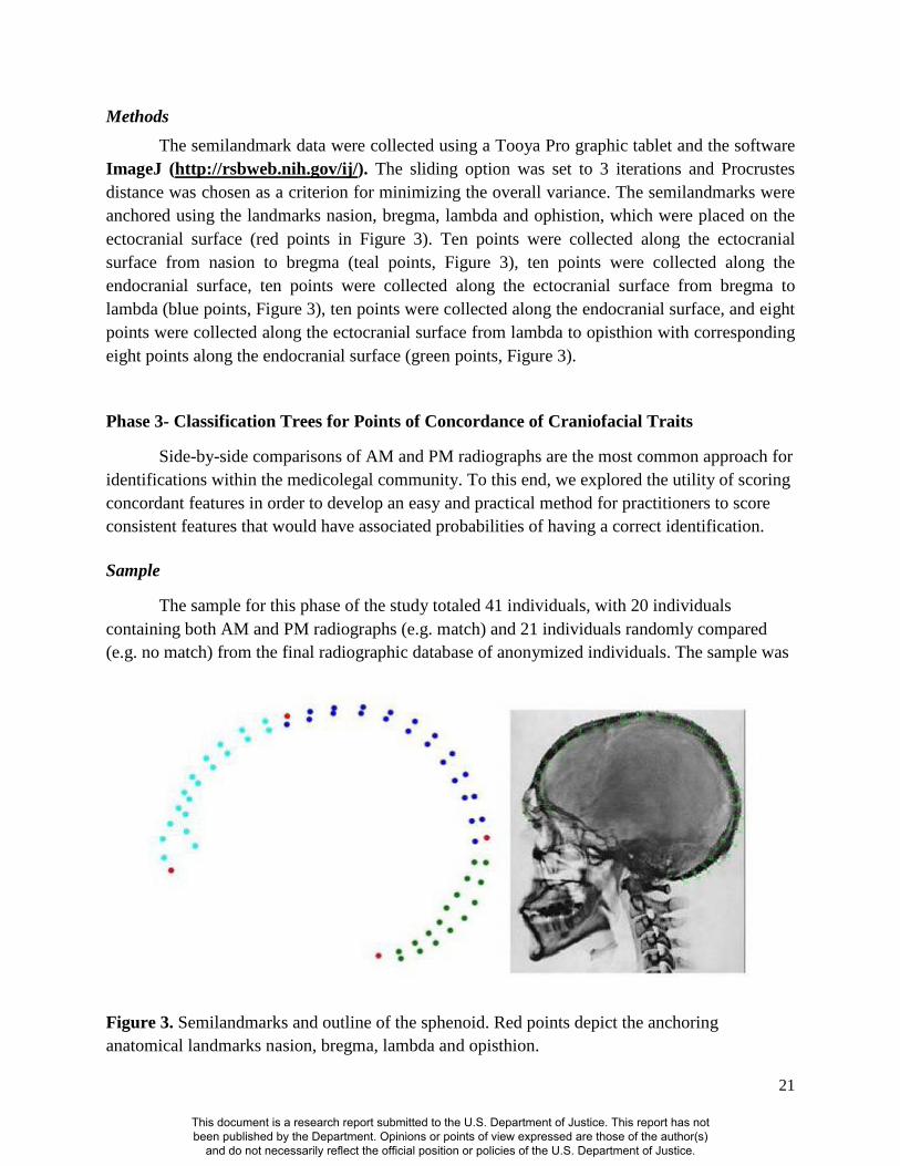
Methods The semilandmark data were collected using a Tooya Pro graphic tablet and the software
ImageJ (http://rsbweb.nih.gov/ij/). The sliding option was set to 3 iterations and Procrustes distance was chosen as a criterion for minimizing the overall variance. The semilandmarks were anchored using the landmarks nasion, bregma, lambda and ophistion, which were placed on the ectocranial surface (red points in Figure 3). Ten points were collected along the ectocranial surface from nasion to bregma (teal points, Figure 3), ten points were collected along the endocranial surface, ten points were collected along the ectocranial surface from bregma to lambda (blue points, Figure 3), ten points were collected along the endocranial surface, and eight points were collected along the ectocranial surface from lambda to opisthion with corresponding eight points along the endocranial surface (green points, Figure 3).
Phase 3- Classification Trees for Points of Concordance of Craniofacial Traits
Side-by-side comparisons of AM and PM radiographs are the most common approach for identifications within the medicolegal community. To this end, we explored the utility of scoring concordant features in order to develop an easy and practical method for practitioners to score consistent features that would have associated probabilities of having a correct identification.
Sample
The sample for this phase of the study totaled 41 individuals, with 20 individuals containing both AM and PM radiographs (e.g. match) and 21 individuals randomly compared (e.g. no match) from the final radiographic database of anonymized individuals. The sample was
Figure 3. Semilandmarks and outline of the sphenoid. Red points depict the anchoring anatomical landmarks nasion, bregma, lambda and opisthion.
This document is a research report submitted to the U.S. Department of Justice. This report has not been published by the Department. Opinions or points of view expressed are those of the author(s)
and do not necessarily reflect the official position or policies of the U.S. Department of Justice.

22
constrained by the number of individuals who had both AM and PM radiographs. These numbers were consistent with the percentage of clinical head radiographs taken, which account for approximately 5% of all radiographs in clinical settings (Brodgon, 1998, Watamaniuk and Rogers, 2010).
Methods
In this phase of the study, the following features were scored as concordant or non-concordant:
1. Morphology of Glabella 2. Frontal sinus 3. Orbital plates 4. Cribriform plates 5. Sella Turcica
In addition, both AM and PM radiographs were scored for quality (1= good, 2 = average, 3 = poor). Good quality radiographs have good optical density (e.g. radiograph is not too white), good contrast, easily visible detail, are in correct anatomical positioning, and no enhancement is needed (e.g. Figures 4-6). In average quality radiographs, features are visible, although the radiograph may have too little or too much contrast, some detail, but not all features may be visible, and correct anatomical position (e.g. Figure 7). In poor quality radiographs, the features are not in the same anatomical position, features are not visible without enhancement, or may not be visible at all (Figure 8).
The anatomical landmarks observable in a lateral cranial radiograph are demonstrated in Figure 4. The visibility of these features is directly associated with the quality of the radiographs taken. An example of the concordant features depicted between AM and PM radiographs from two identified individuals is presented in Figure 5. To explore patterns and relationships of the data, a robust data mining partition technique called classification decision trees was applied. For categorical data, a G2 or the likelihood-ratio chi-square is produced, which minimizes the residual log-likelihood and is used when the response variable (y) is a categorical variable (in this study it is “match” or “no match”). The advantages of decision trees are that they are easy to interpret, they are able to handle both categorical and numerical data, and they are robust such that they are not unduly affected by outliers and perform well on a wide range of probability distributions particularly for distributions that are not normally distributed (Huber, 1996). The nodes are split based on the logworth statistic (-log 10 p-value).
This document is a research report submitted to the U.S. Department of Justice. This report has not been published by the Department. Opinions or points of view expressed are those of the author(s)
and do not necessarily reflect the official position or policies of the U.S. Department of Justice.

23
Figure 4. Lateral cranial radiograph depicting observable structures.
This document is a research report submitted to the U.S. Department of Justice. This report has not been published by the Department. Opinions or points of view expressed are those of the author(s)
and do not necessarily reflect the official position or policies of the U.S. Department of Justice.

24
Figure 5. Concordant characteristics illustrated in two individuals with AM and PM radiographs.
Chest Radiographs In the clinical diagnostic setting more than 40% of radiographs are taken of the chest
(Brodgon, 1998; Watamaniuk and Rogers, 2010). Although vertebral radiographs are commonly used in the identification process, standards do not currently exist, and only one to four points of concordance with no discrepancies have been used to determine identity. In order for a morphological feature to be used in an ID it must be unique to the individual and it must be stable over time (Mundorff et al., 2006). Vertebrae are inherently variable as they form from three primary and five secondary ossification centers. This variation is characterized as differences in the size and shape of each vertebra, as well as between segments of the spinal column when viewed as a whole. A potential complication arises as clinical chest x-rays are generally taken posterior to anterior, with the individual in a standing position, whereas
This document is a research report submitted to the U.S. Department of Justice. This report has not been published by the Department. Opinions or points of view expressed are those of the author(s)
and do not necessarily reflect the official position or policies of the U.S. Department of Justice.

25
postmortem x-rays are taken anterior to posterior with the body lying in a supine position. Thus, the purpose of this part of the study was to examine the vertebral column in a known sample of 100 antemortem and postmortem chest and abdominal radiographs from the NC Office of the Chief Medical Examiner to: 1) evaluate the utility of features observed in the vertebrae; and 2) explore the minimum number of corresponding features necessary to make a positive identification in order to address the issue of probabilities- a growing concern in the court systems.
Sample The final radiographic database of anonymized individuals contained 100 chest
radiographs, which included 50 individuals with both AM and PM radiographs and 50 individuals either without an AM or PM radiograph. These 100 radiographs were scored for concordant features.
Methods Of the 100 comparisons 50 were ante- and postmortem x-rays from known individuals. For
the 50 comparisons where individuals either did not have an AM or PM radiograph, either an ante or postmortem x-ray was compared to a randomly selected individual from the sample to represent the unknown or no-match sample. Chest radiographs were evaluated and scored as concordant or non-concordant for the following features:
1. Cervical morphology (e.g. pedicle, etc.) 2. Thoracic morphology 3. Lumbar morphology 4. Quality of AM x-ray (good, average, poor) 5. Quality of PM x-ray (good, average, poor) 6. Congenital anomalies present
The concordant anatomical features and anomalies observed were scored as total counts
observed and the quality of AM and PM radiographs were scored as ordinal variables with a natural rank (1 = good, 2 = average, 3 = poor, Figure 6). To explore patterns and relationships of the data classification decision trees were used. Classification decision trees represent a data mining technique that for categorical data, a G2 or the likelihood-ratio chi-square for the variables is computed and used for multi-level split of the data. The advantages of decision trees are that they are easy to interpret, they are able to handle both categorical and numerical data, and they are robust.
This document is a research report submitted to the U.S. Department of Justice. This report has not been published by the Department. Opinions or points of view expressed are those of the author(s)
and do not necessarily reflect the official position or policies of the U.S. Department of Justice.
26
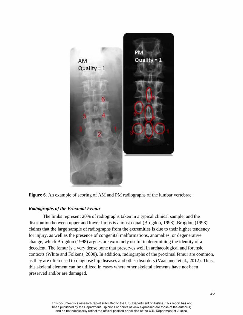
Figure 6. An example of scoring of AM and PM radiographs of the lumbar vertebrae.
Radiographs of the Proximal Femur The limbs represent 20% of radiographs taken in a typical clinical sample, and the
distribution between upper and lower limbs is almost equal (Brogdon, 1998). Brogdon (1998) claims that the large sample of radiographs from the extremities is due to their higher tendency for injury, as well as the presence of congenital malformations, anomalies, or degenerative change, which Brogdon (1998) argues are extremely useful in determining the identity of a decedent. The femur is a very dense bone that preserves well in archaeological and forensic contexts (White and Folkens, 2000). In addition, radiographs of the proximal femur are common, as they are often used to diagnose hip diseases and other disorders (Vaananen et al., 2012). Thus, this skeletal element can be utilized in cases where other skeletal elements have not been preserved and/or are damaged.
This document is a research report submitted to the U.S. Department of Justice. This report has not been published by the Department. Opinions or points of view expressed are those of the author(s)
and do not necessarily reflect the official position or policies of the U.S. Department of Justice.

27
In general, the proximal femur reaches its adult shape around the age of five years old (Wescott, 2006). It is comprised of trabecular bone, which is encased in a layer of cortical bone (White and Folkens, 2000). Clinical research has shown that bone density distributions in the proximal femur are based on individualized factors, and are thus unique (Vahdati et al., 2014). This is due to variations in musculoskeletal loading, and the load-adaptive response in different individuals (Vahdati et al., 2014). In addition, there are many muscle attachments present in the proximal femur, which creates variability in the shape of certain features. For example, the greater trochanter is the insertion site for the gluteus minimus and gluteus medius, while the lesser trochanter is the insertion point for the iliopsoas tendon and iliacus muscle (White and Folkens, 2000).
At present, there are no established standards for concordant features of the proximal femur in positive identifications. Therefore, the purpose of this study was to assess the morphology of the proximal femur in a known sample of 49 antemortem and postmortem pelvic and leg radiographs from the NC Office of the Chief Medical Examiner and North Carolina Forensic Analysis Lab to: 1) evaluate the utility of the features in the proximal femur; and 2) explore the minimum number of corresponding features necessary to make a positive identification.
Sample A total of 49 individuals with radiographs of the proximal femora were scored for
concordant features. Of these 49 individuals, 23 individuals had both AM and PM radiographs, while 26 individuals had either an AM or PM radiograph. To represent the unknown or no-match sample, either an ante or postmortem x-ray was compared to a randomly selected individual from the sample.
Methods Pelvic and leg radiographs were evaluated and scored as concordant or non-concordant
for the following features:
1. Femoral head morphology (e.g. pathology, trabecular bone etc.) 2. Femoral neck morphology 3. Greater trochanter morphology 4. Lesser trochanter morphology 5. Anomalies present 6. Elapsed time between AM and PM x-rays 7. Radiograph quality
The concordant anatomical features and anomalies observed were scored as total counts and
the quality of the AM and PM radiographs were scored as ordinal variables with a natural rank (1 = good, 2 = average, 3 = poor, Figure 7). To explore patterns and relationships of the data, a
This document is a research report submitted to the U.S. Department of Justice. This report has not been published by the Department. Opinions or points of view expressed are those of the author(s)
and do not necessarily reflect the official position or policies of the U.S. Department of Justice.

28
robust data mining technique called classification decision trees was used. For categorical data, a G2 or the likelihood-ratio chi-square for the variables is computed and used for multi-level split of the data. The advantages of decision trees are that they are easy to interpret, they are able to handle both categorical and numerical data, and they are robust.
Figure 7. Example of scoring of AM and PM Femur Radiographs
Figure 8. Example of a poor quality radiograph.
This document is a research report submitted to the U.S. Department of Justice. This report has not been published by the Department. Opinions or points of view expressed are those of the author(s)
and do not necessarily reflect the official position or policies of the U.S. Department of Justice.
29
III. RESULTS
Lateral Cranial Study
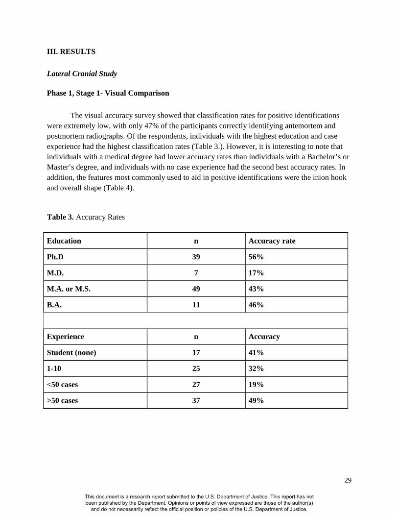
Phase 1, Stage 1- Visual Comparison The visual accuracy survey showed that classification rates for positive identifications were extremely low, with only 47% of the participants correctly identifying antemortem and postmortem radiographs. Of the respondents, individuals with the highest education and case experience had the highest classification rates (Table 3.). However, it is interesting to note that individuals with a medical degree had lower accuracy rates than individuals with a Bachelor’s or Master’s degree, and individuals with no case experience had the second best accuracy rates. In addition, the features most commonly used to aid in positive identifications were the inion hook and overall shape (Table 4). Table 3. Accuracy Rates
Education n Accuracy rate
Ph.D 39 56%
M.D. 7 17%
M.A. or M.S. 49 43%
B.A. 11 46%
Experience n Accuracy
Student (none) 17 41%
1-10 25 32%
<50 cases 27 19%
>50 cases 37 49%
This document is a research report submitted to the U.S. Department of Justice. This report has not been published by the Department. Opinions or points of view expressed are those of the author(s)
and do not necessarily reflect the official position or policies of the U.S. Department of Justice.

30
Table 4. Features Used for Accuracy
Feature % used
Inion hook 22%
Bregma 12%
Frontal shape 17%
Glabella 11%
Vault thickness 16%
Overall shape 22%
Phase 1, Stage 2 - Elliptic Fourier Analysis
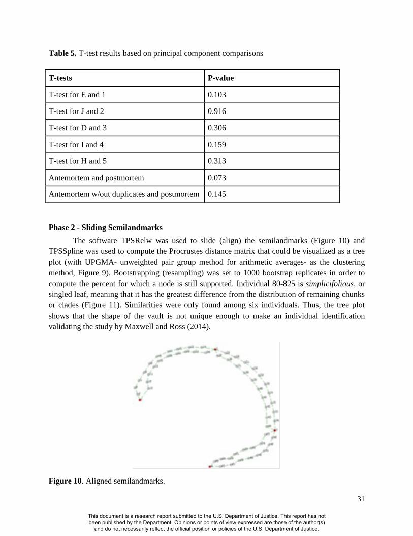
A total of 117 principal components were produced after the elliptic Fourier analysis was completed. Of these, it was determined that 10 components accounted for 100% of the variation. These 10 principal components for the AM and PM radiograph comparisons were graphically depicted (Figure 9) and used for the t-test analysis, to determine if there were significant differences between the radiographs. The results in table 5 showed that none of the comparisons were significantly different, which corresponded with the results of the visual accuracy test.
Figure 9. Graphical representation of 10 principal components
This document is a research report submitted to the U.S. Department of Justice. This report has not been published by the Department. Opinions or points of view expressed are those of the author(s)
and do not necessarily reflect the official position or policies of the U.S. Department of Justice.
31
Table 5. T-test results based on principal component comparisons
T-tests P-value
T-test for E and 1 0.103
T-test for J and 2 0.916
T-test for D and 3 0.306
T-test for I and 4 0.159
T-test for H and 5 0.313
Antemortem and postmortem 0.073
Antemortem w/out duplicates and postmortem 0.145
Phase 2 - Sliding Semilandmarks The software TPSRelw was used to slide (align) the semilandmarks (Figure 10) and
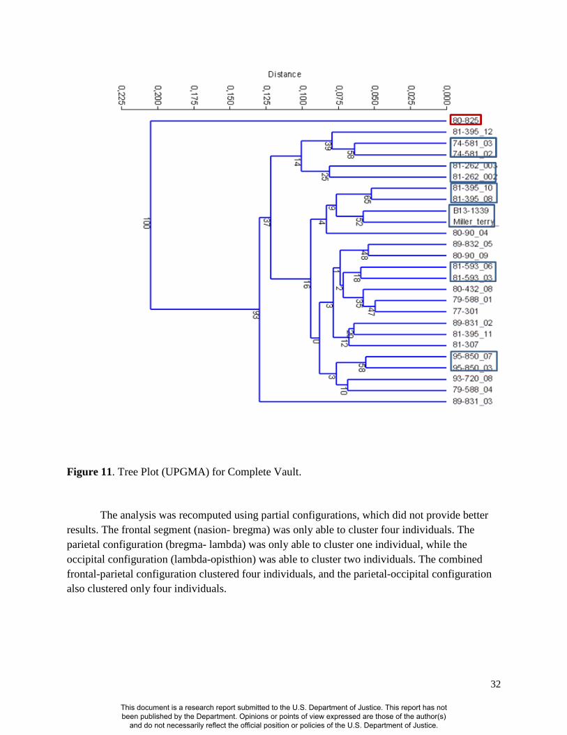
TPSSpline was used to compute the Procrustes distance matrix that could be visualized as a tree plot (with UPGMA- unweighted pair group method for arithmetic averages- as the clustering method, Figure 9). Bootstrapping (resampling) was set to 1000 bootstrap replicates in order to compute the percent for which a node is still supported. Individual 80-825 is simplicifolious, or singled leaf, meaning that it has the greatest difference from the distribution of remaining chunks or clades (Figure 11). Similarities were only found among six individuals. Thus, the tree plot shows that the shape of the vault is not unique enough to make an individual identification validating the study by Maxwell and Ross (2014).
Figure 10. Aligned semilandmarks.
This document is a research report submitted to the U.S. Department of Justice. This report has not been published by the Department. Opinions or points of view expressed are those of the author(s)
and do not necessarily reflect the official position or policies of the U.S. Department of Justice.
32
Figure 11. Tree Plot (UPGMA) for Complete Vault.
The analysis was recomputed using partial configurations, which did not provide better results. The frontal segment (nasion- bregma) was only able to cluster four individuals. The parietal configuration (bregma- lambda) was only able to cluster one individual, while the occipital configuration (lambda-opisthion) was able to cluster two individuals. The combined frontal-parietal configuration clustered four individuals, and the parietal-occipital configuration also clustered only four individuals.
This document is a research report submitted to the U.S. Department of Justice. This report has not been published by the Department. Opinions or points of view expressed are those of the author(s)
and do not necessarily reflect the official position or policies of the U.S. Department of Justice.

33
Phase 3 - Points of Concordance
The classification tree results showed that if two or more cranial concordant features are present the probability of the individual being correctly matched or positively identified is 97% (Figure 12).
Figure 12. Classification Tree for Cranial Concordant Characteristics
The accuracy of the model to discriminate between matches (1) and no matches (2) using classification trees of the training set was also evaluated using a receiver operating characteristic or ROC. The ROC measures the true positives by false positives, and the area under the curve (AUC) is a measure of how well a parameter can distinguish between two groups. If the model cannot distinguish between two groups the AUC will equal .5 and if there is complete separation the AUC will equal 1. The AUC for separation for matches = .90 and .90 for no matches indicating strong separation.
The validation set has a misclassification rate of 10%, which represents the model’s predictive power on future observations. The generalized R-squared equals .75 indicating that model is predicting well on data not used to train the model. The confusion rate or the number of false positives, false negatives, true positives and true negatives is 80 % for matches and 100% for no matches.
This document is a research report submitted to the U.S. Department of Justice. This report has not been published by the Department. Opinions or points of view expressed are those of the author(s)
and do not necessarily reflect the official position or policies of the U.S. Department of Justice.

34
Chest Radiographs
Cervical, thoracic and lumbar vertebrae
The classification tree results using the entire chest variables showed that if there are one or more cervical vertebral concordant features the probability of the individual being correctly matched or positively identified is 99% (Figure 13). However, in the absence of concordant cervical vertebral features, four or more lumbar vertebral concordant features are required for the probability of the individual being correctly positively identified at 98%. If there are less than four lumbar concordant features, one or more concordant thoracic feature are required for a 79% probability of a correct match.
The accuracy of the model to discriminate between matches (1) and no matches (2) using classification trees of the training set was also evaluated using a receiver operating characteristic or ROC. The ROC measures the true positives by false positives and the area under the curve (AUC) is a measure of how well a parameter can distinguish between two groups. If the model cannot distinguish between two groups the AUC will equal .5 and if there is complete separation the AUC will equal 1. The AUC for separation for matches = .96 and .96 for no matches indicating strong separation.
The validation set had a misclassification rate of 7%, which represents the model’s predictive power on future observations. The generalized R-squared equals .82 indicating that the model is predicting well on data not used to train the model. The confusion rate or the number of false positives, false negatives, true positives and true negatives is 94 % for matches and 92% for no matches.
This document is a research report submitted to the U.S. Department of Justice. This report has not been published by the Department. Opinions or points of view expressed are those of the author(s)
and do not necessarily reflect the official position or policies of the U.S. Department of Justice.

35
Figure 13. Classification Tree for All Vertebrae.
Cervical and thoracic vertebrae
The classification tree results using only the cervical and thoracic vertebrae showed that if there are one or more cervical vertebral concordant features, the probability of the individual being correctly matched or positively identified is 99% (Figure 14). However, in the absence of a concordant cervical vertebral feature, one or more thoracic vertebral concordant features are required to attain an 84% probability that the individual is being correctly positively identified.
The accuracy of the model to discriminate between matches (1) and no matches (2) using classification trees of the training set was also evaluated using a receiver operating characteristic or ROC. The ROC measures the true positives by false positives and the area under the curve (AUC) is a measure of how well a parameter can distinguish between two groups. If the model cannot distinguish between two groups the AUC will equal .5 and if there is complete separation
This document is a research report submitted to the U.S. Department of Justice. This report has not been published by the Department. Opinions or points of view expressed are those of the author(s)
and do not necessarily reflect the official position or policies of the U.S. Department of Justice.

36
the AUC will equal 1. The AUC for separation for matches = .84 and .84 for no matches indicating strong separation.
The validation set had a misclassification rate of 20%, which represents the model’s predictive power on future observations. The generalized R-squared equals .52 indicating that the model is not predicting adequately (for a forensic setting) on data not used to train the model. The confusion rate or the number of false positives, false negatives, true positives and true negatives is 72 % for matches and 92% for no matches.
Figure 14. Classification Tree for Cervical and Thoracic Vertebrae.
Lumbar Vertebrae
The classification tree results using only the lumbar vertebrae showed that if there are four or more lumbar vertebral concordant features the probability of the individual being correctly matched or positively identified is 98% (Figure 15). The accuracy of the model to discriminate between matches (1) and no matches (2) using classification trees of the training set was also evaluated using a receiver operating characteristic or ROC. The ROC measures the true
This document is a research report submitted to the U.S. Department of Justice. This report has not been published by the Department. Opinions or points of view expressed are those of the author(s)
and do not necessarily reflect the official position or policies of the U.S. Department of Justice.

37
positives by false positives and the area under the curve (AUC) is a measure of how well a parameter can distinguish between two groups. If the model cannot distinguish between two groups the AUC will equal .5 and if there is complete separation the AUC will equal 1. The AUC for separation for matches = .66 and .66 for no matches indicating that the model can barely distinguish between the two groups.
The validation set had a misclassification rate of 40%, which represents the model’s predictive power on future observations. The generalized R-squared equals .27 indicating that the model is predicting poorly on data not used to train the model. The confusion rate or the number of false positives, false negatives, true positives and true negatives is 32 % for matches and 100% for no matches. Based on the results of this analysis it is not recommended to rely solely on the lumbar vertebrae in the positive identification of unknown individuals.
Figure 15. Classification Tree for Lumbar Vertebrae.
Radiographs of the Proximal Femur
Femoral Head and Neck The classification tree results using the femoral head and neck showed that if there are
one or more femoral head concordant features the probability of the individual being correctly matched or positively identified is 93% (Figure 16). If there is also a concordant femoral neck feature the probability of correctly matching or positively identifying an unknown decedent is 97%.
This document is a research report submitted to the U.S. Department of Justice. This report has not been published by the Department. Opinions or points of view expressed are those of the author(s)
and do not necessarily reflect the official position or policies of the U.S. Department of Justice.
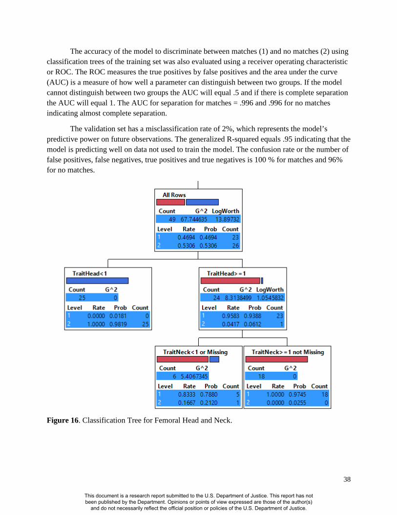
38
The accuracy of the model to discriminate between matches (1) and no matches (2) using classification trees of the training set was also evaluated using a receiver operating characteristic or ROC. The ROC measures the true positives by false positives and the area under the curve (AUC) is a measure of how well a parameter can distinguish between two groups. If the model cannot distinguish between two groups the AUC will equal .5 and if there is complete separation the AUC will equal 1. The AUC for separation for matches = .996 and .996 for no matches indicating almost complete separation.
The validation set has a misclassification rate of 2%, which represents the model’s predictive power on future observations. The generalized R-squared equals .95 indicating that the model is predicting well on data not used to train the model. The confusion rate or the number of false positives, false negatives, true positives and true negatives is 100 % for matches and 96% for no matches.
Figure 16. Classification Tree for Femoral Head and Neck.
This document is a research report submitted to the U.S. Department of Justice. This report has not been published by the Department. Opinions or points of view expressed are those of the author(s)
and do not necessarily reflect the official position or policies of the U.S. Department of Justice.

39
Femoral Lesser and Greater Trochanters
The classification tree results using the femoral greater and lesser trochanter showed that if there is one or more femoral greater trochanter concordant features the probability of the individual being correctly matched or positively identified is 76% (Figure 17). If there are also two or more concordant femoral lesser trochanter features, the probability of correctly matching or positively identifying an unknown decedent is 93%
The accuracy of the model to discriminate between matches (1) and no matches (2) using classification trees of the training set was also evaluated using a receiver operating characteristic or ROC. The ROC measures the true positives by false positives and the area under the curve (AUC) is a measure of how well a parameter can distinguish between two groups. If the model cannot distinguish between two groups the AUC will equal .5 and if there is complete separation the AUC will equal 1. The AUC for separation for matches = .96 and .96 for no matches indicating almost complete separation.
The validation set has a misclassification rate of 10%, which represents the model’s predictive power on future observations. The generalized R-squared equals .81 indicating that the model is predicting well on data not used to train the model. The confusion rate or the number of false positives, false negatives, true positives and true negatives is 83 % for matches and 96% for no matches.
This document is a research report submitted to the U.S. Department of Justice. This report has not been published by the Department. Opinions or points of view expressed are those of the author(s)
and do not necessarily reflect the official position or policies of the U.S. Department of Justice.

40
Figure 17. Classification Tree for Femoral Greater and Lesser Trochanters
IV. CONCLUSIONS
Discussion of findings This research project started by examining the utility of using outline shapes to identify
vault and cranial outlines as a means of confirming identification. Three different methods were employed to assess whether cranial vault outlines were a useful tool for positive identifications. These included a visual accuracy test (a method currently employed), elliptic Fourier analysis, and sliding semi-landmarks. The results of each of these analyses suggest that cranial outlines are not informative enough to use on their own for positive identifications. Only 47% of the individuals that visually assessed the radiographs correctly assigned antemortem and postmortem x-rays, and there were no significant differences when t-tests were performed on the resulting principal components of the elliptic Fourier analysis. In addition, the sliding semi-landmark study confirmed these results by showing that vault shape was not variable enough to consistently positively identify individuals. It should be noted that these results were obtained
This document is a research report submitted to the U.S. Department of Justice. This report has not been published by the Department. Opinions or points of view expressed are those of the author(s)
and do not necessarily reflect the official position or policies of the U.S. Department of Justice.
41
using two time-separated images of the same (dry) bone that were used to simulate AM-PM radiographs. Therefore, they may not be generating success rates applicable to “real-life” casework. Success rates may in fact be overestimated using this methodology and it is interesting that even with the potential for overestimating accuracy results are not significant. These results are significant, as they tested the utility of using vault and cranial outlines according to Daubert guidelines. These studies statistically showed that vault and cranial outlines should not be the only identifying anatomical feature when attempting to positively identify an individual. While this does not mean that some individuals may not have vault or cranial anomalies that can aid in identification, it suggests that outlines should be used in combination with other identifying features.
A standard system for making radiographic comparisons was developed based on the assessment of points of concordance and utilized a robust statistical method, classification decision trees. This standard system of making radiographic comparisons was used on three standard radiograph views, lateral cranial radiographs as well as radiographs of the chest and proximal femur. This standard system allowed for the evaluation of the utility of various anatomical features in these standard radiographs as well as assessing their validity in positive identifications.
This system involved examining AM and PM radiographs of the cranium, chest, and hip, and assessing them for quality, as well as identifying and comparing features of various anatomical regions. The relevant regions are as follows: for the cranium, glabella, frontal sinuses, orbital plates, cribriform plates, and sella turcica; for the chest, the cervical region (including pedicles, etc.), the thoracic region, and the lumbar region; for the femur, the femoral head, neck, greater trochanter, and lesser trochanter. The features visible in these anatomical regions were assessed for concordance with no discrepancies and were counted. The standard system for making radiographic comparisons enabled the statistical determination of the minimum number of concordant features needed to meet a high probability to make a positive identification.
The results of the chest radiographic analysis showed that the anatomical elements with the most predictive values were the cervical vertebrae, which showed that if there are more than one concordant feature present, there is a 99% probability of correct identification. If there are no cervical concordant features and there are four or more concordant features in the other vertebrae, there is a 98% probability of having a correct identification. This probability drops to 79% if there are less than four concordant lumbar features. In the situation where only thoracic or lumbar vertebrae are present, the current models are not able to predict well enough for a forensic setting and other means of identification should also be used to confirm identification. Stephan et al. (2011) and 2014) demonstrated success with using the clavicles in positive
This document is a research report submitted to the U.S. Department of Justice. This report has not been published by the Department. Opinions or points of view expressed are those of the author(s)
and do not necessarily reflect the official position or policies of the U.S. Department of Justice.

42
identifications. It is possible that adding features of the clavicle to the analysis could increase classification rates, particularly in combination with features of the thoracic or lumbar vertebrae.
These results are significant, because they showed that the cervical vertebrae, and in the absence of the cervical vertebrae, the thoracic vertebrae are more reliable for establishing a positive identification. Another significant finding is that if only thoracic and lumbar vertebrae are present these are not reliable enough for use as the sole identification criteria. Cranial and femoral features were also demonstrated to be useful with the highest probabilities (97%) for two or more concordant features when using these body loci in a positive identification. The results of these studies are significant, as it is the first attempt to statistically quantify the number of concordant points needed to meet the Daubert criteria for making a positive identification using radiographs.
Table 6. Probabilities Associated with the number of concordant features via direct comparison of ante and postmortem radiographs.
Body Area # of Concordant Features Probability of Positive ID
Cranial > = 2 97%
Chest All areas Cervical, thoracic, lumbar
Cervical > = 1 Cervical < 1, Lumbar > = 4 Cervical < 1, Lumbar < 4, Thoracic > = 1
99% 98% 80%
Cervical and Thoracic only Cervical > = 1 Cervical < 1, Thoracic > = 1
99% 80% use with caution, need additional tests to confirm ID
Lumbar only Lumbar > = 4 98% use with caution, need additional tests to confirm ID
Femora Femoral head and neck
F head > = 1, F neck > = 1
94% 97%
Femoral Greater and Lesser Trochanters
GT > = 1, LT > = 2
76% 93%
This document is a research report submitted to the U.S. Department of Justice. This report has not been published by the Department. Opinions or points of view expressed are those of the author(s)
and do not necessarily reflect the official position or policies of the U.S. Department of Justice.

43
Implications for policy and practice This research marks the start of systematic validation of radiographic comparison
methods based on several anatomical and skeletal landmarks. This research expands the ability of forensic anthropologists and pathologists to positively identify previously unidentified remains as new comparative methods have been developed based on regions of the body not previously studied. This makes the completed research an invaluable tool for identification, particularly in cases of incomplete skeletal recovery, which lack some for the more commonly analyzed anatomical sites previously used for identification. The developed methods expressly meet Daubert criteria in that they are testable, peer reviewed, generally accepted within the scientific community, and possess known potential error rates (509 US.579, 1993). Following the recommendations of the recent NAS report on Forensic Science, issues of accuracy, reliability, and validity have also been addressed in these methods and serves as a model for future research in other areas pertaining to positive identification using radiographs.
Implications for further research While this study demonstrated that visual and geometric morphometric methods for
cranial vault outlines were not significant when used alone as a means of positive identification, it does not mean that geometric morphometric methods should not be pursued. Stephan et al., (2014) points out that methods such as the one developed here, which relies on visual comparisons of concordant points, works well in situations where the pool of potential matching candidates is small; however, when the pool of potential matching candidates is large its usefulness is diminished and “risks error due to analyst fatigue” (Stephan et al., 2014:306). Exploring methods of comparison using geometric morphometrics would allow for a computerized identification system that would be especially useful in situations where the pool of potential matches is large, as the case when ascertaining the identity of missing U.S. soldiers by the Joint POW/MIA Accounting Command (Stephan et al., 2014). In the more typical situation, such as that in a medical examiner’s office, the pool of potential matching candidates is relatively small as it is limited by the circumstances of death (Stephan et al., 2014). Therefore, pursuing further development and refinement of the standard system for making radiographic comparisons developed in this study should be the primary focus of research in the utility of radiographs in positive identification as it is best suited to the situation that most identifications are conducted in.
As stated previously, this research marks the start of a systematic validation of radiographic comparison methods based on several anatomical and skeletal features. The demonstrated success of the developed standardized method of radiographic comparisons based on areas of concordance will need to be expanded upon so as to achieve peer review and general acceptance within the scientific community, allowing it to fully meet Daubert standards of admissibility. This study was limited by the number of radiographs available to the investigators and it is hoped that by demonstrating the success of this method other sources of radiographs may be utilized to further expand the assessment of this method to other standard radiographic
This document is a research report submitted to the U.S. Department of Justice. This report has not been published by the Department. Opinions or points of view expressed are those of the author(s)
and do not necessarily reflect the official position or policies of the U.S. Department of Justice.

44
views as well as expanding the number of radiographic comparisons available to reduce the standard error of these estimates. The completed radiographic database will be publicly available for use and researchers will be encouraged to use this as a source for research not only in the area of positive identification but other areas of forensic inquiry.
This document is a research report submitted to the U.S. Department of Justice. This report has not been published by the Department. Opinions or points of view expressed are those of the author(s)
and do not necessarily reflect the official position or policies of the U.S. Department of Justice.
45
V. REFERENCES
D.C. Adams, F.J. Rolhf, D.E. Slice. 2004. Geometric morphometrics: ten years of progress following the ‘revolution.’ Italian Journal of Zoology 71:5-16.
B.G. Brogdon. 1998. Forensic Radiology. CRC Press: Boca Raton. R. Cameriere, L. Ferrante, D. Mirtella, F.U. Rollo, M. Cingolani. 2005. Frontal sinuses for
identification: quality of classifications, possible error and potential corrections. Journal of Forensic Sciences 50:770-773.
R. Cameriere, L. Ferrante, T. Molleson, B. Brown. 2008. Frontal Sinus Accuracy in Identification as Measured by False Positives in Kin Groups. Journal of Forensic Sciences 53:1-3.
A.M. Christensen. 2004a. The Impact of Daubert: Implications for Testimony and Research in Forensic Anthropology (and the Use of Frontal Sinuses in Personal Identification). Journal of Forensic Sciences. 49:427-430.
A.M. Christensen. 2004b. Assessing the Variation in Individual Frontal Sinus Outlines. American Journal of Physical Anthropology 127:291-295.
A.M. Christensen. 2005. Testing the reliability of frontal sinuses in positive identification. Journal of Forensic Sciences 50:18-22.
Committee on Identifying the Needs of the Forensic Sciences Community, National Research Council. 2009. Strengthening Forensic Science in the United States: A Path Forward, National Academies of Sciences Press.
W.L. Culbert, F.M. Law. 1927. Identification by Comparison of Roetgenograms. The Journal of the American Medical Association 88:1634-1636.
Daubert v. Merrell Dow Pharmaceuticals. 1993. 113 S.Ct. 2786; U.S. LEXIS 4408. S.L. Fischman. 1985. The use of medical and dental radiographs in identification. International
Dental Journal 35:301-306. P. Huber. 1996. Robust Statistical Procedures, 2nd Edition, Society for Industrial and Applied
Mathematics, Philadelphia. H. Iwata. 2006. SHAPE 1.3. Entebbe, Uganda: National Archaeological Research Organization. N.G. Jablonski, B.S.F. Shum. 1989. Identification of unknown remains by comparison of
antemortem and postmortem radiographs. Forensic Science International 42:221-230. T. Kahana, L. Goldin, J. Hiss. 2002. Personal Identification Based on Radiographic Vertebral
Features. The American Journal of Forensic Medicine and Pathology 23:36-41. C.P. Klingenberg. 2008. MorphoJ. Faculty of Life Sciences, University of Manchester, UK.
http://www.flywings.org.uk/MorphoJ_page.htm. D.A. Komar, J.E. Buikstra. 2008. Forensic Anthropology: Contemporary Theory and Practice.
Oxford University Press: Oxford, New York. C.M. Kuehn, K.M. Taylor, F.A. Mann, A.J. Wilson, R.C. Harruff. 2002. Validation of Chest
Xray Comparisons for Unknown Decedent Identification. Journal of Forensic Sciences. 47:725-729.
This document is a research report submitted to the U.S. Department of Justice. This report has not been published by the Department. Opinions or points of view expressed are those of the author(s)
and do not necessarily reflect the official position or policies of the U.S. Department of Justice.

46
G.P. Lohmann, Schweitzer P.N. 1990. On Eigenshape Analysis In F.J. Rohlf & F.L. Bookstein (eds). Proceedings of the Michigan morphometrics workshop. Special Publication No.2. University of Michigan Museum of Zoology, Ann Arbor. pp. 147-166.
R.W. Mann. 1998. Use of trabeculae to establish positive identification. Forensic Science International 98:91-99.
A.B. Maxwell, A.H. Ross. 2014. A Radiographic Study on the Utility of Cranial Vault Outlines for Positive Identifications. Journal of Forensic Sciences 59:314–318.
A.Z. Mundorff, G. Vidoli, J. Melinek. 2006. Anthropological and Radiographic Comparison of Vertebrae for Identification of Decomposed Human Remains. Journal of Forensic Sciences 51:1002-1004.
F.A. Pasler, H. Visser. 2007. Pocket Atlas of Dental Radiology. Thieme. pp. 84. F.A. Ribeiro. 2000. Standardized measurements of radiographic films of the frontal sinuses: an
aid to identifying unknown persons. Ear Nose and Throat Journal 79:26-33. T.L. Rogers, T.T. Allard. 2004. Expert Testimony and Positive Identification of Human Remains
Through Cranial Suture Patterns. Journal of Forensic Sciences. 49:203-207. F.J. Rolhf, I.F. Marcus. 1993. A revolution in morphometrics. Tree 8:129-132. F.J. Rolhf. 1990. Fitting curves to outlines. In F.J. Rolhf & F.L. Bookstein (eds). Proceedings of
the Michigan morphometrics workshop. Special Publication No. 2. University of Michigan Museum of Zoology, Ann Arbor. pp. 167-177.
A. Schuller. 1921. Das Rontgenogram der Stirnhohle: ein Hilfsmittel für die Identitätsbestimmung von Schadeln. Monatsschr Ohrenheilkd Laryngorhinol 5:1617-1620.
A. Schuller. 1943 A note on the identification of skulls by X-ray pictures of the frontal sinuses. The Medical Journal of Australia 1:554-556.
D.E. Slice. 2005. Modern Morphometrics. In Slice, D. E. (ed.) Modern Morphometrics in Physical Anthropology. Kluwer Academic Publishers, pp. 1-45.
D.E. Slice. 2007. Geometric Morphometrics. The Annual Review of Anthropology 36:261-81. D.E. Slice, A.H. Ross. 2009. Geometric Morphometric Classification of Crania for Forensic
Scientists. D.J. Spitz. 2006. Identification of Human Remains: Part 1. In Spitz, W.U. (ed.) Spitz and
Fisher’s Medicolegal Investigation of Death. Charles C. Thomas Publisher, pp 184-225. C.N. Stephan, B. Amidan, H. Trease, P. Guyomarc’h, T. Pulsipher, J.E. Byrd. 2014.
Morphometric Comparison of Clavicle Outlines from 3D Bone Scans and 2D Chest Radiographs: A Shortlisting Tool to Assist Radiographic Identification of Human Skeletons. Journal of Forensic Sciences 59;2:306-313.
C.N. Stephan, A.P. Winburn, A.F. Christensen, A.J. Tyrrell. 2011. Skeletal Identification by Radiographic Comparison: Blind Tests of a Morphoscopic Method Using Antemortem Chest Radiographs. Journal of Forensic Sciences 56;2:320-332.
J.P. Tang, D.Y. Hu, F.H. Jiang, X.J. Yu. 2009. Assessing forensic applications of the frontal sinus in a Chinese Han population. Forensic Science International 183:104.e1-104.e3.
This document is a research report submitted to the U.S. Department of Justice. This report has not been published by the Department. Opinions or points of view expressed are those of the author(s)
and do not necessarily reflect the official position or policies of the U.S. Department of Justice.
47
T. Treumann. 2010. Radiology of Craniofacial Features. In N. Hardt and J. Kuttenberger (ed.) Craniofacial Trauma: Diagnosis and Management. Springer-Verlag, pp 12-29.
S.P. Vaananen, H. Isaksson, J.H. Waarsing, A.A. Zadpoor, J.S. Jurvelin, H. Weinans. 2012. Estimation of 3D Rotation of Femur in 2D Hip Radiographs. Journal of Biomechanics 45:2279-2283.
A. Vahdati, S. Walscharts, I. Jonkers, J.M. Garcia-Aznar, J. Vander Sloten, G.H. van Lenthe. 2014. Role of Subject Specific Musculoskeletal Loading on the Prediction of Bone Density Distribution in the Proximal Femur. Journal of the Mechanical Behavior of Biomedical Materials 30:244-252.
A. Valenzuela 1997. Radiographic Comparison of the Lumbar Spine for Positive Identification of Human Remains: A Case Report. American Journal of Forensic Medicine and Pathology 18:215-217.
J.C. Wankmiller. 2010. Positive Identification Using Comparisons of Lumbar Spine Radiographs: A Validation Study (Master’s thesis). Michigan State University, East Lansing, MI.
D.J. Wescott. 2006. Ontogeny of Femur Subtrochanteric Shape in Native Americans and American Blacks and Whites. Journal of Forensic Sciences 51:1240-1245.
L. Watamaniuk, T. Rogers. 2010. Positive Personal Identification of Human Remains Based on Thoracic Vertebral Margin Morphology. Journal of Forensic Sciences 55:1162–1170.
T. White, A. Folkens. 2000. The Human Bone Manual. Burlington, MA: Elsevier Academic Press.
M.L. Zelditch, D.L. Swiderski, H.D. Sheets, 2012. Geometric Morphometrics for Biologists: A Primer. Academic Press, London.
This document is a research report submitted to the U.S. Department of Justice. This report has not been published by the Department. Opinions or points of view expressed are those of the author(s)
and do not necessarily reflect the official position or policies of the U.S. Department of Justice.
48
VI. DISSEMINATION OF RESEARCH FINDINGS
Ross AH, Simmons S. A radiographic study on the utility of cranial vault outlines for positive identifications. Proceedings of the American Academy of Forensic Sciences Volume 18 2012:419-420, Atlanta, GA.
Ross AH, Simmons S. Examining the validity of using postcranial radiographic comparisons in positive identifications. International Academy of Legal Medicine Final Program 2012:38, Istanbul, Turkey.
Ross AH, Simmons S. Positive identification using chest radiographs: Standards for minimum number of concordant points. Proceedings of the American Academy of Forensic Sciences Volume 19 2013:401-402, Washington, DC.
Ross AH. Radiograph Database. https://sites.google.com/a/ncsu.edu/radiograph-database-project/
Zemlock R, Dew E, Urbanova P, Ross AH. The Use of Antemortem and Postmortem Lateral Cranial Radiographs in the Identification of Unknown Human Remains: Creating a Standard. Presented at the Forensic Anthropology Society of Europe 2013, Heidelberg, Germany.
Maxwell, AB. and Ross AH. A Radiographic Study on the Utility of Cranial Vault Outlines for
Positive Identifications. Journal of Forensic Sciences, 2014; 59:314–318. doi: 10.1111/1556-4029.12346
Maxwell, AB, Ross AH, Lanfear AK. The utility of radiographs of the proximal femur in positive identifications: Establishing a standard and minimum number of concordant points. Abstract submitted to present at the American Academy of Forensic Sciences Annual Meeting February, 2015 in Orlando, FL.
This document is a research report submitted to the U.S. Department of Justice. This report has not been published by the Department. Opinions or points of view expressed are those of the author(s)
and do not necessarily reflect the official position or policies of the U.S. Department of Justice.

49
VII. ACKNOWLEDGEMENTS
The authors would like to thank the following individuals who assisted on this project: NC Office of the Chief Medical Examiner, Clyde Gibbs and Dr. Sam Simmons from the NC OCME for assisting with the radiographic samples, Petra Urbanová from Masaryk University, Czech Republic, who was a Fulbright Research Fellow at NC State Forensic Analysis Laboratory and assisted with the semi-landmark aspect of the study.
This document is a research report submitted to the U.S. Department of Justice. This report has not been published by the Department. Opinions or points of view expressed are those of the author(s)
and do not necessarily reflect the official position or policies of the U.S. Department of Justice.