text assignment lesson objectives - … · md0568 1-1 lesson assignment lesson 1 anatomy and...
TRANSCRIPT

MD0568 1-1
LESSON ASSIGNMENT LESSON 1 Anatomy and Physiology of the Respiratory System. TEXT ASSIGNMENT Paragraphs 1-1 through 1-17. LESSON OBJECTIVES After completing this lesson, you should be able to: 1-1. Identify the structure and function of the respiratory organs. 1-2. Identify the accessory structures of the respiratory system. 1-3. Describe the physiology of respiration. 1-4. Describe factors in air capacity of the lungs. SUGGESTION After completing the assignment, complete the exercises at the end of this lesson. These exercises will help you to achieve the lesson objectives.

MD0568 1-2
LESSON 1
ANATOMY AND PHYSIOLOGY OF THE RESPIRATORY SYSTEM
Section I. STRUCTURE AND FUNCTION OF THE RESPIRATORY ORGANS 1-1. INTRODUCTION a. Respiration. Respiration is the exchange of gases between the atmosphere and the cells of the body. It is a physiological process. There are two types of respiration: external respiration and internal respiration (figure 1-1). External respiration is the exchange of gases between the air in the lungs and blood. Internal respiration is the exchange of gases between the blood and the individual cells of the body.
Figure 1-1. Respiration.
b. Breathing. Breathing is the process that moves air into and out of the lungs. It is a mechanical process. There are two types of breathing in humans: costal (thoracic) breathing and diaphragmatic (abdominal) breathing (figure 1-2). In costal breathing, the major structure causing the movement of air is the rib cage. In diaphragmatic breathing, interaction between the diaphragm and the abdominal wall causes air to move into and out of the lungs.
Figure 1-2. Breathing.

MD0568 1-3
1-2. COMPONENTS AND SUBDIVISIONS OF THE HUMAN RESPIRATORY SYSTEM
See figure 1-3 for an illustration of the human respiratory system. a. Components. The components of the human respiratory system consist of air passageways and two lungs. Air moves from the outside of the body into tiny sacs in the lungs called alveoli (pronounced al-VE-oh-lie).
Figure 1-3. The human respiratory system.
MD0568 1-4
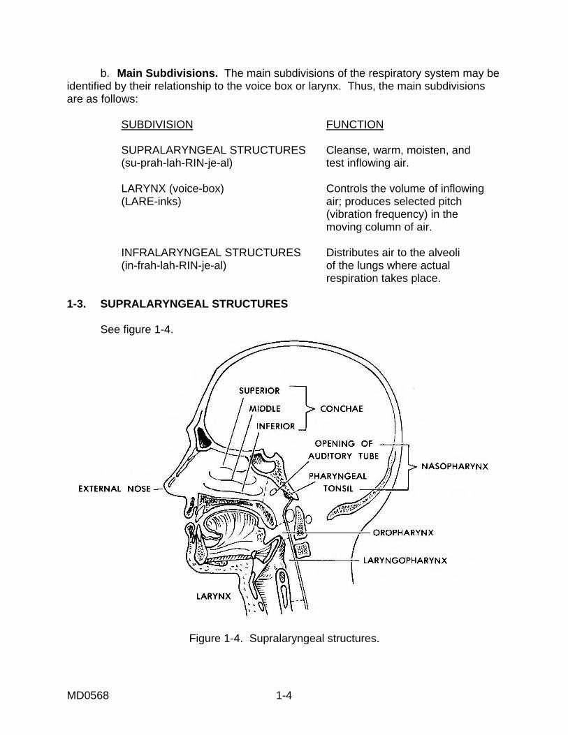
b. Main Subdivisions. The main subdivisions of the respiratory system may be identified by their relationship to the voice box or larynx. Thus, the main subdivisions are as follows: SUBDIVISION FUNCTION SUPRALARYNGEAL STRUCTURES Cleanse, warm, moisten, and (su-prah-lah-RIN-je-al) test inflowing air. LARYNX (voice-box) Controls the volume of inflowing (LARE-inks) air; produces selected pitch (vibration frequency) in the moving column of air. INFRALARYNGEAL STRUCTURES Distributes air to the alveoli (in-frah-lah-RIN-je-al) of the lungs where actual respiration takes place. 1-3. SUPRALARYNGEAL STRUCTURES See figure 1-4.
Figure 1-4. Supralaryngeal structures.

MD0568 1-5
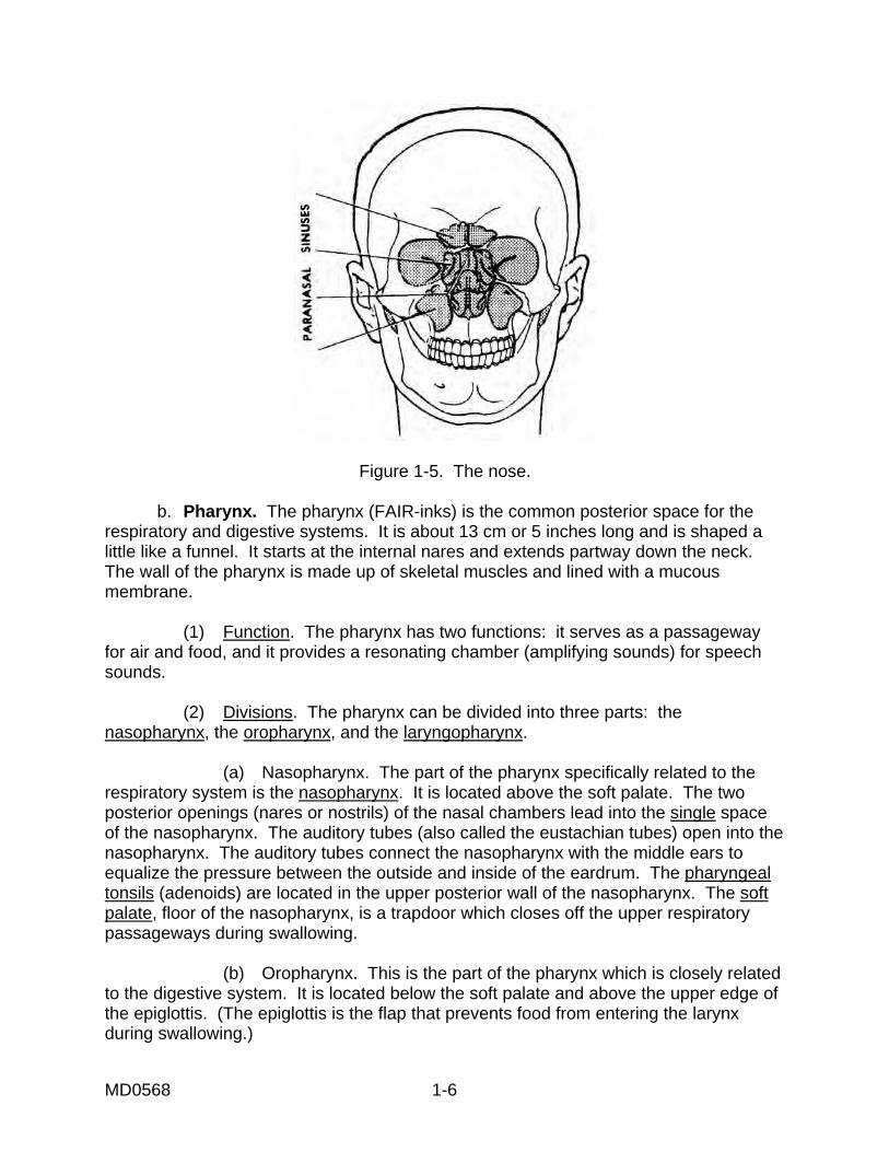
a. Nose. There is an external nose, the part projecting from the face, and an internal nose, the portion that is inside the skull. (1) External portion of the nose. A supporting framework of bone and cartilage covered with skin and lined with mucous membrane makes up the external nose. Nasal bones form the bridge of the nose and hold it in a fixed position. The rest of the external nose is flexible because it has a framework of pliable cartilage. The midline divider of the external nose is called the nasal septum. Two openings called the nostrils or external nares are on the underside of the external nose. These nostrils lead to paired spaces (vestibules). (2) Internal portion of the nose. Behind each vestibule of the external nose is a nasal chamber. The two chambers together form the internal nose. These chambers are separated by the nasal septum. (a) Mucoperiosteum. The walls of the nasal chambers are lined with a thick mucous-type membrane known as the mucoperiosteum. This lining is moist and full of blood vessels. As air is inhaled and passes over this lining, several changes happen to the inhaled air. Blood in the lining's blood vessels warms the air. The air picks up moisture from the lining. Dust sticks to the mucous of the lining resulting in relatively dust-free air. (b) Conchae. The lateral wall of each nasal chamber has three scroll-like extensions into the chamber, which help increase the surface area exposed to inflowing air. These scroll-like extensions are known as conchae. CONCHA = sea shell CONCHA (singular), Conchae (plural) pronounced KON-kah) (c) Olfactory epithelium (membrane). The olfactory epithelium is a membrane that lines the upper nasal chambers. The olfactory receptors (nerve endings responsible for the sense of smell) are located in this membrane. (d) Paranasal sinuses. Paranasal sinuses (figure 1-5) are air "cells" or cavities in the skull. They are connected with the nasal chambers and are lined with the same ciliated mucoperiosteum. These sinuses are extensions of the nasal chambers into the skull bones. For this reason, they are known as paranasal sinuses.
MD0568 1-6
Figure 1-5. The nose. b. Pharynx. The pharynx (FAIR-inks) is the common posterior space for the respiratory and digestive systems. It is about 13 cm or 5 inches long and is shaped a little like a funnel. It starts at the internal nares and extends partway down the neck. The wall of the pharynx is made up of skeletal muscles and lined with a mucous membrane. (1) Function. The pharynx has two functions: it serves as a passageway for air and food, and it provides a resonating chamber (amplifying sounds) for speech sounds. (2) Divisions. The pharynx can be divided into three parts: the nasopharynx, the oropharynx, and the laryngopharynx. (a) Nasopharynx. The part of the pharynx specifically related to the respiratory system is the nasopharynx. It is located above the soft palate. The two posterior openings (nares or nostrils) of the nasal chambers lead into the single space of the nasopharynx. The auditory tubes (also called the eustachian tubes) open into the nasopharynx. The auditory tubes connect the nasopharynx with the middle ears to equalize the pressure between the outside and inside of the eardrum. The pharyngeal tonsils (adenoids) are located in the upper posterior wall of the nasopharynx. The soft palate, floor of the nasopharynx, is a trapdoor which closes off the upper respiratory passageways during swallowing. (b) Oropharynx. This is the part of the pharynx which is closely related to the digestive system. It is located below the soft palate and above the upper edge of the epiglottis. (The epiglottis is the flap that prevents food from entering the larynx during swallowing.)

MD0568 1-7
(c) Laryngopharynx. The part of the pharynx which is common to the respiratory and digestive systems is the laryngopharynx. It is the part of the pharynx below the edge of the epiglottis. The digestive and respiratory systems lead into this part from above and lead off from it below. 1-4. LARYNX The larynx (figure 1-6), also called the voice box, connects the pharynx with the trachea. The larynx is located in the anterior region of the neck and has a box-like shape (see figure 1-3). The voice box of the male becomes larger and heavier during puberty causing the male's voice to get deeper. An adult male's voice box tends to be located lower in the neck than the female's voice box. The female's larynx remains higher and smaller causing the female's voice to be a higher pitch. a. Structure. Nine pieces of cartilage support the larynx: three single pieces of cartilage and three paired pieces of cartilage. The three single pieces are the thyroid cartilage, epiglottic cartilage (epiglottis), and cricoid cartilage. The three paired pieces of cartilage are arytenoid cartilages, corniculate cartilages, and cuneiform cartilages. (1) Thyroid cartilage. Sometimes called the Adam's apple, this cartilage is made up of two fused plates which form the anterior wall of the larynx. These plates cause the larynx to be triangular in shape. The thyroid cartilage is larger in males than in females. (2) Epiglottic cartilage (epiglottis). This cartilage lies on top of the larynx and is shaped like a large leaf. The "stem" of the epiglottis is attached to the thyroid cartilage. The "leaf" of the epiglottis is unattached and moves up and down freely. During swallowing, the larynx moves up causing the free edge of the epiglottis to form a lid over the glottis, and the glottis is closed. The glottis is the hole between the vocal cords in the larynx. Air passes through the glottis into the main chamber of the larynx (below the cords) and then into the trachea. The covering over the glottis allows the larynx to be closed off. Liquids and foods then move into the esophagus and are kept out of the trachea. A cough reflex takes place in an effort to expel anything other than air that gets into the larynx. (3) Cricoid (KRI-koyd) cartilage. This is a ring of cartilage that forms the interior wall of the larynx. It is attached to the first ring of the trachea. (4) Arytenoid (ar-i-TE-noyd) cartilages (paired). The arytenoid cartilages are located at the superior border of the cricoid cartilage and are shaped like pyramids. These cartilages are attached to the vocal folds and pharyngeal muscles. The action of these cartilages causes the vocal cords to move. (5) Corniculate (kor-NIK-yoo-lat) cartilages (paired). These cartilages are located at the apex of each arytenoid cartilage. They are shaped like a cone.

MD0568 1-8
(6) Cuneiform (kyoo-NE-i-form) cartilages (paired). Cuneiform cartilages are shaped like rods. These cartilages connect the epiglottis to the arytenoid cartilages. b. Voice Cords. The mucous membrane of the larynx contains two pairs of folds: the superior folds (false vocal cords) and the inferior folds (true vocal cords). (1) Superior folds (false vocal cords). If these folds are brought together, they function in holding the breath against pressure in the thoracic cavity. Humans hold their breath when they push against something heavy or pick up a heavy object. (2) Inferior folds (true vocal cords). The movement of air across these vocal cords produces sounds. Tension on the vocal cords controls pitch. Men usually have thicker and longer vocal folds. This causes the vocal folds to vibrate more slowly giving males a lower range of pitch than females.
Figure 1-6. The larynx.

MD0568 1-9
1-5. INFRALARYNGEAL STRUCTURES a. Trachea and Bronchi. The respiratory tree (figure 1-7) is the set of tubular structures that carry air from the larynx to the alveoli of the lungs. If you were to turn figure 1-7 UPSIDE DOWN, the trachea would become the trunk of the tree, and the bronchi would be the branches. (NOTE: Figure 1-7 is right side up.) These tubular parts are held open by rings of cartilage. The lining is ciliated to remove mucus and other materials that get into the passageway. b. Alveoli. The alveoli (alveolus, singular) are tiny round (balloon-like) sacs that are connected to larger tubes of the lungs by tiny tubes known as alveolar ducts and bronchioles. The alveoli are so small that there are billions in adult lungs. This very small size produces a maximum surface area through which external respiration takes place. External respiration is the actual exchange of gases between the air in the alveolar spaces and the adjacent blood capillaries through their walls. The inner surfaces of the alveoli must be kept wet in order for this transfer of gases to be possible.
Figure 1-7. Infralaryngeal structures ("respiratory tree").
MD0568 1-10
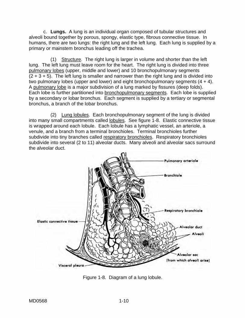
c. Lungs. A lung is an individual organ composed of tubular structures and alveoli bound together by porous, spongy, elastic type, fibrous connective tissue. In humans, there are two lungs: the right lung and the left lung. Each lung is supplied by a primary or mainstem bronchus leading off the trachea. (1) Structure. The right lung is larger in volume and shorter than the left lung. The left lung must leave room for the heart. The right lung is divided into three pulmonary lobes (upper, middle and lower) and 10 bronchopulmonary segments (2 + 3 + 5). The left lung is smaller and narrower than the right lung and is divided into two pulmonary lobes (upper and lower) and eight bronchopulmonary segments (4 + 4). A pulmonary lobe is a major subdivision of a lung marked by fissures (deep folds). Each lobe is further partitioned into bronchopulmonary segments. Each lobe is supplied by a secondary or lobar bronchus. Each segment is supplied by a tertiary or segmental bronchus, a branch of the lobar bronchus. (2) Lung lobules. Each bronchopulmonary segment of the lung is divided into many small compartments called lobules. See figure 1-8. Elastic connective tissue is wrapped around each lobule. Each lobule has a lymphatic vessel, an arteriole, a venule, and a branch from a terminal bronchioles. Terminal bronchioles further subdivide into tiny branches called respiratory bronchioles. Respiratory bronchioles subdivide into several (2 to 11) alveolar ducts. Many alveoli and alveolar sacs surround the alveolar duct.
Figure 1-8. Diagram of a lung lobule.

MD0568 1-11
Section II. ACCESSORY STRUCTURES OF THE RESPIRATORY SYSTEM NOTE: All of the structures in this section play a part in the functioning of the respiratory system. 1-6. DIAPHRAGM The diaphragm, the chief muscle of respiration, is a thin, but strong, dome-shaped muscular membrane. It separates the abdominal and thoracic cavities. The diaphragm is attached to the inferior margin of the rib cage and to the bodies of the lumbar vertebrae behind. As a muscular membrane, it domes upward into the thoracic cavity. Upon contraction, the fibers of the diaphragm shorten and pull downward. This downward motion produces a piston-like pressure on the contents of the abdominopelvic cavity. 1-7. INTERCOSTAL MUSCLES a. The intercostal spaces are filled by two layers of intercostal muscles. The intercostal muscles extend from the vertebrae behind to the sternum in front. A strengthening "plywood effect" is created by the arrangement of the two layers at a right angle to each other. These muscles help maintain the "solid-wall" condition of the thorax. For this reason, a pressure gradient can be maintained between the inside and outside of the thorax. b. The intercostal muscles play a part in the mechanics of breathing. Quiet breathing takes place due to the alternate contraction and relaxation of the diaphragm and the internal intercostal muscles. As an individual breathes in, the diaphragm contracts and, at the same time, the external intercostal muscles contract causing the ribs to be pulled upward and the sternum to be pushed forward. This increases the anterior-posterior diameter of the thoracic cavity. (The volume of the chest cavity increases.) When the individual breathes out, the external intercostal muscles relax, the ribs move downward, and, as the diaphragm relaxes, the thoracic cage moves upward. These movements decrease the vertical and anterior-posterior diameters of the thoracic cavity. The thoracic cavity (smaller in volume) returns to its resting size. 1-8. THORAX The thorax is the chest. The thorax is the portion of the trunk consisting of the sternum, the costal cartilages, the ribs, and the bodies of the thoracic vertebrae. Located above the diaphragm, the thoracic cage is roughly cone-shaped, the narrow portion being superior and the broad portion inferior. It is flattened from front to back. The thoracic cage is open to the outside by way of the neck and head. This bony cage encloses and protects the lungs and other structures of the chest cavity. The thorax also provides support for the bones of the shoulder girdle and upper extremities. Since the wall of the thorax is reinforced by special muscles, bones, and cartilages, we can consider the thorax to be a "solid-walled container."

MD0568 1-12
1-9. PLEURA Surrounding each lung individually is a serous cavity called the pleural cavity (figure 1-9). The minute quantity of serous fluid in the cavity serves as a lubricant. This serves to minimize friction for the expansion and contraction of the lungs during breathing. a. Each lung is covered with a serous membrane called the visceral pleura. The outer wall of the pleural cavity is lined with another serous membrane known as the parietal pleura. Areas of the parietal pleura are variously named according to their location. The mediastinal pleura form the lateral wall of the mediastinum. The diaphragmatic pleura cover the superior surface of the diaphragm. The costal pleura line the inner surface of the rib cage. The cupolar pleura form a dome-like extension into the root of the neck. It contains the apex of the lung. b. When each lung is in its smaller volume, its corresponding diaphragmatic pleura lies close to the lower costal pleura. The slit-like cavity between them is called the costophrenic sinus. Fluids of each pleural cavity tend to collect in this sinus since it is the lowest area for each. When the diaphragm contracts and flattens out, each costophrenic sinus opens up, and the inferior portion of the expanding lung occupies this space.
Figure 1-9. Lungs, visceral, and parietal pleura.
MD0568 1-13
Section III. PHYSIOLOGY OF RESPIRATION 1-10. INTRODUCTION a. Boyle's law tells us that as the volume (V) of a gas-filled container increases, the pressure (P) inside decreases; as the volume (V) of a closed container decreases, the pressure (P) inside increases. When the connected spaces of air have different pressures, the air moves from the space with greater pressure to the one with lesser pressure. b. In regard to breathing, we can consider the air pressure around the human body to be constant. The pressure inside the lungs may be greater or less than the pressure outside the body. Thus, a greater internal pressure causes air to flow out; a greater external pressure causes air to flow in. c. We can compare the human trunk to a hollow cylinder. This cylinder is divided into upper and lower cavities by the diaphragm. The upper is the thoracic cavity and is essentially gas-filled. The lower is the abdominopelvic cavity and is essentially water-filled. 1-11. COSTAL (THORACIC) BREATHING a. Inhalation. Muscles attached to the thoracic cage raise the rib cage. A typical rib might be compared to a bucket handle, attached at one end to the sternum (breastbone) and at the other end to the vertebral column. The "bucket handle" is lifted by the overall movement upward and outward of the rib cage. These movements increase the thoracic diameters from right to left and from front to back. Thus, the intrathoracic volume increases. Recalling Boyle's law, the increase in volume leads to a decrease in pressure. The air pressure outside the body then forces air into the lungs and inflates them. b. Exhalation. The rib cage movements and pressure relationships are reversed for exhalation. Thus, intrathoracic volume decreases. The intrathoracic pressure increases and forces air outside the body. 1-12. DIAPHRAGMATIC (ABDOMINAL) BREATHING a. Inhalation. As the diaphragm contracts, the dome flattens, and the diaphragm descends. This increases the depth (vertical diameter) of the thoracic cavity thus increasing the cavity's volume. At the same time, air pressure in the thoracic cavity decreases. Now, air pressure outside the body is greater, a condition which forces air into the lungs. b. Exhalation. As the diaphragm relaxes, the elastic abdominal wall forces the diaphragm back up by pushing the watery tissues of the abdomen against the underside of the relaxed diaphragm. The dome extends upward. Air is forced out of the lungs.

MD0568 1-14
1-13. CARBON DIOXIDE TRANSPORT AND ELIMINATION a. The Reason for Breathing. Humans breathe to supply oxygen to the cells of the body and to remove the waste product, carbon dioxide (CO2). About one-fifth of the air around us is oxygen. In the process of breathing, or respiration, the body brings air into the lungs. In the lungs, some of the oxygen moves into the bloodstream. A waste gas, carbon dioxide, moves from the blood into the air in the lungs where it is breathed out. The name of this process is the exchange of gases. b. The Exchange of Gases. The actual exchange of oxygen and carbon dioxide takes place in tiny air sacs, the alveoli. The alveoli are in direct contact with capillaries that carry blood. The alveoli walls and the capillary walls are moist and very thin--much thinner than tissue paper. Oxygen molecules seep from the alveoli through the membranes (thin walls) into the blood of the capillaries. Hemoglobin picks up oxygen in the red blood cells. At the same time, blood plasma gives up carbon dioxide to the air in the alveoli. Figure 1-10 illustrates this exchange.
Figure 1-10. Exchange of gases.
1-14. REGULATION OF RESPIRATION The basic rhythm is set and coordinated by the respiratory center; however, the rhythm can be modified in response to the demands of the body by the nerve (neural) center. RESPIRATORY CENTER = neurons (any of the conducting cells of the nervous system) in the reticular formation of the brain stem that regulate the rate of respiration. VENTILATION = the volume of air exchanged in one minute.

MD0568 1-15
a. Brain Influences (Cortical Influences). The brain can send a message to the respiratory center allowing humans to voluntarily control breathing and even stop breathing for a short time. The fact that we can voluntarily stop breathing is a protection because in this way we can keep water or irritating gases from entering our lungs. However, when CO2 builds up to a certain level in our blood, impulses are sent to the inspiratory muscles, causing us to breathe whether we want to or not. It is not possible to kill yourself by holding your breath. b. Medullary Rhythmicity Area. This area controls the basic rhythm of respiration. The respiratory center in the medulla (an inner layer of an organ) adjusts the alveolar ventilation almost exactly to the demands of the body. The result is that there is hardly any change of oxygen and carbon dioxide in arterial blood. The respiratory center of the medulla or its immediate vicinity is the primary site of action of carbon dioxide and hydrogen ions. Reduced oxygen concentrations in the blood stimulate chemoreceptors (a receptor sensitive to chemical changes in the blood stream). Impulses are transmitted to the respiratory center to increase ventilation (the volume of air exchanged in one minute) when these receptors are stimulated. Normally, the increase in ventilation prevents a rise in carbon dioxide to the point that the respiratory center would be stimulated. 1-15. FACTORS THAT CAN DECREASE OXYGEN TRANSPORT TO TISSUES a. Decreased Levels of Hb. Hb is reduced hemoglobin (hemoglobin that has not combined with oxygen). Since oxygen does not dissolve well in water, very little oxygen can be carried by water through the body. Hb (hemoglobin) combines with oxygen and carries about 97 percent of the needed oxygen to various parts of the body. If there is less Hb in the body to combine with and carry oxygen around, there will necessarily be less oxygen transported to tissues of the body. b. Cardiac Failure. The cardiovascular system transports gases in the blood between the lungs and the cells. The heart pumps blood carrying oxygen through the body. The cells of the body cannot survive long if they are starved for blood carrying oxygen. If oxygen is withheld from the cells of the brain for 5 to 6 minutes, a person can suffer severe and permanent brain injury or death. c. Decreased Rate of Respiration. In order for a human to survive, there must be a continuous supply of oxygen to the blood as well as a continuous removal of carbon dioxide from the body. Respiration, the inhaling of oxygen and expelling of carbon dioxide, takes care of the oxygen and carbon dioxide levels in the body. If the respiration rate is lowered, an individual may have enough oxygen but too much carbon dioxide in the body.

MD0568 1-16
d. Disease Processes of Lungs or Component Parts. (1) Lung cancer. People inhale many irritating substances as a part of ordinary breathing. Inhaled smoke and almost all pollutants have an irritating effect on the bronchial tubes and lungs. These pollutants act as stresses or irritating stimuli. A common lung cancer, bronchogenic carcinoma, starts in the walls of the bronchi and is caused by stress and irritation. (2) Nasal polyps. These polyps, protruding growths of mucous membrane hanging down from the posterior wall of the nasal septum, are bluish-white tumors. As they become larger, they may fill the nasopharynx making breathing through the nose difficult. A doctor can remove these polyps easily. (3) Bronchial asthma. Usually, bronchial asthma is caused by an allergy to edible or air-borne substances--for example, wheat or dust. Muscles in the walls of the small bronchi and bronchioles go into spasms caused by the allergy. Also, the smaller bronchi and bronchioles may be clogged with excessive amounts of mucous making breathing difficult. (4) Bronchitis. Bronchitis is inflammation of the bronchi. The most important cause of chronic bronchitis is cigarette smoking. (Chronic bronchitis is bronchitis that lasts for at least three months of the year for two successive years.) (5) Emphysema. In this disease, the alveolar walls lose their elasticity and remain filled with air during expiration. The word "emphysema" means "blown up" or "full of air." A person with emphysema must actively work to exhale. Also, as a result of damage to the alveolar-capillary membrane, the respiration rate slows down. Removing the irritating stimuli-- air pollution, occupational exposure to dust, cigarette smoking--can slow down the progressive deterioration. (6) Pneumonia. Pneumonia is an acute infection or inflammation of the alveoli. The amount of air space in the lungs is reduced because the alveolar sacs fill up with fluid and dead white cells. A bacteria called pneumococcus bacterium is the most common cause of this disease, but other bacteria or a fungus may also cause pneumonia. Several viruses may cause viral pneumonia. (7) Tuberculosis. Tuberculosis is caused by bacteria-- Mycobacterium tuberculosis--which destroys parts of the lung tissue. Tuberculosis bacteria are spread by inhaling, can live through some disinfectants, but are killed by sunlight. This disease is sometimes associated with crowded, poorly lit housing. A person with tuberculosis must have rest, sunlight, and good diet.

MD0568 1-17
(8) Coryza (common cold) and Influenza (flu). Common colds are caused by viruses and typical symptoms include sneezing, excessive nasal secretion, and congestion. (A fever is not usually one of the symptoms.) A virus also causes influenza (flu) with accompanying symptoms of chills, fever (usually higher than 101ºF), headache, and muscular aches. As the fever subsides, cold-like symptoms appear. 1-16. RESPIRATORY CYCLE a. A respiratory cycle (figure 1-11) consists of one inspiration, one rest period, and one expiration--in that order.
Figure 1-11. Respiratory cycle. b. Air is made up of several gases, the most important being oxygen. Carbon dioxide and nitrogen are also present along with some other gases in small amounts. The air we breathe in is not the same as the air we breathe out. Look at the gases in the air in figure 1-12 below.
Figure 1-12. Gases in the air. c. You can see that air breathed out has less oxygen than air breathed in. The body has used some of the oxygen. Air breathed out has more carbon dioxide than air breathed in. The body is expelling carbon dioxide that has been formed as body cells work and use up oxygen.

MD0568 1-18
1-17. AIR CAPACITY OF THE LUNGS The amount of air in the lung of a man of average size who is resting is about 3 liters. When the man breathes in (inspiration or inhalation), he pulls in about 500 milliliters of air. When he deliberately breathes in (forced maximum inspiration), the amount of air in the lung rises to about 6 liters. When the same man forces the maximum amount of air out of the lung, the volume of air is down to about 1 liter. Here are important facts about volume of air and the lungs: NOTE: 1 milliliter (ml) = 1 cubic centimeter (cc). a. The total capacity of the lungs is about 6,000cc (maximum). b. Tidal air volume is the amount of air inhaled or exhaled with each breath, whether the body is at rest or engaged in an activity. For the average adult male who is resting, the tidal volume is about 500cc of air. c. Reserve air is air in the lungs that can be exhaled after normal exhalation. The amount of this air is about 1,500cc. d. Residual air is air that is in the lungs after normal, forceful expiration (about 1,200cc). e. Vital capacity (usually 4,800cc) is the volume of air that can be expelled after full inspiration.
Continue with Exercises
Return to Table of Contents

MD0568 1-19
EXERCISES, LESSON 1 INSTRUCTIONS: Answer these exercises by writing the answer in the space provided. After you have answered all the exercises, turn to "Solutions to Exercises" at the end of the lesson and check your answers. 1. The exchange of gases between the atmosphere and the cells of the body is ______________________________________. 2. The exchange of gases between the air in the lungs and the blood is ______________________________________.. 3. The exchange of gases between the blood and individual cells of the body is ______________________________________. 4. The process that moves air into and out of the lungs is a mechanical process called ______________________________________. 5. In _________________________ breathing, air movement is caused by the rib cage; in ____________________________ breathing, movement between the abdominal wall and the diaphragm cause air movement in and out of the lungs. 6. The main subdivisions of the respiratory system are the supralaryngeal structures, the ________________ and the _________________ structures. 7. The paranasal sinuses are ______________________________________. 8. The olfactory epithelium, a membrane lining the __________________, houses the nerve endings responsible for the ____________________________.

MD0568 1-20
9. The ________________________ is the portion of the nose inside the skull. 10. The three parts of the pharynx are the ________________________________, ______________________, and the ____________________________. 11. The _________________________ cartilage is a ring of cartilage forming the interior wall of the larynx. 12. The ______________________________ cartilage is larger in males than females and causes the larynx to be triangular shaped. 13. The trachea and the bronchi are tubular structures which carry air from the larynx to _________________________________. 14. A lung is an individual organ composed of ___________________ structures and __________________ bound together by porous, spongy, elastic-type, fibrous connective tissue. 15. ____________________________is a membrane enfolding lungs and lining the walls of the chest cavity. 16. Humans can voluntarily control breathing and even stop breathing for a short time because the brain sends a message to the ______________________. 17. Three factors which could decrease oxygen transportation to body tissues are decreased levels of Hb, c___________ f______________ and d_______________ r_______________ of r___________________.
MD0568 1-21
18. Air breathed out has more carbon dioxide than air breathed in because ______________________________. 19. The total capacity of the lungs is about ___________________ cc. 20. Vital capacity of the lungs (usually about 4,800 cc) is ______________________ ________________________________________________________________ 21. The inner surfaces of the alveoli must be kept wet to __________________ ______________________. 22. The chief muscle of respiration is the ______________________________.
Check Your Answers on Next Page

MD0568 1-22
SOLUTIONS TO EXERCISES, LESSON 1 1. Respiration. (para 1-1a) 2. External respiration. (para 1-1a) 3. Internal respiration. (para 1-1a) 4. Breathing. (para 1-1b) 5. Costal; diaphragmatic. (para 1-1b) 6. Larynx; infralaryngeal. (para 1-2b(1) through (3)) 7. Air cells or cavities in the skull. (para 1-3a(2)(d)) 8. Upper nasal chambers; sense of smell. (para 1-3a(2)(c)) 9. Internal nose. (para 1-3a(2)) 10. Nasopharynx, oropharynx, laryngopharynx. (para 1-3b(2)) 11. Cricoid. (para 1-4a(3)) 12. Thyroid. (para 1-4a(1)) 13. The alveoli of the lungs. (para 1-5a) 14. Tubular; alveoli. (para 1-5c) 15. Pleura. (para 1-9) 16. Respiratory center. (para 1-14a) 17. Cardiac failure; decreased rate of respiration. (para 1-15a, b, c) 18. The body is expelling carbon dioxide that has been formed as the body cells work and use up oxygen. (para 1-16c) 19. 6,000. (para 1-17a) 20. The volume of air that can be expelled after a full inspiration. (para 1-17e) 21. Make the transfer of gases possible. (para 1-5b) 22. Diaphragm. (para 1-6) Return to Table of Contents