thai j astroenterol imaging of the small bowel x-ray 2015 … · 2015-05-29 · thai j...
TRANSCRIPT

THAI JGASTROENTEROL
201542 Imaging of the Small Bowel
Imaging of the Small Bowel
Pantongrag-Brown L
Advanced Diagnostic Imaging Center, Ramathibodi Hospital, Bangkok, Thailand.
Address for Correspondence: Linda Pantongrag-Brown, M.D., Advanced Diagnostic Imaging Center, Ramathibodi Hospi-
tal, Bangkok, Thailand.
X-rayCorner
Small bowel is the longest tubular organ in the
body, about 18-22 feet. It is anchored to the body by a
15 cm mesentery, folded between ligament of Treitz
and ileocecal junction. Rule of “3” is usually applied
to images of normal small bowel, which includes <3
mm wall thickness, <3 cm diameter, and <3 air-fluid
levels.
Imaging modalities used in small bowel include
plain radiographs, barium study of small bowel, US,
CT, PET CT and MRI. However, the most common
modalities used in standard practice are plain radio-
graphs and CT. Small-bowel, follow-through study is
Figure 1. Case 1.
mostly replaced by CT because of CT ability to visual-
ize both intraluminal and extraluminal abnormalities.
Air within the small bowel makes other modalities sub-
optimal for good quality images. In this article,
several small bowel abnormalities will be demon-
strated, using case-based approach, and emphasizing
on imaging findings.
Case 1. A 63-year-old man presented with
abdominal pain, nausea and vomiting.
Supine plain radiograph shows multiple dilated
loops of small bowel, lying layer by layer, similar to

THAI J GASTROENTEROL 2015Vol. 16 No. 1
Jan. - Apr. 201543
Pantongrag-Brown L
multiple steps of the ladder (step ladder pattern sign).
The dilated air-filled bowel loops are more than 3 cm
in diameter. Upright radiograph shows multiple air-
fluid levels and different height of air in the same loop
(red horizontal lines). Marked small bowel dilatation
with absence of colonic air is indicative of small bowel
obstruction (SBO). Axial views of CT abdomen show
cecal mass (arrow) causing distal SBO. This mass is
surgically proved to be cecal adenocarcinoma.
Causes of small bowel obstruction are numer-
ous and could be categorized as following(1):
1. Intrinsic conditions
1.1 Inflammatory diseases such as Crohn’s,
TB, and eosinophilic gastroenteritis
1.2 Neoplasms such as GIST, adenocarcinoma,
lymphoma, and metastasis
1.3 Vascular diseases such as ischemia, vas-
culitis, and radiation enteropathy
2. Extrinsic conditions
2.1 Adhesion
2.2 Volvulus
2.3 Hernias
2.4 Endometriosis
2.5 Hematoma
3. Intraluminal causes
3.1 Gallstones
3.2 Bezoars
3.3 Foreign bodies
The 3 most common etiologies in developed
countries are adhesion, Crohn’s disease, and neoplasms.
The 3 most common etiologies in developing coun-
tries are adhesion, hernia, and neoplasms(1).
Case 2. A 58-year-old man presented with ab-
dominal pain, nauseas, and vomiting.
Figure 2. Case 2.
Supine plain radiograph shows a large calcifica-
tion (arrow) at the duodenal jejunal junction causing
partial obstruction and mild dilatation of the proximal
duodenum. Upright radiograph shows air-containing,
branching tubular structure at the RUQ with central
predilection (arrow), indicative of aerobilia. Combi-
nation of findings are consistent with gallstone ileus.
Axial view of CT abdomen shows a lamellar ectopic
gallstone (arrow).
Gallstone ileus is an unusual complication of
chronic cholecystitis. Cholecystoenteric fistula may
occur, as a result of chronic gallbladder (GB) perfora-
tion and fistulous communication with bowel. Once a
fistula is established, air may pass from bowel to the
GB and biliary tract, and stone may pass from the GB
to bowel. This stone may cause mechanical bowel ob-
struction, hence the term “gallstone ileus”.
“Rigler triad”, named after Leo George Rigler,
who described this triad in 1941(2). It is the imaging
triad help for diagnosis of gallstone ileus. The triad is
found in about 25% of gallstone ileus and includes
pneumobilia, SBO, and gallstone in ectopic location.
The gallstone that causes bowel obstruction is relatively
large, at least 2 cm in size. It is usually impacted at the

THAI JGASTROENTEROL
201544 Imaging of the Small Bowel
Figure 3. Case 3.
ileum or ileocecal valve. However, rarely, the stone
may be impacted at the duodenum resulting in gastric
outlet obstruction. This unusual site of gallstone im-
paction is termed “Bouveret syndrome”. This syn-
drome was first described by Leon Bouveret in 1896(3).
Case 3. A 72-year-old man presented with ab-
dominal pain, and fever.
Supine plain radiograph shows abnormal collec-
tion of extraluminal bubbly air at the RLQ, best shown
at the magnified view (arrows). Mixed pattern of lo-
calized ileus and partial SBO is observed. Axial view
of CT abdomen confirms collection of abnormal air,
indicative of abscess. Surgery proves the presence of
RLQ abscess, caused by infarcted small bowel perfo-
ration.
Abscess at the RLQ may cause mixed pattern of
partial SBO and reflex ileus. The most common cause
of RLQ abscess is ruptured appendicitis. Other etiolo-
gies include ruptured diverticulitis, infarcted small
bowel perforation, and colonic cancer perforation.
Distinguishing abnormal bubbly air from feces
within the colon by plain radiograph may be difficult.
A clue that may help to diagnosis in this particular case
is lacking of stool in other part of the colon, as well as
dilatation of small bowel loops surrounding the bub-
bly air collection. If there is any suspicion, CT should
be performed to confirm the diagnosis.
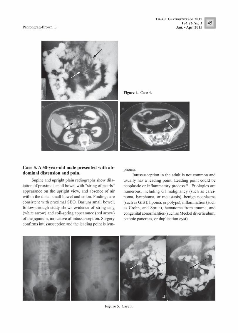
Case 4. A 58-year-old man presented with ab-
dominal pain.
Barium study of the small bowel shows relatively
regular, thickened small bowel folds with some
nodularity, giving the “stack coin” or “picket fence”
pattern (arrows). Findings are indicative of submucosal
deposition. Axial views of CT scan shows SMV throm-
bosis (arrow) and diffuse small bowel edema. Find-
ings are consistent with small bowel ischemia second-
ary to SMV thrombosis.
Barium study of the small bowel is good for de-
termining its mucosal folds. The pattern of relatively
smooth, thickened folds (such as in this case) implies
even distribution of the submucosal depositions. It is
usually caused by edema or hemorrhage. Etiologies of
edema are usually systemic and diffuse, which include
hypoproteinemia (such as from cirrhosis, nephrotic syn-
drome, or protein losing enteropathy), congestive heart
failure and portal hypertension. Etiologies of hemor-
rhage include coagulopathies, ischemia (such as from
SMA/SMV thrombosis, or hypoperfusion), and vas-
culitis. CT is the best imaging modality to identify vas-
cular thrombosis(4).
THAI J GASTROENTEROL 2015Vol. 16 No. 1
Jan. - Apr. 201545
Pantongrag-Brown L
Figure 4. Case 4.
Case 5. A 58-year-old male presented with ab-
dominal distension and pain.
Supine and upright plain radiographs show dila-
tation of proximal small bowel with “string of pearls”
appearance on the upright view, and absence of air
within the distal small bowel and colon. Findings are
consistent with proximal SBO. Barium small bowel,
follow-through study shows evidence of string sing
(white arrow) and coil-spring appearance (red arrow)
of the jejunum, indicative of intussusception. Surgery
confirms intussusception and the leading point is lym-
phoma.
Intussusception in the adult is not common and
usually has a leading point. Leading point could be
neoplastic or inflammatory process(5). Etiologies are
numerous, including GI malignancy (such as carci-
noma, lymphoma, or metastasis), benign neoplasms
(such as GIST, lipoma, or polyps), inflammation (such
as Crohn, and Sprue), hematoma from trauma, and
congenital abnormalities (such as Meckel diverticulum,
ectopic pancreas, or duplication cyst).
Figure 5. Case 5.

THAI JGASTROENTEROL
201546 Imaging of the Small Bowel
Case 6. A 74-year-old man presented with ab-
dominal pain and fever.
Axial and coronal views of CT abdomen show
several jejunal diverticulosis (black arrow, coronal
view) and multiloculated fluid collections surround-
ing the jejunum, indicative of jejunal diverticulitis.
Several extraluminal air bubbles (white arrows in axial
and coronal views) are consistent with perforation, a
complication of diverticulitis.
Jejunal diverticulosis is rare and usually asymp-
tomatic(6). Complication, such as diverticulitis or per-
foration, may result in high morbidity and mortality,
particularly in elderly patients. CT appearance of di-
verticulosis as outpouching lesions are similar to those
found in colon. Therefore, imaging diagnosis is not
difficult. However, awareness of this rare disease is
important to reach the right investigative tool and cor-
rect diagnosis for the patient.
Figure 6. Case 6.
Case 7. A 41-year-old woman presented with
acute abdominal pain.
Axial views of CT abdomen show thin wall of
the distal small bowel with poor enhancement (white
arrow), suggestive of small bowel arterial ischemia.
Feces-like material within the small bowel is sugges-
tive of necrotic material. On magnified sagittal and
axial views, an intraluminal thrombus within the SMA
is noted (red arrows). Surgery confirms long segment
of small bowel infarction.
Mesenteric arterial thrombosis is a life threaten-
ing condition. Thin-walled small bowel with poor en-
hancement is secondary to loss of muscular tone and
lack of arterial blood supply. CT is the best imaging
modality to identify clot within the lumen(7).

THAI J GASTROENTEROL 2015Vol. 16 No. 1
Jan. - Apr. 201547
Pantongrag-Brown L
Case 8. A 43-year-old man, post cardiac valvu-
lar replacement with anticoagulant medication,
presented with acute abdominal pain.
Axial and coronal views of CT abdomen show
thickening of jejunum with soft-tissue encasement of
mesenteric vessel (arrows). There is marked enhance-
ment of the mucosa of the involved jejunum. Combi-
nation of history and CT findings are consistent with
mesenteric venous ischemia secondary to mesenteric
hematoma. Patient is improved after symptomatic treat-
ment.
Mesenteric venous ischemia is less common than
arterial ischemia(8). Hyper-enhancement of the mucosa
is secondary to venous engorgement. This is opposite
to hypo-enhancement from arterial ischemia. Diffuse
bowel wall thickening is also a common finding in me-
senteric venous ischemia, which is opposite to thin
bowel wall secondary to arterial ischemia.
Figure 7. Case 7.

THAI JGASTROENTEROL
201548 Imaging of the Small Bowel
Figure 8. Case 8.
Case 9. A 45-year-old man presented with anemia.
US shows an intra-abdominal mass. Axial and
coronal views of CT abdomen give more information
that the 7.5 cm mass is exophytic from the jejunum,
suggestive of jejunal neoplasm. Lacking of lymphad-
enopathy makes jejunal GIST the most likely diagno-
sis. Surgery confirms the diagnosis of GIST.
Small bowel GIST is second most common site,
after stomach. It is usually more aggressive than gas-
tric GIST. The tumor originates from the submucosal
or intramural layer of the GI tract and tend to show
exophytic growth with cavitation and necrosis. D/Dx
of exophytic bowel mass should include lymphoma,
adenocarcinoma, and metastasis(9).
Figure 9. Case 9.

THAI J GASTROENTEROL 2015Vol. 16 No. 1
Jan. - Apr. 201549
Pantongrag-Brown L
REFERENCES
1. Silva AC, Pimenta M, Guimares LS. Small bowel obstruc-
tion: what to look for. RadioGraphics 2009;29:423-39.
2. Rigler LG, Borman CN, Noble JF. Gallstone obstruction:
pathogenesis and roentgen manifestations. J Am Med Assoc
1941;117:1753-9.
3. Bouveret L. Stenose du pylore adherent a la vesicule. Rev
Med (Paris) 1896;16:1-16.
4. Furukawa A, Kanasaki S, Kono N, et al. CT diagnosis of acute
mesenteric ischemia from various causes. AJR Am J Roentgenol
2009;192:408-16.
5. Choi SH, Han JK, Kim SH, et al. Intussusception in adults:
from stomach to rectum. AJR Am J Roentgenol 2004;183:691-
8.
6. Tsiotos GG, Farnell MB, IIstrup DM. Nonmeckelian jejunal
or ileal diverticulosis: An analysis of 112 cases. Surgery 1994;
116:726-32.
7. Schieda N, Fasih N, Shabana W. Triphasic CT in the diagno-
sis of acute mesenteric ischaemia. Eur Radiol 2013;23:1891-
900.
8. Warshauer DM, Lee JK, Mauro MA, et al. Superior mesen-
teric vein thrombosis with radiologically occult cause: a ret-
rospective study of 43 cases. AJR Am J Roentgenol 2001;
177:837-41.
9. Levy AD, Remotti HE, Thompson WM, et al. Gastrointesti-
nal stromal tumors: radiologic features with pathologic corre-
lation. RadioGraphics 23:283-304, 456.
CONCLUSIONS
Nine cases of small bowel with various diseases
are illustrated, emphasizing on the imaging appear-
ances. These diseases are as following:
1. Small bowel obstruction: secondary to cecal
carcinoma, GS ileus, RLQ abscess from SB perfora-
tion, and intussusception from lymphoma
2. Neoplastic pathology: jejunal GIST
3. Inflammatory pathology: jejunal diverticulitis
with perforation
4. Vascular pathology: mesenteric venous is-
chemia from SMV thrombosis and hematoma encas-
ing the mesenteric venous branches, and mesenteric
arterial ischemia from SMA thrombosis.