the acute abdomen: plain radiographic evaluation · the acute abdomen: plain radiographic...
TRANSCRIPT
November 1984 Special Focus Session
The acute abdomen:
Plain radiographic evaluation
Charles D. Johnson, M.D.
Reed P. Rice, M.D.
Introduction
With the development of computed tomography, ultrasonography and spe-
cialized scintigraphic techniques, conventional radiographic imaging of the abdo-
men has generally been relegated to a minor position in each of the three clinical
entities under discussion in this series. On the other hand, one can make a good
case for routinely obtaining radiographs of the abdomen in most patients with an
acute abdomen. The information available on the plain radiographs regarding the
amount and distribution of intestinal gas, recognition of extratuminal gas collec-
tions, significant calcifications, organomegaly and the demonstration of soft tissue
masses or fluid collections can be critical in making a specific diagnosis or in
suggesting a possible diagnosis. This information may be useful in planning the
subsequent workup. In this section, the role of conventional radiography, includ-
ing plain radiographs and traditional contrast studies of the abdomen, will be dis-
cussed in the workup of patients with suspected acute cholecystitis, subhepatic ab-
scess and splenic trauma.
Acute Cholecystitis
Conventional radiographic techniques including plain radiographs and con-
trast studies have a minor role in the imaging workup of patients suspected of hay-
ing acute cholecystitis. Oral cholecystography and intravenous cholangiography
have no role in the evaluation of a patient with acute right upper quadrant symp-
toms. A plain radiograph of the abdomen, however, is usually obtained in patients
with acute abdominal symptoms and may provide some useful information.
In a patient with abdominal pain, the plain radiographic demonstration of
gallstones is an important clue that the gallbladder may be the source of the acute
symptoms. Approximately 15% of gallstones are recognizable on plain radio-
graphs (2). The demonstration of gallstones is useful information, although it does
not necessarily incriminate the gallbladder as the source of the acute symptoms.
Rarely, a soft tissue mass can be detected on plain radiographs in a patient with
an obstructed cystic duct and hydrops of the gallbladder (2). In a patient whose
symptoms are atypical, an upper gastrointestinal series may be performed in the
workup of upper abdominal symptoms. The demonstration of an enlarged gall-
bladder with compression and spasm of the second portion of the duodenum may
first suggest an inflamed gallbladder (3) (Figure 1).
From the Department of Radiology, Duke University Medical Center, Durham, NC
Volume 5, Number 2 March 1985 RadioGraphics 259

Acute abdomen: Plain radiography Johnson and Rice
260 RadioGraphics March 1985 Volume 5, Number 2
FIgure 1Acute cholecystitis The enlarged gallbladder resultingfrom cystic duct obstruction (hydrops), causes a pressuredeformity (arrows) on the second portion of the duodenumwith secondary spasm and fold thickening. These findingssuggest acute cholecystitis in a patient complaining of rightupper quadrant discomfort.
A rare form of acute cholecystitis which can be spe-
cifically diagnosed on plain films is the entity of emphy-
sematous chotecystitis. This form of acute cholecystitis is
more common in men than in women, and frequently occurs
in diabetics (14). The cystic duct may or may not be ob-
structed, and it is believed that this form of cholecystitis may
occur as a result of ischemia of the gallbladder (12). There
is a higher incidence of gangrene and perforation of the
gallbladder in emphysematous cholecystitis than with the
classic form of cholecystitis, and the mortality rate is higher
(14). In spite of this heightened frequency of severe com-
plications, the disease may be surprisingly occult (14).
Radiographically, emphysematous cholecystitis is
characterized by demonstration of gas within the wall or
2C
within the lumen of the gallbladder or both. In some cases
in which the gallbladder has already perforated, the linear
outline of the gallbladder can be identified with bubbles of
gas outside the gallbladder wall (Figures 2-4). In other pa-
tients, there is only gas within the lumen of the gallbladder.
In these patients, the possibility of bitiary-enteric fistula,
resulting from a gallstone eroding into the intestinal tract,
or an ulcer penetrating into the common bile duct, needs to
be considered as an explanation for the symptoms and for
the demonstration of gas in the gallbladder lumen (14).
In our experience, the demonstration of localized ileus,
scoliosis, and obliteration of psoas or liver margins is non-
specific and has virtually no radiographic significance in
patients suspected of acute cholecystitis.

‘‘�.
I
2B
Volume 5, Number 2 March 1985 RadioGraphics
Johnson and Rice Acute abdomen: Plain radiography
Figure 2Emphysematous cholecystltis (A&B) An amor-phous collection of gas (arrow) is seen within thelumen of the gallbladder. The absence of intramuralgas should not dissuade the radiologist from makingthe correct diagnosis. (C) Computed tomographyconfirms the plain radiographic findings and, in addi-tion, identifies a pericholecystic fluid collection (openarrow), as well as fluid in the left anterior pararenalspace (solid arrow) resulting from pancreatitis.
261

3A 3B
Acute abdomen: Plain radiography Johnson and Rice
262 RadioGraphics March 1985 Volume 5, Number 2
Figure 3Emphysematous cholecystitis (A&B) These figures show obviousintramural and pericholecystic gas collections (arrows) in two differentpatients.
Figure 4Emphysematous cholecystitis Linear collectionsof intramural gas (arrows) are seen within this gall-bladder.
Johnson and Rice Acute abdomen: Plain radiography
Volume 5, Number 2 March 1985 RadioGraphics 263
Traumatic Rupture of the Spleen
The spleen is the intraabdominal organ that is most
often injured in blunt abdominal trauma, but plain radio-
graphic findings suggesting the diagnosis are often absent
and usually nonspecific. Patients suffering significant tho-
racoabdominal trauma usually have a plain radiograph of
the chest and abdomen because of the value of these studies
in establishing the diagnoses of skeletal injury to the spine
or thoracic cage and soft tissue injury to the diaphragm,
lungs and pleura. The radiologist should not overlook the
opportunity to evaluate these films for signs suggesting
splenic injury. If such signs are present, he should suggest
the expeditious use of more definitive imaging modalities,
including computed tomography, ultrasound, and radio-
nuclide scintigraphy.
RADIOGRAPHIC FINDINGS
The reported frequency with which plain radiographs
of the chest and abdomen contribute to the diagnosis of
splenic rupture varies markedly, from 90% as stated by
Haentel and Ryder (11 ) to less than 3% in Stivetman’s series
(21 ). Our experience suggests that significant plain radio-
graphic findings are uncommon and rarely diagnostic.
Serial films, when available, are sometimes valuable in
suggesting splenic trauma or free peritoneal bleeding. The
following radiographic findings may be useful in suggesting
splenic rupture.
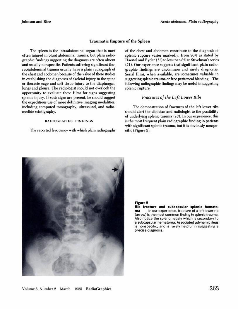
Fractures of the Left Lower Ribs
The demonstration of fractures of the left lower ribs
should alert the clinician and radiologist to the possibility
of underlying splenic trauma (13). In our experience, this
is the most frequent plain radiographic finding in patients
with significant splenic trauma, but it is obviously nonspe-
cific (Figure 5).
Figure 5Rib fracture and subcapsular splenic hemato-ma In our experience, fracture of a left lower rib(arrow) is the most common finding in splenic trauma.Also notice the splenomegaly which is secondary toa subcapsular hematoma. Associated adynamic ileusis nonspecific, and is rarely helpful in suggesting aprecise diagnosis.

Acute abdomen: Plain radiography J ohnson and Rice
264 RadioGraphics March 1985 Volume 5, Number 2
Enlargement of the Spleen
Observable enlargement of the spleen on serial radio-
graphs made following blunt abdominal trauma usually
indicates splenic laceration, commonly associated with
subeapsular hematoma (Figure 6). The enlarging spleen or
associated hematoma may displace the gastric air bubble
medially (Figure 7). Rarely, a serrated appearance of the
lateral aspect of a gas filled stomach is evident on plain ra-
diographs. The enlarging spleen may also displace the
splenic flexure of the colon caudally and medially, and there
may be elevation of the left hemidiaphragm (9) (Figure
8).
Figure 6Subcapsular splenic hematoma Progressive enlargementof the spleen is highly suggestive of splenic injury. This casedemonstrates splenomegaly resulting from subcapsular hema-toma in a patient with known, recent, blunt abdominal trauma, anda normal abdominal plain radiograph 24 hours earlier.

‘�4� I I
Johnson and Rice Acute abdomen: Plain radiography
Volume 5, Number 2 March 1985 RadioGraphics
Figure 7Subcapsular splenic hematoma Splenomegalywith medial displacement of the gastric air bubblesuggests the diagnosis of subcapsular splenic hema-toma in this patient who had suffered blunt abdominaltrauma several hours earlier.
Figure 8Subcapsular splenic hematoma In this case,massive splenic enlargement developed over a periodof six months following abdominal trauma. Arteriog-raphy confirmed a subcapsular hematoma. Notice themarked medial displacement of the gastric air bubbleand the caudal displacement of the transverse colon.Contrast material is seen within the normal urinarycollecting system.
265
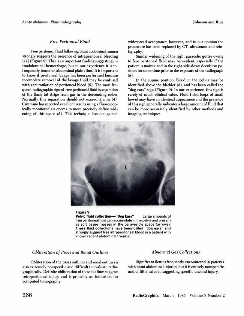
Figure 9Pelvic fluid collection-”Dog Ears” Large amounts offree peritoneal fluid can accumulate in the pelvis and presentas soft tissue masses in the paravesicle space (arrows).These fluid collections have been called ‘ ‘dog ears’ ‘ andstrongly suggest free intraperitoneal blood in a patient withknown recent abdominal trauma.
Acute abdomen: Plain radiography Johnson and Rice
266 RadioGraphics March 1985 Volume 5, Number 2
Free Peritoneal Fluid
Free peritoneal fluid following blunt abdominal trauma
strongly suggests the presence of intraperitoneal bleeding
(11 ) (Figure 9). This is an important finding suggesting in-
traabdominal hemorrhage, but in our experience it is in-
frequently found on abdominal plain films. It is important
to know if penitoneal lavage has been performed because
incomplete removal of the lavage fluid may be confused
with accumulation of penitoneal blood (9). The most fre-
quent radiographic sign of free peritoneal fluid is separation
of the flank fat stripe from gas in the descending colon.
Normally this separation should not exceed 2 mm (4).
Cimmino has reported excellent results using a fluoroscop-
ically monitored air enema to more precisely define wid-
ening of this space (5). This technique has not gained
Obliteration of Psoas and Renal Outlines
Obliteration of the psoas outlines and renal outlines is
also extremely nonspecific and difficult to evaluate radio-
graphically. Definite obliteration of these fat lines suggests
retropenitoneal injury and is probably an indication for
computed tomography.
widespread acceptance, however, and in our opinion the
procedure has been replaced by CT, ultrasound and scm-
tigraphy.
Similar widening of the right paracolic gutter owing
to free peritoneal fluid may be evident, especially if the
patient is maintained in the right-side-down decubitus po-
sition for some time prior to the exposure of the radiograph
(8).
In the supine position, blood in the pelvis may be
identified above the bladder (8), and has been called the
“dog ears” sign (Figure 9). In our experience, this sign is
rarely of much clinical value. Fluid filled loops of small
bowel may have an identical appearance and the presence
of this sign generally indicates a large amount of fluid that
can be more accurately identified by other methods and
imaging techniques.
Abnormal Gas Collections
Significant ileus is frequently encountered in patients
with blunt abdominal injuries, but it is entirely nonspecific
and of little value in suggesting specific visceral injury.
Johnson and Rice Acute abdomen: Plain radiography
Volume 5, Number 2 March 1985 RadioGraphics 267
Subhepatic Abscess
Intraabdominal abscess remains an important clinical
problem with a mortality rate of approximately 30% (1 ), in
spite of better antibiotics, improved drainage techniques,
and more accurate localization with the newer imaging
modalities before attempting drainage. This discussion will
be limited to the diagnosis of subhepatic abscesses, the ma-
jonity of which occur in patients who have recently under-
gone surgery or sustained trauma (18, 22). In the postop-
erative patient, physical findings are difficult to evaluate,
and fever is often attributed to pulmonary or urinary in-
fection. Postoperative abscesses may be clinically occult; a
surprising number of patients with sizable intraabdominal
abscesses demonstrate few clinical signs and symptoms (10,
20). Many patients who continue to have abdominal pain
and ileus in the postoperative period will have more or less
routine abdominal radiographs. These conventional radio-
graphic images of the abdomen may provide the first mdi-cation that a postoperative abscess is present. This is espe-
cially true when clinical signs or symptoms are partially
masked by antibiotics, steroids, immunosuppressive therapy,
or chemotherapy (20).
ANATOMIC CONSIDERATIONS
The subhepatic space is one of the most common sites
for abscess accumulation. Meyers and Whalen have precisely
defined the normal anatomic spaces and pathways by which
free peritoneal fluid disseminates (16, 17). Free peritoneal
fluid may reach the subhepatic space via the right paracolic
gutter from either the subdiaphragmatic space or the pelvis.
The subhepatic space is the most dependent region in the
upper abdomen when a patient is in the supine position (23).
Meyers refers to this region as the “sewer” of the upper
abdomen, for it is the most frequent site for the accumula-
tion of infected penitoneal fluid (15, 16).
Anatomically, the subhepatic space is bounded ante-
niorly by the posterior surface of the right lobe of the liver,
posteriorly by the right kidney, medially by the second
portion of the duodenum, and laterally by the right paracotic
gutter. Anterior and posterior subdivisions of this space have
been defined, but the posterior compartment (Monison’s
pouch) is more often the site of infection. The epiploic
foramen (Foramen of Winslow) connects Morison’s pouch
with the lesser sac. This communication is rarely patent in
the presence of infection; and a lesser sac abscess, therefore,
is rarely associated with infection in the subhepatic space.
Concurrent infection in the right subphrenic space is com-
monty present owing to its communication with the sub-
hepatic space via the right paracolic gutter (16).
The close relationship of the gallbladder, the duodenal
bulb, and the hepatic flexure of the colon to the subhepatic
space is important, and subhepatic abscess may result from
surgery, perforation or other diseases involving any of these
organs.
RADIOGRAPHIC CONSIDERATIONS
Though the obvious rote of computed tomography,
ultrasound, and improved scmntigraphic techniques has re-
sulted in less emphasis on plain radiography, there are sev-
eral studies that demonstrate that plain abdominal radio-
graphs are surprisingly accurate in detecting upper ab-
dominal abscesses. Connell et al. reported radiographic
findings either diagnostic or suspicious for upper abdominal
abscess in 71 % of patients on plain radiographs alone. The
addition of contrast studies of the gastrointestinal tract im-
proved the diagnostic accuracy to 88% in those patients who
received contrast examinations (6). Similar results have been
reported by Fataan and Schulman (7).
The most important plain radiographic finding to
suggest the diagnosis of a subhepatic abscess is the demon-
stration of a toculated pocket of extraluminal gas in the
subhepatic space (Figure bA). The fact that the pocket of
gas is extraluminal may be subtle, and it may be very diffi-
cult to differentiate extraluminal abscess gas from normal
intraluminal gas. Many patients with a subhepatic abscess
will have an associated ileus which may compound the
problem (20).
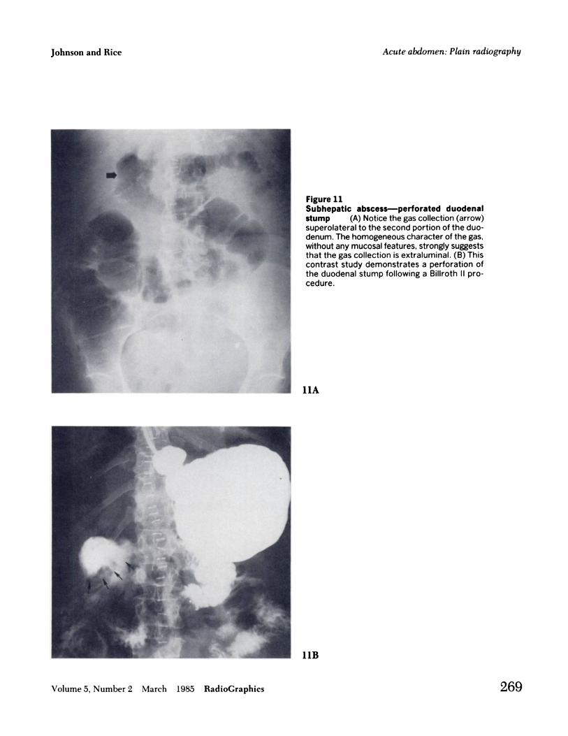
Extraluminal gas may appear as a mottled lucency
resembling stool in the colon. Less commonly emphasized
in the radiologic literature, but equally as common in our
experience, is the demonstration of a homogeneous abscess
gas collection. Such a collection can sometimes be differ-
entiated from intraluminal gas by a featureless wall which
is devoid of any mucosal pattern (19) (Figure 1 1). Because
the pocket of gas in an abscess is loculated and may contain
pus, an air fluid level can frequently be demonstrated.
Normally, there is no gas containing viscus above the hepatic
flexure, so when a gas pocket is identified which is clearly
superior to the stool or gas filled hepatic flexure, the presence
of extraluminal gas can be strongly suggested. Occasionally,
subhepatic abscess gas may be difficult to differentiate from
normal gas within the duodenal bulb or loop, and a limited
gastrointestinal series is useful in making this distinction
(Figure lOB).

1OA
Acute abdomen: Plain radiography Johnson and Rice
268 RadioGraphics March 1985 Volume 5, Number 2
Figure 10Subhepatic abscess (A) Loculated, amor-phous gas collection (arrows) might lie in theduodenum. (B) An upper gastrointestinal seriesproves the extraluminal nature of the gas col-lection (arrows) which, in fact, lies in the sub-hepatic space.
lOB
Johnson and Rice Acute abdomen: Plain radiography
Volume 5, Number 2 March 1985 RadioGraphics 269
Figure 11Subhepatic abscess-perforated duodenalstump (A) Notice the gas collection (arrow)superolateral to the second portion of the duo-denum. The homogeneous character of the gas,without any mucosal features, strongly suggeststhat the gas collection is extraluminal. (B) Thiscontrast study demonstrates a perforation ofthe duodenal stump following a Billroth II pro-cedure.
llA
11B

12A . 12B
Acute abdomen: Plain radiography Johnson and Rice
270 RadioGraphics March 1985 Volume 5, Number 2
Abscesses that do not contain gas are rarely demon-
strable on plain radiographs of the abdomen. In contrast
examinations of the upper gastrointestinal tract or colon, an
abscess may be demonstrable as a soft tissue mass displacing
or indenting the hepatic flexure or duodenal loop (Figure
12). A more important contribution of the contrast exami-
nation is the demonstration of the site of perforation or
anastomotic leak which may be the underlying cause of a
postoperative or spontaneous abscess (6) (Figures 13 and
14).
Subhepatic abscesses alone are rarely associated with
right pleural effusions, lower lobe atelectasis or other changes
in the lung base. These radiographic signs are more likely
to be present if a subhepatic abscess is associated with a
subdiaphragmatic abscess (1 7). Scoliosis and loss of fat
planes, as demonstrable on plain radiographs, are rarely of
much value.
Figure 12Subhepatic abscess (A) Extrinsic compression of the superior as-pect of the hepatic flexure (arrows) is due to the mass effect of a sub-hepatic abscess. (B) This upper gastrointestinal study demonstratesan extrinsic mass effect on the lateral aspect of the second portion ofthe duodenum (arrow).
Johnson and Rice Acute abdomen: Plain radiography
Volume 5, Number 2 March 1985 RadioGraphics 271
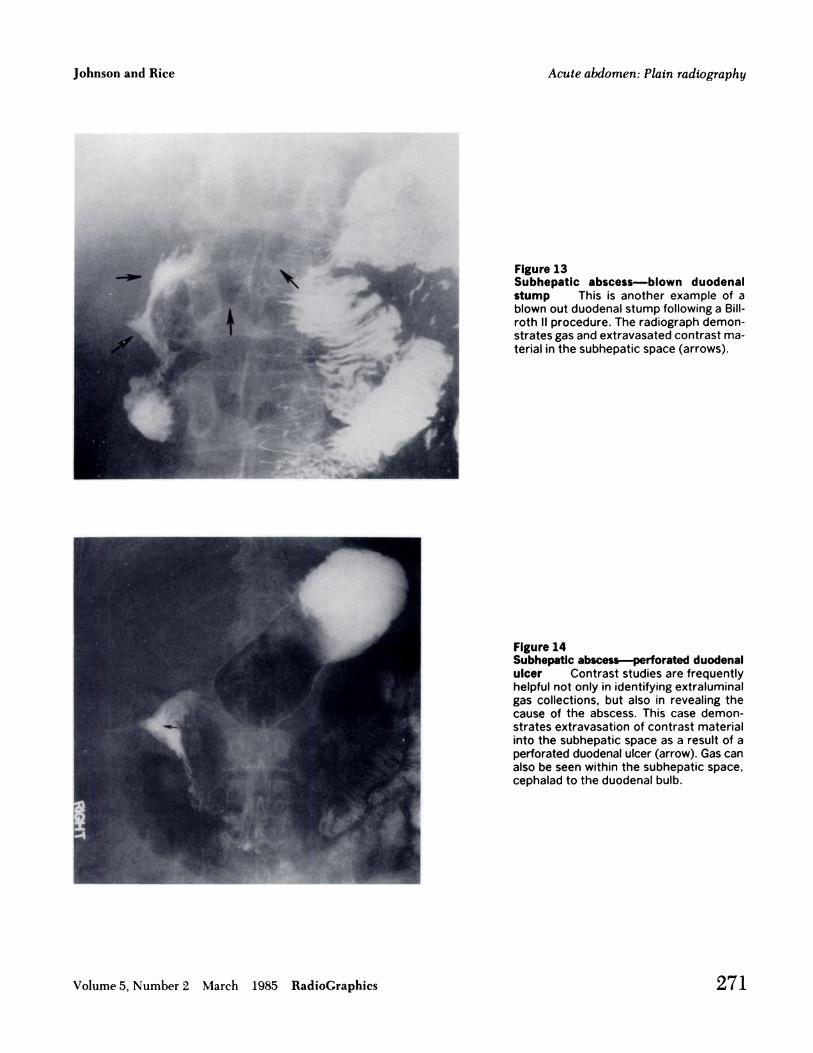
Figure 13Subhepatic abscess-blown duodenalstump This is another example of ablown out duodenal stump following a Bill-roth II procedure. The radiograph demon-strates gas and extravasated contrast ma-terial in the subhepatic space (arrows).
Figure 14Subhepatic abscess--perforated duodenalulcer Contrast studies are frequentlyhelpful not only in identifying extraluminalgas collections, but also in revealing thecause of the abscess. This case demon-strates extravasation of contrast materialinto the subhepatic space as a result of aperforated duodenal ulcer (arrow). Gas canalso be seen within the subhepatic space,cephalad to the duodenal bulb.
Acute abdomen: Plain radiography Johnson and Rice
272 RadioGraphics March 1985 Volume 5, Number 2
Summary
There is still a significant role for plain radiographs of
the abdomen in the workup of patients with an acute
abdomen, and we believe that radiographs of the abdomen
are a reasonable place to start the imaging evaluation of these
patients.
In patients suspected of having acute cholecystitis, the
demonstration of opaque gallstones or of an obvious soft
tissue mass in the region of the gallbladder is obviously im-
portant. In the rare patient with emphysematous cholecys-
titis, the demonstration of gas in the wall or lumen of the
gallbladder, or both, can be diagnostic.
Specific plain radiographic findings of splenic trauma
are unusual, but plain radiographs may demonstrate nib
fractures or other skeletal injuries that raise one’s index of
suspicion with respect to the possibility of splenic rupture.
Plain radiographs may also demonstrate splenic enlargement
or free peritoneal fluid. These findings strongly suggest
splenic trauma.
Plain radiographs of the abdomen in a postoperative
patient may demonstrate gas in a subhepatic abscess that has
not been suspected clinically. Any loculated collection of gas
above the proximal transverse colon is an indication for
further imaging evaluation. Contrast studies of the gas-
trointestinal tract can define leaks and perforations that
cannot be diagnosed preoperatively by any other tech-
nique.
References
1. Ariel IM, Kazarian KK. Classification, diagnosis, and treatmentof subphrenic abscesses. In: Ariel IM, Kazarian K, eds. Diag-
nosis and treatment of abdominal abscesses. Williams and
Wilkins, Baltimore, 1971:174-206.
2. Berk RN. The plain abdominal radiograph. In: Berk RN,
Ferrucci JT Jr, Leopold GR, eds. Radiology of the gallbladder
and bile ducts: Diagnosis and intervention. W.B. SaundersCompany, Philadelphia, 1983:1-29.
3. Berk RN. Barium studies of the gastrointestinal tract. In: BerkRN, Ferrucci JT Jr, Leopold GR, eds. Radiology of the gall-
bladder and bile ducts: Diagnosis and intervention. W.B.
Saunders Company, Philadelphia, 1983:30-54.
4. Cimmino CV. Ruptured spleen: Some refinements in its
roentgenologic diagnosis. Radiology 1964; 82:57-62.
5. Cimmino CV, Southworth LE. Further refinements in the
plain radiologic diagnosis of splenic rupture: The air enema.
Radiology 1978; 127:649-653.6. Connell TR, Stephens DH, Carlson HC, Brown ML. Upper
abdominal abscess: A continuing and deadly problem. AJR
1980; 134:759-765.
7. Fataar 5, Schulman A. Subphrenic abscess: The radiologicapproach. Ctin Radiot 1981; 32:147-156.
8. Frimann-DahI J. Roentgen examinations in abdominal trauma.In: Roentgen examinations in acute abdominal diseases.
Charles C Thomas, Springfield, Illinois, 1974:480-489.9. Gold RE, Hoskins PA. Radiologic evaluation of splemc trauma.
CRC Crit Rev Radiol Sci 1972; 3:453-487.
10. Goldman R, Hunter TB, Haber K. The silent abdominal ab-
Scess: Rote of the radiologist. AJR 1984; 141:21-25.
1 1. Haertel M, Ryder D. Radiologic investigation of splenic
trauma. Cardiovasc Radiol 1979; 2:27-33.
12. Harley WD, Kirkpatrick RH, Ferrucci JT Jr. Gas in the bileducts (pneumobilia) in emphysematous chotecystitis. AJR
1978; 131:661-663.
13. Kurtzman RS. Radiology of blunt abdominal trauma. Sang Clin
North Am 1977; 57:211-226.14. Mentzer RM Jr, Golden GT, Chandler JG, Horstey JS. A
comparative appraisal of emphysematous cholecystitis. AmJ Surg 1975; 129:10-15.
15. Meyers MA. The spread and localization of acute intraperi-
toneal effusions. Radiology 1970; 95:547-554.
16. Meyers MA. Dynamic radiology of the abdomen. Normal and
pathologic anatomy, 2nd ed. Springer-Verlag, New York,1982:20-54.
17. Meyers MA, Whalen JP. Radiologic aspects of intra-abdominal
abscesses. In: Aniel IM, Kazarian KK, eds. Diagnosis and
treatment of abdominal abscess. Williams and Wilkins, Bal-
timore, 1971:87-97.
18. Miller WT, Talman EA. Subphrenic abscess. AJR 1967; 101:
961-969.
19. Rice RP, Masters SJ. Intraabdominal abscess. Semin Roentgenol1973: 8:365-374.
20. Rice RP, Thompson WM, Gedgaudas RK. The diagnosis and
significance of extraluminal gas in the abdomen. Radiol ClinNorth Am 1982; 20:819-837.
21. Stivetman RL, Glaubitz JP, Crampton RS. Laceration of the
spleen due to nonpenetrating trauma. One hundred cases. Am
J Surg 1963; 106:888-891.
22. Wetterfors J. Subphrenic abscess. A clinical study of 101 cases.
Acta Chir Scand 1959; 117:388-408.23. Whalen JP, Bierny J. Classification of perihepatic abscesses.
Radiology 1969; 92: 1427-1437.