the affordable care act timeline - new york american ... state epic 30-02-12.pdf · the affordable...
TRANSCRIPT

online at www.nyacep.orgvolume 30:02:12
In July, the U.S. Supreme Court announced its decision to uphold the 2010 Affordable Care Act. Although some aspects of the Act are designed to begin now, other aspects are designed to begin in the months and years to come. See below for a timeline to help better map out what changes will be happening and when.
2012Improving Quality and Lowering Costs
Linking Payment to Quality Outcomes (effective October 1, 2012). The law establishes a hospital Value-Based Purchasing Program (VBP) in traditional Medicare. This program offers financial incentives to hospitals to improve the quality of care. Hospital performance is required to be publicly reported, beginning with measures on treating heart attacks, heart failure, pneumonia, surgical care, health-care associated infections, and patients’ perception of care.
Encouraging Integrated Health Systems (effective January 1, 2012). The new law provides incentives for physicians to join together to form “Accountable Care Organizations,” through which doctors can better coordinate patient care and improve the quality, help prevent disease and illness and reduce unnecessary hospital admissions. If Accountable Care Organizations provide high quality care and reduce costs to the health care system, they can keep some of the money that they have helped to save.
Reducing Paperwork and Administrative Costs (first regulation effective October 1, 2012). Health care remains one of the few industries that relies on paper records. The new law will institute a series of changes to standardize billing and requires health plans to begin adopting and implementing rules for the secure, confidential, electronic exchange of health information. Using electronic health records will reduce paperwork and administrative burdens, cut costs, reduce medical errors and most importantly, improve the quality of care.
Understanding and Fighting Health Disparities (effective March, 2012). To help understand and combat persistent health disparities, the law requires any ongoing or new Federal health program to collect and report racial, ethnic and language data. The Secretary of Health and Human Services will use this data to help identify and fight disparities.
Increasing Access to Affordable Care Providing New, Voluntary Options
for Long-Term Care Insurance (effective October 1, 2012). The law creates a voluntary long-term care insurance program -- called CLASS -- to provide cash benefits to adults who become disabled.
2013
Improving Quality and Lowering Costs Improving Preventive Health
Coverage (effective January 1, 2013). To expand the number of Americans receiv-ing preventive care, the law provides new funding to state Medicaid programs that choose to cover preventive services for patients at little or no cost.
continued on page 32
The Affordable Care Act TimelineDaniel G. Murphy, MD MBA FACEP, Chairman, Department of Emergency Medicine, Good Samaritan Hospital Medical Center; Chief of Emergency Medicine, Catholic Health System of Long Island
inside this
president's message .............................................2salute to past presidents .......................................2in the wake of aurora ............................................3ultrasound sound rounds ......................................4discharging AMA ...................................................6new york acep awards ..........................................7toxicology column ................................................8ads .................... 9, 12, 15, 23-25, 28, 31, 33, 35-36albany update .....................................................10career day supporters .........................................11new york state of mind .......................................13under your wing .................................................22pediatric column .................................................26a long time ago ...................................................29holiday closings ..................................................31calendar ..............................................................34classified ads .......................................................34

2 empire state epic vol. 30:02:12
A Salute to New York ACEP Past Presidents
1971-1973 Edward W Gilmore, MD
1973-1976 Edward L McNeil, MD
1976-1977 Alan Davidson III, MD FACEP
1977-1978 Cyril Cameron, MD
1978-1979 Alvin L Scott, MD
1979-1980 Ralph Altman, MD
1980-1981 Julius P Duic, MD
1981-1983 Richard M Siegel, MD
1983-1985 Frank E Coughlin Jr, MD FACEP
1985-1987 Ann Furtado, MD FACEP
1987-1989 Jeffrey L Margulies, MD FACEP
1989-1991 Michael F Jacobius, MD FACEP
1991-1993 Mark C Henry, MD FACEP
1993-1995 Stephan G Lynn, MD FACEP
1995-1997 Peter Viccellio III, MD FACEP
1997-1999 Richard Salluzzo, MD FACEP
1999-2000 Samuel F Bosco, MD FACEP
2000-2002 Vincent P Verdile, MD FACEP
2002-2004 Andrew E Sama, MD FACEP
2004-2006 Theodore J Gaeta, DO MPH FACEP
2006-2008 Jerry R Balentine Jr, DO FACEP
2008-2010 Gerard X Brogan, MD FACEP
2010-2012 Joel M Bartfield, MD FACEP
Daniel G. Murphy, MD MBA FACEP
Chairman, Department of Emergency Medicine,
Good Samaritan Hospital Medical Center; Chief
of Emergency Medicine, Catholic Health System
of Long Island
president's The Future Continuum of Outpatient Care
The success of the urgent care business in some communities has enticed many emergency physicians. Perhaps they invest and are partners in the business. Perhaps they just work shifts, none of which are nights. The hectic, high acuity environment of the emergency department is left behind and the money is often pretty good.
The market often welcomes such ventures because the patients’ experiences can also be less hectic with less ‘stimulation’ than the typical emergency department experience in a convenient place closer to home. Insurance companies also appreciate not having to pay hospital based facility charges for unscheduled care.
Many of the emergency physicians who leave the emergency department environment miss it however. They were trained to resuscitate and address high acuity emergencies. They therefore often work per diem or part-time in an emergency department to stay in touch with their inner self.
Such is the healthcare market place and emergency medicine needs to adapt.
Medicaid, Medicare and the commercial insurers all want only the sickest patients to be hospitalized. “Denials” are increasing and Recovery Audit Contractor activity is taking off exponentially. Consequently, observation services will likely be expanding in your hospital. There is no physician better than an emergency physician to perform or oversee observation services. We are skilled at making time-dependent, cost-effective clinical decisions across the entire breadth of pathology.
Such is the healthcare marketplace and emergency medicine needs to adapt.
Please feel free to communicate with New York ACEP about theses and other issues that impact or will impact emergency medicine and your career. Our committees and your Board include many leading clinicians, educators and thinkers. We welcome your input. Send me an email ([email protected]) if you want to get involved.
New York ACEP committees include: Education, Emergency Medicine Resident Committee, EMS, Government Affairs, Practice Management, Professional Development and Research. New York ACEP Exclusive Supporter
2012 Resident Research Conference

new york american college of emergency physicians 3
2012-13 Officers
PresidentDaniel G. Murphy, MD MBA FACEPGood Samaritan Hospital Medical Center; Catholic Health System of Long Island, 631/376-4094
President-electLouise A. Prince, MD FACEPSUNY Upstate Medical University, 315/464-4235
Secretary-TreasurerBrahim Ardolic, MD FACEPStaten Island University Hospital, 718/226-9158
Immediate Past PresidentJoel M. Bartfield, MD FACEP Albany Medical Center, 518/262-7302
Executive DirectorJoAnne TarantelliNew York ACEP, 585/872-2417
2012-13 Board Members
Jay M. Brenner, MD FACEPSUNY Upstate Medical University, 315/464-4363
Susan Cheng, MD resident representativeSUNY Health Sciences Center at Brooklyn, 718/245-3318
Jeremy T. Cushman, MD MS FACEPUniversity of Rochester, 585/463-2900
Keith E. Grams, MD FACEPRochester General Health System, 585/276-3653
Sanjey Gupta, MD FACEPNew York Hospital Queens, 718/670-1426
Stuart G. Kessler, MD FACEPElmhurst Hospital Center, 718/334-3050
David C. Lee, MD FACEPNorth Shore University Hospital, 516/562-1252
Penelope C. Lema, MD RDMS FACEPUniversity of Rochester, 585/463-2925
David H. Newman, MD FACEPMount Sinai School of Medicine, 212/824-8067
Gary S. Rudolph, MD FACEPNorth Shore University Hospital, 526/562-3090
Andrew E. Sama, MD FACEPNorth Shore University Hospital, 516/562-3090
Kaushal Shah, MD FACEPElmhurst Hospital Center, 718/334-1454
new york
The mass shooting in Aurora, Colorado was a grim reminder of our worst fear: after just settling in for a night shift the phone rings and your worst nightmare is about to suffocate you. Without a doubt, the heroic actions of the emergency physicians and hundreds of others that night made a difference in the lives of many; but in the wake of Aurora, there are some practical considerations that we can implement in our own emergency department disaster plans so that we can prepare for the event we hope will never happen. In this article, I hope to provide you with a few practical considerations that might help you adjust (or at least pick up and read again) your disaster plan.
Many patients will arrive by means other than EMS. The Sarin Gas attacks in Tokyo found that only 11% of patients came to Emergency Departments (ED) by EMS; the Oklahoma City Bombing had 33% arrive via EMS; and the 9/11 attacks had only 6.8% of all patients arrive by EMS. If a mass casualty event occurs in your community, it is highly likely that a significant number of patients may refer themselves to the hospital by means other than EMS, meaning your conventional triage processes may be quickly over-whelmed. Build into your disaster plan additional staff for ambulatory triage, and recognize that patients may arrive to ar-eas other than the emergency department (hospital main entrance, other publicly visible entrances, etc) and may require direction.
Speaking of triage, field triage is notoriously difficult and imprecise. We know better than any specialty that the
condition of patients change and often very quickly. Re-triaging arriving patients is criti-cal and requires dedicated and experienced staff. Conventional “workups” by EMS may not have been completed, and patients may not have been transported according to acuity. “Reverse triage” is almost universal in mass casualty incidents whereby some of the least injured tend to arrive first, with the more acute arriving later. The Israeli’s un-fortunately have significant experience with this and have generally found that about 20% of victims are dead (most at the scene), 20% are admitted, and 60% are treated and released. For our purposes, recognize that if the patient gets to your ED alive, then they are probably going to survive. The take home message: it is critical to re-triage arriv-ing patients to assure critical resources are not “wasted” on the minor injuries.
The “disaster” will quickly move from the field to your ED. This has a number of implications, the most important of which is your safety. It is imperative to lock down the ED and minimize or completely exclude visitors for a period of time in order to gain control of your environment. The usual security contingent assigned to the ED will be quickly overwhelmed and they must have in place procedures for assigning additional staff, or utilizing other law enforcement resources (if even available).
Establish your call-back list, and make sure it can be implemented by administrative staff. Your ED should have some way of pulling back staff in a disaster or other emergency, but it should not be the ED physician pulling out their smartphone to call the ED Director and a few colleagues to come help. Having a list and system by
continued on page 7
In the Wake of Aurora: What Can We Learn?Jeremy T. Cushman, MD MS EMT-P FACEP, Assistant Professor, Division of Prehospital Medicine, Department of Emergency Medicine, University of Rochester; Monroe County and City of Rochester EMS Medical Director, Monroe-Livingston Regional EMS Medical Director

4 empire state epic vol. 30:02:12
Penelope C. Lema, MD RDMS FACEP, Director, Emergency Ultrasound, Assistant Professor, University of
Rochester Medical Center
ultrasound sound roundsPractical point-of-care
ultrasound applications for the emergency physician
Ultrasound Evaluation for Abdominal Aortic Aneurysm
Indication• Suspected Abdominal Aortic Aneurysm (AAA) in
patients with abdominal pain, back or flank pain, signs of retroperitoneal bleeding, pulsatile abdominal mass, syncope or unexplained hypotension. Pain may also radiate to the groin, lower extremities, and chest.
Technique• Place the patient initially in the supine position with
the knees slightly flexed to relax the abdominal musculature.
• Left lateral decubitus or supine longitudinal views may be necessary in patients where the full length of the aorta is not visualized transversely. This enables the sonographer to utilize the liver as an acoustic window. These longitudinal views may also allow the sonographer to better delineate pathology and location of AAA.
• Curvilinear low frequency transducer for most patients to allow for adequate penetration.
• For transverse imaging point the probe marker towards the patient’s right with the surface of the probe at ninety degrees to the patient’s skin. Begin in the subxiphoid region to localize the proximal aorta coursing through the diaphragm and scan distally towards the patient’s umbilicus where the vessel bifurcates into the common iliac arteries.
• Identify the vertebral body in the far field with its posterior spinal shadow *MAJOR LANDMARK*
• Use gentle, graded compression to displace bowel gas while scanning caudally.
• Scan entire length of aorta and obtain proximal/mid/distal measurements in transverse views. An AAA is defined by an aortic diameter >3cm.
• Measurement of the common iliac arteries is also recommended (normal <1.5cm).
• Measure the aorta in the transverse plane from outer wall to outer wall. Measurements should be taken in the horizontal and anterior-posterior dimensions.
Landmarks for Proximal/Mid/Distal Aorta
Figure 1a and 1b: Transducer positioning for transverse and longitudinal scanning of the abdominal aorta.
Figure 2: Transverse view
of proximal aorta demonstrating
the celiac trunk, hepatic artery
and splenic artery known as the
“seagull sign.” Note the vertebral
body and spinal shadow in the far
field.
Figure 3: Transverse view of the mid aorta
demonstrating the SMA, left renal vein (coursing
between the SMA and aorta), and splenic vein (traveling above the SMA). Note the hyperechoic
mesentery surrounding the
SMA.
Guest authors: Vu Huy Tran, MD, Emergency Ultrasound Fellow, North Shore University Hospital-Lenox Hill HospitalChristopher C. Raio, MD RDMS FACEP, Associate Chairman, Director, Emergency Ultrasound, North Shore University Hospital

new york american college of emergency physicians 5
Guest authors Vu Huy Tran, MD (left) and
Christopher C. Raio, MD RDMS FACEP
(right)
Figure 4a: Transverse view
of the distal aorta demonstrating its
bifurcation into the left and right
common iliac arteries. Note the tear-drop shaped
IVC to left of image.
Figure 4b: Longitudinal
view of the aorta utilizing the liver
as an acoustic window.
Figures 5a and 5b:
Two cases of AAA demonstrating
mural thrombus.
Figure 5b shows horizontal
measurement of aneurismal
size. Care should always be taken
in measuring from outer wall
to outer wall.
Scanning Pearls• The aorta becomes more superficial as it progresses
distally. It may be easier in some patients to locate the aorta distally and then track it more proximal to obtain appropriate measurements.
• The aorta should taper as it descends. An aorta that does not taper as it descends even with a normal diameter is concerning for aneurysm. An aorta that is between 2.5-3cm is considered to be “ectatic.”
• Branches of aorta listed proximal to distal: celiac trunk (divides into common hepatic/left gastric/splenic arteries), SMA, left and right renal arteries.
• The majority of AAA’s are infrarenal in location.• Most AAA’s rupture into the retroperitoneal space,
thus free fluid is typically not identified in cases of AAA rupture.
• Most AAA’s are fusiform, but focal segmental saccular aneurysms can occur.
Pitfalls and Limitations• Incomplete visualization of the abdominal aorta.• Not applying appropriate pressure to displace bowel
gas.• Pain; this can limit the amount of pressure the
operator is able to apply with the transducer leading to inadequate imaging.
• Body habitus and bowel gas can obscure aortic views.• Inaccurate anterior-posterior measurement due to
posterior enhancement.• Underestimating diameter of aorta secondary to
failure to measure outer wall to outer wall (measuring only aortic lumen not recognizing thrombus).
• Underestimating diameter of aorta in the longitudinal plane due to the cylinder tangent effect (beam directed at a tangent to the aorta).
• Misinterpreting the inferior vena cava as the aorta. • Lack of sensitivity for ruptured AAA. • Not measuring dimensions perpendicular to the axis
of the aorta. This occurs more commonly in cases where the vessel is tortuous.
• Large para-aortic lymph nodes can be mistaken for the aorta.

6 empire state epic vol. 30:02:12
Discharging Against Medical Advice (AMA): When is it Legal? When is it Ethical?Jay M. Brenner, MD FACEP, Assistant Medical Director, Upstate University Hospital at Community General Emergency Department; Assistant Professor of Emergency Medicine, SUNY Upstate Medical University
Case: A 44 year-old woman presented to the emergency department complaining of left arm pain and was found to have an abnormal EKG. The initial Emergency Physician (EP) recommended admission, which she refused. He negotiated with her to consent to serial cardiac enzymes, which were signed out to the next EP. The patient requested to be discharged, and the second EP complied with her. The patient returned to the emergency department several hours later in cardiac arrest. The second EP resus-citated her and she survived to discharge, however she was no longer able to work in her previous occupation. The patient sued both emergency physicians. Should the second EP have had the patient sign an Against Medical Advice (AMA) form?
To answer this question, one must consult the guiding principles of bioethics: autonomy, beneficence, non-maleficence and justice.
Autonomy: Does the patient have deci-sion-making capacity? In this case, yes, but it needs to be documented. While 82% of charts of patients discharged AMA include a form, only 23% include documentation of the patient’s competency.
Beneficence: Is discharging AMA dan-gerous? Yes. 500,000 patients/year in the United States are discharged AMA. 2.4% of 148,810 discharges from Montefiore Medical Center in Bronx, New York were discharged AMA, and they had a higher 30-day mortality (OR 2.05) and a higher 30-day readmission (OR 1.84). Asthmatic patients discharged AMA were four times more likely to be readmitted with asth-
matic exacerbations, and 60% of patients discharged AMA after an MI were more likely to die.
Non-maleficence: Is the patient a harm to themselves or a harm to others? An individual who is clearly suicidal or psychotic can simply not be allowed to sign out AMA.
Justice: Is there a predilection for cer-tain populations to be discharged AMA?
Yes, for the young, male, low socio-economic status, substance abusers with a history of discharge AMA, who are feeling better, have a sickness in the fam-ily, or receive social assistance, or lack a physician. Discharging AMA seems to be more common in the African American population, however when one controls for lower socioeconomic status this distinction disappears.
Even after these bioethical principles have been considered, there are underly-ing conflicting philosophies for the EP to balance. Many ethicists worry that having a patient sign a discharge AMA form harms the patient-physician relationship. Many at-torneys worry that not having a patient sign a form creates exposure to litigation.
The answer lies somewhere in the middle with EPs doing everything possible to avoid discharging a patient AMA, but then ultimately allowing autonomy of the individual to rule. We live in the United States of America, and you have the right to be stupid, but not ignorant of the choice you are making with shared responsibil-ity. I would argue that there is significant contributory negligence in the case of the
woman with left arm pain and an abnormal EKG who wanted to leave. Of course, only a jury of one’s peers could say for certain.
Works CitedAlfandre, DJ. “I’m going home”: discharges against medical advice. Mayo Clin Proc. 2009;84:255-260.
Baptist, AP et al. “Hospitalized patients with asthma who leave against medical advice: characteristics, reasons, and outcomes.” J Allergy Clin Immunol. 2007;119:924-9.
Fiscella, K et al. “Hospital discharge against advice after myocardial infarction: deaths and readmissions.” The American Journal of Medicine. 2008;120:1047-1053.
Franks, P et al. “Discharges against medical advice: Are race/ethnicity predictors?” JGIM. 2006;21:955-960.
Magauran, BG. “Risk management for the emergency physician: Competency and decision-making capacity, informed consent, and refusal of care against medical advice.” Emerg Med Clin N Am. 2009;27:605-614.
Moyse, HS and Osmun, WE. “Discharges against medical advice: a community hospital’s experience.” Canadian Journal of Rural Medicine. 2004; 9:148-153.
Roberts, JR. “Lack of liability protections with standard AMA forms.” EMN. 2010;6:12-15.
Southern, WN et al. “Increased risk of mortality and readmission among patients discharged against medical advice.” Am J Med. 2012;125(6):594.

new york american college of emergency physicians 7
In the Wake of Aurora continued from page 3
which ED administrative staff can do that work for you is critical to allow you to get the help you need in the ED while allowing the physician to do what we do best: triage and treat critically ill patients.
Recognize and plan for convergent volunteerism. From environmental services to ophthalmology, psychiatric nurses to food services, everyone comes out of the wood-work when a disaster strikes. This is only detrimental if you do not plan for it. Identify “staging areas” for staff that want to help, or plan to integrate them into treatment teams that are led by a member of the ED staff. Span of control should not exceed 5-7 persons, so these teams should not exceed that size or else their efficiency will dramatically decrease.
Assign teams of physicians, nurses, and support staff to specific geographic areas and ensure they stay in their space and with the patients assigned to them. A number of inci-dents have seen groups of caregivers move through the wounded rendering some aid and then move on, only to have another group re-peat the same actions. This is not only a waste of resources, but can limit accountability and close monitoring of the victims. Initial ED treatment efforts should be coordinated, ef-fective, and safe. Assigning teams is one way to leverage the phenomenon of convergent volunteerism while assuring every patient is accountable to a patient care team.
Identify your alternative care sites now. Whether it is where you will put those that require simple suturing, eye evaluations, or the deceased, locate those spaces that can be used and staffed by non-ED personnel to decompress the ED of the less critically ill. These can be outpatient clinics, lounges, cafeterias, radiology waiting areas, non-critical access hallways or a myriad of other spaces. Identify and scout those locations now so they can be integrated as potential treatment/holding areas in the event of a disaster.
To review, a few key points when di-saster strikes: lock down the ED, establish triage, re-triage, establish your call-back list, plan for convergent volunteerism, assign teams, and identify alternative care sites now. Although there is a lot more to a disaster plan, these few principles are hopefully universally applicable to your community. A favorite unattributed quote of mine is “No one ever plans to fail, but many fail to plan.” This is quite apt in the world of disaster planning whereby it is not unusual for us to fall victim to the “Paper Plan Syndrome” whereby the plan comes out when JCAHO or auditors come through, but is not practical and never prac-ticed, and remains on an unfamiliar shelf (or network drive). Don’t let that happen to you, and take the time to learn and apply the experiences of others to your disaster plan.
New York ACEP Awards
Honoring individuals for their contributions to the advancement
of emergency care in New York State
Advancing Emergency CareLeadership in Government
National Leadership Physician of the Year
For more information and to download a nomination form, go to http://www.nyacep.org/
content/ 9-awards.
Nomination deadline: January 2, 2013
New York ACEP Young Physician and Resident Leadership and Advocacy Award
To fund one young physician and one resident to attend and
participate in leadership training at the ACEP Leadership and Advocacy
Conference, May 19-22, 2013 in Washington, DC.
Young physician candidates must be within their first three
years of practice. Resident candidates must be in good
standing in an accredited residency program within New York State.
Special consideration will be given to resident candidates planning to
practice in New York State.
Maximum reimbursement $1,000 per recipient. One award for a young physician and one award
for a resident.
Read more about award requirements, selection criteria and to download a nomination
form online at www.nyacep.org.
Applications Due: January 2, 2013Award Date: March 1, 2013

8 empire state epic vol. 30:02:12
Have a toxicology question? Email it to Dr. Lee at nyacep@
nyacep.org for possible inclusion in the next issue of the EPIC.
David C. Lee, MD FACEP, Research Director, Associate Professor,
Department of Emergency Medicine, Hofstra North Shore-LIJ
School of Medicine brings this feature column to the EPIC.
feature column toxicology
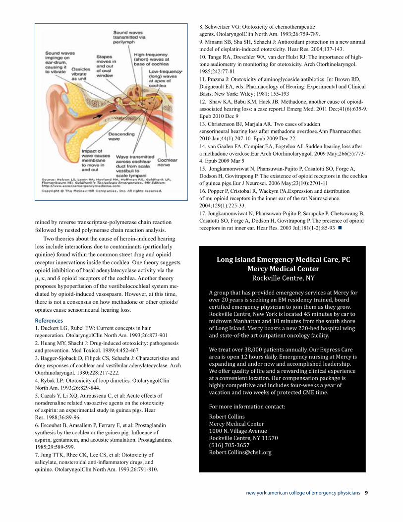
Occasionally our toxicology service has encountered patients who awaken after recovering from their opiate overdose with difficulty hearing. Dr. Amit Gupta has reviewed the literature on the cause of this puzzling scenario.
Ototoxicity includes effects on the cochlear and vestibular system. Ototoxic xenobiotics primarily affect two different sites in the cochlea: the organ of Corti—specifically the outer hair cells—and the striavascularis (see figure). Because of the limited regenerative capacity of the sensory hair cells and other supporting cells, when significant cellular damage occurs, the loss is often prolonged or permanent.1,2
Drugs that cause toxicity at the strisvascu-laris include:I. Loop diuretics (furosemide,
bumetanide, ethacrynic acid) cause edema which is reversible. The underlying mechanisms appear to be the inhibition of potassium pumps and G proteins associated with adenylcyclase. This decreased potassium activity in the endolymph leads to a decreased endocochlear potential.3,4
II. NSAIDS and ASA inhibit cyclooxygenase, which converts arachidonic acid to prostaglandin G2 and prostaglandin H2. These effects interfere with Na+-K+-ATPase pump function at the striavascularis, and also decrease cochlear blood flow.5,6
III. Quinine’s mechanism of action is thought to be similar to that of NSAIDS via prostaglandin inhibition.7
Drugs that cause toxicity at the hair cells include:I. Certain antineoplastics, such
as cisplatin, vinblastine, and vincristine, can cause permanent ototoxicity. Cisplatin is the most
toxic of the group, with clinically apparent hearing loss noted in 30%–70% of the patients receiving doses of 50–100 mg/m2. These antineoplastics typically damage the outer hair cells by the formation of oxygen free radicals.8,9 The generation of oxygen free radicals and the depletion of antioxidants result in the irreversible damage to the hair cells.
II. The aminoglycosides are best known for their association with irreversible ototoxicity. Several mechanisms of ototoxicity have been postulated including antagonism of calcium channels of the outer hair cells of the cochlea, blocking transduction of the hair cells and resulting in acute, reversible hearing deficits as well as binding to polyphosphoinositides of cell membranes to alter their functions.10,11 Polyphosphoinositides are essential for the generation of the second messengers’ diacylglycerol and inositol triphosphate and their ultimate cellular function, for the maintenance of lipid membrane structure and permeability, and as a source for arachidonic acid. Aminoglycosides interact with iron and copper to generate free radicals, damaging the hair cells.
Several case reports have been published describing methadone induced hearing loss.12,13,14 Unfortunately none of them have been able to confirm the exact mechanism of otoxicity from methadone.
Several animal models have been able to demonstrate that opioid receptors are present in the middle ear.15,16,17
Jongkamonwiwat N, et al identified and localized the mu (MOR), delta (DOR) and kappa (KOR) opioid receptor subtypes within the rat cochlea. The expression of these opioid receptor subtypes was deter-
Patients Who Awaken After Recovering from their Opiate Overdose with Difficulty Hearing Guest author: Amit Gupta, MD, Staten Island University Hospital; Assistant Professor of Emergency Medicine, Downstate University
new york american college of emergency physicians 9
mined by reverse transcriptase-polymerase chain reaction followed by nested polymerase chain reaction analysis.
Two theories about the cause of heroin-induced hearing loss include interactions due to contaminants (particularly quinine) found within the common street drug and opioid receptor innervations inside the cochlea. One theory suggests opioid inhibition of basal adenylatecyclase activity via the µ, κ, and δ opioid receptors of the cochlea. Another theory proposes hypoperfusion of the vestibulocochleal system me-diated by opioid-induced vasospasm. However, at this time, there is not a consensus on how methadone or other opioids/opiates cause sensorineural hearing loss.
References1. Duckert LG, Rubel EW: Current concepts in hair regeneration. OtolaryngolClin North Am. 1993;26:873-9012. Huang MY, Shacht J: Drug-induced ototoxicity: pathogenesis and prevention. Med Toxicol. 1989;4:452-4673. Bagger-Sjoback D, Filipek CS, Schacht J: Characteristics and drug responses of cochlear and vestibular adenylatecyclase. Arch Otorhinolaryngol. 1980;228:217-222.4. Rybak LP: Ototoxicity of loop diuretics. OtolaryngolClin North Am. 1993;26:829-844.5. Cazals Y, Li XQ, Aurousseau C, et al: Acute effects of noradrenaline related vasoactive agents on the ototoxicity of aspirin: an experimental study in guinea pigs. Hear Res. 1988;36:89-96.6. Escoubet B, Amsallem P, Ferrary E, et al: Prostaglandin synthesis by the cochlea or the guinea pig. Influence of aspirin, gentamicin, and acoustic stimulation. Prostaglandins. 1985;29:589-599. 7. Jung TTK, Rhee CK, Lee CS, et al: Ototoxicity of salicylate, nonsteroidal anti-inflammatory drugs, and quinine. OtolaryngolClin North Am. 1993;26:791-810.
8. Schweitzer VG: Ototoxicity of chemotherapeutic agents. OtolaryngolClin North Am. 1993;26:759-789. 9. Minami SB, Sha SH, Schacht J: Antioxidant protection in a new animal model of cisplatin-induced ototoxicity. Hear Res. 2004;137-143.10. Tange RA, Dreschler WA, van der Hulst RJ: The importance of high-tone audiometry in monitoring for ototoxicity. Arch Otorhinolaryngol. 1985;242:77-8111. Prazma J: Ototoxicity of aminoglycoside antibiotics. In: Brown RD, Daigneault EA, eds: Pharmacology of Hearing: Experimental and Clinical Basis. New York: Wiley; 1981: 155-19312. Shaw KA, Babu KM, Hack JB. Methadone, another cause of opioid-associated hearing loss: a case report.J Emerg Med. 2011 Dec;41(6):635-9. Epub 2010 Dec 913. Christenson BJ, Marjala AR. Two cases of sudden sensorineural hearing loss after methadone overdose.Ann Pharmacother. 2010 Jan;44(1):207-10. Epub 2009 Dec 2214. van Gaalen FA, Compier EA, Fogteloo AJ. Sudden hearing loss after a methadone overdose.Eur Arch Otorhinolaryngol. 2009 May;266(5):773-4. Epub 2009 Mar 515. Jongkamonwiwat N, Phansuwan-Pujito P, Casalotti SO, Forge A, Dodson H, Govitrapong P. The existence of opioid receptors in the cochlea of guinea pigs.Eur J Neurosci. 2006 May;23(10):2701-1116. Popper P, Cristobal R, Wackym PA.Expression and distribution of mu opioid receptors in the inner ear of the rat.Neuroscience. 2004;129(1):225-33.17. Jongkamonwiwat N, Phansuwan-Pujito P, Sarapoke P, Chetsawang B, Casalotti SO, Forge A, Dodson H, Govitrapong P. The presence of opioid receptors in rat inner ear. Hear Res. 2003 Jul;181(1-2):85-93
Long Island Emergency Medical Care, PCMercy Medical Center
Rockville Centre, NY
A group that has provided emergency services at Mercy for over 20 years is seeking an EM residency trained, board certified emergency physician to join them as they grow.Rockville Centre, New York is located 45 minutes by car to midtown Manhattan and 10 minutes from the south shore of Long Island. Mercy boasts a new 220-bed hospital wing and state-of-the art outpatient oncology facility.
We treat over 38,000 patients annually. Our Express Care area is open 12 hours daily. Emergency nursing at Mercy is expanding and under new and accomplished leadership. We offer quality of life and a rewarding clinical experience at a convenient location. Our compensation package is highly competitive and includes four-weeks a year of vacation and two weeks of protected CME time.
For more information contact:
Robert CollinsMercy Medical Center1000 N. Village AvenueRockville Centre, NY 11570(516) [email protected]

10 empire state epic vol. 30:02:12
Albany UpdateWeingarten, Reid & McNally, New York ACEP Legislative & Regulatory Representatives
prescriber consultation for all Schedule II, III and IV controlled substances with some limited exemptions. It also requires all pre-scriptions to be transmitted electronically by December 31, 2014, updates the State’s controlled substance schedules, expands the duties and membership of the work-group established under the Prescription Pain Medication Awareness Program, and requires the New York State Department of Health to establish a safe drug disposal program.
Changes to Observation Services in Hospitals (A10518-A Gottfried/ S7031-A Hannon)
Governor Cuomo signed this legislation October 3, 2012. A “Chapter Amendment” bill to further revise the law is expected to pass in 2013.
This new law revises recently enacted New York State Department of Health regulations related to observation services in hospitals. New York ACEP strongly supported and championed the New York State Department of Health regulations to require hospitals to set up separate observation units supervised by emergency physicians. Unfortunately, the regulation was strongly opposed by the state hospital associations and they were successful in getting legislation introduced and passed this session to eliminate many of the re-quirements in the regulation.
New York ACEP strongly opposed this legislation through a series of meet-ings, issuing a memo to the full Legislature and a number of action alerts requesting member action. When meeting with the bill sponsors, we requested amendments to the bill to ensure that observation services are in a discrete, separate unit and are emer-gency physician directed. Despite all of the New York ACEP’s efforts, the final bill did not include these amendments.
After the bill passed both houses of the Legislature, New York ACEP had a lengthy meeting with key health policy advisors to Governor Cuomo and the New York State Department of Health. During the meet-ing, New York ACEP demonstrated that observation units supervised by emergency physicians are critical to improving patient care and reducing health care costs.
New York ACEP issued a letter to the Governor in opposition to the bill and asked the membership to weigh in with their own individual letters. Unfortunately, Governor Cuomo signed the bill. He stated in his approval message that “hospitals should have the flexibility in the provision of observation services, as long as those services meet the clinical needs of the patient and are appropriately overseen.”
Governor Cuomo also noted in his approval message: “I believe that the dura-tion of such services is a matter for regu-lation by the Department, to be exercised with the objective of achieving alignment with Medicare requirements. The sponsors have agreed to enact additional legislation to that effect.” The “additional legisla-tion” referred to in the approval message is expected to address a provision in the new law which allows the provision of observa-tion services for up to 48 hours, instead of 24 hours as required in the New York State Department of Health regulations. We will provide New York ACEP with additional information as it becomes available.
Affordable Care Act (ACA) Update
New York State Health Benefit ExchangeOn April 12, 2012, Governor Cuomo
created, by Executive Order, New York’s Health Benefit Exchange. Since that time, the State has been working to establish the Exchange including appointing an Exchange Board, establishing Regional Advisory Committees, hiring staff, creating
Governor Andrew Cuomo sent his annual “Budget Call” letter to state agency heads in late September instructing them to submit “no growth” spending plans for the 2013-14 proposed State Budget. Cuomo told reporters that this direction is neces-sary because the State faces a $1 billion shortfall next year. As a result, budget negotiations will dominate activity for the first three months of the 2013 Legislative Session.
Final action on New York ACEP 2012 session priorities is summarized below. In addition, this article provides an update on State actions to implement the federal Af-fordable Care Act (ACA) and an overview of issues for the 2013 Legislative Session.
Legislative Update
Prescription Drug Reform (I-STOP) Legislation (S7637 Lanza/ A10623 Cusick)
Governor Cuomo signed this legislation August 27, 2012, Chapter 447 of the Laws of 2012.
During the 2012 Legislative Session, New York ACEP worked very hard with Governor Cuomo, Attorney General Schneiderman, and State legislators to make emergency medicine a part of the solution to the serious controlled substance abuse and diversion problem in New York State. The effort was focused on doing so in a reasonable way that did not overburden the State’s hospital emergency departments and that protected access to pain and other medications for patients who legitimately need them. New York ACEP was successful in doing both. New York ACEP was able to get inserted into the final bill an exemption for 5-day prescriptions written in emergency departments.
The new law, Chapter 447 of the Laws of 2012, changes the State Prescription Monitoring Program to require more fre-quent pharmacy reporting and health care

new york american college of emergency physicians 11
the online platform, identifying the State’s benchmark plan for essential health benefits and a number of other activities in order to show that it has made significant progress in the establishment of its Exchange by January 1, 2013, as required by ACA.
Essential Health Benefits (EHBs), Benchmark Plan
On October 1, 2012, New York State formally submitted its’ selection of an Essential Health Benefits benchmark plan to the US Department of Health and Human Services (HHS). This plan defines the essential health benefits for the individual and small group health insurance markets under the State Health Benefit Exchange beginning in 2014. New York selected the State’s largest small group plan, Oxford EPO, as the benchmark plan.
Oxford EPO was selected from among 10 possible plans authorized under federal law: the three largest federal employee plans; the three largest state employee plans; the three largest commercial small group products; and the largest commercial group HMO offered in each state.
The State contracted with Milliman to provide a report (Essential Health Benefits for the New York Health Benefits Exchange) that outlines the covered benefits under Oxford EPO. The report shows that emergency services are a covered benefit as mandated by federal law. We will continue to closely monitor the implementation of New York’s Health Benefit Exchange and the Essential Health Benefits package and keep you updated as additional information is provided and new developments arise.
To see the full text of the Milliman report go to: http://www.healthcarereform.ny.gov/health_insurance_exchange/ and click on the link to the Milliman report.
2013 Legislative SessionAs noted earlier, the State is facing a
$1 billion State Budget deficit next year so the economy will continue to occupy center stage. We expect a renewed effort by Governor Cuomo’s Department of Financial Services to reach an agreement on rules effecting insurance companies and health care providers for medical bills for “out-of-network” providers.
All of us at Weingarten, Reid & McNally will continue to work on behalf of New York ACEP to ensure patient access to the highest quality emergency services in New York State.
New York American College of Emergency Physicians1130 Crosspointe Lane, Suite 10BWebster, NY 14580-2986(585) 872-2417 phone (585) 872-2419 faxwww.nyacep.org online
EMPIRE STATE EPIC is the newsletter of the New York American College of Emergency Physicians (New York ACEP). The opinions expressed in this newsletter are not necessarily those of New York ACEP. New York ACEP makes a good faith effort to ascertain that contributors are experts in their field. Readers are advised that the statements and opinions expressed by the author are those of the author and New York ACEP does not accept responsibility for information or statements made by contributing authors.
NEWS STAFF . JoAnne Tarantelli, Executive DirectorBetsy Hawes, Director of Marketing & Communications
© 2012
Apollo MDEmCare, Inc.
Emergency Medical AssociatesEmergency Medicine Physicians
Good Samaritan Hospital Medical CenterIndiana Emergency Care
Island Medical Physicians, PCMedExcel USA, Inc.
Mercy Medical Center & LIEMC, PCNES HealthCare Group
North Shore-LIJ Health SystemPhoenix Physicians, LLC
Physician Affiliate Group of New YorkPremier Physician Services
Rochester General Health SystemSchumacher Group
TEAMHealthUpstate New York Physician Recruiters
Winthrop University Hospital
Special thanks to our November 7 Emergency
Medicine Residents Career Day
Supporters

12 empire state epic vol. 30:02:12

new york american college of emergency physicians 13
New York State of Mind
Gaeta
Interstitial Ectopic Pregnancy Presenting After Failed Termination of Pregnancy.Egan DJ, Li M, Lewiss RE., Department of Emergency Medicine, St Luke’s-Roosevelt Hospital Center; Emerg Med Australas. 2012 Oct;24(5):573-6.
Pregnant women frequently present to the ED for complaints related to the first trimester of pregnancy. The emergency physician must confirm the presence of an intrauterine pregnancy for many such complaints. Bedside ultrasound with well-delineated criteria has become standard practice for many emergency physicians for this purpose. In the following case report, an interstitial pregnancy was identified by the emergency physician using bedside sonography in a 29-year-old woman pre-senting 2 weeks after dilation and aspira-tion for termination of pregnancy. The ED physician identified an inappropriately thin endomyometrial mantle raising suspicion for the diagnosis of an interstitial preg-nancy. The case illustrates the importance of this uterine wall measurement given the otherwise normal appearance of a preg-nancy within the uterus.
Mastoiditis and Meningitis Complicating an Aural Foreign Body.Burke RT, Gatton B, Melville LD, Department of Emergency Medicine, New
York Methodist Hospital, Brooklyn; Pediatr Emerg Care. 2012 Oct;28(10):1070-1.
ABSTRACT: Children commonly present to emergency departments with foreign bodies in the ear. In addition, physicians place wicks in the ear canal as part of the treatment of otitis externa. Usually, these foreign bodies are easily removed, but occasionally, removal must be deferred or is delayed by parents. Therefore, the dangers of retained foreign bodies are important for the emergency physician to be aware of. We report the highly unusual case of a 12-year-old girl who presented with ear pain for 3 weeks. She was found to have an ear wick in place as part of the treatment of otalgia. She was subsequently diagnosed with mastoiditis and meningitis. This is first time mastoiditis and meningitis has been reported as a complication of ear wick placement, although not the only case of an intracranial complication of an aural foreign body.
Treatment of Guanfacine Toxicity with Naloxone.Tsze DS, Dayan PS, Division of Pediatric Emergency Medicine, Columbia University College of Physicians and Surgeons, New York; Pediatr Emerg Care. 2012 Oct;28(10):1060-1.
ABSTRACT: We describe a 4-year-old boy who presents to the emergency depart-ment with lethargy, bradycardia, and initial hypertension followed by hypotension due to guanfacine toxicity after ingestion of standard doses of the extended release formulation. This is the first case report to describe the use of naloxone to treat these
symptoms and document improvements in level of consciousness, blood pressure, and heart rate associated with this therapy.
A Review of Acetaminophen Poisoning.Hodgman MJ, Garrard AR, Department of Emergency Medicine, Upstate New York Poison Center, SUNY Upstate Medical University, Syracuse; Crit Care Clin. 2012 Oct;28(4):499-516.
Acetaminophen poisoning remains one of the more common drugs taken in overdose with potentially fatal consequences. Early recognition and prompt treatment with N-acetylcysteine can prevent hepatic injury. With acute overdose, the Rumack-Matthew nomogram is a useful tool to assess risk and guide management. Equally common to acute overdose is the repeated use of excessive amounts of acetaminophen. Simultaneous ingestion of several different acetaminophen-containing products may result in excessive dosage. These patients also benefit from N-acetylcysteine. Standard courses of N-acetylcysteine may need to be extended in patients with persistently elevated plasma concentrations of acetaminophen or with signs of hepatic injury.
Acute Superior Vena Cava Syndrome after Rupture of Kommerell’s Diverticulum: A Case Report.Suarez AE, Slivka R, Department of Emergency Medicine, New York Hospital Queens, Flushing, NY; and Weill Cornell Medical College, New York; Ann Emerg Med. 2012 Sep 13. [Epub ahead of print].
Kommerell’s diverticulum, a rare con-genital aortic anomaly, is dilatation at the region in which an aberrant subclavian artery branches from either a left-sided or right-sided thoracic aorta. We report a rare case of acute superior vena cava syndrome that developed in a young healthy male patient who presented to the emergency department in imminent respiratory arrest after rupture of this diverticulum.
Is Nonperforated Pediatric Appendicitis Still Considered a Surgical Emergency? A Survey of Pediatric Surgeons.Dunlop JC, Meltzer JA, Silver EJ, Crain EF, Department of Pediatrics, Division of Emergency Medicine, Jacobi Medical Center, Bronx; Acad Pediatr. 2012 Sep 12. [Epub ahead of print].
This column is compiled by Theodore J. Gaeta, DO MPH FACEP, Residency Program Director at New York Methodist Hospital; and Member, New York ACEP Research Committee.

14 empire state epic vol. 30:02:12
New York State of Mind
OBJECTIVE: To describe the beliefs and preferences of pediatric surgeons regard-ing the emergent nature of nonperforated appendicitis. METHODS: An electronic mailing was sent to all 1,052 members of the American Pediatric Surgical Association (APSA) inviting participation in a 26-item survey, which was administered by Survey Monkey (www.surveymonkey.com). Chi-square and Mann-Whitney tests were used for bivari-ate analysis. Spearman’s rho was used for nonparametric correlation. RESULTS: Four hundred eighty-four pediatric surgeons (46%) responded to the survey. Few respondents (4%) considered nonperforated appendicitis to be a surgical emergency. A minority (14%) would come in from home to perform an overnight appendectomy. Most (92%) believe that postponing overnight appendectomy until daytime does not result in a clinically significant increase in perforation. Respon-dents endorsed surgeon fatigue (56%) and limited operating room availability (56%) most often among factors that would make them more likely to postpone surgery. Sixty-eight percent reported no departmen-tal guideline regarding delay of overnight appendectomy. CONCLUSION: Most pediatric surgeons in our study believe nonperforated appendi-citis is not a surgical emergency and prefer to postpone overnight appendectomy.
Victims of Bullying in the Emergency Department with Behavioral Issues.Waseem M, Arshad A, Leber M, Perales O, Jara F, Department of Emergency Medicine, Lincoln Medical & Mental Health Center, Bronx; J Emerg Med. 2012 Sep 11. [Epub ahead of print].
BACKGROUND: Bullying has become one of the most significant school problems experienced by our children. Victims of bullying are prone to a variety of psycho-logical and behavioral symptoms. We noted that many children referred to the Emer-gency Department (ED) with behavioral symptoms provided a history of bullying.
OBJECTIVES: To measure the preva-lence of bullying in children referred to the ED for behavioral symptoms and to determine its association with psychiatric disorders. METHODS: A retrospective cohort study was conducted in an urban hospital, iden-tifying children from 8 to 19 years of age who presented to the ED with behavioral symptoms. We reviewed the ED psychia-try notes to retrieve the report indicating whether these children were bullied and had previous psychiatric diagnoses. These children were classified into bullied and non-bullied groups. RESULTS: Over the study period, 591 children visited the ED with behavioral issues. Out of 591, 143 (24%) children reported bullying. More boys (100) than girls (43) reported bullying (p = 0.034). The mean age of children in the bullied group was 10.6 years (95% confidence interval 10.1-11.2). One hundred eleven (77.6%) children in the bullied group had a prior psychiatric diagnosis. Children in the bullied group were hospitalized signifi-cantly less than children in the non-bullied group (10/143 [7%] vs. 80/368 [18%]; p = 0.002).CONCLUSION: The prevalence of bullying among the ED children with behavioral symptoms is substantial. Every fourth child with behavioral symptoms re-ported bullying. Four in five children who reported bullying had a prior diagnosis of “disorder of behavior.”
Wheezing and Asthma are Independent Risk Factors for Increased Sickle Cell Disease Morbidity.Glassberg JA, Chow A, Wisnivesky J, Hoffman R, Debaun MR, Richardson LD, Department of Emergency Medicine, Mount Sinai School of Medicine, New York; Br J Haematol. 2012 Sep 12. [Epub ahead of print].
To assess the associations between a doctor diagnosis of asthma and wheezing (independent of a diagnosis of asthma) with sickle cell disease (SCD) morbid-ity, we conducted a retrospective review of Emergency Department (ED) visits to
the Mount Sinai Medical Center for SCD between 1 January 2007 and 1 January 2011. Outcomes were ED visits for pain and acute chest syndrome. The cohort included 262 individuals, median age 23·8 years, (range: 6 months to 67·5 years). At least one episode of wheezing recorded on a physical examination was present in 18·7% (49 of 262). Asthma and wheezing did not overlap completely, 53·1% of patients with wheezing did not carry a diagnosis of asthma. Wheezing was associated with a 118% increase in ED visits for pain (95% confidence interval [CI]: 56-205%) and a 158% increase in ED visits for acute chest syndrome (95% CI: 11-498%). A diagno-sis of asthma was associated with a 44% increase in ED utilization for pain (95% CI: 2-104%) and no increase in ED utilization for acute chest syndrome (rate ratio 1·00, 95% CI 0·41-2·47). In conclusion, asthma and wheezing are independent risk factors for increased painful episodes in individuals with SCD. Only wheezing was associated with more acute chest syndrome.
Dynamic Anatomic Relationship of the Esophagus and Trachea On Sonography: Implications for Endotracheal Tube Confirmation in Children.Tsung JW, Fenster D, Kessler DO, Novik J, Department of Emergency Medicine, Mount Sinai School of Medicine, New York; J Ultrasound Med. 2012 Sep;31(9):1365-70.
OBJECTIVES: Sonographic visualization of an empty esophagus to confirm endo-tracheal tube placement during intubation may be more reliable than identifying an endotracheal tube within the trachea. Our objective was to determine the frequency in which the normal empty esophagus can be identified at or below the level of the cricoid ring in children. METHODS: A prospective cohort of children and young adults presenting to the emergency department were examined by sonography to determine the dynamic anatomic relationship of the trachea and esophagus at or below the level of the cricoid ring. For children with the esopha-gus behind or partially behind the trachea, cricoid pressure was applied using a linear array transducer to visualize the presence of lateral sliding of the esophagus from behind the trachea. RESULTS: A total of 55 patients 21 years or younger were examined; 51% (28) were male. Sixty-two percent (34) had esophagi

new york american college of emergency physicians 15
positioned partially to the left of the cricoid ring, 20% (11) completely to the left of the cricoid ring, 16% (9) behind the cricoid ring, and 2% (1) partially to the right of the cricoid ring. When cricoid pressure was applied using the ultrasound transducer, the esophagus was visualized lateral to the trachea in all patients (54 to the left and 1 to the right; n = 55 of 55; 95% confidence interval, 94%-100%). CONCLUSION: With cricoid pressure applied using a linear transducer, the esophagus was visualized lateral to the trachea in all children and young adults. Visualizing an empty esophagus by point-of-care sonography may be feasible to confirm endotracheal tube placement by a process of elimination.
Improving Front-End Flow in an Urban Academic Medical Center Emergency Department: The Emergency Department Discharge Facilitator Team.Sharma R, Mulcare MR, Graetz R, Greenwald PW, Mustalish AC, Miluszusky B, Flomenbaum NE, Department of Emergency Medicine, New York-Presbyterian Hospital, New York; J Urban Health. 2012 Aug 9. [Epub ahead of print].
Length of stay (LOS) is an important de-terminant of patient satisfaction and overall emergency department (ED) operational
efficiency. In an effort to reduce LOS for low-acuity “treated and released” patients, our department created a discharge facilita-tor team (DFT) composed of an attending physician, physician assistant, and regis-tered nurse. The DFT identified patients who could be rapidly treated and released in the low-acuity treatment Adult Urgent Care Center (AUCC) and provided them rapid treatment and discharge. To assess the efficacy of the DFT, linear regression was used to compare AUCC LOS at times the team was and was not active. Patients seen by the DFT had a LOS that was 35% shorter than other AUCC patients. There was a 28-min reduction in AUCC LOS during periods where the DFT was active (95% CI 22 to 33 min). We conclude that the establishment of a DFT was associated with a significant reduction in LOS for all low-acuity patients. Other academic medi-cal centers may consider implementing a similar program in order to reduce LOS and improve ED throughput for low acuity patients.
Emergency Department Diagnosis of Upper Extremity Deep Venous Thrombosis Using Bedside Ultrasonography.Rosen T, Chang B, Kaufman M, Soderman M, Riley DC, Emergency Medicine Department, Columbia University Medical
Center, New York; Crit Ultrasound J. 2012 Apr 16;4(1):4.
ABSTRACT: A 27-year-old man presents to the emergency department with a 1-day history of severe right upper extremity pain and swelling. The patient’s status is post open reduction internal fixation for a left tibial plateau fracture, which was compli-cated by methicillin-sensitive Staphylo-coccus aureus osteomyelitis. A peripher-ally inserted central catheter (PICC) line was subsequently placed for intravenous antibiotic therapy. Emergency depart-ment bedside ultrasound examination of both the right axillary vein and subclavian vein near the PICC line tip revealed deep venous thrombosis of both veins. Bedside upper extremity vascular ultrasonography can assist in the rapid diagnosis of upper extremity deep venous thrombosis in the emergency department.
Emergency Department Ultrasonography Guided Long-Axis Antecubital Intravenous Cannulation: How To Do It.Riley DC, Garcia S, Emergency Medicine Department, Columbia University Medical Center, New York; Crit Ultrasound J. 2012 Apr 16;4(1):3.
ABSTRACT: An 85-year-old woman with a past medical history of severe peripheral

16 empire state epic vol. 30:02:12
vascular disease and right below knee amputation presented to the emergency department with a 1-day history of non-positional dizziness and weakness. The patient required intravenous access to work up her dizziness and weakness. The patient had multiple failed blind ED peripheral IV attempts performed in the past. Emergency department bedside ultrasonography with a high frequency linear array vascular probe was used to guide antecubital brachial vein cannulation on the first attempt using the long-axis approach.
Derivation of a Pediatric Growth Curve for Inferior Vena Caval Diameter in Healthy Pediatric Patients: Brief Report of Initial Curve Development.Haines EJ, Chiricolo GC, Aralica K, Briggs WM, Van Amerongen R, Laudenbach A, O’Rourke K, Melniker L, Department of Emergency Medicine, New York Methodist Hospital, Brooklyn; Crit Ultrasound J. 2012 May 28;4(1):12.
BACKGROUND: A validated tool has long been sought to provide clinicians with a uniform and accurate method to assess hydration status in the pediatric emergency medicine population. Outpatient clinicians use CDC height- and weight-based curves for the assessment of physical develop-ment. In hospital, daily weights provide objective data; however, these are usually not available at presentation. One of the most promising techniques for the rapid assessment of volume is ultrasound (US) to obtain an indexed inferior vena cava diameter (IVCDi); as previously described. Prior studies have focused on IVCDi in dehydrated patients and have shown that it provides accurate estimates of right atrial pressure and volume status. The objective of this study is to derive an IVC growth curve in healthy pediatric patients.METHODS: Prospective cohort design enrolled healthy children between the ages of 4 weeks and 20 years. Patients presenting with fever, illnesses, or diag-noses known to affect the volume will be excluded. All eligible patients under 21, who have provided self or parental written
consent, will undergo a brief ultrasound to obtain transverse and long images of both the IVC and the aorta; all scans will be digitally saved. Image quality will be sub-jectively rated as poor, fair, or good based on wall clarity. Poor quality images will be recorded but may be omitted from our analysis. Five clinicians completed a 1-h introduction to IVC-US and ten supervised scans prior to enrollment. Still images will be measured in order to determine IVCDi in both transverse and longitudinal planes. To assess inter-rater reliability, in 10% of cases, two clinicians will complete scans. All study scans will be over-read by a fel-lowship-trained sonologist. IVCDi will be plotted independently as functions of age, gender, BMI, and aortic diameter. Within each group, means with means or medians with 95% CIs will be calculated. Following uni- and bivariate analyses and assessment for colinearity, a variety of parametric and nonparametric regression procedures will be conducted. The smoothed curves will be approximated using a modified LMS estimation procedure.RESULTS: Data for the initial curve deri-vation includes 25 patients ranging from 13 months to 20 years (mean 102 months or 8.5 years). Sixty-five percent of patients were enrolled from the ED, while 35% were enrolled from well-child clinic visits. When evaluating the size of IVC as a func-tion of time linear growth, increasing size was found to proportionately increase with age of patient in months.CONCLUSION: Data suggest a linear correlation between IVC size and age. Such data, when plotted as a new growth curve, may allow clinicians to plot a patient’s so-nographic measurements in order to assess hydration health.
Prospective Application of Clinician-Performed Lung Ultrasonography During the 2009 H1N1 Influenza A Pandemic: Distinguishing Viral from Bacterial Pneumonia.Tsung JW, Kessler DO, Shah VP., Division of Pediatric Emergency Medicine, Departments of Pediatrics and Emergency
Medicine, Bellevue Hospital Center/NYU School of Medicine, New York; Crit Ultrasound J. 2012 Jul 10;4(1):16.
BACKGROUND: Emergency department visits quadrupled with the initial onset and surge during the 2009 H1N1 influenza pandemic in New York City from April to June 2009. This time period was unique in that >90% of the circulating virus was surveyed to be the novel 2009 H1N1 influenza A according to the New York City Department of Health. We describe our experience using lung ultrasound in a case series of patients with respiratory symptoms requiring chest X-ray during the initial onset and surge of the 2009 H1N1 influenza pandemic.METHODS: We describe a case series of patients from a prospective observational cohort study of lung ultrasound, enrolling patients requiring chest X-ray for suspected pneumonia that coincided with the onset and surge of the 2009 H1N1 influenza pandemic.RESULTS: Twenty pandemic 2009 H1N1 influenza patients requiring chest X-ray were enrolled during this time period. Median age was 6.7 years. Lung ultrasound via modified Bedside Lung Ultrasound in Emergency protocol assisted in the identification of viral pneumonia (n = 15; 75%), viral pneumonia with superimposed bacterial pneumonia (n = 7; 35%), isolated bacterial pneumonia only (n = 1; 5%), and no findings of viral or bacterial pneumonia (n = 4; 20%) in this cohort of patients. Based on 54 observations, interobserver agreement for distinguishing viral from bacterial pneumonia using lung ultrasound was ĸ = 0.82 (0.63 to 0.99).CONCLUSION: Lung ultrasound may be used to distinguish viral from bacterial pneumonia. Lung ultrasound may be useful during epidemics or pandemics of acute respiratory illnesses for rapid point-of-care triage and management of patients.
Intranasal Ketamine for Procedural Sedation in Pediatric Laceration Repair: A Preliminary Report.Tsze DS, Steele DW, Machan JT, Akhlaghi F, Linakis JG., Division of Pediatric Emergency Medicine, Department of Pediatrics, Columbia University College of Physicians and Surgeons, New York; Pediatr Emerg Care. 2012 Aug;28(8):767-70.
New York State of Mind

new york american college of emergency physicians 17
OBJECTIVE: The objective of this study was to compare the efficacy of 3 doses of intranasal ketamine (INK) for sedation of children from 1 to 7 years old requiring laceration repair.METHODS: This was a randomized, prospective, double-blind trial of children requiring sedation for laceration repair.Patients with simple lacerations were randomized by age to receive 3, 6, or 9 mg/kg INK. Adequacy and efficacy of sedation were measured with the Ramsay sedation score and the Observational Scale of Be-havioral Distress-Revised. Serum ketamine and norketamine levels were drawn during the procedure. Sedation duration and ad-verse events were recorded.RESULTS: Of the 12 patients enrolled, 3 patients achieved adequate sedation, all at the 9-mg/kg dose. The study was sus-pended at that time as per predetermined criteria.CONCLUSION: Nine milligrams of INK per kilogram produced a significantly higher proportion of successful sedations than the 3- and 6-mg/kg doses.
Identifying Adolescent Females at High Risk of Pregnancy in a Pediatric Emergency Department.Chernick L, Kharbanda EO, Santelli J, Dayan P., Division of Pediatric Emergency Medicine, New York-Presbyterian Morgan Stanley, Children’s Hospital, New York, New York, USA; J Adolesc Health. 2012 Aug;51(2):171-8.
PURPOSE: Emergency departments (EDs) care for adolescent females with unmet reproductive health care needs. Our objec-tive was, among adolescents presenting to a pediatric ED, to estimate pregnancy risk, describe pregnancy intentions, and identify potentially modifiable factors associated with pregnancy risk.METHODS: Using a paper-based questionnaire, we surveyed females aged 15-19 years presenting to our ED, assess-ing health care access, sexual behaviors, pregnancy intentions, and receptivity to interventions. We calculated the pregnancy risk index (PRI), which estimates preg-nancy risk in the subsequent 12 months, by assessing recent sexual activity, contra-ception at last intercourse, and known contraceptive failure rates. Independent sample t tests and analysis of variance were used to identify risk factors associated with increased PRI.
RESULTS: Of 459 females enrolled, 13% were pregnant and 20% reported prior pregnancy. Among 399 nonpregnant females, 238 (60%) had intercourse in the prior 3 months and 73 (31%) used no contraception at last intercourse. Among nonpregnant adolescents, the PRI was 19.5, which equates to 19.5 expected pregnancies per 100 females per year. Factors associ-ated with higher PRI included lacking a primary provider, prior ED visits, wanting a baby now, and reported partner wantedness of pregnancy. Half believed ED doctors should discuss pregnancy prevention, and one-quarter were interested in starting contraception in the ED.CONCLUSION: Nearly one-third of adolescent females in a pediatric ED were either pregnant or could be expected to become pregnant within a year. Screening questions can identify adolescents at high risk of pregnancy in the ED setting. These females should be the target for future pregnancy prevention interventions.
Triage Vital Signs Do Not Correlate with Serum Lactate or Base Deficit, and Are Less Predictive of Operative Intervention in Penetrating Trauma Patients: A Prospective Cohort Study.Caputo N, Fraser R, Paliga A, Kanter M, Hosford K, Madlinger R., Department of Emergency Medicine, Lincoln Medical and Mental Health Center, Bronx; Emerg Med J. 2012 Jul 16. [Epub ahead of print].
BACKGROUND: Triage vital signs are often used to help determine a trauma patient’s haemodynamic status. Recent studies have demonstrated that these may not be very specific in determining major injury. The purpose of this study was to determine if there is any correlation between triage vital signs, base deficit (BD) and lactate, and to determine the odds of operative intervention in penetrating trauma patients.METHODS: A prospective observational cohort study was undertaken. Baseline vital signs, BD and lactate were recorded in all patients for whom the trauma team was ac-tivated. Pearson correlation and coefficient (ρ) were calculated. ORs were calculated.RESULTS: 75 patients were enrolled. Pearson correlations and coefficients calcu-lated for lactate to systolic blood pressure were: -0.052 (ρ=0.0011, 95% CI -0.225 to 0.228); lactate and HR: 0.23 (ρ=0.0166, 95% CI -0.211 to 0.242); lactate and RR:
0.23 (ρ=0.054, 95% CI -0.174 to 0.277). BD to systolic blood pressure were: 0.003 (ρ=0.00001, 95% CI -0.229 to 0.224); BD and HR: -0.19 (ρ=0.038, 95% CI -0.399 to 0.038); BD and RR: -0.019 (ρ=0.0004, 95% CI -0.244 to 0.208). Odds of operative intervention were greater in patients with abnormally high lactate, OR 4.17 (95% CI 1.57 to 11), but not for BD, OR 2.53 (95% CI 0.99 to 6.45), or any of the vital signs.CONCLUSION: Triage vital signs have no correlation to lactate or BD levels in penetrating trauma patients. Odds of opera tive intervention are greater in patients with abnormally high serum lactate levels, but not in those with abnormal triage vital signs or BD.
The Quality of Cardiopulmonary Resuscitation Using Supraglottic Airways and Intraosseous Devices: A Simulation Trial.Reiter DA, Strother CG, Weingart SD., Department of Emergency Medicine, Mount Sinai School of Medicine, New York; Resuscitation. 2012 Jul 13. [Epub ahead of print].
STUDY OBJECTIVE: To assess whether using interventions such as laryngeal mask airways (LMA) and IO lines lead to improved resuscitation in a simulated cardiac arrest when compared to standard methods of endotracheal intubation (ETI) and central line placement. METHODS: Emergency Medicine residents at a single academic center were grouped into teams of four. Each team participated in two simulated ventricular fibrillation cardiac arrests using a high fidelity simulator. Peripheral IV access was unobtainable. Only ETI supplies and a central line kit were available in one case (control) and in the other case those supplies were replaced by an LMA and an EZ-IO drill kit (experimental). Groups were randomized to which set up they were given first. Data examined included time to airway placement, duration and success rate of airway placement, time to vascular access, time to defibrillation, and percent hands off time.
RESULTS: 44 residents in 11 teams participated. Mean time to airway was shorter in the experimental group (122.8 seconds (s) vs. 265.6s, p=0.001). Mean duration of airway attempt was also shorter (7.6s vs. 22.7s, p=0.002). Time to access was shorter in the experimental group

18 empire state epic vol. 30:02:12
(49.0s vs. 194.6s, p=<0.001). Time to defibrillation and percent hands off time did not significantly differ between the two groups. CONCLUSION: Use of an LMA and an IO device led to significantly faster estab-lishment of an airway and vascular access in a simulated cardiac arrest. The variation in devices did not affect time to defibrilla-tion or percent hands off time.
Hospital Administrators’ Views on Barriers and Opportunities to Delivering Palliative Care in the Emergency Department.Grudzen CR, Richardson LD, Major-Monfried H, Kandarian B, Ortiz JM, Morrison RS, Department of Emergency Medicine, Mount Sinai School of Medicine, New York; Ann Emerg Med. 2012 Jul 6. [Epub ahead of print].
STUDY OBJECTIVE: We identify hospital-level factors from the administra-tive perspective that affect the availability and delivery of palliative care services in the emergency department (ED). METHODS: Semistructured interviews were conducted with 14 key informants, including hospital executives, ED directors, and palliative care directors at a tertiary care center, a public hospital, and a com-munity hospital. The discussions were digi-tally recorded and transcribed to conduct a thematic analysis using grounded theory. A coding scheme was iteratively developed to subsequently identify themes and sub-themes that emerged from the interviews. RESULTS: Barriers to integrating pallia-tive care and emergency medicine from the administrative perspective include the ED culture of aggressive care, limited knowledge, palliative care staffing, and medicolegal concerns. Incentives to the delivery of palliative care in the ED from these key informants’ perspective include improved patient and family satisfaction, opportunities to provide meaningful care to patients, decreased costs of care for admit-ted patients, and avoidance of unnecessary admissions to more intensive hospital settings, such as the ICU, for patients who have little likelihood of benefit.
CONCLUSION: Though hospital ad-ministration at 3 urban hospitals on the East coast has great interest in integrating palliative care and emergency medicine to improve quality of care, patient and family satisfaction, and decrease length of stay for admitted patients, palliative care staffing, medicolegal concerns, and logistic issues need to be addressed.
Validation of a Pre-Existing Formula to Calculate the Contribution of Ethanol to the Osmolar Gap.Garrard A, Sollee DR, Butterfield RC, Johannsen L, Wood A, Bertholf RL, Department of Emergency Medicine, Upstate New York Poison Center at Upstate Medical University, Syracuse; Clin Toxicol (Phila). 2012 Aug;50(7):562-6. Epub 2012 Jul 6.
PURPOSE: The aim of this study was to validate the formula derived by Purssell et al. that relates blood ethanol concentra-tion to the osmolar gap and determine the best coefficient for use in the formula. The osmolar gap is often used to help diagnose toxic alcohol poisoning when direct mea-surements are not available.METHODOLOGY: Part I of the study consisted of a retrospective review of 603 emergency department patients who had a concurrent ethanol, basic metabolic panel and a serum osmolality results avail-able. Estimated osmolarity (excluding ethanol) was calculated using a standard formula. The osmolar gap was determined by subtracting estimated osmolarity from the actual osmolality measured by freez-ing point depression. The relationship between the osmolar gap and the measured ethanol concentration was assessed by linear regression analysis. In Part II of this study, predetermined amounts of ethanol were added to aliquots of plasma and the estimated and calculated osmolarities were subjected to linear regression analysis.RESULTS: In the cases of 603 patients included in Part I of the study, the median ethanol concentration in these patients was 166 mg/dL (Q1: 90, Q3: 254) and the range ethanol concentrations was 10-644 mg/
dL. The mean serum osmolality was 338 mOsm kg (SD: 30) and a range of 244-450 mOsm/kg. The mean osmolar gap was 47 (SD: 29) and a range of - 15 to 55. There was a significant proportional relation-ship between ethanol concentration and osmolar gap (r(2) = 0.9882). The slope of the linear regression line was 0.2498 (95% CI: 0.2472-0.2524). The slope of the linear regression line derived from the data in Part II of the study was 0.2445 (95% CI: 0.2410-0.2480).CONCLUSION: The results of our study are in fairly close agreement with previous studies that used smaller samples and suggest that an accurate conversion factor for estimating the contribution of ethanol to the osmolar gap is [Ethanol (mg/dL)]/4.0.
Systematic Review: Is Real-Time Ultrasonic-Guided Central Line Placement by ED Physicians More Successful than the Traditional Landmark Approach?Mehta N, Valesky WW, Guy A, Sinert R, Department of Emergency Medicine, Kings County Hospital/State University of New York Downstate Medical Center, Brooklyn; Emerg Med J. 2012 Jun 26. [Epub ahead of print].
INTRODUCTION: The superiority of ultrasonic-guided compared with land-mark-guided central venous catheter (CVC) placement is not well documented in the Emergency Department. OBJECTIVE: To systematically review the literature comparing success rates between ultrasonic- and landmark-guided CVC placement by ED physicians.METHODS: PubMed and EMBASE databases were searched for randomised controlled trials from 1965 to 2010 using a search strategy derived from the follow-ing PICO formulation: Patients: Adults requiring emergent CVC placement except during cardiopulmonary resuscitation.INTERVENTION: CVC placement us-ing real-time ultrasonic guidance. Com-parator: CVC placement using anatomical landmarks.OUTCOME: Comparison of success rates of CVC placement between ultrasonic- ver-sus landmark-guided techniques. ANALYSIS: Success rates between CVC placement methods using a Forest Plot (95% CI) calculated by Review Manager Version 5.0.
New York State of Mind

new york american college of emergency physicians 19
RESULTS: Search identified 944 articles of which 938 were excluded by title/ab-stract relevance, two not randomised, one cardiac arrest, one no landmark control, one success rate not calculated. A single study of 130 patients (65 ultrasonic- vs 65 landmark-guided) selected for internal jugular vein placement remained. Success-ful internal jugular CVC was significantly (p=0.02) more likely in the ultrasound-guided (93.9%) compared with landmark-guided (78.5%) techniques with an OR of 1.2 (95% CI 1.0 to 1.4). Complications rates were significantly (p=0.04) lower in ultrasonic (4.6%) versus landmark (16.9%) technique, OR=3.7 (95% CI 1.1 to 12.5).CONCLUSION: Only one single high quality study illustrating that ED ultra-sound- versus landmark-guided internal jugular catheter placement had higher success rates with lower complication rates.
A Sedentary Job? Measuring the Physical Activity of Emergency Medicine Residents.Josephson EB, Caputo ND, Pedraza S, Reynolds T, Sharifi R, Waseem M, Kornberg RJ, Department of Emergency Medicine, Lincoln Medical and Mental Health Center, Bronx; J Emerg Med. 2012 Jun 23. [Epub ahead of print].
BACKGROUND: The debate on the quality of health care provided in the United States has continued to be waged as concerns have grown over the years. Stress, sleep deprivation, poor diet, and lack of exercise may lead to inadequate work performance by physicians. OBJECTIVE: This study was undertaken to determine whether Emergency Medicine (EM) residents satisfy daily recommenda-tions for total number of steps taken per day set forth by the Centers for Disease Control and Prevention and Surgeon Gen-eral in a 12-h shift. METHODS: An observational prospec-tive cohort study was conducted between August 2009 and November 2009 at an urban Level I trauma center with an an-nual census of over 165,000 Emergency Department (ED) visits per year. The mean number of steps taken by EM residents dur-ing 12-h shifts was measured. RESULTS: Mean steps taken during a shift were 7333 (95% confidence interval 6901-7764). Only nine (9.9%) pedometer readings reached the target level of 10,000 (10K) steps or above. A t-test was used to
compare steps with the hypothesized 10K steps target. Recordings of 10K steps or greater were not correlated with ED sec-tions (p=0.60) shift (medical vs. surgical, p=0.65) or ED census (r(2)<0.0017). CONCLUSION: A majority of residents (90%) did not meet the target number of steps for shifts. More rigorous chart-ing needs, overcrowding, or even spatial limitations may explain this. This warrants further investigation to determine if some daily physical activity regimens may help improve the overall well-being of EM residents.
Risk Factors for Increased ED Utilization in a Multinational Cohort of Children with Sickle Cell Disease.Glassberg JA, Wang J, Cohen R, Richardson LD, DeBaun MR, Department of Emergency Medicine, Mount Sinai School of Medicine, New York; Acad Emerg Med. 2012 Jun;19(6):664-72.
OBJECTIVES: The objective was to iden-tify clinical, social, and environmental risk factors for increased emergency depart-ment (ED) use in children with sickle cell disease (SCD).METHODS: This study was a secondary analysis of ED utilization data from the international multicenter Silent Cerebral Infarct Transfusion (SIT) trial. Between December 2004 and June 2010, baseline demographic, clinical, and laboratory data were collected from children with SCD participating in the trial. The primary outcome was the frequency of ED visits for pain. A secondary outcome was the frequency of ED visits for acute chest syndrome.RESULTS: The sample included 985 children from the United States, Canada, England, and France, for a total of 2,955 patient-years of data. There were 0.74 ED visits for pain per patient-year. A past medical history of asthma was associated with an increased risk of ED utilization for both pain (rate ratio [RR] = 1.28, 95% confidence interval [CI] = 1.04 to 1.58) and acute chest syndrome (RR = 1.60, 95% CI = 1.03 to 2.49). Exposure to environmental tobacco smoke in the home was associated with 73% more ED visits for acute chest syndrome (RR = 1.73, 95% CI = 1.09 to 2.74). Each $10,000 increase in household income was associated with 5% fewer ED visits for pain (RR = 0.95, 95% CI = 0.91 to 1.00, p = 0.05). The association between
low income and ED utilization was not significantly different in the United States versus countries with universal health care (p = 0.51).CONCLUSION: Asthma and exposure to environmental tobacco smoke are poten-tially modifiable risk factors for greater ED use in children with SCD. Low income is associated with greater ED use for SCD pain in countries with and without univer-sal health care.
Electroencephalographic Findings in Consecutive Emergency Department Patients with Altered Mental Status: A Preliminary Report.Zehtabchi S, Abdel Baki SG, Grant AC, Department of Emergency Medicine, Downstate Medical Center & Kings County Hospital Center, Brooklyn; Eur J Emerg Med. 2012 May 24. [Epub ahead of print].
Electroencephalography (EEG) can help narrow the differential diagnosis of altered mental status (AMS) and is necessary to diagnose nonconvulsive seizure (NCS). The objective of this prospective observa-tional study is to identify the prevalence of EEG abnormalities in emergency depart-ment patients with AMS. Patients of at least 13 years of age with AMS were enrolled, whereas those with an easily identifiable cause (e.g. hypoglycemia) underlying their AMS were excluded. Easily identifiable cause of AMS (e.g. hypoglycemia). A 30-min EEG with the standard 19 electrodes was performed on each patient. Descriptive statistics (%, 95% confidence interval) are used to report EEG findings of the first 50 enrolled patients. Thirty-five EEGs (70%, 57-81%) were abnormal. The most com-mon abnormality was slowing of back-ground activities (46%, 33-60%), reflecting an underlying encephalopathy. NCS was diagnosed in three (6%, 1-17%), includ-ing one patient in nonconvulsive status epilepticus. Nine patients (18%, 10-31%) had interictal epileptiform abnormalities, indicating an increased risk of spontaneous seizure. Patients presenting to the emer-gency department with AMS have a high prevalence of EEG abnormalities, includ-ing NCS.
Case Series of 64 Slice Computed Tomography-Computed Tomographic Angiography with 3D Reconstruction to Diagnose Symptomatic Cerebral Aneurysms: New Standard of Care?

20 empire state epic vol. 30:02:12
Jehle D, Chae F, Wai J, Cloud S, Pierce D, Meyer M, State University of New York at Buffalo; Neurol Int. 2012 Jan 9;4(1):e2. Epub 2012 Feb 23.
CT angiography (CTA) has improved sig-nificantly over the past few years such that the reconstructed images of the cerebral arteries may now be equivalent to conven-tional digital angiography. The new tech-nology of 64 slice multi-detector CTA can reconstruct detailed images that can reliably identify small cerebral aneurysms, even those <3mm. In addition, it is estimated that CT followed by lumbar puncture (LP) miss-es up to 4% of symptomatic aneurysms. We present a series of cases that illustrates how CT followed by CTA may be replacing CT-LP as the standard of care in work-ing up patients for symptomatic cerebral aneurysms and the importance of perform-ing three dimensional (3D) reconstructions. A series of seven cases of symptomatic cerebral aneurysms were identified that illustrate the sensitivity of CT-CTA versus CT-LP and the importance of 3D recon-struction in identifying these aneurysms. Surgical treatment was recommended for 6 of the 7 patients with aneurysms and strict hypertension control was recommended for the seventh patient. Some of these patients demonstrated subarachnoid hemorrhage on presentation while others had negative LPs. A number of these patients with negative LPs were clearly symptomatic from their aneurysms. At least one of these cerebral aneurysms was not apparent on CTA without 3D reconstruction. 3D reconstruc-tion of CTA is crucial to adequately identify cerebral aneurysms. This case series helps reinforce the importance of 3D reconstruc-tion. There is some data to suggest that 64 slice CT-CTA may be equivalent or superior to CT-LP in the detection of symptomatic cerebral aneurysms.
Comparison of Sitting Face-to-Face Intubation (Two-Person Technique) with Standard Oral-tracheal Intubation in Novices: A Mannequin Study.Venezia D, Wackett A, Remedios A, Tarsia V, Department of Emergency Medicine, Stony Brook Medical Center, Stony Brook; J Emerg
Med. 2012 May 3. [Epub ahead of print].
BACKGROUND: Few studies have evalu-ated the impact of the upright position on the success of oral-tracheal intubation. Yet, for patients with airway difficulties (i.e, ac-tive intraoral bleeding or morbidly obese), the upright position may both benefit the patient and facilitate intubation. OBJECTIVES: We compared the suc-cess rates of subjects performing standard intubation to a modified version of the sitting face-to-face oral-tracheal intubation technique on a simulation model. We also reviewed the possible advantages and limi-tations of the sitting face-to-face intubation technique. METHODS: Volunteer medical and para-medical students were given instruction, then tested, performing in random order both standard oral-tracheal and two-person sitting face-to-face oral-tracheal intuba-tion on full-bodied mannequins. Observ-ers reviewed video recordings, noting the number of successful intubations and the time to completion of each procedure at 15, 20, and 30 s. RESULTS: All of the sitting face-to-face intubations were successful, 53/53 (100%, 95% confidence interval [CI] 93-100%); whereas of the 53 subjects who performed standard intubation, 48 were successful (91%, 95% CI 80-96%). The difference between successful intubations using the standard vs. sitting face-to-face technique was 9% (95% CI 1.3-9.4%, p=0.025). At times 15 and 20 s, medical student subjects who successfully performed both tech-niques were less successful at completing the procedure when performing the stan-dard technique as compared to the sitting face-to-face technique (p=0.016). A post-procedural survey found that the majority of subjects preferred the sitting technique. CONCLUSION: Subjects were signifi-cantly more successful at performing and preferred the sitting face-to-face intubation when compared to standard intubation.
A Descriptive Comparison of Alcohol-Related Presentations at a Large Urban Hospital Center from 1902 to 2009.
Shy BD, Hoffman RS, Department of Emergency Medicine, Mount Sinai School of Medicine, New York; J Med Toxicol. 2012 Sep;8(3):271-7.
Although alcohol use has long been a significant cause of hospital presentations, little is published regarding the long-term demographic changes that have occurred at a single hospital site. To address this deficit, we prospectively studied all acute alcohol-related presentations to Bellevue Hospital Center (New York, NY) and compared this contemporary data set with one from the same institution from 1902 to 1935. We prospectively identified all patients presenting to the emergency de-partment because of acute alcohol use over an 8-week period in 2009. We described the basic attributes of patients presenting currently because of alcohol and compared these data to those previously described between 1902 and 1935. We also compared our census data with contemporaneous data from all patients presenting to this hospital site. During the study period, 560 patients presented because of acute alcohol use which extrapolated to an estimated 3,800 patients over the calendar year. This com-pares to 7,600 presentations recorded annu-ally early in the twentieth century. Twelve percent of patients in 2009 were female as compared to 18 % of patients between 1934 and 1935. Patients with alcohol-related presentations in 2009 were more likely to be admitted than contemporaneous patients without an alcohol-related presentation (30 vs. 19 % admitted; p < 0.001). Since first measured 110 years ago at one large New York City hospital, alcohol-related presen-tations remain common representing 5 % of all emergency department visits. This demonstrates alcoholism’s continuing toll on society’s limited medical resources and on public health as a whole.
Characteristics of Emergency Department Patients Who Receive a Palliative Care Consultation.Grudzen CR, Hwang U, Cohen JA, Fischman M, Morrison RS., Department of Emergency Medicine, Mount Sinai School of Medicine, New York; J Palliat Med. 2012 Apr;15(4):396-9. Epub 2012 Apr 2.
BACKGROUND: A large gap exists between the practice of emergency medi-cine and palliative care. Although hospice and palliative medicine has recently been recognized as a subspecialty of emergency
New York State of Mind

new york american college of emergency physicians 21
medicine, few palliative care teams rou-tinely interact with emergency providers, and primary palliative care skills among emergency providers are lacking.OBJECTIVE: To identify the proportion and characteristics of patients who receive a palliative care consultation and arrive via the emergency department (ED). METHODS: A descriptive study of adult ED patients from an urban, academic tertiary care hospital who received a pal-liative care consultation in January 2005 or January 2009.RESULTS: In January 2005, 100 of the 161 consults (62%) arrived via the ED versus 63 of 124 consults (51%) in January 2009 (p=0.06). Mean days from admission to consultation in January 2005 were six days (standard deviation 11), versus nine days (SD 26) in January 2009 (p=0.35). Three of the 100 consultations (3%) in January 2005 were initiated in the ED, versus 4 of the 64 (6%) in January 2009.CONCLUSIONS: At an urban academic medical center with a well-developed palliative care service, the majority of pal-liative care consultations were for patients who arrive via the ED. Despite this, only a small minority of consultations originated from emergency providers and consulta-tion was on average initiated days into a patient’s hospital stay.
Prevalence of Occult Anemia in an Urban Pediatric Emergency Department: What is Our Response?Kristinsson G, Shtivelman S, Hom J, Tunik MG., Departments of Pediatrics and Emergency Medicine, New York University School of Medicine, New York; Pediatr Emerg Care. 2012 Apr;28(4):313-5.
OBJECTIVE: Treating or referring patients who are found to be anemic during pediatric emergency department (ED) encounters should lead to improved health in children and young adults. Before establishing guidelines how to approach the anemic in the pediatric ED, it is essential to determine the prevalence of anemia in the ED and our response to the presence of anemia.METHODS: We performed a retrospec-tive cross-sectional study on hemoglobin levels from patients 1 to 23 years evaluated in an inner-city public hospital pediatric ED during a 12-month period. The primary outcome measure was the prevalence of
prior unknown or “occult” anemia, strati-fied by age, sex, and insurance status. The secondary outcome was the proportion of patients with “occult” anemia who had their condition diagnosed during their ED encounter. Descriptive data analysis was performed.RESULTS: Our study population consisted of 2131 patients who had a complete blood count drawn in the ED. Prevalence of “oc-cult” anemia was 13.9% (95% confidence interval [CI], 12.5%-15.4%). Proportions among the subpopulations were 14.8% (95% CI, 10.0%-19.5%) in preschool children, 16.3% (95% CI, 14.2%-18.3%) in females, 18.5% (95% CI, 15.4%-21.7%) in the uninsured, and 20.7% (95% CI, 16.5%-24.9%) in females of childbearing age without insurance. Only 24 patients (8%) with “occult” anemia had the condition identified on discharge.CONCLUSIONS: Anemia has a high prevalence in this pediatric ED population, especially among females of childbearing age and the uninsured. Pediatric emergency medicine physicians are missing on an op-portunity to address a common health prob-lem that is easily corrected with appropriate therapy and outpatient follow-up.
Inter-Rater Reliability of Sonographic Measurements of the Inferior Vena Cava.Saul T, Lewiss RE, Langsfeld A, Radeos MS, Del Rios M, Department of Emergency Medicine, Emergency Ultrasound Division, St. Luke’s/Roosevelt Hospital Center, New York; J Emerg Med. 2012 May;42(5):600-5. Epub 2012 Jan 12.
BACKGROUND: Bedside ultrasound is emerging as a useful tool in the assessment of intravascular volume status by examin-ing measurements of the inferior vena cava (IVC). Many previous studies do not fully describe their scanning protocol.OBJECTIVES: The objective of this study was to evaluate which of three commonly reported IVC scanning methods demon-strates the best inter-rater reliability.METHODS: Three physicians visual-ized the IVC in three common views and utilized M-mode to measure the maximal and minimal diameter during quiet respira-tion. Pairwise correlation coefficients were determined using Pearson product-moment correlation.RESULTS: The most reliable pair of mea-surements (inspiratory and expiratory) was
found to be using the anterior midaxillary line longitudinal view with a Kappa value for both at 0.692.CONCLUSION: Imaging with the anterior midaxillary longitudinal approach using the liver as an acoustic window provides the best inter-rater reliability when measur-ing the IVC. Our findings demonstrate that IVC measurements differ based on anatomic location.
Lipemic Serum in a Toddler with New-Onset Diabetes Mellitus Presenting with Diabetic Ketoacidosis.Waseem M, Dave-Sharma S, Kin LL, Jara F, Department of Emergency Medicine, Lincoln Medical and Mental Health Center, Bronx; JOP. 2012 Jan 10;13(1):73-5.
CONTEXT: Significant hyperlipidemia causing lipemic serum in patients with poorly controlled diabetes is under-reported in children. The recognition of the severe hyperlipidemia is important for proper management and to prevent associated morbidities. Severe hyperlipidemia in patients with diabetic ketoacidosis should be considered.CASE REPORT: In this case we report a 2-year-old girl with new onset type 1 dia-betes mellitus, who presented with severe diabetic ketoacidosis and hyperlipidemia. Hyperlipidemia was resolved with hydra-tion and insulin therapy.CONCLUSION: It is important to diagnose hyperlipidemia by checking serum lipid profile for all pediatric patients presenting with hyperglycemic crisis to prevent morbidities.
Novel Serum and Urine Markers for Pediatric Appendicitis.Kharbanda AB, Rai AJ, Cosme Y, Liu K, Dayan PS., Division of Pediatric Emergency Medicine, Columbia University College of Physicians and Surgeons, New York; Acad Emerg Med. 2012 Jan;19(1):56-62.
OBJECTIVES: The objective was to describe the association between two novel biomarkers, calprotectin and leucine-rich alpha glycoprotein-1 (LRG), and appendi-citis in children.METHODS: This was a prospective, cross-sectional study of children 3 to 18 years old presenting to a pediatric emer-gency department (ED) with possible appendicitis. Blood and urine samples were assayed for calprotectin and LRG

22 empire state epic vol. 30:02:12
via enzyme-linked immunosorbent assay (ELISA). Final diagnosis was determined by histopathology or telephone follow-up. Biomarker levels were compared for subjects with and without appendicitis. Recursive partitioning was used to iden-tify thresholds that predicted appendicitis.RESULTS: Of 176 subjects, mean (±SD) age was 11.6 (±4.0) years and 52% were male. Fifty-eight patients (34%) were diagnosed with appendicitis. Median plasma calprotectin, serum LRG, and urine LRG levels were higher in appendi-citis versus nonappendicitis (p < 0.008). When stratified by perforation status, median plasma calprotectin and serum LRG levels were higher in nonperforated appendicitis versus nonappendicitis (p < 0.01). Median serum LRG, urine LRG, and plasma calprotectin levels were high-er in perforated appendicitis compared to nonperforated appendicitis (p < 0.05). Urine calprotectin did not differ among groups. A serum LRG < 40,150 ng/mL, a urine LRG < 42 ng/mL, and a plasma calprotectin < 159 ng/mL, each provided a sensitivity and negative predictive value of 100% to identify children at low risk for appendicitis, but with specificities ranging from 23% to 35%. The standard white blood cell (WBC) count achieved 100% sensitivity at a higher specificity than both novel biomarkers.CONCLUSION: Plasma calprotectin and serum/urine LRG are elevated in pediatric appendicitis. No individual marker performed as well as the WBC count.
Have you ever had a teacher, resident, or employer have such a dramatic influence on you or your career that you have never forgotten them? Maybe it was just someone with more experience than you who ‘showed you the ropes’ some time during your medical school or residency training or when you began a new job. As medical providers, we have unique opportunities of being mentors and preceptors for future generations of physicians and mid-level providers.
A mentor is a person who provides guidance and support and enhances the professional development of their junior colleagues.
Mentoring is sharing one’s knowledge, skills, information and perspective in order to foster the personal and professional growth of someone else.
This action is not only beneficial for the mentee, but can also be very rewarding for the physician who provides this guidance.
Surprisingly, even though it is known that mentoring is an essential component to career development for all medical professionals, less than 40% of medical students reported having a mentor. Other studies have shown mentoring prevalence at institutions is highly variable and can be as low as 20% in some places. Because of this it is recommended that a student or new physician actively seek out a mentor, rather than just expect it will occur automatically. If you are unable to find one at your institution New York ACEP may be of assistance in helping you find one. Mentoring can occur for
an individual or group and even by a mentor from a different hospital. All these methods have been shown to be valuable.
Benefits of MentoringMentorship may be perceived as a
burden to some busy providers, but it takes very little time and effort. It has many potential benefits for the mentor. It gives you an opportunity to share your knowledge and experience and you receive satisfaction witnessing the growth and successes of the mentee.
Studies have shown that medical faculty who were mentored have higher career satisfaction scores and were 2.3 times more likely to achieve academic promotions.
The power of mentoring is that it creates an opportunity for collaboration, goal achievement and problem-solving.
Mentoring starts by sharing your knowledge and experience with someone that is starting out on the road that you once travelled. In addition to your successes, it is very important to reveal problems and mistakes that you have encountered during you career, as it is likely that the mentee will face similar instances during their career.
Mentoring improves clinical practice for all involved. It develops leadership qualities in someone who has yet to develop these. By allowing the mentee to network with folks that you already know, you accelerate the development of their own networks.
continued on next page 24
Under Your WingKevin P. O’Connor MD FACEP, Chairman,
Department of Emergency Medicine, Arnot Ogden Medical Center
In our continuing effort to foster community and academic research, New York ACEP’s Research Committee is here to serve as a resource for your research projects. For inquiries on how to get started, email [email protected].

new york american college of emergency physicians 23

24 empire state epic vol. 30:02:12
Under Your Wing continued from page 22
Mentoring new physicians is also a wonderful retention strategy. Studies have shown that junior faculty were only half as likely to leave their organization if they had been mentored. As many young physicians tend to change jobs two to three times during their early careers, it is important to get them feeling comfortable and connected early on.
It can also prevent many problems associated with new physicians starting out in an established group practice. For example, newly trained emergency department physicians of-ten desire a work-life balance that more established physicians have seen as less important when they started their careers. Mentoring allows sharing of these different philosophies and creates a better understanding and expectations of each other.
Mentoring provides some teaching as well. There are tricks of the trade that experienced physicians can share with their mentored physicians that they might not have received during training that will improve their clinical skills. They also can teach them the so called ‘art of medicine’. An example might be how to interact with particular physicians on the medical staff. This can be very frustrating for a new physician who has yet to experience this. Likewise, the mentor might learn that some of his/her knowledge or skills are antiquated and have been replaced with newer ideas.
Qualities of a Good MentorAlthough there are probably mentors for everyone,
everyone cannot be a good mentor to all. There needs to be some form of compatibility between mentor and mentee. For example, for some people, there may be some distinct advan-tages to being mentored by someone of same sex or ethnic background. Good mentors should be active listeners, have the ability to clearly identify a person’s strengths and good at helping people refine their goals. They should have good lis-tening, communication and leadership skills. A mentor should be motivational, supportive and be a good problem solver. It is important to provide positive and constructive feedback. Although formal training on mentoring is desirable, it is not mandatory for success of this relationship.
A mentor should help a mentee set realistic goals. These goals should be specific, time-framed, results oriented, relevant, and reachable. He/she should foster independence and not simply answer questions and solve the problems for the mentee. They should communicate regularly with your mentee. Usually at least once a month is recommended. If possible, this should be done in a location with little distrac-tions.
Mentors should listen to the needs and expectations of the mentee. If the mentee’s is having serious problems they should be supportive, yet know their boundaries. As a mentor, you are not expected to be their therapist.
Benefits and Tips for Mentee Mentees often will have to seek out a mentor. Don’t be
afraid to ask someone you admire and trust to become your mentor. Mentees can expect to see an increase in awareness of
professional issues and a rapid growth in their sense of competence, identity and effectiveness as a professional. They will improve their understanding of work issues and discover different approaches to dealing with them. You may have talents and interests revealed to you that you did not know existed. For example, you may find yourself joining a New York ACEP committee or find you are great with lobbying government officials. The mentee should clearly commu-nicate their goals and fears to their mentor. Both parties should also respect the basic privacy and confidentiality within this relationship. The mentee should express their appreciation for the mentor’s time, information, counsel, and sharing of opportunities. They should have a sincere interest in developing a personal and professional relation-ship with a mentor, and be committed to fostering that relationship by making regular contact with him or her. They should accept feedback and suggestions from the mentor. They should explore all opportuni-ties presented by the mentor.
In conclusion, mentorship is an important determinant of success-ful professional careers. Emergency physicians have many opportuni-ties to become involved in this activity, which holds rewards for both them and their protégés. The key ingredient to a successful mentoring relationship is a genuine commitment from both the mentor and the mentee. Mentoring leads to greater retention of staff members and to greater career satisfaction and success of each mentee.
Most important, mentored doctors are more likely to take some-one from the next generation of physicians under their wings. So, what are you waiting for?
Emergency Medicine. The Department of Emergency Medicine at Albany Medical College is recruiting for emergency medicine faculty. The Department currently has 28 faculty members and 36 residents per year in a fully accredited emergency medicine residency program. The Department hosts a variety of subspecialty areas including ultrasound, toxicology, pediatrics, EMS and critical care. We are seeking individuals with excellent clinical skills, a strong interest in teaching residents and medical students, and a willingness to contribute to research.
Albany Medical College is part of the Albany Medical Center, northeastern New York’s only academic health sciences center, which also includes the 651-bed Albany Medical Center Hospital, one of upstate New York’s largest teaching hospitals. The hospital is currently expanding with construction of a 150-bed, $360 million patient pavilion now underway. The institution’s Emergency Department, which has an annual census of over 70,000 patients, serves as the region’s only Level One Trauma Center and also features an integrated Pediatric ED. In addition, the Department hosts the region’s only hospital based helicopter service.
BC/BE candidates should send curriculum vitae with a letter of interest to:
Christopher King, MD, FACEPChair, Department of Emergency MedicineAlbany Medical College47 New Scotland AvenueAlbany, New York 12208Phone: (518) 262-3443

new york american college of emergency physicians 25

26 empire state epic vol. 30:02:12
A seven-month-old presents with sev-eral episodes of non bilious vomiting. The parents deny diarrhea, fevers, or lethargy. No obvious pain or severe crying episodes. Normal wet diapers. Physical exam reveals a well appearing male infant who is afebrile. The lungs and abdomen are benign, and there is no physical evidence of dehydra-tion. The baby passes a PO challenge, and is discharged home with instructions to follow up with their pediatrician. Diagnosis: Gastroenteritis. The father fails to tell you that he “briefly” shook the baby to stop him from crying.
A six-month-old girl arrives via EMS with labored breathing. She has a mild cough. There are no fevers. Parents are con-cerned because she is “breathing fast.” She looks well on exam and is afebrile. Respira-tory rate is slightly elevated, but the oxygen saturation is normal. Lungs are clear. Chest X-ray reveals no pneumonia. The infant receives a nebulizer, improves, and is sent home. Diagnosis: Bronchiolitis. The treating physicians do not notice two posterior rib fractures on the chest X-ray.
A father brings in his two-month-old girl for a nosebleed. He states he saw some blood coming out of the nose yesterday, and then a little more today. No fevers or bruising. Vitals and physical exam are normal. Labs including CBC and coags are sent off and return normal. The father is a little vague about the history, because he works a lot and the babysitter does most of the caretaking. The infant is discharged with instructions to follow up with her pediatrician. Diagnosis: Nosebleed, resolved. A very small bruise on the back of the scalp and neck go unnoticed.
A four-month-old presents with an episode of “coughing then turning blue.” The mother states that the baby was drinking from a bottle, started to cough, appeared to have choked and turned blue and limp for a short period of time. They were admitted for a similar episode three weeks ago. Physical exam reveals a well appearing infant who is awake and alert. Vitals normal. The only abnormality is a small bruise on the cheek. The mother explains she tried to do CPR when her son turned blue, and may have grabbed too hard on his face. The male infant
is admitted to the hospital for an ALTE. Work up is negative, and he is discharged home with a diagnosis of reflux. He will be abused again, and next time will arrive as a respiratory arrest. We missed our opportunity, twice, to save this baby.
A five-month-old male presents in respira-tory distress. He arrives limp and grey. He is grunting. He has a bulging anterior fontanel with multiple bruises on the face and chest. He has a swollen arm and leg. He subsequently starts to seize. An IO line is placed. Bedside glucose is 30mg/dl. He is treated with benzos and glucose. He is immediately intubated. Emergent head CT reveals SDH, frontal contusions, and a skull fracture. You review his medical records and find out he was seen in your ED three weeks ago. He was diagnosed with nonspecific bruising after “a fall” from the parents’ arms.
IntroductionFive infants with five different presentations.
All with the same diagnosis--- Abusive Head Trauma (AHT). Over one million children suffer from some type of abuse annually in the United States.1 Many children will present with obvious injuries, while other abused children, especially infants, may present with common complaints. They pass through our emergency departments with life threatening injuries that often go un-detected. This article focuses on Abusive Head Trauma (AHT) in our most vulnerable popula-tion, children less then one year of age.
Frightening Facts: CDC Data for Child Maltreatment
According to the CDC website,1 Child Protective Services received over three million reports of potential child abuse in 2010. Ap-proximately 1,560 children died from some form of abuse, with the vast majority (nearly 80%) in the 0-4 year age group. The highest incidence occurred in children less than one year of age. That means that four children die every day from abuse. Four unnecessary deaths. Every day.
AHT is the most common cause of deaths from child abuse in the pediatric population.
Landmark Article: Analysis of Missed Cases of Abusive Head Trauma
In 1999, Dr. Jenny and colleagues published “Analysis of Missed Cases of Abusive Head Trauma.”2 This article is a must read for all emergency medicine residents. These authors looked at children less then three years of age
Abusive Head Trauma in Infants: Pearls and Pitfallsfeature column
emergencies in the pediatric
patient
Denis R. Pauze MD FACEP FAAP, Associate Professor of Emergency
Medicine and Pediatrics, Vice Chair, Department of Emergency
Medicine at Albany Medical College, brings this feature
column to the EPIC.
Co-authored by Daniel K. Pauze MD FACEP,
Assistant Professor of Emergency Medicine, Medical Director, Department of Emergency
Medicine at Albany Medical College
Featuring topics pertaining to pediatric emergency medicine ~ challenging cases, literature
updates and interesting pediatric illnesses.
new york american college of emergency physicians 27
who had a diagnosis of AHT. They sub-sequently reviewed their medical records and asked, “did these children see a health care provider prior to their presentation of AHT?” Their results were very surprising. They found that 31% of children visited a physician prior to being diagnosed with AHT. Nearly one-third (54 children) with unrecognized AHT. Nearly three visits until the diagnosis was made. They presented with common complaints. Erroneous dis-charge diagnoses included gastroenteritis, accidental head injury, rule out sepsis, otitis media, seizure disorder, reflux, colic, vomit-ing unknown cause, anxiety, and myositis.
This must read article goes on to highlight several additional points: 1. Radiographic errors led to the delay in
diagnosis of several patients; 2. Nearly 30% of infants were re-injured; 3. Four deaths may have been preventable
with earlier recognition; 4. In younger infants, the diagnosis is
much more likely to be missed; 5. Abnormal respiratory rate, seizures,
facial and/or scalp injury, and parents not living together were factors that predicted AHT on the first visit.2
A complete review of this publication is beyond the scope of this article. It is very much worth the read when you have a chance.
Making the DiagnosisAHT has many different presentations.
Some presentations are dramatic—seizures, lethargy, limpness, irritability, respiratory distress or apnea. These striking presenta-tions are not a diagnostic challenge. How-ever, some infants with inflicted brain injury may not have such alarming presentations. They arrive in your emergency department looking well. They may come in with a common complaint: vomiting, ear infection, fever, respiratory symptoms, crying, or not eating. They can have a completely normal physical exam and a normal neurological exam. As there are no biomarkers for abuse, laboratory work is not conclusive. We can’t CT or X-ray every infant who presents with vomiting or respiratory complaints. But the diagnosis is vitally important — up to a third of these children will be abused again. The emergency medicine physician/provider must rely on a detailed History and Physical Exam (H&P) and have a high level of alertness for this elusive and challenging diagnosis.
H&PThe history can be challenging in
these patients; they are non-verbal and the caregiver may be unaware of someone else harming the child. Even worse, the caregiver may lie or mislead the clinician. So the history becomes vitally important. Be alert for red flags. Was there a delay in seeking care? Is there an injury that is not plausible with the developmental milestones of the baby? Does the history change, or is it too vague? Are there signs of trauma (bruising, swelling, fracture, etc.) and no history or explanation of trauma? Did this child have a previous CPS case (investiga-tion, injury, etc).
The physical exam should be thorough with a detailed head to toe exam looking for any signs of trauma. Does the infant have any bruises to the head, scalp, face, or neck? What does the fontanelle feel like? Is there evidence of intra oral trauma? Is the frenu-lum intact? Is there swelling or bruises over the neck, chest, abdomen or extremities? Non ambulatory infants, especially those under six months of age, should rarely (if ever) have any bruises on their body. Facial or neck bruises in this age group should be a red flag for abuse.
Retinal hemorrhages are highly sugges-tive (but not pathognomonic) for abuse. Any infant with potential abuse, especially when AHT is in the differential, should have their retinas examined by an ophthalmologist.
Special Circumstances: Bruising, ALTEs, Nasal Bleeding and Seizures
Bruising: Another landmark article for Child Abuse was published by Dr. Sugar and colleagues.3 They looked at bruises in children less than three years of age. They found that bruises were rarely found in the non ambulatory infant. In children less then nine months of age, only 1.7% infants were found to have bruises. They concluded that “Those who don’t cruise, rarely bruise.” Bruises in non ambulatory infants should be further evaluated for abuse or a medical condition.
ALTEs: In a 2010 study by Guenther et al, 1.4% (9/627) of all infants presenting with ALTE were ultimately found to have an abusive etiology.4 Other studies have placed this number much higher.5 A prior history of ALTE may provide clues to the diagnosis. Ultimately, AHT has to be considered in all infants who present with ALTE, even in the well appearing child.
OroNasal bleeding: Is uncommon in children less than one year of age.6 Keep abuse in your differential when an infant presents with bleeding from the nose or mouth.
Seizures: First time seizures in an infant could be the initial manifestation of in-flicted neurotrauma. Keep AHT in your dif-ferential when infants arrive with a seizure.
DiagnosisA high level of suspicion is needed
to make the diagnosis. Your history and physical exam are crucial to look for red flags. Helpful diagnostic tools include Neuroimaging (CT or MRI), evaluation for retinal hemorrhages, and a skeletal survey. For patients undergoing a lumbar puncture, Xanthrochomia or fresh RBCs may be found.
ConclusionChild abuse is an epidemic in this
country. Millions of children are abused annually, especially those less than one year of age. A detailed history and physical exam, in association with a high level of suspicion is paramount to diagnose subtle presentations. Children who are abused will be abused again and again. Remember, we as emergency medicine providers represent a safety net for abused children. We are their goalie. Their safety net. The last line of defense to save a child from repeated and repetitive abuse.
References1. CDC website. Accessed October 19th, 2012. http://www.cdc.gov/ViolencePrevention/pub/CM_datasheet.html2. Jenny C, Hymel KP, Ritzen A, Reinert SE, Hay TC. Analysis of missed cases of abusive head trauma. JAMA. 1999 Feb 17;281(7):621-6.3. Sugar NF, Taylor JA, Feldman KW. Bruises in infants and toddlers: those who don’t cruise rarely bruise. Puget Sound Pediatric Research Network Arch Pediatr Adolesc Med. 1999 Apr;153(4):399-4034. Guenther E, Powers, A, et al. Abusive head trauma in children presenting with an apparent life-threatening event. Journal of Pediatrics. 2010. Vol 157; No. 5. 821-8255. Altman R, Brand D, et al. Abusive head injury as a cause of apparent life-threatening events in infancy. Archives of Pediatric Adolescent Medicine. 2003. Vol 157; 1011-1015.6. McIntosh N, Mok JY, Margerison A. Epidemiology of oronasal hemorrhage in the first 2 years of life: implications for child protection. Pediatrics. 2007 Nov;120(5):1074-8

28 empire state epic vol. 30:02:12

new york american college of emergency physicians 29
Usage of patient simulation is rapidly becoming the standard of care for medical education. A literature search of keywords “Patient Simulation” and “Education” yielded 1,368 published articles, rang-ing from resident to nursing education. A similar search of “Patient Simulation” and “Emergency Medical Services” found only 72 articles. Very few of these focused on direct patient care, and even more seemed to have no bearing on prehospital care. It can be said that EMS is again underrepre-sented in the usage of novel educational techniques.
Staten Island University Hospital is a 700-bed tertiary care facility participating in the New York City 911 EMS system. At our facility we have a campus of the North-Shore Long Island Jewish Medical Center’s Patient Safety Institute. Our simulation center has the following manikins and skills training stations which are used for education of faculty, residents and nurses (see chart below).
The simulation students are trained in both procedural efficacy as well as team-building exercises in resuscitation situa-tions. Having completed residency training in 2008 without any formal simulation experience, it is awe-inspiring to think that so many advances have occurred in a short period of time.
During a dedicated EMS CME Sym-posium focusing on pediatric patients, we decided to open our Sim Center to the attending prehospital providers, giving them but a small taste of patient simula-tion techniques. This CME was held in December 2011 and was well received by the various providers that attended. Our participants ranged from local volunteer CERT (Community Emergency Response Team) members to veteran municipal EMS providers, all who came back with the same unified statement: “I want to do more Sim.” The group coordinating the CME de-cided to attempt a full-scale open competi-tion between local crews, and so the (now
annual) “SIUH EMS SimWars” was born.Our competition was scheduled to com-
memorate EMS Week 2012. This full-day event was held at the Staten Island Uni-versity Hospital North campus. We drafted two individual cases for the teams, one Basic Life Support and one Advanced Life Support. The cases were run in sequence with the upcoming competitors seques-tered from the scenario. These blinded competitors were eventually taken to our simulation room where they encountered the patient (SimMan 3G) at hand, and were advised to work the patient as if they were ‘on the street.’ Judging was performed and two winners were chosen, one from each level of care. The overall winner was given a trophy as well, to take back to their agency in honor. A post-competition survey was conducted online via SurveyMonkey.com to obtain feedback and evaluate the days’ events.
We present the lessons learned from these experiences.
Event PlanningIt can be harrowing to both run an
event as well as participate in the crucial activity (Simulation cases). We had a team of 10 employees on our event staff. Four of these were emergency medicine attending physicians (two subspecialty trained in EMS, two with simulation backgrounds). We had a paramedic supervisor present as a field judge and to assist with manikin setup. Our trauma nurse coordinator, also an EMT, functioned as the ‘actor’ on scene and also assisted with logistics of case turnover. We brought three senior emergency medicine residents as well, two of whom have a dedicated interest
continued on next page
A Long Time Ago in a Hospital Far Far Away . . . Paul Barbara, MD FACEP, Assistant Medical Director, Staten Island University Hospital EMS; Heidi Baer, MD, Attending Physician, Staten Island University Hospital; Eric Cohen, RN CEN, Trauma Program Manager, Staten Island University Hospital; Christopher Graziano, MD FAAEM, Assistant Director, Department of Emergency Medicine, Medical Director, Staten Island University Hospital EMS; Brian Levinsky, BS CCEMT-P, Paramedic Supervisor, North Shore-LIJ Center for EMS; Rakesh Patel, MD, Attending Physician, Staten Island University Hospital

30 empire state epic vol. 30:02:12
continued from page 29
in patient simulation and one with an interest in EMS. They functioned as extra ‘boots on the ground’ for the attending phy-sicians, responsible to complete tasks as needed. Our group was completed with the administrative manager (simulation room computer expert), who was present for the day’s events. We had a schedule of events and kept to it, building in time buffers after each case to avoid being off schedule.
CompetitionWe set up two EMS cases prior to the event. The BLS case
was a burn patient from a questionably safe scene. The actor prompted the crews with pertinent history surrounding the call to expedite the evolution of the call. The manikin was dressed with false burns and left prone. Despite the manikin’s significant ability to change vital signs and cardiac monitoring, we chose to focus on teamwork and scene safety for BLS. The ALS case was a dyspnea patient with numerous clues to a COPD history but presenting hypertensive and with rales. The crews were judged on their ability to recognize and treat acute decompensated heart failure. The actor provided COPD clues to attempt throwing the crews off. The ability of the manikin to have physical exam findings that do not correspond with the patient’s proxy history was the key part of this exercise. Based on our post-competition survey, 75% felt the manikin was either “Realistic” (65%) or “Very Realistic” (10%). 20% felt the manikin was only “Some-what Realistic.”
Teams volunteered to participate in groups of four and self-declared as either BLS or ALS. We allowed for combined BLS/ALS teams to compete in the ALS division as long as they had at least 2 ALS providers. Seventy percent of our competitors had no prior simulation experience. Of the competitors, 55% were hospital based (both our facility and another local hospital-based EMS agency), 35% were municipal, 25% were volunteer, and 20% were from a local private ambulance company. More than half (55%) were paramedics, the rest were EMTs.
Judging was achieved through five judges each having a 20-point scale. The “Field Judge” was a paramedic supervi-sor who remained in the room during each resuscitation. The “Online Medical Control Judge” took the required call from each team, acting as a physician for the ALS team and as a
field officer for the BLS team. The other EMS physician acted as the emergency medicine attending physician who presented himself to the room at the end of the allotted time, prompting the end of the case, and took a verbal report from each crew. The Simulation Physicians judged teamwork and response to patient decompensation. Fifty percent of our competitors felt the judging was “Fair,” and 45% felt it was “Very Fair.” Only 5% felt it was unfair. Judging sheets were devised by the lead EMS physician for all judges. Each team had 10 minutes for the case, this seemed to be ample time for the cases we devised.
We utilized the hospital Public Relations Department to advertise the competition. We had local news coverage of the event, as well as visits from various administrators of the competing agencies. We also had an official photographer sitting in the room during each resuscitation. The photos taken were shared with the competitors for personal use.
BenefitsMany providers stated they studied prior to the competition.
Of our 20 competitors, 75% studied prior to the competition. This was divided as such: 15% studied protocols only, 20% practiced together only, and 40% studied protocols and practiced. Eighty percent stated they would be “Very Likely” to compete again, and 20% claimed they would be “Likely” to compete again.
The most relevant feedback criteria from our survey was the overwhelming desire for Simulation to be incorporated into the EMS curriculum. Ninety-five percent of our competitors felt that Simulation should be utilized for “Both Practice and Examinations.” An additional 5% felt that Sim was only useful for Practicing only. No one said Simulation should not be part of a curriculum.

new york american college of emergency physicians 31
CamaraderieOn our ALS side, the winning team and our lowest
scoring team were separated by a total of 6 points on a 100-point scale. The judging was very difficult. We gave trophies to the top ALS and top BLS team. Despite the competitors coming from various agencies, they were noticed to be gregarious with each other while waiting to compete. The competition was a topic of good conversation before the event, during the event, and after the event. Since the event completed, we’ve been asked numerous times about the timing of the next competition!
ImprovementsEveryone associated with this event felt it was a rousing
success. It was a day where we brought veteran providers into a new field of training. Without many realizing it, they studied offline for sheer bragging rights.
Nevertheless, we found there were improvements to be made. Some competitors felt the patients weren’t “sick enough.” Others desired to “utilize their skills” (IV place-ment, airway management). Some preferred we let them “use their own bags & equipment.” Another section felt that a crew of “2 ALS, 2 BLS” was more realistic. We allowed teams to choose their own numbers of ALS providers; some had 2, others had 3 or 4.
Lessons Learned1. One of the most rewarding experiences we have been
a part of. 2. Have plenty of staff available on the day.3. Give lots of awards.4. Codify your judges and maintain the same judges for
each competitor.5. Realize you cannot make everyone happy, but note
improvements to make for the next competition. The Department of Emergency Medicine at Maimonides Medical Center has an available position for a BC/BE Emergency Physician interested in working in our high volume, high acuity academic community facility. Maimonides, a nationally recognized teaching hospital and research center, is a 705 bed tertiary medical center with an annual ED census of 118,000 patients, a dedicated Pediatric ED, Level 2 trauma designation, 48 Emergency Medicine residents, and fellowships in Pediatric Emergency Medicine, Medical Education, Emergency Ultrasonography, and Simulation. Maimonides offers a highly competitive salary and consistently receives accolades for patient outcomes and its cutting edge IT infrastructure. Emergency Physicians who enjoy working in an academic environment as well as living in or around New York City are encouraged to contact:
John Marshall, MD, ChairmanDepartment of Emergency MedicineMaimonides Medical Center4802 Tenth Avenue, Brooklyn, NY 11219Tel: (718) 283-6031; fax (718) 635-7274email: [email protected]
Wishing you the happiest of holidays
The New York ACEP office will be closed
December 24-28 January 1
online at http://nyacep.org

32 empire state epic vol. 30:02:12
The Affordable Care Act Timelinecontinued from page 1
Expanded Authority to Bundle Payments (effective January 1, 2013). The law establishes a national pilot program to encourage hospitals, doctors, and other providers to work together to improve the coordination and quality of patient care. Under payment “bundling,” hospitals, doc-tors, and providers are paid a flat rate for an episode of care rather than the current fragmented system where each service or test is billed separately to Medicare. For example, instead of a surgical procedure generating multiple claims from multiple providers, the entire team is compensated with a “bundled” payment that provides incentives to deliver health care services more efficiently while maintaining or improving quality of care. It aligns the incentives of those delivering care, and savings are shared between providers and the Medicare program.
Increasing Access to Affordable Care Increasing Medicaid Payments for
Primary Care Doctors (effective January 1, 2013). As Medicaid programs and providers prepare to cover more patients in 2014, the Act requires states to pay primary care physicians no less than 100 percent of Medicare payment rates in 2013 and 2014 for primary care services. The increase is fully funded by the federal government.
Additional Funding for the Children’s Health Insurance Program (effective October 1, 2013). Under the new law, states will receive two more years of funding to continue coverage for children not eligible for Medicaid. To learn more, visit http://www.insurekidsnow.gov.
2014
New Consumer Protections No Discrimination Due to Pre-
Existing Conditions or Gender (effective January 1, 2014). The law implements strong reforms that prohibit insurance companies from refusing to sell coverage or renew policies because of an individual’s pre-existing conditions. Also limits the ability of insurance companies to charge higher rates due to gender, health status, or other factors.
Eliminating Annual Limits on Insurance Coverage (effective January 1, 2014). The law prohibits plans from imposing annual dollar limits on the amount of coverage an individual may receive.
Ensuring Coverage for Individuals Participating in Clinical Trials (effective January 1, 2014). Insurers will be prohibited from dropping or limiting coverage because an individual chooses to participate in a clinical trial. Applies to all clinical trials that treat cancer or other life-threatening diseases.
Improving Quality and Lowering Costs Makes Care More Affordable
(effective January 1, 2014). The act includes tax credits to make it easier for the middle class to afford insurance will become available for people with incomes above 100 percent and below 400 percent of poverty ($43,000 for an individual or $88,000 for a family of four in 2010) who are not eligible for or offered other affordable coverage. These individuals may also qualify for reduced cost-sharing (e.g. copayments, coinsurance, and deductibles).
Establishing Health Insurance Exchanges (effective January 1, 2014). The law calls for health insurance exchanges to open in each State to enable all Americans to easily shop for more affordable private insurance. Plans offered in the exchange provide at least a basic level of benefits and services. The Exchanges will increase competition and consumer choice, make our health insurance marketplace more transparent and help bring down costs.
Small Business Tax Credit (effective January 1, 2014). The law implements the second phase of the small business tax credit for qualified small businesses and small non-profit organizations. In this phase, the credit is up to 50 percent of the employer’s contribution to provide health insurance for employees. There is also up to a 35 percent credit for small nonprofit organizations. Go to http://www.white-house.gov/blog/2010/05/17/tax-credits-are-first-step-health-insurance-reform-small-businesses to learn more.
Increasing Access to Affordable Care Increasing Access to Medicaid
(effective January 1, 2014). Americans who earn less than 133 percent of poverty (approximately $14,000 for an individual
and $29,000 for a family of four) will be eligible to enroll in Medicaid. States will receive 100 percent federal funding for the first three years to support this expanded coverage, phasing to 90 percent federal funding in subsequent years.
Promoting Individual Responsibility (effective January 1, 2014). Under the new law most individuals who can afford it will be required to obtain basic health insurance coverage or pay a fee to help offset the costs of caring for uninsured Americans. If affordable coverage is not available to an individual, they will be eligible for an exemption.
Ensuring Free Choice (effective January 1, 2014). Workers who cannot afford the coverage provided by their employer may take whatever funds their employer might have contributed to their insurance and use these resources to help purchase a more affordable plan in the new health insurance exchanges.
2015
Improving Quality and Lowering Costs Paying Physicians Based on Value
Not Volume (effective January 1, 2015). A new provision will tie physician payments to the quality of care they provide. Physi-cians will see their payments modified to reflect the quality of care they provide so that providers who provide higher value care will receive higher payments than those who provide lower quality care.
My thanks to Grassi & Co., a premier professional service organization special-izing in accounting, auditing, tax, technol-ogy, and business consulting services, with offices on Long Island and in Manhattan. www.grassicpas.com.
Source: http://www.whitehouse.gov/healthreform/timeline

new york american college of emergency physicians 33
Please contact or send your CV to:
Megan EvansNES HealthCare Group
(800) 394-6376 phone(631) 265-8875 fax
Bonnie Simmons, DO FACEP(718) 630-8383 phone
(718) 630-8653 [email protected]
Full-Time Emergency Medicine Physician OpportunitiesLutheran Medical Center, Brooklyn, NY
Emergency Department Summary:
• Annual Volume of 65,000 Visits
• 15 Full-Time Emergency Medicine Trained Physicians
• 35 Physician Assistants/Nurse Practitioners
• 75 Hours of Physician Coverage Per Day
• Dr. Bonnie Simmons, Chair of Emergency Medicine, is an expert in ED operations, patient flow, customer satisfaction and disaster preparedness.
Physicians Must be BC or BP in Emergency Medicine and Emergency
Medicine Residency Trained
Lutheran Medical Center is a Level I Trauma
Center, Stroke Center, STEMI Center and
Hypothermic Center. Luthern Medical Center
has cared for the citizens of Brooklyn
since 1883.
NES HealthCare Group offers a very competitive compensation
package with a monthly incentive bonus.
www.neshealthcaregroup.com

34 empire state epic vol. 30:02:12
To submit a classified ad, contact New York ACEP by email at [email protected], phone (585) 872-2417 or online at http://nyacep.org/content/30-newsletter-advertising.
New York ACEP assumes the statements made in classified advertisements are accurate, but cannot investigate the statement and assumes no responsibility or liability concerning their content. The Publisher reserves the right to decline, withdraw, or edit advertisements. Every effort will be made to avoid mistakes, but responsibility cannot be accepted for clerical or printer errors.
december 12 Education Committee Conference Call, 1:30 pm 12 Professional Development Conference Call, 3:30 pm 13 Practice Management Conference Call, 1:00 pm 19 Government Affairs Conference Call, 11:00 am 19 Research Committee Conference Call, 3:00 pm 20 EMS Committee Conference Call, 2:30 pm
january 9 Education Committee Conference Call, 1:30 pm 9 Professional Development Conference Call, 3:30 pm 10 Practice Management Conference Call, 1:00 pm 15 Empire State EPIC articles due (February issue) 16 Government Affairs Conference Call, 11:00 am 16 Research Committee Conference Call, 3:00 pm 17 EMS Committee Conference Call, 2:30 pm
february 12 Board of Directors Meeting and Lobby Day,
MSSNY Albany Headquarters, 9:00 am-4:30 pm 13 Education Committee Conference Call, 1:30 pm 13 Professional Development Conference Call, 3:30 pm 14 Practice Management Conference Call, 1:00 pm 20 Government Affairs Conference Call, 11:00 am 20 Research Committee Conference Call, 3:00 pm 21 EMS Committee Conference Call, 2:30 pm
march 13 Education Committee Conference Call, 1:30 pm 13 Professional Development Conference Call, 3:30 pm 14 Medical Student Symposium, 5:30 pm-9:30 pm 14 Practice Management Conference Call, 1:00 pm 20 Government Affairs Conference Call, 11:00 am 20 Research Committee Conference Call, 3:00 pm 21 EMS Committee Conference Call, 2:30 pm
april 10 Education Committee Conference Call, 1:30 pm 10 Professional Development Conference Call, 3:30 pm 11 Practice Management Conference Call, 1:00 pm 15 Empire State EPIC articles due (May issue) 17 Government Affairs Conference Call, 11:00 am 17 Research Committee Conference Call, 3:00 pm 18 EMS Committee Conference Call, 2:30 pm
may 2 Board of Directors Meeting, New York Academy
of Medicine, 1:30-5:30 pm 3 ED Leadership Forum, New York Academy
of Medicine, 8:00 am-4:00 pm 8 Education Committee Conference Call, 1:30 pm 8 Professional Development Conference Call, 3:30 pm 9 Practice Management Conference Call, 1:00 pm 15 Government Affairs Conference Call, 11:00 am 15 Research Committee Conference Call, 3:00 pm 16 EMS Committee Conference Call, 2:30 pm 19-22 ACEP Leadership and Advocacy Conference, Washington, DC
Lutheran Medical Center in Brooklyn, NYThe Department of Emergency Medicine of Lutheran Medical Center is seeking a Director for Medical Student & Resident Education/Assistant Director. The individual will be responsible for providing lectures on core topics in Emergency Medicine to students and residents as well as organizing guest lecturers. In this capacity, the Director of Education will attend meetings, and become a GME Committee member. The individual will have the responsibility of scheduling medical students and residents during their Emergency Medicine rotations. At the end of the rotation this individual will be responsible for grading/evaluating all students and residents. Other administrative responsibilities will be discussed at the time of the interview. Candidates must be Board Certified/Eligible in Emergency Medicine.
The Brooklyn Hospital CenterRare opportunity to join the EM attending staff at Brooklyn’s original hospital is now available. Since 1845, The Brooklyn Hospital Center has been at the forefront of healthcare delivery to the greater Brooklyn community and beyond. Hosting Brooklyn’s original Emergency Medicine Residency, the opportunity presents an outgoing, dynamic, and academically oriented BE/BC individual with the professional challenge of helping to mold the future of Emergency Care. Level 2 Trauma facility, approximately 40,000 adult patient visits per year, fully integrated Electronic Medical Record. TBHC is located within the culturally vibrant community of Fort Greene, Brooklyn - the new home of the Brooklyn Nets. Interested individuals should forward curriculum vitae with cover letter to: Charles Jarmon MBA, Administrator - Department of Emergency Medicine at The Brooklyn Hospital Center, 121 Dekalb Ave, Brooklyn NY Fax 718-250-6528.
We’ve gone social!
Become a fan and “Like” our Page. Join
the conversation!

new york american college of emergency physicians 35
EMA WILL OUTFIT YOU WITH THE RIGHT TOOLS TO GET THERE.EMA WILL OUTFIT YOU WITH THE RIGHT TOOLS TO GET THERE.
Learn more about career opportunities: call 877-692-4665, x1162 or email us at [email protected]
We are seeking BC/BE EM physicians for positions in NJ, NY, NC and RI. Our physician-owned partnership
will provide the support you need for the life you deserve. Support resources include:

PRSRT STDU.S. Postage
PAIDRochester, New York
Permit No. 1611130 Crosspointe Lane, Suite 10BWebster, New York 14580-2986
The unending sprint
in the changing race of
healthcare. There’s no
finish line, but there is
a mission that keeps us
strong and ensures our
speed to unique solutions.
To care for patients.
A mission perfected
by our winning culture
and reflected in our
patient outcomes.
Here‘s to those dedicated
to productive change and
tireless patient care. Here‘s
to excellence.
4/C Process Mission – physician audience 7˝ x 7.5˝˝
Call Ann Benson at 800-828-0898 or visit emp.com.Opportunities in 60 locations across the USA. AZ, CA, CT, HI, IL, MI, NV, NY, NC, OH, OK, PA, WV