the anatomy of the head of callorhynchus antarcticus - europe
TRANSCRIPT

THE ANATOMY OF THE HEAD OFCALLORHYNCHUS ANTARCTICUS
By H. LEIGHTON KESTEVEN, D.Sc., M.D., CH.M.SYD.
IT is not proposed to describe completely, or in detail, the anatomical featuresof the head, but only to deal with those portions of the arrangement andrelation one to the other of the skeletal, muscular, vascular, and nervoussystems, which past experience has shown to be of prime importance to thestudent of phylogeny.
EXTERNAL FEATURES
(Plate I, figs. 1 and 2)One notes first, the absence of the spiracles and the possession of a mem-
branous- and cartilage-supported operculum which carries on its inner sidethe first hemibranch and forms the outer wall of the bronchial pouch, whichis gathered under the head as in the teleostomes.
There are two skin folds above the upper jaw and one around the lower.The upper of the two superior folds, the velum (Plate I, fig. 1), commences infront at the root of the rostral process on each side and passes directly backto the angle of the mouth. Of the second superior fold Parker and Haswell(1897, p. 174) significantly remark that they are supported by labial cartilages,and "resemble the folds in which the premaxillae and maxillae of the bonyFishes are enclosed."
The "lip " of the lower jaw is strengthened by a large leaf-shaped cartilage.The nasal aperture is incompletely divided by a flap of skin containing
a squame of cartilage (middle nasal cartilage), into upper and lower apertures.The partition is attached to the edge of the nasal septum and its free edgelies against the inner side of the outer wall of the aperture it divides (Plate I,fig. 2).
The lips call for detailed description. The upper, double " hare-lip " in form,when closed presents two folds. The anterior fold is thick and forms the outerboundary of the nasal aperture. It rises above from the nasal septum andarches out and down (Plate I, fig. 2). This anterior fold has within it the lowerportion of the anterior of the two superior labial cartilages. The posteriorfold of the upper lip lies behind the other, overlapped by it when closed. Inthis position the two folds are separated by a sulcus, and a shallower sulcusseparates the posterior fold from the lower lip. The posterior superior labialcartilage lies in the upper lip above the two sulci. The posterior sulcus is theangle of the closed mouth, and the posterior superior labial cartilage ends

H. Leighton Kesteven
just above and behind the inner end of this sulcus, joining the small inferiorlabial cartilage in this situation. The lower lip also presents two parts. Thefirst is a thin fold which lies internal to the posterior fold of the upper lip,joining this to the outer aspect of the main part of the lower lip well towardsthe anterior end. The main portion of the lower lip is a very thick fold ofskin, containing the large lower labial cartilage, which is losely attached tothe outer aspect of the lower jaw. The lower portion of the small inferiorlabial cartilage is enclosed in the small component of the lower lip, passingdown to this position on the inner side of the lower portion of the posteriorfold of the upper lip.
5~~~~~~~
'fS~k.~'.3
A 4lI
,.,-1
-3
4B
Text-fig. 1. To show the manner in which their skeletal framework moves with the lips.1, anterior fold of the upper lip. 2, septum nasi; supported by the median nasal cartilage.3, posterior fold of the upper lip. 4, lower lip. 5, velum, cut and folded back.
In text-fig. 1 I have indicated the position of the lips and their cartilaginousskeleton with the lips closed and open. It is of particular interest that theselips are not only supported by a special framework, but are capable of beingopened and closed independently of the jaws by a special set of muscles.
THE CRANIUM
(Plate II, figs. 3 and 4)The most striking feature about the cranium is the situation of the orbit
far towards the posterior end; this produces the impression that the quadrate-Meckelian joint is far forward. This latter is a fact, but it is not so far forwardas it appears to be.
Behind the orbit the cranium is broadened by the otocranes, but the hindwall of the orbit stands out further than these capsules. In front of the orbit
444

The Anatomy of the Head of Callorhynlchus antarcticus 445
the cranium is markedly compressed and surmounted by an expansive verticalcrest. The base of this crest is tunnelled (Plate II, fig. 4, Oph.) for the transmis-sion of the two ophthalmic nerves. Below the canal for these nerves lies thesphenoidal division of the cranial cavity. A vertical partition divides thisanteriorly into the two olfactory passages.
The orbit is provided with complete roof, posterior wall, and floor, and isoverhung in front by an incomplete front wall.
In front of the orbit the sheet of cartilage that forms its floor is continuouswith the quadrate (?) and this with the side and floor of the sphenoidal cavityand palatine expansion beneath the olfactory passages. Whereas the floor ofthe orbit is nearly horizontal the sheet of cartilage which connects the quadrateto the cranium slopes sharply down.
Just in front of the anterior aperture of the ophthalmic canal the rostralspine is attached. This "spine " is a fibrocartilaginous structure; below it apair of little spurs project (Plate II, fig. 4) towards the roof of the nasal capsulefrom the sagittal crest.
The outer boundary of the anterior aperture of the ophthalmic canal oneach side is lifted as a ridge and then produced into a short spine, post-nasalspine (Plate II, fig. 4). This marks the posterior and upper limit of the nasalcapsule. The roof, outer wall, and half the floor of the nasal capsule are formedfrom a curved sheet of cartilage which springs from the sagittal crest beside andjust in front of the rostral spine, and arches round to be attached below. Thereis also contributing to the roof a little squame which is continuous with thespur above described, and fits under the fore and upper corner of the roof,and is bound thereto by strong fibrous tissue.
There are two accessory nasal cartilages. The first (median nasal cartilage)continues the floor and medial wall forward; in shape it may be comparedto the front half of a little spoon. It is attached to the sagittal crest in frontof the anterior limit of the capsule. To the upper and median edge of thisthere is attached a little squame, inferior nasal cartilage, which, projectingout and downward, forms a roof, as it were, to the hollow of the mediannasal cartilage. This little squame reaches the outer edge of the median nasalcartilage but is not attached thereto. It divides the nasal aperture into alarger upper and smaller lower part.
The cartilaginous support of the upper lip is quite a complex little struc-ture; five separate pieces enter into its formation. The anterior piece (Plate II,fig. 3, A.c.) is the largest, its shape as seen from the side is shown in thedrawing. The lower portion is flattened and spread out in the margin of theupper lip in front. Higher up, in the thicker part of the " hare-lip," the cartilagethickens, and then presents a flattened surface as seen from in front, beside,lateral to, the nasal aperture. Above its point of articulation with the middlepiece it becomes fibrocartilaginous and is bound to the lateral tendon of therostrum, just as that turns down to reach the post-nasal spine. The shapeand position of the middle piece (Plate II, fig. 3, P.c.) are well shown in the

H. Leighton Kesteven
drawing. This, where it joins the anterior piece, lies underneath the velumat some distance from the margin of the mouth, but approaches more nearlyto the margin as it passes downward and backward towards the angle of thelips. The inferior piece (Plate II, fig. 3, S.i.c.) lies along the upper margin ofthe lower lip, its spathulate end is firmly bound to the skin of the lip andfore-end of the large lower labial cartilage. This framework is attached to thecranium above and below the nasal aperture by other two cartilages. Thefirst of these is a tiny splinter interposed between the upper end of the anteriorpiece and the sagittal crest, to which it is bound along with the median nasaland inferior nasal cartilages. The fifth cartilage (Plate II, fig. 3, S.c.) is acurved cylinder which is situated in the outer boundary of the nasal aperture.It is attached to the inner side of the anterior piece just below the junctionthereof with the middle piece. The deep end is planted very firmly in a littlepit just in front of the nasal floor.
The inferior labial cartilage (Plate IL, fig. 3, L.i.c.) is a large, flattened,leaf-like plate placed in the lower lip and loosely bound to the outer surfaceof Meckel's cartilage. The two large inferior labial cartilages are bound togetheraround the front of the symphysis of the lower jaw by firm, compact, con-nective tissue, the transition from cartilage to this connecting tissue being sogradual that it is impossible to state where the one begins and the other ends.
In the description of the lips the anterior and middle pieces have beenregarded as anterior and posterior upper labial cartilages and the posterioras the posterior lower labial cartilage. This, I believe, is the correct identifi-cation of the pieces.
The cranial cavity, and the nerve and vascularforamina
The shape of the cranial cavity is clearly shown in Plate LI, fig. 4. Theolfactory lobes lie in an elongated sphenoidal compartment of the cavity, thepeduncles are quite short, reaching the organ through a widely open passage.
There are two large deficiencies in the cartilaginous side wall of the cavity.The extent of the orbital deficiency is indicated by an interrupted line in thedrawing; this is made good by a very strong (sphenoptic) membrane. The oticdeficiency is made good by a delicate membrane, of which it has been foundimpossible to define the limits. Actually portions of the labyrinth appear tolie in the side of the cranial cavity supported by loose connective tissue.
The optic nerves perforate the sphenoptic membrane low down close tothe anterior margin.
The third nerves perforate the cartilaginous wall at the hinder margin ofthe sella, just behind the membrane.
The fourth nerve perforates the sphenoptic membrane a little higher thanis shown in the drawing.
The fifth and seventh nerves leave the cavity through two foramina. Theupper of these foramina transmits the ophthalmic branch only of the seventh
446

The Anatomy of the Head of Callorhynchus antarcticus 447
nerve. The lower and larger, funnel-shaped, foramen prooticum, is divided byfine cartilaginous strands into four divisions.
The sixth nerve perforates the floor of the cavity immediately behind thesella.
The eighth nerve proceeds direct to the various parts of the otic labyrinth.The ninth nerve leaves the cavity through a little foramen close to the floor.The foramen for the tenth nerve is'close behind that for the ninth.The external apertures of all these nerve foramina, except those for the
first, eighth, ninth, and tenth, are actually within the orbit (text-fig. 3).A fine canal for the cerebral artery passes from the depth of the sella
turcica on each side to the orbit.
The auditory capsule
(Text-fig. 2)Although the three semicircular canals make their presence evident on
the outer surface of the skull by corresponding ridges, the capsule is, as it
Em
E2
Fig. 2. The otic region of the inner wall of the cranial cavity. A, anterior vertical semicircularcanal. Al, its ventral, A', its dorsal aperture. Dw. cranial roof. E, the horizontal semi-circular canal, E1, its anterior, E2, its posterior aperture. PI, and P2, dorsal and ventralapertures of the posterior vertical semicircular canal. 0.8. cavum saculi. F.m. foramenmagnum. Pw. posterior cranial wall, in section. Pt.f. sell turcica. Sp. spinal nerve foramina.V, VII, foramen prooticum. IV, foramen pathetici. IX, foramen glossopharyngeL X, fora-men vagi.
were, so neatly packed in behind the orbit that one does not realise its extenttill it is examined from within. The deficiency in the inner wall, lateral cranialfenestra (Kesteven, 1926), is so large that quite a large portion of all thechambers is visible through it. The cava saculi and utriculi are almost devoidof inner wall, only the lower portion of the former being separated below fromthe cavum cerebri. The cavum saculi is defined from the utricular area aboveit only by a low eminence which crosses the outer wall from in front, below

H. Leighton Kesteven
the inferior opening of the anterior vertical canal, backward and downwardand finally mediad to just in front of the inferior aperture of the posteriorvertical canal. In the depth of the upper forepart of the utricular recess theventral aperture of the anterior vertical canal is seen, with the anterior openingof the horizontal canal behind and a little above it. The inferior opening of theposterior vertical canal lies in the utricular recess level with the cranial floor,imperfectly separated from the cranial! cavity by a bar of cartilage medialto it. The posterior aperture of the horizontal canal is situated above andbehind this last, appearing to open into the cranial cavity in the angle betweenside and posterior walls. The recess for the common stem of the superior endsof the vertical canals is above the last aperture and a little in front of it.I have endeavoured to depict the course of the three canals in the cartilagebehind with dotted lines.
The crus communis of the two vertical canals and the posterior end of thehorizontal membranous canal lie freely in the cranial cavity, joining the utri-culus, which also is separated from the brain by loose connective tissue only.
The cartilaginous, common, foramen endolymphaticum is a remarkablefunnel-shaped recess in the cranial roof.
The orbital cavity(Text-fig. 3)
The orbital cavity externally has a quadrilateral outline, or perhaps itwere better likened to a triangle with a truncated apex. The base of thetriangle is anterior, the truncated apex, posterior. The inner wall, however, istriangular, with the same base, and the apex posterior and inferior. The changein outline is brought about by the gradual extinction of the angle betweenroof and posterior wall as the inner wall is approached. At the antero-dorsalangle of the inner wall there is a large foramen through which the ophthalmicnerves reach the ophthalmic canal below the sagittal crest. This canal iscommon to the nerves of both sides, which run forward side by side. Thecanal is deeper and wider behind than in front. The anterior aperture of thecanal is crossed in the mid-sagittal plane by a laterally flattened bar of cartilagewhich divides the aperture into right and left halves.
At the postero-inferior angle of the inner wall there are four small foraminagrouped together, so that they are divided from each other by quite fine barsof cartilage; these are the foramina of exit of the branches of the fifth andseventh nerves, except the superficial ophthalmic branch of the seventh. Theforamen of exit of the superficial ophthalmic nerve is directly above it on thehinder wall of the orbit, just where the inner and hinder walls merge. Belowand in front of the trigemino-facialis foramen, close to the floor, there is aforamen for the internal carotid artery on its way to the pituitary fossa. Thelittle foramen for the otic branch of the external carotid artery is situatedbelow and lateral to the foramen ophthalmicum. The sphenoptic membranefills an irregular area in the inner wall. The foramen oculomotorius is situated
-448
The Anatomy of the Head of Callorhynchus antarcticus 449
in the cartilage just below the postero-inferior angle of this membrane. Theabducent foramen just above the last penetrates the edge of the membrane,whilst the pathetic foramen perforates the membrane high up. The opticforamen perforates the membrane at its antero-ventral angle.
On the floor of the orbit, the hyomandibularis foramen is placed far backand towards the inner side, and the anterior foramen for the ophthalmicusprofundus branch of the trigeminus nerve is just above the floor at the antero-inferior angle of the wall. A foramen situated medial to the hyomandibularisforamen is for the palatine branch of the facial nerve and the internal carotidartery.
P. ~. -7.sp.-o.
Fig. 3. The orbital cavity. The points of origin of the six ocular muscles. E.r. external rectus.I.o. inferior oblique. In.r. internal rectus. M.8p.-o. membrane spheno-obturatoria. Inf.r.inferior rectus. S.o. superior oblique. S.r. superior rectus. Oph. foramen ophthalmici andposterior aperture of the ophthalmic canal. II, III, IV, V, VII and VI, nerve foramina.
THE CRANIAL NERVES
It was naturally assumed that the cranial nerves of Callorhynchus would bevery similar to those of Chimaera, therefore the dissection of these nerveswas carried out with Cole's account (1896) before me. So close is the resem-blance that in all essential points, as far as one may judge of what is essential,Cole's account describes the cranial nerves of both forms.
The following account will therefore be regarded as supplementary toCole's, and will have to be read in conjunction with that description. It issupplementary because it does not repeat the account he gave, but notesonly those differences observed, and gives the origin of the various motornerves, and the relation of the main trunks and rami to the muscular andskeletal structures where Cole omitted to do so.
The roots of the cranial nerves agree absolutely with the illustration ofthe roots of those of Chimaera as given by Johnston (1907, p. 23). In otherwords I find the roots of nerves five, seven, eight, nine, and ten to be placedmore dorsally than Cole indicated; in all probability Cole was in error inthese particulars, and the two forms are really similar.
The Trigeminus and Facial Nerves (text-fig. 4). The ophthalmicus pro-

H. Leighton Kesteven
fundus branch in Callorhynchus can be definitely stated to arise from theanterior aspect of the trigeminus root, for the nerve maintains its identityand can be traced right back to the origin without much difficulty. It leavesthe cranial cavity through the most anterior and median of the four fenestra-tions of the foramen prooticum, and in so passing is unaccompanied by anyother nerve. The two branches to supra-orbital sense organs are not given offtill after the nerve has penetrated the anterior wall of the orbit, and theythen come off together and pass straight to their destination without againentering any cartilage. In front of the departure of these twigs the nervepenetrates the outer wall of the ophthalmic canal. Within the canal the nervemaintains its identity and can be very readily separated from the ophthalmicussuperficialis of the facial nerve till after it has emerged again from the canaland passed under the ligamentum radicis rostris.
I have not found the superficial ophthalmic branch of the fifth nerve.The maxillary and mandibular branches of the fifth nerve cross the floor
of the orbit below all the ocular muscles along with the buccal division ofthe seventh nerve. The trigeminal branches lie medial to the buccal nerve.As the orbit is crossed the nerves broaden out and the buccal and mandibularnerve come towards the surface, the former overlying the latter. Their situa-tion then is upon the surface of the ribbon-like levators of the upper and lowerlips and rostrum, and beneath the velar muscles.
As soon as the anterior wall of the orbit is reached the buccal nerve com-mences to break up into a fan of twigs which spread both upward and down-ward and perforating the deeper tissues of the rostrum are distributed to thesense organs both above and below the orbit. The first of these branches aregiven off within the orbit and penetrate the fibrous anterior wall separatelyand turn backward to reach organs above the orbit.
The maxillary division of the trigeminal nerve runs forward under themain stream of the buccal branches, and its terminal fibres end on the surfaceof the muscles beneath it. Branches to the adductor mandibulae are givenoff within the orbit and reach the muscle deeply.
The buccal division of the facial nerve does not divide into inner and outerrami as in Chimaera.
The mandibular division of the fifth nerve leaves the orbit deeply in thevalley between the two heads of the adductor mandibulae muscle. It givesoff a number of twigs deeply to that muscle, some three or four of whichturn downwards to supply the small posterior head of the muscle. The nervecomes to lie upon the same ribbon-like muscles as the maxillary division atthe anterior end of the valley between the two heads of the adductor, andcontinues forward to the angle of the mouth. A communicating branch tothe external mandibular division of the hyomandibular trunk is given off hereand it then divides into two. One of these divisions breaks up on the innerface of the large lower labial cartilage, the other turns upward along theantero-inferior border of the superior protractor of the loweer lip, and, passing
450

The Anatomy of the Head of Callorhynchus antarcticus 451
under the small inferior labial cartilage, was traced to the deep surface of theprotractor of the upper lip. This is the motor nerve to these two last muscles,but doubtless it also carries general cutaneous sensory components.
The ganglia of the buccal and hyomandibular divisions were dissectedapart only with great difficulty, and it was then found that there was acommunicating branch from the former to the latter beyond the origin of thepalatine nerve. Only two roots for the seventh nerve were found. This is inaccord with Johnston's illustration, but one short of Cole's finding.
I have been unable to follow Cole's description of the hyomandibular trunkof the facial nerve, and have therefore reluctantly applied a different nomen-clature to the branches, rather than cause confusion by the incorrect applicationof his names.
The palatine branch is given off first from the antero-lateral corner of theganglion; it leaves the orbit through a foramen in the floor thereof medianto that for the rest of the trunk. Having reached the inferior surface of theskull it runs forwards and inwards and dives into a short tunnel just beneaththe surface, or more correctly just above the inferior surface. At the anterior,end of this little tunnel it turns and runs directly forward close to the inferiorsurface of the fatty tissue which fills the hollow behind the teeth. In thissituation, just above the skin of the roof of the mouth, it breaks up into twigsdistributed to the skin on one side of the mid-line.
Immediately the main hyomandibular trunk perforates the floor of theorbit it gives off two branches, one in front and one behind.
The posterior, occipital, branch perforates the cucullaris muscle in threeparts to reach the skin behind the orbit; these twigs doubtless supply thesense organs in the canals in that region.
The anterior, which I propose to term the deep internal mandibular branch,drops straight on to the dorsum of the bronchial basket and runs forwardand anteriorly and laterally, first inside the superior perimysium of the anteriorlevator of the bronchial arches and then under the perichondrium of thepharyngohyal, grooving the cartilage as it crosses it; just here it crosses belowthe superficial internal mandibular branch. In front of the pharyngohyalcartilage it comes to lie just beneath the skin of the roof of the mouth. Inthis situation it divides in two and continues forward to the mid-line againstthe lower jaw below the teeth.
Continuing forward and outward from the foramen in the floor of the orbitthe main hyomandibular trunk comes to lie beneath the superior depressorof the lower jaw. It here divides in two, and one of these passes toward thesurface between the cartilage and the muscle above the mandibular joint, theother continues forward and laterally behind the cartilage and lower jaw, deepto the muscle.
I propose to designate the latter the superficial internal mandibular branchand the former the external mandibular branch.
The superficial internal mandibular branch is at first placed rather deeply,Anatomy LXVII 30
452 H. Leighton Kesteven
but comes to lie between the genio-hyoid and anterior deep inferior constrictora little lower down. It gives its motor twig to the genio-hyoid muscle as itpasses over it and is then continued forward parallel with, but superficial to,the deep internal mandibular branch to the mid-line. It also gives off a branchwhich is similarly continued forward, but superficial to the genio-hyoid muscle.This latter is essentially a superficial cutaneous nerve, all its twigs turningtowards the skin; to reach the skin they perforate the superficial and deepconstrictors and may supply motor twigs to those muscles.
The external mandibular branch runs forward, first beneath the anteriorfibres of the superior depressor muscle of the lower jaw and then upon thesuperior protractor of the lower jaw. In front of this last muscle it reachesthe internal surface of the large labial cartilage, supplies its motor twig tothe inferior protractor of the lower jaw and breaking up intermingles withthe mandibular division of the fifth nerve, from which, it will be remembered,it received a communicating twig behind the angle of the mouth. The motortwig to the superior depressor muscle of the jaw is given off as the nerve passesforward under the muscle.
It will be noted that the only branch of the hyomandibular trunk whichreaches the skin in the neighbourhood of lateral line organs is that which Ihave termed the occipital nerve, unless it be that the external mandibularbranch sends twigs to the organs in the lower canal near the angle of themouth. Even so, the occipital nerve hardly seems comparable with Cole'sposterior external mandibular lateral line nerve. Our palatine nerves areundoubtedly identical. My deep internal mandibular is apparently his chordatympani, whilst the two divisions of the main trunk, superficial, internal, andexternal mandibular seem to represent his post-branchial B and C1. Unfortu-nately Cole's description of the course and situation of these nerves is vague.
The crossed arrangement of the roots of the superficial ophthalmic nervedescribed by Cole was observed in this form also. The " ventral " root, however,is placed more dorsally than he depicts.
I have depicted the roots and main branches of the fifth and seventhnerves semischematically in text-fig. 4.
The foramen of exit for the vagus nerve is circular at the internal end, butit becomes very markedly antero-posteriorly increased as it penetrates thewall, so that seen in section along that diameter it would be triangular withthe apex of the triangle at the inner end.
The vagus divisions separate within the canal, and perforate the cranialperichondrium separately. These issue from the cranium medial to the cucul-laris muscle and above the posterior levator of the bronchial arches. Theyturn outward and forward, each having a more forward direction than theone behind. All four branches drop at once on to the dorsum of the bronchialbasket and split up as described by Cole. The glossopharyngeal nerve issimilarly situated in front of vagus root I.
1 Cole, I.c., p. 651.

The Anatomy of the Head of Callorhynchus antarcticus 453
I have been unable to see more than the proximal end of the fourth orintestinal root, and of the nervus lineae lateralis I have seen only the endtorn off as it issues from the cranium behind the other rami.
The arrangement of the roots of the glossopharyngeal, vagus, and nervuslineae lateralis in Callorhynchus is not quite like that described by Cole orfigured by Johnston for Chimaera.
The four roots of the vagus arise close together from the side of the medullaa little below the mid-line. Of these roots, the first and second are equal insize and are composed of two strands very close together and one above the
S.i.m.4E.M.
F; S .o~v.VIII
V VII
Fig. 4. A semi-schematic presentation of the roots and main divisions and branches of the fifthand seventh nerves. Bu. buccal, VII. D.i.m. deep internal mandibular ramus of the hyo-mandibular, VII. E.m. external mandibular ramus of the hyomandibular, VII. Fen. fene-strated outer end of the foramen prooticum. F.hy. foramen hyomandibulare, VII. F. fdra-men ophthalmici, VII. F.pa. foramen palatini, VII. F.pr. foramen prooticum. Hy. Hyo-mandibular trunk, VII. Mn. mandibular, V. Mx. maxillary, V. Oc. occipital ramus ofhyomandibular, VII. Op.p. ophthalmicus profundus, V. Pa. palatine, VII. S.i.m. superficialinternal mandibular ramus of the hyomandibular, VII. S.op. superficial ophthalmic nerve.VIII, Auditory nerve.
other. The third and fourth one-third larger than the other two are com-posed of three strands each, placed in line behind one another. These tenstrands are all capable of being separated, and the last is so free from theothers that it looks almost like a small fifth root. Each of the roots bears itsown ganglion, and, though with difficulty, these ganglia may be separatedone from the other.
The nervus lineage lateralis arises by a single large root, in which no com-ponent strands are recognisable, a little in front of the anterior vagus root,and at a slightly higher level. From this point it passes back and ventrally andcomes to lie on the under side ofthe vagus roots and ganglia in the vagus canal.
30-2

454 H. Leighton Kesteven
The glossopharyngeal root is placed directly below that of the lateralisroot, and is quite distinct therefrom, and presents no component fascicules.The nerve passes out and slightly down to the glossopharyngeal foramen.
Of these roots those of the ninth and lateralis lie closest. They are, however,quite separate and placed one below the other, not, as Johnston depicts them,side by side and fused. Cole's description, which is at variance with his illus-tration, places the roots too close together, for he states that the vagus rootsare obscured by the overlying lateralis.
Like Cole I have been unable to find any evidence of lateralis componentsin the glossopharyngeal nerve. This absence of lateralis components in theglossopharyngeal is interesting. It is later suggested that the Dipnoi, primitiveamphibians, may have evolved from forms similar to the Holocephali withoutthe intervention of a teleostome stage.
The absence of the lateralis component of the glossopharyngeal nerve hasbeen observed, so far as I know, only in the Holocephali and the Amphibia;I know of no detailed account of the innervation of the lateral line organs inthe Dipnoi.
ARTERIAL SYSTEM
There are the usual five afferent bronchial arteries, the anterior two, asin the Elasmobranchs generally, coming off close together. The efferentbronchial system is teleostean in type; only the anterior efferent artery ispresent in each bronchial arch. The first, hyobranchial, artery joins the second;their common vessel is next joined by the third. The fourth and fifth uniteclose to the epi-branchi-cerato-branchial junction, and their common vesselunites with the common vessel of the first three, and the similar vessels fromthe opposite side, all four together, at the posterior limit of the visceral arches,to form the dorsal aorta.
The hyobranchial efferent artery gives off the mandibular artery seen afterit becomes large enough to find. Further dorsally, but before it reaches sohigh as the top of the epi-branchial, it gives off at the same point two vessels,which I have ventured to designate internal (seu posterior) and external (seuanterior) carotid arteries. T. J. Parker (1886) described these vessels as arisingseparately, and found an anastomosis between his anterior carotid and themandibular. I have been able to dissect ten heads, and in none of them haveI been able to find that anastomotic branch. In my dissections I find themandibular arising from the hyobranchial in much the position Parker depictshis anterior carotid. I have been unable to follow for any distance the tinyvessels which arise from the fine ventral end of the hyobranchial and inter-branchial arteries, and have assumed that they are nutrient vessels for thetissues of the septa.
The internal carotid artery passes directly across to the mid-line where itis joined by its fellow of the opposite side. This anastomosis is so free andaccomplished so entirely free from reduction or change in the catlibre of the
The Anatomy of the Head of Callorhynchus antarcticus 455
vessels that it was impossible to determine the limit of each vessel but forthe presence of its terminal branch, the cerebral artery, leaving each vessela little distance on each side of the mid-line. This terminal cerebral vesselpasses dorsally through the palatine foramen in the floor of the orbit on itsway to that in the sella turcica described above. Before going further onemay remark that so free was the anastomosis between the two internal carotidarteries that it was possible in every instance to inject the right and leftanterior arterial systems through the common efferent vessel of the hyo-branchial and first interbranchial arteries of either side, and in several dis-sections the fluid was observed passing by this anastomosis.
Inside the cranial cavity the cerebral artery gives off another commissuralbranch to its fellow of the other side, and one which passes outward and backround the crista sellaris and across the side wall of the cranium to anastomosewith that branch of the external carotid which enters the cavity through theprootic foramen (for nerves seven and five). The ophthalmic artery is givenoff between these two, and it was possible to trace this across the floor of thecranium to the optic foramen.
My injection masses were of necessity very free, and on removing thebrain the ruptured ends of the vessels immediately permitted the escape ofthe mass, so that I lost the continuity of the vessels on to the brain. It wouldbe largely conjectural work, under these circumstances, to attempt the descrip-tion of those cerebral arteries which retain the injection mass.
I have failed to find any palatine branch of the internal carotid artery.I have ventured to designate this vessel the internal carotid artery, be-
cause it gives off- that vessel which perforates the floor of the brain case inthe region of the hypophysis cerebri; therein, I believe, presenting a trulyhomolognomic resemblance to the similarly named vessel in the Amphibiaand the Amniota.
On comparing this vessel with the posterior carotid artery of Mustelis(T. J. Parker, loc. cit.) it will be apparent that it corresponds with that portionof the posterior carotid artery which extends from the hyoidean vessel andthe commissure provided by the bifurcation of the anterior continuation ofthe dorsal aorta, together with the anastomotic trunk which Parker describesas communicating with the anterior carotid of the other side. This latter isof course my terminal branch, the cerebral artery, but in Callorhynchus theanastomosis is not crossed but communal. The continuation anteriorly whichParker describes in Mustelis is not present in our subject, or at least has notbeen found. I find a carotid foramen in the floor of the sella turcica of Carcha-rhinus brachyurus, and I conclude that Parker failed to observe in Mustelisthat the cerebral artery was given off from the cross commissure between thetwo internal carotid arteries.
The external carotid artery courses upward and slightly backward andperforates the floor of the orbit by way of the hyomandibular foramen. Inthe orbit it gives off a large branch which passes medially and slightly back-

H. Leighton Kesteven
ward to enter the cranium through the foramen prooticum. Before it traversesthis foramen the branch gives off a fairly large twig which passes directlybackwards, and, perforating the posterior wall of the orbit, enters the recessussaculi of the auditory capsule immediately below the anterior opening of theanterior vertical canal into that same recess. Within the cranial cavity this"lateral cerebral artery" communicates, as described above, with the cerebralartery. The main vessel after giving off the lateral cerebral and auditoryarteries, gives off three to five (the number is thus variable in my dissections),quite small vessels to the contents of the orbit (fat, orbital gland and orbitalmuscles), and then continues forward to the anterior limit of the orbit. Hereit splits up into a number of vessels which are distributed to the structuresin front of the orbit. These vessels come off in rapid succession and in somecases amount to an actual dichotomy of the trunk, so that one cannot followany branch farther and confidently designate it the main trunk.
The mandibular artery courses forward and upward till it comes to lieover the quadrato-mandibular articulation. In this situation it gives off atolerably large branch which, coursing forward, supplies the tissues of theupper lip. The main branch turns downward, and runs behind the mandiblegiving branches to the lower lip and jaw.
THE CRANIAL MUSCLES
Muscles innervated by the fifth and seventh nerves
On removing the skin, the lower portion of the head is found to be enclosedin a loose muscular and fascial sheet of tissue, in which three separate muscularentities are recognisable.
1. The levator operculi (text-fig. 5, L.o.) arises from the fascia overlyingthe pectoral (?) muscles. 'It is, under the skin, the outermost layer of thehinder portion of the wall of the gill cover, and it would appear that its con-traction must serve to open the gill slit. Innervated by facial from theexternal mandibular ramus of the hyomandibular trunk of the facial nervein its lower part, but also by maxillary of trigeminus above.
2. The depressor rostri et veli (text-fig. 5, D.v.) is composed of two sets offibres. The upper set of fibres arise from the fascial covering of the gills, andis inserted into the upper margin of the velum near its attachment to thecheek. The lower set of fibres are continuous with the anterior fibres of thesuperficial ventral constrictor, or elevator of the floor of the mouth. They areinserted into the lower free, margin of the velum. Innervated by maxillarydivision of the trigeminus.
3. The superficial ventral constrictor (text-fig. 5, S.v.c.) crosses below thefloor of the mouth transversely. The anterior fibres are continuous with thelower fibres of the depressor rostri, and the posterior with the lower fibresof the levator operculi. Innervated by mandibular division of the trigeminus.
These three muscles and the fascia connecting them may be dissected off
456

The Anatomy of the Head of Callorhynchus antarcticus 457
the gill cover and the surface of the velum, leaving the deeper portion of thevelum, and a deeper layer of fascia over the gill cover, and exposing threedeeper muscles.
l. The anterior deep constrictor (text-fig. 6, D.c.a.) arises from the deepfascia of the velum and passes down behind the angle of the lips and posteriorlimit of the velum, underneath the anterior end of the depressor rostri et veli.The fibres are nearly parallel with those of the superficial constrictor as theypass from under the other muscle, and soon thereafter become completelyparallel and fused with it. Innervated by mandibular division of the fifthnerve, and perhaps also by the external mandibular branch of the hyomandi-bular of the seventh.
2. The posterior deep constrictor (text-fig. 6, D.c.p.) arises from a toughmass of fibrous tissue which is attached deeply behind the jaw, and from the
- ..1.8 t ' O.
Fig. 5. The most superficial layer of muscles. D v. depressor et tensor veli. L.o. levator operculi.0. opercular. cartilaginous rays S v c. superficial ventral constrictor. V. velum.
deep fascia over the gill cover. This group of fibres lies directly beneath (deepto) the central portion of the superficial constrictor, and its fibres becomeintermingled therewith. The whole of the fibres of the ventral constrictorsar~e inserted into a median longitudinal raphe under the lower jaw. Innervatedby the mandibular division of the trigeminus nerve, and probably also bythe seventh nerve by the superficial internal mandibular ramus of the hyo-mandibular.
8. The depressor mandibulae superior (text-fig. 6, D.m.s.) lies beneath, buthast he same origin as the last muscle in its upper portion. It also arises fromthe lower edge of the upper portion of the suspensorial lamina of the cranium(this portion is shown in text-fig. 6, D.s.m.) and its superficial aponeurosis isfirmly bound to the edge of that lamina down almost to the lower jaw point.The insertion is threefold, firstly into the inferior edge of the mandible,secondly more deeply into the inner surface of the mandible beneath the

H. Leighton Kesteven
insertion of the coraco-mandibularis and just above the origin of the genio-hyoideus. These fibres are continued along the inner surface of the twocartilages of the lower jaw right to the mid-line, and some are continued rightacross to the other muscle. Thirdly, the more posterior fibres take a moretransverse course, and dipping in under the coraco-mandibularis behind thelast set of fibres reach the outer wall of the bronchial basket far forward andare attached thereto. Meeting across the mid-line, the two muscles togetherconstitute an intermandibularis. Their action is to depress the lower jaw andslightly elevate the floor of the mouth in front. An interesting tendinous slipfrom this muscle is continued forward, so that the muscle tends to act as anelevator of the rostrum at the same time that it depresses the jaw. This
Y X.... -0P..*T 1
Fig. 6. The second layer of muscles. C.n. capiti-nuchal muscles. Cu. cucullaris. D.c.a. anteriordeep ventral constrictor. D.c.p. posterior deep ventral constrictor. D.m.s. lower fibres ofthe depressor mandibuli superior. D.8.m. depressor mandibuli superior. G.r. opercular rays.Op. posterior boundary of the opercular fold. V. lower edge of the velum.
muscle is innervated by a twig from the hyomandibular division of the facialnerve as it comes forward between the muscle and the cartilage.
When the remainder of the velum and the fascial layer which is con-tinuous with it over the side of the head right up to the sagittal crest isdissected off there comes into view a layer of muscles which operate uponthe lips and elevate the rostrum (text-fig. 7). Five muscles are thus exposedand a sixth of the series lies beneath the large inferior labial cartilage.
Before describing these muscles it is necessary to describe briefly thestructure of the rostrum. The rostral spine has already been described. Itterminates beneath the skin as indicated by the dotted lines in text-figs. 5, 6,and 7. Running forward on either side of it is the strong lateral ligament ofthe rostrum (text-fig. 7, L.l.r.). This arises from the post-nasal spine, andbending sharply forward is attached to the rostral spine. This lateral ligamenttapers gradually and is finally lost in the median septum of the rostrum near
458

The Anatomy of the Head of Callorhynchus antarcticus 459
the tip. The median septum is a very strong fascial partition which dividesthe rostrum into right and left halves. The terminal branches of the ophthalmicnerves pass forward on either side of the septum above the lateral ligament;the terminal branches of the maxillary nerves are similarly placed below theligament. The fibrocartilaginous tip of the anterior upper labial cartilage isplaced in the root of the rostrum attached by fibrous connective tissue to thelateral ligament. A thin, but very strong, ligament rises from the sagittalcrest immediately above the root of the rostral spine and passing out crossesthe lateral ligament at its origin, and then turns down outside, and superficialto the protractor muscle of the upper lip and the anterior upper labial cartilage,and is attached below to the nasal septum just above the margin of the teeth.This I have designated the ligamentum radicis rostri (text-fig. 7, L.r.r.).
Fig. 7. The third layer of muscles. Ad.m. the posterior head of the adductor mandibuli. E.m.external mandibular ramus of the hyomandibular trunk of the facial nerve. L.i. levatorlabii inferioris. L.l.r. lateral ligament of the rostrum. L.l.6. levator labii superioris. L.r.levator rostri. L.r.r. ligamentum radicis rostri. Mn. V. mandibular ramus of the trigeminalnerve. P.1.8. protractor labii superioris. P.s.1.i. protractor superior labii inferioris.
1. The levator rostri (text-fig. 7, L.r.) arises from the supra-orbital ridgeand the sagittal crest just in front of it, and from the deep surface of thefascia which must be removed to expose the muscle. It passes down andforward, turns directly forward under the ligament of the root of the rostrumto be inserted into the lateral ligament of the rostrum. As it passes underthe ligamentum radicis rostri it is joined by the upper fibres of the protractorof the upper lip, and with that muscle is also bound to the fibrocartilaginoustip of the anterior upper labial cartilage. Though that impression may notbe gathered from the drawing, it was found that the ligamentum radicis wassufficiently loose to permit of the rostrum being elevated by traction on thetwo levators together. Either acting separately deflected the rostrum to itsown side. The motor nerve comes from the maxillary division of the trigeminus,as also do the motor nerves of the other two muscles parallel with this one.
H. Leighton Kesteven
2. The levator labii superioris (text-fig. 7, L.l.s.) rises from the supra-orbital crest lateral to the last muscle, and from the fascia over it. Passingdown parallel to and behind the levator rostri, it is inserted underneath,deep to, the protractor labii superioris and the labial cartilages into the tissuesof the upper lip.
3. The levator labii inferioris (text-fig. 7, L.l.i.) arises from the supra-orbital crest behind, and to a slight extent underneath the levator labiisuperioris, and from the strong periorbital fascial ring. Nearly as broad asthe last muscle, its fibres gather to a relatively narrow tendon which passesbeneath the posterior upper labial and small lower labial cartilages to gainattachment to the inner aspect of the large lower labial cartilage and tissuesof the lower lip just behind the angle of the mouth.
4. The protractor labWi superioris (text-fig. 7, P.l.s.) arises from the uppermargin of the posterior upper labial cartilage and from the spine thereof, andis inserted into the anterior upper labial cartilage above the junction of thetwo cartilages, and into the flexible fibrocartilaginous tip of the anteriorcartilage. It has already been noted that the upper fibres of this muscle jointhe tendon of the levator rostri. Be it noted that the elevation of the rostrumand its flap would be an advantage when the lips are opened and gropingafter food.
5. The protractor superior labii inferioris (text-fig. 7, P.s.l.i.) arises fromthe back of the spine of the posterior upper labial cartilage and from theregion of the union of that cartilage with the small lower labial cartilage, andis inserted into the perichondrium of the large lower labial cartilage on itsouter side close to the posterior end. These last two muscles are innervatedby twigs from the mandibular division of the fifth nerve.
6. The protractor inferior labii inferioris (text-fig. 8, P.) arises from theouter aspect of the upper margin of Meckel's cartilage near the anterior endand gains attachment to the inner aspect of the posterior one-third of thelarge lower labial cartilage. The muscle is innervated by the external mandi-bular branch of the hyomandibular division of the facial nerve.
The adductor mandibulae (Masseter of authors) is exposed by the removalof the last six muscles described, together with the labial cartilages. It is avery massive muscle which arises by two heads from the whole of the lateralsurface of the cranium in front of the orbit. The anterior head (text-fig. 8, A)is much the larger, the posterior (text-fig. 8, B) being confined to the quad-rate portion of the lateral expansion to which the lower jaw is articulated.Both portions find attachment to the lower jaw per medium of a very strongbroad fascial band which is firmly bound to the lateral surface of the jaw,and is continuous below with the broad tendon of the coraco-mandibularismuscle.
This muscle receives motor twigs from both the maxillary and mandibulardivisions of the fifth nerve.
The genio-hyoideus arises on each side from the fibrous tissue about the
460

The Anatomy of the Head of Callorhynchus antarcticus 461
symphysis of the lower jaw. From this origin each muscle passes outward,backward, and slightly down to reach the apex of the angular expansion onthe posterior border of the cerato-hyal about the centre of the length of thecartilage. The muscle is ribbon-like, tapering from its tendinous origin to itsbroader muscular insertion.
I have traced two nerves to this muscle. One a fine twig from the super-ficial internal mandibular ramus of the hyomandibular trunk of the facial,which reaches the muscle as previously described; the other and larger is abranch of the glossopharyngeal, which is given off from the main trunk ofthe nerve and is continued forward against the outer aspect of the secondand first cerato-branchial cartilages and the cerato-hyal to reach and breakup in the mucosa of the side of the mouth, but which gives off a fine twig
P.r
Fig. 8. The adductor mandibulae; anterior head A, posterior head B.P, protractor inferior labii inferioris.
that was traced to the muscle. I believe the former to be the motor nerve,being to some extent persuaded to that view by Cole's statement that inChimaera monstrosa branch e of the post-branchial ramus B of the hyomandi-bular "runs downwards and forwards, and becomes imbedded in the extensormuscle of the hyoid arch-i.e. the muscle which draws the arch forwards..."(Cole, 1896, p. 653).
Muscles innervated by the glossopharyngeal and vagusThe elevator arcuum branchialium posterior arises from the edge of the
skull below the posterior orbital wall and just behind it and from a depressionon the inferior surface of the skull underneath the hinder and lateral part ofthe orbit. It has a broad muscular insertion on to the superior surfaces ofthe second and fused third, fourth, and fifth pharyngo-branchial cartilages.In its course mediad and slightly backward it lies between the inferior surface

462 H. Leighton Kesteven
of the cranium above and the branchial basket below. This muscle is inner-vated by a small branch which comes from the main trunk of the second rootof the vagus.
The levator arcuum branchialium anterior arises from a depression onthe inferior surface of the cranium underneath the fore and median portionof the orbit. From this origin it passes laterad and slightly backward to beinserted into the junction of the first and second epi- and cerato-branchialcartilages. The muscle is innervated by a branch from the post-trematicbranch of the glossopharyngeal nerve.
The designations of these two muscles are defended later.I can find no muscle which could be regarded as representing a subarcualis
rectus (Edgeworth, 1928), but there would appear to be at least one, and itmay be four, muscles referable to the transverse ventralis series.
A sphincter oesophagi is formed by a fairly broad ribbon of muscle fibresin the wall of the oesophagus immediately at the hinder end of the bronchialbasket. These fibres are attached above to the fifth phAryngo-branchial, andpassing down in the wall of the oesophagus they gain attachment to theelongated fifth basi-branchial and to a median raphe for a short distancebehind it. The muscle is innervated by the post-trematic branch of the vagusroot three. I have ventured to designate this muscle a sphincter oesophagifor reasons given below.
The other possible representatives of the transverse ventrales series arethree long slender muscles, each of which arises from each of the first threebasi-branchial cartilages and extends along the outer curve of the cerato-branchial of the same arch. From the origin some of the muscle fibres are"fanned" out into the interbranchial septum in front of the bronchial rays.The main bundle, intimately bound to a fine strong tendon which lies againstthe afferent vessel, is inserted high up on the cerato-branchial cartilage nearthe insertion of the corresponding external dorsal interarcual muscle.
These may be interbranchial muscles similar to those described by Marionin Acanthias and Raia (Marion, 1905). On the other hand the very definitemain bundle of fibres certainly constitute a transverse muscle capable ofacting to open out the three arches they are attached to. They are innervatedby the longest post-trematic branch of the glossopharyngeal and vagus rootsone and two.
Of dorsal oblique interarcual muscles there are three sets, one external andtwo internal; these latter may be designated medial and lateral internal dorsaloblique muscles.
There are three medial internal dorsal oblique muscles; they arise fromthe proximal portion of the first, second and third pharyngo-branchial car-tilages and are inserted on to the little epi-branchial of the arch behind. Toreach this situation they pass above the anterior corner of the pharyngo-branchial cartilage of the arch behind and are inserted on to the dorsal surfaceof the epi-branchial.

The Anatomy of the Head of Callorhynchus antarcticus 463
There are four lateral internal dorsal oblique interarcual muscles, and ofthese the fourth, most posterior, is probably a fused pair. Each arises fromthe upper end of a cerato-branchial cartilage. The first crosses the ventralsurface of the first epi-branchial and is inserted on that piece of the firstpharyngo-branchial which extends forward-to be attached to the pharyngo-hyal. This resembles the other muscles so closely that although it is attachedto components of only one arch I have ventured to regard it as an obliquemuscle.
The second and third also cross the ventral surface of their own epi-branchial, but they are inserted on to the pharyngo-branchial cartilage ofthe arch behind as well as to that of their own arch.
The fourth arises from the cerato-branchials of the fourth and fifth archesand from a membrane between the two and is inserted on to the fusedpharyngo-branchial cartilages of the third, fourth and fifth arches.
In all four cases the muscle is broader at its insertion than at its origin,the insertion is muscular, and the area of insertion is quite short and close tothe epi- and pharyngo-branchial joint.
All these seven muscles are innervated by post-trematic branches of thevagus and glossopharyngeal nerves.
Three external dorsal oblique muscles are present. Each is a short musclewhich arises from the dorsal surface of the epi-branchial of the fourth, thirdand second arches, and is inserted into the top of the cerato-branchial of thethird, second and first arches. They are innervated by post-trematic branchesof the glossopharyngeal and roots one and two of the vagus nerve.
The cucullaris is apparently represented by a muscle which arises imme-diately under the skin from the lateral edge of the skull behind the orbit.Tolerably thick and ribbon-like at its origin, it tapers as it passes down, andslightly back, under the fore-edge of the clavicle, to be attached to the anteriorborder of the supraclavicular cartilage. This in turn is firmly bound, by acontinuation of the tendon of the muscle, to the coracoid at about the coraco-clavicular junction and also to the fused pharyngo-branchial cartilages of thelast three arches, behind the insertion of the abductor of the posterior portionof the bronchial arcade. The cucullaris muscle is therefore an elevator of thepector arch and bronchial arcade together.
I have traced four nerves to this muscle. Two of them are twigs from theexternal mandibular branch of the hyomandibular trunk, which turns back-ward from the foramen of issue to pass direct to the under side of the cucullarismuscle. Just before reaching the muscle it gives off the two twigs mentioned,and these penetrate the muscle obliquely down, back and out near its surfacewithout giving off discoverable branches. The main branch also penetratesthe deep surface of the muscle and ends abruptly without giving off dis-coverable branches. The three terminals of this nerve had pretty certainlybeen cut. All my heads had been cut from the body just behind, through ora little in front of the cucullaris muscle. The fourth nerve traced to the muscle

H. Leighton Kesteven
is a fine twig from the trunk of the second vagus root; this divides into severalfilaments and is doubtless the motor nerve to the muscle. It is probable thatthe other nerves are lateral line nerves destined to supply organs behind theorbit. The occipital branch of the hyomandibular.
Muscles probably innervated by anterior spinal nervesMy evidence relative to the innervation of the muscles of this group is
purely negative. After the most painstaking care in following the branchesof the trigemino-facial nerves, and of the glossopharyngeal and vagus nerves,I have been unable to find any twigs of these nerves ending in one of thesemuscles, or, indeed, terminating so close to one of them as to give rise to asuspicion that, had I been able to follow it further, it might have proventhe motor nerve of one of them. A slight reservation should, perhaps, bemade here; it is possible that one of the long pre-branchial (post-trematic)branches of the divisions of the vagus and glossopharyngeal nerves mayinnervate the several branches of the coraco-branchialis. This, however, seemshighly improbable; it does not seem reasonable to suppose that branches sofine that I was unable to follow them should be the motor nerves to muscleslarger than others whose motor nerves were clearly seen. In none of myspecimens of the head of Callorhynchus was the proximal portions of theintestinal branch of the vagus and the first spinal nerves preserved. It isassumed that the intestinal branch (root 4) of the vagus has the same distri-bution as described for Chimaera by Cole (1896) and that the muscles of thegroup are innervated (as in Squalus, as described by Norris and Hughes, 1920)by branches from one or more of the anterior spinal nerves.
The coraco-mandibularis is brought into view by the removal of the super-ficial and deep constrictors of the throat. It is a massive roughly pyramid-shaped muscle which occupies much of the space between the lower jaw andthe pectoral girdle, below, between and in front of the bronchial pouches.It arises from the anterior and lateral surfaces of the coracoid portion of thepectoral arch, and from the deep surface of a strong membranous clothingwhich invests its inferior surface, and which in turn is firmly attached to theinferior border of the median portion of the arch. One may recognise fiveportions of the muscle, two paired and one median. The posterior pairedportions arise as a single stout bundle of fibres on the ventral surface of themuscle, taking origin in large part from the aponeurotic covering on eitherside of the mid-line close to the posterior limit of the muscle. They courseforward for a short distance only and then turn sharply laterad, in two equalbundles, to become inserted on to the inferior border of the mandible a littlein front of the joint, by a strong ribbon of tendinous nature. The anteriorpaired portions take origin from a broader area below the first pair, andpassing more directly forward, but diverging, become attached in similarmanner to the inferior border of the mandible in front of the posterior pair.The unpaired portion lies centrally, and deep to the last pair at its origin,
464

The Anatomy of the Head of Callorhynchus antarcticus 465
reaching the surface of the muscle as it passes forward to be inserted, similarlyto the other portions, into the mandible on either side of the symphasis.
Inasmuch as the fibres of the posterior paired component of this musclemeet in the mid-line whilst still their anterior fibres are almost transverse indirection, they are capable of acting as a posterior deep constrictor of thethroat.
The coraco-hyoideus is a deeply placed muscle which arises from the superiormembranous investment of the coraco-mandibularis, about the mid-line, nearthe centre of the length of the muscle, and is inserted into the basi-hyal. Themuscle is rounded, and tapers all the way from its origin to its fine tendon ofinsertion. Since its surface of origin is almost in the horizontal plane, themuscle is much longer along its inferior than along its superior surface.
The coraco-branchialis is composed of five slips, each of which is easilydivisible throughout its length from the others. The muscle arises, withoutany obvious tendinous assistance, from the anterior face of the coracoid andlower portion of the clavicle. Each slip is inserted by a very short tendoninto the junction of hypo- and cerato-branchial cartilages of the five arches.This muscle lies between the bronchial pouch and the coraco-mandibularismuscle. The fifth slip has, in addition to the insertion similar to the others,a muscular insertion into the lower half of the outer surface of the fifth cerato-branchial cartilage.
Review of the musculature
The presence of superficial longitudinal muscle fibres over the head is, asfar as I can ascertain, a unique feature in the Holocephali. One concludesas one dissects them and finds that they are continuous with transverse fibresventrally that they arc but special modifications of the superficial dorsal con-strictors of such a form as Acanthias (Marion, 1905). It is further probablethat my two deep ventral constrictors are also simply portions of the samemuscle layer, more or less dissociated from the more superficial strands bythe same force that brought about the changed direction of the dorsal con-strictors.
The deep portion of the superior depressor of the mandible is possibly theretractor hyoidei of other elasmobranchs, which has transferred its insertionto the mandible as the hyomandibular cartilage became reduced in size anddivorced from the suspensorium. On the other hand it also may be a modifiedportion of the superficial constrictor. Having in mind the development ofthese muscles it seems that it is possible that this is the homologue of theanterior intermandibularis, and that the genio-hyoideus is a ventral portionof the same muscle. Compare in this connection the condition in Polyodonas described by Edgeworth (1928, p. 25).
The six muscles of the third layer are discussed in more detail later.Suffice it here to state that it would appear probable that they are modifica-tions of the superficial portions of the great adductor mandibularis over which
H. Leighton Kesteven
they lie, and that they are homoplastically paralleled by modifications foundamongst the teleostomes, if, indeed, some of them are not actually homo-logous.
The adductor mandibulae is, of course, completely homologous with thesimilarly named muscles in other fishes.
It is with some little hesitation that I have designated the two muscleswhich pass from the dorsum of the bronchial basket to the under surface ofthe skull levatores arcuum branchialium. I cannot, however, find any othermuscle with which they may be compared.
Edgeworth (1928, p. 47) has stated that, like the previous muscles, thesphincter oesophagi is one of the muscles that is "absent in all Elasmo-branchii." The dicta of this careful investigator may not be lightly disre-garded, therefore my designation of this muscle calls for an explanation.Edgeworth (loc. cit. pp. 76-8) has described the constrictor oesophagi asdeveloping as a circular investment from the splanchnic mesoblast, whilstthe sphincter develops in two halves from the two fifth transverse ventraleswhich in turn are developed from the fifth bronchial muscle plates, except inPolypterus, where it is developed from the fourth muscles and plates. In anycase the one muscle is developed as a complete circular entity, the other isa paired muscle in origin. Allis (1922, p. 259) describes the transversusventralis [IV] in Polypterus and figures half the muscle. Hle states that it iscontinuous behind with the constrictor oesophagi, a muscle which Edgeworth(loc. cit. p. 76) states is present in no teleostome. The cogency of these obser-vations is that the description and illustration of Allis applies perfectly tomy sphincter oesophagi, except that, in Callorhynchus, it is continuous behindwith no muscle whatever. There can be no doubt that the muscle in questionis a paired muscle, and could not well have been developed as a circularentity.
In one sense this is not a sphincter muscle, because its fibres do not meetabove the oesophagus. It is then a matter of choice as to whether it shouldbe designated transverse ventralis V or as I have chosen. Actually it may besaid to represent the condition when the two derivatives of the one muscleplate have not yet established their individuality.
Whichever of these names we determine on (implying complete homologywith the similarly named muscle in the teleostomes) the little muscle losesnone of its interest, for it is present in no other elasmobranch.
Turning next to the dorsal oblique interarcual muscles, Edgeworth (loc.cit. p. 63) writes: "The adductores arcuum branchialium are small muscleson the inner side of the bronchial bars passing from the epi- to the cerato-branchial cartilages. They are developed in all five bronchial segments in thePlagiostomi." My lateral internal dorsal oblique muscles fit this descriptionexcept that they cross the epi- to reach the pharyngo-branchial cartilages.If we accept them as slightly modified " adductores " then we have only oneset of peculiar interarcual muscles to discuss; my external dorsal oblique
466

The Anatomy of the Head of Callorhynchus antarcticus 467
present only the peculiarity that they pass from epi- to cerato- instead offrom pharyngo- to epi-branchial cartilages.
If we may regard the muscles of my lateral internal set as external, havingregard for their insertion on the upper surface of the epi-branchial rather thantheir origin from the lower surface of the pharyngo-branchial, then theycorrespond with the lateral dorsal interarcual muscles of the elasmobranchs-generally (vide Acanthias, Marion, 1905).
Although apparently a smaller muscle the cucullaris of our form comparesclosely with the trapezius of Acanthias, more especially in its insertion than in itsorigin. I think that there is little doubt that the muscle is correctly designated.
None of the muscles innervated by the hypobranchial (spinal) nerve callfor discussion.
Bridge (1904, p. 467) has drawn attention to the many features whereinthe Holocephali resemble the teleostomes. He omits from his list the absenceof the posterior efferent bronchial artery in each arch, although he recordedit on an earlier page (p. 333).
Callorhynchus in its complicated lip skeleton and musculature presentsanother feature which belongs to this category. True, our subject presentsthese features in a more advanced condition than some, if not all, of theother recent Holocephali. They are little less significant to the student ofevolution on that account, because there is a range of variation in the develop-ment of these structures, no less wide, amongst the members of the teleostomi.With a view to a better comprehension of the significance of the structuresin Callorhynchus it is proposed briefly to pass in review some of the variantsin the teleostomes.
It would appear that the various muscles of the fish head innervated bythe fifth nerve are not subject to homologisation by considerations of theirpoints of origin; and though, in their insertions and functions, the musclespresent a much greater constancy, the attempt, which follows, to homologisethem on this evidence must be regarded as presenting only the probabilities.
Just how many variants are presented by the teleostomes it is not yetpossible to say. The following muscles, however, appear to be tolerably con-stant, and may be regarded as standard for the whole group:
(1) A small muscle which so acts upon the maxillary labial as to drawthat bone back and up into the cheek, and more or less under the lachrymalbone. This, I have ventured to designate the adductor labii superioris.
(2) A superficial portion of the adductor mandibulae, which, though con-nected to the tendon of the deeper portion, acts to retract and elevate theangle of the mouth. This by reason of its insertion into a very strong (andconstantly present) band of ligament which is attached above to the distalends of the maxillary and premaxillary labial bones, and below into thecoronoid process of the lower jaw (here termed the retractor anguli oris).
(3) The adductor mandibulae superior.(4) The adductor mandibulae inferior,
31Anatomy Lxvu
H. Leighton Kesteven
I have not been able to consult Vetter's classical works, but they havebeen so extensively quoted by later workers that one is able to state quiteconfidently that my adductor labii superioris and retractor anguli oris areportions of Vetter's adductor mandibulae 1, my adductor mandibulae su-perioris comprises -his add.m. 2 and 3, whilst my add.m. inferioris is hisadd.m.co (= ad.m. 4 of Adams, 1919).
So far as my investigation enables me to judge, the features of the adductormandibulae inferioris are so constant that the description of the muscle inScomber or Amia given so thoroughly by Allis (1903, 1897) would apply in allessential points to the muscle in every, case where it is present. That being soit were a waste of time on my part to describe it in any form.
On the other hand the other three muscles vary within fairly wide limits,and the following notes and sketches are offered.
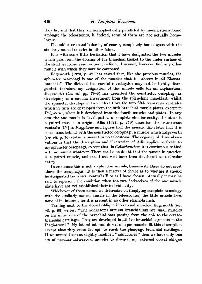
Girella cuspidator (text-fig. 9). The adductor labii superioris arises fromthe upper end of the preoperculum, runs horizontally forward below the floor
Ad.~~~~~~~~~~Adn
Q.
R.a.o. Add Adm.s
Fig. 9. Girella cu8pidator. Add.m. adductor mandibulae. Ad.l.8. adductor labii superioris. Q. quad-rate bone. R.a.o. retractor anguli oris.
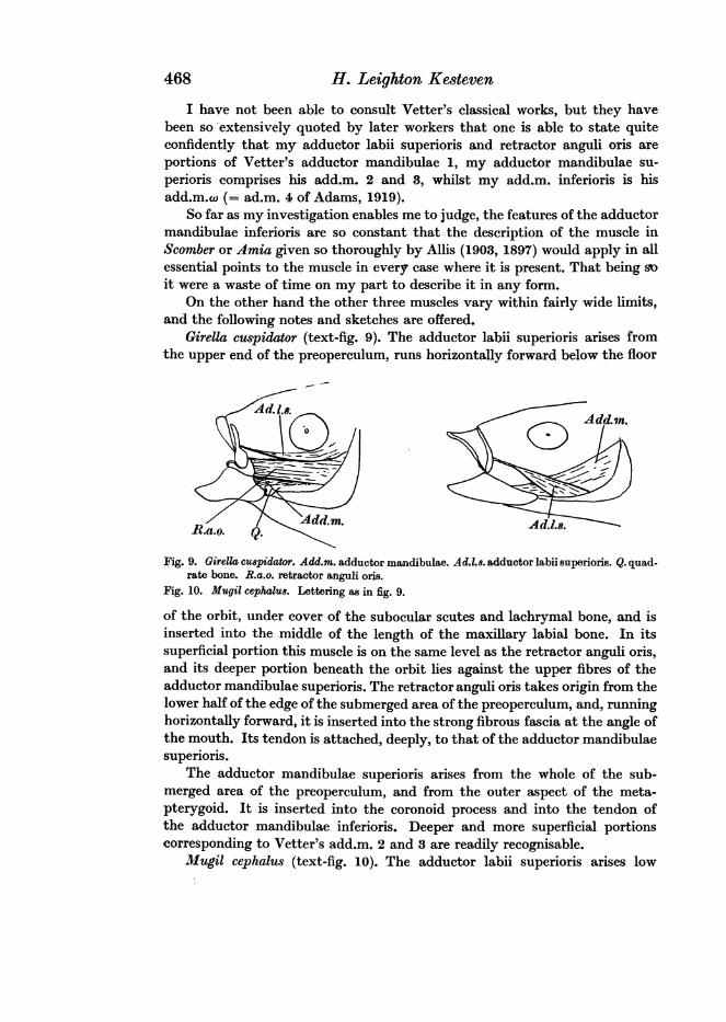
Fig. 10. Mugil cephalu8. Lettering as in fig. 9.
of the orbit, under cover of the subocular scutes and lachrymal bone, and isinserted into the middle of the length of the maxillary labial bone. In itssuperficial portion this muscle is on the same level as the retractor anguli oris,and its deeper portion beneath the orbit lies against the upper fibres of theadductor mandibulae superioris. The retractor anguli oris takes origin from thelower half of the edge of the submerged area of the preoperculum, and, runninghorizontally forward, it is inserted into the strong fibrous fascia at the angle ofthe mouth. Its tendon is attached, deeply, to that of the adductor mandibulaesuperiors.
The adductor mandibulae superioris arises from the whole of the sub-merged area of the preoperculum, and from the outer aspect of the meta-pterygoid. It is inserted into the coronoid process and into the tendon ofthe adductor mandibulae inferioris. Deeper and more superficial portionscorresponding to Vetter's add.m. 2 and 3 are readily recognisable.
Mugil cephalus (text-fig. 10). The adductor labii superioris arises low
468

The Anatomy of the Head of Callorhynchus antarcticus 469
down, on the horizontal edge of the submerged portion of the preoperculum.From its origin it passes up and forward to be inserted by a fine, but thoroughlystrong, tendon into the back of the maxillary labial bone at the junction ofthe middle and outer thirds of its length. The muscle lies entirely superficialto the adductor mandibulae superioris.
The retractor anguli oris is not developed as a separate entity, but is com-pletely blended with the adductor mandibulae superioris. The compoundmuscle arises from the outer surface of the posterior components of thequadrato-palatine arch. It is inserted into the fascia of the angle of themouth, the coronoid process and the tendon of the adductor mandibulaeinferioris. In this species I have been unable to find any definition of themuscle into portions which could be regarded as equivalent to Vetter's com-ponents of the add.m. except the add.m.w.
These two forms may be said to present variations of the arrangement ofthese muscles which is to be regarded as typical of the acanthopterygianFishes as a class.
_ ~~~~~~R.a.o. 3
@: 5 X R~~~~~~~~~~~~~~a~~Rao.
Add.m Addm.mFig. 11. GJonorhynwhua Greyi. Lettering
as in fig. 9.
Fig. 12. Balidtapwu aculeatus. Lettering as in fig. 9.
Gonorhynchus Greyi (text-fig. 11). The muscles of the group are all, in myspecimens, inseparably fused at their origin. The lower fibres are continuedforward, above the mouth, in the velum, and by the fibres of this structuregain insertion into the side of the pre-ethmoid bone under the lachrymal.As the velum is firmly bound to the maxillary labial as it crosses it, thismuscle acts both as a tensor veli and as an adductor labii superioris. Theupper fibres of the adductor mandibuli superioris are gathered on to a tendonwhich is inserted into the fascia of the angle of the mouth. Some of this lastbundle of fibres join the tensor veli muscle. The adductor mandibulae su-perioris takes origin from the hinder components of the quadrate-palatinearch between the divergent bundles of the other two muscles. The insertionis into the upper edge of the lower jaw in front of the joint. The adductormandibulae inferioris is reduced to a mere remnant.
Balistapus aculeatus (text-fig. 12) resembles Girella in the general arrange-31-2
470 H. Leighton Kesteven
ment of the muscles, but with the difference that all of them arise in frontof the orbit. Herein this group of Fishes resemble the Holocephali. I knowof no other forms in which the whole of the muscles of the face and of masti-cation are situated entirely in front of the orbit.
The adductor labii superioris arises from the under side of the lamina ofthe prefrontal (text-fig. 12) and is inserted into the spur on the back of themaxillary labial.
The retractor anguli oris arises from the descending portion of the pre-frontal, and, passing down and forward beneath the lower end of the pre-maxillary labial, is inserted into the short strong fibrous band which joinsthat bone to the coronoid process of the mandible.
The adductor mandibulae arises from the covered area of the preoperculum,from the hyomandibular bone and from the floor of the orbit and is insertedinto the inner aspect of the mandible, in front of and above the joint.
There is no adductor mandibulae inferioris.Hemirhamphus intermedis, In this form there is present only the adductor
mandibulae superioris. This arises from the outer aspect of the hyomandi-bular and preoperculum, and is inserted by a strong tendon into the upperedge of the mandible just in front of and above the joint, and into the fasciaanguli oris. No one of the other muscles of the group is present.
Fistularia petimba. Here also only the adductor mandibulae superioris ispresent. This arises from the front wall of the orbit and from the roof, floor,and inner wall of the sulcus formed by the union of the hyomandibular andprefrontal bones. The area of origin does not extend far forward of the orbit.In front of the muscle its long tendon extends forward along the sulcus, andis inserted into the coronoid process, well in front of the joint. Just beforereaching this point it passes inside the raphe joining the upper and lowerjaws and is attached thereto.
Anguilla reinherdti. There is no trace of the retractor anguli oris or ofthe adductor labii superioris. This is, of course, associated with and due tothe fact that there are in the Apodes no labial bones and no complicated lipfolded upon itself when the mouth is closed and drawn down to make a sidescreen to the mouth when open. In short there is no labial bone to be adductedand no secondary anguli oris to be retracted.
The adductor mandibulae superioris is a very massive muscle; its widearea of origin includes (1) the posterior fascial wall of the orbit; (2) a mediansagittal intermuscular septum (between the muscles of either side) whichextends from the posterior boundary of the orbit to the hinder limit of theskull; (3) the dorsum of the skull lateral to this septum; (4) the dorsal surfaceof the post-frontal; (5) a small area on the anterior face of the quadrate;(6) the deep anterior surface of the preoperculum; (7) a fibrous septum be-tween it and the muscles behind it. It is inserted by a very stout tendoninto the coronoid process, and into a deep pit on the inner side of the bone.Tracing this tendon back again, it is found to expand fanwise, or rather into
The Anatomy of the Head of CaUorhynchus antarcticus 471
an open brush. The fibres of this brush extend a long way back into the massof the muscle. Anteriorly the fan-like expansion becomes intimately asso-ciated with the strong fibrous strengthening of the angle of the lips, andposteriorly a strong band finds attachment to the outer edge of the quadrateat a little distance above the articular surface. This last slip doubtless acts asa check ligament, limiting the extent of the gape.
There is no adductor mandibulae inferioris.It may be pointed out now that these modifications affect muscles which
are undoubtedly derivatives of the main adductor mass of the lower jaw.Reviewing the complicated musculature and skeletal support of the lips
and lower jaw of Callorhynchus, one may be permitted to offer the suggestionthat they present us with a complicated attempt to carry out the generaldesign perfected in the teleostomes, the skeletal support an imperfect forecastof the superior labial bones (heretofore, except by the writer, termed maxillaand premaxilla), the musculature containing a more perfect forerunner of theretractor labii superioris and retractor anguli oris so constantly present inthe teleostomes.
If we now briefly survey the foregoing description of the musculature ofthe head of Callorhynchus the outstanding features of importance will befound to be: (1) the presence of three of those four muscles which Edgeworthpointed out were absent in all elasmobranchii, viz. levatores arcuum bran-chialium, transversus ventralis, and sphincter oesophagi; (2) the presence ofa complicated musculature activating the lips and lower jaw, which seems toforeshadow the more simple arrangement found in the teleostomes.
CONCLUSIONIt may be said of the extinct forms of animal life preserved to us in the
palaeontological record, that were we able to interpret them aright, we shoulddivide them into those which had proven failures and those which had beensuccesses in Nature's try-outs along the courses of evolution. The successes,of course, arranged in their proper sequence would present us with a moreor less complete genealogical tree for the existing forms. The failures in theirturn properly arranged as branches of this tree (whose single tap-root is burieddeep in past ages) would appear as abruptly broken branches, attached neareror further from the root.
If Nature's objective was the evolution of the human species, then shewas entirely successful only with the growth of that branch which producedas its ripened fruit Man. This, however, were a foolish conceit. Nature wassuccessful so long as she maintained plastic, and capable of further modifica-tion, each and every branch. She was, in fact, successful with every one ofthe branches whose fruit may be recognised in the various classes of theliving animals.
But each and every one of these branches bore broken branchlets andtwigs; the fossils are their record. Some of these broken branchlets bore many

H. Leighton Ke8teven
twigs, and some of these twigs were able to adapt themselves to the changingconditions as successive geological ages passed. We call them "persistenttypes." Perhaps the most remarkable of these persistent types is thebrachiopod Lingula, which comes to us practically unchanged from Cambriantimes. Among the Vertebrata one may mention all three of the Dipnoi andall the living members of that heterogeneous group of Fishes which we knowas the ganoids. The Holocephali also fall among the number.
There is, however, another and more valuable way of regarding thesepersistent types. They may be looked upon as unsuccessful essays in evolu-tion, as variants of the successful forms in the centre of the full current ofevolution.
Regarded thus the Holocephali become much more significant and inter-esting to the student of evolution. If this concept be a correct one, then theywill be regarded as presenting us many of the features which Nature earlyimported into the elasmobranch stock, in her efforts to evolve the bony Fishes.Perhaps they also present us with some resemblance to the early conditionfrom which the Dipnoi and the Amphibia derive their solidly anchored sus-pensorium of the lower jaw.
The complex skeletal and muscular apparatus for the movement of thelips may be regarded as having failed, because the skeleton of the upper lipneglected to develop the teeth that were the secret of the success of the bonyFishes nearer the centre of the evolutionary stream of the Fishes. Deep downbeneath this failure, however, was the germ of another line of evolution. Hiddenunderneath the futile lip mechanism was the solid hinge of a powerful lowerjaw, and a compact and adequate adductor musculature attached to the skullitself, not to the suspensorium of the jaw. These are features which are repro-duced with remarkable faithfulness in the Dipnoi.
In recent papers on the Evolution of the Anamniota (1931) I maintainedthat the Chondrostei were more correctly regarded as bony elasmobranchsthan as members of the bony-fish family. I further pointed out that a naturalcorollary of this was that the elasmobranchs and bony fishes came from acommon stock. The Holocephali present us, as it were, the converse of thissame proposition. They are undoubted elasmobranchs which present some ofthe teleostean features possessed by individual members of that unstablemenstruum from which were ultimately crystallised out the elasmobranchs,as a first successfully established homomorphic group.
Consideration of the fixed lower jaw joint and its compact musculature,on the other hand, gives rise to legitimate doubt as to whether it may nothave been that already that unstable menstruum did at the same time containthe evolutionary potential which was capable of producing both the dipnoanand ganoid stock side by side.
The geological antiquity of the various groups does not contraindicatesuch a possibility.
In addition to the list of features given by Bridge, wherein the Holocephali
472
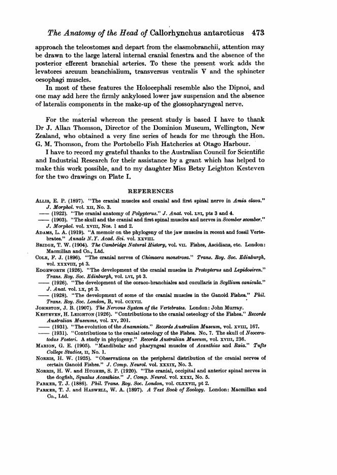
The Anatomy of the Head of Callorhynchus antaroticus 473
approach the teleostomes and depart from the elasmobranchii, attention maybe drawn to the large lateral internal cranial fenestra and the absence of theposterior efferent bronchial arteries. To these the present work adds thelevatores arcuum branchialium, transversus ventralis V and the sphincteroesophagi muscles.
In most of these features the Holocephali resemble also the Dipnoi, andone may add here the firmly ankylosed lower jaw suspension and the absenceof lateralis components in the make-up of the glossopharyngeal nerve.
For the material whereon the present study is based I have to thankDr J. Allan Thomson, Director of the Dominion Museum, Wellington, NewZealand, who obtained a very fine series of heads for me through the Hon.G. M. Thomson, from the Portobello Fish Hatcheries at Otago Harbour.
I have to record my grateful thanks to the Australian Council for Scientificand Industrial Research for their assistance by a grant which has helped tomake this work possible, and to my daughter Miss Betsy Leighton Kestevenfor the two drawings on Plate I.
REFERENCES
ALLis, E. P. (1897). "The cranial muscles and cranial and first spinal nerve in Amia clava."J. Morphol. vol. x1, No. 3.
-(1922). "The cranial anatomy of Polypterus." J. Anat. vol. Lvi, pts 3 and 4.(1903). "The skull and the cranial and first spinal muscles and nerves in Scomber scomber."
J. Morphol. vol. xvvm, Nos. 1 and 2.ADAMS, L. A. (1919). "A memoir on the phylogeny of the jaw muscles in recent and fossil Verte.
brates." Annals N. Y. Acad. Sci. vol. xxVIIL.BRIDGE, T. W. (1904). The Cambridge Natural Hi8tory, vol. vII. Fishes, Ascidians, etc. London:
Macmillan and Co., Ltd.COLE, F. J. (1896). "The cranial nerves of Chimaera monstrosa." Trams. Roy. Soc. Edinburgh,
vol. xxxvm, pt 3.EDGEWORTH (1926). "The development of the cranial muscles in Protopterus and Lepidosiren."
Trans. Roy. Soc. Edinburgh, vol. Lvi, pt 3.(1926). "The development of the coraco-branchiales and cucullaris in Scyllium canicula."
J. Anat. vol. LX, pt 3.- (1928). "The development of some of the cranial muscles in the Ganoid Fishes." Phil.
Trans. Roy. Soc. London, B, vol. ccxvii.JOHNSTON, J. B. (1907). The Nervous System of the Vertebrates. London: John Murray.KESTEvEN, H. LEIGHTON (1926). " Contributions to the cranial osteology of the Fishes." Records
Australian Museums, vol. xv, 201.- (1931). "The evolution of the Anamniota." RecordsAustralian Museum, vol. xviii, 167.
(1931). "Contributions to the cranial osteology of the Fishes. No. 7. The skull of Neocera-todus Fosteri. A study in phylogeny." Records Australian Museum, vol. xviii, 236.
MARION, G. E. (1905). "Mandibular and pharyngeal muscles of Acanthias and Raia." TuftsCollege Studies, II, No. 1.
NORRIS, H. W. (1925). "Observations on the peripheral distribution of the cranial nerves ofcertain Ganoid Fishes." J. Comp. Neurol. vol. xxxfx, No. 3.
NORRIS, H. W. and HUGHES, S. P. (1920). "The cranial, occipital and anterior spinal nerves inthe dogfish, Squalus Acanthias." J. Comp. Neurol. vol. xxxi, No. 5.
PARKER, T. J..(1886). Phil. Trans. Roy. Soc. London, vol. cLxxvIu, pt 2.PARKER, T. J. and HASWELL, W. A. (1897). A Text Book of Zoology. London: Macmillan and
Co., Ltd.

474 H. Leighton Keateven
EXPLANATION OF PLATES
PILATE IFigs. 1 and 2. Side view of the head and front view of the mouth and nasal aperture.
PLATE IIFig. 3. Side view of the skull. A.c. anterior superior labial cartilage. L.i.c. large inferior labial
cartilage. M.c. median nasal cartilage. P.c. posterior superior labial cartilage. R.l. root ofthe lateral ligament of the rostrum. S.c. superior anchoring labial cartilage. S.i.c. smallinferior labial cartilage. IX and X, nerve foramina.
Fig. 4. Skull cut open along the median sagittal plane, seen from within. D.e. canal for theductus endolymphaticus. I.e. inferior, anchoring labial cartilage. Oph. VII. foramen forsuperficial ophthalmic division of facial nerve. Oph.c. posterior aperture of the ophthalmiccanal. Oph. V. foramen of entry of the ophthalmicus profundus. 0.1. otic labyrinth. Pi. pinealrecess. Pt. subpituitary fossa. S.t. sell turcica. II, III, IV, V, VI, VII, IX and X, nerveforamina.
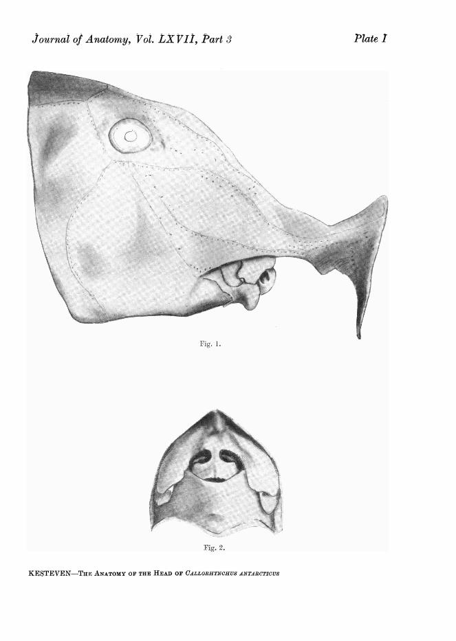
Journal of Anatomy, Vol. LXVII, Part S
I
I
:i
K .
lINir. 1.
Fig. 2.
KESTEVEN-THE ANATOMY OF THE HEAD OF CALLORHYNCHUS ANTARCTICUS
fOr'' I
._ .. .i
-~!0,
>;s
'Ii
i
Plate I
A..z
or-Il'---
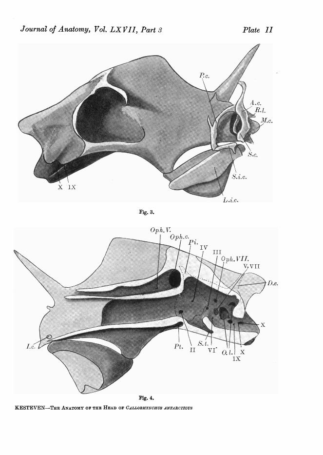
Journal of Anatomy, Vol. LX JII, Part 3 Plate II
L.i.c.
Fig. 3.
-.C.
IV
x
I.c.
pmoc:; I;:
13 \ S. t.t. II VI' 0. . x
ix
Fvig. 4.
KESTEVEN-THE ANATOMY OF THE HEAD OF CALLORHYNCHUSJ ANTARCTICUS