the application of fast-nmr for the identification of novel drug discovery targets
TRANSCRIPT

Review
s�P
OSTSCREEN
REVIEWS Drug Discovery Today � Volume 13, Numbers 3/4 � February 2008
The application of FAST-NMR for theidentification of novel drug discoverytargets
Robert Powers, Kelly A. Mercier and Jennifer C. CopelandDepartment of Chemistry, University of Nebraska-Lincoln, Lincoln, NE 68522, USA
The continued success of genome sequencing projects has resulted in a wealth of information, but 40–
50% of identified genes correspond to hypothetical proteins or proteins of unknown function. The
functional annotation screening technology by NMR (FAST-NMR) screen was developed to assign a
biological function for these unannotated proteins with a structure solved by the protein structure
initiative. FAST-NMR is based on the premise that a biological function can be described by a similarity in
binding sites and ligand interactions with proteins of known function. The resulting co-structure and
functional assignment may provide a starting point for a drug discovery effort.
The completion of the human genome project is spurring tremen-
dous progress in cell biology, development, evolution and phy-
siology [1]. The expanding number of protein structures emerging
from the protein structure initiative (PSI) is contributing to these
advancements [2]. As of January 2007, the sequencing of 607
genomes has been completed with 1676 ongoing projects. Also,
nearly 2500 protein structures have been solved by PSI [3,4]. Drug
discovery is benefiting from these successes through the identifi-
cation of novel therapeutic targets and the development of new
tools to optimize chemical leads [5–7]. As an example, the identi-
fication of novel anti-infectious targets may aid in avoiding com-
mon mechanisms of resistance and extend the lifetime of new
antibiotics [8,9].
An underlying challenge to capitalizing on genome sequencing
efforts is the abundance of hypothetical proteins, proteins that
lack a functional annotation. Our recent analysis of various bac-
terial genomes from the August 2007 Gold release shows that, even
with improved computational methods, approximately 40% of
bacterial proteins have not been assigned to a functional category
(Figure 1) [3]. There are more than 11,000 proteins from the ten
bacterial organisms listed in Figure 1 that lack a functional anno-
tation. Considering this list is only from a small segment of
currently sequenced genomes, the prospect of obtaining experi-
mental functional information for all hypothetical proteins iden-
tified from completed and ongoing sequencing efforts is a
Corresponding author: Powers, R. ([email protected])
172 www.drugdiscoverytoday.com 1359-6446/06/$ - s
daunting proposition. Valuable information is hidden among this
multitude of unannotated proteins that could be associated with
cell viability, biofilm formation, infection, and pathogenesis.
These proteins may provide key information for developing
new antibiotics, where drug discovery efforts would benefit greatly
from new functional annotations methodology.
Most high-throughput experimental methods to assign func-
tion have focused primarily on generating knockout libraries to
analyze cell phenotypes, monitoring changes in gene expression
or determining protein interaction maps [10–12]. These methods
generally do not provide functional information for a specific
protein without additional detailed bioinformatics [13,14]. Global
sequence similarity is routinely used to infer the function of
hypothetical proteins, despite analysis that suggest error rates
are as high as 30% [15,16]. Conversely, amino-acid residues asso-
ciated with the active sites and biological activities of proteins are
stable evolutionarily relative to the remainder of the protein’s
sequence and provide an alternative approach for functional
annotation [17,18]. A basic definition of biological function is
derived from a protein’s interaction with small molecules and
other biomolecules. Thus, the identification of functional
ligand(s), an active site and a corresponding protein–ligand co-
structure is instrumental to defining a function for a hypothetical
protein. The comparison and prediction of ligand-binding sites
from both structural and sequence information is a proven
approach for functional assignments of proteins [19]; however,
these predictions may lead to ambiguous or incorrect annotations
ee front matter � 2007 Elsevier Ltd. All rights reserved. doi:10.1016/j.drudis.2007.11.001

Drug Discovery Today � Volume 13, Numbers 3/4 � February 2008 REVIEWS
FIGURE 1
Functional analysis of bacterial genomes. The blue partitions are thepercentage of proteins that have assigned functional categories; the red
partitions are the percentage of unannotated proteins. Bacillus subtlus (BS);
Heliobactor pylori (HP); Staphylococcus aureus (SAu); Staphylococcus epidermis
(SE); Treponema pallidum (TP); Vibrio cholerae (VC); Streptococcus pneumoniae(SP); Streptococcus agalactiae (SAg); Bacillus anthracis (BA); Haemophilus
influenzae (HI).
Reviews�POSTSCREEN
[20,21]. A combination of experimental protein–ligand-binding
data with bioinformatic analysis will minimize the uncertainties
commonly associated with pure computational approaches.
Functional annotation screening technology by NMR (FAST-
NMR) provides functional information for hypothetical proteins
byexperimentally characterizingandanalyzing ligand-bindingsites
[22]. Once the functional ligands [23] are identified and the binding
site is located, a co-structure is obtained. A functional assignment is
deduced by comparing the ligand-defined active-site from FAST-
NMR to a database of protein–ligand-binding sites for proteins of
known function using our comparison of protein active-site struc-
tures (CPASS) database and software [24]. The information obtained
from FAST-NMR furthers our understanding of the basic biological
role of hypothetical proteins and provides a potential starting point
fordrugdiscovery.Asummaryof somecommonapplications for the
functional annotation of proteins of unknown proteins and a com-
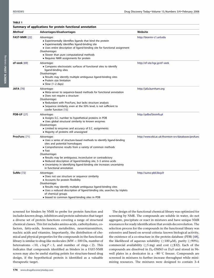
parison to our FAST-NMR method are listed in Table 1.
The FAST-NMR methodFAST-NMR was developed to assign biological functions to
hypothetical protein structures solved by PSI. Recent statistical
analysis indicates that �20–40% of protein structures determined
by PSI may be amenable to analysis by NMR [25,26]. The protein
structure database (PDB) currently contains �2200 proteins of
unknown function [27]. These proteins tend to be ‘orphaned’
from any further functional analysis because of a complete absence
of information to guide a research project [28–31]. These orphaned
proteins are ideal targets for analysis by FAST-NMR. An assigned
2D 1H–15N heteronuclear single quantum coherence (HSQC) NMR
spectrum and a corresponding structure for a protein of unknown
function are the primary requirements for the FAST-NMR meth-
odology. In general, high-resolution NMR structures and assign-
ments can be routinely obtained for proteins <25 kDa using
standard 13C and 15N protein labeling techniques [32,33]. This
molecular-weight upper limit may be extended by upwards of
900 kDa [34–40] by the application of deuterium labeling, specific
methyl labeling and transverse relaxation-optimized NMR spec-
troscopy (TROSY)-based experiments [41–43].
The FAST-NMR assay applies a tired approach to screening,
where an overview of the methodology is illustrated in Figure 2.
The first 1D 1H line-broadening (LB) experiments are amenable for
screening a large number of compounds in a relatively short time
(<10 min/sample) and requiring a minimal amount of unlabeled
protein material (<0.1 mg/sample). Only positive ‘hits’ from the
LB experiments are further screened in 2D 1H–15N HSQC spectra.
In this manner, the tiered approach minimizes resources and
increases throughput by funneling only the most promising can-
didates forward to the more resource intensive 2D 1H–15N HSQC
experiments.
In the first LB experiment, the formation of a protein–ligand
complex can be determined by monitoring changes in the ligand’s
spectrum. Binding of the ligand to the protein causes line width
broadening that may result in the complete disappearance of the
ligand’s NMR peak(s). This is caused by the large differences in
molecularweightand correlation time (tc, time it takes the molecule
to rotate one radian) between the protein and small molecule. In the
second NMR experiment, changes in the protein’s NMR spectrum
are followed to further confirm a specific interaction and identify a
potential binding site. Ligand binding causes local environmental
changes in the protein resulting in the observation of chemical shift
perturbations (CSPs) in the 2D 1H–15N HSQC spectrum. Since each
peak in the 2D 1H–15N HSQC spectrum has been sequentially
assigned to a specific amino acid in the protein’s sequence, the
CSPs can be mapped onto the surface of the protein. A consensus
clustering of CSPs identifies the location of the ligand-binding site.
A lack of CSPs or a random distribution of CSPs over the protein’s
surface indicates non-specific binding of the ligand to the protein.
A rapid protein–ligand co-structure is determined by combining
the experimental CSPs with molecular modeling. AutoDock [44]
has been demonstrated to outperform two other well-known
docking tools, FlexX and DOCK [45] in a virtual screen, and is
currently the most cited of molecular modeling applications [46].
The experimental CSPs define a grid used by AutoDock to guide the
ligand docking into the NMR defined binding site. Our AutoDock-
Filter (ADF) program is then used to filter the AutoDock confor-
mers and select a pose that best fits the CSPs. In general, amino
acid residues with the largest CSPs are expected to be closer to the
bound ligand relative to residues with smaller CSPs. The best
structure is then used to determine a ligand-defined binding site
for CPASS analysis. CPASS identifies ligand-defined binding sites
for proteins of known function from a PDB derived database that
best matches the sequence and structural details of the ligand-
defined binding site determined by FAST-NMR. A similarity
between these ligand-defined binding sites infers a function that
can be assigned to the hypothetical protein.
Functional chemical libraryA critical component of the FAST-NMR methodology is the func-
tional chemical library [47] (Figure 3). The library contains com-
pounds with demonstrated protein affinity and, as completely as
possible, covers the diversity of biological activity. The library is
www.drugdiscoverytoday.com 173
REVIEWS Drug Discovery Today � Volume 13, Numbers 3/4 � February 2008
TABLE 1
Summary of applications for protein functional annotation
Method Advantages/disadvantages Website
FAST-NMR [22] Advantages http://bionmr-c1.unl.edu
� Experimentally identifies ligands that bind the protein
� Experimentally identifies ligand-binding site
� Uses entire description of ligand-binding site for functional assignmentDisadvantages
� Slower than pure computational methods
� Requires NMR assignments for protein
eF-seek [69] Advantages http://ef-site.hgc.jp/eF-seek� Compares electrostatic surfaces of functional sites to identify
ligand-binding sites
Disadvantages
� Results may identify multiple ambiguous ligand-binding sites� Protein size limitation
� Slow (1–2 days)
JAFA [70] Advantages http://jafa.burnham.org
� Meta-server to sequence-based methods for functional annotation� Does not require a structure
Disadvantages
� Redundant with ProcFunc, but lacks structure analysis
� Sequence similarity, even at the 50% level, is not sufficient toconfer function [15]
PDB-UF [27] Advantages http://pdbuf.bioinfo.pl
� Assigns E.C. number to hypothetical proteins in PDB
� Uses global structural similarity to known enzymesDisadvantages
� Limited to enzymes and accuracy of E.C. assignments
� Majority of proteins still unassigned
ProcFunc [71] Advantages http://www.ebi.ac.uk/thornton-srv/databases/profunc� Uses a series of structure-based methods to identify ligand-binding
sites and potential homologues
� Comprehensive results from a variety of common methods
� FastDisadvantages
� Results may be ambiguous, inconclusive or contradictory
� Reduced description of ligand-binding site, 3–5 amino acids
� Uncertainty in identifying ligand-binding site increases uncertaintyin functional annotation
SuMo [72] Advantages http://sumo-pbil.ibcp.fr
� Does not use structure or sequence similarity
� Accounts for protein flexibilityDisadvantages
� Results may identify multiple ambiguous ligand-binding sites
� Uses a reduced description of ligand-binding site, searches by tripletsof chemical groups
� biased to common ligand-binding sites in PDB
Review
s�P
OSTSCREEN
screened for binders by NMR to probe for protein function and
includes known drugs, inhibitors and protein substrates that target
a diverse set of protein functions covering a range of structural
chemical classes. This list includes amino-acids, carbohydrates, co-
factors, fatty-acids, hormones, metabolites, neurotransmitters,
nucleic acids and vitamins. Importantly, the distribution of che-
mical and physical properties for the compounds in the functional
library is similar to drug-like molecules (MW < 500 Da, number of
heteroatoms <10, c log P < 5, and number of rings < 2). This
indicates that compounds identified as binders in a FAST-NMR
screen may also be useful starting points for structure-based drug
design, if the hypothetical protein is identified as a valuable
therapeutic target.
174 www.drugdiscoverytoday.com
The design of the functional chemical library was optimized for
screening by NMR. The compounds are soluble in water, do not
aggregate, precipitate or react in mixtures and have unique NMR
resonances for ready identification that avoids deconvolution. The
selection process for the compounds in the functional library was
extensive and based on several criteria: known biological activity,
the existence of a co-structure in the protein database (PDB) [48],
the likelihood of aqueous solubility (�100 mM), purity (�90%),
commercial availability (�5 mg) and cost (�$32). Each of the
compounds are dissolved in D6–DMSO or D2O and stored in 96-
well plates in a dessicator in a �80 8C freezer. Compounds are
screened in mixtures to further increase throughput while mini-
mizing resources. The mixtures were designed to contain 3–4

Drug Discovery Today � Volume 13, Numbers 3/4 � February 2008 REVIEWS
FIGURE 2
Flow chart of FAST-NMR. The hypothetical proteins are screened against mixtures of ligands from the functional chemical library. Reference 1D 1H NMR spectra of
the mixtures are compared to those containing protein, where a hit is identified by changes in NMR line-width. Only the ligands identified as binding in the
primary screen are further assayed in the secondary 2D 1H–15N HSQC NMR experiment. Chemical shift changes confirm a specific interaction and identify the
binding site from mapping of the CSPs on the protein’s surface. The binding site and CSPs are utilized to determine a rapid co-structure using AutoDock. This co-structure is then used by CPASS to compare the ligand-defined binding site from the hypothetical protein to all other protein–ligand interactions present in the
PDB. A general biological function can then be assigned based on an observed similarity to a ligand-defined binding site for a protein of known function.
Reviews�POSTSCREEN
compounds based on our statistical analysis of the optimal mix-
ture size for NMR screens [49].
CPASSCPASS incorporates a computer program with a structural database
to compare ligand-binding sites and provide a putative function
for hypothetical proteins screened by FAST-NMR [24]. CPASS is a
structure-based functional annotation program that differs from a
variety of 3D template or sequence-based annotation programs
(for a review see Watson et al. [50]) routinely used to predict ligand-
binding sites and protein function. These programs attempt to
predict the location of ligand-binding sites using various sequence
and structure heuristics. Instead, CPASS aligns experimentally deter-
mined ligand-defined binding sites from FAST-NMR and the PDB
using sequence and structural descriptors. Simply, CPASS identi-
fies matches between functionally relevant ligand-binding sites to
leverage an annotation.
CPASS may also aid the development of selective chemical leads.
Drug toxicity is a common cause of clinical failures [51], where this
toxicity is associated with non-specific in vivo protein activity [52].
In practice, it is notpossible to screen against every potential protein
target that may bind a chemical lead. Instead, a small panel of
homologous proteins is used in secondary assays to infer compound
specificity. The proteins are generally selected based on global
sequence similarity to the protein target of interest. Unfortunately,
there are also other proteins that share a high similarity in the
ligand-binding site that may lack global sequence similarity. Our
previous CPASS analysis of ATP binding proteins indicates a sig-
nificant cluster of proteins with sequence similarity<20% that had
high CPASS similarity of >40% [24]. Similarly, we identified two
alanine racemases that share only an 8% sequence similarity, but
had essentially identical PLP binding sites. Clearly, proteins that
share high ligand-binding site similarity, but lack global sequence
similarity pose serious risks of causing toxic side-effects in clinical
trials unless identified using applications like CPASS.
Protein–ligand databaseThe CPASS database is continuously updated from the PDB and
contains proteins in complex with small molecules, peptides, and
oligonucleotides. Proteins may bind one ligand, multiple ligands,
or the same ligand more than once. Each unique ligand-binding
site (<80% sequence similarity, distinct ligand) is incorporated
into the CPASS database. There are �55,000 protein–ligand-bind-
ing sites currently present in the PDB, where �21,000 are unique.
These ligand-defined binding sites include all the amino acids in
the protein sequences that have at least one atom within 6 A of any
atom of the ligand. Both the structure coordinates and the
sequence identity are then used in a comparison with ligand-
defined binding sites from other proteins. The ligand structure
is not included in the ligand-defined binding site, but is used to
classify the type of binding site (i.e. ATP binding site, FAD binding
site, and so on).
www.drugdiscoverytoday.com 175

REVIEWS Drug Discovery Today � Volume 13, Numbers 3/4 � February 2008
FIGURE 3
Functional chemical library. A subset of compounds from four different functional categories from the functional chemical library is displayed. Proteins arescreened against mixtures of compounds, and these mixtures were designed to have diverse structure and function to minimize spectral and functional overlap.
Review
s�P
OSTSCREEN
Similarity scoring functionAlthough the CPASS program will allow the user to search binding-
sites based on the type of ligand that defines the binding site, it is
not required. The comparison can be made against all ligand-
defined binding sites present in the CPASS database or any ligand-
type subset. The CPASS scoring function is based on the simulta-
neous structure and sequential alignments of two ligand-defined
binding sites. A BLOSUM62 probability function weighted by root-
mean square distance (rmsd) is used to compare the similarity of
spatially aligned residues:
Sab ¼Xi�n j�m
i; j¼1
dmin
diðe�Drmsdi; jÞ2 pi; j
Drmsdi; j ¼rmsdi; j rmsdi; j > 1 A
0 rmsdi; j � 1 A
(
Active site a contains n residues and is compared to active site b
of m residues from the CPASS database. pi,j is the BLOSUM62
probability for replacement of amino acid i from active site a
with residue j from active site b, Drmsdi,j is the corrected root-
mean square difference in the Ca positions between the residues i
176 www.drugdiscoverytoday.com
and j, and dmin/di is the ratio of the shortest distance to an atom
in the ligand from any atom in the residue i. This last term
minimizes boundary effects. Small structural changes may result
in residues entering or leaving the 6 A cut-off used to define
a ligand-defined binding site. This may result in relatively
large changes in the scoring function due to modest structural
fluctuations.
The similarities between the active sites are then calculated by:
S ¼ Sab
Saa� 100
where S is the similarity score, Sab is the similarity score for the
protein target against an active site from the CPASS database, and
Saa is the similarity of the active site compared to itself used for
normalization. In effect, a percent similarity is determined based
on how well the sequence and structures of the two ligand-binding
sites overlap. The scoring function is not symmetrical since it
depends on the size of the binding site.
CPASS functional prediction of hypothetical proteinsTo illustrate further the utility of CPASS, a recent protein deposited
in the PDB was chosen that only had a putative functional

Drug Discovery Today � Volume 13, Numbers 3/4 � February 2008 REVIEWS
s�POSTSCREEN
annotation. A human protein (PDB-ID 2PL3) was tentatively
assigned as a probable ATP-dependent RNA helicase DDX10 and
the structure contained a bound ADP molecule. CPASS analysis
identified PDB-ID 2OXC as having the highest similarity (56.26%).
Both proteins bind ADP and are hypothetical DEAD domains. The
highest CPASS similarity score (50.30%) to a protein of known
function was to PDB-ID 1XTJ, a DECD to DEAD mutation of
human UAP56, which is also in complex with ADP [53]. Recently,
the UAP56 protein has been shown experimentally to exhibit
RNA-stimulated ATPase activity and ATP-dependent RNA helicase
activity [54]. Thus, the CPASS analysis supports the prior putative
assignments of hypothetical proteins 2PL3 and 2OXC as ATP-
dependent RNA helicases. The top panel in Figure 4 shows the
FIGURE 4
(Top panel) Binding sites of 1XTJ and 2PL3. Binding-site residues for proteins (A) 1XT
is colored pink. The amino acid alignment for the ADP binding sites is shown at the
site residues for proteins (A) 1MKYand (B) 2E87. Residues within 6 A of GDP are coloGDP binding sites is shown at the bottom of the figure.
alignment of the ADP binding sites for the 2PL3 and 1XTJ struc-
tures. This figure clearly highlights the overall similarity in the
structure and sequence alignments for the ADP binding sites.
A crystal structure of hypothetical protein PH1320 from Pyrococ-
cus horikoshii OT3 was recently released by the PDB (PDB-ID 2E87).
The protein is complexed to guanosine-50-diphosphate (GDP), but
completely lacks a functional assignment and a paper describing the
structure has yet to be published. A CPASS analysis using only
proteins complexed to GDP indicates hypothetical protein
PH1320 has a very high similarity (70.47%) to an Escherichia coli
elongation factor Der (PDB-ID 1MKY), an EngA homolog [55]. The
bottom panel in Figure 4 clearly demonstrates the high overall
similarity in the structure and sequence alignments for the GDP
J and (B) 2PL3. Residues within 6 A of ADP are colored blue and the ligand ADP
bottom of the figure. (Bottom panel) Binding sites of 1MKYand 2E87. Binding-
red blue and the ligand GDP is colored pink. The amino acid alignment for the
www.drugdiscoverytoday.com 177
Review

REVIEWS Drug Discovery Today � Volume 13, Numbers 3/4 � February 2008
Review
s�P
OSTSCREEN
binding sites between these two proteins. Hypothetical protein
PH1320 shows CPASS similarity scores of 50–70% to EngB, EngC,
EI-F2g, EI-F5B, EF-Tu, EF-1a, EF-2 and EF-G, which are also members
of the elongation factor super family. Hypothetical protein PH1320
also exhibits a slightly smaller similarity (50–60%) to Arf, Sar and
Rab, members of the small GTPase super family that regulate a
diverse range of cellular events [56]. Thus, the CPASS results suggest
PH1320 is probably an elongation factor or potentially involved in
GTP signal regulation similar to either Arf, Sar or Rab.
Functional annotation of Staphylococcus aureusprotein SAV1430Staphylococcus aureus protein SAV1430, a hypothetical protein of
unknown function, was selected to demonstrate the FAST-NMR
methodology (Figure 2). SAV1430 is a typical target of the North-
East Structural Genomic Consortium (NESG) [29], where a struc-
ture was previously determined [57,58]. A Dali analysis suggested
that SAV1430 has a similar topology to a ferredoxin-like fold, but
the Z-score of<3 was insignificant [59]. The only proteins that had
any significant sequence homology to SAV1430 were other
hypothetical proteins, so a reliable function could not be assigned
based on structure homology alone.
O-phospho-L-tyrosine (pTyr) was identified as one of 21 com-
pounds that exhibited line-broadening and chemical shift pertur-
bations in the FAST-NMR screen with SAV1430. The other
compounds are chemically similar to pTyr and were all shown
to interact in a consensus binding-site that comprises residues I6-
P10, T14-K16 and I61-V63. This binding site contains a shallow
cleft on the SAV1430 surface surrounded by relatively flat struc-
tural features strongly suggestive of a protein–protein interaction
site. A rapid structure of the pTyr-SAV1430 complex was deter-
mined using CSPs and AutoDock for CPASS analysis.
CPASS identified PDB ID 1oo4 as a significant hit (37% similar-
ity), a Src SH2 domain complexed with a pTyr containing peptide.
SH2 domains are typically part of multi-domain proteins involved
in cell signaling and form a protein–protein complex with a kinase
after phosphorylation of a tyrosine [60]. Phosphorylation of Ser,
Thr and Tyr are also common mechanisms for regulating protein
activity in bacteria [61,62]. The similarity in the characteristics of
178 www.drugdiscoverytoday.com
the SAV1430 and Src SH2 ligand-binding sites, and the fact that
SAV1430 binds pTyr, further supports the general proposal that
SAV1430 functions by forming a protein–protein complex.
Rosetta Stone [63] analysis suggests hypothetical protein
SAV0936 may be a binding partner of SAV1430. SAV0936 Exhibits
47% sequence identity with the N-terminal region of the C-term-
inal NifU domain. NifU is a multi-protein complex that is a critical
component of the [Fe-S] cluster assembly pathway [64–66] and is
essential for the viability of bacteria [67]. A more exhaustive
sequence analysis of SAV1430, based on the results with
SAV0936, indicates the protein shares �30% sequence identity
with the C-terminal region of the C-terminal domain of the NifU
multi-domain structure. These results imply that SAV1430 may
interact with SAV0936 to form a complex that exhibits similar
activity as the full length NifU domain or may regulate NifU
activity. Thus, inhibiting the SAV1430-SAV0936 complex forma-
tion may represent a novel target for developing next generation
antibiotics.
ConclusionFAST-NMR provides a high-throughput approach to obtain func-
tional assignments for hypothetical proteins, based on experi-
mentally determined protein–ligand interactions. FAST-NMR
also addresses the current lack in high-throughput experimental
methods to obtain functional information [68] where current
methods [14] primarily rely on sequence similarity to confer
function despite error rates as high as 30% [15,16]. FAST-NMR
is based on basic tenets of biochemistry where detailed structural
information of a protein–ligand interaction is paramount for
understanding the function of a protein. Active-site residues
are evolutionarily stable relative to the remainder of the protein’s
sequence decreasing the likelihood of annotation errors [17,18].
FAST-NMR compliments the success of the human genome pro-
ject and the protein structure initiative by providing a means to
functionally annotate the rapidly expanding number (�2200) of
hypothetical proteins currently deposited in the PDB [31]. Under-
standing protein function is a paramount necessity for drug
discovery programs ability to make successful contributions to
human health issues.
References
1 Venter, C. et al. (2001) The sequence of the human genome. Science 291 (5507),
1304–1351
2 Burley, S.K. (2000) An overview of structural genomics. Nat. Struct. Biol. 7 (Suppl.),
932–934
3 Bernal, A. et al. (2001) Genomes online database (GOLD): a monitor of genome
projects world-wide. Nucleic Acids Res. 29, 126–127
4 Todd, A.E. et al. (2005) Progress of structural genomics initiatives: an analysis of
solved target structures. J. Molec. Biol. 348, 1235–1260
5 Sioud, M. (2007) Main approaches to target discovery and validation. In Methods in
Molecular Biology (Vol. 360), pp. 1–12, Totowa, NJ, United States (Target Discovery
and Validation, Volume 1)
6 Zheng, C.J. et al. (2006) Therapeutic targets: progress of their exploration and
investigation of their characteristics. Pharmacol. Rev. 58, 259–279
7 Manning, A.M. (2006) Impact of the human genome on the discovery of
immune-modulatory therapeutics. Curr. Opin. Investig. Drugs (Thomson Scientific)
7, 406–411
8 Schrenzel, J. et al. (2004) A randomized clinical trial to compare fleroxacin-
rifampicin with flucloxacillin or vancomycin for the treatment of staphylococcal
infection. Clin. Infect. Dis. 39, 1285–1292
9 Natsch, S. et al. (1998) Guidelines for the prevention of antimicrobial resistance in
hospitals. Clin. Infect. Dis. 26, 1482–1483 An official publication of the Infectious
Diseases Society of America
10 Lee, Y.-H. et al. (2005) Gene knockdown by large circular antisense for high-
throughput functional genomics. Nat. Biotechnol. 23, 591–599
11 Michiels, F. et al. (2003) One step further towards real high throughput functional
genomics. Trends Biotechnol. 21, 147–148
12 Tucker, C.L. (2002) High-throughput cell-based assays in yeast. Drug Discov. Today 7
(Suppl.), S125–S130
13 del Val, C. et al. (2004) High-throughput protein analysis integrating bioinformatics
and experimental assays. Nucleic Acids Res. 32, 742–748
14 Joshi, T. et al. (2004) Genome-scale gene function prediction using multiple sources
of high-throughput data in yeast Saccharomyces cerevisiae. Omics 8, 322–333
15 Rost, B. (2002) Enzyme function less conserved than anticipated. J. Molec. Biol. 318,
595–608
16 Devos, D. and Valencia, A. (2001) Intrinsic errors in genome annotation. Trends
Genet. 17, 429–431
17 Zvelebil, M.J. et al. (1987) Prediction of protein secondary structure and active sites
using the alignment of homologous sequences. J. Molec. Biol. 195, 957–961

Drug Discovery Today � Volume 13, Numbers 3/4 � February 2008 REVIEWS
Reviews�POSTSCREEN
18 Livingstone, C.D. and Barton, G.J. (1993) Protein sequence alignments: a strategy
for the hierarchical analysis of residue conservation. CABIOS, Comput. Appl. Biosci. 9,
745–756
19 Campbell, S.J. et al. (2003) Ligand binding: functional site location, similarity and
docking. Curr. Opin. Struct. Biol. 13, 389–395
20 Greaves, R. and Warwicker, J. (2005) Active site identification through geometry-
based and sequence profile-based calculations: burial of catalytic clefts. J. Molec. Biol.
349, 547–557
21 Bateman, A. and Birney, E. (2000) Searching databases to find protein domain
organization. Adv. Prot. Chem. 54 (Analysis of Amino Acid Sequences), 137–157
22 Mercier, K.A. et al. (2006) FAST-NMR: functional annotation screening technology
using NMR spectroscopy. J. Am. Chem. Soc. 128, 15292–15299
23 Mercier, K.A. et al. (2006) Design and characterization of a functional library for
NMR screening against novel protein targets. Comb. Chem. High Throughput Screen
FIELD 9, 515–534
24 Powers, R. et al. (2006) Comparison of protein active site structures for functional
annotation of proteins and drug design. Prot.: Struct. Funct., Bioinform. 65, 124–
135
25 Snyder, D.A. et al. (2005) Comparisons of NMR spectral quality and success in
crystallization demonstrate that NMR and X-ray crystallography are
complementary methods for small protein structure determination. J. Am. Chem.
Soc. 127, 16505–16511
26 Yee, A.A. et al. (2005) NMR and X-ray Crystallography, Complementary Tools in
Structural Proteomics of Small Proteins. J. Am. Chem. Soc. 127, 16512–16517
27 von Grotthuss, M. et al. (2006) PDB-UF: database of predicted enzymatic functions
for unannotated protein structures from structural genomics. BMC Bioinform. 7
28 Lattman, E. (2004) The state of the Protein Structure Initiative. Prot.: Struct. Funct.
Bioinform. 54, 611–615
29 Anon, (2004) The protein target list of the Northeast Structural Genomics
Consortium. Prot.: Struct. Funct. Bioinform. 56, 181–187
30 Bonanno, J.B. et al. (2005) New York-structural genomiX research consortium
(NYSGXRC): A large scale center for the protein structure initiative. J. Struct. Funct.
Genom. 6, 225–232
31 von Grotthuss, M. et al. (2006) PDB-UF: database of predicted enzymatic functions
for unannotated protein structures from structural genomics. BMC Bioinform. 7, 53
32 Lian, L.-Y. and Middleton, D.A. (2001) Labeling approaches for protein structural
studies by solution-state and solid-state NMR. Progr. Nucl. Magn. Reson. Spectrosc. 39,
171–190
33 Ferentz, A.E. and Wagner, G. (2000) NMR spectroscopy: a multifaceted approach to
macromolecular structure. Quart. Rev. Biophys. 33, 29–65
34 Sprangers, R. et al. (2005) Quantitative NMR spectroscopy of supramolecular
complexes: Dynamic side pores in ClpP are important for product release. Proc. Natl.
Acad. Sci. U. S. A. 102, 16678–16683
35 Jain, N.U. et al. (2004) Rapid analysis of large protein–protein complexes using
NMR-derived orientational constraints: The 95 kDa complex of LpxA with acyl
carrier protein. J. Molec. Biol. 343, 1379–1389
36 Tugarinov, V. et al. (2002) Four-dimensional NMR spectroscopy of a 723-residue
protein: chemical shift assignments and secondary structure of malate synthase G. J.
Am. Chem. Soc. 124, 10025–10035
37 Tugarinov, V. and Kay, L.E. (2005) Quantitative 13C and 2H NMR relaxation studies
of the 723-residue enzyme malate synthase G reveal a dynamic binding interface.
Biochemistry 44, 15970–15977
38 Peterson, F.C. and Gettins, P.G.W. (2001) Insight into the mechanism of
serpin-proteinase inhibition from 2D [1H–15N] NMR studies of the 69 kDa
a1-proteinase inhibitor Pittsburgh-trypsin covalent complex. Biochemistry 40,
6284–6292
39 Liu, D. et al. (2004) Backbone resonance assignments of the 45.3 kDa catalytic
domain of human BACE1. J. Biomolec. NMR 29, 425–426
40 Revington, M. and Zuiderweg, E.R.P. (2004) Letter to the editor: TROSY-driven NMR
backbone assignments of the 381-residue nucleotide-binding domain of the
Thermus thermophilus DNAK molecular chaperone. J. Biomolec. NMR 30, 113–114
41 Riek, R. et al. (2000) TROSY and CRINEPT: NMR with large molecular and
supramolecular structures in solution. Trends Biochem. Sci. 25, 462–468
42 Gardner, K.H. and Kay, L.E. (1998) The use of 2H, 13C, 15N multidimensional NMR
to study the structure and dynamics of proteins. Annu. Rev. Biophys. Biomol. Struct
27, 357–406
43 Kay, L.E. (1998) The development of NMR methods to study protein structure and
dynamics. NATO ASI Ser., Ser. C 510, 285–293 (New Methods for the Study of
Biomolecular Complexes)
44 Morris, G.M. et al. (1998) Automated docking using a Lamarckian genetic algorithm
and an empirical binding free energy function. J. Comput. Chem. 19, 1639–1662
45 Park, H. et al. (2006) Critical assessment of the automated AutoDock as a new
docking tool for virtual screening. Prot.: Struct. Funct. Bioinform. 65, 549–554
46 Sousa, S.F. et al. (2006) Protein–ligand docking: current status and future challenges.
Prot.: Struct. Funct. Bioinform. 65, 15–26
47 Mercier, K.A. et al. (2006) Design and characterization of a functional library for
NMR screening against novel protein targets. Comb. Chem. High Throughput Screen.
9, 515–534
48 Berman, H.M. et al. (2000) The protein data bank. Nucleic Acids Res. 28, 235–242
49 Mercier, K.A. and Powers, R. (2005) Determining the optimal size of small molecule
mixtures for high throughput NMR screening. J. Biomolec. NMR 31, 243–258
50 Watson, J.D. et al. (2005) Prediction protein function from sequence and structural
data. Curr. Opin. Struct. Biol. 15, 275–284
51 Kubinyi, H. (2003) Opinion: drug research: myths, hype and reality. Nat. Rev. Drug
Discov. 2, 665–668
52 Ekins, S. (2004) Predicting undesirable drug interactions with promiscuous proteins
in silico. Drug Discov. Today 9, 276–285
53 (2004) Crystal structure of the human ATP-dependent splicing and export factor
UAP56. Proc. Natl. Acad. Sci. U. S. A. 101, 17628–17633
54 Shen, J. et al. (2007) Biochemical characterization of the ATPase and helicase
activity of UAP56, an essential Pre-mRNA splicing and mRNA export factor. J. Biol.
Chem. 282, 22544–22550
55 Robinson, V.L. et al. (2002) Domain arrangement of Der, a switch protein containing
two GTPase domains. Structure (Cambridge, MA, United States) 10, 1649–1658
56 Wennerberg, K. et al. (2005) The Ras superfamily at a glance. J. Cell Sci. 118, 843–846
57 Baran, M.C. et al. (2003) Solution Structure Determination of the Staphylococcus aureus
hypothetical protein SAV1430. Northeast Structure Consortium Target ZR18.
Department of Biochemistry, University of Wisconson-Madison BMRB accession
number 5844
58 Baran, M.C. et al. (2003) Solution Structure of the Hypothetical Staphylococcus aureus
protein SAV1430, Northeast Structural Genomics Consortium Target ZR18;PDB
ID:1PQX
59 Dietmann, S. et al. (2001) A fully automatic evolutionary classification of protein
folds: Dali Domain Dictionary version 3. Nucleic Acids Res. 29, 55–57
60 Marengere, L.E.M. and Pawson, T. (1994) Structure and function of SH2 domains. J.
Cell Sci. Suppl. Cell Biol. Cancer 18, 97–104
61 Kennelly, P.J. and Potts, M. (1996) Fancy meeting you here! A fresh look at
‘prokaryotic’ protein phosphorylation J. Bacteriol. 178, 4759–4764
62 Alzari, P.M. (2004) First Structural Glimpse at a Bacterial Ser/Thr Protein
Phosphatase. Structure (Cambridge, MA, United States) 12, 1923–1924
63 Suhre, K. and Claverie, J.-M. (2004) FusionDB: a database for in-depth analysis of
prokaryotic gene fusion events. Nucleic Acids Res. 32 (Database), D273–D276
64 Frazzon, J. and Dean, D.R. (2003) Formation of iron–sulfur clusters in bacteria: an
emerging field in bioinorganic chemistry. Curr. Opin. Chem. Biol. 7, 166–173
65 Dos Santos, P.C. et al. (2004) Iron–sulfur cluster assembly: NifU-directed activation
of the nitrogenase Fe protein. J. Biol. Chem. 279, 19705–19711
66 Dos Santos, P.C. et al. (2004) Formation and insertion of the nitrogenase iron-
molybdenum cofactor. Chem. Rev. (Washington, DC, United States) 104, 1159–1173
67 Olson, J.W. et al. (2000) Characterization of the NifU and NifS Fe–S cluster
formation proteins essential for viability in Helicobacter pylori. Biochemistry 39,
16213–16219
68 Flook, P.K. et al. (2003) Target validation through high throughput proteomics
analysis. Targets 2, 217–223
69 Kinoshita, K. et al. (2007) eF-seek: prediction of the functional sites of proteins by
searching for similar electrostatic potential and molecular surface shape. Nucleic
Acids Res. 35 (Web Server Issue), W398–W402
70 Friedberg, I. et al. (2006) JAFA: a protein function annotation meta-server. Nucleic
Acids Res. 34 (Web Server Issue), W379–W381
71 Laskowski, R.A. et al. (2005) ProFunc: a server for predicting protein function from
3D structure. Nucleic Acids Res. 33 (Web Server Issue), W89–W93
72 Jambon, M. et al. (2005) The SuMo server: 3D search for protein functional sites.
Bioinformatics 21, 3929–3930
www.drugdiscoverytoday.com 179