the association of mast cells and serotonin in children ... · the association of mast cells and...
TRANSCRIPT

The association of mast cells and serotonin inchildren with chronic abdominal pain ofunknown etiologyTaylor et al.
Taylor et al. BMC Research Notes 2010, 3:265http://www.biomedcentral.com/1756-0500/3/265 (21 October 2010)

RESEARCH ARTICLE Open Access
The association of mast cells and serotonin inchildren with chronic abdominal pain ofunknown etiologyTara J Taylor1†, Nader N Youssef2†, Ravi Shankar1†, David E Kleiner3†, Wendy A Henderson1*†
Abstract
Background: Abdominal pain of unknown origin affects up to 20% of school-aged children. Evaluation of childrenis symptom-based without clear guidelines to investigate molecular mechanisms of abdominal pain. Aberrantmolecular mechanisms may increase intestinal permeability leading to interactions between the immune andnervous systems, subclinical inflammation, and visceral pain. This study evaluated the association betweeninterleukin-6 (IL-6), mast cell infiltrates, and serotonin (5-HT) levels in gastrointestinal (GI) biopsies, with perceivedabdominal pain in a pediatric cohort.
Methods: Clinical data and biopsy samples from pediatric patients (n = 48) with chronic abdominal pain, with andwithout inflammation were included. Formalin-fixed paraffin-embedded GI biopsies were sectioned andimmunohistochemistry performed for IL-6 and 5-HT; mast cells were identified with toluidine blue stain.Histological findings were compared to self-reported abdominal pain between groups.
Results: There was significantly greater IL-6 immunoreactivity in biopsies with confirmed histologic inflammation(p = 0.004). There was a greater number of mast cells per HPF in non-inflammatory biopsies (3.5 ± 2.9) comparedto the inflammatory biopsies (2.6 ± 1.8) p = 0.049. The non-inflammatory biopsy group was significantly less likelyto respond to standard treatment as evidenced by higher pain reports (p = .018). Mast cells (p = .022) and 5-HT(p = .02) were significantly related to abdominal pain scores.
Conclusions: A potential association between self-reported abdominal pain, number of mast cells, and 5-HT levels,which may contribute to perceived GI pain in pediatric patients may exist.
BackgroundUnspecified chronic abdominal pain in children that hasno identified biologic marker or known organic causeand has occurred for greater than two months is definedby Rome III criteria as chronic abdominal pain [1]. Thesecriteria assume that no metabolic or structural causescan be related to the continued symptoms of chronicabdominal pain for an eight week period over the pre-vious year. Current Rome III criteria divide pediatricabdominal pain diagnosis into several subsets whichinclude functional abdominal pain syndrome and irritablebowel syndrome (IBS) [2]. Approximately 15-20% of
children [3] and adults living in the United States sufferfrom chronic abdominal pain [4,5]. Children with chronicabdominal pain experience lessened quality of life incomparison to their healthy peers which leads to numer-ous absences from school for medical care [6,7]. Chronicabdominal pain in pediatric patients is a significant bur-den on the health system which warrants further investi-gation on the pathogenesis of disease to identify noveltargets for intervention.Recognition of the role of inflammation and its interac-
tion with the neuro-immune system of the gastrointest-inal tract is an emerging area of interest in patients withchronic abdominal pain [8]. The mechanisms of chronicabdominal pain of unknown origin may be related tointeractions between the immune and nervous system inthe gut thereby leading to visceral hypersensitivity of theintestinal mucosa [8]. Mast cells have been shown to
* Correspondence: [email protected]† Contributed equally1National Institute of Nursing Research, National Institutes of Health,Bethesda, MD, USAFull list of author information is available at the end of the article
Taylor et al. BMC Research Notes 2010, 3:265http://www.biomedcentral.com/1756-0500/3/265
© 2010 Henderson et al; licensee BioMed Central Ltd. This is an Open Access article distributed under the terms of the CreativeCommons Attribution License (http://creativecommons.org/licenses/by/2.0), which permits unrestricted use, distribution, andreproduction in any medium, provided the original work is properly cited.

interact with colonic nerve endings by the secretion oftryptase, histamine, and possibly serotonin (5-hydroxy-tryptamine, 5-HT) in humans [9]. There is evidence ofcloser proximity of mast cells to nerve fibers in the colonof IBS patients leading to augmented visceral sensitivity[10]. A possible relationship between the number ofmucosal mast cells and rectal sensitivity has also beendemonstrated in humans [11]. There is evidence of a sig-nificant increase in mast cell numbers in patients withIBS [12]. Along with increased mast cell counts there issupport that mast cell numbers directly correlate withabdominal pain in IBS patients [13]. Recently, a studyconducted by Mahjoub, et al. found increased mast celldensity in pediatric patients with recurrent abdominalpain [14]. They propose that mast cell density measure-ments be incorporated with routine GI biopsies due to asignificant correlation between increase GI complaintsand mast cell count. Inflammation and increased perme-ability of GI tract mucosa may also induce pain in pedia-tric patients with functional abdominal pain [15].Chronic abdominal pain of unknown origin may be
due to low grade inflammatory changes in the GI tract[16,17]. Low level inflammation may result in increasedpermeability across the mucosal barrier; which in turnpermits the entrance of antigens into the intestinal wall[16]. Other potential mechanisms include altered func-tion at the level of neurotransmitter receptors of 5-HT,increased visceral hypersensitivity, and impaired colonicmucosal permeability [18]. Alleviation of symptoms withmedication that target 5-HT receptors suggest that5-HT is involved in gut motility regulation [19].Increased permeability and its role in pediatric
abdominal pain has recently been the focus of work byShulman (2008). They explored the relationship of sub-clinical inflammation and its relationship to chronicpain of the GI tract. In a well-designed prospective con-trolled study investigating the difference in GI perme-ability and fecal calprotectin (marker for intestinalinflammation) concentration in children with abdominalpain versus control [15]. Proximal GI permeability, colo-nic permeability, and fecal calprotectin were found to besignificantly greater in the abdominal pain group com-pared to the control group. Fecal calprotectin concen-trations correlated with pain interference with activities.However, there was no correlation between GI perme-ability and pain-related symptoms. This study high-lighted that more research is needed to examine theinteractions at the molecular level between mast cellsand 5-HT.These converging lines of evidence suggest that mast
cells and 5-HT contribute to increased colonic perme-ability and thereby lead to chronic abdominal pain. Weassessed this hypothesis at the molecular level by explor-ing the relationship between mast cell and 5-HT levels
and perceived abdominal pain in a pediatric cohort withand without the diagnosis of an inflammatory GIdisorder.
MethodsPatientsThis retrospective study sample included pediatricpatients who had undergone an initial outpatient GI andendoscopic evaluation with biopsies as part of routineevaluation for persistent abdominal pain and other asso-ciated symptoms (Table 1). After diagnostic evaluation,patients were classified as having abdominal pain thatwas related to gross inflammation such as Crohn’s dis-ease or ulcerative colitis and whose pain subsequentlyresolved on anti-inflammatory therapies versus thosewith no evidence of both gross and histologic inflamma-tion who continued to have abdominal pain. Thesepatients were considered to have functional abdominalpain consistent with Rome III criteria. Formalin-fixedparaffin-embedded biopsies (n = 48) from the esophagus(4), antrum (1), stomach/gastric body (7), duodenum(3), and colon/cecum (33) were available for pathologicevaluation. A selection of samples from the prior 3 yearswas purposely selected by a blinded pathologist acrossphenotype, gender and age. The biopsies and data col-lected were de-identified and received through a mate-rial transfer agreement approved by the InstitutionalReview Board (IRB) of Goryeb Children’s Hospital atAtlantic Health, Morristown, New Jersey with theNational Institutes of Health (NIH).
Clinical data collectionAll abdominal pain reports (categorized as no pain, mild,moderate, or severe) were recorded before and after 6months of endoscopic examination and medical treat-ment as a part of routine clinical care. All patientsreported abdominal pain for more than 6 months priorto endoscopy. Pre-existing data were extracted and re-coded into a secure database without personal identifiers.Clinical data included sex, race, age, body mass index(BMI), and lactase deficiency. Data on race were collectedby electronic medical record review and categorized asCaucasian, Asian, African American, Hispanic, andmixed race. BMI was calculated from the initial outpati-ent visit information with the formula age (years) byweight (kilograms)/height (meters) squared. Medicalrecord data of confirmed lactase deficiency was collectedand categorized as positive or negative based on a duode-nal biopsy disaccharidase assay (Women’s and Children’sHospital of Buffalo, Buffalo, NY).
Pathological examinationBiopsies were stained with hematoxylin and eosin, tolui-dine blue and immunohistochemical markers at the
Taylor et al. BMC Research Notes 2010, 3:265http://www.biomedcentral.com/1756-0500/3/265
Page 3 of 8

National Cancer Institute, Science Applications Interna-tional Corporation (Frederick, MD). Microscopic histolo-gic review of stains was performed by either a pathologistor trained technicians who were blinded to clinical infor-mation. Biopsies were categorized as either inflamed (withevidence of chronic mucosal changes, villous/mucosalatrophy, ulceration, cryptitis, crypt abscesses, or cryptdestruction) or not inflamed.Mast cells identification was performed after the biop-
sies were sectioned (5-6 microns), de-paraffinized andrinsed with 60% ethanol. Slides were then stained intoluidine blue for 2 minutes, rinsed, and dehydrated inacetone (2 times for 2 minutes). Slides were cleared inxylene and mounted. Mast cell number was recorded asthe number of cells per 40× high power field (HPF) thenaveraged 10 fields containing the maximum number ofmast cells.For 5-HT immunohistochemistry, colonic GI tract
biopsies were sectioned (7 microns), de-paraffinized,and incubated with 2% normal horse serum (VectorLaboratories Inc. Burlingame, CA) for 20 minutes fol-lowed by incubation with anti-5-HT antibody diluted1:40 (Vision Biosystems, Newcastle, UK) for 30 minutesat room temperature. Slides were rinsed and biotiny-lated horse anti-mouse IgG antibody (Vector Labora-tories Inc., Burlingame, CA) was applied for 30 minuteincubation. Immunoreactivity of 5-HT was identified bypositive stained entrochromaffin (EC) cells. The slideswere scored as: minimal (1-2 EC), mild (3-5 EC), mod-erate (6-10 EC), or marked (more than 16 EC) by apathologist.
For IL-6 immunohistochemistry, biopsies were sec-tioned (7 microns), de-paraffinized and blocked with 2%normal goat serum (Vector Laboratories Inc. Burlin-game, CA) for 20 minutes followed by incubation withanti-IL-6 antibody diluted 1:400 (Vision Biosystems,Newcastle, UK) at room temperature for 30 minutes.Slides were then incubated for 30 minutes with biotiny-lated goat anit-rabbit IgG (Vector Laboratories Inc.,Burlingame, CA). The level of IL-6 immunoreactivitywas scored as: minimal, mild, moderate, or marked by apathologist.
Statistical methodsData were collected, coded and doubly entered intoSPSS version 15.0. Statistical analysis included: means,frequencies, standard deviation, independent t test andChi square analysis.
Ethical considerationsThis study was approved by the NIH Office of HumanSubject Research (protocol #3906) and the InstitutionalReview Board of Goryeb Children’s Hospital- AtlanticHealth (IRB #R07-09-009).
ResultsThe overall sample included pediatric patients (n = 48),54% female, with a mean age of 11.9 ± 2.9 yrs (range5-17 yrs). After histological review, 22 pediatric GI biop-sies were categorized as inflammatory and 26 pediatricGI biopsies as non-inflammatory which coincided withthe original clinical diagnosis of disease (inflammatory
Table 1 Demographic and clinical indicators of sample
Variable Overall (n = 48) GroupAll subjects were pain positive
Statistic
Non-Inflammatory(n = 26)
Inflammatory(n = 22)
n (%) n (%) n (%) p value(c2 /t-test)
Sex
Male 22 (45.8) 11 11 0.59
Female 26 (54.2) 15 11
Race
Caucasian 43 (89.6) 23 20 0.58
Asian 2 (4.2) 1 1
African American 1 (2.1) 1 0
Mixed 1 (2.1) 0 1
Hispanic 1 (2.1) 1 0
Age (M ± SD)Range (yrs)
11.9 ± 2.9(5-17)
11.9 ± 2.4(8-16)
12 ± 2.9(5-17
0.21
BMI (M ± SD)Range
18.9 ± 4.3(13-38.8)
19.5 ± 5.2(13.8-38.8)
18.2 ± 4.3(13-24.5)
0.431
Lactase Deficiency 24 (48) 16 8 0.387
Taylor et al. BMC Research Notes 2010, 3:265http://www.biomedcentral.com/1756-0500/3/265
Page 4 of 8
bowel disease, gastritis, IBS, or functional abdominalpain). There was no significant difference between thephenotype (non-inflammatory and inflammatory GImucosa) with regard to sex, race, age, BMI or lactasedeficiency (Table 1).Although there was no significant difference between
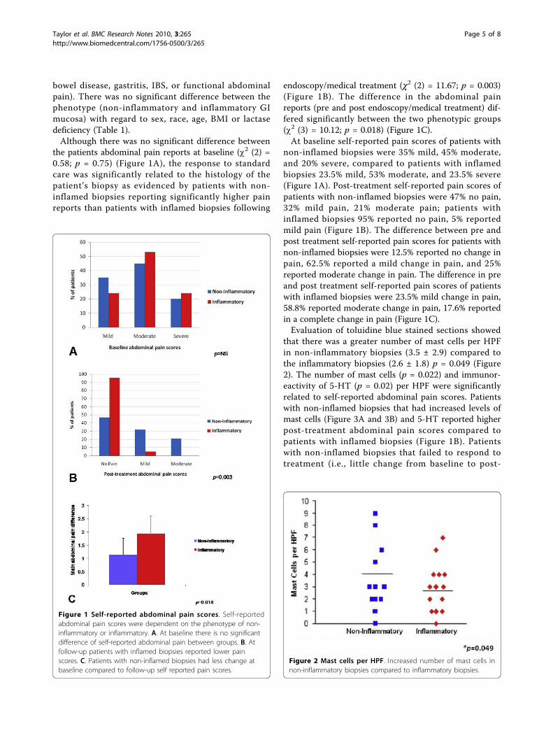
the patients abdominal pain reports at baseline (c2 (2) =0.58; p = 0.75) (Figure 1A), the response to standardcare was significantly related to the histology of thepatient’s biopsy as evidenced by patients with non-inflamed biopsies reporting significantly higher painreports than patients with inflamed biopsies following
endoscopy/medical treatment (c2 (2) = 11.67; p = 0.003)(Figure 1B). The difference in the abdominal painreports (pre and post endoscopy/medical treatment) dif-fered significantly between the two phenotypic groups(c2 (3) = 10.12; p = 0.018) (Figure 1C).At baseline self-reported pain scores of patients with
non-inflamed biopsies were 35% mild, 45% moderate,and 20% severe, compared to patients with inflamedbiopsies 23.5% mild, 53% moderate, and 23.5% severe(Figure 1A). Post-treatment self-reported pain scores ofpatients with non-inflamed biopsies were 47% no pain,32% mild pain, 21% moderate pain; patients withinflamed biopsies 95% reported no pain, 5% reportedmild pain (Figure 1B). The difference between pre andpost treatment self-reported pain scores for patients withnon-inflamed biopsies were 12.5% reported no change inpain, 62.5% reported a mild change in pain, and 25%reported moderate change in pain. The difference in preand post treatment self-reported pain scores of patientswith inflamed biopsies were 23.5% mild change in pain,58.8% reported moderate change in pain, 17.6% reportedin a complete change in pain (Figure 1C).Evaluation of toluidine blue stained sections showed
that there was a greater number of mast cells per HPFin non-inflammatory biopsies (3.5 ± 2.9) compared tothe inflammatory biopsies (2.6 ± 1.8) p = 0.049 (Figure2). The number of mast cells (p = 0.022) and immunor-eactivity of 5-HT (p = 0.02) per HPF were significantlyrelated to self-reported abdominal pain scores. Patientswith non-inflamed biopsies that had increased levels ofmast cells (Figure 3A and 3B) and 5-HT reported higherpost-treatment abdominal pain scores compared topatients with inflamed biopsies (Figure 1B). Patientswith non-inflamed biopsies that failed to respond totreatment (i.e., little change from baseline to post-
Figure 1 Self-reported abdominal pain scores. Self-reportedabdominal pain scores were dependent on the phenotype of non-inflammatory or inflammatory. A. At baseline there is no significantdifference of self-reported abdominal pain between groups. B. Atfollow-up patients with inflamed biopsies reported lower painscores. C. Patients with non-inflamed biopsies had less change atbaseline compared to follow-up self reported pain scores.
Figure 2 Mast cells per HPF. Increased number of mast cells innon-inflammatory biopsies compared to inflammatory biopsies.
Taylor et al. BMC Research Notes 2010, 3:265http://www.biomedcentral.com/1756-0500/3/265
Page 5 of 8
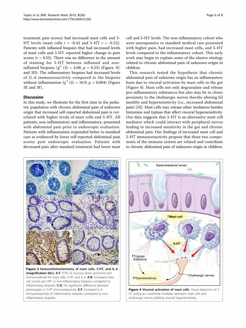
treatment pain scores) had increased mast cells and 5-HT levels (mast cells r = -0.42 and 5-HT r = -0.52).Patients with inflamed biopsies that had increased levelsof mast cells and 5-HT reported higher change in painscores (r = 0.55). There was no difference in the amountof staining for 5-HT between inflamed and non-inflamed biopsies (c2 (3) = 4.08; p = 0.25) (Figure 3Cand 3D). The inflammatory biopsies had increased levelsof IL-6 immunoreactivity compared to the biopsieswithout inflammation (c2 (2) = 10.9; p = 0.004) (Figure3E and 3F).
DiscussionIn this study, we illustrate for the first time in the pedia-tric population with chronic abdominal pain of unknownorigin that increased self-reported abdominal pain is cor-related with higher levels of mast cells and 5-HT. Allpatients, non-inflammatory and inflammatory, presentedwith abdominal pain prior to endoscopic evaluation.Patients with inflammation responded better to standardcare as evidenced by lower self-reported abdominal painscores post endoscopic evaluation. Patients withdecreased pain after standard treatment had lower mast
cell and 5-HT levels. The non-inflammatory cohort whowere unresponsive to standard medical care presentedwith higher pain, had increased mast cells, and 5-HTlevels compared to the inflammatory cohort. This earlywork may begin to explain some of the elusive etiologyrelated to chronic abdominal pain of unknown origin inchildren.This research tested the hypothesis that chronic
abdominal pain of unknown origin has an inflammatorybasis due to visceral activation by mast cells in the gut(Figure 4). Mast cells not only degranulate and releasepro-inflammatory substances but also may be in closerproximity to the cholinergic nerves thereby altering GImotility and hypersensitivity (i.e., increased abdominalpain) [10]. Mast cells may release other mediators besideshistamine and typtase that affect visceral hypersensitivity.Our data suggests that 5-HT is an alternative mast cellmediator which could interact with peripheral nervesleading to increased sensitivity in the gut and chronicabdominal pain. Our findings of increased mast cell and5-HT immunoreactivity propose that these two compo-nents of the immune system are related and contributeto chronic abdominal pain of unknown origin in children.
Figure 3 Immunohistochemistry of mast cells, 5-HT, and IL-6(magnification 60×). A-F. FFPE GI mucosa block sectioned andimmunostained for mast cells, 5-HT, and IL-6. A-B. Increased mastcell counts per HPF in non-inflammatory biopsies compared toinflammatory biopsies. C-D. No significant difference betweenphenotypes in 5-HT immunoreactivity. E-F. Increased IL-6immunoreactivity of inflammatory biopsies compared to non-inflammatory biopsies.
Figure 4 Visceral activation of mast cells. Visual depiction of 5-HT acting as a potential mediator between mast cells andcholinergic nerves yielding visceral hypersensitivity.
Taylor et al. BMC Research Notes 2010, 3:265http://www.biomedcentral.com/1756-0500/3/265
Page 6 of 8

Current proposed mechanisms of IBS give additionalinformation which may be related to the mechanismbehind chronic abdominal pain of unknown origin. Forexample, in post infectious IBS there may be a transloca-tion of microbiota due to increased mucosa permeabilityallowing for the interaction between inflammatory med-iators and the enteric nervous system thereby stimulatingsmooth muscle motility [20]. Patients with chronicabdominal pain may be affected by a similar mechanismat the molecular level of the GI mucosa. Increased per-meability across the mucosal barrier may occur, in turnleading to hypersensitivity and augmented pain.
LimitationsThere are some limitations to this study including theretrospective nature of data collection using medicalrecord review. Additionally, the limited sample sizemakes the findings not necessarily generalizable to thegreater population of patients with chronic abdominalpain of unknown origin. Furthermore, because of theretrospective nature only associations are presented hereand care should be taken in the interpretation of thesefindings as they do not imply direct causality. A futurestudy could include both direct objective and subjectivemeasures of abdominal pain as well as real-time mea-sures of 5-HT, IL-6, and mast cell activation.
ConclusionsRecent advances on the role of 5-HT in the enteric ner-vous systems and its relationship to intestinal visceralhyperalgesia have contributed significantly to the under-standing of abdominal pain and IBS in adults. Thisincreased knowledge has helped shift the paradigm thatthese disorders are exclusively behavioral in nature andthat pathophysiologic disturbances at the cellular levelexist. To date, the consideration of inflammation andintestinal pain has been traditionally reserved for condi-tions such as Crohn’s disease and ulcerative colitis. Thepreliminary findings presented in this manuscript indi-cate that similar relationships may exist in the pediatricpopulation with chronic abdominal pain of unknownorigin. Future research is needed in order to uncoverthe associated causes of abdominal pain without inflam-matory pathology in pediatric patients. Novel medicaltreatments can arise by unveiling the role of mast cellsand 5-HT in the pathophysiology of chronic abdominalpain of unknown origin in children.
AbbreviationsGI: gastrointestinal; 5-HT: 5-hydroxytryptamine (serotonin); IBS: irritable bowelsyndrome.
AcknowledgementsThe authors would like to acknowledge support from the National Instituteof Nursing Research (NINR) Intramural Research Program (PI: W. Henderson,
1 ZIA NR000018-01 SML, 2009). Additionally, support from SAIC-Fredrickcontractor to National Cancer Institute (NCI Contract No.HHSN261200800001E). The options expressed herein are those of theauthors and do not represent the position of the NIH or the USGovernment. The authors would like to thank Dr. Miriam Anver, Ms. DonnaButcher, and Dr. Mones Abu-Asab for immunohistochemical support. Theauthors would also like to thank Dr. Jessica Gill for manuscript review andMs. Annette Langseder for clinical support.
Author details1National Institute of Nursing Research, National Institutes of Health,Bethesda, MD, USA. 2Center for Pediatric Functional Gastrointestinal andMotility Disorders, Goryeb Children’s Hospital at Atlantic Health, University ofMedicine & Dentistry of New Jersey, Morristown, NJ, USA. 3National CancerInstitute, National Institutes of Health, Bethesda, MD, USA.
Authors’ contributionsTT, NY, RS, DK, WH: all contributed to both the manuscript development andanalysis. All authors read and approved the final draft.
Competing interestsThe authors declare that they have no competing interests.
Received: 6 October 2010 Accepted: 21 October 2010Published: 21 October 2010
References1. Drossman D, Corazziari E, Delvaux M, Spiller RC, Talley NJ, Thompson WG,
Whitehead WE: Rome III; The Functional Gastrointestinal Disorders DegnonAssociates, Inc., Third 2006.
2. Hahn B, Saunders W, Maier W: Differences between individuals with self-reported irritable bowel syndrome (IBS) and IBS-like symptoms. Dig DisSci 1997, 42(12):2585-2590.
3. Petersen S, Brulin C, Bergström E: Recurrent pain symptoms inyoung schoolchildren are often multiple. Pain 2006,121(1-2):145-150.
4. Creed F, Ratcliffe J, Fernandez L, Tomenson B, Palmer S, Rigby C, Guthrie E,Read N, Thompson D: Health-Related Quality of Life and Health CareCosts in Severe, Refractory Irritable Bowel Syndrome. Ann Intern Med2001, 134(9_Part_2):860-868.
5. Russo MW, Gaynes BN, Drossman DA: A National Survey of PracticePatterns of Gastroenterologists With Comparison to the Past TwoDecades. Journal of Clinical Gastroenterology 1999, 29(4):339-343.
6. Youssef NN, Murphy TG, Langseder AL, Rosh JR: Quality of life for childrenwith functional abdominal pain: a comparison study of patients’ andparents’ perceptions. Pediatrics 2006, 117(1):54-59.
7. Whitehead W, Burnett C, Cook E, et al: Impact of IBS on quality of life. DigDis Sci 1996, 41:2248-2253.
8. De Giorgio R, Barbara G: Is irritable bowel syndrome an inflammatorydisorder? Curr Gastroenterol Rep 2008, 10(4):385-390.
9. Barbara G, Stanghellini V, De Giorgio R, Cremon C, Cottrell GS, Santini D,et al: Activated mast cells in proximity to colonic nerves correlate withabdominal pain in irritable bowel syndrome. Gastroenterology 2004,126(3):693-702.
10. Barbara G, Wang B, Stanghellini V, de Giorgio R, Cremon C, Di Nardo G,et al: Mast cell-dependent excitation of visceral-nociceptive sensoryneurons in irritable bowel syndrome. Gastroenterology 2007, 132(1):26-37.
11. Park JH, Rhee PL, Kim HS, Lee JH, Kim YH, Kim JJ, et al: Mucosal mast cellcounts correlate with visceral hypersensitivity in patients with diarrheapredominant irritable bowel syndrome. J Gastroenterol Hepatol 2006, 21(1Pt 1):71-78.
12. Piche T, Saint-Paul MC, Dainese R, Marine-Barjoan E, Iannelli A, Montoya ML,Peyron JF, Czerucka D, Cherikh F, Filippi J, et al: Mast cells and cellularityof the colonic mucosa correlated with fatigue and depression in irritablebowel syndrome. Gut 2008, 57(4):468-473.
13. Akbar A, Yiangou Y, Facer P, Walters JR, Anand P, Ghosh S: Increasedcapsaicin receptor TRPV1-expressing sensory fibres in irritable bowelsyndrome and their correlation with abdominal pain. Gut 2008,57(7):923-929.
14. Mahjoub FE, Farahmand F, Pourpak Z, Asefi H, Amini Z: Mast cell gastritis:children complaining of chronic abdominal pain with histologically
Taylor et al. BMC Research Notes 2010, 3:265http://www.biomedcentral.com/1756-0500/3/265
Page 7 of 8

normal gastric mucosal biopsies except for increase in mast cells,proposing a new entity. Diagn Pathol 2009, 4:34.
15. Shulman RJ, Eakin MN, Czyzewski DI, Jarrett M, Ou CN: Increasedgastrointestinal permeability and gut inflammation in children withfunctional abdominal pain and irritable bowel syndrome. J Pediatr 2008,153(5):646-650.
16. Barbara G: Mucosal barrier defects in irritable bowel syndrome. Who leftthe door open? Am J Gastroenterol 2006, 101(6):1295-1298.
17. Spiller R, Garsed K: Postinfectious irritable bowel syndrome.Gastroenterology 2009, 136(6):1979-1988.
18. Crowell MD: Role of serotonin in the pathophysiology of the irritablebowel syndrome. Br J Pharmacol 2004, 141(8):1285-1293.
19. Costedio MM, Hyman N, Mawe GM: Serotonin and its role in colonicfunction and in gastrointestinal disorders. Dis Colon Rectum 2007,50(3):376-388.
20. Barbara G, Cremon C, Pallotti F, De Giorgio R, Stanghellini V, Corinaldesi R:Postinfectious irritable bowel syndrome. J Pediatr Gastroenterol Nutr 2009,48(Suppl 2):S95-97.
doi:10.1186/1756-0500-3-265Cite this article as: Taylor et al.: The association of mast cells andserotonin in children with chronic abdominal pain of unknownetiology. BMC Research Notes 2010 3:265.
Submit your next manuscript to BioMed Centraland take full advantage of:
• Convenient online submission
• Thorough peer review
• No space constraints or color figure charges
• Immediate publication on acceptance
• Inclusion in PubMed, CAS, Scopus and Google Scholar
• Research which is freely available for redistribution
Submit your manuscript at www.biomedcentral.com/submit
Taylor et al. BMC Research Notes 2010, 3:265http://www.biomedcentral.com/1756-0500/3/265
Page 8 of 8