the bcr/abl oncogene alters the chemotactic response to...
TRANSCRIPT

1999 94: 4233-4246
Andrian, Lan Bo Chen, Jose-Carlos Gutierrez-Ramos, Ann-Marie Pendergast and James D. GriffinKevin M. Klucher, George Q. Daley, Stine K. Kraeft, Robert Sackstein, Edwin P. Alyea III, Ulrich H. von Ravi Salgia, Elizabeth Quackenbush, Jeffrey Lin, Natalia Souchkova, Martin Sattler, Darren S. Ewaniuk, Stromal-Derived Factor-1?The BCR/ABL Oncogene Alters the Chemotactic Response to
http://bloodjournal.hematologylibrary.org/content/94/12/4233.full.htmlUpdated information and services can be found at:
(4217 articles)Neoplasia �Articles on similar topics can be found in the following Blood collections
http://bloodjournal.hematologylibrary.org/site/misc/rights.xhtml#repub_requestsInformation about reproducing this article in parts or in its entirety may be found online at:
http://bloodjournal.hematologylibrary.org/site/misc/rights.xhtml#reprintsInformation about ordering reprints may be found online at:
http://bloodjournal.hematologylibrary.org/site/subscriptions/index.xhtmlInformation about subscriptions and ASH membership may be found online at:
Copyright 2011 by The American Society of Hematology; all rights reserved.20036.the American Society of Hematology, 2021 L St, NW, Suite 900, Washington DC Blood (print ISSN 0006-4971, online ISSN 1528-0020), is published weekly by
For personal use only. at Harvard Libraries on October 18, 2012. bloodjournal.hematologylibrary.orgFrom

NEOPLASIA
The BCR/ABL Oncogene Alters the Chemotactic Responseto Stromal-Derived Factor-1a
By Ravi Salgia, Elizabeth Quackenbush, Jeffrey Lin, Natalia Souchkova, Martin Sattler, Darren S. Ewaniuk,Kevin M. Klucher, George Q. Daley, Stine K. Kraeft, Robert Sackstein, Edwin P. Alyea III, Ulrich H. von Andrian,
Lan Bo Chen, Jose-Carlos Gutierrez-Ramos, Ann-Marie Pendergast, and James D. Griffin
The chemokine stromal-derived factor-1a (SDF-1a) is a che-
moattractant for CD341 progenitor cells, in vitro and in vivo.
The receptor for SDF-1a, CXCR-4, is a 7 transmembrane
domain receptor, which is also a coreceptor for human
immunodeficiency virus (HIV). Here we show that transforma-
tion of hematopoietic cell lines by BCR/ABL significantly
impairs their response to SDF-1a. Three different hematopoi-
etic cell lines, Ba/F3, 32Dcl3, and Mo7e, were found to
express CXCR-4 and to respond to SDF-1a with increased
migration in a transwell assay. In contrast, after transforma-
tion by the BCR/ABL oncogene, the chemotactic response to
SDF-1a was reduced in all 3 lines. This effect was directly due
to BCR/ABL, because Ba/F3 cells, in which the expression of
BCR/ABL could be regulated by a tetracycline-inducible
promoter, also had reduced chemotaxis to SDF-1a when
BCR/ABL was induced. The reduced response to SDF-1a was
not due to an inability of BCR/ABL-transformed cell lines to
migrate in general, as spontaneous motility of BCR/ABL-
transformed cells was increased. In mice, injection of SDF-1ainto the spleen resulted in a transient accumulation of
untransformed Ba/F3 cells, but not Ba/F3.p210BCR/ABL cells
administered simultaneously. The mechanism may involve
inhibition of CXCR-4 receptor function, because in BCR/ABL-
transformed cells, CXCR-4 receptors were expressed on the
cell surface, but SDF-1a calcium flux was inhibited. Because
SDF-1a and CXCR-4 are felt to be involved in progenitor cell
homing to marrow, the abnormality decribed here could
contribute to the homing and retention defects typical of
immature myeloid cells in chronic myelogenous leukemia.
r 1999 by The American Society of Hematology.
CHRONIC MYELOGENOUS leukemia (CML) is a myelo-proliferative disorder caused by the BCR/ABL fusion
oncogene. CML is characterized by premature release ofmyeloid cells from the marrow and massive accumulation ofboth mature and immature myeloid cells in blood, spleen, andmarrow.1 There is growing evidence to suggest that the interac-tion of CML cells with the marrow microenvironment isabnormal. Gordon et al2 showed in 1987 that CML progenitorcells had diminished capacity to adhere to stromal cell layers.More recently, it was reported that CML cells have reducedlong-term adhesion to the extracellular matrix protein fibronec-tin, and this impairment was thought to be due to abnormalfunction ofb1 integrins.3,4 p210BCR/ABL is partially localized tothe cytoskeleton, and we have previously shown that severalcytoskeletal proteins associated with integrin regulation, suchas paxillin, FAK, CRKL, and vinculin, are prominent substratesof the BCR/ABL tyrosine kinase.5,6 The functions of theseproteins are altered by phosphorylation and/or direct interactionwith p210BCR/ABL, presumably contributing to the abnormalintegrin function in CML.3
The ability of CML cells to leave the marrow at an immaturestage of differentiation, circulate in high numbers in the blood,and accumulate in the spleen indicates that events that normallyregulate migration and retention of hematopoietic progenitorcells are also defective. We have previously shown thatspontaneous motility of BCR/ABL-transformed cells on fibro-nectin-coated surfaces is increased, and this prompted us to askif more specific migration and retention functions of CMLprogenitor cells might also be abnormal.7
The events controlling homing of CD341 cells to marrow andthe retention of normal immature myeloid cells in the marroware not well understood. Recently, however, the chemokinestromal-derived factor-1a (SDF-1a), which is produced bymarrow stromal cells, has been shown to be a potent chemoat-tractant for human CD341 progenitor cells.8 SDF-1a is a mem-ber of thea (CXC) chemokine family and it has homology tointerleukin-8 (IL-8) and MIP-1a.9 However, unlike these chemo-
kines, SDF-1aelicits transendothelial chemotaxis of humanCD341 cells both in vitro and in vivo.8,10 SDF-1a11 andCXCR-412,13 null mutant mice have profound defects in myeloidand lymphoid development, further suggesting that this chemo-kine is important in regulating hematopoietic cell migration.
In this report, we provide evidence that BCR/ABL transfor-mation inhibits migration of hematopoietic cells in response toSDF-1a in vitro and in vivo. BCR/ABL did not affect expres-sion of SDF-1areceptors, but directly inhibited signal transduc-tion. This defect may explain some of the homing and retentiondefects typical of immature myeloid cells in CML.
From the Department of Medical Oncology and Division of Hemato-logic Oncology, Division of Cellular and Molecular Biology, Dana-Farber Cancer Institute, Harvard Medical School, Boston, MA; Centerfor Blood Research and Harvard Medical School, Boston, MA; TheChildren’s Hospital, Boston, MA; Whitehead Institute BiomedicalResearch, Cambridge, MA; Department of Pharmacology and CancerBiology, Duke University Medical Center, Levine Science ResearchCenter, Durham, NC; Millennium Pharmaceuticals Inc, Cambridge,MA; and Department of Medicine, Brigham and Women’s Hospital/Massachusetts General Hospital, Boston, MA.
Submitted January 25, 1999; accepted August 10, 1999.R.S. and E.Q. contributed equally to this work.Supported by National Institutes of Health Grants No. CA 75348
(R.S.), DK 560654 (J.D.G.), Jose Carreras International LeukemiaFoundation fellowship FIJC-95/INT (M.S.), and the Pfizer ScholarsGrant for new faculty (E.Q.)
Address correspondence to Ravi Salgia, MD, PhD, Department ofAdult Oncology, Dana 530C, Dana-Farber Cancer Institute, 44 BinneySt, Boston, MA 02115; e-mail: [email protected].
The publication costs of this article were defrayed in part by pagecharge payment. This article must therefore be hereby marked‘‘adver-tisement’’ in accordance with 18 U.S.C. section 1734 solely to indicatethis fact.
r 1999 by The American Society of Hematology.0006-4971/99/9412-0037$3.00/0
Blood, Vol 94, No 12 (December 15), 1999: pp 4233-4246 4233
For personal use only. at Harvard Libraries on October 18, 2012. bloodjournal.hematologylibrary.orgFrom

MATERIALS AND METHODS
Cells and cell culture. The murine hematopoietic cell lines Ba/F3and 32Dcl3 (also identified as 32D) were cultured at 37°C with 5%CO2 in RPMI 1640 (Mediatech, Washington, DC) containing 10%(vol/vol) WEHI-3B conditioned medium as a source of interleukin-3and 10% (vol/vol) fetal calf serum (FCS) (PAA Laboratories Inc,Newport Beach, CA). The human megakaryoblastic cell line Mo7ewas maintained in Dulbecco’s Modified Eagle’s Medium (DMEM;Mediatech), 10 ng/mL granulocyte-macrophage colony-stimulatingfactor (GM-CSF: Genetics Institute, Cambridge, MA), and 20%(vol/vol) FCS at 37°C with 10% CO2. The BCR/ABL-expressing celllines Ba/F3.p210BCR/ABL, Ba/F3.p185BCR/ABL, 32D.p210BCR/ABL,32D.p185BCR/ABL , and Mo7e.p210BCR/ABL were generated by transfec-tion with the pGD vector containing the BCR/ABL cDNA, as previ-ously described.14 p185 and p210 are different forms of BCR/ABL.BCR/ABL-containing cells were cultured in RPMI 1640 medium with10% FCS, but without any source of IL-3 or GM-CSF. A Ba/F3 cell line,TonB210.1, with tetracycline-dependent BCR/ABL expression, wasgrown in RPMI 1640 medium containing 10% WEHI-conditionedmedium and 10% FCS.15 For induction experiments of less than 5hours, cells were growth factor deprived for 18 hours in RPMI 1640containing 0.5% bovine serum albumin (BSA). For induction experi-ments longer than 1 day, cells were grown in RPMI 1640 containingWEHI. The BCR/ABL expression was induced by treatment withdoxycycline (2 µg/mL) for 1 day.
Conditioned medium from the bone marrow-derived mouse stromalcell line, MS-5, was used as one source of SDF-1a.8 Once the growingcells reached subconfluence, the medium was replaced with serum-freemedium (UltraCULTURE; Biowhittaker, Walkersville, MD), whichwas collected after 2 days of culture and passed through a 0.45-mmfilter. Recombinant, purified human SDF-1a was purchased from R&DSystems (Minneapolis, MN).
Confocal microscopy. F-actin was visualized in fixed cells (1%paraformaldehyde in phosphate-buffered saline [PBS]) using rhoda-mine phalloidin (Molecular Probes, Eugene, OR), as described.5 Thefocal adhesion protein paxillin was visualized using indirect immunoflu-orescence with the antipaxillin monoclonal antibody clone 5H11 (UBI;Lake Placid, NY), detected by a fluorescein isothiocyanate (FITC)-conjugated goat antimouse antibody. Confocal image analysis wasperformed using a Zeiss model LSM4 confocal laser scanning micro-scope (Zeiss, Jena, Germany) equipped with an external argon-kryptonlaser (488 nm and 568 nm). Optical sections of 5123 512 pixels weredigitally recorded within 2 seconds and 23 line-averaging. Images wereprinted with a Fujix Pictography 3000 printer (Fuji, Japan) using AdobePhotoshop software (Adobe Systems, Mountain View, CA).
Scanning electron microscopy.Cells grown in suspension wereconcentrated to 13 105 cells/mL, and the cells were either unstimulatedor stimulated with SDF-1a. One drop of such suspension was placedonto a plastic cover slip previously coated with 1% poly-L-lysine(Sigma, St. Louis, MO) in water. The coverslip was then allowed tostand in a small Petri dish at room temperature for 15 to 30 minutes inorder for the cells to adhere to the slip. Fixative (1.5% glutaradehyde in0.01 mol/L phosphate buffer, pH 7.4) was added to the petri dish tocover the slip and the cells were fixed for 1 hour at 40°C. The coverslipwas then taken through graded alcohols and dried in a Ladd CriticalPoint Dryer (Model 28000; Ladd Research Industries, Williston, VT),coated with platinum in a polaron SEM coating system. Samples wereexamined with a JEOL JSM-35 CF scanning electron microscope at anaccelerating voltage of 20 KV.
Calcium flux analysis. Cells were resuspended and analyzed forcalcium flux using fluorescent indicator indo-1 AM (Molecular Probes),at a final concentration of 5 µg/mL at 37°C for 30 minutes. Cells werewashed with PBS and starved in RPMI 1640 media containing 10%FCS, 1% penicillin-streptomycin for 10 hours at 53 105/mL. Afterstarving in the absence of growth factors, 33 106 cells were treated
with pertussis toxin (Sigma) at 100 ng/mL. Then the remaining cellswere suspended in starvation media at 13 107 per mL. Indo-1 AM (1mg/mL in DMSO) was added to the resuspending cells, making theconcentration 1:100, and the cells were placed in the dark for 30minutes. Afterwards, they were washed and resuspended in starvationmedia at 53 105 per mL. Finally, 1 mL of resuspended cells was placedinto a FACS analysis tube. Samples were analyzed on an EPICS ELITE(Coulter Corporation, Miami, FL) flow cytometer equipped with anultraviolet enhanced Argon Ion laser tuned to 351 to 363 nm (10nWoutput power.) The fluorescent emission of indo-1 AM loaded cells wasdetected by measuring both the violet bound form (405 nm) andblue/green (525 nm) unbound form of the dye in separate photomulti-plier tubes (PMT). A ratio parameter was created using the 405 nm PMTas the denominator and the 525nm PMT as the numerator.
Recombinant SDF-1a was used at a concentration of 1 µg/mL, whileionomycin (Sigma) was used at 5 µg/mL. The tube was then placed backon the cytometer and reanalyzed for 3 minutes to detect any shift in theratio of 525 nm/405 nm fluorescence. Data was initially analyzed usingthe Multigraph analysis program that is standard for the instrument.Processed 2 parameter histograms portraying ratio (ordinate) versustime (abscissa) were then subsequently analyzed for degree of respon-siveness using the Multitime Software program (Phoenix Flow Sys-tems) and plotted graphically as percent response versus time.
Adhesion assay.Adhesion of hematopoietic cells (with and withoutBCR/ABL) was measured on plastic plates that were uncoated, orcoated with bovine serum albumin (5µg/mL) or fibronectin (5µg/mL)(Becton Dickinson Labware, Bedford, MA), as described previously.16
Time-lapse video microscopy.Cells were cultured on fibronectin-coated tissue culture plates (Becton Dickinson Labware) in a tempera-ture and CO2 controlled chamber in their standard growth media andstimulated with either SDF-1a or supernatant from the MS-5 cell line(1:1).7 The cells were examined using an Olympus IX70 invertedmicroscope (Olympus, Lake Success, NY), Omega temperature controldevice (Therm-Omega-Tech, Warmington, PA), Optronics EngineeringDEI-750 3CCD digital video camera (Optronics, Galeta, CA), and SonySVT-S3100 time-lapse S-VHS video recorder (Sony, Tokyo, Japan). Forimage presentation, video images were captured and printed with theSony Color Video Printer UP-5600MD.
Chemotaxis assay.Chemotaxis assays were performed using thechemotaxis microplate system (Neuro Probe, Inc, Cabin John, MD).Cells were incubated for 4 hours in IL-3– and GM-CSF–deficient mediaat 37°C, resuspended in serum-free medium, and rewarmed to 37°C for20 minutes before loading. UltraCULTURE alone was used as a nega-tive control and SDF-1a was diluted to 100 ng/mL in UltraCULTUREmedium. MS-5 supernatant was used undiluted. One3 105 cells (in 25µL) were allowed to migrate for 3 hours at 37°C, and migrating cellswere resuspended in 200 µL of 1% formalin in phosphate-bufferedsaline (PBS), then counted for 30 seconds on a flow cytometer (BectonDickinson, Mountain View, CA). The chemotactic index, a measure ofthe specificity of migration, was determined by dividing: (the number ofcells migrating to chemokine)/(the number that migrated to mediumalone). For blocking studies, 50 µg/mL of anti–SDF-1a antibody (R&DSystems) was added to the chemokine-containing medium 30 minutesbefore initiation of chemotaxis.
Detection of CXCR-4 by immunoblotting and immunofluorescentstaining. Immunoblots using the anti–CXCR-4 rabbit polyclonalantibody were performed as previously described.17 For indirectimmunofluorescent staining, cells (13 106) were washed and resus-pended in PBS containing 0.1% bovine serum albumin and 0.1%sodium azide (staining buffer). The cells were incubated for 30 minutesat 4°C with mouse antihuman CXCR-4 (R&D Systems; 0.1 mg/mL finalconcentration) or rabbit-antimouse-CXCR-4 (1:1000) polyclonal anti-body, washed 3 times in staining buffer, and incubated with fluoresceinisothiocyanate (FITC)-conjugated goat-antimouse or goat-antirabbitantibody (30 minutes, 4°C). After 3 washes, the stained cells were
4234 SALGIA ET AL
For personal use only. at Harvard Libraries on October 18, 2012. bloodjournal.hematologylibrary.orgFrom

analyzed by FACS-sort flow cytometry using Cell Quest software(Becton Dickinson).
Reverse transcription-polymerase chain reaction (RT-PCR).Onemicrogram of RNA was reverse transcribed using a Perkin ElmerRT-PCR kit (Roche Molecular Systems, Inc, Branchburg, NJ). RTreactions were brought to a final volume of 40 µL, and cyclingconditions were 25°C3 10 minutes, 42°C3 1 hour, then 99°C3 5minutes. Primer sequences for SDF-1a are: (58)GACGGTAAACC-AGTCAG and (38)ACTGCCCTTGCATCTCCCCAC; mouse CXCR-4:(58)GGCTGTAGAGCGAGTGTTGCC and (38)GTAGAGGTTGACA-GTGTAGAT; and mouseb-actin: (58)TGGAATCCTGTGGCATCCAT-GAAAC and (38)TAAAACGCAGC-TCAGTAACAGTCCG. PCR wasconducted using 1 to 5 µL of the RT products in a final volume of 25 µL,for 30 cycles (94°C, 1 minute; 54°C, 30 seconds; 72°C, 30 seconds).18
Homing assays. Ba/F3 and Ba/F3.p210BCR/ABL cells were washedthree times in DMEM (Biowhittaker) containing 20 mmol/L HEPESand 1% FCS, pH 7.4. Cells were resuspended at 203 106/mL and thenincubated for 20 minutes with TRITC (30 µg/mL; Molecular Probes) orCalcein-AM (200 nmol/L; Molecular Probes).19 Labeling was per-formed for 10 minutes at 37°C and cells were mixed frequently duringthe incubation. Cells were then spun through an equal volume of freshFCS or 20% BSA at 1300 rpm for 10 minutes. Viability was checkedand cells were rewarmed at 37°C for 15 minutes before injection.
For homing experiments, C57BL/6 mice of both sexes were anesthe-tized by intraperitoneal injection of physiological saline containingketamine-HCL (5 mg/mL) and xylazine (1 mg/mL) under InstitutionalReview Board approved protocols.19 The left carotid artery wascannulated with PE-10 polyethylene tubing to allow injection of cellsinto the descending aorta. Just before cell injection, a left flank incisionwas made and the exposed spleen was injected with 50 µL of PBS aloneor PBS containing 1 µg of SDF-1a. After suturing the incision, equalamounts of labeled Ba/F3 and Ba/F3.p210BCR/ABL cells were pooled forinjection (total volume of 0.5 mL, containing 107 cells). The cells wereinjected slowly, over a 15 to 20 minute period, and the animals wereallowed to awaken. After 3 hours, the animals were sacrificed andspleen, lungs, bone marrow, and peripheral blood (via cardiac puncture)were harvested. Single-cell suspensions were prepared from mincedtissues by gently passing them through a 70µ nylon mesh. Thesingle-cell suspensions were separated on a Histopaque-1077 (Sigma)gradient and the cell populations recovered at the gradient interfacewere washed in DMEM, pelleted and fixed in 4% paraformaldehyde foranalysis on a FACScan flow cytometer (Becton Dickinson, San Jose,CA). The residual red and green fluorescent cells present in the catheterafter injection were counted to determine the ratio of each cell line in theinput population. Between 5 to 103 105 cells were analyzed for eachtissue and fluorescent cells were quantitated within a gated regionwhose borders were determined from the injected pool. The percentagesof gated cells recovered in each organ were corrected for differences inthe percentage of each labeled cell type in the input population.
In vitro methylcellulose colony forming unit (CFU) assay.Chemo-taxis assays were performed using bone marrow (BM) cells harvestedfrom patients with untreated CML or from normal donors with IRBapproved protocol. Peripheral blood samples from patients with un-treated CML were also tested. Mononuclear cell populations wereobtained using Histopaque-1077, and the cells were washed 3 times at4oC in UltraCULTURE medium. Immunofluorescent staining withmonoclonal antibodies to human CD34 (Pharmingen, San Diego, CA)and CXCR-4 (R&D Systems) was done on selected samples. Chemo-taxis assays were performed, as described above, using the CorningCostar Transwell assay (5 micron pore) and SDF-1a (100 ng/mL) orUltraCULTURE alone (control). Inserts were loaded with 100 µLcontaining 53 105 viable cells (input population) and the assay wasallowed to proceed for 3 hours at 37°C. Separate aliquots of input cells(10, 20, and 60 µL) were also plated in methylcellulose to determine theCFU-GM capacity of the input population.
The migratory cells (output population) were harvested, diluted withUltraCULTURE, spun gently at 900 rpm for 10 minutes and thesupernatants were aspirated carefully. The cell pellets were resuspendedin 700 µL of a cocktail containing GM-CSF (50 ng/mL). The cocktailwas prepared as described.8 An equal volume of methylcellulose wasadded to the tube, and the cells, GM-CSF cocktail, and methylcellulosewere mixed using a 3 mLsyringe and 16 gauge needle. Bubbles wereallowed to dissipate and the mixture was plated into 353 10 mm petridishes. Colonies were counted after 7 days in culture. Colonies weredefined as containing greater than 50 cells and clusters less than 50cells. The total number of colonies and clusters were scored by ablinded observer in 10 high power fields. The percent CFU-GM wereplotted as: (total number of colonies and clusters obtained with theoutput population)/(the number of colonies and clusters obtained withthe input population)3 100. Colony counts obtained with input cellswere multiplied by the appropriate correction factor to determine thetotal CFU-GM capacity of 100 µL (53 105) of input cells.
Detection of p70 S6 kinase by immunoblotting.The murine Ba/F3and Ba/F3.p210BCR/ABL cell lines were washed in the absence of IL-3and lysed in lysis buffer (20 mmol/L Tris pH 8.0, 150 mmol/L NaCl,10% glycerol, 1% NP-40, 0.42% NaF) containing inhibitors (10 µL of100 µmol/L PMSF, 10 µL of 100 mmol/L Na3VO4, 5 µL aprotinin(Sigma), and 2 µL of 10 mg/mL leupeptin). Whole cell lysates (25 µg)were separated by a 7.5% SDS-PAGE, and electrophoretically trans-ferred to Immobilon PVDF membrane (Millipore, Bedford, MA). Themembrane was immunoblotted with the following rabbit polyclonalantibodies: phosphospecific p70 S6 kinase (Thr421/Ser424) antibody(1:1000, rabbit polyclonal; New England Biolabs, Beverly, MA) and S6kinase antibody (1:1000, rabbit polyclonal; New England Biolabs).
RESULTS
Expression of CXCR-4 receptors in hematopoietic cell linesand their BCR/ABL-transformed counterparts.The expres-sion of CXCR-4 and SDF-1a was determined by RT-PCR in theIL-3–dependent murine cell lines Ba/F3 (pre-B) and 32D(myeloid), before and after transformation to growth factor-independence by BCR/ABL (Fig 1A). Expression of CXCR-4messenger RNA (mRNA) was detected in all cell lines tested. Inaddition, FACS analysis using a rabbit polyclonal antibody tomurine CXCR-4 showed surface expression of CXCR-4 recep-tors (Fig 1B). Using an antihuman CXCR-4 antibody, surfaceexpression of CXCR-4 was also found to be equivalent in themegakaryoblastic cell lines Mo7e and Mo7e.p210BCR/ABL (Fig1B). SDF-1aexpression was weakly detected in K562 cell linesby RT-PCR but not in the 2 murine cell lines. However,conditioned medium from K562 cells was unable to inducecytoskeletal changes or migration of Ba/F3 cells (data notshown), suggesting that there was no significant secretion ofSDF-1a. CXCR-4 was also detected by immunoblotting in 32Dand Ba/F3 cells and their BCR/ABL transformed counterpartsusing an antimurine CXCR-4 antibody (Fig 1C), which has avery low cross-reactivity with human cell lines.
SDF-1a induces actin cytoskeleton changes in untrans-formed, but not in BCR/ABL-transformed, hematopoietic celllines. Ba/F3 and Ba/F3.p210BCR/ABL cells were treated withSDF-1aand examined for changes in cytoskeletal morphologyand actin content by confocal microscopy. SDF-1a increasedthe content of F-actin in Ba/F3 cells and also induced amorphological change characterized by polarization, increasedlamellipodia, filopodia, and uropod-like structures (Fig 2A).The focal adhesion protein paxillin was detected in lamellipodiaupon stimulation with SDF-1a. F-actin and paxillin both form
DECREASED RESPONSE TO SDF-1a IN CML CELLS 4235
For personal use only. at Harvard Libraries on October 18, 2012. bloodjournal.hematologylibrary.orgFrom

punctate (podosome-like) structures at sites of contact betweenthe cell and the substratum in the BCR/ABL transformed cells,and no changes were appreciated after stimulation by SDF-1a.Similar morphological changes were observed by scanningelectron microscopy (Fig 2B).
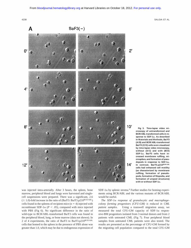
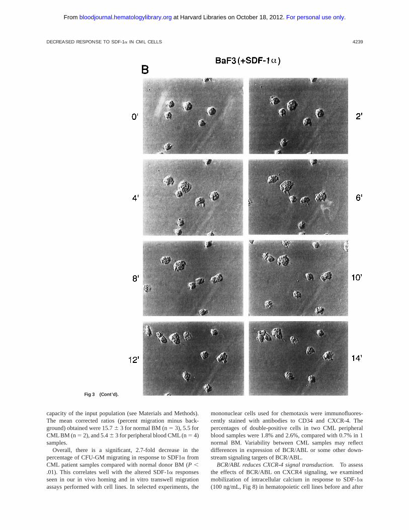
SDF-1aalters cell motility of untransformed hematopoieticcell lines, but not BCR/ABL-transformed cell lines.Becausethere is actin cytoskeleton rearrangement and altered adhesionin response to SDF-1a, we asked if SDF-1a also affectsspontaneous in vitro motility of untransformed and BCR/ABL-transformed cell lines. Time-lapse video microscopy (TLVM)showed that unstimulated Ba/F3 cells exhibited a round morphol-ogy with little movement on a fibronectin-coated surface (Fig3). In response to SDF-1a, however, Ba/F3 cells underwent adramatic increase in spontaneous motility. BCR/ABL-trans-formed Ba/F3 cells, in contrast, constitutively exhibited a highdegree of spontaneous motility in the absence of SDF-1a. Incontrast to untransformed cells, these transformed cells did notfurther increase their spontaneous motility in response to
SDF-1a. Similar results were observed comparing 32D and the32D.p210BCR/ABL cells (data not shown).
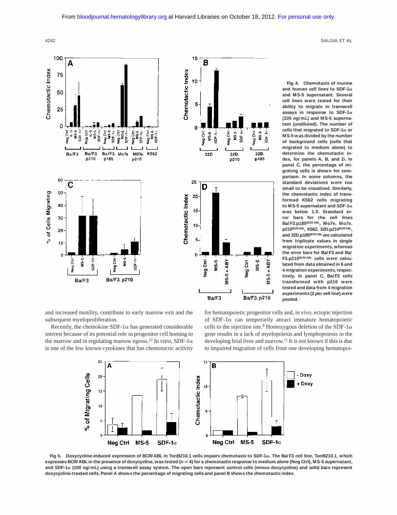
BCR/ABL-transformed cell lines have a reduced chemotacticresponse to SDF-1a. Using a transwell migration assay, thechemotactic index of Ba/F3, 32D, Mo7e cells, and theirBCR/ABL-transformed counterparts was determined (Fig 4).Ba/F3 cells have a dramatic chemotactic response to eithersupernatant harvested from the murine bone marrow-derivedstromal cell line MS-5 (as a source of SDF-1a) or recombi-nant SDF-1a, whereas BCR/ABL-transformed cells (both p185and p210 forms of BCR/ABL) are less responsive. Similarresults were obtained when comparing Mo7e cells andMo7e.p210BCR/ABL cells. Untransformed 32D cells did not havea high chemotactic response to SDF-1a, but again, BCR/ABL-transformed 32D cells had an even lower response. Both thepercent migrating cells and the chemotactic index were reducedin BCR/ABL-transformed cells (Fig 4C). Also, using a doxycy-cline-inducible BCR/ABL cell line, TonB210.1,15 we found thatactivation of BCR/ABL was associated with a reduced chemo-
Fig 1. Analysis of SDF-1a mRNA and CXCR-4
expression in murine and human cell lines with and
without BCR/ABL. (A) The expression of SDF-1a and
CXCR-4 mRNA was evaluated by RT-PCR in the
murine pre-B cell line Ba/F3, the murine myeloid cell
line 32D, and in their BCR/ABL-transformed counter-
parts. The human erythroleukemia cell line K562 was
also evaluated. Primers for b-actin were used to
equalize the amount of RT-products used. (B) Immu-
nostaining with antibodies to murine CXCR-4 was
performed with the Ba/F3 and Ba/F3.p210BCR/ABL cell
lines (solid histogram). Background staining was
determined using a nonspecific isotype matched IgG
(clear histogram). Immunostaining with antibodies
to human CXCR-4 was performed with the human
Mo7e, Mo7e.p210BCR/ABL megakaryocytic, and K562
cell lines (solid histogram). Background staining (clear
histogram) was determined using a nonspecific iso-
type matched IgG. (C) Expression of murine CXCR-4
by immunoblotting of protein extracts prepared from
Ba/F3, 32D, Mo7e, and their BCR/ABL counterparts.
The molecular weight marker of 45 kD is shown and
CXCR-4 has an approximate molecular weight of
48 kD.
4236 SALGIA ET AL
For personal use only. at Harvard Libraries on October 18, 2012. bloodjournal.hematologylibrary.orgFrom

tactic response to SDF-1a (Fig 5). We found an averagedecrease of 2.9-fold in migration of doxycyline-induced BCR/ABL in the TonB210.1 cell line (n5 4). Uninduced TonB210.1cells have a lower migratory response to SDF-1a comparedwith Ba/F3 cells, possibly because these cells express low levelsof p210BCR/ABL even in the absence of doxycycline induction(data not shown).
To ensure that the response of hematopoietic cells to theMS-5 supernatant was due to SDF-1a, we determined thechemotactic index in the presence of an SDF-1a blockingantibody (Fig 4D). The chemotactic response of Ba/F3 cells toMS-5 supernatant was nearly completely abrogated by ablocking antibody, indicating that the chemotactic response ofthese cells in the transwell assay was due to the SDF-1apresentin the MS-5 supernatant.
When Mo7e and Ba/F3 cell lines were pretreated for 4 hours
with pertussis toxin (100 ng/mL), transwell migratory responseto SDF-1a was markedly reduced (Mo7e: 47 %v 8 %; Ba/F3:26%v 6%, without or with pertussis toxin, respectively).
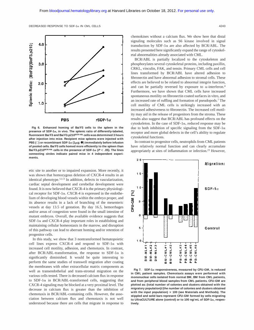
Homing of untransformed and BCR/ABL-transformed cellsin response to SDF-1a, in vivo. To test the in vivo homing ofcells to SDF-1a, we injected SDF-1a(1 µg in 50 µL of PBS) oran equal volume of PBS into a subcapsular site in the spleens ofanesthetized C57Bl/6 mice, and then injected Ba/F3 andBa/F3.p210BCR/ABL cells intra-arterially. The Ba/F3 and Ba/F3.p210BCR/ABL cells were differentially-labeled with the fluores-cent dyes, calcein-AM (green) or TRITC (red), respectively.Two-color flow cytometry was then used to quantitate these 2populations in single-cell suspensions of different organs har-vested 3 hours later. Both dye combinations were tested, withsimilar results found. Equal numbers of labeled wild-type andtransformed cells (53 106 of each) were mixed and the pool
Fig 2. (A) Actin and paxillin staining, as visualized by confocal microscopy, of untransformed and BCR/ABL-transformed hematopoietic cells
in response to SDF-1a. Cells were fixed and stained for actin using rhodamine-labeled phalloidin, and for paxillin using indirect immunofluores-
cent staining with the antipaxillin monoclonal antibody 5H11. Note the difference in shape and staining pattern of actin and paxillin in response
to SDF-1a of normal Ba/F3 cells. There is no change in shape or staining pattern in response to SDF-1a for BCR/ABL-transformed Ba/F3 cells. The
bar is 10 µm. (B) Scanning electron microscopy of untransformed and BCR/ABL-transformed hematopoietic cells in response to SDF-1a. Ba/F3
cells and BCR/ABL-transformed Ba/F3 cells were either unstimulated or stimulated with SDF-1a and scanning electron micrographs were taken.
Shown is the ruffling of an untransformed Ba/F3 cell; BCR/ABL containing cells had numerous extensions but there was no change in response to
SDF-1a. The bar represents 1.0 U.
DECREASED RESPONSE TO SDF-1a IN CML CELLS 4237
For personal use only. at Harvard Libraries on October 18, 2012. bloodjournal.hematologylibrary.orgFrom
was injected intra-arterially. After 3 hours, the spleen, bonemarrow, peripheral blood and lungs were harvested and single-cell suspensions were prepared. There was a significant, 2.6(6 1.0)-fold increase in the ratio of (Ba/F3: Ba/F3.p210BCR/ABL)cells found in the spleens of recipient mice (n5 4) injected withrecombinant SDF-1a (P , .05), compared with mice injectedwith PBS (Fig 6). No significant difference in the ratio ofwild-type to BCR/ABL-transformed Ba/F3 cells was found inthe peripheral blood, lung, or bone marrow (data not shown). In2 of 4 experiments, the ratio of Ba/F3 to Ba/F3.p210BCR/ABL
cells that homed to the spleen in the presence of PBS alone wasgreater than 1.0, which may be due to endogenous expression of
SDF-1aby splenic stroma.8 Further studies for homing experi-ments using BCR/ABL and the various mutants of BCR/ABLwould be useful.
The SDF-1a response of granulocytic and macrophage-colony forming progenitors (CFU-GM) is reduced in CMLpatient samples. Using a transwell migration assay, wemeasured the total CFU-GM capacity of SDF-1a respon-sive-BM progenitors isolated from 3 normal donors and from 2patients with untreated CML (Fig 7). Four peripheral bloodsamples from untreated CML patients were also tested. Theresults are presented as the percentage of CFU-GM formed bythe migrating cell population compared to the total CFU-GM
Fig 3. Time-lapse video mi-
croscopy of untransformed and
BCR/ABL transformed cells in re-
sponse to SDF-1a. As described
in Materials and Methods, Ba/F3
(A/B) and BCR/ABL-transformed
Ba/F3 (C/D) cells were visualized
by time-lapse video microscopy,
without (A/C) and with (B/D)
SDF-1a. Ba/F3 cells have in-
creased membrane ruffling, mi-
crospikes, and formation of pseu-
dopods in response to SDF-1a.
In contrast, Ba/F3.p210BCR/ABL
cells had enhanced cell motility
(as characterized by membrane
ruffling, formation of pseudo-
pods, formation of filopodia, and
formation of uropod structures)
with or without SDF-1a.
4238 SALGIA ET AL
For personal use only. at Harvard Libraries on October 18, 2012. bloodjournal.hematologylibrary.orgFrom
capacity of the input population (see Materials and Methods).The mean corrected ratios (percent migration minus back-ground) obtained were 15.76 3 for normal BM (n5 3), 5.5 forCML BM (n 5 2), and 5.46 3 for peripheral blood CML (n5 4)samples.
Overall, there is a significant, 2.7-fold decrease in thepercentage of CFU-GM migrating in response to SDF1a fromCML patient samples compared with normal donor BM (P ,.01). This correlates well with the altered SDF-1aresponsesseen in our in vivo homing and in vitro transwell migrationassays performed with cell lines. In selected experiments, the
mononuclear cells used for chemotaxis were immunofluores-cently stained with antibodies to CD34 and CXCR-4. Thepercentages of double-positive cells in two CML peripheralblood samples were 1.8% and 2.6%, compared with 0.7% in 1normal BM. Variability between CML samples may reflectdifferences in expression of BCR/ABL or some other down-stream signaling targets of BCR/ABL.
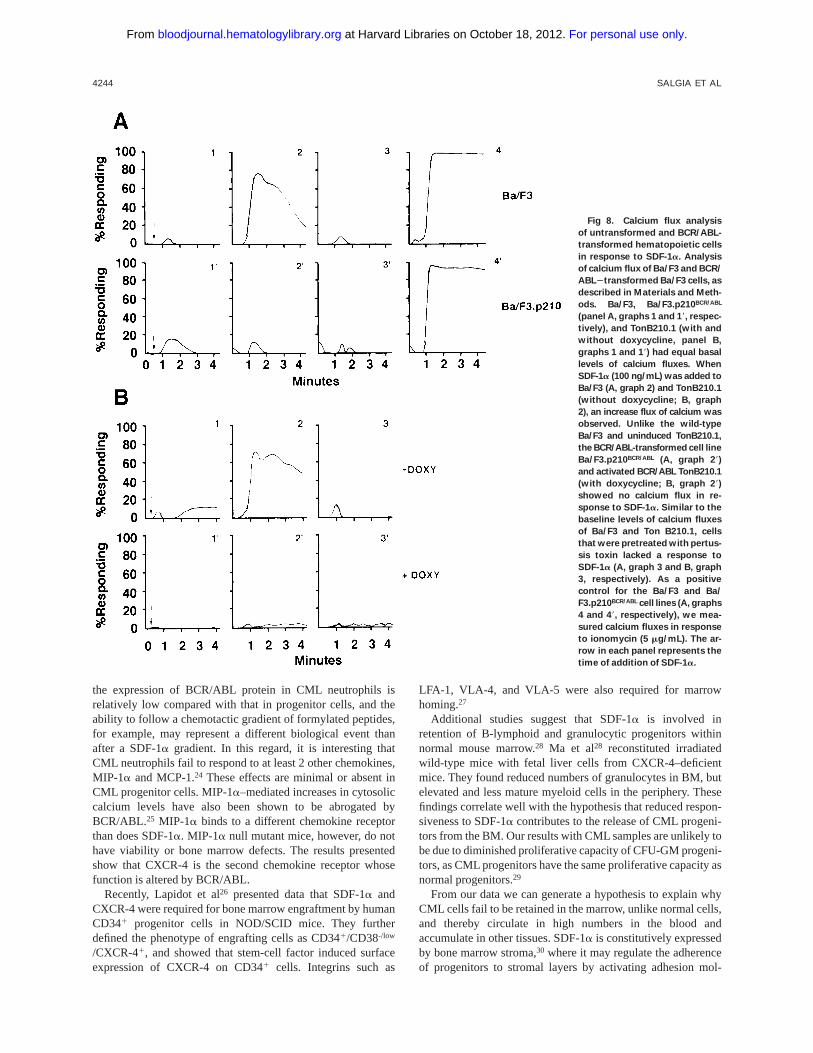
BCR/ABL reduces CXCR-4 signal transduction.To assessthe effects of BCR/ABL on CXCR4 signaling, we examinedmobilization of intracellular calcium in response to SDF-1a(100 ng/mL, Fig 8) in hematopoietic cell lines before and after
Fig 3 (Cont’d).
DECREASED RESPONSE TO SDF-1a IN CML CELLS 4239
For personal use only. at Harvard Libraries on October 18, 2012. bloodjournal.hematologylibrary.orgFrom

transformation by BCR/ABL. SDF-1a induced a rapid, tran-sient flux of intracellular calcium in wild-type Ba/F3 (panel A,graph 2) and uninduced TonB210.1 cells (panel B, graph 2) butnot in BCR/ABL-expressing Ba/F3 cells (panel A, graph 28) orin the doxycycline-treated TonB210.1 cells (panel B, graph 28).Positive responses to SDF-1awere blocked by preincubatingthe cell lines with pertussis toxin (panels A and B, graphs 3).Similar results were obtained in 32D and Mo7e cells before andafter BCR/ABL transformation (data not shown). BCR/ABL-transformation of hematopoietic cell lines did not affect expres-
sion of CXCR-4 (Fig 1), but reduced SDF-1a–mediatedcalcium flux in each case.
We have previously determined that S6 kinase is rapidlyphosphorylated in response to SDF-1a in Ba/F3 cells (R. Salgiaet al, unpublished observation). As shown in Fig 9, S6 kinase isphosphorylated in response to SDF-1a in Ba/F3 cells and notBa/F3.p210 cells. Thus, BCR/ABL blocks a very proximalCXCR-4 signaling event, calcium flux, and also blocks adownstream event, phosphorylation of S6 kinase. We have alsodetermined that there is phosphorylation of paxillin in
Fig 3 (Cont’d).
4240 SALGIA ET AL
For personal use only. at Harvard Libraries on October 18, 2012. bloodjournal.hematologylibrary.orgFrom

response to SDF-1ain Ba/F3 cells, but not in Ba/F3.p210 cells(data not shown).
DISCUSSION
Chronic myelogenous leukemia is caused by the t(9,22)(q34.1;q11.21) translocation that generates the BCR/ABL oncogene.20
p210BCR/ABL is an active tyrosine kinase, and this increasedkinase activity has been shown to be required for cell transfor-mation. For normal hematopoietic cells, migration of myeloidcells from the marrow to the blood is tightly linked todifferentiation, and in the absence of inflammation or infection
myeloid cells stay in the marrow until they have fully maturedto neutrophils. In contrast, CML myeloid cells circulate in largenumbers in the blood at virtually all stages of differentiation,and it is clear that one of the defining characteristics of thisillness is the uncoupling of differentiation from the ability toleave the marrow.21 In a variety of model systems, transforma-tion of myeloid cells by BCR/ABL results in abnormal adhesionto the extracellular matrix component fibronectin, growth factorindependence, decreased apoptosis, and cytoskeletal abnormali-ties (not in model systems).1 It is likely that the effects ofBCR/ABL on cytoskeletal function, including altered adhesion
Fig 3 (Cont’d).
DECREASED RESPONSE TO SDF-1a IN CML CELLS 4241
For personal use only. at Harvard Libraries on October 18, 2012. bloodjournal.hematologylibrary.orgFrom
and increased motility, contribute to early marrow exit and thesubsequent myeloproliferation.
Recently, the chemokine SDF-1a has generated considerableinterest because of its potential role in progenitor cell homing tothe marrow and in regulating marrow egress.22 In vitro, SDF-1ais one of the few known cytokines that has chemotactic activity
for hematopoietic progenitor cells and, in vivo, ectopic injectionof SDF-1a can temporarily attract immature hematopoieticcells to the injection site.8 Homozygous deletion of the SDF-1agene results in a lack of myelopoiesis and lymphopoiesis in thedeveloping fetal liver and marrow.11 It is not known if this is dueto impaired migration of cells from one developing hematopoi-
Fig 4. Chemotaxis of murine
and human cell lines to SDF-1a
and MS-5 supernatant. Several
cell lines were tested for their
ability to migrate in transwell
assays in response to SDF-1a
(100 ng/mL) and MS-5 superna-
tant (undiluted). The number of
cells that migrated to SDF-1a or
MS-5 was divided by the number
of background cells (cells that
migrated to medium alone) to
determine the chemotactic in-
dex, for panels A, B, and D. In
panel C, the percentage of mi-
grating cells is shown for com-
parison. In some columns, the
standard deviations were too
small to be visualized. Similarly,
the chemotactic index of trans-
formed K562 cells migrating
to MS-5 supernatant and SDF-1a
was below 1.0. Standard er-
ror bars for the cell lines
Ba/F3.p185BCR/ABL, Mo7e, Mo7e.
p210BCR/ABL, K562, 32D.p210BCR/ABL,
and 32D.p185BCR/ABL are calculated
from triplicate values in single
migration experiments, whereas
the error bars for Ba/F3 and Ba/
F3.p210BCR/ABL cells were calcu-
lated from data obtained in 6 and
4 migration experiments, respec-
tively. In panel C, Ba/F3 cells
transformed with p210 were
tested and data from 4 migration
experiments (2 per cell line) were
pooled.
Fig 5. Doxycycline-induced expression of BCR/ABL in TonB210.1 cells impairs chemotaxis to SDF-1a. The Ba/F3 cell line, TonB210.1, which
expresses BCR/ABL in the presence of doxycycline, was tested (n 5 4) for a chemotactic response to medium alone (Neg Ctrl), MS-5 supernatant,
and SDF-1a (100 ng/mL) using a transwell assay system. The open bars represent control cells (minus doxycycline) and solid bars represent
doxycycline-treated cells. Panel A shows the percentage of migrating cells and panel B shows the chemotactic index.
4242 SALGIA ET AL
For personal use only. at Harvard Libraries on October 18, 2012. bloodjournal.hematologylibrary.orgFrom
etic site to another or to impaired expansion. More recently, itwas shown that homozygous deletion of CXCR-4 results in anidentical phenotype.12,13 In addition, defects in vascularization,cardiac septal development and cerebellar development werefound. It is now believed that CXCR-4 is the primary physiologi-cal receptor for SDF-1a. CXCR-4 is expressed in the endothe-lium of developing blood vessels within the embryo proper, andits absence results in a lack of branching of the mesentericvessels at day 13.5 of gestation. By day 16.5, hemorrhagesand/or areas of congestion were found in the small intestine ofmutant embryos. Overall, the available evidence suggests thatSDF-1aand CXCR-4 play important roles in establishing andmaintaining cellular homeostasis in the marrow, and disruptionof this pathway can lead to aberrant homing and/or retention ofprogenitor cells.
In this study, we show that 3 nontransformed hematopoieticcell lines express CXCR-4 and respond to SDF-1a withincreased cell motility, adhesion, and chemotaxis. In contrast,after BCR/ABL-transformation, the response to SDF-1aissignificantly diminished. It would be quite interesting toperform the same studies of transwell migration after coatingthe membranes with other extracellular matrix components aswell as transendothelial and trans-stromal migration on thevarious cells tested. There is decreased calcium flux in responseto SDF-1a in BCR/ABL-transformed cells, suggesting thatCXCR-4 signaling may be blocked at a very proximal level. Thedecrease in calcium flux is greater than the inhibition ofchemotaxis in BCR/ABL-containing cells. However, the asso-ciation between calcium flux and chemotaxis is not wellunderstood because there are cells that migrate in response to
chemokines without a calcium flux. We show here that distalsignaling molecules such as S6 kinase involved in signaltransduction by SDF-1a are also affected by BCR/ABL. Theresults presented here significantly expand the range of cytoskel-etal abnormalities already associated with CML.
BCR/ABL is partially localized to the cytoskeleton andphosphorylates several cytoskeletal proteins, including paxillin,CRKL, vinculin, FAK, and tensin. Primary CML cells and celllines transformed by BCR/ABL have altered adhesion tofibronectin and have abnormal adhesion to stromal cells. Theseeffects are believed to be related to abnormal integrin function,and can be partially reversed by exposure toa-interferon.4
Furthermore, we have shown that CML cells have increasedspontaneous motility on fibronectin coated surfaces in vitro, andan increased rate of ruffling and formation of pseudopods.7 Thecell motility of CML cells is strikingly increased with anincreased adhesiveness to fibronectin. The increased cell motil-ity may aid in the release of progenitors from the stroma. Theseresults also suggest that BCR/ABL has profound effects on thecytoskeleton. In the case of SDF-1a, reduced response may bedue to both inhibition of specific signaling from the SDF-1areceptor and more global defects in the cell’s ability to regulatecytoskeletal functions.
In contrast to progenitor cells, neutrophils from CML patientshave relatively normal function and can clearly accumulateappropriately at sites of inflammation or infection.23 However,
Fig 6. Enhanced homing of Ba/F3 cells to the spleen in the
presence of SDF-1a, in vivo. The splenic ratio of differently-labeled,
fluorescent Ba/F3 and Ba/F3.p210BCR/ABL cells was determined 3 hours
after injection into mice. Recipient mice spleens were injected with
PBS (s) or recombinant SDF-1a (1mg;s) immediately before infusion
of pooled cells. Ba/F3 cells homed more efficiently to the spleen than
Ba/F3.p210BCR/ABL cells in the presence of SDF-1a (P F .05). The lines
connecting circles indicate paired mice in 4 independent experi-
ments.
Fig 7. SDF-1a responsiveness, measured by CFU-GM, is reduced
in CML patient samples. Chemotaxis assays were performed with
mononuclear cells isolated from normal BM, BM from CML patients,
and from peripheral blood samples from CML patients. CFU-GM are
plotted as: (total number of colonies and clusters obtained with the
migratory population)/(the number of colonies and clusters obtained
with the input population) 3 100 (see Materials and Methods). The
stippled and solid bars represent CFU-GM formed by cells migrating
to UltraCULTURE alone (control) or to 100 ng/mL of SDF-1a, respec-
tively.
DECREASED RESPONSE TO SDF-1a IN CML CELLS 4243
For personal use only. at Harvard Libraries on October 18, 2012. bloodjournal.hematologylibrary.orgFrom
the expression of BCR/ABL protein in CML neutrophils isrelatively low compared with that in progenitor cells, and theability to follow a chemotactic gradient of formylated peptides,for example, may represent a different biological event thanafter a SDF-1agradient. In this regard, it is interesting thatCML neutrophils fail to respond to at least 2 other chemokines,MIP-1a and MCP-1.24 These effects are minimal or absent inCML progenitor cells. MIP-1a–mediated increases in cytosoliccalcium levels have also been shown to be abrogated byBCR/ABL.25 MIP-1a binds to a different chemokine receptorthan does SDF-1a. MIP-1a null mutant mice, however, do nothave viability or bone marrow defects. The results presentedshow that CXCR-4 is the second chemokine receptor whosefunction is altered by BCR/ABL.
Recently, Lapidot et al26 presented data that SDF-1a andCXCR-4 were required for bone marrow engraftment by humanCD341 progenitor cells in NOD/SCID mice. They furtherdefined the phenotype of engrafting cells as CD341/CD38-/low
/CXCR-41, and showed that stem-cell factor induced surfaceexpression of CXCR-4 on CD341 cells. Integrins such as
LFA-1, VLA-4, and VLA-5 were also required for marrowhoming.27
Additional studies suggest that SDF-1ais involved inretention of B-lymphoid and granulocytic progenitors withinnormal mouse marrow.28 Ma et al28 reconstituted irradiatedwild-type mice with fetal liver cells from CXCR-4–deficientmice. They found reduced numbers of granulocytes in BM, butelevated and less mature myeloid cells in the periphery. Thesefindings correlate well with the hypothesis that reduced respon-siveness to SDF-1acontributes to the release of CML progeni-tors from the BM. Our results with CML samples are unlikely tobe due to diminished proliferative capacity of CFU-GM progeni-tors, as CML progenitors have the same proliferative capacity asnormal progenitors.29
From our data we can generate a hypothesis to explain whyCML cells fail to be retained in the marrow, unlike normal cells,and thereby circulate in high numbers in the blood andaccumulate in other tissues. SDF-1ais constitutively expressedby bone marrow stroma,30 where it may regulate the adherenceof progenitors to stromal layers by activating adhesion mol-
Fig 8. Calcium flux analysis
of untransformed and BCR/ABL-
transformed hematopoietic cells
in response to SDF-1a. Analysis
of calcium flux of Ba/F3 and BCR/
ABL2transformed Ba/F3 cells, as
described in Materials and Meth-
ods. Ba/F3, Ba/F3.p210BCR/ABL
(panel A, graphs 1 and 18, respec-
tively), and TonB210.1 (with and
without doxycycline, panel B,
graphs 1 and 18) had equal basal
levels of calcium fluxes. When
SDF-1a (100 ng/mL) was added to
Ba/F3 (A, graph 2) and TonB210.1
(without doxycycline; B, graph
2), an increase flux of calcium was
observed. Unlike the wild-type
Ba/F3 and uninduced TonB210.1,
the BCR/ABL-transformed cell line
Ba/F3.p210BCR/ABL (A, graph 28)
and activated BCR/ABL TonB210.1
(with doxycycline; B, graph 28)
showed no calcium flux in re-
sponse to SDF-1a. Similar to the
baseline levels of calcium fluxes
of Ba/F3 and Ton B210.1, cells
that were pretreated with pertus-
sis toxin lacked a response to
SDF-1a (A, graph 3 and B, graph
3, respectively). As a positive
control for the Ba/F3 and Ba/
F3.p210BCR/ABL cell lines (A, graphs
4 and 48, respectively), we mea-
sured calcium fluxes in response
to ionomycin (5 mg/mL). The ar-
row in each panel represents the
time of addition of SDF-1a.
4244 SALGIA ET AL
For personal use only. at Harvard Libraries on October 18, 2012. bloodjournal.hematologylibrary.orgFrom

ecules.31 Oncogenic transformation by BCR/ABL may thenaccelerate proliferation, increase spontaneous motility, andreduce long-term adhesiveness to stromal cells by reducing thenumber, accessibility, or function of adhesion-related mol-ecules, such as CXCR-4. These changes may then aid progeni-tors in their exit from the marrow as the cellularity increases.We also have preliminary evidence that egress of cells fromintact newborn murine femurs can occur, in vitro, under theinfluence of distally located MS-5 adherent cells (data notshown), suggesting that even in the absence of blood flow, bonemarrow cells are capable of releasing themselves from normalintact stroma in response to a chemotactic gradient. In summary,the defect in migration to SDF-1a may represent 1 step in thecomplex pathway of events that are needed to transform cells byBCR/ABL and result in CML. Also, because at least 2chemokine receptors are now shown to be inhibited by BCR/ABL, this suggests that BCR/ABL blocks a signaling stepcommon to general chemokine receptors.
ACKNOWLEDGMENT
The authors thank Ms Li Zhang and Dr Yuhui Xu of the Core EMFacility at the Dana-Farber Cancer Institute for their help in scanningelectron microscopy. Also, we thank Herb Levine, FACS Core Facility,DFCI for his help in calcium flux assays.
REFERENCES
1. Sattler M, Salgia R: Dysregulation of hematopoietic growth factorsignal transduction by the oncogene BCR/ABL. Cytokine GrowthFactor Rev 8:63, 1997
2. Gordon MY, Dowding CR, Riley GP, Goldman JM, Greaves MF:
Altered adhesive interactions with marrow stroma of haematopoieticprogenitor cells in chronic myeloid leukaemia. Nature 328:342, 1987
3. Verfaillie CM, McCarthy JB, McGlave PB: Mechanisms underly-ing abnormal trafficking of malignant progenitors in chronic myelog-enous leukemia. J Clin Invest 90:1232, 1992
4. Bhatia R, McCarthy J, Verfaillie C: Interferon-a restores normalb1 integrin-mediated inhibition of hematopoietic progenitor prolifera-tion by the marrow microenvironment in chronic myelogenous leuke-mia. Blood 87:3883, 1996
5. Salgia R, Brunkhorst B, Pisick E, Li J-L, Lo SH, Chen LB, GriffinJD: Increased tyrosine phosphorylation of focal adhesion proteins inmyeloid cell lines expressing p210BCR/ABL. Oncogene 11:1149, 1995
6. Salgia R, Uemura N, Okuda K, Li J-L, Pisick E, Sattler M, deJongR, Druker B, Heisterkamp N, Chen L, Groffen J, Griffin J: CRKL linksp210BCR/ABL with paxillin in chronic myelogenous leukemia cells. JBiol Chem 270:29145, 1995
7. Salgia R, Li J-L, Ewaniuk D, Pear W, Pisick E, Burky S, Ernst T,Sattler M, Chen L, Griffin J: BCR/ABL induces multiple abnormalitiesof cytoskeletal function. J Clin Invest 100:46, 1997
8. Aiuti A, Webb IJ, Bleul C, Springer T, Gutierrez-Ramos JC: Thechemokine SDF-1 is a chemoattractant for human CD341 hematopoi-etic progenitor cells and provides a new mechanism to explain themobilization of CD341 progenitors to peripheral blood. J Exp Med185:111, 1997
9. Nagasawa T, Nakajima T, Tachibana K, Iizasa H, Bleul CC,Yoshie O, Matsushima K, Yoshida N, Springer TA, Kishimoto T:Molecular cloning and characterization of a murine pre-B-cell growth-stimulating factor/stromal cell-derived factor 1 receptor, a murinehomolog of the human immunodeficiency virus 1 entry coreceptorfusin. Proc Natl Acad Sci USA 93:14726, 1996
10. Kim CH, Broxmeyer HE: In vitro behavior of hematopoieticprogenitor cells under the influence of chemoattractants: Stromalderived factor-1, steel factor, and the bone marrow enviroment. Blood91:100, 1998
11. Nagasawa T, Hirota S, Tachibana K, Takakura N, Nishikawa S,Kitamura Y, Yoshida N, Kikutani H, Kishimoto T: Defects of b-celllymphopoiesis and bone-marrow myelopoiesis in mice lacking the cxcchemokine pbsf/sdf-1. Nature 382:635, 1996
12. Tachibana K, Hirota S, Iizasa H, Yoshida H, Kawabata K,Kataoka Y, Kitamura Y, Matsushima K, Yoshida N, Nishikawa S,Kishimoto T, Nagasawa T: The chemokine receptor CXCR4 is essentialfor vascularization of the gastrointestinal tract. Nature 393:591, 1998
13. Zou YR, Kottmann AH, Kuroda M, Taniuchi I, Littman DR:Function of the chemokine receptor CXCR4 in haematopoiesis and incerebellar development. Nature 393:595, 1998
14. Matulonis U, Salgia R, Okuda K, Druker B, Griffin J: Interleu-kin-3 and p210 BCR/ABL activate both unique and overlappingpathways of signal transduction in a factor-dependent myeloid cell line.Exp Hematol 21:1460, 1993
15. Klucher K, Lopez D, Daley G: Secondary mutation maintains thetransformed state in BaF3 cells with inducible BCR/ABL expression.Blood 91:3927, 1998
16. Bazzoni G, Carlesso N, Griffin J, Hemler M: BCR/ABL expres-sion stimulates integrin function in hematopoietic cell lines. J ClinInvest 98:521, 1996
17. Salgia R, Li J, Lo SH, Brunkhorst B, Kansas GS, Sobhany ES,Sun YP, Pisick E, Hallek M, Ernst T, Tantravahi R, Chen LB, Griffin JD:Molecular cloning of human paxillin, a focal adhesion protein phosphory-lated by p210(bcr/abl). J Biol Chem 270:5039, 1995
18. Quackenbush E, Wershil B, Aguirre V, Gutierrez-Ramos J-C:Eotaxin modulates myelopoiesis and mast cell development fromembryonic hematopoietic progenitors. Blood 92:1, 1998
19. Stockton B, Cheng G, Manjunath N, Ardman B, von Andrian U:Negative regulation of T cell homing by CD43. Immunity 8:373, 1998
20. Chissoe SL, Bodenteich A, Wang YF, Wang YP, Burian D,
Fig 9. Phosphorylation status of p70 S6 kinase in response to
SDF-1a. Immunoblot analysis of p70 S6 kinase phosphorylation of
Ba/F3 and Ba/F3.p210 in response to SDF-1a. SDF-1a induces serine
421/threonine 424 phosphorylation of p70 S6 kinase in normal Ba/F3
cells, but not in BCR/ABL-transformed cell lines. The same blot was
stripped and probed with anti-S6 kinase showing that the amount of
p70 S6 kinase is equivalent in all lanes.
DECREASED RESPONSE TO SDF-1a IN CML CELLS 4245
For personal use only. at Harvard Libraries on October 18, 2012. bloodjournal.hematologylibrary.orgFrom

Clifton SW, Crabtree J, Freeman A, Iyer K, Li JA, Ma YC, Mclaury HJ,Pan HQ, Sarhan OH, Toth S, Wang ZL, Zhang GZ, Heisterkamp N,Groffen J, Roe BA: Sequence and analysis of the human abl gene, thebcr gene, and regions involved in the Philadelphia chromosomaltranslocation. Genomics 27:67, 1995
21. Galbraith P, Abu-Zahra H: Granulopoiesis in chronic granulo-cytic leukaemia. Br J Hematol 22:135, 1972
22. Baggiolini M: Chemokines and leukocyte traffic. Nature 392:565, 1998
23. Cramer E, Auclair C, Hakim J: Metabolic activity of phagocyto-sis granulocytes in chronic granulocytic leukemia. Blood 50:93, 1977
24. Eaves C, Cashman J, Wolpe S, Eaves A: Unresponsiveness ofprimitive chronic myeloid leukemia cells to macrophage inflammatoryprotein 1 alpha, an inhibitor of primitive normal hematopoietic cells.Proc Natl Acad Sci USA 90:12015, 1993
25. Wark G, Heyworth C, Spooncer E, Czaplewski L, Francis J,Dexter T, Whetton A: Abl protein kinase abrogates the response ofmultipotent haemopoietic cells to the growth inhibitor macrophageinflammatory protein-1 alpha. Oncogene 16:1319, 1998
26. Lapidot T, Peled A, Petit I, Kollet O, Magid M, Ponomaryov T,
Franitza S, Grabovsky V, Nagler A, Lider O, Alon R: Ex-vivo expansionof migrating human CXCR41-SCID repopulating cells by upregulationof surface CXCR-4 expression in response to stimulation with SCF orIL-6. Blood 92:717a, 1998 (abstr, suppl 1)
27. Lapidot T, Peled A, Petit I, Kollet O, Magid M, Ponomaryov T,Franitza S, Grabovsky V, Nagler A, Lider R, Alon R, Zipori D: Thechemokine SDF-1a and the cytokine SCF mediate CXCR-4 dependenthoming of human CD341/CD38- stem cells to the bone marrow ofnod/scid mice. Blood 92:504a, 1998 (abstr, suppl 1)
28. Ma Q, Jones D, Springer T: The chemokine receptor CXCR4 isrequired for the retention of B lineage and granulocytic precursorswithin the bone marrow microenvironment. Immunity 10:1, 1999
29. Cannistra S: Chronic myelogenous leukemia as a model for thegenetic basis of cancer. Hematol Oncol Clin North Am 4:337, 1990
30. Bleul CC, Fuhlbrigge RC, Casasnovas JM, Aiuti A, Springer TA:A highly efficacious lymphocyte chemoattractant, stromal cell-derivedfactor 1 (SDF-1). J Exp Med 184:1101, 1996
31. Luster AD: Chemokines–Chemotactic cytokines that mediateinflammation. N Engl J Med 338:436, 1998
4246 SALGIA ET AL
For personal use only. at Harvard Libraries on October 18, 2012. bloodjournal.hematologylibrary.orgFrom