the biology of proteostasis in aging and...
TRANSCRIPT

BI84CH32-Morimoto ARI 4 March 2015 12:39
RE V I E W
S
IN
AD V A
NC
E
The Biology of Proteostasis inAging and DiseaseJohnathan Labbadia and Richard I. MorimotoDepartment of Molecular Biosciences, Rice Institute for Biomedical Research, NorthwesternUniversity, Evanston, Illinois 60208; email: [email protected]
Annu. Rev. Biochem. 2015. 84:32.1–32.30
The Annual Review of Biochemistry is online atbiochem.annualreviews.org
This article’s doi:10.1146/annurev-biochem-060614-033955
Copyright c© 2015 by Annual Reviews.All rights reserved
Keywords
chaperones, protein folding, stress response, neurodegenerative disease,protein misfolding, aggregation
Abstract
Loss of protein homeostasis (proteostasis) is a common feature of aging anddisease that is characterized by the appearance of nonnative protein aggre-gates in various tissues. Protein aggregation is routinely suppressed by theproteostasis network (PN), a collection of macromolecular machines thatoperate in diverse ways to maintain proteome integrity across subcellularcompartments and between tissues to ensure a healthy life span. Here, wereview the composition, function, and organizational properties of the PNin the context of individual cells and entire organisms and discuss the mech-anisms by which disruption of the PN, and related stress response pathways,contributes to the initiation and progression of disease. We explore emergingevidence that disease susceptibility arises from early changes in the composi-tion and activity of the PN and propose that a more complete understandingof the temporal and spatial properties of the PN will enhance our ability todevelop effective treatments for protein conformational diseases.
32.1
Review in Advance first posted online on March 12, 2015. (Changes may still occur before final publication online and in print.)
Changes may still occur before final publication online and in print
Ann
u. R
ev. B
ioch
em. 2
015.
84. D
ownl
oade
d fr
om w
ww
.ann
ualr
evie
ws.
org
Acc
ess
prov
ided
by
NO
RT
HW
EST
ER
N U
NIV
ER
SIT
Y -
Law
Lib
rary
(C
hica
go C
ampu
s) o
n 04
/02/
15. F
or p
erso
nal u
se o
nly.

BI84CH32-Morimoto ARI 4 March 2015 12:39
Contents
INTRODUCTION . . . . . . . . . . . . . . . . . . . . . . . . . . . . . . . . . . . . . . . . . . . . . . . . . . . . . . . . . . . . . . . 32.2DEFINING THE PROTEOSTASIS NETWORK . . . . . . . . . . . . . . . . . . . . . . . . . . . . . . . . 32.3
Molecular Chaperones . . . . . . . . . . . . . . . . . . . . . . . . . . . . . . . . . . . . . . . . . . . . . . . . . . . . . . . . . . 32.5Protein Degradation Pathways . . . . . . . . . . . . . . . . . . . . . . . . . . . . . . . . . . . . . . . . . . . . . . . . . . 32.6
STRESS RESPONSE PATHWAYS ALTER PROTEOSTASISNETWORK COMPOSITION . . . . . . . . . . . . . . . . . . . . . . . . . . . . . . . . . . . . . . . . . . . . . . . . 32.7
ORGANISMAL CONNECTIVITY OF THE PROTEOSTASISNETWORK AND STRESS RESPONSES . . . . . . . . . . . . . . . . . . . . . . . . . . . . . . . . . . . . . 32.9
DIFFERENTIAL REGULATION OF THE PROTEOSTASISNETWORK ACROSS TISSUES AND CELL TYPES . . . . . . . . . . . . . . . . . . . . . . . . . 32.9
PROTEOSTASIS NETWORK DISRUPTION AND PROTEOSTASISCOLLAPSE IN NEURODEGENERATIVE DISEASES . . . . . . . . . . . . . . . . . . . . . . .32.11Dysregulation of Molecular Chaperones . . . . . . . . . . . . . . . . . . . . . . . . . . . . . . . . . . . . . . . . .32.11Disruption of Protein Degradation Pathways . . . . . . . . . . . . . . . . . . . . . . . . . . . . . . . . . . . . .32.12Stress Response Pathway Impairment . . . . . . . . . . . . . . . . . . . . . . . . . . . . . . . . . . . . . . . . . . . .32.13Spreading of Protein Aggregates . . . . . . . . . . . . . . . . . . . . . . . . . . . . . . . . . . . . . . . . . . . . . . . .32.13
COULD PROGRAMMED CHANGES IN THE PROTEOSTASISNETWORK UNDERLIE DISEASE ONSET AND PROGRESSION? . . . . . . . . .32.14Changes in Proteostasis Capacity Occur in Early Adulthood . . . . . . . . . . . . . . . . . . . . . .32.14Requirements of Stress Response Pathways Through Life . . . . . . . . . . . . . . . . . . . . . . . .32.17Temporal Requirements of Protein Degradation Pathways . . . . . . . . . . . . . . . . . . . . . . .32.18
PHARMACOLOGICAL ENHANCEMENT OF THE PROTEOSTASISNETWORK AS A MODIFIER OF AGING AND DISEASE. . . . . . . . . . . . . . . . . . . .32.18
THE FUTURE OF THE PROTEOSTASIS NETWORKIN HEALTH AND HUMAN DISEASE . . . . . . . . . . . . . . . . . . . . . . . . . . . . . . . . . . . . . . .32.20
INTRODUCTION
Proteome fidelity is maintained by the protein homeostasis (proteostasis) network (PN), a multi-compartmental system that coordinates protein synthesis, folding, disaggregation, and degradation(1). Despite the common factors required for protein synthesis and maintenance, the expression ofmany PN components is tailored to the specific proteomic demands of different cells and tissues(1). Furthermore, the activity of the PN can be altered permanently or transiently by develop-ment and aging, alterations in physiology, or exposure to environmental stress (1). As PN activitychanges, so too does the capacity of cells to buffer against the accumulation of misfolded anddamaged proteins. Therefore, temporal and spatial fluctuations in the PN could have profoundconsequences for disease presentation and progression.
Whereas the classical view of the PN in relation to aging and disease has been described as“young versus old” or “before and after,” there has been considerably less attention devoted to themalleability and dynamism of PN properties and composition with respect to different tissue typesand life stages; however, it is likely that the relationship between the PN and disease encompassesthese complexities. Here, we review the intra- and intercellular organization of the PN and discusshow temporal and spatial changes in PN composition and activity can influence proteostasis, aging,and disease. We focus on the relationship between the PN and neurodegenerative disorders such as
32.2 Labbadia · Morimoto
Changes may still occur before final publication online and in print
Ann
u. R
ev. B
ioch
em. 2
015.
84. D
ownl
oade
d fr
om w
ww
.ann
ualr
evie
ws.
org
Acc
ess
prov
ided
by
NO
RT
HW
EST
ER
N U
NIV
ER
SIT
Y -
Law
Lib
rary
(C
hica
go C
ampu
s) o
n 04
/02/
15. F
or p
erso
nal u
se o
nly.
BI84CH32-Morimoto ARI 4 March 2015 12:39
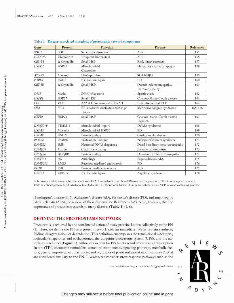
Table 1 Disease-associated mutations of proteostasis network components
Gene Protein Function Disease ReferenceSOD1 SOD1 Superoxide dismutase ALS 155UBQLN2 Ubiquilin-2 Ubiquitin-like protein ALS 156CRYAA α-Crystallin Small HSP Early-onset cataracts 157HSPD1 HSP60 Mitochondrial
ChaperoneHereditary spastic paraplegia 158
ATXN3 Ataxin-3 Deubiquitylase SCA3/MJD 159PARK1 Parkin E3 ubiquitin ligase PD 160CRYAB α-Crystallin Small HSP Desmin-related myopathy,
cardiomyopathy161
SACS Sacsin DNAJ chaperone Spastic ataxia 162HSPB1 HSP27 Small HSP Charcot–Marie–Tooth disease 163VCP VCP AAA ATPase involved in ERAD Paget disease and FTD 164SIL1 SIL1 ER-associated nucleotide exchange
factorMarinesco–Sjogren syndrome 165, 166
HSPB8 HSP22 Small HSP Charcot–Marie–Tooth diseasetype 2L
167
DNAJC19 TIMM14 Mitochondrial import DCMA syndrome 168HSPA9 Mortalin Mitochondrial HSP70 PD 169HSPA8 HSC70 Protein folding Cardiovascular disease 170PSMB8 PSMB8 Proteasomal subunit Nakajo–Nishimura syndrome 171DNAJB2 HSJ1 Neuronal DNAJ chaperone Distal hereditary motor neuropathy 172DNAJC6 Auxilin Clathrin uncoating Juvenile parkinsonism 173DNAJB6 DNAJB6 Protein folding Dominantly inherited myopathy 174SQSTM1 p62 Autophagy Paget’s disease, ALS 175DNAJC13 RME8 Receptor-mediated endocytosis PD 176P4HB PDI Protein disulfide isomerase ALS 177UBE3A UBE3A E3 ubiquitin ligase Angelman syndrome 178
Abbreviations: ALS, amyotrophic lateral sclerosis; ERAD, endoplasmic reticulum (ER)-associated degradation; FTD, frontotemporal dementia;HSP, heat shock protein; MJD, Machado–Joseph disease; PD, Parkinson’s disease; SCA, spinocerebellar ataxia; VCP, valosine-containing protein.
Huntington’s disease (HD), Alzheimer’s disease (AD), Parkinson’s disease (PD), and amyotrophiclateral sclerosis (ALS) (for reviews of these diseases, see References 2–5). Note, however, that theimportance of proteostasis extends to many diseases (Table 1) (1, 6).
DEFINING THE PROTEOSTASIS NETWORK
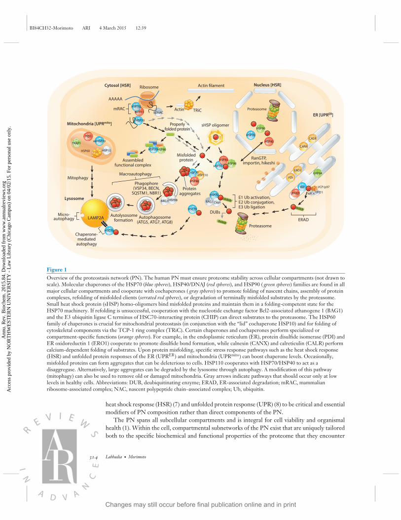
Proteostasis is achieved by the coordinated action of many proteins known collectively as the PN(1). Here, we define the PN as a protein network with an immediate role in protein synthesis,folding, disaggregation, or degradation. This definition encompasses the translational machinery,molecular chaperones and cochaperones, the ubiquitin–proteasome system (UPS), and the au-tophagy machinery (Figure 1). Although essential for PN function and proteostasis, transcriptionfactors (TFs), chromatin remodelers, structural components, signaling pathways, metabolic fac-tors, general import/export machinery, and regulators of posttranslational modifications (PTMs)are considered auxiliary to the PN. Likewise, we consider stress response pathways such as the
www.annualreviews.org • Proteostasis in Aging and Disease 32.3
Changes may still occur before final publication online and in print
Ann
u. R
ev. B
ioch
em. 2
015.
84. D
ownl
oade
d fr
om w
ww
.ann
ualr
evie
ws.
org
Acc
ess
prov
ided
by
NO
RT
HW
EST
ER
N U
NIV
ER
SIT
Y -
Law
Lib
rary
(C
hica
go C
ampu
s) o
n 04
/02/
15. F
or p
erso
nal u
se o
nly.
BI84CH32-Morimoto ARI 4 March 2015 12:39
Actin filament
Actin
Mitochondria [UPRmito]
TRiC
Properlyfolded protein
Assembledfunctional complex
VCP/p97
RanGTP,importin, hikeshiHSP90
HSP90
HSP90
Mitophagy
Misfoldedprotein
Ribosome
AAAAA
Proteasome
Macroautophagy
Nucleus [HSR]
E1 Ub activation,E2 Ub conjugation,E3 Ub ligation
ER [UPRER]
ERAD
mRAC
sHSP oligomer
Cytosol [HSR]
HSC70
BAG3 HSPB8
Autophagosome(ATG5, ATG7, ATG8)
Phagophore(VSP34, BECN,
SQSTM1, NBR1)
Autolysosomeformation
HSC70
LAMP2AMicro-
autophagy
Lysosome
Chaperone-mediated
autophagy
Proteasome
HSP70
CHIPBAG1
Protein aggregates
HSP40
HSP40
HSP40
HSP110HSP70
HSP70
HSP70
HSP70
HSP60
mtHSP70TRAP1
HSP10
TID1
HSP70
MPP11
HSP70L
αβNAC
GRP94
BiP
ERdj3
UbUbUb
UbUbUb
DUBsUbUbUb
UFD1NPL4
CANX
ERO1
PDI
CALR
Figure 1Overview of the proteostasis network (PN). The human PN must ensure proteome stability across cellular compartments (not drawn toscale). Molecular chaperones of the HSP70 (blue spheres), HSP40/DNAJ (red spheres), and HSP90 ( green spheres) families are found in allmajor cellular compartments and cooperate with cochaperones ( gray spheres) to promote folding of nascent chains, assembly of proteincomplexes, refolding of misfolded clients (serrated red spheres), or degradation of terminally misfolded substrates by the proteasome.Small heat shock protein (sHSP) homo-oligomers bind misfolded proteins and maintain them in a folding-competent state for theHSP70 machinery. If refolding is unsuccessful, cooperation with the nucleotide exchange factor Bcl2-associated athanogene 1 (BAG1)and the E3 ubiquitin ligase C terminus of HSC70-interacting protein (CHIP) can direct substrates to the proteasome. The HSP60family of chaperones is crucial for mitochondrial proteostasis (in conjunction with the “lid” cochaperone HSP10) and for folding ofcytoskeletal components via the TCP-1 ring complex (TRiC). Certain chaperones and cochaperones perform specialized orcompartment-specific functions (orange spheres). For example, in the endoplasmic reticulum (ER), protein disulfide isomerase (PDI) andER oxidoreductin 1 (ERO1) cooperate to promote disulfide bond formation, while calnexin (CANX) and calreticulin (CALR) performcalcium-dependent folding of substrates. Upon protein misfolding, specific stress response pathways such as the heat shock response(HSR) and unfolded protein responses of the ER (UPRER) and mitochondria (UPRmito) can boost chaperone levels. Occasionally,misfolded proteins can form aggregates that can be deleterious to cells. HSP110 cooperates with HSP70/HSP40 to act as adisaggregase. Alternatively, large aggregates can be degraded by the lysosome through autophagy. A modification of this pathway(mitophagy) can also be used to remove old or damaged mitochondria. Gray arrows indicate pathways that should occur only at lowlevels in healthy cells. Abbreviations: DUB, deubiquitinating enzyme; ERAD, ER-associated degradation; mRAC, mammalianribosome-associated complex; NAC, nascent polypeptide chain–associated complex; Ub, ubiquitin.
heat shock response (HSR) (7) and unfolded protein response (UPR) (8) to be critical and essentialmodifiers of PN composition rather than direct components of the PN.
The PN spans all subcellular compartments and is integral for cell viability and organismalhealth (1). Within the cell, compartmental subnetworks of the PN exist that are uniquely tailoredboth to the specific biochemical and functional properties of the proteome that they encounter
32.4 Labbadia · Morimoto
Changes may still occur before final publication online and in print
Ann
u. R
ev. B
ioch
em. 2
015.
84. D
ownl
oade
d fr
om w
ww
.ann
ualr
evie
ws.
org
Acc
ess
prov
ided
by
NO
RT
HW
EST
ER
N U
NIV
ER
SIT
Y -
Law
Lib
rary
(C
hica
go C
ampu
s) o
n 04
/02/
15. F
or p
erso
nal u
se o
nly.
BI84CH32-Morimoto ARI 4 March 2015 12:39
and to the cellular environments they must inhabit (9). Importantly, these subnetworks cooperatesubstantially to monitor and maintain proteostasis across the cell (9).
Molecular Chaperones
Molecular chaperones are central to the function of the PN and can be broadly grouped into theHSP70, HSP90, DNAJ/HSP40, chaperonin/HSP60, and small HSP (sHSP) families (10, 11).Chaperones can act alone or in various combinations with different cochaperones to regulateclient–substrate interactions, folding, disaggregation, degradation, and trafficking within the cell(11). Many molecular chaperones are highly conserved and pivotal, but the best studied are mem-bers of the HSP70 and HSP90 families. Although more than 15 mammalian HSP70 homologsand 4 HSP90 homologs exist, most of our understanding of HSP70 and HSP90 function comesfrom studies of the cytosolic/nuclear HSC70, HSP90, and inducible HSP70 (HSPA1A/B). How-ever, homologs of HSP70 and HSP90 are also integral to endoplasmic reticulum (ER) (BiP andGRP94) and mitochondrial (Mortalin and TRAP1) function (12).
HSP70 and HSP90 are highly abundant proteins with levels estimated to make up 1–2% of thetotal protein in some cells (12). ATP hydrolysis is essential for the chaperone activity of HSP70 andHSP90, causing conformational changes that result in substrate binding (11). Nucleotide exchangereleases bound substrates; several rounds of binding and release by HSP70 are sometimes requiredfor complete client refolding (11, 13). When protein refolding is inefficient, the cochaperones Cterminus of HSC70-interacting protein (CHIP) and Bcl2-associated athanogene 1 (BAG1) caninteract with HSP70 and HSP90 complexes to promote substrate ubiquitylation, thereby redirect-ing HSP70 and HSP90 clients to the proteasome for degradation (14, 15). As such, HSP70 andHSP90 are central to the process of triaging proteins for refolding or elimination. The client speci-ficity and functional properties of HSP70 and HSP90 are strongly influenced by interactions witha range of cochaperones (16). HSP90 associates with more than 20 cochaperones, the combina-tions of which dictate HSP90 function, whereas HSP70 is regulated principally by HSP40/DNAJchaperones (12, 13). Although distinct, the HSP90 and HSP70 machines also cooperate, mostnotably in steroid hormone and signal transduction kinase maturation through the cochaperoneHSC70/HSP90 organizing protein (HOP/STI1). HOP/STI1 simultaneously binds HSC70 andHSP90 through multiple tetratricopeptide repeat (TPR) domains, thereby facilitating maturationof clients through the sequential action of both HSP70 and HSP90 (12). HSP70 is also recruited tonewly synthesized polypeptides by the ribosome-associated complex (RAC), which in mammals isformed from the HSP40/DNAJ protein MPP11 and HSP70L1 (17). RAC binds near the ribosomeexit tunnel and stimulates HSP70 binding to nascent polypeptides, thus complementing the role ofthe nascent polypeptide chain–associated complex (NAC), the initial factor that binds and protectsnewly synthesized proteins, maintaining them in a folding-competent state for HSP70 (17).
HSP70 and HSP90 homologs are exceeded by the number of HSP40/DNAJ chaperones.HSP40/DNAJ chaperones contain a conserved J domain that mediates interactions with HSP70but otherwise exhibit heterogeneity in structure, localization, and function. Due to this diver-sity, HSP40/DNAJ chaperones can be thought of as adaptors that provide versatility to HSP70function (13). HSP40/DNAJ chaperones assist protein refolding by presenting misfolded pro-teins to HSP70 and by stimulating HSP70 ATPase activity (13). Efficient protein folding byHSP70 is also influenced by nucleotide exchange factors (NEFs), such as BAG1, that promoteADP/ATP exchange at the N terminus of HSP70 and form distinct functional complexes with theHSP70/HSP40 machinery (13). Perhaps the most significant example of this process is that theHSP110 molecular chaperone, long considered simply a NEF, can catalyze protein disaggregationas part of a complex containing HSP70 and DNAJ/HSP40 (18). This capability provides an extra
www.annualreviews.org • Proteostasis in Aging and Disease 32.5
Changes may still occur before final publication online and in print
Ann
u. R
ev. B
ioch
em. 2
015.
84. D
ownl
oade
d fr
om w
ww
.ann
ualr
evie
ws.
org
Acc
ess
prov
ided
by
NO
RT
HW
EST
ER
N U
NIV
ER
SIT
Y -
Law
Lib
rary
(C
hica
go C
ampu
s) o
n 04
/02/
15. F
or p
erso
nal u
se o
nly.

BI84CH32-Morimoto ARI 4 March 2015 12:39
mechanism by which mammalian cells can eliminate protein aggregates and suppress proteotoxi-city, a function carried out by the dedicated chaperones HSP104 and ClpB in yeast and bacteria,respectively (19).
In addition to the HSP40/DNAJ family and cochaperones, the sHSP family facilitates clientrefolding by HSP70. There are 10 mammalian sHSPs, all of which are cytosolic and most of whichcontain an α-crystalline domain (10). sHSPs influence protein folding by forming large homo-oligomeric cages that trap misfolded clients and prevent them from forming undesirable intra-or intermolecular interactions in the cytosol. This process is ATP independent and presumedto create a reservoir of refolding competent clients for the HSP70 machinery. Although the fullrepertoire of sHSP clients is still unclear, it is assumed that sHSPs act broadly, although somedegree of selectivity is expected between family members (10).
In addition to the more general chaperone functions carried out by the HSP70/HSP40and HSP90 machines, some proteins require the specialized functions afforded by theHSP60/chaperonin family. Chaperonins consist of two large hexameric ring complexes that forma cylindrical structure whose core provides a protected environment for protein refolding (11).Two classes of chaperonins exist, the class I mitochondrial HSP60, which is similar to bacterialGroEL, and the class II TCP-1 ring complex (TRiC), which is found in the eukaryotic cytosol(11). HSP60 is essential for maturation and maintenance of the mitochondrial proteome and istherefore intimately linked to energy production. TRiC is essential for proper posttranslationalfolding of the cytoskeletal components actin and tubulin and is therefore essential for cell structure,division, and cargo delivery (11).
Protein Degradation Pathways
When protein functionality cannot be restored from misfolded and aggregated states, chaperoneshelp redirect nonnative clients toward degradation pathways. Proteins can be degraded eitherindividually or en masse by proteasomes (20) or lysosomes (21), respectively. Proteasomes are largemultisubunit complexes that consist of a 19S regulatory cap and a 20S proteolytic core (22). The19S regulatory particle recognizes ubiquitylated substrates, removes ubiquitin chains, and unfoldsthe client to allow entry into the 20S core, where it is rapidly degraded into peptides (20, 23). Thisprocess is initiated by the addition of polyubiquitin chains through the stepwise activity of E1ubiquitin–activating enzymes, E2 ubiquitin–conjugating enzymes, and E3 ubiquitin ligases (20).
Ubiquitin chains are formed through conjugation of ubiquitin monomers to clients via distinctlysine residues. This process can take the form of a variety of linkages; however, K48-linkedubiquitin chains are directed primarily to the proteasome (24). Once ubiquitylated, chaperone–cochaperone complexes direct clients to the proteasome, where deubiquitylating enzymes (DUBs)remove ubiquitin chains to allow substrate entry into the 20S core (20). DUBs can be associatedwith a commitment to degradation and bulk removal of ubiquitin, as is the case for PSMD14, orthey can operate independently of client degradation through sequential “trimming” of ubiquitinchains, as is observed with UCHL37 and USP14 (25–28). These opposing activities are proposedto allow “tuning” of proteasomal degradation to be either general or selective as required (20).Despite their cytosolic and nuclear localization, proteasomes are an important site of ER proteinquality control through the ER-associated degradation (ERAD) pathway (29). Misfolded proteinsin the ER lumen are recognized by the ER membrane–associated HRD1–SEL1–HERP complexand retrotranslocated to the cytoplasm by the AAA ATPase VCP/p97–NPL4–UFD-1 complex(several client-specific forms of this process requiring specialized adapters of the VCP/p97 andHRD1 complexes have been described). The retrotranslocated protein is then ubiquitylated and
32.6 Labbadia · Morimoto
Changes may still occur before final publication online and in print
Ann
u. R
ev. B
ioch
em. 2
015.
84. D
ownl
oade
d fr
om w
ww
.ann
ualr
evie
ws.
org
Acc
ess
prov
ided
by
NO
RT
HW
EST
ER
N U
NIV
ER
SIT
Y -
Law
Lib
rary
(C
hica
go C
ampu
s) o
n 04
/02/
15. F
or p
erso
nal u
se o
nly.

BI84CH32-Morimoto ARI 4 March 2015 12:39
delivered to the proteasome for degradation, thereby eliminating terminally misfolded proteinsfrom the ER. Distinct ERAD mechanisms are utilized depending on whether protein misfoldingis exposed to the ER lumen (ERAD-L), exposed within the ER membrane (ERAD-M), or exposedto the cytosolic face of the ER membrane (ERAD-C), demonstrating the degree to which quality-control pathways can be tailored to substrate type and location (29). A recent study showed thatsome mutant forms of gonadotropin-releasing hormone receptor (GnRHR) are resistant to ERADand are instead degraded by ER quality-control (ERQC) autophagy, a complementary pathwaymodulated by DNAJB12 when ERAD fails (30).
Although the proteasome is the primary source of protein degradation in the cell, biophysicallimitations of the central pore of the 20S core do not permit the degradation of unfolded or largeprotein complexes (20). How, then, do cells remove large protein aggregates? As discussed above,one approach is to employ specialized molecular chaperone machines to release misfolded proteinsfrom aggregates and direct them to the proteasome for degradation (19). Alternatively, bulkiersubstrates, such as large inclusions, can be directed to the lysosome, a membrane-bound organellecontaining a host of nonspecific proteases that can degrade a wide range of substrates (21). Pro-teins and organelles are directed to lysosomes as the terminal step of autophagy. Autophagy com-plements the UPS in three mechanistically distinct forms: macroautophagy, chaperone-mediatedautophagy (CMA), and microautophagy (21). Macroautophagy is the best-studied form and entailsthe sequestration of organelles or regions of the cytosol into a double-membrane vesicle struc-ture known as an autophagosome. The resulting autophagosome is then transported to, and fuseswith, the lysosome, thereby delivering its cargo for degradation (21). In contrast, microautophagyoccurs by direct engulfment of the cytosol at the lysosome membrane, and CMA occurs throughHSC70-mediated delivery of proteins across the lysosomal membrane via the LAMP2A receptor(21, 31). Autophagy is regulated by the mammalian target of rapamycin complex 1 (mTORC1) andmTORC2, thereby integrating the PN with the nutritional status of the organism, the metabolicstate of the cell, and rates of protein synthesis (21).
STRESS RESPONSE PATHWAYS ALTER PROTEOSTASISNETWORK COMPOSITION
The composition of the PN is highly dynamic; the levels of molecular chaperones, cochaperones,and proteasomal subunits can be increased globally or in a compartment-specific manner to provideadditional protection against acute and chronic protein misfolding in the cell (1). Malleability of thePN is provided by dedicated stress responsive TFs with distinct and complementary transcriptionaltargets. Although a number of stress response pathways can greatly influence proteostasis, aging,and disease (see the sidebar), here we focus our attention on the HSR, which is regulated by heatshock transcription factors (HSFs) and augments the cytosolic/nuclear arm of the PN (7) andthe UPRs of the ER (UPRER) and mitochondria (UPRmito), which respond to protein misfoldingthrough the TFs XBP1, ATF6, and ATF4 (8) and through ATFS1, respectively (32).
Stress response TFs are maintained in a repressed or inactive state that is distinct for eachstress response pathway. In mammalian cells, despite the existence of multiple HSFs, HSF1 isconsidered the master regulator of the HSR (7). HSF1 is maintained as an inactive monomerin the cytosol through transient interactions with HSP70 and HSP90 (33, 34). Upon increasedlevels of nonnative proteins, HSF1 is released from its repressive complex, acquires DNA-bindingactivity through homotrimerization, and rapidly translocates to the nucleus to induce expressionof genes encoding molecular chaperones (7, 35). Once stress has been relieved, HSF1 activity isrepressed through acetylation and binding to molecular chaperones (34, 36, 37).
www.annualreviews.org • Proteostasis in Aging and Disease 32.7
Changes may still occur before final publication online and in print
Ann
u. R
ev. B
ioch
em. 2
015.
84. D
ownl
oade
d fr
om w
ww
.ann
ualr
evie
ws.
org
Acc
ess
prov
ided
by
NO
RT
HW
EST
ER
N U
NIV
ER
SIT
Y -
Law
Lib
rary
(C
hica
go C
ampu
s) o
n 04
/02/
15. F
or p
erso
nal u
se o
nly.
BI84CH32-Morimoto ARI 4 March 2015 12:39
THE OXIDATIVE STRESS RESPONSE IN AGING AND DISEASE
Increased protein oxidation is strongly linked to aging and disease (148). Reactive oxygen species (ROS) are preventedfrom causing oxidative protein damage by antioxidant enzymes such as superoxide dismutases (SODs), glutamatecysteine ligases (GCLs), and glutathione S-transferases (GSTs). In response to ROS accumulation in cells, the TFSKN-1/NRF2 is activated and drives the oxidative stress response (OxSR) (149). Mouse striatal cells expressingmutant huntingtin (mHTT) exhibit a reduced OxSR when challenged with tert-butylhydroxyquinone (tBHQ),independent of changes in the level of Nrf2 (150). In contrast, mutant SOD1 impairs the OxSR in ALS micethrough reduced expression of Nrf2 messenger RNA (mRNA) in motor neurons (151). Similarly, transcriptionaldysregulation of Nrf2 and sequestration of Nrf2 protein into Aβ1−42 aggregates underlie dysregulation of the OxSRin the hippocampus of AD mice (152). During normal aging in flies, the level of carbonylated proteins is increasedtwofold by middle age (153). These effects were attributed to reduced expression of proteasomal subunits andimpaired turnover of damaged proteins; however, it is likely that dysregulation of the OxSR also contributes toincreased protein oxidation with age (153, 154). Although not a direct PN component or modifier, protein oxidationand its control are intimately linked to aging and age-related disease.
In contrast, the UPRER is a more elaborate process that involves three stress responsive arms.IRE1 is a transmembrane protein with kinase and endoribonuclease (RNase) activity that sensesmisfolding in the ER directly, leading to autophosphorylation, oligomerization, and acquisitionof RNase activity (8). This process allows active IRE1 to cleave XBP1 messenger RNA (mRNA),thereby generating a spliced transcript (XBP1s) that encodes a stable form of XBP1 that bindsDNA and induces transcription of UPR target genes (8). In parallel, ER stress promotes the re-location of ATF6 from the ER membrane to the Golgi apparatus, where it is cleaved by SP1 andSP2 proteases. The cytosolic N-terminal fragment of ATF6 that is generated translocates to thenucleus, binds DNA, and drives expression of a complementary set of UPR genes (8). Finally, athird ER transmembrane protein, PERK, promotes translation of the TF ATF4 by phosphory-lating the translation initiation factor eIF2α in response to ER stress. Under these conditions,ATF4 mRNA is preferentially translated, leading to selective expression of the proapoptotic TFCHOP, which elicits apoptosis if ER stress is not resolved, presumably to ensure that irreversiblydamaged cells are removed from the population.
Although the UPRmito has been less extensively studied than the HSR and UPRER, a mechanisticbasis for this process is emerging. In the absence of stress, the TF ATFS1 is transported into themitochondria and degraded by LON protease (32). However, upon mitochondrial stress, importis impaired, allowing ATFS1 to accumulate in the cytosol and translocate to the nucleus, whereit regulates transcription of genes encoding mitochondrial chaperones, mitochondrial importmachinery, and glycolysis components (32).
As a complement to stress-inducible transcription, global reductions in RNA splicing andtranslation are also observed upon stress. These changes suppress the de novo synthesis of themajority of the proteome and prioritize the expression of chaperones and other stress responseproteins until more favorable conditions are achieved (38, 39). In yeast, the expression of chaper-ones involved in nascent chain folding is reduced upon stress, consistent with a global repressionof protein synthesis (40). Similarly, in mammalian cells, heat shock results in a global pausing oftranslation elongation due to reduced association of HSP70 with translating ribosomes (39). In-terestingly, a recent screen for regulators of HSF1 activity in yeast suggests that stalled ribosomescan also signal back to the PN through the ribosome-associated quality-control complex (RQC)
32.8 Labbadia · Morimoto
Changes may still occur before final publication online and in print
Ann
u. R
ev. B
ioch
em. 2
015.
84. D
ownl
oade
d fr
om w
ww
.ann
ualr
evie
ws.
org
Acc
ess
prov
ided
by
NO
RT
HW
EST
ER
N U
NIV
ER
SIT
Y -
Law
Lib
rary
(C
hica
go C
ampu
s) o
n 04
/02/
15. F
or p
erso
nal u
se o
nly.

BI84CH32-Morimoto ARI 4 March 2015 12:39
(41). Therefore, stress responses are tightly coupled to the translational state of the cell in orderto coordinate changes in PN composition with the quality and quantity of protein biogenesis.Although the extent to which stress responses communicate with one another remains unknown,it is clear that they are integrated into a network that enshrouds the PN to influence proteostasisacross subcellular compartments.
ORGANISMAL CONNECTIVITY OF THE PROTEOSTASISNETWORK AND STRESS RESPONSES
With the evolution of multicellular organisms, regulation of the PN has acquired additional layersof complexity to ensure optimal proteostasis both within the cell (cell-autonomous control) andbetween cells and tissues (cell-nonautonomous control) (42). To this end, cells have evolved theability to communicate local environmental and proteostasis states to distal cells and tissues. Thisability has been demonstrated primarily in the nematode Caenorhabditis elegans, in which geneticdisruption of AFD thermosensory neurons alters the organismal response to acute or chronic stress(43, 44). Subsequent complementary studies investigating the role of the UPRER and reducedmitochondrial function in C. elegans life-span extension revealed that XBP1 overexpression ordisruption of mitochondrial homeostasis, specifically in neurons, enhances the PN throughoutthe organism through activation of the UPRER or UPRmito, respectively (45, 46).
Cell-nonautonomous control of the PN and stress responses does not occur solely throughneuronal signaling. Altered expression of HSP90 in muscle cells, intestinal cells, or neurons in-fluences the folding environment and stress responses in unperturbed tissues through the TFPHA-4/FOXA (47), whereas overexpression of dFOXO in the muscle tissue of flies can influenceprotein aggregation in neurons and the retina (48). Furthermore, removal of germ-line stem cells(GSCs) enhances the PN and organismal proteostasis, whereas damage of GSC DNA leads toenhanced somatic stress resistance in worms (49–52).
Together, these findings reveal that the PN and stress response pathways are organized tosense and respond to both intercellular and intracellular stress signals in metazoans. Althoughthe extent to which these observations extend to vertebrate biology is unknown, it is tempting tospeculate that in mammals, local signals from affected cells could act to galvanize entire tissuesagainst imminent proteostasis threats such as infection or infarction, or that signals from one tissuecould prime the PN of a distal tissue in preparation for periods of intense activity and chronicstress.
DIFFERENTIAL REGULATION OF THE PROTEOSTASISNETWORK ACROSS TISSUES AND CELL TYPES
The preceding section describes the composition of the PN (and its subnetworks) in terms of themajor individual components and their function, an approach that has been adopted by previousreviews (1, 9). However, the inconvenient truth is that the PN is unlikely to exist as a singleidentity across all cell and tissue types. Certain observations support the hypothesis that the PNand stress responses are not equivalent between cell types. For instance, differential requirementsfor PN components can be inferred from the disease pathology associated with mutations in genesencoding PN components (Table 1).
Microarray profiling across 80 human tissues revealed that the expression of PN genes is highlyheterogeneous, even for central chaperones such as HSC70 and HSP90 (1). PN heterogeneitywas not restricted to specific components or chaperone families; instead, all classes of chaperone,
www.annualreviews.org • Proteostasis in Aging and Disease 32.9
Changes may still occur before final publication online and in print
Ann
u. R
ev. B
ioch
em. 2
015.
84. D
ownl
oade
d fr
om w
ww
.ann
ualr
evie
ws.
org
Acc
ess
prov
ided
by
NO
RT
HW
EST
ER
N U
NIV
ER
SIT
Y -
Law
Lib
rary
(C
hica
go C
ampu
s) o
n 04
/02/
15. F
or p
erso
nal u
se o
nly.

BI84CH32-Morimoto ARI 4 March 2015 12:39
autophagy mediators, UPS components, and stress response regulators exhibited profoundlyaltered expression patterns between tissues. Significantly altered PN profiles were also observedwith age and development, as exemplified by the finding that adult liver was more similar toalmost every other tissue tested than to its fetal form (1). Differential PN requirements havealso been linked to developmental potential through studies in human embryonic stem cells(hESCs), in which maintenance of pluripotency and efficient differentiation require high levelsof proteasome activity through increased expression of the 19S subunit PSMD11 (53).
Such differences in the PN are further supported by the Allen Brain Atlas, a catalog of geneexpression based on in situ hybridization studies in 17 mouse brain regions (54). HSP70, HSP90,and HSP110 family members were found to be expressed at high levels throughout the brain; how-ever, the expression of DNAJ/HSP40 chaperones and cochaperones was highly heterogeneous,suggesting that differences in the levels of these proteins may tailor HSP70 and HSP90 functionto accommodate region-specific clients (54). Although these findings are in keeping with expecteddifferences in the composition of the transcriptome and proteome between tissues, the degree ofheterogeneity in the PN remains surprising, given the common folding requirements of proteins(1).
A genetic investigation of HSR regulation in C. elegans also supports the hypothesis that differ-ent cell types have different PN requirements. A genome-wide RNA interference (RNAi) screenfor genes that influence induction of a fluorescent reporter of the HSR [hsp-70 promoter driv-ing green fluorescent protein (GFP) expression] identified 52 genes whose knockdown resultedin reporter activation, of which 39 genes encode PN components (55). Whereas knockdown ofHSC70 or HSP90 resulted in reporter activation in all tissues, knockdown of other PN compo-nents resulted in tissue-specific patterns of reporter activation, despite the ubiquitous expression ofthese genes. For example, knockdown of TRiC subunits activated the HSR exclusively in muscle,whereas knockdown of a mitochondrial HSP70 homolog induced the HSR reporter only in theintestine. Conversely, depletion of proteasomal subunits or an ER HSP70 homolog induced theHSR in muscle and the spermatheca but not in the intestine. These findings indicate that differenttissues likely have distinct PN composition and requirements (55).
Studies to elucidate the relationship between chaperone levels, regulation of stress responses,and proteotoxicity have hinted at how PN heterogeneity can influence disease presentation andprogression. An investigation of the HSR in cultured spinal cord cells revealed that comparedwith glial cells, motor neurons do not robustly upregulate HSP70 in response to heat shock due toan inability to activate HSF1 (56). The basis for this is unknown; however, these findings suggestthat an inability to activate the HSR in motor neurons may contribute to ALS.
A complementary investigation of the selective pathology associated with polyglutamine(polyQ) disease conducted microarray profiling of primary cortical, striatal, and cerebellar neu-rons expressing either mutant huntingtin (mHTT) or mutant ataxin-1 (mAtaxin-1). Cerebellargranular neurons were found to express high levels of HSP70 in response to mHTT expressionbut not mAtaxin-1. This effect was independent of HSF1 and did not occur in striatal and corti-cal neurons (57). Furthermore, increased levels of HSP70 were found in the cerebellar cortex ofhuman HD brain samples but not in tissue from unaffected individuals or from brains of PD pa-tients (57). A more recent study investigating mHTT turnover in different neuronal types foundthat the ability to efficiently degrade mHTT correlates strongly with neuronal health and canvary among individual neurons (58). Turnover of mHTT was faster in cerebellar neurons thanin cortical or striatal cells, suggesting that the degradative capacity of the PN can also contributeto the differential toxicity of mHTT associated with neuronal subtypes (58). Although limited inscope, these studies act as a proof of principle that tissue- and cell-specific regulation of the PN isentwined with cellular function, disease presentation, and progression.
32.10 Labbadia · Morimoto
Changes may still occur before final publication online and in print
Ann
u. R
ev. B
ioch
em. 2
015.
84. D
ownl
oade
d fr
om w
ww
.ann
ualr
evie
ws.
org
Acc
ess
prov
ided
by
NO
RT
HW
EST
ER
N U
NIV
ER
SIT
Y -
Law
Lib
rary
(C
hica
go C
ampu
s) o
n 04
/02/
15. F
or p
erso
nal u
se o
nly.

BI84CH32-Morimoto ARI 4 March 2015 12:39
PROTEOSTASIS NETWORK DISRUPTION AND PROTEOSTASISCOLLAPSE IN NEURODEGENERATIVE DISEASES
Whether due to the chronic expression of mutant proteins with altered stability or mutations thatarise in PN genes, a large number of human disorders can be categorized as PN disruptions. Thisobservation highlights the general importance of the PN in human health (Table 1) (59, 60).
Protein aggregation is recognized as a hallmark of neurodegenerative disease by the consistentappearance of detergent-insoluble inclusions and aggregates in the nucleus and cytoplasm of neu-rons. These structures contain amyloid, large-ordered fibrils of cross-β-sheet-enriched proteins,and form in HD, PD, AD, and ALS despite the lack of sequence similarity between the respectivedisease-causing proteins. Similar aggregate structures have also been detected in type II diabetesand a range of amyloid disorders throughout the periphery; some 50 human diseases have beenlinked to amyloid formation (59). Note that protein aggregates can also take many amyloid-freeforms, each of which may have a unique role in disease presentation and progression, with someaggregates proposed to represent active compartmentalization of misfolded species by the PN(59–61).
The commonality of these observations has led to fundamental questions of how and why pro-teostasis collapse occurs and how this affects disease onset and progression. Does chronic expres-sion of misfolding-prone proteins overwhelm or progressively disrupt the PN, or do age-relatedchanges in the PN compromise its robustness, leading to accelerated misfolding, aggregation, andprogression of disease? Intriguingly, there is evidence to support both disease models.
The expression of polyQ fused to yellow fluorescent protein (polyQ::YFP) in C. elegans muscle,intestine, or neurons leads to aggregate formation in a polyQ length- and age-dependent manner(62). In the presence of endogenous metastable proteins, polyQ disrupts the folding capacityof the PN, enhances the misfolding of metastable proteins, and causes cellular dysfunction andspecific loss-of-function phenotypes (63). In return, aggregation and toxicity of polyQ::YFP arefurther intensified by the presence of metastable proteins, providing support for a “feed-forward”amplification of protein misfolding toxicity (63). These findings suggest that the chronic expressionof aggregation-prone proteins reduces the folding capacity of the PN, resulting in the misfolding ofmetastable proteins across the proteome. Although these observations support PN dysregulationas a pivotal feature of neurodegenerative disease, these studies did not address which componentsof the PN are compromised.
Dysregulation of Molecular Chaperones
Studies of chaperone levels in tissue culture and mouse models of polyQ disease showed thatthe levels of HSP70 (HSPA1A/B) and DNAJ/HSP40 (DNAJB1), as well as some cochaperones,decline with protein aggregation (64, 65). This reduction in chaperone levels can occur througha combination of transcriptional dysregulation and sequestration of chaperones by mHTT orpolyQ aggregates (64–66). Sequestration of chaperones by insoluble aggregates also leads to re-duced levels of soluble HSP70, HSP40, HSP90, and sHSPs in mouse and C. elegans models ofAD, tissue culture and Drosophila models of PD (67–70), and tissue culture and mouse modelsof spinocerebellar ataxia 3 [SCA3; also known as Machado–Joseph disease (MJD)] (71). Theseobservations suggest that perturbation of molecular chaperones may be central to global pro-teostasis collapse in disease. How, then, might dysregulation of molecular chaperones contributeto neuronal dysfunction?
Sequestration of HSC70 by intracellular aggregates interferes with clathrin-mediated endo-cytosis (CME) in mammalian cells, including neurons (72). These effects were observed in cellsexpressing aggregation-prone forms of mHTT, mAtaxin-1, and SOD1, suggesting that protein
www.annualreviews.org • Proteostasis in Aging and Disease 32.11
Changes may still occur before final publication online and in print
Ann
u. R
ev. B
ioch
em. 2
015.
84. D
ownl
oade
d fr
om w
ww
.ann
ualr
evie
ws.
org
Acc
ess
prov
ided
by
NO
RT
HW
EST
ER
N U
NIV
ER
SIT
Y -
Law
Lib
rary
(C
hica
go C
ampu
s) o
n 04
/02/
15. F
or p
erso
nal u
se o
nly.

BI84CH32-Morimoto ARI 4 March 2015 12:39
aggregation and chaperone sequestration may contribute to a range of neurodegenerative diseasephenotypes (72). These observations have led to a so-called chaperone competition model, in whichthe compulsive requirement of misfolded proteins for HSC70/HSP70, and likely other chaper-ones, reduces the chaperone pool available to maintain normal cellular function. This model couldexplain the pleiotropic effects observed in all neurodegenerative diseases; however, it is unknownwhether chaperone sequestration causes a gradual or sudden impairment of cellular pathways,which chaperones are affected, or whether all chaperone-regulated processes are equally affected.
Disruption of Protein Degradation Pathways
One of the principal features of protein folding diseases is the ubiquitination of proteins withinaggregates. The presence of ubiquitinated inclusions suggests that disease-related proteins markedfor degradation may be inefficiently targeted to proteasomes or are resistant to degradation. Alter-natively, investigators have reasoned that impairment of the UPS could underlie the accumulationof ubiquitylated proteins and therefore significantly contribute to multiple neurodegenerativediseases. Experiments examining the effects of Aβ on proteasomal activity in vitro revealed aninhibitory effect on the chymotrypsin-like properties of the 20S core (73), consistent with ob-servations of impaired proteasome function in AD patient brains (74). Further evidence for UPSdysfunction in AD comes from studies showing that a mutant form of ubiquitin (UBB+1) accumu-lates in AD brain tissue and causes proteasome inhibition and neurotoxicity via the activity of theE2 ubiquitin–conjugating enzyme HIP-2 (75). The expression of mutant SOD1 also causes dys-regulation of the UPS in mammalian cells and in spinal cord motor neurons of mice (76, 77). Thiseffect is proposed to occur through transcriptional dysregulation and sequestration of proteasomalsubunits by mutant SOD1 aggregates (76, 77). Similarly, turnover of a fluorescent proteasomereporter (GFPU degron) is impaired in mammalian cells expressing mutant α-synuclein, and re-duced proteasome activity has been reported in the substantia nigra of PD patients compared withthat of unaffected individuals (78). This reduction in activity may be exacerbated in familial formsof PD caused by mutations in the E3 ubiquitin ligase parkin (79).
Although linked to AD, PD, and ALS, UPS dysfunction has been most intensely studied inthe context of polyQ disease. Initial biochemical studies suggested that expansions of polyQ maydirectly inhibit or “choke” the proteasome (80), causing the accumulation of lysine 48– and ly-sine 63–linked ubiquitin chains in brain tissue from HD patients and mice (81). However, morerecent data have shown that the inhibitory effect of mHTT on the proteasome is not associatedwith increased polyQ length or aggregation state, suggesting that ubiquitin chain accumulation inHD does not occur due to direct negative interactions between mHTT and the proteasome (82).Recent findings in yeast and HEK293 cells support this idea by demonstrating that sequestrationof Sis1p (human DNAJB1) by mHTT or mAtaxin-3 aggregates decreases the trafficking of mis-folded proteins to nuclear proteasomes for degradation, thus exacerbating proteostasis collapse(83). This finding suggests that UPS impairment arises from a progressive titration of chaperonesaway from clients, thereby interfering with the movement of ubiquitylated clients to the protea-some (82). Thus, it is the growing “queue” of proteasomal substrates that causes an accumulationof ubiquitin conjugates in HD (82), indicating that, like CME, dysregulation of the UPS is a symp-tom of chaperone competition in disease. Given that chaperone sequestration is also observed inAD, PD, and ALS models, this mechanism may contribute to proteostasis collapse in multipleneurodegenerative diseases.
In addition to their role in proteasomal impairment, alterations in autophagy have been linkedto health and disease progression. Mice deficient for the autophagy-related genes Atg5 and Atg7exhibit severe neurodegeneration (84, 85), and the expression of disease-associated proteins is
32.12 Labbadia · Morimoto
Changes may still occur before final publication online and in print
Ann
u. R
ev. B
ioch
em. 2
015.
84. D
ownl
oade
d fr
om w
ww
.ann
ualr
evie
ws.
org
Acc
ess
prov
ided
by
NO
RT
HW
EST
ER
N U
NIV
ER
SIT
Y -
Law
Lib
rary
(C
hica
go C
ampu
s) o
n 04
/02/
15. F
or p
erso
nal u
se o
nly.

BI84CH32-Morimoto ARI 4 March 2015 12:39
reported to exert differential inhibitory effects on autophagic pathways. Consistent with a roleof autophagy in disease, AD patient tissues exhibit impaired initiation of macroautophagy andan excess of autophagic vacuoles in dystrophic neurites, possibly due to impaired targeting ofthe vacuolar ATPase to the lysosome (86, 87). In contrast, α-synuclein overexpression impairsautophagy in mammalian cells and mice through reduced expression of RAB1A, thereby inhibitingautophagosome formation (88). In addition to macroautophagy, mutant α-synuclein may impairCMA, suggesting that defects in multiple autophagic arms could contribute to PD (89, 90). InHD, autophagosome formation and lysosomal fusion appear to be unaffected by the expression ofmHTT; instead, it has been proposed that the loading of cytosolic cargo, particularly organelles,is inefficient, possibly due to aberrant interactions between p62, ubiquitin chains, and mHTT(91). In contrast to findings in HD, AD, and PD, a recent study has suggested that autophagyis enhanced in ALS mice. Reduced levels of lipofuscin, LC3, and p62 have been observed inmotor neurons of SOD1G85R mice (92). Treatment with the autophagy inhibitor chloroquinerestored lipofuscin, LC3, and p62 levels in motor neurons, suggesting that mutant SOD1 causeshyperactive autophagy in mice (92). Together, these findings support the idea that impairment ofprotein degradation pathways is likely fundamental to much of the cellular disruption observed inneurodegenerative diseases. However, as reported for molecular chaperones, it is clear that PNdisruption can have disease-specific components.
Stress Response Pathway Impairment
Dysregulation of the HSR with disease progression is associated with toxicity in tissue culture,Drosophila melanogaster, and mouse models of HD, SCA-17, and SCA-3/MJD, as well as in mam-malian cells expressing a synthetic amyloid-forming peptide (93–99). Impaired binding of HSF1to DNA is observed genome wide in HD cells and is reported to affect the expression of manyimportant nonchaperone genes (100). Although the mechanism of HSR impairment is unclear,it is possible that changes in HSF1 activation, reduced HSF1 levels, and altered chromatin ar-chitecture underlie transcriptional dysregulation observed at HSP genes (95, 97, 98). Therefore,disease-causing proteins robustly impair the PN by sequestering existing chaperones while simul-taneously preventing their replacement through the activation of the HSR.
Surprisingly few studies have investigated the relationship between the UPR and neurodegen-erative diseases, although several groups have reported that ER stress is an early feature of AD,PD, HD, ALS, and prion diseases (101). Disruption of the ATF6 arm of the UPRER is reportedto occur in mouse models of HD and a VAPB cell model of ALS, suggesting that differentialchanges in UPR arms may be a feature of disease progression (102, 103). These observationspoint to the progressive loss of stress response pathways as a possibly crucial feature of multi-ple neurodegenerative diseases. The loss of the HSR may leave neurons increasingly vulnerableto transient environmental insults and to the chronic presence of aggregation-prone proteins,thereby exacerbating disease progression.
Spreading of Protein Aggregates
In addition to cell-autonomous disruptions of the PN, several observations indicate that α-synuclein, polyQ, mHTT, and mutant SOD1 aggregates exhibit spreading behavior in tissue cul-ture cells and rodent and patient brains. Postmortem analyses of fetal mesencephalic dopaminergicneurons transplanted into PD patients, and neuronal transplants grafted into the striatum of HDpatients, revealed disease-like degeneration of healthy tissues accompanied by Lewy body forma-tion in PD patient grafts (104, 105). Transmission and internalization of α-synuclein and mHTTaggregates can be recapitulated in cell culture and rodent models of PD and HD, respectively,
www.annualreviews.org • Proteostasis in Aging and Disease 32.13
Changes may still occur before final publication online and in print
Ann
u. R
ev. B
ioch
em. 2
015.
84. D
ownl
oade
d fr
om w
ww
.ann
ualr
evie
ws.
org
Acc
ess
prov
ided
by
NO
RT
HW
EST
ER
N U
NIV
ER
SIT
Y -
Law
Lib
rary
(C
hica
go C
ampu
s) o
n 04
/02/
15. F
or p
erso
nal u
se o
nly.

BI84CH32-Morimoto ARI 4 March 2015 12:39
and are also observed in mammalian cells expressing mutant SOD1 and in C. elegans expressinga yeast prion domain protein (106–110). Although aggregate spreading appears to be a commonfeature of these diseases, the mechanism by which internalization and transmission occur may bedisease specific and has been proposed to occur through endosomal pathways, secretory vesicles,and macropinocytosis in models of PD, HD, and ALS, respectively. Furthermore, the incorpora-tion of aggregates from culture media or neighboring cells initiates nucleation and aggregation ofotherwise-soluble proteins in the cytosol, raising the possibility that proteostasis collapse and PNdysregulation can be propagated between neurons. Collectively, these findings illustrate that thepresence of aggregation-prone proteins can disrupt the PN and drive proteostasis collapse bothwithin and between cells in, but not limited to, numerous neurodegenerative diseases (Figure 2).
COULD PROGRAMMED CHANGES IN THE PROTEOSTASISNETWORK UNDERLIE DISEASE ONSET AND PROGRESSION?
Despite being expressed from birth, disease-associated proteins often trigger disease only laterin life. This observation has led to the hypothesis that the pathways necessary to maintain pro-teostasis and suppress protein misfolding and aggregation are progressively compromised withage, eventually leading to disease onset. In support of this idea, reduced expression of molecularchaperones, altered proteasome activity, and disruption of stress responses have been observedin aged rodent tissues and senescent human cells (36, 111, 112). Additionally, an investigation ofchaperone and cochaperone gene expression in young (36 ± 4 years of age) and aged (73 ± 4 yearsof age) human brain tissue revealed that of 332 genes examined, 101 are significantly repressedwith age, including HSP70, HSP40, HSP90, and TRiC genes (113). Furthermore, 62 chaperonegenes, including several small HSPs, were found to be significantly induced, likely as a result ofthe cellular response to accumulating protein damage with age (113). Concordant changes wereobserved in HD and AD brain tissues, and RNAi of genes dysregulated in human aging exac-erbated polyglutamine aggregation and toxicity in C. elegans muscles and in HeLa cells (113).Together, these data suggest that the PN undergoes significant remodeling in human brain tissueduring aging and that these changes have functional consequences for the onset and progression ofneurodegenerative diseases. However, these findings did not address whether the age-associatedcompromise in PN function is late onset, gradual, or abrupt. It has been widely assumed thatproteostasis decline with age is gradual; however, studies in invertebrates have begun to challengethis model and suggest that the initiating factors for proteostasis collapse and disease susceptibilitymay be part of programmed events that occur earlier in adulthood than anticipated (Figure 3).
Changes in Proteostasis Capacity Occur in Early Adulthood
Protein folding capacity during aging was initially assessed using C. elegans expressing endogenousmetastable temperature-sensitive proteins (myosin, perlecan, dynamin, ras, and acetylcholine re-ceptor subunit) in various tissues (114). The metastability of temperature-sensitive protein foldingsensors allows a prediction to be tested: If protein folding capacity declines during aging, thenmetastable proteins should misfold even in animals grown under permissive temperatures. There-fore, by monitoring the folding state of these proteins through life, one can pinpoint preciselywhen protein folding capacity declines.
Metastable proteins were found to mislocalize and aggregate between day 2 and day 5 ofadulthood, indicating that, relative to the mean life span of C. elegans (∼21 days at 15◦C), proteinfolding capacity declines in early adulthood. For example, the misfolding of temperature-sensitiveparamyosin at day 4 of adulthood occurs long before the cytological decline of myofilamentstructure and the appearance of gross motility defects (114). The fact that these observations were
32.14 Labbadia · Morimoto
Changes may still occur before final publication online and in print
Ann
u. R
ev. B
ioch
em. 2
015.
84. D
ownl
oade
d fr
om w
ww
.ann
ualr
evie
ws.
org
Acc
ess
prov
ided
by
NO
RT
HW
EST
ER
N U
NIV
ER
SIT
Y -
Law
Lib
rary
(C
hica
go C
ampu
s) o
n 04
/02/
15. F
or p
erso
nal u
se o
nly.

BI84CH32-Morimoto ARI 4 March 2015 12:39
UbUbUb
UbUbUb
UbUbUb
UbUbUb
UbUbUb
UbUbUb
UbUbUb
HSPsHSE
Clathrin uncoating
Clathrin pit formation
CME
Macroautophagy
Lysosome
Nucleus
HSP70
MPP11 αβNAC
Ribosome
AAAAA
mRAC
Cytosol
Defective neuronal signaling?
DNAJB1
HSP40
DNAJB1DNAJB1
HSP40
HSP40
Auxilin
DNAJB1
HSF1
HSP70L
HSP70
Proteasome
HSP70
HSP70
HSP70
HSC70
HSP110
HSP70
Transcellularchaperonesignaling?
Spreadingof misfolded/
oligomeric speciesbetween cells?
Figure 2Disruption of the proteostasis network (PN) in aging and disease. Numerous mechanisms contribute to proteostasis collapse inneurodegenerative disease and aging. Principal among these are reduced levels of soluble chaperones and disruption of proteindegradation pathways. The action of HSP70 (blue spheres), HSP40/DNAJ (red spheres), and cochaperones ( gray spheres) ensures thatnewly synthesized proteins fold properly ( green spheres). Upon protein misfolding (serrated green spheres), molecular chaperones caneither refold substrates to a functional conformation or direct substrates to the proteasome for degradation in the cytosol or nucleus.The presence of aggregation-prone proteins (serrated red spheres) sequesters chaperones and impairs the heat shock response (HSR).Reduced chaperone levels impair clathrin-mediated endocytosis (CME) (actually, HSC70 and auxilin reside within the clathrin cage)and inefficient degradation of ubiquitin (Ub)-conjugated substrates. As chaperone levels become compromised, the likelihood ofprotein misfolding and aggregation is exacerbated, driving neuronal dysfunction and disease. Protein aggregates can also spreadbetween cells, thereby propagating proteostasis collapse. Red arrows denote pathways dysregulated in aging and/or neurodegenerativedisease. Abbreviations: HSF1, heat shock factor 1; HSE, heat shock element; HSP, heat shock protein; mRAC, mammalianribosome-associated complex; NAC, nascent polypeptide chain–associated complex.
made with multiple unrelated metastable proteins expressed in different tissues suggests that theearly decline in proteostasis is not limited to one group of proteins or to one tissue or cell type,but rather reflects a general decline in the PN (114).
Complementary studies in C. elegans adopted an unbiased proteomics-based approach to char-acterize the composition of detergent-insoluble protein aggregates collected from worm lysatesat different days of life (115, 116). A subset of the proteome was aggregated at day 3 of adulthood;however, little to no aggregation was reported at day 1 of adulthood, matching the observations
www.annualreviews.org • Proteostasis in Aging and Disease 32.15
Changes may still occur before final publication online and in print
Ann
u. R
ev. B
ioch
em. 2
015.
84. D
ownl
oade
d fr
om w
ww
.ann
ualr
evie
ws.
org
Acc
ess
prov
ided
by
NO
RT
HW
EST
ER
N U
NIV
ER
SIT
Y -
Law
Lib
rary
(C
hica
go C
ampu
s) o
n 04
/02/
15. F
or p
erso
nal u
se o
nly.

BI84CH32-Morimoto ARI 4 March 2015 12:39
Progressive decline of proteostasis
Pro
teo
sta
sis
cap
aci
ty
Young Middle aged Old Young Middle aged Old
Programmed collapse of proteostasis
Young Middle aged Old
Tissue-specific programs of collapse
Tissue A
Tissue B
Tissue C
Properly folded
Misfolded
Figure 3Proposed models for the relationship between proteostasis collapse, aging, and disease. Proteostasis collapse is associated with agingand disease in multiple tissues. High proteostasis capacity early in life maintains proteome integrity ( green spheres) and minimizes therisk of disease (unshaded areas). The conventional model of aging proposes that proteostasis capacity declines progressively with age,leading to increased incidence of protein misfolding (red spheres). Once proteostasis capacity falls below a certain threshold, proteomeintegrity is widely compromised and leads to disease (shaded areas). An alternate model emerging from studies in Caenorhabditis elegans isthat proteostasis capacity collapses early in adulthood and may be part of a programmed event, thereby resulting in a longer window ofdisease susceptibility. It is possible that tissues (colored lines) exhibit the same or differential patterns of proteostasis collapse during life,which, coupled with specific mutations, result in a specific pattern of disease presentation and progression.
described above using metastable proteins. Aggregation increased with age, occurred in both thesoma and the germ line, and contained proteins with sequence and structural similarities (highβ-sheet content), suggesting that protein aggregation in early adulthood is influenced by proteinsequence (115). These findings suggest that protein aggregation is an early, nonrandom eventand that early adulthood is the critical threshold for proteostasis collapse. However, a large frac-tion of the proteome is clearly protected against aggregation, and even among proteins that formaggregates the rate may be markedly different. Furthermore, even though all animals displayedaggregation in early adulthood, heterogeneity in the timing of aggregation was observed acrossthe population (115).
Age-associated protein aggregates were consistently found to contain molecular chaperones,translation factors, and ribosomal subunits (115–117), perhaps explaining observations that trans-lation, as measured by polysome profiling, also declines early in C. elegans adulthood (117). RNAiknockdown of proteins associated with aggregates was found to increase life span in ∼50% ofcases, suggesting that these proteins contribute to normal aging, perhaps due to their propensityto form aggregates. Although informative, the approaches employed by this first wave of stud-ies could not detect oligomers and small aggregates that remain detergent soluble. Therefore,protein misfolding may be even more widespread than suggested. Why, then, does early loss ofproteostasis occur in C. elegans, and is this mechanism conserved?
Studies on normal aging in D. melanogaster indicate that protein aggregation with age is aconserved feature in both muscle and brain tissue (48). However, it is unclear how early in lifeit occurs. Ubiquitin-positive aggregates are observed in flight muscles, retina, brain, and adiposetissue of adult flies. These aggregates dramatically increase in size and shape between days 7 and35 of adulthood; however, the precise timing, progression, and consequence of aggregation inflies have not been determined (48). Furthermore, the composition of aggregates in old Drosophila
32.16 Labbadia · Morimoto
Changes may still occur before final publication online and in print
Ann
u. R
ev. B
ioch
em. 2
015.
84. D
ownl
oade
d fr
om w
ww
.ann
ualr
evie
ws.
org
Acc
ess
prov
ided
by
NO
RT
HW
EST
ER
N U
NIV
ER
SIT
Y -
Law
Lib
rary
(C
hica
go C
ampu
s) o
n 04
/02/
15. F
or p
erso
nal u
se o
nly.
BI84CH32-Morimoto ARI 4 March 2015 12:39
is currently unknown, and it will be interesting to determine whether they are also enrichedfor ribosomal subunits, translation factors, and other abundant proteins, as is observed in agedC. elegans (115–117). It will also be important to determine the extent to which protein aggregationcontributes to normal aging in mammals.
Requirements of Stress Response Pathways Through Life
Stress response pathways are traditionally regarded as a safeguard against an increase in misfoldedproteins due to translational errors, altered pH, inflammation, and fluctuating temperatures. How-ever, stress response pathways should also be considered an important process by which cells canrapidly and transiently alter the composition of the PN and shift the proteostasis boundary tomeet acute temporal demands (1). In this context, stress response arms of the PN are essentialto all physiological cellular commitments, as demonstrated by the fact that HSF1 is essential forproper development and growth in yeast, C. elegans, D. melanogaster, and mice (118–121). How-ever, inducing a fully fledged cell stress response represents a substantial cellular commitment.Constitutive activation of HSF1 is detrimental to cells and increased expression, and activity ofHSF1 has been linked to multiple forms of cancer, highlighting the need for appropriate andbalanced activation of stress response pathways as and when required throughout life (122).
How, then, are cell stress responses controlled to meet the changing demands of metazoans?Recent studies have shown that when challenged with a toxic insult such as heat shock or the ERstress inducer tunicamycin, the induction levels of HSR- and UPR-regulated genes is reducedthroughout the soma by ∼80% by day 2 or 3 of adulthood in C. elegans (46, 114). This resultsin markedly reduced stress resistance and suggests that collapse of the HSR and UPR precedesproteostasis collapse. We do not know how these observations translate to a complex mammaliansystem with distinct sexes and different parent/progeny requirements following birth; however,a similarly timed collapse of restraint stress–induced Hsp70 induction occurs in the aging ratadrenal cortex, and both male and female Drosophila subjected to hyperthermia exhibit reducedtolerance and Hsp70 induction early in adulthood (123). These findings support the idea that earlytranscriptional dysregulation of stress responses may be a conserved event in metazoans, at leastin some tissues.
The ability of HSF1 to modify aging and disease also depends on the life stage of the organ-ism. RNAi of the phosphatidylinositol 3-kinase age-1 or the sole C. elegans insulin/insulin-likesignaling (IIS) receptor daf-2 impairs IIS, extends life span, and suppresses the age-dependentaggregation and toxicity of polyQ and Aβ expressed in body wall muscle cells (62, 124). Theseeffects are strictly dependent on HSF1 and DAF-16/FOXO3A, which minimize proteotoxicity bypartitioning misfolded proteins into large nontoxic aggregates (DAF-16-regulated pathway) or bysuppressing aggregation entirely (HSF1-controlled pathway) (124).
Experiments exploring the temporal requirements of HSF1 and DAF-16/FOXO3A for pro-tection against protein misfolding and toxicity demonstrated that HSF1 and DAF-16/FOXO3Aare strongly required early in life, suggesting that molecular events in early adulthood may becritical to disease suppression in later life (125, 126). Furthermore, RNAi of daf-2 extends lifespan most effectively when initiated before day 2 of adulthood, with the degree of life span ex-tension progressively decreasing to day 8 of adulthood (127). Collectively, these findings suggestthat in C. elegans, the PN is in its most robust state early in life and confers protection acrossthe proteome. However, as animals commence reproduction, the proteostasis boundary shifts andprotein aggregation accelerates.
Why, then, do stress responses decline in early adulthood, and does this decline represent arandom, passive, or programmed event? If stress response collapse is programmed, what are the
www.annualreviews.org • Proteostasis in Aging and Disease 32.17
Changes may still occur before final publication online and in print
Ann
u. R
ev. B
ioch
em. 2
015.
84. D
ownl
oade
d fr
om w
ww
.ann
ualr
evie
ws.
org
Acc
ess
prov
ided
by
NO
RT
HW
EST
ER
N U
NIV
ER
SIT
Y -
Law
Lib
rary
(C
hica
go C
ampu
s) o
n 04
/02/
15. F
or p
erso
nal u
se o
nly.
BI84CH32-Morimoto ARI 4 March 2015 12:39
organismal benefits of such an event? Future experiments to determine the molecular basis ofthese events, as well as which other stress response pathways also decline early in life, will greatlyenhance our understanding of aging and disease onset.
Temporal Requirements of Protein Degradation Pathways
Cells can sculpt proteome composition and functionality through protein degradation pathways;however, as described for neurodegenerative diseases, disruption of these pathways also leads tocellular dysfunction. As such, it is feasible that any reduction in the protein degradation capacity ofa cell could contribute to proteostasis collapse and promote aging. Studies in C. elegans expressinga photoconvertible proteasomal substrate showed that by day 5 of adulthood, reporter turnoveroccurred at a much slower rate in dorsorectal neurons than was observed at day 2 of adulthood,suggesting a significant reduction in proteasome activity in these cells. In contrast, no such declinewas observed in body wall muscle cells, suggesting that age-related changes in the UPS may betissue specific (128).
Another study that monitored proteasome activity between day 1 and day 2 of adulthood founda dramatic increase in ubiquitin G76V::GFP turnover in all somatic tissues as animals beganreproducing (129). This phenomenon correlated with a global increase in levels of K48-linkedpolyubiquitylated proteins, indicating that increased activity of the UPS as a whole occurs in earlyadulthood, as opposed to a specific increase in the proteolytic properties of the proteasome (129).Together, these findings suggest that the overall activity of the UPS increases substantially in thesoma as animals reach reproductive maturity and declines thereafter in a tissue specific manner.Reduced proteasome activity has also been reported in early adulthood in D. melanogaster heads(between day 1 and day 5 of adulthood) and in rat spinal cord (between 3 and 12 months of age),indicating that early change in UPS activity may be a conserved feature of aging in some tissues(130, 131).
PHARMACOLOGICAL ENHANCEMENT OF THE PROTEOSTASISNETWORK AS A MODIFIER OF AGING AND DISEASE
If the programmed collapse of proteostasis has a role in disease presentation, is it possible tointervene? If so, what are the consequences? Genetically enhancing the expression or activity ofindividual PN components suppresses disease onset and progression in a multitude of cell andanimal models. Overexpression of many PN components can suppress protein folding toxicity,an observation consistent with the importance of all PN nodes for proteostasis. Although geneticstudies have been invaluable in developing the paradigm that enhancing the PN is a viable ap-proach to treating neurodegenerative disease, future therapies will require the identification anddevelopment of small-molecule enhancers of PN function. In this section, we focus on efforts todevelop small molecules that can enhance various aspects of the PN, as well as their effects ondisease (Table 2).
One strategy is to directly activate HSF1, thereby increasing the expression of multiple molec-ular chaperones simultaneously. This approach has been traditionally achieved by inhibition ofHSP90 with compounds that bind the N-terminal ATP-binding pocket, such as radicicol, gel-danamycin, or 17-AAG (64, 132–134). Treatment with HSP90 inhibitors activates HSF1 andincreases chaperone levels, suppressing polyQ aggregation and toxicity in cell, Drosophila, C. el-egans, and mouse models of disease (95, 132, 134–136). Although these studies are an importantproof of principle, inhibition of HSP90 is not a viable long-term option given the central roleof HSP90 in numerous essential cellular processes (12). Therefore, we must look beyond thesestudies to new classes of HSP90-independent HSF1 activators for future therapeutics.
32.18 Labbadia · Morimoto
Changes may still occur before final publication online and in print
Ann
u. R
ev. B
ioch
em. 2
015.
84. D
ownl
oade
d fr
om w
ww
.ann
ualr
evie
ws.
org
Acc
ess
prov
ided
by
NO
RT
HW
EST
ER
N U
NIV
ER
SIT
Y -
Law
Lib
rary
(C
hica
go C
ampu
s) o
n 04
/02/
15. F
or p
erso
nal u
se o
nly.

BI84CH32-Morimoto ARI 4 March 2015 12:39
Table 2 Effects of pharmacological proteostasis network (PN) modifiers on animal models of neurodegenerative disease
Compound Effect on PN Disease model Effect ReferenceRadicicol Activates HSF1 HD mouse brain slices Delays aggregation 64Geldanamycin Activates HSF1 PD flies Suppresses loss of dopaminergic neurons 13517-AAG Activates HSF1 SCA3/MJD flies Suppresses compound eye degeneration
and SCA3 aggregation, improvessurvival
134
17-AAG Activates HSF1 HD flies Suppresses degeneration ofphotoreceptor neurons
134
HSP990 Activates HSF1 HD mice Transiently suppresses aggregation andmotor decline
95
Arimoclomol Coactivates HSF1 ALS mice (G93A) Suppresses motor neuron death, rescueshind-limb function, and increases lifespan
179
NG-094 Coactivates HSF1 Worms expressing polyQin body wall
Reduces aggregation and suppressesmotility defects
180
Dexamethasone Increases HSF1levels
HD mice Suppresses aggregation and improvesmotor performance
97
Dexamethasone Increases HSF1levels
HD flies Suppresses aggregation and improvesbehavioral phenotypes
97
Geranylgeranyl-acetone
Increases HSP70levels
SBMA mice Suppresses aggregation and musclewasting and improves motor function
181
HSF1A Activates HSF1 SCA3/MJD flies Restores eye morphology, size, andpigment color
137
F1 Activates multiplestress responses
Worms expressing polyQin body wall muscle
Suppresses aggregation and movementdefects
136
Rapamycin Increases autophagy HD flies Suppresses photoreceptor neuron death 141CCI-779(rapamycin ester)
Increases autophagy HD mice Reduces striatal aggregation andimproves motor function
141
SMER10 Increases autophagy HD flies Suppresses photoreceptor neurondegeneration
146
SMER18 Increases autophagy HD flies Suppresses photoreceptor neurondegeneration
146
SMER28 Increases autophagy HD flies Suppresses photoreceptor neurondegeneration
146
Verapamil Increases autophagy HD zebrafish Reduces aggregation and restoresrhodopsin expression
182
Clonidine Increases autophagy HD zebrafish Reduces aggregation and restoresrhodopsin expression
182
Abbreviations: ALS, amyotrophic lateral sclerosis; HD, Huntington’s disease; HSF1, heat shock factor 1; PD, Parkinsons disease; SBMA, spinobulbarmuscular atrophy; polyQ, polyglutamine; SMER, small-molecule enhancer of rapamycin.
High-throughput screens in yeast and HeLa cells identified HSF1A and F1, respectively, astwo small molecules that activate HSF1 independently of HSP90 inhibition (136, 137). HSF1Asuppresses toxicity in cell and tissue culture models of HD and SCA-3/MJD and appears to activateHSF1 by impairing the activity of TRiC, a recently discovered negative regulator of HSF1. HSF1Atreatment blocks the direct interaction between TRiC and HSF1, thereby eliciting an HSR (137,
www.annualreviews.org • Proteostasis in Aging and Disease 32.19
Changes may still occur before final publication online and in print
Ann
u. R
ev. B
ioch
em. 2
015.
84. D
ownl
oade
d fr
om w
ww
.ann
ualr
evie
ws.
org
Acc
ess
prov
ided
by
NO
RT
HW
EST
ER
N U
NIV
ER
SIT
Y -
Law
Lib
rary
(C
hica
go C
ampu
s) o
n 04
/02/
15. F
or p
erso
nal u
se o
nly.

BI84CH32-Morimoto ARI 4 March 2015 12:39
138). Although the precise mechanism by which F1 activates HSF1 is unknown, F1 treatmentincreases the expression of chaperones and antioxidant enzymes and markedly suppresses polyQtoxicity in C. elegans and tissue culture models of HD. Despite only modest induction of stressresponse genes, F1 is able to correct protein misfolding and toxicity in diverse models of proteinconformational disease, suggesting that subtle, holistic changes to the PN could be a powerfulway to manage protein folding disorders (136). However, sustained activation of the HSR can bedetrimental in the context of some protein misfolding diseases. For example, a recent study of theHSR in the context of cell models of α1-antitrypsin deficiency, Niemann–Pick type C1 disease,and cystic fibrosis revealed that the HSR is hyperactive in these diseases and that reduced HSF1activity suppresses toxicity (139).
An alternative and complementary approach to the treatment of neurodegenerative diseasecould be to enhance degradation pathways and thus the turnover of misfolded proteins. Treat-ment of cell, Drosophila, and mouse models of HD, SCA3/MJD, AD, PD, and ALS with themTOR inhibitor rapamycin (or a derivative) reduces aggregation and suppresses disease (140–143). However, similar to HSP90 inhibitors, the role of rapamycin as an immunosuppressantmeans that lifelong treatment of patients may not be practical. As such, small-molecule screenshave attempted to identify new classes of autophagy inducers.
An in silico screen based on the structure of 10-NCP, an Akt inhibitor that potently inducesautophagy (144), identified the molecules FPZ and MTM as potent activators of autophagicflux and clearance of TDP-43 in mammalian cells (145). Treatment of induced pluripotent stemcell–derived motor neurons and astrocytes with these molecules suppressed mutant TDP-43-dependent cell death (145). A screen for autophagy inducers in yeast identified the moleculesSMER-10, -18, and -28 as TOR-independent activators of autophagy (146). Treatment of PC12cells stably expressing mutant α-synuclein (A53T) with SMER-10, -18, or -28 significantly reducedlevels of mutant α-synuclein, an effect that was enhanced by cotreatment with rapamycin (146). Inaddition, SMER-10, -18, and -28 were effective at suppressing mHTT aggregation and toxicity inCOS-7 cells and flies (146). Together, these results suggest that novel small-molecule modifiersof autophagy have promise as future disease therapeutics.
In addition to autophagy activators, small molecules that affect the activity of the proteasomerepresent an effective means to clear misfolded proteins from cells. The DUB USP14 suppressesturnover of Tau and TDP-43 in mouse embryonic fibroblasts (MEFs) by impairing the protea-some; therefore, small-molecule inhibitors of USP14 could help clear these toxic proteins fromcells (147). To identify small-molecule inhibitors of USP14, investigators set up an in vitro sys-tem containing proteasomes, reconstituted USP14, and a 7-amido-4-methylcoumarin (Ub-AMC)fluorogenic degradation substrate. By screening ∼63,000 molecules for their ability to restore pro-teasomal substrate degradation, these authors identified the potent and selective USP14 inhibitorIU1. Treatment with IU1 reduced the levels of Tau, TDP-43, and ataxin-3 in MEFs in a USP14-dependent manner and independently of changes in proteasome levels or composition (147).These findings suggest that small molecules that target the PN have promise in the fight againstage-related disease. Future studies to identify further small-molecule regulators of the PN and toelucidate to what extent existing PN modifiers can act synergistically to suppress proteotoxicitywill pave the way for exciting new therapeutic options for largely untreatable diseases.
THE FUTURE OF THE PROTEOSTASIS NETWORKIN HEALTH AND HUMAN DISEASE
Maintaining proteostasis is a demanding job that must be accomplished efficiently by every cellto avoid dysfunction. Efforts to understand the fundamental components involved in protein
32.20 Labbadia · Morimoto
Changes may still occur before final publication online and in print
Ann
u. R
ev. B
ioch
em. 2
015.
84. D
ownl
oade
d fr
om w
ww
.ann
ualr
evie
ws.
org
Acc
ess
prov
ided
by
NO
RT
HW
EST
ER
N U
NIV
ER
SIT
Y -
Law
Lib
rary
(C
hica
go C
ampu
s) o
n 04
/02/
15. F
or p
erso
nal u
se o
nly.

BI84CH32-Morimoto ARI 4 March 2015 12:39
synthesis, folding, and degradation, and the extent to which they are connected, have resultedin the concept of the PN and recognition of the high degree of cooperation required to ensureproteome stability (1, 9). Unraveling the complete repertoire of PN function and connectivitywill be a demanding endeavor; however, such studies will elucidate the extent to which the PNand auxiliary factors can regulate aging and age-related disease. A more challenging aspect of PNbiology is to integrate these findings with an understanding of the extent to which the PN is tailoredto individual cells, tissues, and life stages and the potential implications of this understanding toaging and disease. Given the complexity of this scenario, the temporal and spatial particulars ofthe PN have been only tentatively addressed to date. Nevertheless, we are discovering that manylong-held assumptions regarding proteostasis control and decline are incorrect, and that wide-scalechanges in the composition and function of the PN are precise early events, likely coordinatedwith reproduction. Although we do not know how these observations will extend to complexmammalian systems, a complete understanding of the signals and mechanisms responsible for PNreprogramming will undoubtedly provide important new insights into the means and necessity fororganismal control of the PN. As we continue to unravel the foundations of proteostasis biology, wepredict that future discoveries encompassing spatial and temporal control of the PN will allow usto develop new combinations of small molecules with the potential to provide long-term effectivetreatment for a range of human diseases.
SUMMARY POINTS
1. The PN spans multiple cellular compartments and is coordinated to maintain organismalproteostasis.
2. The PN exhibits substantial heterogeneity in composition and capacity between cellsand tissues.
3. Loss of proteostasis and dysregulation of the PN are common features of neurodegen-erative disease and normal aging.
4. Changes in proteostasis occur earlier in life than anticipated.
5. Small-molecule enhancers of molecular chaperone activity or protein degradation path-ways are effective in suppressing protein aggregation and related toxicity in cell andanimal disease models.
FUTURE ISSUES
1. How do cells transmit proteostasis states to one another?
2. What is the mechanism behind the transmission of aggregates between cells?
3. What is the relationship between the PN and the proteome in different tissues?
4. What is the mechanistic basis for proteostasis collapse, and what are the consequenceson organismal health if it is prevented?
5. Do all tissues exhibit identical patterns of proteostasis collapse during aging and disease?If not, which tissues are spared and why?
www.annualreviews.org • Proteostasis in Aging and Disease 32.21
Changes may still occur before final publication online and in print
Ann
u. R
ev. B
ioch
em. 2
015.
84. D
ownl
oade
d fr
om w
ww
.ann
ualr
evie
ws.
org
Acc
ess
prov
ided
by
NO
RT
HW
EST
ER
N U
NIV
ER
SIT
Y -
Law
Lib
rary
(C
hica
go C
ampu
s) o
n 04
/02/
15. F
or p
erso
nal u
se o
nly.

BI84CH32-Morimoto ARI 4 March 2015 12:39
DISCLOSURE STATEMENT
R.M. is a founder and member of the scientific advisory board of Proteostasis Therapeutics, Inc.J.L. is not aware of any affiliations, memberships, funding, or financial holdings that might affectthe objectivity of this review.
ACKNOWLEDGMENTS
We apologize profusely to any authors whose important contributions were not cited in this reviewdue to space limitations. Our studies were supported by a postdoctoral fellowship to J.L. fromthe ALS Association and grants from the National Institutes of Health (the National Institute ofGeneral Medical Sciences, the National Institute on Aging, and the National Institute of MentalHealth), the Ellison Medical Foundation, and the Daniel F. and Ada L. Rice Foundation to R.I.M.We thank the members of the Morimoto laboratory for their support and critical reading of themanuscript.
LITERATURE CITED
1. Powers ET, Morimoto RI, Dillin A, Kelly JW, Balch WE. 2009. Biological and chemical approaches todiseases of proteostasis deficiency. Annu. Rev. Biochem. 78:959–91
2. Krstic D, Knuesel I. 2013. Deciphering the mechanism underlying late-onset Alzheimer disease. Nat.Rev. Neurol. 9:25–34
3. Labbadia J, Morimoto RI. 2013. Huntington’s disease: underlying molecular mechanisms and emergingconcepts. Trends Biochem. Sci. 38:378–85
4. Robberecht W, Philips T. 2013. The changing scene of amyotrophic lateral sclerosis. Nat. Rev. Neurosci.14:248–64
5. Trinh J, Farrer M. 2013. Advances in the genetics of Parkinson disease. Nat. Rev. Neurol. 9:445–546. Chiti F, Dobson CM. 2006. Protein misfolding, functional amyloid, and human disease. Annu. Rev.
Biochem. 75:333–667. Akerfelt M, Morimoto RI, Sistonen L. 2010. Heat shock factors: integrators of cell stress, development
and lifespan. Nat. Rev. Mol. Cell Biol. 11:545–558. Walter P, Ron D. 2011. The unfolded protein response: from stress pathway to homeostatic regulation.
Science 334:1081–869. Powers ET, Balch WE. 2013. Diversity in the origins of proteostasis networks—a driver for protein
function in evolution. Nat. Rev. Mol. Cell Biol. 14:237–4810. Haslbeck M, Franzmann T, Weinfurtner D, Buchner J. 2005. Some like it hot: the structure and function
of small heat-shock proteins. Nat. Struct. Mol. Biol. 12:842–4611. Kim YE, Hipp MS, Bracher A, Hayer-Hartl M, Hartl FU. 2013. Molecular chaperone functions in
protein folding and proteostasis. Annu. Rev. Biochem. 82:323–5512. Taipale M, Jarosz DF, Lindquist S. 2010. HSP90 at the hub of protein homeostasis: emerging mechanistic
insights. Nat. Rev. Mol. Cell Biol. 11:515–2813. Kampinga HH, Craig EA. 2010. The HSP70 chaperone machinery: J proteins as drivers of functional
specificity. Nat. Rev. Mol. Cell Biol. 11:579–9214. Connell P, Ballinger CA, Jiang J, Wu Y, Thompson LJ, et al. 2001. The co-chaperone CHIP regulates
protein triage decisions mediated by heat-shock proteins. Nat. Cell Biol. 3:93–9615. Demand J, Alberti S, Patterson C, Hohfeld J. 2001. Cooperation of a ubiquitin domain protein and an
E3 ubiquitin ligase during chaperone/proteasome coupling. Curr. Biol. 11:1569–7716. Taipale M, Tucker G, Peng J, Krykbaeva I, Lin ZY, et al. 2014. A quantitative chaperone interaction
network reveals the architecture of cellular protein homeostasis pathways. Cell 158:434–4817. Preissler S, Deuerling E. 2012. Ribosome-associated chaperones as key players in proteostasis. Trends
Biochem. Sci. 37:274–83
32.22 Labbadia · Morimoto
Changes may still occur before final publication online and in print
Ann
u. R
ev. B
ioch
em. 2
015.
84. D
ownl
oade
d fr
om w
ww
.ann
ualr
evie
ws.
org
Acc
ess
prov
ided
by
NO
RT
HW
EST
ER
N U
NIV
ER
SIT
Y -
Law
Lib
rary
(C
hica
go C
ampu
s) o
n 04
/02/
15. F
or p
erso
nal u
se o
nly.

BI84CH32-Morimoto ARI 4 March 2015 12:39
18. Rampelt H, Kirstein-Miles J, Nillegoda NB, Chi K, Scholz SR, et al. 2012. Metazoan Hsp70 machinesuse Hsp110 to power protein disaggregation. EMBO J. 31:4221–35
19. Doyle SM, Genest O, Wickner S. 2013. Protein rescue from aggregates by powerful molecular chaperonemachines. Nat. Rev. Mol. Cell Biol. 14:617–29
20. Finley D. 2009. Recognition and processing of ubiquitin–protein conjugates by the proteasome. Annu.Rev. Biochem. 78:477–513
21. Yang Z, Klionsky DJ. 2010. Mammalian autophagy: core molecular machinery and signaling regulation.Curr. Opin. Cell Biol. 22:124–31
22. Coux O, Tanaka K, Goldberg AL. 1996. Structure and functions of the 20S and 26S proteasomes. Annu.Rev. Biochem. 65:801–47
23. Kisselev AF, Akopian TN, Woo KM, Goldberg AL. 1999. The sizes of peptides generated from proteinby mammalian 26 and 20 S proteasomes. Implications for understanding the degradative mechanism andantigen presentation. J. Biol. Chem. 274:3363–71
24. Chau V, Tobias JW, Bachmair A, Marriott D, Ecker DJ, et al. 1989. A multiubiquitin chain is confinedto specific lysine in a targeted short-lived protein. Science 243:1576–83
25. Yao T, Cohen RE. 2002. A cryptic protease couples deubiquitination and degradation by the proteasome.Nature 419:403–7
26. Verma R, Aravind L, Oania R, McDonald WH, Yates JR 3rd, et al. 2002. Role of Rpn11 metalloproteasein deubiquitination and degradation by the 26S proteasome. Science 298:611–15
27. Hanna J, Hathaway NA, Tone Y, Crosas B, Elsasser S, et al. 2006. Deubiquitinating enzyme Ubp6functions noncatalytically to delay proteasomal degradation. Cell 127:99–111
28. Lam YA, Xu W, DeMartino GN, Cohen RE. 1997. Editing of ubiquitin conjugates by an isopeptidasein the 26S proteasome. Nature 385:737–40
29. Nakatsukasa K, Kamura T, Brodsky JL. 2014. Recent technical developments in the study of ER-associated degradation. Curr. Opin. Cell Biol. 29C:82–91
30. Houck SA, Ren HY, Madden VJ, Bonner JN, Conlin MP, et al. 2014. Quality control autophagy degradessoluble ERAD-resistant conformers of the misfolded membrane protein GnRHR. Mol. Cell 54:166–79
31. Kaushik S, Cuervo AM. 2012. Chaperone-mediated autophagy: a unique way to enter the lysosomeworld. Trends Cell Biol. 22:407–17
32. Haynes CM, Fiorese CJ, Lin YF. 2013. Evaluating and responding to mitochondrial dysfunction: themitochondrial unfolded-protein response and beyond. Trends Cell Biol. 23:311–18
33. Zou J, Guo Y, Guettouche T, Smith DF, Voellmy R. 1998. Repression of heat shock transcription factorHSF1 activation by HSP90 (HSP90 complex) that forms a stress-sensitive complex with HSF1. Cell94:471–80
34. Shi Y, Mosser DD, Morimoto RI. 1998. Molecular chaperones as HSF1-specific transcriptional repres-sors. Genes Dev. 12:654–66
35. Baler R, Dahl G, Voellmy R. 1993. Activation of human heat shock genes is accompanied by oligomer-ization, modification, and rapid translocation of heat shock transcription factor HSF1. Mol. Cell. Biol.13:2486–96
36. Westerheide SD, Anckar J, Stevens SM Jr, Sistonen L, Morimoto RI. 2009. Stress-inducible regulationof heat shock factor 1 by the deacetylase SIRT1. Science 323:1063–66
37. Raychaudhuri S, Loew C, Korner R, Pinkert S, Theis M, et al. 2014. Interplay of acetyltransferase EP300and the proteasome system in regulating heat shock transcription factor 1. Cell 156:975–85
38. Shalgi R, Hurt JA, Lindquist S, Burge CB. 2014. Widespread inhibition of posttranscriptional splicingshapes the cellular transcriptome following heat shock. Cell Rep. 7:1362–70
39. Shalgi R, Hurt JA, Krykbaeva I, Taipale M, Lindquist S, Burge CB. 2013. Widespread regulation oftranslation by elongation pausing in heat shock. Mol. Cell 49:439–52
40. Albanese V, Yam AY, Baughman J, Parnot C, Frydman J. 2006. Systems analyses reveal two chaperonenetworks with distinct functions in eukaryotic cells. Cell 124:75–88
41. Brandman O, Stewart-Ornstein J, Wong D, Larson A, Williams CC, et al. 2012. A ribosome-bound qual-ity control complex triggers degradation of nascent peptides and signals translation stress. Cell 151:1042–54
www.annualreviews.org • Proteostasis in Aging and Disease 32.23
Changes may still occur before final publication online and in print
Ann
u. R
ev. B
ioch
em. 2
015.
84. D
ownl
oade
d fr
om w
ww
.ann
ualr
evie
ws.
org
Acc
ess
prov
ided
by
NO
RT
HW
EST
ER
N U
NIV
ER
SIT
Y -
Law
Lib
rary
(C
hica
go C
ampu
s) o
n 04
/02/
15. F
or p
erso
nal u
se o
nly.

BI84CH32-Morimoto ARI 4 March 2015 12:39
42. van Oosten–Hawle P, Morimoto RI. 2014. Organismal proteostasis: role of cell-nonautonomous regu-lation and transcellular chaperone signaling. Genes Dev. 28:1533–43
43. Prahlad V, Cornelius T, Morimoto RI. 2008. Regulation of the cellular heat shock response in Caenorhab-ditis elegans by thermosensory neurons. Science 320:811–14
44. Prahlad V, Morimoto RI. 2011. Neuronal circuitry regulates the response of Caenorhabditis elegans tomisfolded proteins. PNAS 108:14204–9
45. Durieux J, Wolff S, Dillin A. 2011. The cell-non-autonomous nature of electron transport chain–mediated longevity. Cell 144:79–91
46. Taylor RC, Dillin A. 2013. XBP-1 is a cell-nonautonomous regulator of stress resistance and longevity.Cell 153:1435–47
47. van Oosten–Hawle P, Porter RS, Morimoto RI. 2013. Regulation of organismal proteostasis by trans-cellular chaperone signaling. Cell 153:1366–78
48. Demontis F, Perrimon N. 2010. FOXO/4E-BP signaling in Drosophila muscles regulates organism-wideproteostasis during aging. Cell 143:813–25
49. Shemesh N, Shai N, Ben-Zvi A. 2013. Germline stem cell arrest inhibits the collapse of somatic pro-teostasis early in Caenorhabditis elegans adulthood. Aging Cell 12:814–22
50. Vilchez D, Morantte I, Liu Z, Douglas PM, Merkwirth C, et al. 2012. RPN-6 determines C. eleganslongevity under proteotoxic stress conditions. Nature 489:263–68
51. Ermolaeva MA, Segref A, Dakhovnik A, Ou HL, Schneider JI, et al. 2013. DNA damage in germ cellsinduces an innate immune response that triggers systemic stress resistance. Nature 501:416–20
52. Lapierre LR, Gelino S, Melendez A, Hansen M. 2011. Autophagy and lipid metabolism coordinatelymodulate life span in germline-less C. elegans. Curr. Biol. 21:1507–14
53. Vilchez D, Boyer L, Morantte I, Lutz M, Merkwirth C, et al. 2012. Increased proteasome activity inhuman embryonic stem cells is regulated by PSMD11. Nature 489:304–8
54. Tebbenkamp AT, Borchelt DR. 2010. Analysis of chaperone mRNA expression in the adult mouse brainby meta analysis of the Allen Brain Atlas. PLOS ONE 5:e13675
55. Guisbert E, Czyz DM, Richter K, McMullen PD, Morimoto RI. 2013. Identification of a tissue-selectiveheat shock response regulatory network. PLOS Genet. 9:e1003466
56. Batulan Z, Shinder GA, Minotti S, He BP, Doroudchi MM, et al. 2003. High threshold for inductionof the stress response in motor neurons is associated with failure to activate HSF1. J. Neurosci. 23:5789–98
57. Tagawa K, Marubuchi S, Qi ML, Enokido Y, Tamura T, et al. 2007. The induction levels of heat shockprotein 70 differentiate the vulnerabilities to mutant huntingtin among neuronal subtypes. J. Neurosci.27:868–80
58. Tsvetkov AS, Arrasate M, Barmada S, Ando DM, Sharma P, et al. 2013. Proteostasis of polyglutaminevaries among neurons and predicts neurodegeneration. Nat. Chem. Biol. 9:586–92
59. Knowles TP, Vendruscolo M, Dobson CM. 2014. The amyloid state and its association with proteinmisfolding diseases. Nat. Rev. Mol. Cell Biol. 15:384–96
60. Hipp MS, Park SH, Hartl FU. 2014. Proteostasis impairment in protein-misfolding and -aggregationdiseases. Trends Cell Biol. 9:506–14
61. Sontag EM, Vonk WI, Frydman J. 2014. Sorting out the trash: the spatial nature of eukaryotic proteinquality control. Curr. Opin. Cell Biol. 26:139–46
62. Morley JF, Brignull HR, Weyers JJ, Morimoto RI. 2002. The threshold for polyglutamine-expansionprotein aggregation and cellular toxicity is dynamic and influenced by aging in Caenorhabditis elegans.PNAS 99:10417–22
63. Gidalevitz T, Ben-Zvi A, Ho KH, Brignull HR, Morimoto RI. 2006. Progressive disruption of cellularprotein folding in models of polyglutamine diseases. Science 311:1471–74
64. Hay DG, Sathasivam K, Tobaben S, Stahl B, Marber M, et al. 2004. Progressive decrease in chaperoneprotein levels in a mouse model of Huntington’s disease and induction of stress proteins as a therapeuticapproach. Hum. Mol. Genet. 13:1389–405
65. Kim S, Nollen EA, Kitagawa K, Bindokas VP, Morimoto RI. 2002. Polyglutamine protein aggregatesare dynamic. Nat. Cell Biol. 4:826–31
32.24 Labbadia · Morimoto
Changes may still occur before final publication online and in print
Ann
u. R
ev. B
ioch
em. 2
015.
84. D
ownl
oade
d fr
om w
ww
.ann
ualr
evie
ws.
org
Acc
ess
prov
ided
by
NO
RT
HW
EST
ER
N U
NIV
ER
SIT
Y -
Law
Lib
rary
(C
hica
go C
ampu
s) o
n 04
/02/
15. F
or p
erso
nal u
se o
nly.
BI84CH32-Morimoto ARI 4 March 2015 12:39
66. Yamanaka T, Miyazaki H, Oyama F, Kurosawa M, Washizu C, et al. 2008. Mutant Huntingtin reducesHSP70 expression through the sequestration of NF-Y transcription factor. EMBO J. 27:827–39
67. Xu G, Stevens SM Jr, Moore BD, McClung S, Borchelt DR. 2013. Cytosolic proteins lose solubilityas amyloid deposits in a transgenic mouse model of Alzheimer-type amyloidosis. Hum. Mol. Genet.22:2765–74
68. Auluck PK, Chan HY, Trojanowski JQ, Lee VM, Bonini NM. 2002. Chaperone suppression of α-synuclein toxicity in a Drosophila model for Parkinson’s disease. Science 295:865–68
69. Fonte V, Kapulkin WJ, Taft A, Fluet A, Friedman D, Link CD. 2002. Interaction of intracellular β
amyloid peptide with chaperone proteins. PNAS 99:9439–4470. McLean PJ, Kawamata H, Shariff S, Hewett J, Sharma N, et al. 2002. TorsinA and heat shock proteins
act as molecular chaperones: suppression of α-synuclein aggregation. J. Neurochem. 83:846–5471. Chai Y, Koppenhafer SL, Bonini NM, Paulson HL. 1999. Analysis of the role of heat shock protein
(Hsp) molecular chaperones in polyglutamine disease. J. Neurosci. 19:10338–4772. Yu A, Shibata Y, Shah B, Calamini B, Lo DC, Morimoto RI. 2014. Protein aggregation can inhibit
clathrin-mediated endocytosis by chaperone competition. PNAS 111:e1481–9073. Gregori L, Fuchs C, Figueiredo-Pereira ME, Van Nostrand WE, Goldgaber D. 1995. Amyloid β-protein
inhibits ubiquitin-dependent protein degradation in vitro. J. Biol. Chem. 270:19702–874. Keller JN, Hanni KB, Markesbery WR. 2000. Impaired proteasome function in Alzheimer’s disease.
J. Neurochem. 75:436–3975. Lam YA, Pickart CM, Alban A, Landon M, Jamieson C, et al. 2000. Inhibition of the ubiquitin–
proteasome system in Alzheimer’s disease. PNAS 97:9902–676. Cheroni C, Marino M, Tortarolo M, Veglianese P, De Biasi S, et al. 2009. Functional alterations of
the ubiquitin–proteasome system in motor neurons of a mouse model of familial amyotrophic lateralsclerosis. Hum. Mol. Genet. 18:82–96
77. Matsumoto G, Stojanovic A, Holmberg CI, Kim S, Morimoto RI. 2005. Structural properties andneuronal toxicity of amyotrophic lateral sclerosis–associated Cu/Zn superoxide dismutase 1 aggregates.J. Cell Biol. 171:75–85
78. McNaught KS, Jenner P. 2001. Proteasomal function is impaired in substantia nigra in Parkinson’sdisease. Neurosci. Lett. 297:191–94
79. Um JW, Im E, Lee HJ, Min B, Yoo L, et al. 2010. Parkin directly modulates 26S proteasome activity.J. Neurosci. 30:11805–14
80. Holmberg CI, Staniszewski KE, Mensah KN, Matouschek A, Morimoto RI. 2004. Inefficient degradationof truncated polyglutamine proteins by the proteasome. EMBO J. 23:4307–18
81. Bennett EJ, Shaler TA, Woodman B, Ryu KY, Zaitseva TS, et al. 2007. Global changes to the ubiquitinsystem in Huntington’s disease. Nature 448:704–8
82. Hipp MS, Patel CN, Bersuker K, Riley BE, Kaiser SE, et al. 2012. Indirect inhibition of 26S proteasomeactivity in a cellular model of Huntington’s disease. J. Cell Biol. 196:573–87
83. Park SH, Kukushkin Y, Gupta R, Chen T, Konagai A, et al. 2013. PolyQ proteins interfere with nucleardegradation of cytosolic proteins by sequestering the Sis1p chaperone. Cell 154:134–45
84. Komatsu M, Waguri S, Chiba T, Murata S, Iwata J, et al. 2006. Loss of autophagy in the central nervoussystem causes neurodegeneration in mice. Nature 441:880–84
85. Hara T, Nakamura K, Matsui M, Yamamoto A, Nakahara Y, et al. 2006. Suppression of basal autophagyin neural cells causes neurodegenerative disease in mice. Nature 441:885–89
86. Boland B, Kumar A, Lee S, Platt FM, Wegiel J, et al. 2008. Autophagy induction and autophagosomeclearance in neurons: relationship to autophagic pathology in Alzheimer’s disease. J. Neurosci. 28:6926–37
87. Lee JH, Yu WH, Kumar A, Lee S, Mohan PS, et al. 2010. Lysosomal proteolysis and autophagy requirepresenilin 1 and are disrupted by Alzheimer-related PS1 mutations. Cell 141:1146–58
88. Winslow AR, Chen CW, Corrochano S, Acevedo-Arozena A, Gordon DE, et al. 2010. α-Synucleinimpairs macroautophagy: implications for Parkinson’s disease. J. Cell Biol. 190:1023–37
89. Cuervo AM, Stefanis L, Fredenburg R, Lansbury PT, Sulzer D. 2004. Impaired degradation of mutantα-synuclein by chaperone-mediated autophagy. Science 305:1292–95
www.annualreviews.org • Proteostasis in Aging and Disease 32.25
Changes may still occur before final publication online and in print
Ann
u. R
ev. B
ioch
em. 2
015.
84. D
ownl
oade
d fr
om w
ww
.ann
ualr
evie
ws.
org
Acc
ess
prov
ided
by
NO
RT
HW
EST
ER
N U
NIV
ER
SIT
Y -
Law
Lib
rary
(C
hica
go C
ampu
s) o
n 04
/02/
15. F
or p
erso
nal u
se o
nly.

BI84CH32-Morimoto ARI 4 March 2015 12:39
90. Martinez-Vicente M, Talloczy Z, Kaushik S, Massey AC, Mazzulli J, et al. 2008. Dopamine-modifiedα-synuclein blocks chaperone-mediated autophagy. J. Clin. Investig. 118:777–88
91. Martinez-Vicente M, Talloczy Z, Wong E, Tang G, Koga H, et al. 2010. Cargo recognition failure isresponsible for inefficient autophagy in Huntington’s disease. Nat. Neurosci. 13:567–76
92. Bandyopadhyay U, Nagy M, Fenton WA, Horwich AL. 2014. Absence of lipofuscin in motor neuronsof SOD1-linked ALS mice. PNAS 111:11055–60
93. Chan WM, Tsoi H, Wu CC, Wong CH, Cheng TC, et al. 2011. Expanded polyglutamine domainpossesses nuclear export activity which modulates subcellular localization and toxicity of polyQ diseaseprotein via exportin-1. Hum. Mol. Genet. 20:1738–50
94. Huang S, Ling JJ, Yang S, Li XJ, Li S. 2011. Neuronal expression of TATA box–binding proteincontaining expanded polyglutamine in knock-in mice reduces chaperone protein response by impairingthe function of nuclear factor Y transcription factor. Brain: J. Neurol. 134:1943–58
95. Labbadia J, Cunliffe H, Weiss A, Katsyuba E, Sathasivam K, et al. 2011. Altered chromatin architectureunderlies progressive impairment of the heat shock response in mouse models of Huntington disease.J. Clin. Investig. 121:3306–19
96. Silva-Fernandes A, Duarte-Silva S, Neves-Carvalho A, Amorim M, Soares-Cunha C, et al. 2014. Chronictreatment with 17-DMAG improves balance and coordination in a new mouse model of Machado–Josephdisease. J. Am. Soc. Exp. Neurother. 11:433–49
97. Maheshwari M, Bhutani S, Das A, Mukherjee R, Sharma A, et al. 2014. Dexamethasone induces heatshock response and slows down disease progression in mouse and fly models of Huntington’s disease.Hum. Mol. Genet. 23:2737–51
98. Chafekar SM, Duennwald ML. 2012. Impaired heat shock response in cells expressing full-lengthpolyglutamine-expanded huntingtin. PLOS ONE 7:e37929
99. Olzscha H, Schermann SM, Woerner AC, Pinkert S, Hecht MH, et al. 2011. Amyloid-like aggregatessequester numerous metastable proteins with essential cellular functions. Cell 144:67–78
100. Riva L, Koeva M, Yildirim F, Pirhaji L, Dinesh D, et al. 2012. Poly-glutamine expanded huntingtindramatically alters the genome wide binding of HSF1. J. Huntington’s Dis. 1:33–45
101. Hetz C, Mollereau B. 2014. Disturbance of endoplasmic reticulum proteostasis in neurodegenerativediseases. Nat. Rev. Neurosci. 15:233–49
102. Gkogkas C, Middleton S, Kremer AM, Wardrope C, Hannah M, et al. 2008. VAPB interacts with andmodulates the activity of ATF6. Hum. Mol. Genet. 17:1517–26
103. Fernandez-Fernandez MR, Ferrer I, Lucas JJ. 2011. Impaired ATF6α processing, decreased Rheb andneuronal cell cycle re-entry in Huntington’s disease. Neurobiol. Dis. 41:23–32
104. Cicchetti F, Saporta S, Hauser RA, Parent M, Saint-Pierre M, et al. 2009. Neural transplants in patientswith Huntington’s disease undergo disease-like neuronal degeneration. PNAS 106:12483–88
105. Li JY, Englund E, Holton JL, Soulet D, Hagell P, et al. 2008. Lewy bodies in grafted neurons in subjectswith Parkinson’s disease suggest host-to-graft disease propagation. Nat. Med. 14:501–3
106. Hansen C, Angot E, Bergstrom AL, Steiner JA, Pieri L, et al. 2011. α-Synuclein propagates from mousebrain to grafted dopaminergic neurons and seeds aggregation in cultured human cells. J. Clin. Investig.121:715–25
107. Ren PH, Lauckner JE, Kachirskaia I, Heuser JE, Melki R, Kopito RR. 2009. Cytoplasmic penetrationand persistent infection of mammalian cells by polyglutamine aggregates. Nat. Cell Biol. 11:219–25
108. Pecho-Vrieseling E, Rieker C, Fuchs S, Bleckmann D, Esposito MS, et al. 2014. Transneuronal prop-agation of mutant huntingtin contributes to non-cell autonomous pathology in neurons. Nat. Neurosci.17:1064–72
109. Nussbaum-Krammer CI, Park KW, Li L, Melki R, Morimoto RI. 2013. Spreading of a prion domainfrom cell-to-cell by vesicular transport in Caenorhabditis elegans. PLOS Genet. 9:e1003351
110. Munch C, O’Brien J, Bertolotti A. 2011. Prion-like propagation of mutant superoxide dismutase 1misfolding in neuronal cells. PNAS 108:3548–53
111. Carnemolla A, Labbadia JP, Lazell H, Neueder A, Moussaoui S, Bates GP. 2014. Contesting the dogmaof an age-related heat shock response impairment: implications for cardiac-specific age-related disorders.Hum. Mol. Genet. 23:3641–56
32.26 Labbadia · Morimoto
Changes may still occur before final publication online and in print
Ann
u. R
ev. B
ioch
em. 2
015.
84. D
ownl
oade
d fr
om w
ww
.ann
ualr
evie
ws.
org
Acc
ess
prov
ided
by
NO
RT
HW
EST
ER
N U
NIV
ER
SIT
Y -
Law
Lib
rary
(C
hica
go C
ampu
s) o
n 04
/02/
15. F
or p
erso
nal u
se o
nly.

BI84CH32-Morimoto ARI 4 March 2015 12:39
112. Blake MJ, Udelsman R, Feulner GJ, Norton DD, Holbrook NJ. 1991. Stress-induced heat shock protein70 expression in adrenal cortex: an adrenocorticotropic hormone-sensitive, age-dependent response.PNAS 88:9873–77
113. Brehme M, Voisine C, Rolland T, Wachi S, Soper JH, et al. 2014. A chaperome subnetwork safeguardsproteostasis in aging and neurodegenerative disease. Cell Rep. 3:1135–50
114. Ben-Zvi A, Miller EA, Morimoto RI. 2009. Collapse of proteostasis represents an early molecular eventin Caenorhabditis elegans aging. PNAS 106:14914–19
115. David DC, Ollikainen N, Trinidad JC, Cary MP, Burlingame AL, Kenyon C. 2010. Widespread proteinaggregation as an inherent part of aging in C. elegans. PLOS Biol. 8:e1000450
116. Reis-Rodrigues P, Czerwieniec G, Peters TW, Evani US, Alavez S, et al. 2012. Proteomic analysis ofage-dependent changes in protein solubility identifies genes that modulate lifespan. Aging Cell 11:120–27
117. Kirstein-Miles J, Morimoto RI. 2013. Ribosome-associated chaperones act as proteostasis sentinels. CellCycle 12:2335–36
118. Xiao X, Zuo X, Davis AA, McMillan DR, Curry BB, et al. 1999. HSF1 is required for extra-embryonic de-velopment, postnatal growth and protection during inflammatory responses in mice. EMBO J. 18:5943–52
119. Jedlicka P, Mortin MA, Wu C. 1997. Multiple functions of Drosophila heat shock transcription factor invivo. EMBO J. 16:2452–62
120. Sorger PK, Pelham HR. 1988. Yeast heat shock factor is an essential DNA-binding protein that exhibitstemperature-dependent phosphorylation. Cell 54:855–64
121. Walker GA, Thompson FJ, Brawley A, Scanlon T, Devaney E. 2003. Heat shock factor functions atthe convergence of the stress response and developmental pathways in Caenorhabditis elegans. FASEB J.17:1960–62
122. Dai C, Whitesell L, Rogers AB, Lindquist S. 2007. Heat shock factor 1 is a powerful multifacetedmodifier of carcinogenesis. Cell 130:1005–18
123. Pappas C, Hyde D, Bowler K, Loeschcke V, Sørensen JG. 2007. Post-eclosion decline in ‘knock-down’thermal resistance and reduced effect of heat hardening in Drosophila melanogaster. Comp. Biochem.Physiol. A 146:355–59
124. Cohen E, Bieschke J, Perciavalle RM, Kelly JW, Dillin A. 2006. Opposing activities protect againstage-onset proteotoxicity. Science 313:1604–10
125. Cohen E, Du D, Joyce D, Kapernick EA, Volovik Y, et al. 2010. Temporal requirements of insulin/IGF-1signaling for proteotoxicity protection. Aging Cell 9:126–34
126. Volovik Y, Maman M, Dubnikov T, Bejerano-Sagie M, Joyce D, et al. 2012. Temporal requirements ofheat shock factor 1 for longevity assurance. Aging Cell 11:491–99
127. Dillin A, Crawford DK, Kenyon C. 2002. Timing requirements for insulin/IGF-1 signaling in C. elegans.Science 298:830–34
128. Hamer G, Matilainen O, Holmberg CI. 2010. A photoconvertible reporter of the ubiquitin–proteasomesystem in vivo. Nat. Methods 7:473–78
129. Liu G, Rogers J, Murphy CT, Rongo C. 2011. EGF signalling activates the ubiquitin–proteasome systemto modulate C. elegans lifespan. EMBO J. 30:2990–3003
130. Keller JN, Huang FF, Markesbery WR. 2000. Decreased levels of proteasome activity and proteasomeexpression in aging spinal cord. Neuroscience 98:149–56
131. Tonoki A, Kuranaga E, Tomioka T, Hamazaki J, Murata S, et al. 2009. Genetic evidence linking age-dependent attenuation of the 26S proteasome with the aging process. Mol. Cell. Biol. 29:1095–106
132. Sittler A, Lurz R, Lueder G, Priller J, Lehrach H, et al. 2001. Geldanamycin activates a heat shockresponse and inhibits huntingtin aggregation in a cell culture model of Huntington’s disease. Hum. Mol.Genet. 10:1307–15
133. Waza M, Adachi H, Katsuno M, Minamiyama M, Sang C, et al. 2005. 17-AAG, an Hsp90 inhibitor,ameliorates polyglutamine-mediated motor neuron degeneration. Nat. Med. 11:1088–95
134. Fujikake N, Nagai Y, Popiel HA, Okamoto Y, Yamaguchi M, Toda T. 2008. Heat shock transcriptionfactor 1–activating compounds suppress polyglutamine-induced neurodegeneration through inductionof multiple molecular chaperones. J. Biol. Chem. 283:26188–97
www.annualreviews.org • Proteostasis in Aging and Disease 32.27
Changes may still occur before final publication online and in print
Ann
u. R
ev. B
ioch
em. 2
015.
84. D
ownl
oade
d fr
om w
ww
.ann
ualr
evie
ws.
org
Acc
ess
prov
ided
by
NO
RT
HW
EST
ER
N U
NIV
ER
SIT
Y -
Law
Lib
rary
(C
hica
go C
ampu
s) o
n 04
/02/
15. F
or p
erso
nal u
se o
nly.

BI84CH32-Morimoto ARI 4 March 2015 12:39
135. Auluck PK, Bonini NM. 2002. Pharmacological prevention of Parkinson disease in Drosophila. Nat. Med.8:1185–86
136. Calamini B, Silva MC, Madoux F, Hutt DM, Khanna S, et al. 2012. Small-molecule proteostasis regu-lators for protein conformational diseases. Nat. Chem. Biol. 8:185–96
137. Neef DW, Turski ML, Thiele DJ. 2010. Modulation of heat shock transcription factor 1 as a therapeutictarget for small molecule intervention in neurodegenerative disease. PLOS Biol. 8:e1000291
138. Neef DW, Jaeger AM, Gomez-Pastor R, Willmund F, Frydman J, Thiele D. 2014. A direct regulatoryinteraction between chaperonin TRiC and stress-responsive transcription factor HSF1. Cell Rep. 3:955–66
139. Roth DM, Hutt DM, Tong J, Bouchecareilh M, Wang N, et al. 2014. Modulation of the maladaptivestress response to manage diseases of protein folding. PLOS Biol. 11:e1001998
140. Ravikumar B, Duden R, Rubinsztein DC. 2002. Aggregate-prone proteins with polyglutamine andpolyalanine expansions are degraded by autophagy. Hum. Mol. Genet. 11:1107–17
141. Ravikumar B, Vacher C, Berger Z, Davies JE, Luo S, et al. 2004. Inhibition of mTOR induces autophagyand reduces toxicity of polyglutamine expansions in fly and mouse models of Huntington disease. Nat.Genet. 36:585–95
142. Berger Z, Ravikumar B, Menzies FM, Oroz LG, Underwood BR, et al. 2006. Rapamycin alleviatestoxicity of different aggregate-prone proteins. Hum. Mol. Genet. 15:433–42
143. Menzies FM, Huebener J, Renna M, Bonin M, Riess O, Rubinsztein DC. 2010. Autophagy inductionreduces mutant ataxin-3 levels and toxicity in a mouse model of spinocerebellar ataxia type 3. Brain: J.Neurol. 133:93–104
144. Tsvetkov AS, Miller J, Arrasate M, Wong JS, Pleiss MA, Finkbeiner S. 2010. A small-molecule scaffoldinduces autophagy in primary neurons and protects against toxicity in a Huntington disease model. PNAS107:16982–87
145. Barmada SJ, Serio A, Arjun A, Bilican B, Daub A, et al. 2014. Autophagy induction enhances TDP43turnover and survival in neuronal ALS models. Nat. Chem. Biol. 10:677–85
146. Sarkar S, Perlstein EO, Imarisio S, Pineau S, Cordenier A, et al. 2007. Small molecules enhance autophagyand reduce toxicity in Huntington’s disease models. Nat. Chem. Biol. 3:331–38
147. Lee BH, Lee MJ, Park S, Oh DC, Elsasser S, et al. 2010. Enhancement of proteasome activity by asmall-molecule inhibitor of USP14. Nature 467:179–84
148. Smith CD, Carney JM, Starke-Reed PE, Oliver CN, Stadtman ER, et al. 1991. Excess brain proteinoxidation and enzyme dysfunction in normal aging and in Alzheimer disease. PNAS 88:10540–43
149. Sykiotis GP, Bohmann D. 2010. Stress-activated cap’n’collar transcription factors in aging and humandisease. Sci. Signal. 3:re3
150. Jin YN, Yu YV, Gundemir S, Jo C, Cui M, et al. 2013. Impaired mitochondrial dynamics and Nrf2signaling contribute to compromised responses to oxidative stress in striatal cells expressing full-lengthmutant huntingtin. PLOS ONE 8:e57932
151. Kirby J, Halligan E, Baptista MJ, Allen S, Heath PR, et al. 2005. Mutant SOD1 alters the motor neuronaltranscriptome: implications for familial ALS. Brain: J. Neurol. 128:1686–706
152. Kanninen K, Malm TM, Jyrkkanen HK, Goldsteins G, Keksa-Goldsteine V, et al. 2008. Nuclear factorerythroid 2–related factor 2 protects against β amyloid. Mol. Cell. Neurosci. 39:302–13
153. Tsakiri EN, Sykiotis GP, Papassideri IS, Gorgoulis VG, Bohmann D, Trougakos IP. 2013. Differentialregulation of proteasome functionality in reproductive versus somatic tissues of Drosophila during agingor oxidative stress. FASEB J. 27:2407–20
154. Rahman MM, Sykiotis GP, Nishimura M, Bodmer R, Bohmann D. 2013. Declining signal dependenceof Nrf2-MafS-regulated gene expression correlates with aging phenotypes. Aging Cell 12:554–62
155. Rosen DR, Siddique T, Patterson D, Figlewicz DA, Sapp P, et al. 1993. Mutations in Cu/Zn superoxidedismutase gene are associated with familial amyotrophic lateral sclerosis. Nature 362:59–62
156. Deng HX, Chen W, Hong ST, Boycott KM, Gorrie GH, et al. 2011. Mutations in UBQLN2 causedominant X-linked juvenile and adult-onset ALS and ALS/dementia. Nature 477:211–15
157. Litt M, Kramer P, LaMorticella DM, Murphey W, Lovrien EW, Weleber RG. 1998. Autosomal dom-inant congenital cataract associated with a missense mutation in the human α crystallin gene CRYAA.Hum. Mol. Genet. 7:471–74
32.28 Labbadia · Morimoto
Changes may still occur before final publication online and in print
Ann
u. R
ev. B
ioch
em. 2
015.
84. D
ownl
oade
d fr
om w
ww
.ann
ualr
evie
ws.
org
Acc
ess
prov
ided
by
NO
RT
HW
EST
ER
N U
NIV
ER
SIT
Y -
Law
Lib
rary
(C
hica
go C
ampu
s) o
n 04
/02/
15. F
or p
erso
nal u
se o
nly.
BI84CH32-Morimoto ARI 4 March 2015 12:39
158. Hansen JJ, Durr A, Cournu-Rebeix I, Georgopoulos C, Ang D, et al. 2002. Hereditary spastic paraplegiaSPG13 is associated with a mutation in the gene encoding the mitochondrial chaperonin Hsp60. Am. J.Hum. Genet. 70:1328–32
159. Takiyama Y, Nishizawa M, Tanaka H, Kawashima S, Sakamoto H, et al. 1993. The gene for Machado–Joseph disease maps to human chromosome 14q. Nat. Genet. 4:300–4
160. Kitada T, Asakawa S, Hattori N, Matsumine H, Yamamura Y, et al. 1998. Mutations in the parkin genecause autosomal recessive juvenile parkinsonism. Nature 392:605–8
161. Rajasekaran NS, Connell P, Christians ES, Yan LJ, Taylor RP, et al. 2007. Human αB-crystallin mutationcauses oxido-reductive stress and protein aggregation cardiomyopathy in mice. Cell 130:427–39
162. Engert JC, Berube P, Mercier J, Dore C, Lepage P, et al. 2000. ARSACS, a spastic ataxia commonin northeastern Quebec, is caused by mutations in a new gene encoding an 11.5-kb ORF. Nat. Genet.24:120–25
163. Evgrafov OV, Mersiyanova I, Irobi J, Van Den Bosch L, Dierick I, et al. 2004. Mutant small heat-shockprotein 27 causes axonal Charcot–Marie–Tooth disease and distal hereditary motor neuropathy. Nat.Genet. 36:602–6
164. Watts GD, Wymer J, Kovach MJ, Mehta SG, Mumm S, et al. 2004. Inclusion body myopathy associatedwith Paget disease of bone and frontotemporal dementia is caused by mutant valosin-containing protein.Nat. Genet. 36:377–81
165. Senderek J, Krieger M, Stendel C, Bergmann C, Moser M, et al. 2005. Mutations in SIL1 causeMarinesco–Sjogren syndrome, a cerebellar ataxia with cataract and myopathy. Nat. Genet. 37:1312–14
166. Anttonen AK, Mahjneh I, Hamalainen RH, Lagier-Tourenne C, Kopra O, et al. 2005. The gene dis-rupted in Marinesco–Sjogren syndrome encodes SIL1, an HSPA5 cochaperone. Nat. Genet. 37:1309–11
167. Tang BS, Zhao GH, Luo W, Xia K, Cai F, et al. 2005. Small heat-shock protein 22 mutated in autosomaldominant Charcot–Marie–Tooth disease type 2L. Hum. Genet. 116:222–24
168. Davey KM, Parboosingh JS, McLeod DR, Chan A, Casey R, et al. 2006. Mutation of DNAJC19, ahuman homologue of yeast inner mitochondrial membrane co-chaperones, causes DCMA syndrome, anovel autosomal recessive Barth syndrome–like condition. J. Med. Genet. 43:385–93
169. Burbulla LF, Schelling C, Kato H, Rapaport D, Woitalla D, et al. 2010. Dissecting the role of themitochondrial chaperone mortalin in Parkinson’s disease: functional impact of disease-related variantson mitochondrial homeostasis. Hum. Mol. Genet. 19:4437–52
170. He M, Guo H, Yang X, Zhou L, Zhang X, et al. 2010. Genetic variations in HSPA8 gene associated withcoronary heart disease risk in a Chinese population. PLOS ONE 5:e9684
171. Arima K, Kinoshita A, Mishima H, Kanazawa N, Kaneko T, et al. 2011. Proteasome assembly defectdue to a proteasome subunit β type 8 (PSMB8) mutation causes the autoinflammatory disorder, Nakajo–Nishimura syndrome. PNAS 108:14914–19
172. Blumen SC, Astord S, Robin V, Vignaud L, Toumi N, et al. 2012. A rare recessive distal hereditarymotor neuropathy with HSJ1 chaperone mutation. Ann. Neurol. 71:509–19
173. Edvardson S, Cinnamon Y, Ta-Shma A, Shaag A, Yim YI, et al. 2012. A deleterious mutation inDNAJC6 encoding the neuronal-specific clathrin-uncoating co-chaperone auxilin, is associated withjuvenile parkinsonism. PLOS ONE 7:e36458
174. Harms MB, Sommerville RB, Allred P, Bell S, Ma D, et al. 2012. Exome sequencing reveals DNAJB6mutations in dominantly inherited myopathy. Ann. Neurol. 71:407–16
175. Teyssou E, Takeda T, Lebon V, Boillee S, Doukoure B, et al. 2013. Mutations in SQSTM1 encodingp62 in amyotrophic lateral sclerosis: genetics and neuropathology. Acta Neuropathol. 125:511–22
176. Vilarino-Guell C, Rajput A, Milnerwood AJ, Shah B, Szu-Tu C, et al. 2014. DNAJC13 mutations inParkinson disease. Hum. Mol. Genet. 23:1794–801
177. Kwok CT, Morris AG, Frampton J, Smith B, Shaw CE, de Belleroche J. 2013. Association studiesindicate that protein disulfide isomerase is a risk factor in amyotrophic lateral sclerosis. Free Radic. Biol.Med. 58:81–86
178. Kishino T, Lalande M, Wagstaff J. 1997. UBE3A/E6-AP mutations cause Angelman syndrome. Nat.Genet. 15:70–73
www.annualreviews.org • Proteostasis in Aging and Disease 32.29
Changes may still occur before final publication online and in print
Ann
u. R
ev. B
ioch
em. 2
015.
84. D
ownl
oade
d fr
om w
ww
.ann
ualr
evie
ws.
org
Acc
ess
prov
ided
by
NO
RT
HW
EST
ER
N U
NIV
ER
SIT
Y -
Law
Lib
rary
(C
hica
go C
ampu
s) o
n 04
/02/
15. F
or p
erso
nal u
se o
nly.
BI84CH32-Morimoto ARI 4 March 2015 12:39
179. Kieran D, Kalmar B, Dick JR, Riddoch-Contreras J, Burnstock G, Greensmith L. 2004. Treatment witharimoclomol, a coinducer of heat shock proteins, delays disease progression in ALS mice. Nat. Med.10:402–5
180. Haldimann P, Muriset M, Vigh L, Goloubinoff P. 2011. The novel hydroxylamine derivative NG-094suppresses polyglutamine protein toxicity in Caenorhabditis elegans. J. Biol. Chem. 286:18784–94
181. Katsuno M, Sang C, Adachi H, Minamiyama M, Waza M, et al. 2005. Pharmacological induction ofheat-shock proteins alleviates polyglutamine-mediated motor neuron disease. PNAS 102:16801–6
182. Williams A, Sarkar S, Cuddon P, Ttofi EK, Saiki S, et al. 2008. Novel targets for Huntington’s diseasein an mTOR-independent autophagy pathway. Nat. Chem. Biol. 4:295–305
32.30 Labbadia · Morimoto
Changes may still occur before final publication online and in print
Ann
u. R
ev. B
ioch
em. 2
015.
84. D
ownl
oade
d fr
om w
ww
.ann
ualr
evie
ws.
org
Acc
ess
prov
ided
by
NO
RT
HW
EST
ER
N U
NIV
ER
SIT
Y -
Law
Lib
rary
(C
hica
go C
ampu
s) o
n 04
/02/
15. F
or p
erso
nal u
se o
nly.