the brain
DESCRIPTION
The Brain. Lecture Overview. Methods of Studying the Brain Structure of the Brain Localization of Function Brain Lateralization Plasticity. Studying the Brain. Study Brain Damage Animal Studies Cases of Human Brain Damage TMS Recording the Brain EEG Neuroimaging. Brain Damage. - PowerPoint PPT PresentationTRANSCRIPT

The Brain

Lecture Overview
• Methods of Studying the Brain
• Structure of the Brain
• Localization of Function
• Brain Lateralization
• Plasticity
Studying the Brain
• Study Brain Damage– Animal Studies– Cases of Human Brain Damage– TMS
• Recording the Brain– EEG– Neuroimaging
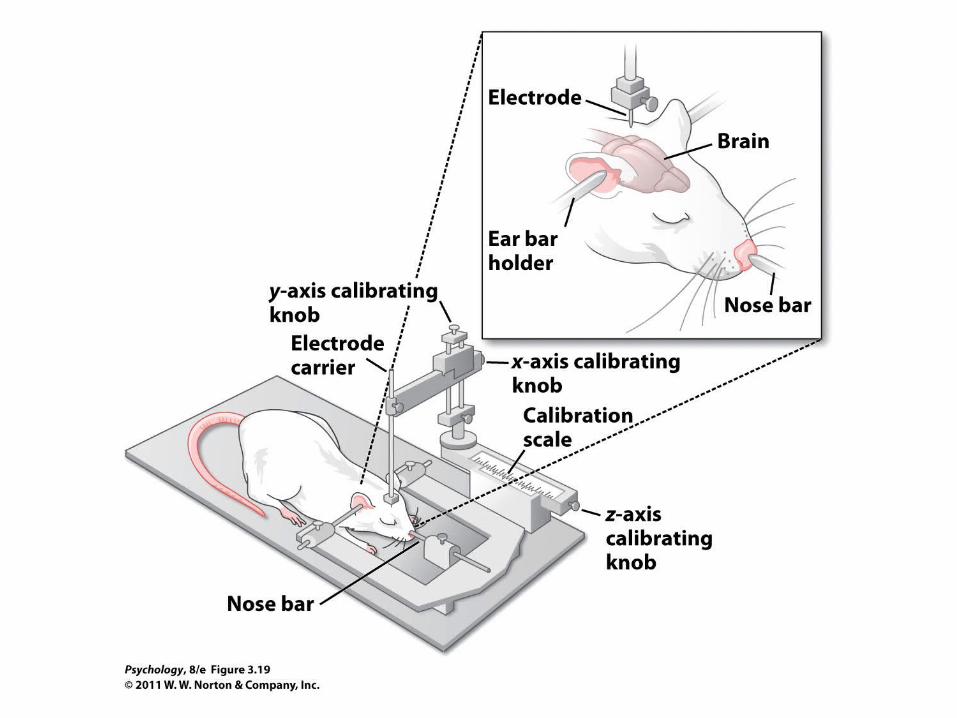

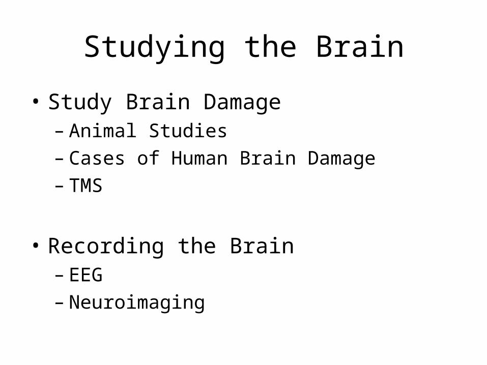
Brain Damage
• In some animal studies, damage is produced in the laboratory.
• But neuropsychologists often study naturally occurring cases of brain damage.
• Transcranial magnetic stimulation (TMS):
• Scientists can use TMS to study the effects of temporary brain damage.


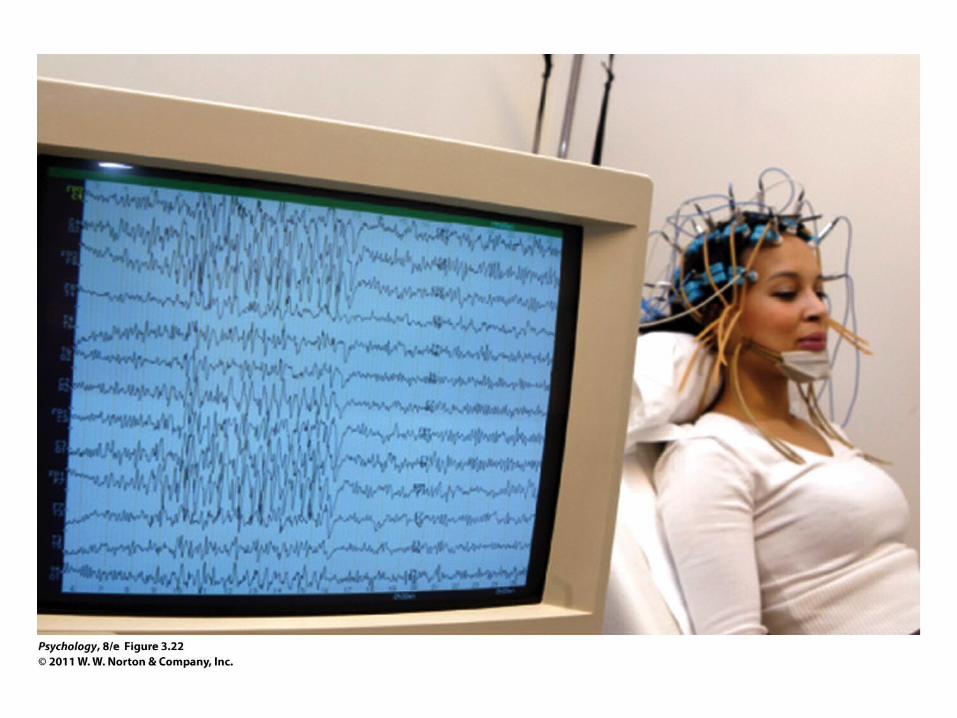
Recording the Brain
• Techniques are used to study the whole brain:
• Electroencephalography
• Uses sensitive electrodes on the scalp to measure voltages produced by brain activity
• Neuroimaging
• CT, MRI, fMRI, PET scans

Recording the Brain
• MRI and CT scans
• Study the brain’s anatomy—the size and location of individual structures
• PET and fMRI scans
• Reveal which brain locations are particularly active at any moment in time


Recording the Brain
• All these techniques make it clear that most activities rely on many brain sites.
• Activities like reading or making decisions are supported by coordinated functioning of many different parts of brain.

Brain Anatomy

Brain Structure
• The very top of the spinal cord forms the brain stem.
• It includes the medulla and the pons.
• Just behind these is the cerebellum.
• The midbrain is on top of the pons, and on top of them all is the forebrain.


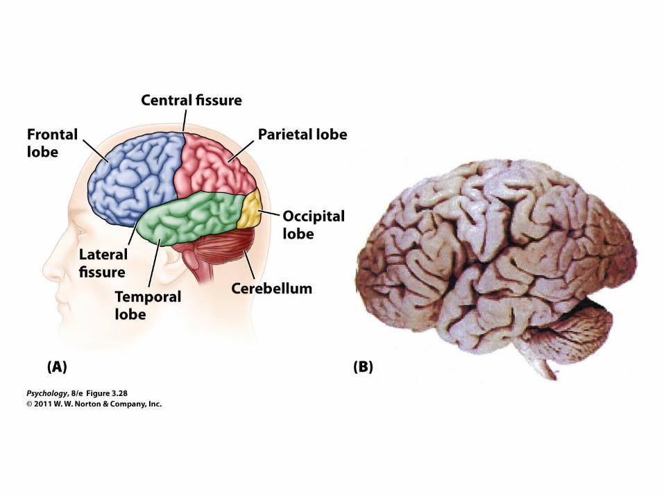
The Cortex
• The outer surface of forebrain is the cerebral cortex.
• The cortex is a large, thin sheet of tissue crumpled inside the skull.
• Some of the convolutions divide the brain into sections:
• The frontal lobes, the parietal lobes, the occipital lobes, and the temporal lobes

Left and Right Hemispheres
• The brain is symmetrical around the midline.
• Most structures come in pairs:
• One on the left side
• One on the right side

Localization of Function
• Different parts of the brain serve specialized functions
• Sensory Information
• Motor Control
• Perception
• Language
• Planning and Social Cognition

Cerebral Cortex
• Some parts serve as projection areas:
• The first receiving stations for information coming from the sense organs (e.g., somatosensory projection areas)
• Departure points for signals going to the muscles (e.g., motor projection area)

Cerebral Cortex
• Adjacent sites in the brains usually represent adjacent parts of the body.
• Assignment of space is disproportionate:
• Usually the parts of the body that are most sensitive to touch receive the most space (in somatosensory projection area).
• Parts of the body that we can move with more precision receive the most space (in primary motor projection area)


Cerebral Cortex
• Most projection areas have contralateral organization:
– Left hemisphere receives information from right side of body (sensory), or controls right side of body (motor)
– Right hemisphere receives information from left side of body (sensory), or controls left side of body (motor)


Cortical Damage
• Much of what we know about the cortex comes from studying brain damage.
• Damage at identifiable sites can produce:
• Apraxias (disorders in action)
• Agnosias (disorders in perception)
• Aphasias (disorders of language)
• Disorders of planning or social cognition
Apraxias
• Difficulty in carrying out purposeful movements without the loss of muscle strength or coordination– Disconnection between primary and non-
primary motor areas– Able to carry out each part of a complex
movement, but disruption lies in coordination of the movements

Agnosias• Visual agnosia: disturbance in recognizing visual stimuli despite the
ability to see and describe them
• Prosopagnosia: inability to recognize faces (fusiform face area)– http://www.youtube.com/watch?v=vwCrxomPbtY&feature=related – http://www.youtube.com/watch?v=VKa-PuJCrO4&feature=related
• Neglect Syndrome: complete inattentiveness to stimuli on one side of the body– http://www.youtube.com/watch?v=ADchGO-0kGo&feature=related
• Akinetopsia: inability to perceive movement– “I see the world in snapshots – like frames of a move but most of the
frames are missing”

Aphasias
• Broca’s Aphasia: disturbance in speech production, caused by damage to Broca’s area– http://www.youtube.com/watch?v=f2IiMEbMnPM
• Agrammaticism• Anomia• Difficulty with articulation
• Wernicke’s Aphasia: disturbance in speech comprehension, caused by damage to Wernicke’s area– http://www.youtube.com/watch?v
=aVhYN7NTIKU&feature=related • Disruption in recognition of spoken words• Disruption in comprehension of the meaning of words• Inability to convert thought into words


Disorders of Planning and Social Cognition
• Caused by damage to prefrontal area– Disrupts executive control– processes that
allow us to direct our own cognitive activities• e.g., setting priorities, planning, strategizing,
ignoring distractors

Lateralization
• The left and right hemispheres are generally similar
• However, the two hemispheres have specialized capacities
– Left hemisphere: language
– Right Hemisphere: visual and spatial tasks
• The two halves of the brain work as an integrated whole
– Communicate with each another through commissures
• Split Brain Patients


Other Split Brain Experiments
http://www.youtube.com/watch?v=ZMLzP1VCANo

Plasticity
• The brain is plastic—subject to alteration in the way it functions, such as:
• Changes in the brain’s overall architecture
• The central nervous system can grow new neurons:
• But appears unable to do so with cortical injury
• This promotes stability in the brain’s connections but is an obstacle to recovery from brain damage.

Plasticity
• Neurons are subject to alteration in the way they function, such as:
• Changes in how much neurotransmitter a presynaptic neuron releases
• Changes in neuron sensitivity to neurotransmitters
• Creating new connections by growing new dendritic spines