the calibration of a software programme to assess...
TRANSCRIPT

The Calibration of a Software Programme to Assess Ceramic Crown
Preparations in a Pre-clinical Setting
VARIZA DAYA ROOPA
A research report submitted to the Faculty of Health Sciences, University of the
Witwatersrand, in partial fulfilment of the requirements for the degree of Master of Science
in Dentistry.
Supervisor:
Professor CP Owen
Co-supervisor:
Dr VJ Hoods-Moonsammy
School of Oral Health Sciences, Faculty of Health Sciences, University of the Witwatersrand,
South Africa
Johannesburg, 2016

ii
DECLARATION
I, Variza Daya Roopa, declare that this research report is my own work. It has been submitted
for the degree of Master of Science in Dentistry in the Faculty of Health Sciences at the
University of the Witwatersrand, Parktown, Johannesburg, South Africa. It has not been
submitted before for any other degree or examination at this or any other University.
Signed ………………………………………….
This 13th day of April 2016

iii
DEDICATION
I dedicate this research report to my niece, Dhiyana, who from the day she was born has been
the light of my life.

iv
PRESENTATIONS
1. Poster Presentation: The 46th
Scientific Meeting of the International Association for
Dental Research (South African Division), 3-4 September 2015, Pretoria, South
Africa
2. Poster Presentation: The 16th
Biennial Meeting of the International College of
Prosthodontics, 16-21 September 2015, Seoul, South Korea.
v
ACKNOWLEDGEMENTS
Prof CP Owen (BDS, MScDent, MDent, FICD) for his undivided supervision, continued
support, invaluable guidance, encouragement and inspiration throughout this research project.
Dr VJ Hoods-Moonsammy (BSc Hons, BDS, MPH, MDent) for her supervision, guidance
and support throughout this research project.
Mr Roland Felber (Goethe University, Frankfurt) for training on PrepCheck 1.1 and his
continued support and advice on the use of the software.
Mr Andy Cyprianos (Sirona Dental Systems, South Africa) for donating the PrepCheck
license and for sponsoring training on the software.
Dr Karen Bennie, Dr Noland Naidoo, Dr Michael Michael, Dr Meriting Thokoane and
Dr Sandile Mpungose for their participation in the study.
Dr Marc Slabbert and Mr Marlo Bester for their kind assistance and advice.
My parents, Bharad and Priya Daya Roopa, for encouraging me to continue furthering my
education and to pursue this degree.
My dear friend, Dane Schovell, for his assistance, continued support and motivation.

vi
ABSTRACT
Purpose
The use of the PrepCheck (v.1.1) software (Sirona Dental Systems GmbH, Germany) is to
enable assessment of student tooth preparations to be digital, objective, accurate, consistent
and reliable so that students can work independently. The software has its default parameters
configured to arbitrary values which are not based on clinical evidence of tooth preparations.
The aim of this study was to set new parameters of the software to realistically assess the
quality of clinically acceptable ceramic crown preparations.
Method
Based on evidence in the literature a new assessment rubric for the evaluation of ceramic
tooth preparations was created which allowed for grading of the preparations as Acceptable,
Requires Modification or Unacceptable. Sixty preparations were made on typodont teeth for
tooth numbers 11, 13, 15, 16, 36 (FDI system). For each tooth four preparations were made to
meet all the requirements under the Acceptable Category, four with variations in taper,
incisal/ occlusal reduction and axial reduction to be categorised as Requires Modification and
four had further variations made so that they fall under the Unacceptable Category. The sixty
preparations were assessed by five faculty instructors (acting as raters) at baseline and again
after two weeks to assess intra- and inter-rater reliability. Once sufficient agreement had been
reached, the software’s parameters were adjusted and the preparations were scanned and
compared with the categorical assessment from the instructors. This process was repeated
once to test whether the software had been successfully calibrated.

vii
Results
The intra-rater agreement was substantial, with two raters having excellent intra-rater
agreement. However, two raters had poor inter-rater agreement and were then excluded, after
which the inter-rater reliability measured by Cohen’s kappa was 0.71, corresponding to
‘substantial’ agreement on both occasions. The majority decision rating from these
assessments accurately resembled the intended rating and was used to compare the ratings of
the software assessment using the default and new (rubric) parameter settings. The default
settings performed poorly, whereas the new settings resulted in substantial agreement with
the majority decision of the instructors (raters).
Conclusions
It was found that the default parameters of the software were unrealistic and not clinically
based. The parameters required considerable modification to assist in the development of
clinically acceptable preparations. The software shows great promise but the parameters have
to be modified to be able to assess preparations that are more realistic for the clinical
situation.

viii
TABLE OF CONTENTS
Chapter 1 ........................................................................................................................ 1
Introduction and Literature Review ............................................................................... 1
1.1 Background to study ............................................................................................................ 1
1.2 Rationale for study ............................................................................................................... 5
Chapter 2 ........................................................................................................................ 6
Aim and Objectives ......................................................................................................... 6
2.1 Aim ....................................................................................................................................... 6
2.2 Objectives ............................................................................................................................. 6
2.3 Null Hypothesis .................................................................................................................... 6
Chapter 3 ........................................................................................................................ 7
Method and Materials ..................................................................................................... 7
3.1 Method ................................................................................................................................. 7
3.1.1 Development of Assessment Rubric ...................................................................................... 7
3.1.2 Preparation of Test Teeth ....................................................................................................... 7
3.1.3 Assessment of Tooth Preparations by Instructors .................................................................. 9
3.1.4 Assessment of Tooth Preparations by Software ................................................................... 10
3.2 Sample Size ........................................................................................................................ 12
3.3 Statistical Analysis ............................................................................................................. 13
3.4 Ethical Considerations ....................................................................................................... 13
Chapter 4 ...................................................................................................................... 14
Results ........................................................................................................................... 14
4.1 Assessment of Tooth Preparations by Instructors .............................................................. 14
4.1.1 Instructor Ratings ................................................................................................................. 14
4.1.2 Intra-rater Reliability............................................................................................................ 15
4.1.3 Inter-rater Reliability............................................................................................................ 15
4.2 Assessment of Tooth Preparations by Software .................................................................. 17
4.2.1 Inter-rater Reliability: PrepCheck before calibration vs. majority decision rating .............. 18
4.2.2 Inter-rater Reliability: PrepCheck run after calibration vs. majority decision rating ........... 18
4.2.3 Intra-rater Reliability: First vs. second PrepCheck run after calibration ............................. 18
Chapter 5 ...................................................................................................................... 19
Discussion ...................................................................................................................... 19
5.1 Instructor Assessment ........................................................................................................ 19
5.1.1 Intra-rater Reliability............................................................................................................ 19
ix
5.1.2 Inter-rater Reliability............................................................................................................ 19
5.1.3 Majority Decision ................................................................................................................ 20
5.2 Software Use ...................................................................................................................... 20
5.2.1 Analysis Tools ..................................................................................................................... 20
5.2.2 Default Settings .................................................................................................................... 31
5.2.3 Issues with Default Settings ................................................................................................. 32
5.3 Parameters ......................................................................................................................... 33
5.3.1 Parameter Setting ................................................................................................................. 33
5.3.2 Comparison of Rubric Settings to the Majority Decision .................................................... 34
5.3.3 Reliability ............................................................................................................................. 35
5.4 Limitations/ challenges encountered ................................................................................... 35
Chapter 6 ...................................................................................................................... 37
Conclusions and Recommendations .............................................................................. 37
6.1 Conclusions ........................................................................................................................ 37
6.2 Implications ........................................................................................................................ 37
6.3 Recommendations .............................................................................................................. 38
References ..................................................................................................................... 39
Appendices .................................................................................................................... 42
Appendix 1: Assessment Rubric for Posterior All-ceramic Crown Preparation ........................ 42
Appendix 2: Assessment Rubric for Anterior All-ceramic Crown Preparation ......................... 43
Appendix 3: Table showing Evidence for Posterior All-ceramic Crown Preparation Criteria .. 44
Appendix 4: Table showing Evidence for Anterior All-ceramic Crown Preparation Criteria ... 45
Appendix 5: Summary of Analysis Tools: Functions, Interpretations and Parameters ............. 46
Appendix 6: Human Research Ethics Committee (Medical) Waiver ......................................... 47
Appendix 7: Modified Assessment Rubric for Posterior All-ceramic Crown Preparation ......... 48
Appendix 8: Modified Assessment Rubric for Anterior All-ceramic Crown Preparation ......... 49

x
LIST OF FIGURES
Figure 1. Taper tool on the PrepCheck software ...................................................................... 4
Figure 2. (a) An example of an all-ceramic preparation on tooth 36 that would be
Acceptable; (b) An example of an all-ceramic preparation on tooth 36 that would Require
Modification due to undercuts present and insufficient occlusal reduction; (c) An example of
an all-ceramic preparation on tooth 36 that would be Unacceptable due to increased occlusal
reduction .................................................................................................................................... 9
Figure 3. Screenshot of Taper tool indicating 87% of the preparation on tooth 15 requires
modification. The cursor indicates the degrees in the selected area and a description that the
walls are too parallel ................................................................................................................ 11
Figure 4. Screenshot of Undercut tool indicating 45% of the preparation on tooth 15 requires
modification to eliminate the undercuts present ...................................................................... 11
Figure 5. (a) Screenshot of PrepCheck using Undercut tool for preparation on 15 after
defining the axis on occasion 1 indicating 24% of undercuts present; (b) Screenshot of the
same preparation after defining the axis on occasion 2, indicating 25% of undercuts present 21
Figure 6. Screenshot of Reduction tool with superimposed Splice tool and manual Measure
Distance tool. The measured distance is 4.04 mm and the measured value based on the
Reduction tool is 2.38 mm. Each grid block represents 1 mm ................................................ 23
Figure 7. Screenshot of Reduction tool with manual Measure Distance tool. The measured
distance and the measured value based on Reduction tool are both 1.44 mm ......................... 24
Figure 8. Screenshot of Reduction tool. The tolerance for the margin width is set from 1.0 -
1.5 mm. The inner axial line of the margin is blue indicating it falls within these parameters
and is thus Acceptable. The walls of the preparation are highlighted in red indicating that
more than 1.5 mm of tooth structure was removed ................................................................. 25

xi
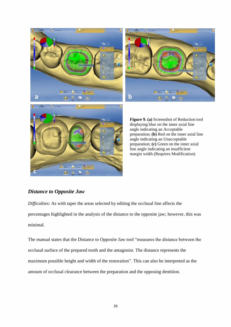
Figure 9. (a) Screenshot of Reduction tool displaying blue on the inner axial line angle
indicating an Acceptable preparation; (b) Red on the inner axial line angle indicating an
Unacceptable preparation; (c) Green on the inner axial line angle indicating an insufficient
margin width (Requires Modification) .................................................................................... 26
Figure 10. Screenshot of Distance to Opposite Jaw tool on tooth 11. Only the colours
displayed on the preparation that opposes the 41 should be interpreted. In this case it is the
green colour which will indicate insufficient occlusal clearance ........................................... 27
Figure 11. Screenshot of Distance to Opposite Jaw tool and manual Measure Distance tool.
A distance of approximately 1.39 mm exists between the maxillary and mandibular molars of
KaVo model teeth .................................................................................................................... 28
Figure 12. (a) Screenshot of Preparation Type indicating 100% blue margin; (b) Red areas in
the margin of 13%; (c) 85% of the margin is green ................................................................. 29
Figure 13. Screenshot of Margin Quality tool indicating 5 areas of orange that will require
modification ............................................................................................................................. 30
Figure 14. Screenshot of Surface Quality tool displaying a grey (smooth) preparation that is
Acceptable................................................................................................................................ 31

xii
LIST OF TABLES
Table 1. Combinations of test preparations ............................................................................... 8
Table 2. Instructor ratings and majority decision on occasion 1 and occasion 2 .................... 14
Table 3. Intra-rater reliability for each of the fives assessors (raters) ..................................... 15
Table 4. Agreement between each pair of assessors (raters) on occasion 1; A: percentage
agreement; B: kappa / weighted kappa; C: p-value for Stuart Maxwell test ........................... 16
Table 5. Agreement between each pair of assessors (raters) on occasion 2; A: percentage
agreement; B: kappa / weighted kappa; C: p-value for Stuart Maxwell test ........................... 17
Table 6. Differences between default settings (before calibration) and rubric settings (after
calibration) ............................................................................................................................... 17

1
Chapter 1
Introduction and Literature Review
1.1 Background to study
Computer Assisted Design and Computer Assisted Manufacturing (CAD/CAM) technology
has improved the way in which health care professionals can provide a service to patients.
The concept of digital dentistry has not only been beneficial to the dental practitioner in terms
of keeping a digital workflow, saving time and reducing laboratory and other costs but more
importantly to the patient who receives restorations often at reduced cost and usually at
reduced clinical time.
Since its introduction in the 1980s, the popularity of CAD/CAM has increased amongst
dental practitioners and their patients. This has inevitably placed a demand on universities
training dentists to include education on this technology into the undergraduate curriculum.
At the University of the Witwatersrand, School of Oral Health Sciences, CAD/CAM dentistry
has been incorporated into the Fixed Prosthodontics undergraduate and postgraduate
curriculums. Many universities around the world have done the same, equipping future
dentists with the knowledge and practical skill of creating milled ceramic indirect
restorations.
In most dental schools a pre-clinical fixed prosthodontics course is a pre-requisite to clinical
training. Students will be trained to prepare crown (or inlay/onlay) tooth preparations on
acrylic teeth in a simulated clinical environment. Evaluation and feedback of these
preparations are provided by instructors (trained dentists of the faculty) which enable the

2
student to develop and improve in order to provide accurate tooth preparations that will
receive accurate permanent restorations.
Evaluation by instructors can be a weak link in dental education, where feedback is often
subjective. Traditional evaluation, adopted by many schools, is carried out visually and
mostly without any assessment aid. This makes grading of students’ work less accurate and
variable when there is a lack of calibration between multiple evaluators. Many studies (Lilley
et al, 1968; Fuller, 1972; Salvendy et al, 1973; Mackenzie et al, 1982; Feil and Gatti, 1993;
Haj-Ali and Feil, 2006; Sharaf et al, 2007) have highlighted these aspects, and have pointed
out the many variables in assessment of pre-clinical work. Several investigators concluded
that to achieve accurate feedback, it is necessary to remove the human element from
evaluation and develop objective evaluation methods (Salvendy et al, 1973; Schiff et al,
1975).
There are currently four systems which have been reported on that assess tooth preparations
using software. E4D Compare (D4D Technologies, Texas, USA) was evaluated by Renne et
al (2013), who concluded “This study demonstrated a reliable method of scanning and
comparing student tooth preparations to a known ideal preparation. Using this method makes
it feasible to accurately and consistently assess student work without dependence on
subjective evaluation criteria.” Another system is PrepAssistant (Kavo Dental GmbH,
Biberach, Germany). Cardoso et al. (2006) used this software by combining it with visual
assessment in a 70:30 percentage ratio. PrepAssistant was reported to be a labour intensive
system and did not show a statistically significant difference in results when compared with
the visual evaluation method. However, the study showed that when used in conjunction with
visual assessment, the software did provide a more objective assessment and reduced

3
problems in calibration amongst different evaluators. Esser et al (2006) used Scan 3D and
Match 3D Software (from Gloger W, Ludwig-Maximilians-University, Munich, Germany) to
evaluate the extent to which the software’s analysis of prepared teeth could replace visual
assessment from a dental educator. The results of their study showed that it was possible to
digitally measure quality criteria for a ceramo-metal crown preparation, but the measuring
method applied required improvement as did the calibration of the evaluators.
PrepCheck 1.1 is the current version available of a computer aided training system developed
by Sirona Dental Systems GmbH (Bensheim, Germany) in 2014. The software works with
Cerec 4.2 + and provides fully automated 3D analysis of tooth preparations and applied
restorative material. The software works using Bluecam or Omnicam scanners and can assess
all types of crowns and inlays. The programme is aimed at universities to aid in training
students as well as to aid instructors in evaluating students’ work.
There are several analysis functions that assist both the student and the instructor to assess
tooth preparations digitally. The analysis functions include tools that can automatically detect
the presence of undercuts, measure the taper of any aspect of the tooth preparation, measure
the amount of axial reduction (and margin width), measure the amount of restorative space
available occlusally, compare the preparation margin type to a pre-defined margin type
(shoulder or chamfer) and can assess the margin and surface quality for smoothness or sharp
edges. The analysis functions also include manual tools that enable the user to measure the
distance between any two points on the preparation or measure an angle between any three
points on the preparation.
Other features of the software include a 3D comparison of the preparation to a pre-selected,
gold standard “Master Preparation”, a 3D comparison of the preparation to the pre-operative

4
tooth to identify the amount of reduction that has taken place, a 3D comparison of the
preparation to the proposed restoration to measure the thickness of the restoration and a 3D
comparison of one preparation to another so that the user can follow their progress.
A colour scale provides the user with feedback about the quality of the preparation. Areas
that are highlighted in blue indicate those within the specified tolerance range. Areas of the
preparation highlighted in green are those that are below the minimum tolerance value and
would require some modifications to achieve an acceptable result. Areas highlighted in red
are those which are beyond the maximum tolerated values and are not acceptable.
An example of the colour scale feedback can be seen using the analysis tool that measures
taper in Figure 1.
Figure 1. Taper tool on the PrepCheck software
Current studies on the use of PrepCheck are of low level evidence and include pilot studies at
Goethe University in Frankfurt showing that 80% of students find PrepCheck easy to use.
Another study from the same institution showed that PrepCheck was useful to objectively
assess reduction, taper and height of preparations and that the results were reproducible
(Felber R. Personal communication, 05 May 2014).

5
A pilot study carried out at Harvard University indicated that the software was reliable to
measure undercut, taper and reduction, but the authors concluded that a comparison between
assessors and the software needed to be made (Hou and Kristiansen, 2014).
The School of Oral Health Science at the University of the Witwatersrand has standardised
the use of Sirona’s Cerec CAD/CAM software, and Sirona has donated a license to use the
PrepCheck software as well. Our experience has been that the software is complicated to use,
but with modification might be more user-friendly and ultimately its benefit ought to include
use in general practice, to improve the quality of the preparations being made. Initially,
though, it requires calibration against clinically acceptable preparations, and against
experienced instructors, and this is the purpose of the study.
1.2 Rationale for study
The rationale for the use of PrepCheck is to ensure that assessment of student tooth
preparations can be digital, objective, accurate, consistent and reliable. Students can work
independently and be motivated to improve. PrepCheck software has its parameters
(tolerance values) configured at default settings. The problem this poses is that the analysis of
tooth preparations is set according to arbitrary values and not that which is evidence-based.
The solution and hence rationale for this research project is thus to calibrate the parameters of
the various PrepCheck analysis tools according to the criteria required for assessment of all-
ceramic tooth preparations at the Department of Oral Rehabilitation, University of the
Witwatersrand. These criteria are not only based on evidence and widely recognised, but can
be applied in the laboratory for techniques training and utilised clinically during patient
treatment. By calibrating the software to these criteria student tooth preparations can be
assessed digitally during the techniques course and then subsequently clinically (which would
be the topic of a further study).

6
Chapter 2
Aim and Objectives
2.1 Aim
To set new parameters of the PrepCheck software to realistically assess the quality of all-
ceramic crown preparations.
2.2 Objectives
1. To develop an assessment rubric to assess all-ceramic crown tooth preparations
2. To make standardised preparations for all-ceramic crowns for anterior and posterior teeth
3. To vary the standardised preparations in height, taper, and axial reduction
4. To determine the inter-rater and intra-rater reliability between calibrated laboratory
instructors when assessing these preparations
5. To assess the preparations according to the default settings and compare these ratings to
that of the majority decision rating of the instructors
6. To set new parameters for the analysis tools of the PrepCheck software to conform to the
agreed assessment criteria
2.3 Null Hypothesis
The PrepCheck software cannot be successfully calibrated to the requirement criteria for
assessing all-ceramic crown tooth preparations.

7
Chapter 3
Method and Materials
3.1 Method
3.1.1 Development of Assessment Rubric
A new assessment rubric for evaluation of anterior and posterior all-ceramic tooth
preparations was created for this study (see Appendices 1 and 2). Currently the students’
tooth preparations are assessed visually by instructors using a set of criteria but without any
measurement or grading aids. The criteria required for preparations have been defined by
evidence in the literature (see Appendices 3 and 4) (Smith, 1986; Goodacre et al, 2001;
Rosenstiel et al, 2006; Beuer et al, 2009; Coehlo, 2009; Giannetopoulos et al, 2010; Ender,
2011).
The assessment will allow for grading of tooth preparations as either Acceptable (preparation
conforms to all of the required criteria), Requires Adjustment (preparation conforms to some
criteria, but adjustments can be made without making the preparation unacceptable) or
Unacceptable where a preparation has failed to meet at least one criterion and no adjustment
is possible without causing further damage to the tooth.
3.1.2 Preparation of Test Teeth
A full set of Typodont model teeth (KaVo Dental GmbH, Biberach, Germany) inserted into a
KaVo basic study model placed into a simulated patient (Frasaco, North Carolina, USA) was
used. The principal investigator produced sixty all-ceramic crown preparations on the

8
following teeth: 11; 13; 15; 16; 36, under 2,5x loupe binocular magnification (HEINE
Optotechnik, Herrsching, Germany).
For each tooth four preparations were made such that they met all the requirements under the
Acceptable category of the assessment rubric, and four preparations were made with
variations in taper, incisal/ occlusal reduction and axial reduction such that they should be
categorised as Requires Modification and a further four preparations with further variations
have been made so that they should fall under the Unacceptable category of the assessment
rubric (See Table 1).
Table 1. Combinations of test preparations
Tooth
Category
Acceptable Requires Modification Unacceptable/ Rejected Total
11 P P P P ↓ T ↓ IR ↓ AR ↓ IR ↓ T ↑ T ↑ IR ↑ AR ↑ AR ↓ T 12
13 P P P P ↓ T ↓ IR ↓ AR ↓ IR ↓ T ↑ T ↑ IR ↑ AR ↑ AR ↓ T 12
15 P P P P ↓ T ↓ OR ↓ AR ↓ OR ↓ T ↑ T ↑ OR ↑ AR ↑ AR ↓ T 12
16 P P P P ↓ T ↓ OR ↓ AR ↓ OR ↓ T ↑ T ↑ OR ↑ AR ↑ AR ↓ T 12
36 P P P P ↓ T ↓ OR ↓ AR ↓ OR ↓ T ↑ T ↑ OR ↑ AR ↑ AR ↓ T 12
Total 20 20 20 60
P, Perfect; T, Taper; IR, Incisal Reduction; AR, Axial Reduction; OR, Occlusal Reduction;
↓ Decrease; ↑ Increase.
Examples of how the preparations on tooth 36 have been varied to show changes in occlusal
reduction are shown in Figure 2. .

9
Figure 2. (a) An example of an all-ceramic
preparation on tooth 36 that would be
Acceptable; (b) An example of an all-ceramic
preparation on tooth 36 that would Require
Modification due to undercuts present and
insufficient occlusal reduction; (c) An
example of an all-ceramic preparation on
tooth 36 that would be Unacceptable due to
increased occlusal reduction
3.1.3 Assessment of Tooth Preparations by Instructors
There were five faculty supervisors / laboratory instructors who had been calibrated to
achieve inter- and intra-rater reliability when visually analysing randomised tooth crown
preparations by examining twenty-one crown preparations prior to the study. These
preparations were from an undergraduate student course and varied in quality to reflect each
of the three categories of the assessment rubric. The principal investigator had made any
necessary changes to these preparations so that in the investigator’s opinion they reflected the
three categories of Acceptable, Requires Modification or Unacceptable.
After achieving the required calibration, the sixty test preparations were coded, and the order
in which they were to be assessed was randomised using Excel Rand function (Microsoft
Excel 2010, Microsoft, Washington, USA) throughout the study. The instructors then
assessed the sixty tooth preparations under 2.5 x loupe magnification. After 2 weeks all sixty
a b
c

10
test preparations were randomised again and re-assessed by the same instructors. From each
of the five instructors each preparation had been categorized as Acceptable, Requires
Modification or Unacceptable. The data from the two occasions of assessments were
collected and analysed to determine the inter-rater reliability, intra-rater reliability and the
majority decision of the five instructors.
3.1.4 Assessment of Tooth Preparations by Software
The sixty test preparations were again randomised and each one captured in the PrepCheck
software using a Cerec AC and Omnicam scanner (Sirona Dental Systems GmbH, Bensheim,
Germany). The preparations were assessed digitally using the following tools: Undercut,
Taper, Reduction, Distance to Opposite Jaw, Preparation Type, Margin Quality, and Surface
Quality. See Appendix 5 for more details on the analysis tools.
The interpretation of the analysis is based on different colours displayed:
Blue indicates the preparation falls within the tolerance range and is thus acceptable
Green indicates areas that require further modification
Red indicates areas that are over-prepared and thus unacceptable
Yellow indicates the presence of an undercut and will require modification
Orange indicates an area of an indentation or sharp edge, and will require
modification
Grey indicates a smooth surface which is acceptable
White indicates areas that are acceptable, such as a smooth margin or areas free of
undercuts
The outcome or category of the preparation took into consideration all of the above
mentioned tools used and was based on the colours displayed. For example if a preparation
was highlighted green for taper (Figure. 3) and yellow for undercuts (Figure. 4) and all other
features fell in the acceptable range, it was categorised as Requires Modification based on the
insufficient taper and presence of undercuts. If, however, the same preparation was

11
highlighted red on reduction or distance to opposite jaw it would be automatically categorised
as Unacceptable.
Figure 3. Screenshot of Taper tool indicating 87% of the preparation on tooth 15 requires
modification. The cursor indicates the degrees in the selected area and a description that the walls
are too parallel.
Figure 4. Screenshot of Undercut tool indicating 45% of the preparation on tooth 15 requires
modification to eliminate the undercuts present.

12
The test preparations were randomised and first assessed and recorded using the default
settings of the software parameters. The software’s parameters were then manually re-set to
the criteria according to the rubric and other additional changes were made (See Appendix 5).
The test preparations were again randomised and re-scanned into the software and analysed
using the new rubric settings. The final outcome/ selected category for each tooth preparation
using the new settings was recorded and compared against that of the default settings.
Each difference found was recorded and tabulated and compared with the categorical
assessment from the assessors.
The parameters of the software did not require further adjustment based on the results. The
tooth preparations were then randomised again and re-scanned and assessed by the
PrepCheck software with the new parameters to test whether the software had been
successfully calibrated.
3.2 Sample Size
An estimation of the sample size was based on the determination of the inter-rater reliability
for the five instructors, and the inter-rater reliability of the software versus the instructor
majority decision. The sample size depended on the relative error, the number of subjects and
the difference between the overall agreement probability and chance.
Assuming a relative error of 20%, a large number of subjects, and that the chance-agreement
probability is 0, for an overall agreement probability of at least 0.6 (0.7), the required sample
size is 69 (51). The chosen sample size of 60 was thus adequate for the purposes of this
study.

13
3.3 Statistical Analysis
Descriptive statistics were presented as frequencies and percentages. The inter-rater and intra-
rater reliability for the instructors were assessed using Cohen’s kappa for ordinal data.
Cohen’s kappa for ordinal data was also used for the comparison between the majority
instructor rating and each of the derived software ratings. For two raters and ordinal
responses, Fleiss’ quadratically weighted kappa was used to allow each category combination
to be weighted according to the degree of agreement between the categories (Shoukri, 2010).
The magnitude of kappa was interpreted according to the classification by Landis and Koch
(1977). Inter-rater and intra-rater bias was assessed by the Stuart-Maxwell test. Data analysis
was carried out using SAS Software Version 9.3 for Windows (SAS Institute Inc., North
Carolina, USA). The 5% significance level was used throughout.
3.4 Ethical Considerations
This was a laboratory based study. A waiver for ethical clearance has been granted by the
Human Research Ethics Committee (Reference: W-JC-150205-2). See Appendix 6.
The donation of PrepCheck 1.1 from Sirona Dental Systems to the School of Oral Health
Science has not resulted in any bias in favour of the software.

14
Chapter 4
Results
4.1 Assessment of Tooth Preparations by Instructors
4.1.1 Instructor Ratings
The descriptive statistics of the data for the intended rating, the 10 instructor ratings (5 raters
on each of 2 occasions), and the majority decision of both occasions, are presented in
Table 2.
Table 2. Instructor ratings and majority decision on occasion 1 and occasion 2
Rater
1 2 3 4 5 Majority Decision
Occasion 1 2 1 2 1 2 1 2 1 2 1 2
% of samples rated as: Intended
rating
Acceptable 33.3 31.7 33.3 33.3 36.7 28.3 25.0 23.3 26.7 8.3 11.7 26.7 28.3
Requires Modification 33.3 28.3 30.0 45.0 43.3 30.0 38.3 40.0 35.0 35.0 38.3 31.7 35.0
Unacceptable 33.3 40.0 36.7 21.7 20.0 41.7 36.7 36.7 38.3 56.7 50.0 41.7 36.7
% correct with respect to intended rating: 81.7 83.3 81.7 83.3 80.0 78.3 85.0 90.0 65.0 73.3 90.0 91.7
The figures highlighted in red show that rater 5 stands out from the other raters as this rater
has a lower percentage of preparations rated as Acceptable and a higher percentage rated as
Unacceptable on both occasions. This rater also has a lower percentage correct with respect
to the intended rating on both occasions. These results show that rater 5 was far stricter than
the other raters.

15
4.1.2 Intra-rater Reliability
The raw percentage agreement, Cohen’s kappa, Fleiss’ weighted kappa and interpretation of
the Fleiss’ weighted Kappa and the results of the Stuart-Maxwell test are shown in Table 3.
Table 3. Intra-rater reliability for each of the fives assessors (raters)
Rater %
agreement kappa
weighted kappa
interpretation of weighted kappa
p-value for Stuart-
Maxwell test
1 80.0 0.70 0.68 substantial agreement 0.77
2 90.0 0.84 0.86 almost perfect agreement 0.55
3 83.3 0.75 0.75 substantial agreement 0.14
4 88.3 0.82 0.90 almost perfect agreement 0.51
5 76.7 0.59 0.63 substantial agreement 0.35
The Stuart-Maxwell test results indicate that there was no evidence of intra-rater bias
between the two occasions.
4.1.3 Inter-rater Reliability
On the first occasion of ratings, the raw agreement for all raters was 45%, with a Cohen’s
Kappa of 0.59, which corresponds to a ‘moderate’ agreement. Therefore the agreements
between pairs of raters were calculated, and are shown in Table 4.

16
Table 4. Agreement between each pair of assessors (raters) on occasion 1; A: percentage agreement;
B: kappa / weighted kappa; C: p-value for Stuart Maxwell test
Rater 1 2 3 4 5
1
2 A: 78.3% B: 0.68 / 0.71 C: 0.0003
3 A: 80.0% B: 0.70 / 0.61 C: 0.74
A: 71.7% B: 0.58 / 0.62 C: 0.011
4 A: 80.0% B: 0.70 / 0.73 C: 0.047
A: 76.7% B: 0.65 / 0.67 C: 0.007
A: 81.7% B: 0.72 / 0.74 C: 0.08
5 A: 66.7% B: 0.49 / 0.43 C: 0.0007
A: 53.3% B: 0.33 / 0.34 C: <0.0001
A: 70.0% B: 0.53 / 0.47 C: 0.002
A: 71.7% B: 0.55 / 0.57 C: 0.0004
There are significant inter-rater biases (values in red), but also some low Kappa values
(values in blue) in the pairwise comparisons. In particular, rater 5 had significant biases with
all other raters (was stricter and had the lowest pairwise inter-rater agreement with the other
four raters). Rater 2 had similar inter-rater biases although the kappa values were reasonable.
On the second occasion of ratings with all 5 raters, the raw agreement was 48.3% and
Cohen’s Kappa 0.60 which corresponds to ‘substantial’ agreement. However, the pairwise
evaluations were identical to the first round. The agreements between pairs of raters were
calculated, and are shown in Table 5.
17
Table 5. Agreement between each pair of assessors (raters) on occasion 2; A: percentage agreement;
B: kappa / weighted kappa; C: p-value for Stuart Maxwell test
Rater 1 2 3 4 5
1
2 A: 73.3% B: 0.60 / 0.63 C: 0.005
3 A: 75.0% B: 0.63 / 0.66 C: 0.18
A: 70.0% B: 0.55 / 0.55 C: 0.001
4 A: 85.0% B: 0.77 / 0.74 C: 0.31
A: 73.3% B: 0.60 / 0.59 C: 0.004
A: 83.3% B: 0.75 / 0.78 C: 0.077
5 A: 66.7% B: 0.50 / 0.55 C: 0.001
A: 56.7% B: 0.37 / 0.33 C: <0.0001
A: 78.3% B: 0.66 / 0.68 C: 0.003
A: 76.7% B: 0.64 / 0.72 C: 0.002
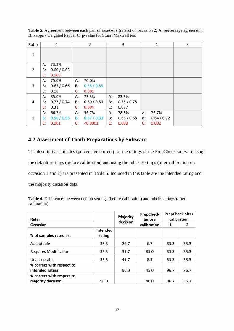
4.2 Assessment of Tooth Preparations by Software
The descriptive statistics (percentage correct) for the ratings of the PrepCheck software using
the default settings (before calibration) and using the rubric settings (after calibration on
occasion 1 and 2) are presented in Table 6. Included in this table are the intended rating and
the majority decision data.
Table 6. Differences between default settings (before calibration) and rubric settings (after
calibration)
Rater
Majority decision
PrepCheck before
calibration
PrepCheck after calibration
Occasion 1 2
% of samples rated as: Intended
rating
Acceptable 33.3 26.7 6.7 33.3 33.3
Requires Modification 33.3 31.7 85.0 33.3 33.3
Unacceptable 33.3 41.7 8.3 33.3 33.3
% correct with respect to intended rating:
90.0 45.0 96.7 96.7
% correct with respect to majority decision: 90.0
40.0 86.7 86.7

18
4.2.1 Inter-rater Reliability: PrepCheck before calibration vs. majority
decision rating
The raw agreement with the majority decision was 40.0% and Cohen’s kappa and Fleiss’
weighted kappa were 0.12 and 0.15 respectively. The latter corresponds to ‘slight agreement’.
The Stuart-Maxwell test was significant (p<0.001), indicating significant bias between the
two sets of data.
4.2.2 Inter-rater Reliability: PrepCheck run after calibration vs. majority
decision rating
The raw agreement with the majority decision was 86.7% and Cohen’s kappa and Fleiss’
weighted kappa were 0.80 and 0.72 respectively. The latter corresponds to ‘substantial
agreement’. The Stuart-Maxwell test was not significant (p=0.19), indicating no significant
bias between the two sets of data.
4.2.3 Intra-rater Reliability: First vs. second PrepCheck run after
calibration
The raw agreement was 100% and Cohen’s kappa and Fleiss’ weighted kappa were both
1.00, i.e. there was perfect agreement.

19
Chapter 5
Discussion
5.1 Instructor Assessment
5.1.1 Intra-rater Reliability
There was no evidence of intra-rater bias and the results in Table 3 show that all five
instructors (raters) had been consistent in their ratings between occasion 1 and 2. Raters 2 and
4 had almost perfect intra-rater agreement and raters 1, 3 and 5 each had substantial
agreement between the 2 occasions.
5.1.2 Inter-rater Reliability
Occasion 1
In Table 4 the p-values for the Stuart Maxwell test show many significant inter-rater biases.
Rater 5 was more strict than the other four raters (significant inter-rater bias in all four cases)
with low kappa values and rater 2 was less strict than the other four raters (significant inter-
rater bias in all four cases), although the kappa values were reasonable.
Given the low overall kappa (0.59), it was then decided to exclude rater 5 and rater 2, to keep
an uneven number of raters and to simplify the majority decision derivation. By making this
exclusion and only using data for raters 1, 3 and 4, the data were validated as the raw
agreement was 71.7% and Cohen’s kappa was 0.71, corresponding to ‘substantial’
agreement. The majority decision data remained unchanged from the 5-rater version, but the
basis for the majority decision outcome was much improved.

20
Occasion 2
The conclusions from the pairwise evaluation in occasion 2 were identical to those from
occasion 1, though perhaps not as extreme (Table 5).
Using the ratings from occasion 2 (with all five raters) the Cohen’s kappa was a low of 0.60,
corresponding to ‘substantial’ agreement.
By using only data for raters 1, 3 and 4, the raw agreement was 73.3% and Cohen’s kappa
was 0.71, corresponding to ‘substantial’ agreement.
5.1.3 Majority Decision
The majority decision data from raters 1, 3 and 4 from occasion 1 were used in the
subsequent phase of the data analysis. Cohen’s kappa was identical for this scenario for both
occasions; thus the results from the first occasion were used by default. Table 2 shows the
accuracy (% correctly classified preparations) of the majority decision rating vs. the intended
rating on occasion 1 was 90.0%. This indicated the intended accuracy of the assessment when
categorising the tooth preparations.
5.2 Software Use
5.2.1 Analysis Tools
The use of the analysis tools of the PrepCheck software came with inherent difficulties.
These were overcome by creating a method or protocol to interpret the digital data and
percentages displayed in a way that made the assessment realistic. The difficulties in using
the tools and the method of analysing the interpretations to reach categorisation of the tooth
preparations are explained for each analysis tool below. A summary can also be found in
Appendix 5.

21
Undercut
This tool detects undercuts present on the preparation and displays the areas which will
require modification in yellow.
Difficulties: The extent of the undercuts measured was affected by defining the tooth axis
which is a preceding step before analysis, i.e. if the tooth axis was defined in a different way
on two occasions the percentage of undercuts present would change as the tooth axis changed
(Figure 5.) This, however, was a change in 1-2%.
Figure 5. (a) Screenshot of PrepCheck using Undercut tool for preparation on 15 after defining the
axis on occasion 1 indicating 24% of undercuts present; (b) Screenshot of the same preparation after
defining the axis on occasion 2, indicating 25% of undercuts present
Interpretation: Acceptable preparations have 0-4% undercut. The rest of the preparation is
white indicating no undercuts present. The presence of “undercuts” on an Acceptable
preparation was mostly due to inaccuracies in the scan quality. Under-tapered preparations,
which will require modification, show undercuts in yellow equal to and greater than 5%.
a b
22
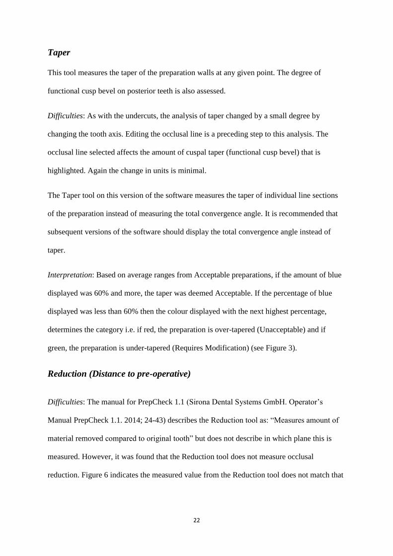
Taper
This tool measures the taper of the preparation walls at any given point. The degree of
functional cusp bevel on posterior teeth is also assessed.
Difficulties: As with the undercuts, the analysis of taper changed by a small degree by
changing the tooth axis. Editing the occlusal line is a preceding step to this analysis. The
occlusal line selected affects the amount of cuspal taper (functional cusp bevel) that is
highlighted. Again the change in units is minimal.
The Taper tool on this version of the software measures the taper of individual line sections
of the preparation instead of measuring the total convergence angle. It is recommended that
subsequent versions of the software should display the total convergence angle instead of
taper.
Interpretation: Based on average ranges from Acceptable preparations, if the amount of blue
displayed was 60% and more, the taper was deemed Acceptable. If the percentage of blue
displayed was less than 60% then the colour displayed with the next highest percentage,
determines the category i.e. if red, the preparation is over-tapered (Unacceptable) and if
green, the preparation is under-tapered (Requires Modification) (see Figure 3).
Reduction (Distance to pre-operative)
Difficulties: The manual for PrepCheck 1.1 (Sirona Dental Systems GmbH. Operator’s
Manual PrepCheck 1.1. 2014; 24-43) describes the Reduction tool as: “Measures amount of
material removed compared to original tooth” but does not describe in which plane this is
measured. However, it was found that the Reduction tool does not measure occlusal
reduction. Figure 6 indicates the measured value from the Reduction tool does not match that

23
of the manual measured distance. This image and many other similar images, verified that
this tool does not measure the occlusal reduction.
Figure 6. Screenshot of Reduction tool with superimposed Splice tool and manual Measure Distance
tool. The measured distance is 4.04 mm and the measured value based on the Reduction tool is
2.38 mm. Each grid block represents 1 mm.
The Reduction tool does, however, measure axial reduction and margin width accurately.
Figure 7 shows the measured value using the Reduction tool accurately matched that of the
manual measured distance when applied to the axial walls. It is for this reason that the
occlusal aspect of the tooth was ignored during the analysis and the focus was placed on the
axial reduction and margin width.

24
Figure 7. Screenshot of Reduction tool with manual Measure Distance tool. The measured distance
and the measured value based on Reduction tool are both 1.44 mm.
However, since axial reduction is always greater for bulbous teeth such as the KaVo
Typodont teeth used in this study, the amount of axial reduction will always be greater than
the margin width. If the tolerance values are set according to the margin width, the axial walls
will always be beyond this tolerance and be displayed in red as if they were over-prepared
and the inner axial line angle of the margin will show up blue if correct (see Figure 8). For
this reason one cannot set the parameters to assess axial reduction and margin width
simultaneously.
Therefore, it was decided that the interpretation will only involve observing the inner axial
line angle to determine the margin width as this coincides with what was assessed visually
and what would be assessed clinically.

25
Figure 8. Screenshot of Reduction tool. The tolerance for the margin width is set from 1.0 -1.5 mm.
The inner axial line of the margin is blue indicating it falls within these parameters and is thus
Acceptable. The walls of the preparation are highlighted in red indicating that more than 1.5 mm of
tooth structure was removed.
Interpretation: For the purpose of this study it was decided to ignore colours displayed on the
axial and occlusal surfaces as this was not what the parameter was measuring. The Reduction
tool was in fact assessing the margin width, displayed as the colour seen on the inner axial
line angle of the margin. If the colour displayed was blue the margin was within the limits
and was Acceptable; if red the margin width was greater than the maximum tolerance value
(and therefore categorised as Unacceptable); and if green, the margin width was less than the
minimum tolerance value (and therefore categorised as Requires Modification) (See Figure 9
for examples).
26
Figure 9. (a) Screenshot of Reduction tool
displaying blue on the inner axial line
angle indicating an Acceptable
preparation; (b) Red on the inner axial line
angle indicating an Unacceptable
preparation; (c) Green on the inner axial
line angle indicating an insufficient
margin width (Requires Modification)
Distance to Opposite Jaw
Difficulties: As with taper the areas selected by editing the occlusal line affects the
percentages highlighted in the analysis of the distance to the opposite jaw; however, this was
minimal.
The manual states that the Distance to Opposite Jaw tool “measures the distance between the
occlusal surface of the prepared tooth and the antagonist. The distance represents the
maximum possible height and width of the restoration”. This can also be interpreted as the
amount of occlusal clearance between the preparation and the opposing dentition.
b a
c
c

27
For anterior teeth the tool measures the distance of the entire preparation towards the
opposing tooth and not just the incisal/ palatal aspect (Figure 10). Therefore it was decided to
read the palatal aspect of the anterior tooth where it occludes only and to ignore the other
colours as they were not a representation of occlusal clearance. In addition, it was noted that
incisal reduction was also not indicated.
Figure 10. Screenshot of Distance to Opposite Jaw tool on tooth 11. Only the colours displayed on
the preparation that opposes the 41 should be interpreted. In this case it is the green colour which will
indicate insufficient occlusal clearance.
For posterior teeth the tool displays colour on the occlusal surface only, and shows the
measurement of the distance from the occlusal surface to the opposing teeth. This tool is not a
representative of occlusal reduction either. It was also found that there is a space that exists
between lower molars and upper molars of KaVo models (inter occlusal 16 and 46) as shown
in Figure 11. This space is created by the deep fissures. Hence a more accurate tool for
measuring occlusal reduction is required.

28
Figure 11. Screenshot of Distance to Opposite Jaw tool and manual Measure Distance tool. A
distance of approximately 1.39 mm exists between the maxillary and mandibular molars of KaVo
model teeth.
Interpretation: For anterior teeth, for the purpose of this study, the palatal area showing the
distance to the opposing tooth was regarded as the only significant area to interpret. This area
represents no more than 12% of the tooth. It was concluded that the percentages were not
important, but rather the colour in that area: blue representing an Acceptable category, green
indicating insufficient clearance and therefore Requires Modification, and red indicating a
preparation that was over-prepared and therefore Unacceptable.
When assessing the posterior teeth the parameter had to be increased in order to compensate
for the occlusal space between the fissural areas of opposing teeth.
For posterior teeth to be Acceptable more than 60% of the occlusal surface had to be
displayed as blue. If less than 60%, the colour displaying the greatest percentage determined
the category. Green would be insufficient occlusal clearance (Requires Modification) and red
over-reduced occlusally and therefore catergorised as Unacceptable.

29
Preparation Type
This tool compares the margin type created to a pre-defined curve which can be a shoulder or
a chamfer or any curve set in the parameters. For this study a shoulder preparation type was
selected.
Difficulties: Preparation type is also affected by defining the tooth axis by a small percentage.
Interpretation: A margin displaying 90-100 % blue would conform to a shoulder
configuration and be categorised as Acceptable. If more than 10% is shown as red the margin
is over-prepared (too much material removed, thus Unacceptable). If more than 10% is
shown as green the margin is under-prepared (conforms to a chamfer i.e. Requires
Modification). See Figure 12 for examples of different preparation types.
Figure 12. (a) Screenshot of Preparation
Type indicating 100% blue margin; (b) Red
areas in the margin of 13%; (c) 85% of the
margin is green
Margin Quality
a b
c

30
Margin Quality
This tool assesses the quality of the margin for indentations or sharp edges by displaying
these areas in orange.
Difficulties: the amount of orange displayed changes with differences in scan quality. Often,
the areas that are displayed as orange (indentations/ sharp edges) are not detectable on the
actual preparation visually or on probing. This would suggest that the tool is unnecessarily
sensitive.
Interpretation: It was decided that Acceptable preparations should display as completely
white or up to 2 areas of orange. More than 2 areas of orange and those which are detectible
on the actual preparation would require modification. Figure 13 is an example of a
preparation that will require smoothing of the margin.
Figure 13. Screenshot of Margin Quality tool indicating 5 areas of orange that will require
modification

31
Surface Quality
This tool measures the surface of the preparation for sharp edges. Any angle greater than 50
degrees is considered a sharp angle and is highlighted orange.
Interpretation: Should a preparation have grey displayed it is Acceptable (smooth) as shown
in Figure 14. Should a preparation have orange displayed, these sharp edges will require
modification.
Figure 14. Screenshot of Surface Quality tool displaying a grey (smooth) preparation
that is Acceptable
5.2.2 Default Settings
As seen in Table 6, the software assessment using the default settings before calibration
performed very poorly. When comparing the default ratings against the intended rating the
accuracy (percentage correct) was only 45.0%. When comparing the default ratings with the
majority decision the raw agreement was 40.0%. There was a significant bias towards the
Requires Modification category where 85% of the preparations were classified.

32
5.2.3 Issues with Default Settings
When using the default settings the interpretations of the preparations were unrealistic, due to
the parameters having been set with unrealistic tolerance values. This resulted in the majority
of the preparations being assessed as Requiring Modification. Several issues were noted with
these default settings that resulted in the poor performance of the software when compared
with the rubric settings. Some of the important issues are discussed here, and Appendix 5
provides a summary.
Firstly, the parameters have the same settings for all teeth, when criteria for anterior,
maxillary posterior and mandibular posterior teeth should all be different.
In terms of measuring taper the minimum (4 degrees) and maximum (12 degrees) tolerance
values are too high and apply to all teeth irrespective of position in the mouth. This results in
many over-tapered and under-tapered preparations being classified as Acceptable. In
addition, the cusp taper (functional cusp bevel) is set on the incorrect side of maxillary
posterior teeth, hence preparations on these teeth were classified as requiring modification.
For the Reduction tool, the tolerance band is too wide with the minimum tolerance value set
too high at 1.75 mm and the maximum set too high at 3.0 mm. These values would not only
result in possible exposure of the pulp clinically, but would also result in preparations with
margins below 1.75 mm being classified as Requires Modification.
For the Distance to Opposite Jaw tool, as with the Reduction tool, the tolerance band is too
wide and the minimum value (1.75 mm) and maximum value (3.0 mm) are set too high.
These values would result in the incorrect interpretation of preparations as Requiring
Modification.

33
In terms of preparation type the tolerance value is set at 0.4 mm. This appears to be too strict
and results in Acceptable preparation types being classified as Requires Modification or
Unacceptable.
For margin quality, many preparations displayed many orange areas on the digital margin,
resulting in the preparation being classified as Requiring Modification. However these
preparations did not display any indentations or sharp edges visually.
Therefore the parameters were adjusted as set out in the following section.
5.3 Parameters
5.3.1 Parameter Setting
All of the analysis tools required adjustments except two, the surface quality and undercut
tools. Fortunately, it was possible to re-set the parameters and to interpret the colours
displayed in such a way that the preparations could be placed in the three different categories.
The parameter changes were based on evidence in the literature as outlined in Appendices 3
and 4 for measurable tools as well as trial and error investigation to re-set tools such as
Margin Quality and Preparation Type.
The description of the parameter changes are outlined below and summarised in Appendix 5.
Taper
The minimum taper was set according to ideal Total Occlusal Convergence angle required as
6 degrees, but the evidence shows that most crowns are prepared with larger convergence
angles: anteriorly from 12-17 degrees and posteriorly from 12-20 degrees. By taking 6
degrees as a minimum and 17 and 20 degrees as a maximum for anterior teeth and posterior
teeth respectively and if these values are halved the minimum and maximum taper becomes
3-8.5 degrees for anterior teeth and 3-10 for posterior teeth (0.1 unit added for leniency).

34
Therefore the taper was set as 2.9 – 8.6 degrees for anterior teeth and 2.9 - 10.1 for posterior
teeth.
Cusp taper (functional cusp bevel) for posterior maxillary teeth was adjusted to analyse the
palatal surface and not the buccal surface as it had in the default settings.
Reduction (Distance to pre-operative)
This was set to achieve an acceptable margin width of 1.0 -1.5mm for all teeth. The minimum
tolerance value was set at 1.0 mm and the maximum at 1.5 mm.
Distance to Opposite Jaw
This was adjusted to for anterior teeth to achieve an acceptable occlusal clearance of
1.0-1.5 mm. For posterior teeth the setting had to take into consideration the extra inter-
occlusal space inherent in KaVo models, and was changed to 1.5 -2.5 mm to assess both
functional and non-functional cusps.
Preparation Type
The tolerance value was increased to 0.5 mm for leniency and to eliminate the unnecessary
green and red areas showing up with the default settings. Anything beyond this tolerance was
classified as Requiring Modification or Unacceptable.
Margin Quality
These settings were adjusted so that Acceptable preparations would have up to 2 areas of
orange highlighted.
5.3.2 Comparison of Rubric Settings to the Majority Decision
As seen in Table 6 the software assessment using the new parameters performed well.
When compared with the majority decision the raw agreement was 86.7%.

35
Only one “Acceptable” preparation was classified as “Unacceptable” and one “Unacceptable”
preparation was classified as “Acceptable”. Since there were only a few changes in the
categories, it suggests that certain features are highlighted and measured more accurately
digitally.
The accuracy when using the rubric settings compared to the intended rating was 96.7%. This
is similar yet higher than the accuracy / percentage correct of the instructors’ majority
decision which was 90%. This result suggests that the software with the same criteria can be
more accurate at categorising the preparations than the assessment done by the instructors.
5.3.3 Reliability
Again in Table 6 one can see that the PrepCheck software performed well using the rubric
settings (after calibration) on both occasions, with 100% agreement. This result shows that
the software is reliable at categorising preparations on two different occasions and was
successfully calibrated, thus rejecting the null hypothesis.
5.4 Limitations/ challenges encountered
Below are some of the challenges encountered and how these can be compensated for and
avoided in the future.
1. It was found during the first calibration exercise that the initial assessment rubrics
created were cumbersome and had to be refined to allow assessment to take place
more efficiently. The modified assessment rubrics (See Appendices 7 and 8) were
used for the second calibration exercise and throughout the duration of the study.

36
2. The models required re-scanning to include the pre-operative models for some of the
software analysis tools to function. Creating a protocol for use of the software was
necessary to avoid missing any steps.
3. Undercut, Preparation type, Margin Quality and Surface Quality tools were affected
by the quality of the scanned model. Different scans of the same preparations may
appear different, however the change was minimal and did not affect the
categorisation.
4. Preceding steps such as the ‘Edit Occlusal Line’, ‘Define Tooth Axis’ and edit ‘Copy
line’ affected the interpretation of the tooth preparations to a small degree.
They did not change the ultimate category, however care must be taken when carrying
out these steps
5. With KaVo basic study models there is no definite occlusal relation hence it is
difficult to assess the exact distance to the opposing jaw visually and digitally. The
buccal bite registration can also be captured differently on different scans since the
two opposing models are not stable in occlusion. This can affect the reading of the
Distance to Opposite Jaw tool.
6. The software required considerable manipulation and interpretation in order for the
analysis tools to provide useful feedback. This will require modification from the
developers to make the software easier to use.

37
Chapter 6
Conclusions and Recommendations
6.1 Conclusions
Under the conditions of this study the following conclusions can be drawn:
• The default parameters of the software are not adequately related to tooth
morphology, nor are they based on appropriate clinical evidence for the features of
crown preparation.
• It is possible to re-set and manipulate the parameters of the software so that tooth
preparations can be realistically assessed. However, some aspects of the software
have to be ignored to correctly categorise the preparations.
• It is possible to calibrate instructors to an evidence-based rubric for ceramic
preparation features, and by aligning the instructors and re-calibrated software, a high
degree of correlation can be obtained.
• The calibrated software achieved a greater correlation with the rubric than the
instructors. This confirms that the use of PrepCheck software for the assessment of
student tooth preparations can be digital, objective, accurate, and reliable.
6.2 Implications
In a pre-clinical setting, dental students will be able to have their all-ceramic tooth
preparations assessed independent of a supervisor/ laboratory instructor and can then carry
out the modifications required, if necessary, by visualising what changes are required
digitally.

38
6.3 Recommendations
1. The PrepCheck software should be appropriately updated using the proposed
parameters to conform to evidence-based guidelines for tooth preparations.
2. Its benefit as a teaching and learning tool for assessing crown preparations in both the
pre-clinical and clinical settings needs to be assessed for ease of use by the students
and time taken to use it.

39
References
Beuer F, Aggstaller H, Richter J, Edelhoff D, Gernet W. Influence of preparation angle on
marginal and internal fit of CAD/CAM- fabricated zirconia crown copings. Quintessence Int.
2009; 40: 243-250
Cardoso JA, Barbosa C, Fernandes S, Silva CL, Pinho A. Reducing subjectivity in the
evaluation of pre-clinical dental preparations for fixed prosthodontics using the Kavo
PrepAssistant. Eur. J. Dent. Ed. 2006; 10: 149-156
Coelho PG, Silva NR, Thompson VP, Rekow D, Zhang G. Effect of proximal wall height on
all-ceramic crown core stress distribution: A finite element analysis study. Int. J. Prosthodont.
2009; 22: 78-86
Ender A. Cerec Basic Information 4.0: A Clinical Guide. Manual, Sirona Dental Systems
GmbH, Germany. 2011; 14
Esser C, Kerschbaum T, Winkelmann V, Krage T & Faber FJ. A comparison of the visual
and technical assessment of preparations made by dental students. Eur. J. Dent. Ed. 2006; 10:
157-161
Feil PH, Gatti JJ. Validation of a motor skills performance theory with applications for dental
education. J. Dent. Ed. 1993; 57: 628-633

40
Fuller JL. The effects of training and criterion models on inter-judge reliability. J. Dent. Ed.
1972; 36: 19-22
Giannetopoulos S, van Noort R, Tsitrou E. Evaluation of the marginal integrity of ceramic
copings with different marginal angles using two different CAD/CAM systems. J. Dent.
2010; 38: 980-986
Goodacre CJ, Campagni WV, Aquilino SA. Tooth preparations for complete crowns: An art
form based on scientific principles. J. Prosthet. Dent. 2001; 85: 364-376
Haj-Ali R, Feil P. Rater reliability: short and long-term effects of calibration training. J. Dent.
Ed. 2006; 70: 428-433
Hou D, Kristiansen J. Evaluating CAD/CAM as an Objective Grading Tool. Poster presented
at: Annual session and exhibition of the American Dental Education Association. 2014; San
Antonio, USA.
Landis JR, Koch GG. The measurement of observer agreement for categorical
data. Biometrics. 1977; 33: 159-174
Lilley J, Bruggen Cate HJ, Holloway P, Holt JK, Start KB. Reliability of practical tests in
operative dentistry. Br. Dent. J. 1968; 125: 194-197
Mackenzie RS, Antonson DE, Weldy PL, Welsch BB, Simpson WJ. Analysis of
disagreement in the evaluation of clinical products. J. Dent. Ed. 1982; 46: 284-289

41
Renne WG, McGill ST, Mennito AS, Wolf BJ, Marlow NM, Shaftman S, Holmes JR. E4D
Compare Software: An Alternative to Faculty Grading in Dental Education. J. Dent. Ed.
2013; 77: 168-175
Rosenstiel SF, Land MF, Fujimoto J. Contemporary Fixed Prosthodontics: Fourth Edition.
Mosby Elsevier, St. Louis, Missouri. 2006; 323-327
Salvendy G, Hinton WM, Ferguson GW, Cunningham PR. Pilot study on criteria in cavity
preparation. J. Dent. Ed. 1973; 37: 27-31
Schiff AJ, Salvendy G, Root CM, Ferguson GW, Cunningham PR. Objective evaluation of
quality in cavity preparation. J. Dent. Ed. 1975; 39: 92-96
Sharaf AA, Abdel Aziz AM, El Meligy OAS. Intra- and inter-examiner variability in
evaluating preclinical pediatric dentistry operative procedures. J. Dent. Ed. 2007; 71: 540-544
Shoukri MM. Measures of Interobserver Agreement and Reliability: Second Edition.
Chapman & Hall/CRC Press, Boca Raton, FL. 2010
Smith BGN. Planning and making crowns and bridges: Third Edition. Martin Dunitz Ltd,
London. 1986; 40-49

42
Appendices
Appendix 1: Assessment Rubric for Posterior All-ceramic Crown Preparation
Grading Reduction Margin Configuration Additional
Acceptable (only if all of the following criteria are met)
Occlusal: o 1.5 mm up to 2 mm over
functional cusps o Occlusal configuration with a
convex surface according to pulpal morphology
o Length of preparation should not exceed 9mm
Axial: o 1.0 – 1.3 mm all around
o 1.0 -1.3 mm internally rounded shoulder
o Inner axial line angle of shoulder smooth and rounded
o Finish lines should be equi/ supra-gingival and on sound tooth tissue (may extend up to the level of the depressed crest if needed for aesthetics and retention)
o All shoulders should be at a 90° cavo-surface angle
o Smooth transitional line angles
o No unsupported tooth structure left at edge of finish lines
o No damage to adjacent teeth
o Clearance in centric occlusion and lateral and protrusive movements
o No undercuts o Visible convergence
angle of 12-20° o Contact areas
between adjacent teeth clearly broken
Requires Modification (if at least one of the following is present)
Occlusal: o <1.5 mm o Occlusal configuration not
according to pulpal morphology but can be modified
o Length of preparation >9 mm Axial: o <1.0 mm
o <1.0 mm internally rounded shoulder
o Inner axial line angle of shoulder not smooth and rounded
o Finish lines too far supra-gingival resulting in short axial walls
o Finish lines not on sound tooth tissue but can be modified
o >/<90° cavo-surface angle
o Transitional line angles not smooth
o Unsupported tooth structure left at edge of finish lines
o No clearance in centric occlusion and lateral and protrusive movements
o Undercuts present o Visible convergence
angle <12 ° o Contact areas
between adjacent teeth not clearly broken but can be modified
Unacceptable/ Rejected (if at least one of the following is present)
Occlusal: o >2 mm o If correction of occlusal
configuration requires further damage
Axial: o >1.5 mm
o >1.5 mm internally rounded shoulder
o Inner axial line angle of shoulder cannot be smooth/rounded without causing further destruction of tooth structure
o Finish lines sub-gingival o Finish lines not on sound tooth
tissue and cannot be modified
o Damage to adjacent teeth
o Damage to the gingival tissues
o Damage to/ exposure of pulp
o Over tapered preparation
o Contact areas between adjacent teeth clearly not broken

43
Appendix 2: Assessment Rubric for Anterior All-ceramic Crown Preparation
Grading Reduction Margin Configuration Additional
Acceptable (only if all of the following criteria are met)
Incisally: o 2 mm (up to 4 mm for long crowns
such as typodont teeth) o Length of preparation should not
exceed 9 mm o Labio-palatal thickness of incisal
edge should not be less than 1 mm Palatally: o 1 mm reduction on cingulum o Sufficient height of palatal/lingual
wall for retention and resistance form
Labially: o 1 mm o Reduction in 2 planes/ follows
external tooth contour Axial: o 1 mm (up to 1.5 mm for tapered
crowns such as typodont teeth)
o 1 mm internally rounded shoulder all around
o Inner axial line angle of shoulder smooth and rounded
o Labial shoulder at the level of the depressed gingiva with a 90° cavo-surface –angle
o Palatal shoulder equi/supra gingival
o Interproximal shoulder follows gingival contours
o Smooth transitional line angles
o No unsupported tooth structure left at edge of finish lines
o No damage to adjacent teeth
o Clearance in centric occlusion and excursive movements
o No undercuts o Visible convergence
angle of 12-17° o Contact areas
between adjacent teeth clearly broken
Requires Modification (if at least one of the following is present)
Incisally: o <2 mm (<4 mm for long crowns such
as typodont teeth) o Length of preparation >9 mm
Palatally: o <1 mm cingulum reduction
Labially: o <1 mm o Reduction in 1 plane/ not following
external tooth contour Axial: o <1 mm
o Shoulder < 1 mm o Inner axial line angle of
shoulder not smooth /rounded
o Labial shoulder above the level of the depressed gingiva with a cavo-surface-angle </>90°
o Palatal shoulder too supra gingival compromising palatal vertical wall length
o Interproximal shoulder does not follow gingival contours
o Transitional line angles not smooth
o Unsupported tooth structure left at edge of finish lines
o No clearance in centric occlusion and excursive movements
o Undercuts present o Visible convergence
angle <12° o Contact areas
between adjacent teeth not clearly broken but can be modified
Unacceptable/ Rejected (if at least one of the following is present)
Incisally: o >2 mm (>4 mm for long crowns such
as typodont teeth) o Labio-palatal thickness of incisal
edge <1mm Palatally: o >1.5 mm cingulum reduction o Insufficient height of palatal/lingual
wall for retention and resistance form
Labially: o >1.5 mm
Axial: o >1.5 mm
o Internally rounded shoulder >1.5 mm
o Inner axial line angle of shoulder cannot be smooth/rounded without causing further destruction of tooth structure
o Any part of shoulder sub-gingival
o Damage to adjacent teeth
o Damage to the gingival tissues
o Damage to/ exposure of pulp
o Over tapered preparation
o Contact areas between adjacent teeth clearly not broken

44
Appendix 3: Table showing Evidence for Posterior All-ceramic Crown Preparation
Criteria
Posterior All-ceramic Crown
Feature Requirements Reference Check with PrepCheck
Analysis Tool
Reduction Occlusal: 1.5 mm up to 2 mm over functional cusps
Goodacre et al, 2001 Smith,1986 Rosenstiel et al, 2006
Reduction
Occlusal configuration with a convex surface according to pulpal morphology
Smith, 1986 Reduction Surface Quality
Length of preparation should not exceed 9 mm
Ender, 2011 Distance
Axial: 1.0 – 1.3 mm all around
Goodacre et al,2001 Reduction
Margin Configuration
1.0 -1.3 mm internally rounded shoulder
Goodacre et al, 2001 Preparation Type
Inner axial line angle of shoulder smooth and rounded
Goodacre etal,2001 Smith, 1986
Preparation Type Margin Quality
Finish lines should be equi/ supra-gingival and on sound tooth tissue (may extend up to the level of the depressed crest if needed for aesthetics and retention)
Goodacre et al,2001
No tool, but assess digitally when drawing margin
All shoulders should be at a 90° cavo-surface angle
Giannetopoulos et al, 2010 Smith, 1986
Angle
Additional Smooth transitional line angle Goodacre et al, 2001 Smith,1986
Surface Quality
No unsupported tooth structure left at edge of finish lines
Rosenstiel et al, 2006 Surface Quality Margin Quality
No damage to adjacent teeth Smith, 1986 No tool, but assess digital image
Clearance in centric occlusion and lateral and protrusive movements
Rosenstiel et al, 2006 3D Comparison to opposite jaw
No undercuts Smith,1986 Undercuts
Visible convergence angle of 12-20°
Beuer et al, 2009 Goodacre et al, 2001 Smith, 1986
Taper
Contact areas between adjacent teeth clearly broken
Rosenstiel et al, 2006 Assess when drawing margin

45
Appendix 4: Table showing Evidence for Anterior All-ceramic Crown Preparation
Criteria
Anterior All-ceramic Crown
Feature Requirements Reference Check with PrepCheck
Analysis Tool
Reduction Incisally: 2 mm (up to 4 mm for long crowns such as typodont teeth)
Goodacre et al, 2001 Rosenstiel et al, 2006
Reduction
Length of preparation should not exceed 9 mm
Ender, 2011 Distance
Labio-palatal thickness of incisal edge should not be less than 1mm
Ender, 2011 Distance
Palatally: 1 mm reduction on cingulum
Smith, 1986 Rosenstiel et al, 2006
Reduction
Sufficient height of palatal/lingual wall for retention and resistance form
Coehlo et al, 2009 Distance ( Manual)
Labially: 1 mm
Goodacre et al, 2001 Smith, 1986 Rosenstiel et al, 2006
Reduction
Reduction in 2 planes/ follows external tooth contour
Rosenstiel et al, 2006 Reduction
Axial: 1 mm
Goodacre et al, 2001 Reduction
Margin Configuration
1 mm internally rounded shoulder all around
Goodacre et al, 2001 Preparation Type
Inner axial line angle of shoulder smooth and rounded
Goodacre et al, 2001 Smith, 1986
Preparation Type Margin Quality
Labial shoulder at the level of the depressed gingiva with a 90° cavo-surface –angle
Giannetopoulos et al, 2010 Smith, 1986
Angle
Palatal shoulder equi/supra gingival
Goodacre et al,2001 Assess when drawing margin
Interproximal shoulder follows gingival contours
Rosenstiel et al, 2006 Assess when drawing margin
Additional Smooth transitional line angles Goodacre et al, 2001 Smith,1986
Surface Quality
No unsupported tooth structure left at edge of finish lines
Rosenstiel et al, 2006 Surface Quality Margin Quality
No damage to adjacent teeth Smith, 1986 No tool, but assess digital image
Clearance in centric occlusion and excursive movements
Rosenstiel et al, 2006 3D Comparison to opposite jaw
No undercuts Smith,1986 Undercuts
Visible convergence angle of 12-17°
Beuer et al, 2009 Goodacre et al, 2001 Smith, 1986
Taper
Contact areas between adjacent teeth clearly broken
Rosenstiel et al, 2006 Assess when drawing margin

46
Appendix 5: Summary of Analysis Tools: Functions, Interpretations and Parameters
Analysis Tool Function Affected by Issues with default settings Parameter changes Interpretation
Undercut Detect undercuts Define tooth axis None None No parameter to set
0-4% Yellow Acceptable > 4% Yellow Requires Modification
Taper Measures outer wall angle relative to defined tooth axis
Define tooth axis Edit occlusal line
Set at 4°-12° for all teeth irrespective of anterior or posterior teeth Minimum and maximum tolerance value too high Cusp taper for maxillary posterior teeth set on buccal surface and not palatal
Set according to total occlusal angles in literature Set anterior teeth at 3.9°-8.6° Set posterior teeth at 3.9°-10.1° Changed cusp taper for maxillary posterior teeth to palatal surface
If Blue displayed > 60% Acceptable If Blue displayed <60%: Green highest percentage Requires Modification Red highest percentage Unacceptable
Reduction Measures axial reduction / margin width compared to pre-operative tooth
Copy line of bio-generic copy
Tolerance band too wide Minimum (1.75 mm) and maximum (3.0 mm) tolerance value too high
Set to assess margin width only Set for anterior and posterior teeth at 1.0-1.5 mm
Assessment of margin width only: Blue = 1.0-1.5 mm Acceptable Green = <1.0 mm Requires Modification Red = >1,5 mm Unacceptable
Distance to Opposite Jaw
Measures occlusal clearance between preparation and opposing tooth
Edit occlusal line Tolerance band too wide Minimum (1.75 mm) and maximum (3.0 mm) tolerance value too high
Set to assess occlusal clearance Anterior teeth: 1.0-1.5 mm Posterior teeth: 1.5-2.5 mm
If Blue displayed > 60% Acceptable If Blue displayed <60%: Green highest percentage Requires Modification Red highest percentage Unacceptable
Preparation Type
Detects chamfer or shoulder margin preparation
Draw margin Scan quality Define tooth axis
Too strict unnecessary red areas displayed Tolerance value = 0.4 mm
Set to reduce amount of red displayed Tolerance value =0.5 mm
Blue 90-100% = Adequate reduction (Shoulder) Acceptable Green > 10% = Too little removed (chamfer) Requires Modification Red > 10% = Too much removed Unacceptable
Margin Quality
Detects indentations or sharp angles
Scan quality Too strict: unnecessary orange areas displayed Tube Window length = 0.4 mm Tube Radius = 0.055 mm
Set to reduce amount of orange displayed Tube Window length = 0.24 mm Tube Radius = 0.065 mm
White margin or up to 2 orange spots displayed Acceptable Greater than 2 orange spots displayed and detectable on preparation Requires Modification
Surface Quality
Detects smooth surfaces or sharp angles
Scan quality None 50° adequate to assess difference between smooth surfaces and sharp angles
None 50° adequate to assess difference between smooth surfaces and sharp angles
Grey areas or up to 2 orange spots displayed Acceptable Greater than 2 orange spots displayed and detectable on preparation Requires Modification
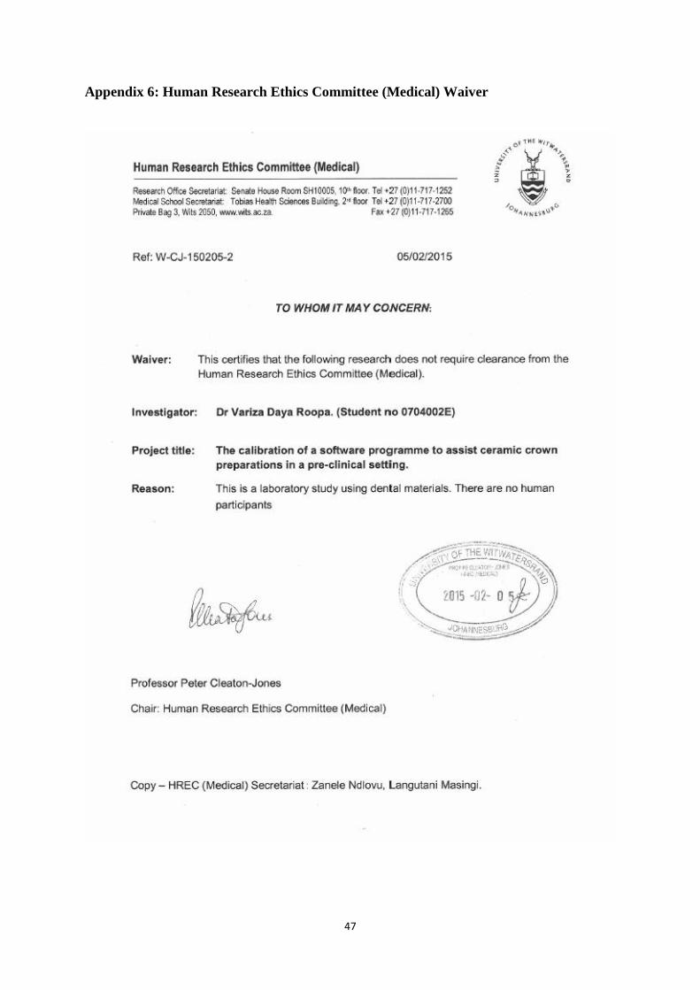
47
Appendix 6: Human Research Ethics Committee (Medical) Waiver

48
Appendix 7: Modified Assessment Rubric for Posterior All-ceramic Crown Preparation
Grading Reduction Margin Configuration Taper Additional
Acceptable (only if all of the following criteria are met)
Occlusal: 1.5 mm up to 2 mm over functional cusps 45° functional cusp bevel Follows external contour Axial: 1.0 – 1.3 mm all around
1.0 -1.3 mm shoulder all around
Internally rounded shoulder Shoulder with a 90° cavo-surface angle
Adequate convergence angle No undercuts present
Smooth line angles No unsupported tooth structure remaining
Requires Modification (if at least one of the following is present)
Occlusal: <1.5 mm No/ insufficient functional cusp bevel Does not follow external contour Axial: <1.0 mm
<1.0 mm shoulder Inner axial line angle of shoulder not smooth and rounded
A cavo-surface angle >/<90°
Inadequate convergence angle Undercuts present
Line angles not smooth Unsupported tooth structure remaining
Unacceptable/ Rejected (if at least one of the following is present)
Occlusal: >2 mm Axial: >1.5 mm
>1.5 mm shoulder
Over tapered preparation
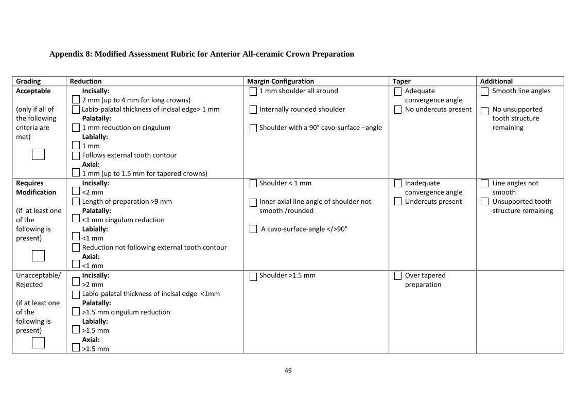
49
Appendix 8: Modified Assessment Rubric for Anterior All-ceramic Crown Preparation
Grading Reduction Margin Configuration Taper Additional
Acceptable (only if all of the following criteria are met)
Incisally: 2 mm (up to 4 mm for long crowns)
Labio-palatal thickness of incisal edge> 1 mm Palatally: 1 mm reduction on cingulum Labially: 1 mm Follows external tooth contour Axial: 1 mm (up to 1.5 mm for tapered crowns)
1 mm shoulder all around Internally rounded shoulder Shoulder with a 90° cavo-surface –angle
Adequate convergence angle No undercuts present
Smooth line angles No unsupported tooth structure remaining
Requires Modification (if at least one of the following is present)
Incisally: <2 mm Length of preparation >9 mm Palatally: <1 mm cingulum reduction Labially: <1 mm Reduction not following external tooth contour Axial: <1 mm
Shoulder < 1 mm Inner axial line angle of shoulder not smooth /rounded A cavo-surface-angle </>90°
Inadequate convergence angle Undercuts present
Line angles not smooth Unsupported tooth structure remaining
Unacceptable/ Rejected (if at least one of the following is present)
Incisally: >2 mm Labio-palatal thickness of incisal edge <1mm Palatally: >1.5 mm cingulum reduction Labially: >1.5 mm Axial: >1.5 mm
Shoulder >1.5 mm
Over tapered preparation
