the canadian diabetes association_s (cda) 2013 clinical practice guidelines.pdf
TRANSCRIPT

AS
Ud
Publication Mail Agreement 41536048 Return undeliverable Canadian addTranscontinental Printing, 737 Moray St, Winnipeg, MB R3J 3S9 Printed in Ca
Publication of the Professionalections of the Canadian Diabetes Association
ne publication des sections professionnellese l'Association canadienne du diab�ete
CONTENTS: April 2013 - Volume 37 - Supplement 1
S1 Introduction
S4 Methods
S8 Definition, Classification and Diagnosis of Diabetes, Prediabetes and Metabolic Syndrome
S12 Screening for Type 1 and Type 2 Diabetes
S16 Reducing the Risk of Developing Diabetes
Management
S20 Organization of Diabetes Care
S26 Self-Management Education
S31 Targets for Glycemic Control
S35 Monitoring Glycemic Control
S40 Physical Activity and Diabetes
S45 Nutrition Therapy
S56 Pharmacotherapy in Type 1 Diabetes
S61 Pharmacologic Management of Type 2 Diabetes
S69 Hypoglycemia
S72 Hyperglycemic Emergencies in Adults
S77 In-hospital Management of Diabetes
S82 Weight Management in Diabetes
S87 Diabetes and Mental Health
S93 Influenza and Pneumococcal Immunization
S94 Pancreas and Islet Transplantation
S97 Natural Health Products
Macrovascular and Microvascular Complications
S100 Vascular Protection in People with Diabetes
S105 Screening for the Presence of Coronary Artery Disease
(continued)resses to:nada

CONTENTS (continued): April 2013 - Volume 37 - Supplement 1
S110 Dyslipidemia
S117 Treatment of Hypertension
S119 Management of Acute Coronary Syndromes
S124 Management of Stroke in Diabetes
S126 Treatment of Diabetes in People with Heart Failure
S129 Chronic Kidney Disease in Diabetes
S137 Retinopathy
S142 Neuropathy
S145 Foot Care
S150 Erectile Dysfunction
Diabetes in Children
S153 Type 1 Diabetes in Children and Adolescents
S163 Type 2 Diabetes in Children and Adolescents
Diabetes in Special Populations
S168 Diabetes and Pregnancy
S184 Diabetes in the Elderly
S191 Type 2 Diabetes in Aboriginal Peoples
Appendices
S197 Appendix 1: Etiologic Classification of Diabetes Mellitus
S198 Appendix 2: Sample Diabetes Patient Care Flow Sheet for Adults
S200 Appendix 3: Examples of Insulin Initiation and Titration Regimens in People with Type 2Diabetes
S202 Appendix 4: Self-Monitoring of Blood Glucose (SMBG) Recommendation Tool forHealthcare Providers
S204 Appendix 5: Approximate Cost Reference List for Antihyperglycemic Agents
S207 Appendix 6: Therapeutic Considerations for Renal Impairment
S209 Appendix 7: Sick Day Medication List
S210 Appendix 8: Rapid Screening for Diabetic Neuropathy
S211 Appendix 9: Diabetes and Foot Care: A Patient’s Checklist
S212 Appendix 10: Diabetic Foot Ulcers: Essentials of Management
S212 Appendix 11: A1C Conversion Chart

Can J Diabetes 37 (2013) A3–A13

Acknowledgment / Can J Diabetes 37 (2013) A3–A13

Acknowledgment / Can J Diabetes 37 (2013) A3–A13

Acknowledgment / Can J Diabetes 37 (2013) A3–A13

Acknowledgment / Can J Diabetes 37 (2013) A3–A13

Acknowledgment / Can J Diabetes 37 (2013) A3–A13

Acknowledgment / Can J Diabetes 37 (2013) A3–A13

Acknowledgment / Can J Diabetes 37 (2013) A3–A13

Acknowledgment / Can J Diabetes 37 (2013) A3–A13

Acknowledgment / Can J Diabetes 37 (2013) A3–A13

Acknowledgment / Can J Diabetes 37 (2013) A3–A13

APC
Usl'
Publication of therofessional Sections of theanadian Diabetes Association
ne publication desections professionnelles deAssociation canadienne du diab�ete
Editor-in-Chief
David Lau MD PHD FRCPC
Editor Emeritus
Heather J. Dean MD FRCPC
Associate Editors
Lori Berard RN CDESarah Capes MD MSC FRCPCAlice Y.Y. Cheng MD FRCPCJ. Robin Conway MDCarol Fawcett RD CDERejeanne Gougeon PHD
Timothy J. Kieffer PHDSora Ludwig MD FRCPCGail MacNeill RN, MED CDESara Meltzer MD FRCPCDani�ele Pacaud MD FRCPCRémi Rabasa-Lhoret MD PHD
Michael Riddell PHDElizabeth Sellers MD MSC FRCPCDiana Sherifali RN PHD CDEScot H. Simpson BSP PHARMD MSCT. Michael Vallis PHD R.PSYCH
National Editorial Board
Gillian Booth MD MSC FRCPCPeter E. Light PHD
Peter A. Senior MBBS PHDArya M. Sharma MD PHD FRCPC
Garry X. Shen MD PHD
International Editorial Board
Stephanie Amiel MD FRCPBarbara J. Anderson PHDAlan Baron MDStuart J. Brink MDSuad Efendic MD PHDGeorge Eisenbarth MD PHDMartha Funnell RN MS CDEDiana Guthrie PHD RN CDE
Phillipe Halban PHDLen Harrison MD DSC FRACP
FRCPARobert Henry MDCheri Ann Hernandez RN PHD CDERyuzo Kawamori MD PHDKarmeen Kulkarni RD MS CDEWilly Malaisse MD PHD
Kathy Mulcahy RN MSN CDEStephen O'Rahilly MD FRCOI FRCFDaniel Porte, Jr. MDPaul Robertson MDAlicia Schiffrin MDMeng Tan MD FACP FRCPCVirginia Valentine RN MS CDEPaul Zimmett AM
Managing Editor
Ryan [email protected]
Publications Coordinator
Alarica [email protected]
Notes to Readers
Overview
The Canadian Diabetes Association 2013 Clinical Practice Guide-lines for the Prevention and Management of Diabetes in Canada areintended to guide practice and are not intended to serve as a compre-hensive text of diabetes management, nor are they intended to setcriteria for research protocols. These guidelines are intended toinform general patterns of care. These guidelines are also intendedto enhance diabetes prevention efforts in Canada and to reduce theburden of diabetes complications in people living with this disease.
As per the Canadian Medical Association Handbook on ClinicalPractice Guidelines (Davis D, et al. Ottawa, ON: Canadian MedicalAssociation; 2007), guidelines should not be used as a legalresource in malpractice cases as “their more general nature rendersthem insensitive to the particular circumstances of the individualcases.” Healthcare professionals must consider the needs, valuesand preferences of individual patients, use clinical judgement andwork with available human and healthcare service resources intheir settings. These guidelines were developed using the bestavailable evidence. It is incumbent upon healthcare professionalsto stay current in this rapidly changing field.
Unless otherwise specified, these guidelines pertain to the careof adults with diabetes. Two chaptersdType 1 Diabetes in Childrenand Adolescents and Type 2 Diabetes in Children and Adoles-centsdare included to highlight aspects of care that must betailored to the pediatric population.
Suggested Citation
To cite as a whole:Canadian Diabetes Association Clinical Practice Guidelines
Expert Committee. Canadian Diabetes Association 2013 ClinicalPractice Guidelines for the Prevention andManagement of Diabetesin Canada. Can J Diabetes 2013;37(suppl 1):S1-S212.
To cite a specific chapter:Last, First M. "Chapter Title." Journal Year;Vol(Number):XX-XX.
Example:Harper W, Clement M, Goldenberg R, et al. Canadian Diabetes
Association 2013 Clinical Practice Guidelines for the Preventionand Management of Diabetes in Canada: pharmacologicmanagement of type 2 diabetes. Can J Diabetes 2013;37(suppl 1):S61-S68.
Reproduction of the Guidelines
Reproduction of the Canadian Diabetes Association 2013 ClinicalPractice Guidelines for the Prevention andManagement of Diabetesin Canada, in whole or in part, is prohibited without writtenconsent of the publisher.
Extra Copies
Copies of this document may be ordered, for a nominal fee, atorders.diabetes.ca.
To order 50 or more copies for educational, commercial orpromotional use, contact Zoe Aarden, Elsevier Canada, 905 KingSt. W, Toronto, ON M6K 3G9; E-mail: [email protected].
Website
These guidelines are available at guidelines.diabetes.ca.

Contents lists available at SciVerse ScienceDirect
Can J Diabetes 37 (2013) S1eS3
Canadian Journal of Diabetesjournal homepage:
www.canadianjournalofdiabetes.com
Clinical Practice Guidelines
Introduction
Canadian Diabetes Association Clinical Practice Guidelines Expert Committee
The initial draft of this chapter was prepared by Alice Y.Y. Cheng MD, FRCPC
Every5 years, since 1992, theClinical & Scientific Section (C&SS) ofthe Canadian Diabetes Association has published comprehensive,evidence-based recommendations for healthcare professionals toconsider in the prevention and management of diabetes in Canada.They have served as a helpful resource and aid for anyone caring forpeople with diabetes and are recognized, not only in Canada but alsointernationally, as high-quality, evidence-based clinical practiceguidelines (1). In fact, an analysis by Bennett et al (1) demonstratedthat the Canadian Diabetes Association clinical practice guidelinesare among the best in the world with respect to quality, rigour andprocess (1). For these 2013 Clinical Practice Guidelines for thePrevention and Management of Diabetes in Canada, volunteermembers of the Clinical Practice Guidelines Expert Committeeassessed the peer reviewed evidence published since 2008 relevantto the prevention and management of diabetes. They then incorpo-rated the evidence into revised diagnostic, prognostic and thera-peutic recommendations for the care of Canadians living withdiabetes, aswell as recommendations formeasures to delay theonsetof diabetes for populations at high risk of developing type 2 diabetes.
A number of important changes have occurred in the develop-ment of the 2013 clinical practice guidelines:
� Expansion of the Expert Committee to include 120 healthcareprofessional volunteers from across Canada; Expert Committeemembers bring expertise from diverse practice settings andinclude professionals from family medicine, endocrinology,internal medicine, infectious disease, neurology, nephrology,cardiology, urology, psychology, obstetrics, ophthalmology, pedi-atrics, nursing,dietetics, pharmacy, exercisephysiologyandothers.
� Inclusion and active participation of people with diabetes onthe Expert Committee to ensure that their views and prefer-ences informed the guideline development process and therecommendations.
� Update and expansion of previous chapters and, in some cases,amalgamation of previous chapters into others to increaseutility and relevance.
� Inclusion of a drug cost appendix for pharmacological thera-pies as a reference for clinicians.
� Update and expansion of our Methodology process (e.g.updated literature searches throughout the guideline devel-opment process, expansion of the Duality of Interest policy)(see Methods chapter, p. S4).
� Inclusion of a “Practical Tips” box, where appropriate, to facil-itate implementation of the recommendations.
1499-2671/$ e see front matter � 2013 Canadian Diabetes Associationhttp://dx.doi.org/10.1016/j.jcjd.2013.01.009
� Expanded harmonization of recommendations throughcollaboration with other organizations, including the CanadianHypertension Education Program (CHEP), the Society of Obste-tricians and Gynecologists of Canada (SOGC), the CanadianCardiovascular Society (CCS) and the Canadian CardiovascularHarmonization of National Guidelines Endeavour (C-CHANGE).
� Expanded dissemination and implementation strategy withincreased use of technology.
It is hoped that primary care physicians and other healthcareprofessionals who care for people with diabetes or those at risk ofdiabetes will continue to find the evidence compiled in theseguidelines a vital aid and resource in their efforts. We are confidentthat, ultimately, if applied properly, these guidelines will lead toimproved quality of care, reduced morbidity and mortality fromdiabetes and its complications, and a better quality of life for peopleliving with this chronic disease.
The Challenge of Diabetes
Diabetes mellitus is a serious condition with potentially devas-tating complications that affects all age groups worldwide. In 1985,an estimated 30 million people around the world were diagnosedwith diabetes; in 2000, that figure rose to over 150 million; and, in2012, the International Diabetes Federation (IDF) estimated that371 million people had diabetes (2). That number is projected torise to 552 million (or 1 in 10 adults) by 2030, which equates to 3new cases per second (2). Although the largest increase is expectedto be in countries with developing economies, Canada also will beimpacted significantly. As of 2009, the estimated prevalence ofdiabetes in Canada was 6.8% of the populationd2.4 million Cana-dians (3)da 230% increase compared to prevalence estimates in1998. By 2019, that number is expected to grow to 3.7 million (3).Diabetes is the leading cause of blindness, end stage renal disease(ESRD) and nontraumatic amputation in Canadian adults. Cardio-vascular disease is the leading cause of death in individuals withdiabetes and occurs 2- to 4-fold more often than in people withoutdiabetes. People with diabetes are over 3 times more likely to behospitalized with cardiovascular disease, 12 times more likely to behospitalized with ESRD and over 20 times more likely to behospitalized for a nontraumatic lower limb amputation comparedto the general population (3). Diabetes and its complicationsincrease costs and service pressures on Canada’s publicly fundedhealthcare system. Among adults aged 20 to 49 years, those with

A.Y.Y. Cheng / Can J Diabetes 37 (2013) S1eS3S2
diabetes were 2 times more likely to see a family physician and 2 to3 timesmore likely to see a specialist (3). Also, peoplewith diabeteswere 3 times more likely to require hospital admission in thepreceding year with longer lengths of stay (3). Therefore, theimpact of diabetes is significant not only for individuals but also fortheir families and for society as a whole.
Delaying the Onset of Type 2 Diabetes
Prevention of type 1 diabetes has not yet been successful, butremains an active area of research. However, there is good evidencethat delaying theonset of type2diabetes results in significant healthbenefits, including lower rates of cardiovascular disease and renalfailure (4). In 2007, the IDF released a “Consensus onType 2DiabetesPrevention” and called upon the governments of all countries todevelop and implement aNational Diabetes Prevention Plan (4). TheIDF proposed that strategies be implemented for 2 separate groups:those at high risk of developing type 2 diabetes, and the entirepopulation at large. Among those at high risk, the proposed 3-stepapproach was to A) identify those who may be at higher risk, B)measure the risk, andC) intervene todelay/prevent the onset of type2 diabetes using predominantly health behaviour strategies to affectthe modifiable risk factors for type 2 diabetes. As of 2013, Canadadoes not have such a strategy in place. There remains an urgent andincreasing need for governments to invest in research to defineeffective strategies and programs to prevent and treat obesity and toencourage physical activity. In addition, Canada’s diverse pop-ulation, with some ethnic groups disproportionally affected bydiabetes, requires that health promotion, and disease preventionandmanagement strategies be culturallyappropriate and tailored tospecific populations. They also should include policies aimed ataddressing poverty and other systemic barriers to healthcare (5).
Optimal Care of Diabetes
Effective diabetes care should be deliveredwithin the frameworkof the Chronic CareModel and centred around the individual who ispracticing, and supported in, self-management (see Organization ofCare chapter, p. S20). To achieve this, an interprofessional teamwiththe appropriate expertise is required, and the system needs tosupport and allow for sharing and collaboration between primarycare and specialist care as needed. A multifactorial approachutilizing an interprofessional team addressing healthy behaviours,glycemic control, blood pressure control, lipid management andvascular protection measures has been shown to effectively anddramatically lower the risk of development and progression ofserious complications for individuals with diabetes (6e9). In addi-tion, individuals with diabetes must be supported in the skills ofself-management since their involvement in diseasemanagement isabsolutely necessary for success. People with diabetes requiretraining in goal setting, problem solving and health monitoring, allof which are critical components of self-management. They alsoneed access to a broad range of tools, includingmedications, devicesand supplies to help them achieve the recommended blood glucose,cholesterol and blood pressure targets. Health outcomes depend onmanaging the disease effectively, and, without access to the neces-sary tools and strategies, Canadians living with diabetes will not beable to achieve optimal results. All levels of government shouldcommit to investing in chronic caremanagement and support of thetools needed for successful self-management to ensure that optimalcare can be delivered.
Research
Canada continues to be a world leader in diabetes research. Thisresearch is essential forcontinued improvement in the livesofpeople
withdiabetes. Regulatoryagencies shouldnot apply these guidelinesin a rigid way with regard to clinical research in diabetes. It is sug-gested that study protocols may include guideline recommenda-tions, but individual decisions belong in the domain of the patient-physician relationship. The merits of each research study must beassessed individually so as to not block or restrict the pursuit of newinformation. The Canadian Diabetes Association welcomes theopportunity toworkwith regulatoryagencies to enhance research inCanada and, ultimately, to improve the care of people with diabetes.
Cost Considerations
When it comes to the issue of cost, caution is required whenidentifying direct, indirect and induced costs for treating diabetes(10). In fact, the 2011 Diabetes in Canada report from the PublicHealthAgencyof Canada couldnot report the total economic burdenof diabetes, but concluded that the costs will only increasesubstantially as theprevalence of the disease increases over time (3).Nonetheless, in 2009, the Canadian Diabetes Association commis-sioned a report to evaluate the economic burden of diabetes usinga Canadian Diabetes Cost Model, which utilizes the data from theCanadian National Diabetes Surveillance System (NDSS) and theEconomic Burden of Illness in Canada (EBIC) (11). In this report, theestimatedeconomicburdenof diabeteswas$12.2 billion in2010andprojected to increase by another $4.7 billion by 2020. It is certainlythe hope and expectation of all stakeholders that the evidence-based prevention and management of diabetes in a multifactorialfashion will reduce the economic burden of the disease (3,6,12).
These clinical practice guidelines, like those published before,have purposefully not taken into account cost effectiveness in theevaluation of the evidence surrounding best practice. The numerousreasons for this have been outlined in detail previously (13). Some ofthese reasons include the paucity of cost-effectiveness analysesusing Canadian data, the difficulty in truly accounting for all theimportant costs (e.g. hypoglycemia) in any cost-effectivenessanalysis, the lack of expertise and resources to properly addressthe cost-effectiveness analyses needed for all the clinical questionswithin these clinical practice guidelines and, perhaps more impor-tantly, the philosophical question of which is more important:clinical benefit to the patient or cost to the system? At what level ofcost effectiveness should one consider a therapy worth recom-mending? For these 2013 clinical practice guidelines, the question ofwhether the committee should incorporate cost considerations wasdiscussed again, and a Cost Consideration Working Group, consist-ing of health economists and health outcomes researchers, wasconvened. Themandateof thegroupwas todevelopaproposal to theClinical Practice Guidelines Steering Committee describing howcostissues might be incorporated into the guidelines, consideringfeasibility and impact. Based on issues of feasibility and philosoph-ical considerations of our role as recommendationdevelopers, itwasdecided that cost would not be included in the recommendations toensure that they reflect the best available clinical evidence for thepatient. The issue of evidence-based vs. cost-effective healthcare isan ethical debate that should involve all citizens because theoutcomeof thisdebateultimately impactseveryCanadian.However,it is recognized and acknowledged that both the healthcare profes-sional and the patient should consider cost when deciding on ther-apies. Therefore, drug costs are included in Appendix 5, allowing foreasy reference for both clinicians and patients alike.
Other Considerations
In Canada, the glycated hemoglobin (A1C) continues to be re-ported using National Glycohemoglobin Standardization Program(NGSP) units (%). In 2007, a consensus statement from the AmericanDiabetes Association, the European Association for the Study of
Diabetes and the IDF called for A1C reporting worldwide to changeto dual reporting of A1Cwith the International Federation of ClinicalChemistry and LaboratoryMedicine (IFCC) SI units (mmol/mol) andderivedNGSPunits (%)with the hope of fully converting to exclusivereporting in SI units (14). However, this has not been adoptedworldwide, with both Canada and the United States still using theNGSP units (%) (15). Although there are some advantages toreporting in SI units, the most notable disadvantage is the massiveeducation effort that would be required to ensure recognition andadoption of the new units. At this time, Canada is not performingdual reporting. Therefore, throughout this document, the A1C willstill be written in NGSP units (%). For those who wish to convertNGSP units to SI units, the following equation can be used: (16)IFCC ¼ 10.93(NGSP) e 23.50.
Dissemination and Implementation
Despite the strength of the evidence supporting the multifacto-rial treatment of people with diabetes to reduce complications,a recent national cross-sectional survey conducted around WorldDiabetes Day (November 14, 2012) demonstrated that only 13% of5123 patients with type 2 diabetes had achieved all 3 metabolictargets (glycemia, lipids and blood pressure) (17). Therefore, a caregap remains and the effective dissemination and implementation ofthese 2013 clinical practice guidelines is critical. A Dissemination &Implementation Chair was appointed at the beginning of theguidelines process. Strategies were developed to increase practi-tioner implementation and to improve patient care and healthoutcomes. A Dissemination & Implementation Committee wascreated to develop a strategic plan to be implemented at the launchof the guidelines and to continue for years thereafter. These volun-teers from across Canada are involved in creating a 3-year plan totranslate the evidence compiled in the guidelines into communitypractice. An Executive Summary will be distributed to healthcareprofessionals in Canada. The full guidelines will continue to beavailable online, and summary articleswill be strategically placed injournals and newsletters. In addition, key messages and tools sup-porting specific themes from the guidelines will be highlighted intechnology-based and paper-based awareness campaigns over thenext few years. Primary care physicians, healthcare providers,governmentofficials, Canadians livingwithdiabetes and the generalpublic continue to be the audiences for these campaigns.
Clinical Practice Guidelines and Clinical Judgement
“Neither evidence nor clinical judgment alone is sufficient.Evidence without judgment can be applied by a technician. Judgmentwithout evidence can be applied by a friend. But the integration ofevidence and judgment is what the healthcare provider does in orderto dispense the best clinical care.” (Hertzel Gerstein, 2012)
People with diabetes are a diverse and heterogeneous group;therefore, it must be emphasized that treatment decisions need tobe individualized. Guidelines are meant to aid in decision makingby providing recommendations that are informed by the bestavailable evidence. However, therapeutic decisions are made at thelevel of the relationship between the healthcare professional andthe patient. That relationship, along with the importance of clinicaljudgement, can never be replaced by guideline recommendations.Evidence-based guidelines try to weigh the benefit and harm ofvarious treatments; however, patient preferences are not alwaysincluded in clinical research, and, therefore, patient values andpreferences must be incorporated into clinical decision making(18). For some of the clinical decisions that we need to make withour patients, strong evidence is available to inform those decisions,and these are reflected in the recommendations within theseguidelines. However, there are many other clinical situations where
strong evidence may not be available, or may never becomeavailable, for reasons of feasibility. In those situations, theconsensus of expert opinions, informed by whatever evidence isavailable, is provided to help guide and aid the clinical decisionsthat need to be made at the level of the patient. It is also importantto note that clinical practice guidelines are not intended to bea legal resource in malpractice cases as outlined in the CanadianMedical Association Handbook on Clinical Practice Guidelines (19).
Conclusions
Diabetes is a complex and complicated disease. The burgeoningevidence on new technologies and therapeutic treatments israpidly expanding our knowledge and ability to manage diabetesand its complications; at the same time, however, it is challengingfor physicians and other healthcare professionals who care forpeople with diabetes. These 2013 clinical practice guidelinescontain evidence-based recommendations that provide a usefulreference tool to help healthcare professionals translate the bestavailable evidence into practice. The hope is that these guidelineswill provide government officials with the evidence they needwhen rationalizing access to healthcare so that the potentiallybeneficial health outcomes are maximized for people living withdiabetes. Healthcare professionals are encouraged to judge inde-pendently the value of the diagnostic, prognostic and therapeuticrecommendations published in the 2013 Clinical Practice Guide-lines for the Prevention and Management of Diabetes in Canada.
References
1. Bennett WL, Odelola OA, Wilson LM, et al. Evaluation of guideline recom-mendations on oral medications for type 2 diabetes mellitus. Ann Intern Med2012;156:27e36.
2. International Diabetes Federation. IDF Diabetes Atlas. 5th ed. Brussels: Inter-national Diabetes Federation, www.idf.org/diabetesatlas; 2012. AccessedFebruary 21, 2013.
3. Public Health Agency of Canada. Diabetes in Canada: Facts and Figures froma Public Health Perspective. Ottawa; 2011.
4. Alberti KGMM, Zimmet P, Shaw J. International Diabetes Federation:a consensus on type 2 diabetes prevention. Diabet Med 2007;24:451e63.
5. McManus R. Time for action: a Canadian proposal for primary prevention oftype 2 diabetes. Can J Diabetes 2012;36:44e9.
6. The Diabetes Control and Complications Trial Research Group. The effect ofintensive treatment of diabetes on the development and progression of long-term complications in insulin-dependent diabetes mellitus. N Engl J Med1993;329:977e86.
7. Nathan DM, Cleary PA, Backlund JY, et al. Diabetes Control and ComplicationsTrial/Epidemiology of Diabetes Interventions and Complications (DCCT/EDIC)Study Research Group. Intensive diabetes treatment and cardiovascular diseasein patients with type 1 diabetes. N Engl J Med 2005;353:2643e53.
8. Gaede P, Vedel P, Larsen N, et al. Multifactorial intervention and cardiovasculardisease in patients with type 2 diabetes. N Engl J Med 2003;348:383e93.
9. Gaede P, Lund-Andersen H, Parving HH, Pedersen O. Effect of a multifactorialintervention on mortality in type 2 diabetes. N Engl J Med 2008;358:580e91.
10. Ettaro L, Songer TJ, Zhang P, Engelgau MM. Cost of illness studies in diabetesmellitus. Pharmacoeconomics 2004;22:149e64.
11. Canadian Diabetes Association. An Economic Tsunami: The Cost of Diabetes inCanada. Toronto: Canadian Diabetes Association; 2009.
12. Gaede P, Valentine WJ, Palmet AJ, et al. Cost-effectiveness of intensified versusconventional multifactorial intervention in type 2 diabetes. Diabetes Care2008;31:1510e5.
13. Harris SB, McFarlane P, Lank CN. Consensus, cost-effectiveness and clinicalpractice guidelines: author’s response. Can J Diabetes 2005;29:376e8.
14. Consensus Committee. Consensus statement on the worldwide standardizationof the hemoglobin A1C measurement: the American Diabetes Association,European Association for the Study of Diabetes, International Federation ofClinical Chemistry and Laboratory Medicine, and the International DiabetesFederation. Diabetes Care 2007;30:2399e400.
15. Sacks D. Measurement of hemoglobin A1c: a new twist on the path toharmony. Diabetes Care 2012;35:2674e80.
16. Weykamp C, John WG, Mosca A, et al. The IFCC reference measurement systemfor HbA1c: a 6-year progress report. Clin Chem 2008;54:240e8.
17. Leiter LA, Berard L, Bowering K, et al. Type 2 diabetes mellitus management inCanada: Is it improving? Can J Diabetes 2013: in press.
18. McCormack JP, Loewen P. Adding “value” to clinical practice guidelines. CanFam Physician 2007;53:1326e7.
19. Davis D, Goldman J, Palda VA. Canadian Medical Association Handbook onClinical Practice Guidelines. Ottawa: Canadian Medical Association; 2007.
A.Y.Y. Cheng / Can J Diabetes 37 (2013) S1eS3 S3

Contents lists available at SciVerse ScienceDirect
Can J Diabetes 37 (2013) S4eS7
Canadian Journal of Diabetesjournal homepage:
www.canadianjournalofdiabetes.com
Clinical Practice Guidelines
Methods
Canadian Diabetes Association Clinical Practice Guidelines Expert Committee
The initial draft of this chapter was prepared by Gillian Booth MD, MSc, FRCPC,Alice Y.Y. Cheng MD, FRCPC
Process
Following the process used to develop previous Canadian Dia-betes Association clinical practice guidelines (1,2), an ExecutiveCommittee, Steering Committee and Expert Committee with broadexpertise and geographic representation were assembled. In total,120volunteers, includinghealthprofessionals from familymedicine,endocrinology, internal medicine, infectious disease, neurology,nephrology, cardiology, urology, psychology, obstetrics, ophthal-mology, pediatrics, nursing, dietetics, pharmacy, exercise physiologyand others, as well as people with diabetes, participated in theguideline development process.
The following basic principles were adopted to ensure that thevalues and empirical basis underlying each recommendation wereexplicitly identified and to facilitate the critical scrutiny and analysisof each recommendation by other organizations and individuals.
Elements covered by the Appraisal of Guidelines for Researchand Evaluation (AGREE) II instrument were incorporated into theguideline development process.
� Each recommendation had to address a clinically importantquestion related to 1 or more of the following: detection,prognosis, prevention or management of diabetes and itssequelae. Health benefits, risks and side effects of interventionswere considered in formulating the recommendations. Patientpreferences and values were sought from expert panelmembers with diabetes and the literature (where available).
� Whenever possible, each recommendation had to be justifiedby the strongest clinically relevant, empirical evidence thatcould be identified; the citation(s) reporting this evidence hadto be noted adjacent to the relevant guideline.
� The strength of this evidence, based on prespecified criteriafrom the epidemiological literature and other guidelinesprocesses, had to be noted (3e8).
� Each recommendation had to be assigned a grade based on theavailable evidence, its methodological strength and its appli-cability to the Canadian population.
� Each recommendation had to be approved by the SteeringCommittee and Executive Committee, with 100% consensus.
� Guidelines based on biological or mechanistic reasoning,expert opinion or consensus had to be explicitly identified andgraded as such; harmonization was sought with other Cana-dian guideline bodies, including the Canadian CardiovascularSociety (CCS), the Canadian Hypertension Education Program
1499-2671/$ e see front matter � 2013 Canadian Diabetes Associationhttp://dx.doi.org/10.1016/j.jcjd.2013.01.010
(CHEP), the Canadian Cardiovascular Harmonization ofNational Guidelines Endeavour (C-CHANGE) and the Society ofObstetricians and Gynecologists of Canada (SOGC).
Identifying and Appraising the Evidence
Authors for each chapter were assembled based on their relevantfields of expertise. Each chapter had 1 lead author, 1 or 2 “evidenceresource” persons trained or experienced in clinical epidemiology orclinical research methodology, and additional authors, as needed. Atthe outset of the process, committee members from each section ofthe guidelines attended a workshop on evidence-based method-ology, in order to ensure a consistent approach to the development ofrecommendations. Committee members identified clinically impor-tant questions related to diagnosis, prognosis, prevention and treat-mentof diabetes and its complications,whichwereusedas a basis forour literature search strategy (outlined below).
Authors were to explicitly define A) the population to whicha guidelinewould apply; B) the test, risk factor or intervention beingaddressed; C) the “gold standard” test or relevant intervention towhich the test or intervention in questionwas compared; andD) theclinically relevant outcomes being targeted. This information wasused to develop specific, clinically relevant questions that were thefocus of literature searches. For each question, individual strategieswere developed combining diabetes terms with methodologicalterms. A librarian with expertise in literature reviews performeda comprehensive search of the relevant English-language, pub-lished, peer-reviewed literature using validated search strategies(http://hiru.mcmaster.ca/hiru/) of electronic databases (MEDLINE,EMBASE, CINAHL, the Cochrane Central Register of Trials, andPsycINFO [where appropriate]). This was complemented by theauthors’ own manual and electronic searches.
For topics thatwere covered in the 2008 guidelines, the literaturesearches focused on new evidence published since those guidelines,including literature published in September 2007 or later. For newtopics, the search time frame included the literature published since1990 or earlier, where relevant. Updated literature searches wereperformedat regular intervals throughout the developmentprocess.
Key citations retrieved from the literature searches were thenreviewed. Each citation that was used to formulate or revisea recommendationwas assigned a level of evidence according to theprespecified criteria in Table 1, reflecting themethodological qualityof the paper. When evaluating papers, authors were required to usestandardized checklists that highlighted the most important

Table 1Criteria for assigning levels of evidence to the published studies
Level CriteriaStudies of diagnosisLevel 1 a) Independent interpretation of test results (without
knowledge of the result of the diagnostic or gold standard)b) Independent interpretation of the diagnostic standard
(without knowledge of the test result)c) Selection of people suspected (but not known) to have
the disorderd) Reproducible description of both the test and diagnostic
standarde) At least 50 patients with and 50 patients without the
disorderLevel 2 Meets 4 of the Level 1 criteriaLevel 3 Meets 3 of the Level 1 criteriaLevel 4 Meets 1 or 2 of the Level 1 criteria
Studies of treatment and preventionLevel 1A Systematic overview or meta-analysis of high quality RCTs
a) Comprehensive search for evidenceb) Authors avoided bias in selecting articles for inclusionc) Authors assessed each article for validityd) Reports clear conclusions that are supported by the data
and appropriate analyses
ORAppropriately designed RCT with adequate power to answerthe question posed by the investigatorsa) Patients were randomly allocated to treatment groupsb) Follow-up at least 80% completec) Patients and investigators were blinded to the treatment*
d) Patients were analyzed in the treatment groups to whichthey were assigned
e) The sample size was large enough to detect the outcomeof interest
Level 1B Nonrandomized clinical trial or cohort study with indisputableresults
Level 2 RCT or systematic overview that does not meet Level 1 criteriaLevel 3 Nonrandomized clinical trial or cohort study; systematic
overview or meta-analysis of level 3 studiesLevel 4 Other
Studies of prognosisLevel 1 a) Inception cohort of patients with the condition of interest
but free of the outcome of interestb) Reproducible inclusion/exclusion criteriac) Follow-up of at least 80% of subjectsd) Statistical adjustment for extraneous prognostic factors
(confounders)e) Reproducible description of outcome measures
Level 2 Meets criterion a) above, plus 3 of the other 4 criteriaLevel 3 Meets criterion a) above, plus 2 of the other criteriaLevel 4 Meets criterion a) above, plus 1 of the other criteria
RCT, randomized, controlled trial.* In cases where such blinding was not possible or was impractical (e.g. intensive
vs. conventional insulin therapy), the blinding of individuals who assessed andadjudicated study outcomes was felt to be sufficient.
Table 2Criteria for assigning grades of recommendations for clinical practice
Grade Criteria
Grade A The best evidence was at Level 1Grade B The best evidence was at Level 2Grade C The best evidence was at Level 3Grade D The best evidence was at Level 4 or consensus
G. Booth, A.Y.Y. Cheng / Can J Diabetes 37 (2013) S4eS7 S5
elements of a well-conducted study. The level of evidence was thendetermined by the cited paper’s objectives, methodological rigour,susceptibility to bias and generalizability (Table 1). Becausethey could not be critically appraised, meeting abstracts, narrativereview articles, news reports and other sources could not be usedto support recommendations. Papers evaluating the cost effective-ness of therapies or diagnostic tests also were not included.
A number of considerations were made when evaluating theevidencewithin a given area. For example, peoplewith diabetes are athigh risk for several sequelae that are not exclusive to diabetes (e.g.cardiovasculardisease, renal failureanderectiledysfunction).As such,some evidence relating to these problems was identified that eitherexcluded, did not report on or did not focus on people with diabetes.
Whenever such evidence was identified, a level was assignedusing the approach described above. Higher levels were assigned ifA) people with diabetes comprised a predefined subgroup; B) theresults in the diabetes subgroup were unlikely to have occurred by
chance; and C) the evidencewas generated in response to questionsthat were formulated prior to the analysis of the results. Lowerlevels (than those indicated in Table 1) were assigned to evidencethat did not meet these criteria.
Guideline Development
Expert Committee members evaluated the relevant literature,and guidelines were developed and initially reviewed by the ExpertCommittee. In the absence of new evidence since the publication ofthe 2008 clinical practice guidelines, recommendations from the2008 document were not changed.
The studies used to develop and support each recommendationare cited beside the level of evidence. In some cases, key citationsthat influenced the final recommendation were not assigned thesame level of evidence but rather were of varying levels of evidence.In those circumstances, all relevant studies were cited, regardless ofthe grading assigned to the recommendation. The final gradingdepended on the overall evidence available, including the relativestrengths of the studies from a methodological perspective and thestudies’ findings. Studies with conflicting outcomes were alsoconsidered and cited in the final recommendation where relevant.Further details on the grading process are described below.
Finally, several treatment recommendations were based onevidence generated from the use of one therapeutic agent froma given class (e.g. one of the statins). Whenever evidence relating to1 or more agents from a recognized class of agents was available,the recommendation was written so as to be relevant to the class,but specifically studied therapeutic agents were identified withinthe recommendation and/or cited reference(s). Only medicationswith Health Canada Notice of Compliance granted by February 15,2013 were included in the recommendations.
Grading the Recommendations
After formulating new recommendations or modifying existingones based on new evidence, each recommendation was assigneda grade from A through D (Table 2). The highest possible grade thata recommendation couldhavewasbasedon the strengthof evidencethat supported the recommendation (i.e. the highest level ofevidence assigned to studies on which the recommendation wasbased). However, the assigned grading was lowered in some cases,for example, if the evidence was found not to be applicable to theCanadianpopulationor, if basedon the consensusof theSteeringandExecutiveCommittees, therewereadditional concerns regarding therecommendation. In some situations, the grading also was loweredfor subgroups that were not well represented in the study or inwhom the beneficial effect of an interventionwas less clear. Gradingalsowas lowered if the findings from relevant (and equally rigorous)studies on the topicwere conflicting. Thus, a recommendationbasedon Level 1 evidence, deemed to be very applicable to Canadiansand supported by strong consensus, was assigned a grade of A.A recommendation not deemed to be applicable to Canadians, orjudged to require further supporting evidence, was assigned a lowergrade.Where available, thenumberofpatients thatwouldneed tobetreated in order to prevent 1 clinical event (number needed to treat[NNT])or to cause anadverse event (numberneeded toharm[NNH])was considered in assessing the impact of a particular intervention.

G. Booth, A.Y.Y. Cheng / Can J Diabetes 37 (2013) S4eS7S6
The degree to which evidence derived from other populations wasfelt to be relevant to diabetes also was reflected in the wording andgrading of the recommendation. Finally, in the absence of Level 1, 2or3 supportingevidence, or if the recommendationwasbasedon theconsensus of the Steering and Executive Committees, the highestgrade that could be assigned was D.
Interpreting the Assigned Grade of a Recommendation
The grade assigned to each recommendation is closely linked tothe methodological rigour and robustness of the relevant clinicalresearch. Therefore, as noted above, a high grade reflects a highdegree of confidence that following the recommendation will leadto the desired outcome. Similarly, a lower grade reflects weakerevidence and a greater possibility that the recommendation willchange when more evidence is generated in the future. Of note, theassigned grade contains no subjective information regarding theimportance of the recommendation or how strongly members ofthe committee felt about it; it only contains information regardingthe evidence upon which the recommendation is based. Thus,many Grade D recommendations were deemed to be very impor-tant to the contemporary management of diabetes, based onclinical experience, case series, physiological evidence and currentconcepts of disease pathophysiology. However, the paucity ofclinical evidence addressing the areas of therapy, prevention,diagnosis or prognosis precluded the assignment of a higher grade.
Clearly, clinicians need to base clinical decisions on the bestavailable relevant evidence that addresses clinical situations.However, they also frequently are faced with having to act in theabsence of clinical evidence, and there are many situations wheregood clinical evidence may be impossible, impractical or tooexpensive to generate (which implies that it would be impossible todevelop Grade A recommendations). For example, it tookthe United Kingdom Prospective Diabetes Study (UKPDS) Group>20 years to collect and publish Level 1 evidence leading to a GradeA recommendation in support of the role of tight glycemic controlto reduce microvascular disease in people with type 2 diabetes.Prior to the publication of the UKPDS results, the recommendationfor glycemic control to prevent microvascular consequences wasa Grade B recommendation (9).
Varying grades of recommendations, therefore, reflect varyingdegrees of certainty regarding the strength of inference that can bedrawn from the evidence in support of the recommendation.Therefore, these evidence-based guidelines and their gradedrecommendations are designed to satisfy 2 important needs: 1) theexplicit identification of the best research upon which the recom-mendation is based and an assessment of its scientific relevanceand quality (captured by the assignment of a level of evidence toeach citation); and 2) the explicit assignment of strength of therecommendation based on this evidence (captured by the grade). Inthis way, they provide a convenient summary of the evidence tofacilitate clinicians in the task of “weighting” and incorporatingever increasing evidence into their daily clinical decision making.They also facilitate the ability of clinicians, healthcare planners,healthcare providers and society, in general, to critically examineany recommendation and arrive at their own conclusions regardingits appropriateness. Thus, these guidelines facilitate their ownscrutiny by others according to the same principles that they use toscrutinize the literature.
It is important to note that the system chosen for gradingrecommendations differs from the approach used in some otherguideline documents, such as the one pertaining to the periodichealth examination in Canada, in which harmful practices wereassigned a grade of D (8). In this Canadian Diabetes Associationguidelines document, recommendation to avoid any harmfulpractices would be graded in the same manner as all other
recommendations. However, it should be noted that the authors ofthese guidelines focused on clinical practices that were thought tobe potentially beneficial and did not seek out evidence regardingthe harmfulness of interventions.
External Peer Review and Independent MethodologicalReview
In May 2012, a draft document was circulated nationally andinternationally for review by numerous stakeholders and experts inrelevant fields. This input was then considered by the Executive andSteering Committees, and revisions were made accordingly.Subsequently, a panel of 6 methodologists, who were not directlyinvolved with the initial review and assessment of the evidence,independently reviewed each recommendation, its assigned gradeand supportive citations. Based on this review, the wording,assigned level of evidence and grade of each recommendationwerereassessed and modified as necessary. Revised recommendationswere reviewed and approved by the Executive and SteeringCommittees. Selected recommendations were presented at a publicforum at the Canadian Diabetes Association/Canadian Society ofEndocrinology andMetabolismProfessional Conference andAnnualMeetings in Vancouver, British Columbia, on October 13, 2012.
Disclosure of Duality of Interest
Committee members were volunteers and received no remu-neration or honoraria for their participation. Members of allcommittees signed an annual duality of interest form listing allfinancial interests or relationships with manufacturer(s) of anycommercial product(s) and/or provider(s) of commercial services.Dualities of interest were discussed during deliberations whererelevant. In the case of a potential duality or outright conflict ofinterest, committee members removed themselves from discus-sions. Funding for the development of the guidelines was providedfrom the general funds of the Canadian Diabetes Association andfrom unrestricted educational grants from Novo Nordisk CanadaInc, Eli Lilly Canada Inc, Merck Canada Inc, Bristol-Myers Squibband AstraZeneca, and Novartis Pharmaceuticals Canada Inc. Thesecompanies were not involved in any aspect of guideline develop-ment, literature interpretation, the decision to publish or any otheraspect related to the publication of these guidelines, and they didnot have access to guideline meetings, guideline drafts orcommittee deliberations.
Guideline Updates
A process to update the full guidelines will commence within5 years and will be published in 2018. Updates to individualchapters may be published sooner in the event of significantchanges in evidence supporting the recommendations. The Exec-utive and Steering Committees of the 2013 revisionwill continue toremain intact to deliberate any potential updates to individualchapters until such time as the Executive and Steering Committeesfor the 2018 revision have been created.
Other Relevant Guidelines
Introduction, p. S1
References
1. Canadian Diabetes Association Clinical Practice Guideline Expert Committee.Canadian Diabetes Association 2003 clinical practice guidelines for the preven-tion and management of diabetes in Canada. Can J Diabetes 2003;27(suppl 2):S1e152.

G. Booth, A.Y.Y. Cheng / Can J Diabetes 37 (2013) S4eS7 S7
2. Canadian Diabetes Association Clinical Practice Guideline Expert Committee.Canadian Diabetes Association 2008 clinical practice guidelines for theprevention and management of diabetes in Canada. Can J Diabetes 2008;32-(suppl 1):S1e201.
3. Straus SE,McAlister FA.What is theprognosis? In:GersteinHC,Haynes RB, editors.Evidence-based Diabetes Care. Hamilton, ON: BC Decker Inc; 2001. p. 6e12.
4. American Medical Association. Users’ Guides to the Medical Literature: Essentials ofEvidence-based Clinical Practice. Chicago, IL: American Medical Association; 2001.
5. Jaeschke R, Guyatt GH. How should diagnostic tests be chosen and used? In:Gerstein HC, Haynes RB, editors. Evidence-based Diabetes Care. Hamilton, ON:BC Decker Inc; 2001. p. 13e23.
6. Holbrook AM, Clarke J-A, Raymond C, et al. How should a particular problem bemanaged? Incorporating evidence about therapies into practice. In: Gerstein HC,Haynes RB, editors. Evidence-based Diabetes Care. Hamilton, ON: BC Decker Inc;2001. p. 24e47.
7. Harris SB, Webster-Bogaert SM. Evidence-based clinical practice guidelines. In:Gerstein HC, Haynes RB, editors. Evidence-based Diabetes Care. Hamilton, ON:BC Decker Inc; 2001. p. 48e61.
8. Goldbloom R, Battista RN. The periodic health examination: 1. Introduction.CMAJ 1986;134:721e3.
9. Meltzer S, Leiter L, Daneman D, et al. 1998 clinical practice guidelines for themanagement of diabetes in Canada. CMAJ 1998;159(suppl 8):S1e29.

Contents lists available at SciVerse ScienceDirect
Can J Diabetes 37 (2013) S8eS11
Canadian Journal of Diabetesjournal homepage:
www.canadianjournalofdiabetes.com
Clinical Practice Guidelines
Definition, Classification and Diagnosis of Diabetes, Prediabetesand Metabolic Syndrome
Canadian Diabetes Association Clinical Practice Guidelines Expert Committee
The initial draft of this chapter was prepared by Ronald Goldenberg MD, FRCPC, FACE,Zubin Punthakee MD, MSc, FRCPC
KEY MESSAGES
� The chronic hyperglycemia of diabetes is associated with significant long-term microvascular and macrovascular complications.
� A fasting plasma glucose level of �7.0 mmol/L, a 2-hour plasma glucosevalue in a 75 g oral glucose tolerance test of �11.1 mmol/L or a glycatedhemoglobin (A1C) value of �6.5% can predict the development of reti-nopathy. This permits the diagnosis of diabetes to be made on the basis ofeach of these parameters.
� The term “prediabetes” refers to impaired fasting glucose, impaired glucosetolerance or an A1C of 6.0% to 6.4%, each of which places individuals athigh risk of developing diabetes and its complications.
Definition of Diabetes and Prediabetes
Diabetes mellitus is a metabolic disorder characterized by thepresence of hyperglycemia due to defective insulin secretion,defective insulin action or both. The chronic hyperglycemia ofdiabetes is associated with relatively specific long-term microvas-cular complications affecting the eyes, kidneys and nerves, as wellas an increased risk for cardiovascular disease (CVD). The diagnosticcriteria for diabetes are based on thresholds of glycemia that areassociated with microvascular disease, especially retinopathy.
“Prediabetes” is a practical and convenient term referring toimpaired fasting glucose (IFG), impaired glucose tolerance (IGT) (1)or a glycated hemoglobin (A1C) of 6.0% to 6.4%, each of whichplaces individuals at high risk of developing diabetes and itscomplications.
Classification of Diabetes
The classification of type 1 diabetes, type 2 diabetes andgestational diabetes mellitus (GDM) is summarized in Table 1.Appendix 1 addresses the etiologic classification of diabetes.Distinguishing between type 1 and type 2 diabetes is importantbecause management strategies differ, but it may be difficult at thetime of diagnosis in certain situations. Physical signs of insulinresistance and autoimmune markers, such as anti-glutamic aciddecarboxylase (GAD) or anti-islet cell antibody (ICA) antibodies,may be helpful, but have not been adequately studied as diagnostictests in this setting. While very low C-peptide levels measured after
1499-2671/$ e see front matter � 2013 Canadian Diabetes Associationhttp://dx.doi.org/10.1016/j.jcjd.2013.01.011
months of clinical stabilization may favour type 1 diabetes (2), theyare not helpful in acute hyperglycemia (3). Clinical judgement withsafe management and ongoing follow-up is a prudent approach.
Diagnostic Criteria
Diabetes
The diagnostic criteria for diabetes are summarized in Table 2(1). These criteria are based on venous samples and laboratorymethods.
A fasting plasma glucose (FPG) level of 7.0 mmol/L correlatesmost closely with a 2-hour plasma glucose (2hPG) value of �11.1mmol/L in a 75 g oral glucose tolerance test (OGTT), and eachpredicts the development of retinopathy (5e11).
The relationship between A1C and retinopathy is similar to thatof FPG or 2hPG with a threshold at around 6.5% (5e7,11,12).Although the diagnosis of diabetes is based on an A1C threshold fordeveloping microvascular disease, A1C is also a continuouscardiovascular (CV) risk factor and a better predictor of macro-vascular events than FPG or 2hPG (13,14). Although many peopleidentified by A1C as having diabetes will not have diabetes bytraditional glucose criteria and vice versa, there are severaladvantages to using A1C for diabetes diagnosis (15). A1C can bemeasured at any time of day and is more convenient than FPGor 2hPG in a 75 g OGTT. A1C testing also avoids the problem ofday-to-day variability of glucose values as it reflects the averageplasma glucose (PG) over the previous 2 to 3 months (1).
In order to use A1C as a diagnostic criterion, A1C must bemeasured using a validated assay standardized to the NationalGlycohemoglobin Standardization Program-Diabetes Control andComplications Trial reference. It is important to note that A1C maybemisleading in individuals with various hemoglobinopathies, irondeficiency, hemolytic anaemias, and severe hepatic and renaldisease (16). In addition, studies of various ethnicities indicate thatAfrican Americans, American Indians, Hispanics and Asians haveA1C values that are up to 0.4% higher than those of Caucasianpatients at similar levels of glycemia (17,18). The frequency ofretinopathy begins to increase at lower A1C levels in Americanblacks than in American whites, which suggests a lower thresholdfor diagnosing diabetes in black persons (19). Research is required

Table 1Classification of diabetes (1)
� Type 1 diabetes* encompasses diabetes that is primarily a result ofpancreatic beta cell destruction and is prone to ketoacidosis. This formincludes cases due to an autoimmune process and those for which theetiology of beta cell destruction is unknown.
� Type 2 diabetes may range from predominant insulin resistance withrelative insulin deficiency to a predominant secretory defect with insulinresistance.
� Gestational diabetes mellitus refers to glucose intolerancewith onset or first recognition during pregnancy.
� Other specific types include a wide variety of relatively uncommonconditions, primarily specific genetically defined forms of diabetes ordiabetes associated with other diseases or drug use (Appendix 1).
* Includes latent autoimmune diabetes in adults (LADA); the term used todescribe the small number of people with apparent type 2 diabetes who appear tohave immune-mediated loss of pancreatic beta cells (4).
Table 3Advantages and disadvantages of diagnostic tests for diabetes* (22)
Parameter Advantages Disadvantages
FPG � Established standard� Fast and easy� Single sample� Predicts microvascularcomplications
� Sample not stable� High day-to-day variability� Inconvenient (fasting)� Reflects glucose homeostasisat a single point in time
2hPG ina 75 gOGTT
� Established standard� Predicts microvascularcomplications
� Sample not stable� High day-to-day variability� Inconvenient� Unpalatable� Cost
A1C � Convenient (measure anytime of day)
� Single sample� Predicts microvascularcomplications
� Better predictor of macro-vascular disease than FPGor 2hPG in a 75 g OGTT
� Low day-to-day variability� Reflects long-term glucoseconcentration
� Cost� Misleading in variousmedical conditions (e.g.hemoglobinopathies, irondeficiency, hemolyticanaemia, severe hepatic orrenal disease)
� Altered by ethnicity andaging
� Standardized, validatedassay required
� Not for diagnostic use inchildren, adolescents, preg-nant women or those withsuspected type 1 diabetes
2hPG, 2-hour plasma glucose; A1C, glycated hemoglobin; FPG, fasting plasmaglucose; OGTT, oral glucose tolerance test.
* Adapted from Sacks D. A1C versus glucose testing: a comparison. Diabetes Care.2011;34:518e523.
R. Goldenberg, Z. Punthakee / Can J Diabetes 37 (2013) S8eS11 S9
to determine if A1C levels differ in African Canadians or CanadianFirst Nations. A1C values also are affected by age, rising by up to0.1% per decade of life (20,21). More studies may help to determineif age- or ethnic-specific adjusted A1C thresholds are required fordiabetes diagnosis. Also, A1C is not recommended for diagnosticpurposes in children, adolescents, pregnant women or those withsuspected type 1 diabetes.
Thedecisionofwhich test touse fordiabetesdiagnosis (Table2) isleft to clinical judgement. Each diagnostic test has advantages anddisadvantages (Table 3). In the absence of symptomatic hypergly-cemia, if a single laboratory test result is in the diabetes range,a repeat confirmatory laboratory test (FPG,A1C,2hPG in a75gOGTT)must be done on another day. It is preferable that the same test berepeated (in a timely fashion) for confirmation, but a random PG inthe diabetes range in an asymptomatic individual should beconfirmed with an alternate test. In the case of symptomatichyperglycemia, the diagnosis has beenmade and a confirmatory testis not required before treatment is initiated. In individuals inwhom type 1 diabetes is likely (younger or lean or symptomatichyperglycemia, especially with ketonuria or ketonemia), confirma-tory testing should not delay initiation of treatment to avoid rapiddeterioration. If results of 2 different tests are available and both areabove the diagnostic cutpoints, the diagnosis of diabetes isconfirmed.When theresults ofmore than1 test are available (among
Table 2Diagnosis of diabetes
FPG ‡7.0 mmol/LFasting ¼ no caloric intake for at least 8 hours
orA1C ‡6.5% (in adults)
Using a standardized, validated assay in the absence of factors that affect theaccuracy of the A1C and not for suspected type 1 diabetes (see text)
or2hPG in a 75 g OGTT ‡11.1 mmol/L
orRandom PG ‡11.1 mmol/L
Random¼ any time of the day, without regard to the interval since the last meal
In the absence of symptomatic hyperglycemia, if a single laboratory test result isin the diabetes range, a repeat confirmatory laboratory test (FPG, A1C, 2hPG ina 75 g OGTT) must be done on another day. It is preferable that the same test berepeated (in a timely fashion) for confirmation, but a random PG in the diabetesrange in an asymptomatic individual should be confirmedwith an alternate test.In the case of symptomatic hyperglycemia, the diagnosis has been made anda confirmatory test is not required before treatment is initiated. In individuals inwhom type 1 diabetes is likely (younger or lean or symptomatic hyperglycemia,especially with ketonuria or ketonemia), confirmatory testing should not delayinitiation of treatment to avoid rapid deterioration. If results of 2 different testsare available and both are above the diagnostic cutpoints, the diagnosis ofdiabetes is confirmed.
2hPG, 2-hour plasma glucose; A1C, glycated hemoglobin; FPG, fasting plasmaglucose; OGTT, oral glucose tolerance test; PG, plasma glucose.
FPG,A1C,2hPG ina75gOGTT)andthe results arediscordant, the testwhose result is above the diagnostic cutpoint should be repeatedand the diagnosis made on the basis of the repeat test.
Prediabetes
The term “prediabetes” refers to IFG, IGT or an A1C of 6.0% to6.4% (Table 4), each of which places individuals at high risk ofdeveloping diabetes and its complications. Not all individuals withprediabetes will necessarily progress to diabetes. Indeed, a signifi-cant proportion of people who are diagnosed with IFG or IGT willrevert to normoglycemia. People with prediabetes, particularly inthe context of the metabolic syndrome, would benefit from CV riskfactor modification.
While people with prediabetes do not have the increased riskfor microvascular disease as seen in diabetes, they are at risk for thedevelopment of diabetes and CVD (23). IGT is more stronglyassociated with CVD outcomes than is IFG. Individuals identified ashaving both IFG and IGT are at higher risk for diabetes as well asCVD. While there is no worldwide consensus on the definition ofIFG (24,25), the Canadian Diabetes Association defines IFG as anFPG value of 6.1 to 6.9 mmol/L due to the higher risk of developingdiabetes in these individuals compared to defining IFG as an FPGvalue of 5.6 to 6.9 mmol/L (25).
While there is a continuum of risk for diabetes in individualswith A1C levels between 5.5% and 6.4%, population studiesdemonstrate that A1C levels of 6.0% to 6.4% are associated with
Table 4Diagnosis of prediabetes
Test Result Prediabetes category
FPG (mmol/L) 6.1e6.9 IFG2hPG in a 75 g OGTT (mmol/L) 7.8e11.0 IGTA1C (%) 6.0e6.4 Prediabetes
2hPG, 2-hour plasma glucose; A1C, glycated hemoglobin; FPG, fasting plasmaglucose; IFG, impaired fasting glucose; IGT, impaired glucose tolerance; OGTT, oralglucose tolerance test.

Table 5Harmonized definition of the metabolic syndrome: �3 measures to make the diagnosis of metabolic syndrome* (29)
Measure Categorical cutpoints
Men Women
Elevated waist circumference (population- and country-specific cutpoints):� Canada, United States� Europid, Middle Eastern, sub-Saharan African, Mediterranean� Asian, Japanese, South and Central American
�102 cm�94 cm�90 cm
�88 cm�80 cm�80 cm
Elevated TG (drug treatment for elevated TG is an alternate indicatory) �1.7 mmol/LReduced HDL-C (drug treatment for reduced HDL-C is an alternate indicatory) <1.0 mmol/L in males,
<1.3 mmol/L in femalesElevated BP (antihypertensive drug treatment in a patient with a history of hypertension is an alternate indicator) Systolic �130 mm Hg and/or
diastolic �85 mm HgElevated FPG (drug treatment of elevated glucose is an alternate indicator) �5.6 mmol/L
BP, blood pressure; FPG, fasting plasma glucose; HDL-C, high-density lipoprotein cholesterol; TG, triglycerides.Three or more criteriaare required for diagnosis.
* Adapted from Alberti KGMM, Eckel R, Grundy S, et al. Harmonizing the metabolic syndrome. Circulation. 2009;120:1640-1645.y The most commonly used drugs for elevated TG and reduced HDL-C are fibrates and nicotinic acid. A patient taking 1 of these drugs can be presumed to have high TG and
reduced HDL-C. High-dose omega-3 fatty acids presumes high TG.
R. Goldenberg, Z. Punthakee / Can J Diabetes 37 (2013) S8eS11S10
a higher risk for diabetes compared to levels between 5.5% and 6.0%(26). While the American Diabetes Association defines prediabetesas an A1C between 5.7% and 6.4%, the Canadian Diabetes Associa-tion has based the definition on a higher risk group and includes anA1C of 6.0% to 6.4% as a diagnostic criterion for prediabetes (1).However, A1C levels below 6.0% can indeed be associated with anincreased risk for diabetes (26). The combination of an FPG of 6.1 to6.9 mmol/L and an A1C of 6.0% to 6.4% is predictive of 100%progression to type 2 diabetes over a 5-year period (27).
Metabolic syndrome
Prediabetes and type 2 diabetes are often manifestations ofa much broader underlying disorder (28), including the metabolic
RECOMMENDATIONS
1. Diabetes should be diagnosed by any of the following criteria:� FPG �7.0 mmol/L [Grade B, Level 2 (11)]� A1C �6.5% (for use in adults in the absence of factors that affect theaccuracy of A1C and not for use in those with suspected type 1 dia-betes) [Grade B, Level 2 (11)]
� 2hPG in a 75 g OGTT �11.1 mmol/L [Grade B, Level 2 (11)]� Random PG �11.1 mmol/L [Grade D, Consensus]
2. In the absence of symptomatic hyperglycemia, if a single laboratory testresult is in the diabetes range, a repeat confirmatory laboratory test (FPG,A1C, 2hPG in a 75 g OGTT) must be done on another day. It is preferablethat the same test be repeated (in a timely fashion) for confirmation, buta random PG in the diabetes range in an asymptomatic individual shouldbe confirmed with an alternate test. In the case of symptomatic hyper-glycemia, the diagnosis has been made and a confirmatory test is notrequired before treatment is initiated. In individuals in whom type 1diabetes is likely (younger or lean or symptomatic hyperglycemia, espe-cially with ketonuria or ketonemia), confirmatory testing should not delayinitiation of treatment to avoid rapid deterioration. If results of twodifferent tests are available and both are above the diagnostic cutpoints,the diagnosis of diabetes is confirmed [Grade D, Consensus].
3. Prediabetes (defined as a state which places individuals at high risk ofdeveloping diabetes and its complications) is diagnosed by any of thefollowing criteria:� IFG (FPG 6.1e6.9 mmol/L) [Grade A, Level 1 (23)]� IGT (2hPG in a 75 g OGTT 7.8e11.0 mmol/L) [Grade A, Level 1 (23)]� A1C 6.0%e6.4% (for use in adults in the absence of factors that affect theaccuracy of A1C and not for use in suspected type 1 diabetes) [Grade B,Level 2 (26)].
Abbreviations:2hPG, 2-hour plasma glucose; A1C, glycated hemoglobin; FPG, fastingplasma glucose; IFG, impaired fasting glucose; IGT, impaired glucosetolerance; OGTT, oral glucose tolerance test; PG, plasma glucose.
syndromeda highly prevalent, multifaceted condition charac-terized by a constellation of abnormalities that include abdominalobesity, hypertension, dyslipidemia and elevated blood glucose.Individuals with the metabolic syndrome are at significant risk ofdeveloping CVD. While metabolic syndrome and type 2 diabetesoften coexist, those with metabolic syndrome without diabetesare at significant risk of developing diabetes. Evidence exists tosupport an aggressive approach to identifying and treatingpeople, not only those with hyperglycemia but also those withthe associated CV risk factors that make up the metabolicsyndrome, such as hypertension, dyslipidemia and abdominalobesity, in the hope of significantly reducing CV morbidity andmortality.
Various diagnostic criteria for the metabolic syndrome havebeen proposed. In 2009, a harmonized definition of the metabolicsyndrome was established, with at least 3 or more criteria requiredfor diagnosis (Table 5) (29).
Other Relevant Guidelines
Screening for Type 1 and Type 2 Diabetes, p. S12Reducing the Risk of Developing Diabetes, p. S16Type 1 Diabetes in Children and Adolescents, p. S153Type 2 Diabetes in Children and Adolescents, p. S163
Relevant Appendix
Appendix 1. Etiologic Classification of Diabetes Mellitus
References
1. American Diabetes Association. Diagnosis and classification of diabetesmellitus. Diabetes Care 2012;35(suppl 1):S64e71.
2. Patel P, Macerollo A. Diabetes mellitus: diagnosis and screening. Am FamPhysician 2010;81:863e70.
3. Unger RH, Grundy S. Hyperglycemia as an inducer as well as a consequence ofimpaired islet cell function and insulin resistance: implications for themanagement of diabetes. Diabetologia 1985;28:119e21.
4. Turner R, Stratton I, Horton V, et al. UKPDS 25: autoantibodies to islet-cellcytoplasm and glutamic acid decarboxylase for prediction of insulin require-ment in type 2 diabetes. UK Prospective Diabetes Study Group. Lancet 1997;350:1288e93.
5. McCance DR, Hanson RL, Charles MA, et al. Comparison of tests for glycatedhemoglobin and fasting and two hour plasma glucose concentrations asdiagnostic methods for diabetes. BMJ 1994;308:1323e8.
6. Engelgau MM, Thompson TJ, Herman WH, et al. Comparison of fasting and2-hour glucose and HbA1c levels for diagnosing diabetes. Diagnostic criteriaand performance revisited. Diabetes Care 1997;20:785e91.
7. The Expert Committee on the Diagnosis and Classification of Diabetes Mellitus.Report of the expert committee on the diagnosis and classification of diabetesmellitus. Diabetes Care 1997;20:1183e97.

R. Goldenberg, Z. Punthakee / Can J Diabetes 37 (2013) S8eS11 S11
8. Ito C, Maeda R, Ishida S, et al. Importance of OGTT for diagnosing diabetesmellitus based on prevalence and incidence of retinopathy. Diabetes Res ClinPract 2000;49:181e6.
9. Miyazaki M, Kubo M, Kiyohara Y, et al. Comparison of diagnostic methods fordiabetes mellitus based on prevalence of retinopathy in a Japanese population:the Hisayama Study. Diabetologia 2004;47:1411e5.
10. Tapp RJ, Zimmett PZ, Harper CA, et al. Diagnostic thresholds for diabetes: theassociation of retinopathy and albuminuria with glycaemia. Diabetes Res ClinPract 2006;73:315e21.
11. The DETECT-2 Collaboration Writing Group. Glycemic thresholds for diabetesspecific retinopathy. Diabetes Care 2011;34:145e50.
12. International Expert Committee. International Expert Committee report on therole of the A1C assay in the diagnosis of diabetes. Diabetes Care 2009;32:1327e34.
13. Sarwar N, Aspelund T, Eiriksdottir G, et al. Markers of dysglycaemia and risk ofcoronary heart disease in people without diabetes: Reykjavik ProspectiveStudy and systematic review. PLoS Med 2010;7:e1000278.
14. Selvin E, Steffes MW, Zhu H, et al. Glycated hemoglobin, diabetes,and cardiovascular risk in nondiabetic adults. N Engl J Med 2010;362:800e11.
15. Report of a World Health Organization Consultation. Use of glycatedhaemoglobin (HbA1C) in the diagnosis of diabetes mellitus. Diabetes Res ClinPract 2011;93:299e309.
16. Gallagher EJ, Bloomgarden ZT, Roith D. Review of hemoglobin A1c in themanagement of diabetes. J Diabetes 2009;1:9e17.
17. Herman WH, Ma Y, Uwaifo G, et al. Differences in A1C by race and ethnicityamong patients with impaired glucose tolerance in the Diabetes PreventionProgram. Diabetes Care 2007;30:2453e7.
18. Ziemer DC, Kolm P, Weintraub WS, et al. Glucose-independent, black-white differences in hemoglobin A1c levels. Ann Intern Med 2010;152:770e7.
19. Tsugawa Y, Mukamal K, Davis R, et al. Should the HbA1c diagnostic cutoff differbetween blacks and whites? A cross-sectional study. Ann Intern Med 2012;157:153e9.
20. Davidson MB, Schriger DL. Effect of age and race/ethnicity on HbA1c levels inpeople without known diabetes mellitus: implications for the diagnosis ofdiabetes. Diabetes Res Clin Pract 2010;87:415e21.
21. Pani L, Korenda L, Meigs JB, Driver C, Chamany S, Fox CS, et al. Effect of agingon A1C levels in persons without diabetes: evidence from the FraminghamOffspring Study and NHANES 2001-2004. Diabetes Care 2008;31:1991e6.
22. Sacks D. A1C versus glucose testing: a comparison. Diabetes Care 2011;34:518e23.
23. Santaguida PL, Balion C, Morrison K, et al. Diagnosis, prognosis, and treatment ofimpairedglucose toleranceand impaired fastingglucose.Evidencereport/technologyassessment no. 128. Agency Healthcare Research and Quality Publication No05-E026-2. Rockville, MD: Agency for Healthcare Research and Quality; September2005.
24. Shaw JE, Zimmet PZ, Alberti KG. Point: impaired fasting glucose: the case for thenew American Diabetes Association criterion. Diabetes Care 2006;29:1170e2.
25. Forouhi NG, Balkau B, Borch-Johnsen K, et al, EDEG. The threshold fordiagnosing impaired fasting glucose: a position statement by the EuropeanDiabetes Epidemiology Group. Diabetologia 2006;49:822e7.
26. Zhang X, Gregg E, Williamson D, et al. A1C level and future risk of diabetes:a systematic review. Diabetes Care 2010;33:1665e73.
27. Heianza Y, Arase Y, Fujihara K, et al. Screening for pre-diabetes to predict futurediabetes using various cut-off points forHbA1c and impaired fasting glucose: theToranomon Hospital Health Management Center Study 4 (TOPICS 4). DiabeticMed 2012;29:e279e85.
28. Reaven GM. Banting Lecture 1988. Role of insulin resistance in human disease.Diabetes 1988;37:1595e607.
29. Alberti KGMM, Eckel R, Grundy S, et al. Harmonizing the metabolic syndrome.Circulation 2009;120:1640e5.

Contents lists available at SciVerse ScienceDirect
Can J Diabetes 37 (2013) S12eS15
Canadian Journal of Diabetesjournal homepage:
www.canadianjournalofdiabetes.com
Clinical Practice Guidelines
Screening for Type 1 and Type 2 Diabetes
Canadian Diabetes Association Clinical Practice Guidelines Expert Committee
The initial draft of this chapter was prepared by Jean-Marie Ekoé MD, CSPQ, PD,Zubin Punthakee MD, MSc, FRCPC, Thomas Ransom MD, MSc, FRCPC,Ally P.H. Prebtani BScPhm, MD, FRCPC, Ronald Goldenberg MD, FRCPC, FACE
KEY MESSAGES
� In the absence of evidence for interventions to prevent or delay type 1diabetes, screening for type 1 diabetes is not recommended.
� Screening for type 2 diabetes using a fasting plasma glucose (FPG) and/orglycated hemoglobin (A1C) should be performed every 3 years in indi-viduals�40 years of age or in individuals at high risk using a risk calculator.
� Diabetes will be diagnosed if A1C is �6.5% (see Definition, Classificationand Diagnosis chapter, p. S8).
� Testing with a 2-hour plasma glucose (2hPG) in a 75 g oral glucosetolerance test (OGTT) should be undertaken in individuals with an FPG of6.1e6.9 mmol/L and/or an A1C of 6.0%e6.4% in order to identify individualswith impaired glucose tolerance (IGT) or diabetes.
� Testing with a 2hPG in a 75 g OGTT may be undertaken in individuals withan FPG 5.6e6.0mmol/L and/or A1C 5.5%e5.9% and�1 risk factor in order toidentify individuals with IGT or diabetes.
The clinical spectrum of diabetes ranges from a low-risk toa higher-risk individual or to the symptomatic patient who needsimmediate treatment. Screening for diabetes implies testing fordiabetes in individuals without symptoms who are unaware oftheir condition. Screening for diabetes will also detect individualsat increased risk for diabetes (prediabetes) or individuals with lesssevere states of dysglycemia who may still be at risk for type 2diabetes. Screening strategies vary according to the type of diabetesand evidence of effective interventions to prevent progression ofprediabetes to diabetes and/or reduce the risk of complicationsassociated with diabetes. The growing importance of diabetesscreening is undeniable (1).
In contrast to other diseases, there is no distinction betweenscreening and diagnostic testing. Therefore, to screen for diabetesand prediabetes, the same tests would be used as for diagnosisof both medical conditions (see Definition, Classification andDiagnosis chapter, p. S8).
Screening for Type 1 Diabetes
Type1diabetesmellitus is primarily a result of pancreatic beta celldestruction due to an immune-mediated process that is likely incitedby environmental factors in genetically predisposed individuals. Anindividual’s risk of developing type 1 diabetes can be estimated byconsidering family history of type 1 diabetes with attention to age ofonset and sex of the affected family members (2) and profiling
1499-2671/$ e see front matter � 2013 Canadian Diabetes Associationhttp://dx.doi.org/10.1016/j.jcjd.2013.01.012
immunity and geneticmarkers (3). The loss of pancreatic beta cells inthe development of type 1 diabetes passes through a subclinicalprodrome that can be detected reliably in first- and second-degreerelatives of persons with type 1 diabetes by the presence of pancre-atic islet autoantibodies in their sera (4). However, in a recent largestudy, one-time screening for glutamic acid decarboxylase antibodies(GADAs) and islet antigen-2 antibodies (IA-2As) in the generalchildhood population in Finland would identify 60% of those indi-vidualswhowill develop type1diabetesover thenext27years. Initialpositivity for GADAs and/or IA-2As had a sensitivity of 61% (95%confidence interval [CI] 36e83%) for type 1 diabetes. The combinedpositivity for GADAs and IA-2As had both a specificity and a positivepredictive value of 100% (95% CI 59e100%) (5). Ongoing clinicalstudies are testing different strategies for preventing or reversingearly type 1 diabetes in the presence of positive autoimmunity. Giventhat the various serological markers are not universally available andin the absence of evidence for interventions to preventordelay type1diabetes, no widespread recommendations for screening for type 1diabetes can be made.
Screening for Type 2 Diabetes
Adults
Undiagnosed type 2 diabetes may occur in >2.8% of the generaladult population (6), and the number increases to >10% in somepopulations (7,8). Tests for hyperglycemia can identify theseindividuals, many of whom will have, or will be at risk for,preventable diabetes complications (5,6). To be effective,population-based screening would have to involve wide coverageand would have the goal of early identification and subsequentintervention to reduce morbidity and mortality. Using variousmultistaged screening strategies, the ADDITION-Europe studyshowed that 20% to 94% of eligible people in primary care practicesattended the first blood glucose test of the screening process, anddiabetes was detected in 0.33% to 1.09% of the target populations,which was lower than expected (9). In the subsequent ADDITION-Europe cluster randomized trial of intensive multifacetedcardiovascular risk factor management vs. routine diabetes careamong screening-identified type 2 diabetes patients, intensivemanagement did not reduce cardiovascular events (hazard ratio0.83; 95% CI 0.65e1.05) or all-cause mortality (hazard ratio 0.91;

Table 1Risk factors for type 2 diabetes
� Age �40 years� First-degree relative with type 2 diabetes� Member of high-risk population (e.g. Aboriginal, African, Asian, Hispanic orSouth Asian descent)
� History of prediabetes (IGT, IFG or A1C 6.0%e6.4%)*� History of gestational diabetes mellitus� History of delivery of a macrosomic infant� Presence of end organ damage associated with diabetes:
B Microvascular (retinopathy, neuropathy, nephropathy)B Macrovascular (coronary, cerebrovascular, peripheral)
� Presence of vascular risk factors:B HDL cholesterol level <1.0 mmol/L in males, <1.3 mmol/L in females*B Triglycerides �1.7 mmol/L*B Hypertension*B Overweight*B Abdominal obesity*
� Presence of associated diseases:B Polycystic ovary syndrome*B Acanthosis nigricans*B Psychiatric disorders (bipolar disorder, depression, schizophreniay)B HIV infectionz
B OSAx
� Use of drugs associated with diabetes:B GlucocorticoidsB Atypical antipsychoticsB HAARTz
B Other (see Appendix 1)� Other secondary causes (see Appendix 1)
A1C, glycated hemoglobin; HAART, highly active antiretroviral therapy; HDL,high-density lipoprotein; HIV, human immunodeficiency virus-1; IFG, impairedfasting glucose; IGT, impaired glucose tolerance; OSA, obstructive sleep apnea.
* Associated with insulin resistance.y The incidence of type 2 diabetes is at least 3 times higher in people with
schizophrenia than in the general population (25,26). Using data collected in 1991,the prevalence of diabetes was assessed in >20,000 individuals diagnosed withschizophrenia. The rate of diagnosed diabetes was 9% to 14%, exceeding rates for thegeneral population prior to the widespread use of new antipsychotic drugs (27).
z HIV and HAART increase the risk of prediabetes (IGT) and type 2 diabetes by1.5- to 4-fold compared to the general population (28).
x OSA is an independent risk factor for diabetes (hazard ratio 1.43) (29).
RECOMMENDATIONS
1. All individuals should be evaluated annually for type 2 diabetes risk on thebasis of demographic and clinical criteria [Grade D, Consensus].
2. Screening for diabetes using FPG and/or A1C should be performed every3 years in individuals�40 years of age or at high risk using a risk calculator[Grade D, Consensus]. More frequent and/or earlier testing with either FPGand/or A1C or 2hPG in a 75 g OGTT should be considered in those at veryhigh risk using a risk calculator or in people with additional risk factors fordiabetes [Grade D, Consensus]. These risk factors include:� First-degree relative with type 2 diabetes� Member of high-risk population (e.g. Aboriginal, African, Asian,Hispanic or South Asian descent)
� History of prediabetes (IGT, IFG, or A1C 6.0%e6.4%)� History of gestational diabetes mellitus� History of delivery of a macrosomic infant� Presence of end organ damage complications associatedwith diabetes:
B Microvascular (retinopathy, neuropathy, nephropathy)B Macrovascular (coronary, cerebrovascular, peripheral)
� Presence of vascular risk factors:B HDL cholesterol <1.0 mmol/L in males, <1.3 mmol/L in femalesB Triglycerides �1.7 mmol/LB HypertensionB OverweightB Abdominal obesity
� Presence of associated diseases:B Polycystic ovary syndromeB Acanthosis nigricansB Obstructive sleep apneaB Psychiatric disorders (bipolar disorder, depression, schizophrenia)B HIV infection
� Use of drugs associated with diabetes:B GlucocorticoidsB Atypical antipsychoticsB HAARTB Other (see Appendix 1)
� Other secondary causes (see Appendix 1)
3. Testing with 2hPG in a 75 g OGTT should be undertaken in individuals withFPG 6.1e6.9 mmol/L and/or A1C 6.0%e6.4% in order to identify individualswith IGT or diabetes [Grade D, Consensus].
4. Testing with 2hPG in a 75 g OGTT may be undertaken in individuals withFPG 5.6e6.0 mmol/L and/or A1C 5.5%e5.9% and �1 risk factor(s) in orderto identify individuals with IGT or diabetes [Grade D, Consensus].
Abbreviations:2hPG, 2-hour plasma glucose; A1C, glycated hemoglobin; FPG, fasting
plasma glucose; HAART, highly active antiretroviral therapy; HDL,high-density lipoprotein; HIV, human immunodeficiency virus-1; IFG,impaired fasting glucose; IGT, impaired glucose tolerance; OGTT, oralglucose tolerance test.
J.-M. Ekoé et al. / Can J Diabetes 37 (2013) S12eS15 S13
95% CI 0.69e1.21) (10). Of note, a very high proportion of theroutine care group also received optimal cardiovascular risk factormanagement, which may have diluted any potential benefits. InADDITION-Cambridge, population-based screening for type 2 dia-betes was not associated with a reduction in all-cause, cardiovas-cular or diabetes-related mortality within 10 years compared toa no-screening control group. However, the low rate of type 2diabetes in the screened population (3%) was likely too small toaffect overall population mortality (11). Nonetheless, there is nocurrent evidence of clinical benefit to support a strategy ofpopulation-based screening for type 2 diabetes.
Although the relatively low prevalence of diabetes in the generalpopulation makes it unlikely that mass screening will be costeffective, testing for diabetes in people with risk factors for type 2diabetes or with diabetes-associated conditions is likely to result inmore benefit than harm and will lead to overall cost savings(12e17). Routine testing for type 2 diabetes is, therefore, justifiablein some but not all settings (18,19). Screening individuals as early asage 40 years in family physicians’ offices has proved to be useful indetecting unrecognized diabetes (20).
While fasting plasma glucose (FPG) and/or glycated hemoglobin(A1C) are the recommended screening tests, a 75 g oral glucosetolerance test (OGTT) is indicated when the FPG is 6.1 to 6.9 mmol/L(14) and/or A1C is 6.0% to 6.4%. It may be indicated when the FPG is5.6 to 6.0 mmol/L and/or A1C is 5.5% to 5.9% and suspicion of type 2diabetes or impaired glucose tolerance (IGT) is high (e.g. forindividuals with risk factors listed in Table 1) (Figure 1).
People with prediabetes, especially those with IGT or an A1C of6.0% to 6.4%, not only are at increased risk of developing type 2diabetes, but they also have an increased risk of macrovascular
complications, particularly in the context of the metabolicsyndrome (21). These individuals would benefit from cardiovas-cular risk factor reduction strategies (1). Members of high-riskethnic populations (Table 1) should be screened for prediabetesand type 2 diabetes using the recommended screening tests, suchas FPG, OGTT and A1C. However, the high prevalence of hemoglo-binopathies among these populations may considerably reduce theaccuracy of A1C as a reliable screening tool in these populations.Furthermore, high-risk ethnic groups may have A1C levels that areslightly higher than those of Caucasians at the same level ofglycemia, and more studies may help determine ethnic-specificA1C thresholds for diabetes diagnosis (see Definition, Classifica-tion and Diagnosis chapter, p. S8).
Risk prediction tools for type 2 diabetes mellitus
A number of risk scores based on clinical characteristics havebeen developed to identify individuals at high risk of havingundiagnosed diabetes. However, the impact of known risk factors

Figure 1. Screening and diagnosis algorithm for type 2 diabetes.*If both fasting plasma glucose (FPG) and glycated hemoglobin (A1C) are available but discordant, use the test that appears furthest to the right side of the algorithm.**Prediabetes ¼ impaired fasting glucose (IFG), impaired glucose tolerance (IGT), or A1C 6.0% to 6.4% (see Table 4 in Definition, Classification and Diagnosis of Diabetes, Prediabetesand Metabolic Syndrome, p. S8). zIn the absence of symptomatic hyperglycemia, if a single laboratory test is in the diabetes range, a repeat confirmatory test (FPG, A1C, 2hPG in a 75g OGTT) must be done on another day. It is preferable that the same test be repeated (in a timely fashion) for confirmation. If results of 2 different tests are available and both areabove the diagnostic cutpoints, the diagnosis of diabetes is confirmed. NA ¼ not available; OGTT ¼ oral glucose tolerance test.
J.-M. Ekoé et al. / Can J Diabetes 37 (2013) S12eS15S14
on having undiagnosed type 2 diabetes differs between populationsof different ethnic origins, and risk scores developed in Caucasianpopulations cannot be applied to populations of other ethnicgroups (22). Furthermore, the prevalence of individuals at risk fordeveloping type 2 diabetes varies considerably according to thescoring system. Risk scoring systems must, therefore, be validatedfor each considered population in order to adequately detectindividuals at risk and eventually implement efficacious preven-tative strategies (23). The Canadian Diabetes Risk AssessmentQuestionnaire (CANRISK) is a statistically valid tool that may besuitable for diabetes risk assessment in the Canadian populationand is available on the Internet at www.phac-aspc.gc.ca/cd-mc/diabetes-diabete/canrisk/index-eng.php (24).
Other Relevant Guidelines
Definition, Classification and Diagnosis of Diabetes, Prediabetesand Metabolic Syndrome, p. S8
Reducing the Risk of Developing Diabetes, p. S16Type 1 Diabetes in Children and Adolescents, p. S153Type 2 Diabetes in Children and Adolescents, p. S163
Relevant Appendix
Appendix 1. Etiologic Classification of Diabetes Mellitus
References
1. Gilmer TP, O’Connor PJ. The growing importance of diabetes screening.Diabetes Care 2010;33:1695e7.
2. Harjutsalo V, Reunanen A, Tuomilehto J. Differential transmission of type 1diabetes from diabetic fathers and mothers to their offspring. Diabetes 2006;55:1517e24.
3. Decochez K, Truyen I, van der Auwera B, et al, Belgian Diabetes Registry.Combined positivity for HLA DQ2/DQ8 and IA-2 antibodies defines pop-ulation at high risk of developing type 1 diabetes. Diabetologia 2005;48:687e94.
4. Bingley PJ. Interactions of age, islet cell antibodies, insulin autoantibodies, andfirst-phase insulin response in predicting risk of progression to IDDM in ICAþrelatives: the ICARUS data set. Islet Cell Antibody Register Users Study.Diabetes 1996;45:1720e8.
5. Knip M, Korhonen S, Kulmala P, et al. Prediction of type 1 diabetes in thegeneral population. Diabetes Care 2010;33:1206e12.
6. Cowie CC, Rust KF, Byrd-Holt DD, et al. Prevalence of diabetes and impairedfasting glucose in adults in the U.S. population: National Health And NutritionExamination Survey 1999-2002. Diabetes Care 2006;29:1263e8.

J.-M. Ekoé et al. / Can J Diabetes 37 (2013) S12eS15 S15
7. Rolka DB, Narayan KM, Thompson TJ, et al. Performance of recommendedscreening tests for undiagnosed diabetes and dysglycemia. Diabetes Care 2001;24:1899e903.
8. Rathmann W, Haastert B, Icks A, et al. High prevalence of undiagnosed diabetesmellitus in Southern Germany: target populations for efficient screening. TheKORA survey 2000. Diabetologia 2003;46:182e9.
9. Van den Donk M, Sandbaek A, Borch-Johnsen, et al. Screening for type 2 dia-betes: lessons from the ADDITION-Europe study. Diabet Med 2011;28:1416e26.
10. GriffinSJ, Borch-JohnsenK,DaviesMJ, et al. Effect of early intensivemultifactorialtherapy on 5-year cardiovascular outcomes in individuals with type 2 diabetesdetected by screening (ADDITION-Europe): a cluster-randomised trial. Lancet2011;378:156e67.
11. Simmons RK, Echouffo-Tcheugui JB, Sharp SJ, et al. Screening for type 2diabetes and population mortality over 10 years (ADDITION-Cambridge):a cluster-randomised controlled trial. Lancet 2012;380:1741e8.
12. Raikou M, McGuire A. The economics of screening and treatment in type 2diabetes mellitus. Pharmacoeconomics 2003;21:543e64.
13. The cost-effectiveness of screening for type 2 diabetes. CDC DiabetesCost-Effectiveness Study Group, Centers for Disease Control and Prevention.JAMA 1998;280:1757e63.
14. Kahn R, Alperin P, Eddy D, et al. Age at initiation of screening to detecttype 2 diabetes: a cost-effectiveness analysis. Lancet 2010;375:1365e74.
15. Simmons RK, Rahman M, Jakes RW, et al. Effect of population screeningfor type 2 diabetes on mortality: long term follow-up of the Ely cohort.Diabetologia 2011;54:312e9.
16. Gilles CL, Lambert PC, Abrahms KR, et al. Different strategies for screening andprevention of type 2 diabetes in adults: cost-effectiveness analysis. BMJ 2008;336:1180e4.
17. Hoerger TJ, Hicks KA, Sorensen SW, et al. Cost-effectiveness of screening forprediabetes among overweight and obese U.S adults. Diabetes Care 2007;30:2874e9.
18. Knowler WC. Screening for NIDDM. Opportunities for detection, treatment, andprevention. Diabetes Care 1994;17:445e50.
19. Simmons RK, Echouffo-Tcheugui JB, Griffin SJ. Screening for type 2 diabetes: anupdate of evidence. Diabetes Obes Metab 2010;12:838e44.
20. Leiter LA, Barr A, Bélanger A, et al, Diabetes Screening in Canada (DIASCAN)Study. Diabetes Screening in Canada (DIASCAN) Study: prevalence of undiag-nosed diabetes and glucose intolerance in family physician offices. DiabetesCare 2001;24:1038e43.
21. Hu G, Qiao Q, Tuomilehto J, et al, DECODE Study Group. Prevalence of themetabolic syndrome and its relation to all- cause and cardiovascularmortality innondiabetic European men and women. Arch Intern Med 2004;164:1066e76.
22. Glumer C, Vistisen D, Borch-Johnsen K, et al, Detect 2 Collaboration. Risk scoresfor type 2 diabetes can be applied in some populations but not all. DiabetesCare 2006;29:410e4.
23. Schmid R, Vollenweider P, Waeber G, et al. Estimating the risk of developingtype 2 diabetes: a comparison of several risk scores. The Cohorte LausannoiseStudy. Diabetes Care 2011;34:1863e8.
24. Robinson CA, Agarwal G. Validating the CANRISK prognostic model forassessing diabetes risk in Canada’s multi-ethnic population. Chron Dis Inj Can2011;32:19e31.
25. McKee HA, D’Arcy PF, Wilson PJ. Diabetes and schizophrenia: a preliminarystudy. J Clin Hosp Pharm 1986;11:297e9.
26. Mukherjee S, Decina P, Bocola V, et al. Diabetes mellitus in schizophrenicpatients. Compr Psychiatry 1996;37:68e73.
27. Dixon L, Weiden P, Delahanty J, et al. Prevalence and correlates of diabetes innational schizophrenia samples. Schizophr Bull 2000;26:903e12.
28. Samaras K, Chisholm DJ. Diabetes, insulin resistance and glucose metabolism inHIV infection and its treatment. In: Ekoe JM, Rewers M, Williams R, et al.,editors. The Epidemiology of Diabetes Mellitus. Chichester, UK: WileyBlackwell; 2008. p. 665e75.
29. Botros N, Concato J, Mohsenin V, et al. Obstructive sleep apnea as a risk factorfor type 2 diabetes. Am J Med 2009;122:1122e7.

Contents lists available at SciVerse ScienceDirect
Can J Diabetes 37 (2013) S16eS19
Canadian Journal of Diabetesjournal homepage:
www.canadianjournalofdiabetes.com
Clinical Practice Guidelines
Reducing the Risk of Developing Diabetes
Canadian Diabetes Association Clinical Practice Guidelines Expert Committee
The initial draft of this chapter was prepared by Thomas Ransom MD, MSc, FRCPC,Ronald Goldenberg MD, FRCPC, FACE, Amanda Mikalachki RN, CDE,Ally P.H. Prebtani BScPhm, MD, FRCPC, Zubin Punthakee MD, MSc, FRCPC
KEY MESSAGES
� As safe and effective preventive therapies for type 1 diabetes have not yetbeen identified, any attempts to prevent type 1 diabetes should beundertaken only within the confines of formal research protocols.
� Intensive and structured lifestyle modification that results in loss ofapproximately 5% of initial body weight can reduce the risk of progressionfrom impaired fasting glucose or impaired glucose tolerance to type 2diabetes by almost 60%.
� Progression from prediabetes to type 2 diabetes can also be reduced bypharmacological therapy with metformin (w30% reduction), acarbose(w30% reduction) and thiazolidinediones (w60% reduction).
Introduction
Ideal preventive strategies for both type 1 and type 2 diabetesshould range from focusing efforts on individuals identified asbeing at risk for developing diabetes to broader, more group- orpopulation-based strategies. For the practicing healthcare profes-sional, it is the individualized strategies that are sought. Theprevention or delay of diabetes not only would alleviate the indi-vidual of the burden of disease but also could decrease associatedmorbidity and mortality. Preventive strategies would differdepending on the type of diabetes. Given the increasing incidenceand prevalence of diabetes, development of safe and cost-effectiveinterventions to reduce the risk of diabetes are needed to helpdecrease the burden of diabetes on individuals and the healthcaresystem.
Reducing the Risk of Developing Type 1 Diabetes
Type 1 diabetes is a chronic autoimmune condition character-ized by destruction of pancreatic beta cells. The causes are multi-factorial, with both genetic and environmental factors playinga part. There is a long preclinical period before the onset of overtsymptoms, which may be amenable to therapeutic intervention toprevent disease. The exact nature of causative environmentalfactors continues to be debated. Immunotherapeutic interventionscontinue to be the main focus of disease prevention. Enhancementof “regulatory” immune mechanisms currently shows the mostpromise in terms of slowing the progression of the disease andpreserving beta cell mass (secondary prevention).
1499-2671/$ e see front matter � 2013 Canadian Diabetes Associationhttp://dx.doi.org/10.1016/j.jcjd.2013.01.013
Twomajor trials of interventions to prevent or delay the onset oftype 1 diabetes have been completed. The European NicotinamideDiabetes Intervention Trial (ENDIT), a randomized, double-blind,placebo-controlled trial of high-dose nicotinamide therapy,recruited first-degree relatives of people who were <20 years oldwhen diagnosed with type 1 diabetes, were islet cell antibodypositive, were <40 years of age and had a normal oral glucosetolerance test (OGTT) result. Although nicotinamide had provedprotective in animal studies, no effect was observed in ENDITduring the 5-year trial period (1). The Diabetes Prevention Trial-Type 1 (DPT-1) studied the efficacy of low-dose insulin injectionsin high-risk (projected 5-year risk of >50%) first-degree relatives ofsubjects with type 1 diabetes. Overall, the insulin treatments hadno effect (2), but in a subset of participants with high levels ofinsulin autoantibodies, a delay, and perhaps a reduction, in theincidence of type 1 diabetes was observed (3). A third large trial, theongoing Trial to Reduce IDDM in the Genetically at Risk (TRIGR)study, is investigating the effect of excluding cow’s milk protein andreplacing it with hydrolyzed formula milk in genetically at-riskinfants until 6 to 8 months of age. Preliminary data suggest therewere fewer autoantibody-positive children at 10 years (4), but dataon the overt development of diabetes by age 10will not be availableuntil 2017.
A second strategy is to try and halt, at the time of diagnosis, theimmune-mediated destruction of beta cells so as to preserve anyresidual capacity to produce insulin. Progress in the field has beenappropriately slow due to important ethical considerations.Namely, side effects from excessive immunosuppression/modula-tion cannot be tolerated because of the reasonable life expectancywith insulin substitution therapy.
As safe and effective preventive therapies for type 1 diabeteshave not yet been identified, any attempts to prevent type 1 dia-betes should be undertaken only within the confines of formalresearch protocols.
Reducing the Risk of Developing Type 2 Diabetes
Preventing type 2 diabetes may result in significant publichealth benefits, including lower rates of cardiovascular disease(CVD), renal failure, blindness and premature mortality. An epide-miological analysis projected that if all diabetes could be avoided inwhite American males through effective primary prevention, the

T. Ransom et al. / Can J Diabetes 37 (2013) S16eS19 S17
risk of all-cause and cardiovascular mortality in the entire pop-ulation could be reduced by up to 6.2% and 9.0%, respectively (5).Data from the US indicate that 28% of cardiovascular expendituresare attributable to diabetes (6). Primary approaches to preventingdiabetes in a population include the following: 1) programs tar-geting high-risk individuals in the community (such as those withimpaired glucose tolerance [IGT] or obesity); 2) programs targetinghigh-risk subgroups of the population, such as high-risk ethnicgroups; and 3) programs for the general population, such as thosedesigned to promote physical activity and healthy eating in adultsor children (7e9). Prospective cohort studies have identifiedhistorical, physical and biochemical variables associated with thesubsequent development of type 2 diabetes. These include olderage, certain ethnic backgrounds, obesity (especially abdominalobesity), physical inactivity, history of gestational diabetes mellitus,overt coronary artery disease, high fasting insulin levels and IGT(10e12). Results of large, well-designed studies assessing lifestyleand pharmacological interventions in adults to prevent theprogression from IGT to diabetes have been published. No phar-macological agent is approved for diabetes prevention in Canada.
Lifestyle
Changes in lifestyle were assessed in the Finnish DiabetesPrevention Study (DPS) (13) and the Diabetes Prevention Program(DPP) (14). Dietary modification that targeted a low-calorie, low-fat, low-saturated fat, high-fibre diet and moderate-intensityphysical activity of at least 150 minutes per week resulted ina moderate weight loss of approximately 5% of initial body weight.In both studies, the risk reduction for diabetes was 58% at 4 years.These studies included comprehensive, sustained programs toachieve these outcomes. On the basis of the observed benefits oflifestyle in the DPP, all participants were offered further lifestyleinterventions for a median of 5.7 more years and benefits weresustained for up to 10 years (15).
In another lifestyle intervention trial, 458 Japanese males withIGT were randomly assigned in a 4:1 ratio to a standard interven-tion (n¼356) or an intensive intervention (n¼102) and followed for4 years (16). Intensive treatment was associated with a 67.4%reduction in risk of diabetes (p<0.001). IGT and diabetes werediagnosed using a 100 g OGTT and the following diagnostic criteria:IGT ¼2-hour plasma glucose (2hPG) 8.8 to 13.1 mmol/L; diabetes ¼2hPG �13.2 mmol/L (16). These levels corresponded to the 1980World Health Organization (WHO) diagnostic criteria using a 75 gOGTT (17,18).
In a more recent trial, 641 overweight Japanese men (aged 30 to60 years) with impaired fasting glucose (IFG) were randomized toeither a frequent intervention group (n¼311) or a control group(n¼330) for 36 months. The frequent intervention group receivedindividual instructions and follow-up support for lifestyle modifi-cation frommedical staff 9 times. The control group received similarindividual instructions 4 times at 12-month intervals during thesameperiod. Results showedan incidence of type2diabetes of 12.2%in the frequent intervention group and 16.6% in the control group,with an adjusted hazard ratio (HR) in the frequent interventiongroup of 0.56 (95% confidence interval [CI] 0.36e0.87). Post hocsubgroup analyses showed the HR reduced to 0.41 (95% CI0.24e0.69) among participantswith IGTat baseline and to 0.24 (95%CI 0.12e0.48) among those with a baseline A1C level >5.6% (19).
A 20-year follow-up of the Chinese Da Qing Diabetes PreventionTrial showed that after 6 years of active lifestyle interventions vs. notreatmentandanadditional 14years of passive follow-up, a 43% (95%CI 19e59) relative risk reduction for incident diabetes persisted,and vision-threatening retinopathy was reduced by 47% (95% CI1e71). There were, however, no identified reductions in nephrop-athy, neuropathy, cardiovascular events or mortality (20,21).
Pharmacotherapy
MetforminMetforminwas used in a second arm of the DPP (14). A dosage of
850 mg bid for an average of 2.8 years significantly decreasedprogression to diabetes by 31%. In the DPP population, metformindid not have any significant effect in the older age group (�60 years)and in less obese subjects (body mass index [BMI] <35 kg/m2).To determine whether the observed benefit was a transient phar-macological effect or was more sustained, a repeat OGTT wasundertaken after a short washout period. The results of this studysuggested that 26% of the diabetes prevention effect could beaccounted for by the pharmacological action of metformin (whichdid not persist when the drug was stopped). After the washout, theincidence of diabetes was still reduced by 25% (22). The benefitspersisted for up to 10 years (15).
ThiazolidinedionesThe DPP Research Group published the results from the trogli-
tazone arm, which was part of the original protocol (23). The drugwas discontinued after a mean follow-up of 0.9 years due to livertoxicity. Troglitazone 400 mg once daily resulted in a relative riskreduction of 75% (p¼0.02) during the short period of time. Thiseffect was not sustained after discontinuation of troglitazone.
The Diabetes Reduction Assessment with Ramipril and Rosigli-tazone Medication (DREAM) trial randomized 5269 subjects withIGT and/or IFG, in a 2 � 2 factorial fashion, to ramipril (up to 15 mg/day) and/or rosiglitazone (8 mg/day) vs. placebo (24,25). Eligiblesubjects were 30 years old and not known to have CVD. Theprimary outcome of DREAM was a composite of development ofdiabetes or death. The conclusion of the DREAM investigators wasthat the results suggest that ramipril may have favourable effectson glucose metabolism, a finding that is consistent with otherreports. However, not all trials have found such an association. Fornow, the routine use of ramipril for the express purpose of pre-venting diabetes is not indicated.” Treatment with rosiglitazoneresulted in a 60% reduction in the primary composite outcome ofdiabetes or death (HR 0.40, 95% CI 0.35e0.46), primarily due toa 62% relative reduction in the risk of progression to diabetes (HR0.38, 95% CI 0.33e0.44). Although the trial was not powered toprovide a definitive estimate of the effect of rosiglitazone on CVoutcomes, there was a trend toward an increase in risk of the CVcomposite outcome with rosiglitazone (HR 1.37, 95% CI 0.97e1.94)driven primarily by a significant increase in nonfatal congestiveheart failure (HR 7.03, 95% CI 1.60e30.9; p¼0.01).
In theActosNow for the Prevention ofDiabetes (ACTNOW) study,602 high-risk IGT subjects were randomized to receive pioglitazoneor placebo and were followed for 2.4 years. Pioglitazone decreasedthe conversion of IGT to type 2 diabetes by 72% (p<0.00001) (26).
Despite the favourable effects of thiazolidinediones on delayingthe development of type 2 diabetes, the multiple potential adverseeffects of this class of medication makes it difficult to recommendtheir widespread use in IFG or IGT.
Combination therapyThe combination of metformin 500 mg twice daily and rosiglita-
zone 2 mg twice daily was found to reduce the progression to dia-betes by 66% (95%CI 41e80) among 103peoplewith IGTcompared to104 people randomized to placebo over a median of 3.9 years (27).
Alpha-glucosidase inhibitorsThe Study to Prevent Non-Insulin Dependent Diabetes (STOP-
NIDDM) used acarbose at a dosage of 100 mg tid in a 5-year studywith a mean follow-up of 3.3 years (28). Overall, there was a 25%reduction in the risk of progression to diabetes when the diagnosiswas based on 1 OGGT and a 36% reduction in the risk of progression

T. Ransom et al. / Can J Diabetes 37 (2013) S16eS19S18
to diabetes when the diagnosis was based on 2 consecutive OGTTs.This beneficial effect was not affected by age or BMI. However,when the drug was discontinued, the effect of acarbose did notpersist (28). In this IGT population, acarbose treatment was alsoassociated with a 49% reduction in CV events (p¼0.032) and a 50%reduction in the progression of carotid intima-media thickness(29,30). In another trial, 1780 Japanese patients with IGT wererandomly assigned to oral voglibose 0.2 mg three times per day(n¼897) or placebo (n¼883). Results showed that, over a mean of48.1 weeks, voglibose was better than placebo at reducing theprogression to type 2 diabetes (5.6% vs. 11.9%; HR 0.595, 95% CI0.433e0.818; (p¼0.0014). More subjects in the voglibose groupachieved normoglycemia than in the placebo group (66.8% vs.51.5%; HR 1.539, 95% CI 1.357e1.746; p<0.0001).
OrlistatThe Xenical in the Prevention of Diabetes in Obese Subjects
(XENDOS) study examined the effect of orlistat in combinationwithan intensive lifestyle modification program (diet and exercise) onthe prevention of diabetes in 3305 obese individuals (31). Subjectswere randomized to orlistat 120 mg or placebo tid with meals for 4years. Weight loss was observed in both groups, but the orlistatgroup lost significantly more (5.8 vs. 3 kg; p<0.001). Compared toplacebo, orlistat treatment was associated with a further 37%reduction in the incidence of diabetes. However, 2 importantmethodological limitations affect the interpretation of theseresults. First, therewas a very high dropout rated48% in the orlistatgroup and 66% in the placebo group. Second, the last observationcarried forward was used for analysis, which is generally not fav-oured for prevention or survival studies. Nonetheless, the signifi-cant weight loss would be expected to decrease the risk of diabetesas already shown in the DPS and the DPP.
Incretin-based therapiesLiraglutide has been shown to effectively prevent IGT conver-
sion to type 2 diabetes and cause reversion to normoglycemia(32). In a 20-week study, liraglutide was administered to 564obese individuals who did not have diabetes, 31% of whom hadIGT. Subjects were randomized to 1 of 4 liraglutide doses (1.2 mg,1.8 mg, 2.4 mg, or 3.0 mg; n¼90e95) or to placebo (n¼98), or toorlistat (120 mg; n¼95) three times daily. A1C was reduced by0.14% to 0.24%. The prevalence of prediabetes decreased by 84% to96% with liraglutide 1.8 mg, 2.4 mg and 3.0 mg doses.
Diabetes prevention in high-risk ethnicities
Ethnic groups, such as South Asians, Hispanics and Aboriginals,are at very high risk for and have a high prevalence of type 2 dia-betes (12%e15% in the Western world) (33,34).
RECOMMENDATIONS
1. A structured program of lifestyle modification that includes moderateweight loss and regular physical activity should be implemented to reducethe risk of type 2 diabetes in individuals with IGT [Grade A, Level 1A (13,14)]and IFG [Grade B, Level 2 (18)] and A1C 6.0%e6.4% [Grade D, Consensus].
2. In individuals with IGT, pharmacological therapy with metformin [Grade A,Level 1A (14)] or acarbose [Grade A, Level 1A (27)] may be used to reducethe risk of type 2 diabetes.
Abbreviations:A1C, glycated hemoglobin; IFG, impaired fasting glucose; IGT, impairedglucose tolerance.
The reasons for this are multifactorial and include geneticsusceptibility, altered fat distribution (more visceral fat withgreater insulin resistance) and higher prevalence of metabolicsyndrome. Many of them develop diabetes at a younger age andoften have complications at the time of diagnosis due to long-standing, preexistent diabetes. As a result, there may be a benefitof delaying the onset of diabetes in this population. The IndianDiabetes Prevention Programme showed similar results in pre-venting diabetes with both lifestyle and pharmacological inter-ventions where the progression to diabetes from IGTwas quite high(55%) over 3 years (35). The Indian Diabetes Prevention Programmeshows that lifestyle modification and metformin prevent type 2diabetes in Asian subjects with IGT (IDPP-1).
This approach of prevention may lead to cost savings, fewercomplications and lower morbidity, but it remains to be provenwith hard clinical endpoints. Lifestyle measures not only reduce therisk of diabetes but have other health benefits, so the overall benefitis positive with little harm. One must keep in mind that themeasures of prevention must be delivered in a culturally sensitivemanner to these populations.
References
1. Gale EA, Bingley PJ, Emmett CL, et al, European Nicotinamide Diabetes Inter-vention Trial (ENDIT) Group. European Nicotinamide Diabetes InterventionTrial (ENDIT): a randomized controlled trial of intervention before the onset oftype 1 diabetes. Lancet 2004;363:925e31.
2. Diabetes Prevention TrialeType 1 Diabetes Study Group. Effects of insulin inrelatives of patients with type 1 diabetes mellitus. N Engl J Med 2002;346:1685e91.
3. Skyler JS, Krischer JP, Wolfsdorf J, et al. Effects of oral insulin in relatives ofpatients with type 1 diabetes: the Diabetes Prevention TrialeType 1. DiabetesCare 2005;28:1068e76.
4. Knip M, Virtanen SM, Seppa K, et al. Dietary intervention in infancy and latersigns of betacell autoimmunity. N Engl J Med 2010;363:1900e8.
5. Narayan KM, Thompson TJ, Boyle JP, et al. The use of population attributablerisk to estimate the impact of prevention and early detection of type 2 diabeteson population-wide mortality risk in US males. Health Care Manag Sci 1999;2:223e7.
6. American Diabetes Association. Economic costs of diabetes in the US in 2007.Diabetes Care 2008;31:596e615.
7. Micucci S, Thomas H, Vohra J. The effectiveness of school based strategies for theprimary prevention of obesity and for promoting physical activity and/or nutri-tion, the major modifiable risk factors for type 2 diabetes: a review of reviews.Hamilton, ON: Public Health Research, Education and Development Program,Ministry of Health and Long-Term Care. Available at: http://old.hamilton.ca/phcs/ephpp/Research/Full-Reviews/Diabetes-Review.pdf. Accessed April 25,2012.
8. Daniel M, Green LW, Marion SA, et al. Effectiveness of community-directeddiabetes prevention and control in a rural Aboriginal population in BritishColumbia, Canada. Soc Sci Med 1999;48:815e32.
9. Simmons D, Voyle J, Swinburn B, et al. Community-based approaches for theprimary prevention of non-insulin-dependent diabetes mellitus. Diabet Med1997;14:519e26.
10. Charles MA, Fontbonne A, Thibult N, et al. Risk factors for NIDDM in whitepopulation. Paris prospective study. Diabetes 1991;40:796e9.
11. Eastman RC, Cowie CC, Harris MI. Undiagnosed diabetes or impaired glucosetolerance and cardiovascular risk. Diabetes Care 1997;20:127e8.
12. Tuomilehto J, Knowler WC, Zimmet P. Primary prevention of non-insulin-dependent diabetes mellitus. Diabetes Metab Rev 1992;8:339e53.
13. Tuomilehto J, Lindström J, Eriksson JG, et al, Finnish Diabetes PreventionStudy Group. Prevention of type 2 diabetes mellitus by changes in lifestyleamong subjects with impaired glucose tolerance. N Engl J Med 2001;344:1343e50.
14. Knowler WC, Barrett-Connor E, Fowler SE, et al, Diabetes Prevention ProgramResearch Group. Reduction in the incidence of type 2 diabetes with lifestyleintervention or metformin. N Engl J Med 2002;346:393e403.
15. Diabetes Prevention Program Research Group. 10-year follow-up of diabetesincidence and weight loss in the Diabetes Prevention Program OutcomesStudy. Lancet 2009;374:1677e86.
16. Kosaka K, Noda M, Kuzuya T. Prevention of type 2 diabetes by lifestyle inter-vention: a Japanese trial in IGT males. Diabetes Res Clin Pract 2005;67(2):152e62.
17. Kosaka K. Diagnostic criteria for diabetes mellitus in Japan: from a report of theJapan Diabetes Society (JDS) Committee on the Diagnosis of Diabetes Mellitus,1982. Diabetes Res Clin Pract 1994;24(suppl):S59e62.
18. WHO Expert Committee on Diabetes Mellitus. 2nd report. WHO TechnicalReport Series No. 646. SFr 5. Geneva, Switzerland: World Health Organization;1980.

T. Ransom et al. / Can J Diabetes 37 (2013) S16eS19 S19
19. Saito T, Watanabe M, Nishida J, et al, Zensharen Study for Prevention of Life-style Diseases Group. Lifestyle modification and prevention of type 2 diabetesin overweight Japanese with impaired fasting glucose levels: a randomizedcontrolled trial. Arch Intern Med 2011;171:1352e60.
20. Gong Q, Gregg EW, Wang J, et al. Long-term effects of a randomised trial ofa 6-year lifestyle intervention in impaired glucose tolerance on diabetes-related microvascular complications: the China Da Qing Diabetes PreventionOutcome Study. Diabetologia 2011;54:300e7.
21. Li G, Zhang P, Wang J, et al. The long-term effect of lifestyle interventions toprevent diabetes in the China Da Qing Diabetes Prevention Study: a 20-yearfollow-up study. Lancet 2008;371:1783e9.
22. Diabetes Prevention Program Research Group. Effects of withdrawal frommetformin on the development of diabetes in the Diabetes PreventionProgram. Diabetes Care 2003;26:977e80.
23. Knowler WC, Hamman RF, Edelstein SL, et al, Diabetes Prevention ProgramResearch Group. Prevention of type 2 diabetes with troglitazone in the Dia-betes Prevention Program. Diabetes 2005;54:1150e6.
24. DREAM (Diabetes Reduction Assessment with ramipril and rosiglitazoneMedication) Trial InvestigatorsGerstein HC, Yusuf S, Bosch J, et al. Effect ofrosiglitazone on the frequency of diabetes in patients with impaired glucosetolerance or impaired fasting glucose: a randomized controlled trial. Lancet2006;368:1096e105.
25. DREAM Trial InvestigatorsBosch J, Yusuf S, Gerstein HC. Effect of ramipril onthe incidence of diabetes. N Engl J Med 2006;355:1551e62.
26. DeFronzo RA, Tripathy D, Schwenke DC, et al. Pioglitazone for diabetesprevention in impaired glucose tolerance. N Engl J Med 2011;364:1104e15.
27. Zinman B, Harris SB, Neuman J, et al. Low-dose combination therapy withrosiglitazone and metformin to prevent type 2 diabetes mellitus (CANOE
trial): a double-blind randomised controlled study. Lancet 2010;376:103e11.
28. Chiasson JL, Josse RG, Gomis R, et al, STOP-NIDDM Trial Research Group.Acarbose for prevention of type 2 diabetes mellitus: the STOP-NIDDM rando-mised trial. Lancet 2002;359:2072e7.
29. Chiasson JL, Josse RG, Gomis R, et al, STOP-NIDDM Trial Research Group.Acarbose treatment and the risk of cardiovascular disease and hypertension inpatients with impaired glucose tolerance: the STOP-NIDDM Trial. JAMA 2003;290:486e94.
30. Hanefeld M, Chiasson JL, Koehler C, et al. Acarbose slows progression of intima-media thickness of the carotid arteries in subjects with impaired glucosetolerance. Stroke 2004;35:1073e8.
31. Torgerson JS, Hauptman J, Boldrin MN, et al. XENical in the prevention ofdiabetes in obese subjects (XENDOS) study: a randomized study of orlistat asan adjunct to lifestyle changes for the prevention of type 2 diabetes in obesepatients. Diabetes Care 2004;27:155e61.
32. Astrup A, Rossner S, Van Gaal L, et al, NN8022e1807 Study Group. Effects ofliraglutide in the treatment of obesity: a randomised, double-blind, placebo-controlled study. Lancet 2009;374:1606e16.
33. Chowdhury TA. Preventing diabetes in South Asians. BMJ 2003;327:1059e60.
34. Egede LE, Dagogo-Jack S. Epidemiology of type 2 diabetes: focus on ethnicminorities. Med Clin North Am 2005;89:949e75.
35. Ramachandran A, Snehalatha C, Mary S, et al, Indian Diabetes PreventionProgramme (IDPP). The Indian Diabetes Prevention Programme shows thatlifestyle modification and metformin prevent type 2 diabetes in Asian Indiansubjects with impaired glucose tolerance (IDPP-1). Diabetologia 2006;49:289e97.

Contents lists available at SciVerse ScienceDirect
Can J Diabetes 37 (2013) S20eS25
Canadian Journal of Diabetesjournal homepage:
www.canadianjournalofdiabetes.com
Clinical Practice Guidelines
Organization of Diabetes Care
Canadian Diabetes Association Clinical Practice Guidelines Expert Committee
The initial draft of this chapter was prepared by Maureen Clement MD, CCFP,Betty Harvey RNEC, BScN, MScN, Doreen M. Rabi MD, MSc, FRCPC,Robert S. Roscoe BScPharm, ACPR, CDE, Diana Sherifali RN, PhD, CDE
KEY MESSAGES
� Diabetes care should be organized around the person living with diabeteswho is practising self-management and is supported by a proactive,interprofessional team with specific training in diabetes.
� Diabetes care should be delivered using asmany elements as possible of thechronic care model.
� The following strategies have the best evidence for improved outcomes andshould be used: promotion of self-management, including self-management support and education; interprofessional team-based carewith expansion of professional roles, in cooperation with the collaboratingphysician, to include monitoring or medication adjustment and disease(case) management, including patient education, coaching, treatmentadjustment, monitoring and care coordination.
� Diabetes care should be structured, evidence based and supported bya clinical information system that includes electronic patient registries,clinician and patient reminders, decision support, audits and feedback.
HELPFUL HINTS BOX: ORGANIZATION OF CARE
Recognize: Consider diabetes risk factors for all of yourpatients and screen appropriately for diabetes.
Register:Develop a registry for all of yourpatientswithdiabetes.
Resource: Support self-management through the use ofinterprofessional teams which could include the primary careprovider, diabetes educator, dietitian, nurse, pharmacist andother specialists.
Relay: Facilitate information sharing between the personwithdiabetes and the team for coordinated care and timelymanagement changes.
Recall: Develop a system to remind your patients and care-givers of timely review and reassessment.
Introduction
In Canada, there is a care gap between the clinical goals outlinedin evidence-based guidelines for diabetes management and real-life clinical practice (1,2). Since almost 80% of the care of peoplewith diabetes takes place in the primary care setting, there hasbeen a shift toward delivering diabetes care in the primary care
1499-2671/$ e see front matter � 2013 Canadian Diabetes Associationhttp://dx.doi.org/10.1016/j.jcjd.2013.01.014
setting using the chronic care model (CCM) (3e5). The CCM is anorganizational approach as well as a quality improvement (QI)strategy in caring for people with chronic diseases, the elements ofwhich are evidence based. These elements facilitate planning andcoordination among providers while helping patients play aninformed role in managing their own care (6). Previous recom-mendations in this chapter, in 2008, focused on the dailycommitment of the individual with diabetes to self-management,with the support of the interprofessional diabetes healthcareteam. Although these are still critical elements of diabetes care,increasing evidence suggests that the CCM, which includeselements beyond the patient and healthcare provider, providesa framework for the optimal care of persons with diabetes (6e8).This chapter has been revised to reflect the importance of the CCMdesign, delivery and organization of diabetes care. Despite the useof new terminology (Table 1), many of the previous recommen-dations have remained the same but have been reorganized to fallunder specific components of the CCM and broadened to includeelements such as the health system and the community (9). This isintended to assist the readers in increasing their understanding anduse of the CCM framework in their daily practice.
The CCM and Organization of Diabetes Care
In many ways, diabetes care has been the prototype for the CCM(Figure 1). Developed in the late 1990s, this model aims to transformthe care of patients with chronic illnesses from acute and reactive toproactive, planned and population based. This model has beenadopted by many countries as well as several provinces in Canada(13). Early studies showed that the following interventions improvedcare in the chronically ill: educatingandsupporting thepatient, team-based care, increasing the healthcare provider’s skills and use ofregistry-based information systems (7,8,10). The current CCM hasexpanded on this evidence to include the following 6 elements thatwork together to strengthen the provider-patient relationship andimprove health outcomes: 1) delivery systems design, 2) self-management support, 3) decision support, 4) clinical informationsystems, 5) the community, and 6) health systems. A recent system-atic review found thatprimarycarepracticeswere able to successfullyimplement the CCM (6). Furthermore, incorporatingmost or all of theCCM elements has been associatedwith improved quality of care anddisease outcomes inpatientswith various chronic illnesses, including

Table 1Definition of terms (9e12)
TerminologyChronic care model (CCM) The CCM is an organizational approach to caring for people with chronic diseases as well as a quality improvement strategy, the
elements of which are evidence based. These elements facilitate planning and coordination among providers while helping patientsplay an informed role in managing their own care. This model has evolved from the original Wagner CCM (1999) to the expanded caremodel (9).
Elements of CCM 1) Delivery systems designs2) Self-management support3) Decision support4) Clinical information5) The community6) Health systems
Primary care First contact and ongoing healthcare: family physicians, general practitioners and nurse practitioners
Shared care Joint participation of primary care physicians and specialty care physicians in the planned delivery of care, informed by an enhancedinformation exchange over and above routine discharge and referral noticesCan also refer to the sharing of responsibility for care between the patient and provider or team
Quality Improvement StrategiesAudit and feedback Summary of provider or group performance on clinical or process indicators delivered to clinicians to increase awareness of
performance
Clinical information systems The part of an information system that helps organize patient and population data to facilitate efficient and effective care.May provide timely reminders for providers and patients, identify relevant subpopulations for proactive care, facilitate individualpatient care planning, and share information with patients and providers to coordinate care or monitor performance of practice teamand care system.
Clinician reminders Paper-based or electronic system to prompt healthcare professionals to recall patient-specific information (e.g. A1C) or do a specifictask (e.g. foot exam)
Collaboration A collaborative intervention is a method used to help healthcare organizations apply continuous quality improvement techniques andaffect organizational change.
Continuous qualityimprovement
Techniques for examining andmeasuring clinical processes, designing interventions, testing their impacts and then assessing the needfor further improvement
Decision support Integration of evidence-based guidelines into the flow of clinical practice
Disease (case) management A structured, multifaceted intervention that supports the practitioner/patient relationship and plan of care; emphasizes prevention ofexacerbations and complications utilizing evidence-based practice guidelines and patient empowerment strategiesMay include education, coaching, treatment adjustment, monitoring and care coordination, often by a nurse, pharmacist or dietitian
Facilitated relay of informationto clinician
Clinical information collected from patients and sent to clinicians, other than the existing medical record (e.g. pharmacist sendingSMBG results)
Patient registry A list of patients sharing a common characteristic, such as a diabetes registryMay be paper based but increasingly is electronic, ranging from a simple spreadsheet to one embedded in an electronic health record
Patient reminders Any effort to remind patients about upcoming appointments or aspects of self-care (e.g. glucose monitoring)
Self-management education(SME)
A systematic intervention that involves active patient participation in self-monitoring (physiological processes) and/or decisionmaking (managing) (see Self-Management Education chapter, p. S26)
Self-management support In addition to SME strategies that enhance patients’ ability to manage their condition, including internal and community resources,such as disease management with patient reminders, monitoring and linage to self-management support/interest groups
Team changes Changes to the structure of a primary healthcare team, such as:� Adding a team member or shared care, such as a physician, nurse specialist or pharmacist� Using an interdisciplinary team in primary routine management� Expansion of professional role (e.g. nurse or pharmacist has a more active role in monitoring or adjusting medications)
Other TermsLay leader Trained and accredited non-healthcare professional delivering a program that adopts a philosophy of self-management rather than
the medical model
Telehealth Delivery of health-related services and information via telecommunications technology
A1C, glycated hemoglobin; SMBG, self-monitoring of blood glucose.
M. Clement et al. / Can J Diabetes 37 (2013) S20eS25 S21
diabetes (6,8,10,14e16). A recent systematic review and meta-analysis of QI strategies on the management of diabetes concludedthat interventions targeting the system of chronic disease manage-ment along with patient-mediated QI strategies should be animportant component of interventions aimed at improving care.Although some of the improvements were modest, it may be that,when the QI components are used together, there is a synergisticeffect as noted in the above studies (12).
CCM in Diabetes
Initial analyses of CCM interventions for improving diabetescare suggested that a multifaceted intervention was the key to QI
(8,15,17). Organizations that provided diabetes care in accordancewith the CCM provided better quality care than did organizationsthat were less likely to use elements of this model (18). Further-more, the degree to which care delivered in a primary care settingconforms to the CCM has been shown to be an important predictorof the 10-year risk of coronary heart disease (CHD) in patients withtype 2 diabetes (19). Initially, it appeared as if only processoutcomes, such as behaviours of patients and caregivers, areimproved with the CCM; however, with longer-term use of themodel in clinical practice, improvements in clinical outcomes alsoare noted, such as reductions in glycated hemoglobin (A1C) andlow-density lipoprotein cholesterol (LDL-C) levels (20). A large,2-arm, cluster-randomized, QI trial, using all 6 dimensions of the

Figure 1. The Expanded Chronic Care Model: Integrating Population Health Promotion.Used with permission from Barr VJ, Robinson S, Marin-Link B, et al. The expanded chronic care model: an integration of concepts and strategies from population health promotionand the chronic care model. Hosp Q. 2003;7:73e80.
M. Clement et al. / Can J Diabetes 37 (2013) S20eS25S22
CCM, found significant improvements in A1C and LDL-C and anincrease in the use of statins and antiplatelet therapy amongpatients with diabetes (5). A recent meta-analysis of randomizedcontrolled trials (RCTs) assessing the effectiveness of diseasemanagement programs for improving glycemic control foundsignificant reductions in A1C with programs that included thefundamental elements of the CCM (21). Other trials found that useof the CCM improved cardiovascular (CV) risk factors in patientswith diabetes (19,22). One large-scale analysis of a nationwidedisease management program using the CCM and based in primarycare reduced overall mortality as well as drug and hospital costs(23). The Assessment of Chronic Illness Care (ACIC) is a practicalassessment as well as a research tool. It can help teams strategicallyinvolve themselves in a structured way to assess and identify gapsto develop into a more robust CCM (11).
Elements of the CCM that Improve Care
Delivery systems design
The teamCurrent evidence continues to support the importance of a multi-
and interprofessional team with specific training in diabetes withinthe primary care setting (10,12,21). The team should work collabo-ratively with the primary care provider who, in turn, should be sup-ported by a diabetes specialist. Specialist support may be direct orindirect through shared care, an interdisciplinary team member oreducational support (5,12). In adults with type 2 diabetes, this caremodel has been associated with improvements in A1C, blood pres-sure (BP), lipids and care processes compared to care that is deliveredby a specialist or primary care physician alone (5,24e27). A reductioninpreventable, diabetes-related emergency roomvisits also has beennoted when the team includes a specifically trained nurse whofollows detailed treatment algorithms for diabetes care (25). InCanada, observational data from primary care networks, whoseapproach is to improve access and coordinate care, suggest thatpatients who are part of these interdisciplinary teams have betteroutcomes and fewer hospital visits than patients who are not (28).
Teammembership may be extensive and should include variousdisciplines. Those disciplines associated with improved diabetesoutcomes include nurses, nurse practitioners, dietitians, pharma-cists and providers of psychological support.
Nurses have always been, and continue to be, core members ofthe team. A systematic review (26) and recent meta-analysis (29)found that case management led by specialist nurses or dietitiansimproved both glycemic control and CV risk factors. Another studyfound improved BP outcomes with nurse-led interventions vs.usual care, particularly when nurses followed algorithms and wereable to prescribe (30). In addition, a large RCT found that nurse-led,guideline-based, collaborative care management was associatedwith improvements in A1C, lipids, BP and depression in patientswith depression and type 2 diabetes and/or CHD (31). Practiceswith nurse practitioners also were found to have better diabetesprocess outcomes than those with physicians alone or thoseemploying only physician assistants (32). Small-group or individ-ualized nutrition counselling by a registered dietitian with exper-tise in diabetes management is another important element ofteam-based care. A variety of individual and community health-care support systems, particularly psychological support, can alsoimprove glycemic control (33).
Recentmeta-analyses involvingpeoplewithboth type1and type2diabetes showed a significant 0.76% drop in A1C (34) as well asimproved adherence and quality of life (QOL) and reductions inadverse drug reactions and LDL-C with collaborative pharmacistintervention (35). A Canadian randomized trial that added a pharma-cist to primary care teams showed a significant reduction in BP forpeoplewith type2diabetes (36). Therefore,pharmacists canplayakeyrole in diabetesmanagement, beyond that of dispensingmedications.
Roles within the team and disease managementFlexibility in the operation of the team is important. Team
changes, such as adding a team member, active participation ofprofessionals from more than one discipline and role expansion,have been associated with improved clinical outcomes (10,12,21).The greatest body of evidence for improved clinical outcomes indiabetes is with promotion of self-management, team changes andcase or disease management programs (5,10,12,21,27,37,38). Ina systematic review and meta-analysis of QI strategies, thefollowing QI strategy improved clinical outcomes, such as A1C, BPand cholesterol, as well as process outcomes, medication use andscreening for complications: promotion of self-management, teamchanges, case management, patient education, facilitated relay,electronic patient registries, patient reminders, audits and feed-back, and clinician reminders. The effectiveness of different QI
M. Clement et al. / Can J Diabetes 37 (2013) S20eS25 S23
strategies may vary based on the baseline A1C, with QI targetingprofessionals only beneficial when the baseline A1C control is poor.In practice, many of these QI strategies occur in concert with oneanother through the use of interprofessional teams.
Another recent meta-analysis by Pimouguet et al. (21) definesdisease management as the “ongoing and proactive follow-up ofpatients that includes at least 2 of the following 5 components:patient education, coaching, treatment adjustment (where themanager is able to start or modify treatment with or without priorapproval from the primary care physician), monitoring, care coor-dination (where the manager reminds the patient about upcomingappointments or important aspects of self-care and informs thephysician about complications, treatment adjustments, or thera-peutic recommendations).” The meta-analysis found that a highfrequencyofpatient contact and theabilityof thediseasemanager tostart or modify treatment with or without prior approval from theprimary care physician had the greatest impact on A1C lowering.Diseasemanagementprogramsalsoweremoreeffective forpatientswith poor glycemic control (A1C�8%) at baseline (21). Other diseasemanagement strategies that have been associated with positiveoutcomes are the delegation of prescription authority and themonitoring of complications using decision support tools (26,27,30).
The primary care provider, who is usually a family physician, hasa unique role in the team, particularly with regard to providingcontinuity of care. He or she is often the principal medical contactfor the person with diabetes and has a comprehensive under-standing of all health issues and social supports (39). In the past,therewas some debate over whether specialist care or primary careyields better diabetes outcomes (40e43). Although physicianspractising in hospital-based diabetes centres may be more likely toadhere to guidelines (44), general practice-based care is associatedwith higher patient follow-up (45). Certainly, there are patientswith diabetes who may require ongoing, specialized care, such aschildren and pregnant women. There is also evidence that special-ized care may be more beneficial in people with type 1 diabetes(46,47). In the CCM, collaborative, shared care is the ideal. However,the results of one Cochrane review did not support shared care (48).It should be noted, however, that several of the studies included inthis analysis did not use all the elements of the CCM. Other, morerecent studies have supported the shared care model (49) and haveshown that specialist input into specialized diabetes teams at theinterface of primary and secondary care improves care (5,50).
Self-management support
Self-management support, including self-managementeducation, is the cornerstone of diabetes care in the CCM. Self-management education goes well beyond didactic disease-specificinformation. It is a systematic intervention that involves activepatient participation in self-monitoring (physiological processes)and/or decision making (managing). Self-management enables thepersonwithdiabetes to takeanactive role inmanaginghis orherowncare through problem solving and goal setting, which can be facili-tated through the use of motivational interviewing techniques. Self-management support, often through disease or case management,with strategies such aspatient reminders, helps the individual in self-management. Evidence for diabetes self-management support andeducation is robust (12) and is covered in more detail in the nextchapter (see Self-Management Education chapter, p. S26).
Decision support
Providing healthcare practitioners with best practice informa-tion at the point of care to help support decision making has beenshown to improve outcomes. In a systematic review, evidence-based guideline interventions, particularly those that used
interactive computer technology to provide recommendations andimmediate feedback of personally tailored information, were themost effective in improving patient outcomes (51). A randomizedtrial using electronic medical record (EMR) decision support inprimary care found improvement in A1C (52), and a clusterrandomized trial of a QI program found that the provision of a cleartreatment protocoldsupported by tailored postgraduate educationof the primary care physician and case coaching by an endo-crinologistdsubstantially improved the overall quality of diabetescare provided, as well as major diabetes-related outcomes (50).Incorporation of evidence-based treatment algorithms has beenshown in several studies to be an integral part of diabetes casemanagement (10,26,30,31). Even the use of simple decision supporttools, such as clinical flow sheets, have been associated withimproved adherence to clinical practice guidelines for diabetes (53).
Clinical information systems
Clinical information systems (CIS) that allow for a population-based approach to diabetes assessment and management, such asEMRs or electronic patient registries, have been shown to havea positive impact on evidence-based diabetes care (10,12,54,55).Practice-level clinical registries give an overview of an entirepractice, which may assist in the delivery andmonitoring of patientcare. In addition to providing clinical information at the time ofa patient encounter, CIS also can help promote timely managementand reduce the tendency toward clinical inertia (56). Provincial-and national-level registries are also essential for benchmarking,tracking diabetes trends, determining the effect of QI programs, andfor resource planning.
Other quality improvement strategies
Audits and feedback generally lead to small but potentiallyimportant improvements in professional practice and seem todepend on baseline performance and how the feedback is provided(57). Facilitated relay of information to clinicians may includeelectronic or web-based methods through which patients provideself-care data and the clinician reviews have been shown toimprove care. Ideally, this should occur in case management witha team member who has prescribing or ordering ability (12).Physician and patient reminders also have shown benefit (12,50).
Community
Environmental factors, such as food security, the ability to leadan active lifestyle, as well as access to care and social supports, alsoimpact diabetes outcomes. Although community resources havenot traditionally been integrated into care, community partner-ships should be considered as a means of obtaining better care forpatients with diabetes. For example, in addition to the diabeteshealth team, peer- or lay leader-led self-management groups havebeen shown to be beneficial in persons with type 2 diabetes (58,59).
Health systems
Support for diabetes care from the broader level of the health-care system, such as the national and provincial systems, isessential. A number of provinces have adopted an expanded CCM(9) that includes health promotion and disease prevention (13).Many provinces and health regions also have developed diabetesstrategies, diabetes service frameworks and support diabetescollaboratives. Some trials on diabetes-specific collaboratives havebeen shown to improve clinical outcomes (22,50,60), althougha recent meta-analysis on continuous QI failed to show benefit (12).
Provider incentives represent another area of health systemsupport. Some provinces have added incentive billing codes for

M. Clement et al. / Can J Diabetes 37 (2013) S20eS25S24
patients with diabetes so that providers can be financiallycompensated for the use of flow sheets as well as time spentcollaborating with the patient for disease planning (61). Pay-for-performance programs, which encourage the achievement ofgoals through reimbursement, are more commonly used outside ofCanada. To date, these programs have had mixed results (62e64).Various payment systems also have been studied, but it is stillunclear which of these may improve diabetes outcomes (65,66).Incentives to physicians to enroll patients and provide care withina nation-wide disease management program appears to be effec-tive (23), as does infrastructure incentive payments that encouragethe CCM (16). A meta-analysis that included physician incentives asa QI has shown mixed results for improved outcomes (12).
Telehealth
Although not a specific element of the CCM, telehealth tech-nologies may help facilitate many components of this model. Thesetechnologies may be used for conferencing or education of teammembers; telemonitoring of health data, such as glucose readings
RECOMMENDATIONS
1. Diabetes care should be proactive, incorporate elements of the chroniccare model (CCM), and be organized around the person living with dia-betes who is supported in self-management by an interprofessional teamwith specific training in diabetes [Grade C, Level 3 (6,23)].
2. The following quality improvement strategies should be used, alone or incombination, to improve glycemic control [Grade A, Level 1 (12)]:a) Promotion of self-managementb) Team changesc) Disease (case) managementd) Patient educatione) Facilitated relay of clinical informationf) Electronic patient registriesg) Patient remindersh) Audit and feedbacki) Clinician educationj) Clinician reminders (with or without decision support)
3. Diabetes care management by an interprofessional team with specifictraining in diabetes and supported by specialist input should be integratedwithin diabetes care delivery models in the primary care [Grade A, Level1A (12,21)] and specialist care [Grade D, Consensus] settings.
4. The role of the diabetes case manager should be enhanced, in cooperationwith the collaborating physician [Grade A, Level 1A (12,21)], includinginterventions led by a nurse [Grade A, Level 1A (29,30)], pharmacist [GradeB, Level 2 (34)] or dietitian [Grade B, Level 2 (70)], to improve coordinationof care and facilitate timely diabetes management changes.
5. As part of a collaborative, shared care approach within the CCM, aninterprofessional teamwith specialized training in diabetes, and includinga physician diabetes expert, should be used in the following groups:a) Children with diabetes [Grade D, Level 4 (71)]b) Type 1 diabetes [Grade C, Level 3 (46)]c) Women with diabetes who require preconception counselling [Grade
C, Level 3 (72e74)] and women with diabetes in pregnancy [Grade D,Consensus]
d) Individuals with complex (multiple diabetes-related complications)type 2 diabetes who are not reaching targets [Grade D, Consensus]
6. Telehealth technologies may be used as part of a disease managementprogram to:1. Improve self-management in underserviced communities [Grade B,
Level 2 (67)]2. Facilitate consultation with specialized teams as part of a shared-care
model [Grade A, Level 1A (69)]
Abbreviation:CCM, chronic care model.
or BP; disease management via telephone or internet; or tele-consultationwith specialists. Telehealth also appears to be effectivefor diabetes self-management education and has been associatedwith improvements in metabolic control and reductions in CV risk(67). One RCTand 2 systematic reviews of telemonitoring of variousdisease management parameters, ranging from blood glucoseresults to foot temperature, found improved outcomes with tele-monitoring, such as A1C lowering, a lower incidence of footulcerations and better QOL (4,68,69). These benefits were notedregardless of whether the teleconsultation was asynchronous orsynchronous (69).
Other Relevant Guidelines
Self-Management Education, p. S26Type 1 Diabetes in Children and Adolescents, p. S153Type 2 Diabetes in Children and Adolescents, p. S163Diabetes and Pregnancy, p. S168
Relevant Appendix
Appendix 2. Sample Diabetes Patient Care Flow Sheet for Adults
References
1. Harris SB, Ekoé J-M, Zdanowicz Y, et al. Glycemic control and morbidity in theCanadian primary care setting (results of the Diabetes in Canada EvaluationStudy). Diabetes Res Clin Pract 2005;70:90e7.
2. Braga M, Casanova A, Teoh H, et al. Treatment gaps in the management ofcardiovascular risk factors in patients with type 2 diabetes in Canada. Can JCardiol 2010;26:297e302.
3. Jaakkimainen L, Shah B, Kopp A. Sources of physician care for people withdiabetes. In: Hux J, Booth G, Slaughter P, et al., editors. Diabetes in Ontario: AnICES Practice Atlas, 6. Toronto, ON: Institute for Clinical Evaluative Sciences;2003. p. 161e92.
4. Jaana M, Pare G. Home telemonitoring of patients with diabetes: a systematicassessment of observed effects. J Eval Clin Pract 2007;13:242e53.
5. Borgermans L, Goderis G, Van Den Broeke C, et al. Interdisciplinary diabetescare teams operating on the interface between primary and specialty care areassociated with improved outcomes of care: findings from the Leuven DiabetesProject. BMC Health Serv Res 2009;9:179.
6. Coleman K, Austin B, Brach C, Wagner EH. Evidence on the chronic care modelin the new millennium. Health Affairs 2009;28:75e85.
7. Wagner EH, Austin BT, Von Korff M. Organizing care for patients with chronicillness. Milbank Q 1996;74:511e44.
8. Renders CM, Valk GD, Griffin S, et al. Interventions to improve the managementof diabetes mellitus in primary care, outpatient, and community settings.Cochrane Database Syst Rev 2001;1:CD001481.
9. Barr VJ, Robinson S, Marin-Link B, Underhill L, et al. The expanded chronic caremodel: an integration of concepts and strategies from population healthpromotion and the chronic care model. Hosp Q 2003;7:73e80.
10. Shojania KG, Ranjii SR, McDonald KM, et al. Effects of quality improvementstrategies for type 2 diabetes on glycemic control: a meta-regression analysis.JAMA 2006;296:427e40.
11. Improving Chronic Illness Care. Available at: http://www.improvingchroniccare.org/index.php?p¼ACIC_Survey&s¼35. Accessed February 24, 2013.
12. Tricco AC, Ivers NM, Grimshaw JM, et al. Effectiveness of quality improvementstrategies on the management of diabetes: a systematic review and meta-analysis. Lancet; 2012:12e21.
13. Health Canada Council. Progress Report 2011: Health care renewal in Canada.May 2011. Available at: http://www.healthcouncilcanada.ca/tree/2.45-2011Progress_ENG.pdf. Accessed February 24, 2013.
14. Minkman M, Kees A, Robbert H. Performance improvement based on inte-grated quality management models: what evidence do we have? A systematicliterature review. Int J Qual Health Care 2007;19:90e104.
15. Piatt GA, Orchard TJ, Emerson S, et al. Translating the chronic care model intothe community. Diabetes Care 2006;29:811e7.
16. Gabbay RA, Bailit MH, Mauger DT, et al. Multipayer patient-centered medicalhome implementation guided by the chronic care model. Jt Comm J QualPatient Saf 2011;37:265e73.
17. Bodenheimer T, Wagner EH, Grumbach K. Improving primary care for patientswith chronic illness: the chronic care model, part 2. JAMA 2002;288:1909e14.
18. Fleming B, Silver A, Ocepek-Welikson K, et al. The relationship betweenorganizational systems and clinical quality in diabetes care. Am J Manag Care2004;10:934e44.

M. Clement et al. / Can J Diabetes 37 (2013) S20eS25 S25
19. Parchman ML, Zeber JE, Romero RR, et al. Risk of coronary artery disease intype 2 diabetes and the delivery of care consistent with the chronic care modelin primary care settings: a STARNet study. Med Care 2007;45:1129e34.
20. Chin MH, Drum ML, Guillen M, et al. Improving and sustaining diabetes care incommunity health centers with the health disparities collaboratives. Med Care2007;45:1135e43.
21. Pimouguet C, Le GM, Thiebaut R, et al. Effectiveness of disease-managementprograms for improving diabetes care: a meta-analysis. CMAJ 2011;183:e115e27.
22. Vargas RB, Mangione CM, Asch S, et al. Can a chronic care model collaborativereduce heart disease risk in patients with diabetes. J Gen Intern Med 2007;22:215e22.
23. Stock S, Drabik A, Buscher G, et al. German diabetes management programsimprove quality of care and curb costs. Health Affairs 2010;29:2197e205.
24. vanBruggen R, Gorter K, Stolk R, et al. Clinical inertia in general practice:widespread and related to the outcome of diabetes care. Fam Pract 2009;26:428e36.
25. Davidson MB, Blanco-Castellanos M, Duran P. Integrating nurse-directed dia-betes management into a primary care setting. Am J Manag Care 2010;16:652e6.
26. Saxena S, Misra T, Car J, et al. Systematic review of primary healthcare inter-ventions to improve diabetes outcomes in minority ethnic groups. J AmbulCare Manage 2007;30:218e30.
27. Willens D, Cripps R, Wilson A, et al. Interdisciplinary team care for diabeticpatients by primary care physicians, advanced practice nurses and clinicalPharmacists. Clin Diabetes 2011;29:60e8.
28. Manns BJ, Tonelli M, Zhang J, et al. Enrolment in primary care networks: impacton outcomes and processes of care for patients with diabetes. CMAJ 2012;184:e144e52.
29. Welch G, Garb J, Zagarins S, et al. Nurse diabetes case management interven-tions and blood glucose control: results of a meta-analysis. Diabetes Res ClinPract 2010;88:1e6.
30. Clark CE, Smith LFP, Taylor RS, et al. Nurse-led interventions used to improvecontrol of high blood pressure in people with diabetes: a systematic reviewand meta-analysis. Diabet Med 2011;28:250e61.
31. Katon WJ. Collaborative care for patients with depression and chronic disease.N Engl J Med 2010;363:2611e20.
32. Ohman-Strickland PA, Orzano AJ, Hudson SV, et al. Quality of diabetes care infamily medicine practices: influence of nurse-practitioners and physician’sassistants. Ann Fam Med 2008;6:14e22.
33. Ismail K, Winkley K, Rabe-Hesketh S. Systemic review and meta-analysis ofrandomized controlled trial of psychological interventions to improve glycae-mic control in patients with type 2 diabetes. Lancet 2004;363:1589e97.
34. Collins C, Limone BL, Scholle JM, et al. Effect of pharmacist interventions onglycemic control in diabetes. Diabetes Res Clin Pract 2011;92:145e52.
35. Chisholm-Burns MA, Kim Lee J, Spivey CA, et al. U.S. pharmacists’ effect as teammembers on patient care: systematic review and meta-analyses. Med Care2010;48:923e33.
36. SimpsonSH,MajumdarSR, TsuyukiRT, et al. Effectof addingpharmacists toprimarycare teamsonbloodpressure control in patientswith type2diabetes: a randomizedcontrolled trial (ISRCTN97121854). Diabetes Care. Available at: http://care.diabetesjournals.org/content/early/2010/10/05/dc10-1294.full.pdfþhtml; 2010.Accessed February 24, 2013.
37. vanBruggen JAR, Gorter K, Stolk R. Shared and delegated systems are not quickremedies for improving diabetes care: a systematic review. Prim Care Diabetes2007;1:59e68.
38. Cleveringa FGW, Gorter KJ, Van Donk MD, et al. Combined task delegation,computerized decision support, and feedback improve cardiovascular risk fortype 2 diabetic patients. Diabetes Care 2008;31:2273e5.
39. Cabana MD, Jee SH. Does continuity of care improve patient outcomes? J FamPract 2004;53:974e80.
40. Aron D, Pogach L. Specialists versus generalists in the era of pay for perfor-mance: “a plague o’ both your houses!”. Qual Saf Health Care 2007;16:3e5.
41. McAlister FA, Majumdar SR, Eurich DT, et al. The effect of specialist care withinthe first year on subsequent outcomes in 24 232 adults with new onsetdiabetes mellitus: population-based cohort study. Qual Saf Health Care 2007;16:6e11.
42. Shah BR, Hux JE, Laupacis A, et al. Diabetic patients with a prior specialist carehave better glycaemic control than those with prior primary care. J Eval ClinPract 2005;11:568e75.
43. Post PN, Wittenberg J, Burgers JS. Do specialized centers and specialist producebetter outcomes for patients with chronic disease than primary care general-ists? A systematic review. Int J Qual Health Care 2009;21:387e96.
44. Gnavi R, Picariello R, Karaghiosoff L, et al. Determinants of quality in diabetescare process: the population-based Torino study. Diabetes Care 2009;32:1986e92.
45. Griffin SJ, Kimonth AL. WITHDRAWN: Systems for routine surveillance inpeople with diabetes mellitus. Cochrane Database Syst Rev 2009;1:CD000541.
46. Zgibor JC, Songer TJ, Kelsey SF, et al. Influence of health care providers on thedevelopment of diabetes complications: long term follow-up from the Pitts-burgh Epidemiology of Diabetes Complications Study. Diabetes Care 2002;25:1584e90.
47. Tabak A, Tamas G, Zgibor J. Targets and reality: a comparison of health careindicators in the U.S. (Pittsburgh Epidemiology of Diabetes ComplicationsStudy) and Hungary (Diab Care Hungary). Diabetes Care 2000;23:1284e9.
48. Smith SM, Allwright S, O’Dowd T. Effectiveness of shared care across theinterface between primary and specialty care in chronic disease management.Cochrane Database Syst Rev 2007;3:CD004910.
49. Cheung N, Yue D, Kotowitz MA, et al. Care delivery: a comparison of diabetesclinics with different emphasis on routine care, complications assessment andshared care. Diabet Med 2008;25:974e8.
50. Goderis G, Borgermans L, Grol R, et al. Start improving the quality of care forpeople with type 2 diabetes through a general practice support program:a cluster randomized trial. Diabetes Res Clin Pract 2010;88:56e64.
51. de Belvis A, Pelone F, Biasco A, et al. Can primary care professionals’ adherenceto evidence based medicine tools improve quality of care in type 2 diabetes? Asystematic review. Diabetes Res Clin Pract 2009;85:119e31.
52. O’Connor PJ, Sperl-Hillen JM, Rush WA, et al. Impact of electronic health recordclinical decision support on diabetes care: a randomized trial. Ann Fam Med2011;9:12e21.
53. Hahn K, Ferrante C, Crosson J, et al. Diabetes flow sheet use associated withguideline adherence. Ann Fam Med 2008;6:235e8.
54. Grant RW, Hamrick HE, Sullivan CM, et al. Impact of population managementwith direct physician feedback on care of patients with type 2 diabetes. Dia-betes Care 2003;26:2275e80.
55. Boren S, Puchbauer A, Williams F. Computerized prompting and feedback ofdiabetes care: a review of the literature. J Diabetes Sci Technol 2009;3:944e50.
56. Sperl-Hillen J, Averbeck B, Palattao K, et al. Outpatient EHR-based diabetesclinical decision support that works: lessons learned from implementing dia-betes wizard. Diabetes Spectrum 2010;23:2010.
57. Ivers N, Jamtvedt G, Flottorp S, et al. Audit and feedback: effects on professionalpractice and healthcare outcomes. Cochrane Database Syst Rev 2012;6:CD000259.
58. Deakin T, McShane CE, Cade JE, et al. Group based training for self-management strategies in people with type 2 diabetes mellitus. CochraneDatabase Syst Rev 2005;2:CD003417.
59. Foster G, Taylor SJ, Eldridge SE, et al. Self-management education programmesby lay leaders for people with chronic conditions. Cochrane Database Syst Rev2007;4:CD005108.
60. Schouten LM, Hulscher ME, van Everdingen JJ, et al. Evidence for the impact ofquality improvement collaboratives: systematic review. BMJ 2008;336:1491e4.
61. CanadianDiabetes Association. Typeof incentive billings byprovince. Available at:www.diabetes.ca/documents/for-professionals/Billing-Chart-Final.pdf. AccessedFebruary 24, 2013.
62. Chen TT, Chung KP, Lin IC, et al. The unintended consequence of diabetesmellitus pay-for-performance (P4P) program in Taiwan: are patients withmore comorbidities or more severe conditions likely to be excluded from theP4P program? Health Serv Res 2011;46:47e60.
63. Mannion R, Davies H. Payment for performance in health care. BMJ 2008;336:306e8.
64. Dalton AR, Alshamsan R, Majeed A, Millett C. Exclusion of patients from qualitymeasurement of diabetes care in the UK pay-for-performance programme.Diabet Med 2011;28:525e31.
65. Tu K, Cauch-Dudek K, Chen Z. Comparison of primary care physician paymentmodels in the management of hypertension. Can Fam Physician 2009;55:719e27.
66. Yan C, Kingston-Riechers J, Chuck A. Institute of Health Economics (IHE) report:financial incentives to physician practices. A literature review of evaluations ofphysician remuneration models. Alberta, Canada: IHE; March 2009.
67. Davis RM, Hitch AD, Salaam MM. TeleHealth improves diabetes self-management in an underserved community: Diabetes TeleCare. DiabetesCare 2010;33:1712e7.
68. Stone RA, Rao RH, Sevick MA. Active care management supported by hometelemonitoring in veterans with type 2 diabetes: The DiaTel randomizedcontrolled trial. Diabetes Care 2010;33:478e84.
69. Verhoeven F, Tanja-Dijkstra K, Nijland N, et al. Asynchronous and synchronousteleconsultation for diabetes care: a systematic literature review. J Diabetes SciTechnol 2010;4:666e84.
70. Wolf AM, Conaway MR, Crowther JQ, et al. Translating lifestyle intervention topractice in obese patients with type 2 diabetes: Improving Control withActivity and Nutrition (ICAN) study. Diabetes Care 2004;27:1570e6.
71. Glasgow AM, Weissberg-Benchall J, Tynan WD, et al. Readmissions of chil-dren with diabetes mellitus to a children’s hospital. Pediatrics 1991;88:98e104.
72. Ray JG, O’Brien TE, Chan WS. Preconception care and the risk of congenitalanomalies in the offspring of women with diabetes mellitus: a meta-analysis.QJM 2001;94:435e44.
73. Kitzmiller JL, Gavin LA, Gin GD, et al. Preconception care of diabetes. Glycemiccontrol prevents congenital anomalies. JAMA 1991;265:731e6.
74. McElvy SS, Miodovnik M, Rosenn B, et al. A focused preconceptional and earlypregnancy program in women with type 1 diabetes reduces perinatal mortalityand malformation rates to general population levels. J Matern Fetal Med 2000;9:14e20.

Contents lists available at SciVerse ScienceDirect
Can J Diabetes 37 (2013) S26eS30
Canadian Journal of Diabetesjournal homepage:
www.canadianjournalofdiabetes.com
Clinical Practice Guidelines
Self-Management Education
Canadian Diabetes Association Clinical Practice Guidelines Expert Committee
The initial draft of this chapter was prepared by Helen Jones RN, MSN, CDE, Lori D. Berard RN, CDE,Gail MacNeill BNSc, RN, MEd, CDE, Dana Whitham RD, MS, CDE, Catherine Yu MD, FRCPC, MHSc
14htt
KEY MESSAGES
� Offer collaborative and interactive self-management education (SME)interventions as they are more effective than didactic SME.
� Incorporate problem-solving skills for ongoing self-management ofmedical, social and emotional aspects of care into the traditional knowl-edge and technical skills content of educational interventions.
� Design patient-centred learning to empower individuals to make informeddecisions toward achievement of patient-chosen goals.
� Individualize SME interventions according to type of diabetes and recom-mended therapy, the patient’s ability and motivation for learning andchange, and his or her culture and literacy level.
� Provide ongoing SME and comprehensive healthcare collaboratively tomake SME most effective.
Introduction
Self-management education (SME) is defined as a systematicintervention that involves active patient participation in self-monitoring (physiological processes) and/or decision making(managing) (1). It recognizes that patient-provider collaborationand the enablement of problem-solving skills are crucial to theindividual’s ability for sustained self-care (2).
Several meta-analyses have demonstrated that SME is associ-ated with clinically important benefits in persons with type 2diabetes, such as reductions in glycated hemoglobin (A1C) of 0.36%to 0.81% (1,3,4). Improved quality of life (QOL) for persons witheither type 1 or type 2 diabetes also has been demonstrated (5), ashave other important self-care outcomes in those with type 2diabetes, such as sustained weight loss and cardiovascular (CV)fitness for up to 4 years (6). One systematic review involving bothtype 1 and type 2 diabetes found that, as measures progressed fromimmediate to long-term outcomes, percentage of improvedoutcomes reduced (immediate learning 78.6%, intermediatebehaviour change 50.0%, long-term clinical improvement 38.5%)(7). A 5-year follow-up of a patient-centred type 2 diabetes SMEprogram resulted in no worsening of A1C, whereas the A1C in thecontrol group rose 1.3% over the 5 years (8).
Diabetes SME is evolving from a traditional didactic teachingprogram to one using a variety of educational, psychological andbehavioural interventions, and a combination of didactic, interac-tive and collaborative teaching methods that are tailored to the
99-2671/$ e see front matter � 2013 Canadian Diabetes Associationp://dx.doi.org/10.1016/j.jcjd.2013.01.015
individual’s specific needs (9). The content and skill-trainingcomponents of SME must be individualized according to the typeof diabetes and recommended therapy, the patient’s ability,barriers, motivation for learning and change, culture and literacylevel, and available resources (4,10,11). Models for systematizing,organizing and/or guiding the development of SME programs(12,13) share a 5-step problem-solving process aligned with theempowerment protocol (14) based on the principle that adults aremore likely to make and maintain behaviour changes if thesechanges are personally meaningful and freely chosen (14). In orderto meet the definition of “self-management education,” problem-solving skills for ongoing self-management of medical, social andemotional aspects of care must be integrated into the traditionalknowledge and technical skills content of educational interventions(2). These skills are needed to inform decisions and increase theindividual’s capacity and confidence to apply these skills in dailylife situations (2). SME refers to any of the educational processesthat provide persons with the knowledge, skills and motivationrequired to inform decisions and increase the individual’s capacityand confidence to apply these skills in daily life situations.Self-management support (addressed in the Organization of Carechapter, p. S20) refers to policies and people that may supportcontinuation of self-management behaviours across the lifespanbut that are not specific to educational processes.
Self-identification of a problem or need for self-care behaviourby the individual is crucial to all cognitive-behavioural interven-tions (14,15). The healthcare provider’s role is to collaborativelyfacilitate this awareness process (2). Standardized instruments,such as the Problem Areas in Diabetes (PAID) (16), Self-careInventory-Revised (SCI-R 2005) (17) or Summary of Diabetes Self-Care Activities (18), may have value in this process (19), althoughthey have been used mainly for research purposes.
Interventions targeting knowledge and skills
Basic knowledge and skill areas that are essential for SME aremonitoring of relevant health parameters, healthy eating, physicalactivity, pharmacotherapy, prevention and management of hypo-/hyperglycemia, and prevention and surveillance of complications.Skill training should include using self-monitoring of blood glucose(SMBG), making appropriate dietary choices, incorporating anexercise regimen, using medications as recommended and adjust-ing medication (20,21).

H. Jones et al. / Can J Diabetes 37 (2013) S26eS30 S27
In general, education sessions provided to patients withdiabetes have resulted in positive changes in diabetes-relatedknowledge (22), as well as psychological (23e26) and behav-ioural (23,27) domains. With respect to A1C, most trials involvinggroup-based education have shown sustained A1C reductions (i.e.between 4 and 12months), ranging from 0.4% to 0.7% (22,23,26,28).The Diabetes Education and Self-Management for Ongoing andNewly Diagnosed (DESMOND) trial, a structured group educationprogram for persons with newly diagnosed type 2 diabetes, resul-ted in greater improvements in weight loss, smoking cessation andpositive improvements in illness beliefs up to 12 months afterdiagnosis; however, no significant effect on A1C was noted at12-month follow-up (25).
In those studies that used print-based education, significantchanges in behaviours related to physical activity (27), stage-of-change progression (29), weight loss (27) and improvements inglucose control (30) have been noted. Randomized trials ofcomputer- or video-based education models have demonstratedimprovements in at least 1 behaviour change related to healthyeating and physical activity (7,31).
All trials evaluating a culturally appropriate education module(which incorporated cultural or religious beliefs, were offered inthe patient’s native language, adapted dietary advice to reflectcultural traditions and the patient’s needs, and/or involved familymembers) have noted improvements in diabetes-related knowl-edge, self-management behaviours and clinical outcomes, with A1Creductions ranging from 0.5% to 1.8% (32e35). These findingsdemonstrate the importance of creating culturally relevanteducational materials.
Interventions for content and materials geared toward patientswith low literacy and numeracy can be successful in improvingoutcomes, such as A1C, self-efficacy and blood pressure (BP) (36).Training healthcare professionals in specific communication skillsto address low literacy can also be effective (37,38).
While the majority of randomized controlled trials (RCTs)examining skill-training interventions used face-to-face individualsessions (39e43), some have used face-to-face group sessions (44),a combination of face-to-face group and individual sessions (26)and video-based programs for home viewing (45). One study thatcompared insulin-initiation skills training provided in a group vs.an individual setting found no difference in A1C, rate of hypogly-cemia, BP, lipid profile or QOL between the 2 approaches; however,differences in weight gain and time spent in follow-up appoint-ments or calls favoured individual training sessions (44). Mostinterventions were delivered by nurses (26,39,43,44) or diabeteseducators (42). In general, skill-training interventions demon-strated positive changes or no significant differences in outcomescompared to control. For example, contrasting results were foundin the 2 trials examining the impact of SMBG skills training: 1 studyfound an improvement in A1C, low-density lipoprotein cholesterol(LDL-C), body mass index (BMI) and self-care activities with skillstraining (40), whereas the other found no difference in A1C andBMI but an improvement in total cholesterol (TC) and TC to high-density lipoprotein cholesterol (HDL-C) ratio (41).
Cognitive-behavioural interventions
The acquisition of knowledge should be augmented withbehavioural interventions to achieve longer-term change inself-care behaviours (3,23,25,46). Behavioural interventions hada larger effect size (ES) on self-management behaviours (ES �0.92)and on metabolic outcomes (ES 0.63) than knowledge-based orother psychological interventions (9). The more appropriate termmay be “cognitive behavioural” interventions, which includecognitive restructuring, problem solving, cognitive-behaviouraltherapy (CBT), stress management, goal setting and relaxation. All
of these recognize that personal awareness and alteration ofcausative (possibly unconscious) thoughts and emotions areessential for effective behaviour change (47).
Several trials have found various cognitive-behavioural inter-ventions to be effective in lowering A1C (4,15,48), improving QOL(49,50) and increasing self-care behaviours (15,23), whereas othershave shown mixed results (3,46). Interventions that combine strat-egies for knowledge acquisition and self-care management (25,46)have been proven to be more effective in increasing knowledge,self-efficacy and self-care behaviours and in achieving metaboliccontrol than programs that are didactic and knowledge orientedalone (4,9,15,51). Cognitive-behavioural interventions share commonelements, including a patient-centred approach, shared decisionmaking, the development of problem-solving skills, and the use ofaction plans directed toward patient-chosen goals (23,25,52).
A trusting, collaborative patient-healthcare professional rela-tionship is also important for improving self-care behaviours (4).Frequent communication is a key indicator for successful inter-ventions, whether done by a multidisciplinary team in a hospital ora community setting (33,53). Effective patient-clinician communi-cation may improve adherence to recommendations (54).Communication technologies, such as e-Health and telemedicinewith videoconferencing and teletransmission of home glucosemonitoring, show promise for delivering individualized messagesover an extended time period (52). Using a combination of differentinstructional methods that consistently incorporate an interactivecomponent has been found to have somewhat more favourableeffects than didactic programs (9,53).
Family and social support has positively impacted metaboliccontrol and self-carebehaviours (32,33,55). In both type1and type2diabetes, interventions that have targeted the family’s ability to copewith stress have resulted in fewer conflicts, and having partnersinvolved in care has been found to impact glycemic control (55).
Family and culturally tailored interventions are particularlyrelevant in minority communities. Several RCTs and systematicreviews have demonstrated that culturally competent healthcareinterventions have resulted in lower A1C levels and improvementsin diabetes-related knowledge and QOL (32,33,49).
Both individual and group settings have been used for cognitive-behavioural interventions, but there is no definitive conclusion asto which setting is superior (9,23). In general, group settings havebeen found to be more effective for weight loss and short-termglycemic control, whereas group interventions combined withindividual follow-up sessions have resulted in lower A1C levelsthan either setting alone (10). Connecting with community part-ners and other chronic care model programs has proven to bea successful adjunct to cognitive-behavioural interventions(49,52,56). RCTs have concluded that different behavioural strate-gies are needed at different times to sustain behaviour change inthe long term (56,57).
SME reinforcers and technological innovation
Incorporating booster sessions enhances the effectiveness ofSME interventions (9). While healthcare providers play an essentialrole in SME delivery, patients are largely responsible for themajority of their own diabetes management. Historically, health-care providers have been challengedwith providing continued self-management support between visits. More recently, however, theavailability of several different technologies (e.g. the internet,web-based education, text messaging [58e62], email, automatictelephone reminders [63], telehealth/telephone education [64e67]and reinforcement [68e72]) has provided an effective and time-efficient means of providing this ongoing support.
Several small trials have demonstrated improved outcomeswhen utilizing these technologies, reminder systems and

Figure 1. Steps to success in SME.CHW, community health worker. PAID, Problem Areas In Diabetes scale.
H. Jones et al. / Can J Diabetes 37 (2013) S26eS30S28
scheduled follow-ups compared to controls. Outcomes includeincreased frequency of SMBG (58,63,71), improved adherence totreatment algorithms (31), improved self-efficacy (64e66) and QOL(70), as well as improved clinical outcomes, including reductions inA1C (59e62,65,69,73) and weight (67,68). However, 1 study ofonline diabetes education found no improvement in outcomeswiththe use of reinforcement methods (74).
A meta-analysis of studies examining the use of telemonitoring,home monitoring, telecare and telemedicine demonstrateda significant impact at the behavioural, clinical and structural levels(75). These strategies also resulted in significant reductions in A1Cand diabetes-related complications, patient empowerment andimproved patient understanding. However, the magnitude of theeffect varied across studies and appeared to be dependent on thebackground characteristics of the patient population (e.g. ability forself-management, medical condition), sample selection and theapproach to the treatment of control subjects.
Professional and peer delivery
Peer facilitators may augment multidisciplinary team practicesin providing SME and/or social support, especially when developedas culturally relevant behavioural interventions for underservedpopulations (35). Two studies of the 6-week Diabetes Self-Management Program (DSMP) demonstrated the feasibility, butmixed effectiveness, of peer delivery of this standardized diabeteseducation program in Hispanic (71) and non-Hispanic populations(76). The DSMP was associated with significant A1C reductions inthe Hispanic group (�0.4%) but not in the non-Hispanic group.Significant improvements in other outcomes, including decreasedhealth distress, improved global health, decreased depressive
symptoms, improved self-efficacy and improved communicationwith physicians, were noted in both groups (71,76). In anotherstudy, a culturally tailored outreach and education program deliv-ered by trained community health workers (CHW) was associatedwith significant improvements in self-care behaviours and similarA1C reductions compared to nurse-led case management andstandard clinic care (77). Of note, the dropout rate was significantlylower in the CHW group (28% vs. 50% in the standard group),suggesting that the CHWmay provide a trusted, culturally relevantand sustainable component to standard diabetes care (77).
The superiority of peer-delivered programs over similarprograms delivered by health professionals has not been demon-strated in general populations with type 2 diabetes. A large studyfound that a peer-support intervention (i.e. 9 group sessions over2 years) was not effective when targeted at all patients with type2 diabetes (78). Another large study comparing specialist (nurseand physician) delivery to peer delivery of a 6-week, structured,interactive diabetes education program found no significantdifferences in either knowledge or A1C outcomes between thegroups. However, the specialist group scored significantly higher inprocess and participant evaluations (79). Studies of the incrementaleffect of peer educators show much variability in terms of behav-iour change and clinical outcomes (80,81). The specifics of trainingrequirements for peer educators have not been clarified, andsignificant variations in training, scope of practice and issues ofgovernance remain.
Delivery
No particular delivery strategy (e.g. video, web-based/online,phone, face-to-face, mixed) appears to result in consistently

H. Jones et al. / Can J Diabetes 37 (2013) S26eS30 S29
superior outcomes in persons with type 2 diabetes; however, largereffect sizes have been noted with strategies that involve personalcontact with healthcare providers, either via face-to-face interac-tions or by telephone (9). A combination of didactic and interactiveteachingmethods, as well as group and individual sessions, appearsto be most effective for persons with type 2 diabetes (9).
Conclusions
Since 2004, there has been a clear increase in the use ofmultifaceted programs that incorporate behavioural/psychosocialinterventions, as well as knowledge and skills training, witha marked reduction in didactic educational programs that focus onknowledge or skill acquisition only (3). Interventions that includeface-to-face delivery, a cognitive-behavioural method and thepractical application of content are more likely to improve glycemiccontrol (11,48). The most effective behavioural interventionsinvolve a patient-centred approach, shared decision making, theenablement of problem-solving skills and the use of action plansdirected toward patient-chosen goals. Steps to success in SME aresummarized in Figure 1.
RECOMMENDATIONS
1. People with diabetes should be offered timely diabetes education that istailored to enhance self-care practices and behaviours [Grade A, Level 1A(3,11,53)].
2. All people with diabetes who are able should be taught how to self-manage their diabetes [Grade A, Level 1A (53)].
3. SME that incorporates cognitive-behavioural educational interventions,such as problem solving, goal setting, and self-monitoring of healthparameters, should be implemented for all individuals with diabetes[Grade B, Level 2 (11,23,48,82)].
4. Interventions that increase patient participation and collaboration inhealthcare decision making should be used by providers [Grade B, Level 2(53)].
5. For people with type 2 diabetes, SME interventions should be offered insmall group and/or one-on-one settings, since both may be effective[Grade A, Level 1A (83,84)].
6. In both type 1 and 2 diabetes, interventions that target families’ ability tocope with stress or diabetes-related conflict should be included ineducational interventions when indicated [Grade B, Level 2 (55)].
7. Technologically based home blood glucose monitoring systems may beintegrated into SME interventions in order to improve glycemic control[Grade C, Level 3 (75,85)].
8. Culturally appropriate SME, which may include peer or lay educators, maybe used to increase diabetes-related knowledge and self-care behavioursand to decrease A1C [Grade B, Level 2 (32,34,77)].
9. Adding literacy- and numeracy-sensitive materials to a comprehensivediabetes management and education program may be used to improveknowledge, self-efficacy and A1C outcomes for patients with low literacy[Grade C, Level 3 (36)].
Abbreviations:A1C, glycated hemoglobin; SME, self-management education.
Other Relevant Guidelines
Organization of Diabetes Care, p. S20Monitoring Glycemic Control, p. S35Diabetes and Mental Health, p. S87Type 1 Diabetes in Children and Adolescents, p. S153
References
1. Chodosh J, Morton SC, Mojica W, et al. Meta-analysis: chronic diseases self-management programs for older adults. Ann Intern Med 2005;143:427e58.
2. Bodenheimer T, Lorig K, Holman H, et al. Patient self-management of chronicdisease in primary care. JAMA 2001;288:3470e5.
3. Minet L, Moller S, Lach V, et al. Mediating the effect of self-care managementintervention in type 2 diabetes: a meta-analysis of 47 randomised controlledtrials. Patient Educ Couns 2010;80:29e41.
4. Gary T, Genkinger J, Guallar E, et al. Meta-analysis of randomized educationaland behavioral interventions in type 2 diabetes. Diabetes Educ 2003;29:488e501.
5. Cochrane J, Conn VS. Meta-analysis of quality of life outcomes following self-management training. Diabetes Educ 2008;34:815e23.
6. Look AHEAD Research group. Long-term effects of a lifestyle intervention onweightandcardiovascular risk factors in individualswith type2diabetesmellitus:four-year results of the Look AHEAD trial. Arch Intern Med 2010;170:1566e74.
7. Boren SA, Gunlock TL, Schaefer J, et al. Reducing risks in diabetes self-management: a systematic review of the literature. Diabetes Educ 2007;33:1053e77.
8. Hornsten A, Stenlund H, Lundman B, Sandstrom H. Improvements in HbA1cremain after 5 years: a follow up of an educational intervention focusing onpatients’ personal understandings of type 2 diabetes. Diabetes Res Clin Pract2008;81:50e5.
9. Fan L, Sidani S. Effectiveness of diabetes self-management education inter-vention elements: a meta-analysis. Can J Diabetes 2009;33:18e26.
10. Norris SL, Lau J, Smith CH, et al. Self-management education for adults withtype 2 diabetes: a meta-analysis of the effect on glycemic control. DiabetesCare 2002;25:1159e71.
11. Ellis S, Speroff T, Dittus R, et al. Diabetes patient education: a meta analysis andmeta-regression. Patient Educ Couns 2004;52:97e105.
12. Wolfe S, Glasgow RE, Krist A, et al. Putting it together: finding success inbehavior change through integration of services. Ann Fam Med 2005;3(suppl2):s20e7.
13. Hill-Briggs F, Gemmell L. Problem solving in diabetes self-management andcontrol: a systematic review of the literature. Diabetes Educ 2007;33:1032e50.
14. Funnell M, Nwankwo R, Gillard ML, et al. Implementing an empowerment-based diabetes self-management education program. Diabetes Educ 2005;31:53e6.
15. Ismail K, Maissi E, Thomas S, et al. A randomised controlled trial of cognitivebehaviour therapy and motivational interviewing for people with type 1 dia-betes mellitus with persistent sub-optimal glycemic control: a diabetes andpsychological therapies study. Health Technol Assess 2010;14:1e101.
16. Polonsky W, Fisher L, Earles J, et al. Assessing psychosocial distress in diabetes:development of the diabetes distress scale. Diabetes Care 2005;28:626e31.
17. Weinger K, Butler H, Welch G, LaGreca A. measuring diabetes self-care:a psychometric analysis of the Self-Care Inventory-revised with adults. Dia-betes Care 2005;28:1346e52.
18. Toobert D, Hampson S, Glasgow R. The Summary of Diabetes Self-Care Activ-ities measure: results from 7 studies and a revised scale. Diabetes Care 2000;23:943e55.
19. Sigurdardottir A, Benediktsson R, Jonsdottir H. Instruments to tailor care ofpeople with diabetes. J Adv Nurs 2009;65:2118e30.
20. Canadian Diabetes Association. In: Jones H, editor. Building Competency inDiabetes Education: The Essentials. Toronto, ON: Canadian Diabetes Associa-tion; 2008.
21. American Association of Diabetes Educators. Standards for outcome measuresof diabetes self-management. Diabetes Educ 2003;29:804e16.
22. Scain SF, Friedman R, Gross JL. A structured educational program improvesmetabolic control in patients with type 2 diabetes: a randomized controlledtrial. Diabetes Educ 2009;35:603e11.
23. Kulzer B, Hermanns N, Reinhecker H, et al. Effects of self-management trainingin type 2 diabetes: a randomized prospective trial. Diabetic Med 2007;24:415e23.
24. Sturt JA, Whitlock S, Fox C, et al. Effects of the Diabetes Manual 1:1 structurededucation in primary care. Diabet Med 2008;25:722e31.
25. Davies MJ, Heller S, Skinner TC, et al. Effectiveness of the diabetes educationand self-management for ongoing and newly diagnosed (DESMOND) pro-gramme for people with newly diagnosed type 2 diabetes: cluster randomisedcontrolled trial. BMJ 2008;336:491e5.
26. Wattana C, Srisuphan W, Pothiban L, et al. Effects of a diabetes self-management program on glycemic control, coronary heart disease risk, andquality of life among Thai patients with type 2 diabetes. Nurs Health Sci 2007;9:135e41.
27. Christian J, Bessesen D, Byers T, et al. Clinic based support to help overweightpatients with type 2 diabetes increase physical activity and lose weight. ArchIntern Med 2008;168:141e6.
28. Wens J, Vermeire E, Hearnshaw H, et al. Educational interventions aiming atimproving adherence to treatment recommendations in type 2 diabetes: a sub-analysis of a systematic review of randomised controlled trials. Diabetes ResClin Pract 2008;79:377e88.
29. Dutton G, Provost B, Tan F, et al. A tailored print-based physical activityintervention for patients with type 2 diabetes. Prev Med 2008;47:409e11.
30. Chen HS, Wu TE, Jap TS, et al. Improvement of glycaemia control in subjectswith type 2 diabetes by self-monitoring of blood glucose: comparison of two

H. Jones et al. / Can J Diabetes 37 (2013) S26eS30S30
management programs adjusting bedtime insulin dosage. Diabetes Obes Metab2008;10:34e40.
31. Dyson PA, Beatty S, Matthews DR. An assessment of lifestyle video educationfor people newly diagnosed with type 2 diabetes. J Hum Nutr Diet 2008;23:353e9.
32. Whittemore R. Culturally competent interventions for Hispanic adults withtype 2 diabetes: a systematic review. J Transcult Nurs 2007;18:157e66.
33. Samuel-Hodge C, Keyserling T, Park S, et al. A randomized trial of a church-based diabetes self-management program for African Americans with type 2diabetes. Diabetes Educ 2009;35:439e54.
34. Hawthorne K, Robles Y, Cannings-John R, et al. Culturally appropriate healtheducation for type 2 diabetes in ethnic minority groups: a systematic andnarrative review of randomized control trials. Diabet Med 2010;27:613e23.
35. Lujan J, Ostwald S, Ortiz M. Promotora diabetes intervention for MexicanAmericans. Diabetes Educ 2007;33:660e70.
36. Van Scoyoc EF, deWalt DA. Interventions to Improve diabetes outcomes forpeople with low literacy and numeracy: a systematic literature review. Dia-betes Spectrum 2010;23:228e37.
37. Cavenaugh K, Wallston KA, Gerretsadik T, et al. Addressing literacy andnumeracy to improve diabetes care. Diabetes Care 2009;32:2149e55.
38. Osborn C, Cavenaugh K, Wallston KA, et al. Diabetes Numeracy: an overlookedfactor in understanding racial disparities in glycemic control. Diabetes Care2009;32:1614e9.
39. Borges WJ, Ostwald SK. Improving foot self-care behaviors with Pies Sanos.West J Nurs Res 2008;30:325e41.
40. Duran A, Martin P, Runkle I, et al. Benefits of self-monitoring blood glucose inthe management of new-onset type 2 diabetes mellitus: the St. Carlos Study,a prospective randomized clinic-based interventional study with parallelgroups. J Diabetes 2010;2:203e11.
41. Farmer AJ, Wade AN, French DP, et al. Blood glucose self-monitoring in type 2diabetes: a randomised controlled trial. Health Technol Assess 2009;13: iii-iv,ix-xi,1e50.
42. Moriyama M, Nakano M, Kuroe Y, et al. Efficacy of a self-managementeducation program for people with type 2 diabetes: results of a 12 monthtrial. Jpn J Nurs Sci 2009;6:51e63.
43. Skelly AH, Carlson J, Leeman J, et al. Controlled trial of nursing interventions toimprove health outcomes of older African American women with type 2 dia-betes. Nurs Res 2009;58:410e8.
44. Yki-Jarvinen H, Juurinen L, Alvarsson M, et al. Initiate insulin by aggressivetitration and education (INITIATE): a randomized study to compare initiation ofinsulin combination therapy in type 2 diabetic patients individually and ingroups. Diabetes Care 2007;30:1364e9.
45. Krousel-Wood MA, Berger L, Jiang X, et al. Does home-based exercise improvebody mass index in patients with type 2 diabetes? Results of a feasibility trial.Diabetes Res Clin Pract 2008;79:230e6.
46. Magwood G, Zapka J, Jenkins C. A review of systematic reviews evaluatingdiabetes interventions. Focus on quality of life and disparities. Diabetes Educ2008;34:242e65.
47. Ismail K, Winkley K, Rae-Hesketh S. Systematic review and meta-analysis ofrandomized controlled trials of psychological interventions to improve gly-caemic control in patients with type 2 diabetes. Lancet 2004;363:1589e97.
48. Weinger K, Beverly E, Lee Y, et al. The effect of a structured behavioral inter-vention in poorly controlled diabetes. Arch Intern Med 2011;171:1990e8.
49. Kim M, Han H, Song H, et al. A community-based, culturally tailored behavioralintervention for Korean Americans with type 2 diabetes. Diabetes Educ 2009;35:986e94.
50. Toobert D, Glasgow R, Strycker L, et al. Long-term effects of the Mediterraneanlifestyle program: a randomized clinical trial for postmenopausal women withtype 2 diabetes. Int J Behav Nutr Phys Act 2007;4:1479e81.
51. Mulcahy K, Maryniuk M, Peeples M, et al. Diabetes self-management educationcore outcomes measures. Diabetes Educ 2003;29:768e803.
52. Gambling T, Long A. The realisation of patient-centered care during a 3 yearproactive telephone counselling self-care intervention for diabetes. PatientEduc Couns 2010;80:219e26.
53. Norris SL, Engelgau MM, Narayan KMV. Effectiveness of self-managementtraining in type 2 diabetes: a systematic review of randomized controlledtrials. Diabetes Care 2001;24:561e87.
54. Nam S, Chesla C, Stotts NA, et al. Barriers to diabetes management: patient andprovider factors. Diabetes Res Clin Pract 2011;923:1e9.
55. Armour TA, Norris SL, Jack Jr L, et al. The effectiveness of family interventions inpeople with diabetes mellitus: a systematic review. Diabet Med 2005;22:1295e305.
56. Piatt G, Anderson R, Brooks M, et al. 3-Year follow-up of clinical and behav-ioural improvements following a multifaceted diabetes care intervention:results of a randomized controlled trial. Diabetes Educ 2010;36:301e9.
57. Thoolen B, Ridder D, Bensing J, et al. Beyond good intentions: the role ofproactive coping in achieving sustained behavioural change in the context ofdiabetes management. Psychol Health 2009;24:237e54.
58. Hanauer DA, Wentzell K, Laffel N, et al. Computerized automated reminderdiabetes system (CARDS): e-mail and SMS cell phone text messagingreminders to support diabetes management. Diabetes Technol Ther 2009;11:99e106.
59. Yoon KH, Kim HS. A short message service by cellular phone in type 2 diabeticpatients for 12 months. Diabetes Res Clin Pract 2008;79:256e61.
60. Kim HS. A randomized controlled trial of a nurse short-message service bycellular phone for people with diabetes. Int J Nurs Stud 2007;44:687e92.
61. Kim HS, Jeong HS. A nurse short message service by cellular phone in type-2diabetic patients for six months. J Clin Nurs 2007;16:1082e7.
62. Kim HS, Song MS. Technological intervention for obese patients with type 2diabetes. Appl Nurs Res 2008;21:84e9.
63. Graziano JA, Gross CR. A randomized controlled trial of an automated tele-phone intervention to improve glycemic control in type 2 diabetes. Adv NursSci 2009;32:E42e57.
64. Weinstock RS, Brooks G, Palmas W, et al. Lessened decline in physical activityand impairment of older adults with diabetes with telemedicine and pedom-eter use: results from the IDEATel study. Age Ageing 2008;40:98e105.
65. Trief PM, Teresi JA, Eimicke JP, et al. Improvement in diabetes self-efficacy andglycaemic control using telemedicine in a sample of older, ethnically diverseindividuals who have diabetes: the IDEATel project. Age Ageing 2009;38:219e25.
66. Trief PM, Teresi JA, Izquierdo R, et al. Psychosocial outcomes of telemedicinecase management for elderly patients with diabetes: the randomized IDEATeltrial. Diabetes Care 2007;30:1266e8.
67. Franciosi M, Lucisano G, Pellegrini F. ROSES: role of self-monitoring of bloodglucose and Intensive education in patients with type 2 diabetes not receivinginsulin. A pilot randomized clinical trial. Diabet Med 2011;28:789e96.
68. Wu L, Forbes A, White A. Patients’ experience of a telephone booster inter-vention to support weight management in type 2 diabetes and its acceptability.J Telemed Telecare 2010;16:221e3.
69. Stone RA, Rao RH, Sevick MA, et al. Active care management supported byhome telemonitoring in veterans with type 2 diabetes: the DiaTel randomizedcontrolled trial. Diabetes Care 2010;33:478e84.
70. Jansa M, Vidal M, Viaplana J, et al. Telecare in a structured therapeuticeducation programme addressed to patients with type 1 diabetes and poormetabolic control. Diabetes Res Clin Pract 2006;74:26e32.
71. Lorig K, Ritter PL, Villa F, et al. Spanish diabetes self-management with andwithout automated telephone reinforcement. Diabetes Care 2008;31:408e13.
72. Walker E, Shmukler C, Ullman R. Results of a successful telephonic interventionto improve diabetes control in urban adults: a randomized trial. Diabetes Care2011;34:2e7.
73. Pare G, Moqadem K, Pineau G, et al. Clinical effects of home telemonitoring inthe context of diabetes, asthma, heart failure and hypertension: a systematicreview. J Med Internet Res 2010;12:e21.
74. Lorig K, Ritter PL, Laurent DD, et al. Online diabetes self-management program:a randomized study. Diabetes Care 2010;33:1275e81.
75. Jaana M, Pare G. Home telemonitoring of patients with diabetes: a systematicassessment of observed effects. J Eval Clin Pract 2007;13:242e53.
76. Lorig K, Ritter PL, Villa FJ, et al. Community-based peer-led diabetes self-management: a randomized trial. Diabetes Educ 2009;35:641e51.
77. Babamato K, Sey KA, Karlan V, et al. Improving diabetes care and healthmeasures among Hispanics using community health workers: results froma randomized controlled trial. Health Educ Behav 2009;36:113e26.
78. Smith SM, Paul G, Kelly A, et al. Peer support for patients with type 2 diabetes:cluster randomised controlled trial. BMJ 2011;342:d715.
79. Baksi AK, Al-Mrayat M, Hogan D, et al. Peer advisers compared with specialisthealth professionals in delivering a training programme on self-managementto people with diabetes: a randomized controlled trial. Diabet Med 2008;25:1076e82.
80. Norris SL, Chowdhury FM, Van Le K, et al. Effectiveness of community healthworkers in the care of persons with diabetes. Diabetic Med 2006;23:544e56.
81. Perez-Escamilla R, Hromi-Fiedler A, Vega-Lopez S, et al. Impact of peer nutri-tion education on dietary behaviors and health outcomes among Latinos:a systematic literature review. J Nutr Educ Behav 2008;40:208e25.
82. Steed L, Cooke D, Newman S. A systematic review of psychosocial outcomesfollowing education, self-management and psychological interventions indiabetes mellitus. Patient Educ Couns 2003;51:5e15.
83. Rickheim PL, Weaver TW, Flader JL, et al. Assessment of group versus individualdiabetes education: a randomized study. Diabetes Care 2002;25:269e74.
84. Deakin T, McShane CE, Cade JE, et al. Group based training for self-management strategies in people with type 2 diabetes mellitus. CochraneDatabase System Rev 2005;2:CD003417.
85. Tildesley HD, Mazanderani AB, Ross SA. Effect of internet therapeutic inter-vention on A1C levels in patients with type 2 diabetes treated with insulin.Diabetes Care 2010;33:1738e40.

Contents lists available at SciVerse ScienceDirect
Can J Diabetes 37 (2013) S31eS34
Canadian Journal of Diabetesjournal homepage:
www.canadianjournalofdiabetes.com
Clinical Practice Guidelines
Targets for Glycemic Control
Canadian Diabetes Association Clinical Practice Guidelines Expert Committee
The initial draft of this chapter was prepared by S. Ali Imran MBBS, FRCP(Edin), FRCPC,Rémi Rabasa-Lhoret MD, PhD, Stuart Ross MB, ChB, FRACP, FRCPC
KEY MESSAGES
� Optimal glycemic control is fundamental to the management of diabetes.� Both fasting and postprandial plasma glucose levels correlate with the riskof complications and contribute to the measured glycated hemoglobin(A1C) value.
� Glycemic targets should be individualized based on the individual’s age,duration of diabetes, risk of severe hypoglycemia, presence or absence ofcardiovascular disease and life expectancy.
Introduction
Optimal glycemic control is fundamental to the management ofdiabetes. In epidemiological analyses, glycated hemoglobin (A1C)levels >7.0% are associated with a significantly increased risk ofboth microvascular and macrovascular complications, regardless ofunderlying treatment (1e3). Data from the Diabetes Control andComplications Trial (DCCT; type 1 diabetes) (2) and the UnitedKingdom Prospective Diabetes Study (UKPDS; type 2 diabetes) (3)demonstrated a continuous relationship between A1C and dia-betes complications, with no apparent threshold of benefit. In theDCCT, a 10% reduction in A1C was associated with a 40% to 50%lower risk of retinopathy progression, although the absolutereduction in risk was substantially less at lower A1C levels (2). Inthe UKPDS, this relationship was directly linear, with each 1.0%(absolute) reduction in mean A1C associated with a 37% decline inthe risk of microvascular complications, a 14% lower rate ofmyocardial infarction (MI) and a 21% reduction in deaths fromdiabetes (3).
Both fasting plasma glucose (FPG) and postprandial plasmaglucose (PPG) are directly correlated to the risk of complications,with some evidence that postprandial might constitute a strongerrisk factor for cardiovascular (CV) complications (4e9). In a meta-analysis of 102 prospective studies, FPG >5.6 mmol/L was associ-ated with an increased risk of CV events (10). Postprandialhyperglycemia and the 2-hour post-challenge PG appears to bea better predictor of cardiovascular disease (CVD) and all-causemortality than FPG (7). This association between CVD and 2-hourpostprandial PG appears to be linear (6,7). Values >7.8 mmol/Lare associated with an increase in all-cause mortality (8), and
1499-2671/$ e see front matter � 2013 Canadian Diabetes Associationhttp://dx.doi.org/10.1016/j.jcjd.2013.01.016
values >10.0 mmol/L are associated with both microvascularcomplications (11) and the highest risk of MI (12).
There is compelling evidence from randomized controlledstudies that improved glycemic control reduces the risk ofmicrovascular complications but has no significant effect onmacrovascular outcomes in recently diagnosed type 1 (13) andtype 2 diabetes (1,11,14), as well as more long-standing type 2diabetes (15e19). The initial prospective randomized controlledtrials were conducted in patients with recently diagnosed dia-betes. These trialsdthe DCCT in type 1 diabetes (13) and theKumamoto (11) and the UKPDS (1,14) in type 2 dia-betesdconfirmed that improved glycemic control significantlyreduced the risk of microvascular complications but had nosignificant effect on macrovascular (particularly CV) outcomes.Subsequent observational data from long-term follow-up of boththe DCCT and UKPDS cohorts showed a persistence of significantmicrovascular benefits in patients who had previously been inthe intensively treated groups despite the fact that, during thesubsequent follow-up period, their glycemic control becamesimilar to that of patients who were previously in the standardarm (20e22). The follow-up data from these 2 studies alsodemonstrated a beneficial effect of improved glycemic control onCV outcomes. In the DCCT cohort, there was a significantreduction in CV outcomes (42%) as well as non-fatal MI, strokeand CV death (57%) in previously intensively treated patientscompared with those who were previously in the standard arm(23). Similarly, there was a significant reduction in MI (15%e33%)and all-cause mortality (13%e27%) in the UKPDS cohort inpatients who had been originally randomized to intensivetreatment (22).
Three major trialsdthe Action to Control Cardiovascular Risk inDiabetes (ACCORD), Action in Diabetes and Vascular Disease: Pre-terax and Diamicron MR Controlled Evaluation (ADVANCE), andVeterans Affairs Diabetes Trial (VADT)dlooked at the effect ofintensive glycemic control on patients with long-standing type 2diabetes. The ACCORD trial randomly assigned 10251 patients tointensive therapy targeting an A1C <6.0% or standard therapy tar-geting an AIC level of 7.0% to 7.9% (17,24). Patients included hadeither a previous history of CVD or multiple risk factors for CVD,and a baseline A1C level �7.5%. At inclusion, participants hada mean age of 62 years, diabetes duration of 10 years and a medianbaseline A1C level of 8.1%. A difference in A1C was rapidly obtained

S.A. Imran et al. / Can J Diabetes 37 (2013) S31eS34S32
and maintained throughout the trial at 6.4% and 7.5% in theintensive and standard therapy groups, respectively. The primaryoutcome of this study was a composite of major CV events: non-fatal MI, nonfatal stroke or death from CV causes. The intensiveglucose control arm was prematurely terminated after 3.5 yearsdue to higher mortality associated with assignment to this treat-ment (17,24).
The ADVANCE trial randomly assigned 11 140 patients to stan-dard (targeting A1C based on local guidelines) or intensive glucosecontrol therapy aimed at reducing A1C levels to�6.5% (15). Patientswere at least 55 years old with a history of major macrovascular ormicrovascular disease or at least 1 other risk factor for vasculardisease. Median baseline A1C level and diabetes duration werelower than in the ACCORD trial at 7.2% and 8 years, respectively,whereas mean agewas slightly higher at 66 years. The difference inA1C in both arms was obtained less rapidly, and, after a 5-yearfollow-up, mean A1C was 6.5% in the intensive group and 7.3% inthe standard group. The primary outcome in the ADVANCE trial wasa composite of microvascular events (nephropathy and retinop-athy) and macrovascular disease defined by major adverse CVevents.
VADT randomly assigned 1791 United States military veteranswith poor glycemic control (�7.5%) to either standard or intensiveglucose therapy, which aimed for an overall reduction in A1C levelsby1.5% (18,19). Following amedian follow-up of 5.6 years, A1C levelswere 8.4% and 6.9% in the standard and intensive therapy groups,respectively. The primary outcome of the study was the time fromrandomization to the first occurrence of a major CV event (18,19).
These 3 trials confirmed the benefit of intensive glycemic controlon microvascular outcomes. In the VADT study, the progression toalbuminuria was significantly reduced in the intensive-treatmentpatients, with 9.1% of patients having significantly reducedprogression compared to 13.8% in the standard therapy group (19).Similarly, intensive therapy in ACCORD patients showed a favour-able effect onmicrovascular outcomes, particularly albuminuria anddiabetic retinopathy (16). In ADVANCE, patients in the intensivecontrol group demonstrated a reduction in the incidence of majormicrovascular events, mainly through a 21% relative reduction innephropathy (15). A recent meta-analysis confirmed the positiveimpact of intensive glycemic control on microalbuminuria (25).
None of the above studies independently confirmed a significantbenefit of tight glycemic control on macrovascular outcomes.However, meta-analysis of clinical trials designed to assess differ-ences in CV outcomes in patients who had achieved lower versushigher levels of glycemia demonstrated that those treated withmore intensive therapy, compared to less intensive glycemiccontrol, were found to have a 10% to 15% reduction in the risk ofmajor CV events, primarily because of a 15% reduced risk of MI, butwith no effect on stroke, CV death or all cause mortality (26).Intensive glycemic control, however, was associated with morethan a 2-fold increase in the risk of severe hypoglycemia (25).
The unexpected higher mortality rates seen in the intensive armof the ACCORD study and the lack of clear macrovascular benefit inthe ADVANCE and VADT trials have been further reviewed. Severalpotential reasons for these findings have been suggested, includingpatient age, duration of diabetes, presence of CVD, history of severehypoglycemic events, weight gain and the rapid decrease in A1Cvalues. Increased mortality associated with intensive treatmentcould not be explained by the type of pharmacological treatment,rapidity to implement the intensive strategy or weight gain (24).Hypothesis-generating secondary analysis from the ACCORD trialreported a nonsignificant trend toward lower all-cause mortality inindividuals assigned to the standard arm who were younger than65 years at baseline (27). Similarly, the ADVANCE trial also reporteda nonsignificant trend toward fewer events among youngerpatients in the intensive therapy group (15). Duration of diabetes
also may have played a role. Compared with the UKPDS and theDCCT, which were conducted in younger individuals with recent-onset diabetes, the duration of diabetes in the ACCORD, ADVANCEand VADT trials ranged from 8 to 11.5 years. Further emphasis of theimportance of duration of diabetes was identified in a substudy ofthe VADT patients when measurement of the coronary calciumscore, utilizing computed tomography, revealed fewer CVD eventsin these younger patients enrolled in the intensive treatment arm(28). The frequency of severe hypoglycemia in these trials was 2 to 3times higher in the intensive therapy groups, and a highermortalitywas reported in participants with 1 or more episodes of severehypoglycemia in both the ACCORD (29) and the ADVANCE (30)trials, irrespective of the different treatment arms in which indi-vidual patients were allocated. However, these subanalysesconfirmed that hypoglycemic events could not account for thedifference in mortality between the intensive and standard therapygroups. Finally, in the ACCORD trial, mortality was increased inpatients randomized in the intensive arm but who failed to reducetheir A1C despite treatment intensification (31).
These findings suggest that microvascular and macrovascularevents may be reduced by intensifying therapy targeting an A1C<7.0% in younger patients with recently diagnosed diabetes anda lower initial A1C value but with an increased risk of hypoglycemicrisk. Individualized and higher A1C targets may be indicated inolder type 2 patients with longer duration of diabetes, establishedCV risk factors, severe hypoglycemia episodes and/or without A1Creduction despite treatment intensification. Similarly, individuali-zation of A1C targets may be needed in some patients with type 1diabetes who are unable to achieve an A1C <7.0% without being atincreased risk of severe hypoglycemia.
It also must be recognized that A1C measurement is a compo-nent of both the FPG and PPG. When A1C values are higher, themajor contribution is the FPG levels, but as the A1C valueapproaches the target value of�7.0%, there is a greater contributionfrom PPG values (32,33). Another study using continuous glucosemonitoring demonstrated that a 2-hour postprandial PG <8.0mmol/L correlates best with A1C <7.0% (34). In view of this, if A1Ctargets cannot be achieved with a postprandial target of 5.0 to 10.0mmol/L, further postprandial BG lowering to 5.0 to 8.0 mmol/L canbe considered. The role of pre- vs. postprandial glucose control onreducing CV outcomes has been controversial (35,36).
A major difficulty in attempting to use evidence-based obser-vations to determine the value of tighter postprandial glucosecontrol has been the lack of well-designed, long-term outcomestudies where assessing postprandial glucose values is the majorobjective of the study. Most of the large outcome trials conductedso far have been mostly based on preprandial glucose and A1Ctargets. Although there is evidence in type 2 diabetes that targetingpostprandial hyperglycemia to<8.0 mmol/L reduces progression ofcarotid atherosclerosis (35), a randomized controlled trial of type 2diabetes patients treated with insulin therapy after acute MIshowed no benefit of insulin regimen targeting postprandialhyperglycemia compared with the regimen targeting preprandialglucose (36).
Conclusions
Contrasting results from recent studies should not discouragephysicians from controlling blood glucose levels. Intensive glucosecontrol, lowering A1C values to �7% in both type 1 and type 2diabetes, provides strong benefits for microvascular complicationsand, if achieved early in the disease, might also provide a significantmacrovascular benefit, especially as part of a multifactorial treat-ment approach. More intensive glucose control, A1C �6.5%, may besought in patients with a shorter duration of diabetes, no evidenceof significant CVD and longer life expectancy, provided this does

Figure 1. Recommended targets for glycemic control.
S.A. Imran et al. / Can J Diabetes 37 (2013) S31eS34 S33
not result in a significant increase in hypoglycemia. An A1C target�8.5% may be more appropriate in type 1 and type 2 patients withlimited life expectancy, higher level of functional dependency,a history of severe hypoglycemia, advanced comorbidities, anda failure to attain established glucose targets despite treatmentintensification (Figure 1).
RECOMMENDATIONS
1. Glycemic targets should be individualized based on age, duration of dia-betes, risk of severe hypoglycemia, presence or absence of cardiovasculardisease, and life expectancy [Grade D, Consensus].
2. Therapy in most individuals with type 1 or type 2 diabetes should betargeted to achieve an A1C �7.0% in order to reduce the risk of micro-vascular [Grade A, Level 1A (1,2)] and, if implemented early in the course ofdisease, macrovascular complications [Grade B, Level 3 (22,23)].
3. An A1C �6.5% may be targeted in some patients with type 2 diabetes tofurther lower the risk of nephropathy [Grade A, Level 1 (15)] and reti-nopathy [Grade A, Level 1 (24), but this must be balanced against the riskof hypoglycemia [Grade A, Level 1 (15)].
4. Less stringent A1C targets (7.1%e8.5% in most cases) may be appropriate inpatients with type 1 or type 2 diabetes with any of the following [Grade D,Consensus]:a) Limited life expectancyb) High level of functional dependencyc) Extensive coronary artery disease at high risk of ischemic eventsd) Multiple comorbiditiese) History of recurrent severe hypoglycemiaf) Hypoglycemia unawarenessg) Longstanding diabetes for whom it is difficult to achieve an A1C�7.0%
despite effective doses ofmultiple antihyperglycemic agents, includingintensified basal-bolus insulin therapy
5. In order to achieve an A1C �7.0%, people with diabetes should aim for:� FPG or preprandial PG target of 4.0e7.0 mmol/L and a 2-hour PPGtarget of 5.0e10.0 mmol/L [Grade B, Level 2 (2) for type 1; Grade B,Level 2 (1,11) for type 2 diabetes].
� If an A1C target�7.0% cannot be achievedwith a PPG target of 5.0e10.0mmol/L, further PPG lowering to 5.0e8.0 mmol/L should be achieved[Grade D, Consensus, for type 1 diabetes; Grade D, Level 4 (32,33) fortype 2 diabetes].
Abbreviations:A1C, glycated hemoglobin; BG, blood glucose; FPG, fasting plasmaglucose; PG, plasma glucose; PPG, postprandial plasma glucose.
References
1. UK Prospective Diabetes Study (UKPDS) Group. Intensive blood-glucose controlwith sulphonylureas or insulin compared with conventional treatment and riskof complications in patients with type 2 diabetes (UKPDS 33). Lancet 1998;352:837e53.
2. The Diabetes Control and Complications Trial Research Group. The relationshipof glycemic exposure (HbA1c) to the risk of development and progression ofretinopathy in the Diabetes Control and Complications Trial. Diabetes 1995;44:968e83.
3. Stratton IM, Adler AI, Neil HAW, et al. Association of glycaemia with macro-vascular and microvascular complications of type 2 diabetes (UKPDS 35):prospective observational study. BMJ 2000;321:405e12.
4. Service FJ, O’Brien PC. The relation of glycaemia to the risk of development andprogression of retinopathy in the Diabetic Control and Complications Trial.Diabetologia 2001;44:1215e20.
5. Coutinho M, Gerstein HC, Wang Y, et al. The relationship between glucose andincident cardiovascular events. A metaregression analysis of published datafrom 20 studies of 95,783 individuals followed for 12.4 years. Diabetes Care1999;22:233e40.
6. Levitan EB, Song Y, Ford ES, et al. Is nondiabetic hyperglycemia a risk factor forcardiovascular disease? Arch Intern Med 2004;164:2147e55.
7. DECODE Study Group, European Diabetes Epidemiology Group. Is currentdefinition for diabetes relevant to mortality risk from all causes and cardio-vascular and noncardiovascular causes? Diabetes Care 2003;26:688e96.
8. Sorkin JD, Muller DC, Fleg JL, et al. The relation of fasting and 2-h postchallengeplasma glucose to mortality: data from the Baltimore Longitudinal Study ofAging with a critical review of the literature. Diabetes Care 2005;28:2626e32.
9. Cavalot F, Pagliarino A, Valle M, et al. Postprandial blood glucose predictscardiovascular events and all-cause mortality in type 2 diabetes in a 14-yearfollow-up: lessons from the San Luigi Gonzaga Diabetes Study. Diabetes Care2011;34:2237e43.
10. The Emerging Risk Factor Collaboration. Diabetes mellitus, fasting bloodglucose concentration, and risk of vascular disease: a collaborative meta-analysis of 102 prospective studies. Lancet 2010;375:2215e22.
11. Ohkubo Y, Kishikawa H, Araki E, et al. Intensive insulin therapy prevents theprogression of diabetic microvascular complications in Japanese patients withnon-insulin-dependent diabetes mellitus: a randomized prospective 6-yearstudy. Diabetes Res Clin Pract 1995;28:103e17.
12. Hanefeld M, Fischer S, Julius U, et al. Risk factors for myocardial infarction anddeath in newly detected NIDDM: the Diabetes Intervention Study, 11-yearfollow-up. Diabetologia 1996;39:1577e83.
13. The Diabetes Control and Complications Trial Research Group. The effect ofintensive treatment of diabetes on the development and progression of long-term complications in insulin-dependent diabetes mellitus. N Engl J Med1993;329:977e86.
14. United Kingdom Prospective Diabetes Study (UKPDS). Effect of intensive bloodglucose control with metformin on complications in overweight patients withtype 2 diabetes (UKPDS 34). Lancet 1998;352:854e65.
15. The ADVANCE Collaborative Group. Intensive blood glucose control and vascularoutcomes in patients with type 2 diabetes. N Engl J Med 2008;358:2560e72.
16. Ismail-Beigi F, Craven T, BanerjiMA, et al, ACCORDTrial Group. Effect of intensivetreatment of hyperglycaemia on microvascular outcomes in type 2 diabetes: ananalysis of the ACCORD randomized trial. Lancet 2010;376:419e30.
17. The Action to Control Cardiovascular Risk in Diabetes Study Group. Effects ofintensive glucose lowering in type 2 diabetes. N Eng J Med 2008;358:2545e59.

S.A. Imran et al. / Can J Diabetes 37 (2013) S31eS34S34
18. Duckworth W, Abraira C, Moritz T, et al, VADT Investigators. Glucose controland vascular complications in veterans with type 2 diabetes. N Engl J Med2009;360:129e39.
19. Moritz T, Duckworth W, Abraira C. Veterans Affairs Diabetes Trial: corrections.N Engl J Med 2009;361:1024e5.
20. TheWritingTeam for theDiabetesControl andComplications Trial / Epidemiologyof Diabetes Interventions and Complications Research Group. Effects of intensivetherapy on the microvascular complications of type 1 diabetes mellitus. JAMA2002;287:2563e9.
21. Martin CL, Albers J, Herman WH, et al. Neuropathy among the Diabetes Controland Complications Trial cohort 8 Years after trial completion. Diabetes Care2006;29:340e4.
22. Holman RR, Paul SK, Bethel MA, et al. 10-Year follow-up of intensive glucosecontrol in type 2 diabetes. N Engl J Med 2008;359:1577e89.
23. Nathan DM, Cleary PA, Backlund JY, et al. Intensive diabetes treatment andcardiovascular disease in patients with type 1 diabetes. N Eng J Med 2005;353:2643e53.
24. The ACCORD Study Group and ACCORD Eye Study Group. Effects of medicaltherapies on retinopathy progression in type 2 diabetes. N Engl J Med 2010;363:233e44.
25. Boussageon R, Bejan-Angoulvant T, Saadatian-Elahi M, et al. Effect of intensiveglucose lowering treatment on all cause mortality, cardiovascular death, andmicrovascular events in type 2 diabetes: meta-analysis of randomisedcontrolled trials. BMJ 2011;343:d4169.
26. Hemmingsen B, Lund SS, Gluud C, et al. Targeting intensive glycaemic controlversus targeting conventional glycaemic control for type 2 diabetes mellitus.Cochrane Database Syst Rev 2011;6:CD008143.
27. Calles-Escandón J, Lovato LC, Simons-Morton DG, et al. Effect of intensivecompared with standard glycemia treatment strategies on mortality by base-line subgroup characteristics: the Action to Control Cardiovascular Risk inDiabetes (ACCORD) trial. Diabetes Care 2010;33:721e7.
28. Reaven PD, Moritz TE, Schwenke DC, et al. Intensive glucose-lowering therapyreduces cardiovascular disease events in veterans affairs diabetes trial partic-ipants with lower calcified coronary atherosclerosis. Diabetes 2009;58:2642e8.
29. Bonds DE, Miller ME, Bergenstal RM, et al. The association between symp-tomatic, severe hypoglycaemia and mortality in type 2 diabetes: retrospectiveepidemiological analysis of the ACCORD study. BMJ 2010;340:4909.
30. Zoungas S, Patel A, Chalmers J, et al. Severe hypoglycemia and risks of vascularevents and death. N Eng J Med 2010;363:1410e8.
31. Riddle MC, Ambrosius WT, Brillo DJ, et al, Action to Control Cardiovascular Riskin Diabetes (ACCORD) Investigators. Epidemiologic relationships between A1Cand all-cause mortality during a median 3.4-year follow-up of glycemictreatment in the ACCORD trial. Diabetes Care 2010;33:983e90.
32. Monnier L, Lapinski H, Colette C. Contributions of fasting and postprandialplasma glucose increments to the overall diurnal hyperglycemia of type 2diabetic patients: variations with increasing levels of HbA1c. Diabetes Care2003;26:881e5.
33. Woerle HHJ, Neumann C, Zschau S, et al. Impact of fasting and postprandialglycemia on overall glycemic control in type 2 diabetes. Importance of post-prandial glycemia to achieve target HbA1c levels. Diabetes Res Clin Pract 2007;77:280e5.
34. Monnier L, Colette C, Dunseath GJ, et al. The loss of postprandial glycemiccontrol precedes stepwise deterioration of fasting with worsening diabetes.Diabetes Care 2007;30:263e9.
35. Esposito K, Giugliano D, Nappo F, et al, Campanian Postprandial Hyper-glycemia Study Group. Regression of carotid atherosclerosis by control ofpostprandial hyperglycemia in type 2 diabetes mellitus. Circulation 2004;110:214e9.
36. Raz I, Wilson PWF, Strojek K, et al. Effects of prandial versus fasting glycemiaon cardiovascular outcomes in type 2 Diabetes: the HEART2D trial. DiabetesCare 2009;32:381e6.

Contents lists available at SciVerse ScienceDirect
Can J Diabetes 37 (2013) S35eS39
Canadian Journal of Diabetesjournal homepage:
www.canadianjournalofdiabetes.com
Clinical Practice Guidelines
Monitoring Glycemic Control
Canadian Diabetes Association Clinical Practice Guidelines Expert Committee
The initial draft of this chapter was prepared by Lori D. Berard RN, CDE, Ian Blumer MD, FRCPC,Robyn Houlden MD, FRCPC, David Miller MD, FRCPC, Vincent Woo MD, FRCPC
KEY MESSAGES
� Glycated hemoglobin (A1C) is a valuable indicator of treatment effective-ness and should be measured every 3 months when glycemic targets arenot being met and when diabetes therapy is being adjusted.
� Awareness of both measures of glycemia, self-monitoring of blood glucose(SMBG) results and A1C, provide the best information to assess glycemiccontrol.
� SMBG should not be viewed as an intervention but rather as an aid to assessinterventions and hypoglycemia.
� Timing and frequency of SMBG should be determined individually based onthe type of diabetes, the treatment prescribed, the need for informationabout blood glucose (BG) levels and the individual’s capacity to use theinformation from testing to modify behaviours or adjust medications.
� SMBG and continuous glucose monitoring (CGM) should be linked witha structured educational and therapeutic program designed to facilitatebehaviour change for improving BG levels.
Glycated Hemoglobin Testing
Glycated hemoglobin (A1C) is a reliable estimate of mean plasmaglucose (PG) levels over the previous 3 to 4 months for most indi-viduals (1). The mean level of blood glucose (BG) in the 30 daysimmediatelypreceding theblood sampling (days 0 to 30) contributes50%of the result and theprior 90 to 120days contributes 10% (2,3). Inuncommon circumstances, where the rate of red blood cell turnoveris significantly shortenedorextended,or thestructureofhemoglobinis altered, A1C may not accurately reflect glycemic status (Table 1).
A1C is the preferred standard for assessing glycated hemoglobin,and laboratories are encouraged to use assay methods for this testthat are standardized to the Diabetes Control and ComplicationsTrial (DCCT) reference (4e6). A1C is avaluable indicatorof treatmenteffectiveness and should be measured every 3 months whenglycemic targets are not being met and when diabetes therapy isbeing adjusted. Testing at 6-month intervals may be considered insituationswhere glycemic targets are consistently achieved (4). A1Cis now also being used for diagnosis of diabetes (see Screening forType 1 and Type 2 Diabetes chapter, p. S12).
In Canada, the A1C continues to be reported using the NationalGlycohemoglobin Standardization Program (NGSP) units (%). In2007, a consensus statement from the American Diabetes Associa-tion, European Association for the Study of Diabetes and the Inter-national Diabetes Federation called for A1C reporting worldwide tochange to dual reporting of A1C with the International Federation
1499-2671/$ e see front matter � 2013 Canadian Diabetes Associationhttp://dx.doi.org/10.1016/j.jcjd.2013.01.017
of Clinical Chemistry and Laboratory Medicine (IFCC) SI units(mmol/mol) and derived NGSP units (%) with the hope of fullyconverting to exclusive reporting in SI units (7). However, this hasnot been adopted worldwide, with both Canada and the UnitedStates still using the NGSP units (%) (8). Although there are someadvantages to reporting in SI units, themost notable disadvantage isthe massive education effort that would be required to ensurerecognition and adoption of the new units. At this time, Canada isnot performing dual reporting. Therefore, throughout this docu-ment, the A1C will still be written in NGSP units (%). For those whowish to convert NGSP units to SI units, the following equation canbe used: IFCC ¼ 10.93(NGSP) e 23.50 (9) (see Appendix 11 forconversion of A1C from NGSP units to IFCC SI units).
Self-Monitoring of Blood Glucose
Self-monitoring of blood glucose (SMBG) can serve as a usefuladjunct to other measures of glycemia, including A1C. Most peoplewith diabetes will benefit from SMBG for a variety of individualreasons (10,11). SMBG is the only way to confirm, and appropriatelytreat, hypoglycemia. It can provide feedback on the results of life-style and pharmacological treatments, and increase patientempowerment and adherence to treatment. It can provide infor-mation to both the patient and healthcare professionals to facilitatelonger-term treatment modifications and titrations as well asshorter-term treatment decisions, such as insulin dosing for peoplewith type 1 or type 2 diabetes. In situations where A1C does notaccurately reflect glycemia (Table 1), SMBG is essential (12).
SMBG is most effective when combined with an educationalprogram that incorporates behavioural changes (lifestyle modifica-tion and/or oral hypoglycemic agents) in response to BG values(13e17). As part of this education, patients should receive instruc-tion on (1) how and when to perform SMBG, (2) how to record theresults in an organized fashion, (3) themeaning of various BG levels,and (4) how behaviour and actions affect SMBG results.
Frequency of SMBG
The recommended frequency of SMBGmust be individualized toeach person’s unique circumstances. Factors influencing thisrecommendation will include type of diabetes, type of therapy,adequacy of glycemic control, literacy and numeracy skills,propensity to hypoglycemia, awareness of hypoglycemia, occupa-tional requirements and acute illness.

Table 1Factors that can affect A1C (74)
Factor Increased A1C Decreased A1C Variable Change in A1C
Erythropoiesis Iron deficiencyB12 deficiencyDecreased erythropoiesis
Use of erythropoietin, iron or B12ReticulocytosisChronic liver disease
Altered hemoglobin Fetal hemoglobinHemoglobinopathiesMethemoglobinGenetic determinants
Altered glycation AlcoholismChronic renal failureDecreased erythrocyte pH
Ingestion of aspirin, vitamin C or vitamin EHemoglobinopathiesIncreased erythrocyte pH
Erythrocyte destruction Increased erythrocyte lifespan:Splenectomy
Decreased erythrocyte lifespan:Chronic renal failureHemoglobinopathiesSplenomegalyRheumatoid arthritisAntiretroviralsRibavirinDapsone
Assays HyperbilirubinemiaCarbamylated hemoglobinAlcoholismLarge doses of aspirinChronic opiate use
Hypertriglyceridemia Hemoglobinopathies
A1C, glycated hemoglobin.
L.D. Berard et al. / Can J Diabetes 37 (2013) S35eS39S36
Type 1 and type 2 treated with insulinFor people with type 1 diabetes, SMBG is an essential daily
activity. In a large cohort study, performance of �3 self-tests perday was associated with a statistically and clinically significant1.0% absolute reduction in A1C (7). The evidence is less certain inpeople with type 2 diabetes treated with insulin, although theabove principles likely apply (7). In a large, nonrandomized studyof individuals with stable type 2 diabetes using insulin, testingat least 3 times a day was associated with improved glycemiccontrol (18).
More frequent testing, including preprandial and 2-hour post-prandial PG (18,19) and occasional nocturnal BG measurements, isoften required to provide the information needed to reduce hypo-glycemia risk, including unrecognized nocturnal hypoglycemia(20e24).
Type 2 diabetes not treated with insulinFor people with type 2 diabetes treated with lifestyle manage-
ment, with or without oral antihyperglycemic agents, the effec-tiveness of SMBG in terms of improving glycemic control, as well asthe optimal frequency, is less clear (10,11,25e34). However, a seriesof recent meta-analyses, all using different methodologies andinclusion criteria, have generally shown a small benefit to reducingA1C in those individuals performing SMBG compared to those whodid not (35e41). The magnitude of the benefit was small, with(absolute) A1C reductions in these meta-analyses ranging from0.2% to 0.5%. These analyses demonstrated greater A1C reductionsin those performing SMBG when the baseline A1C was >8%(17,35,38,42). SMBG has been demonstrated to be most effectivein persons with type 2 diabetes within the first 6 months afterdiagnosis (43). Also of significance, there is no evidence thatSMBG affects patient satisfaction, general well-being or generalhealth-related quality of life (43).
It is important to recognize that most trials in non-insulin-treated patients with type 2 diabetes are of limited value as base-line A1C levels were typically<8.0%, and these trials did not includea component of educational and therapeutic intervention inresponse to BG values. Several recent, well-designed randomizedcontrolled trials that have included this component have demon-strated reductions in A1C (17,44,45). In the STeP trial, 483 poorly
controlled subjects, not on insulin (mean A1C >8.9%), wererandomized to either an active control group with enhanced usualcare or a structured testing group with enhanced usual care and atleast quarterly use of structured SMBG (17). At 1 year, there wasa significantly greater reduction in mean A1C in the structuredtesting group compared with the active control group (-0.3%,p¼0.04). Significantly, more structured testing group subjectsreceived a treatment change recommendation compared withactive control group subjects. In the ROSES (Role of Self-Monitoringof Blood Glucose and Intensive Education in Patients with Type 2Diabetes Not Receiving Insulin) trial, subjects were randomly allo-cated to either a self-monitoring-based disease managementstrategy with education on how to modify lifestyle accordingto SMBG readings or to usual care (44). Results of SMBG werediscussed during monthly telephone contact. After 6 months,significantly greater reductions in mean A1C (-0.5%, p¼0.04) andbody weight (-4.0 kg, p¼0.02) were observed in the SMBG groupcompared with the usual care group. In the St. Carlos trial, newlydiagnosed patients with type 2 diabetes were randomized to eitheran SMBG-based intervention or an A1C-based intervention (45).In the SMBG intervention group, SMBG results were used as bothan educational tool to adhere to lifestyle changes as well asa therapeutic tool for adjustment of pharmacologic therapy.Treatment decisions for the A1C cohort were based strictly on A1Ctest results. After 1 year of follow-up, the median A1C level andbody mass index (BMI) were significantly reduced in patients in theSMBG intervention group (from 6.6% to 6.1%, p<0.05; and from 29.6to 27.9 kg/m2, p<0.01). In the A1C group, there was no change inmedian A1C or BMI.
The evidence is less clear about how often, once recommended,SMBG should be performed by persons with type 2 diabetes nottreated with insulin. Separate from one’s ability to use SMBG inorder to lower A1C, SMBG should be considered for the prevention,recognition and treatment of hypoglycemia in persons whoseregimens include an insulin secretagogue due to the higher risk ofhypoglycemia with this class of agents (46). On the other hand, forpatients with type 2 diabetes who are managed with lifestyle, withor without oral antihyperglycemic agents associated with low riskof hypoglycemia, and who are meeting glycemic targets, veryinfrequent checking may be needed.

L.D. Berard et al. / Can J Diabetes 37 (2013) S35eS39 S37
The Canadian Diabetes Association has published the “Self-Monitoring of Blood Glucose (SMBG) Recommendation Tool forHealthcare Providers,” which defines basic SMBG requirementsto provide guidance to healthcare professionals regardingappropriate utilization of SMBG (Appendix 4) (available at:(http://www.diabetes.ca/documents/for-professionals/SMBG_HCP_Tool_9.pdf).
RECOMMENDATIONS
1. For most individuals with diabetes, A1C should be measured every3 months to ensure that glycemic goals are being met or maintained.Testing at least every 6 months should be performed in adults duringperiods of treatment and lifestyle stability when glycemic targets have
Verification of accuracy of SMBG performance and results
Variability can exist between BG results obtained using SMBGdevices and laboratory testing of PG. At BG levels >4.2 mmol/L,a difference of <20% between SMBG and simultaneous venousFPG is considered acceptable (47). In order to ensure accuracy ofSMBG, results should be compared with a laboratory measure-ment of FPG at least annually or when indicators of glycemiccontrol (A1C) do not match SMBG readings. Periodic re-educationon correct SMBG technique may improve the accuracy of SMBGresults (48,49). In rare situations, therapeutic interventions mayinterfere with the accuracy of some SMBG devices. For example,icodextrin-containing peritoneal dialysis solutions may causefalsely high readings in meters utilizing glucose dehydrogenase.Care should be taken to select an appropriate meter in suchsituations.
been consistently achieved [Grade D, Consensus].
2. For individuals using insulin more than once a day, SMBG should be usedas an essential part of diabetes self-management [Grade A, Level 1 (21), fortype 1 diabetes; Grade C, Level 3 (10), for type 2 diabetes] and should beundertaken at least 3 times per day [Grade C, Level 3 (10,18)] and includeboth pre- and postprandial measurements [Grade C, Level 3 (18,19,73)]. Inthose with type 2 diabetes on once-daily insulin in addition to oral anti-hyperglycemic agents, testing at least once a day at variable times is rec-ommended [Grade D, Consensus].
3. For individuals with type 2 diabetes not receiving insulin therapy, SMBGrecommendations should be individualized depending on type of anti-hyperglycemic agents, level of glycemic control and risk of hypoglycemia[Grade D, Consensus].� When glycemic control is not being achieved, SMBG should be insti-tuted [Grade B, Level 2 (33,38)] and should include periodic pre- andpostprandial measurements and training of healthcare providers and
Alternate site testing
Meters are available that allow SMBG using blood samples fromsites other than the fingertip (forearm, palm of the hand, thigh).Accuracy of results over a wide range of BG levels and duringperiods of rapid change in BG levels is variable across sites. Duringperiods of rapid change in BG levels (e.g. after meals, after exerciseand during hypoglycemia), fingertip testing has been shown tomore accurately reflect glycemic status than forearm or thightesting (50,51). In comparison, blood samples taken from the palmnear the base of the thumb (the thenar area) demonstrate a closercorrelation to fingertip samples at all times of day and duringperiods of rapid change in BG levels (52,53).
patients on methods to modify lifestyle and medications in responseto SMBG values [Grade B, Level 2 (17)].
� If achieving glycemic targets or receiving medications not associatedwith hypoglycemia, infrequent SMBG is appropriate [Grade D,Consensus].
4. In many situations, for all individuals with diabetes, more frequent testingshould be undertaken to provide information needed to make behaviouralor treatment adjustments required to achieve desired glycemic targets andavoid risk of hypoglycemia [Grade D, Consensus].
5. In people with type 1 diabetes, real-time continuous glucose monitoringmay be used to improve glycemic control [Grade B, Level 2 (58)] andreduce hypoglycemia [Grade B, Level 2 (65,69)].
6. In order to ensure accuracy of BG meter readings, meter results should becompared with laboratory measurement of simultaneous venous FPG atleast annually andwhen indicators of glycemic control do notmatchmeterreadings [Grade D, Consensus].
7. Individuals with type 1 diabetes should be instructed to perform ketonetesting during periods of acute illness accompanied by elevated BG, whenpreprandial BG levels remain >14.0 mmol/L or in the presence of symp-toms of DKA [Grade D, Consensus]. Blood ketone testing methods may bepreferred over urine ketone testing, as they have been associated withearlier detection of ketosis and response to treatment [Grade B, Level 2(55)].
Abbreviations:BG, blood glucose; DKA, diabetic ketoacidosis; FPG, fasting plasmaglucose; SMBG, self-monitoring of blood glucose.
Ketone Testing
Ketone testing is recommended for all individuals with type 1diabetes during periods of acute illness accompanied by elevatedBG, when preprandial BG levels remain elevated (>14.0mmol/L), orwhen symptoms of diabetic ketoacidosis (DKA), such as nausea,vomiting or abdominal pain, are present (4). If all of these condi-tions are present in type 2 diabetes, ketone testing should beconsidered, as DKA also can occur in these individuals.
During DKA, the equilibrium that is usually present betweenketone bodies shifts toward formation of beta-hydroxybutyricacid (beta-OHB). As a result, testing methods that measure bloodbeta-OHB levels may provide more clinically useful informationthan those that measure urine acetoacetate or acetone levels.Assays that measure acetoacetate through urine testing may notidentify the onset and resolution of ketosis as quickly as those thatquantify beta-OHB levels in blood, since acetoacetate or acetonecan increase as beta-OHB decreases with effective treatment (47).Meters that quantify beta-OHB from capillary sampling may bepreferred for self-monitoring of ketones, as they have been asso-ciated with earlier detection of ketosis and may provide informa-tion required to prevent progression to DKA (54e56). This may beespecially useful for individuals with type 1 diabetes usingcontinuous subcutaneous insulin infusion, as interruption of insulindelivery can result in rapid onset of DKA (54).
Continuous Glucose Monitoring Systems
Continuous glucose monitoring systems (CGMSs) measureglucose concentrations in the interstitial fluid. Two types of devicesare available. The “real time” (also called “personal”) CGMSprovides information directly to the user by displayingmoment-to-moment absolute glucose levels and trending arrows, and byproviding alarm notifications in the event that the glucose level isabove or below a preset limit. A “blinded” (sometimes referred to as“professional”) CGMS captures, but does not display, the glucosereadings, which are then downloaded onto a computer for viewingand retrospective analysis by the healthcare provider (typically inconjunction with the user).
Continuous glucose monitoring (CGM) technology incorporatesa subcutaneously inserted sensor, an attached transmitter and, inthe case of real-time CGM, a display unit (which may be a stand-alone unit or be integrated into an insulin pump). In professional

L.D. Berard et al. / Can J Diabetes 37 (2013) S35eS39S38
CGM, the “transmitter” captures and retains the data. In Canada,one real-time CGMS and two professional CGMSs are available.Real-time CGM has been consistently shown to reduce A1C in bothadults (57e66) and children (58,60,62,63,65e67) with type 1 dia-betes, and to reduce A1C in adults with type 2 diabetes (68).Real-time CGM also has been shown to reduce the time spent inhypoglycemia (65,69). Professional CGM has been shown to reduceA1C in adults with type 2 diabetes (70) and in pregnant womenwith type 1 or type 2 diabetes (71).
Successful use of CGM is, unsurprisingly, dependent on adher-ence with using the CGMS; the greater the time wearing thedevice, typically the better the A1C (59,60,63,64,67,72). Like SMBG,CGM provides the best outcomes if it is associated with structurededucational and therapeutic programs. CGM is not a replacementfor SMBG because SMBG is still required for calibration of the CGMdevice and, for real-time CGM, to confirm interstitial measure-ments prior to making therapeutic changes or treating suspectedhypoglycemia.
Other Relevant Guidelines
Self-Management Education, p. S26Targets for Glycemic Control, p. S31Hypoglycemia, p. S69Type 1 Diabetes in Children and Adolescents, p. S153Type 2 Diabetes in Children and Adolescents, p. S163Diabetes and Pregnancy, p. S168
Relevant Appendix
Appendix 4. Self-Monitoring of Blood Glucose (SMBG) Recom-mendation Tool for Healthcare Providers
Appendix 11. A1C Conversion
References
1. McCarter RJ, Hempe JM, Chalew SA. Mean blood glucose and biologicalvariation have greater influence on HbA1c levels than glucose instability: ananalysis of data from the Diabetes Control and Complications Trial. DiabetesCare 2006;29:352e5.
2. Goldstein D, Little R, Lorenz R, et al. Tests of Glycemia in Diabetes. DiabetesCare 2004;27:1761e73.
3. Calisti L, Tognetti S. Measure of glycosylated hemoglobin. Acta Biomed 2005;76(suppl 3):59e62.
4. American Diabetes Association. Standards of medical care in diabetese2007.Diabetes Care 2007;30(suppl 1):S4e41.
5. Sacks DB, Bruns DE, Goldstein DE, et al. Guidelines and recommendations forlaboratory analysis in the diagnosis and management of diabetes mellitus. ClinChem 2002;48:436e72.
6. American Diabetes Association, European Association for the Study of Diabetes,International Federation of Clinical Chemistry and Laboratory Medicine, andthe International Diabetes Federation. Consensus statement on the worldwidestandardization of the HbA1c measurement. Diabetologia 2007;50:2042e3.
7. Consensus Committee. Consensus statement on the worldwide standardizationof the hemoglobin A1C measurement: the American Diabetes Association,European Association for the Study of Diabetes, International Federation ofClinical Chemistry and Laboratory Medicine, and the International DiabetesFederation. Diabetes Care 2007;30:2399e400.
8. Sacks D. Measurement of hemoglobin A1c: a new twist on the path toharmony. Diabetes Care 2012;35:2674e80.
9. Weykamp C, John WG, Mosca A, et al. The IFCC reference measurement systemfor HbA1c: a 6-year progress report. Clin Chem 2008;54:240e8.
10. Karter AJ, Ackerson LM, Darbinian JA, et al. Self-monitoring of blood glucoselevels and glycemic control: the Northern California Kaiser PermanenteDiabetes Registry. Am J Med 2001;111:1e9.
11. Karter AJ, Parker MM, Moffet HH, et al. Longitudinal study of new and preva-lent use of self-monitoring of blood glucose. Diabetes Care 2006;29:1757e63.
12. Malekiani CL, Ganesan A, Decker CF. Effect of hemoglobinopathies on hemo-globin A1c measurements. Am J Med 2008;121:e6.
13. Parkin CG, Davidson JA. Value of self-monitoring blood glucose pattern analysisin improving diabetes outcomes. J Diabetes Sci Technol 2009;3:500e8.
14. Franciosi M, Pellegrini F, De Berardis G, et al, QuED Study Group. The impact ofblood glucose self-monitoring on metabolic control and quality of life in type 2diabetic patients: an urgent need for better educational strategies. DiabetesCare 2001;24:1870e7.
15. Norris SL, Lau J, Smith SJ, et al. Self-management education for adults with type2 diabetes: a meta-analysis of the effect on glycemic control. Diabetes Care2002;25:1159e71.
16. Polonsky WH, Earles J, Smith S, et al. Integrating medical management withdiabetes self-management training: a randomized control trial of the DiabetesOutpatient Intensive Treatment program. Diabetes Care 2003;26:3048e53.
17. Polonsky WH, Fisher L, Schikman CH, et al. Structured self-monitoring of bloodglucose significantly reduces A1C levels in poorly controlled, noninsulin-treated type 2 diabetes: results from the Structured Testing Program study.Diabetes Care 2011;34:262e7.
18. Sheppard P, Bending JJ, Huber JW. Pre- and post-prandial capillary glucoseself-monitoring achieves better glycaemic control than pre-prandial onlymonitoring. A study in insulin treated diabetic patients. Practical Diabetes Int2005;22:15e22.
19. Murata GH, Shah JH, Hoffman RM, et al, Diabetes Outcomes in Veterans Study(DOVES). Intensified blood glucose monitoring improves glycemic control instable, insulin-treated veterans with type 2 diabetes: the Diabetes Outcomes inVeterans Study (DOVES). Diabetes Care 2003;26:1759e63.
20. The Diabetes Control and Complications Trial Research Group. The effectof intensive treatment of diabetes on the development and progression oflong-term complications in insulin-dependent diabetes mellitus. N Engl J Med1993;329:977e86.
21. Epidemiology of severe hypoglycemia in the Diabetes Control and Complica-tions Trial. The DCCT Research Group. Am J Med 1991;90:450e9.
22. Gale EAM, Tattersall RB. Unrecognised nocturnal hypoglycaemia in insulin-treated diabetics. Lancet 1979;1:1049e52.
23. Vervoort G, Goldschmidt HM, van Doorn LG. Nocturnal blood glucose profilesin patients with type 1 diabetes mellitus on multiple (> or ¼4) daily insulininjection regimens. Diabet Med 1996;13:794e9.
24. Jones TW, Porter P, Sherwin RS, et al. Decreased epinephrine responses tohypoglycemia during sleep. N Engl J Med 1998;338:1657e62.
25. Boutati EI, Raptis SA. Self-Monitoring of blood glucose as part of the integralcare of type 2 diabetes. Diabetes Care 2009;32(suppl 2):S205e10.
26. Faas A, Schellevis FG, van Eijk JT. The efficacy of self-monitoring of bloodglucose in NIDDM subjects. A criteria-based literature review. Diabetes Care1997;20:1482e6.
27. Harris MI, National Health and Nutrition Examination Survey (NHANES III).Frequency of blood glucose monitoring in relation to glycemic control inpatients with type 2 diabetes. Diabetes Care 2001;24:979e82.
28. Coster S, Gulliford MC, Seed PT, et al. Self-monitoring in type 2 diabetesmellitus: a meta-analysis. Diabet Med 2000;17:755e61.
29. Welschen LM, Bloemendal E, Nijpels G, et al. Self-monitoring of blood glucosein patients with type 2 diabetes who are not using insulin: a systematic review.Diabetes Care 2005;28:1510e7.
30. Welschen LM, Bloemendal E, Nijpels G, et al. Self-monitoring of blood glucosein patients with type 2 diabetes who are not using insulin. Cochrane DatabaseSyst Rev 2005;2:CD005060.
31. Davidson MB, Castellanos M, Kain D, et al. The effect of self monitoring of bloodglucose concentrations on glycated hemoglobin levels in diabetic patients nottaking insulin: a blinded, randomized trial. Am J Med 2005;118:422e5.
32. Davis WA, Bruce DG, Davis TM. Is self-monitoring of blood glucose appropriatefor all type 2 diabetic patients? The Fremantle Diabetes Study. Diabetes Care2006;29:1764e70.
33. Davis WA, Bruce DG, Davis TM. Does self-monitoring of blood glucose improveoutcome in type 2 diabetes? The Fremantle Diabetes Study. Diabetologia 2007;50:510e5.
34. Farmer A, Wade A, Goyder E, et al. Impact of self monitoring of blood glucose inthe management of patients with non-insulin treated diabetes: open parallelgroup randomised trial. BMJ 2007;335:132.
35. Allemann S, Houriet C, Diem P, Stettler C. Self-monitoring of blood glucose innon-insulin treated patients with type 2 diabetes: a systematic review andmeta-analysis. Curr Med Res Opin 2009;25:2903e14.
36. Jansen JP. Self-monitoring of glucose in type 2 diabetes mellitus: a Bayesianmeta-analysis of direct and indirect comparisons. Curr Med Res Opin 2006;22:671e81.
37. McGeoch G, Derry S, Moore RA. Self-monitoring of blood glucose in type-2diabetes: what is the evidence? Diabetes Metab Res Rev 2007;23:423e40.
38. Poolsup N, Suksomboon N, Rattanasookchit S. Meta-analysis of the benefits ofself-monitoring of blood glucose on glycemic control in type 2 diabetespatients: an update. Diab Technol Ther 2009;11:775e84.
39. St John A, Davis WA, Price CP, Davis TME. The value of self-monitoring of bloodglucose: a review of recent evidence. J Diabetes Complications 2010;24:129e41.
40. Towfigh A, Romanova M, Weinreb JE, et al. Self-monitoring of blood glucoselevels in patients with type 2 diabetes mellitus not taking insulin: a meta-analysis. Am J Manag Care 2008;14:468e75.
41. Canadian Optimal Medication Prescribing and Utilization Service. Systematicreview of use of blood glucose test strips for the management of diabetesmellitus. 2009. Available at http://www.cadth.ca/media/pdf/BGTS_SR_Report_of_Clinical_Outcomes.pdf. Accessed April 25, 2012.
42. Skeie S, Kristensen GBB, Carlsen S, Sandberg S. Self-monitoring of blood glucosein type 1 diabetes patients with insufficient metabolic control: focused self-monitoring of blood glucose intervention can lower glycated hemoglobinA1C. J Diabetes Sci Technol 2009;3:83e8.
43. Malanda UL, Welschen LMC, Riphagen II, et al. Self-monitoring of blood glucosein patients with type 2 diabetes mellitus who are not using insulin. CochraneDatabase Syst Rev 2012;1:CD005060.
L.D. Berard et al. / Can J Diabetes 37 (2013) S35eS39 S39
44. Franciosi M, Lucisano G, Pellegrini F. ROSES: role of self-monitoring of bloodglucose and Intensive education in patients with type 2 diabetes not receivinginsulin. A pilot randomized clinical trial. Diabet Med 2011;28:789e96.
45. Duran A, Martin P, Runkle I, et al. Benefits of self-monitoring blood glucose inthe management of new-onset Type 2 diabetes mellitus: the St Carlos Study,a prospective randomized clinic-based interventional study with parallelgroups. J Diabetes 2010;2:203e11.
46. UK Prospective Diabetes Study (UKPDS) Group. Intensive blood-glucose controlwith sulphonylureas or insulin compared with conventional treatment and riskof complications in patients with type 2 diabetes (UKPDS 33). Lancet 1998;352:837e53.
47. Sacks DB, Bruns DE, Goldstein DE, et al. Guidelines and recommendations forlaboratory analysis in the diagnosis and management of diabetes mellitus. ClinChem 2002;48:436e72.
48. Norris SL, Engelgau MM, Narayan KM. Effectiveness of self-managementtraining in type 2 diabetes: a systematic review of randomized controlledtrials. Diabetes Care 2001;24:561e87.
49. Bergenstal R, Pearson J, Cembrowski GS, et al. Identifying variables associatedwith inaccurate self-monitoring of blood glucose: proposed guidelines toimprove accuracy. Diabetes Educ 2000;26:981e9.
50. Jungheim K, Koschinsky T. Glucose monitoring at the arm: risky delays ofhypoglycemia and hyperglycemia detection. Diabetes Care 2002;25:956e60.
51. Ellison JM, Stegmann JM, Colner SL, et al. Rapid changes in postprandial bloodglucose produce concentration differences at finger, forearm, and thighsampling sites. Diabetes Care 2002;25:961e4.
52. Bina DM, Anderson RL, Johnson ML, et al. Clinical impact of prandial state,exercise, and site preparation on the equivalence of alternative-site bloodglucose testing. Diabetes Care 2003;26:981e5.
53. Jungheim K, Koschinsky T. Glucose monitoring at the thenar: evaluation ofupper dermal blood glucose kinetics during rapid systemic blood glucosechanges. Horm Metab Res 2002;34:325e9.
54. Guerci B, Benichou M, Floriot M, et al. Accuracy of an electrochemical sensor formeasuring capillary blood ketones by finger stick samples during metabolicdeterioration after continuous subcutaneous insulin infusion interruption intype 1 diabetic patients. Diabetes Care 2003;26:1137e41.
55. Bektas F, Eray O, Sari R, et al. Point of care blood ketone testing of diabeticpatients in the emergency department. Endocr Res 2004;30:395e402.
56. Khan AS, Talbot JA, Tieszen KL, et al. Evaluation of a bedside blood ketonesensor: the effects of acidosis, hyperglycaemia and acetoacetate on sensorperformance. Diabet Med 2004;21:782e5.
57. Guerci B, Floriot M, Böhme P, et al. Clinical performance of CGMS in type 1diabetic patients treated by continuous subcutaneous insulin infusion usinginsulin analogs. Diabetes Care 2003;26:582e9.
58. Deiss D, Bolinder J, Riveline JP, et al. Improved glycemic values control inpoorly controlled patients with type 1 diabetes using real-time continuousglucose monitoring. Diabetes Care 2006;29:2730e2.
59. Juvenile Diabetes Research Foundation Continuous Glucose Monitoring StudyGroup. Continuous glucose monitoring and intensive treatment of type 1diabetes. N Engl J Med 2008;359:1464e76.
60. Juvenile Diabetes Research Foundation. Continuous Glucose Monitoring StudyGroup. The effect of continuous glucose monitoring in well-controlled type 1diabetes. Diabetes Care 2009;32:1378e83.
61. Juvenile Diabetes Research Foundation. Continuous Glucose Monitoring StudyGroup. Sustained benefit of continuous glucose monitoring on A1C, glucoseprofiles, and hypoglycemia in adults with type 1 diabetes. Diabetes Care 2009;32:2047e9.
62. O’Connell MA, Donath S, O’Neal DN, et al. Glycaemic impact of patient-led useof sensor-guided pump therapy in type 1 diabetes: a randomised controlledtrial. Diabetologia 2009;52:1250e7.
63. Raccah D, Sulmont V, Reznik Y, et al. Incremental value of continuous glucosemonitoring when starting pump therapy in patients with poorly controlledtype 1 diabetes. Diabetes Care 2009;32:2245e50.
64. Juvenile Diabetes Research Foundation Continuous Glucose Monitoring StudyGroup. Effectiveness of continuous glucose monitoring in a clinical care envi-ronment. Diabetes Care 2010;33:17e22.
65. Battelino T, Phillip M, Bratina N, et al. Effect of continuous glucose monitoringon hypoglycemia in type 1 diabetes. Diabetes Care 2011;34:795e800.
66. Bergenstal RM, Tamborlane WV, Ahmann A, et al. Effectiveness of sensor-augmented insulin-pump therapy in type 1 diabetes. N Engl J Med 2010;363:311e20.
67. Chase HP, Beck RW, Xing D, et al. Continuous glucose monitoring in youthwith type 1 diabetes: 12-month follow-up of the Juvenile Diabetes ResearchFoundation continuous glucose monitoring randomized trial. Diabetes TechnolTher 2010;12:507e15.
68. Yoo HJ, An HG, Park SY, et al. Use of a real-time continuous glucose monitoringsystem as a motivational device for poorly controlled type 2 diabetes. DiabetesRes Clin Pract 2008;82:73e9.
69. Garg S, Voelmle M, Beatson C, et al. Use of continuous glucose monitoringin subjects with type 1 diabetes on multiple daily injections versuscontinuous subcutaneous insulin infusion therapy. Diabetes Care 2011;34:574e9.
70. Cosson E, Hamo-Tchatchouang E, Dufaitre-Patouraux L, et al. Multicentre,randomised, controlled study of the impact of continuous subcutaneousglucose monitoring (GlucoDay) on glycaemic control in type 1 and type 2diabetes patients. Diabetes Metab 2009;35:312e8.
71. Murphy HR, Rayman G, Lewis K, et al. Effectiveness of continuous glucosemonitoring in pregnant women with diabetes: randomised clinical trial. BMJ2008;337:a1680.
72. Hirsch IB, Abelseth J, Bode BW, et al. Sensor-augmented insulin pump therapy:results of the first randomized treat-to-target study. Diabetes Technol Ther2008;10:377e83.
73. Rohlfing CL, Wiedmeyer HM, Little RR, et al. Defining the relationshipbetween plasma glucose and HbA(1c): analysis of glucose profiles andHbA(1c) in the Diabetes Control and Complications Trial. Diabetes Care 2002;25:275e8.
74. Goldenberg RM, Cheng AYY, Punthakee Z, Clement M. Use of glycated hemo-globin (A1C) in the diagnosis of type 2 diabetes mellitus in adults. Can J Diab2011;35:247e9.

Contents lists available at SciVerse ScienceDirect
Can J Diabetes 37 (2013) S40eS44
Canadian Journal of Diabetesjournal homepage:
www.canadianjournalofdiabetes.com
Clinical Practice Guidelines
Physical Activity and Diabetes
Canadian Diabetes Association Clinical Practice Guidelines Expert Committee
The initial draft of this chapter was prepared by Ronald J. Sigal MD, MPH, FRCPC,Marni J. Armstrong CEP, PhD candidate, Pam Colby BSc, RD, Glen P. Kenny PhD,Ronald C. Plotnikoff PhD, Sonja M. Reichert MD, MSc, CCFP, Michael C. Riddell PhD
14htt
KEY MESSAGES
� Moderate to high levels of physical activity and cardiorespiratory fitnessare associated with substantially lowermorbidity andmortality inmen andwomen with and without diabetes.
� For most people, being sedentary has far greater adverse health conse-quences than exercise would. However, before beginning a program ofphysical activity more vigorous than walking, people with diabetes shouldbe assessed for conditions that might place the individual at increased riskfor an adverse event associated with certain types of exercise.
� For people with type 2 diabetes, supervised exercise programs have beenparticularly effective in improving glycemic control, reducing the need forantihyperglycemic agents and insulin, and producing modest but sustainedweight loss.
� Both aerobic and resistance exercise are beneficial for patients with dia-betes, and it is optimal to do both types of exercise. At least 150minutes perweek of aerobic exercise, plus at least two sessions per week of resistanceexercise, is recommended.
Types of Exercise
Aerobic exercise is physical activity, such as walking, bicycling orjogging, that involves continuous, rhythmic movements of largemuscle groups lasting for at least 10 minutes at a time. Resistanceexercise is physical activity involving brief repetitive exercises withweights,weightmachines, resistancebandsorone’s ownbodyweight(e.g. pushups) to increase muscle strength and/or endurance. Flexi-bility exercise is a form of activity, such as lower back or hamstringstretching, thatenhances theabilityof joints tomove through their fullrange ofmotion. Some types of exercise, such as yoga, can incorporateelements of both resistance and flexibility exercise.
Benefits of Physical Activity
Physical activity can help people with diabetes achieve a varietyof goals, including increased cardiorespiratory fitness, increasedvigour, improved glycemic control, decreased insulin resistance,improved lipid profile, blood pressure reduction and maintenanceof weight loss (1e4). The terms “physical activity” and “exercise”are used interchangeably in this chapter.
99-2671/$ e see front matter � 2013 Canadian Diabetes Associationp://dx.doi.org/10.1016/j.jcjd.2013.01.018
Benefits of Aerobic Exercise
Moderate to high levels of aerobic physical activity and higherlevels of cardiorespiratory fitness are associated with substantialreductions in morbidity and mortality in both men and women andin both type 1 and type 2 diabetes. Large cohort studies havedemonstrated that, in people with type 2 diabetes, regular physicalactivity (5e7) and/or moderate to high cardiorespiratory fitness (8)are associated with reductions in cardiovascular and overallmortality of 39% to 70% over 15 to 20 years of follow-up. Similarly,a cohort study in people with type 1 diabetes found that 7-yearmortality was 50% lower in those reporting more than 2000 kcal ofweekly exercise (equivalent to about 7 hours per week of briskwalking) compared to those reporting<1000kcal of physical activityper week (9). Additional benefits of aerobic exercise includeincreased cardiorespiratoryfitness in both type 1 and type 2 diabetes(10) and slowing of the development of peripheral neuropathy (11).In contrast to trials in type 2 diabetes, most clinical trials evaluatingexercise interventions in people with type 1 diabetes have notdemonstrated a beneficial effect of exercise on glycemic control (12).
Benefits of Resistance Exercise
A systematic review of randomized trials found that resistancetraining improves glycemic control (as reflected by reduced gly-cated hemoglobin [A1C]), decreases insulin resistance andincreases muscular strength in adults with type 2 diabetes (13).Additionally, resistance training has been shown to increase leanmuscle mass (14) and bone mineral density (15,16), leading toenhanced functional status and prevention of sarcopenia andosteoporosis. Resistance exercise in these studies was carried outusing weight machines and/or free weights, and cannot necessarilybe generalized to other types of resistance exercise, such as resis-tance bands or exercises utilizing only one’s own body weight.
Benefits of Other Types of Exercise
To date, evidence for the beneficial effects of other types ofexercise is not as extensive or as supportive as the evidence foraerobic and resistance exercise. For example, we found no studydemonstrating any impact of a pure flexibility program onmetabolic control, injury risk or any diabetes-related outcome.

R.J. Sigal et al. / Can J Diabetes 37 (2013) S40eS44 S41
A systematic review found that tai chi had no significant effects onglycemic control or quality of life (17). In one trial, 40 people withtype 2 diabetes were randomized towhole-body vibration, strengthtraining or flexibility training (18). A1C decreased nonsignificantlyin the vibration group while it increased nonsignificantly in thestrength and flexibility training groups. Baseline A1Cs were 7.3%,6.8%, and 6.7% in vibration, strength and flexibility groups, respec-tively. This study’s sample size was small (n¼13 to 14 per group) sostatistical power was limited; therefore, further studies would beneeded before we can be confident regarding the degree of effec-tiveness of vibration therapy. A systematic review of trials evalu-ating yoga as an intervention for type 2 diabetes found modestreductions in A1C, fasting glucose and total cholesterol, as well asmodest increases in high-density lipoprotein cholesterol (HDL-C)(19). The quality of the included studies was low, results werehighly heterogenous and there was evidence of significant publi-cation bias. A trial published after this systematic review’s searchdate found that Hatha yoga reduced A1C, fasting glucose, totalcholesterol, bodymass index and blood pressure to the same extentas aerobic exercise, with fewer self-reported symptoms of hypo-glycemia (20,21). It is important to note that the Hatha yogaprogram in this trial incorporated elements of both strengthtraining (where the resistance load was the individual’s bodyweight) and aerobic exercise (repeated movements, done ina flowing manner, resulting in increased heart rate), and that itinvolved three 2-hour exercise sessions per week. This study’sfindings cannot necessarily be extrapolated to all Hatha Yogaprograms or to other forms of yoga.
Supervised vs. Unsupervised Exercise
A systematic review and meta-analysis found that supervisedprograms involving aerobic or resistance exercise improved gly-cemic control in adults with type 2 diabetes, whether or not theyincluded dietary co-intervention (22). The same meta-analysisfound that unsupervised exercise only improved glycemic controlif therewas concomitant dietary intervention. A 1-year randomizedtrial compared exercise counselling plus twice-weekly supervisedaerobic and resistance exercise vs. exercise counselling alone inpatients with type 2 diabetes and the metabolic syndrome (23).Although self-reported total physical activity increased substan-tially in both groups, the group receiving the supervised aerobicand resistance exercise training had significantly better results:greater reductions in A1C, blood pressure, body mass index, waistcircumference and estimated 10-year cardiac risk, and greaterincreases in aerobic fitness, muscle strength and HDL-C.
Minimizing Risk of Exercise-Related Adverse Events
People with diabetes should be prescribed and encouraged toincorporate regular exercise as a key part of their treatment plan.For most people with and without diabetes, being sedentary isassociated with far greater health risks than exercise would be.However, before beginning a program of vigorous physical activity,people with diabetes should be assessed for conditions that mightincrease risks associated with certain types of exercise or predis-pose them to injury (2,24). Examples of such conditions includesevere autonomic neuropathy, severe peripheral neuropathy,preproliferative or proliferative retinopathy and unstable angina.Preproliferative or proliferative retinopathy should be treated andstabilized prior to commencement of vigorous exercise. Peoplewith severe peripheral neuropathy should be instructed to inspecttheir feet daily, especially on days they are physically active, and towear appropriate footwear. Although previous guidelines statedthat persons with severe peripheral neuropathy should avoidweight-bearing activity, recent studies indicate that individuals
with peripheral neuropathy may safely participate in moderateweight-bearing exercise provided they do not have active footulcers (25). Studies also suggest that patients with peripheralneuropathy in the feet, who participate in daily weight-bearingactivity, are at decreased risk of foot ulceration compared withthose who are less active (26).
A resting electrocardiogram (ECG) should be performed, and anexercise ECG stress test should be considered, for individuals withpossible cardiovascular disease who wish to undertake exercisemore intense than brisk walking, especially if they are consideringintense, prolonged endurance exercise, such as marathon running.Maximal exercise testing can be useful for exercise prescription.Exercise intensity can be prescribed and assessed more accuratelywhen the actual maximum heart rate or maximum oxygenconsumption (VO2max) is known fromexercise testing, as opposed toestimating target heart rate or work rate from age-predicted calcu-lations. In addition, in cases where ischemia or arrhythmias areinduced at higher exercise intensities, exercise test results could beused to keep exercise intensity below the ischemic threshold.Exercise testing also can be useful for risk stratification, given thatlower aerobic capacity (27) and the presence of ischemic changes onECG (28) are each associatedwith higher risks of cardiovascular andoverallmorbidity andmortality. Exercise testing can also sometimesdetect previously unsuspected coronary disease. However, no trialhas specifically assessed whether exercise stress testing beforebeginning an exercise program reduces coronary morbidity ormortality. Furthermore, a recent randomized trial found thatscreening of asymptomatic people with diabetes and additionalcardiac risk factors using exercise ECG stress testing (or dipyr-idamole single photon emission computed tomography for thoseunable to exercise) did not reduce the risk of major cardiovascularevents in the subsequent 3.5 years compared to a no-screeningstrategy (29). Another trial, in which 1123 asymptomatic peopleaged 50 to 75 years with type 2 diabetes were randomized toscreening adenosine-stress radionuclide myocardial perfusionimaging or usual care found that screening had no impact on majorcardiovascular events over the subsequent 5 years (30). Subjects inthese 2 screening trials were not necessarily planning to beginexercise programs. Nevertheless, the evidence for exercise ECGstress testing of asymptomatic individuals with type 2 diabetesbeforebeginning an exercise program is neither strongnor clear-cut.
The risk of hypoglycemia during exercise is of concern for peoplewith diabetes, particularly thosewith type 1 diabetes, and to a lesserextent in those with type 2 diabetes using insulin or insulin secre-tagogues (sulfonylureas and meglitinides). In these individuals, ifpre-exercise blood glucose levels are<5.5mmol/L, approximately15to 30 g carbohydrate should be ingested before exercise. (The actualamountwill bedependenton injected insulindose, exercisedurationand intensity, and results of blood glucose monitoring). In individ-uals whose diabetes is controlled by lifestyle or oral hypoglycemicagents that do not increase insulin levels, the risk of developinghypoglycemia during exercise is minimal, and most individuals willnot need to monitor their blood glucose levels or be required tosupplement with carbohydrate for exercise lasting <1 hour.
Hyperglycemia prior to exercise alsomay be of concern inpeoplewith diabetes. In individuals with type 1 diabetes who are severelyinsulin deficient (e.g. due to insulin omission or illness), hypergly-cemia can beworsened by exercise. In patients with type 1 diabetes,if capillary glucose is>16.7 and the patient does not feel well, bloodor urine ketones should be tested. If ketone levels are elevated, it issuggested that vigorous exercise be postponed and the patient takeadditional insulin. If ketones are negative and the patient feels well,it is not necessary to defer exercise due to hyperglycemia. Individ-ualswith type 2 diabetes generally do not need to postpone exercisebecause of high blood glucose, provided they feel well. If capillaryglucose levels are elevated >16.7 mmol/L, it is important to ensure

Table 2Resistance exercise
Definition Recommended frequency Examples
Activities of briefduration involvingthe use of weights,weight machinesor resistancebands to increasemuscle strengthand endurance
Three times per week� Start with 1 set using a weightwith which you can perform15 to 20 repetitions whilemaintaining proper form.
� Progress to 2 sets and decreasethe number of repetitions to10e15 while increasing theweight slightly. If you cannotcomplete the requiredrepetitions while maintainingproper form, reduce the weight.
� Progress to 3 sets of 8repetitions performed using anincreased weight, ensuringproper form is maintained.
� Exercise withweightmachines
� Exercise withfree weights
Initial instruction and periodic supervision are recommended.Note: The evidence supporting exercise with resistance bands is not as strong as theevidence for free weights or weight machines.
R.J. Sigal et al. / Can J Diabetes 37 (2013) S40eS44S42
proper hydration and monitor for signs and symptoms (e.g.increased thirst, nausea, severe fatigue, blurred vision or headache),especially for exercise to be performed in the heat.
Minimizing Risk of Heat-Related Illness
People with diabetes may have greater susceptibility to adverseeffects from heat than those without diabetes (31). Metabolic,cardiovascular and neurological dysfunctions associated with dia-betes, along with associated health issues and advanced age, reducethe body’s ability to detect heat and impair its capacity to dissipateheat (32e34). Reductions in sweating (32,33) and skin blood flow(35e37) decrease the body’s ability to maintain core temperature atsafe levels, especially during extended heat exposure and/or exercisein the heat. Patients with diabetes, especially those who are elderlyand/or have autonomic neuropathy, cardiac or pulmonary disease,shouldbeaware that theyare athigher risk forheat illness.Wheneverpossible, exercise should beperformed in a cool environment, such asan air-conditioned training centre. When weather is hot, exerciseoutdoors should be performed in the early or later hours of the daywhen the temperatures are cooler and the sun is not at its peak.
Acute Effects of Exercise on Blood Glucose
During and after all but the most intense exercise, blood glucosetends to decline due to increased glucose disposal and insulinsensitivity (38). However, during, and especially after, brief, veryintense exercise (e.g. competitive track and field, hockey, basket-ball, intense resistance training), blood glucose often increases asa result of increases in glucose production that exceed increases inglucose disposal (39). These diverging effects of exercise on bloodglucose concentrations can make management challenging,particularly for patients with type 1 diabetes, although somestrategies are provided below.
Exercise Prescription Details
Both aerobic and resistance exercise are recommended for mostpeople with diabetes (Tables 1 and 2). Walking is often the mostpopular and most feasible type of aerobic exercise in overweight,middle-aged, and elderly people with diabetes. For those whostruggle with pain upon walking (e.g. due to osteoarthritis), semi-recumbent cycling may provide an alternative. For most middle-aged individuals, moderately brisk walking on level ground orsemirecumbent cycling would be an example of moderate aerobicexercise, while brisk walking up an incline or jogging would bevigorous aerobic exercise. Resistanceexerciseperformed2or3 timesper week may provide benefits that complement those of aerobic
Table 1Aerobic exercise
Definition andrecommended frequency
Intensity Examples
Rhythmic, repeated Moderate: � Bikingand continuous 50%e70% � Brisk walkingmovements of the of person’s � Continuous swimmingsame large muscle maximum � Dancinggroups for at least heart rate � Raking leaves10 minutes at a time � Water aerobics
Recommended for Vigorous: � Brisk walking upa minimum of 150 >70% of an inclineminutes per week person’s � Jogging(moderate intensity) maximum heart rate � Aerobics
� Hockey� Basketball� Fast swimming� Fast dancing
training (e.g. increased strength and vigour, reduced body fat,increased restingmetabolic rate) (3,13,40). The studies reporting thegreatest impact of resistance exercise on A1C had subjects progressto3 sets (with approximately8 repetitionsper set)of resistance-typeexercises at moderate to high intensity (i.e. the maximum weightthat canbe lifted8 timeswhilemaintainingproper form), 3 timesperweek (41,42) ormore (43,44).However, significant reductions inA1Cand body fat have been achieved with twice-weekly resistanceexercise in combination with regular aerobic exercise (23,45). Theeffects of resistance exercise and aerobic exercise on glycemiccontrol are additive (46). Individuals who wish to begin resistanceexercise should receive initial instruction and periodic supervisionby a qualified exercise specialist to maximize benefits while mini-mizing risk of injury. A meta-analysis found that trials evaluatingresistance exercise with less supervision showed less beneficialimpact on glycemic control, insulin resistance andbodycompositionthan studieswith greater supervision (13). Individualswith diabetesshould also be recommended to reduce the amount of time spentdoing sedentary activities.
Physical Activity in Children with Type 2 Diabetes
The pathophysiology of type 2 diabetes in children is similar tothat of type 2 diabetes in adults; therefore, it seems logical toexpect similar benefits from physical activity in children with type2 diabetes as has been achieved in adults. A recent systematicreview found no good-quality studies directly assessing the effectsof physical activity in youth with type 2 diabetes (47). In theabsence of direct evidence in this population, it is reasonable torecommend that childrenwith type 2 diabetes strive to achieve thesame activity level recommended for children in general:60 minutes daily of moderate to vigorous physical activity and limitsedentary screen time to no more than 2 hours per day. Canadianphysical activity guidelines for children and youth are availablefrom the Canadian Society for Exercise Physiology (www.csep.ca).
Beginning Exercise in People with Low Baseline Fitness Levels
Previously sedentary individuals with limited exercise tolerancemay have to gradually build up their amount of exercise, startingwith as little as 5 to 10 minutes per day. Multiple, shorter exercisesessions (each lasting at least 10 minutes) in the course of a dayshould be considered as this regimen is as effective as a singlelonger session of equivalent length and intensity (48).

RECOMMENDATIONS
1. People with diabetes should accumulate a minimum of 150 minutes ofmoderate- to vigorous-intensity aerobic exercise each week, spread over atleast 3 days of the week, with no more than 2 consecutive days withoutexercise [Grade B, Level 2, for type 2 diabetes (1,3,22); Grade C, Level 3, fortype 1 diabetes (9)].
2. People with diabetes (including elderly people) should perform resistanceexercise at least twice a week (23) and preferably 3 times per week [GradeB, Level 2 (13)] in addition to aerobic exercise [Grade B, Level 2 (23,45,46)].Initial instruction and periodic supervision by an exercise specialist arerecommended [Grade C, Level 3 (13)].
3. People with diabetes should set specific physical activity goals, anticipatelikely barriers to physical activity (e.g. weather, competing commitments),develop strategies to overcome these barriers [Grade B, Level 2 (61)] andkeep records of their physical activity [Grade B, Level 2 (62)].
4. Structured exercise programs supervised by qualified trainers should beimplemented when feasible for people with type 2 diabetes to improveglycemic control, CVD risk factors and physical fitness [Grade B, Level 2(22,23)].
5. People with diabetes with possible CVD or microvascular complications ofdiabetes who wish to undertake exercise that is substantially morevigorous than brisk walking should havemedical evaluation for conditionsthat might increase exercise-associated risk. The evaluationwould includehistory, physical examination (including funduscopic exam, foot exam, andneuropathy screening), resting ECG and, possibly, exercise ECG stresstesting [Grade D, Consensus].
Abbreviations:CVD, cardiovascular disease; ECG, electrocardiogram.
R.J. Sigal et al. / Can J Diabetes 37 (2013) S40eS44 S43
Exercise in Type 1 Diabetes
Moderate intensity aerobic exercise causes increased insulinsensitivity during, and for many hours after, the activity in peoplewith and without diabetes. In type 1 diabetes, there is little or noendogenous insulin secretion and no physiological regulation ofinsulin levels. Therefore, if exogenous insulin and/or carbohydrateingestion is not adjusted, hypoglycemia often occurs. Fear ofhypoglycemia is an important barrier to exercise in people withtype 1 diabetes (49) and advice on physical activity to patients withtype 1 diabetes should include strategies to reduce risk of hypo-glycemia. Small studies have explored several types of strategies forthe prevention of hypoglycemia in type 1 diabetes. These strategiesinclude the consumption of extra carbohydrates for exercise (50),limiting preprandial bolus insulin doses (51) and altering basalinsulin for insulin pump users (52). These strategies can be usedalone or in combination (53,54). Another strategy to avoid hypo-glycemia is to perform intermittent, very brief (10 seconds),maximal-intensity sprints either at the beginning (55) or end (56)of an otherwise moderate-intensity exercise session, or intermit-tently during an exercise session (57). The attenuation of hypo-glycemia is due to transiently reduced glucose disposal (58).Another strategy is to perform resistance exercise immediatelyprior to aerobic exercise (59). Exercise performed late in the day orin the evening can be associated with increased risk of overnighthypoglycemia in people with type 1 diabetes (50). To reduce thisrisk, one can reduce bedtime intermediate or long-acting injectedinsulin dose, or reduce overnight basal insulin infusion rates byapproximately 20% from bedtime to 3 AM for insulin pump users(60). For more detailed, case-based discussions of insulin andcarbohydrate adjustment for exercise in type 1 diabetes, seereferences 53 and 54.
Hyperglycemia can occur after very intense exercise. If it occurs,it can be addressed by giving a small bolus of a short-acting insulinanalogue or, in insulin pump users, by temporarily increasing thebasal insulin infusion until euglycemia is restored.
Motivating People with Diabetes to Be Physically Active
Physicians and other healthcare professionals can heightenawareness of the importance of physical activity by promotingregular exercise as a key component of therapy and identifyingresources in the community (61). Patients should be encouraged toset specific physical activity goals, anticipate likely barriers tophysical activity (e.g. weather, competing time commitments) anddevelop strategies to overcome these barriers (62). Having patientsrecord their daily physical activity has been shown to increasephysical activity levels and improve self-efficacy (confidence inone’s own ability to successfully carry out a behaviour) (63). Self-efficacy is a very strong cognitive predictor of both aerobic andresistance exercise participation in people with diabetes (64). Somestudies have found that structured physical activity counselling bya physician (65), skilled healthcare personnel or case managers(66,67) increases physical activity levels, improves glycemic control(66), reduces the need for oral antihyperglycemic agents andinsulin (67), and produces modest but sustained weight loss (68).However, the impact of physical activity counselling on glycemiccontrol, fitness, body composition and lipids is not as great ascan be achieved through a supervised aerobic and resistanceexercise program (23). Having social support (e.g. exercising witha friend or partner) facilitates regular physical activity, especiallyfor women (69).
In youth with type 1 diabetes, physical activity adherencelevels can be increased through structured programs involvingpedometers, text messaging, social media and exercise trainers(70e72).
Other Relevant Guidelines
Monitoring Glycemic Control, p. S35Pharmacotherapy in Type 1 Diabetes, p. S56Hypoglycemia, p. S69Screening for the Presence of Coronary Artery Disease, p. S105
References
1. Chudyk A, Petrella RJ. Effects of exercise on cardiovascular risk factors in type 2diabetes: a meta-analysis. Diabetes Care 2011;34:1228e37.
2. Colberg SR, Sigal RJ, Fernhall B, et al. Exercise and type 2 diabetes: the Amer-ican College of Sports Medicine and the American Diabetes Association: jointposition statement. Diabetes Care 2010;33:e147e67.
3. Snowling NJ, Hopkins WG. Effects of different modes of exercise training onglucose control and risk factors for complications in type 2 diabetic patients:a meta-analysis. Diabetes Care 2006;29:2518e27.
4. Wing RR, Goldstein MG, Acton KJ, et al. Behavioral science research in diabetes:lifestyle changes related to obesity, eating behavior, and physical activity.Diabetes Care 2001;24:117e23.
5. Gregg EW, Gerzoff RB, Caspersen CJ, et al. Relationship of walking to mortalityamong US adults with diabetes. Arch Intern Med 2003;163:1440e7.
6. Hu FB, Stampfer MJ, Solomon C, et al. Physical activity and risk for cardiovas-cular events in diabetic women. Ann Intern Med 2001;134:96e105.
7. Hu G, Jousilahti P, Barengo NC, et al. Physical activity, cardiovascular riskfactors, and mortality among Finnish adults with diabetes. Diabetes Care 2005;28:799e805.
8. Church TS, LaMonte MJ, Barlow CE, Blair SN. Cardiorespiratory fitness and bodymass index as predictors of cardiovascular disease mortality among men withdiabetes. Arch Intern Med 2005;165:2114e210.
9. Moy CS, Songer TJ, LaPorte RE, et al. Insulin-dependent diabetes mellitus,physical activity, and death. Am J Epidemiol 1993;137:74e81.
10. Nielsen PJ, Hafdahl AR, Conn VS, et al. Meta-analysis of the effect of exerciseinterventions on fitness outcomes among adults with type 1 and type 2 dia-betes. Diabetes Res Clin Pract 2006;74:211e20.
11. BalducciS, IacobellisG,Parisi L,etal. Exercise trainingcanmodify thenaturalhistoryof diabetic peripheral neuropathy. J Diabetes Complications 2006;20:216e23.
12. Laaksonen DE, Atalay M, Niskanen LK, et al. Aerobic exercise and the lipidprofile in type 1 diabetic men: a randomized controlled trial. Med Sci SportsExerc 2000;32:1541e8.
13. Gordon BA, Benson AC, Bird SR, Fraser SF. Resistance training improvesmetabolic health in type 2 diabetes: a systematic review. Diabetes Res ClinPract 2009;83:157e75.

R.J. Sigal et al. / Can J Diabetes 37 (2013) S40eS44S44
14. Ryan AS, Hurlbut DE, Lott ME, et al. Insulin action after resistive training ininsulin resistant older men and women. J Am Geriatr Soc 2001;49:247e53.
15. Nelson ME, Fiatarone MA, Morganti CM, et al. Effects of high-intensity strengthtraining on multiple risk factors for osteoporotic fractures. JAMA 1994;272:1909e14.
16. Engelke K, Kemmler W, Lauber D, et al. Exercise maintains bone density atspine and hip EFOPS: a 3-year longitudinal study in early postmenopausalwomen. Osteoporos Int 2006;17:133e42.
17. Lee MS, Choi TY, Lim HJ, Ernst E. Tai chi for management of type 2 diabetesmellitus: a systematic review. Chin J Integr Med; 2011. Jul 30 [Epub ahead ofprint].
18. Baum K, Votteler T, Schiab J. Efficiency of vibration exercise for glycemiccontrol in type 2 diabetes patients. Int J Med Sci 2007;4:159e63.
19. Aljasir B, Bryson M, Al-Shehri B. Yoga practice for the management of type IIdiabetes mellitus in adults: a systematic review. Evid Based ComplementAlternat Med 2010;7:399e408.
20. Gordon L, Morrison EY, McGrowder DA, et al. Changes in clinical and metabolicparameters after exercise therapy in patients with type 2 diabetes. Arch MedSci 2008;4:427e37.
21. Gordon LA, Morrison EY, McGrowder DA, et al. Effect of exercise therapy onlipid profile and oxidative stress indicators in patients with type 2 diabetes.BMC Complement Altern Med 2008;8:21.
22. Umpierre D, Ribeiro PA, Kramer CK, et al. Physical activity advice only orstructured exercise training and association with HbA1c levels in type 2diabetes: a systematic review and meta-analysis. JAMA 2011;305:1790e9.
23. Balducci S, Zanuso S, Nicolucci A, et al. Effect of an intensive exercise inter-vention strategy on modifiable cardiovascular risk factors in subjects with type2 diabetes mellitus: a randomized controlled trial: the Italian Diabetes andExercise Study (IDES). Arch Intern Med 2010;170:1794e803.
24. Riddell MC, Burr J. Evidence-based risk assessment and recommendations forphysical activity clearance: diabetes mellitus and related comorbidities. ApplPhysiol Nutr Metab 2011;36(suppl 1):S154e89.
25. Lemaster JW, Mueller MJ, Reiber GE, et al. Effect of weight-bearing activity onfoot ulcer incidence in people with diabetic peripheral neuropathy: feet firstrandomized controlled trial. Phys Ther 2008;88:1385e98.
26. Lemaster JW, Reiber GE, Smith DG, et al. Daily weight-bearing activity doesnot increase the risk of diabetic foot ulcers. Med Sci Sports Exerc 2003;35:1093e9.
27. Church TS, Cheng YJ, Earnest CP, et al. Exercise capacity and body composition aspredictors of mortality among men with diabetes. Diabetes Care 2004;27:83e8.
28. Faglia E, Favales F, Calia P, et al. Cardiac events in 735 type 2 diabetic patientswho underwent screening for unknown asymptomatic coronary heart disease:5-year follow-up report from the Milan Study on Atherosclerosis and Diabetes(MiSAD). Diabetes Care 2002;25:2032e6.
29. Lievre MM, Moulin P, Thivolet C, et al. Detection of silent myocardial ischemiain asymptomatic patients with diabetes: results of a randomized trial andmeta-analysis assessing the effectiveness of systematic screening. Trials 2011;12:23.
30. Young LH, Wackers FJ, Chyun DA, et al. Cardiac outcomes after screening forasymptomatic coronary artery disease in patients with type 2 diabetes: theDIAD study: a randomized controlled trial. JAMA 2009;301:1547e55.
31. Semenza JC, McCullough JE, Flanders WD, et al. Excess hospital admissionsduring the July 1995 heat wave in Chicago. Am J Prev Med 1999;16:269e77.
32. Fealey RD, Low PA, Thomas JE. Thermoregulatory sweating abnormalities indiabetes mellitus. Mayo Clin Proc 1989;64:617e28.
33. Kihara M, Opfer-Gehrking TL, Low PA. Comparison of directly stimulated withaxon-reflex-mediated sudomotor responses in human subjects and in patientswith diabetes. Muscle Nerve 1993;16:655e60.
34. Petrofsky J, Lee H, Trivedi M, et al. The influence of aging and diabetes on heattransfer characteristics of the skin to a rapidly applied heat source. DiabetesTechnol Ther 2010;12:1003e10.
35. Stansberry KB, Hill MA, Shapiro SA, et al. Impairment of peripheral blood flowresponses in diabetes resembles an enhanced aging effect. Diabetes Care 1997;20:1711e6.
36. Petrofsky JS, Lee S, Patterson C, et al. Sweat production during global heatingand during isometric exercise in people with diabetes. Med Sci Monit 2005;11:CR515e521.
37. Wick DE, Roberts SK, Basu A, et al. Delayed threshold for active cutaneousvasodilation in patients with Type 2 diabetes mellitus. J Appl Physiol 2006;100:637e41.
38. Riddell MC, Perkins BA. Type 1 diabetes and vigorous exercise: applications ofexercise physiology to patient management. Can J Diabetes 2006;30:63e71.
39. Sigal RJ, Purdon C, Fisher SJ, et al. Hyperinsulinemia prevents prolongedhyperglycemia after intense exercise in insulin-dependent diabetic subjects.J Clin Endocrinol Metab 1994;79:1049e57.
40. Eves ND, Plotnikoff RC. Resistance training and type 2 diabetes. Considerationsfor implementation at the population level. Diabetes Care 2006;29:1933e41.
41. Castaneda C, Layne JE, Munoz-Orians L, et al. A randomized controlled trial ofresistance exercise training to improve glycemic control in older adults withtype 2 diabetes. Diabetes Care 2002;25:2335e41.
42. Dunstan DW, Daly RM, Owen N, et al. High-intensity resistance trainingimproves glycemic control in older patients with type 2 diabetes. Diabetes Care2002;25:1729e36.
43. Durak EP, Jovanovic-Peterson L, Peterson CM. Randomized crossover study ofeffect of resistance training on glycemic control, muscular strength, andcholesterol in type I diabetic men. Diabetes Care 1990;13:1039e43.
44. Cauza E, Hanusch-Enserer U, Strasser B, et al. The relative benefits of enduranceand strength training on the metabolic factors and muscle function of peoplewith type 2 diabetes mellitus. Arch Phys Med Rehabil 2005;86:1527e33.
45. Church TS, Blair SN, Cocreham S, et al. Effects of aerobic and resistance trainingon hemoglobin A1c levels in patients with type 2 diabetes: a randomizedcontrolled trial. JAMA 2010;304:2253e62.
46. Sigal RJ, Kenny GP, Boule NG, et al. Effects of aerobic training, resistancetraining, or both on glycemic control in type 2 diabetes: a randomized trial.Ann Intern Med 2007;147:357e69.
47. Johnson S, Newton A, Chopra M, et al. In search of quality evidence for lifestylemanagement and glycemic control in children and adolescents with type 2diabetes: a systematic review. BMC Pediatr 2010;10:97.
48. Eriksen L, Dahl-Petersen I, Haugaard SB, Dela F. Comparison of the effect ofmultiple short-duration with single long-duration exercise sessions on glucosehomeostasis in type 2 diabetes mellitus. Diabetologia 2007;50:2245e53.
49. Brazeau AS, Rabasa-Lhoret R, Strychar I, Mircescu H. Barriers to physicalactivity among patients with type 1 diabetes. Diabetes Care 2008;31:2108e9.
50. Dube MC, Weisnagel SJ, Prud’homme D, Lavoie C. Exercise and newer insulins:how much glucose supplement to avoid hypoglycemia? Med Sci Sports Exerc2005;37:1276e82.
51. Rabasa-Lhoret R, Bourque J, Ducros F, Chiasson JL. Guidelines for premealinsulin dose reduction for postprandial exercise of different intensities anddurations in type 1 diabetic subjects treated intensively with a basal-bolusinsulin regimen (ultralente- lispro). Diabetes Care 2001;24:625e30.
52. Sonnenberg GE, Kemmer FW, Berger M. Exercise in type I (insulin-dependent)diabetic patients treated with continuous subcutaneous insulin infusion:prevention of exercise-inducedhypoglycaemia. Diabetologia 1990;33:696e703.
53. Chu L, Hamilton J, Riddell MC. Clinical management of the physically activepatient with type 1 diabetes. Phys Sportsmed 2011;39:64e77.
54. Perkins BA, Riddell MC. Type 1 diabetes and exercise: using the insulin pump tomaximum advantage. Can J Diabetes 2006;30:72e9.
55. Bussau VA, Ferreira LD, Jones TW, Fournier PA. A 10-s sprint performed prior tomoderate-intensity exercise prevents early post-exercise fall in glycaemia inindividuals with type 1 diabetes. Diabetologia 2007;50:1815e8.
56. Bussau VA, Ferreira LD, Jones TW, Fournier PA. The 10-s maximal sprint:a novel approach to counter an exercise-mediated fall in glycemia in individ-uals with type 1 diabetes. Diabetes Care 2006;29:601e6.
57. Guelfi KJ, Ratnam N, Smythe GA, et al. Effect of intermittent high-intensitycompared with continuous moderate exercise on glucose production andutilization in individuals with type 1 diabetes. Am J Physiol Endocrinol Metab2007;292:E865e70.
58. Fahey AJ, Paramalingam N, Davey RJ, et al. The effect of a short sprint onpost-exercise whole body glucose production and utilisation rates in indi-viduals with type 1 diabetes mellitus. J Clin Endocrinol Metab 2012;97:4193e200.
59. Yardley JE, Kenny GP, Perkins BA, et al. Effects of performing resistance exercisebefore versus after aerobic exercise on glycemia in type 1 diabetes. DiabetesCare 2012;35:669e75.
60. Taplin CE, Cobry E, Messer L, et al. Preventing post-exercise nocturnal hypo-glycemia in children with type 1 diabetes. J Pediatr 2010;157:784e8.
61. Lin JS, O’Connor E, Whitlock EP, Beil TL. Behavioral counseling to promotephysical activity and a healthful diet to prevent cardiovascular disease inadults: a systematic review for the U.S. Preventive Services Task Force. AnnIntern Med 2010;153:736e50.
62. DeWaltDA,Davis TC,WallaceAS, et al. Goal setting indiabetes self-management:taking the baby steps to success. Patient Educ Couns 2009;77:218e23.
63. Gleeson-Kreig JM. Self-monitoring of physical activity: effects on self-efficacy and behavior in people with type 2 diabetes. Diabetes Educ 2006;32:69e77.
64. Plotnikoff RC, Trinh L, Courneya KS, et al. Predictors of physical activity inadults with type 2 diabetes. Am J Health Behav 2011;35:359e70.
65. Petrella RJ, Koval JJ, Cunningham DA, Paterson DH. Can primary care doctorsprescribe exercise to improve fitness?. The step test exercise prescription(STEP) project. Am J Prev Med 2003;24:316e22.
66. Kirk A, Mutrie N, MacIntyre P, Fisher M. Effects of a 12-month physical activitycounselling intervention on glycaemic control and on the status of cardiovas-cular risk factors in people with Type 2 diabetes. Diabetologia 2004;47:821e32.
67. Wolf AM, Conaway MR, Crowther JQ, et al. Translating lifestyle intervention topractice in obese patients with type 2 diabetes: Improving Control withActivity and Nutrition (ICAN) study. Diabetes Care 2004l;27:1570e6.
68. Norris SL, Zhang X, Avenell A, et al. Long-term non-pharmacologic weight lossinterventions for adults with type 2 diabetes. Cochrane Database Syst Rev2005;2:CD004095.
69. van Dam HA, van der Horst FG, Knoops L, et al. Social support in diabetes:a systematic review of controlled intervention studies. Patient Educ Couns2005;59:1e12.
70. Newton KH, Wiltshire EJ, Elley CR. Pedometers and text messaging to increasephysical activity: randomized controlled trial of adolescents with type 1diabetes. Diabetes Care 2009;32:813e5.
71. Nansel TR, Iannotti RJ, Simons-Morton BG, et al. Diabetes personal traineroutcomes: short-term and 1-year outcomes of a diabetes personal trainerintervention among youthwith type 1 diabetes. Diabetes Care 2007;30:2471e7.
72. Nansel TR, Iannotti RJ, Simons-Morton BG, et al. Long-term maintenance oftreatment outcomes: diabetes personal trainer intervention for youth withtype 1 diabetes. Diabetes Care 2009;32:807e9.
Contents lists available at SciVerse ScienceDirect
Can J Diabetes 37 (2013) S45eS55
Canadian Journal of Diabetesjournal homepage:
www.canadianjournalofdiabetes.com
Clinical Practice Guidelines
Nutrition Therapy
Canadian Diabetes Association Clinical Practice Guidelines Expert Committee
The initial draft of this chapter was prepared by Paula D. Dworatzek PhD, RD, Kathryn Arcudi PDt, CDE,Réjeanne Gougeon PhD, Nadira Husein MD, FRCPC, John L. Sievenpiper MD, PhD,Sandra L. Williams MEd, RD, CDE
KEY MESSAGES
� People with diabetes should receive nutrition counselling by a registereddietitian.
� Nutrition therapycan reduceglycatedhemoglobin (A1C) by 1.0% to2.0% and,when used with other components of diabetes care, can further improveclinical and metabolic outcomes.
� Reduced caloric intake to achieve and maintain a healthier body weightshould be a treatment goal for people with diabetes who are overweight orobese.
� The macronutrient distribution is flexible within recommended ranges andwill depend on individual treatment goals and preferences.
� Replacing high glycemic index carbohydrates with low glycemic indexcarbohydrates in mixed meals has a clinically significant benefit for gly-cemic control in people with type 1 and type 2 diabetes.
� Intensive lifestyle interventions in people with type 2 diabetes can produceimprovements in weight management, fitness, glycemic control andcardiovascular risk factors.
� A variety of dietary patterns and specific foods have been shown to be ofbenefit in people with type 2 diabetes.
� Consistency in carbohydrate intake and in spacing and regularity in mealconsumption may help control blood glucose and weight.
Introduction
Nutrition therapy and counselling are an integral part of thetreatment and self-management of diabetes. The goals of nutritiontherapy are to maintain or improve quality of life and nutritionaland physiological health; and to prevent and treat acute and long-term complications of diabetes, associated comorbid conditionsand concomitant disorders.
It is well documented that nutrition therapy can improve gly-cemic control (1) by reducing glycated hemoglobin (A1C) by 1.0% to2.0% (2e5) and, when usedwith other components of diabetes care,can further improve clinical and metabolic outcomes (3,4,6,7),resulting in reduced hospitalization rates (8). Furthermore,frequent follow-up (i.e. every 3 months) with a registered dietitian(RD) has been associated with better dietary adherence in type 2diabetes (7).
Nutrition therapy provided by an RD with expertise in diabetesmanagement (9,10), delivered in either a small group and/or anindividual setting (11e13), has demonstrated benefits for thosewith, or at risk for, diabetes. Individual counselling may be
1499-2671/$ e see front matter � 2013 Canadian Diabetes Associationhttp://dx.doi.org/10.1016/j.jcjd.2013.01.019
preferable for people of lower socioeconomic status (8),while groupeducation has been shown to be more effective than individualcounselling when it incorporates principles of adult education,including hands-on activities, problem solving, role playing andgroup discussions (14). Additionally, in people with type 2 diabetes,culturally sensitive peer education has been shown to improve A1C,nutrition knowledge and diabetes self-management (15), and web-based care management has been shown to improve glycemiccontrol (16). Diabetes education programs serving vulnerable pop-ulations should evaluate the presence of barriers to healthy eating(e.g. cost of healthy food, stress-related overeating) (17) and worktoward solutions to facilitate behaviour change.
In general, people with diabetes should follow the healthy dietrecommended for the general population in Eating Well withCanada’s Food Guide (18). This involves consuming a variety of foodsfrom the 4 food groups (vegetables and fruits; grain products; milkand alternatives; meat and alternatives), with an emphasis on foodsthat are low in energy density and high in volume to optimizesatiety and discourage overconsumption. This diet may helpa person attain and maintain a healthy body weight while ensuringan adequate intake of carbohydrate (CHO), fibre, fat and essentialfatty acids, protein, vitamins and minerals.
Overall, nutrition counselling should be individualized, regu-larly evaluated and reinforced in an intensive manner (19-21), andincorporate self-management education (22). As evidence islimited for the rigid adherence to any single dietary prescription(23,24), nutrition therapy and meal planning should be individu-alized to accommodate the individual’s age, type and duration ofdiabetes, concurrent medical therapies, treatment goals, values,preferences, needs, culture, lifestyle, economic status (25), activitylevel, readiness to change and abilities. Applying the evidence fromthe sections that follow, Figure 1 and Table 1 present an algorithmwhich allows for this level of individualization of therapy in anevidence-based framework.
Energy
As an estimated 80% to 90% of people with type 2 diabetes areoverweight or obese, strategies that include energy restriction toachieve weight loss are a primary consideration (26). A modestweight loss of 5% to 10% of initial body weight can substantiallyimprove insulin sensitivity, glycemic control, hypertension and

Figure 1. Nutritional management of hyperglycemia in type 2 diabetes.
P.D. Dworatzek et al. / Can J Diabetes 37 (2013) S45eS55S46
dyslipidemia in people with type 2 diabetes and those at risk fortype 2 diabetes (27e29). The sustainability of weight loss remainsan important issue. Long-term follow-up of 7 to 10 years of inten-sive lifestyle intervention (ILI) programs targeting 5% to 7% weightloss in people at risk for type 2 diabetes suggests that there is someweight regain following discontinuation of the intervention,although the diabetes prevention benefits persist (30,31). Totalcalories should reflect the weight management goals for over-weight and obese people with diabetes (i.e. to prevent furtherweight gain, to attain and maintain a healthy or lower body weightfor the long term or to prevent weight regain).
Macronutrients
The ideal macronutrient distribution for the management ofdiabetes may vary, depending on the quality of the variousmacronutrients, the goals of the dietary treatment regimen and theindividual’s preferences and lifestyle.
Carbohydrate
The current recommended minimum intake for CHO is not lessthan 130 g/day, to provide glucose to the brain (32). A systematicreview and meta-analysis of controlled feeding studies in peoplewith type 2 diabetes found that CHO-restricted diets (mean CHOfrom 4% to 45% of total energy per day) improved A1C andtriglycerides (TG), but not total cholesterol (TC), high-densitylipoprotein cholesterol (HDL-C), low-density lipoprotein choles-terol (LDL-C) or body weight compared with higher-CHO diets over
the short term (33). The long-term sustainability and safety of thesediets, however, remain uncertain. Very-low-CHO diets may notensure sufficient vitamin, mineral and fibre intake. It is recom-mended that the percentage of total daily energy from CHO shouldbe no less than 45% to prevent high intakes of fat, as this is asso-ciated with reduced risk of chronic disease for adults (32). If CHO isderived from low glycemic index (GI) and high-fibre foods, it maycontribute up to 60% of total energy, with improvements in glyce-mic and lipid control in adults with type 2 diabetes (34).
Glycemic index
The GI provides an assessment of the quality of CHO-containingfoods based on their ability to raise blood glucose (BG) (35). Todecrease the glycemic response to dietary intake, low-GI CHO foodsare exchanged for high-GI CHO foods. Examples of typical low-GIfood sources include beans, peas, lentils, pasta, pumpernickel orrye breads, parboiled rice, bulgur, barley, oats, quinoa andtemperate fruit (apples, pears, oranges, peaches, plums, apricots,cherries, berries). Examples of higher-GI food sources includewhiteor whole wheat bread, potatoes, highly extruded or crispy puffedbreakfast cereals (corn flakes, puffed rice, puffed oats, puffedwheat), and tropical fruit (pineapple, mango, papaya, cantaloupe,watermelon). More detailed lists can be found in the InternationalTables of Glycemic Index and Glycemic Load Values (36).
Meta-analyses of controlled feeding trials of interventionsreplacing high-GI CHOs with low-GI CHOs in mixed meals haveshown clinically significant improvements in glycemic control over2 weeks to 6 months in people with type 1 or type 2 diabetes(37e39). This dietary strategy also leads to improvements incardiovascular risk factors, such as TC, over 2 to 24 weeks (38),improvements in postprandial glycemia and high-sensitivityC-reactive protein (hsCRP) over 1 year (40) in people with type 2diabetes, and reduces the number of hypoglycemic events over 24to 52 weeks in adults and children with type 1 diabetes (39).Dietary advice to consume a low-GI diet was shown to sustainimprovements in glycemic control and HDL-C compared witha high cereal fibre diet over 6 months (41), and to improve beta-cellfunction compared with a low-CHO, high monounsaturated fat dietover 1 year (42) in people with type 2 diabetes. A low-GI diet hasalso been shown to improve glycemic control compared with die-tary advice based on the nutritional recommendations of theJapanese Diabetes Society over 3 months in Japanese people withimpaired glucose tolerance (IGT) or type 2 diabetes (43) and todecrease the need for antihyperglycemic medications comparedwith the nutritional recommendations of the American DiabetesAssociation over 1 year in people with poorly controlled type 2diabetes (44). Teaching a person to use the GI is recommended, butshould be based on the individual’s interest and ability.
Dietary fibre
Evidence suggests that the addition of soluble dietary fibre (e.g.eggplant, okra, oat products, beans, psyllium, barley) slows gastricemptying and delays the absorption of glucose in the small intes-tine, thereby improving postprandial BG control (45). In addition,cohort studies demonstrate that diets high in dietary fibre,especially cereal fibre, are associated with a decreased risk ofcardiovascular disease (46). Due to the recognized beneficial effectsof dietary fibre intake in people with diabetes, higher intakes thanthose recommended for the general population [25 g and 38 g forwomen and men, and 21 g and 30 g for women and men over 51years, respectively (47)] are recommended for adults with diabetes(25 to 50 g/day or 15 to 25 g per 1000 kcal) (45,48).

Table 1Properties of dietary interventions
*Y ¼ <1% decrease in A1C. y adjusted for medication changes.A1C¼ glycated hemoglobin; BMI¼ bodymass index; BP¼ blood pressure; CHO¼ carbohydrate; CRP¼ C reactive protein; CV¼ Cardiovascular; FPG¼ fasting plasma glucose;GI ¼ gastrointestinal; HDL ¼ high-density lipoprotein; LDL ¼ low-density lipoprotein; MUFA ¼ monounsaturated fatty acid; TC ¼ total cholesterol; TG ¼ triglycerides.
P.D. Dworatzek et al. / Can J Diabetes 37 (2013) S45eS55 S47
Sugars
Added sucrose intake of up to 10% of total daily energy (e.g. 50 to65 g/day in a 2000 to 2600 kcal/day diet) is acceptable, as there is noevidence that sucrose intake up to this level has any deleteriouseffect on glycemic control or lipid profile in people with type 1 ortype 2 diabetes (49e51). Intake of sucrose>10% of total daily energymay increase BG and TG concentrations in some individuals (52,53).
Systematic reviewsandmeta-analysesof controlled feeding trialshave shown that consumption of added fructose in place of equalamounts of other sources of CHO (mainly starch or sucrose) isunlikely to have any harmful effect on body weight (54,55), bloodpressure (56) oruric acid (55,57), andmayeven lowerA1C (55,58,59)in most people with diabetes. However, at doses>60 g/day or>10%of total daily energy, fructose may have a small TG-raising effect inpeople with type 2 diabetes (60). As a source of excess energy,fructose has also been shown to contribute to weight gain and anadverse metabolic profile in people without diabetes (54,57).
Eating Well with Canada’s Food Guide recommends up to 7 to 10servings of vegetables and fruit per day (18). Consuming naturallyoccurring fructose obtained from fruit does not show evidence ofharm. A randomized controlled feeding trial showed that naturallyoccurring fructose from fruit at a level providing w60 g/daydecreased body weight without adverse effects on lipids, bloodpressure, uric acid or insulin resistance compared with a low-fructose control diet under matched hypocaloric feeding
conditions in overweight subjects without diabetes (61). Encour-aging low-GI fruit over high-GI fruit as sources of small doses offructose also provided glycemic benefit without adverse metaboliceffects in people with type 2 diabetes over 6 months (62).
Fat
Current recommendations for the general population toconsume fats in the range of 20% to 35% of energy intake applyequally to people with diabetes (47). As the risk of coronary arterydisease (CAD) in people with diabetes is 2 to 3 times that of thosewithout diabetes, saturated fats (SFAs) should be restricted to <7%of total daily energy intake (63), and trans fatty acids arising fromindustrial hydrogenation should be kept to a minimum. Meal plansshould favour fats rich in monounsaturated fatty acids (MUFAs)(e.g. olive oil, canola oil) with up to 20% of total calories (63).Polyunsaturated fats (PUFAs), such as plant oils (e.g. canola, walnut,flax, salba) and long-chain omega-3 fatty acids (e.g. fatty fish)should be included in the diet up to 10% of total energy intake (63).
A comprehensive review found that although long-chainomega-3 fatty acids from fish oils, which include eicosapentae-noic acid (EPA) and docosahexaenoic acid (DHA), do not show aneffect on glycemic control, these fatty acids do improve lipid profile,modify platelet aggregation and decrease cardiovascular mortalityin people with diabetes (64). In a prospective cohort study ofwomen with type 2 diabetes, higher consumption (1 to 3 servings

P.D. Dworatzek et al. / Can J Diabetes 37 (2013) S45eS55S48
per month) of omega-3 long-chain polyunsaturated fatty acids(LC-PUFAs) from fish was associated with a 40% reduction in CADcompared with those with a low intake (<1 serving per month)(65). Those who consumed fatty fish >5 times per week had a 64%reduction in CAD compared with those in the low-intake category(65). A cohort analysis of the Diabetes Control and ComplicationsTrial (DCCT) also showed that higher consumptions of omega-3 LC-PUFAs from fish are associated with a decrease in the degree ofalbuminuria in type 1 diabetes (66).
Large clinical outcome trials of supplementation with omega-3LC-PUFAs have shown a significant reduction in cardiovascularevents in participants, including people with diabetes who haveelevated TC (67), those with chronic heart failure (68) or those whohad a previous myocardial infarction (MI) (69). Although the AlphaOMEGA trial did not show a significant mortality benefit after3.5 years of supplementation with omega-3 LC-PUFAs in all partici-pants who had a previous MI, it did show a significant reduction inincident cardiovascular disease and death from coronary heartdisease (CHD) in the subgroup of participants with diabetes (70).However, there remains uncertainty regarding the benefits ofsupplementation with omega-3 LC-PUFAs. The Outcome Reductionwith Initial Glargine Intervention (ORIGIN) trial failed to showa cardiovascular or mortality benefit of supplementation withomega-3 LC-PUFAs in 12536 people with or at risk for diabetes (71).When the data from this trialwere included in themost recentmeta-analysis, the overall risk estimates for cardiovascular events andmortality were not significant (72). There remains a need for moreevidence related to the benefits of supplementation with omega-3LC-PUFAs in people with diabetes. The Study of CardiovascularEvents in Diabetes (ASCEND) in 15480 people with diabetes free ofcardiovascular disease (clinicaltrials.gov registration numberNCT00135226) will provide more data on the outcomes of supple-mentation with omega-3 LC-PUFAs.
Protein
There is no evidence that the usual protein intake for mostindividuals (1 to 1.5 g per kg body weight per day), representing15% to 20% of total energy intake, needs to be modified for peoplewith diabetes (73). However, this intake in grams per kg per dayshould be maintained or increased with energy-reduced diets.
In people with diabetes who have chronic kidney disease (CKD),targeting a level of intake that does not exceed the recommendeddietary allowance (RDA) of 0.8 g per kilogram body weight per dayis an important consideration (74). This level of restriction is basedon evidence of reductions in end stage renal disease or mortalityseen in a single randomized controlled trial in people with type 1diabetes who have CKD (75), as well as improvements in albu-minuria or proteinuria and A1C frommeta-analyses of randomizedcontrolled trials from 6 months to 4 years of follow-up in peoplewith varying degrees of diabetic nephropathy (76). Protein qualityhas been shown to be another important consideration in thiscohort. Several randomized trials have shown that replacement ofanimal protein with plant protein (mainly from soy) results inimprovements in albuminuria or proteinuria, LDL-C, TG and CRP upto 4 years (77e79). Replacement of red meat with either chicken ora low-protein diet with vegetable and dairy sources of protein hasalso been shown to result in significant reductions in albuminuriaafter 4 weeks in a randomized trial (80). In patients on low-proteindiets, harm due to malnutrition should not be ignored (81). Boththe quantity and quality (high biological value) of protein intakemust be optimized to meet requirements for essential amino acids,necessitating adequate clinical and laboratory monitoring ofnutritional status in the individual with diabetes and CKD. Greaterincorporation of plant sources of protein may also require closermonitoring of potassium as CKD progresses.
Macronutrient substitutions
The ideal macronutrient distribution for the management ofdiabetes may need to be individualized based on individual prefer-ences andperceived palatability, as several studies suggest thatwidevariations canbeeffective (82). Forexample, similar beneficial effectson body weight, body composition, cardiovascular risk factors andglycemic control have been reported in individuals with type 2 dia-betes who followed either a high-MUFA diet (46% CHO, 15% protein,38% fat, halfMUFAs) or a higher CHOdiet (54% CHO,15% protein, 28%fat) for 1 year (83). Similarly, 6-week crossover feeding trialscomparing high MUFA with high CHO isoenergetic diets, empha-sizing natural foods, vegetables and fish, showed similar energybalance, glycemic control and lipid profile (82). Furthermore, it hasbeen shown that a high MUFA diet is as successful as a conventionaldiet in improving metabolic and anthropometric parameters inpersons with type 2 diabetes (84). However, postprandial glucose,insulin and LDL-C concentrations are lower in response to a mealwith a low GI and low glycemic load compared with a MUFA-richmeal (85).
Replacing fat with refined CHOs should be avoided as it has beenshown to elevate fasting insulin, TG, postprandial glucose andinsulin concentrations and to lower HDL-C (86). A 15% increase ofenergy from dietary protein with a parallel decrease in fat, whilemaintaining CHO intake constant, does not affect postprandialplasma glucose and insulin concentrations in obese individualswith type 2 diabetes and, over 4 weeks, improves TG and bloodpressure (87). Furthermore, in nondiabetic adults, increasingprotein intake to 1.5 to 2 g per kilogram body weight was shown topromote satiety (88) and preserve lean body mass (89), whichwould be of potential benefit in energy-reduced diets.
Theremay be benefit of replacing SFAs with PUFAs. A systematicreview and meta-analysis of large clinical outcome trials replacingSFAs with PUFAs showed a 19% reduction in MI or CHD death inpeople with and without CHD, in which some of the trials includedpeople with diabetes (90). This result was supported by a pooledanalysis of prospective cohort studies, which showed similarreductions in the risk of CHD in people without diabetes (91). Thepooled analysis, however, did not show a cardiovascular benefit ofreplacing SFAs with MUFAs, and neither the pooled analysis nor theWomen’s Health Initiative Randomized Controlled Dietary Modi-fication Trial in postmenopausal womendof whom approximately5% were treated for diabetes and 36% had the metabolic syndrome(92)dshowed a cardiovascular benefit of replacing SFAs with CHO.The CHO in these studies, however, was not differentiated by its GI.
A 12-month study comparing a high-protein/low-fat vs. a high-CHO/low-fat diet in the treatment of type 2 diabetes showed thatneither diet was superior in helping tomanage type 2 diabetes (93).Rather, it is the degree of energy reduction, not the variation in dietmacronutrient composition, which was related to the long-termimprovement in glycemic control (93,94). Better improvement ofcardiovascular risk profile has been observed with a high- vs. low-protein diet in persons with type 2 diabetes despite similar weightloss with normal renal function being maintained (95). Two eggsper day, provided as part of a high-protein, low-saturated-fat,energy-reduced diet, improved HDL-C compared with a similarlow-cholesterol diet, without adversely affecting other blood lipidsin individuals with type 2 diabetes (96). Adjustments in medicationtype and dosage may be required when embarking on a differentmacronutrient distribution (97) or energy reduction (98).
Intensive Lifestyle Intervention
ILI programs in diabetes usually consist of behavioural inter-ventions combining dietary modification and increased physicalactivity. A multidisciplinary team, including RDs, nurses and

P.D. Dworatzek et al. / Can J Diabetes 37 (2013) S45eS55 S49
kinesiologists, usually leads the ILI programs, with the intensity offollow-up varying from weekly to every 3 months with graduallydecreasing contact asprogramsprogress. Large, randomized, clinicaltrials have shown benefit of ILI programs using different lifestyleapproaches in diabetes. Twenty-year follow-up of the ChinaDaQingDiabetes Prevention Outcome Study showed that 6 years of an ILIprogram targeting an increase in vegetable intake, decrease inalcohol and sugar intake, weight loss through energy restriction inoverweight and obese participants, and an increase in leisure-timephysical activity (e.g. 30 minutes walking per day) reduced severeretinopathy by 47%, whereas nephropathy and neuropathyoutcomes were not affected compared with usual care in high-riskpeople with IGT (99). Interim analyses of the Look AHEAD (ActionforHealth inDiabetes) trial have shownthat an ILI programtargetingat least a7%weight loss througha restriction in energy (1200 to1800total kcal/day based on initial weight), a reduction in fat (<30% ofenergy as total fat and<10% as saturated fat), an increase in protein(�15% of energy) and an increase in physical activity (175min/weekwith an intensity similar to brisk walking) produced sustainedweight loss and improvements in fitness, glycemic control andcardiovascular risk factors (blood pressure, TG and HDL-C)compared with diabetes support and education over 4 years offollow-up in overweight people with type 2 diabetes (29). In 2012,the Look AHEAD trial was stopped early as it was determined that11 years of an ILI did not decrease the occurrence of cardiovascularevents compared to the control group and further interventionwasunlikely to change this result. It was noted, however, that bothgroups had a lower number of cardiovascular events compared toprevious studies of people with diabetes (http://www.nih.gov/news/health/oct2012/niddk-19.htm). The Lifestyle Over and AboveDrugs in Diabetes (LOADD) trial showed that a 6-month ILI programof individualised dietary advice (according to the nutritionalrecommendations of the European Association for the Study ofDiabetes) (100) improved glycemic control and anthropometricmeasures comparedwith usual care in persons with type 2 diabeteswho had unsatisfactory glycemic control (A1C >7%) on optimizedantihyperglycemic drug treatment (101). The Mediterranean Life-style Program (MLP) trial showed that a comprehensive 6-month ILIpromoting aMediterranean-style dietary pattern increased physicalactivity (including aerobic, strength-training and stress manage-ment exercises) and led to weight loss and improvements in glyce-mic control andqualityof life inpostmenopausalwomenwith type2diabetes (102). Although the available trials suggest an overallbenefit of different ILI programs in people with diabetes, the feasi-bility of implementing an ILI programwill depend on the availabilityof resources and access to a multidisciplinary team.
Dietary Patterns
There are now several large studies that have suggested thata variety of dietary patterns are beneficial for people with diabetes.An individual’s values, preferences and abilities may influence thedecisions to use these dietary patterns.
Vegetarian diets
A low-fat, ad libitum vegan diet has been shown to be just asbeneficial as conventional American Diabetes Association dietaryguidelines in promoting weight loss and improving fasting BG, TCand LDL-C over 74 weeks in adults with type 2 diabetes, and, whentaking medication changes into account, the vegan diet improvedglycemia and plasma lipids more than the conventional diet (103).One must note that, with both diets, weekly or biweekly nutritionand cooking instruction was provided by a dietitian or cookinginstructor (103). Similarly, a calorie-restricted vegetarian diet wasshown to improve body mass index (BMI) and LDL-C more than
a conventional diet in peoplewith type 2 diabetes (104).While bothdiets were effective in reducing A1C, more participants on thevegetarian diet had a decrease in diabetesmedications compared tothose on the conventional diet (43% vs. 5%, respectively).
Mediterranean diets
A “Mediterraneandiet”primarily refers to a plant-based dietfirstdescribed in the 1960s (105). General features include a highconsumption of fruits, vegetables, legumes, nuts, seeds, cereals andwhole grains; moderate-to-high consumption of olive oil (as theprincipal source of fat); low to moderate consumption of dairyproducts,fish andpoultry; and lowconsumption of redmeat, aswellas low to moderate consumption of wine, mainly during meals(105,106). A systematic review of randomized controlled feedingtrials showed that a Mediterranean-style dietary pattern improvesglycemic control and cardiovascular risk factors, including systolicblood pressure, TC, HDL-C, TC:HDL-C ratio, and TG in type 2 diabetes(107). Individually, well-powered, randomized controlled trials inpeople with type 2 diabetes have also shown evidence of long-termbenefits. A low-CHO Mediterranean-style diet reduced A1C anddelayed the need for antihyperglycemic drug therapy comparedwith a low-fat diet in overweight individuals with newly diagnosedtype 2 diabetes at 4 years (108). The Dietary Intervention Random-ized Controlled Trial (DIRECT) showed that a calorie-reduced,Mediterranean-style diet lowered fasting plasma glucosecompared with calorie-reduced low-fat or low-CHO diets ina subgroup of moderately obese people with type 2 diabetes at2 years (109). Comparedwith a diet based on the American DiabetesAssociation recommendations, both traditional and low-CHOMediterranean-style diets were shown to decrease A1C and TG,whereas only the low-CHO Mediterranean-style diet improvedLDL-C and HDL-C at 1 year in overweight persons with type 2diabetes (110). These metabolic advantages of a Mediterranean dietappear to have benefits for the primary prevention of cardiovasculardisease in people with type 2 diabetes. The Prevención con DietaMediterránea (PREDIMED) study, a Spanish multicentre, random-ized trial of the effect of a Mediterranean diet supplemented withextra-virgin olive oil or mixed nuts comparedwith a low-fat controldiet on major cardiovascular events in 7447 participants at highcardiovascular risk (including 3614 participants [49%] with type 2diabetes), was stopped early for benefit. Both types of Mediterra-nean diets were shown to reduce the incidence of major cardio-vascular events by approximately 30% without any subgroupdifferences between participants with and without diabetes overa median follow-up of 4.8 years (111).
DASH and low-sodium dietary patterns
Dietary approaches to reducing blood pressure have focused onsodium reduction and the Dietary Approaches to Stop Hyperten-sion (DASH) dietary pattern. Although advice to the general pop-ulation over 1 year of age is to achieve a sodium intake that meetsthe adequate intake (AI) target of 1000 to 1500 mg/day (dependingon age, sex, pregnancy and lactation) (112), there is recent concernfrom prospective cohort studies that low sodium intakes may beassociated with increased mortality in people with type 1 (113) andtype 2 diabetes (114).
The DASH dietary pattern does not target sodium reductions butrather emphasizes vegetables, fruits and low-fat dairy products,and includes whole grains, poultry, fish and nuts. It containssmaller amounts of red and processed meat, sweets and sugar-containing beverages, total and saturated fat, and cholesterol, andlarger amounts of potassium, calcium, magnesium, dietary fibreand protein than typical Western diets (115,116). The DASH dietarypattern has been shown to lower systolic and diastolic blood

Table 2Acceptable daily intake of sweeteners
Sweetener Acceptable daily intake (mg/kg body weight/day)
Acesulfame potassium 15Aspartame 40Cyclamate 11Erythritol 1,000Neotame 2Saccharin 5Steviol glycosides 4Sucralose 8.8Tagatose 80Thaumatin 0.9
P.D. Dworatzek et al. / Can J Diabetes 37 (2013) S45eS55S50
pressure compared with a typical American diet matched forsodium intake in people with and without hypertension, inclusiveof people with well-controlled diabetes (115,116). These improve-ments in blood pressure have been shown to hold across high(3220 mg), medium (2300 mg), and low (1495 mg) levels ofmatched sodium intake (116). In people with type 2 diabetes, theDASH dietary pattern compared with a control diet matched fora moderate sodium intake (2400 mg) has been shown to decreasesystolic and diastolic blood pressure, as well as decrease A1C,fasting BG, weight, waist circumference, LDL-C and CRP and toincrease HDL-C over 8 weeks (117,118).
Popular weight-loss diets
Numerous popular weight-loss diets are available to peoplewith diabetes. Several of these diets, including the Atkins, Zone,Ornish, Weight Watchers, and Protein Power Lifeplan diets, havebeen subjected to investigation in longer-term, randomizedcontrolled trials in overweight and obese participants that includedsome people with diabetes, although no available trials have beenconducted exclusively in people with diabetes. A systematic reviewand meta-analysis of 4 trials of the Atkins diet and one trial of theProtein Power Lifeplan diet (a diet with a similar extreme CHOrestriction) showed that these diets were no more effective thanconventional energy-restricted, low-fat diets in inducing weightloss with improvements in TG and HDL-C offset by increases in TCand LDL-C for up to 1 year (119). The Protein Power Lifeplan diet,however, did show improved A1C compared with an energy-reduced, low-fat diet at 1 year in a subset of participants withtype 2 diabetes (120). DIRECT showed that, although an Atkins dietproduced weight loss and improvements in the TC:HDL-C ratio,HDL-C and TG compared with a calorie-restricted, low-fatconventional diet, its effects were not different from that ofa calorie-restricted Mediterranean-style diet at 2 years (109).Furthermore, the Mediterranean-style diet had a more favourableeffect on fasting plasma glucose at 2 years in the subgroup ofparticipants with type 2 diabetes (109). Another trial comparing theAtkins, Ornish, Weight Watchers, and Zone diets showed similarweight loss and improvements in the LDL-C:HDL-C ratio withouteffects on fasting plasma glucose at 1 year in overweight and obeseparticipants, of whom 28% had diabetes (121). A common findingacross most of the available trials was poor dietary adherence(119,120), although greater adherence was associated with greaterweight loss and reductions in cardiovascular risk factors irre-spective of the diet (121). The development of nutritional defi-ciencies must also be considered in the context of diets that restrictfood groups. The available evidence on popular weight-loss dietssupports the approach of selecting the diet best suited to thepreferences and treatment goals of the individual; however, morestudies conducted specifically in people with diabetes arewarranted.
Diets emphasizing specific foods
A systematic review and meta-analysis of randomizedcontrolled trials found that diets high in dietary pulses (e.g. beans,peas, chickpeas, lentils), either alone or as part of low-GI or high-fibre diets, lowered fasting BG and/or glycated blood proteins,including A1C, in people with and without diabetes (122). Inaddition to decreasing fasting BG, an increase in HDL-C was alsofound in a randomized controlled trial of a combination of dietarypulses andwhole grains in partial replacement for rice in the diet ofpeople with type 2 diabetes (123). Another systematic review andmeta-analysis of randomized controlled trials found that diets highin dietary pulses reduced TC and LDL-C compared with
macronutrient and energy-matched control diets in nondiabeticparticipants with normal to high cholesterol (124).
Another novel, and yet simple, technique of encouraging intakeof vegetables first and other CHOs last at each meal was alsosuccessful in achieving better glycemic control (A1C) than anexchange-based meal plan after 24 months of follow-up in peoplewith type 2 diabetes (125).
Two ounces of mixed, unsalted nuts daily (or 50 to 75 g,depending on individual energy needs of participants) for 13 weeksas a replacement for CHO foods in people with type 2 diabetes low-ered A1C, TC and LDL-C with no decrease in HDL-C, resulting in animproved TC:HDL-C ratio and no concomitant weight gain (126). Instudies of shorter duration and/or with smaller sample sizes, similarresults have been reported. In 1 pilot study in people with type 2diabetes, five 28-g servings of almonds per week for 12 weeksresulted in improvements in A1C and BMI (127). In another study,60 g of almonds per day for 4 weeks, compared to a NationalCholesterol Education Program (NCEP) Step II diet, in people withtype 2 diabetes, improved fasting glucose, percentage of body fat, TC,LDL-C and LDL-C:HDL-C ratio (128). Furthermore, in a pooled anal-ysis of 25 nut intervention trials in people with normolipidemia orhypercholesterolemia, including 1 trial in people with type 2 dia-betes (129), it was concluded that different types of nuts wereeffective in reducing TC and LDL-C, with no decrease in HDL-C, anda decrease in TG only in those with elevated TG levels. Overall, theeffect of nut consumption was dose dependent, and the greatestlipid-lowering benefits were seen in thosewith high baseline LDL-C,lowBMI and consumers ofWestern diets (130).Whilemore researchin people with diabetes would be beneficial, these studies supportthe inclusion of nuts as a dietary strategy to improve lipid and A1Clevels in this population.
Special Considerations for People with Type 1 Diabetesand Type 2 Diabetes on Insulin
Consistency in CHO intake (131), and spacing and regularity inmeal consumption, may help control BG levels (21,131,132). Inclu-sion of snacks as part of a person’s meal plan should be individu-alized based on meal spacing, metabolic control, treatmentregimen and risk of hypoglycemia, and should be balanced againstthe potential risk of weight gain (133,134).
Intensively treated individuals with type 1 diabetes show worsediabetes control with diets high in total and saturated fat and low inCHO (135). People with type 1 diabetes or type 2 diabetes requiringinsulin, using a basal-bolus regimen, should adjust their insulin basedon the CHOcontent of theirmeals. Intensive insulin therapy regimensthat include multiple injections of rapid-acting insulin matched toCHO allow for flexibility in meal size and frequency (136,137).Improvements in A1C, BG and quality of life, as well as less require-ment for insulin, can be achieved when individuals with type 1 dia-betes (138) or type 2 diabetes (139) receive education on matchinginsulin to CHO content (e.g. CHO counting) (140,141). In doing so,

P.D. Dworatzek et al. / Can J Diabetes 37 (2013) S45eS55 S51
dietaryfibreand sugaralcohol shouldbesubtracted fromtotal CHO. Inaddition, new interactive technologies, utilizing mobile phones toprovide information, CHO/insulinbolus calculationsand telemedicinecommunications with care providers, have been shown to decreaseboth weight gain and the time required for education. They alsoimproved individual quality of life and treatment satisfaction (142).
Other Considerations
Nonnutritive sweeteners
Acesulfame potassium, aspartame, cyclamate, neotame,saccharin, steviol glycosides, sucralose, tagatose and thaumatinhave been approved by Health Canada for use as either table-topsweeteners or food additives, or for use in chewing gum(Personal Communication with Health Canada, http://www.hc-sc.gc.ca/fn-an/securit/addit/list/9-sweetener-edulcorant-eng.php, andhttp://www.hc-sc.gc.ca/fn-an/securit/addit/sweeten-edulcor/index-eng.php). Health Canada has set acceptable daily intake (ADI)values, which are expressed on a body weight basis and areconsidered safe daily intake levels over a lifetime (Table 2). Theselevels are considered high and are rarely achieved. Indeed, 1 can ofpop contains about 42 mg Ace-K or 200 mg aspartame, 1 packet ofsweeteners, 12 mg sucralose or 12 mg saccharin. Most have beenshown to be safe when used by people with diabetes (143);however, there are limited data on the newer sweeteners, such asneotame and thaumatin. Stevia extracts are approved by HealthCanada for use in foods and beverages. The ADI is set at 4 mg/kg/day of steviol, a level in agreement with that of the Food andAgricultural Organization (FAO) and theWorld Health Organization(WHO) (144). Intake of up to 1 g steviol glycosides per day wasshown to be safe in people with type 1 or type 2 diabetes and wasnot associated with hypoglycemia or hypotension (145,146).
Sugar alcohols (erythritol, isomalt, lactitol, maltitol, mannitol,sorbitol, xylitol) are also approved for use in Canada; however,there is no ADI (except for erythritol) as their use is considered self-limiting due to the potential for adverse gastrointestinal symptoms.They vary in the degree to which they are absorbed, and theirconversion rate to glucose is slow, variable and usually minimal,and may have no significant effect on BG. Thus, matching rapid-acting insulin to the intake of sugar alcohols is not recommended(147). Although there are no long-term, randomized controlledtrials of consumption of sugar alcohols by people with diabetes,consumption of up to 10 g/day by people with diabetes does notappear to result in adverse effects (148).
Dietary advanced glycation endproducts
Thermal food processing at very high temperatures, such asfrying, broiling and grilling, results in formation of dietaryadvanced glycation endproducts (dAGEs), a class of pro-oxidants ofwhich 10% are absorbed. Meals high in dAGEs increase markers ofendothelial and adipocyte dysfunction in adults with type 2 dia-betes (149) and impair vascular function (150). A 4-month,randomized dietary study in 36 participants with or without type 2diabetes showed that restricting dAGEs by cooking foods at a lowtemperature, preferably in liquid, improved insulin resistance inthose with diabetes; however, A1C was not measured (151).
Meal replacements
Weight loss programs for people with diabetes may use partialmeal replacement plans. Commercially available, portion-controlled, vitamin- and mineral-fortified meal replacementproducts usually replace 1 or 2 meals per day in these plans.Randomized controlled feeding trials have shown partial meal
replacement plans to result in comparable (152) or better(153,154) weight loss compared with conventional reduced-caloriediets up to 1 year with maintenance up to 86 weeks in overweightpeople with type 2 diabetes. This weight loss results in greaterimprovements in glycemic control over 3 months to 34 weeks(154,155) and reductions in the need for antihyperglycemic medi-cations up to 1 year (153,155) without an increase in adverse orhypoglycemic events (153e155).
Meal replacements have also shown benefit as part of ILIs.Overweight participants with type 2 diabetes during week 3 toweek 19 on the ILI intervention arm of the Look AHEAD trial wereprescribed meal replacements: Glucerna (Abbott Laboratories,Abbott Park, USA), HMR (Health Management Resources Corp.,Boston, USA), Optifast (Nestlé, Vevey, Switzerland) or Slimfast(Unilever, London, UK and Rotterdam, Netherlands). Those partic-ipants in the highest quartile of meal replacement usage wereapproximately 4 times more likely to reach the 7% and 10% weightloss goal than participants in the lowest quartile (156). Mealreplacements with differing macronutrient compositions designedfor people with diabetes have shown no clear advantage, althoughstudies remain lacking (157,158).
Alcohol
The same precautions regarding alcohol consumption in thegeneral population apply to people with diabetes (159). Alcoholconsumption should be limited to �2 standard drinks per day and<10 drinks per week for women and �3 standard drinks per day or<15 drinks per week for men (1 standard drink: 10 g alcohol,341 mL 5% alcohol beer, 43 mL 40% alcohol spirits, 142 mL 12%alcohol wine) (160).
Alcohol ingestion may mask the symptoms of hypoglycemia(161), reduce hepatic production of glucose and increase ketones(162). Moderate alcohol consumption (6 to 18 g/day) is associatedwith a 25% to 66% lower risk of total and fatal CHD in persons withtype 2 diabetes (163) and, consumed with food, does not causehyperglycemia or hypoglycemia (164). Daily moderate red wineconsumption for 12 months reverses the increased oxidative stressand inflammation associated with MI in persons with type 2 dia-betes (165) and shows renoprotective effects and lower bloodpressure after 6 months in those with nephropathy; effects notobserved with white wine (166). In contrast, visual acuity declines,but retinopathy does not, with increasing amounts of alcohol intake(167). Chronic high intake (w44 g ethanol per day) is associatedwith elevated blood pressure and TG in men with type 2 diabetes(168), while light to moderate intake shows an inverse associationwith A1C (169).
For people with type 1 diabetes, moderate consumption ofalcohol with, or 2 or 3 hours after, an evening meal may result indelayed hypoglycemia the next morning after breakfast or as late as24 hours after alcohol consumption (161,170) and may impedecognitive performance during mild hypoglycemia (171). The sameconcern may apply to sulphonylurea- and insulin-treated individ-uals with type 2 diabetes (172). Healthcare professionals shoulddiscuss alcohol use with their patients (173) to inform them of thepotential weight gain and risks of hypoglycemia (172).
Vitamin and mineral supplements
People with diabetes should be encouraged to meet theirnutritional needs by consuming a well-balanced diet by followingEating Well with Canada’s Food Guide (18). Routine vitamin andmineral supplementation is generally not recommended. Supple-mentation with 10 mg (400 IU) vitamin D is recommended forpeople >50 years of age (18). Supplementation with folic acid(0.4 to 1.0 mg) is recommended for women who could become
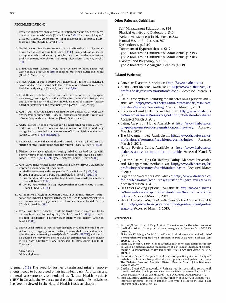
RECOMMENDATIONS
1. People with diabetes should receive nutrition counselling by a registereddietitian to lower A1C levels [Grade B, Level 2 (3)], for those with type 2diabetes; Grade D, Consensus, for type1 diabetes] and to reduce hospi-talization rates [Grade C, Level 3 (8)].
2. Nutrition education is effective when delivered in either a small group ora one-on-one setting [Grade B, Level 2 (13)]. Group education shouldincorporate adult education principles, such as hands-on activities,problem solving, role playing and group discussions [Grade B, Level 2(14)].
3. Individuals with diabetes should be encouraged to follow Eating Wellwith Canada’s Food Guide (18) in order to meet their nutritional needs[Grade D, Consensus].
4. In overweight or obese people with diabetes, a nutritionally balanced,calorie-reduced diet should be followed to achieve and maintain a lower,healthier body weight [Grade A, Level 1A (28,29)].
5. In adults with diabetes, the macronutrient distribution as a percentage oftotal energy can range from 45% to 60% carbohydrate, 15% to 20% proteinand 20% to 35% fat to allow for individualization of nutrition therapybased on preferences and treatment goals [Grade D, Consensus].
6. Adults with diabetes should consume no more than 7% of total dailyenergy from saturated fats [Grade D, Consensus] and should limit intakeof trans fatty acids to a minimum [Grade D, Consensus].
7. Added sucrose or added fructose can be substituted for other carbohy-drates as part of mixed meals up to a maximum of 10% of total dailyenergy intake, provided adequate control of BG and lipids is maintained[Grade C, Level 3 (50,51,54,58,60)].
8. People with type 2 diabetes should maintain regularity in timing andspacing of meals to optimize glycemic control [Grade D, Level 4 (132)].
9. Dietary advice may emphasize choosing carbohydrate food sources witha low glycemic index to help optimize glycemic control [type 1 diabetes:Grade B, Level 2 (34,35,169); type 2 diabetes: Grade B, Level 2 (41)].
10. Alternative dietary patternsmay be used in peoplewith type 2 diabetes toimprove glycemic control, (including):a. Mediterranean-style dietary pattern [Grade B, Level 2 (107,108)]b. Vegan or vegetarian dietary pattern [Grade B, Level 2 (103,104)]c. Incorporation of dietary pulses (e.g. beans, peas, chick peas, lentils)
[Grade B, Level 2 (122)]d. Dietary Approaches to Stop Hypertension (DASH) dietary pattern
[Grade C, Level 2 (118)]
11. An intensive lifestyle intervention program combining dietary modifi-cation and increased physical activity may be used to achieve weight lossand improvements in glycemic control and cardiovascular risk factors[Grade A, Level 1A (29)].
12. People with type 1 diabetes should be taught how to match insulin tocarbohydrate quantity and quality [Grade C, Level 2 (138)] or shouldmaintain consistency in carbohydrate quantity and quality [Grade D,Level 4 (131)].
13. People using insulin or insulin secretagogues should be informed of therisk of delayed hypoglycemia resulting from alcohol consumed with orafter the previous evening’s meal [Grade C, Level 3 (170,172)] and shouldbe advised on preventive actions such as carbohydrate intake and/orinsulin dose adjustments and increased BG monitoring [Grade D,Consensus].
Abbreviation:BG, blood glucose.
P.D. Dworatzek et al. / Can J Diabetes 37 (2013) S45eS55S52
pregnant (18). The need for further vitamin and mineral supple-ments needs to be assessed on an individual basis. As vitamin andmineral supplements are regulated as Natural Health products(NHP) in Canada, the evidence for their therapeutic role in diabeteshas been reviewed in the Natural Health Products chapter.
Other Relevant Guidelines
Self-Management Education, p. S26Physical Activity and Diabetes, p. S40Weight Management in Diabetes, p. S82Natural Health Products, p. S97Dyslipidemia, p. S110Treatment of Hypertension, p. S117Type 1 Diabetes in Children and Adolescents, p. S153Type 2 Diabetes in Children and Adolescents, p. S163Diabetes and Pregnancy, p. S168Type 2 Diabetes in Aboriginal Peoples, p. S191
Related Websites
� Canadian Diabetes Association (http://www.diabetes.ca)� Alcohol and Diabetes. Available at: http://www.diabetes.ca/for-professionals/resources/nutrition/alcohol. Accessed March 3,2013.
� Basic Carbohydrate Counting for Diabetes Management. Avail-able at: http://www.diabetes.ca/for-professionals/resources/nutrition/basic-carb-counting. Accessed March 3, 2013.
� Cholesterol and Diabetes. Available at: http://www.diabetes.ca/for-professionals/resources/nutrition/cholesterol-diabetes.Accessed March 3, 2013.
� Eating Away from Home. Available at: http://www.diabetes.ca/for-professionals/resources/nutrition/eating-away. AccessedMarch 3, 2013.
� The Glycemic Index. Available at: http://www.diabetes.ca/for-professionals/resources/nutrition/glycemic-index. AccessedMarch 3, 2013.
� Handy Portion Guide. Available at: http://www.diabetes.ca/diabetes-and-you/nutrition/portion-guide. Accessed March 3,2013.
� Just the Basics: Tips for Healthy Eating, Diabetes Preventionand Management. Available at: http://www.diabetes.ca/for-professionals/resources/nutrition/just-basics. Accessed March3, 2013.
� Sugars and Sweeteners. Available at: http://www.diabetes.ca/for-professionals/resources/nutrition/sugars-sweeteners.Accessed March 3, 2013.
� Healthier Cooking Options: Available at: http://www.diabetes.ca/for-professionals/resources/nutrition/healthier-cooking-options. Accessed March 3, 2013.
� Health Canada. Eating Well with Canada’s Food Guide. Availableat: http://www.hc-sc.gc.ca/fn-an/food-guide-aliment/index-eng.php. Accessed March 3, 2013.
References
1. Pastors JG, Warshaw H, Daly A, et al. The evidence for the effectiveness ofmedical nutrition therapy in diabetes management. Diabetes Care 2002;25:608e13.
2. Pi-Sunyer FX, Maggio CA, McCarron DA, et al. Multicenter randomized trial ofa comprehensive prepared meal program in type 2 diabetes. Diabetes Care1999;22:191e7.
3. Franz MJ, Monk A, Barry B, et al. Effectiveness of medical nutrition therapyprovided by dietitians in the management of non-insulin-dependent diabetesmellitus: a randomized, controlled clinical trial. J Am Diet Assoc 1995;95:1009e17.
4. Kulkarni K, Castle G, Gregory R, et al. Nutrition practice guidelines for type 1diabetes mellitus positively affect dietitian practices and patient outcomes.The Diabetes Care and Education Dietetic Practice Group. J Am Diet Assoc1998;98:62e70.
5. Gaetke L, Stuart M, Truszczynska H. A single nutrition counseling session witha registered dietitian improves short-term clinical outcomes for rural ken-tucky patients with chronic diseases. J Am Diet Assoc 2006;106:109e12.
6. Imai S, Kozai H, Matsuda M, et al. Intervention with delivery of diabetic mealsimproves glycemic control in patients with type 2 diabetes mellitus. J ClinBiochem Nutr 2008;42:59e63.

P.D. Dworatzek et al. / Can J Diabetes 37 (2013) S45eS55 S53
7. Huang M-C, Hsu C-C, Wang H-S, et al. Prospective randomized controlled trialto evaluate effectiveness of registered dietitian-led diabetes management onglycemic and diet control in a primary care setting in Taiwan. Diabetes Care2010;33:233e9.
8. Robbins JM, Thatcher GE, Webb DA, et al. Nutritionist visits, diabetes classes,and hospitalization rates and charges: the Urban Diabetes Study. DiabetesCare 2008;31:655e60.
9. Willaing I, Ladelund S, Jørgensen T, et al. Nutritional counselling inprimary health care: a randomized comparison of an intervention bygeneral practitioner or dietician. Eur J Cardiovasc Prev Rehabil 2004;11:513e20.
10. Wilson C, Brown T, Acton K, et al. Effects of clinical nutrition education andeducator discipline on glycemic control outcomes in the Indian health service.Diabetes Care 2003;26:2500e4.
11. Brekke HK, Jansson PA, Lenner RA. Long-term (1- and 2-year) effects of life-style intervention in type 2 diabetes relatives. Diabetes Res Clin Pract 2005;70:225e34.
12. Lemon CC, Lacey K, Lohse B, et al. Outcomes monitoring of health, behavior,and quality of life after nutrition intervention in adults with type 2 diabetes.J Am Diet Assoc 2004;104:1805e15.
13. Rickheim PL,Weaver TW, Flader JL, et al. Assessment of group versus individualdiabetes education: a randomized study. Diabetes Care 2002;25:269e74.
14. Trento M, Basile M, Borgo E, et al. A randomised controlled clinical trial ofnurse-, dietitian- and pedagogist-led Group Care for the management of type2 diabetes. J Endocrinol Invest 2008;31:1038e42.
15. Pérez-Escamilla R, Hromi-Fiedler A, Vega-López S, et al. Impact of peernutrition education on dietary behaviors and health outcomes among Latinos:a systematic literature review. J Nutr Educ Behav 2008;40:208e25.
16. Ralston JD, Hirsch IB, Hoath J, et al. Web-based collaborative care for type 2diabetes: a pilot randomized trial. Diabetes Care 2009;32:234e9.
17. Marcy TR, Britton ML, Harrison D. Identification of barriers to appropriatedietary behavior in low-income patients with type 2 diabetes mellitus.Diabetes Ther 2011;1:9e19.
18. Health Canada. Eating Well with Canada’s Food Guide. Publication H39e166/1990E. Ottawa, ON: Health Products and Food Branch, Office of Nutrition andPromotion; 2007.
19. Norris SL, Engelgau MM, Narayan KM. Effectiveness of self-managementtraining in type 2 diabetes: a systematic review of randomized controlledtrials. Diabetes Care 2001;24:561e87.
20. Ash S, Reeves MM, Yeo S, et al. Effect of intensive dietetic interventions onweight and glycaemic control in overweight men with Type II diabetes:a randomised trial. Int J Obes Relat Metab Disord 2003;27:797e802.
21. Clement S. Diabetes self-management education. Diabetes Care 1995;18:1204e14.
22. Vallis TM, Higgins-Bowser I, Edwards L, et al. The role of diabetes education inmaintaining lifestyle changes. Can J Diabetes 2005;29:193e202.
23. Christensen NK, Terry RD, Wyatt S, et al. Quantitative assessment of dietaryadherence in patients with insulin-dependent diabetes mellitus. DiabetesCare 1983;6:245e50.
24. Toeller M, Klischan A, Heitkamp G, et al. Nutritional intake of 2868 IDDMpatients from 30 centres in Europe. EURODIAB IDDM Complications StudyGroup. Diabetologia 1996;39:929e39.
25. Glazier RH, Bajcar J, Kennie NR, et al. A systematic review of interventions toimprove diabetes care in socially disadvantaged populations. Diabetes Care2006;29:1675e88.
26. Wing RR. Weight loss in the management of type 2 diabetes. In: Gerstein HC,Haynes B, editors. Evidence-Based Diabetes Care. Ontario, Canada: B.C.Decker, Inc; 2000. p. 252e76.
27. Tuomilehto J, Lindström J, Eriksson JG, et al, Finnish Diabetes PreventionStudy Group. Prevention of type 2 diabetes mellitus by changes in lifestyleamong subjects with impaired glucose tolerance. N Engl J Med 2001;344:1343e50.
28. Knowler WC, Barrett-Connor E, Fowler SE, et al, Diabetes Prevention ProgramResearch Group. Reduction in the incidence of type 2 diabetes with lifestyleintervention or metformin. N Engl J Med 2002;346:393e403.
29. Look AHEAD Research GroupWing RR. Long-term effects of a lifestyle inter-vention on weight and cardiovascular risk factors in individuals with type 2diabetes mellitus: four-year results of the Look AHEAD trial. Arch Intern Med2010;170:1566e75.
30. Diabetes Prevention Program Research GroupKnowler WC, Fowler SE,Hamman RF, et al. 10-year follow-up of diabetes incidence and weight loss inthe Diabetes Prevention Program Outcomes Study. Lancet 2009;374:1677e86. Published erratum in: Lancet. 2009;374:2054.
31. Lindström J, Ilanne-Parikka P, Peltonen M, et al, Finnish Diabetes PreventionStudy Group. Sustained reduction in the incidence of type 2 diabetes bylifestyle intervention: follow-up of the Finnish Diabetes Prevention Study.Lancet 2006;368:1673e9.
32. Institute of Medicine, Food and Nutrition Board. Dietary Reference Intakes forEnergy, Carbohydrate, Fiber, Fat, Fatty Acids, Cholesterol, Protein, and AminoAcids. Washington, DC: National Academies Press; 2002.
33. Kirk JK, Graves DR, Carven TE, et al. Restricted carbohydrate diet inpatients with type 2 diabetes: A meta-analysis. J Am Diet Assoc 2008;108:91e100.
34. Barnard ND, Cohen J, Jenkins DJ, et al. A low-fat vegan diet improves glycemiccontrol and cardiovascular risk factors in a randomized clinical trial in indi-viduals with type 2 diabetes. Diabetes Care 2006;29:1777e83.
35. Jenkins DJ, Wolever TM, Taylor RH, et al. Glycemic index of foods:a physiological basis for carbohydrate exchange. Am J Clin Nutr 1981;34:362e6.
36. Atkinson FS, Foster-Powell K, Brand-Miller JC. International tables of glycemicindex and glycemic load values. Diabetes Care 2008;31:2281e3.
37. Brand-Miller J, Hayne S, Petocz P, et al. Low glycemic index diets in themanagement of diabetes: a meta-analysis of randomized controlled trials.Diabetes Care 2003;26:2261e7.
38. Opperman AM, Venter CS, Oosthuizen W, et al. Meta-analysis of the healtheffects of using the glycaemic index in meal-planning. Br J Nutr 2004;92:367e81.
39. Thomas DE, Elliott EJ. The use of low-glycaemic index diets in diabetescontrol. Br J Nutr 2010;104:797e802.
40. Wolever TM, Gibbs AL, Mehling C, et al. The Canadian Trial of Carbohydratesin Diabetes (CCD), a 1-y controlled trial of low-glycemic-index dietarycarbohydrate in type 2 diabetes: no effect on glycated hemoglobin butreduction in C-reactive protein. Am J Clin Nutr 2008;87:114e25.
41. Jenkins DJ, Kendall CW, McKeown-Eyssen G, et al. Effect of a low-glycemicindex or a high-cereal fiber diet on type 2 diabetes: a randomized trial.JAMA 2008;300:2742e53.
42. Wolever TM, Mehling C, Chiasson JL, et al. Low glycaemic index diet anddisposition index in type 2 diabetes (the Canadian trial of carbohydrates indiabetes): a randomised controlled trial. Diabetologia 2008;51:1607e15.
43. Amano Y, Sugiyama M, Lee JS, et al. Glycemic index-based nutritionaleducation improves blood glucose control in Japanese adults: a randomizedcontrolled trial. Diabetes Care 2007;30:1874e6.
44. Ma Y, Olendzki BC, Merriam PA, et al. A randomized clinical trial comparinglow-glycemic index versus ADA dietary education among individuals withtype 2 diabetes. Nutrition 2008;24:45e56.
45. Anderson JW, Randles KM, Kendall CW, et al. Carbohydrate and fiberrecommendations for individuals with diabetes: a quantitative assessmentand meta-analysis of the evidence. J Am Coll Nutr 2004;23:5e17.
46. Jacobs Jr DR, Meyer KA, Kushi LH, et al. Whole-grain intake may reduce therisk of ischemic heart disease death in post-menopausal women: the IowaWomen’s Health Study. Am J Clin Nutr 1998;68:248e57.
47. Acceptable macronutrient distribution ranges. In: Otten J, Hellwig J, Meyer L,editors. Dietary Reference Intakes. Washington, DC: National AcademiesPress; 2006. p. 70.
48. Chandalia M, Garg A, Lutjohann D, et al. Beneficial effects of high dietary fiberintake in patients with type 2 diabetes mellitus. N Engl J Med 2000;342:1392e8.
49. Cooper PL, Wahlqvist ML, Simpson RW. Sucrose versus saccharin as an addedsweetener in non-insulin-dependent diabetes: short- and medium-termmetabolic effects. Diabet Med 1988;5:676e80.
50. Chantelau EA, Gösseringer G, Sonnenberg GE, et al. Moderate intake ofsucrose does not impair metabolic control in pump-treated diabetic out-patients. Diabetologia 1985;28:204e7.
51. Colagiuri S, Miller JJ, Edwards RA. Metabolic effects of adding sucrose andaspartame to the diet of subjects with non-insulin-dependent diabetes mel-litus. Am J Clin Nutr 1989;50:474e8.
52. Coulston AM, Hollenbeck CB, Donner CC, et al. Metabolic effects of addeddietary sucrose in individuals with noninsulin-dependent diabetes mellitus(NIDDM). Metabolism 1985;34:962e6.
53. Jellish WS, Emanuele MA, Abraira C. Graded sucrose/carbohydrate diets inovertly hypertriglyceridemic diabetic patients. Am J Med 1984;77:1015e22.
54. Sievenpiper JL, de Souza RJ, Mirrahimi A, et al. Effect of fructose on bodyweight in controlled feeding trials: a systematic review and meta-analysis.Ann Intern Med 2012;156:291e304.
55. Sievenpiper JL, Chiavaroli L, de Souza RJ, et al. “Catalytic” doses of fructosemay benefit glycaemic control without harming cardiometabolic risk factors:a small meta-analysis of randomised controlled feeding trials. Br J Nutr 2012;108:418e23.
56. Ha V, Sievenpiper JL, de Souza RJ, et al. Effect of fructose on blood pressure:a systematic review and meta-analysis of controlled feeding trials. Hyper-tension 2012;59:787e95.
57. Wang DD, Sievenpiper JL, de Souza RJ, et al. Effect of fructose on uric acid:a meta-analysis of controlled feeding trials. J Nutr 2012;142:916e23.
58. Cozma AI, Sievenpiper JL, de Souza RJ, et al. Effect of fructose on glycemiccontrol in diabetes: a meta-analysis of controlled feeding trials. Diabetes Care2012;35:1611e20.
59. Livesey G, Taylor R. Fructose consumption and consequences for glycation,plasmid triacylglycerol, and body weight: meta-analyses and meta-regressionmodels of intervention studies. Am J Clin Nutr 2008;88:1419e37.
60. Sievenpiper JL, Carleton AJ, Chatha S, et al. Heterogeneous effects of fructoseon blood lipids in individuals with type 2 diabetes: systematic review andmeta-analysis of experimental trials in humans. Diabetes Care 2009;32:1930e7.
61. Madero M, Arriaga JC, Jalal D, et al. The effect of two energy-restricted diets,a low-fructose diet versus a moderate natural fructose diet, on weight lossand metabolic syndrome parameters: A randomized controlled trial. Metab-olism 2011;60:1551e9.
62. Jenkins DJ, Srichaikul K, Kendall CW, et al. The relation of low glycaemic indexfruit consumption to glycaemic control and risk factors for coronary heartdisease in type 2 diabetes. Diabetologia 2011;54:271e9.
63. Expert Panel on Detection, Evaluation, and Treatment of High Blood Choles-terol in Adults. Executive Summary of The Third Report of The National
P.D. Dworatzek et al. / Can J Diabetes 37 (2013) S45eS55S54
Cholesterol Education Program (NCEP) Expert Panel on Detection, Evaluation,and Treatment of High Blood Cholesterol in Adults (Adult Treatment Panel III).JAMA 2001;285:2486e97.
64. McEwan B, Morel-Kopp M-C, Tofler G, Ward C. Effect of omega-3 fish oil oncardiovascular risk in diabetes. Diabetes Educ 2010;36:565e84.
65. Hu FB, Cho E, Rexrode KM, et al. Fish and long-chain omega-3 fatty acid intakeand risk of coronary heart disease and total mortality in diabetic women.Circulation 2003;107:1852e7.
66. Lee CC, Sharp S, Wexler D, Adler A. Dietary intake of eicosapentaenoicand docosahexaenoic acid and diabetic nephropathy: cohort analysis ofthe diabetes control and complications trial. Diabetes Care 2010;33:1454e6.
67. Yokoyama M, Origasa H, Matsuzaki M, et al. Effects of eicosapentaenoic acidon major coronary events in hypercholesterolaemic patients (JELIS): a rand-omised open-label, blinded endpoint analysis. Lancet 2007;369:1090e8.
68. GISSI-HF Investigators. Effect of n-3 polyunsaturated fatty acids in patientswith chronic heart failure (the GISSI-HF trial): a randomised, double-blind,placebo-controlled trial. Lancet 2008;372:1223e30.
69. Dietary supplementation with n-3 polyunsaturated fatty acids and vitamin Eafter myocardial infarction: results of the GISSI-Prevenzione trial. GruppoItaliano per lo Studio della Sopravvivenza nell’Infarto miocardico. Lancet1999;354:447e55.
70. Kromhout D, Giltay EJ, Geleijnse JM, Alpha Omega Trial Group. n-3 fatty acidsand cardiovascular events after myocardial infarction. N Engl J Med 2010;363:2015e26.
71. ORIGIN Trial Investigators, Bosch J, Gerstein HC, Dagenais GR, et al. n-3 fattyacids and cardiovascular outcomes in patients with dysglycemia. N Engl J Med2012;367:309e18.
72. Rizos EC, Ntzani EE, Bika E, et al. Association between omega-3 fatty acidsupplementation and risk of major cardiovascular disease events: a system-atic review and meta-analysis. JAMA 2012;308:1024e33.
73. Hamdy O, Horton ES. Protein content in diabetes nutrition plan. Curr Diab Rep2011;11:111e9.
74. Food and Nutrition Board, Institute of Medicine. Dietary Reference Intakes forEnergy, Carbohydrate, Fiber, Fat, Fatty Acids, Cholesterol, Protein, and AminoAcids. Washington, DC: National Academies Press; 2005.
75. Hansen HP, Tauber-Lassen E, Jensen BR, Parving HH. Effect of dietary proteinrestriction on prognosis in patients with diabetic nephropathy. Kidney Int2002;62:220e8.
76. Pan Y, Guo LL, Jin HM. Low-protein diet for diabetic nephropathy: a meta-analysis of randomized controlled trials. Am J Clin Nutr 2008;88:660e6.
77. Azadbakht L, Esmaillzadeh A. Soy-protein consumption and kidney-relatedbiomarkers among type 2 diabetics: a crossover, randomized clinical trial.J Ren Nutr 2009;19:479e86.
78. Azadbakht L, Atabak S, Esmaillzadeh A. Soy protein intake, cardiorenalindices, and C-reactive protein in type 2 diabetes with nephropathy: a longi-tudinal randomized clinical trial. Diabetes Care 2008;31:648e54.
79. Teixeira SR, Tappenden KA, Carson L, et al. Isolated soy protein consumptionreduces urinary albumin excretion and improves the serum lipid profile inmen with type 2 diabetes mellitus and nephropathy. J Nutr 2004;134:1874e80.
80. de Mello VD, Zelmanovitz T, Perassolo MS, et al. Withdrawal of red meat fromthe usual diet reduces albuminuria and improves serum fatty acid profile intype 2 diabetes patients with macroalbuminuria. Am J Clin Nutr 2006;83:1032e8.
81. Brodsky IG, Robbins DC, Hiser E, et al. Effects of low-protein diets on proteinmetabolism in insulin-dependent diabetes mellitus patients with earlynephropathy. J Clin Endocrinol Metab 1992;75:351e7.
82. Rodrigues-Villar C, Pérez-Heras A, Mercadé I, et al. Effects of olive oil on lipidprofiles and blood glucose in type 2 diabetic patients. Diabet Med 2004;21:142e9.
83. Brehm BJ, Lattin BL, Summer SS, et al. One-year comparison of a high-monounsaturated fat diet with a high-carbohydrate diet in type 2 diabetes.Diabetes Care 2009;32:215e20.
84. Brunerova L, Smejkalova V, Potockova J, et al. A comparison of the influence ofa high-fat diet enriched in monounsaturated fatty acids and conventional dieton weight loss and metabolic parameters in obese non-diabetic and Type 2diabetic patients. Diabet Med 2007;24:533e40.
85. De Natale C, Annuzzi G, Bozzetto L, et al. Effects of a plant-based highcarbohydrate/high fibre diet versus high monounsaturated fat/low carbohy-drate diet on postprandial lipids in type 2 diabetic patients. Diabetes Care2009;32:2168e73.
86. Kodama S, Saito K, Tanaka S, et al. Influence of fat and carbohydrateproportions on the metabolic profile in patients with type 2 diabetes: a meta-analysis. Diabetes Care 2009;32:959e65.
87. Papakonstantinou E, Zampelas A. The effect of dietary protein intake oncoronary heart disease risk. J Hum Nutr Diet 2010;23:183e9.
88. Paddon-Jones D, Westman E, Mattes RD, et al. Protein, weight management,and satiety. Am J Clin Nutr 2008;87:1558Se61S.
89. Lejeune MP, Westerterp KR, Adam TC, et al. Ghrelin and glucagon-like peptide1 concentrations, 24-h satiety, and energy and substrate metabolism duringa high-protein diet and measured in a respiration chamber. Am J Clin Nutr2006;83:89e94.
90. Mozaffarian D, Micha R, Wallace S. Effects on coronary heart disease ofincreasing polyunsaturated fat in place of saturated fat: a systematic reviewand meta-analysis of randomized controlled trials. PLoS Med 2010;7:e1000252.
91. Jakobsen MU, O’Reilly EJ, Heitmann BL, et al. Major types of dietary fat andrisk of coronary heart disease: a pooled analysis of 11 cohort studies. Am J ClinNutr 2009;89:1425e32.
92. Howard BV, Van Horn L, Hsia J, et al. Low-fat dietary pattern and risk ofcardiovascular disease: the Women’s Health Initiative Randomized ControlledDietary Modification Trial. JAMA 2006;295:655e66.
93. Larsen RN, Mann NJ, Maclean E, et al. The effect of high-protein, low-carbo-hydrate diets in the treatment of type 2 diabetes: a 12 month randomisedcontrolled trial. Diabetologia 2011;54:731e40.
94. McLaughlin T, Carter S, Lamendola C, et al. Clinical efficacy of two hypo-caloric diets that vary in overweight patients with type 2 diabetes:comparison of moderate fat versus carbohydrate reductions. Diabetes Care2007;30:1877e9.
95. Brinkworth GD, Noakes M, Parker B, et al. Long-term effects of advice toconsume a high-protein, low-fat diet, rather than a conventional weight-lossdiet, in obese adults with Type 2 diabetes: one-year follow-up of a random-ized trial. Diabetologia 2004;47:1677e86.
96. Pearce KL, Clifton PM, Noakes M. Egg consumption as part of an energy-restricted high-protein diet improves blood lipid and blood glucose profilesin individuals with type 2 diabetes. Br J Nutr 2011;105:585e92.
97. Haimoto H, Iwata M, Wakai K, et al. Long-term effects of a diet looselyrestricting carbohydrates on HbA1c levels, BMI and tapering of sulfonylureasin type 2 diabetes: a 2-year follow-up study. Diabetes Res Clin Pract 2008;79:350e6.
98. Davis NJ, Tomuta N, Schechter C, et al. Comparative study of the effects ofa 1-year dietary intervention of a low-carbohydrate diet versus a low-fat dieton weight and glycemic control in type 2 diabetes. Diabetes Care 2009;32:1147e52.
99. Gong Q, Gregg EW, Wang J, et al. Long-term effects of a randomised trial ofa 6-year lifestyle intervention in impaired glucose tolerance on diabetes-related microvascular complications: the China Da Qing Diabetes PreventionOutcome Study. Diabetologia 2011;54:300e7.
100. Mann JI, De Leeuw I, Hermansen K, et al. Evidence-based nutritionalapproaches to the treatment and prevention of diabetes mellitus. Nutr MetabCardiovasc Dis 2004;14:373e94.
101. Coppell KJ, Kataoka M, Williams SM, et al. Nutritional intervention in patientswith type 2 diabetes who are hyperglycaemic despite optimised drugtreatmenteLifestyle Over and Above Drugs in Diabetes (LOADD) study:randomised controlled trial. BMJ 2010;341:c3337.
102. Toobert DJ, Glasgow RE, Strycker LA, et al. Biologic and quality-of-lifeoutcomes from the Mediterranean Lifestyle Program: a randomized clinicaltrial. Diabetes Care 2003;26:2288e93.
103. Barnard ND, Cohen J, Jenkins DJA, et al. A low-fat vegan diet and a conven-tional diabetes diet in the treatment of type 2 diabetes: a randomized,controlled, 74-wk clinical trial. Am J Clin Nutr 2009;89:1588Se96S.
104. Kahleova H, Matoulek M, Malinska H, et al. Vegetarian diet improves insulinresistance and oxidative stress markers more than conventional diet insubjects with Type 2 diabetes. Diabet Med 2011;28:549e59.
105. Willett WC, Sacks F, Trichopoulou A, et al. Mediterranean diet pyramid:a cultural model for healthy eating. Am J Clin Nutr 1995;61:1402Se6S.
106. Trichopoulou A, Costacou T, Bamia C, et al. Adherence to a Mediterranean dietand survival in a Greek population. N Engl J Med 2003;348:2599e608.
107. Esposito K, Maiorino MI, Ceriello A, et al. Prevention and control of type 2diabetes by Mediterranean diet: a systematic review. Diabetes Res Clin Pract2010;89:97e102.
108. EspositoK,MaiorinoMI, CiotolaM, et al. Effects of aMediterranean-style diet onthe need for antihyperglycemic drug therapy in patients with newly diagnosedtype 2 diabetes: a randomized trial. Ann Intern Med 2009;151:306e14.
109. Shai I, Schwarzfuchs D, Henkin Y, et al. Weight loss with a low-carbohydrate,Mediterranean, or low-fat diet. N Engl J Med 2008;359:229e41. Publishederratum in: N Engl J Med. 2009;361:2681.
110. Elhayany A, Lustman A, Abel R, et al. A low carbohydrate Mediterranean dietimproves cardiovascular risk factors and diabetes control among overweightpatients with type 2 diabetes mellitus: a 1-year prospective randomizedintervention study. Diabetes Obes Metab 2010;12:204e9.
111. Estruch R, Ros E, Salas-Salvadó J, et al. Primary Prevention of CardiovascularDisease with a Mediterranean Diet. N Engl J Med. 2013. [Epub ahead of print]
112. Food and Nutrition Board, Institute of Medicine. Dietary Reference Intakes forWater, Potassium, Sodium, Chloride, and Sulfate. Washington, DC: NationalAcademies Press; 2004.
113. Thomas MC, Moran J, Forsblom C, et al. The association between dietarysodium intake, ESRD, and all-cause mortality in patients with type 1 diabetes.Diabetes Care 2011;34:861e6.
114. Ekinci EI, Clarke S, Thomas MC, et al. Dietary salt intake and mortality inpatients with type 2 diabetes. Diabetes Care 2011;34:703e9.
115. Appel LJ, Moore TJ, Obarzanek E, et al. A clinical trial of the effects of dietarypatterns on blood pressure. DASH Collaborative Research Group. N Engl J Med1997;336:1117e24.
116. Sacks FM, Svetkey LP, Vollmer WM, et al, DASH-Sodium CollaborativeResearch Group. Effects on blood pressure of reduced dietary sodium and theDietary Approaches to Stop Hypertension (DASH) diet. N Engl J Med 2001;344:3e10.
117. Azadbakht L, Surkan PJ, Esmaillzadeh A, et al. The Dietary Approaches to StopHypertension eating plan affects C-reactive protein, coagulation abnormali-ties, and hepatic function tests among type 2 diabetic patients. J Nutr 2011;141:1083e8.

P.D. Dworatzek et al. / Can J Diabetes 37 (2013) S45eS55 S55
118. Azadbakht L, Fard NR, Karimi M, et al. Effects of the Dietary Approaches toStop Hypertension (DASH) eating plan on cardiovascular risks among type 2diabetic patients: a randomized crossover clinical trial. Diabetes Care 2011;34:55e7.
119. Nordmann AJ, Nordmann A, Briel M, et al. Effects of low-carbohydrate vslow-fat diets on weight loss and cardiovascular risk factors: a meta-analysisof randomized controlled trials. Arch Intern Med 2006;166:285e93.
120. Stern L, Iqbal N, Seshadri P, et al. The effects of low-carbohydrate versusconventional weight loss diets in severely obese adults: one-year follow-up ofa randomized trial. Ann Intern Med 2004;140:778e85.
121. Dansinger ML, Gleason JA, Griffith JL, et al. Comparison of the Atkins, Ornish,Weight Watchers, and Zone diets for weight loss and heart disease riskreduction: a randomized trial. JAMA 2005;293:43e53.
122. Sievenpiper JL, Kendall CWC, Esfahani A, et al. Effect of non-oil-seed pulses onglycaemic control: a systematic review and meta-analysis of randomisedcontrolled experimental trials in people with and without diabetes.Diabetologia 2009;52:1479e95.
123. Jang Y, Lee JH, Kim OY, et al. Consumption of whole grain and legume powderreduces insulin demand, lipid peroxidation, and plasma homocysteineconcentrations in patients with coronary artery disease: randomizedcontrolled clinical trial. Arterioscler Thromb Vasc Biol 2001;12:2065e71.
124. Bazzano LA, Thompson AM, Tees MT, et al. Non-soy legume consumptionlowers cholesterol levels: a meta-analysis of randomized controlled trials.Nutr Metab Cardiovasc Dis 2011;2:94e103.
125. Imai S, Matsuda M, Hasegawa G, et al. A simple meal plan of “eating vege-tables before carbohydrate”was more effective for achieving glycemic controlthan an exchange-based meal plan in Japanese patients with type 2 diabetes.Asia Pac J Clin Nutr 2011;20:161e8.
126. Jenkins DJ, Kendall CW, Banach MS, et al. Nuts as a replacement for carbo-hydrates in the diabetic diet. Diabetes Care 2011;34:1706e11.
127. Cohen AE, Johnston CS. Almond ingestion at mealtime reduces postprandialglycemia and chronic ingestion reduces hemoglobin A1C in individuals withwell-controlled type 2 diabetes mellitus. Metabolism 2011;6:1312e7.
128. Li S-C, Liu Y-H, Liu J-F, et al. Almond consumption improved glycemic controland lipid profiles in patients with type 2 diabetes mellitus. Metabolism 2011;60:474e9.
129. Lovejoy JC, Most MM, Lefevre M, et al. Effect of diets enriched in almonds oninsulin action and serum lipids in adults with normal glucose tolerance ortype 2 diabetes. Am J Clin Nutr 2002;76:1000e6.
130. Sabaté J, Oda K, Ros E. Nut consumption and blood lipid levels. Arch InternMed 2010;170:821e7.
131. Wolever TM, Hamad S, Chiasson JL, et al. Day-to-day consistency in amountand source of carbohydrate associated with improved blood glucose controlin type 1 diabetes. J Am Coll Nutr 1999;18:242e7.
132. Savoca MR, Miller CK, Ludwig DA. Food habits are related to glycemiccontrol among people with type 2 diabetesmellitus. J AmDiet Assoc 2004;104:560e6.
133. Kalergis M, Schiffrin A, Gougeon R, et al. Impact of bedtime snack compositionon prevention of nocturnal hypoglycemia in adults with type 1 diabetesundergoing intensive insulin management using lispro insulin beforemeals: a randomized, placebo-controlled, crossover trial. Diabetes Care 2003;26:9e15.
134. Arnold L, Mann JI, Ball MJ. Metabolic effects of alterations in meal frequency intype 2 diabetes. Diabetes Care 1997;20:1651e4.
135. Delahanty LM, Nathan DM, Lachin JM, et al. Association of diet with glycatedhemoglobin during intensive treatment of type 1 diabetes in the DiabetesControl and Complications Trial. Am J Clin Nutr 2009;89:518e24.
136. Tunbridge FK, Home PD, Murphy M, et al. Does flexibility at mealtimes disturbblood glucose control on a multiple insulin injection regimen? Diabet Med1991;8:833e8.
137. DAFNE Study Group. Training in flexible, intensive insulin management toenable dietary freedom in people with type 1 diabetes: dose adjustment fornormal eating (DAFNE) randomised controlled trial. BMJ 2002;325:746.
138. Scavone G, Manto A, Pitocco D, et al. Effect of carbohydrate counting andmedical nutritional therapy on glycaemic control in Type1 diabetic subjects:a pilot study. Diabet Med 2010;27:477e9.
139. Bergenstal RM, JohnsonM, PowersMA, et al. Adjust to target in type 2 diabetes:comparison of a simple algorithm with carbohydrate counting for adjustmentof mealtime insulin glulisine. Diabetes Care 2008;31:1305e10.
140. Gillespie SJ, Kulkarni KD, Daly AE. Using carbohydrate counting in diabetesclinical practice. J Am Diet Assoc 1998;98:897e905.
141. Kelley DE. Sugars and starch in the nutritional management of diabetesmellitus. Am J Clin Nutr 2003;78:858Se64S.
142. Rossi MC, Nicolucci A, Di Bartolo P, et al. Diabetes Interactive Diary: a newtelemedicine system enabling flexible diet and insulin therapy whileimproving quality of life: an open-label, international, multicenter, random-ized study. Diabetes Care 2010;33:109e15.
143. Gougeon R, Spidel M, Lee K, et al. Canadian Diabetes Association NationalNutrition Committee Technical Review: non-nutritive intense sweeteners indiabetes management. Can J Diabetes 2004;28:385e99.
144. JECFA 2008: Joint FAO/WHO Expert Committee on Food Additives. Sixty-ninthmeeting. Rome, Italy, June 17-26, 2008.
145. Maki KC, Curry LL, Reeves MS, et al. Chronic consumption of rebaudioside A,a steviol glycoside, in men and women with type 2 diabetes mellitus. FoodChem Toxicol 2008;46:S47e53.
146. Barriocanal LA, Palacios M, Benitez G, et al. Apparent lack of pharmaco-logical effect of steviol glycosides used as sweeteners in humans. A pilotstudy of repeated exposures in some normotensive and hypotensive indi-viduals and in Type 1 and Type 2 diabetics. Regul Toxicol Pharmacol 2008;51:37e41.
147. Wang YM, van Eys J. Nutritional significance of fructose and sugar alcohols.Annu Rev Nutr 1981;1:437e75.
148. Wolever TMS, Piekarz A, Hollands M, et al. Sugar alcohols and diabetes:a review. Can J Diabetes 2002;26:356e62.
149. Stirban A, Negrean M, Götting C, et al. Dietary advanced glycation end-products and oxidative stress: in vivo effects on endothelial function andadipokines. Ann N Y Acad Sci 2008;1126:276e9.
150. Negrean M, Stirban A, Stratmann B, et al. Effects of low- and high-advancedglycation endproduct meals on macro- and microvascular endothelial func-tion and oxidative stress in patients with type 2 diabetes mellitus. Am J ClinNutr 2007;85:1236e43.
151. Uribarri J, Cai W, Ramdas M, et al. Restriction of advanced glycation endproducts improves insulin resistance in human type 2 diabetes: potential roleof AGER1 and SIRT1. Diabetes Care 2011;34:1610e6.
152. Heymsfield SB, van Mierlo CA, van der Knaap HC, et al. Weight managementusing a meal replacement strategy: meta and pooling analysis from sixstudies. Int J Obes Relat Metab Disord 2003;27:537e49.
153. Li Z, Hong K, Saltsman P, et al. Long-term efficacy of soy-based mealreplacements vs an individualized diet plan in obese type II DM patients:relative effects on weight loss, metabolic parameters, and C-reactive protein.Eur J Clin Nutr 2005;59:411e8.
154. Cheskin LJ, Mitchell AM, Jhaveri AD, et al. Efficacy of meal replacementsversus a standard food-based diet for weight loss in type 2 diabetes:a controlled clinical trial. Diabetes Educ 2008;34:118e27.
155. Yip I, Go VL, DeShields S, et al. Liquid meal replacements and glycemic controlin obese type 2 diabetes patients. Obes Res 2001;9:341Se74S.
156. Wadden TA, West DS, Neiberg RH, et al. One-year weight losses in theLook AHEAD study: factors associated with success. Obesity 2009;17:713e22.
157. McCargar L, Innis S, Bowron E, et al. Effect of enteral nutritional productsdiffering in carbohydrate and fat on indices of carbohydrate and lipidmetabolism in patients with NIDDM. Mol Cell Biochem 1998;188:81e9.
158. Lansink M, van Laere KM, Vendrig L, et al. Lower postprandial glucoseresponses at baseline and after 4 weeks use of a diabetes-specific formula indiabetes type 2 patients. Diabetes Res Clin Pract 2011;93:421e9.
159. Stockwell T, Zhao J, Thomas G. Should alcohol policies aim to reduce totalalcohol consumption? New analyses of Canadian drinking patterns. AddictRes Theory 2009;17:135e51.
160. Butt P, Beirness D, Cesa F, et al. Alcohol and health in Canada: a summary ofevidence and guidelines for low-risk drinking. Ottawa, ON: Canadian Centreon Substance Abuse; 2011.
161. Kerr D, Macdonald IA, Heller SR, et al. Alcohol causes hypoglycaemicunawareness in healthy volunteers and patients with type 1 (insulin-dependent) diabetes. Diabetologia 1990;33:216e21.
162. Kerr D, Penfold S, Zouwail S, et al. The influence of liberal alcoholconsumption on glucose metabolism in patients with type 1 diabetes: a pilotstudy. QJM 2009;102:169e74.
163. Koppes LLJ, Dekker JM, Hendricks HFJ, et al. Meta-analysis of the relationshipbetween alcohol consumption and coronary heart disease and mortality intype 2 diabetic patients. Diabetologia 2006;49:648e52.
164. Bantle AE, Thomas W, Bantle JP. Metabolic effects of alcohol in the form ofwine in persons with type 2 diabetes mellitus. Metabolism 2008;57:241e5.
165. Marfella R, Cacciapuoti F, Siniscalchi M, et al. Effect of moderate red wineintake on cardiac prognosis after recent acute myocardial infarction ofsubjects with Type 2 diabetes mellitus. Diabet Med 2006;23:974e81.
166. Nakamura T, Fujiwara N, Sugaya T, et al. Effect of red wine on urinaryprotein, 8-hydroxydeoxyguanosine, and liver-type fatty acidebindingprotein excretion in patients with diabetic nephropathy. Metabolism 2009;58:1185e90.
167. Lee CC, Stolk RP, Adler AI, et al. Association between alcohol consumption anddiabetic retinopathy and visual acuity: the AdRem Study. Diabet Med 2010;27:1130e7.
168. Wakabayashi J. Association between alcohol drinking and metabolicsyndrome in Japanese male workers with diabetes mellitus. J AtherosclerThromb 2011;18:684e91.
169. Ahmed AT, Karter AJ, Warton M, et al. The relationship between alcoholconsumption and glycemic control among patients with diabetes: the KaiserPermanente Northern California Diabetes Registry. J Gen Intern Med 2008;23:275e82.
170. Richardson T, Weiss M, Thomas P, et al. Day after the night before: influenceof evening alcohol on risk of hypoglycemia in patients with type 1 diabetes.Diabetes Care 2005;28:1801e2.
171. Cheyne EH, Sherwin RS, Lunt MJ, et al. Influence of alcohol on cognitiveperformance during mild hypoglycaemia: implications for Type 1 diabetes.Diabet Med 2004;21:230e7.
172. Pietraszek A, Gregersen S, Hermansen K. Alcohol and type 2 diabetes. Areview. Nutr Metab Cardiovasc Dis 2010;20:366e75.
173. Gallagher A, Connolly V, Kelly WF. Alcohol consumption in patients withdiabetes. Diabet Med 2001;18:72e3.

Contents lists available at SciVerse ScienceDirect
Can J Diabetes 37 (2013) S56eS60
Canadian Journal of Diabetesjournal homepage:
www.canadianjournalofdiabetes.com
Clinical Practice Guidelines
Pharmacotherapy in Type 1 Diabetes
Canadian Diabetes Association Clinical Practice Guidelines Expert Committee
The initial draft of this chapter was prepared by Angela McGibbon MD, PhD, FRCPC,Cindy Richardson MD, FRCPC, Cheri Hernandez RN, PhD, CDE, John Dornan MD, FRCPC, FACP
KEY MESSAGES
� Basal-bolus insulin regimens (e.g. multiple daily injections or continuoussubcutaneous insulin infusion) are the insulin regimens of choice for alladults with type 1 diabetes.
� Insulin regimens should be tailored to the individual’s treatment goals,lifestyle, diet, age, general health, motivation, hypoglycemia awarenessstatus and ability for self-management.
� All individuals with type 1 diabetes should be counseled about the risk,prevention and treatment of insulin-induced hypoglycemia.
Introduction
Insulin is lifesaving pharmacological therapy for people withtype 1 diabetes. Insulin preparations are primarily produced byrecombinant DNA technology and are formulated either as struc-turally identical to human insulin or as a modification of humaninsulin (insulin analogues) to alter pharmacokinetics. Humaninsulin and insulin analogues are preferred and used bymost adultswith type 1 diabetes; however, preparations of animal-sourcedinsulin are still accessible in Canada (1).
Insulin preparations are classified according to their duration ofaction and are further differentiated by their time of onset and peakactions (Table 1). Premixed insulin preparations are available andare not generally suitable for intensive treatment in patients withtype 1 diabetes in whom frequent adjustments of insulin arerequired.
There may be a role for adjunctive therapy in some people withtype 1 diabetes to aid in achieving optimal glycemic targets. Phar-macotherapy for prevention of complications and treatment of riskfactors will be addressed in other chapters.
Insulin Delivery Systems
Insulin can be administered by syringe, pen or pump (contin-uous subcutaneous insulin infusion [CSII]). Insulin pen devicesfacilitate the use of multiple injections of insulin. CSII therapy isa safe and effective method of intensive insulin therapy in type 1diabetes and has shown improvements in glucose control overNPH-based regimens and, in fewer studies, over long-actinganalogue regimens with less severe hypoglycemia (2,3). Advance-ments in basal insulins may lessen the value of CSII in type 1 dia-betes. CSII may provide some advantages over other methods of
1499-2671/$ e see front matter � 2013 Canadian Diabetes Associationhttp://dx.doi.org/10.1016/j.jcjd.2013.01.020
intensive therapy, particularly in individuals with higher baselineglycated hemoglobin (A1C) (4e9) In patients using CSII, insulinaspart and insulin lispro have been shown to be superior to regularinsulin by improving postprandial glycemic control and reducinghypoglycemia (10e13). Advances in continuous glucose monitoringsystems (CGMSs) may augment CSII (14,15). For CSII and CGMS,adverse events, cost and mortality data are lacking (3).
Initiation of Insulin Therapy
Patients with type 1 diabetes will be initiated on insulin therapyimmediately at diagnosis. This will involve both the selection of aninsulin regimen and the start of education. Patients must receiveinitial and ongoing education that includes comprehensive infor-mation on how to care for and use insulin; prevention, recognitionand treatment of hypoglycemia; sick-day management; adjust-ments for food intake (e.g. carbohydrate counting) and physicalactivity; and self-monitoring of blood glucose (SMBG).
Insulin Regimens
Insulin regimens should be tailored to the individual’s treatmentgoals, lifestyle, diet, age, general health, motivation, hypoglycemiaawareness status and ability for self-management. Social andfinancial aspects also should be considered. After insulin initiation,some patients go through a “honeymoon period,” during whichinsulin requirements may decrease. This period is, however, tran-sient (usually weeks to months), and insulin requirements willincrease with time.
While fixed-dose regimens (conventional therapy) once werecommon and still may be used in some circumstances, they are notpreferred. The Diabetes Control and Complications Trial (DCCT)conclusively demonstrated that intensive treatment of type 1 dia-betes significantly delays the onset and slows the progression ofmicrovascular and macrovascular complications (16,17). The mostsuccessful protocols for type 1 diabetes rely on basal-bolus (basal-prandial) regimens that are used as a component of intensive dia-betes therapy. Basal insulin is provided by an intermediate-actinginsulin or a long-acting insulin analogue once or twice daily.Bolus insulin is provided by a short-acting insulin or a rapid-actinginsulin analogue given at each meal. Such protocols attempt toduplicate normal pancreatic insulin secretion. Prandial insulin dosemust take into account the carbohydrate content and glycemicindex of the carbohydrate consumed, exercise around mealtime

Table 1Types of insulin
Insulin type (trade name) Onset Peak Duration
Bolus (prandial) insulinsRapid-acting insulin
analogues (clear)Insulin aspart (NovoRapid�) 10e15 min 1e1.5 h 3e5 hInsulin glulisine (Apidra�) 10e15 min 1e1.5 h 3e5 hInsulin lispro (Humalog�) 10e15 min 1e2 h 3.5e4.75 h
Short-acting insulins (clear)Humulin�-R 30 min 2e3 h 6.5 hNovolin� ge Toronto
Basal insulinsIntermediate-acting (cloudy)Humulin�-N 1e3 h 5e8 h Up to 18 hNovolin� ge NPH
Long-acting insulinanalogues (clear)
Insulin detemir (Levemir�) 90 min Notapplicable
Up to 24 h(glargine24 h,detemir16e24 h)
Insulin glargine (Lantus�)
Premixed insulinsPremixed regular insulineNPH
(cloudy)Humulin� 30/70Novolin� ge 30/70, 40/60, 50/50
A single vial or cartridge containsa fixed ratio of insulin (% ofrapid-acting or short-actinginsulin to % of intermediate-acting insulin)Premixed insulin analogues (cloudy)
Biphasic insulin aspart(NovoMix� 30)
Insulin lispro/lispro protamine(Humalog� Mix25 and Mix50)
Physicians should refer to the most current edition of Compendium of Pharmaceu-ticals and Specialties (Canadian Pharmacists Association, Ottawa, Ontario, Canada)and product monographs for detailed information.
A. McGibbon et al. / Can J Diabetes 37 (2013) S56eS60 S57
and the fact that the carbohydrate-to-insulin ratio may not be thesame for each meal (breakfast, lunch and dinner). Prandial insulinsalso can be used for correction doses to manage hyperglycemia.
Compared with regular insulin, insulin aspart, insulin glulisineor insulin lispro, in combinationwith adequate basal insulin, resultsin improved postprandial glycemic control and A1C while mini-mizing the occurrence of hypoglycemia (when using insulin lisproor insulin aspart) (18e23). Regular insulin should ideally beadministered 30 to 45 minutes prior to a meal. In contrast, insulinaspart, insulin glulisine and insulin lispro should be administered0 to 15 minutes before meals. In fact, their rapid onset of actionallows for these insulins to be administered up to 15 minutes aftera meal. However, preprandial injections achieve better post-prandial control and, possibly, better overall glycemic control(22,24,25). Insulin aspart has been associated with improvedquality of life (26). Insulin glulisine has been shown to be equiva-lent to insulin lispro for glycemic control, with greater A1C reduc-tionwhen given preprandially as opposed to postprandially (22,27).
When used as a basal insulin in patients with good glycemiccontrol, the long-acting analogues, insulin detemir and insulinglargine (with regular insulin or rapid-acting insulin analogues formeals), result in lower fasting plasma glucose levels and lessnocturnal hypoglycemia compared with once- or twice-daily NPHinsulin (18,28e37). Given the potential severe consequences ofnocturnal hypoglycemia (discussed below), the avoidance of thiscomplication is of critical clinical importance. Patients reportincreased treatment satisfaction and quality of life with use ofinsulin glargine compared with use of NPH in a basal-bolus insulinregimen (38,39).
When compared with 4-times-daily NPH insulin, insulin glar-gine was associated with lower A1C and less hypoglycemia (32).Among people with type 1 diabetes, insulin glargine has beenshown to have a longer duration of action compared with detemir
(40); however, 15% to 30% of patients using insulin glargine willexperience preinjection hyperglycemia, which is prevented bytwice daily administration of the insulin (41). Insulin detemir hasa flatter pharmacodynamic profile than NPH insulin (33). Twice-daily insulin detemir as the basal component of a basal-bolusinsulin regimen has been shown to reduce nocturnal hypogly-cemia compared with twice-daily NPH insulin (34,42). There hasbeen a trend toward improved A1C with both insulin detemir andinsulin glargine that has reached significance in several studies(36,38,42e46). Due to concerns that alterations in the pharmaco-kinetics may occur, mixing detemir or glargine with other insulinsin the same syringe is not recommended by the manufacturers.
An ultra-long-acting insulin analogue, insulin degludec, hasbeen shown to have comparable safety and tolerability to insulinglargine when used as a basal insulin in type 1 diabetes and lesshypoglycemia (47).
Adjunctive therapy for glycemic control
As the incidents of obesity and overweight increase in thepopulation, including thosewith type 1 diabetes, there is increasinginterest in the potential use of oral medications that improveinsulin sensitivity for these patients. The use of metformin in type 1diabetes reduces insulin requirements and the total cholesterol/low-density lipoprotein ratio and may lead to modest weight loss,but it does not result in improved A1C (48). Metformin use in type 1diabetes is off-label and potentially harmful in the setting of renalor heart failure.
Hypoglycemia
Insulin-induced hypoglycemia is amajor obstacle for individualstrying to achieve glycemic targets. Hypoglycemia can be severe andresult in confusion, coma or seizure, requiring the assistance ofother individuals. Significant risk of hypoglycemia often necessi-tates less stringent glycemic goals. The negative social andemotional impact of hypoglycemia may make patients reluctant tointensify therapy. The diabetes healthcare team should review thepatient’s experience with hypoglycemia at each visit. This shouldinclude an estimate of cause, frequency, symptoms, recognition,severity and treatment, as well as the risk of driving mishaps withhypoglycemia.
Intensive vs. conventional insulin therapy
Hypoglycemia is the most common adverse effect of intensiveinsulin therapy in patients with type 1 diabetes. In the DCCT, 35% ofpatients in the conventional treatment group and 65% in theintensive group experienced at least 1 episode of severe hypogly-cemia (49,50). In a meta-analysis of 14 trials, the median incidenceof severe hypoglycemia was 4.6 and 7.9 episodes per 100 patient-years in the conventionally treated and intensively treatedpatients, respectively (51). Studies have suggested that withadequate self-management education, appropriate glycemictargets, SMBG and professional support, intensive therapy mayresult in less hypoglycemia than reported in the DCCT (52e55). CSIIleads to reductions in severe hypoglycemia compared to multipledaily injections (56). CGMS used in addition to CSII or withmultipledaily injections is associated with less hypoglycemia than with theuse of traditional glucose testing (57,58).
Insulin analogues vs. regular and intermediate-acting insulins
Although there are no differences in the magnitude andtemporal pattern of the physiological, symptomatic and counter-regulatory hormonal responses to hypoglycemia induced by

regular human insulin or rapid-acting analogues (59,60), thefrequency of hypoglycemic events has been shown to be reducedwith rapid-acting insulin analogues compared with regular insulin(18e21).
Long-acting insulin analogues may reduce the incidence ofhypoglycemia and nocturnal hypoglycemia when compared tointermediate-acting insulin as the basal insulin (36,37,61e64).
Lifestyle factors
Deviations from recommended or appropriate self-managementbehaviours (e.g. eating less food, taking more insulin, engaging inmore activity) account for 85% of hypoglycemic episodes (65,66).For patientsmanagedwith fixed-dose insulin regimens, care shouldbe taken to develop an individualized meal and activity plan thatthe person can and will follow (67). Adding bedtime snacks may behelpful to prevent nocturnal hypoglycemia among those takingNPH as the basal insulin or in those individuals at high risk of severehypoglycemia (regardless of insulin type), particularly whenbedtime plasma glucose levels are <7.0 mmol/L (68,69).
Knowledge of the acute effects of exercise is mandatory. Low- tomoderate-intensity exercise lowers blood glucose (BG) levels bothduring and after the activity, increasing the risk of a hypoglycemicepisode. These effects on BG levels can be modified by altering diet,insulin, and the type and timing of exercise. In contrast, high-intensity exercise raises BG levels during and immediately afterthe event. SMBG before, during and especially for many hours afterexercise is important for establishing response to exercise andguiding the appropriate management of exercise. If ketosis ispresent (urine ketone level>8.0 mmol/L or blood ketone level>3.0mmol/L), exercise should not be performed as metabolic deterio-ration will occur (70). Exercise-induced hypoglycemia may belessened with the use of detemir as the basal insulin (71).
Hypoglycemia unawareness and nocturnal hypoglycemia
Hypoglycemia unawareness occurs when the threshold for thedevelopment of autonomic warning symptoms is close to, or lowerthan, the threshold for the neuroglycopenic symptoms, such thatthe first sign of hypoglycemia is confusion or loss of consciousness.Severe hypoglycemia is often the primary barrier to achievingglycemic targets in people with type 1 diabetes (72) and occursfrequently during sleep or in the presence of hypoglycemiaunawareness (73,74). The sympathoadrenal response to hypogly-cemia is reduced during sleep (75,76). Asymptomatic nocturnalhypoglycemia is common and often lasts >4 hours (73,77e80).Severe hypoglycemia, resulting in seizures, is more likely to occur atnight than during the day (81). To reduce the risk of asymptomaticnocturnal hypoglycemia, individuals using intensive insulintherapy should periodically monitor overnight BG levels at a timethat corresponds with the peak action time of their overnightinsulin.
In type 1 diabetes, hypoglycemia was reported to occur ata mean rate of approximately 2 episodes per week. Frequenthypoglycemia can decrease normal responses to hypoglycemia (82)and lead to hypoglycemia unawareness and defective glucosecounterregulation. Both hypoglycemia unawareness and defectiveglucose counterregulation are potentially reversible. Strict avoid-ance of hypoglycemia for a period of 2 days to 3 months has beenassociated with improvement in the recognition of severe hypo-glycemia, the counterregulatory hormone responses or both(52,82e88). Structured educational and psychobehaviouralprograms (e.g. BG awareness training) may help improve detectionof hypoglycemia and reduce the frequency of severe hypoglycemia(89,90).
Other Relevant Guidelines
Targets for Glycemic Control, p. S31Monitoring Glycemic Control, p. S35Physical Activity and Diabetes, p. S40Pharmacologic Management of Type 2 Diabetes, p. S61Hypoglycemia, p. S69In-hospital Management of Diabetes, p. S77Management of Acute Coronary Syndromes, p. S119Type 1 Diabetes in Children and Adolescents, p. S153Type 2 Diabetes in Children and Adolescents, p. S163Diabetes and Pregnancy, p. S168Diabetes in the Elderly, p. S184
References
1. Health Canada website. www.hc-sc.gc.ca. Accessed February 28, 2013.2. Hoogma RP, Hammond PJ, Gomis R, et al. Comparison of the effects of
continuous subcutaneous insulin infusion (CSII) and NPH-based multiple dailyinsulin injections (MDI) on glycaemic control and quality of life: results of the5-nations trial. Diabetic Medicine 2005;23:141e7.
3. Misso ML, Egberts KJ, Page M, et al. Continuous subcutaneous insulin infusion(CSII) versus multiple insulin injections for type 1 diabetes mellitus. CochraneDatabase Syst Rev 2010;1:CD005103.
4. Pickup J, Mattock M, Kerry S. Glycaemic control with continuous subcutaneousinsulin infusion compared with intensive insulin injections in patients withtype 1 diabetes: meta-analysis of randomised controlled trials. BMJ 2002;324:705.
RECOMMENDATIONS
Insulin regimens for type 1 diabetes
1. To achieve glycemic targets in adults with type 1 diabetes, basal-bolusinsulin regimens or CSII as part of an intensive diabetes managementregimen should be used [Grade A, Level 1A (16)].
2. Rapid-acting bolus insulin analogues, in combination with adequate basalinsulin, should be used instead of regular insulin to minimize the occur-rence of hypoglycemia, improve A1C [Grade B, Level 2 (19,21,23)] andachieve postprandial glucose targets [Grade B, Level 2 (23,91)].
3. Rapid-acting insulin analogues (aspart or lispro) should be used with CSIIin adults with type 1 diabetes [Grade B, Level 2 (10,11)].
4. A long-acting insulin analogue (detemir, glargine) may be used as the basalinsulin [Grade B, Level 2 (28-31)] to reduce the risk of hypoglycemia[Grade B, Level 2 (63) for detemir; Grade C, Level 3 (64) for glargine],including nocturnal hypoglycemia [Grade B, Level 2 (63) for detemir;Grade D, Consensus for glargine].
Hypoglycemia
5. All individuals with type 1 diabetes should be counselled about the riskand prevention of insulin-induced hypoglycemia, and risk factors forsevere hypoglycemia should be identified and addressed [Grade D,Consensus].
6. In individuals with hypoglycemia unawareness, the following strategiesmay be used to reduce the risk of hypoglycemia and to attempt to regainhypoglycemia awareness:a. Increased frequency of SMBG, including periodic assessment during
sleeping hours [Grade D, Consensus]b. Less stringent glycemic targets with avoidance of hypoglycemia for up
to 3 months [Grade C, Level 3 (87,88)]c. A psychobehavioural intervention program (blood glucose awareness
training) [Grade B, Level 2 (90)]
Abbreviations:CSII, continuous subcutaneous insulin infusion; SMBG, self-monitoring ofblood glucose.
A. McGibbon et al. / Can J Diabetes 37 (2013) S56eS60S58

A. McGibbon et al. / Can J Diabetes 37 (2013) S56eS60 S59
5. Tsui E, Barnie A, Ross S, et al. Intensive insulin therapy with insulin lispro:a randomized trial of continuous subcutaneous insulin infusion versus multipledaily insulin injection. Diabetes Care 2001;24:1722e7.
6. DeVries JH, Snoek FJ, Kostense PJ, et al, Dutch Insulin Pump Study Group.A randomized trial of continuous subcutaneous insulin infusion and intensiveinjection therapy in type 1 diabetes for patients with long-standing poorglycemic control. Diabetes Care 2002;25:2074e80.
7. Hirsch IB, Bode BW, Garg S, et al, Insulin Aspart CSII/MDI Comparison StudyGroup. Continuous subcutaneous insulin infusion (CSII) of insulin aspart versusmultiple daily injection of insulin aspart/insulin glargine in type 1 diabeticpatients previously treated with CSII. Diabetes Care 2005;28:533e8.
8. Retnakaran R, Hochman J, DeVries JH, et al. Continuous subcutaneous insulininfusion versus multiple daily injections: the impact of baseline A1c. DiabetesCare 2004;27:2590e6.
9. Monami M, Lamanna C, Marchionni N, Mannucci E. Continuous subcutaneousinsulin infusion versus multiple daily insulin injections in type 1 diabetes:a meta-analysis. Acta Diabetologica 2010;47(suppl 1):S77e81.
10. Zinman B, Tildesley H, Chiasson JL, et al. Insulin lispro in CSII: results ofa double-blind crossover study. Diabetes 1997;46:440e3.
11. Bode B, Weinstein R, Bell D, et al. Comparison of insulin aspart with bufferedregular insulin and insulin lispro in continuous subcutaneous insulin infusion:a randomized study in type 1 diabetes. Diabetes Care 2002;25:439e44.
12. Radermecker RP, Scheen AJ. Continuous subcutaneous insulin infusion withshort-acting insulin analogues or human regular insulin: efficacy, safety,quality of life, and cost-effectiveness. Diabetes Metab Res Rev 2004;20:178e88.
13. Siebenhofer A, Plank J, Berghold A, et al. Meta-analysis of short-acting insulinanalogues in adult patients with type 1 diabetes: continuous subcutaneousinsulin infusion versus injection therapy. Diabetologia 2004;47:1895e905.
14. O’Connell MA, Donath S, O’Neal DN, et al. Glycaemic impact of patient-led useof sensor-guided pump therapy in type 1 diabetes: a randomised controlledtrial. Diabetologia 2009;52:1250e7.
15. Bergenstal RM, Tamborlane WV, Ahmann A, et al. Effectiveness of sensor-augmented insulin-pump therapy in type 1 diabetes. N Engl J Med 2010;4:311e20.
16. The Diabetes Control and Complications Trial Research Group. The effect ofintensive treatment of diabetes on the development and progression of long-term complications in insulin-dependent diabetes mellitus. N Engl J Med1993;329:977e86.
17. Nathan DM, Cleary PA, Backlund JY, et al, Diabetes Control and ComplicationsTrial/Epidemiology of Diabetes Interventions and Complications (DCCT/EDIC)Study Research Group. Intensive diabetes treatment and cardiovascular diseasein patients with type 1 diabetes. N Engl J Med 2005;353:2643e53.
18. DeWitt DE, Hirsch IB. Outpatient insulin therapy in type 1 and type 2 diabetesmellitus: scientific review. JAMA 2003;289:2254e64.
19. Siebenhofer A, Plank J, Berghold A, et al. Short acting insulin analogues versusregular human insulin in patients with diabetes mellitus. Cochrane DatabaseSyst Rev 2006;2:CD003287.
20. Heller SR, Colagiuri S, Vaaler S, et al. Hypoglycaemia with insulin aspart:a double-blind, randomised, crossover trial in subjects with type1 diabetes.Diabet Med 2004;21:769e75.
21. Plank J, Siebenhofer A, Berghold A, et al. Systematic review and meta-analysisof short-acting insulin analogues in patients with diabetes mellitus. Arch InternMed 2005;165:1337e44.
22. Garg SK, Rosenstock J, Ways K. Optimized basal-bolus insulin regimens in type1 diabetes: insulin glulisine versus regular human insulin in combination withbasal insulin glargine. Endocr Pract 2005;11:11e7.
23. Gough S. A review of human and analogue insulin trials. Diabetes Res Clin Pract2007;77:1e15.
24. Schernthaner G, Wein W, Shnawa N, et al. Preprandial vs. post-prandial insulinlispro: a comparative crossover trial in patients with type 1 diabetes. DiabetMed 2004;21:279e84.
25. Jovanovic L, Giammattei J, Acquistapace M, et al. Efficacy comparison betweenpreprandial and postprandial insulin aspart administration with dose adjust-ment for unpredictable meal size. Clin Ther 2004;26:1492e7.
26. Bott U, Ebrahim S, Hirschberger S, et al. Effect of the rapid-acting insulinanalogue insulin aspart on quality of life and treatment satisfaction in patientswith type 1 diabetes. Diabet Med 2003;20:626e34.
27. Dreyer M, Prager R, Robinson A, et al. Efficacy and safety of insulin glulisine inpatients with type 1 diabetes. Horm Metab Res 2005;37:702e7.
28. Warren E, Weatherley-Jones E, Chilcott J, et al. Systematic review andeconomic evaluation of a long-acting insulin analogue, insulin glargine. HealthTechnol Assess 2004;8:1e57.
29. Wang F, Carabino JM, Vergara CM. Insulin glargine: a systematic review ofa long-acting insulin analogue. Clin Ther 2003;25:1541e77.
30. Dunn CJ, Plosker GL, Keating GM, et al. Insulin glargine: an updated review ofits use in the management of diabetes mellitus. Drugs 2003;63:1743e78.
31. Chapman TM, Perry CM. Insulin detemir: a review of its use in the manage-ment of type 1 and 2 diabetes mellitus. Drugs 2004;64:2577e95.
32. Rossetti P, Pampanelli S, Fanelli C, et al. Intensive replacement of basal insulinin patients with type 1 diabetes given rapid-acting insulin analog at mealtime:a 3-month comparison between administration of NPH insulin four times dailyand glargine insulin at dinner or bedtime. Diabetes Care 2003;26:1490e6.
33. Plank J, Bodenlenz M, Sinner F, et al. A double-blind, randomized, dose-response study investigating the pharmacodynamic and pharmacokinetic
properties of the long-acting insulin analog detemir. Diabetes Care 2005;28:1107e12.
34. De Leeuw I, Vague P, Selam JL, et al. Insulin detemir used in basal-bolus therapyin people with type 1 diabetes is associated with a lower risk of nocturnalhypoglycaemia and less weight gain over 12 months in comparison to NPHinsulin. Diabetes Obes Metab 2005;7:73e82.
35. Garg S, Moser E, Dain M-P, Rodionova A. Clinical experience with insulinglargine in type 1 diabetes. Diabetes Technol Ther 2010;12:835e46.
36. Monami M, Marchionni N, Mannucci E. Long-acting insulin analogues vs. NPHhuman insulin in type 1 diabetes. A meta-analysis. Diabetes Obes Metab 2009;11:372e8.
37. Szypowska A, Golicki D, Groele L, Pankowska E. Long-acting insulin analoguedetemir compared with NPH insulin in type 1 diabetes. Pol Arch Med Wewn2011;121:237e45.
38. Chatterjee S, Jarvis-Kay J, Rengarajan T, et al. Glargine versus NPH insulin:efficacy in comparison with insulin aspart in a basal bolus regimen in type 1diabetes: the Glargine and Aspart Study (GLASS): a randomized cross-overstudy. Diabetes Res Clin Pract 2007;77:215e22.
39. Ashwell SG, Bradley C, Stephens JW, et al. Treatment satisfaction and quality oflife with insulin glargine plus insulin lispro compared with NPH insulin plusunmodified human insulin in individuals with type 1 diabetes. Diabetes Care2008;31:1112e7.
40. Porcellati F, Rossetti P, Busciantella NR, et al. Comparison of pharmacokineticsand dynamics of the long-acting insulin analogs glargine and detemir at steadystate in type 1 diabetes: a double-blind, randomized, crossover study. DiabetesCare 2007;30:2447e52.
41. Ashwell SG, Gebbie J, Home PD. Twice-daily compared with once-daily insulinglargine in people with Type 1 diabetes using meal-time insulin aspart. DiabetMed 2006;23:879e86.
42. Home P, Bartley P, Russell-Jones D, et al, Study to Evaluate the Administrationof Detemir Insulin Efficacy, Safety and Suitability (STEADINESS) Study Group.Insulin detemir offers improved glycemic control compared with NPH insulinin people with type 1 diabetes: a randomized clinical trial. Diabetes Care 2004;27:1081e7.
43. Hermansen K, Fontaine P, Kukolja KK, et al. Insulin analogues (insulin detemirand insulin aspart) versus traditional human insulins (NPH insulin and regularhuman insulin) in basal-bolus therapy for patients with type 1 diabetes. Dia-betologia 2004;47:622e9.
44. Kudva YC, Basu A, Jenkins GD, et al. Randomized controlled clinical trial ofglargine versus ultralente insulin in the treatment of type 1 diabetes. DiabetesCare 2005;28:10e4.
45. Ashwell SG, Amiel SA, Bilous RW, et al. Improved glycaemic control withinsulin glargine plus insulin lispro: a multicentre, randomized, cross-over trialin people with type 1 diabetes. Diabet Med 2006;23:285e92.
46. Bartley PC, Bogoev M, Larsen J, Philotheou A. Long-term efficacy and safety ofinsulin detemir compared to Neutral Protamine Hagedorn insulin in patientswith Type 1 diabetes using a treat-to-treat basal-bolus regimen with insulinaspart at meals: a 2-year, randomized, controlled trial. Diabet Med 2008;25:442e9.
47. Birkeland KI, Home PD, Wendisch U, et al. Insulin degludec in type 1 diabetes. Arandomized controlled trial of a new-generation ultra-long-acting insulincompared with insulin glargine. Diabetes Care 2011;34:661e5.
48. Vella S, Buetow L, Royle P, et al. The use of metformin in type 1 diabetes:a systematic review of efficacy. Diabetologia 2010;53:809e20.
49. The Diabetes Control and Complications Trial Research Group. Adverse eventsand their association with treatment regimens in the Diabetes Control andComplications Trial. Diabetes Care 1995;18:1415e27.
50. The Diabetes Control and Complications Trial Research Group. Hypogly-cemia in the Diabetes Control and Complications Trial. Diabetes 1997;46:271e86.
51. Egger M, Davey Smith G, Stettler C, et al. Risk of adverse effects of intensifiedtreatment in insulin-dependent diabetes mellitus: a meta-analysis. Diabet Med1997;14:919e28.
52. Fanelli CG, Epifano L, Rambotti AM, et al. Meticulous prevention of hypogly-cemia normalizes the glycemic thresholds and magnitude of most of neuro-endocrine responses to, symptoms of, and cognitive function duringhypoglycemia in intensively treated patients with short-term IDDM. Diabetes1993;42:1683e9.
53. Bott S, Bott U, Berger M, et al. Intensified insulin therapy and the risk of severehypoglycaemia. Diabetologia 1997;40:926e32.
54. Ahern J, Tamborlane WV. Steps to reduce the risks of severe hypoglycemia.Diabetes Spectrum 1997;10:39e41.
55. Bolli GB. How to ameliorate the problem of hypoglycemia in intensive as wellas nonintensive treatment of type 1 diabetes. Diabetes Care 1999;22(suppl 2):B43e52.
56. Pickup JC, Sutton AJ. Severe hypoglycaemia and glycaemic control in Type1 diabetes: meta-analysis of multiple daily insulin injections comparedwith continuous subcutaneous insulin infusion. Diabet Med 2008;25:765e74.
57. Battelino T, Phillip M, Bratina N, et al. Effect of continuous glucose monitoringon hypoglycemia in type 1 diabetes. Diabetes Care 2011;34:795e800.
58. Pickup JC, Freeman SC, Sutton AJ. Glycaemic control in type 1 diabetes duringreal time continuous glucose monitoring compared with self monitoring ofblood glucose: meta-analysis of randomised controlled trials using individualpatient data. BMJ 2011;343:d3805.

A. McGibbon et al. / Can J Diabetes 37 (2013) S56eS60S60
59. Torlone E, Fanelli C, Rambotti AM, et al. Pharmacokinetics, pharmacodynamicsand glucose counterregulation following subcutaneous injection of themonomeric insulin analogue [Lys(B28), Pro(B29)] in IDDM. Diabetologia 1994;37:713e20.
60. McCrimmon RJ, Frier BM. Symptomatic and physiological responses to hypo-glycaemia induced by human soluble insulin and the analogue lispro humaninsulin. Diabet Med 1997;14:929e36.
61. Garg SK, Gottlieb PA, Hisatomi ME, et al. Improved glycemic control without anincrease in severe hypoglycemic episodes in intensively treated patients withtype 1 diabetes receiving morning, evening, or split dose insulin glargine.Diabetes Res Clin Pract 2004;66:49e56.
62. Garg SK, Paul JM, Karsten JI, et al. Reduced severe hypoglycemia with insulinglargine in intensively treated adults with type 1 diabetes. Diabetes TechnolTher 2004;6:589e95.
63. Goldman-Levine JD, Lee KW. Insulin detemir: a new basal insulin analog. AnnPharmacother 2005;39:502e7.
64. Mullins P, Sharplin P, Yki-Jarvinen H, et al. Negative binomial meta-regressionanalysis of combined glycosylated hemoglobin and hypoglycemia outcomesacross eleven phase III and IV studies of insulin glargine compared with neutralprotamine Hagedorn insulin in type 1 and type 2 diabetes mellitus. Clin Ther2007;29:1607e19.
65. Clarke WL, Cox DJ, Gonder-Frederick LA, et al. The relationship betweennonroutine use of insulin, food, and exercise and the occurrence of hypogly-cemia in adults with IDDM and varying degrees of hypoglycemic awarenessand metabolic control. Diabetes Educ 1997;23:55e8.
66. Fritsche A, Stumvoll M, Renn W, et al. Diabetes teaching program improvesglycemic control and preserves perception of hypoglycemia. Diabetes Res ClinPract 1998;40:129e35.
67. Cryer PE, Fisher JN, Shamoon H. Hypoglycemia. Diabetes Care 1994;17:734e55.68. Kaufman FR, Halvorson M, Kaufman ND. A randomized, blinded trial of
uncooked cornstarch to diminish nocturnal hypoglycemia at diabetes camp.Diabetes Res Clin Pract 1995;30:205e9.
69. Kalergis M, Schiffrin A, Gougeon R, et al. Impact of bedtime snack compositionon prevention of nocturnal hypoglycemia in adults with type 1 diabetesundergoing intensive insulin management using lispro insulin before meals:a randomized, placebo-controlled, crossover trial. Diabetes Care 2003;26:9e15.
70. Berger M, Berchtold P, Cüppers HJ, et al. Metabolic and hormonal effectsof muscular exercise in juvenile type diabetics. Diabetologia 1977;13:355e65.
71. Arutchelvam V, Heise T, Dellweg S, et al. Plasma glucose and hypoglycaemiafollowing exercise in people with Type 1 diabetes: a comparison of three basalinsulins. Diabet Med 2009;26:1027e32.
72. Cryer PE. Banting Lecture. Hypoglycemia: the limiting factor in the manage-ment of IDDM. Diabetes 1994;43:1378e89.
73. The DCCT Research Group. Epidemiology of severe hypoglycemia in the Dia-betes Control and Complications Trial. Am J Med 1991;90:450e9.
74. Daneman D, Frank M, Perlman K, et al. Severe hypoglycemia in children withinsulin-dependent diabetes mellitus: frequency and predisposing factors.J Pediatr 1989;115:681e5.
75. Berlin I, Sachon CI, Grimaldi A. Identification of factors associated withimpaired hypoglycaemia awareness in patients with type 1 and type 2 diabetesmellitus. Diabetes Metab 2005;31:246e51.
76. Schultes B, Jauch-Chara K, Gais S, et al. Defective awakening response tonocturnal hypoglycemia in patients with type 1 diabetes mellitus. PLOS Med2007;4:0361e9.
77. Porter PA, Byrne G, Stick S, et al. Nocturnal hypoglycaemia and sleep distur-bances in young teenagers with insulin dependent diabetes mellitus. Arch DisChild 1996;75:120e3.
78. Gale EA, Tattersall RB. Unrecognised nocturnal hypoglycaemia in insulin-treated diabetics. Lancet 1979;1:1049e52.
79. Beregszàszi M, Tubiana-Rufi N, Benali K, et al. Nocturnal hypoglycemia inchildren and adolescents with insulin-dependent diabetes mellitus: prevalenceand risk factors. J Pediatr 1997;131:27e33.
80. Vervoort G, Goldschmidt HM, van Doorn LG. Nocturnal blood glucose profilesin patients with type 1 diabetes mellitus on multiple (> or ¼ 4) daily insulininjection regimens. Diabet Med 1996;13:794e9.
81. Davis EA, Keating B, Byrne GC, et al. Hypoglycemia: incidence and clinicalpredictors in a large population-based sample of children and adolescents withIDDM. Diabetes Care 1997;20:22e5.
82. Ovalle F, Fanelli CG, Paramore DS, et al. Brief twice-weekly episodes of hypo-glycemia reduce detection of clinical hypoglycemia in type 1 diabetes mellitus.Diabetes 1998;47:1472e9.
83. Dagogo-Jack S, Rattarasarn C, Cryer PE. Reversal of hypoglycemia unawareness,but notdefective glucosecounterregulation, in IDDM.Diabetes1994;43:1426e34.
84. Fanelli C, Pampanelli S, Epifano L, et al. Long-term recovery from unawareness,deficient counterregulation and lack of cognitive dysfunction during hypo-glycaemia, following institution of rational, intensive insulin therapy in IDDM.Diabetologia 1994;37:1265e76.
85. Dagogo-Jack S, Fanelli CG, Cryer PE. Durable reversal of hypoglycemiaunawareness in type 1 diabetes. Diabetes Care 1999;22:866e7.
86. Davis M, Mellman M, Friedman S, et al. Recovery of epinephrine response butnot hypoglycemic symptom threshold after intensive therapy in type 1 dia-betes. Am J Med 1994;97:535e42.
87. Liu D, McManus RM, Ryan EA. Improved counter-regulatory hormonal andsymptomatic responses to hypoglycemia in patients with insulin-dependentdiabetes mellitus after 3 months of less strict glycemic control. Clin InvestMed 1996;19:71e82.
88. Lingenfelser T, Buettner U, Martin J, et al. Improvement of impaired counter-regulatory hormone response and symptom perception by short-term avoid-ance of hypoglycemia in IDDM. Diabetes Care 1995;18:321e5.
89. Kinsley BT, Weinger K, Bajaj M, et al. Blood glucose awareness training andepinephrine responses to hypoglycemia during intensive treatment in type 1diabetes. Diabetes Care 1999;22:1022e8.
90. Schachinger H, Hegar K, Hermanns N, et al. Randomized controlled clinical trialof Blood Glucose Awareness Training (BGAT III) in Switzerland and Germany.J Behav Med 2005;28:587e94.
91. DeVries JH, Lindholm A, Jacobsen JL, et al, Tri-Continental Insulin Aspart StudyGroup. A randomized trial of insulin aspart with intensified basal NPH insulinsupplementation in people with type 1 diabetes. Diabet Med 2003;20:312e8.

Contents lists available at SciVerse ScienceDirect
Can J Diabetes 37 (2013) S61eS68
Canadian Journal of Diabetesjournal homepage:
www.canadianjournalofdiabetes.com
Clinical Practice Guidelines
Pharmacologic Management of Type 2 Diabetes
Canadian Diabetes Association Clinical Practice Guidelines Expert Committee
The initial draft of this chapter was prepared by William Harper MD, FRCPC,Maureen Clement MD, CCFP, Ronald Goldenberg MD, FRCPC, FACE, Amir Hanna MB, BCh, FRCPC, FACP,Andrea Main BScPhm, CDE, Ravi Retnakaran MD, MSc, FRCPC, Diana Sherifali RN, PhD, CDE,Vincent Woo MD, FRCPC, Jean-François Yale MD, CSPQ, FRCPC
KEY MESSAGES
� If glycemic targets are not achieved within 2 to 3 months of lifestylemanagement, antihyperglycemic pharmacotherapy should be initiated.
� Timely adjustments to, and/or additions of, antihyperglycemic agentsshould be made to attain target glycated hemoglobin (A1C) within 3 to6 months.
� In patients with marked hyperglycemia (A1C �8.5%), antihyperglycemicagents should be initiated concomitantly with lifestyle management, andconsideration should be given to initiating combination therapy with 2agents, 1 of which may be insulin.
� Unless contraindicated, metformin should be the initial agent of choice,with additional antihyperglycemic agents selected on the basis of clinicallyrelevant issues, such as contraindication to drug, glucose lowering effec-tiveness, risk of hypoglycemia and effect on body weight.
Introduction
As people with type 2 diabetes form a heterogeneous group,treatment regimens and therapeutic targets should be individual-ized. As type 2 diabetes is characterized by insulin resistance andongoing decline in beta cell function, glucose levels likely willworsen over time (1), and treatment must be dynamic as thera-peutic requirements increase with longer duration of disease. Thenumber of available antihyperglycemic agents is ever expanding,requiring the clinician to consider many of the following factorswhen choosing medications: degree of hyperglycemia, risk ofhypoglycemia, medication effectiveness at reducing diabetescomplications (microvascular and/or macrovascular), medicationeffects on body weight, medication side effects, concomitantmedical conditions, ability to adhere to regimen and patient pref-erences. Lifestyle modification, including nutritional therapy andphysical activity, should continue to be emphasized while phar-macotherapy is being used as many agent classes can cause weightgain as a side effect.
Treatment Regimens
The diagnosis of type 2 diabetes is often delayed, and 20% to 50%of people with type 2 diabetes present with microvascular and/or
1499-2671/$ e see front matter � 2013 Canadian Diabetes Associationhttp://dx.doi.org/10.1016/j.jcjd.2013.01.021
macrovascular complications at the time of diagnosis (2,3). Whenlifestyle interventions fail to control blood glucose (BG) levelsadequately, pharmacological treatment becomes necessary.
In the face of more severe hyperglycemia (i.e. glycated hemo-globin [A1C] �8.5%), combinations of agents are usually required.The lag period before adding other antihyperglycemic agent(s)should be kept to a minimum, taking into account the character-istics of the different medications. With timely adjustments to and/or additions of antihyperglycemic agents, the target A1C levelshould be attainable within 3 to 6 months.
In general, A1C will decrease by about 0.5% to 1.5% with mon-otherapy, depending on the agent used and the baseline A1C level,with the maximum effect of oral antihyperglycemic agent mono-therapy seen at 3 to 6 months (4,5). By and large, the higher thebaseline A1C, the greater the A1C reduction seen for each givenagent. In general, as A1C levels decrease toward target levels(<7.3%), postprandial BG control assumes greater importance forfurther A1C reduction (6). Several classes of antihyperglycemicagents have greater efficacy at lowering postprandial BG levels(7e20), although adopting an approach of specifically targetingpostprandial BG control has not been shown to be effective atreducing macrovascular diabetes complications (21).
The initial use of combinations of submaximal doses of anti-hyperglycemic agents produces more rapid and improved glycemiccontrol and fewer side effects compared to monotherapy atmaximal doses (22e25). Furthermore, many patients on mono-therapy with the late addition of another antihyperglycemic agentmay not readily attain target BG levels (1). When combining anti-hyperglycemic agents with or without insulin, classes of agents thathave different mechanisms of action should be used. Simultaneoususe of agents within the same class and/or from different classesbut with similar mechanisms of action (e.g. sulfonylureas andmeglitinides or dipeptidyl peptidase [DPP]-4 inhibitors andglucagon-like peptide [GLP]-1 agonists) is currently untested, maybe less effective at improving glycemia and is not recommended atthis time. Table 1 identifies the mechanism of action for all classesof antihyperglycemic agents to aid the reader in avoiding theselection of agents with overlapping mechanisms.
There is debate over which antihyperglycemic agent (includinginsulin) should be used initially and which agents should be addedsubsequently. There is also debate over which agents within a given

Table 1Antihyperglycemic agents for use in type 2 diabetes
Class* and mechanism of action Drug (brand name) Expectedy
decreasein A1C
Relativey
A1Clowering
Hypoglycemia Other therapeutic considerations
Alpha-glucosidase inhibitor: inhibits pancreaticalpha-amylase and intestinal alpha-glucosidase
Acarbose (Glucobay) (7,81,82) 0.6% Y Negligible risk asmonotherapy
� Not recommended as initial therapy in people with marked hyperglycemia(A1C �8.5%)
� Weight neutral as monotherapy� GI side effects
Combined formulations Avandamet (metformin þrosiglitazone)Janumet (metformin þ sitagliptin)Jentadueto (metformin þlinagliptin)
0.8%
0.7%
YY
YY
Negligible risk asmonotherapy
� See metformin, TZDs, DPP-4 inhibitors and sulfonylureas
Avandaryl (glimepiride þrosiglitazone)
1.6% YYY Moderate risk
DPP-4 inhibitor: amplifies incretin pathwayactivation byinhibition of enzymatic breakdown ofendogenous GLP-1 and GIP (45)
Sitagliptin (Januvia)Saxagliptin (Onglyza)Linagliptin (Trajenta)
0.7% YY Negligible risk asmonotherapy
� Weight neutral� Improved postprandial control� Rare cases of pancreatitis
GLP-1 receptor agonist: activates incretinpathway by utilizing DPP-4 resistant analogueto GLP-1 (45e48)
Exenatide (Byetta)Liraglutide (Victoza)
1.0% YY toYYY
Negligible risk asmonotherapy
� Improved postprandial control� Significant weight loss� Nausea and vomiting� Administration parenteral� Rare cases of pancreatitis� Parafollicular cell hyperplasia� Contraindicated with personal/family history of medullary thyroid cancer ormultiple endocrine neoplasia syndrome type 2
Insulin: activates insulin receptors to regulatemetabolism of carbohydrate, fat and protein(3,10,11,50,53,83e85)
Bolus (prandial) insulinsRapid-acting analoguesAspart (NovoRapid)Glulisine (Apidra)Lispro (Humalog)Short-actingRegular (Humulin-R, Novolin geToronto)Basal insulinsIntermediate-actingNPH (Humulin-N, Novolin ge NPH)Long-acting basal analoguesDetemir (Levemir)Glargine (Lantus)Premixed insulinsPremixed Regular-NPH (Humulin30/70; Novolin ge 30/70, 40/60, 50/50)Biphasic insulin aspart (NovoMix30)Insulin lispro/lispro protaminesuspension (Humalog Mix25,Mix50)
0.9%d1.1% YYY Significant risk (hypoglycemiarisk highest with regular andNPH insulin)
� Potentially greatest A1C reduction and no maximal dose� Numerous formulations and delivery systems (including subcutaneous-injectable)
� Allows for regimen flexibility� When initiating insulin, consider adding bedtime long-acting basal analogue orintermediate-acting NPH to daytime oral antihyperglycemic agents (althoughother regimens can be used)
� Basal-bolus regimen recommended if above fails to attain glycemic targets� Increased risk of weight gain relative to sulfonylureas and metformin
W.H
arperet
al./Can
JDiabetes
37(2013)
S61eS68
S62

Insulin secretagogue: activates sulfonylureareceptor on beta cell to stimulate endogenousinsulin secretion
SulfonylureasGliclazide (Diamicron, DiamicronMR, generic) (86,87)
0.8% YY
Minimal/moderate risk� Relatively rapid BG-lowering response� All insulin secretagogues reduce glycemia similarly (except nateglinide, whichis less effective)
� Postprandial glycemia is especially reduced by meglitinides� Hypoglycemia and weight gain are especially common with glyburide� Consider using other class(es) of antihyperglycemic agents first in patients at
high risk of hypoglycemia (e.g. the elderly, renal/hepatic failure)� If a sulfonylurea must be used in such individuals, gliclazide is associated withthe lowest incidence of hypoglycemia (94) and glimepiride is associated withless hypoglycemia than glyburide (90)
� Nateglinide and repaglinide are associated with less hypoglycemia thansulfonylureas due to their shorter duration of action allowing medication to beheld when forgoing a meal
Glimepiride (Amaryl) (88e90) Moderate riskGlyburide (Diabeta, Euglucon,generic) (3)
Significant risk
(Note: Chlorpropamide andtolbutamide are still available inCanada but rarely used)MeglitinidesNateglinide (Starlix) (91)Repaglinide (GlucoNorm) (92,93)
0.7%Y
YY
Minimal/moderate riskMinimal/moderate risk
Metformin: enhances insulin sensitivity in liverand peripheral tissues by activation of AMP-activated protein kinase
Glucophage, Glumetza, generic(52,95)
1.0%d1.5% YY Negligible risk asmonotherapy
� Improved cardiovascular outcomes in overweight subjects� Contraindicated if CrCl/eGFR <30 mL/min or hepatic failure� Caution if CrCl/eGFR <60 mL/min� Weight neutral as monotherapy, promotes less weight gain when combinedwith other antihyperglycemic agents, including insulin
� B12 deficiency (96)� GI side effects
Thiazolidinedione (TZD): enhances insulinsensitivity inperipheral tissues and liver by activation ofperoxisomeproliferator-activated receptor-gammareceptors(28e30,33,35,97e104)
Pioglitazone (Actos)Rosiglitazone (Avandia)
0.8% YY Negligible risk asmonotherapy
� Longer duration of glycemic control with monotherapy compared to metfor-min or glyburide
� Mild BP lowering� Between 6 and 12 weeks required to achieve full glycemic effect� Weight gain� May induce edema and/or congestive heart failure� Contraindicated in patients with known clinical heart failure or evidence of leftventricular dysfunction on echocardiogram or other heart imaging
� Higher rates of heart failure when combined with insulinz
� Rare occurrence of macular edema� Higher occurrence of fractures (29,30,33)� Possibility of increased risk of myocardial infarction with rosiglitazone(31,108)
� Rare risk bladder cancer with pioglitazone (109)
Weight loss agent: inhibits lipase Orlistat (Xenical) (105e107,110) 0.5% Y None � Promote weight loss� Orlistat can cause diarrhea and other GI side effects
A1C, glycated hemoglobin; BG, blood glucose; BP, blood pressure; CrCl, creatinine clearance; DPP-4, dipeptidyl peptidase 4; eGFR, estimated glomerular filtration rate; GI, gastrointestinal; GIP, gastric inhibitory peptide; GLP-1,glucagon-like peptide 1; AMP, adenosine monophosphate.Physicians should refer to the most recent edition of the Compendium of Pharmaceuticals and Specialties (Canadian Pharmacists Association, Ottawa, Ontario, Canada) for product monographs and detailed prescribing information.
* Listed in alphabetical order.y A1C percentage/relative reduction expected when agent from this class is added to metformin therapy (37,105,111) with exception of metformin where A1C percentage/relative reduction reflects expected monotherapy
efficacy.z Combining insulin with a TZD is not an approved indication in Canada.
W.H
arperet
al./Can
JDiabetes
37(2013)
S61eS68
S63
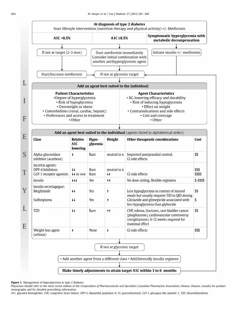
Figure 1. Management of hyperglycemia in type 2 diabetes.Physicians should refer to the most recent edition of the Compendium of Pharmaceuticals and Specialties (Canadian Pharmacists Association, Ottawa, Ontario, Canada) for productmonographs and for detailed prescribing information.A1C, glycated hemoglobin; CHF, congestive heart failure; DPP-4, dipeptidyl peptidase 4; GI, gastrointestinal; GLP-1, glucagon-like peptide 1; TZD, thiazolidinedione.
W. Harper et al. / Can J Diabetes 37 (2013) S61eS68S64

Figure 2. Antihyperglycemic medications and renal function. Based on productmonograph precautions. CKD, chronic kidney disease; GFR, glomerular filtration rate;TZD, thiazolidinedione. Designed by and used with the permission of Jean-FrançoisYale MD CSPQ FRCPC.
W. Harper et al. / Can J Diabetes 37 (2013) S61eS68 S65
class might be preferred in specific situations. Symptomaticpatients with high BG and A1C levels require agents that lower BGlevels substantially and quickly (e.g. insulin). However, the issue ofhow to reach glycemic targets may be less important than the needto achieve that target. Improved BG and A1C levels are associatedwith better outcomes, even if recommended glycemic targetscannot be reached (3). Each of the agents listed in Table 1 andFigure 1 has advantages and disadvantages to consider. Figure 2illustrates the basis on which agent selection is influenced byrenal function as dictated by product monograph precautions.
The recommendation to use metformin as the initial agent inmost patients is based on its effectiveness in lowering BG, its rela-tively mild side effect profile, its long-term safety track record, itsnegligible risk of hypoglycemia and its lack of causing weight gain.The demonstrated cardiovascular benefit in overweight patients isalso cited as a reason to select metformin as first-line treatment(26), but more recent evidence has been equivocal on this matter(27). While monotherapy with the thiazolidinedione (TZD) rosigli-tazone produces more long-lasting glycemic control compared tometformin or glyburide therapy (28), the edema, weight gain, riskof congestive heart failure (CHF), increased risk of fractures (29,30)and inconsistent data regarding myocardial infarction (MI) risk(31e33) significantly limit the clinical utility of this drug class.Although meta-analyses of smaller, underpowered studies sug-gested possible risk of MI with rosiglitazone (31,32), this has notbeen demonstrated in a larger randomized clinical trial (33,34).Conversely, the evidence for pioglitazone suggests a possiblereduced risk of cardiovascular events, although heart failure andincreased fractures are still concerning side effects (35,36).
Table 1 and Figure 1 provide information to aid decisionmaking.In deciding upon which agent to add after metformin, there mustbe consideration of multiple factors. First of all, the agent’s effec-tiveness at BG lowering must be considered in terms of both thedegree of baseline hyperglycemia needing correction and anyheightened concerns regarding hypoglycemia (e.g. elderly patientsor thosewith renal or hepatic dysfunction). The relative BG and A1Clowering of the various antihyperglycemic agent classes whenadded to metformin is shown in both Table 1 and Figure 1 and isbased on network meta-analysis allowing the comparison betweenclasses that have not yet had direct head-to-head comparison ina randomized clinical trial (37). Ideally, consideration would bemade towards the selection of agents with evidence demonstratingability to not only lower glucose levels, but also reduce the risk ofdiabetic microvascular and/or macrovascular complications.Unfortunately, the majority of evidence remains equivocal in thisregard as most clinical trials compared varying levels of glycemiclowering as opposed to direct comparison between agents used toachieve such glycemic control (38e40). More recent studies look-ing at the benefits seenwith select agents are of such short durationthat their results are still preliminary with respect to provingclinical event reduction (41e44) and confirmation awaits theresults of more definitive long-term studies.
Multiple other agent-specific advantages and disadvantagesshould be weighed as treatment is individualized to best suit thepatient’s needs and preferences. In particular, attention should bepaid to the agent’s effects on body weight as this is a clinicallyrelevant issue for many people with type 2 diabetes, and someagents cause significant weight gain while others can help topromote significant weight loss. GLP-1 receptor agonists areparticularly effective at promoting concomitant glycemic controland weight reduction (45e48), but long-term efficacy and safetydata are currently lacking for this class.
A combination of oral antihyperglycemic agents and insulinoften effectively controls glucose levels. When insulin is added tooral antihyperglycemic agent(s), a single injection ofintermediate-acting (NPH) (49) or a long-acting insulin analogue
(insulin glargine or insulin detemir) (50) may be added. Thisapproach may result in better glycemic control with a smallerdose of insulin (51), and may induce less weight gain and lesshypoglycemia than that seen when oral agents are stopped andinsulin is used alone (52). The addition of bedtime insulin tometformin therapy leads to less weight gain than insulin plusa sulfonylurea or twice-daily NPH insulin (53). While combininginsulin with a TZD is not an approved indication in Canada, theaddition of such agents to insulin in carefully selected patientsimproves glycemic control and reduces insulin requirements(54). Such combinations can result in increased weight, fluidretention and, in few patients, CHF. DPP-4 inhibitors and GLP-1receptor agonists have been shown to be effective at furtherlowering glucose levels when combined with insulin therapy(55e58).
Insulin can be used at diagnosis in individuals with markedhyperglycemia and can also be used temporarily during illness,pregnancy, stress or for a medical procedure or surgery. There isno evidence that exogenous insulin accelerates the risk of mac-rovascular complications of diabetes, and its appropriate useshould be encouraged (59,60). The Outcome Reduction with InitialGlargine Intervention (ORIGIN) trial studied the use of basalinsulin titrated to a fasting glucose of �5.3 mmol/L in people athigh cardiovascular risk with prediabetes or early type 2 diabetesover 6 years. There was a neutral effect on cardiovascularoutcomes and cancer, a reduction in new-onset diabetes anda slight increase in hypoglycemia and weight. Indeed, use ofinsulin earlier in the course of type 2 diabetes can be an effectivestrategy over oral antihyperglycemic agents (60,61). When insulinis used in type 2 diabetes, the insulin regimen should be tailoredto achieve good metabolic control while trying to avoid excessivehypoglycemia. With intensive glycemic control, there is anincreased risk of hypoglycemia, but this risk is lower in peoplewith type 2 diabetes than in those with type 1 diabetes. Thenumber of insulin injections (1 to 4 per day) and the timing ofinjections may vary, depending on each individual’s situation (62).The reduction in A1C achieved with insulin therapy depends onthe dose and number of injections per day (63). Insulin regimensbased on basal or bolus insulin appear to be equally effective(21,64) and superior with respect to glycemic lowering comparedto biphasic insulin-based regimens (63).
As type 2 diabetes progresses, insulin requirements will likelyincrease, additional doses of basal insulin (intermediate-acting or
W. Harper et al. / Can J Diabetes 37 (2013) S61eS68S66
long-acting analogues) may need to be added and bolus insulin(short-acting or rapid-acting analogues) may also be required.Generally, once bolus insulin is introduced into a treatmentregimen, either as a separate meal time bolus or as part of a pre-mixed containing regimen, insulin secretagogues, such as sulfo-nylureas and meglitinides, are usually discontinued. Concomitantmetformin therapy, unless contraindicated, should be continuedwith regimens containing bolus insulin, including intensive basal-bolus regimen, to allow for improved glycemic control with lessrisk of weight gain and hypoglycemia (65).
Although not commonly practiced, the use of intensive insulintherapy (basal-bolus regimen or continuous subcutaneous insulininfusion pump), for a transient period of approximately 2 to 3weeks at the time of diagnosis or early in the disease course, hasbeen shown to induce diabetes remission, subsequently allowingadequate glycemic control with lifestyle management alone (66).This normoglycemic state is often transient, however, and suchinterventions have been tested only in patients early in the course
RECOMMENDATIONS
1. In people with type 2 diabetes, if glycemic targets are not achieved usinglifestyle management within 2 to 3 months, antihyperglycemic agenttherapy should be initiated [Grade A, Level 1A (3)]. Metforminmay be usedat the time of diagnosis, in conjunction with lifestyle management (GradeD, Consensus).
i. If A1C �8.5%, antihyperglycemic agents should be initiatedconcomitantly with lifestyle management, and considerationshould be given to initiating combination therapy with 2 agents,one of which may be insulin (Grade D, Consensus).
ii. Individuals with symptomatic hyperglycemia and metabolicdecompensation should receive an initial antihyperglycemicregimen containing insulin [Grade D, Consensus].
2. Metformin should be the initial drug used [Grade A, Level 1A (26,80) foroverweight patients; Grade D, Consensus for nonoverweight patients].
3. Other classes of antihyperglycemic agents, including insulin, should beadded to metformin, or used in combination with each other, if glycemictargets are not met, taking into account the information in Figure 1 andTable 1 [Grade D, Consensus], and these adjustments to and/or additions ofantihyperglycemic agents should be made in order to attain target A1Cwithin 3 to 6 months [Grade D, Consensus].
4. Choice of pharmacological treatment agents should be individualized,taking into consideration [Grade D, Consensus]:� Patient characteristics:
B Degree of hyperglycemiaB Presence of comorbiditiesB Patient preference and ability to access treatments
� Properties of the treatment:B Effectiveness and durability of lowering BGB Risk of hypoglycemiaB Effectiveness in reducing diabetes complicationsB Effect on body weightB Side effectsB Contraindications
5. When basal insulin is added to antihyperglycemic agents, long-actinganalogues (detemir or glargine) may be used instead of intermediate-acting NPH to reduce the risk of nocturnal and symptomatic hypogly-cemia [Grade A, Level 1A (19,78,79)].
6. When bolus insulin is added to antihyperglycemic agents, rapid-actinganalogues may be used instead of regular insulin to improve glycemiccontrol [Grade B, Level 2 (20)] and to reduce the risk of hypoglycemia[Grade D, Consensus)].
7. All individuals with type 2 diabetes currently using or starting therapywith insulin or insulin secretagogues should be counseled about theprevention, recognition and treatment of drug-induced hypoglycemia[Grade D, Consensus].
of disease where the degree of residual beta cell function is rela-tively preserved (67).
Epidemiological evidence suggesting a possible link betweeninsulin glargine and cancer has not been substantiated in review ofclinical trial data for either glargine or detemir (60,68,69).
Hypoglycemia
Medication-induced hypoglycemia is themost common cause ofhypoglycemia. It is estimated that hypoglycemia of any severityoccurs annually in up to approximately 20% of patients takinginsulin secretagogues (70). Although these hypoglycemic episodesare rarely fatal, they can be associated with serious clinicalsequelae. Therefore, it is important to prevent, recognize and treathypoglycemic episodes secondary to the use of insulin secreta-gogues. Few large, randomized clinical trials have compared therates of hypoglycemia between these agents.
In the United Kingdom Prospective Diabetes Study (UKPDS),the proportion of adults with type 2 diabetes who experienceda severe hypoglycemic episode per year was significantly higherin the intensive group than in the conventional group, particu-larly for patients using insulin therapy (3). Although the risk ofhypoglycemia was less than that seen in the patients with type 1diabetes in the Diabetes Control and Complications Trial (DCCT),each year approximately 3% of patients treated with insulin inthe UKPDS experienced a severe hypoglycemic episode, and 40%had a hypoglycemic episode of any severity (3). Protocolsdesigned to achieve normoglycemia targets (A1C �6.5%) furtherincrease the risk of severe hypoglycemia without providing anysubstantial reduction in the incidence of diabetes complications(71,72).
Lower rates of hypoglycemia have been observed in somestudies of patients with type 2 diabetes treated with rapid-acting insulin analogues (insulin aspart, insulin lispro, insulinglulisine) compared to those treated with short-acting (regular)insulin (19,73,74). Use of long-acting basal insulin analogues(insulin detemir, insulin glargine) reduces the risk of nocturnalhypoglycemia compared to treatment with NPH insulin(19,50,75e79).
Other Relevant Guidelines
Targets for Glycemic Control, p. S31Pharmacotherapy in Type 1 Diabetes, p. S56Hypoglycemia, p. S69Weight Management in Diabetes, p. S82Type 2 Diabetes in Children and Adolescents, p. S163Diabetes and Pregnancy, p. S168Diabetes in the Elderly, p. S184
Relevant Appendix
Appendix 3: Examples of Insulin Initiation and Titration Regi-mens in People With Type 2 Diabetes
References
1. Turner RC, Cull CA, Frighi V, et al. Glycemic control with diet, sulfonylurea,metformin, or insulin in patients with type 2 diabetes mellitus: progressiverequirement for multiple therapies (UKPDS 49). JAMA 1999;281:2005e12.
2. Klein R, Klein BE, Moss SE, et al. The Wisconsin epidemiological study ofdiabetic retinopathy. III. Prevalence and risk of diabetic retinopathy when ageat diagnosis is 30 or more years. Arch Ophthalmol 1984;102:527e32.
3. UK Prospective Diabetes Study (UKPDS) Group. Intensive blood-glucosecontrol with sulphonylureas or insulin compared with conventional treat-ment and risk of complications in patients with type 2 diabetes (UKPDS 33).Lancet 1998;352:837e53.

W. Harper et al. / Can J Diabetes 37 (2013) S61eS68 S67
4. Bloomgarden ZT, Dodis R, Viscoli CM, et al. Lower baseline glycemia reducesapparent oral agent glucose-lowering efficacy: a meta-regression analysis.Diabetes Care 2006;29:2137e9.
5. Sherifali D, Nerenberg K, Pullenayegum E, et al. The effect of oral antidiabeticagents on A1C levels: a systematic review and meta-analysis. Diabetes Care2010;33:1859e64.
6. Monnier L, Lapinski H, Colette C. Contributions of fasting and postprandialplasma glucose increments to the overall diurnal hyperglycemia of type 2diabetic patients: variations with increasing levels of HbA(1c). Diabetes Care2003;26:881e5.
7. Chiasson J-L, Josse RG, Hunt JA, et al. The efficacy of acarbose in the treatmentof patients with non-insulin-dependent diabetes mellitus. A multicentercontrolled clinical trial. Ann Intern Med 1994;121:928e35.
8. Aschner P, Kipnes MS, Lunceford JK, et al. Effect of the dipeptidyl peptidase-4inhibitor sitagliptin as monotherapy on glycemic control in patients with type2 diabetes. Diabetes Care 2006;29:2632e7.
9. Charbonnel B, Karasik A, Liu J, et al. Efficacy and safety of the dipeptidylpeptidase-4 inhibitor sitagliptin added to ongoing metformin therapy inpatients with type 2 diabetes inadequately controlled with metformin alone.Diabetes Care 2006;29:2638e43.
10. Weiss SR, Cheng SL, Kourides IA, et al, Inhaled Insulin Phase II Study Group.Inhaled insulin provides improved glycemic control in patients with type 2diabetes mellitus inadequately controlled with oral agents: a randomizedcontrolled trial. Arch Intern Med 2003;27:2277e82.
11. Dailey G, Rosenstock J, Moses RG, et al. Insulin glulisine provides improvedglycemic control in patients with type 2 diabetes. Diabetes Care 2004;27:2363e8.
12. Roach P, Yue L, Arora V. Improved postprandial glycemic control duringtreatment with Humalog Mix25, a novel protamine-based insulin lisproformulation. Humalog Mix25 Study Group. Diabetes Care 1999;22:1258e61.
13. Boehm BO, Home PD, Behrend C, et al. Premixed insulin aspart 30 vs. pre-mixed human insulin 30/70 twice daily: a randomized trial in type 1 and type2 diabetic patients. Diabet Med 2002;19:393e9.
14. Ahren B, Gomis R, Standl E, et al. Twelve- and 52-week efficacy of thedipeptidyl peptidase IV inhibitor LAF237 in metformin-treated patients withtype 2 diabetes. Diabetes Care 2004;27:2874e80.
15. Derosa G, Mugellini A, Ciccarelli L, et al. Comparison between repaglinide andglimepiride in patients with type 2 diabetes mellitus: a one-year, randomized,double-blind assessment of metabolic parameters and cardiovascular riskfactors. Clin Ther 2003;25:472e84.
16. Ristic S, Collober-Maugeais C, Pecher E, et al. Comparison of nateglinide andgliclazide in combination with metformin, for treatment of patients with Type2 diabetes mellitus inadequately controlled on maximum doses of metforminalone. Diabet Med 2006;23:757e62.
17. Ross SA, Zinman B, Campos RV, et al. A comparative study of insulin lispro andhuman regular insulin in patients with type 2 diabetes mellitus and secondaryfailure of oral hypoglycemic agents. Clin Invest Med 2001;24:292e8.
18. Rosenfalck AM, Thorsby P, Kjems L, et al. Improved postprandial glycaemiccontrol with insulin aspart in type 2 diabetic patients treated with insulin.Acta Diabetol 2000;37:41e6.
19. Sumeet R, Singh SR, Ahmad F, et al. Efficacy and safety of insulin analogues forthe management of diabetes mellitus: a meta-analysis. CMAJ 2009;180:385e97.
20. Mannucci E, Monami M, Marchionni N. Short-acting insulin analogues vs.regular human insulin in type 2 diabetes: a meta-analysis. Diabetes ObesMetab 2009;11:53e9.
21. Raz I, Wilson PW, Strojek K, et al. Effects of prandial versus fasting glycemiaon cardiovascular outcomes in type 2 diabetes: the HEART2D trial. DiabetesCare 2009;32:381e6.
22. Garber AJ, Larsen J, Schneider SH, et al. Simultaneous glyburide/metformintherapy is superior to component monotherapy as an initial pharmacologicaltreatment for type 2 diabetes. Diabetes Obes Metab 2002;4:201e8.
23. Rosenstock J, Goldstein BJ, Vinik AI, et al. Effect of early addition of rosigli-tazone to sulphonylurea therapy in older type 2 diabetes patients (>60years): the Rosiglitazone Early vs. Sulphonylurea Titration (RESULT) study.Diabetes Obes Metab 2006;8:49e57.
24. Rosenstock J, Rood J, Cobitz A, et al. Improvement in glycaemic control withrosiglitazone/metformin fixed-dose combination therapy in patients withtype 2 diabetes with very poor glycaemic control. Diabetes Obes Metab 2006;8:643e9.
25. Rosenstock J, Rood J, Cobitz A, et al. Initial treatment with rosiglitazone/metformin fixed-dose combination therapy compared with monotherapywith either rosiglitazone or metformin in patients with uncontrolled type 2diabetes. Diabetes Obes Metab 2006;8:650e60.
26. UK Prospective Diabetes Study (UKPDS) Group. Effect of intensive blood-glucose control with metformin on complications in overweight patientswith type 2 diabetes (UKPDS 34). Lancet 1998;352:854e65.
27. Boussageon R, Supper I, Bejan-Angoulvant T, et al. Reappraisal of metforminefficacy in the treatment of type 2 diabetes: a meta-analysis of randomisedcontrolled trials. PLoS Med 2012;9:e1001204.
28. Kahn SE, Haffner SM, Heise MA, et al. Glycemic durability of rosiglitazone,metformin, or glyburide monotherapy. N Engl J Med 2006;355:2427e43.
29. Meymeh RH, Wooltorton E. Diabetes drug pioglitazone (Actos): risk of frac-ture. CMAJ 2007;177:723e4.
30. Kahn SE, Zinman B, Lachin JM, et al, A Diabetes Outcome Progression Trial(ADOPT) Study Group. Rosiglitazone-associated fractures in type 2 diabetes:
an analysis from A Diabetes Outcome Progression Trial. Diabetes Care 2008;31:845e51.
31. Nissen SE, Wolski K. Effect of rosiglitazone on the risk of myocardial infarctionand death from cardiovascular causes. N Engl J Med 2007;356:2457e71.
32. Nissen SE, Wolski K. Rosiglitazone revisited: an updated meta-analysis of riskfor myocardial infarction and cardiovascular mortality. Arch Intern Med 2010;170:1191e201.
33. Home PD, Pocock SJ, Beck-Nielsen H, et al. Rosiglitazone evaluated forcardiovascular outcomes in oral agent combination therapy for type 2 dia-betes (RECORD): a multicentre, randomised, open-label trial. Lancet 2009;373:2125e35.
34. Home PD, Pocock SJ, Beck-Nielsen H, et al. Rosiglitazone evaluated forcardiovascular outcomes: an interim analysis. N Engl J Med 2007;357:28e38.
35. Dormandy JA, Charbonnel B, Eckland DJ, et al. Secondary prevention ofmacrovascular events in patients with type 2 diabetes in the PROactive Study(PROspective pioglitAzone Clinical Trial In macroVascular Events): a rando-mised controlled trial. Lancet 2005;366:1279e89.
36. Lincoff AM, Wolski K, Nicholls SJ, Nissen SE. Pioglitazone and risk of cardio-vascular events in patients with type 2 diabetes mellitus: a meta-analysis ofrandomized trials. JAMA 2007;298:1180e8.
37. Liu SC, Tu YK, Chien KL. Effect of antidiabetic agents added to metformin onglycemic control, hypoglycemia, and weight change in patients with type 2diabetes: a network meta-analysis. Diabetes Obes Metab 2012;14:810e20.
38. Ray KK, Seshasai SR, Wijesuriya S, et al. Effect of intensive control of glucoseon cardiovascular outcomes and death in patients with diabetes mellitus:a meta-analysis of randomised controlled trials. Lancet 2009;373:1765.
39. Kelly TN, Bazzano LA, Fonseca VA, et al. Systematic review: glucose controland cardiovascular disease in type 2 diabetes. Ann Intern Med 2009;151:394.
40. Boussageon R, Bejan-Angoulvant T, Saadatian-Elahi M, et al. Effect of intensiveglucose lowering treatment on all cause mortality, cardiovascular death, andmicrovascular events in type 2 diabetes: meta-analysis of randomisedcontrolled trials. BMJ 2011;343:d4169.
41. Chiasson JL, Josse RG, Gomis R, et al, STOP-NIDDM Trial Research Group.Acarbose treatment and the risk of cardiovascular disease and hypertensionin patients with impaired glucose tolerance: the STOP-NIDDM trial. JAMA2003;290:486e94.
42. Gaziano JM, Cincotta AH, O’Connor CM, et al. Randomized clinical trial ofquick-release bromocriptine among patients with type 2 diabetes on overallsafety and cardiovascular outcomes. Diabetes Care 2010;33:1503e8.
43. Frederich R, Alexander JH, Fiedorek FT, et al. A systematic assessment ofcardiovascular outcomes in the saxagliptin drug development program fortype 2 diabetes. Postgrad Med 2010;122:16e27.
44. Monami M, Cremasco F, Caterina L, et al. Glucagon-like peptide-1 receptoragonists and cardiovascular events: a meta-analysis of randomized clinicaltrials. Exp Diabetes Res 2011;2011:215764.
45. Amori RE, Lau J, Pittas AG. Efficacy and safety of incretin therapy in type 2diabetes: systematic review and meta-analysis. JAMA 2007;298:194e206.
46. Fakhoury WK, Lereun C, Wright D. A meta-analysis of placebo-controlledclinical trials assessing the efficacy and safety of incretin-based medicationsin patients with type 2 diabetes. Pharmacology 2010;86:44e57.
47. Monami M, Marchionni N, Mannucci E. Glucagon-like peptide-1 receptoragonists in type 2 diabetes: a meta-analysis of randomized clinical trials. Eur JEndocrinol 2009;160:909e17.
48. Pinelli NR, Cha R, Brown MB, Jaber LA. Addition of thiazolidinedione or exe-natide to oral agents in type 2 diabetes: a meta-analysis. Ann Pharmacother2008;42:1541e51.
49. Yki-Järvinen H, Kauppila M, Kujansuu E, et al. Comparison of insulin regimensin patients with non-insulin-dependent diabetes mellitus. N Engl J Med 1992;327:1426e33.
50. Rosenstock J, Schwartz SL, Clark Jr CM, et al. Basal insulin therapy in type 2diabetes: 28-week comparison of insulin glargine (HOE 901) and NPH insulin.Diabetes Care 2001;24:631e6.
51. Johnson JL, Wolf SL, Kabadi UM. Efficacy of insulin and sulfonylurea combi-nation therapy in type II diabetes. A metaanalysis of the randomized placebo-controlled trials. Arch Intern Med 1996;156:259e64.
52. United Kingdom Prospective Diabetes Study Group. United KingdomProspective Diabetes Study 24: a 6-year, randomized, controlled trialcomparing sulfonylurea, insulin, and metformin therapy in patients withnewly diagnosed type 2 diabetes that could not be controlled with diettherapy. Ann Intern Med 1998;128:165e75.
53. Yki-Järvinen H, Ryysy L, Nikkilä K, et al. Comparison of bedtime insulinregimens in patients with type 2 diabetes mellitus. A randomized, controlledtrial. Ann Intern Med 1999;130:389e96.
54. Tan A, Cao Y, Xia N, et al. The addition of pioglitazone in type 2 diabeticspoorly controlled on insulin therapy: a meta-analysis. Eur J Intern Med 2010;21:398e403.
55. Buse JB, Bergenstal RM, Glass LC, et al. Use of twice-daily exenatide in basalinsulin-treated patients with type 2 diabetes: a randomized, controlled trial.Ann Intern Med 2011;154:103e12.
56. Arnolds S, Dellweg S, Clair J, et al. Further improvement in postprandialglucose control with addition of exenatide or sitagliptin to combinationtherapy with insulin glargine and metformin: a proof-of-concept study. Dia-betes Care 2010;33:1509e15.
57. Barnett AH, Charbonnel B, Donovan M, et al. Effect of saxagliptin as add-on therapy in patients with poorly controlled type 2 diabetes on insulin
W. Harper et al. / Can J Diabetes 37 (2013) S61eS68S68
alone or insulin combined with metformin. Curr Med Res Opin 2012;28:513e23.
58. Vilsboll T, Rosenstock J, Yki-Jarvinen H, et al. Efficacy and safety of sitagliptinwhen added to insulin therapy in patients with type 2 diabetes. Diabetes ObesMetab 2010;12:167e77.
59. American Diabetes Association. Implications of the United KingdomProspective Diabetes Study. Diabetes Care 1998;21:2180e4.
60. ORIGIN Trial InvestigatorsGerstein HC, Bosch J, Dagenais GR, et al. Basalinsulin and cardiovascular and other outcomes in dysglycemia. N Engl J Med2012;367:319e28.
61. Gerstein HC, Yale JF, Harris SB, et al. A randomized trial of adding insulinglargine vs. avoidance of insulin in people with Type 2 diabetes on either nooral glucose-lowering agents or submaximal doses of metformin and/or sul-phonylureas. The Canadian INSIGHT (Implementing New Strategies withInsulin Glargine for Hyperglycaemia Treatment) Study. Diabet Med 2006;23:736e42.
62. Abraira C, Colwell JA, Nuttall FQ, et al. Veterans Affairs Cooperative Study onglycemic control and complications in type II diabetes (VA CSDM). Results ofthe feasibility trial. Diabetes Care 1995;18:1113e23.
63. Holman RR, Farmer AJ, Davies MJ, et al. Three-year efficacy of complex insulinregimens in type 2 diabetes. N Engl J Med 2009;361:1736e47.
64. Bretzel RG, Nuber U, Landgraf W, et al. Once-daily basal insulin glargineversus thrice-daily prandial insulin lispro in people with type 2 diabetes onoral hypoglycaemic agents (APOLLO): an open randomised controlled trial.Lancet 2008;371:1073e84.
65. Wulffelé MG, Kooy A, Lehert P, et al. Combination of insulin and metformin inthe treatment of type 2 diabetes. Diabetes Care 2002;25:2133e40.
66. Weng J, Li Y, Xu W, et al. Effect of intensive insulin therapy on beta-cellfunction and glycaemic control in patients with newly diagnosed type 2diabetes: a multicentre randomised parallel-group trial. Lancet 2008;371:1753e60.
67. Ryan EA, Imes S, Wallace C. Short-term intensive insulin therapy in newlydiagnosed type 2 diabetes. Diabetes Care 2004;27:1028e32.
68. Home PD, Lagarenne P. Combined randomised controlled trial experience ofmalignancies in studies using insulin glargine. Diabetologia 2009;52:2499e506.
69. Dejgaard A, Lynggaard H, Rastam J, Krogsgaard TM. No evidence of increasedrisk of malignancies in patients with diabetes treated with insulin detemir:a meta-analysis. Diabetologia 2009;52:2507e12.
70. Jennings AM, Wilson RM, Ward JD. Symptomatic hypoglycemia in NIDDMpatients treated with oral hypoglycemic agents. Diabetes Care 1989;12:203e8.
71. The Action to Control Cardiovascular Risk in Diabetes Study Group. Effects ofintensive glucose lowering in type 2 diabetes. N Engl J Med 2008;358:2545e59.
72. ADVANCE Collaborative GroupPatel A, MacMahon S, Chalmers J, et al. Inten-sive blood glucose control and vascular outcomes in patients with type 2diabetes. N Engl J Med 2008;358:2560e72.
73. Anderson Jr JH, Brunelle RL, Keohane P, et al. Mealtime treatment with insulinanalog improves postprandial hyperglycemia and hypoglycemia in patientswith non-insulin-dependent diabetes mellitus. Arch Intern Med 1997;157:1249e55.
74. Anderson Jr JH, Brunelle RL, Koivisto VA, et al. Improved mealtime treatmentof diabetes mellitus using an insulin analogue. Clin Ther 1997;19:62e72.
75. Yki-Järvinen H, Dressler A, Ziemen M. Less nocturnal hypoglycemia and betterpost-dinner glucose control with bedtime insulin glargine compared withbedtime NPH insulin during insulin combination therapy in type 2 diabetes.Diabetes Care 2000;23:1130e6.
76. Fritsche A, Schweitzer MA, Haring HU, et al. Glimepiride combined withmorning insulin glargine, bedtime neutral protamine hagedorn insulin, orbedtime insulin glargine in patients with type 2 diabetes. A randomized,controlled trial. Ann Intern Med 2003;138:952e9.
77. Janka HU, Plewe G, Riddle MC, et al. Comparison of basal insulin added to oralagents versus twice-daily premixed insulin as initial insulin therapy for type 2diabetes. Diabetes Care 2005;28:254e9.
78. Horvath K, Jeitler K, Berghold A, et al. A long-acting insulin analogues versusNPH insulin (human isophane insulin) for type 2 diabetes mellitus (review).Cochrane Database Syst Rev 2007;2:CD005613.
79. Monami M, Marchionni N, Mannucci E. Long-acting insulin analogues versusNPH human insulin in type 2 diabetes: a meta-analysis. Diabetes Res ClinPract 2008;81:184e9.
80. Holman RR, Paul SK, Bethel MA, et al. 10-year follow-up of intensive glucosecontrol in type 2 diabetes. N Engl J Med 2008;359:1577e89.
81. Hoffmann J, Spengler M. Efficacy of 24-week monotherapy with acarbose,glibenclamide, or placebo in NIDDM patients. The Essen Study. Diabetes Care1994;17:561e6.
82. Holman RR, Cull CA, Turner RC. A randomized double-blind trial of acarbose intype 2 diabetes shows improved glycemic control over 3 years (U.K.Prospective Diabetes Study 44). Diabetes Care 1999;22:960e4.
83. Wright A, Burden AC, Paisey RB, et al. Sulfonylurea inadequacy: efficacy ofaddition of insulin over 6 years in patients with type 2 diabetes in the U.K.Prospective Diabetes Study (UKPDS 57). Diabetes Care 2002;25:330e6.
84. Ohkubo Y, Kishikawa H, Araki E, et al. Intensive insulin therapy prevents theprogression of diabetic microvascular complications in Japanese patients with
non-insulin-dependent diabetes mellitus: a randomized prospective 6-yearstudy. Diabetes Res Clin Pract 1995;28:103e17.
85. Haak T, Tiengo A, Draeger E, et al. Lower within-subject variability offasting blood glucose and reduced weight gain with insulin detemir comparedto NPH insulin in patients with type 2 diabetes. Diabetes Obes Metab 2005;7:56e64.
86. Harrower A. Gliclazide modified release: from once-daily administration to 24hour blood glucose control. Metabolism 2000;49(10 suppl 2):7e11.
87. Tessier D, Dawson K, Tétrault JP, et al. Glibenclamide vs gliclazide in type 2diabetes of the elderly. Diabet Med 1994;11:974e80.
88. Schade DS, Jovanovic L, Schneider J. A placebo-controlled, randomized studyof glimepiride in patients with type 2 diabetes mellitus for whom diet therapyis unsuccessful. J Clin Pharmacol 1998;38:636e41.
89. Dills DG, Schneider J. Clinical evaluation of glimepiride versus glyburidein NIDDM in a double-blind comparative study. Horm Metab Res 1996;28:426e9.
90. Holstein A, Plaschke A, Egberts EH. Lower incidence of severe hypoglycaemiain patients with type 2 diabetes treated with glimepiride versus glibencla-mide. Diabetes Metab Res Rev 2001;17:467e73.
91. Black C, Donnelly P, McIntyre L, et al. Meglitinide analogues for type 2 dia-betes mellitus. Cochrane Database Syst Rev 2007;2:CD004654.
92. Wolffenbuttel BHR, Landgraf R. A 1-year multicenter randomized double-blind comparison of repaglinide and glyburide for the treatment of type 2diabetes. Diabetes Care 1999;22:463e7.
93. Damsbo P, Clauson P, Marbury TC, et al. A double-blind randomizedcomparison of meal-related glycemic control by repaglinide and glyburide inwell-controlled type 2 diabetic patients. Diabetes Care 1999;22:789e94.
94. Schernthaner G, Grimaldi A, Di Mario U, et al. GUIDE study: double-blindcomparison of once-daily gliclazide MR and glimepiride in type 2 diabeticpatients. Eur J Clin Invest 2004;34:535e42.
95. Garber AJ, Duncan TG, Goodman AM, et al. Efficacy of metformin in type IIdiabetes: results of a double-blind, placebo-controlled, dose-response trial.Am J Med 1997;103:491e7.
96. de Jager J, Kooy A, Lehert P, et al. Long term treatment with metformin inpatients with type 2 diabetes and risk of vitamin B-12 deficiency: randomisedplacebo controlled trial. BMJ 2010;340:c2198.
97. Aronoff S, Rosenblatt S, Braithwaite S, et al. Pioglitazone hydrochloridemonotherapy improves glycemic control in the treatment of patients withtype 2 diabetes: a 6-month randomized placebo-controlled dose-responsestudy. Diabetes Care 2000;23:1605e11.
98. Raskin P, Rappaport EB, Cole ST, et al. Rosiglitazone short-term monotherapylowers fasting and post-prandial glucose in patients with type II diabetes.Diabetologia 2000;43:278e84.
99. Nolan JJ, Jones NP, Patwardhan R, et al. Rosiglitazone taken once dailyprovides effective glycaemic control in patients with type 2 diabetes mellitus.Diabet Med 2000;17:287e94.
100. Lebovitz HE, Dole JF, Patwardhan R, et al. Rosiglitazone monotherapy iseffective in patients with type 2 diabetes. J Clin Endocrinol Metab 2001;86:280e8.
101. Fonseca V, Rosenstock J, Patwardhan R, et al. Effect of metformin and rosi-glitazone combination therapy in patients with type 2 diabetes mellitus:a randomized controlled trial. JAMA 2000;283:1695e702.
102. Kipnes MS, Krosnick A, Rendell MS, et al. Pioglitazone hydrochloride incombination with sulfonylurea therapy improves glycemic control in patientswith type 2 diabetes mellitus: a randomized, placebo-controlled study. Am JMed 2001;111:10e7.
103. Einhorn D, Rendell M, Rosenzweig J, et al. Pioglitazone hydrochloride incombination with metformin in the treatment of type 2 diabetes mellitus:a randomized, placebo-controlled study. Clin Ther 2000;22:1395e409.
104. Yale J-F, Valiquett TR, Ghazzi MN, et al. The effect of a thiazolidinedionedrug, troglitazone, on glycemia in patients with type 2 diabetes mellituspoorly controlled with sulfonylurea and metformin. A multicenter,randomized, double-blind, placebo-controlled trial. Ann Intern Med 2001;134:737e45.
105. Miles JM, Leiter L, Hollander P, et al. Effect of orlistat in overweight and obesepatients with type 2 diabetes treated with metformin. Diabetes Care 2002;25:1123e8.
106. Kelley DE, Bray GA, Pi-Sunyer FX, et al. Clinical efficacy of orlistat therapy inoverweight and obese patients with insulin-treated type 2 diabetes: a 1-yearrandomized controlled trial. Diabetes Care 2002;25:1033e41.
107. Hollander PA, Elbein SC, Hirsch IB, et al. Role of orlistat in the treatment ofobese patients with type 2 diabetes. A 1-year randomized double-blind study.Diabetes Care 1998;21:1288e94.
108. Singh S, Loke YK, Furberg CD. Long-term risk of cardiovascular events withrosiglitazone: a meta-analysis. JAMA 2007;298:1189e95.
109. Lewis JD, Ferrara A, Peng T, et al. Risk of bladder cancer among diabeticpatients treated with pioglitazone: interim report of a longitudinal cohortstudy. Diabetes Care 2011;34:916e22.
110. Jacob S, Rabbia M, Meier MK, Hauptman J. Orlistat 120 mg improves gly-caemic control in type 2 diabetic patients with or without concurrent weightloss. Diabetes Obes Metab 2009;11:361e71.
111. Berne C. A randomized study of orlistat in combination with a weightmanagement programme in obese patients with Type 2 diabetes treated withmetformin. Diabet Med 2004;22:612e8.
Contents lists available at SciVerse ScienceDirect
Can J Diabetes 37 (2013) S69eS71
Canadian Journal of Diabetesjournal homepage:
www.canadianjournalofdiabetes.com
Clinical Practice Guidelines
Hypoglycemia
Canadian Diabetes Association Clinical Practice Guidelines Expert Committee
The initial draft of this chapter was prepared by Dale Clayton MHSc, MD, FRCPC,Vincent Woo MD, FRCPC, Jean-François Yale MD, CSPQ, FRCPC
Table 1Symptoms of hypoglycemia
Neurogenic (autonomic) Neuroglycopenic
TremblingPalpitationsSweatingAnxietyHungerNauseaTingling
Difficulty concentratingConfusionWeaknessDrowsinessVision changesDifficulty speakingHeadacheDizziness
KEY MESSAGES
� It is important to prevent, recognize and treat hypoglycemic episodessecondary to the use of insulin or insulin secretagogues.
� The goals of treatment for hypoglycemia are to detect and treat a low bloodglucose (BG) level promptly by using an intervention that provides thefastest rise in BG to a safe level, to eliminate the risk of injury and to relievesymptoms quickly.
� It is important to avoid overtreatment, since this can result in reboundhyperglycemia and weight gain.
Introduction
Drug-induced hypoglycemia is a major obstacle for individualstrying to achieve glycemic targets. Hypoglycemia can be severe andresult in confusion, coma or seizure, requiring the assistance ofother individuals. Significant risk of hypoglycemia often necessi-tates less stringent glycemic goals. Frequency and severity ofhypoglycemia negatively impact on quality of life (1) and promotefear of future hypoglycemia (2,3). This fear is associated withreduced self-care and poor glucose control (4e6). As such, it isimportant to prevent, recognize and treat hypoglycemic episodessecondary to the use of insulin or insulin secretagogues (seePharmacotherapy in Type 1 Diabetes, p. S56, and PharmacologicManagement of Type 2 Diabetes, p. S61, for further discussion ofdrug-induced hypoglycemia).
Definition of Hypoglycemia
Hypoglycemia is defined by 1) the development of autonomic orneuroglycopenic symptoms (Table 1); 2) a low plasma glucose level(<4.0 mmol/L for patients treated with insulin or an insulinsecretagogue); and 3) symptoms responding to the administrationof carbohydrate (7). The severity of hypoglycemia is defined byclinical manifestations (Table 2).
Complications of Severe Hypoglycemia
Short-term risks of hypoglycemia include the dangerous situ-ations that can arise while an individual is hypoglycemic,whether at home or at work (e.g. driving, operating machinery).In addition, prolonged coma is sometimes associated with
1499-2671/$ e see front matter � 2013 Canadian Diabetes Associationhttp://dx.doi.org/10.1016/j.jcjd.2013.01.022
transient neurological symptoms, such as paresis, convulsions andencephalopathy. The potential long-term complications of severehypoglycemia are mild intellectual impairment and permanentneurologic sequelae, such as hemiparesis and pontine dysfunc-tion. The latter are rare and have been reported only in casestudies.
Recurrent hypoglycemia may impair the individual’s ability tosense subsequent hypoglycemia (8,9). The neurohormonal coun-terregulatory responses to hypoglycemia may become blunted;however, this is potentially reversible (see Pharmacotherapy inType 1 Diabetes, p. S56).
Retrospective studies have suggested a link between frequentsevere hypoglycemia (�5 episodes since diagnosis) and a decreasein intellectual performance. These changes were small but,depending on an individual’s occupation, could be clinicallymeaningful. Prospective studies in type 1 diabetes have not foundan association between intensive insulin therapy and cognitivefunction (10e12). A meta-analysis concluded that lowered cogni-tive performance in people with type 1 diabetes appeared to beassociated with the presence of microvascular complications butnot with the occurrence of severe hypoglycemic episodes or withpoor metabolic control (13). Unlike patients with type 1 diabetes,those with type 2 diabetes and previous severe hypoglycemiarequiring presentation to the hospital have increased risk ofsubsequent dementia (14).
In patients with type 2 diabetes and established, or very highrisk for, cardiovascular disease, symptomatic hypoglycemia (<2.8mmol/L) is associated with increased mortality (15). The mecha-nism for this increase is not certain; however, acute hypoglycemiais proinflammatory (16) and may affect cardiac conduction (depo-larization, QT prolongation). This effect, however, may be related tosympathetic tone rather than glucose per se (17,18).
Table 2Severity of hypoglycemia
Mild: Autonomic symptoms are present. The individual is able to self-treat.Moderate: Autonomic and neuroglycopenic symptoms are present.
The individual is able to self-treat.Severe: Individual requires assistance of another person.
Unconsciousness may occur. PG is typically <2.8 mmol/L.
PG, plasma glucose.
Table 4Examples of 15 g carbohydrate for treatment of mild to moderate hypoglycemia
� 15 g glucose in the form of glucose tablets� 15 mL (3 teaspoons) or 3 packets of table sugar dissolved in water� 175 mL (3/4 cup) of juice or regular soft drink� 6 LifeSavers (1 ¼ 2.5 g carbohydrate)� 15 mL (1 tablespoon) of honey
D. Clayton et al. / Can J Diabetes 37 (2013) S69eS71S70
The major risk factors for severe hypoglycemia in patientswith type 1 diabetes include prior episode of severehypoglycemia (19e21), current low glycated hemoglobin (A1C)(<6.0%) (20,22e24), hypoglycemia unawareness (25), long dura-tion of diabetes (23,26), autonomic neuropathy (27), adolescence(28) and preschool-age children unable to detect and/or treat mildhypoglycemia on their own. Risk factors for hypoglycemia inpatients with type 2 diabetes include advancing age (29), severecognitive impairment (30), poor health literacy (31), food insecurity(32), increased A1C (29,33), hypoglycemia unawareness (34),duration of insulin therapy, renal impairment and neuropathy (33).In patients with type 2 diabetes and established cardiovasculardisease (CVD) or age >54 years and 2 CVD risk factors, the risk ofhypoglycemia is also increased by female gender (29). Patients athigh risk for severe hypoglycemia should be informed of their riskand counselled, along with their significant others, on preventingand treating hypoglycemia (including use of glucagon), preventingdriving and industrial accidents through self-monitoring of bloodglucose (BG) and taking appropriate precautions prior to theactivity, and documenting BG readings taken during sleepinghours. Individuals may need to have their insulin regimen adjustedappropriately to lower their risk. Risk factors for severe hypogly-cemia are listed in Table 3.
RECOMMENDATIONS
1. Mild to moderate hypoglycemia should be treated by the oral ingestionof 15 g carbohydrate, preferably as glucose or sucrose tablets or solution.These are preferable to orange juice and glucose gels [Grade B, Level 2(35)]. Patients should retest BG in 15 minutes and re-treat with another15 g carbohydrate if the BG level remains <4.0 mmol/L [Grade D,Consensus]. Note: This does not apply to children. See Type 1 Diabetes inChildren and Adolescents, p. S153, and Type 2 Diabetes in Children andAdolescents, p. S163, for treatment options in children.
2. Severe hypoglycemia in a conscious person should be treated by oralingestion of 20 g carbohydrate, preferably as glucose tablets or equivalent.BG should be retested in 15 minutes and then re-treated with another 15 gglucose if the BG level remains <4.0 mmol/L [Grade D, Consensus].
3. Severe hypoglycemia in an unconscious individuala. With no IV access: 1 mg glucagon should be given subcutaneously or
intramuscularly. Caregivers or support persons should call for emer-gency services and the episode should be discussed with the diabeteshealthcare team as soon as possible [Grade D, Consensus].
b. With IV access: 10e25 g (20e50 cc of D50W) of glucose should begiven intravenously over 1e3 minutes [Grade D, Consensus].
Treatment of Hypoglycemia
The goals of treatment for hypoglycemia are to detect and treata low BG level promptly by using an intervention that provides thefastest rise in BG to a safe level, to eliminate the risk of injury and torelieve symptoms quickly. It is also important to avoid over-treatment since this can result in rebound hyperglycemia andweight gain.
Evidence suggests that 15 g glucose (monosaccharide) isrequired to produce an increase in BG of approximately 2.1 mmol/Lwithin 20 minutes, with adequate symptom relief for most people(Table 4) (35e39). This has not been well studied in patients withgastropathy. A 20 g oral glucose dose will produce a BG incrementof approximately 3.6 mmol/L at 45 minutes (36,37). Other choices,such as milk and orange juice, are slower to increase BG levelsand provide symptom relief (36,37). Glucose gel is quite slow(<1.0 mmol/L increase at 20 minutes) and must be swallowed tohave a significant effect (35,40). Patients taking an alpha-
Table 3Risk factors for severe hypoglycemia
� Prior episode of severe hypoglycemia� Current low A1C (<6.0%)� Hypoglycemia unawareness� Long duration of insulin therapy� Autonomic neuropathy� Low economic status� Food insecurity� Low health literacy� Cognitive impairment� Adolescence� Preschool-age children unable to detect and/or treat mild hypoglycemia ontheir own
A1C, glycated hemoglobin.
glucosidase inhibitor (acarbose) must use glucose (dextrose)tablets (41) or, if unavailable, milk or honey to treat hypoglycemia.Glucagon 1 mg given subcutaneously or intramuscularly producesa significant increase in BG (from 3.0 to 12.0 mmol/L) within 60minutes (42). The effect is impaired in individuals who haveconsumed more than 2 standard alcoholic drinks in the previousfew hours or in those who have advanced hepatic disease (43,44).
Hypoglycemia and driving
Individuals with diabetes are a heterogenous group, and the riskof motor vehicle accidents and driving violations may be onlyslightly increased or markedly increased (relative risk [RR] 1.04 to3.24) (45). Factors include age, level of A1C, degree of hypoglycemicawareness, miles driven, presence of complications and manyothers.
Advances in treatment, medical technology and self-monitoringhave increased the ability of patients with diabetes to control theirdisease and operate a motor vehicle safely. The fitness of thesepatients to drive must be assessed on an individual basis. Individ-uals with diabetes should be encouraged to take an active role inassessing their ability to drive. Patients should have informationconcerning avoidance, recognition and appropriate therapeutic
4. For individuals at risk of severe hypoglycemia, support persons should betaught how to administer glucagon by injection [Grade D, Consensus].
5. Once the hypoglycemia has been reversed, the person should have theusual meal or snack that is due at that time of the day to prevent repeatedhypoglycemia. If a meal is >1 hour away, a snack (including 15 g carbo-hydrate and a protein source) should be consumed [Grade D, Consensus].
6. Patients receiving antihyperglycemic agents that may cause hypoglycemiashould be counselled about strategies for prevention, recognition andtreatment of hypoglycemia related to driving and be made aware ofprovincial driving regulations [Grade D, Consensus].
Abbreviation:BG, blood glucose.

D. Clayton et al. / Can J Diabetes 37 (2013) S69eS71 S71
intervention for hypoglycemia. Drivers with diabetes should beassessed for possible complications, including eye disease,neuropathy (autonomic, sensory, motor), renal disease andcardiovascular disease. In general, a patient is considered fit to driveif he or she is medically fit, is knowledgeable about controlling BGlevels and is able to avoid severe hypoglycemic episodes.
Other Relevant Guidelines
Targets for Glycemic Control, p. S31Monitoring Glycemic Control, p. S35Pharmacotherapy in Type 1 Diabetes, p. S56Pharmacologic Management of Type 2 Diabetes, p. S61Type 1 Diabetes in Children and Adolescents, p. S153Type 2 Diabetes in Children and Adolescents, p. S163Diabetes and Pregnancy, p. S168Diabetes in the Elderly, p. S184
Related Website
Begg IS, Yale J-F, Houlden RL, et al. Canadian Diabetes Associa-tion’s clinical practice guidelines for diabetes and private andcommercial driving. Can J Diabetes. 2003;27:128e140. Availableat: http://www.diabetes.ca/files/DrivingGuidelinesBeggJune03.pdf.Accessed April 30, 2012.
References
1. Alvarez-Guisasola F, Yin DD, Nocea G, et al. Association of hypoglycemicsymptoms with patients’ rating of their health-related quality of life state: Across sectional study. Health Qual Life Outcomes 2010;8:86.
2. Anderbro T, Amsberg S, Adamson U, et al. Fear of hypoglycaemia in adults withType 1 diabetes. Diabet Med 2010;27:1151e8.
3. Belendez M, Hernandez-Mijares A. Beliefs about insulin as a predictor of fear ofhypoglycaemia. Chronic Illn 2009;5:250e6.
4. Barnard K, Thomas S, Royle P, et al. Fear of hypoglycaemia in parents of youngchildren with type 1 diabetes: a systematic review. BMC Pediatr 2010;10:50.
5. Di Battista AM, Hart TA, Greco L, Gloizer J. Type 1 diabetes among adolescents:reduced diabetes self-care caused by social fear and fear of hypoglycemia.Diabetes Educ 2009;35:465e75.
6. Haugstvedt A, Wentzel-Larsen T, Graue M, et al. Fear of hypoglycaemia inmothers and fathers of children with Type 1 diabetes is associated with poorglycaemic control and parental emotional distress: a population-based study.Diabet Med 2010;27:72e8.
7. Hepburn DA. Symptoms of hypoglycaemia. In: Frier BM, Fisher BM, editors.Hypoglycaemia and Diabetes: Clinical and Physiological Aspects. London, UK:Edward Arnold; 1993. p. 93e103.
8. Davis SN, Mann S, Briscoe VJ, et al. Effects of intensive therapy and antecedenthypoglycemia on counterregulatory responses to hypoglycemia in type 2 dia-betes. Diabetes 2009;58:701e9.
9. Diabetes Research in Children Network (DirecNet) Study GroupTsalikian E,Tamborlane W, Xing D, et al. Blunted counterregulatory hormone responses tohypoglycemia in young children and adolescents with well-controlled type 1diabetes. Diabetes Care 2009;32:1954e9.
10. The Diabetes Control and Complications Trial Research Group. Effects ofintensive diabetes therapy on neuropsychological function in adults in theDiabetes Control and Complications Trial. Ann Intern Med 1996;124:379e88.
11. Reichard P, Pihl M. Mortality and treatment side-effects during long-termintensified conventional insulin treatment in the Stockholm Diabetes Inter-vention Study. Diabetes 1994;43:313e7.
12. Diabetes Control and Complications Trial/Epidemiology of Diabetes Interven-tions and Complications Study Research GroupJacobson AM, Musen G,Ryan CM, et al. Long-term effect of diabetes and its treatment on cognitivefunction. N Engl J Med 2007;356:1842e52. Published erratum appears in NEngl J Med. 2009;361:1914.
13. Brands AM, Biessels GJ, de Haan EH, et al. The effects of type1 diabetes oncognitive performance: a meta-analysis. Diabetes Care 2005;28:726e35.
14. Whitmer RA, Karter AJ, Yaffe K, et al. Hypoglycemic episodes and risk ofdementia in older patients with type 2 diabetes mellitus. JAMA 2009;301(15):1565e72.
15. Bonds DE, Miller ME, Bergenstal RM, et al. The association between symp-tomatic, severe hypoglycaemia and mortality in type 2 diabetes: retrospectiveepidemiological analysis of the ACCORD study. BMJ 2010;340:b4909.
16. Wright RJ, Newby DE, Stirling D, et al. Effects of acute insulin-induced hypo-glycemia on indices of inflammation: putative mechanism for aggravatingvascular disease in diabetes. Diabetes Care 2010;33:1591e7.
17. Koivikko ML, Karsikas M, Salmela PI, et al. Effects of controlled hypoglycaemiaon cardiac repolarisation in patients with type 1 diabetes. Diabetologia 2008;51:426e35.
18. Kubiak T, Wittig A, Koll C, et al. Continuous glucose monitoring reveals asso-ciations of glucose levels with QT interval length. Diabetes Technol Ther 2010;12:283e6.
19. The Diabetes Control and Complications Trial Research Group. Adverse eventsand their association with treatment regimens in the Diabetes Control andComplications Trial. Diabetes Care 1995;18:1415e27.
20. The Diabetes Control and Complications Trial Research Group. Hypogly-cemia in the Diabetes Control and Complications Trial. Diabetes 1997;46:271e86.
21. Mühlhauser I, Overmann H, Bender R, et al. Risk factors of severe hypo-glycaemia in adult patients with type I diabetes: a prospective populationbased study. Diabetologia 1998;41:1274e82.
22. The DCCT Research Group. Epidemiology of severe hypoglycemia in the Dia-betes Control and Complications Trial. Am J Med 1991;90:450e9.
23. Davis EA, Keating B, Byrne GC, et al. Hypoglycemia: incidence and clinicalpredictors in a large population-based sample of children and adolescents withIDDM. Diabetes Care 1997;20:22e5.
24. Egger M, Davey Smith G, Stettler C, et al. Risk of adverse effects of intensifiedtreatment in insulin-dependent diabetes mellitus: a meta-analysis. Diabet Med1997;14:919e28.
25. Gold AE, MacLeod KM, Frier BM. Frequency of severe hypoglycemia in patientswith type I diabetes with impaired awareness of hypoglycemia. Diabetes Care1994;17:697e703.
26. Mokan M, Mitrakou A, Veneman T, et al. Hypoglycemia unawareness in IDDM.Diabetes Care 1994;17:1397e403.
27. Meyer C, Grossmann R, Mitrakou A, et al. Effects of autonomic neuropathy oncounterregulation and awareness of hypoglycemia in type 1 diabetic patients.Diabetes Care 1998;21:1960e6.
28. Diabetes Control and Complications Trial Research Group. Effect of intensivediabetes treatment on the development and progression of long-termcomplications in adolescents with insulin-dependent diabetes mellitus: Dia-betes Control and Complications Trial. J Pediatr 1994;125:177e88.
29. Miller ME, Bonds DE, Gerstein HC, et al. The effects of baseline characteristics,glycaemia treatment approach, and glycated haemoglobin concentration onthe risk of severe hypoglycaemia: post hoc epidemiological analysis of theACCORD study. BMJ 2010;340:b5444.
30. de Galan BE, Zoungas S, Chalmers J, et al. Cognitive function and risks ofcardiovascular disease and hypoglycaemia in patients with type 2 diabetes: theAction in Diabetes and Vascular Disease: Preterax and Diamicron ModifiedRelease Controlled Evaluation (ADVANCE) trial. Diabetologia 2009;52:2328e36.
31. Sarkar U, Karter AJ, Liu JY, et al. Hypoglycemia is more common among type 2diabetes patients with limited health literacy: the Diabetes Study of NorthernCalifornia (DISTANCE). J Gen Intern Med 2010;25:962e8. Published erratumappears in J Gen Intern Med. 2010;25:1258.
32. Seligman HK, Davis TC, Schillinger D, Wolf MS. Food insecurity is associ-ated with hypoglycemia and poor diabetes self-management in a low-income sample with diabetes. J Health Care Poor Underserved 2010;21:1227e33.
33. Davis TM, Brown SG, Jacobs IG, et al. Determinants of severe hypoglycemiacomplicating type 2 diabetes: the Fremantle diabetes study. J Clin EndocrinolMetab 2010;95:2240e7.
34. Schopman JE, Geddes J, Frier BM. Prevalence of impaired awareness of hypo-glycaemia and frequency of hypoglycaemia in insulin-treated type 2 diabetes.Diabetes Res Clin Pract 2010;87:64e8.
35. Slama G, Traynard PY, Desplanque N, et al. The search for an optimizedtreatment of hypoglycemia. Carbohydrates in tablets, solution, or gel for thecorrection of insulin reactions. Arch Intern Med 1990;150:589e93.
36. Wiethop BV, Cryer PE. Alanine and terbutaline in treatment of hypoglycemia inIDDM. Diabetes Care 1993;16:1131e6.
37. Brodows RG, Williams C, Amatruda JM. Treatment of insulin reactions in dia-betics. JAMA 1984;252:3378e81.
38. Special problems. In: Skyler JS, editor. Medical Management of Type 1 Diabetes.3rd ed. Alexandria, VA: American Diabetes Association; 1998. p. 134e43.
39. Canadian Diabetes Association. The role of dietary sugars in diabetes mellitus.Beta Release 1991;15:117e23.
40. Gunning RR, Garber AJ. Bioactivity of instant glucose. Failure of absorptionthrough oral mucosa. JAMA 1978;240:1611e2.
41. Glucobay� (acarbose) [product monograph]. Toronto, ON: Bayer Inc.; 2007.42. Cryer PE, Fisher JN, Shamoon H. Hypoglycemia. Diabetes Care 1994;17:
734e55.43. Glucagon [product monograph]. Toronto, ON: Eli Lilly Canada Inc.; 2007.44. GlucaGen� (glucagon) [product monograph]. Bagsvaerd, Denmark: Novo
Nordisk; 2002.45. Kagan A, Hashemi G, Korner-Bitensky N. Diabetes and fitness to drive:
a systematic review of the evidence with a focus on older drivers. Canadian JDiabetes 2010;34:233e42.

Contents lists available at SciVerse ScienceDirect
Can J Diabetes 37 (2013) S72eS76
Canadian Journal of Diabetesjournal homepage:
www.canadianjournalofdiabetes.com
Clinical Practice Guidelines
Hyperglycemic Emergencies in Adults
Canadian Diabetes Association Clinical Practice Guidelines Expert Committee
The initial draft of this chapter was prepared by Jeannette Goguen MD, MEd, FRCPC,Jeremy Gilbert MD, FRCPC
KEY MESSAGES
� Diabetic ketoacidosis (DKA) and hyperosmolar hyperglycemic state (HHS)should be suspected in ill patients with diabetes. If either DKA or HHS isdiagnosed, precipitating factors must be sought and treated.
� DKA and HHS are medical emergencies that require treatment andmonitoring for multiple metabolic abnormalities and vigilance forcomplications.
� A normal blood glucose does not rule out DKA in pregnancy.� Ketoacidosis requires insulin administration (0.1 U/kg/h) for resolution;bicarbonate therapy should be considered only for extreme acidosis(pH �7.0).
Note to readers: Although the diagnosis and treatment of diabetic ketoaci-dosis (DKA) in adults and in children share general principles, there aresignificant differences in their application, largely related to the increasedrisk of life-threatening cerebral edema with DKA in children and adoles-cents. The specific issues related to treatment of DKA in children andadolescents are addressed in the Type 1 Diabetes in Children and Adoles-cents chapter, p. S153.
Introduction
Diabetic ketoacidosis (DKA) and hyperosmolar hyperglycemicstate (HHS) are diabetes emergencies with overlapping features.With insulin deficiency, hyperglycemia causes urinary losses ofwater and electrolytes (sodium, potassium, chloride) and the resul-tant extracellularfluidvolume (ECFV) depletion. Potassium is shiftedout of cells, and ketoacidosis occurs as a result of elevated glucagonlevels and absolute insulin deficiency (in the case of type 1 diabetes)or high catecholamine levels suppressing insulin release (in the caseof type 2 diabetes). In DKA, ketoacidosis is prominent, while in HHS,the main features are ECFV depletion and hyperosmolarity.
Risk factors for DKA include new diagnosis of diabetes mellitus,insulin omission, infection, myocardial infarction, abdominal crisis,trauma and, possibly, treatment with insulin infusion pumps,thyrotoxicosis, cocaine, atypical antipsychotics and, possibly,interferon. HHS is much less common than DKA (1,2). In addition tothe precipitating factors noted above for DKA, HHS also has beenreported following cardiac surgery and with the use of certaindrugs, including diuretics, glucocorticoids, lithium and atypicalantipsychotics.
The clinical presentation of DKA includes symptoms of hyper-glycemia, Kussmaul respiration, acetone-odoured breath, ECFVcontraction, nausea, vomiting and abdominal pain. There also maybe a decreased level of consciousness. In HHS, there is often more
1499-2671/$ e see front matter � 2013 Canadian Diabetes Associationhttp://dx.doi.org/10.1016/j.jcjd.2013.01.023
profound ECFV contraction and decreased level of consciousness(proportional to the elevation in plasma osmolality). In addition, inHHS, there can be a variety of neurological presentations, includingseizures and a stroke-like state that can resolve once osmolalityreturns to normal (2e4). In both conditions, there also may beevidence of a precipitating condition.
Prevention
Sick day management that includes capillary beta-hydroxybutyrate monitoring reduces emergency room visits andhospitalizations in young people (5).
Diagnosis
DKA or HHS should be suspected whenever patients havesignificant hyperglycemia, especially if they are ill or highly symp-tomatic (see above). As outlined in Figure 1, to make the diagnosisand determine the severity of DKA or HHS, the following should beassessed: plasma levels of electrolytes (and anion gap), glucose,creatinine, osmolality and beta-hydroxybutyric acid (beta-OHB)(if available),bloodgases, serumandurineketones,fluidbalance, levelof consciousness, precipitating factors and complications (6). Arterialblood gases may be required for sicker patients, when knowing theadequacy of respiratory compensation and the A-gradient is neces-sary. Otherwise, venous blood gases are usually adequatedthe pH istypically 0.015 to 0.03 lower than arterial pH (7e9).
Point-of-care capillary blood beta-hydroxybutyrate measure-ment in emergency is sensitive and specific for DKA and, asa screening tool, may allow more rapid identification of hypergly-cemic patients at risk for DKA (10e15).
There are no definitive criteria for the diagnosis of DKA. Typi-cally, the arterial pH is �7.3, serum bicarbonate is �15 mmol/L,and the anion gap is >12 mmol/L with positive serum and/or urineketones (6,16,17). Plasma glucose is usually �14.0 mmol/L but canbe lower (18). DKA is more challenging to diagnose in the presenceof the following conditions: 1) mixed acid-base disorders (e.g.associated vomiting, which will raise the bicarbonate level); 2) ifthere has been a shift in the redox potential favouring the pres-ence of beta-OHB (rendering serum ketone testing negative); or 3)if the loss of keto anions with sodium or potassium in osmoticdiuresis has occurred, leading to a return of the plasma anion gaptoward normal. It is, therefore, important to measure ketones inboth the serum and urine. If there is an elevated anion gap andserum ketones are negative, beta-OHB levels should be measured.

Figure 1. Management of diabetic ketoacidosis (DKA) in adults.*Anion gap ¼ Plasma [Naþ] e Plasma [Cl�] e Plasma [HCO3
�]. yCorrected plasma [Naþ] ¼Measured [Naþ] þ [3/10 � ([Glucose (mmol/L)] � 5)]. zEffective plasma osmolality ¼ ([Naþ]� 2) þ [Glucose (mmol/L)] þ [Urea (mmol/L)] reported as mmol/kg. Beta-OHB, beta-hydroxybutyric acid; ECFV, extracellular fluid volume; IV, intravenous.
J. Goguen, J. Gilbert / Can J Diabetes 37 (2013) S72eS76 S73

Table 2Summary of fluid therapy for DKA and HHS in adults
1. Administer IV normal saline initially. If the patient is in shock, give1e2 L/h initially to correct shock; otherwise, give 500 mL/h for 4 hours,then 250 mL/h for 4 hours.
2. Add potassium immediately if patient is normo- or hypokalemic.Otherwise, if initially hyperkalemic, only add potassium once serumpotassium falls to <5 to 5.5 mmol/L and patient is diuresing.
3. Once plasma glucose reaches 14.0 mmol/L, add glucose to maintainplasma glucose at 12.0e14.0 mmol/L.
4. After hypotension has been corrected, switch normal saline tohalf-normal saline (with potassium chloride). However, if plasmaosmolality is falling more rapidly than 3 mmol/kg/h and/or the correctedplasma sodium is reduced, maintain IV fluids at higher osmolality(i.e. may need to maintain on normal saline).
DKA, diabetic ketoacidosis;HHS, hyperosmolar hyperglycemic state; IV, intravenous.
J. Goguen, J. Gilbert / Can J Diabetes 37 (2013) S72eS76S74
Measurement of serum lactate should be considered in hypoxicstates. In HHS, a more prolonged duration of relative insulininsufficiency and inadequate fluid intake (or high glucose intake)results in higher glucose levels (typically �34.0 mmol/L) andgreater ECFV contraction, but minimal acid-base disturbance(6,16).
Pregnant women in DKA typically present with lower glucoselevels than nonpregnant women (19), and there are case reports ofeuglycemic DKA in pregnancy (20,21).
Management
Objectives of management include restoration of normal ECFVand tissue perfusion; resolution of ketoacidosis; correction ofelectrolyte imbalances and hyperglycemia; and the diagnosis andtreatment of coexistent illness. The issues that must be addressedin the patient presenting with DKA or HHS are outlined in Table 1. Asummary of fluid therapy is outlined in Table 2, and a managementalgorithm and formulas for calculating key measurements areprovided in Figure 1.
Patients with DKA and HHS are best managed in an intensivecare unit or step-down setting (6,16,17) with specialist care (22,23).Protocols, when followed, may be beneficial (24,25), but there canbe challenges with achieving adherence (26,27). Volume status(including fluid intake and output), vital signs, neurological status,plasma concentrations of electrolytes, anion gap, osmolality andglucose need to be monitored closely, initially as often as every 2hours (6,16,17). Precipitating factors must be diagnosed and treated(6,16,17).
ECFV contraction
The sodiumdeficit is typically 7-10mmol/kg in DKA (28) and 5 to13mmol/kg in HHS (29), which, along with water losses (100 mL/kgand 100 to 200 mL/kg, respectively), results in decreased ECFV,usually with decreased intracellular fluid volume (28,29). RestoringECFV improves tissue perfusion and reduces plasma glucoselevels bothbydilution andby increasingurinaryglucose losses. ECFVre-expansion, using a rapid rate of initial fluid administration, wasassociated with an increased risk of cerebral edema (CE) in 1 study(30) but not in another (31). In adults, one should initially administer
Table 1Priorities* to be addressed in the management of patients presenting with hyper-glycemic emergencies
Metabolic Precipitating causeof DKA/HHS
Other complicationsof DKA/HHS
� ECFV contraction� Potassium deficit and
abnormal concentration� Metabolic acidosis� Hyperosmolality(water deficit leadingto increased correctedsodium concentrationplus hyperglycemia)
� New diagnosisof diabetes
� Insulin omission� Infection� Myocardialinfarction
� ECG changesmay reflecthyperkalemia(57,58)
� A small increasein troponin mayoccur withoutovert ischemia(59)
� Thyrotoxicosis(60)
� Drugs
� Hyper/hypokalemia� ECFV overexpansion� Cerebral edema� Hypoglycemia� Pulmonary emboli� Aspiration� Hypocalcemia(if phosphate used)
� Stroke� Acute renal failure� Deep vein thrombosis
ECFV, extracellular fluid volume; ECG, electrocardiographic; DKA, diabetic ketoaci-dosis; HHS, hyperosmolar hyperglycemic state.
* Severity of issue will dictate priority of action.
intravenous (IV) normal saline 1 to 2 L/h to correct shock, otherwise500 mL/h for 4 hours, then 250 mL/h of IV fluids (32,33).
Potassium deficit
The typical potassium deficit range is 2 to 5mmol/kg in DKA and4 to 6 mmol/kg in HHS (29,30). There have been no randomizedtrials that have studied strategies for potassium replacement.Typical recommendations suggest that potassium supplementationshould be started for plasma potassium <5.0 to 5.5 mmol/L oncediuresis has been established, usually with the second litre ofsaline. If the patient at presentation is normo- or hypokalemic,potassium should be given immediately, at concentrations in the IVfluid between 10 and 40 mmol/L, at a maximum rate of 40 mmol/h.In the case of frank hypokalemia (potassium <3.3 mmol/L), insulinshould be withheld until potassium replacement at 40 mmol/h hasrestored plasma potassium to �3.3 mmol/L (6,16). It is reasonableto treat the potassium deficit of HHS in the same way.
Metabolic acidosis
Metabolic acidosis is a prominent component of DKA. Patientswith HHS have minimal or no acidosis. Insulin is used to stopketoacid production; IV fluid alone has no impact on parameters ofketoacidosis (34). Short-acting insulin (0.1 U/kg/h) is recommended(35e37). Although the use of an initial bolus of IV insulin is rec-ommended in some reviews (6), there has been only 1 randomizedcontrolled trial (RCT) in adults examining the effectiveness of thisstep (38). In this study, there were 3 arms: a bolus arm (0.07 units/kg, then 0.07 units/kg/h), a low-dose infusion group (no bolus, 0.07units/kg/h), and a double-dose infusion group (no bolus, 0.14 units/kg/h). Outcomes were identical in the 3 groups, except 5 of 12patients needed extra insulin in the no-bolus/low-dose infusiongroup, and the double dose group had the lowest potassium (nadirof 3.7 mmol/L on average). Unfortunately, this study did notexamine the standard dose of insulin in DKA (0.1 units/kg/h). Inchildren, using an initial bolus of IV insulin does not result in fasterresolution of ketoacidosis (39,40) and increases the risk of CE. Theuse of subcutaneous boluses of rapid-acting insulin analogues at1- to 2-hour intervals results in similar duration of ketoacidosiswith no more frequent occurrence of hypoglycemia compared toshort-acting IV insulin 0.1 U/kg/h (41e43). The dose of insulinshould subsequently be adjusted based on ongoing acidosis (44),using the plasma anion gap or beta-OHB measurements. Plasmaglucose levels will fall due to multiple mechanisms, including ECFVre-expansion (45), glucose losses via osmotic diuresis (34), insulin-mediated reduced glucose production and increased cellularuptake of glucose. Once plasma glucose reaches 14.0 mmol/L, IVglucose should be started to prevent hypoglycemia, targetinga plasma glucose of 12.0 to 14.0 mmol/L.

J. Goguen, J. Gilbert / Can J Diabetes 37 (2013) S72eS76 S75
Similar doses of IV insulin can be used to treat HHS, althoughsubjects are not acidemic, and the fall in plasma glucose concen-tration is predominantly due to re-expansion of ECFV and osmoticdiuresis (45). Insulin has been withheld successfully in HHS (46),but generally its use is recommended to reduce plasma glucoselevels (6,16).
Use of IV sodium bicarbonate to treat acidosis did not affectoutcome in RCTs (47e49). Sodium bicarbonate therapy can beconsidered in adult patients in shock or with arterial pH �7.0. Forexample, one can administer 1 ampoule (50 mmol) sodium bicar-bonate added to 200 mL D5W (or sterile water, if available) over 1hour, repeated every 1 to 2 hours until pH is �7.0 (6,16). Potentialrisks associated with the use of sodium bicarbonate includehypokalemia (50) and delayed occurrence of metabolic alkalosis.
Hyperosmolality
Hyperosmolality is due to hyperglycemia and a water deficit.However, serum sodium concentration may be reduced due to shiftof water out of cells. The concentration of sodium needs to becorrected for the level of glycemia to determine if there is alsoawater deficit (Figure 1). In patients with DKA, plasma osmolality isusually�320mmol/kg. In HHS, plasma osmolality is typically>320mmol/kg. Because of the risk of CE with rapid reductions inosmolality (51), it has been recommended that the plasma osmo-lality be lowered no faster than 3 mmol/kg/h (6,16). This can beachieved by monitoring plasma osmolality, by adding glucose tothe infusions when plasma glucose reaches 14.0 mmol/L to main-tain it at that level and by selecting the correct concentration of IVsaline. Typically, after volume re-expansion, IV fluid is switched to
RECOMMENDATIONS
1. In adult patients with DKA, a protocol should be followed that incorporatesthe following principles of treatment: 1) fluid resuscitation, 2) avoidanceof hypokalemia, 3) insulin administration, 4) avoidance of rapidly fallingserum osmolality, and 5) search for precipitating cause (as illustrated inFigure 1) [Grade D, Consensus].
2. In adult patients with HHS, a protocol should be followed that incorporatesthe following principles of treatment: 1) fluid resuscitation, 2) avoidanceof hypokalemia, 3) avoidance of rapidly falling serum osmolality, 4) searchfor precipitating cause, and 5) possibly insulin to further reduce hyper-glycemia (as illustrated in Figure 1) [Grade D, Consensus].
3. Point-of-care capillary beta-hydroxybutyrate may be measured inthe hospital in patients with type 1 diabetes with capillary glucose>14.0 mmol/L to screen for DKA, and a beta-hydroxybutyrate>1.5 mmol/Lwarrants further testing for DKA [Grade B, Level 2 (10e15)].
4. In individuals with DKA, IV 0.9% sodium chloride should be administeredinitially at 500 mL/h for 4 hours, then 250 mL/h for 4 hours [Grade B, Level2 (32)] with consideration of a higher initial rate (1e2 L/h) in the presenceof shock [Grade D, Consensus]. For persons with a HHS, IV fluid adminis-tration should be individualized based on the patient’s needs [Grade D,Consensus].
5. In individuals with DKA, an infusion of short-acting IV insulin of 0.10 U/kg/h should be used ([Grade B, Level 2 (36,37)]. The insulin infusion rateshould be maintained until the resolution of ketosis [Grade B, Level 2(42)] as measured by the normalization of the plasma anion gap [GradeD, Consensus]. Once the plasma glucose concentration reaches 14.0mmol/L, IV dextrose should be started to avoid hypoglycemia [Grade D,Consensus].
Abbreviations:DKA, diabetic ketoacidosis; HHS, hyperosmolar hyperglycemic state;IV, intravenous.
half-normal saline because urinary losses of electrolytes in thesetting of osmotic diuresis are usually hypotonic. The potassium inthe infusion will also add to the osmolality. If osmolality falls toorapidly despite the administration of glucose, consideration shouldbe given to increasing the sodium concentration of the infusingsolution (6,16). Water imbalances can also be monitored using thecorrected plasma sodium. Central pontine myelinolysis has beenreported in association with overly rapid correction of hypona-tremia in HHS (52).
Phosphate deficiency
There is currently no evidence to support the use of phosphatetherapy for DKA (53e55), and there is no evidence that hypo-phosphatemia causes rhabdomyolysis in DKA (56). However,because hypophosphatemia has been associated with rhabdo-myolysis in other states, administration of potassium phosphate incases of severe hypophosphatemia may be considered for thepurpose of trying to prevent rhabdomyolysis.
Complications
In Ontario, in-hospital mortality in patients hospitalized foracute hyperglycemia ranged from<1% at ages 20 to 49 years to 16%in those over 75 years (61). Reported mortality in DKA ranges from0.65% to 3.3% (2,22,62e64). In HHS, recent studies found mortalityrates to be 12% to 17%, but included patients with mixed DKA andhyperosmolality (1,3,65). About 50% of deaths occur in the first 48to 72 hours. Mortality is usually due to the precipitating cause,electrolyte imbalances (especially hypo- and hyperkalemia) and CE.
Other Relevant Guidelines
Type 1 Diabetes in Children and Adolescents, p. S153
References
1. Hamblin PS, Topliss DJ, Chosich N, et al. Deaths associated with diabeticketoacidosis and hyperosmolar coma, 1973-1988. Med J Aust 1989;151:439e44.
2. Holman RC, Herron CA, Sinnock P. Epidemiologic characteristics of mortalityfrom diabetes with acidosis or coma, United States, 1970-78. Am J PublicHealth 1983;73:1169e73.
3. Wachtel TJ, Tetu-Mouradjian LM, Goldman DL, et al. Hyperosmolarity andacidosis in diabetes mellitus: a three year experience in Rhode Island. J GenMed 1991;6:495e502.
4. Malone ML, Gennis B, Goodwin JS. Characteristics of diabetic ketoacidosis inolder versus younger adults. J Am Geriatr Soc 1992;40:1100e4.
5. Laffel LM, Wentzell K, Loughlin C, et al. Sick day management using blood3-hydroxybutyrate (3-OHB) compared with urine ketone monitoring reduceshospital visits in young people with T1DM: a randomized clinical trial. DiabetMed 2006;23:278e84.
6. Kitabchi AE, Umpierrez GE, Murphy MB, et al. Management of hyperglycemiccrises in patients with diabetes. Diabetes Care 2001;24:131e53.
7. Malatesha G, Singh NK, Bharija A, et al. Comparison of arterial and venous pH,bicarbonate, PCO2 in initial emergency department assessment. Emerg Med J2007;24:569e71.
8. Brandenburg MA, Dire DJ. Comparison of arterial and venous blood gas valuesin the initial emergency department evaluation of patients with diabeticketoacidosis. Ann Emerg Med 1998;31:459e65.
9. Ma OJ, Rush MD, Godfrey MM, Gaddis G. Arterial blood gas results rarelyinfluence emergency physician management of patients with suspected dia-betic ketoacidosis academic. Emerg Med 2003;10:836e41.
10. Charles RA, Bee YM, Eng PH, Goh SY. Point-of-care blood ketone testing:screening for diabetic ketoacidosis at the emergency department. SingaporeMed J 2007;48:986e9.
11. Naunheim R, Jang TJ, Banet G, et al. Point-of-care test identifies diabeticketoacidosis at triage. Acad Emerg Med 2006;13:683e5.
12. Sefedini E, Prasek M, Metelko Z, et al. Use of capillary beta-hydroxybutyrate forthe diagnosis of diabetic ketoacidosis at emergency room: Our one-yearexperience. Diabetol Croat 2008;37:73e8.
13. MacKay L, Lyall MJ, Delaney S, et al. Are blood ketones a better predictor thanurine ketones of acid base balance in diabetic ketoacidosis? Pract Diabetes Int2010;27:396e9.
J. Goguen, J. Gilbert / Can J Diabetes 37 (2013) S72eS76S76
14. Bektas F, Eray O, Sari R, Akbas H. Point of care blood ketone testing of diabeticpatients in the emergency department. Endocr Res 2004;30:395e402.
15. Harris S, Ng R, Syed H, Hillson R. Near patient blood ketone measurements andtheir utility in predicting diabetic ketoacidosis. Diabet Med 2004;22:221e4.
16. Chiasson JL, Aris-Jilwan N, Belanger R, et al. Diagnosis and treatment of diabeticketoacidosis and the hyperglycemic hyperosmolar state. CMAJ 2003;168:859e66.
17. Lebovitz HE. Diabetic ketoacidosis. Lancet 1995;345:767e72.18. Munro JF, Campbell IW, McCuish AC, et al. Euglycemic diabetic ketoacidosis.
BMJ 1973;2:578e80.19. GuoRX, Yang LZ, Li LX, ZhaoXP.Diabetic ketoacidosis inpregnancy tends to occur
at lower blood glucose levels: case-control study and a case report of euglycemicdiabetic ketoacidosis in pregnancy. J Obstet Gynaecol Res 2008;34:324e30.
20. Oliver R, Jagadeesan P, Howard RJ, Nikookam K. Euglycaemic diabetic ketoa-cidosis in pregnancy: an unusual presentation. J Obstet Gynaecol 2007;27:308.
21. Chico A, Saigi I, Garcia-Patterson A, et al. Glycemic control and perinataloutcomes of pregnancies complicated by type 1 diabetes: Influence ofcontinuous subcutaneous insulin infusion and lispro insulin. Diabetes TechnolTher 2010;12:937e45.
22. May ME, Young C, King J. Resource utilization in treatment of diabetic ketoa-cidosis in adults. Am J Med Sci 1993;306:287e94.
23. Levetan CS, Jablonski KA, Passaro MD, et al. Effect of physician specialty onoutcomes in diabetic ketoacidosis. Diabetes Care 1999;22:1790e5.
24. Bull SV, Douglas IS, Foster M, Albert RK. Mandatory protocol for treating adultpatients with diabetic ketoacidosis decreases intensive care unit and hospitallengths of stay: results of a nonrandomized trial. Crit Care Med 2007;35:41e6.
25. Waller SL, Delaney S, Strachan MW. Does an integrated care pathway enhancethe management of diabetic ketoacidosis? Diabet Med 2007;24:359e63.
26. Devalia B. Adherence to protocol during the acute management of diabeticketoacidosis: would specialist involvement lead to better outcomes? Int J ClinPract 2010;64:1580e2.
27. Salahuddin M, Anwar MN. Study on effectiveness of guidelines and highdependency unit management on diabetic ketoacidosis patients. J PostgradMed Inst 2009;23:120e3.
28. Kreisberg RA. Diabetic ketoacidosis: new concepts and trends in pathogenesisand treatment. Ann Intern Med 1978;88:681e95.
29. Ennis ED, Stahl EJ, Kreisberg RA. The hyperosmolar hyperglycemic syndrome.Diabetes Rev 1994;2:115e26.
30. Mahoney CP, Vlcek BW, DelAguila M. Risk factors for developing brain herni-ation during diabetic ketoacidosis. Pediatr Neurol 1999;2:721e7.
31. Rosenbloom AL. Intracerebral crises during treatment of diabetic ketoacidosis.Diabetes Care 1990;13:22e33.
32. Adrogue HJ, Barrero J, EknoyanG. Salutary effects ofmodest fluid replacement inthe treatment of adults with diabetic ketoacidosis. JAMA 1989;262:2108e13.
33. Fein IA, Rackow EC, Sprung CL, et al. Relation of colloid osmotic pressure toarterial hypoxemia and cerebral edema during crystalloid volume loading ofpatients with diabetic ketoacidosis. Ann Intern Med 1982;96:570e5.
34. Owen OE, Licht JH, Sapir DG. Renal function and effects of partial rehydrationduring diabetic ketoacidosis. Diabetes 1981;30:510e8.
35. Kitabchi AE, Ayyagari V, Guerra SM, et al. The efficacy of low dose versusconventional therapy of insulin for treatment of diabetic ketoacidosis. AnnIntern Med 1976;84:633e8.
36. Heber D, Molitch ME, Sperling MA. Low-dose continuous insulin therapy fordiabetic ketoacidosis. Prospective comparison with “conventional” insulintherapy. Arch Intern Med 1977;137:1377e80.
37. Butkiewicz EK, Leibson CL, O’Brien PC, et al. Insulin therapy for diabeticketoacidosis. Bolus insulin injection versus continuous insulin infusion. Dia-betes Care 1995;18:1187e90.
38. Kitabchi AE, Murphy MB, Spencer J, et al. Is a priming dose of insulin necessaryin a low-dose insulin protocol for the treatment of diabetic ketoacidosis?Diabetes Care 2008;31:2081e5.
39. Fort P, Waters SM, Lifshitz F. Low-dose insulin infusion in the treatment ofdiabetic ketoacidosis: bolus versus no bolus. J Pediatr 1980;96:36e40.
40. Lindsay R, Bolte RG. The use of an insulin bolus in low-dose insulin infusion forpediatric diabetic ketoacidosis. Pediatr Emerg Care 1989;5:77e9.
41. Della Manna T, Steinmetz L, Campos PR, et al. Subcutaneous use of a fast-actinginsulin analog: an alternative treatment for pediatric patients with diabeticketoacidosis. Diabetes Care 2005;28:1856e61.
42. Umpierrez GE, Latif K, Stoever J, et al. Efficacy of subcutaneous insulin lisproversus continuous intravenous regular insulin for the treatment of patientswith diabetic ketoacidosis. Am J Med 2004;117:291e6.
43. Umpierrez GE, Cuervo R, Karabell A, et al. Treatment of diabetic ketoacidosiswith subcutaneous insulin aspart. Diabetes Care 2004;27:1873e88.
44. Wiggam MI, O’Kane MJ, Harper R, et al. Treatment of diabetic ketoacidosisusing normalization of blood 3-hydroxybutyrate concentration as the endpointof emergency management. Diabetes Care 1997;20:1347e52.
45. Waldhäusl W, Kleinberger G, Korn A, et al. Severe hyperglycemia: effects ofrehydration on endocrine derangements and blood glucose concentration.Diabetes 1979;28:577e84.
46. Gerich JE, Martin MM, Recant LL. Clinical and metabolic characteristics ofhyperosmolar nonketotic coma. Diabetes 1971;20:228e38.
47. Morris LR, Murphy MB, Kitabchi AE. Bicarbonate therapy in severe diabeticketoacidosis. Ann Intern Med 1986;105:836e40.
48. Gamba G, Oseguera J, Castrejon M, et al. Bicarbonate therapy in severe diabeticketoacidosis: a double blind, randomized placebo controlled trial. Rev InvestClin 1991;43:234e48.
49. Hale PJ, Crase J, Nattrass M. Metabolic effects of bicarbonate in the treatment ofdiabetic ketoacidosis. Br Med J Clin Res Ed 1984;289:1035e8.
50. Soler NG, Bennet MA, Dixon K, et al. Potassium balance during treatment ofdiabetic ketoacidosis with special reference to the use of bicarbonate. Lancet1972;2:665e7.
51. Carlotti AP, Bohn D, Mallie JP, et al. Tonicity balance, and not electrolyte-freewater calculations, more accurately guides therapy for acute changes innatremia. Intensive Care Med 2001;27:921e4.
52. O’Malley G, Moran C, Draman MS, et al. Central pontine myelinolysis compli-cating treatment of the hyperglycaemic hyperosmolar state. Ann Clin Biochem2008;45(pt 4):440e3.
53. Keller U, Berger W. Prevention of hypophosphatemia by phosphate infusionduring treatment of diabetic ketoacidosis and hyperosmolar coma. Diabetes1980;29:87e95.
54. Wilson HK, Keuer SP, Lea AS, et al. Phosphate therapy in diabetic ketoacidosis.Arch Intern Med 1982;142:517e20.
55. Fisher JN, Kitabchi AE. A randomized study of phosphate therapy in thetreatment of diabetic ketoacidosis. J Clin Endocrinol Metab 1983;57:177e80.
56. Singhal PC, Abromovici M, Ayer S, et al. Determinants of rhabdomyolysis in thediabetic state. Am J Nephrol 1991;11:447e50.
57. Bellazzini MA, Meyer T. Pseudo-myocardial infarction in diabetic ketoacidosiswith hyperkalemia. J Emerg Med 2010;39:e139e41.
58. Petrov D, Petrov M. Widening of the QRS complex due to severe hyperkalemiaas an acute complication of diabetic ketoacidosis. J Emerg Med 2008;34:459e61.
59. Geddes J, Deans KA, Cormack A, et al. Cardiac troponin I concentrations inpeople presenting with diabetic ketoacidosis. Ann Clin Biochem 2007;44:391e3.
60. Talapatra I, Tymms DJ. Diabetic ketoacidosis precipitated by subacute (DeQuervain’s) thyroiditis. Pract Diabetes Int 2006;23:76e7.
61. Booth GL, Fang J. Acute complications of diabetes. In: Hux JE, Booth GL,Slaughter PM, et al, editors. Diabetes in Ontario: An ICES Practice Atlas. Toronto,ON: Institute for Clinical Evaluative Sciences; 2003:2.19e2.50. Available at:http://www.ices.on.ca. Accessed May 1, 2012.
62. Bagg W, Sathu A, Streat S, et al. Diabetic ketoacidosis in adults at AucklandHospital, 1988-1996. Aust N Z J Med 1998;28:604e8.
63. Umpierrez GE, Kelly JP, Navarrete JE, et al. Hyperglycemic crises in urbanblacks. Arch Intern Med 1997;157:669e75.
64. Musey VC, Lee JK, Crawford R, et al. Diabetes in urban African-Americans. I.Cessation of insulin therapy is the major precipitating cause of diabeticketoacidosis. Diabetes Care 1995;18:483e9.
65. Wachtel TJ, Silliman RA, Lamberton P. Predisposing factors for the diabetichyperosmolar state. Arch Intern Med 1987;147:499e501.
Contents lists available at SciVerse ScienceDirect
Can J Diabetes 37 (2013) S77eS81
Canadian Journal of Diabetesjournal homepage:
www.canadianjournalofdiabetes.com
Clinical Practice Guidelines
In-hospital Management of Diabetes
Canadian Diabetes Association Clinical Practice Guidelines Expert Committee
The initial draft of this chapter was prepared by Robyn Houlden MD, FRCPC, Sara Capes MD, FRCPC,Maureen Clement MD, CCFP, David Miller MD, FRCPC
KEY MESSAGES
� Hyperglycemia is common in hospitalized patients, even in those withouta previous history of diabetes, and is associated with increased in-hospitalcomplications, length of hospital stay and mortality.
� Insulin is themost appropriate agent for effectively controlling glycemia in-hospital. A proactive approach to management using scheduled basal,bolus and correction (supplemental) insulin is the preferred method. Theuse of sliding-scale insulin (SSI), which treats hyperglycemia after it hasoccurred, should be discouraged.
� For the majority of noncritically ill patients treated with insulin, prepran-dial blood glucose (BG) targets should be 5.0 to 8.0 mmol/L, in conjunctionwith random BG values<10.0 mmol/L, as long as these targets can be safelyachieved. For critically ill patients, BG levels should bemaintained between8.0 and 10.0 mmol/L.
Introduction
Diabetes increases the risk for disorders that predispose indi-viduals to hospitalization, including cardiovascular disease,nephropathy, infection, cancer and lower-extremity amputations.In-hospital hyperglycemia is common. Umpierrez et al. (1)reviewed the medical records of over 2000 adult patientsadmitted to a community teaching hospital in the United States(>85% were non-intensive care unit [non-ICU] patients) and foundthat hyperglycemia was present in 38% of patients. Of thesepatients, 26% had a known history of diabetes, and 12% had nohistory of diabetes prior to admission (1). Diabetes has been re-ported to be the fourth most common comorbid condition listed onall hospital discharges (2).
Acute illness results in a number of physiological changes (e.g.increases in circulating concentrations of stress hormones) ortherapeutic choices (e.g. glucocorticoid use) that can exacerbatehyperglycemia. Hyperglycemia, in turn, causes physiologicalchanges that can exacerbate acute illness, such as decreasedimmune function and increased oxidative stress. This leads toa vicious cycle of worsening illness and poor glucose control (3).
Although a growing body of literature supports the need fortargeted glycemic control in the hospital setting, blood glucose (BG)continues to be poorly controlled and is frequently overlooked ingeneral medicine and surgery services. This is largely explained bythe fact that the majority of hospitalizations for patients withdiabetes are not directly related to themetabolic state, and diabetes
1499-2671/$ e see front matter � 2013 Canadian Diabetes Associationhttp://dx.doi.org/10.1016/j.jcjd.2013.01.024
management is rarely the primary focus of care. Therefore, glyce-mic control and other diabetes care issues are often not adequatelyaddressed (4).
Diagnosis of Diabetes and Hyperglycemia in the HospitalSetting
A history of diabetes should be elicited in all patients admittedto hospital and, if present, should be clearly identified on themedical record. In view of the high prevalence of inpatient hyper-glycemia with associated poor outcomes, an admission BGmeasurement should be considered for all patients even in theabsence of a prior diagnosis of diabetes (1). In-hospital hypergly-cemia is defined as any glucose value >7.8 mmol/L (5). A glycatedhemoglobin (A1C) level should be drawn in all patients with knowndiabetes or with hyperglycemia if this has not been performedwithin 2 to 3 months of the admission. For patients with knowndiabetes, the A1C identifies patients whowould benefit from effortsto improve glycemic control. For patients with newly recognizedhyperglycemia, an elevated A1C may help differentiate patientswith previously undiagnosed diabetes from those with stress-induced hyperglycemia (6).
Glycemic Control in the Noncritically Ill Patient
A number of studies have demonstrated that inpatient hyper-glycemia is associated with increased morbidity and mortality innoncritically ill hospitalized patients (1,7e9). However, due toa paucity of randomized controlled trials on the benefits and risks of“loose” vs. “tight” glycemic control in noncritically ill patients, it isdifficult to define glycemic targets for this population. Currentrecommendations are based on clinical experience and judgement.Glycemic targets for hospitalized patients are modestly higher thanthose routinely advised for outpatients with diabetes given that thehospital setting presents unique challenges for the management ofhyperglycemia, such as variations in patient nutritional status andthe presence of acute illness. For the majority of noncritically illpatients treated with insulin, preprandial glucose targets shouldbe 5.0 to 8.0 mmol/L, in conjunction with random BG values<10.0mmol/L, as long as these targets can be safely achieved. Lowertargets may be considered in clinically stable patients with a priorhistory of successful tight glycemic control in the outpatient setting,while higher targetsmaybe acceptable in terminally ill patients or in
R. Houlden et al. / Can J Diabetes 37 (2013) S77eS81S78
those with severe comorbidities. If BG values are �3.9 mmol/L, theglucose-lowering therapy should be modified, unless the event iseasily explained by other factors (e.g. a missed meal) (5).
Glycemic Control in the Critically Ill Patient
Acute hyperglycemia in the intensive care setting is not unusualand results from a number of factors, including stress-inducedcounterregulatory hormone secretion and the effects of medica-tions administered in the ICU (10). Appropriate glycemic targets forpatients with preexisting diabetes who are critically ill (ICU setting)have not been firmly established. Some trials showed thatachieving normoglycemia (4.4 to 6.1 mmol/L) in cardiac surgerypatients or patients in postoperative surgical ICU settings mayreduce mortality (11). However, subsequent trials in mixed pop-ulations of critically ill patients did not show a benefit of targetingBG levels of 4.4 to 8.3 mmol/L. A meta-analysis of trials of intensiveinsulin therapy in the ICU setting suggested some benefit ofintensive insulin therapy in surgical patients, but not in medicalpatients (12). However, this benefit in surgical ICU patients was notdemonstrated in the Normoglycemia in Intensive Care Evaluation-Survival Using Glucose Algorithm Regulation (NICE-SUGAR) study,the largest trial to date of intensive glucose control in critically illpatients (13). Furthermore, intensive insulin therapy has beenassociatedwith an increased risk of hypoglycemia in the ICU setting(12). Therefore, it is recommended to maintain BG levels between8.0 and 10.0 mmol/L in critically ill patients; a lower BG target (butnot <6.0 mmol/L) may be appropriate in select patients. Insulininfusion protocols with proven efficacy and safety are recom-mended to minimize the risk of hypoglycemia (5).
Perioperative glycemic control
The management of individuals with diabetes at the time ofsurgery poses a number of challenges. Acute hyperglycemia iscommon secondary to the physiological stress associated withsurgery. Preexisting diabetes-related complications and comor-bidities may also influence clinical outcomes. Acute hyperglycemiahas been shown to adversely affect immune function (14) andwound healing (15) in animal models. Observational studies inhumans have shown that hyperglycemia increases the risk ofpostoperative infections (16e18) and renal allograft rejection (19),and is associated with increased resource utilization (20). Inpatients undergoing coronary artery bypass grafting (CABG),a preexisting diagnosis of diabetes has been identified as a riskfactor for postoperative sternal wound infections, delirium, renaldysfunction, respiratory insufficiency and prolonged hospital stays(21e23). Intraoperative hyperglycemia during cardiopulmonarybypass has been associated with increased morbidity and mortalityrates in individuals with and without diabetes (24e26).
Minor and moderate surgery
The appropriate perioperative glycemic targets for minor ormoderate surgeries are less clear. There are few interventionstudies assessing the impact of tight glycemic control on morbidityor mortality in these settings; however, a number of small studiesthat compared different methods of achieving glycemic controlduring minor and moderate surgeries did not demonstrate anyadverse effects of maintaining perioperative glycemic levelsbetween 5.0 and 11.0 mmol/L (27e29).
Rapid institution of perioperative control should be carefullyconsidered in patients with poorly controlled type 2 diabetesundergoing monocular phacoemulsification cataract surgery withmoderate to severe nonproliferative diabetic retinopathy becauseof the possible increased risk of postoperative progression of
retinopathy andmaculopathy (30). The outcome of vitrectomy doesnot appear to be influenced by perioperative control (31).
Given the data supporting tighter perioperative glycemiccontrol during major surgeries and the compelling data showingthe adverse effects of hyperglycemia, it is reasonable to targetglycemic levels between 5.0 and 10.0 mmol/L for minor andmoderate surgeries. However, the benefits of improved perioper-ative glycemic control must be weighed against the risk of peri-operative hypoglycemia. Anesthetic agents and postoperativeanalgesia may alter the patient’s level of consciousness andawareness of hypoglycemia. The risk of hypoglycemia can bereduced by frequent BG monitoring and carefully designedmanagement protocols.
Role of Subcutaneous Insulin
In general, insulin is the preferred treatment for hyperglycemiain hospitalized patients with diabetes (5). Patients with type 1diabetes must be maintained on insulin therapy at all times toprevent diabetic ketoacidosis (DKA). Scheduled subcutaneous (SC)insulin administration that consists of basal, bolus (prandial) andcorrection (supplemental) insulin components is the preferredmethod for achieving and maintaining glucose control in non-critically ill patients with diabetes or stress hyperglycemia who areeating (5). Bolus insulin can bewithheld or reduced in patients whoare not eating regularly; basal insulin should not be withheld.Stable patients can usually be maintained on their home insulinregimenwith adjustments made to accommodate for differences inmeals and activity levels, the effects of illness and the effects ofother medications. In the hospital setting, rapid-acting insulinanalogues are the preferred SC bolus insulins (32). Sliding-scaleinsulin (SSI) (defined as the administration of a preestablishedamount of short-acting insulin in response to hyperglycemia) as thesole regimen for the management of hyperglycemia in the hospitalsetting is ineffective in themajority of patients and, therefore, is notrecommended (5,33e36). Insulin is often required temporarily in-hospital, even in patients with type 2 diabetes not previouslytreated with insulin. In these insulin-naïve patients, there isevidence demonstrating the superiority of basal-bolus-supplemental insulin regimens over SSI (37,38). These studieshave typically started patients on 0.4 to 0.5 units of insulin perkilogram of body weight per day, with 40% to 50% of the total dailydose (TDD) given as basal insulin (detemir, glargine, NPH) and thebalance given as bolus (rapid or short-acting) insulin dividedequally before each meal (i.e. breakfast, lunch, and dinner);supplemental doses of the bolus insulin are to be provided if BGvalues are above target. The patient’s BG measurements should bereviewed daily and the insulin dose adjusted as required.
Role of Oral Antihyperglycemic Drugs
To date, no large studies have investigated the use of oral anti-hyperglycemic drugs (OADs) on outcomes in hospitalized patientswith diabetes. There are often short- and/or long-term contraindi-cations to the use of OADs in the hospital setting, such as irregulareating, acute or chronic renal failure, and exposure to intravenous(IV) contrast dye (39). Stable patients without these contraindica-tions can often have their homemedications continuedwhile in thehospital.However, if contraindicationsdevelopor if glycemic controlis inadequate, these drugs should be discontinued and the patientshould be started on a basal-bolus-supplemental insulin regimen.
Role of Medical Nutrition Therapy
Medical nutrition therapy is an essential component of inpatientglycemic management programs and should include nutritional

R. Houlden et al. / Can J Diabetes 37 (2013) S77eS81 S79
assessment and individualized meal planning. A consistent carbo-hydrate meal planning system may facilitate glycemic control inhospitalized patients and facilitate matching the prandial insulindose to the amount of carbohydrate consumed (35,40).
Special Clinical Situations
Patients receiving enteral or parenteral feedings
In patients receiving parenteral nutrition (PN), insulin can beadministered with the nutrition. An IV infusion of regular insulin isoften used initially to estimate the TDD of insulin required.Approximately 80% of the TDD of insulin needed to maintain BGlevels within the target range on IV insulin is added to the PN bagsas regular insulin. SC correction (supplemental) insulin is oftenused in addition to the insulin mixed with PN for unusual hyper-glycemia. The dose of insulin is adjusted based on BG monitoringresults. To prevent ketoacidosis, patients with type 1 diabetes mustbe given subcutaneous insulin if the total parenteral nutrition (TPN)is interrupted. As an alternative to adding insulin to the PN,a separate IV insulin infusion may be used.
Since nutrition is being provided continuously in patientsreceiving continuous enteral feeds, the TDD of insulin can beadministered as a long-acting, nonpeaking basal insulin alone(once-daily glargine or twice daily detemir). Patients receivingbolus enteral feeds are typically treated like patients who are eatingmeals. Approximately 50% of the TDD is provided as basal insulinand 50% as bolus insulin, which is administered in divided doses tomatch feed times (39). Short-acting regular insulin is usuallyselected over rapid-acting insulin in this group of patients becauseof the longer duration of action. Supplemental insulin should beadministered as needed with the bolus insulin. In the event thattube feeds are interrupted, IV dextrose may be required to preventhypoglycemia.
Patients receiving corticosteroid therapy
Hyperglycemia is a common complication of corticosteroidtherapy, with prevalence between 20% and 50% among patientswithout a previous history of diabetes (41). Although the optimalmanagement of hyperglycemia in patients receiving high-dose oralcorticosteroids has not been clearly defined, glycemic monitoringfor at least 48 hours is recommended for patients with or withouta history of diabetes (5). For management, insulin is generallypreferred, with an emphasis on adjusting bolus insulin doses.Depending on the glucose monitoring results, gradual, persistentinsulin adjustments should be made to prevent hyper- and hypo-glycemia. During corticosteroid tapers, insulin dosing should beproactively adjusted to prevent hypoglycemia.
Patients using insulin pump therapy
Patients on insulin pump therapy do not necessarily need todiscontinue this form of therapy while hospitalized. However, topromote a collaborative relationship between the hospital staff andthe patient, and to ensure patient safety, hospitals must have clearpolicies and procedures in place to guide the continued use ofinsulin pump therapy in the inpatient setting (42). All patientsadmitted to hospital using insulin pumpsmust be assessed for theirphysical and mental competency to use their respective device.Patients should be asked to demonstrate or describe how to adjusttheir basal rate, administer a bolus dose, insert an infusion set, filla reservoir, suspend their pump and correct a capillary BG resultoutside their target range. The patient should also have adequateinsulin pump supplies. If the patient cannot competently demon-strate and/or describe the above-mentioned actions, insulin pump
therapy should be discontinued and the patient placed on a SCinsulin regimen or an IV insulin infusion.
Role of IV Insulin
Most patients with type 1 or type 2 diabetes admitted to generalmedical wards can be treated with SC insulin. IV insulin may beappropriate for patients who are critically ill, patients who are noteating or those who require prompt improvement in their glycemiccontrol. Staff education is a critical component of the imple-mentation of an IV insulin infusion protocol. IV insulin protocolsshould take into account the patient’s current and previous BGlevels (and, therefore, the rate of change in BG), and the patient’susual insulin dose. Several published insulin infusion protocolsappear to be both safe and effective, with low rates of hypogly-cemia; however, most of these protocols have only been validatedin the ICU setting, where the nurse-to-patient ratio is higher thanon general medical and surgical wards (3,43). BG determinationsshould be performed every 1 to 2 hours until BG stability has beendemonstrated. With the exception of the treatment of hypergly-cemic emergencies (e.g. DKA, hyperosmolar hyperglycemic state(HHS)), patients receiving IV insulin should receive some form ofglucose (e.g. IV glucose or through TPN or enteral feeding).
Transition from IV insulin to SC insulin therapy
All patients with type 1 and type 2 diabetes should be transi-tioned to scheduled SC insulin therapy from IV insulin. Short- orrapid-acting insulin should be administered 1 to 2 hours beforediscontinuation of the IV insulin to maintain effective blood levelsof insulin. If intermediate- or long-acting insulin is used, it shouldbe given 2 to 3 hours prior to IV insulin discontinuation. Patientswithout a history of diabetes, who have hyperglycemia requiringmore than 2 units of IV insulin per hour, should be transitioned toscheduled SC insulin therapy.
The initial dose and distribution of SC insulin at the time oftransition can be determined by extrapolating the IV insulinrequirement over the preceding 6- to 8-hour period to a 24-hourperiod. Administering 60% to 80% of the total daily calculated doseas basal insulin has been demonstrated to be safe and efficacious insurgical patients (44). Dividing the total daily dose as a combinationof basal and bolus insulin has been demonstrated to be safe andefficacious in medically ill patients (44,45).
Organization of Care
Healthcare institutions should implement a program to improveglycemic control in the inpatient setting. This should include theformation of a multidisciplinary steering committee to provideeducational programs, implement policies to assess and monitorthe quality of glycemic management, and produce standardizedorder sets, protocols and algorithms for diabetes care within theinstitution. Order sets for basal-bolus-supplemental insulin regi-mens, insulin management algorithms (46,47), computerized orderentry systems (48,49) and specialized nurses reviewing insulinorders (50) all have been shown to improve glycemic control and/orreduce adverse outcomes in hospitalized patients. The timelyconsultation of glycemic management teams has also been found toimprove the quality of care provided, reduce the length of hospitalstays and lower costs (51,52).
Patient self-management in the hospital setting may be appro-priate for competent, adult patients who successfully self-managetheir diabetes at home, have a stable level of consciousness andhave the physical skills needed to self-administer insulin andperform self-monitoring of blood glucose (SMBG). For such indi-viduals, a physician order for self-management should be written

R. Houlden et al. / Can J Diabetes 37 (2013) S77eS81S80
with respect to selection of food, SMBG, self-determination andadministration of insulin dose and type.
Transition from hospital to home
Patients and their family or caregivers should receive writtenand oral instructions regarding their diabetes management at thetime of hospital discharge. The instructions should includerecommendations for timing and frequency of home glucosemonitoring; identification and management of hypoglycemia;a reconciled medication list, including insulin and other glucose-lowering medication; and identification and contact informationfor healthcare providers responsible for ongoing diabetes care andadjustment of glucose-lowering medication. Patients and theirprimary care providers should be aware of the need for potentialadjustments in insulin therapy that may accompany adjustments ofother medications prescribed at the time of discharge, such ascorticosteroids or octreotide.
RECOMMENDATIONS
1. Provided that their medical conditions, dietary intake and glycemiccontrol are acceptable, peoplewith diabetes should bemaintained on theirprehospitalization oral antihyperglycemic agents or insulin regimens[Grade D, Consensus].
2. For hospitalized patients with diabetes treated with insulin, a proactiveapproach that includes basal, bolus and correction (supplemental) insulin,along with pattern management, should be used to reduce adverse eventsand improve glycemic control, instead of the reactive sliding-scale insulinapproach that uses only short- or rapid-acting insulin [Grade B, Level 2(37,38)].
3. For the majority of noncritically ill patients treated with insulin,preprandial BG targets should be 5.0 to 8.0 mmol/L in conjunction withrandom BG values <10.0 mmol/L, as long as these targets can be safelyachieved [Grade D, Consensus].
4. For most medical/surgical critically ill patients with hyperglycemia,a continuous IV insulin infusion should be used to maintain glucose levelsbetween 8 and 10 mmol/L [Grade D, Consensus].
5. To maintain intraoperative glycemic levels between 5.5 and 10.0 mmol/Lfor patients with diabetes undergoing CABG, a continuous IV insulininfusion protocol administered by trained staff [Grade C, Level 3 (60e62)]should be used.
6. Perioperative glycemic levels should be maintained between 5.0 and10.0 mmol/L for most other surgical situations, with an appropriateprotocol and trained staff to ensure the safe and effective implementationof therapy and to minimize the likelihood of hypoglycemia [Grade D,Consensus].
7. In hospitalized patients, hypoglycemia should be avoided.� Protocols for hypoglycemia avoidance, recognition and managementshould be implemented with nurse-initiated treatment, includingglucagon for severe hypoglycemia when IV access is not readily avail-able [Grade D, Consensus].
� Patients at risk of hypoglycemia should have ready access to anappropriate source of glucose (oral or IV) at all times, particularly whenNPO or during diagnostic procedures [Grade D, Consensus].
8. Healthcare professional education, insulin protocols and order sets may beused to improve adherence to optimal insulin use and glycemic control[Grade C, Level 3 (46)].
9. Measures to assess, monitor and improve glycemic control within theinpatient setting should be implemented, as well as diabetes-specificdischarge planning [Grade D, Consensus].
Abbreviations:BG, blood glucose; CABG, coronary artery bypass grafting; IV, intravenous;NPO, nothing by mouth.
Bedside BG monitoring
Currently, there are no studies that have examined the effect ofthe frequency of bedside BG testing on the incidence of hyper- orhypoglycemia in the hospital setting. The frequency and timing ofbedside BG monitoring should be individualized; however, moni-toring is typically performed before meals and at bedtime inpatients who are eating, every 4 to 6 hours in patients who are NPO(nothing by mouth) or receiving continuous enteral feeding, andevery 1 to 2 hours for patients on continuous IV insulin. Somebedside BG testing is indicated in individuals without known dia-betes but receiving treatments known to be associated withhyperglycemia (glucocorticoids, octreotide, PN, enteral nutrition)(53). Healthcare institutions must implement and maintaina quality control program to ensure the accuracy of bedside BGtesting (54,55). The use of meters with bar coding capability hasbeen shown to reduce data entry errors in medical records (56).Data management programs that transfer bedside BG monitoringresults into electronic records allow evaluation of hospital-wideglycemic control (57).
Safety
HypoglycemiaHypoglycemia remains a major barrier to achieving optimal
glycemic control in hospitalized patients. Healthcare institutionsshould have standardized treatment protocols that address mild,moderate and severe hypoglycemia. Healthcare workers should beeducated about factors that increase the risk of hypoglycemia, suchas sudden reduction in oral intake, discontinuation of PN or enteralnutrition, unexpected transfer from the nursing unit after rapid-acting insulin administration or a reduction in corticosteroid dose(35).
Insulin administration errorsInsulin causes the most harm and severe adverse events of the
high alert medications (58). A systems approach that includespreprinted, approved, unambiguous standard orders for insulinadministration and/or a computerized order entry systemmay helpreduce errors (59).
Other Relevant Guidelines
Pharmacotherapy in Type 1 Diabetes, p. S56Pharmacologic Management of Type 2 Diabetes, p. S61Hyperglycemic Emergencies in Adults, p. S72Management of Acute Coronary Syndromes, p. S119Treatment of Diabetes in Patients with Heart Failure, p. S126
References
1. Umpierrez GE, Isaacs SD, Bazargan N, et al. Hyperglycemia: an independentmarker of in-hospital mortality in patients with undiagnosed diabetes. J ClinEndocrinol Metab 2002;87:978e82.
2. Vasa F. Systematic strategies for improved outcomes for the hyperglycemichospitalized patient with diabetes mellitus. Am J Cardiol 2005;96:41Ee6E.
3. Inzucchi SE. Clinical practice. Management of hyperglycemia in the hospitalsetting. N Engl J Med 2006;355:1903e11.
4. Roman SH, Chassin MR. Windows of opportunity to improve diabetes carewhen patients with diabetes are hospitalized for other conditions. DiabetesCare 2001;24:1371e6.
5. Moghissi ES, Korytkowski MT, DiNardo M, et al. American Association ofClinical Endocrinologists and American Diabetes Association consensus state-ment on inpatient glycemic control. Endocr Pract 2009;15:353e69.
6. Norhammar A, Tenerz A, Nilsson G, et al. Glucose metabolism in patients withacute myocardial infarction and no previous diagnosis of diabetes mellitus:a prospective study. Lancet 2002;359:2140e4.
7. Baker EH, Janaway CH, Philips BJ, et al. Hyperglycaemia is associated with pooroutcomes in patients admitted to hospital with acute exacerbations of chronicobstructive pulmonary disease. Thorax 2006;61:284e9.

R. Houlden et al. / Can J Diabetes 37 (2013) S77eS81 S81
8. Cheung NW, Napier B, Zaccaria C, et al. Hyperglycemia is associated withadverse outcomes in patients receiving total parenteral nutrition. Diabetes Care2005;28:2367e71.
9. McAlister FA, Majumdar SR, Blitz S, et al. The relation between hyperglycemiaand outcomes in 2,471 patients admitted to the hospital with community-acquired pneumonia. Diabetes Care 2005;28:810e5.
10. Lewis KS, Lane-Gill SL, Bobek MG, et al. Intensive insulin therapy for critically illpatients. Ann Pharmacother 2004;38:1243e51.
11. van den Berghe G, Wouters P, Weekers F, et al. Intensive insulin therapy in thecritically ill patients. N Engl J Med 2001;345:1359e67.
12. Griesdale DEG, deSouza RJ, vanDam RM, et al. Intensive insulin therapy andmortality among critically ill patients: a meta-analysis including NICE-SUGARstudy data. CMAJ 2009;180:821e7.
13. NICE-SUGAR Study Investigators; Finfer S, Chittock DR, Su SY, et al. Intensiveversus conventional glucose control in critically ill patients. N Engl J Med 2009;360:1283e97.
14. Kwoun MO, Ling PR, Lydon E, et al. Immunologic effects of acute hyperglycemiain nondiabetic rats. J Parenter Enteral Nutr 1997;21:91e5.
15. Verhofstad MH, Hendriks T. Complete prevention of impaired anastomotichealing in diabetic rats requires preoperative blood glucose control. Br J Surg1996;83:1717e21.
16. Golden SH, Peart-Vigilance C, Kao WH, et al. Perioperative glycemic control andthe risk of infectious complications in a cohort of adults with diabetes. DiabetesCare 1999;22:1408e14.
17. Pomposelli JJ, Baxter 3rd JK, Babineau TJ, et al. Early postoperative glucosecontrol predicts nosocomial infection rate in diabetic patients. J ParenterEnteral Nutr 1998;22:77e81.
18. McAlister FA, Man J, Bistritz L, et al. Diabetes and coronary artery bypasssurgery: an examination of perioperative glycemic control and outcomes.Diabetes Care 2003;26:1518e24.
19. Thomas MC, Mathew TH, Russ GR, et al. Early peri-operative glycaemic controland allograft rejection in patients with diabetes mellitus: a pilot study.Transplantation 2001;72:1321e4.
20. Estrada CA, Young JA, Nifong LW, et al. Outcomes and peri-operative hyper-glycemia in patients with or without diabetes mellitus undergoing coronaryartery bypass grafting. Ann Thorac Surg 2003;75:1392e9.
21. Brandt M, Harder K, Walluscheck KP, et al. Coronary artery bypass surgery indiabetic patients. J Card Surg 2004;19:36e40.
22. Bucerius J, Gummert JF, Walther T, et al. Diabetes in patients undergoingcoronary artery bypass grafting. Impact on perioperative outcome. Z Kardiol2005;94:575e82.
23. Bucerius J, Gummert JF, Walther T, et al. Impact of diabetes mellitus on cardiacsurgery outcome. Thorac Cardiovasc Surg 2003;51:11e6.
24. Doenst T, Wijeysundera D, Karkouti K, et al. Hyperglycemia during cardio-pulmonary bypass is an independent risk factor for mortality in patientsundergoing cardiac surgery. J Thorac Cardiovasc Surg 2005;130:1144.
25. Gandhi GY, Nuttall GA, Abel MD, et al. Intraoperative hyperglycemia and peri-operative outcomes in cardiac surgery patients. Mayo Clin Proc 2005;80:862e6.
26. Ouattara A, Lecomte P, Le Manach Y, et al. Poor intraoperative blood glucosecontrol is associated with a worsened hospital outcome after cardiac surgery indiabetic patients. Anesthesiology 2005;103:687e94.
27. Raucoules-Aimé M, Lugrin D, Boussofara M, et al. Intraoperative glycaemiccontrol in non-insulin-dependent and insulin-dependent diabetes. Br J Anaesth1994;73:443e9.
28. Hemmerling TM, Schmid MC, Schmidt J, et al. Comparison of a continuousglucose-insulin-potassium infusion versus intermittent bolus application ofinsulin on perioperative glucose control and hormone status in insulin-treatedtype 2 diabetics. J Clin Anesth 2001;13:293e300.
29. Christiansen CL, Schurizek BA, Malling B, et al. Insulin treatment of the insulin-dependent diabetic patient undergoing minor surgery. Continuous intravenousinfusion compared with subcutaneous administration. Anaesthesia 1988;43:533e7.
30. Suto C, Hori S, Kato S, et al. Effect of perioperative glycemic control inprogression of diabetic retinopathy and maculopathy. Arch Ophthalmol 2006;124:38e45.
31. Kamio S, Kawasaki R, Yamashita H. Influence of systemic conditions and gly-cemic control on complications of vitrectomy for diabetic retinopathy [Japa-nese]. Folia Ophthalmol Jpn 2004;55:105e9.
32. Meyer C, Boron A, Plummer E, et al. Glulisine versus human regular insulin incombination with glargine in noncritically ill hospitalized patients with type 2diabetes: a randomized double-blind study. Diabetes Care 2010;33:2496e501.
33. Becker T, Moldoveanu A, Cukierman T, et al. Clinical outcomes associated withthe use of subcutaneous insulin-by-glucose sliding scales to manage hyper-glycemia in hospitalized patients with pneumonia. Diabetes Res Clin Pract2007;78:392e7.
34. Umpierrez G, Maynard G. Glycemic chaos (not glycemic control) still the rulefor inpatient care: how do we stop the insanity? J Hosp Med 2006;1:141e4.
35. Clement S, Braithwaite SS, Magee MF, et al. Management of diabetes andhyperglycemia in hospitals. Diabetes Care 2004;27:553e91.
36. Umpierrez GE, Palacio A, Smiley D. Sliding scale insulin use: myth or insanity?Am J Med 2007;120:563e7.
37. Umpierrez GE, Smiley D, Zisman A, et al. Randomized study of basal-bolusinsulin therapy in the inpatient management of patients with type 2 dia-betes (RABBIT 2 trial). Diabetes Care 2007;30:2181e6.
38. Umpierrez GE, Smiley D, Jacobs S, et al. Randomized study of basal-bolusinsulin therapy in the inpatient management of patients with type 2 dia-betes undergoing general surgery (RABBIT 2 surgery). Diabetes Care 2011;34:256e61.
39. Wesorick D, O’Malley C, Rushakoff R, et al. Management of diabetes andhyperglycemia in the hospital: a practical guide to subcutaneous insulinuse in the non-critically ill, adult patient. J Hosp Med 2008;3(suppl 5):17e28.
40. Curll M, Dinardo M, Noschese M, Korytkowski MT. Menu selection, glycaemiccontrol and satisfaction with standard and patient-controlled consistentcarbohydrate meal plans in hospitalized patients with diabetes. Qual Saf HealthCare 2010;19:355e9.
41. Donihi AC, Raval D, Saul M, et al. Prevalence and predictors of corticosteroid-related hyperglycemia in hospitalized patients. Endocr Pract 2006;12:358e62.
42. Bailon RM, Partlow BJ, Miller-Cage V, et al. Continuous subcutaneous insulin(insulin pump) therapy can be safely used in the hospital in select patients.Endocr Pract 2009;15:25e9.
43. Goldberg PA, Siegel MD, Sherwin RS, et al. Implementation of a safe andeffective insulin infusion protocol in a medical intensive care unit. DiabetesCare 2004;27:461e7.
44. Schmeltz LR, DeSantis AJ, Schmidt K, et al. Conversion of intravenous insulininfusions to subcutaneously administered insulin glargein in patients withhyperglycemia. Endocr Pract 2006;12:641e50.
45. Bode BW, Braithwaite SS, Steed RD, Davidson PC. Intravenous insulin infusiontherapy: indications, methods, and transition to subcutaneous insulin therapy.Endocr Pract 2004;10(suppl 2):71e80.
46. Maynard G, Lee J, Phillips G. Improved inpatient use of basal insulin reducedhypoglycemia and improved glycemic control: effect of structured subcuta-neous insulin order sets and an insulin management algorithm. J Hosp Med2009;4:3e15.
47. Noschese M, Donihi AC, Koerbel G, et al. Effect of a diabetes order set on gly-caemic management and control in the hospital. Qual Saf Health Care 2008;17:464e8.
48. Schnipper JL, Liang CL, Ndumele CD, et al. Effects of a computerized order seton the inpatient management of hyperglycemia: a cluster-randomizedcontrolled trial. Endocr Pract 2010;16:209e18.
49. Wexler DJ, Shrader P, Burns SM, et al. Effectiveness of a computerized insulinorder template in general medical inpatients with type 2 diabetes: a clusterrandomized trial. Diabetes Care 2010;33:2181e3.
50. Sampson MJ, Crowle T, Dhatariya K, et al. Trends in bed occupancy for inpa-tients with diabetes before and after the introduction of a diabetes inpatientspecialist nurse service. Diabet Med 2006;23:1008e15.
51. Levetan CS, Salas JR, Wilets IF, et al. Impact of endocrine and diabetes teamconsultation on hospital length of stay for patients with diabetes. Am J Med1995;99:22e8.
52. Koproski J, Pretto Z, Poretsky L. Effects of an intervention by a diabetes team inhospitalized patients with diabetes. Diabetes Care 1997;20:1553e5.
53. Umpierrez GE, Hellman R, Korytkowski MT, et al. Management of hypergly-cemia in hospitalized patients in non-critical care setting: an endocrine societyclinical practice guideline. J Clin Endocrinol Metab 2012;97:16e38.
54. Lewandrowski K, Cheek R, Nathan DM, et al. Implementation of capillary bloodglucose monitoring in a teaching hospital and determination of programrequirements to maintain quality testing. Am J Med 1992;93:419e26.
55. Rumley AG. Improving the quality of near-patient blood glucose measurement.Ann Clin Biochem 1997;34:281e6.
56. Scott MG, Bruns DE, Boyd JC, Sacks DB. Tight glucose control in the inten-sive care unit: are glucose meters up to the task? Clin Chem 2001;47:209e14.
57. Cook CB, Castro JC, Schmidt RE, et al. Diabetes care in hospitalized non-critically ill patients: more evidence for clinical inertia and negative thera-peutic momentum. J Hosp Med 2007;2:203e11.
58. Institute of Safe Medical Practice. Professional practice recommendations forsafe use of insulin in hospitals. Available at: http://www.ashp.org/s_ashp/docs/files/Safe_Use_of_Insulin.pdf (Accessed February 1, 2013).
59. Bates DW, Leape LL, Cullen DJ, et al. Effect of computerized physician orderentry and a team intervention on prevention of serious medication errors.JAMA 1998;280:1311e6.
60. Furnary AP, Zerr KJ, Grunkemeier GL, et al. Continuous intravenous insulininfusion reduces the incidence of deep sternal wound infection in diabeticpatients after cardiac surgical procedures. Ann Thorac Surg 1999;67:352e60.
61. Furnary AP, Gao G, Grunkemeier GL, et al. Continuous insulin infusion reducesmortality in patients with diabetes undergoing coronary artery bypass grafting.J Thorac Cardiovasc Surg 2003;125:1007e21.
62. Haga KK, McClymont KL, Clarke S, et al. The effect of tight glycaemic control,during and after cardiac surgery, on patient mortality and morbidity:a systematic review and meta-analysis. J Cardiothorac Surg 2011;6:3.

Contents lists available at SciVerse ScienceDirect
Can J Diabetes 37 (2013) S82eS86
Canadian Journal of Diabetesjournal homepage:
www.canadianjournalofdiabetes.com
Clinical Practice Guidelines
Weight Management in Diabetes
Canadian Diabetes Association Clinical Practice Guidelines Expert Committee
The initial draft of this chapter was prepared by Sean Wharton MD, FRCPC, PharmD,Arya M. Sharma MD, PhD, FRCPC, David C.W. Lau MD, PhD, FRCPC
Table 1Canadian guidelines for body weight classification in adults using BMI (8)
Classification BMI* category (kg/m2) Risk of developing health problems
Underweight <18.5 IncreasedNormal weight 18.5e24.9 LeastOverweight 25.0e29.9 IncreasedObese �30.0Class I 30.0e34.9 HighClass II 35.0e39.9 Very HighClass III �40.0 Extremely High
* Body mass index (BMI) values are age and gender independent and may not becorrect for all ethnic populations.
KEY MESSAGES
� An estimated 80% to 90% of persons with type 2 diabetes are overweight orobese. Higher body mass index in people with diabetes is associated withincreased overall mortality.
� A modest weight loss of 5% to 10% of initial body weight can substantiallyimprove glycemic control and cardiovascular disease risk factors.
� Comprehensive health behaviour intervention should be implemented inoverweight and obese people with diabetes or those at risk for diabetes toprevent weight gain and to achieve and maintain a reduced body weight.Many classes of antihyperglycemic medications are associated with weightgain, while some are weight neutral or associated with weight loss. Thedrug effects on body weight should be considered in glycemicmanagement.
� Bariatric surgery may be considered for appropriate patients when otherinterventions fail to achieve and maintain a healthy body weight.
Introduction
Obesity is widely considered a chronic health problem that isoften progressive and difficult to treat. An estimated 80% to 90% ofpersons with type 2 diabetes are also overweight or obese (1).Obesity is also becoming more prevalent in people with type 1diabetes; a study has indicated a 7-fold increase in obesity in 20years (2). Furthermore, intensive insulin therapy and some glucose-loweringmedications are associated with weight gain (3,4). Weightloss has been shown to improve glycemic control by increasinginsulin sensitivity and glucose uptake and diminishing hepaticglucose output (5). The risk of death from all causes, cardiovasculardisease (CVD) and some forms of cancer increases with excessivebody fat (6). This relationship between increasing body fat accu-mulation and adverse health outcomes exists throughout the rangeof overweight and obese men and women in all age groups,including those �75 years of age (7). Analysis of 57 prospectivestudies in w900,000 adults by the Prospective Studies Collabora-tion indicated that each 5 kg/m2 higher body mass index (BMI)above 25 kg/m2 was associated with about 30% higher overallmortality (8).
Assessment of Overweight and Obesity
The initial assessment of people with diabetes shouldinclude the following measurements: height, weight, calculation
1499-2671/$ e see front matter � 2013 Canadian Diabetes Associationhttp://dx.doi.org/10.1016/j.jcjd.2013.01.025
of BMI (kg/m2) (Table 1) (9) and waist circumference (WC) toassess the degree of abdominal obesity (Table 2) (9). Metaboliccomorbidities, such as hypertension, dyslipidemia and CVD riskfactors, should also be assessed since they are highly correlatedwith increasing BMI (10,11). Excessive abdominal adiposity isa strong independent predictor of metabolic comorbidities(12,13). Cutoff values for WC vary among expert guidelines.Table 2 (14,15) lists National Cholesterol and Education ProgramAdult Treatment Panel III (NCEP-ATP III) WC values. TheInternational Diabetes Federation has proposed populationspecific WC cutoff values (Table 3) (16). These guidelines havenot been fully validated against the development of clinicalevents, and considerable population-based research is neededin this area.
Assessment of overweight and obese patients should includedetermining reasons for the previous or current positive energybalance that led them to become overweight or obese, or tocontinually gainweight. An etiological approach assessing causes oflower metabolic rates, such as medications and hormonal imbal-ances, should be considered (17). People with diabetes often takemedications that are associated with weight gain; these includeantihyperglycemic, antihypertensive, pain relief and antidepres-sant agents (18). Psychological aspects of eating behaviours, such asemotional eating, binge eating and depression, also should beassessed (19). Physical parameters that impede activity, such asosteoarthritis or dyspnea, should be assessed (20). Comorbidconditions, such as osteoarthritis and obstructive sleep apnea, canalso impact the ability to lose weight (21). These conditions shouldbe assessed and treated.
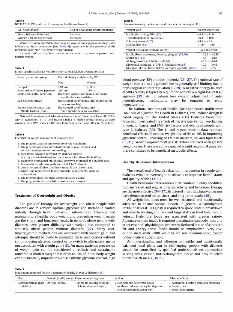
Table 6Glucose-lowering medications and their effects on weight (17)
Weight gain Weight effect (lb)
Insulin (fast acting, NPH) (2) þ8.8 e þ11.0Thiazolidinediones (TZDs) (57) þ5.2 e þ10.6Sulphonylureas (2,57) þ3.5 e þ5.7Meglitinides (58) þ1.54 e þ3.97
Weight neutral or decrease weight Weight effect
Insulin (basal analogues, detemir, glargine) (59,60) �0.22 e þ0.88Metformin (61) �8.4 e þ0.88Alpha-glucosidase inhibitors (62,63) þ0.0 e þ0.44Dipeptidyl peptidase-4 (DPP-4) inhibitors (64,65) þ0.0 e þ0.46Glucagon-like peptide-1 (GLP-1) receptor agonists (66,67) �6.6 e �3.5
Table 2NCEP-ATP III WC and risk of developing health problems (8)
WC cutoff points*,y Risk of developing health problems
Men �102 cm (40 inches) IncreasedWomen �88 cm (35 inches) Increased
* Waist circumference (WC) cutoffs may be lower in some populations (e.g. olderindividuals, Asian population [See Table 3]), especially in the presence of themetabolic syndrome (e.g. hypertriglyceridemia).
y Increased WC can also be a marker for increased risk, even in persons withnormal weight.
Table 3Ethnic-specific values for WC from International Diabetes Federation (13)
Country or ethnic group Central obesity as defined by WC
Men Women
Europid* �94 cm �80 cmSouth Asian, Chinese, Japanese �90 cm �80 cmSouth and Central American Use South Asian cutoff points until more
specific data are available.Sub-Saharan African Use Europid cutoff points until more specific
data are available.Eastern Mediterranean and
Middle Eastern (Arab)Use Europid cutoff points untilmore specific data are available.
* National Cholesterol and Education Program Adult Treatment Panel III (NCEP-ATP III) guidelines (11,12) and Health Canada (6) define central obesity as waistcircumference (WC) values �102 cm (40 inches) in men and �88 cm (35 inches).
Table 4Checklist for weight management programs (46)
1. The program assesses and treats comorbid conditions.2. The program provides individualized nutritional, exercise and
behavioral programs and counselling.3. Nutritional advice is provided by qualified experts
(e.g. registered dietitians) and diets are not less than 900 kcal/day.4. Exercise is encouraged but physical activity is promoted at a gradual pace.5. Reasonable weight loss goals are set at 1 to 2 lb/week.6. Cost is not prohibitive, and there are no financial contracts.7. There is no requirement to buy products, supplements, vitamins
or injections.8. The program does not make unsubstantiated claims.9. The program has an established maintenance program.
S. Wharton et al. / Can J Diabetes 37 (2013) S82eS86 S83
Treatment of Overweight and Obesity
The goals of therapy for overweight and obese people withdiabetes are to achieve optimal glycemic and metabolic controlinitially through health behaviour intervention. Attaining andmaintaining a healthy body weight and preventing weight regainare the short- and long-term goals. In general, obese people withdiabetes have greater difficulty with weight loss compared tosimilarly obese people without diabetes (22). Many anti-hyperglycemic medications are associated with weight gain, andattempts should be made to minimize these medications withoutcompromising glycemic control or to switch to alternative agentsnot associated with weight gain (18). For many patients, preventionof weight gain can be considered a realistic and sustainableoutcome. A modest weight loss of 5% to 10% of initial body weightcan substantially improve insulin sensitivity, glycemic control, high
Table 5Medication approved for the treatment of obesity in type 2 diabetes (36)
Class Generic (trade) name Recommended regimen Action
Gastrointestinal lipaseinhibitor
Orlistat (Xenical) 120 mg tid (during or up to1 hour after each meal)
� Noninhand
blood pressure (BP) and dyslipidemia (23e27). The optimal rate ofweight loss is 1 to 2 kg/month but is generally self-limiting due tophysiological counterregulation (17,28). A negative energy balanceof 500 kcal/day is typically required to achieve a weight loss of 0.45kg/week (29). As individuals lose weight, adjustment in anti-hyperglycemic medications may be required to avoidhypoglycemia.
The National Institutes of Health (NIH)-sponsored multicenterLook AHEAD (Action for Health in Diabetes) trial, whose design isbased largely on the United States (US) Diabetes PreventionProgram, investigated the effects of lifestyle interventionon changesin weight, fitness, and CVD risk factors and events in people withtype 2 diabetes (30). The 1- and 4-year interim data reportedbeneficial effects of modest weight loss of 5% to 10% in improvingglycemic control, lowering of CV risk markers, BP and lipid levels(30,31). Greater improvement in risk factors occurred with greaterweight losses. Therewas someexpectedweight regain at 4 years, yetthere continued to be beneficial metabolic effects.
Healthy Behaviour Interventions
The overall goal of health behaviour intervention in people withdiabetes who are overweight or obese is to improve health statusand quality of life (32,33).
Health behaviour interventions that combine dietary modifica-tion, increased and regular physical activity and behaviour therapyare themosteffective (34e37). Structured interdisciplinaryprogramshave demonstrated better short- and long-term results (36).
All weight-loss diets must be well balanced and nutritionallyadequate to ensure optimal health. In general, a carbohydrateintake of at least 100 g/day is required to spare protein breakdownand muscle wasting and to avoid large shifts in fluid balance andketosis. High-fibre foods are associated with greater satiety.Adequate protein intake is required tomaintain lean bodymass andother essential physiological processes. Reduced intake of saturatedfat and energy-dense foods should be emphasized. Very-low-calorie diets with <900 kcal/day are not recommended, exceptunder medical supervision.
As understanding and adhering to healthy and nutritionallybalanced meal plans can be challenging, people with diabetesshould be counselled by qualified professionals on appropriateserving sizes, caloric and carbohydrate intake and how to selectnutrient-rich meals (38,39).
Adverse effects
systemic pancreatic lipaseibitor reduces dietary fat digestionabsorption by about 30%
� Abdominal bloating, pain and cramping� Steatorrhea� Fecal incontinence
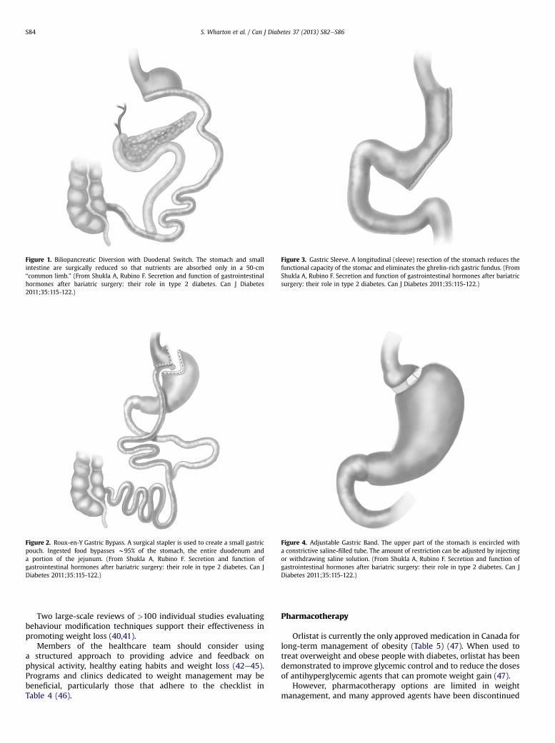
Figure 1. Biliopancreatic Diversion with Duodenal Switch. The stomach and smallintestine are surgically reduced so that nutrients are absorbed only in a 50-cm“common limb.” (From Shukla A, Rubino F. Secretion and function of gastrointestinalhormones after bariatric surgery: their role in type 2 diabetes. Can J Diabetes2011;35:115-122.)
Figure 2. Roux-en-Y Gastric Bypass. A surgical stapler is used to create a small gastricpouch. Ingested food bypasses w95% of the stomach, the entire duodenum anda portion of the jejunum. (From Shukla A, Rubino F. Secretion and function ofgastrointestinal hormones after bariatric surgery: their role in type 2 diabetes. Can JDiabetes 2011;35:115-122.)
Figure 4. Adjustable Gastric Band. The upper part of the stomach is encircled witha constrictive saline-filled tube. The amount of restriction can be adjusted by injectingor withdrawing saline solution. (From Shukla A, Rubino F. Secretion and function ofgastrointestinal hormones after bariatric surgery: their role in type 2 diabetes. Can JDiabetes 2011;35:115-122.)
Figure 3. Gastric Sleeve. A longitudinal (sleeve) resection of the stomach reduces thefunctional capacity of the stomac and eliminates the ghrelin-rich gastric fundus. (FromShukla A, Rubino F. Secretion and function of gastrointestinal hormones after bariatricsurgery: their role in type 2 diabetes. Can J Diabetes 2011;35:115-122.)
S. Wharton et al. / Can J Diabetes 37 (2013) S82eS86S84
Two large-scale reviews of >100 individual studies evaluatingbehaviour modification techniques support their effectiveness inpromoting weight loss (40,41).
Members of the healthcare team should consider usinga structured approach to providing advice and feedback onphysical activity, healthy eating habits and weight loss (42e45).Programs and clinics dedicated to weight management may bebeneficial, particularly those that adhere to the checklist inTable 4 (46).
Pharmacotherapy
Orlistat is currently the only approved medication in Canada forlong-term management of obesity (Table 5) (47). When used totreat overweight and obese people with diabetes, orlistat has beendemonstrated to improve glycemic control and to reduce the dosesof antihyperglycemic agents that can promote weight gain (47).
However, pharmacotherapy options are limited in weightmanagement, and many approved agents have been discontinued
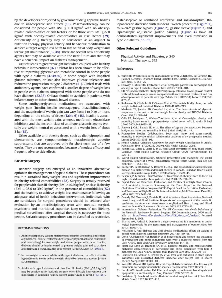
S. Wharton et al. / Can J Diabetes 37 (2013) S82eS86 S85
by the developers or rejected by government drug approval boardsdue to unacceptable side effects (18). Pharmacotherapy can beconsidered for people with BMI �30.0 kg/m2 with no obesity-related comorbidities or risk factors, or for those with BMI �27.0kg/m2 with obesity-related comorbidities or risk factors (29).Antiobesity drug therapy may be considered as an adjunct tonutrition therapy, physical activity and behaviour modification toachieve a target weight loss of 5% to 10% of initial body weight andfor weight maintenance (32,48). There are several new antiobesityagents that may be available within the near future and that mayhave a beneficial impact on diabetes management.
Orlistat leads to greater weight loss when coupled with healthybehaviour interventions (47). Orlistat has been shown to be effec-tive at improving glycemic and metabolic control in obese peoplewith type 2 diabetes (47,49,50). In obese people with impairedglucose tolerance, orlistat also improves glucose tolerance andreduces the progression to type 2 diabetes (51). Clinical trials withantiobesity agents have confirmed a smaller degree of weight lossin people with diabetes compared with obese people who do nothave diabetes (22,38). Orlistat should be avoided in patients withinflammatory or other chronic bowel disease.
Some antihyperglycemic medications are associated withweight gain (insulin, insulin secretagogues, thiazolidinediones),and the magnitude of weight gain can vary from 4 to 9 kg or more,depending on the choice of drugs (Table 6) (18). Insulin is associ-ated with the most weight gain, whereas metformin, glucosidaseinhibitors and the incretin class of antihyperglycemic agents typi-cally are weight neutral or associated with a weight loss of about3 kg (18).
Other available anti-obesity drugs, such as diethylpropion andphentermine, are sympathomimetic noradrenergic appetitesuppressants that are approved only for short-term use of a fewweeks. They are not recommended because of modest efficacy andfrequent adverse side effects.
Bariatric Surgery
Bariatric surgery has emerged as an innovative alternativeoption in themanagement of type 2 diabetes. These procedures canresult in sustained body weight loss and significant improvementin obesity-related comorbidities (52). Surgery is usually reservedfor peoplewith class III obesity (BMI�40.0 kg/m2) or class II obesity(BMI ¼ 35.0 to 39.9 kg/m2) in the presence of comorbidities (52)and the inability to achieve weight loss maintenance following anadequate trial of health behaviour intervention. Individuals whoare candidates for surgical procedures should be selected afterevaluation by an interdisciplinary team with medical, surgical,psychiatric and nutritional expertise. Long-term, if not lifelong,medical surveillance after surgical therapy is necessary for mostpeople. Bariatric surgery procedures can be classified as restrictive,
RECOMMENDATIONS
1. An interdisciplinary weight management program (including a nutrition-ally balanced, calorie-restricted diet; regular physical activity; education;and counselling) for overweight and obese people with, or at risk for,diabetes should be implemented to prevent weight gain and to achieveand maintain a lower, healthy body weight [Grade A, Level 1A (30,56)].
2. In overweight or obese adults with type 2 diabetes, the effect of anti-hyperglycemic agents on bodyweight should be taken into account [GradeD, Consensus].
3. Adults with type 2 diabetes and class II or III obesity (BMI �35.0 kg/m2)may be considered for bariatric surgery when lifestyle interventions areinadequate in achieving healthy weight goals [Grade B, Level 2 (53e55)].
malabsorptive or combined restrictive and malabsorptive. Bil-iopancreatic diversion with duodenal switch procedure (Figure 1),roux-en-Y gastric bypass (Figure 2), gastric sleeve (Figure 3) andlaparoscopic adjustable gastric banding (Figure 4) have alldemonstrated significant improvements and even remission intype 2 diabetes (53e55).
Other Relevant Guidelines
Physical Activity and Diabetes, p. S40Nutrition Therapy, p. S45
References
1. Wing RR. Weight loss in the management of type 2 diabetes. In: Gerstein HC,Haynes B, editors. Evidence-Based Diabetes Care. Ontario, Canada: B.C. Decker,Inc; 2000. p. 252e76.
2. Conway B, Miller RG, Costacou T, et al. Temporal patterns in overweight andobesity in type 1 diabetes. Diabet Med 2010;27:398e404.
3. UK Prospective Diabetes Study (UKPDS) Group. Intensive blood-glucose controlwith sulphonylureas or insulin compared with conventional treatment and riskof complications in patients with type 2 diabetes (UKPDS 33). Lancet 1998;352:837e53.
4. Ruderman N, Chisholm D, Pi-Sunyer X, et al. The metabolically obese, normal-weight individual revisited. Diabetes 1998;47:699e713.
5. Markovic TP, Jenkins AB, Campbell LV, et al. The determinants of glycemicresponses to diet restriction and weight loss in obesity and NIDDM. DiabetesCare 1998;21:687e94.
6. Calle EE, Rodriguez C, Walker-Thurmond K, et al. Overweight, obesity, andmortality from cancer in a prospectively studied cohort of U.S. adults. N EnglJ Med 2003;348:1625e38.
7. Stevens J, Cai J, Pamuk ER, et al. The effect of age on the association betweenbody-mass index and mortality. N Engl J Med 1998;338:1e7.
8. Prospective Studies Collaboration. Body-mass index and cause-specificmortality in 900 000 adults: collaborative analyses of 57 prospective studies.Lancet 2009;373:1083e96.
9. Health Canada. Canadian Guidelines for Body Weight Classification in Adults.Publication H49e179/2003E. Ottawa, ON: Health Canada; 2003.
10. Rabkin SW, Chen Y, Leiter L, et al. Risk factor correlates of body mass index.Canadian Heart Health Surveys Research Group. CMAJ 1997;157(suppl 1):S26e31.
11. World Health Organization. Obesity: preventing and managing the globalepidemic. Report of a WHO consultation. World Health Organ Tech Rep Ser2000;894:1e253. i-xii.
12. Reeder BA, Senthilselvan A, Després JP, et al. The association of cardiovasculardisease risk factors with abdominal obesity in Canada. Canadian Heart HealthSurveys Research Group. CMAJ 1997;157(suppl 1):S39e45.
13. Després JP, Lemieux I, Prud’homme D. Treatment of obesity: need to focus onhigh risk abdominally obese patients. BMJ 2001;322:716e20.
14. Expert Panel on Detection, Evaluation, and Treatment of High Blood Choles-terol in Adults. Executive Summary of the Third Report of the NationalCholesterol Education Program (NCEP) Expert Panel on Detection, Evaluation,and Treatment of High Blood Cholesterol in Adults (Adult Treatment Panel III).JAMA 2001;285:2486e97.
15. Grundy SM, Cleeman JI, Daniels SR, et al, American Heart Association, NationalHeart, Lung, and Blood Institute. Diagnosis and management of the metabolicsyndrome: an American Heart Association/National Heart, Lung, and BloodInstitute Scientific Statement. Circulation 2005;112:2735e52.
16. International Diabetes Federation. The IDF Consensus Worldwide Definition ofthe Metabolic Syndrome. Brussels, Belgium: IDF Communications; 2006. Avail-able at: http://www.idf.org/webdata/docs/IDF_Meta_def_final.pdf. AccessedSeptember 1, 2008.
17. Sharma AM, Padwal R. Obesity is a signeover-eating is a symptom: an aetio-logical framework for the assessment and management of obesity. Obes Rev2010;11:362e70.
18. Hollander P. Anti-diabetes and anti-obesity medications: effects on weight inpeople with diabetes. Diabetes Spectrum 2007;20:159e65.
19. Gorin AA, Niemeier HM, Hogan P, et al. Binge eating and weight loss outcomesin overweight and obese individuals with type 2 diabetes: results from theLook AHEAD trial. Arch Gen Psychiatry 2008;65:1447e55.
20. Ribisl PM, Lang W, Jaramillo SA, et al. Exercise capacity and cardiovascular/metabolic characteristics of overweight and obese individuals with type 2diabetes: the Look AHEAD clinical trial. Diabetes Care 2007;30:2679e84.
21. Grunstein RR, Stenlof K, Hedner JA, et al. Two year reduction in sleep apneasymptoms and associated diabetes incidence after weight loss in severeobesity. Sleep 2007;30:703e10.
22. Wing RR, Marcus MD, Epstein LH, et al. Type II diabetic subjects lose less weightthan their overweight nondiabetic spouses. Diabetes Care 1987;10:563e6.
23. Dattilo AM, Kris-Etherton PM. Effects of weight reduction on blood lipids andlipoproteins: a meta-analysis. Am J Clin Nutr 1992;56:320e8.
24. Goldstein DJ. Beneficial health effects of modest weight loss. Int J Obes RelatMetab Disord 1992;16:397e415.

S. Wharton et al. / Can J Diabetes 37 (2013) S82eS86S86
25. Elmer PJ, GrimmJrR, LaingB, et al. Lifestyle intervention: results of theTreatmentof Mild Hypertension Study (TOMHS). Prev Med 1995;24:378e88.
26. Tuomilehto J, Lindström J, Eriksson JG, et al. Finnish Diabetes Prevention StudyGroup. Prevention of type 2 diabetes mellitus by changes in lifestyle amongsubjects with impaired glucose tolerance. N Engl J Med 2001;344:1343e50.
27. Knowler WC, Barrett-Connor E, Fowler SE, et al. Diabetes Prevention ProgramResearch Group. Reduction in the incidence of type 2 diabetes with lifestyleintervention or metformin. N Engl J Med 2002;346:393e403.
28. Sumithran P, Prendergast LA, Delbridge E, et al. Long-term persistence ofhormonal adaptations to weight loss. N Engl J Med 2011;365:1597e604.
29. National Institutes of Health. Clinical guidelines on the identification, evalua-tion, and treatment of overweight and obesity in adults: the evidence report.Obes Res 1998;6(suppl 2):51Se209S.
30. Look AHEAD Research GroupWing RR. Long-term effects of a lifestyle inter-vention on weight and cardiovascular risk factors in individuals with type 2diabetes mellitus: four-year results of the Look AHEAD trial. Arch Intern Med2010;170:1566e75.
31. Wadden TA, West DS, Neiberg RH, et al. One-year weight losses in the LookAHEAD study: factors associated with success. Obesity 2009;17:713e22.
32. The American Diabetes Association’s (ADA) evidence-based practice guidelines,standards, and related recommendations and documents for diabetes care.Diabetes Care January 2012 vol. 35 no. Supplement 1.
33. Willett WC, Dietz WH, Colditz GA. Guidelines for healthy weight. N Engl J Med1999;341:427e34.
34. Williamson DF, Thompson TJ, Thun M, et al. Intentional weight loss andmortality among overweight individuals with diabetes. Diabetes Care 2000;23:1499e504.
35. Pavlou KN, Krey S, Steffee WP. Exercise as an adjunct to weight loss andmaintenance in moderately obese subjects. Am J Clin Nutr 1989;49(5 suppl):1115e23.
36. Wing RR, Hill JO. Successful weight loss maintenance. Annu Rev Nutr 2001;21:323e41.
37. Wing RR, Goldstein MG, Acton KJ, et al. Behavioral science research in diabetes:lifestyle changes related to obesity, eating behavior, and physical activity.Diabetes Care 2001;24:117e23.
38. Rolls BJ, Morris EL, Roe LS. Portion size of food affects energy intake in normal-weight and overweight men and women. Am J Clin Nutr 2002;76:1207e13.
39. Rolls BJ, Roe LS, Meengs JS. Salad and satiety: energy density and portion size ofa first-course salad affect energy intake at lunch. J Am Diet Assoc 2004;104:1570e6.
40. Wing RR, Jeffery RW. Outpatient treatments of obesity: a comparison ofmethodology and clinical results. Int J Obes 1979;3:261e79.
41. Bennett GA. Behaviour therapy for obesity: a quantitative review of the effectsof selected treatment characteristics on outcome. Behav Ther 1986;17:554e62.
42. Swinburn BA, Walter LG, Arroll B, et al. The green prescription study:a randomized controlled trial of written exercise advice provided by generalpractitioners. Am J Public Health 1998;88:288e91.
43. Logsdon DN, Lazaro CM, Meier RV. The feasibility of behavioral risk reductionin primary medical care. Am J Prev Med 1989;5:249e56.
44. Campbell MK, DeVellis BM, Strecher VJ, et al. Improving dietary behavior: theeffectiveness of tailored messages in primary care settings. Am J Public Health1994;84:783e7.
45. Lewis BS, Lynch WD. The effect of physician advice on exercise behavior. PrevMed 1993;22:110e21.
46. Freedhoff Y, Sharma AM. Best Weight: A Practical Guide to Office-BasedObesity Management. Canadian Obesity Network, Canada; 2010.
47. Hollander PA, Elbein SC, Hirsch IB, et al. Role of orlistat in the treatment ofobese patients with type 2 diabetes. A 1-year randomized double-blind study.Diabetes Care 1998;21:1288e94.
48. National Task Force on the Prevention and Treatment of Obesity. Long-term pharmacotherapy in the management of obesity. JAMA 1996;276:1907e15.
49. Scheen AJ, Lefèbvre PJ. Antiobesity pharmacotherapy in the management oftype 2 diabetes. Diabetes Metab Res Rev 2000;16:114e24.
50. Finer N, Bloom SR, Frost GS, et al. Sibutramine is effective for weight loss anddiabetic control in obesity with type 2 diabetes: a randomised, double-blind,placebo-controlled study. Diabetes Obes Metab 2000;2:105e12.
51. Heymsfield SB, Segal KR, Hauptman J, et al. Effects of weight loss with orlistaton glucose tolerance and progression to type 2 diabetes in obese adults. ArchIntern Med 2000;160:1321e6.
52. Lau DCW. Surgical options for patients with type 2 diabetes: a review ofcurrent and emerging therapies. Can Diabetes 2010;23:3e13.
53. Abbatini F, Rizzello M, Casella G, et al. Long-term effects of laparoscopic sleevegastrectomy, gastric bypass, and adjustable gastric banding on type 2 diabetes.Surg Endosc 2010;24:1005e10.
54. Mingrone G, Panunzi S, De Gaetano A, et al. Bariatric surgery versusconventional medical therapy for type 2 diabetes. N Engl J Med 2012;366:1577e85.
55. Schauer PR, Kashyap SR, Wolski K, et al. Bariatric surgery versus intensivemedical therapy in obese patients with diabetes. N Engl J Med 2012;366:1567e76.
56. Diabetes Program Prevention Research Group. Reduction in the incidence ofType 2 diabetes with lifestyle or metformin. N Engl J Med 2002;346:393e403.
57. Kahn SE, Haffner SM, Heise MA, et al, ADOPT Study Group. Glycemic durabilityof rosiglitazone, metformin, or glyburide monotherapy. N Engl J Med 2006;355:2427e43.
58. Rosenstock J, Hassaman DR, Madder RD, et al. Repaglinide Versus NateglinideComparison Study Group. Repaglinide versus nateglinide monotherapy:a randomized multicenter study. Diabetes Care 2004;27:1265e70.
59. De Leeuw I, Vague P, Selam JL, et al. Insulin detemir used in basal-bolus therapyin people with type 1 diabetes is associated with a lower risk of nocturnalhypoglycaemia and less weight gain over 12 months in comparison to NPHinsulin. Diabetes Obes Metab 2005;7:73e82.
60. Rosenstock J, Schwartz SL, Clark Jr CM, et al. Basal insulin therapy in patientswith type 2 diabetes: 28-week comparison of insulin glargine (HOE 901) andNPH insulin. Diabetes Care 2001;24:631e6.
61. DeFronzo RA, Goodman AM. Efficacy of metformin in patients with non-insulin-dependent diabetes mellitus. N Engl J Med 1995;333:541e9.
62. Coniff R, Sharpiro J, Robbins D, et al. Reduction of glycosylated haemoglobinand postprandial hyperglycemia by acarbose in patients with NIDDM. DiabetesCare 1995;18:831e43.
63. Hollander P, Pi Sunyer X, Coniff R. Acarbose in the treatment of type 1 diabetes.Diabetes Care 1997;20:248e54.
64. Ahrén B, Landin-Olsson M, Jansson PA, et al. Inhibition of dipeptidyl peptidase-4 reduces glycemia, sustains insulin levels, and reduces glucagons levels intype 2 diabetes. J Clin Endocrinol Metab 2004;89:2078e84.
65. Scott R, Wu M, Sanchez M, Stein P. Efficacy and tolerability of thedipeptidylpeptidase-4 inhibitor sitagliptin as monotherapy over 12 weeks inpatients with type 2 diabetes. Int J Clin Pract 2007;61:171e80.
66. Ratner RE, Maggs D, Nielsen LL, et al. Long-term effects of exenatide therapyover 82 weeks on glycaemic control and weight in over-weight metformin-treated patients with type 2 diabetes mellitus. Diabetes Obes Metab 2006;8:419e28.
67. Nauck M, Frid A, Hermansen K, et al, LEAD-2 Study Group. Efficacy and safetycomparison of liraglutide, glimepiride, and placebo, all in combination withmetformin, in type 2 diabetes: the LEAD (liraglutide effect and action indiabetes)-2 study. Diabetes Care 2009;32:84e90.

Contents lists available at SciVerse ScienceDirect
Can J Diabetes 37 (2013) S87eS92
Canadian Journal of Diabetesjournal homepage:
www.canadianjournalofdiabetes.com
Clinical Practice Guidelines
Diabetes and Mental Health
Canadian Diabetes Association Clinical Practice Guidelines Expert Committee
The initial draft of this chapter was prepared by David J. Robinson MD, FRCPC, FAPA,Meera Luthra MD, FRCPC, Michael Vallis PhD, RPsych
KEY MESSAGES
� Psychiatric disorders, particularly major depressive disorder (MDD),generalized anxiety disorder and eating disorders, are more prevalent inpeople with diabetes compared to the general population.
� People diagnosed with serious mental illnesses, such as MDD, bipolardisorder and schizophrenia, have a higher risk of developing diabetes thanthe general population.
� All individuals with diabetes should be regularly screened for the presenceof depressive and anxious symptoms.
� Compared to those with diabetes only, individuals with diabetes andmental health disorders have decreased medication adherence, decreasedcompliance with diabetes self-care, increased functional impairment,increased risk of complications associated with diabetes, increasedhealthcare costs and an increased risk of early mortality.
� The following treatment modalities should be incorporated into primarycare and self-management education interventions to facilitate adaptationto diabetes, reduce diabetes-related distress and improve outcomes:motivational interventions, stress management strategies, coping skillstraining, family therapy and collaborative case management.
� Individuals taking psychiatric medications, particularly atypical antipsy-chotics, benefit from regular screening of metabolic parameters.
Introduction
Research is increasingly demonstrating a relationship betweenmental health disorders and diabetes. Patients with serious mentalillnesses, particularly those with depressive symptoms orsyndromes, and patients with diabetes share reciprocal suscepti-bility and a high degree of comorbidity (Figure 1).
The mechanisms behind these relationships are multifactorial.Some evidence shows that treatment for mental health disordersmay actually increase the risk of diabetes, particularly whensecond-generation (atypical) antipsychotic agents are prescribed(1). Biochemical changes due to the mental health disordersthemselves also may play a role (2). Lifestyle changes and symp-toms of mental health disorders are also likely to contribute (3).
Depression
The prevalence of clinically relevant depressive symptomsamong patients with diabetes is in the range of 30% (4e6). Theprevalence of major depressive disorder (MDD) is approximately10% (7,8), which is double the overall prevalence in people withouta chronic medical illness. Individuals with depression have an
1499-2671/$ e see front matter � 2013 Canadian Diabetes Associationhttp://dx.doi.org/10.1016/j.jcjd.2013.01.026
approximately 60% increased risk of developing type 2 diabetes (9).The prognosis for comorbid depression and diabetes is worse thanwhen each illness occurs separately (10). Depression in patientswith diabetes amplifies symptom burden by a factor of about 4 (11).Episodes of MDD in individuals with diabetes are likely to lastlonger and have a higher chance of recurrence compared to thosewithout diabetes (12).
Studies examining differential rates for the prevalence ofdepression in type 1 vs. type 2 diabetes have yielded inconsistentresults (4,13). One study found that the requirement for insulinwasthe factor associated with the highest rate of depression, regardlessof the type of diabetes involved (14).
Risk factors for developing depression in individuals with dia-betes are as follows:
� Female gender� Adolescents/young adults and older adults� Poverty� Few social supports� Stressful life events� Poor glycemic control, particularly with recurrent hypoglycemia� Longer duration of diabetes� Presence of long-term complications (15e19)
Risk factors (with possible mechanisms) for developing diabetesin patients with depression are as follows:
� Physical inactivity and obesity, which leads to insulin resis-tance, and
� Psychological stress, leading to chronic hypothalamic-pituitary-adrenal activation with cortisol release (20e25).
Comorbid depression worsens clinical outcomes in diabetes,possibly because the accompanying lethargy lowers motivation forself-care, resulting in lowered physical and psychological fitness,higher use of healthcare services and reduced adherence to medi-cation regimens (26,27). Depression also appears to worsencardiovascular mortality (28,29). Treating depressive symptomsmore reliably improvesmood than it does glycemic control (30e33).
Bipolar Disorder
Patients with bipolar disorder have been found to have preva-lence rates estimated to be double that of the general populationfor metabolic syndrome and triple for diabetes (34e36).
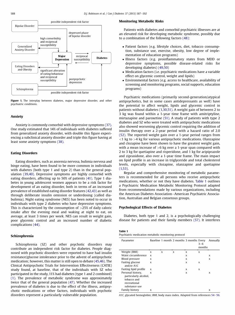
Figure 1. The interplay between diabetes, major depressive disorder, and otherpsychiatric conditions.
D.J. Robinson et al. / Can J Diabetes 37 (2013) S87eS92S88
Anxiety
Anxiety is commonly comorbid with depressive symptoms (37).One study estimated that 14% of individuals with diabetes sufferedfrom generalized anxiety disorder, with double this figure experi-encing a subclinical anxiety disorder and triple this figure having atleast some anxiety symptoms (38).
Eating Disorders
Eating disorders, such as anorexia nervosa, bulimia nervosa andbinge eating, have been found to be more common in individualswith diabetes (both type 1 and type 2) than in the general pop-ulation (39,40). Depressive symptoms are highly comorbid witheating disorders, affecting up to 50% of patients (41). Type 1 dia-betes in young adolescent women appears to be a risk factor fordevelopment of an eating disorder, both in terms of an increasedprevalence of established eating disorder features (42,43) as well asthrough deliberate insulin omission or underdosing (called dia-bulimia). Night eating syndrome (NES) has been noted to occur inindividuals with type 2 diabetes who have depressive symptoms.This is characterized by the consumption of >25% of daily caloricintake after the evening meal and waking at night to eat, onaverage, at least 3 times per week. NES can result in weight gain,poor glycemic control and an increased number of diabeticcomplications (44).
Table 1Psychiatric medication metabolic monitoring protocol
Parameter Baseline 1 month 2 months 3 months Every3e6months
Annually
Weight (BMI) x x x x xWaist circumference x x xBlood pressure x x xFasting glucose
and/or A1Cx x x
Fasting lipid profile x x xPersonal history,
particularly alcohol,tobacco andrecreationalsubstance use
x x x
Family history x x
A1C, glycated hemoglobin; BMI, body mass index. Adapted from references 54e56.
Schizophrenia
Schizophrenia (SZ) and other psychotic disorders maycontribute an independent risk factor for diabetes. People diag-nosed with psychotic disorders were reported to have had insulinresistance/glucose intolerance prior to the advent of antipsychoticmedication; however, this matter is still open to debate (45,46). TheClinical Antipsychotic Trials for Intervention Effectiveness (CATIE)study found, at baseline, that of the individuals with SZ whoparticipated in the study, 11% had diabetes (type 1 and 2 combined)(1). The prevalence of metabolic syndrome was approximatelytwice that of the general population (47). Whether the increasedprevalence of diabetes is due to the effect of the illness, antipsy-chotic medications or other factors, individuals with psychoticdisorders represent a particularly vulnerable population.
Monitoring Metabolic Risks
Patients with diabetes and comorbid psychiatric illnesses are atan elevated risk for developing metabolic syndrome, possibly dueto a combination of the following factors (48):
� Patient factors (e.g. lifestyle choices, diet, tobacco consump-tion, substance use, exercise, obesity, low degree of imple-mentation of education programs)
� Illness factors (e.g. proinflammatory states from MDD ordepressive symptoms, possible disease-related risks fordeveloping diabetes) (49,50)
� Medication factors (i.e. psychiatric medications have a variableeffect on glycemic control, weight and lipids)
� Environmental factors (e.g. access to healthcare, availability ofscreening andmonitoring programs, social supports, educationprograms)
Psychiatric medications (primarily second-generation/atypicalantipsychotics, but in some cases antidepressants as well) havethe potential to affect weight, lipids and glycemic control inpatients without diabetes (1,30,51). A weight gain of between 2 to3 kg was found within a 1-year time frame with amitriptyline,mirtazapine and paroxetine (51). A study of patients with type 2diabetes and SZ who were treated with antipsychotic medicationsalso showed worsening glycemic control requiring the addition ofinsulin therapy over a 2-year period with a hazard ratio of 2.0(52). The reported weight gain over a 1-year period ranges from<1 kg to >4 kg for various antipsychotic medications. Olanzapineand clozapine have been shown to have the greatest weight gain,with a mean increase of >6 kg over a 1-year span compared with2 to 3 kg for quetiapine and risperidone, and 1 kg for aripiprazoleand ziprasidone, also over a 1-year time frame. The main impacton lipid profile is an increase in triglyceride and total cholesterollevels, especially with clozapine, olanzapine and quetiapine(1,53).
Regular and comprehensive monitoring of metabolic parame-ters is recommended for all persons who receive antipsychoticmedications, whether or not they have diabetes. Table 1 outlinesa Psychiatric Medication Metabolic Monitoring Protocol adaptedfrom recommendations made by various organizations, includingthe American Diabetes AssociationeAmerican Psychiatric Associa-tion, Australian and Belgian consensus groups.
Psychological Effects of Diabetes
Diabetes, both type 1 and 2, is a psychologically challengingdisease for patients and their family members (57). It interferes

Table 2Comparison of main features and assessment methods: diabetes distress vs. depression
Diabetes distress Major depressive disorder
Assessment Instrument Diabetes Distress Scale (17 items) Patient Health Questionnaire for Depression: PHQ-9 (9 items)Format Self-report using ratings from 1e6 based on feelings
and experiences over the past weekSelf-report using ratings from 0e3 based on feelings and experiences over the past
2 weeksFeatures Emotional Burden Subscale (5 items)
Physician-Related Distress Subscale (4 items)Regimen-Related Distress Subscale (5 items)Diabetes-Related Interpersonal Distress Subscale(3 items)
Vegetative symptoms, such as sleep, appetite and energy level changesEmotional symptoms, such as low mood and reduced enjoyment of usual activitiesBehavioural symptoms, such as agitation or slowing of movementsCognitive symptoms, such as poor memory or reduced concentration or feelings ofguilt; thoughts of self-harm
D.J. Robinson et al. / Can J Diabetes 37 (2013) S87eS92 S89
with quality of life and is a risk factor for diabetes-related distressas well as the psychiatric disorders listed above. Challengesaccompanying the diagnosis of diabetes include adjustment to thedisease, adherence to the treatment regimen and psychosocialdifficulties at both a personal and an interpersonal level (58,59).Stress, deficient social supports and negative attitudes towarddiabetes can impact on self-care and glycemic control (60e64).Diabetes management strategies ideally incorporate a means ofaddressing the psychosocial factors that impact on individuals andtheir families. Both symptommeasures (e.g. self-report measures ofdepressive or anxiety symptoms) and methods to arrive at mentaldisorder diagnoses (e.g. structured interviews leading to Diagnosticand Statistical Manual of Mental Disorders [Fourth Edition, TextRevision] [DSM-IV-TR] diagnoses [42]) have been assessed. Giventhat the person with diabetes carries out 95% of diabetesmanagement (65), identifying depressive syndromes in diabetes isimportant since depression is a risk factor for poor diabetes self-management (66e68) and outcomes, including early mortality(69,70). MDD has been found to be underdiagnosed in people withdiabetes (71).
Diabetes distress describes the despondency and emotionalturmoil related specifically to having the condition, the need forcontinual monitoring and treatment, persistent concerns aboutcomplications and the potential erosion of personal and profes-sional relationships. Distinguishing between diabetes distress,MDD and the presence of depressive symptoms is important(72,73) because these psychological experiences are different, and,of the three, diabetes distress may be most strongly related toadverse diabetes outcomes (72,74,75).
Screening for Psychological/Psychiatric Symptoms
Individuals with diabetes should be regularly screened forpsychological distress and psychiatric disorders via directed inter-views. No data presently demonstrate the superiority of oneparticular depression screening tool over another (76). Currentlyavailable screening instruments have a sensitivity of between 80%and 90% and a specificity of 70% to 85% (76). Awebsite that containsa wide variety of downloadable scales that are in the public domainis https://www.outcometracker.org/scales_library.php. PatientHealth Questionnaire (PHQ) Screeners are available at www.phqscreeners.com.
Table 3Features of cognitive behavioural therapy that can be applied to diabetes treatment (97)
Cognitive component
Record keeping to identify distressing automatic thoughtsUnderstanding the link between thoughts and feelingsLearning the common “thinking errors” that mediate distress (e.g. all-or-nothing think
personalization, magnification, minimization, etc.)Analyzing negative thoughts and promoting more functional onesIdentifying basic assumptions about oneself (e.g. “unless I am very successful, my life
worth living”) and being encouraged to adopt healthier ones (e.g. “when I am dobest, I should be proud of myself”)
Screening instruments fall into three categories:
� Diabetes-specific measures, such as the Problem Areas in Dia-betes (PAID) Scale (77,78) or the Diabetes Distress Scale (DDS)(79)
� Quality of life measures, such as the WHO-5 screening instru-ment (80)
� Depressive/anxiety symptoms, such as the Hospital Anxietyand Depression Scale (HADS) (81), the Patient Health Ques-tionnaire (PHQ-9) (82,83), the Centre for EpidemiologicalStudieseDepression Scale (CES-D) (84) or the Beck DepressionInventory (BDI) (85)
Table 2 illustrates the differences between the principal featuresand assessment methods of diabetes distress and MDD.
Treatment of Psychological/Psychiatric Risk Factors
Given the burden associated with the demands of diabetes self-management, efforts to promote well-being and moderate distressshould be incorporated into diabetes management for all individ-uals (86). Motivational interventions (68,87,88), coping skills, self-efficacy enhancement, stress management (89,90) and familyinterventions (91e93) all have been shown to be helpful. Casemanagement by a nurse working with the patient’s primary carephysician and providing guideline-based, patient-centred careresulted in improved glycated hemoglobin (A1C), lipids, bloodpressure and depression scores (94). Individuals with diabetesdistress and/or psychiatric disorders benefit from professionalinterventions, either some type of psychotherapy or prescriptionmedication. Evidence from systematic reviews of randomizedcontrolled trials supports cognitive behaviour therapies (CBT) andantidepressant medication, both solely or in combination (33,95).No evidence presently shows that the combination of CBT andmedication is superior to these treatments given individually. Apilot study of 50 patients with type 2 diabetes who initially hadamoderate level of depression at baseline showed an improvementin the severity of their depression (moving to the mild range) witha 12-week intervention of 10 CBT sessions combined with exercisein the form of 150 minutes of aerobic activity weekly. This effectwas sustained at 3 months (96). Online resources are available tohelp healthcare providers learn CBT skills (e.g. www.moodgym.
Behavioural component
ing,
is noting my
Strategies to help get the person moving (behavioural activation)Scheduling pleasant events; learning assertive and effectivecommunication skills
Focusing on feelings of mastery and accomplishmentLearning problem-solving strategiesExposure to new experiencesShaping behaviours by breaking them down into smaller stepsto develop skills
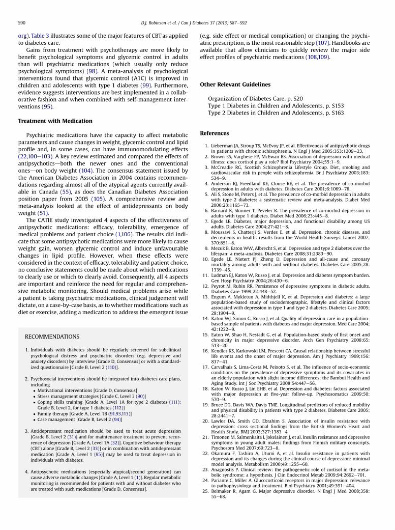
D.J. Robinson et al. / Can J Diabetes 37 (2013) S87eS92S90
org). Table 3 illustrates some of the major features of CBT as appliedto diabetes care.
Gains from treatment with psychotherapy are more likely tobenefit psychological symptoms and glycemic control in adultsthan will psychiatric medications (which usually only reducepsychological symptoms) (98). A meta-analysis of psychologicalinterventions found that glycemic control (A1C) is improved inchildren and adolescents with type 1 diabetes (99). Furthermore,evidence suggests interventions are best implemented in a collab-orative fashion and when combined with self-management inter-ventions (95).
Treatment with Medication
Psychiatric medications have the capacity to affect metabolicparameters and cause changes inweight, glycemic control and lipidprofile and, in some cases, can have immunomodulating effects(22,100e103). A key review estimated and compared the effects ofantipsychoticsdboth the newer ones and the conventionalonesdon body weight (104). The consensus statement issued bythe American Diabetes Association in 2004 contains recommen-dations regarding almost all of the atypical agents currently avail-able in Canada (55), as does the Canadian Diabetes Associationposition paper from 2005 (105). A comprehensive review andmeta-analysis looked at the effect of antidepressants on bodyweight (51).
The CATIE study investigated 4 aspects of the effectiveness ofantipsychotic medications: efficacy, tolerability, emergence ofmedical problems and patient choice (1,106). The results did indi-cate that some antipsychotic medications weremore likely to causeweight gain, worsen glycemic control and induce unfavourablechanges in lipid profile. However, when these effects wereconsidered in the context of efficacy, tolerability and patient choice,no conclusive statements could be made about which medicationsto clearly use or which to clearly avoid. Consequently, all 4 aspectsare important and reinforce the need for regular and comprehen-sive metabolic monitoring. Should medical problems arise whilea patient is taking psychiatric medications, clinical judgement willdictate, on a case-by-case basis, as towhethermodifications such asdiet or exercise, adding a medication to address the emergent issue
RECOMMENDATIONS
1. Individuals with diabetes should be regularly screened for subclinicalpsychological distress and psychiatric disorders (e.g. depressive andanxiety disorders) by interview [Grade D, Consensus] or with a standard-ized questionnaire [Grade B, Level 2 (110)].
2. Psychosocial interventions should be integrated into diabetes care plans,including� Motivational interventions [Grade D, Consensus]� Stress management strategies [Grade C, Level 3 (90)]� Coping skills training [Grade A, Level 1A for type 2 diabetes (111);Grade B, Level 2, for type 1 diabetes (112)]
� Family therapy [Grade A, Level 1B (91,93,113)]� Case management [Grade B, Level 2 (94)]
3. Antidepressant medication should be used to treat acute depression[Grade B, Level 2 (31)] and for maintenance treatment to prevent recur-rence of depression [Grade A, Level 1A (32)]. Cognitive behaviour therapy(CBT) alone [Grade B, Level 2 (33)] or in combination with antidepressantmedication [Grade A, Level 1 (95)] may be used to treat depression inindividuals with diabetes.
4. Antipsychotic medications (especially atypical/second generation) cancause adverse metabolic changes [Grade A, Level 1 (1)]. Regular metabolicmonitoring is recommended for patients with and without diabetes whoare treated with such medications [Grade D, Consensus].
(e.g. side effect or medical complication) or changing the psychi-atric prescription, is the most reasonable step (107). Handbooks areavailable that allow clinicians to quickly review the major sideeffect profiles of psychiatric medications (108,109).
Other Relevant Guidelines
Organization of Diabetes Care, p. S20Type 1 Diabetes in Children and Adolescents, p. S153Type 2 Diabetes in Children and Adolescents, p. S163
References
1. Lieberman JA, Stroup TS, McEvoy JP, et al. Effectiveness of antipsychotic drugsin patients with chronic schizophrenia. N Engl J Med 2005;353:1209e23.
2. Brown ES, Varghese FP, McEwan BS. Association of depression with medicalillness: does cortisol play a role? Biol Psychiatry 2004;55:1e9.
3. McCreadie RG, Scottish Schizophrenia Lifestyle Group. Diet, smoking andcardiovascular risk in people with schizophrenia. Br J Psychiatry 2003;183:534e9.
4. Anderson RJ, Freedland KE, Clouse RE, et al. The prevalence of co-morbiddepression in adults with diabetes. Diabetes Care 2001;6:1069e78.
5. Ali S, Stone M, Peters J, et al. The prevalence of co-morbid depression in adultswith type 2 diabetes: a systematic review and meta-analysis. Diabet Med2006;23:1165e73.
6. Barnard K, Skinner T, Peveler R. The prevalence of co-morbid depression inadults with type 1 diabetes. Diabet Med 2006;23:445e8.
7. Egede LE. Diabetes, major depression, and functional disability among USadults. Diabetes Care 2004;27:421e8.
8. Moussavi S, Chatterji S, Verdes E, et al. Depression, chronic diseases, anddecrements in health: results from the World Health Surveys. Lancet 2007;370:851e8.
9. Mezuk B, EatonWW, Albrecht S, et al. Depression and type 2 diabetes over thelifespan: a meta-analysis. Diabetes Care 2008;31:2383e90.
10. Egede LE, Nietert PJ, Zheng D. Depression and all-cause and coronarymortality among adults with and without diabetes. Diabetes Care 2005;28:1339e45.
11. Ludman EJ, Katon W, Russo J, et al. Depression and diabetes symptom burden.Gen Hosp Psychiatry 2004;26:430e6.
12. Peyrot M, Rubin RR. Persistence of depressive symptoms in diabetic adults.Diabetes Care 1999;22:448e52.
13. Engum A, Mykletun A, Midthjell K, et al. Depression and diabetes: a largepopulation-based study of sociodemographic, lifestyle and clinical factorsassociated with depression in type 1 and type 2 diabetes. Diabetes Care 2005;28:1904e9.
14. Katon WJ, Simon G, Russo J, et al. Quality of depression care in a population-based sample of patients with diabetes and major depression. Med Care 2004;42:1222e9.
15. Eaton W, Shao H, Nestadt G, et al. Population-based study of first onset andchronicity in major depressive disorder. Arch Gen Psychiatry 2008;65:513e20.
16. Kendler KS, Karkowski LM, Prescott CA. Causal relationship between stressfullife events and the onset of major depression. Am J Psychiatry 1999;156:837e41.
17. Carvalhais S, Lima-Costa M, Peixoto S, et al. The influence of socio-economicconditions on the prevalence of depressive symptoms and its covariates inan elderly population with slight income differences; the Bambui Health andAging Study. Int J Soc Psychiatry 2008;54:447e56.
18. Katon W, Russo J, Lin EHB, et al. Depression and diabetes: factors associatedwith major depression at five-year follow-up. Psychosomatics 2009;50:570e9.
19. Bruce DG, Davis WA, Davis TME. Longitudinal predictors of reduced mobilityand physical disability in patients with type 2 diabetes. Diabetes Care 2005;28:2441e7.
20. Lawlor DA, Smith GD, Ebrahim S. Association of insulin resistance withdepression: cross sectional findings from the British Women’s Heart andHealth Study. BMJ 2003;327:1383e4.
21. Timonen M, Salmenkaita I, Jokelainen J, et al. Insulin resistance and depressivesymptoms in young adult males: findings from Finnish military conscripts.Psychosom Med 2007;69:723e8.
22. Okamura F, Tashiro A, Utumi A, et al. Insulin resistance in patients withdepression and its changes during the clinical course of depression: minimalmodel analysis. Metabolism 2000;49:1255e60.
23. Anagnostis P. Clinical review: the pathogenetic role of cortisol in the meta-bolic syndrome: a hypothesis. J Clin Endocrinol Metab 2009;94:2692e701.
24. Pariante C, Miller A. Glucocorticoid receptors in major depression: relevanceto pathophysiology and treatment. Biol Psychiatry 2001;49:391e404.
25. Belmaker R, Agam G. Major depressive disorder. N Engl J Med 2008;358:55e68.
D.J. Robinson et al. / Can J Diabetes 37 (2013) S87eS92 S91
26. Ciechanowski PS, Katon WJ, Russo JE. Depression and diabetes: impact ofdepressive symptoms on adherence, function, and costs. Arch Int Med 2000;160:3278e85.
27. Lin EHB, Katon W, Von Korff M, et al. Relationship of depression and diabetesself-care, medication adherence, and preventative care. Diabetes Care 2004;27:2154e60.
28. Katon WJ, Rutter C, Simon G. The association of comorbid depression withmortality in patients with type 2 diabetes. Diabetes Care 2005;28:2668e72.
29. Zhang X, Norris SL, Gregg EW. Depressive symptoms and mortality amongpersons with and without diabetes. Am J Epidemiol 2005;161:652e60.
30. Lustman PJ, Clouse RE, Freedland KE, et al. Effects of nortriptyline ondepression and glucose regulation in diabetes: results of a double-blindplacebo-controlled trial. Psychosom Med 1997;59:241e50.
31. Lustman PJ, Freedland KE, Griffith L, et al. Fluoxetine for depression in dia-betes: a randomized double-blind placebo-controlled trial. Diabetes Care2000;23:618e23.
32. Lustman PJ, Clouse RE, Nix BD, et al. Sertraline for prevention of depressionrecurrence in diabetes mellitus. Arch Gen Psychiatry 2006;63:521e9.
33. Lustman PJ, Griffith LS, Freedland KE, et al. Cognitive behaviour therapy fordepression in type 2 diabetes mellitus. A randomized, controlled trial. AnnIntern Med 1998;129:613e21.
34. Fagiolini A, Frank E, Scott JA, et al. Metabolic syndrome in bipolar disorder:findings from the Bipolar Disorder Center for Pennsylvanians. Bipolar Disord2005;7:424e30.
35. Taylor V, MacQueen G. Associations between bipolar disorder and metabolicsyndrome: a review. J Clin Psychiatry 2006;67:1034e41.
36. van Winkel R, De Hert M, Van Eyck D, et al. Prevalence of diabetes and themetabolic syndrome in a sample of patients with bipolar disorder. BipolarDisord 2008;10:342e8.
37. Katon WJ, Lin EH, Kroenke K. The association of depression and anxiety withthe medical symptom burden in patients with chronic medical illness. GenHosp Psychiatry 2007;29:147e55.
38. Grigsby AB, Anderson RJ, Freedland KE, et al. Prevalence of anxiety in adultswith diabetes: a systematic review. J Psychosom Res 2004;27:1053e60.
39. Crow S, Kendall D, Praus B, et al. Binge eating and other psychopathology inpatients with type II diabetes mellitus. Int J Eat Disord 2000;30:222e6.
40. Jones JM, Lawson ML, Daneman D. Eating disorders in adolescent femaleswith and without type 1 diabetes: cross sectional study. BMJ 2000;320:1563e6.
41. McCarthy M. The thin ideal. depression and eating disorders in women. BehavRes Ther 1990;28:205e14.
42. American Psychiatric Association. Diagnostic and Statistical Manual of MentalDisorders. 4th ed, text revision. Arlington, VA: American Psychiatric Associ-ation; 2000.
43. Iafusco D, Vanelli M, Gugliotta M, et al. Prevalence of eating disorders inyoung patients with type 1 diabetes from two different Italian cities. DiabetesCare 2004;27:2278.
44. Morse S, Ciechanowski P, et al. Isn’t this just bedtime snacking? The potentialadverse effects of night-eating symptoms on treatment adherence andoutcomes in patients with diabetes. Diabetes Care 2006;29:1800e4.
45. Haupt DW, Newcomber JW. Hyperglycemia and antipsychotic medications.J Clin Psych 2001;62:15e26.
46. Saddichha S, Manjunatha N, Ameen S, et al. Diabetes and schizophrenia: effectof disease or drug? Results from a randomized, double-blind, controlledprospective study in first-episode schizophrenia. Acta Psychiatr Scand 2008;117:342e7.
47. McEvoy JP, Meyer JM, Goff DC, et al. Prevalence of the metabolic syndromein patients with schizophrenia: baseline results from the Clinical Antipsy-chotic Trials of Intervention Effectiveness (CATIE) schizophrenia trial andcomparison with national estimates from NHANES III. Schiz Res 2005;80:19e32.
48. Meyer JM, Nasrallah HA. Medical Illness and Schizophrenia. 2nd ed. Arlington,Virginia (VA): American Psychiatric Publishing, Inc.; 2009.
49. Smith R. The macrophage theory of depression. Med Hypothesis 1991;35:298e306.
50. Fernandez-Real JM, Pickup JC. Innate immunity, insulin resistance and type 2diabetes. Trends Endocrinol Metab 2008;19:10e6.
51. Serretti A, Mandelli L. Antidepressants and body weight: a comprehensivereview and meta-analysis. J Clin Psychiatry 2010;71:1259e72.
52. Spoelstra JA, Stolk RP, Cohen D, et al. Antipsychotic drugs may worsen meta-bolic control in type 2 diabetes mellitus. J Clin Psychiatry 2004;65:674e8.
53. Newcomer JW, Haupt DW. The metabolic effects of antipsychotic medications.Can J Psychiatry 2006;51:480e91.
54. Lambert TJ, Chapman LH. Diabetes, psychotic disorders and antipsychotictherapy: a consensus statement. Med J Aust 2004;181:544e8.
55. American Diabetes Association, American Psychiatric Association, AmericanAssociation of Clinical Endocrinologists, et al. Consensus DevelopmentConference on Antipsychotic Drugs and Obesity and Diabetes. Diabetes Care2004;27:596e601.
56. De Nayer A, De Hert M, Scheen A, Van Gaal L, Peuskens J. Belgian consensus onmetabolic problems associated with atypical antipsychotics. Int J PsychiatryClin Pract 2005;9:130e7.
57. Snoek FJ, Kersch NY, Eldrup E, et al. Monitoring of Individual Needs in Dia-betes (MIND): baseline data from the Cross-National Diabetes Attitudes,Wishes, and Needs (DAWN) MIND study. Diabetes Care 2011;34:601e3.
58. Peyrot M, Rubin RR, Lauritzen T, et al. Psychosocial problems and barriers toimproved diabetes management: results of the Cross-National DiabetesAttitudes, Wishes and Needs (DAWN) Study. Diabet Med 2005;22:1379e85.
59. Goebel-Fabbri AE, Fikkan J, Franko DL, et al. Insulin restriction and associatedmorbidity and mortality in women with type 1 diabetes. Diabetes Care 2008;31:415e9.
60. Fisher L, Glasgow RE. A call for more effectively integrating behavioral andsocial science principles into comprehensive diabetes care. Diabetes Care2007;30:2746e9.
61. Malik JA, Koot HM. Explaining the adjustment of adolescents with type 1diabetes: role of diabetes-specific and psychosocial factors. Diabetes Care2009;32:774e9.
62. Zhang CX, Tse LA, Ye XQ, et al. Moderating effects of coping styles on anxietyand depressive symptoms caused by psychological stress in Chinese patientswith Type 2 diabetes. Diabet Med 2009;26:1282e8.
63. Hampson SE, Tildesley E, Andrews JA, et al. The relation of change in hostilityand sociability during childhood to substance use in mid adolescence. J ResPers 2010;44:103e14.
64. Luyckx K, Seiffge-Krenke I, Hampson SE. Glycemic control, coping, andinternalizing and externalizing symptoms in adolescents with type 1 dia-betes: a cross-lagged longitudinal approach. Diabetes Care 2010;33:1424e9.
65. Anderson RM. Is the problem of compliance all in our heads? Diabetes Educ1985;11:31e4.
66. Gonzalez JS, Peyrot M, McCarl LA, et al. Depression and diabetes treatmentnonadherence: a meta-analysis. Diabetes Care 2008;31:2398e403.
67. Gonzalez JS, Safren SA, Delahanty LM, et al. Symptoms of depressionprospectively predict poorer self-care in patients with Type 2 diabetes. DiabetMed 2008;25:1102e7.
68. Egede LE, Grubaugh AL, Ellis C. The effect of major depression on preventivecare and quality of life among adults with diabetes. Gen Hosp Psychiatry2010;32:563e9.
69. Richardson LK, Egede LE, Mueller M. Effect of race/ethnicity and persistentrecognition of depression on mortality in elderly men with type 2 diabetesand depression. Diabetes Care 2008;31:880e1.
70. Hutter N, Schnurr A, Baumeister H. Healthcare costs in patients with diabetesmellitus and comorbid mental disorders: a systematic review. Diabetologia2010;53:2470e9.
71. Li C, Ford ES, Zhao G, et al. Prevalence and correlates of undiagnoseddepression among U.S. adults with diabetes: the Behavioral Risk FactorSurveillance System, 2006. Diabetes Res Clin Pract 2009;83:268e79.
72. Fisher L, Skaff MM, Mullan JT, et al. Clinical depression versus distress amongpatients with type 2 diabetes: not just a question of semantics. Diabetes Care2007;30:542e8.
73. Gonzalez JS, Fisher L, Polonsky WH. Depression in diabetes: have we beenmissing something important? Diabetes Care 2011;34:236e9.
74. Fisher L, Glasgow RE, Strycker LA. The relationship between diabetes distressand clinical depression with glycemic control among patients with type 2diabetes. Diabetes Care 2010;33:1034e6.
75. Fisher L, Mullan JT, Arean P, et al. Diabetes distress but not clinical depressionor depressive symptoms is associated with glycemic control in both cross-sectional and longitudinal analyses. Diabetes Care 2010;33:23e8.
76. Pignone MP, Gaynes BN, Rushton JL, et al. Screening for depression in adults,a summary of the evidence for the US Preventative Services Task Force. AnnIntern Med 2002;136:765e76.
77. Polonsky WH, Anderson BJ, Lohrer PA, et al. Assessment of diabetes-relateddistress. Diabetes Care 1995;18:754e60.
78. van Bastelaar KM, Pouwer F, Geelhoed-Duijvestijn PH, et al. Diabetes-specificemotional distress mediates the association between depressive symptomsand glycaemic control in Type 1 and Type 2 diabetes. Diabet Med 2010;27:798e803.
79. Polonsky WH, Fisher L, Earles J, et al. Assessing psychosocial distress in dia-betes: development of the diabetes distress scale. Diabetes Care 2005;28:626e31.
80. Furuya M, Hayashino Y, Tsujii S, et al. Comparative validity of the WHO-5Well-Being Index and two-question instrument for screening depressivesymptoms in patients with type 2 diabetes. Acta Diabetol; 2010 Aug 27 [Epubahead of print].
81. Collins MM, Corcoran P, Perry IJ. Anxiety and depression symptoms inpatients with diabetes. Diabet Med 2009;26:153e61.
82. Kroenke K, Spitzer RL. The PHQ-9: a new depression diagnostic and severitymeasure. Psychiatr Ann 2002;32:509e21.
83. van Steenbergen-Weijenburg KM, de Vroege L, Ploeger RR, et al. Validation ofthe PHQ-9 as a screening instrument for depression in diabetes patients inspecialized outpatient clinics. BMC Health Serv Res 2010;10:235.
84. Fisher L, Skaff MM, Mullan JT, et al. A longitudinal study of affective andanxiety disorders, depressive affect and diabetes distress in adults with Type2 diabetes. Diabet Med 2008;25:1096e101.
85. Mantyselka P, Korniloff K, Saaristo T, et al. Association of depressive symp-toms with impaired glucose regulation, screen-detected, and previouslyknown type 2 diabetes: findings from the Finnish D2D Survey. Diabetes Care2011;34:71e6.
86. Piatt GA, Anderson RM, Brooks MM, et al. 3-year follow-up of clinical andbehavioral improvements following a multifaceted diabetes care interven-tion: results of a randomized controlled trial. Diabetes Educ 2010;36:301e9.

D.J. Robinson et al. / Can J Diabetes 37 (2013) S87eS92S92
87. Osborn CY, Egede LE. Validation of an Information-Motivation-BehavioralSkills model of diabetes self-care (IMB-DSC). Patient Educ Couns 2010;79:49e54.
88. Maindal HT, Sandbaek A, Kirkevold M, Lauritzen T. Effect on motivation,perceived competence, and activation after participation in the “Ready to Act”programme for people with screen-detected dysglycaemia: a 1-year rando-mised controlled trial, Addition-DK. Scand J Public Health 2011;39:262e71.
89. Attari A, Sartippour M, Amini M, Haghighi S. Effect of stress managementtraining on glycemic control in patients with type 1 diabetes. Diabetes ResClin Pract 2006;73:23e8.
90. Soo H, Lam S. Stress management training in diabetes mellitus. J HealthPsychol 2009;14:933e43.
91. Keogh KM, Smith SM, White P, et al. Psychological family intervention forpoorly controlled type 2 diabetes. Am J Manag Care 2011;17:105e13.
92. Wysocki T, Harris MA, Buckloh LM, et al. Randomized trial of behavioralfamily systems therapy for diabetes: maintenance of effects on diabetesoutcomes in adolescents. Diabetes Care 2007;30:555e60.
93. Wysocki T, Harris MA, Buckloh LM, et al. Randomized, controlled trial ofbehavioral family systems therapy for diabetes: maintenance and general-ization of effects on parent-adolescent communication. Behav Ther 2008;39:33e46.
94. Katon WJ, Lin EHB, Von Korff M, et al. Collaborative care for patients withdepression and chronic illnesses. N Engl J Med 2010;363:2611e20.
95. van der Feltz-Cornelis CM, Nuyen J, Stoop C, et al. Effect of interventions formajor depressive disorder and significant depressive symptoms in patientswith diabetes mellitus: a systematic review and meta-analysis. Gen HospPsychiatry 2010;32:380e95.
96. de Groot M, Doyle T, Kushnick M, et al. Can lifestyle interventions do morethan reduce diabetes risk? Treating depression in adults with type 2 diabeteswith exercise and cognitive behavioral therapy. Curr Diab Rep 2012;12:157e66.
97. Beck JS. Cognitive Behavior Therapy: Basics and Beyond. 2nd ed. New York:Guilford; 2011.
98. Wang MY, Tsai PS, Chou KR, Chen CM. A systematic review of the efficacy ofnon-pharmacological treatments for depression on glycaemic control in type2 diabetics. J Clin Nurs 2008;17:2524e30.
99. Winkley K, Ismail K, Landau S, Eisler I. Psychological interventions to improveglycaemic control in patients with type 1 diabetes: systematic review andmeta-analysis of randomised controlled trials. BMJ 2006;333:65.
100. Xia Z, DePierre J, Nassberger L. Tricyclic antidepressants inhibit IL-6, IL-1beta,and TNF-alpha release in human blood monocytes and IL-2 and interferon-gamma in T cells. Immunopharmacology 1996;34:27e37.
101. Maes M, Song C, Lin A, et al. Negative immunomodulatory effects of antide-pressants: inhibition of interferon-gamma and stimulation of interleukin-10secretion. Neuropsychopharmacology 1999;20:370e9.
102. Kaufmann RP, Castracane VD, White DL. Impact of the selective serotoninreuptake inhibitor citalopram on insulin sensitivity, leptin and basal cortisolsecretion in depressed and non-depressed euglycemic women of reproduc-tive age. Gynecol Endocrinol 2005;21:129e37.
103. Weber-Hamann B, Giles M, Lederbogen F, et al. Improved insulin sensitivity in80 nondiabetic patients with major depressive disorder after clinical remis-sion in a double-blind, randomized trial of amitriptyline and paroxetine. J ClinPsychiatry 2006;67:1856e61.
104. Allison DB, Mentore JL, Heo M, et al. Antipsychotic-induced weightgain: a comprehensive research synthesis. Am J Psychiatry 1999;156:1686e96.
105. Woo V, Harris SB, Houlden RL. Canadian Diabetes Association Position Paper:Antipsychotic Medications and Associated Risks of Weight Gain and Diabetes.Can J Diabetes 2005;29:111e2.
106. Stroup TS, Lieberman JA, McEvoy JP, et al. Effectiveness of olanzapine, que-tiapine, risperidone, and ziprasidone in patients with chronic schizophreniafollowing discontinuation of a previous atypical antipsychotic. Am J Psychi-atry 2006;163:611e22.
107. Stroup TS, McEvoy JP, Ring KD. A randomized trial examining the effective-ness of switching from olanzapine, quetiapine, or risperidone to aripiprazoleto reduce metabolic risk: comparison of antipsychotics for metabolic prob-lems (CAMP). Am J Psychiatry 2011;168:947e56.
108. Stahl SM. The Prescriber’s Guide. 4th ed. New York, NY: Cambridge UniversityPress; 2011.
109. Virani AS, Bezchlibnyk-Butler KZ, Joel Jeffries JJ, et al. Clinical Handbook ofPsychotropic Drugs. 19th ed. Toronto, Canada: Hogrefe Publishing; 2011.
110. Pignone M, Gaynes BN, Rushton JL, et al. Screening for depression: systematicevidence review. Systematic Evidence Review No. 6. AHRQ Publication. No.02-S002. Rockville, MD Agency for Healthcare Research and Quality 2002.
111. Ismail K, Winkley K, Rabe-Hesketh S. Systematic review and meta-analysisof randomized controlled trials of psychological interventions to improveglycaemic control in patients with type 2 diabetes. Lancet 2004;363:1589e97.
112. Grey M, Boland EA, Davidson M, et al. Short-term effects of coping skillstraining as an adjunct to intensive therapy in adolescents. Diabetes Care1998;21:902e8.
113. Ellis DA, Frey MA, Naar-King S, et al. The effects of multisystemic therapy ondiabetes stress among adolescent wit chronically poorly controlled type 1 dia-betes: findings from a randomized controlled trial. Pediatrics 2005;16:826e32.

Contents lists available at SciVerse ScienceDirect
Can J Diabetes 37 (2013) S93
Canadian Journal of Diabetesjournal homepage:
www.canadianjournalofdiabetes.com
Clinical Practice Guidelines
Influenza and Pneumococcal Immunization
Canadian Diabetes Association Clinical Practice Guidelines Expert Committee
The initial draft of this chapter was prepared by Nadira Husein MD, FRCPC, Vincent Woo MD, FRCPC
KEY MESSAGES
� Influenza immunization can reduce hospitalization rates by approximately40% for those individuals deemed to be at high risk.
� Pneumococcal immunization is desired in people with diabetes as they areconsidered as likely to be infected as those with other chronic diseases.
� For those who are >65 years of age, a 1-time revaccination is recom-mended if the original vaccine was administered when they were <65years of age with at least 5 years between administrations.
Introduction
People with diabetes are considered to be at high risk formorbidity and mortality from influenza and pneumococcal disease(1,2). During recent influenza epidemics, diabetes was considereda significant risk factor for hospitalization (3). Influenza immuni-zation is associated with up to a 40% risk reduction in mortality (2).Clinical recommendations for vaccination are derived from largecohort studies that included people with diabetes as trials specificto individuals with diabetes are currently lacking.
Influenza Immunization in Adults
Data regarding influenzamorbidity andmortality in peoplewithdiabetes are based on retrospective analyses during influenzaepidemics (3,4). A recent epidemiological analysis of pandemicinfluenza demonstrated that people with diabetes are more likelyto be hospitalized or to require intensive care (5).
Over a period of 10 influenza seasons, influenza vaccination wasshown to be effective in reducing both death and hospitalizationfrom influenza and pneumonia in a cohort that included peoplewith diabetes (6).
RECOMMENDATIONS
1. People with diabetes should receive an annual influenza immunization toreduce the risk of complications associated with influenza [Grade D,Consensus].
2. Pneumococcal immunization should be offered to people with diabetes. Asingle dose is recommended for those >18 years of age. A 1-time revac-cination is recommended for those>65 years of age (if the original vaccinewas givenwhen they were <65 years of age) with at least 5 years betweenadministrations [Grade D, Consensus].
1499-2671/$ e see front matter � 2013 Canadian Diabetes Associationhttp://dx.doi.org/10.1016/j.jcjd.2013.01.027
A Dutch case control study documented that the incidence ofcomplications was 2 times higher in the unvaccinated groupcompared to the vaccinated group (7). The rates of hospitalizationfor influenza, pneumonia, other acute respiratory diseases,myocardial infarction, congestive heart failure, and stroke or dia-betes events were reduced by 70%.
Pneumococcal Immunization in Adults
People with diabetes are at increased risk of hospitalization forpneumococcal disease (1,8). Prior pneumococcal vaccination isassociated with a reduction in death and complications in hospi-talized adults with community-acquired pneumonia (9). It isaccepted that people with diabetes are at similar risk of developingpneumococcal disease as those with other chronic conditions (1),and, therefore, those with diabetes are encouraged to receivepneumococcal vaccination. Revaccination is recommended as a 1-time event for individuals >65 years of age if the original vaccinewas given when they were <65 years of age and >5 years earlier.
Related Websites
National Advisory Committee on Immunization. CanadianImmunization Guide. 7th ed. Ottawa, ON: Canadian Medical Asso-ciation; 2006. Available at: http://www.phac-aspc.gc.ca/publicat/cig-gci/index-eng.php. Accessed November 2012.
References
1. Muller LMAJ, Gorter KJ, Hak E, et al. Increased risk of common infections inpatients with type 1 and type 2 diabetes mellitus. Clin Infect Dis 2005;41:281e8.
2. Groenwold RHH, Hoes AW, Hak E. Impact of influenza vaccination on mortalityrisk among the elderly. Eur Respir J 2009;34:56e62.
3. Jain S, Kamimoto L, Bramley AM, et al. Hospitalized patients with 2009 H1Niinfluenza in the United States, April-June 2009. N Engl Med 2009;361:1935e44.
4. Campbell A, Rodin R, Kropp R, et al. Risk of severe outcomes among patientsadmitted to hospital with pandemic (H1N1) influenza. CMAJ 2010;182:349e55.
5. Allard R, Tremblay C, LeClerc P, Tannenbaum T. Diabetes and the severity ofpandemic influenza A (H1N1) infection. Diabetes Care 2010;33:1491e3.
6. Nichol KL, Nordin JD, Nelson DB, et al. Effectiveness of influenza vaccine in thecommunity-dwelling elderly. N Engl J Med 2007;357:1373e81.
7. Looijmans-Van Den Akker I, Nichol KL, Verheij TJM, et al. Clinical effectiveness offirst and repeat influenza vaccination in adult and elderly diabetic patients.Diabetes Care 2006;29:1771e6.
8. Kornum J, Lervang H-H, Thomsen RW, et al. Diabetes, glycemic control, and riskof hospitalization with pneumonia. Diabetes Care 2008;31:1541e5.
9. Fisman D, Abrutyn E, Spaude K, et al. Prior pneumococcal vaccination is asso-ciated with reduced death, complications, and length of stay among hospitalizedadults with community acquired pneumonia. Clin Infect Dis 2006;42:1093e101.

Contents lists available at SciVerse ScienceDirect
Can J Diabetes 37 (2013) S94eS96
Canadian Journal of Diabetesjournal homepage:
www.canadianjournalofdiabetes.com
Clinical Practice Guidelines
Pancreas and Islet Transplantation
Canadian Diabetes Association Clinical Practice Guidelines Expert Committee
The initial draft of this chapter was prepared by Breay W. Paty MD, FRCPC, Angela Koh MD,Peter Senior MBBS, PhD, MRCP
14htt
KEY MESSAGES
� Simultaneous pancreas kidney transplantation in persons with type 1diabetes and end stage renal disease can improve kidney graft survival andresult in prolonged insulin independence.
� Successful pancreas or islet allotransplantation can stabilize glucose andpossibly result in insulin independence in persons with type 1 diabetes andglycemic lability or recurrent hypoglycemia.
� Islet autotransplantation can stabilize glucose and possibly result in insulinindependence in people undergoing total pancreatectomy for benignpancreatic disease.
Introduction
Restoring endogenous insulin secretion by whole pancreas orislet transplantation has been established as an alternative toinsulin injection therapy in select individuals with type 1 diabetes(1,2). Nonrandomized studies demonstrate that both pancreas andislet transplantation can result in insulin independence and glucosestability, especially in the setting of glucose lability or frequent,severe hypoglycemia. Unfortunately, the absence of prospectiverandomized controlled trials makes it difficult to draw firmconclusions about the overall efficacy and safety of these therapiescompared with exogenous insulin treatment. Also, the limitednumber of specialized islet and pancreas transplantation centresand the relatively small number of donor pancreases limit theavailability of these treatments. Nevertheless, general recommen-dations regarding the role of pancreas and islet transplantationmay be made in the context of current clinical experience.
Pancreas Transplantation
Pancreas transplantation can result in complete independencefrom exogenous insulin in the majority of cases (3). As shown inTable 1, worldwide, noncontrolled 1- and 3-year mean pancreasgraft and patient survival rates differ slightly among the 3 majortypes of transplantations (4). Long-term pancreas graft survivaldeclines with time, with a median graft survival of 9 years and<10% survival at 21 years (5).
Glycemic control and glycated hemoglobin (A1C) are markedlyimproved after successful pancreas transplantation, with mostrecipients achieving normal glucose tolerance, albeit with hyper-insulinemia (6,7). A reduction in albuminuria has been noted at 1year (8), and improvements in the histological changes associated
99-2671/$ e see front matter � 2013 Canadian Diabetes Associationp://dx.doi.org/10.1016/j.jcjd.2013.01.028
with diabetic nephropathy have been reported 5 to 10 years post-transplantation (9,10). Whether successful simultaneous pancreaskidney (SPK) transplantation improves renal graft survival isunclear. In 1 study, recipients of SPK transplantations had betterrenal graft survival over 72 months than deceased-donor kidneytransplantations but lower graft survival than living-donor kidneytransplantations (11). The impact of pancreas transplantation onoverall patient survival also is uncertain. Studies suggest lower short-term survival in the perioperative period up to 18 to 24months afterSPK, but for patients with successful functioning pancreas grafts at12 months post-transplantation, survival was similar or improvedcompared to living- or deceased-donor kidney transplantation(12e14). A retrospective cohort study of individuals with diabetesand preserved kidney function who received a solitary pancreastransplantation suggested that overall survival was worse comparedwith wait-listed patients receiving conventional medical therapy(15). Improvement and/or stabilization of diabetic retinopathy havebeen demonstrated (16). Peripheral sensory andmotor neuropathiesalso have been shown to improve after pancreas transplantation(17,18), but these findings are not consistent and may take years toachieve (19e21). Pancreas transplantation appears to improvecardiovascular (CV) function, carotid intimal medial thickness,blood pressure and lipid parameters (22e24). A single, small, non-randomized study showed a reduction in CV events in SPK recipientscompared to those undergoing kidney transplantation alone (25);however, this has not been examined in a randomized controlledfashion. Finally, diabetes-related quality of life (QOL) appears toimprove after pancreas transplantation, although overall QOLappears to be unchanged (26,27).
Islet Transplantation
Islet allotransplantation
Islet allotransplantation involves the infusion of islets isolatedfrom cadaveric pancreata via the portal vein into the liver (28),either alone or in association with a renal transplantation (29,30).Successful islet transplantation can result in stable, near-normalglycemic control (A1C, glycemic variability) with a reduction orelimination of hypoglycemia (31) over and above what can beachieved with insulin injections or even insulin pump therapy (32).The ability of transplant recipients to achieve and maintain insulinindependence varies between transplantation centres and isinfluenced by both donor and recipient factors (33,34). Insulin

Table 1Reported graft survival rates according to type of pancreas transplantation (4)
Transplantation type 1 year 5 years 10 years 15 years
SPK 83% 69% 51% 33%PAK 74% 45% 24% 13%PTA 78% 54% 28% 9%
SPK, simultaneous pancreas kidney; PAK, pancreas after kidney; PTA, pancreastransplant alone.
RECOMMENDATIONS
1. Individuals with type 1 diabetes and ESRD who are being considered forkidney transplantation should also be considered for simultaneouspancreas transplantation [Grade D, Level 4 (12,14)].
2. Individuals with type 1 diabetes with preserved renal function, or whohave undergone successful kidney transplantation but have persistentmetabolic instability characterized by severe glycemic lability and/orsevere hypoglycemia despite best efforts to optimize glycemic control,may be considered for pancreas or islet allotransplantation [Grade D,Consensus].
3. Individuals undergoing total pancreatectomy for benign pancreatic diseasemay be considered for islet autotransplantation but only in the context ofan experienced islet transplantation centre [Grade D, Consensus].
Abbreviation:ESRD, end stage renal disease.
B.W. Paty et al. / Can J Diabetes 37 (2013) S94eS96 S95
independence can be achieved inmost recipients but often requires2 or more transplantation procedures. Insulin independence ratesdecline with time from approximately 70% at 1 year post-transplantation to approximately 10% after 5 years (31). However,patients who are not able to maintain insulin independence maystill benefit frommore stable blood glucose control (35) that resultsfrom ongoing graft function, as evidenced by the sustained secre-tion of C peptide and the reduced requirement for exogenousinsulin (33). Small, short-term studies also suggest stabilization ofretinopathy (36) and neuropathy (37) with islet allotransplantation.Renal outcomes vary, but recent reports suggest that the glomer-ular filtration rate achieved with the procedure does not differsignificantly from that observed in the nondiabetic population(38,39). Successful islet transplantation can improve QOL byreducing the fear of hypoglycemia (40) but can be negativelyimpacted by adverse effects from immunosuppressive agents (27).
Islet autotransplantation
In islet autotransplantation, islets are isolated from an individ-ual’s own resected pancreas following pancreatectomy for benignpancreatic disease (e.g. chronic, painful pancreatitis) (41,42). Isletyields from a resected, diseased pancreas may be lower than thosefrom cadaveric donors, but immunosuppression is not required.Even if insulin independence is not achieved, islet auto-transplantation may result in reduced exogenous insulin require-ments and a lower risk of hypoglycemia (43). As a result, the ratio ofbenefit to risk of this procedure may exceed that noted withislet allotransplantation (44).
Risks Associated with Pancreas and Islet Transplantation
Pancreas transplantation is associated with significant peri-operative risks, including graft thrombosis, hemorrhage,
Table 2Comparison of beta cell replacement modalities
Islet Pancreas
OutcomesReduce or eliminate
hypoglycemiaYes Yes
Improve A1C Yes YesInsulin
independenceYes* Yes
Effect on diabetes-related complicationsMicrovascular May be stabilizedy ImprovedMacrovascular Not known May be improvedRisksProcedural risks Minor procedural risk Major surgical riskImmunosuppression Similar agents,z life-long immunosuppressionOther considerationsESRD Avoid Consider SPKFunctioning renal
transplantConsider IAK if glycemiclability or hypoglycemiax
Consider PAK if glycemiclability or hypoglycemiax
A1C, glycated hemoglobin; ESRD, end stage renal disease; IAK, islet after kidney; PAK,pancreas after kidney; SPK, simultaneous pancreas kidney.
* More than 1 islet infusion may be required.y Retinopathy and neuropathy may be stabilized.z Steroids are avoided in islet transplantation but may be used in whole pancreas
transplantation.x No additional risk from immunosuppression.
pancreatitis, wound infection, peripancreatic abscesses andduodenal stump leakage (45). Islet transplantation is associatedwith fewer procedural risks, which may include intraperitonealhemorrhage, partial portal vein thrombosis or gallbladder punc-ture. These complications occur in <10% of procedures and usuallyare self-limited (31,34). Both pancreas and islet transplantationsrequire long-term immunosuppression, which is associated witha number of risks and side effects (46). Drug side effects aregenerally mild and often respond to dose or agent adjustment.Although rare, life-threatening opportunistic infections andmalignancies have been reported (34,46). These risks must becarefully weighed against the potential benefits of transplantationfor each individual. See Table 2 for a detailed comparison ofpancreas vs. islet transplantation.
References
1. White SA, Shaw JA, Sutherland DER. Pancreas transplantation. Lancet 2009;373:1808e17.
2. Halban PA, German MS, Kahn SE, et al. Current status of islet cell replacementand regeneration therapy. J Clin Endocrinol Metab 2010;95:1034e43.
3. Robertson RP, Abid M, Sutherland DE, et al. Glucose homeostasis and insulinsecretion in human recipients of pancreas transplantation. Diabetes 1989;38-(suppl 1):97e8.
4. Waki K, Kadowaki T. An analysis of long-term survival from the OPTN/UNOSPancreas Transplant Registry. Clin Transplant 2007:9e17.
5. Everly MJ. Pancreas transplant in the United States: an analysis of the UNOSregistry. Clin Transplant 2009:75e81.
6. Robertson RP, Sutherland DE, Kendall DM, et al. Metabolic characterization oflong-term successful pancreas transplants in type 1 diabetes. J Investig Med1996;44:549e55.
7. Lauria MW, Figueiro JM, Machado LJ, et al. Metabolic long-term follow-up offunctioning simultaneous pancreas-kidney transplantation versus pancreastransplantation alone: insights and limitations. Transplantation 2010;89:83e7.
8. Coppelli A, Giannarelli R, Vistoli F, et al. The beneficial effects of pancreastransplant alone on diabetic nephropathy. Diabetes Care 2005;28:1366e70.
9. Fioretto P, Steffes MW, Sutherland DE, et al. Reversal of lesions of diabeticnephropathy after pancreas transplantation. N Engl J Med 1998;339:69e75.
10. Fioretto P, Sutherland DER, Najafian B, et al. Remodeling of renal interstitial andtubular lesions in pancreas transplant recipients. Kidney Int 2006;69:907e12.
11. Young BY, Gill J, Huang E, Takemoto SK, et al. Living donor kidney versussimultaneous pancreas-kidney transplant in type I diabetics: an analysis of theOPTN/UNOS database. Clin J Am Soc Nephrol 2009;4:845e52.
12. Ojo AO, Meier-Kriesche HU, Hanson JA, et al. The impact of simultaneouspancreas-kidney transplantation on long-term patient survival. Trans-plantation 2001;71:82e90.
13. Reddy KS, Stablein D, Taranto S, et al. Long-term survival following simulta-neous kidney-pancreas transplantation versus kidney transplantation alone inpatients with type 1 diabetes mellitus and renal failure. Am J Kidney Dis 2003;41:464e70.
14. Weiss AS, Smits G, Wiseman AC. Twelve-month pancreas graft functionsignificantly influences survival following simultaneous pancreas-kidneytransplantation. Clin J Am Soc Nephrol 2009;4:988e95.
15. Venstrom JM, McBride MA, Rother KI, et al. Survival after pancreas trans-plantation in patients with diabetes and preserved kidney function. JAMA2003;290:2817e23.
B.W. Paty et al. / Can J Diabetes 37 (2013) S94eS96S96
16. Giannarelli R, Coppelli A, Sartini MS, et al. Pancreas transplant alone hasbeneficial effects on retinopathy in type 1 diabetic patients. Diabetologia 2006;49:2977e82.
17. Mehra S, Tavakoli M, Kallinikos PA, et al. Corneal confocal microscopy detectsearly nerve regeneration after pancreas transplantation in patients with type 1diabetes. Diabetes Care 2007;30:2608e12.
18. Kennedy WR, Navarro X, Goetz FC, et al. Effects of pancreatic transplantationon diabetic neuropathy. N Engl J Med 1990;322:1031e7.
19. Solders G, Tydén G, Persson A, et al. Improvement of nerve conduction indiabetic neuropathy. A follow-up study 4 yr after combined pancreatic andrenal transplantation. Diabetes 1992;41:946e51.
20. Tydén G, Bolinder J, Solders G, et al. Improved survival in patients with insulin-dependent diabetes mellitus and end-stage diabetic nephropathy 10 yearsafter combined pancreas and kidney transplantation. Transplantation 1999;67:645e8.
21. Boucek P, Havrdova T, Voska L, et al. Epidermal innervation in type 1 diabeticpatients: a 2.5-year prospective study after simultaneous pancreas/kidneytransplantation. Diabetes Care 2008;31:1611e2.
22. Coppelli A, Giannarelli R, Mariotti R, et al. Pancreas transplant alone deter-mines early improvement of cardiovascular risk factors and cardiac function intype 1 diabetic patients. Transplantation 2003;76:974e6.
23. Larsen JL, Colling CW, Ratanasuwan T, et al. Pancreas transplantation improvesvascular disease in patients with type 1 diabetes. Diabetes Care 2004;27:1706e11.
24. Luan FL, Miles CD, Cibrik DM, et al. Impact of simultaneous pancreas andkidney transplantation on cardiovascular risk factors in patients with type 1diabetes mellitus. Transplantation 2007;84:541e4.
25. La Rocca E, Fiorina P, di Carlo V, et al. Cardiovascular outcomes after kidney-pancreas and kidney-alone transplantation. Kidney Int 2001;60:1964e71.
26. Sureshkumar KK, Patel BM, Markatos A, et al. Quality of life after organtransplantation in type 1 diabetics with end-stage renal disease. Clin Trans-plant 2006;20:19e25.
27. Speight J, Reaney MD, Woodcock AJ, et al. Patient-reported outcomes followingislet cell or pancreas transplantation (alone or after kidney) in Type 1 diabetes:a systematic review. Diabet Med 2010;27:812e22.
28. Robertson RP. Islet transplantation as a treatment for diabetes: a work inprogress. N Engl J Med 2004;350:694e705.
29. Gerber PA, Pavlicek V, Demartines N, et al. Simultaneous islet-kidney vspancreas-kidney transplantation in type 1 diabetes mellitus: a 5-year singlecentre follow-up. Diabetologia 2008;51:110e9.
30. Deng S, Markmann JF, Rickels M, et al. Islet alone versus islet after kidneytransplantation: metabolic outcomes and islet graft survival. Transplantation2009;88:820e5.
31. Ryan EA, Paty BW, Senior PA, et al. Five year follow-up after clinical islettransplantation. Diabetes 2005;54:2060e9.
32. Vantyghem MC, Marcelli-Tourvieille S, Fermon C, et al. Intraperitoneal insulininfusion versus islet transplantation: comparative study in patients with type 1diabetes. Transplantation 2009;87:66e71.
33. Shapiro AM, Ricordi C, Hering BJ, et al. International trial of the Edmontonprotocol for islet transplantation. N Engl J Med 2006;355:1318e30.
34. Alejandro R, Barton FB, Hering BJ, et al. 2008 update from the CollaborativeIslet Transplant Registry. Transplantation 2008;86:1783e8.
35. Paty BW, Senior PA, Lakey JR, et al. Assessment of glycemic control after islettransplantation using the continuous glucose monitor in insulin-independentversus insulin-requiring type 1 diabetes subjects. Diabetes Technol Ther2006;8:165e73.
36. Thompson DM, Begg IS, Harris C, et al. Reduced progression of diabetic reti-nopathy after islet transplantation compared with intensive medical therapy.Transplantation 2008;85:1400e5.
37. Del Carro U, Fiorina P, Amadio S, et al. Evaluation of polyneuropathy markers intype 1 diabetic kidney transplant patients and effects of islet transplantation:neurophysiological and skin biopsy longitudinal analysis. Diabetes Care 2007;30:3063e9.
38. Senior PA, Zeman M, Paty BW, et al. Changes in renal function after clinicalislet transplantation: four-year observational study. Am J Transplant 2007;7:91e8.
39. Fung MA, Warnock GL, Ao Z, et al. The effect of medical therapy and islet celltransplantation on diabetic nephropathy: an interim report. Transplantation2007;84:17e22.
40. Poggioli R, Faradji RN, Ponte G, et al. Quality of life after islet transplantation.Am J Transplant 2006;6:371e8.
41. Robertson RP, Lanz KJ, Sutherland DE, et al. Prevention of diabetes for up to 13years by autoislet transplantation after pancreatectomy for chronic pancrea-titis. Diabetes 2001;50:47e50.
42. Bellin MD, Sutherland DE. Pediatric islet autotransplantation: indication,technique, and outcome. Curr Diab Rep 2010;10:326e31.
43. Blondet JJ, Carlson AM, Kobayashi T, et al. The role of total pancreatectomy andislet autotransplantation for chronic pancreatitis. Surg Clin North Am 2007;87:1477e501.
44. Sutherland DE, Gruessner AC, Carlson AM, et al. Islet autotransplant outcomesafter total pancreatectomy: a contrast to islet allograft outcomes. Trans-plantation 2008;86:1799e802.
45. Troppmann C. Complications after pancreas transplantation. Curr Opin OrganTransplant 2010;15:112e8.
46. Gruessner RW, Sutherland DER, Gruessner AC. Mortality assessment forpancreas transplants. Am J Transplant 2004;4:2018e26.
Contents lists available at SciVerse ScienceDirect
Can J Diabetes 37 (2013) S97eS99
Canadian Journal of Diabetesjournal homepage:
www.canadianjournalofdiabetes.com
Clinical Practice Guidelines
Natural Health Products
Canadian Diabetes Association Clinical Practice Guidelines Expert Committee
The initial draft of this chapter was prepared by Richard Nahas MD, CCFP,Jeannette Goguen MD, MEd, FRCPC
KEY MESSAGES
� Seventy-eight percent of patients with diabetes reported taking a naturalhealth product (NHP) for various indications.
� Some NHPs have shown a lowering of A1C by �0.5% in trials lasting�3months in adults with type 2 diabetes, but most are single small trials soit would be premature to recommend their widespread use.
� Healthcare providers should always ask about the use of NHPs since somemay result in side effects or drug interactions.
Introduction
Natural Health Products (NHPs) include herbal medicines, vita-mins, minerals and other essential nutrients, probiotics and manyother naturally occurring substances. Since 2004, they have beenregulated in Canada by the Natural Health Products Directorate ofHealth Canada.
Management
These guidelines include NHPs because they are widely used bypatients, but the evidence of their efficacy and safety is unknown tomost prescribing physicians. In a recent Canadian study of 502patients with diabetes, 78% reported taking an NHP, with similarfrequency in people with type 1 and type 2 diabetes (1). While it isimportant to be aware of potential harms, side effects and druginteractions, some NHPs may be potentially important new thera-peutic agents. Interestingly, metformin is derived from French lilac,a remedy used to treat diabetes since the Middle Ages, and iden-tification of the active agent guanidine led to the synthesis ofbiguanides (2).
In general, the current level of evidence for the efficacy andsafety of NHPs in people with type 2 diabetes is lower than that forpharmaceutical drug treatments. Trials tend to be of shorterduration and involve smaller sample sizes. At the present time,concerns remain about standardization and purity of availablecompounds, including their contamination with regular medica-tions and, in some cases, toxic substances (3).
While a number of NHPs have been studied to evaluate theirimpact on cardiovascular and other outcomes in patients with type2 diabetes, this guideline is limited to NHPs for improving glycemiccontrol. Trials were considered for review if they were randomized,controlled, and reported changes in glycated hemoglobin (A1C)over at least 12 weeks of treatment. Positive trials were those that
1499-2671/$ e see front matter � 2013 Canadian Diabetes Associationhttp://dx.doi.org/10.1016/j.jcjd.2013.01.029
demonstrated a reduction in the placebo-subtracted A1C of atleast 0.5%.
The following NHPs lowered A1C by �0.5% in trials lasting atleast 3 months in adults with type 2 diabetes:
� Coccinia cordifolia (4)� Ganoderma lucidum (5)� Salacia reticulata (6)� Soybean-derived pinitol extract (7)� Touchi soybean extract (8)� Pterocarpus marsulium (vijayasar) (9)� Gynostemma pentaphyllum (10)� Marine collagen peptides (11)� Silymarin (12,13)� Citrullus colocynthis (14)� Trigonellafoenum-graecum (fenugreek) (15)
These products are promising and merit consideration andfurther research, but, as they are mostly single, small trials, it ispremature to recommend their widespread use.
The following NHPs failed to lower A1C by�0.5% in trials lastingat least 3 months in adults with type 2 diabetes:
� Tinospora crispa (16)� French maritime pine bark (17)� Soy phytoestrogens (18)� Agaricus blazei (19)� Antioxidants (fruit/vegetable extract) (20), (pomegranateextract) (21)
� Camellia sinensis (22)� Cinnamomum spp (cinnamon) (23e27)� Momordica charantia (bitter melon or bitter gourd) (28)� Flaxseed oil (29)� Ginseng (30)� Coenzyme Q10 (31)� Vitamin C (32)� Vitamin D (33e35)� Vitamin E (36e38)
It should be noted that, in many cases, small sample sizes madethe trials insufficiently powered to establish a significant benefitfrom NHP interventions.
The following NHPs have demonstrated conflicting effects onA1C in trials lasting at least 3months in adults with type 2 diabetes:

Nahas, J. Goguen / Can J Diabetes 37 (2013) S97eS99S98
� Ipomoea batatas (caiapo) (39,40)� Chromium (41e52)� Magnesium (53e57)� L-carnitine (58e61)
It should be noted that vanadium, a trace element that iscommonly used to treat type 2 diabetes, has not been studied intrials evaluating glycemic control over a period of 3 months orlonger.
Complications
It is important to consider potential harm from the use of NHPs.In 1 trial of Tinospora crispa, hepatotoxicity was seen in 2 patients(16). Large doses of Citrullus colocynthis can induce diarrhea, but noside effects were reported in the lower doses used in 1 trial (14).Momordica charantia, an NHP commonly used for glycemic control,is an abortifacient (62). Most clinical trials have evaluated smallsample sizes over relatively short periods of time and, thus, may notidentify side effects or risks.
Drug-herb interactions may also occur. The most well describedis Hypericum perforatum (St. John’s wort), which can affect themetabolism of many drugs, including statins, by inducing CYP3A4.Some studies have reported poorer glycemic control in patientsusing glucosamine sulfate for osteoarthritis, but a systematic reviewconcluded that the evidence does not support this concern (63).
Clinicians should ask all patients with diabetes about their useof NHPs. Potential concerns should be addressed using a patient-centred approach that ensures patient safety while respectingtheir views to maintain a positive therapeutic relationship. Formore detailed information about specific NHPs, practitionersshould consult previously published reviews (64).
RECOMMENDATIONS
1. Natural health products are not recommended for glycemic control forindividuals with diabetes as there is insufficient evidence, at this time,regarding efficacy and safety [Grade D, Consensus].
2. Healthcare providers should ask about the use of natural health products[Grade D, Consensus].
References
1. Ryan EA, Pick ME, Marceau C. Use of alternative medicines in diabetes mellitus.Diabet Med 2001;18:242e5.
2. Oubre AY, Carlson TJ, King SR, Reaven GM. From plant to patient: an ethno-medical approach to the identification of new drugs for the treatment ofNIDDM. Diabetologia 1997;40:614e7.
3. Saper RB, Kales SN, Paquin J, et al. Heavy metal content of ayurvedic herbalmedicine products. JAMA 2004;292:2868e73.
4. Kuriyan R, Rajendran R, Bantwal G, Kurpad AV. Effect of supplementation ofCoccinia cordifolia extract on newly detected diabetic patients. Diabetes Care2008;31:216e20.
5. Gao YH, Lan J, Dai XH, et al. A phase I/II study of Ling Zhi mushroom Ganodermalucidum ( W.Curt.:Fr.) Lloyd (Aphyllophoromycetidae) extract in patients withtype 2 diabetes. Int J Med Mushr 2004;6:33e9.
6. Jayawardena MH, de Alwis NM, Hettigoda V, et al. A double blind randomisedplacebo controlled cross over study of a herbal preparation containing Salaciareticulata in the treatment of type 2 diabetes. J Ethnopharmacol 2005;97:215e8.
7. KangMJ,Kim JI, YoonSY,Kim JC, Cha IJ. Pinitol fromsoybeans reducespostprandialblood glucose in patientswith type 2 diabetesmellitus. JMed Food2006;9:182e6.
8. Fujita H, Yamagami T, Ohshima K. Long-term ingestion of a fermentedsoybean-derived Touchi extract with alpha-glucosidase inhibitory activity issafe and effective in humans with borderline and mild type-2 diabetes. J Nutr2001;131:2105e8.
9. Hariharan RS, Venkataraman S, Sunitha P, et al. Efficacy of vijayasar (Pter-ocarpus marsupium) in the treatment of newly diagnosed patients with type 2diabetes mellitus: a flexible dose double-blind multicenter randomizedcontrolled trial. Diabetol Croat 2005;34:13e20.
10. Huyen VTT, Phan DV, Thang P, et al. Antidiabetic effect of gynostemma pen-taphyllum tea in randomly assigned type 2 diabetic patients. Horm Metab Res2010;42:353e7.
11. Zhu CF, Li GZ, Peng HB, et al. Treatment with marine collagen peptidesmodulates glucose and lipid metabolism in Chinese patients with type 2 dia-betes mellitus. Appl Physiol Nutr Metab 2010;35:797e804.
12. Hussain SA. Silymarin as an adjunct to glibenclamide therapy improves long-term and postprandial glycemic control and body mass index in type 2 dia-betes. J Med Food 2007;10:543e7.
13. Huseini HF, Larijani B, Heshmat R, et al. The efficacy of Silybum marianum (L.)Gaertn. (silymarin) in the treatment of type II diabetes: a randomized, double-blind, placebo-controlled, clinical trial. Phytother Res 2006;20:1036e9.
14. Huseini HF, Darvishzadeh F, Heshmat R, et al. The clinical investigation ofCitrullus colocynthis schrad (L.) fruit in treatment of type II diabetic patients:a randomized, double blind, placebo-controlled clinical trial. Phytother Res2009;23:1186e9.
15. Lu FR, Shen L, Qin Y, et al. Clinical observation on trigonellafoenum-graecum L.total saponins in combination with sulfonylureas in the treatment of type 2diabetes mellitus. Chin J Integr Med 2008;14:56e60.
16. Sangsuwan C, Udompanthurak S, Vannasaeng S, et al. Randomized controlledtrial of Tinospora crispa for additional therapy in patients with type 2 diabetesmellitus. J Med Assoc Thai 2004;87:543e6.
17. Liu X, Wei J, Tan F, et al. Antidiabetic effect of Pycnogenol French maritime pinebark extract in patients with diabetes type II. Life Sci 2004;75:2505e13.
18. Jayagopal V, Albertazzi P, Kilpatrick ES, et al. Beneficial effects of soy phy-toestrogen intake in postmenopausal women with type 2 diabetes. DiabetesCare 2002;25:1709e14.
19. Hsu C-H, Liao Y-L, Lin S-C, Hwang K-C, Chou P. The mushroom Agaricus blazeiMurill in combination with metformin and gliclazide improves insulin resis-tance in type 2 diabetes: a randomized, double-blinded, and placebo-controlled clinical trial. J Altern Complement Med 2007;13:97e102.
20. Rytter E, Vessby B, Asg AR, et al. Supplementation with a combination ofantioxidants does not affect glycaemic control, oxidative stress or inflamma-tion in type 2 diabetes subjects. Free Radic Res 2010;44:1445e53.
21. Fenercioglu AK, Saler T, Genc E, et al. The effects of polyphenol-containingantioxidants on oxidative stress and lipid peroxidation in Type 2 diabetesmellitus without complications. J Endocrinol Invest 2010;33:118e24.
22. MacKenzie T, Leary L, Brooks WB. The effect of an extract of green and black teaon glucose control in adults with type 2 diabetes mellitus: double-blindrandomized study. Metab Clin Exp 2007;56:1340e4.
23. Mang B, Wolters M, Schmitt B, et al. Effects of a cinnamon extract on plasmaglucose, HbA, and serum lipids in diabetes mellitus type 2. Eur J Clin Invest2006;36:340e4.
24. Blevins SM, Leyva MJ, Brown J, et al. Effect of cinnamon on glucose and lipidlevels in noninsulin-dependent type 2 diabetes. Diabetes Care 2007;30:2236e7.
25. Crawford P. Effectiveness of cinnamon for lowering hemoglobin A1C inpatients with type 2 diabetes: a randomized, controlled trial. J Am Board FamMed 2009;22:507e12.
26. Akilen R, Tsiami A, Devendra D, Robinson N. Glycated haemoglobin and bloodpressure-lowering effect of cinnamon in multi-ethnic Type 2 diabetic patientsin the UK: a randomized, placebo-controlled, double-blind clinical trial. DiabetMed 2010;27:1159e67.
27. Suppapitiporn S, Kanpaksi N, Suppapitiporn S. The effect of cinnamon cassiapowder in type 2 diabetes mellitus. J Med Assoc Thail 2006;89(suppl 3):S200e5.
28. Dans AM, Villarruz MV, Jimeno CA, et al. The effect of Momordica charantiacapsule preparation on glycemic control in type 2 diabetes mellitus needsfurther studies. J Clin Epidemiol 2007;60:554e9.
29. Barre DE, Mizier-Barre KA, Griscti O, Hafez K. High dose flaxseed oil supple-mentation may affect fasting blood serum glucose management in human type2 diabetics. J Oleo Science 2008;57:269e73.
30. Vuksan V, Sung MK, Sievenpiper JL, et al. Korean red ginseng (Panax ginseng)improves glucose and insulin regulation in well-controlled, type 2 diabetes:results of a randomized, double-blind, placebo-controlled study of efficacy andsafety. Nutr Metab Cardiovasc Dis 2008;18:46e56.
31. Eriksson JG, Forsén TJ, Mortensen SA, et al. The effect of coenzyme Q10administration on metabolic control in patients with type 2 diabetes mellitus.Biofactors 1999;9:315e8.
32. Chen H, Karne RJ, Hall G, et al. High-dose oral vitamin C partially replenishesvitamin C levels in patients with type 2 diabetes and low vitamin C levels butdoes not improve endothelial dysfunction or insulin resistance. Am J PhysiolHeart Circ Physiol 2006;290:H137e45.
33. Patel P, Poretsky L, Liao E. Lack of effect of subtherapeutic vitamin D treatmenton glycemic and lipid parameters in Type 2 diabetes: a pilot prospectiverandomized trial. J Diabetes 2010;2:36e40.
34. Jorde R, Figenschau Y. Supplementation with cholecalciferol does not improveglycaemic control in diabetic subjects with normal serum 25-hydroxyvitaminD levels. Eur J Nutr 2009;48:349e54.
35. Witham MD, Dove FJ, Dryburgh M, et al. The effect of different doses of vitaminD(3) on markers of vascular health in patients with type 2 diabetes: a rando-mised controlled trial. Diabetologia 2010;53:2112e9.
36. Lonn E, Yusuf S, Hoogwerf B, et al. Effects of vitamin E on cardiovascularand microvascular outcomes in high-risk patients with diabetes: resultsof the HOPE study and MICRO-HOPE substudy. Diabetes Care 2002;25:1919e27.

Nahas, J. Goguen / Can J Diabetes 37 (2013) S97eS99 S99
37. Boshtam M, Rafiei M, Golshadi ID, et al. Long term effects of oral vitamin Esupplement in type II diabetic patients. Int J Vitam Nutr Res 2005;75:341e6.
38. Suksomboon N, Poolsup N, Sinprasert S. Effects of vitamin e supplementationon glycaemic control in type 2 diabetes: systematic review of randomizedcontrolled trials. J Clin Pharm Ther 2011;36:53e63.
39. Ludvik B, Neuffer B, Pacini G. Efficacy of Ipomoea batatas (Caiapo) on diabetescontrol in type 2 diabetic subjects treated with diet. Diabetes Care 2004;27:436e40.
40. Ludvik B, Hanefeld M, Pacini G. Improved metabolic control by Ipomoeabatatas (Caiapo) is associated with increased adiponectin and decreasedfibrinogen levels in type 2 diabetic subjects. Diabetes Obesity Metab 2008;10:586e92.
41. Althuis MD, Jordan NE, Ludington EA, et al. Glucose and insulin responses todietary chromium supplements: a meta-analysis. Am J Clin Nutr 2002;76:148e55.
42. Martin J, Wang ZQ, Zhang XH, et al. Chromium picolinate supplementationattenuates body weight gain and increases insulin sensitivity in subjects withtype 2 diabetes. Diabetes Care 2006;29:1826e32.
43. Kleefstra N, Houweling ST, Jansman FG, et al. Chromium treatment has noeffect in patients with poorly controlled, insulin-treated type 2 diabetes in anobese Western population: a randomized, double-blind, placebo-controlledtrial. Diabetes Care 2006;29:521e5.
44. Ghosh D, Bhattacharya B, Mukherjee B, et al. Role of chromiumsupplementation in Indians with type 2 diabetes mellitus. J Nutr Biochem2002;13:690e7.
45. Anderson RA, Roussel AM, Zouari N, et al. Potential antioxidant effects of zincand chromium supplementation in people with type 2 diabetes mellitus. J AmColl Nutr 2001;20:212e8.
46. Anderson RA, Cheng N, Bryden NA, et al. Elevated intakes of supplementalchromium improve glucose and insulin variables in individuals with type 2diabetes. Diabetes 1997;46:1786e91.
47. Kleefstra N, Houweling ST, Bakker SJ, et al. Chromium treatment has no effectin patients with type 2 diabetes in a Western population: a randomized,double-blind, placebo-controlled trial. Diabetes Care 2007;30:1092e6.
48. Balk EM, Tatsioni A, Lichtenstein AH, et al. Effect of chromium supplementationon glucose metabolism and lipids: a systematic review of randomizedcontrolled trials. Diabetes Care 2007;30:2154e63.
49. Albarracin CA, Fuqua BC, Evans JL, Goldfine ID. Chromium picolinate and biotincombination improves glucose metabolism in treated, uncontrolled over-weight to obese patients with type 2 diabetes. Diabetes Metab Res Rev 2008;24:41e51.
50. Albarracin C, Fuqua B, Geohas J, et al. Combination of chromium and biotinimproves coronary risk factors in hypercholesterolemic type 2 diabetes mel-litus: a placebo-controlled, double-blind randomized clinical trial. J Car-diometab Syndr 2007;2:91e7.
51. Lai M-H. Antioxidant effects and insulin resistance improvement of chromiumcombined with vitamin C and E supplementation for type 2 diabetes mellitus.J Clin Biochem Nutr 2008;43:191e8.
52. Racek J, Trefil L, Rajdl D, et al. Influence of chromium-enriched yeast on bloodglucose and insulin variables, blood lipids, and markers of oxidative stress insubjectswith type 2diabetesmellitus. Biol Trace Element Res 2006;109:215e30.
53. Rodríguez-Morán M, Guerrero-Romero F. Oral magnesium supplementationimproves insulin sensitivity and metabolic control in type 2 diabetic subjects:a randomized double-blind controlled trial. Diabetes Care 2003;26:1147e52.
54. deValk HW, Verkaaik R, van Rijn HJ, et al. Oral magnesium supplementation ininsulin-requiring type 2 diabetic patients. Diabet Med 1998;15:503e7.
55. Eibl NL, Kopp HP, Nowak HR, et al. Hypomagnesemia in type II diabetes: effectof a 3-month replacement therapy. Diabetes Care 1995;18:188e92.
56. Eriksson J, Kohvakka A. Magnesium and ascorbic acid supplementation indiabetes mellitus. Ann Nutr Metab 1995;39:217e23.
57. Song Y, He K, Levitan EB, et al. Effects of oral magnesium supplementation onglycaemic control in Type 2 diabetes: a meta-analysis of randomized double-blind controlled trials. Diabet Med 2006;23:1050e6.
58. Derosa G, Cicero AF, Gaddi A, et al. The effect of L-carnitine on plasma lip-oprotein(a) levels in hypercholesterolemic patients with type 2 diabetes mel-litus. Clin Ther 2003;25:1429e39.
59. Rahbar AR, Shakerhosseini R, Saadat N, et al. Effect of L-carnitine on plasmaglycemic and lipidemic profile in patients with type II diabetes mellitus. Eur JClin Nutr 2005;59:592e6.
60. Derosa G, Maffioli P, Ferrari I, et al. Orlistat and L-carnitine compared to orlistataloneon insulin resistance inobesediabetic patients. Endocr J 2010;57(9):777e86.
61. Derosa G, Maffioli P, Salvadeo SAT, et al. Sibutramine and L-carnitine comparedto sibutramine alone on insulin resistance in diabetic patients. Intern Med2010;49:1717e25.
62. Krawinkel MB, Keding GB. Bitter gourd (Momordica charantia): a dietaryapproach to hyperglycemia. Nutr Rev 2006;64:331e7.
63. Simon RR, Marks V, Leeds AR, Anderson JW. A comprehensive review of oralglucosamine use and effects on glucose metabolism in normal and diabeticindividuals. Diabetes Metab Res Rev 2011;27:14e27.
64. Yeh GY, Eisenberg DM, Kaptchuk TJ, et al. Systematic review of herbs anddietary supplements for glycemic control in diabetes. Diabetes Care 2003;26:1277e94.

Contents lists available at SciVerse ScienceDirect
Can J Diabetes 37 (2013) S100eS104
Canadian Journal of Diabetesjournal homepage:
www.canadianjournalofdiabetes.com
Clinical Practice Guidelines
Vascular Protection in People with Diabetes
Canadian Diabetes Association Clinical Practice Guidelines Expert Committee
The initial draft of this chapter was prepared by James A. Stone MD, PhD, FRCPC,David Fitchett MD, FRCPC, Steven Grover MD, MPA, FRCPC, Richard Lewanczuk MD, PhD, FRCPC,Peter Lin MD, CCFP
14htt
KEY MESSAGES
� Diabetes promotes both the development and adverse impact of cardio-vascular disease (CVD) risk factors (e.g. hypertension, dyslipidemia, renaldysfunction) and, as a consequence, accelerates cardiovascular age. Personswith diabetes generally have a cardiovascular age 10 to 15 years in advanceof their chronological age (1).
� Advanced cardiovascular age substantially increases both the proximateand lifetime risk for CVD events, resulting in a reduced life expectancy ofapproximately 12 years (2).
� Although young patients with diabetes rarely will have a high proximaterisk for CVD events, they have a relative proximate risk many fold greaterthan that of individuals without diabetes (1).
� All adults with diabetes require chronic disease care strategies that includehealth behaviour education and, for many individuals, pharmacologicalvascular protection, in order to promote CVD event risk reduction.
� The requirement for pharmacological vascular protection therapies (sta-tins, angiotensin-converting enzyme inhibitors/angiotensin receptorblockers and acetylsalicylic acid) should be determined by both an indi-vidual’s proximate and lifetime CVD event risk.
Proximate CVD Risk vs. Lifetime CVD Risk
Persons with both type 1 and type 2 diabetes mellitus are atsignificantly increased risk of atherosclerotic cardiovascular disease(CVD) presenting as coronary heart disease, stroke and peripheralvascular disease (1e3). For the vast majority of older persons withdiabetes (age >40 years), both the proximate 10-year and lifetimeCVD event risk becomes sufficiently high (>20%) to justify bothhealth behaviour modification and pharmacological interventions.However, for many younger individuals with diabetes, their prox-imate 10-year CVD event risk may be low (4), yet both proximateand lifetime event rates are many times higher than for individualsof the same age without diabetes (1,5). For these persons, theirvascular age far exceeds their chronological age, significantlyincreasing their relative risk of CVD events. The term “vascular age”refers to models of CVD event risk that predict an individual’s CVDevent risk and compare the event risk to age-adjusted CVD eventrisk (6). The greater the risk factor burden, the greater the vascularage and relative CVD event risk. Such a high relative risk indicatesthat early intervention before the arbitrary high-risk 10-year 20%
99-2671/$ e see front matter � 2013 Canadian Diabetes Associationp://dx.doi.org/10.1016/j.jcjd.2013.01.030
event rate is reached may be beneficial (6e10). Thus, the use ofpharmacotherapy for CVD risk factor reduction in younger personswith diabetes, who are not at a high proximate risk and yet, asa consequence of diabetes, have a steep CVD event risk trajectory,can be justified by the potentially substantial long-term benefits ofearlier interventions and lifelong therapy (8e11).
Traditional CVD event risk models predict an individual’sproximate (5- to 10-year) CVD event risk based on risk factors,such as diabetes, dyslipidemia, hypertension and smoking. Thesemodels discriminate poorly between high- and low-risk individ-uals (12). Furthermore, they underestimate risk in younger indi-viduals and have a low specificity; consequently, they cannotreliably exclude individuals with diabetes who are unlikely tobenefit from long-term pharmacological vascular protectionstrategies before their proximate risk is high. Consequently, asmost individuals with diabetes have a very high lifetime risk fora CVD event, a strategy that includes early vascular protection isjustified (8e15).
Both type 1 and type 2 diabetes are associated with increasedCVD risk. In young adults (aged 20 to 39 years), type 1 diabetes is anindependent risk factor for premature CVD and mortality (16). Thepresence of CVD in people with type 1 diabetes is related to age,duration of diabetes, presence of retinopathy, higher glycatedhemoglobin (A1C) levels and higher albumin excretion rates, aswell as traditional CVD risk factors, such as elevated total choles-terol and low-density lipoprotein-cholesterol (LDL-C), smoking andexcess body weight (17). For all age groups, the majority of peoplewith type 1 diabetes have at least 1 CV risk factor (18). Even if anindividual with type 1 diabetes has a low proximate risk of a CVevent (i.e. younger and shorter duration of diabetes), his or herlong-term risk is very high.
Vascular Protection in the Patient with Diabetes
Vascular protective measures in patients with diabetes includehealth behaviour interventions (diet, weight modification,increased physical activity, smoking cessation) and pharmacolog-ical therapies (anti-platelet agents, statins, angiotensin-convertingenzyme [ACE] inhibitors or angiotensin receptor blockers [ARBs],glycemic and blood pressure [BP] control). A systematic approachto all vascular protective measures has been proven to reduce the

J.A. Stone et al. / Can J Diabetes 37 (2013) S100eS104 S101
risk of CVD events. The STENO-2 trial showed the long-termbenefits of an intensive multifactorial management strategy inpatients with type 2 diabetes and microalbuminuria (8,9). Patientswere randomized to receive either usual care or intensive multi-factorial therapy, where the goal was to optimize health behaviourand control BP, cholesterol and blood glucose to the treatmenttargets recommended by clinical practice guidelines. In theintensively managed group, behaviour interventions were morefrequently achieved, and BP, lipid and glycemic levels were lowerthan in the subjects receiving usual care, although treatmenttargets were usually not achieved. After 8 years of follow-up, therewas a 53% relative risk reduction in major CVD events (hazardratio [HR] 0.47, 95% confidence interval [CI] 0.24e0.73) comparedto usual care with a 20% absolute risk reduction. This meant thatonly 5 patients with type 2 diabetes and microalbuminuria neededto be treated with the intensive multifactorial approach for 8 yearsto prevent 1 cardiovascular event. Microvascular complicationswere also substantially reduced. After 13 years, the originallyintensively managed group had a significantly lower mortality rate(30% vs. 50%, p¼0.02). The number needed to treat (NNT) formortality after 13 years was 5. The STENO-2 trial shows thata process-driven, multifactorial management strategy optimizingbehaviour and pharmacological interventions had a major impacton a wide range of CVD outcomes, including a 46% lowermortality. Thus, all patients with diabetes should participate ina multifactorial strategy to reduce CVD risk.
Strategies for Vascular Protection
Health behaviour interventions for all patients with diabetes
Smoking cessationSmoking, in individuals with diabetes, is an independent risk
factor for all-cause mortality. It increases the risk of myocardialinfarction (MI) 3-fold, stroke by 30%, progression to end stage renaldisease, and is associated with poorer glycemic control. Quittingsmoking reduces CV risk, reduces the risk of renal disease andimproves glycemic control (19,20).
Exercise and physical activityRegular exercise and physical activity are key components in the
vascular protection paradigm as they have been shown to signifi-cantly reduce morbidity and mortality in persons with diabetes(21). The benefits of regular physical activity are described in thePhysical Activity and Diabetes chapter, p. S40.
Nutrition therapyThe benefits of a healthy diet are described in the Nutrition
Therapy chapter, p. S45.
Weight modificationAchievement and maintenance of a healthy body weight
are discussed in the Weight Management in Diabetes chapter,p. S82.
Pharmacological interventions
Glycemic controlWhile optimal glycemic control is central to the prevention of
microvascular complications of diabetes, the benefits of tight gly-cemic control to reduce the risk for macrovascular disease havebeen more difficult to show. The goals for glycemic control and thecardiovascular benefits are discussed in the Targets for GlycemicControl chapter, p. S31, and options for glycemic control arediscussed in the Pharmacotherapy in Type 1 Diabetes chapter,
p. S56, and the Pharmacologic Management of Type 2 Diabeteschapter, p. S61.
BP controlBP control is necessary in a high proportion of patients with
diabetes. The goals of treatment and options to achieve BP targetsare discussed in the Treatment of Hypertension chapter, p. S117.
Antiplatelet therapyPlatelets play a pivotal role in the development of athero-
thrombosis. As patients with diabetes have increased in vitroplatelet reactivity and aggregation, they might be expected to haveenhanced benefit from platelet inhibition with agents such asacetylsalicylic acid (ASA). However, in vitro tests of platelet aggre-gation suggest that patients with diabetes have platelets that aremore likely to be resistant to the inhibitory effect of ASA (22,23).Despite the proven advantages of ASA therapy in patients withestablished CVD, the evidence for benefits of ASA therapy for theprimary prevention of coronary artery disease (CAD) events inpersons with diabetes is less robust.
ASA in primary prevention. In the general population, ASA reducesnonfatal MI in men without a history of CVD (24). In womenwithout a history of CVD, the Women’s Health Study (WHS)indicated that ASA reduces the risk for stroke but not for MI(25). Yet, the benefits in patients with diabetes are less apparent.The Antithrombotic Trialists meta-analysis included 95randomized trials of antiplatelet therapy published up to 1997.Of these, only 9 trials with 5000 people had diabetes. Comparedto a 22% reduction in the risk of major CV events amongall 140 000 high-risk subjects on antiplatelet therapy, subjectswith diabetes showed no significant benefit (7% � 8% riskreduction) (26).
Primary CVD prevention trials conducted specifically inpeople with diabetes also have shown very little benefit. TheEarly Treatment of Diabetic Retinopathy Study (ETDRS) and theJapanese Prevention of Atherosclerosis with ASA in Diabetes(JPAD) trial included patients with diabetes without knownatherosclerotic disease (27,28). The ETDRS trial with ASA 650 mgdemonstrated a borderline significant reduction of fatal andnonfatal MI (relative risk [RR] 0.85, 95% CI 0.73e1.00) yet noreduction of stroke (RR 1.18, 95% CI 0.88e1.58) (27). The JPADtrial used ASA 81 to 100 mg and showed no significant benefitfor either MI or stroke (28). The Prevention of Progression ofArterial Disease and Diabetes (POPADAD) trial in patients withdiabetes and peripheral vascular disease showed that ASA 100mg did not reduce CAD, death, nonfatal MI or stroke (29).However, poor adherence to treatment, with only 50% of patientstaking assigned therapy after 5 years, may have played a role inthe apparently absent treatment effect in these patients withvascular disease.
Meta-analysis of the diabetes cohorts from large clinical trials,such as WHS, British Male Doctors (BMD), Hypertension OptimalTreatment (HOT), Primary Prevention Project (PPP), and Throm-bosis Prevention Trial (TPT), also have suggested that ASA has littleor no benefit for the primary prevention of CAD events (25,30e38).The Baigent meta-analysis included BMD, PHS, WHS, TPT, and HOTand showed a modest 12% reduction of CVD events (RR 0.88, 95% CI0.82e0.94) (34). However, the other 4 meta-analyses that alsoincluded ETDRS, JPAD, and POPADAD showed no significantreduction in either CAD events or stroke for patients with diabetes(35e38).
ASA increases gastrointestinal bleeding 50% to 70% (39), but theabsolute rates of bleeding are low, with a risk of approximately 3per 10 000 in the overall population. It is likely the risk is higher inpatients with diabetes, with an estimate of 1 to 2 per 1000 in

J.A. Stone et al. / Can J Diabetes 37 (2013) S100eS104S102
middle-aged individuals and as high as >5 per 1000 in people >70years old (39).
In summary, pooled estimates suggest that for primaryprevention of CVD events in people with diabetes, ASA results inno reduction of CAD events and stroke but an important increasein gastrointestinal hemorrhage. Thus, despite a plethora of data,there remains sufficient uncertainty about the use of ASA in theprimary prevention of CAD events in persons with diabetes, andits routine use in primary CVD event prevention is notrecommended.
ASA in secondary prevention. ASA has been shown to reduce CVDevents in patients with established CVD disease (40). The clinicaltrial evidence, as reflected in the 2011 Canadian CardiovascularSociety Guidelines on the Use of Antiplatelet Therapy inthe Outpatient Setting, supports the use of ASA 75 to 162 mgdaily for the secondary prevention of CAD events in those withdiabetes (41).
Renin-angiotensin-aldosterone system inhibition
The benefit of ACE inhibition for vascular protection with ram-ipril 10 mg daily was demonstrated by the Heart OutcomesPrevention Evaluation (HOPE) trial (42). It was also shown in theMicro-HOPE subset analysis of patients with diabetes, whichenrolled individuals with diabetes, aged�55 years, with 1 other CVrisk factor (total cholesterol >5.2 mmol/L, high-density lipoprotein<0.9 mmol/L, hypertension, microalbuminuria or smoking) orestablished CVD (43). In subjects with diabetes enrolled in theEuropean Trial on Reduction of Cardiac Events with Perindopril inStable Coronary Artery Disease (EUROPA) study, the benefits fromperindopril 8 mg daily were similar to those observed in the overallgroup; however, in this subgroup, the sample size was too small toshow a statistically significant benefit (44). More recently, theOngoing Telmisartan Alone and in Combination with RamiprilGlobal Endpoint Trial (ONTARGET) indicated similar vascularprotective effect from the ARB telmisartan 80 mg daily as theACE inhibitor ramipril 10 mg daily in a subset of patients withdiabetes (45).
Whether the benefits of ACE inhibition result from a reductionof BP remains controversial. The benefits of ACE inhibition in boththe HOPE and EUROPA trials were observed in individuals with orwithout a history of hypertension, and in those with higher andlower BP readings (42,46). Furthermore, recent analyses of BP trialshave indicated a benefit of ACE inhibition beyond that of BPlowering (47).
A recent meta-analysis indicates that ACE inhibitors and ARBsreduce CVD events in normotensive individuals with and withoutdiabetes (48). Accordingly, the use of ACE inhibitors or ARBs forvascular protection with persons with diabetes �55 years or withany evidence of organ damage is recommended, even in theabsence of hypertension. Furthermore, for patients with diabetesand hypertension, an ACE inhibitor or ARB should be considered asa first-line agent for BP control.
Lipid-modifying therapies
The role of lipid-modifying therapies in the protection againstCAD events in persons with diabetes is presented in the Dyslipi-demia chapter, p. S110. In 2008, the Canadian Diabetes Associationrecommended that all men with diabetes �45 years of age andwomen with diabetes �50 years of age be treated with a statin toreduce LDL-C to �2.0 mmol/L (49). The 2008 guidelines also rec-ommended that, below these ages, patients with diabetes andhigher CVD risk (e.g. those with established macrovascular disease,
microvascular disease, diabetes of >15 years’ duration and age>30years, and individuals with a very elevated single risk factor) shouldreceive statin therapy.
There is clinical trial evidence of the benefits of statin therapyfor primary prevention in patients with diabetes at ages prior toachieving a high proximate 10-year CVD risk. The Heart ProtectionStudy (HPS) enrolled 5963 individuals from age 40 years withdiabetes, of whom 49% had no evidence of CVD. CV events werereduced by 22% (95% CI 13e30) in the patients with diabetesreceiving simvastatin 40 mg daily for the 5-year treatment period(50). The same relative benefit was observed in patients with orwithout evidence of CVD. In the 615 patients with type 1 diabetes,there was a similar (although not statistically significant) riskreduction as observed in the 5438 patients with type 2 diabetes(50). The Collaborative Atorvastatin Diabetes Study (CARDS)included 2838 patients with diabetes, 1 CV risk factor, and age>40 years. They were treated for an average of 3.9 years witheither atorvastatin 10 mg daily or placebo (51). CV events werereduced by 37% (95% CI �52% to �17%; p¼0.001) by atorvastatincompared to placebo, with a 36% reduction of acute coronaryheart disease, a 31% reduction of coronary revascularization anda 48% reduction of stroke. There was a strong trend toward a 27%reduction of all-cause mortality (95% CI �48% to 1%; p¼0.059).Consequently, both the HPS and CARDS studies provided evidencesupporting the use of statin therapy for all patients with diabetes�40 years of age with or without 1 CV risk factor. The CARDSstudy concluded with the statement: “The debate about whetherall patients with type 2 diabetes warrant statin treatment shouldnow focus on whether any patients can reliably be identified asbeing at sufficiently low risk for this safe and efficacious treatmentto be withheld” (52).
As a direct reflection of this evidence, the current guidelineshave taken into account the impact of diabetes on lifetime risk forCVD, increased vascular aging, premature development of CVD andshorter life expectancy for the individual with diabetes. In addition,the poor predictive value of current risk models does not allowadequate selection of individuals who are likely (or not) to benefitfrom statin therapy. Earlier treatment is predicted to result inenhanced cost-effective benefit (52). Consequently, the currentguideline recommendations are for use of statins for primaryprevention of CVD at the earliest time (or youngest age), supportedby clinical trial evidence when there is no other compelling reasonto use statins. Given the wealth of experience with statin use, thereis little safety concern for their long-term use. The cost effective-ness of statin use for primary prevention in patients with diabeteshas been shown to be similar to or greater than the benefits seen inindividuals with established CVD and no diabetes (51). Further-more, the number of years of life saved is greater the earliertreatment is initiated. Now, with highly effective generic statinsavailable, cost effectiveness will likely improve further. Conse-quently, a reasonable position is to recommend statin therapyfor primary CVD prevention for all patients with diabetes�40 yearsof age.
The current guidelines continue to support the use of statins insecondary prevention in those with evidence of end organdamage (macrovascular disease, microvascular disease, particu-larly microalbuminuria). In addition, there are other circum-stances, not specific to diabetes, that may warrant statin therapyfor a particular individual based on the 2012 Canadian Cardio-vascular Society Guidelines for the Diagnosis and Treatment ofDyslipidemia (53).
LDL reduction should aim to achieve targets recommended inthe current guidelines, and statins should be prescribed up tothe maximally tolerated and approved dose. However, the use ofother lipid-lowering agents in addition to statins may benecessary in some patients to achieve LDL goals.

RECOMMENDATIONS
1. All individuals with diabetes (type 1 or type 2) should follow a compre-hensive, multifaceted approach to reduce cardiovascular risk, including:� Achievement and maintenance of healthy body weight� Healthy diet� Regular physical activity� Smoking cessation� Optimal glycemic control (usually A1C �7%)� Optimal blood pressure control (<130/80 mm Hg)� Additional vascular protective medications in the majority of adultpatients (see recommendations below) [Grade D, Consensus, for type 1diabetes; Grade D, Consensus, for children/adolescents; Grade A, Level1 (8,9), for those with type 2 diabetes age >40 years withmicroalbuminuria].
2. Statin therapy should be used to reduce cardiovascular risk in adults withtype 1 or type 2 diabetes with any of the following features:
a. Clinical macrovascular disease [Grade A, Level 1 (50)]b. Age �40 years [Grade A Level 1 (50,51), for type 2 diabetes; Grade D,
Consensus for type 1 diabetes]c. Age <40 years and 1 of the following:
� Diabetes duration >15 years and age >30 years [Grade D,Consensus]
� Microvascular complications [Grade D, Consensus]� Warrants therapy based on the presence of other risk factorsaccording to the 2012 Canadian Cardiovascular Society Guidelinesfor the Diagnosis and Treatment of Dyslipidemia (53). [Grade D,Consensus]
3. ACE inhibitor or ARB, at doses that have demonstrated vascular protection,should be used to reduce cardiovascular risk in adults with type 1 or type 2diabetes with any of the following:
a. Clinical macrovascular disease [Grade A, Level 1 (43,45)]b. Age �55 years [Grade A, Level 1 (43,45), for those with an additional
risk factor or end organ damage; Grade D, Consensus, for all others]c. Age <55 years and microvascular complications [Grade D,
Consensus]
Note: Among women with childbearing potential, ACE inhibitors, ARBs orstatins should only be used if there is reliable contraception.
4. ASA should not be routinely used for the primary prevention of cardio-vascular disease in people with diabetes [Grade A, Level 2 (36)]. ASA maybe used in the presence of additional cardiovascular risk factors [Grade D,Consensus].
5. Low-dose ASA therapy (81e325 mg) may be used for secondary preven-tion in people with established cardiovascular disease [Grade D,Consensus].
6. Clopidogrel 75 mg may be used in people unable to tolerate ASA [Grade D,Consensus].
Abbreviations:A1C, glycated hemoglobin; ACE, angiotensin-converting enzyme;ARB, angiotensin receptor blocker; ASA, acetylsalicylic acid.
J.A. Stone et al. / Can J Diabetes 37 (2013) S100eS104 S103
References
1. Booth GL, Kapral MK, Fung K, Tu JV. Relation between age and cardiovasculardisease in men and women with diabetes compared with non-diabetic people:a population-based retrospective cohort study. Lancet 2006;368:29e36.
2. Manuel DG, Schultz SE. Health-related quality of life and health-adjusted lifeexpectancy of people with diabetes in Ontario, Canada, 1996e1997. DiabetesCare 2004;27:407e14.
3. Haffner SM, Lehto S, Rönnemaa T, et al. Mortality from coronary heart diseasein subjects with type 2 diabetes and in nondiabetic subjects with and withoutprior myocardial infarction. N Engl J Med 1998;339:229e34.
4. Stevens RJ, Kothari V, Adler AI, et al. The UKPDS risk engine: a model for therisk of coronary heart disease in Type II diabetes (UKPDS 56). Clin Sci (Lond)2001;101:671e9.
5. The Diabetes Control and Complications Trial/Epidemiology of Diabetes Inter-ventions and Complications Study Research Group. Intensive diabetes treat-ment and cardiovascular disease in patients with Type 1 diabetes. N Engl J Med2005;353:2643e53.
6. Grover SA, Lowensteyn I, Esrey KL, et al. Do doctors accurately assess coronaryrisk in their patients? preliminary results of the coronary health assessmentstudy. BMJ 1995;310:975e8.
7. Ridker PM, Danielson E, Fonseca FA, et al. Rosuvastatin to prevent vascularevents in men and women with elevated C-reactive protein. N Engl J Med2008;359:2195e207.
8. Gaede P, Vedel P, Larsen N, et al. Multifactorial intervention and CAD inpatients with type 2 diabetes. N Engl J Med 2003;348:383e93.
9. Gaede P, Lund-Andersen H, Parving HH, Pedersen O. Effect of a multifactorialintervention on mortality in type 2 diabetes. N Engl J Med 2008;358:580e91.
10. Stock S, Drabik A, Büscher G, et al. German diabetes management programsimprove quality of care and curb costs. Health Affairs 2010;29:2197e205.
11. Bergner DW, Goldberger JJ. Diabetes mellitus and sudden cardiac death: whatare the data? Cardiol J 2010;17:117e29.
12. Guzder RN, Gatling W, Mullee MA, et al. Prognostic value of the Framinghamcardiovascular risk equation and the UKPDS risk engine for coronary heartdisease in newly diagnosed Type 2 diabetes: results from a United Kingdomstudy. Diabet Med 2005;22:554e62.
13. Lloyd Jones DM, Leip EP, Larson MG, et al. Prediction of lifetime risk forcardiovascular disease by risk factor burden at 50 years of age. Circulation2006;113:791e8.
14. Stone JA. Framing CVD event risk prediction. Can J Cardiol 2011;27:171e3.15. Fox CS, Pencina MJ, Wilson PW, et al. Lifetime risk of CAD among individuals
with and without diabetes stratified by obesity status in the Framingham heartstudy. Diabetes Care 2008;31:1582e4.
16. Laing SP, Swerdlow AJ, Slater SD, et al. The British Diabetic Association CohortStudy, II: cause-specific mortality in patients with insulin-treated diabetesmellitus. Diabet Med 1999;16:466e71.
17. Nathan DM, Cleary PA, Backlund JY, et al, Diabetes Control and ComplicationsTrial/Epidemiology of Diabetes Interventions and Complications (DCCT/EDIC)Study Research Group. Intensive diabetes treatment and cardiovascular diseasein patients with type 1 diabetes. N Engl J Med 2005;353:2643e53.
18. Schwab KO, Doerfer J, Hecker W, et al, DPV Initiative of the German WorkingGroup for Pediatric Diabetology. Spectrum and prevalence of atherogenic riskfactors in 27,358 children, adolescents, and young adults with type 1 diabetes.Cross-sectional data from the German diabetes documentation and qualitymanagement system (DPV). Diabetes Care 2006;29:218e25.
19. Solberg L, Desai JR, O’Connor PJ, et al. Diabetic patients who smoke: are theydifferent? Diabetes Care 1999;22:1887e98.
20. Haire-Joshu D, Glasgow RE, Tibbs TL. American Diabetes Association. Smokingand diabetes. Diabetes Care 2004;27(suppl 1):S74e5.
21. Warburton DE, Nichol CW, Bredin SSD. Health benefits of physical activity: theevidence. CMAJ 2006;174:801e9.
22. Angiolillo DJ, Suryadevara S. Aspirin and clopidogrel: efficacy and resistance indiabetes mellitus. Best Pract Res Clin Endocrinol Metab 2009;23:375e88.
23. Angiolillo DJ, Fernandez-Ortiz A, Bernardo E, et al. Influence of aspirin resis-tance on platelet function profiles in patients on long-term aspirin and clo-pidogrel after percutaneous coronary intervention. Am J Cardiol 2006;97:38e43.
24. Berger JS, Roncaglioni MC, Avanzini F, et al. Aspirin for the primary preventionof cardiovascular events in women and men: a sex-specific meta-analysis ofrandomized controlled trials. JAMA 2006;295:306e13.
25. Ridker PM, Cook NR, Lee IM, et al. A randomized trial of low-dose aspirin in theprimary prevention of cardiovascular disease in women. N Engl J Med 2005;352:1293e304.
26. Collaborative overview of randomised trials of antiplatelet therapydI:prevention of death, myocardial infarction, and stroke by prolonged anti-platelet therapy in various categories of patients. Antiplatelet Trialists’Collaboration. BMJ 1994;308:81e106.
27. Aspirin effects on mortality and morbidity in patients with diabetes mellitus.Early Treatment Diabetic Retinopathy Study report 14. ETDRS Investigators.JAMA 1992;268:1292e300.
28. Ogawa H, Nakayama M, Morimoto T, et al. Low-dose aspirin for primaryprevention of atherosclerotic events in patients with type 2 diabetes:a randomized controlled trial. JAMA 2008;300:2134e41.
29. Belch J, MacCuish A, Campbell I, et al. The prevention of progression of arterialdisease and diabetes (POPADAD) trial: factorial randomised placebo controlledtrial of aspirin and antioxidants in patients with diabetes and asymptomaticperipheral arterial disease. BMJ 2008;337:a1840.
30. Peto R, Gray R, Collins R, et al. Randomised trial of prophylactic daily aspirin inBritish male doctors. Br Med J (Clin Res Ed) 1988;296:313e6.
31. Hansson L, Zanchetti A, Carruthers SG, et al. Effects of intensive blood-pressurelowering and low-dose aspirin in patients with hypertension: principal resultsof the Hypertension Optimal Treatment (HOT) randomised trial. HOT StudyGroup. Lancet 1998;351:1755e62.
32. Sacco M, Pellegrini F, Roncaglioni MC, et al. Primary prevention of cardiovas-cular events with low-dose aspirin and vitamin E in type 2 diabetic patients:results of the Primary Prevention Project (PPP) trial. Diabetes Care 2003;26:3264e72.
33. Thrombosis prevention trial: randomised trial of low-intensity oral anti-coagulation with warfarin and low-dose aspirin in the primary prevention ofischaemic heart disease in men at increased risk. The Medical ResearchCouncil’s General Practice Research Framework. Lancet 1998;351:233e41.
34. Baigent C, Blackwell L, Collins R, et al. Aspirin in the primary and secondaryprevention of vascular disease: collaborative meta-analysis of individualparticipant data from randomised trials. Lancet 2009;373:1849e60.
J.A. Stone et al. / Can J Diabetes 37 (2013) S100eS104S104
35. Calvin AD, Aggarwal NR, Murad MH, et al. Aspirin for the primary prevention ofcardiovascular events: a systematic review and meta-analysis comparingpatients with and without diabetes. Diabetes Care 2009;32:2300e6.
36. de Berardis G, Sacco M, Strippoli GF, et al. Aspirin for primary prevention ofcardiovascular events in people with diabetes: meta-analysis of randomisedcontrolled trials. BMJ 2009;339:b4531.
37. Zhang C, Sun A, Zhang P, et al. Aspirin for primary prevention of cardiovascularevents in patients with diabetes: a meta-analysis. Diabetes Res Clin Pract 2010;87:211e8.
38. Pignone M, Alberts MJ, Colwell JA, et al. Aspirin for primary prevention ofcardiovascular events in people with diabetes: a position statement of theAmerican Diabetes Association, a scientific statement of the American HeartAssociation, and an expert consensus document of the American College ofCardiology Foundation. Diabetes Care 2010;33:1395e402.
39. Derry S, Loke YK. Risk of gastrointestinal haemorrhage with long term use ofaspirin: meta-analysis. BMJ 2000;321:1183e7.
40. Bhatt DL, Marso SP, Hirsch AT, et al. Amplified benefit of clopidogrelversus aspirin in patients with diabetes mellitus. Am J Cardiol 2002;90:625e8.
41. Bell AD, Roussin A, Cartier R, et al. The use of antiplatelet therapy in theoutpatient setting: Canadian Cardiovascular Society guidelines. Can J Cardiol2011;27(suppl A):S1e59.
42. The Heart Outcomes Prevention Evaluation Study Investigators. Effects of anangiotensin-converting-enzyme inhibitor, ramipril, on death from cardiovas-cular causes, myocardial infarction and stroke in high-risk patients. New Engl JMed 2000;342:145e53.
43. Gerstein HC, Yusuf S, Mann JFE, et al, The Heart Outcomes Prevention Evalu-ation (HOPE) Study Investigators. Effects of ramipril on CAD and microvascularoutcomes in people with diabetes mellitus: results of the HOPE study andMICRO-HOPE substudy. Lancet 2000;355:253e9.
44. Daly CA, Fox KM, Remme WJ, et al. The effect of perindopril on cardiovascularmorbidity and mortality in patients with diabetes in the EUROPA study: resultsfrom the PERSUADE substudy. Eur Heart J 2005;26:1369e78.
45. Yusuf S, Teo K, Pogue J, et al. Telmisartan, ramipril, or both in patients at highrisk for vascular events. N Engl J Med 2008;358:1547e59.
46. Fox KM. EURopean trial On reduction of cardiac events with Perindopril instable coronary Artery disease Investigators. Efficacy of perindopril in reduc-tion of cardiovascular events among patients with stable coronary arterydisease: randomised, double-blind, placebo-controlled, multicentre trial (theEUROPA study). Lancet 2003;362:782e8.
47. Verdecchia P, Reboldi G, Angeli F, et al. Angiotensin-converting enzymeinhibitors and calcium channel blockers for coronary heart disease and strokeprevention. Hypertension 2005;46:386e92.
48. McAlister FA, Renin Angiotensin System Modulator Meta-Analysis Investiga-tors. Angiotensin-converting enzyme inhibitors or angiotensin receptorblockers are beneficial in normotensive atherosclerotic patients: a collabora-tive meta-analysis of randomized trials. Eur Heart J 2012;33:505e14.
49. Leiter LA, Harris SB, Lewis G, et al. Canadian Diabetes Association ClinicalPractice Guidelines Expert Committee. Canadian Diabetes Association 2008clinical practice guidelines for the prevention and management of diabetes inCanada. Can J Diabetes 2008;32(suppl 1):S107e14.
50. Collins R, Armitage J, Parish S, et al. MRC/BHF Heart Protection Study ofcholesterol-lowering with simvastatin in 5963 people with diabetes: a rando-mised placebo controlled trial: Heart Protection Study Collaborative Group.Lancet 2003;361:2005e16.
51. Colhoun HM, Betteridge DJ, Durrington PN, et al. Primary prevention ofcardiovascular disease with atorvastatin in type 2 diabetes in the CollaborativeAtorvastatin Diabetes Study (CARDS): multicentre randomised placebo-controlled trial. Lancet 2004;364:685e96.
52. Grover SA, Coupal L, Zowall H, et al. How cost effective is the treatment ofdyslipidemia in patients with diabetes but without cardiovascular disease?Diabetes Care 2001;24:45e50.
53. Anderson TJ, Grégoire J, Hegele RA, et al. 2012 Update of the CanadianCardiovascular Society Guidelines for the Diagnosis and Treatment of Dysli-pidemia for the Prevention of Cardiovascular Disease in the Adult. Can J Cardiol2013;29:151e67.

Contents lists available at SciVerse ScienceDirect
Can J Diabetes 37 (2013) S105eS109
Canadian Journal of Diabetesjournal homepage:
www.canadianjournalofdiabetes.com
Clinical Practice Guidelines
Screening for the Presence of Coronary Artery Disease
Canadian Diabetes Association Clinical Practice Guidelines Expert Committee
The initial draft of this chapter was prepared by Paul Poirier MD, PhD, FRCPC, FACC, FAHA,Robert Dufour MD, MSc, André Carpentier MD, FRCPC, CSPQ, Éric Larose MD, FRCPC
KEY MESSAGES
� Compared to people without diabetes, people with type 1 and type 2(especially women) are at higher risk of developing heart disease, and at anearlier age. Unfortunately, a large proportionwill have no symptoms beforeeither a fatal or a nonfatal myocardial infarction (MI). Hence, it is desirableto identify patients at high risk for vascular events, especially patients withestablished severe coronary artery disease (CAD).
� In individuals at high risk of CAD (based on age, gender, description of chestpain, history of prior MI, abnormal resting electrocardiogram and presenceof several other risk factors), exercise stress testing is useful for the assess-ment of prognosis.
� Exercise capacity is frequently impaired in people with diabetes due to thehigh prevalence of obesity, sedentary lifestyle, peripheral neuropathy (bothsensory and motor) and vascular disease. For those unable to perform anexercise test, functional imaging testing, such as pharmacologic or nuclearstress imaging, may be required. Most imaging techniques have beenshown useful in prospective study in order to identify patients at higherrisk. However, there is, so far, no head-to-head study showing which onewill be best in a cost-effective way.
Cardiovascular Disease in Diabetes
The majority (65% to 80%) of people with diabetes will die fromheart disease (1,2). Compared to people without diabetes, peoplewith diabetes (especially women) are at higher risk of developingheart disease, and at an earlier age. A high proportion of deathsoccur in patients with no prior signs or symptoms of cardiovasculardisease (CVD). Furthermore, people with diabetes have a highprevalence of silent myocardial ischemia, and almost one-third ofmyocardial infarctions (MIs) occur without recognized or typicalsymptoms (silent MIs) (3). The goals of screening are to improve lifeexpectancy and quality of life by preventing MI and heart failurethrough the early detection of coronary artery disease (CAD).
Role of Stress Testing
Exercise stress testing is useful in patients at high risk of CAD forthe assessment of prognosis and the identification of individualswhomay benefit from coronary artery revascularization to improvelong-term survival. The most predictive clinical observation forCAD in the personwith or without diabetes is a history of chest painor discomfort, but these features will be absent in a significantnumber (20% to 50%) of people with diabetes (4e10). Clinicalfindings, such as dyspnea on exertion, resting electrocardiogram
1499-2671/$ e see front matter � 2013 Canadian Diabetes Associationhttp://dx.doi.org/10.1016/j.jcjd.2013.01.031
(ECG) abnormalities ormultiple risk factors for atherosclerosis, mayalso indicate the presence of CAD. Recognition of such features is ofclinical importance, as the outcome of CAD events is worse inpeople with diabetes when shortness of breath is the primarysymptom (4).
The presence of CAD risk factors and resting ECG abnormalitiesidentify patients with diabetes at increased risk of important CADburden and abnormal stress ECG or perfusion imaging results (11).A resting ECG at the time of diagnosis of diabetes also providesa baseline to which future ECGs can be compared. In patientsconsidered to be at high risk for CAD, a repeat resting ECG maydetect changes that result from silent MI and lead to earlierdetection of critical CAD. There is evidence that early screening andintervention in people with diabetes and with silent ischemia isbeneficial and may improve long-term survival (7,12). Screeningwith exercise ECG stress testingwill find 3-vessel CAD in 13% to 15%of those with abnormal stress test findings (13) and lead to angi-ography with revascularization in 1% to 3% of asymptomatic indi-viduals (13e15). The Definition of Ischemia in AsymptomaticDiabetes (DIAD) study was prospectively investigating the value ofroutine adenosine stress myocardial perfusion scanning inasymptomatic patients with type 2 diabetes �55 years for theprevention of coronary events (10). The baseline study showedeither perfusion defects or stress-induced ECG abnormalities in22% of patients and large defects in 6%. In this study, multiplerisk factors for CAD did not help to predict the patients withpositive screening tests for CAD. A substantial portion of the DIADpopulation was defined as having intermediate/high baselinecardiovascular risk. Nevertheless, their annual cardiac event ratewas low and not altered by routine screening for inducibleischemia. Yet, a randomized pilot study on the impact of stresstesting to screen for CAD in asymptomatic subjects with diabetessuggested a significant reduction in cardiac death and MI (16).Larger and adequately powered studies are necessary to supportthis provocative observation before clinical practice is changed.However, it is important to keep in mind that the goals of screeningfor CAD are to improve life expectancy and quality of life bypreventing MI and heart failure through early detection.
The choice of initial stress test should be based on evaluation ofthe resting ECG, the individual’s ability to exercise, and localexpertise and technology. There are data with newer technology,but the add-on effect of the latter on prognosis and quality of life isnot clear. ECG abnormalities that limit the diagnostic accuracy ofa stress ECG include resting ST depression (�1 mm), left bundle

P. Poirier et al. / Can J Diabetes 37 (2013) S105eS109S106
branch block or right bundle branch block, an intraventricularconduction defect with QRS duration >120 ms, ventricular pacedrhythm or preexcitation. Individuals with these resting ECG find-ings should have a stress test with an imaging modality, such asscintigraphic myocardial perfusion imaging or echocardiography.The role of other imaging modalities (anatomical imaging), suchas coronary computed tomography (CT), calcium score, etc.,in comparison to functional imaging, needs to be determined inindividuals with diabetes.
The strongest and most consistent prognostic marker identifiedduring exercise ECG stress testing is the person’s maximumexercise capacity (4). Although exercise capacity is decreased inindividuals with diabetes (17,18), it is still of prognostic importance(4). Silent ischemia is most likely to occur in individuals with dia-betes who are older (mean age 65 years) and have elevated totalcholesterol and proteinuria (14). An ECG with ST-T abnormalities atrest has been shown to be most predictive for silent ischemia (OddsRatio (OR) 9.27, 95% confidence interval [CI], 4.44-19.38) and wasthe only significant predictor of silent ischemia in women (14). Therelevance of ST-T abnormalities as a predictive factor for silentischemia emphasizes the importance of recording a resting ECG inmost individuals with type 2 diabetes. An abnormal ECG mayindicate the need for further investigations and result in the earlierdetection and treatment of CAD (14). An abnormal exercise ECG isassociated with an annual CAD event rate of 2.1%, compared with0.97% in subjects with normal exercise ECG (16). Myocardialischemia (whether silent or symptomatic) detected during exercisestress testing in individuals with diabetes is associated with poorerlong-term survival compared to individuals without diabetes (7).Silent MI is common (40%) in older asymptomatic people with type2 diabetes, but is more frequent (65%) in those with diabetes whoalso have microalbuminuria (19). People with diabetes and silentischemia have an annual event rate for CAD of 6.2% (50% of eventswere new-onset angina and 50% were cardiac death or MIs) (20).Thus, silent MI is a prelude not only to symptomatic ischemia, butalso to potentially fatal events. Also, it has been shown ina randomized trial in patients with silent ischemia (the vastmajority of whom did not have diabetes) that long-term anti-ischemic drug therapy (w11 years follow-up) reduces cardiacevents (cardiac death, non-fatal MI, acute coronary syndrome, orrevascularization) with preservation of ejection fraction (21).
Exercise capacity is frequently impaired in people with dia-betes due to the high prevalence of obesity, sedentary lifestyle,peripheral neuropathy (both sensory and motor), and vasculardisease in this population. Individuals who cannot adequatelyexercise on a stress test have a poorer prognosis than those whocan, regardless of the reason for this incapacity. Perfusion imagingalso provides important prognostic information. Myocardialperfusion imaging has similar predictive value for cardiac deathand non-fatal MI in individuals with diabetes as in those withoutdiabetes (22). For those unable to perform an exercise ECG stresstest, pharmacologic stress imaging, using dipyridamole, adeno-sine, or dobutamine testing, is required. Stress echocardiographyand stress nuclear imaging have similar values for cardiac eventsin the general population (23), but no comparative data areavailable for the person with diabetes. In a meta-analysis ofperfusion imaging, an abnormal scan was predictive of future CADevents in subjects with and without diabetes. However, thecardiac event rate in individuals with diabetes was significantlygreater than in those without diabetes (23). The choice of theoptimal imaging modality to detect stress-induced MI is bestdetermined by local availability and expertise. The utility of newerCAD diagnostic modalities, such as CT angiography, coronaryartery calcium scoring, and cardiac magnetic resonance imaging,is currently unknown in terms of guiding management decisionsin patients with type 2 diabetes (24).
CVD in Type 1 Diabetes
Incidence and prevalence of CVD
CVD complications are important causes of morbidity andmortality among individuals with type 1 diabetes which may havebeen under-recognized in the past. Reported prevalence rates ofCVD in type 1 diabetes vary between 3% and 12.4% (25e27). It isimportant to emphasize that the cardiovascular risk burden andprofile of patients with type 1 diabetes differs from type 2 dia-betes. The Diabetes UK longitudinal cohort study, including morethan 7,000 patients with type 1 diabetes, reported that type 1diabetes is associated with markedly increased adjusted hazardratio for major CAD events (median follow-up of 4.7 years) in bothmen (HR 3.6) and women (HR 9.6). Of such, these risk incrementsare comparable to those observed in patients with type 2 diabetes(27). Major CVD events occurred in type 1 diabetes on average 10to 15 years earlier compared with matched nondiabetic controls.Despite the much younger age of onset, the age-adjusted relativerisk for CVD in type 1 diabetes is ten times that of the generalpopulation (28e30). The Pittsburgh Epidemiology of DiabetesComplications (EDC) study demonstrated that the incidence ofmajor CVD events in young adults with type 1 diabetes (age 28 to38 years) was 0.98% per year (31) and was as high as 3% per yearafter age 55 years, making it the leading cause of death in thatpopulation (26,27,32). Gender and race/ethnicity are importantfeatures of increased risk of CVD; male gender and African-Americans have higher rates of CVD compared to Europeans (31).
Difference from type 2 diabetes
CVD in type 1 diabetes differs from type 2 diabetes, not only inthat it presents at a younger age but also in relation to sex, silentpresentation and disease severity (28,29). The risk of CADmortalityis comparable inwomen andmenwith type 1 diabetes, and there isa high prevalence of silent CAD in young adults with type 1 dia-betes, which may be related to cardiac autonomic neuropathy.Finally, the disease process seems to be more severe in type 1diabetes. Compared with nondiabetic controls, patients with type 1diabetes are more likely to have severe coronary stenosis,involvement of all 3 major coronary arteries and distal segmentdisease, resulting in major cardiovascular events with pooroutcome and/or early development of heart failure (28,29).
Coronary artery disease and cerebrovascular disease
CAD appears more common than stroke. The cumulative inci-dence of CAD ranges between 2.1% (26) and 19% (33) depending onthe characteristics of the population studied. For the most part,studies report incidences around 15% (27,34,35). Mortality ratesfrom CAD are reported between 6 and 8% (33,35), are likely higherin men than women (36), and in those >40 years of age comparedto those <40 years of age (36). Stroke is still an important outcomein type 1 diabetes; the cumulative incidence of strokewas 3.3% over6 years among African-Americans (27), 5.9% over 20 years in theWESDR (Wisconsin Epidemiologic Study of Diabetic Retinopathy)(34), and 0.74% per year in the EURODIAB Study (26). Also, preva-lence of silent brain infarcts or leukoaraiosis is extremely high(34.5%) in type 1 diabetes (37).
Peripheral vascular disease
Peripheral vascular disease (PVD) is an important vascularcomplication of type 1 diabetes. Incidence rates of lowerextremity amputation vary by age from 3.6 per 1000 person-yearsamong individuals 25 to 44 years of age to as high as 7.2% (38). By

RECOMMENDATIONS
1. A baseline resting ECG should be performed in individuals with any of thefollowing [Grade D, Consensus]:� Age >40 years� Duration of diabetes >15 years and age >30 years� End organ damage (microvascular, macrovascular)� Cardiac risk factors
2. A repeat resting ECG should be performed every 2 years in patients withdiabetes [Grade D, Consensus].
3. People with diabetes should undergo investigation for CAD by exerciseECG stress testing as the initial test [Grade D, Consensus] in the presence ofthe following:� Typical or atypical cardiac symptoms (e.g. unexplained dyspnea, chestdiscomfort) [Grade C, Level 3 (4)]
� Signs or symptoms of associated diseasesB Peripheral arterial disease (abnormal ankle-brachial index) [Grade
D, Level 4 (9)]B Carotid bruits [Grade D, Consensus]B Transient ischemic attack [Grade D, Consensus]B Stroke [Grade D, Consensus]
� Resting abnormalities on ECG (e.g. Q waves) [Grade D, Consensus]
4. Pharmacological stress echocardiography or nuclear imaging should beused in individuals with diabetes in whom resting ECG abnormalitiespreclude the use of exercise ECG stress testing (e.g. left bundle branchblock or ST-T abnormalities) [Grade D, Consensus]. In addition, individualswho require stress testing and are unable to exercise should undergopharmacological stress echocardiography or nuclear imaging [Grade C,Level 3 (22)].
5. Individuals with diabetes who demonstrate ischemia at low exercisecapacity (<5 metabolic equivalents [METs]) on stress testing should bereferred to a cardiac specialist [Grade D, Consensus].
Abbreviations:CAD, coronary artery disease; ECG, electrocardiogram.
P. Poirier et al. / Can J Diabetes 37 (2013) S105eS109 S107
age 65, the cumulative probability of PVD is 11% in women and20.7% in men (39). Compared to the general population, the rateof PVD among those with type 1 diabetes may be very high (39). Ifone considers ankle-brachial index (ABI) <0.9 as the criterion forthe presence of peripheral atherosclerotic disease instead of overtclinical events, 45.6% of participants from the Diabetes Controland Complications Trial/Epidemiology of Diabetes Interventionsand Complications (DCCT/EDIC) study developed PVD (41).Predictors of PVD include increasing age, male gender, history ofsores or ulcers, diastolic blood pressure, low-density lipoprotein,glycated hemoglobin (A1C), diabetes duration, hypertension,albumin excretion rate, glomerular filtration rate, smoking andretinopathy (38,40,41). In addition to the clinical endpoints ofCAD, stroke and PVD, subclinical carotid disease may becommonly associated with type 1 diabetes. Compared to age-/sex-matched healthy controls, greater carotid intima-media thickness(IMT) has been observed in studies of children with type 1 dia-betes with a mean age as young as 11 years (42e45).
Time course of events
Although CAD rarely presents within the first 20 years ofdiagnosis, by age 30 years, many individuals will have had type 1diabetes for 20 years and rates of CVD begin to approach theconsidered “high-risk” category (46). The recent decline in dia-betic kidney disease has not been accompanied by a correspond-ing fall in CAD rates. Indeed, no temporal decline was noted for thecumulative incidence of MI/CAD death at 20, 25, or 30 years’duration of diabetes in the Pittsburgh EDC, despite at least a 50%decrement of the cumulative incidence of overt nephropathy (31).In fact, nephropathy or microalbuminuria no longer precedes CADin the majority of cases. In the EDC study, there was no differencein the cumulative incidence of CAD stratified according to year ofdiagnosis (1950-1980), despite substantial declines in renal failureas well as decline in overall mortality over the same time period(31). The DCCT intensive therapy intervention had a significantimpact on the age and the duration of diabetes exposure at onsetof CVD, despite the fact that no overt CVD was apparent at base-line (47). Thus, despite the well-recognized increase in CVD riskassociated with proteinuria, it clearly explains only a portion of theCVD risk. In the DCCT study, the treatment group effect of inten-sive treatment therapy on CVD risk persisted after adjustment formicroalbuminuria (hazard ratio [HR] 0.62) and albuminuria (HR0.58), suggesting that, although diabetic kidney disease is impor-tant, differences in mean A1C are clearly significant drivers (47). Inthe same way, only 15% of the Oslo Study population had micro-albuminuria, despite the fact that all participants had at leastsubclinical CAD (48). In the Pittsburgh EDC study, myocardialischemia by ECG, as the initial manifestation of CAD, was lesscommon and a documented MI as more common in those withprior renal disease compared to those without (49).
Effect of gender
Compared to women without diabetes, women with type 1diabetes had a 3.5 times higher risk of having coronary arterycalcifications (50). While standardized mortality rates fromischemic heart disease were higher in men than women at all agesin the general population, therewas no difference inmortality fromischemic heart disease in men and women with type 1 diabetes<40 years of age (36). Men with type 1 diabetes age �40 years hada higher mortality rate from CVD thanwomenwith type 1 diabetes(51). In a large Norwegian cohort study, mortality rates fromischemic heart disease were higher in women with type 1 diabetesthan inmen orwomenwithout diabetes. However, menwith type 1diabetes had higher mortality rates than women with type 1
diabetes (52). The most recent population-based cohort studyshowed different results (53). This study found that among thosewith type 1 diabetes, women had a 2.5 to 3 times higher stan-dardized mortality rate from CVD than men with type 1 diabetes.Although not all the findings are consistent, the common thread inall these studies is that the presence of type 1 diabetes (as well as intype 2 diabetes) seems to dramatically increase the risk for CVD,particularly in women.
Testing for CVD in type 1 diabetes
In the absence of data to the contrary, 1 approach to identifyingCVD in patients with type 1 diabetes is to apply the same CAD riskassessment and diagnostic strategies used in type 2 diabetes (seediscussion above) or in the population in general (54). This,however, does not support routine CAD screening beyond restingECGs in patients with diabetes who do not have cardiovascularsymptoms or an abnormal ECG, favouring instead global risk factorassessment and treatment.
Patients with type 1 diabetes who have symptoms suggestive ofCAD, an abnormal resting ECG or clustering of cardiac risk factorsyielding an intermediate or high global risk estimate, acknowl-edging that risk scores are more or less accurate in type 1 diabetes,should have additional testing for CAD (54,55). For patients able towalk on a treadmill without significant baseline ST segmentabnormality (see discussion for type 2 diabetes), exercise treadmilltesting remains the first-line diagnostic test due to the high costefficacy and widespread availability. However, treadmill testingmay not be possible due to the burden of peripheral neuropathy,foot pathology, lower extremity amputation and ECG abnormalities
P. Poirier et al. / Can J Diabetes 37 (2013) S105eS109S108
as left ventricular hypertrophy in the patient population with type1 diabetes. Pharmacological stress imaging studies, such as nuclearmyocardial perfusion imaging or pharmacological stress echocar-diographymay be required. Sophisticated testing has been reportedin patients with type 1 diabetes. Coronary artery calcium, assessedby CT imaging, is common (56,57) and more frequent in patientswith type 1 diabetes than in those without, and is seen at higherrates than in those without diabetes. Progression of coronary arterycalcium is reduced by intensive glycemic control (57). The presenceof coronary artery calcium is independently associated withincreased prevalence of CAD, even after adjustment for traditionalrisk factors (56), and test performance in patients with type 1diabetes is comparable to that of the general population. In thePittsburgh EDC longitudinal study, 302 adults with type 1 diabetes,with a mean age of 38 years, underwent coronary artery calciumscreening. The prevalence of coronary artery calcium was 11% inpatients <30 years of age and as high as 88% among those 50 to55 years. Coronary artery calcium was independently associatedwith prevalent CAD across the entire cohort, with a stronger gradedassociation in men than in women. While coronary artery calciumassessment has proven to predict subsequent cardiovascular risk inthe general population and in cohorts of patients with type 2 dia-betes (58), no data are yet available to determine the utility ofcoronary artery calcium assessment for risk prediction in type 1diabetes. Women with type 1 diabetes had just as much coronaryartery calcification as men; women without diabetes have lesscoronary calcium than men (50).
References
1. Lee WL, Cheung AM, Cape D, Zinman B. Impact of diabetes on coronary arterydisease in women and men: a meta-analysis of prospective studies. DiabetesCare 2000;23:962e8.
2. Booth GL, Kapral MK, Fung K, Tu JV. Relation between age and cardiovasculardisease in men and women with diabetes compared with non-diabetic people:a population-based retrospective cohort study. Lancet 2006;368:29e36.
3. Cohn PF, Fox KM, Daly C. Silent myocardial ischemia. Circulation 2003;108:1263e77.
4. Zellweger MJ, Hachamovitch R, Kang X, et al. Prognostic relevance of symptomsversus objective evidence of coronary artery disease in diabetic patients. EurHeart J 2004;25:543e50.
5. Rajagopalan N, Miller TD, Hodge DO, et al. Identifying high-risk asymptomaticdiabetic patients who are candidates for screening stress single-photon emis-sion computed tomography imaging. J Am Coll Cardiol 2005;45:43e9.
6. Weiner DA, Ryan TJ, Parsons L, et al. Significance of silent myocardial ischemiaduring exercise testing in patients with diabetes mellitus: a report from theCoronary Artery Surgery Study (CASS) Registry. Am J Cardiol 1991;68:729e34.
7. Inoguchi T, Yamashita T, Umeda F, et al. High incidence of silent myocardialischemia in elderly patients with non insulin-dependent diabetes mellitus.Diabetes Res Clin Pract 2000;47:37e44.
8. Nesto RW, Phillips RT, Kett KG, et al. Angina and exertional myocardialischemia in diabetic and nondiabetic patients: assessment by exercise thalliumscintigraphy. Ann Intern Med 1988;108:170e5.
9. Bacci S, Villella M, Villella A, et al. Screening for silent myocardial ischaemiain type 2 diabetic patients with additional atherogenic risk factors: applica-bility and accuracy of the exercise stress test. Eur J Endocrinol 2002;147:649e54.
10. Wackers FJ, Young LH, Inzucchi SE, et al. Detection of silent myocardialischemia in asymptomatic diabetic subjects: the DIAD study. Diabetes Care2004;27:1954e61.
11. Myers J, Prakash M, Froelicher V, et al. Exercise capacity and mortality amongmen referred for exercise testing. N Engl J Med 2002;346:793e801.
12. Sorajja P, Chareonthaitawee P, Rajagopalan N, et al. Improved survival inasymptomatic diabetic patients with high-risk SPECT imaging treated withcoronary artery bypass grafting. Circulation 2005;112(suppl):I311e6.
13. Paillole C, Ruiz J, Juliard JM, et al. Detection of coronary artery disease indiabetic patients. Diabetologia 1995;38:726e31.
14. Prevalence of unrecognized silent myocardial ischemia and its association withatherosclerotic risk factors in noninsulin-dependent diabetes mellitus. MilanStudy on Atherosclerosis and Diabetes (MiSAD) Group. Am J Cardiol 1997;79:134e9.
15. Faglia E, Favales F, Calia P, et al. Cardiac events in 735 type 2 diabetic patientswho underwent screening for unknown asymptomatic coronary heart disease:5-year follow-up report from the Milan Study on Atherosclerosis and Diabetes(MiSAD). Diabetes Care 2002;25:2032e6.
16. Faglia E, Manuela M, Antonella Q, et al. Risk reduction of cardiac events byscreening of unknown asymptomatic coronary artery disease in subjects with
type 2 diabetes mellitus at high cardiovascular risk: an open-label randomizedpilot study. Am Heart J 2005;149:e1e6.
17. Poirier P, Garneau C, Bogaty P, et al. Impact of left ventricular diastolicdysfunction on maximal treadmill performance in normotensive subjects withwell-controlled type 2 diabetes mellitus. Am J Cardiol 2000;85:473e7.
18. Poirier P, Bogaty P, Garneau C, et al. Diastolic dysfunction in normotensive menwith well-controlled type 2 diabetes: importance of maneuvers in echocar-diographic screening for preclinical diabetic cardiomyopathy. Diabetes Care2001;24:5e10.
19. Rutter MK, McComb JM, Brady S, Marshall SM. Silent myocardial ischemia andmicroalbuminuria in asymptomatic subjects with non-insulin-dependentdiabetes mellitus. Am J Cardiol 1999;83:27e31.
20. Rutter MK, Wahid ST, McComb JM, Marshall SM. Significance of silent ischemiaand microalbuminuria in predicting coronary events in asymptomatic patientswith type 2 diabetes. J Am Coll Cardiol 2002;40:56e61.
21. Erne P, Schoenenberger AW, Zuber M, et al. Effects of anti-ischaemic drugtherapy in silent myocardial ischaemia type I: the Swiss Interventional Studyon Silent Ischaemia type I (SWISSI I): a randomized, controlled pilot study. EurHeart J 2007;28:2110e7.
22. Shaw LJ, Iskandrian AE. Prognostic value of gated myocardial perfusion SPECT.J Nucl Cardiol 2004;11:171e85.
23. Schinkel AF, Bax JJ, Elhendy A, et al. Long-term prognostic value of dobutaminestress echocardiography compared with myocardial perfusion scanning inpatients unable to perform exercise tests. Am J Med 2004;117:1e9.
24. Bax JJ, Young LH, Frye RL, et al. Screening for coronary artery disease in patientswith diabetes. Diabetes Care 2007;30:2729e36.
25. Soedamah-Muthu SS, Fuller JH, Mulnier HE, et al. High risk of cardiovasculardisease in patients with type 1 diabetes in the U.K.: a cohort study using thegeneral practice research database. Diabetes Care 2006;29:798e804.
26. SchramMT, Chaturvedi N, Fuller JH, Stehouwer CD. Pulse pressure is associatedwith age and cardiovascular disease in type 1 diabetes: the EurodiabProspective Complications Study. J Hypertens 2003;21:2035e44.
27. Caccamo G, Bonura F, Bonura F, et al. Insulin resistance and acute coronarysyndrome. Atherosclerosis 2010;211:672e5.
28. Krolewski AS, Kosinski EJ, Warram JH, et al. Magnitude and determinants ofcoronary artery disease in juvenile-onset, insulin-dependent diabetes mellitus.Am J Cardiol 1987;59:750e5.
29. Libby P, Nathan DM, Abraham K, et al. Report of the National Heart, Lung, andBlood Institute-National Institute of Diabetes and Digestive and KidneyDiseases Working Group on Cardiovascular Complications of Type 1 DiabetesMellitus. Circulation 2005;111:3489e93.
30. Deckert T, Poulsen JE, Larsen M. Prognosis of diabetics with diabetes onsetbefore the age of thirty-one. I. Survival, causes of death, and complications.Diabetologia 1978;14:363e70.
31. Pambianco G, Costacou T, Ellis D, et al. The 30-year natural history of type 1diabetes complications: the Pittsburgh Epidemiology of Diabetes Complica-tions Study experience. Diabetes 2006;55:1463e9.
32. Waden J, Forsblom C, Thorn LM, et al. A1C variability predicts incidentcardiovascular events, microalbuminuria, and overt diabetic nephropathy inpatients with type 1 diabetes. Diabetes 2009;58:2649e55.
33. Conway B, Costacou T, Orchard T. Is glycaemia or insulin dose the stronger riskfactor for coronary artery disease in type 1 diabetes? Diabetes Vasc Dis Res2009;6:223e30.
34. Klein BE, Klein R, McBride PE, et al. Cardiovascular disease, mortality, andretinal microvascular characteristics in type 1 diabetes: Wisconsin epidemio-logic study of diabetic retinopathy. Arch Intern Med 2004;164:1917e24.
35. Weis U, Turner B, Gibney J, et al. Long-term predictors of coronary arterydisease and mortality in type 1 diabetes. QJM 2001;94:623e30.
36. Laing SP, Swerdlow AJ, Slater SD, et al. Mortality from heart disease in a cohortof 23,000 patients with insulin-treated diabetes. Diabetologia 2003;46:760e5.
37. Putaala J, Kurkinen M, Tarvos V, et al. Silent brain infarcts andleukoaraiosis in young adults with first-ever ischemic stroke. Neurology2009;72:1823e9.
38. Moss SE, Klein R, Klein BE. The 14-year incidence of lower-extremityamputations in a diabetic population. The Wisconsin Epidemiologic Study ofDiabetic Retinopathy. Diabetes Care 1999;22:951e9.
39. Jonasson JM, Ye W, Sparen P, et al. Risks of nontraumatic lower-extremityamputations in patients with type 1 diabetes: a population-based cohortstudy in Sweden. Diabetes Care 2008;31:1536e40.
40. Olson JC, Erbey JR, Forrest KY, et al. Glycemia (or, in women, estimated glucosedisposal rate) predict lower extremity arterial disease events in type 1 dia-betes. Metabolism 2002;51:248e54.
41. Carter RE, Lackland DT, Cleary PA, et al. Intensive treatment of diabetes isassociated with a reduced rate of peripheral arterial calcification in thediabetes control and complications trial. Diabetes Care 2007;30:2646e8.
42. Margeirsdottir HD, Stensaeth KH, Larsen JR, et al. Early signs of atherosclerosisin diabetic children on intensive insulin treatment: a population-based study.Diabetes Care 2010;33:2043e8.
43. Jarvisalo MJ, Putto-Laurila A, Jartti L, et al. Carotid artery intima-media thick-ness in children with type 1 diabetes. Diabetes 2002;51:493e8.
44. Yamasaki Y, Kawamori R, Matsushima H, et al. Atherosclerosis in carotid arteryof young IDDM patients monitored by ultrasound high-resolution B-modeimaging. Diabetes 1994;43:634e9.
45. Dalla PR, Bechtold S, Bonfig W, et al. Age of onset of type 1 diabetes inchildren and carotid intima medial thickness. J Clin Endocrinol Metab 2007;92:2053e7.

P. Poirier et al. / Can J Diabetes 37 (2013) S105eS109 S109
46. Executive Summary of The Third Report of The National Cholesterol EducationProgram (NCEP) Expert Panel on Detection, Evaluation, And Treatment of HighBloodCholesterol InAdults (AdultTreatmentPanel III). JAMA2001;285:2486e97.
47. Nathan DM, Cleary PA, Backlund JY, et al. Intensive diabetes treatment andcardiovascular disease in patients with type 1 diabetes. N Engl J Med 2005;353:2643e53.
48. Larsen J, Brekke M, Sandvik L, et al. Silent coronary atheromatosis in type 1diabetic patients and its relation to long-term glycemic control. Diabetes 2002;51:2637e41.
49. Orchard TJ, Costacou T. When are type 1 diabetic patients at risk for cardio-vascular disease? Curr Diab Rep 2010;10:48e54.
50. Colhoun HM, Rubens MB, Underwood SR, Fuller JH. The effect of type 1 dia-betes mellitus on the gender difference in coronary artery calcification. J AmColl Cardiol 2000;36:2160e7.
51. Laing SP, Swerdlow AJ, Slater SD, et al. The British Diabetic Association CohortStudy, II: cause-specific mortality in patients with insulin-treated diabetesmellitus. Diabet Med 1999;16:466e71.
52. Skrivarhaug T, Bangstad HJ, Stene LC, et al. Long-term mortality in a nation-wide cohort of childhood-onset type 1 diabetic patients in Norway.Diabetologia 2006;49:298e305.
53. Secrest AM, Becker DJ, Kelsey SF, et al. Cause-specific mortality trends in a largepopulation-based cohort with long-standing childhood-onset type 1 diabetes.Diabetes 2010;59:3216e22.
54. Greenland P, Alpert JS, Beller GA, et al. 2010 ACCF/AHA guideline for assess-ment of cardiovascular risk in asymptomatic adults: a report of the AmericanCollege of Cardiology Foundation/American Heart Association Task Force onPractice Guidelines. Circulation 2010;122:e584e636.
55. American Diabetes Association. Standards of medical care in diabetes-2011.Diabetes Care 2011;34(suppl 1):S11e61.
56. Olson JC, Edmundowicz D, Becker DJ, et al. Coronary calcium in adults withtype 1 diabetes: a stronger correlate of clinical coronary artery disease in menthan in women. Diabetes 2000;49:1571e8.
57. Cleary PA, Orchard TJ, Genuth S, et al. The effect of intensive glycemictreatment on coronary artery calcification in type 1 diabetic participants ofthe Diabetes Control and Complications Trial/Epidemiology of DiabetesInterventions and Complications (DCCT/EDIC) Study. Diabetes 2006;55:3556e65.
58. Raggi P, Shaw LJ, Berman DS, Callister TQ. Prognostic value of coronary arterycalcium screening in subjects with and without diabetes. J Am Coll Cardiol2004;43:1663e9.

Contents lists available at SciVerse ScienceDirect
Can J Diabetes 37 (2013) S110eS116
Canadian Journal of Diabetesjournal homepage:
www.canadianjournalofdiabetes.com
Clinical Practice Guidelines
Dyslipidemia
Canadian Diabetes Association Clinical Practice Guidelines Expert Committee
The initial draft of this chapter was prepared by G. B. John Mancini MD, FRCPC, FACP, FACC,Robert A. Hegele MD, FRCPC, FACP, Lawrence A. Leiter MD, FRCPC, FACP, FAHA
KEY MESSAGES
� The beneficial effects of lowering low-density lipoprotein cholesterol (LDL-C) with statin therapy apply equallywell to peoplewith diabetes as to thosewithout the disease.
� The primary treatment goal for people with diabetes is LDL-C�2.0 mmol/L,which is generally achievable with statin monotherapy.
� Achievement of the primary goal may require intensification of lifestylechanges and/or statin therapy and, on occasion, the addition of other lipid-lowering medications.
Dyslipidemia in Diabetes
Diabetes is associated with a high risk of vascular disease (i.e.2- to 4-fold greater risk than that of individuals without diabetes).In fact, cardiovascular disease (CVD) is the primary cause of deathamong people with type 1 and type 2 diabetes (1e3). Aggressivemanagement of all CVD risk factors, including dyslipidemia, is,therefore, generally necessary in individuals with diabetes (4). Themost common lipid pattern in people with type 2 diabetesconsists of hypertriglyceridemia (hyper-TG), low high-densitylipoprotein cholesterol (HDL-C), and relatively normal plasmaconcentrations of low-density lipoprotein cholesterol (LDL-C).However, in the presence of even mild hyper-TG, LDL-C particlesare typically small and dense and may be more susceptible tooxidation. In addition, chronic hyperglycemia promotes the gly-cation of LDL-C, and both glycation and oxidation are believed toincrease the atherogenicity of LDL-C. Both of these processes mayimpair function and/or enhance atherogenicity even in those withtype 1 diabetes with a normal lipid profile. Table 1 lists thecomponents of dyslipidemia associated with diabetes (5,6). Manyof these abnormalities also are seen in patients with metabolicsyndrome (7,8).
Risk Assessment of Individuals with Diabetes
A detailed overview of risk assessment deciding in whom to usestatin therapy is provided in the Vascular Protection chapter (p.S100). Principles of risk assessment also are discussed in the 2012Canadian Cardiovascular Society (CCS) Guidelines for theManagement of Dyslipidemia (9), and efforts were made to ensureconsistency between the guidelines.
1499-2671/$ e see front matter � 2013 Canadian Diabetes Associationhttp://dx.doi.org/10.1016/j.jcjd.2013.01.032
Screening
The burden of dyslipidemia is high in people with diabetes. Anational cross-sectional chart audit study of 2473 Canadians withtype 2 diabetes revealed that 55% of individuals with a diabetesdiagnosis of �2 years’ duration also had dyslipidemia. This propor-tion rose to 66% in thosewith diabetes for�15 years (10). Therefore,a fasting lipid profile (total cholesterol [TC], HDL-C, TG and calcu-lated LDL-C) should be conducted at the time of diagnosis of dia-betes, and, if the results are initially normal, the assessment shouldbe repeated annually or as clinically indicated. If treatment fordyslipidemia is initiated, more frequent testing is warranted. A fastof >8 hours may be inappropriate for individuals with diabetes,especially if long-acting basal insulin is part of their treatmentregimen.Under these circumstances,non-HDLcholesterol (TCminusHDL-C) or apolipoprotein B (apo B) measurements (see below),which are valid, even in the nonfasting state, may be used. Forscreening in children and adolescents, please refer to the chaptersdedicated to diabetes in children and adolescents, p. S153 and S163.
Lifestyle Modification
Lifestyle interventions remain a key component of CVDprevention strategies and of diabetes management in general.Achievement of ideal weight and aerobic activity level, adoption ofan energy-restricted, compositionally well-balanced diet that islow in cholesterol, saturated and trans fatty acids and refinedcarbohydrates, inclusion of viscous fibres, plant sterols, nuts andsoy proteins, use of alcohol in moderation and smoking cessationall are fundamental considerations to improve glycemic control, theoverall lipid profile and, most importantly, to reduce CVD risk(11e22). Each of these is discussed in more detail in accompanyingchapters (Physical Activity and Diabetes. p. S40; Nutrition Therapy,p. S45; Weight Management in Diabetes, p. S82).
LDL-C
A number of studies and meta-analyses have shown that thedegree of LDL-C lowering with statins and the beneficial effects oflowering LDL-C apply equally well to people with and withoutdiabetes (23e34). Large, published trials have demonstrated thebenefits of statin therapy in both the primary and secondaryprevention of vascular disease, and subgroup analyses of these

Table 1Dyslipidemia components associated with type 2 diabetes and metabolicsyndrome (5)
� Increased TG and TG-rich lipoproteins� Increased postprandial TG� Low HDL-C� Low apo AI� Small HDL, prebeta-1 HDL, alpha-3 HDL� Increased apo B� Increased LDL particle number� Small, dense LDL� Increased apo C-III� Increased non-HDL-C� Increased oxidized and glycated lipids
Apo, apolipoprotein; HDL, high-density lipoprotein; HDL-C, high-density lipoproteincholesterol; LDL, low-density lipoprotein; TG, triglyceride.
G.B. John Mancini et al. / Can J Diabetes 37 (2013) S110eS116 S111
studies have shown similar benefits in subsets of participants withdiabetes (23e25). Across all subgroups, statin therapy provides thesame relative risk reduction in terms of outcomes, but the absolutebenefit depends on the baseline level of absolute risk, which istypically increased in people with diabetes. Subgroup analysesfrom statin trials also have shown similar benefits of LDL-Clowering, regardless of baseline LDL-C (26,28). Therefore, statinuse should be considered for any person with diabetes at risk ofa vascular event. In the very small group of lower-risk individualswith type 2 diabetes, the relative reduction in CVD risk with statintherapy is likely to be similar to that seen in those at higher globalrisk for CVD, but the absolute benefit from statin therapy is pre-dicted to be smaller. However, the global CVD risk of these indi-viduals will increase with age and in the presence of additional CVDrisk factors. Therefore, repeated monitoring of the CVD risk statusof patients with diabetes (as outlined in the Screening sectionabove) is recommended.
The results of the Heart Protection Study (HPS), which comparedsimvastatin 40 mg daily to placebo, provide considerable insightinto the importance of LDL-C lowering in the general populationand, in particular, patients with diabetes (27). In the overall studyinvolving >20 000 subjects, similar risk-ratio reductions wereobserved in subjects with baseline LDL-C >3.5 mmol/L, 3.0 to3.5 mmol/L and <3.0 mmol/L. In the subgroup with diabetes(n¼5963, including 615 people with type 1 diabetes), treatmentwith 40 mg simvastatin daily resulted in a 27% reduction incardiovascular (CV) events and a 25% reduction in stroke relative totreatment with placebo. The risk reduction was similar in thecohorts with and without diabetes, and the treatment benefitwas independent of baseline HDL-C and LDL-C levels (LDL-C<3.0 mmol/L or �3.0 mmol/L), sex, vascular disease, type of dia-betes (type 1 vs. type 2) and glycated hemoglobin (A1C) (26). Theseresults emphasized the benefits of statin treatment irrespective ofthe pre-existing serum LDL-C level. However, HPS did notdemonstrate the effect of treating LDL-C to any particular presettarget level. In a post hoc analysis of the entire study sample, theinvestigators found similar event reductions in individuals withbaseline LDL-C values <2.6 mmol/L. However, this analysis was notperformed in the subgroup of people with diabetes who hadbaseline LDL-C values <2.6 mmol/L because of insufficient power.These analyses also have implications for patients with diabeteswhose spontaneous LDL-C may already be below treatment goals.In this setting, treatment with a moderate dose of statin, such assimvastatin 40 mg, or equivalent doses of other statins (averageLDL-C reduction of approximately 30% to 40%), would be expectedto provide comparable relative risk reductions to those seen withstatin treatment initiated at higher baseline levels of LDL-C.
The Collaborative Atorvastatin Diabetes Study (CARDS) was thefirst completed statin trial to be conducted exclusively in peoplewith type 2 diabetes without known vascular disease (28). Themean baseline LDL-C of the study population was 3.1 mmol/L, and
all subjects had at least 1 CVD risk factor in addition to diabetes.CARDS demonstrated that treatment with atorvastatin 10 mg dailywas safe and highly efficacious in reducing the risk of a first CVevent, including stroke. Treatment resulted in a mean LDL-C of2.0 mmol/L and was associated with a reduced risk for CV eventsand stroke of 37% and 48%, respectively. These study findingssupport the value of treating even so-called “normal” LDL-C levelsin people with type 2 diabetes and no known vascular disease. Asmentioned previously, all CARDS subjects had at least 1 additionalCVD risk factor (i.e. history of hypertension, retinopathy, micro-albuminuria or macroalbuminuria, or current smoking), a profilethat applies to an estimated 70% to 80% of people with type 2diabetes (28,35). Results from the United States (US) Third NationalHealth and Nutrition Examination Survey (NHANES III) indicatethat 82% of people with diabetes and no clinically evident coronaryartery disease (CAD) have at least 1 of the CARDS entry criteria riskfactors (28). The CARDS investigators concluded that the studyfindings “challenge the use of a particular threshold level of LDL-Cas the sole arbiter of which individuals with type 2 diabetes shouldreceive statin therapy.. The absolute risk, determined by other riskfactors in addition to LDL-C, should drive the target levels” (28,37).Indeed, the investigators questioned whether any individual withtype 2 diabetes can be considered at sufficiently low risk for statintherapy to be withheld (28). A subanalysis of the Anglo-Scandinavian Cardiac Outcomes TrialeLipid-Lowering Arm(ASCOT-LLA) revealed similar benefits of atorvastatin 10 mg vs.placebo in people with type 2 diabetes, hypertension and at least 3additional risk factors (36).
The Atorvastatin Study for the Prevention of Coronary HeartDisease Endpoints in Non-Insulin-Dependent Diabetes Mellitus(ASPEN) assessed the effect of atorvastatin 10 mg daily vs. placeboon CVD prevention in 2410 people with type 2 diabetes (38).Although originally designed as a secondary prevention trial, theprotocol underwent several changes, including the addition ofsubjects without known CAD and the eventual conversion of allpatients with known CAD to open-label, lipid-lowering medication.Over the 4-year study period, mean LDL-C was reduced by 29% inthe atorvastatin group compared to placebo (p<0.0001). Thecomposite primary endpoint was reduced by 13.7%; however, thisfinding was not statistically significant and was generally consid-ered to be related to the methodological limitations of the studydesign and the protocol changes.
In the subgroup with diabetes (n¼1051) of the Treating to NewTargets (TNT) trial conducted in individuals with stable CAD, thosesubjects treated with atorvastatin 80 mg daily who achieveda mean LDL-C of 2.0 mmol/L had 25% fewer major CVD events thandid those treated with atorvastatin 10 mg daily who achieveda mean LDL-C of 2.5 mmol/L (p¼0.026) (30). Intensive therapy withatorvastatin 80 mg daily also reduced the rate of all CVD andcerebrovascular events compared to atorvastatin 10 mg daily.Notably, an increased event rate for all primary and secondaryefficacy outcomes was noted in the diabetes subgroup compared tothe overall study population. This finding provides yet furtherevidence that people with diabetes and CAD are at extremely highrisk of subsequent CVD events.
The Cholesterol Treatment Trialists’ (CTT) Collaboration meta-analysis of >170 000 statin-treated subjects found that for every1.0 mmol/L reduction in LDL-C there was an approximate 20%reduction in CVD events, regardless of baseline LDL-C (39). Theproportional reductions were very similar in all subgroups,including thosewith diabetes without pre-existing vascular disease(39). In fact, the CTT meta-analysis of >18 000 subjects with dia-betes from 14 randomized statin trials found that the effects ofstatins on all fatal and nonfatal CV outcomes were similar forparticipants with or without diabetes (40). The updated CTT meta-analysis of 170 000 subjects showed that additional reductions in

Table 2AFirst-line therapy to achieve a primary lipid target of LDL-C �2.0 mmol/L
Statins*
Generic namey Trade name Considerations
Atorvastatin Lipitor and generics Statins are drugs of choice to lowerLDL-C and have modest TG-loweringand HDL-C raising effects at higherdoses
Fluvastatin LescolLovastatin Mevacor and genericsPravastatin Pravachol and genericsRosuvastatin Crestor and genericsSimvastatin Zocor and generics
HDL-C, high-density lipoprotein cholesterol; LDL-C, low-density lipoproteincholesterol; TG, triglyceride.Note: Physicians should refer to the most current edition of the Compendium ofPharmaceuticals and Specialties (Ottawa, ON: Canadian Pharmacists Association) forproduct monographs and complete prescribing information.
* Prevention of statin-induced myopathy requires attention to factors thatincrease risk, such as age >80 years (especially women); small body frame andfrailty; higher dose of statin; multisystem diseases (e.g. chronic renal insufficiencydue to diabetes); multiple medications; hypothyroidism; perioperative periods;alcohol abuse; excessive grapefruit juice consumption; and specific concomitantmedications, such as fibrates (especially gemfibrozil) (refer to specific statin packageinserts for others) (50).
y Listed in alphabetical order.
G.B. John Mancini et al. / Can J Diabetes 37 (2013) S110eS116S112
LDL-C (down to approximately 1.0 to 2.0 mmol/L) with moreintensive therapy further reduced the incidence of major vascularevents and that these reductions could be achieved safely, even inindividuals with lower baseline LDL-C levels (41).
Although the linear relationship between the proportional CVDrisk reduction and LDL-C lowering would suggest that there is nolower limit of LDL-C or specified LDL-C target (as the CTT authorssuggest), the clinical trial evidence summarized above wouldsuggest that LDL-C �2.0 mmol/L is currently the most appropriatetarget for high-risk individuals. In the vast majority of people, thistarget can be achieved with either a statin alone or a statin incombination with another lipid-lowering agent. However, there ispresently less support for the latter recommendation. For example,there are currently no completed clinical outcome trials usingezetimibe solely in patients with diabetes; however, a mechanistictrial using carotid intima-medial thickness (CIMT) as a surrogateendpoint has been reported in adult native North Americansubjects with diabetes (42,43). In this study, reducing LDL-C toaggressive targets resulted in a similar regression of CIMT inpatients who attained equivalent LDL-C reductions from a statinalone or a statin plus ezetimibe. Patients with diabetes and renaldysfunction or those requiring dialysis constituted 23% of the studypopulation of the recently reported Study of Heart and RenalProtection (SHARP) trial. The study showed that LDL-C reductionswith simvastatin plus ezetimibe were associated with reductions inthe incidence of major atherosclerotic events vs. placebo. Subgroupand heterogeneity analysis revealed no difference in risk reductionbetween patients with or without diabetes using the statin/ezeti-mibe combination (44).
Tables 2A and 2B summarize considerations that should guidethe choice of pharmacological agent(s) for the treatment of dysli-pidemia. Colesevelam, a bile acid sequestrant now approved inCanada, appears to have an ancillary effect on lowering A1C (45,46).People with impaired glucose tolerance (IGT) (particularly in thecontext of metabolic syndrome) are at significant risk for thedevelopment of CVD. Indeed, some studies suggest that theirvascular risk is almost as high as individuals with existing type 2diabetes (47,48). No clinical trials of lipid-lowering agents havebeen conducted exclusively in people with IGT; however, giventheir increased CVD risk, it is reasonable to consider treating thispopulation to the same targets as people with diabetes (49). Toreduce the CVD morbidity and mortality associated with predia-betes and metabolic syndrome, an aggressive approach aimed atassociated CVD risk factors, including dyslipidemia, is warranted.Lifestyle interventions aimed at reducing the risk of developingboth type 2 diabetes and CAD are essential.
Additional lipid markers of CVD risk
The TC/HDL-C ratio is a sensitive and specific index of CVD risk(53) and is considered to be an important determinant of the needfor lipid-lowering therapy. An elevated TC/HDL-C ratio is usuallyassociated with a low HDL-C and/or elevated TG, both of which arecommonly seen in individualswith diabetes andoften in individualswithout diabetes, even in the face of an optimal LDL-C of�2.0mmol/L. The elevated TC/HDL-C ratio is considered to represent a source oflipid-derived, residual risk in treated patients. This form of dyslipi-demia is considered more responsive to lifestyle modification (e.g.an increase in physical activity and weight reduction) andimprovements in glycemic control than is an isolated LDL-C eleva-tion. Accordingly, initial treatment should consist of intensifyinglifestyle modification strategies and improving glycemic controlthrough the use of glucose-lowering therapies as needed.
To reduce the residual CVD risk despite statin therapy, thepotential benefit of additional lipid-lowering efforts with adjuvantpharmacotherapy has garnered tremendous interest. However,
2 recent studies, the Action to Control Cardiovascular Risk in Dia-betes (ACCORD) trial (cohort consisted exclusively of patients withdiabetes) and the Atherothrombosis Intervention in MetabolicSyndrome with Low HDL/High Triglyceride and Impact on GlobalHealth Outcomes (AIM-HIGH) trial (34% of this study cohort haddiabetes), highlight the importance of maintaining LDL-C loweringas the primary focus of treatment, particularly with statins (54,55).The goal of both trials was to optimize the residual dyslipidemicprofile of statin-treated patients with LDL-C at or near target levelsthrough the use of agents known to lower TG and raise HDL.Fenofibrate was used in ACCORD and niacinwas used in AIM-HIGH.Both of these second-line adjunctive therapies failed to show anyadded clinical benefit compared to statin therapy alone. Therefore,neither niacin nor fibrates can be recommended as routineadjunctive therapy in patients already meeting LDL-C targets withstatins since these agents appear to have no additional impact onmacrovascular disease endpoints. In some patients, however, theseagents may be required to help achieve LDL-C targets. The results of4 recent meta-analyses examining the effects of fibrate therapy onCV outcomes found that fibrates may be particularly beneficial inpatients with atherogenic dyslipidemia, which is characterized byelevated TG, small LDL particles and reduced HDL-C (56e59).
Also, recent evidence suggests that fibrate therapy may helpreduce the microvascular complications associated with diabetes(i.e. retinopathy and nephropathy), and it appears as if thesebeneficial effects are not solely due to the lipid changes induced bythis drug class (51,60,61). For example, the Fenofibrate Interventionand Event Lowering in Diabetes (FIELD) study found that long-termtreatment with fenofibrate reduced albuminuria and slowed esti-mated glomerular filtration rate loss over 5 years, despite initiallyand reversibly increasing plasma creatinine (51). Furthermore, ifresidual hyper-TG is high enough to impart a risk of pancreatitis,fibrates or niacin may be warranted. If HDL-C is low and LDL-C isnot at target, niacin in either the immediate-release or extended-release formulation may be effective and is generally safe(62e65). Although niacin can cause deterioration of glycemiccontrol (62), there is now evidence that this particular adverseeffect may have been overemphasized (63,66).
Specific TG targets are not provided in these guidelines becausethere are very few clinical trial data to support recommendationsbased on any specific plasma TG level. Nonetheless, a TG level<1.5 mmol/L is considered optimal since, below this level, there arefewer associatedmetabolic abnormalities, such as lowHDL-C, smalldense LDL particles and postprandial lipemia (32,67e70).

Table 2BOther lipid-modifying medications
Drug class*Generic name* (trade name)
Principal effects Other considerations
Bile acid sequestrants� Cholestyramine resin (Questran)� Colesevelam (Lodalis)� Colestipol HCl (Colestid)
� Lowers LDL-C � GI intolerability, which worsens with increasing doses� May elevate TG� Colesevelam has A1C-lowering effect
Cholesterol absorption inhibitor� Ezetimibe (Ezetrol)
� Lowers LDL-C � Less effective than statins as monotherapy� Effective when used in combination with a statin tofurther lower LDL-C (34)
Fibrates� Bezafibrate (Bezalip SR and generic 200 mg)� Fenofibrate (micronized/microcoated/nano crystals)(Lipidil Supra, Lipidil EZ, and generics)
� Gemfibrozil (Lopid)
� Lowers TG� Variable effect on LDL-C� Highly variable effect on HDL-C(more effective at raising HDL-Cwhen baseline TG is high)
� May increase creatinine and homocysteine levels;however, favourable effects on renal function have beennoted with long-term fenofibrate treatment (51)
� Do not use gemfibrozil in combination with a statindue to increased risk of myopathy and rhabdomyolysisy
Nicotinic acid� Extended-release niacin (Niaspan, Niaspan FCT)� Immediate-release niacin (generic, nonprescription)� Long-acting (e.g. “no-flush”) niacin (generic, nonprescription)
not recommended
� Raises HDL-C� Lowers TG� Lowers LDL-C
� Can cause dose-related deterioration of glycemic control� Extended-release niacin has similar efficacy and
better tolerability than immediate-release niacin� Long-acting niacin should not be used due to increasedhepatotoxicity and decreased efficacy (52)
A1C, glycated hemoglobin; GI, gastrointestinal; HDL-C, high-density lipoprotein cholesterol; LDL-C, low-density lipoprotein cholesterol; TG, triglyceride.Note: Physicians should refer to the most current edition of the Compendium of Pharmaceuticals and Specialties (Ottawa, ON: Canadian Pharmacists Association) for productmonographs and complete prescribing information.
* Listed in alphabetical order.y See Table 2A footnote for prevention of myopathy.
G.B. John Mancini et al. / Can J Diabetes 37 (2013) S110eS116 S113
While several studies have shown that fibrate therapy is asso-ciated with CVD prevention, there is much less evidence for CVDrisk reduction with fibrates relative to statins, specifically in peoplewith diabetes (71e75). In some studies, no statistically significantreduction in the primary endpoint was demonstrated with fibratetherapy (76,77). Combination therapy with fenofibrate (78,79) orbezafibrate plus a statin appears to be relatively safe if appropriateprecautions are taken (Tables 2A and 2B). but, as discussed above,the efficacy of these approaches in improving patient outcomes hasnot been established (54). Although combination treatment withfenofibrate appears to be safe (54,76), statins should not be used incombinationwith gemfibrozil due to an increased risk of myopathyand rhabdomyolysis (80).
To reduce the risk of pancreatitis and to do so rapidly, a fibrate isrecommended for individuals with fasting TG levels >10.0 mmol/Lwho do not respond to other measures, such as intensified glycemiccontrol, weight loss and restriction of refined carbohydrates andalcohol. When there is no overriding concern for acute pancreatitisand when there is evidence of hyper-TG in association with anelevated apo B or high non-HDL-C, it would be reasonable toconsider a statin as first-line therapy with the subsequent additionof a fibrate or niacin as needed.
As discussed above, evidence has emerged to support the use ofapo B in the management of patients with dyslipidemia (9,37).Mechanistically, it is important to consider that there is one apo Bmolecule per LDL, lipoprotein (a) [Lp(a)], very low-density lipopro-tein (VLDL) and intermediate-density lipoprotein (IDL) particle, all ofwhich are atherogenic. Apo B has repeatedly been shown to bea better risk marker for CVD events than LDL-C. Consequently, themeasurement of apo B and its monitoring in response to lipid-lowering therapy have been advocated by some authors (9,37,81).The measurement of apo B is most clinically useful in the individualwith hyper-TG since it provides an indication of the total number ofatherogenic lipoprotein particles in the circulation. Because hyper-TG is commonly seen in people with diabetes, knowledge of theapo B level may guide the aggressiveness with which lipid-loweringtherapy is pursued (i.e. more aggressive therapy in individuals withanelevatedapoB level). Basedonavailableevidence, anoptimal levelof apo B can be considered to be at least w<0.9 g/L (82) or, as sup-ported by the CARDS study in subjects with diabetes, �0.8 g/L (37).
Further important information has emerged from CARDS withrespect to alternative targets and therapeutic goals (28). In anextensive analysis of both spontaneous and statin-induced changesin LDL-C, apo B concentrations and non-HDL-C, apo B was found tobe a more consistent goal for statin treatment than LDL-C or non-HDL-C (37). In statin-treated patients, the average apo B concen-tration in the subgroup with concomitant LDL-C �2 mmol/L was0.708 g/L with an upper 95% confidence limit of 0.720 g/L.
The calculated non-HDL cholesterol (TC minus HDL-C) hasfeatures similar to apo B: the calculation is valid in the nonfastingstate, and it relates mainly to cholesterol contained in atherogenicparticles, each of which has an apo B [atherogenic triglyceride-richelements, such as VLDL and IDL, LDL-C, and Lp(a)]. A linear rela-tionship between apo B and non-HDL exists over a broad range(83). A non-HDL-C level of 2.6 mmol/L is approximately equal to anapo B of 0.8 g/L and may be considered alternate goals of therapy.Although there is general agreement that non-HDL and apo B aremore predictive of CV risk than LDL-C, controversy exists regardingthe superiority of either apo B or non-HDL, presumably becausethey are so closely correlated. Since non-HDL is available withoutfurther cost or separate assay, it is attractive to consider it as sup-ported by several analyses (84e86).
Apo AI is a surrogate marker of the number of HDL particles inthe circulation. The relationship between apo AI and HDL is morecomplicated than the 1:1 relationship of the number of apo Bmolecules and atherogenic particles because there may be 2 to4 apo AI molecules per HDL particle. The apo B/apo AI ratio hasbeen proposed to be the best single predictor of CVD risk,accounting for 50% of population-attributable events in an ethni-cally diverse population without diabetes (although its comparisonto the TC/HDL-C ratio as a risk predictor was not reported in thisstudy) (87). Currently, in Canada, however, the measurement of apoAI is even less widely available than apo B, thus limiting the prac-tical value of both this measurement and the apo B/apo AI ratio forclinical decision making.
In summary, in order to reduce CVD risk among individualswith diabetes, it is important to understand the atherogenicityof small, dense LDL particles, remnant lipoproteins, TG-rich parti-cles and the antiatherogenic role of HDL particles. It is alsoimportant to improve these metabolic parameters through lifestyle

G.B. John Mancini et al. / Can J Diabetes 37 (2013) S110eS116S114
modifications, improvements in glycemic control and, perhaps,pharmacotherapy, when indicated. Despite academic interest invarious lipid parameters, it is of paramount importance to realizethat the current best outcome evidence for minimizing theatherogenic impact of lipid abnormalities in patients with diabetesis to remain focused on achieving very low plasma concentrationsof LDL-C, typically with statin-centred therapy, as this conclusion isbased on the most extensive clinical trial evidence. For patientswho are not at goal, despite maximally tolerated stain therapy or inthe case of statin intolerance, the use of second-line LDL-Celowering therapies (Table 2A) can be considered, includingezetimibe, bile acid sequestrants or niacin.
Statin Therapy and Incident Diabetes
Although statins are the cornerstone of lipid-altering therapy forCVD risk reduction in people with or without diabetes, recentevidence has suggested that chronic statin use is associated with anincreased risk of incident diabetes. The interplay between statintherapy and incident diabetes was highlighted in a prespecifiedanalysis of the West of Scotland Coronary Prevention Study(WOSCOPS), which actually showed a decrease in the incidence ofnew-onset diabetes with statin therapy (88). In contrast, Justifica-tion for the Use of Statins in Prevention: an Intervention TrialEvaluating Rosuvastatin (JUPITER) showed an increase in incidentdiabetes (89). Several meta-analyses suggest that there is indeeda small overall increase in diabetes with chronic statin use (90,91)and that this risk may be related to the statin dose (92).
Although this finding is of little relevance to patients withestablished diabetes, it may be of relevance to patients who are atrisk for developing diabetes irrespective of statin treatment, suchas those who are obese and/or who manifest metabolic syndrome.However, as discussed earlier, even these patients with riskfactors for the development of diabetes enjoy a marked benefit inCVD risk reduction through the LDL-Celowering effects of statins,
RECOMMENDATIONS
1. A fasting (8-hour fast) lipid profile (TC, HDL-C, TG, and calculated LDL-C) ornonfasting lipid profile (apo B, non-HDL-C calculation) should bemeasured at the time of diagnosis of diabetes. If lipid-lowering treatmentis not initiated, (see Vascular Protection chapter. p. S100. for indications)repeat testing is recommended yearly. More frequent testing (every 3e6months) should be performed after treatment for dyslipidemia is initiated[Grade D, Consensus].
2. For patients with indications for lipid-lowering therapy (see VascularProtection chapter, p. S100), treatment should be initiated with a statin[Grade A, Level 1 (26,28), to achieve LDL-C �2.0 mmol/L [Grade C, Level 3(40)].
3. In patients achieving goal LDL-C with statin therapy, the routine additionof fibrates or niacin for the sole purpose of further reducing CV risk shouldnot be used [Grade A, Level 1 (54,55)].
4. For individuals not at LDL-C target despite statin therapy as describedabove, a combination of statin therapy with second-line agents may beused to achieve the LDL-C goal [Grade D, Consensus].
5. For those who have serum TG >10.0 mmol/L, a fibrate should be used toreduce the risk of pancreatitis (Grade D, Consensus) while also optimizingglycemic control and implementing lifestyle interventions (e.g. weightloss, optimal dietary strategies, reduction of alcohol).
Abbreviations:apo B, apolipoprotein B; CV, cardiovascular; HDL-C, high-densitylipoprotein cholesterol; LDL-C, low-density lipoprotein cholesterol;TC, total cholesterol; TG, triglyceride.
which appears to far outweigh any small risk of new-onset dia-betes (47,48). Accordingly, these recent analyses do not affect therecommendation that statins are the preferred agents forlowering LDL-C in most instances, including in patients withestablished diabetes or in those with risk factors for developingthe disease.
Other Relevant Guidelines
Definition, Classification and Diagnosis of Diabetes, Prediabetesand Metabolic Syndrome, p. S8
Physical Activity and Diabetes, p. S40Nutrition Therapy, p. S45Weight Management in Diabetes, p. S82Vascular Protection in People with Diabetes, p. S100Screening for the Presence of Coronary Artery Disease, p. S105Treatment of Hypertension, p. S117Management of Acute Coronary Syndromes, p. S119Treatment of Diabetes in People with Heart Failure, p. S126Type 1 Diabetes in Children and Adolescents, p. S153Type 2 Diabetes in Children and Adolescents, p. S163
References
1. Roglic G, Unwin N, Bennett PH, et al. The burden of mortality attributable todiabetes: realistic estimates for the year 2000. Diabetes Care 2005;28:2130e5.
2. Morrish NJ, Wang SL, Stevens LK, et al. Mortality and causes of death in theWHO Multinational Study of Vascular Disease in Diabetes. Diabetologia 2001;44(suppl 2):S14e21.
3. Booth GL, Rothwell D, Fung K, et al. Diabetes and cardiac disease. In: Hux JE,Booth G, Laupacis A, editors. Diabetes in Ontario: An ICES Practice Atlas:Institute for Clinical Evaluative Sciences; 2002. p. 5.95e5.127.
4. Gaede P, Vedel P, Larsen N, et al. Multifactorial intervention and cardiovasculardisease in patients with type 2 diabetes. N Engl J Med 2003;348:383e93.
5. Fruchart J-C, Sacks FM, Hermans MP, et al. The Residual Risk ReductionInitiative: a call to action to reduce residual vascular risk in dyslipidemicpatients. Diabetes Vasc Dis Res 2008;5:319e32.
6. Parhofer KG. Pathophysiology of diabetic dyslipidemia: implications foratherogenesis and treatment. Clin Lipidol 2011;6:401e11.
7. Leiter LA, Fitchett DH, Gilbert RE, et al. Cardiometabolic risk in Canada:a detailed analysis and position paper by the Cardiometabolic Risk WorkingGroup. Can J Cardiol 2011;27:e1e33.
8. Ginsberg HN, MacCallum PR. The obesity, metabolic syndrome, and type 2diabetes mellitus pandemic: part I. increased cardiovascular disease risk andthe importance of atherogenic dyslipidemia in persons with the metabolicsyndrome and type 2 diabetes mellitus. J Cardiometab Syndr 2009;4:113e9.
9. Anderson TJ, Grégoire J, Hegele RA, et al. 2012 update of the CanadianCardiovascular Society Guidelines for the diagnosis and treatment of dyslipi-demia for the prevention of cardiovascular disease in the adult. Can J Cardiol2013;29:151e67.
10. Harris SB, Ekoé JM, Zdanowicz Y, et al. Glycemic control and morbidity in theCanadian primary care setting (results of the diabetes in Canada evaluationstudy). Diabetes Res Clin Pract 2005;70:90e7.
11. Alberti KGMM, Eckel R, Grundy S, et al. Harmonizing the metabolic syndrome.Circulation 2009;120:1640e5.
12. Dattilo AM, Kris-Etherton PM. Effects of weight reduction on blood lipids andlipoproteins: a meta-analysis. Am J Clin Nutr 1992;56:320e8.
13. Wing RR, Lang W, Wadden TA, et al, AHEAD Research Group. Benefits ofmodest weight loss in improving cardiovascular risk factors in overweight andobese individuals with type 2 diabetes. Diabetes Care 2011;34:1481e6.
14. Kendall CW, Jenkins DJ. A dietary portfolio: maximal reduction of low-densitylipoprotein cholesterol with diet. Curr Atheroscler Rep 2004;6:492e8.
15. Jenkins DJ, Kendall CW, Faulkner DA, et al. Assessment of the longer-termeffects of a dietary portfolio of cholesterol-lowering foods in hypercholester-olemia. Am J Clin Nutr 2006;83:582e91.
16. Wing RR. Weight loss in the management of type 2 diabetes. In: Gerstein HC,Haynes RB, editors. Evidence-based Diabetes Care. Hamilton, ON: BC DeckerInc.; 2001. p. 252e76.
17. Boulé NG, Haddad E, Kenny GP, et al. Effects of exercise on glycemic controland body mass in type 2 diabetes mellitus: a meta-analysis of controlledclinical trials. JAMA 2001;286:1218e27.
18. Moy CS, Songer TJ, LaPorte RE, et al. Insulin-dependent diabetes mellitus,physical activity, and death. Am J Epidemiol 1993;137:74e81.
19. Hu FB, Stampfer MJ, Solomon CG, et al. The impact of diabetes mellitus onmortality from all causes and coronary heart disease in women: 20 years offollow-up. Arch Intern Med 2001;161:1717e23.
20. Wei M, Gibbons LW, Kampert JB, et al. Low cardiorespiratory fitness andphysical inactivity as predictors of mortality in men with type 2 diabetes. AnnIntern Med 2000;132:605e11.

G.B. John Mancini et al. / Can J Diabetes 37 (2013) S110eS116 S115
21. Warburton DE, Nichol CW, Bredin SS. Health benefits of physical activity: theevidence. CMAJ 2006;174:801e9.
22. Church TS, Cheng YJ, Earnest CP, et al. Exercise capacity and body compositionas predictors of mortality among men with diabetes. Diabetes Care 2004;27:83e8.
23. Pyörälä K, Pedersen TR, Kjekshus J, et al. Cholesterol lowering with simvastatinimproves prognosis of diabetic patients with coronary heart disease. Asubgroup analysis of the Scandinavian Simvastatin Survival Study (4S).Diabetes Care 1997;20:614e20.
24. Sacks FM, Pfeffer MA, Moye LA, et al. The effect of pravastatin on coronaryevents after myocardial infarction in patients with average cholesterol levels.Cholesterol and Recurrent Events Trial investigators. N Engl J Med 1996;335:1001e9.
25. Prevention of cardiovascular events and death with pravastatin in patientswith coronary heart disease and a broad range of initial cholesterol levels. TheLong-term Intervention with Pravastatin in Ischaemic Disease (LIPID) StudyGroup. N Engl J Med 1998;339:1349e57.
26. Collins R, Armitage J, Parish S, et al, Heart Protection Study Collaborative Group.MRC/BHF Heart Protection Study of cholesterol-lowering with simvastatin in5963 people with diabetes: a randomised placebo-controlled trial. Lancet2003;361:2005e16.
27. Heart Protection Study Collaborative Group. MRC/BHF Heart Protection Studyof cholesterol lowering with simvastatin in 20,536 high-risk individuals:a randomised placebo-controlled trial. Lancet 2002;360:7e22.
28. Colhoun HM, Betteridge DJ, Durrington PN, et al. CARDS Investigators Primaryprevention of cardiovascular disease with atorvastatin in type 2 diabetes in theCollaborative Atorvastatin Diabetes Study (CARDS): multicentre randomisedplacebo-controlled trial. Lancet 2004;364:685e96.
29. LaRosa JC, Grundy SM, Waters DD, et al. Treating to New Targets (TNT)Investigators Intensive lipid lowering with atorvastatin in patients with stablecoronary disease. N Engl J Med 2005;352:1425e35.
30. Shepherd J, Barter P, Carmena R, et al. Effect of lowering LDL cholesterolsubstantially below currently recommended levels in patients with coronaryheart disease and diabetes: the Treating to New Targets (TNT) study. DiabetesCare 2006;29:1220e6.
31. Costa J, Borges M, David C, Carneiro AV. Efficacy of lipid lowering drug treat-ment for diabetic and non-diabetic patients: meta-analysis of randomizedcontrolled trials. BMJ 2006;332:1115e8.
32. Tkac I. Treatment of dyslipidemia in patients with type 2 diabetes: overviewand meta-analysis of randomized trials. Diabetes Res Clin Pract 2007;78s:s23e8.
33. Brugts JJ, Yetgin T, Hoeks SE, et al. The benefits of statins in people withoutestablished cardiovascular disease but with cardiovascular risk factors: meta-analysis of randomised controlled trials. BMJ 2009;338:b2376.
34. Leiter LA, Betteridge DG, Farnier M, et al. Lipid-altering efficacy and safetyprofile of combination therapy with ezetimibe/statin vs. statin monotherapy inpatients with and without diabetes: an analysis of pooled data from 27 clinicaltrials. Diabetes Obes Metab 2011;13:615e28.
35. Evans JM, Wang J, Morris AD. Comparison of cardiovascular risk betweenpatients with type 2 diabetes and those who had had a myocardial infarction:cross sectional and cohort studies. BMJ 2002;324:939e42.
36. Sever PS, Poulter NR, Dalhof B, et al. Reduction in cardiovascular events withatorvastatin in 2532 patients with type 2 diabetes. Anglo-Scandinavian CardiacOutcomes Trialelipid-lowering arm (ASCOT-LLA). Diabetes Care 2005;28:1151e7.
37. Charlton-Menys V, Betteridge DJ, Colhoun H, et al. Targets of statin therapy:LDL cholesterol, non-HDL cholesterol, and apolipoprotein B in type 2 diabetesin the Collaborative Atorvastatin Diabetes Study (CARDS). Clin Chem 2009;55:473e80.
38. Knopp RH, D’emden M, Smilde SJ, et al. Efficacy and safety of atorvastatin inthe prevention of cardiovascular end points in subjects with type 2 diabetes:the Atorvastatin Study for Prevention of Coronary Heart Disease Endpoints innon-insulin-dependent diabetes mellitus (ASPEN). Diabetes Care 2006;29:1478e85.
39. Baigent C, Keech A, Kearney PM, et al, Cholesterol Treatment Trialists’ (CTT)Collaborators. Efficacy and safety of cholesterol-lowering treatment: prospec-tive meta-analysis of data from 90,056 participants in 14 randomised trials ofstatins. Lancet 2010;366:1267e78.
40. Cholesterol Treatment Trialists’ (CTT) CollaboratorsKearney PM, Blackwell L,Collins R, et al. Efficacy of cholesterol-lowering therapy in 18,686 people withdiabetes in 14 randomised trials of statins: a meta-analysis. Lancet 2008;371:117e25.
41. Cholesterol Treatment Trialists’ (CTT) CollaborationBaigent C, Blackwell L,Emberson J, et al. Efficacy and safety of more intensive lowering of LDLcholesterol: a meta-analysis of data from 170,000 participants in 26 rando-mised trials. Lancet 2010;376:1670e81.
42. Fleg JL, Mete M, Howard BV, et al. Effect of statins alone versus statins plusezetimibe on carotid atherosclerosis in type 2 diabetes. J Am Coll Cardiol 2008;52:2198e205.
43. Howard WJ, Russell M, Fleg JL, et al. Prevention of atherosclerosis with low-density lipoprotein cholesterol lowering-lipoprotein changes and interac-tions: the SANDS study. J Clin Lipid 2009;3:322e31.
44. Baigent C, Landray MJ, Reith C, et al. on behalf of the SHARP Investigators Theeffects of lowering LDL cholesterol with simvastatin plus ezetimibe in patientswith chronic kidney disease (Study of Heart and Renal Protection): a rando-mised placebo-controlled trial. Lancet 2011;377:2181e92.
45. Brunetti L, Hermes-DeSantis ER. The role of colesevelam in hypercholes-terolemia and type 2 diabetes mellitus. Ann Pharmacother 2010;44:1196e206.
46. Avitabile N, Banka A, Fonseca VA. Safety evaluation of colesevelam therapy toachieve glycemic and lipid goals in type 2 diabetes. Exp Opin Drug Saf 2011;10:305e10.
47. Tominaga M, Eguchi H, Manaka H, et al. Impaired glucose tolerance is a riskfactor for cardiovascular disease, but not impaired fasting glucose. DiabetesCare 1999;22:920e4.
48. Deedwania P, Barter P, Carmena R, et al. Reduction of low-density lipo-protein cholesterol in patients with coronary heart disease and metabolicsyndrome: analysis of the Treating to New Targets study. Lancet 2006;368:919e28.
49. Girman CJ, Rhodes T, Mercuri M, et al, 4S Group and the AFCAPS/TexCAPSResearch Group. The metabolic syndrome and risk of major coronary events inthe Scandinavian Simvastatin Survival Study (4S) and the Air Force/TexasCoronary Atherosclerosis Prevention Study (AFCAPS/TexCAPS). Am J Cardiol2004;93:136e41.
50. Mancini GB, Baker S, Bergeron J, et al. Diagnosis, prevention, and managementof statin adverse effects and intolerance: proceedings of a Canadian WorkingGroup Consensus Conference. Can J Cardiol 2011;27:635e62.
51. Davis TME, Ting R, Best JD, et al. Effects of fenofibrate on renal function inpatients with type 2 diabetes mellitus: the Fenofibrate Intervention and EventLowering in Diabetes (FIELD) Study. Diabetologia 2011;54:280e90.
52. McKenney J. New perspectives on the use of niacin in the treatment of lipiddisorders. Arch Intern Med 2004;164:697e705.
53. Genest J, Frohlich J, Fodor G, et al, Working Group on Hypercholesterolemiaand Other Dyslipidemias. Recommendations for the management of dyslipi-demia and the prevention of cardiovascular disease: summary of the 2003update. CMAJ 2003;169:921e4.
54. The ACCORD Study Group. Effects of combination lipid therapy in type 2 dia-betes mellitus. N Engl J Med 2010;362:1563e74.
55. The AIM-HIGH Investigators. The role of niacin in raising high-densitylipoprotein cholesterol to reduce cardiovascular events in patients withatherosclerotic cardiovascular disease and optimally treated low-densitylipoprotein cholesterol: rationale and study design. The AtherothrombosisIntervention in Metabolic syndrome with low HDL/high triglycerides:Impact on Global Health outcomes (AIM-HIGH). Am Heart J 2011;161:471e7.
56. Lee M, Saver JL, Towfighi A, et al. Efficacy of fibrates for cardiovascular riskreduction in persons with atherogenic dyslipidemia: a meta-analysis. Athero-sclerosis 2011;217:492e8.
57. Bruckert E, Labreuche J, Deplanque D, et al. Fibrates effect on cardiovascularrisk is greater in patients with high triglyceride levels or atherogenic dyslipi-demia profile: a systematic review and meta-analysis. J Cardiovasc Pharmacol2011;57:267e72.
58. Loomba RS, Arora R. Prevention of cardiovascular disease utilizing fibrates:a pooled meta-analysis. Am J Ther 2010;17:e182e8.
59. Jun M, Foote C, Lv J, et al. Effects of fibrates on cardiovascular outcomes:a systematic review and meta-analysis. Lancet 2010;375:1875e84.
60. ACCORD Eye Study Group. Effects of medical therapies on retinopathyprogression in type 2 diabetes. N Engl J Med 2010;363:233e44.
61. Valensi P, Picard S. Lipids, lipid-lowering therapy, and diabetes complications.Diabetes Metab 2011;37:15e24.
62. Elam MB, Hunninghake DB, Davis KB, et al. Effect of niacin on lipid and lipo-protein levels and glycemic control in patients with diabetes and peripheralarterial disease: the ADMIT study: a randomized trial. Arterial Disease MultipleIntervention Trial. JAMA 2000;284:1263e70.
63. Grundy SM, Vega GL, McGovern ME, et al, Diabetes Multicenter ResearchGroup. Efficacy, safety, and tolerability of once-daily niacin for the treatment ofdyslipidemia associated with type 2 diabetes: results of the assessment ofdiabetes control and evaluation of the efficacy of niaspan trial. Arch Intern Med2002;162:1568e76.
64. Brown BG, Zhao XQ, Chait A, et al. Simvastatin and niacin, antioxidant vitamins,or the combination for the prevention of coronary disease. N Engl J Med 2001;345:1583e92.
65. Taylor AJ, Sullenburger LE, Lee HJ, et al. Arterial Biology for the Investigation ofthe Treatment Effects of Reducing Cholesterol (ARBITER) 2: a double-blind,placebo-controlled study of extended-release niacin on atherosclerosisprogression in secondary prevention patients treated with statins. Circulation2004;110:3512e7.
66. Goldberg RB, Jacobson TA. Effects of niacin on glucose control in patients withdyslipidemia. Mayo Clin Proc 2008;83:470e8.
67. Griffin BA, Freeman DJ, Tait GW, et al. Role of plasma triglyceride in theregulation of plasma low density lipoprotein (LDL) subfractions: relativecontribution of small, dense LDL to coronary heart disease risk. Atherosclerosis1994;106:241e53.
68. Packard CJ, Shepherd J. Lipoprotein heterogeneity and apolipoprotein Bmetabolism. Arterioscler Thromb Vasc Biol 1997;17:3542e56.
69. Dandotra P, Miller M. The role of triglycerides in cardiovascular risk. CurrCardiol Rep 2008;10:505e11.
70. Miller M, Stone NJ, Ballantyne C, et al. Tryglcerides and cardiovascular disease:a scientific statement from the American Heart Association. Circulation 2011;123:2292e333.
71. Elkeles RS, Diamond JR, Poulter C, et al. Cardiovascular outcomes intype 2 diabetes. A double-blind placebo-controlled study of bezafibrate: the

G.B. John Mancini et al. / Can J Diabetes 37 (2013) S110eS116S116
St. Mary’s, Ealing, Northwick Park Diabetes Cardiovascular Disease Prevention(SENDCAP) Study. Diabetes Care 1998;21:641e8.
72. Rubins HB, Robins SJ, Collins D, et al. Gemfibrozil for the secondary preventionof coronary heart disease in men with low levels of high-density lipoproteincholesterol. Veterans Affairs High-Density Lipoprotein Cholesterol InterventionTrial Study Group. N Engl J Med 1999;341:410e8.
73. Effect of fenofibrate on progression of coronary-artery disease in type 2 dia-betes: the Diabetes Atherosclerosis Intervention Study, a randomised study.Lancet 2001;357:905e10.
74. Frick MH, Elo O, Haapa K, et al. Helsinki Heart Study: primary-prevention trialwith gemfibrozil in middle-aged men with dyslipidemia. Safety of treatment,changes in risk factors, and incidence of coronary heart disease. N Engl J Med1987;317:1237e45.
75. Robins SJ, Rubins HB, Faas FH, et al. Veterans Affairs HDL Intervention Trial(VA-HIT) Insulin resistance and cardiovascular events with low HDL choles-terol: the Veterans Affairs HDL Intervention Trial (VA-HIT). Diabetes Care 2003;26:1513e7.
76. Keech A, Simes RJ, Barter P, et al. FIELD Study Investigators Effects of long-termfenofibrate therapy on cardiovascular events in 9795 people with type 2 dia-betes mellitus (the FIELD study): randomised controlled trial. Lancet 2005;366:1849e61.
77. Secondary prevention by raising HDL cholesterol and reducing triglycerides inpatients with coronary artery disease: the Bezafibrate Infarction Prevention(BIP) Study. Circulation 2000;102:21e7.
78. Durrington PN, Tuomilehto J, Hamann A, et al. Rosuvastatin and fenofibratealone and in combination in type 2 diabetes patients with combined hyper-lipidaemia. Diabetes Res Clin Pract 2004;64:137e51.
79. Athyros VG, Papageorgiou AA, Athyrou VV, et al. Atorvastatin and micronizedfenofibrate alone and in combination in type 2 diabetes with combinedhyperlipidemia. Diabetes Care 2002;25:1198e202.
80. Pasternak RC, Smith Jr SC, Bairey-Merz C, et al. American Heart Associa-tion; National Heart, Lung and Blood Institute ACC/AHA/NHLBI clinicaladvisory on the use and safety of statins. J Am Coll Cardiol 2002;40:567e72.
81. Barter PJ, Ballantyne CM, Carmena R, et al. Apo B versus cholesterol in esti-mating cardiovascular risk and in guiding therapy: report of the thirty-personten-country panel. J Intern Med 2006;259:247e58.
82. Walldius G, Jungner I, Holme I, et al. High apolipoprotein B, low apolipoproteinA-I, and improvement in the prediction of fatal myocardial infarction (AMORISstudy): a prospective study. Lancet 2001;358:2026e33.
83. Hermans MP, Sacks FM, Ahn SA, et al. Non-HDL as a valid surrogate for apoB100 measurements in diabetes: discriminant ratio and unbiased equivalence.Cardiovasc Diabetol 2011;10:20.
84. Robinson JG, Wang S, Smith BJ, Jacobson TA. Meta-analysis of the relationshipbetween non-HDL-C reduction and coronary heart disease risk. J Am CollCardiol 2009;53:316e22.
85. Ramjee V, Sperling LS, Jacobson TA. Non-HDL-C versus apo B in cardiovascularrisk stratification. J Am Coll Cardiol 2011;58:457e63.
86. Mora S, Glynn RJ, Boekholdt SM, et al. On-treatment non-HDL-C, apo B,triglycerides, and lipid ratios in relation to residual vascular risk after treat-ment with potent statin therapy. J Am Coll Cardiol 2012;59:1521e8.
87. Yusuf S, Hawken S, Ounpuu S, et al. INTERHEART Study Investigators Effect ofpotentially modifiable risk factors associated with myocardial infarction in 52countries (the INTER-HEARTStudy): case-control study. Lancet2004;364:937e52.
88. Freeman DJ, Norrie J, Sattar N, et al. Pravastatin and the development of dia-betes mellitus: evidence for a protective treatment effect in the West ofScotland Coronary Prevention Study. Circulation 2001;103:357e62.
89. Ridker PM, Danielson E, Fonseca FA, et al, the JUPITER Study Group. Rosuvas-tatin to prevent vascular events in men and women with elevated C-reactiveprotein. N Engl J Med 2008;359:2195e207.
90. Coleman CI, Reinhart K, Kluger J, White CM. The effect of statins on thedevelopment of new-onset type 2 diabetes: a meta-analysis of randomizedcontrolled trials. Curr Med Res Opin 2008;24:1359e62.
91. Rajpathak SN, Kumbhani DJ, Crandall J, et al. Statin therapy and risk ofdeveloping type 2 diabetes: a meta-analysis. Diabetes Care 2009;32:1924e9.
92. Preiss D, Seshasai SR, Welsh P, et al. Risk of incident diabetes with intensive-dose compared with moderate-dose statin therapy: a meta-analysis. JAMA2011;305:2556e64.
Contents lists available at SciVerse ScienceDirect
Can J Diabetes 37 (2013) S117eS118
Canadian Journal of Diabetesjournal homepage:
www.canadianjournalofdiabetes.com
Clinical Practice Guidelines
Treatment of Hypertension
Canadian Diabetes Association Clinical Practice Guidelines Expert Committee
The initial draft of this chapter was prepared by Richard E. Gilbert MBBS, PhD, FRCPC,Doreen Rabi MD, FRCPC, MSc, Pierre LaRochelle MD, PhD, FRCPC,Lawrence A. Leiter MD, FRCPC, FACP, FAHA, Charlotte Jones PhD, MD, Richard Ogilvie MD, FRCPC, FACP,Sheldon Tobe MD, FRCPC, Nadia Khan MD, FRCPC, MSc, Luc Poirier BPharmMSc, Vincent Woo MD, FRCPC
RECOMMENDATIONS
1. Persons with diabetes mellitus should be treated to attain SBP <130 mmHg [Grade C, Level 3 (6,7)] and DBP <80 mm Hg [Grade B, Level 1 (8)].(These target BP levels are the same as the BP treatment thresholds).Combination therapy using 2 first-line agents may also be considered asinitial treatment of hypertension [Grade C, Level 3 (9,10)] if SBP is 20 mmHg above target or if DBP is 10 mm Hg above target. However, cautionshould be exercised in patients in whom a substantial fall in BP is morelikely or poorly tolerated (e.g. elderly patients, patients with autonomicneuropathy).
2. For persons with cardiovascular or kidney disease, including micro-albuminuria, or with cardiovascular risk factors in addition to diabetes andhypertension, an ACE inhibitor or an ARB is recommended as initialtherapy [Grade A, Level 1A (11e14)].
3. For persons with diabetes and hypertension not included in the aboverecommendation, appropriate choices include (in alphabetical order): ACEinhibitors [Grade A, Level 1A (15)], ARBs [Grade A, Level 1A (12)], dihy-dropyridine CCBs [Grade A, Level 1A (15)], and thiazide/thiazide-likediuretics [Grade A, Level 1A (15)].
4. If target BP levels are not achieved with standard dose monotherapy,additional antihypertensive therapy should be used [Grade D, Consensus].For persons in whom combination therapy with an ACE inhibitor is beingconsidered, a dihydropyridine CCB is preferable to hydrochlorothiazide[Grade A, Level 1A (16)].
Abbreviations:ACE, angiotensin-converting enzyme; ARB, angiotensin receptor blocker;BP, blood pressure; CCB, calcium channel blocker; DBP, diastolic bloodpressure; SBP, systolic blood pressure.
KEY MESSAGES
� People with diabetes should be treated to achieve a blood pressure (BP)<130/80 mm Hg.
Introduction
Hypertension affects the vast majority of individuals with type 2diabetes and many of those with type 1 diabetes also. Its patho-genesis is complex, involving interactions between geneticpredisposition and a range of environmental factors that includesodium retention, obesity, premature arterial stiffening and endo-thelial dysfunction (1). Not only are patients with diabetes morelikely to have coexistent hypertension, but, for any given systolicpressure, diabetes also is associated with an increase in the age-adjusted cardiovascular death rate.
Fortunately, several, large-scale, multicentre clinical trials haveshown that antihypertensive therapy is highly effective at reducingdeath and disability in people with diabetes (1). These clinical trialsalso have provided some guidance in the choice of antihypertensivetherapy, particularly among those with nephropathy or at highcardiovascular risk. Most recently, much discussion has focused onselecting an appropriate, evidence-based target for systolic bloodpressure (SBP). These discussions have, to a large extent, beenprecipitated by the findings of the Action to Control CardiovascularRisk in Diabetes (ACCORD) trial, which compared the effects oftargeting SBP <140 mm Hg with that <120 mm Hg (2). While theprimary outcome (a composite of myocardial infarction, stroke, andcardiovascular death) was not significantly different between the 2groups, stroke, a prespecified outcome, was reduced by 41% in thegroup targeted to achieve the <120 mm Hg systolic target. Thefindings of ACCORD are further supported by 2 meta-analyses,which similarly show that (3,4):
1. Little, if any, additional reduction in cardiac events is achieved bylowering SBP to <140 mm Hg.
2. Additional reduction in stroke can be achieved by lowering SBPto <120 mm Hg.
1499-2671/$ e see front matter � 2013 Canadian Diabetes Associationhttp://dx.doi.org/10.1016/j.jcjd.2013.01.033
3. Lowering SBP is associated with an increased risk of adverseevents, such as hypotension and hyperkalemia. However, themajority of these are associated with SBP <120 mm Hg.
Together, these findings provide the rationale for the currentCanadian Hypertension Education Program (CHEP) and the Cana-dian Diabetes Association harmonized clinical practice recom-mendations, which continue to recommend blood pressure targetsof <130/80 mm Hg in hypertensive patients with diabetes (5).

R.E. Gilbert et al. / Can J Diabetes 37 (2013) S117eS118S118
References
1. Campbell NR, Gilbert RE, Leiter LA, et al. Hypertension in people with type 2diabetes: update on pharmacologic management. Can Fam Physician 2011;57:997e1002.
2. Cushman WC, Evans GW, Byington RP, et al. Effects of intensive blood-pressurecontrol in type 2 diabetes mellitus. N Engl J Med 2010;362:1575e85.
3. Bangalore S, Kumar S, Lobach I, Messerli FH. Blood pressure targets in subjectswith type 2 diabetes mellitus/impaired fasting glucose: observations fromtraditional and bayesian random-effects meta-analyses of randomized trials.Circulation 2011;123:2799e810.
4. Reboldi G, Gentile G, Angeli F, et al. Effects of intensive blood pressurereduction on myocardial infarction and stroke in diabetes: a meta-analysis in73,913 patients. J Hypertens 2011;29:1253e69.
5. Daskalopoulou SS, Khan NA, Quinn RR, et al. The 2012 Canadian hypertensioneducation program recommendations for the management of hypertension:blood pressure measurement, diagnosis, assessment of risk, and therapy. Can JCardiol 2012;28:270e87.
6. Adler AI, Stratton IM, Neil HA, et al. Association of systolic blood pressure withmacrovascular and microvascular complications of type 2 diabetes (UKPDS 36):prospective observational study. BMJ 2000;321:412e9.
7. Schrier RW, Estacio RO, Esler A, Mehler P. Effects of aggressive blood pressurecontrol in normotensive type 2 diabetic patients on albuminuria, retinopathyand strokes. Kidney Int 2002;61:1086e97.
8. Hansson L, Zanchetti A, Carruthers SG, et al. Effects of intensive blood-pressurelowering and low-dose aspirin in patients with hypertension: principal resultsof the Hypertension Optimal Treatment (HOT) randomised trial. Lancet 1998;351:1755e62.
9. UK Prospective Diabetes Study Group. Tight blood pressure control and risk ofmacrovascular and microvascular complications in type 2 diabetes: UKPDS 38.BMJ 1998;317:703e13.
10. Cushman WC, Ford CE, Cutler JA, et al. Success and predictors of blood pressurecontrol in diverse north American settings: the Antihypertensive and Lipid-Lowering Treatment to Prevent Heart Attack Trial (ALLHAT). J Clin Hypertens2002;4:393e404.
11. Lewis EJ, Hunsicker LG, Clarke WR, et al. Renoprotective effect of theangiotensin-receptor antagonist irbesartan in patients with nephropathy dueto type 2 diabetes. N Eng J Med 2001;345:851e60.
12. Lindholm L, Ibsen J, Dahlof B, et al. Cardiovascular morbidity and mortality inpatients with diabetes in the Losartan Intervention For Endpoint reduction inhypertension study (LIFE): a randomised trial against atenolol. Lancet 2002;359:1004e10.
13. The Heart Outcomes Prevention Evaluation Study Investigators. Effects of anangiotensin-converting enzyme inhibitor, ramipril, on cardiovascular events inhigh-risk patients. N Engl J Med 2000;342:145e53.
14. Brenner BM, Cooper ME, de Zeeuw D, et al. The losartan renal protection study:rationale, study design and baseline characteristics of RENAAL (Reduction ofEndpoints in NIDDM with the Angiotensin II Antagonist Losartan). J ReninAngiotensin Aldosterone Syst 2000;1:328e35.
15. Whelton PK, Barzilay J, Cushman WC, et al. Clinical Outcomes in antihyper-tensive treatment of type 2 diabetes, impaired fasting glucose concentration,and normoglycemia: Antihypertensive and Lipid-Lowering Treatment toPrevent Heart Attack Trial (ALLHAT). Arch Intern Med 2005;165:1401e9.
16. Weber MA, Bakris GL, Jamerson K, et al. Cardiovascular events during differinghypertension therapies in patients with diabetes. J Am Coll Cardiol 2010;56:77e85.

Contents lists available at SciVerse ScienceDirect
Can J Diabetes 37 (2013) S119eS123
Canadian Journal of Diabetesjournal homepage:
www.canadianjournalofdiabetes.com
Clinical Practice Guidelines
Management of Acute Coronary Syndromes
Canadian Diabetes Association Clinical Practice Guidelines Expert Committee
The initial draft of this chapter was prepared by Jean-Claude Tardif MD, FRCPC, FACC, FCAHS,Phillipe L. L’Allier MD, David H. Fitchett MD, FRCPC
14htt
KEY MESSAGES
� Diabetes is an independent predictor of increased short- and long-termmortality, recurrent myocardial infarction (MI) and the development ofheart failure in patients with acute MI (AMI).
� Patients with an AMI and hyperglycemia should receive insulin-glucoseinfusion therapy to maintain blood glucose between 7.0 and 10.0 mmol/Lfor at least 24 hours, followed by strategies to achieve recommendedglucose targets long term.
� People with diabetes are less likely to receive recommended treatment,such as revascularization, thrombolysis, beta blockers or acetylsalicylic acidthan people without diabetes. Efforts should be directed at promotingadherence to existing proven therapies in the high-risk patient withMI anddiabetes.
Incidence and Prognosis
Diabetes (together with lipid abnormalities, smoking andhypertension) is 1 of the top 4 independent risk factors formyocardial infarction (MI) (1). Today, approximately 15% to 35% ofpatients admitted with an acute coronary syndrome (ACS) haveknown diabetes (2), and as many as another 15% have undiagnoseddiabetes (3).
Compared to individuals without diabetes, patients with dia-betes have:
1. A 3-fold increased risk of ACS (4),2. Occurrence of acute coronary events 15 years earlier (4).3. A 2-fold increased short- (5,6) and long-term mortality (5,7,8).4. An increased incidence of post-infarction recurrent ischemic
events, heart failure and cardiogenic shock (3,9).5. A similar benefit from guideline recommended management
strategies (see below.)6. Less utilization of guideline recommended care (10e13), which
may contribute to adverse outcomes in the patient with diabetes(14).
Identification of Diabetes in Patients with ACS
Although the absolute number of patients with MI has fallen inthe United States, the prevalence of diabetes in this population has
99-2671/$ e see front matter � 2013 Canadian Diabetes Associationp://dx.doi.org/10.1016/j.jcjd.2013.01.034
steadily increased from 18% in 1997 to 30% in 2006 (2). More thantwo-thirds of patients with MI have either diabetes or impairedglucose regulation (impaired glucose tolerance and impaired fastingglucose) (15). Abnormal glucose regulation is almost twice asprevalent in patients with MI compared to a matched control pop-ulation and is a marker for adverse outcomes (16). The frequency ofpreviously unrecognized diabetes in the ACS population is reportedto be between 4% and 22% depending on the test used for thediagnosis of diabetes (3,17). If fasting glucose criteria is used alone inthe ACS population, diabetes is underdiagnosed in 39% compared towhen the diagnosis is made from an oral glucose tolerance test(OGTT) (18). Glycatedhemoglobin (A1C) at or above6.5% is currentlya diagnostic criterion for diabetes as it captures long-term glucoseexposure, does not require fasting or timed samples and is currentlyused to guide management decisions. A1C has been validated in anacute care population (19). Using the OGTTas a gold standard for thediagnosis of diabetes and an A1C threshold of 6.0%, A1C hada sensitivity of 77% and a specificity of 87%. It is accepted that somepatients with diabetes will be missed by screening with fastingblood glucose and A1C compared to the universal use of an OGTT.However, it is likely that the patients most in need of glycemiccontrol will be detected with these simple tests, which can bewidely applied. It has been suggested that individuals with A1C of6.0% to 6.4% should have an OGTT 6 to 8 weeks after discharge (20).
Management of ACS in the Patient with Diabetes
Guidelines for the management of patients with ACS have beendeveloped by the American College of Cardiology (ACC)/AmericanHeart Association (AHA) (21e23) and the European Society ofCardiology (24,25). In most situations, there are no clinical trialsthat specifically address management of the patient with diabetesand ACS. However, subgroup analyses in patients with diabetes andACS show either a similar or an enhanced benefit from treatmentcompared to the overall group for a) reperfusion with fibrinolysis(26) or primary angioplasty (27) for ST-segment elevation ACS; andb) an early invasive strategy (28), the use of dual antiplatelettherapy with acetylsalicylic acid (ASA) and clopidogrel (29), andglycoprotein (GP) IIb/IIIa inhibitors in patients with noneST-segment elevation ACS (NSTE ACS) at high risk of recurrentischemic events (30).
A significant care gap exists for patients with diabetes notreceiving guideline-recommended treatment compared to patients

J.D Tardif et al. / Can J Diabetes 37 (2013) S119eS123S120
without diabetes (12e14,31). It is possible that underutilization ofrecommended treatment is 1 factor contributing to the adverseoutcome of the ACS patient with diabetes.
Antiplatelet Therapy
Platelet aggregation plays a central role in the development ofthe occlusive thrombus responsible for acute coronary occlusion inpatients with ACS. Patients with diabetes have a prothromboticstate due to dysfunctional and hyperactive platelets, endothelialdysfunction, elevated coagulation factors and decreased fibrinolysis(32). Increased platelet activity is due to multiple metabolic andcellular factors associated with diabetes that include endothelialdysfunction, the impact of hyperglycemia and deficient insulinaction (32).
Diabetes is associated with an increased incidence of recurrentatherothrombotic events (8), including stent thrombosis (33).Antiplatelet therapy has been shown to reduce atherothromboticevents in patients with ACS, both during the acute phase and in thelonger term. The beneficial effect of ASA has been shown inmultiple clinical trials in patients with NSTE ACS and ST-elevationMI (STEMI). The Antithrombotic Trialists’ Collaboration meta-analysis of antiplatelet therapy (mainly ASA) included 212000high-risk patients (with acute or previous vascular disease) andshowed the incidence of vascular events to be reduced in both theoverall population (16.8% to 12.8%; p<0.00001) and in patientswith diabetes (22.3% to 18.5%; p<0.002) (34). Low-dose ASA (75 to150 mg) was as effective as higher doses (>150 mg) with a lowerincidence of bleeding complications. The CURRENT/OASIS 7 trialalso was unable to show any benefit from higher-dose compared tolow-dose (75 to 100 mg) ASA in patients with and without diabetes(35). The use of low-dose ASA is recommended to minimizegastrointestinal bleeding in patients with and without diabetes.
Dual antiplatelet therapy with ASA and clopidogrel, adminis-tered from the time of presentation, has been the recommendedstandard of care for patients with NSTE ACS. Patients with diabetesin the CURE trial had a similar benefit with clopidogrel vs. placebo(14.2% vs. 17.7%, relative risk [RR] 0.84, 95% confidence interval [CI]0.70e1.02) as the overall population (9.3% vs. 11.4%, RR 0.80, 95% CI0.72e0.90) (29).
Despite dual antiplatelet therapy with ASA and clopidogrel,recurrent atherothrombotic events continue to occur, especially inpatient with diabetes. Clopidogrel is a relatively weak inhibitor ofplatelet aggregation with a wide variation of inhibition of in vitroplatelet aggregation. There is a higher incidence of events inpatients with residual platelet activity, and patients with diabeteshave higher residual platelet activity despite ASA and clopidogreltreatment. Two new antiplatelet agents, prasugrel and ticagrelor,which are more effective and predictable inhibitors of plateletaggregation, have recently become available in Canada.
In the TRITON study, prasugrel administered at the time ofcoronary angioplasty in patients with ACS reduced recurrentischemic events, including stent thrombosis, compared to patientsreceiving clopidogrel (36). In subjects with diabetes, prasugreltreatment was associated with greater platelet inhibition and fewerpoor responders (37). Prasugrel resulted in an important net clin-ical benefit in patients with diabetes (14.6% vs. 19.2%, hazard ratio[HR] 0.74; p¼0.001) due to a 30% reduction of the primary endpoint(cardiovascular [CV] death, nonfatal MI, or stroke) over the 14.4months of the study (38). In this subgroup with diabetes, there wasno significant increase in major bleeding. There was no statisticalinteraction between the subgroups with and without diabetes,indicating that the enhanced absolute benefit was the result ofhigher event rates in patients with diabetes.
In the Platelet Inhibition and Patient Outcomes (PLATO) study,the P2Y12 receptor antagonist, ticagrelor, when compared with
clopidogrel and administered early after presentation in patientswith NSTE ACS or STEMI, reduced CV death, nonfatal MI and stroke(10.2% vs. 12.3%, HR 0.84; p¼0.0001), as well as CV death (4.0% vs.5.1%, HR 0.49; p¼0.001) and stent thrombosis (2.2% vs. 2.9%, HR0.75; p¼0.02), with a modest increase in bleeding in patients notundergoing coronary bypass surgery (39). In the diabetic cohort ofthe PLATO study, similar benefits were observed as in the overallgroup (40).
The availability of more potent and reliable antiplatelet agentsfor the management of patients with ACS provides an opportunityto further reduce recurrent ACS and mortality. High-risk patientswith diabetes with either STEMI or NSTE ACS should be consideredfor treatment with either prasugrel (after the coronary diseaseanatomy has been defined) or ticagrelor.
Platelet aggregation is largely mediated by the GPIIb/IIIareceptor through its binding to fibrinogen. The GPIIb/IIIa receptorinhibitors abciximab, eptifibatide and tirofiban were shown to beeffective for the management of ACS in patients with diabetes ina meta-analysis of 6 clinical trials. GPIIb/IIIa inhibitors were shownto reduce 30-day mortality by 26% (4.6% vs. 2.6%; p¼0.007) (30). Incontrast, patients without diabetes had no mortality benefit.Although these trials were performed in an era before dual anti-platelet therapy with ASA and clopidogrel was used, recent studiesindicate an additional benefit from a GPIIb/IIIa inhibitor for patientswith high risk ACS, such as thosewith diabetes who are undergoingpercutaneous coronary intervention (PCI) (41,42).
Glycemic Control
Hyperglycemia during the first 24 to 48 hours after admissionfor ACS is associated with increased early mortality, whether or notthe patient has diabetes (43,44). Furthermore, in-hospital mortalityhas a closer relationship to hyperglycemia than to diabetic status(45,46). Higher baseline glucose and a failure of glucose to decreaseare independent predictors of mortality (47). For patients under-going primary angioplasty, mortality increases when the plasmaglucose is >10.0 mmol/L (48).
Although elevatedmean blood glucose level in the first 24 hoursafter onset of ACS is associated with adverse outcomes (46),evidence to support reducing blood glucose levels (especially tolevels close to the normal range) after ACS remains inconclusive.The Diabetes Mellitus Insulin Glucose Infusion in Acute MyocardialInfarction (DIGAMI 1) study indicated that tight glycemic controlwith the use of intravenous insulin in the early hours afterpresentation, followed by multidose subcutaneous insulin treat-ment over the subsequent months, resulted in a 30% reduction in1-year mortality (49e53). The DIGAMI 2 study failed to achieve thestudy goals, both in the number of patients recruited and in gly-cemic targets (54). However, despite these limitations, it diddemonstrate that outcomes were closely related to glycemiccontrol, however achieved. Studies have shown that glucose-insulin-potassium infusion in patients with AMI do not improveoutcomes (55,56). However, these protocols often resulted inincreased blood glucose levels and, therefore, cannot be used asevidence for outcomes associated with glycemic control. In theHyperglycemia: Intensive Insulin Infusion in Infarction (HI-5) studyof glucose and insulin in patients with AMI, patients with a bloodglucose maintained at <8.0 mmol/L had lower mortality than didsubjects with higher levels (57).
The AHA Diabetes Committee of the Council on Nutrition,Physical Activity, and Metabolism issued a scientific statement in2008 on hyperglycemia and ACS (58). They recommended bloodglucose targets of 5.0 to 7.7 mmol/L in the intensive care unit (ICU)setting or <10.0 mmol/L in the non-ICU setting during hospitali-zation for ACS. More recent ACC/AHA guidelines for the manage-ment of patients with STEMI concluded that it was prudent to

J.D Tardif et al. / Can J Diabetes 37 (2013) S119eS123 S121
change the recommendation for the use of insulin to control bloodglucose in STEMI from a Class I to a Class II recommendation (Levelof Evidence: B) and recommended treatment for hyperglycemia>10.0 mmol/L while avoiding hypoglycemia (59). The CanadianDiabetes Association recommends glucose targets of 8.0 to10.0 mmol/L in the critically ill and premeal glucose of 5.0 to8.0 mmol/L and random glucose levels <10.0 mmol/L (See In-hospital Management of Diabetes chapter, page S77).
Post-ACS long-term glycemic control trials using agents fromnewer drug classes, such as dual peroxisome proliferator-activatedreceptor agonist, glucagon-like peptide-1 (GLP-1) receptoragonists, and acarbose, are currently under way (e.g. A Study ofAleglitazar in Patients With Type 2 Diabetes [ALECARDIO], Evalu-ation of Cardiovascular Outcomes in Patients With Type 2 DiabetesAfter Acute Coronary Syndrome During Treatment With AVE0010[Lixisenatide] [ELIXA], Acarbose Cardiovascular Evaluation Trial).Until results from dedicated post-ACS studies become available,results from 5 large-scale clinical trials of stable patients with type2 diabetes, and either known or at high risk of CAD, are helpful(60e64). A recent meta-analysis of these trials suggested a 17%relative reduction in nonfatal MI (10.0 vs. 12.3 per 1000 person-years) in subjects included in the intensive glucose control group(target A1C �6.0% to 6.5%; median A1C during follow-up 6.6%), yetthere was no reduction of all-cause mortality (65,66). The same
RECOMMENDATIONS
1. Patients with ACS should be screened for diabetes with a fasting bloodglucose, A1C or 75 g OGTT prior to discharge from hospital [Grade D,Consensus].
2. All patients with diabetes and ACS should receive the same treatmentsthat are recommended for patients with ACS without diabetes since theybenefit equally [Grade D, Consensus].
3. Patients with diabetes and ACS undergoing PCI should receive antiplatelettherapy with prasugrel (if clopidogrel naïve, <75 years of age, weight >65kg, and no history of stroke) [Grade A, Level 1 (36,38)] or ticagrelor [GradeB, Level 1 (39,40)], rather than clopidogrel, to further reduce recurrentischemic events. Patients with diabetes and non-STE ACS and higher-riskfeatures destined for a selective invasive strategy should receive tica-grelor rather than clopidogrel [Grade B, Level 2 (39,40)].
4. Patients with diabetes and non-STE ACS and high-risk features shouldreceive an early invasive strategy rather than a selective invasive approachto revascularization to reduce recurrent coronary events, unless contra-indicated [Grade B, Level 2 (28)].
5. In patients with diabetes and STE ACS, the presence of retinopathy shouldnot be a contraindication to fibrinolysis [Grade B, Level 2 (71)].
6. In-hospital management of diabetes in ACS should include strategies toavoid both hyperglycemia and hypoglycemia:� Blood glucose should be measured on admission and monitoredthroughout the hospitalization [Grade D, Consensus]
� Patients with acute MI and blood glucose >11.0 mmol/L on admissionmay receive glycemic control in the range of 7.0 to 10.0 mmol/L, fol-lowed by strategies to achieve recommended glucose targets long term[Grade C, Level 2 (52,54)]. Insulin therapy may be required to achievethese targets [Grade D, Consensus]. A similar approachmay be taken inthose with diabetes and admission blood glucose�11.0 mmol/L [GradeD, Consensus] (see In-hospital chapter, p. S77).
� An appropriate protocol should be developed and staff trained toensure the safe and effective implementation of this therapy and tominimize the likelihood of hypoglycemia [Grade D, Consensus].
Abbreviations:A1C, glycated hemoglobin; ACS, acute coronary syndrome; OGTT, oralglucose tolerance test; PCI, percutaneous coronary intervention; STE, STelevation.
meta-analysis indicated the possibility that a greater benefit couldbe derived from treating patients with recently diagnosed diabetesmore intensively.
Revascularization
ACS practice guidelines promote the same treatment strategiesin patients with diabetes as for those without diabetes (67). Anearly invasive strategy with revascularization when possible inNSTE ACS provides a similar or greater reduction in death and MI(up to 5 years of follow-up) in the subset of patients with diabetescompared to the overall population (28,68,69). The 2011 ACC AHAnon-STE ACS guidelines recommend that NSTE ACS should beconsidered for an early invasive, rather than a selective invasive(conservative), strategy.
In patients with diabetes and NSTE ACS and multivessel disease,CABGwith the use of internal mammary arterymay provide benefitover PCI when revascularization is indicated (70). However, PCI(with drug-eluting stents whenever possible) is acceptable forpatients with less extensive disease (i.e. single-vessel disease) (59).For patients with ST-elevation ACS, immediate reperfusion strate-gies with either fibrinolysis or primary PCI result in similar benefitsfor patients with and without diabetes. The benefits of primary PCIover fibrinolysis in patients with diabetes are similar to thosewithout diabetes (mortality with primary PCI vs. fibrinolysis inpatients with diabetes, odds ratio (OR) 0.49, 95% CI 0.31e0.79) (27).However, fibrinolysis should be administered when primary PCI isnot available within acceptable timeframes. Ocular hemorrhage inpatients with diabetic retinopathy is extremely rare and should notlimit the use of fibrinolysis when it is indicated (71).
References
1. Yusuf S, Hawken S, Ounpuu S, et al. Effect of potentially modifiable risk factorsassociated with myocardial infarction in 52 countries (the INTERHEART study):case-control study. Lancet 2004;364:937e52.
2. Ovbiagele B, Markovic D, Fonarow GC. Recent US patterns and predictors ofprevalent diabetes among acute myocardial infarction patients. Cardiol ResPract; 2011:145615.
3. Aguilar D, Solomon SD, Kober L, et al. Newly diagnosed and previously knowndiabetes mellitus and 1-year outcomes of acute myocardial infarction: theVALsartan In Acute myocardial iNfarcTion (VALIANT) trial. Circulation 2004;110:1572e8.
4. Booth GL, Kapral MK, Fung K, Tu JV. Relation between age and cardiovasculardisease in men and women with diabetes compared with non-diabetic people:a population-based retrospective cohort study. Lancet 2006;368:29e36.
5. Donahoe SM, Stewart GC, McCabe CH, et al. Diabetes and mortality followingacute coronary syndromes. JAMA 2007;298:765e75.
6. Behar S, Boyko V, Reicher-Reiss H, Goldbourt U. Ten-year survival after acutemyocardial infarction: comparison of patients with and without diabetes.SPRINT Study Group. Secondary Prevention Reinfarction Israeli Nifedipine Trial.Am Heart J 1997;133:290e6.
7. Kumler T, Gislason GH, Kober L, Torp-Pedersen C. Diabetes is an independentpredictor of survival 17 years after myocardial infarction: follow-up of theTRACE registry. Cardiovasc Diabetol 2010;9:22.
8. Malmberg K, Yusuf S, Gerstein HC, et al. Impact of diabetes on long-termprognosis in patients with unstable angina and non-Q-wave myocardialinfarction: results of the OASIS (Organization to Assess Strategies for IschemicSyndromes) Registry. Circulation 2000;102:1014e9.
9. Kannel WB, Thomas Jr HE. Sudden coronary death: the Framingham Study. AnnN Y Acad Sci 1982;382:3e21.
10. Hasin T, Hochadel M, Gitt AK, et al. Comparison of treatment and outcome ofacute coronary syndrome in patients with versus patients without diabetesmellitus. Am J Cardiol 2009;103:772e8.
11. Hung J, Brieger DB, Amerena JV, et al. Treatment disparities and effect on latemortality in patients with diabetes presenting with acute myocardial infarc-tion: observations from the ACACIA registry. Med J Aust 2009;191:539e43.
12. Norhammar A, Lindback J, Ryden L, et al, Register of Information and Knowl-edge about Swedish Heart Intensive Care Admission (RIKS-HIA). Improved butstill high short- and long-term mortality rates after myocardial infarction inpatients with diabetes mellitus: a time-trend report from the Swedish Registerof Information and Knowledge about Swedish Heart Intensive Care Admission.Heart 2007;93:1577e83.
13. Brogan Jr GX, Peterson ED, Mulgund J, et al. Treatment disparities in the care ofpatients with and without diabetes presenting with non-ST-segment elevationacute coronary syndromes. Diabetes Care 2006;29:9e14.

J.D Tardif et al. / Can J Diabetes 37 (2013) S119eS123S122
14. Yan RT, Yan AT, Tan M, et al. Underuse of evidence -based treatment partlyexplains the worse clinical outcomes in diabetic patients with acute coronarysyndromes. Am Heart J 2006;152:676e83.
15. Bartnik M, Ryden L, Ferrari R, et al. The prevalence of abnormal glucoseregulation in patients with coronary artery disease across Europe. The EuroHeart Survey on diabetes and the heart. Eur Heart J 2004;25:1880e90.
16. Bartnik M, Malmberg K, Hamsten A, et al. Abnormal glucose tolerance:a common risk factor in patients with acute myocardial infarction incomparison with population-based controls. J Intern Med 2004;256:288e97.
17. Mozaffarian D, Marfisi R, Levantesi G, et al. Incidence of new-onset diabetesand impaired fasting glucose in patients with recent myocardial infarction andthe effect of clinical and lifestyle risk factors. Lancet 2007;370:667e75.
18. Bartnik M, Ryden L, Malmberg K, et al. Oral glucose tolerance test is needed forappropriate classification of glucose regulation in patients with coronary arterydisease: a report from the Euro Heart Survey on Diabetes and the Heart. Heart2007;93:72e7.
19. Silverman RA, Thakker U, Ellman T, et al. Hemoglobin A1c as a screen forpreviously undiagnosed prediabetes and diabetes in an acute-care setting.Diabetes Care 2011;34:1908e12.
20. Gholap N, Davies MJ, Mostafa SA, et al. A simple strategy for screening forglucose intolerance using glycated haemoglobin, in individuals with acutecoronary syndrome. Diabet Med 2012;29:838e43.
21. Anderson JL, Adams CD, Antman EM, et al. ACC/AHA 2007 guidelines for themanagement of patients with unstable angina/non-ST-elevation myocardialinfarction-executive summary: a report of the American College of Cardiology/American Heart Association Task Force on Practice Guidelines (WritingCommittee to Revise the 2002 Guidelines for the Management of Patients WithUnstable Angina/Non-ST-Elevation myocardial Infarction): developed incollaboration with the American College of Emergency Physicians, AmericanCollege of Physicians, Society for Academic Emergency Medicine, Society forCardiovascular Angiography and Interventions, and Society of ThoracicSurgeons. J Am Coll Cardiol 2007;50:652e726.
22. Kushner FG, Hand M, Smith Jr SC, et al. 2009 focused updates: ACC/AHAguidelines for the management of patients with ST-elevation myocardialinfarction (updating the 2004 guideline and 2007 focused update) and ACC/AHA/SCAI guidelines on percutaneous coronary intervention (updating the2005 guideline and 2007 focused update) a report of the American College ofCardiology Foundation/American Heart Association Task Force on PracticeGuidelines. J Am Coll Cardiol 2009;54:2205e41.
23. Wright RS, Anderson JL, Adams CD, et al. 2011 ACCF/AHA focused update of theGuidelines for the Management of Patients with Unstable Angina/Non-ST-Elevation Myocardial Infarction (updating the 2007 guideline): a report ofthe American College of Cardiology Foundation/American Heart AssociationTask Force on Practice Guidelines developed in collaboration with the Amer-ican College of Emergency Physicians, Society for Cardiovascular Angiographyand Interventions, and Society of Thoracic Surgeons. J Am Coll Cardiol 2011;57:1920e59.
24. Van de WF, Bax J, Betriu A, et al. Management of acute myocardial infarction inpatients presenting with persistent ST-segment elevation: the Task Force onthe Management of ST-Segment Elevation Acute Myocardial Infarction of theEuropean Society of Cardiology. Eur Heart J 2008;29:2909e45.
25. Bassand JP, Hamm CW, Ardissino D, et al. Guidelines for the diagnosis andtreatment of non-ST-segment elevation acute coronary syndromes. Eur Heart J2007;28:1598e660.
26. Fibrinolytic Therapy Trialists’ (FTT) Collaborative Group. Indications for fibri-nolytic therapy in suspected acute myocardial infarction: collaborative over-view of early mortality and major morbidity results from all randomised trialsof more than 1000 patients. Lancet 1994;343:311e22.
27. Timmer JR, Van Der Horst ICC, et al. Comparison of myocardial perfusion aftersuccessful primary percutaneous coronary intervention in patients with ST-elevation myocardial infarction with versus without diabetes mellitus. Am JCardiol 2005;95:1375e7.
28. O’Donoghue ML, Vaidya A, Afsal R, et al. An invasive or conservative strategy inpatients with diabetes mellitus and non-ST-segment elevation acute coronarysyndromes: a collaborative meta-analysis of randomized trials. J Am Coll Car-diol 2012;60:106e11.
29. Yusuf S, Zhao F, Mehta SR, et al. Effects of clopidogrel in addition to aspirin inpatients with acute coronary syndromes without ST-segment elevation. N EnglJ Med 2001;345:494e502.
30. Roffi M, Chew DP, Mukherjee D, et al. Platelet glycoprotein IIb/IIIa inhibitorsreduce mortality in diabetic patients with non-ST-segment-elevation acutecoronary syndromes. Circulation 2001;104:2767e71.
31. Hung J, Brieger DB, Amerena JV, et al. Treatment disparities and effect onlate mortality in patients with diabetes presenting with acute myocardialinfarction: observations from the ACACIA registry. Med J Aust 2009;191:539e43.
32. Ferreiro JL, Angiolillo DJ. Diabetes and antiplatelet therapy in acute coronarysyndrome. Circulation 2011;123:798e813.
33. Iakovou I, Schmidt T, Bonizzoni E, et al. Incidence, predictors, and outcome ofthrombosis after successful implantation of drug-eluting stents. JAMA 2005;293:2126e30.
34. Antithrombotic Trialists Collaboration. Collaborative meta-analysis of rando-mised trials of antiplatelet therapy for prevention of death, myocardialinfarction, and stroke in high risk patients. BMJ 2002;324:71e86.
35. Mehta SR, Tanguay JF, Eikelboom JW, et al. Double-dose versus standard-doseclopidogrel and high-dose versus low-dose aspirin in individuals undergoing
percutaneous coronary intervention for acute coronary syndromes (CURRENT-OASIS 7): a randomised factorial trial. Lancet 2010;376:1233e43.
36. Wiviott SD, Braunwald E, McCabe CH, et al. TRITON-TIMI 38 InvestigatorsPrasugrel versus clopidogrel in patients with acute coronary syndromes. N EnglJ Med 2007;357:2001e15.
37. Angiolillo DJ, Badimon JJ, Saucedo JF, et al. A pharmacodynamic comparison ofprasugrel vs. high-dose clopidogrel in patients with type 2 diabetes mellitusand coronary artery disease: results of the Optimizing anti-Platelet Therapy Indiabetes MellitUS (OPTIMUS)-3 Trial. Eur Heart J 2011;32:838e46.
38. Wiviott SD, Braunwald E, Angiolillo DJ, et al. TRITON-TIMI 38 InvestigatorsGreater clinical benefit of more intensive oral antiplatelet therapy with pra-sugrel in patients with diabetes mellitus in the trial to assess improvement intherapeutic outcomes by optimizing platelet inhibition with prasugrel-Thrombolysis in Myocardial Infarction 38. Circulation 2008;118:1626e36.
39. Cannon CP, Harrington RA, James S, et al. Comparison of ticagrelor with clo-pidogrel in patients with a planned invasive strategy for acute coronarysyndromes (PLATO): a randomised double-blind study. Lancet 2010;375:283e93.
40. James S, Angiolillo DJ, Cornel JH, et al. Ticagrelor vs. clopidogrel in patients withacute coronary syndromes and diabetes: a substudy from the PLATelet inhi-bition and patient Outcomes (PLATO) trial. Eur Heart J 2010;31:3006e16.
41. Kastrati A,Mehilli J, NeumannFJ, et al. Abciximab inpatientswith acute coronarysyndromes undergoing percutaneous coronary intervention after clopidogrelpretreatment: the ISAR-REACT 2 randomized trial. JAMA 2006;295:1531e8.
42. De Luca G, Navarese E, Marino P. Risk profile and benefits from Gp IIb-IIIainhibitors among patients with ST-segment elevation myocardial infarctiontreated with primary angioplasty: a meta-regression analysis of randomizedtrials. Eur Heart J 2009;30:2705e13.
43. Angeli F, Verdecchia P, Karthikeyan G, et al. New-onset hyperglycemia andacute coronary syndrome: a systematic overview and meta-analysis. CurrDiabetes Rev 2010;6:102e10.
44. Kosiborod M, Rathore SS, Inzucchi SE, et al. Admission glucose and mortality inelderly patients hospitalized with acute myocardial infarction: Implications forpatients with and without recognized diabetes. Circulation 2005;111:3078e86.
45. Goyal A, Mehta SR, Gerstein HC, et al. Glucose levels compared with diabeteshistory in the risk assessment of patients with acute myocardial infarction. AmHeart J 2009;157:763e70.
46. Kosiborod M, Inzucchi SE, Krumholz HM, et al. Glucometrics in patientshospitalized with acute myocardial infarction: defining the optimal outcomes-based measure of risk. Circulation 2008;117:1018e27.
47. Goyal A, Mahaffey KW, Garg J, et al. Prognostic significance of the change inglucose level in the first 24 h after acute myocardial infarction: results from theCARDINAL study. Eur Heart J 2006;27:1289e97.
48. Porter A, Assali AR, Zahalka A, et al. Impaired fasting glucose and outcomes ofST-elevation acute coronary syndrome treated with primary percutaneousintervention among patients without previously known diabetes mellitus. AmHeart J 2008;155:284e9.
49. Malmberg K, Ryden L, Wedel H, et al. for the DIGAMI 2 Investigators Intensemetabolic control by means of insulin in patients with diabetes mellitus andacute myocardial infarction (DIGAMI 2): effects on mortality and morbidity.Eur Heart J 2005;26:650e61.
50. Malmberg K, Ryden L, Efendic S, et al. Randomized trial of insulin-glucoseinfusion followed by subcutaneous insulin treatment in diabetic patientswith acute myocardial infarction (DIGAMI study): effects on mortality at 1 year.J Am Coll Cardiol 1995;26:57e65.
51. Malmberg K, Ryden L, Hamsten A, et al. Effects of insulin treatment on cause-specific one-year mortality and morbidity in diabetic patients with acutemyocardial infarction. DIGAMI Study Group. Diabetes Insulin-Glucose in AcuteMyocardial Infarction. Eur Heart J 1996;17:1337e44.
52. Malmberg K. Prospective randomised study of intensive insulin treatment onlong term survival after acute myocardial infarction in patients with diabetesmellitus. DIGAMI (Diabetes Mellitus, Insulin Glucose Infusion in AcuteMyocardial Infarction) Study Group. BMJ 1997;314:1512e5.
53. Malmberg KA, Efendic S, Ryden LE. Feasibility of insulin-glucose infusion indiabetic patients with acute myocardial infarction. A report from the multi-center trial: DIGAMI. Diabetes Care 1994;17:1007e14.
54. Malmberg K, Ryden L, Wedel H, et al. Intense metabolic control by means ofinsulin in patients with diabetes mellitus and acute myocardial infarction(DIGAMI 2): effects on mortality and morbidity. Eur Heart J 2005;26:650e61.
55. Diaz R, Goyal A, Mehta SR, et al. Glucose-insulin-potassium therapy in patientswith ST-segment elevation myocardial infarction. JAMA 2007;298:2399e405.
56. Rasoul S, Ottervanger JP, Timmer JR, et al. One year outcomes after glucose-insulin-potassium in ST elevation myocardial infarction. The Glucose-insulin-potassium study II. Int J Cardiol 2007;122:52e5.
57. Cheung NW, Wong VW, McLean M. The hyperglycemia: intensive insulininfusion in infarction (HI-5) study. Diabetes Care 2006;29:765e70.
58. Deedwania P, Kosiborod M, Barrett E, et al. Hyperglycemia and acute coronarysyndrome: a scientific statement from the American Heart Association Dia-betes Committee of the Council on Nutrition, Physical Activity, and Metabo-lism. Circulation 2008;117:1610e9. Published erratum in: Circulation. 2010;121:e444.
59. Kushner FG, Hand M, Smith Jr SC, et al. 2009 Focused Updates: ACC/AHAGuidelines for the Management of Patients With ST-Elevation MyocardialInfarction (updating the 2004 Guideline and 2007 Focused Update) and ACC/AHA/SCAI Guidelines on Percutaneous Coronary Intervention (updating the2005 Guideline and 2007 Focused Update): a report of the American College of

J.D Tardif et al. / Can J Diabetes 37 (2013) S119eS123 S123
Cardiology Foundation/American Heart Association Task Force on PracticeGuidelines. Circulation 2009;120:2271e306.
60. Dormandy JA, Charbonnel B, Eckland DJ, et al. Secondary prevention of mac-rovascular events in patients with type 2 diabetes in the PROactive Study(PROspective pioglitAzone Clinical Trial In macroVascular Events): a rando-mised controlled trial. Lancet 2005;366:1279e89.
61. Duckworth W, Abraira C, Moritz T, et al. Glucose control and vascularcomplications in veterans with type 2 diabetes. N Engl J Med 2009;360:129e39.
62. Action to Control Cardiovascular Risk in Diabetes Study GroupGerstein HC,Miller ME, Byington RP, et al. Effects of intensive glucose lowering in type 2diabetes. N Engl J Med 2008;358:2545e59.
63. Holman RR, Paul SK, Bethel MA, et al. 10-year follow-up of intensive glucosecontrol in type 2 diabetes. N Engl J Med 2008;359:1577e89.
64. ADVANCE Collaborative Group; Patel A, MacMahon S, Chalmers J, et al.Intensive blood glucose control and vascular outcomes in patients with type 2diabetes. N Engl J Med 2008;358:2560e72.
65. Ray KK, Seshasai SR, Wijesuriya S, et al. Effect of intensive control of glucose oncardiovascular outcomes and death in patients with diabetes mellitus: a meta-analysis of randomised controlled trials. Lancet 2009;373:1765e72.
66. Skyler JS, Bergenstal R, Bonow RO, et al. Intensive glycemic control and theprevention of cardiovascular events: implications of the ACCORD, ADVANCE,
and VA diabetes trials: a position statement of the American Diabetes Asso-ciation and a scientific statement of the American College of CardiologyFoundation and the American Heart Association. J Am Coll Cardiol 2009;119:351e7.
67. Task Force on Myocardial Revascularization of the European Society of C, theEuropean Association for Cardio-Thoracic S, European Association for Percu-taneous Cardiovascular I, et al. Guidelines on myocardial revascularization. EurHeart J 2010;31:2501e55.
68. Fox KA, Clayton TC, Damman P, et al. Long-term outcome of a routine versusselective invasive strategy in patients with non-ST-segment elevation acutecoronary syndrome a meta-analysis of individual patient data. J Am Coll Cardiol2010;55:2435e45.
69. Mehta SR, Cannon CP, Fox KA, et al. Routine vs selective invasive strategies inpatients with acute coronary syndromes: a collaborative meta-analysis ofrandomized trials. JAMA 2005;293:2908e17.
70. Farkouh ME, Domanski M, Sleeper LA, et al. Strategies for multivessel revas-cularization in patients with diabetes. N Engl J Med 2012;367:2375e84.
71. Mahaffey KW, Granger CB, Toth CA, et al. Diabetic retinopathy should not bea contraindication to thrombolytic therapy for acute myocardial infarction:review of ocular hemorrhage incidence and location in the GUSTO-I trial.Global Utilization of Streptokinase and t-PA for Occluded Coronary Arteries.J Am Coll Cardiol 1997;30:1606e10.

Contents lists available at SciVerse ScienceDirect
Can J Diabetes 37 (2013) S124eS125
Canadian Journal of Diabetesjournal homepage:
www.canadianjournalofdiabetes.com
Clinical Practice Guidelines
Management of Stroke in Diabetes
Canadian Diabetes Association Clinical Practice Guidelines Expert Committee
The initial draft of this chapterwas prepared byMukul SharmaMSc,MD, FRCPC, Gordon J. GubitzMD, FRCPC
RECOMMENDATIONS
1. Patients with ischemic stroke or transient ischemic attack (TIA) should bescreened for diabetes with a fasting plasma glucose, glycated hemoglobin(A1C) or 75 g oral glucose tolerance test soon after admission to hospital[Grade D, Consensus].
2. All patients with diabetes and ischemic stroke or TIA should receivethe same treatments that are recommended for patients with ischemicstroke or TIA without diabetes since they benefit equally [Grade D,Consensus].
KEY MESSAGES
� The assessment and general management of persons with diabetes whoexperience a stroke, and of persons with a new diagnosis of diabetes afterexperiencing a stroke, are the same as those without a stroke.
� A comprehensive, regularly updated, evidence-based approach to theassessment andmanagement of all patients (including thosewith diabetes)with stroke across the continuum of care is available on the CanadianStroke Strategy (CSS) Best Practices Recommendations website (1) (http://www.strokebestpractices.ca).
Introduction
Diabetes is an important modifiable risk factor for a firstischemic stroke, and the combination of diabetes and stroke isa major cause of morbidity and mortality worldwide (2). Evidencefrom large clinical trials performed in patients with diabetessupports the need for aggressive and early intervention to targetthe cardiovascular (CV) risks of patients to prevent the onset,recurrence and progression of acute stroke (2). Estimates of risk ofischemic stroke in people with diabetes range from a 2- to 3-foldincrease in men and a 2- to 5-fold increase in women (3,4). Dia-betes also doubles the risk of stroke recurrence, and strokeoutcomes are significantly worse among patients with diabetes,with increased hospital and long-term stroke mortality, moreresidual neurological and functional disability, and longer hospitalstays (2). From a clinical perspective, diabetes increases the risk ofischemic stroke more than hemorrhagic stroke, resulting ina greater ischemic to hemorrhagic stroke ratio in people with dia-betes comparedwith the general population. The high stroke risk indiabetes may be due to the complex interplay between the varioushemodynamic and metabolic components of the diabetessyndrome. Other than the many recognized risk factors associatedwith acute stroke (e.g. hypertension, dyslipidemia, atrial fibrilla-tion), specific risk factors attributable to diabetes also have beenreported, such as insulin resistance, central obesity, impairedglucose tolerance and hyperinsulinemia. Both individually andcollectively, these factors are associated with an excess risk ofstroke disease (2). Therefore, the comprehensive, multifactorialstrategy addressing healthy behaviours, blood pressure, lipids,glucose and the possible use of vascular protective medications toreduce overall CV morbidity and mortality among people withdiabetes (see Vascular Protection in People with Diabetes chapter,p. S100) is imperative to reduce the risk of this potentially devas-tating complication.
1499-2671/$ e see front matter � 2013 Canadian Diabetes Associationhttp://dx.doi.org/10.1016/j.jcjd.2013.01.035
Diabetes Management in the Acute Period
The management of hyperglycemia in acute stroke (generallydefined as within the first 24 hours of stroke symptom onset)remains controversial; the evidence to support tight glucosecontrol immediately following acute ischemic stroke has not beensupportive. A Cochrane Systematic Review evaluated randomizedcontrolled trials comparing intensively monitored insulin therapy(target blood glucose range 4.0 to 7.5 mmol/L) vs. usual care inadult patients with acute ischemic stroke, with or without dia-betes (5). The systematic review included 7 trials involving 1296participants (639 participants in the intervention group and 657 inthe control group). There was no difference between treatmentand control groups in the outcome of death or disability anddependence (odds ratio [OR] 1.00, 95% confidence interval [CI]0.78e1.28) or final neurological deficit (Standardized MeanDifference �0.12, 95% CI �0.23 to 0.00). The rate of symptomatichypoglycemia was higher in the intervention group (OR 25.9, 95%CI 9.2e72.7). In the subgroup analysis of those with diabetes vs.those with no diabetes, no difference was found for the outcomesof death and dependency or neurological deficit. Of note, thecontrol groups within the 7 studies achieved mean glucose levelsof <10.5 mmol/L (6e12). It was concluded, by the authors, that theuse of insulin to maintain a glucose of 4.0 to 7.5 mmol/L in the first24 hours after stroke symptom onset is not beneficial compared tousual care and may, in fact, be harmful with increased hypogly-cemia. Therefore, there is no glucose target specific to patientspresenting with stroke. However, the recommendation for themajority of noncritically ill hospitalized patients to have theirglucose levels maintained below 10.0 mmol/L (see In-hospitalManagement of Diabetes chapter, p. S77) remains applicable tothose admitted with acute stroke.

M. Sharma, G.J. Gubitz / Can J Diabetes 37 (2013) S124eS125 S125
References
1. The Canadian Best Practice Recommendations for Stroke Care. Available at:http://www.strokebestpractices.ca/. Accessed February 27, 2013.
2. Idris I, Thomson GA, Sharma JC. Diabetes mellitus and stroke. Int J Clin Pract2006;60:48e56.
3. Karapanayiotides T, Piechowski-Jozwiak B, van Melle G, et al. Stroke patterns,etiology, and prognosis in patients with diabetes mellitus. Neurology 2004;62:1558e62.
4. Lehto S, Rönnemaa T, Pyörälä K, et al. Predictors of stroke in middle-agedpatients with non-insulin-dependent diabetes. Stroke 1996;27:63e8.
5. Bellolio MF, Gilmore RM, Stead LG. Insulin for glycaemic control in acuteischaemic stroke. Cochrane Database Syst Rev 2011;9:CD005346.
6. Gray CS, Hildreth AJ, Sandercock PA, et al. Glucose-potassium insulininfusions in the management of post-stroke hyperglycaemia: theUK Glucose Insulin in Stroke Trial (GIST-UK). Lancet Neurol 2007;6:397e406.
7. Johnston KC, Hall CE, Kissela BM, et al; GRASP Investigators. Glucose Regulationin Acute Stroke Patients (GRASP) trial: a randomized pilot trial. Stroke 2009;40:3804e9.
8. Kreisel SH, Berschin UM, Hammes HP, et al. Pragmatic management ofhyperglycaemia in acute ischaemic stroke: safety and feasibility of intensiveintravenous insulin treatment. Cerebrovasc Dis 2009;27:167e75.
9. Bruno A, Kent TA, Coull BM, et al. Treatment of hyperglycemia in ischemicstroke (THIS): a randomized pilot trial. Stroke 2008;39:384e9.
10. Vinychuk S, Melnyk V, Margitich V. Hyperglycemia after acute ischemic stroke:prediction, significance and immediate control with insulin-potassium salinemagnesium infusions. Heart Drug 2005;5:197e204.
11. Walters MR, Weir CJ, Lees KR. A randomised, controlled pilot study to inves-tigate the potential benefit of intervention with insulin in hyperglycaemicacute ischaemic stroke patients. Cerebrovasc Dis 2006;22:116e22.
12. Staszewski J, Brodacki B, Kotowicz J, Stepien A. Intravenous insulin therapy inthe maintenance of strict glycemic control in nondiabetic acute stroke patientswith mild hyperglycemia. J Stroke Cerebrovasc Dis 2011;20:150e4.

Contents lists available at SciVerse ScienceDirect
Can J Diabetes 37 (2013) S126eS128
Canadian Journal of Diabetesjournal homepage:
www.canadianjournalofdiabetes.com
Clinical Practice Guidelines
Treatment of Diabetes in People with Heart Failure
Canadian Diabetes Association Clinical Practice Guidelines Expert Committee
The initial draft of this chapter was prepared by Jonathan G. Howlett MD, FRCPC, FACC, FSCAI,John C. MacFadyen MD, FRCPC
KEY MESSAGES
� Heart failure is still under-recognized and misdiagnosed. This has signifi-cant clinical implications as the prognosis of untreated or undertreatedheart failure is poor, and yet very effective proven therapies are widelyavailable to most physicians.
� Diabetes can cause heart failure independently of ischemic heart disease bycausing a diabetic cardiomyopathy that may manifest in the setting ofnormal or reduced left ventricular ejection fraction. The incidence of heartfailure is 2- to 4-fold higher in people with diabetes compared to thosewithout and, when present, occurs at an earlier age.
� Even though heart failure in people with diabetes should be treated simi-larly to heart failure in thosewithout diabetes, they are less likely to receiveappropriate therapies. The presence of diabetes should not affect thedecision for treatment of heart failure.
� Comorbidities, such as renal dysfunction and propensity for hyperkalemia,are more prevalent in people with diabetes and may influence heart failuredrug doses and monitoring of therapy but not therapeutic targets.
Introduction
Type 2 diabetes often occurs in association with other cardio-vascular risk factors, such as hypertension, dyslipidemia, smokingand obesity, which together are strongly associated with athero-sclerosis, ischemic heart disease and left ventricular (LV) dysfunc-tion. LV dysfunction can be clinically silent or associated with thetypical clinical signs and symptoms of heart failure (e.g. peripheraledema, shortness of breath, fatigue), although the elderly may haveatypical symptoms (1). These symptoms need to be differentiatedfrom other conditions that may have similar presentations, such aschronic obstructive pulmonary disease, pneumonia, anemia, vari-cose veins, depression, etc.
Heart Failure in People with Diabetes
The diagnosis of heart failure is made by association of typicalclinical signs and symptoms with objective evidence, such as thatobtained from a chest x-ray, an echocardiogram or plasma natri-uretic peptide testing (brain natriuretic peptide [BNP] and pro-hormone of BNP [NT-pro-BNP]) (1). Documentation of systolic anddiastolic myocardial function is recommended at the time ofdiagnosis of heart failure or with any significant change in clinicalstability. Heart failure can occur over the entire range of LV ejection
1499-2671/$ e see front matter � 2013 Canadian Diabetes Associationhttp://dx.doi.org/10.1016/j.jcjd.2013.01.036
fractions (LVEFs), from <10% to >60%. The measurement of plasmaBNP and NT-pro-BNP, which are acutely released by ventricularmyocytes when the myocardium is stretched due to increasedfilling pressures, may help make an accurate diagnosis whereclinical uncertainty exists (2). However, the practising physicianmay still under-recognize and misdiagnose heart failure. This hassignificant clinical implications as the prognosis of untreated orundertreated heart failure is poor, yet very effective proven thera-pies are widely available to most physicians. Because of this, manystudies have explored the clinical utility of screening patients withdiabetes for the presence of reduced LV function with BNP/NT-pro-BNP testing. The results to date are mixed, with no clear consensusto institute this strategy. Diabetes is associated with increasedprevalence of heart failure, both systolic (commonly defined asLVEF <40%) and diastolic (commonly defined as LVEF >50%, butalso referred to as preserved systolic function or preserved EF).However, the overlap between systolic and diastolic heart failure isconsiderable, and many people have a combination of systolic anddiastolic dysfunction, although 1 is often reported to be predomi-nant. Current tests, such as echocardiography, do usually fullycharacterize all aspects of systolic and diastolic dysfunction inindividuals.
It is recognized that diabetes can cause heart failure indepen-dently of ischemic heart disease by causing a diabetic cardiomy-opathy (3). Epidemiological studies have shown that the incidenceof heart failure is 2- to 4-fold higher in people with diabetescompared to those without diabetes (4,5). Additionally, studieshave shown the occurrence of asymptomatic abnormalities ofventricular systolic and diastolic function, independently fromischemic heart disease or systemic hypertension. While an increasein glycated hemoglobin (A1C) among individuals with diabetes isa recognized risk factor for heart failure (6e10), no prospectivestudy to date has demonstrated that improved glycemic controlsignificantly reduces the incidence of heart failure (11). Micro-albuminuria is also an independent risk factor for heart failure,especially in people with diabetes. In individuals with and withoutdiabetes, increasing urinary albumin-to-creatinine ratio is associ-ated with a stepwise increase (2- to 4-fold) in the risk of heartfailure development (8,12). Angiotensin-converting enzyme (ACE)inhibitors significantly reduce urinary albumin excretion, and, inlarge clinical trials of subjects with cardiovascular disease or dia-betes, they have been shown to lower the risk of new-onset heartfailure (13e15).

J.G. Howlett, J.C. MacFadyen / Can J Diabetes 37 (2013) S126eS128 S127
Treatment of Individuals with Both Diabetes and Heart Failure
In nearly every clinical trial involving patients with heartfailure, diabetes is present in over one-third of subjects. In thelarge landmark clinical trials of heart failure, subgroup analysisof diabetic populations has shown that, despite their increasedrisk of morbidity and mortality, they derive greater absolutebenefit from efficacious therapies as compared to patients withoutdiabetes (15e17). As such, heart failure in people with diabetesshould be treated similarly to those without diabetes, althoughcomorbidities, such as renal dysfunction and hyperkalemia,may be more prevalent in people with diabetes (http://www.ccsguidelineprograms.ca).
In particular, patients with diabetes are at increased risk fordevelopment of hyperkalemia and worsening renal dysfunction inthe setting of renin-angiotensin-aldosterone (RAAS) blockingagents (18e23). Clinicians should be aware of this potentialcomplication, especially in view of current guidelines advocatingthe expanded use of combined RAAS blockade in patients withmild-to-moderate heart failure and low EF.
Three beta blockers have been shown to reduce morbidity andmortality for patients with heart failure and diabetes: carvedilol,bisoprolol and metoprolol. Data to date suggest overall glycemiccontrol for these patients improves as their heart failure syndromeimproves on therapy (24e26). Carvedilol, in comparison to otherbeta blockers, has been shown to be associated with improvedglycemic control. In addition, some data suggest the improvementin LVEF is also greater with carvedilol (17,27). For this reason, someclinicians prefer carvedilol as the beta blocker of choice in suchpatients. While there is a theoretical concern for the occurrence ofsevere hypoglycemia without awareness associated with the use ofnonselective beta blockers, this has not been reported in clinicaltrials.
Numerous registries and reports indicate that persons withdiabetes are less likely than those without diabetes to receiveefficacious and evidence-based therapies for systolic heart failure.Perhaps this is due, in part, to the increased incidence of side effectsand/or intolerance to RAAS blockade and the increased prevalenceof renal disease in patients with diabetes. However, even whencontrolled for these conditions, the differences persist. This isparticularly concerning considering the increased absolute benefit
RECOMMENDATIONS
1. Individuals with diabetes and heart failure should receive the same heartfailure therapies as those identified in the evidence-based CanadianCardiovascular Society heart failure recommendations (http://www.ccsguidelineprograms.ca) [Grade D, Consensus].
2. In people with diabetes and heart failure and an estimated glomerularfiltration rate <60 mL/min, or if combined renin-angiotensin-aldosteroneblockade is employed:� Starting doses of angiotensin-converting enzyme inhibitors or angio-tensin receptor II antagonists (angiotensin receptor blockers) should behalved [Grade D, Consensus].
� Serum electrolytes and creatinine, blood pressure and body weight, aswell as heart failure symptoms and signs, should be monitored within7e10 days of any initiation or titration of therapy [Grade D, Consensus].
� Dose-up titration should be more gradual (with monitoring of bloodpressure, serum potassium and creatinine) [Grade D, Consensus].
� The target drug doses should be the same as those identified in theevidence-based Canadian Cardiovascular Society recommendations onheart failure (http://www.ccsguidelineprograms.ca), if well tolerated[Grade D, Consensus].
3. Beta blockers should be prescribed when indicated for systolic heartfailure, as they provide similar benefits in people with diabetes comparedwith people without diabetes [Grade B, Level 2 (17,27)].
the agents confer to patients with heart failure and diabetes incomparison to unselected heart failure populations. As such,prescribers must be diligent in providing these therapies.
Metformin
Metformin is an effective oral antihyperglycemic agent, but,based on isolated case reports and a biochemical rationale for a riskof lactic acidosis, it is approved for use under a warning in thesetting of several conditions, including heart failure. Meta-analyseshave evaluated the occurrence of lactic acidosis with the use ofmetformin (over 70 000 patient-years) or other antihyperglycemicagents (over 55000 patient-years) and they have consistentlyshownno increase in lactic acidosis in themetformin group (28,29).In fact, cardiovascular outcomes in heart failure patients takingmetformin were better than in those taking other anti-hyperglycemic agents (30). The current evidence suggests thatpatients with heart failure fare at least as well, if not better, withmetformin than with other antihyperglycemic agents if they haveonly mild-to-moderate renal dysfunction (estimated glomerularfiltration rate >30 mL/min) (30). As such, metformin should still beconsidered as first-line therapy in heart failure patients with mild-to-moderate renal dysfunction.
A detailed discussion of the rationale and evidence for thetreatment approach to heart failure patients is available in theCanadian Cardiovascular Society consensus recommendations(http://www.ccsguidelineprograms.ca) (31).
References
1. Albertini J-P, Cohen R, Valensi P, et al. B-type natriuretic peptide, a marker ofasymptomatic left ventricular dysfunction in type 2 diabetic patients. DiabetesMetab 2008;34:355e62.
2. O’Donoghue M, Kenney P, Oestreicher E, et al. Usefulness of aminoterminalpro-brain natriuretic peptide testing for the diagnostic and prognostic evalu-ation of dyspneic patients with diabetes mellitus seen in the emergencydepartment (from the PRIDE Study). Am J Cardiol 2007;100:1336e40.
3. Valle R, Bagolin E, Canali C, et al. The BNP assay does not identify mild leftventricular diastolic dysfunction in asymptomatic diabetic patients. Eur JEchocardiogr 2006;7:40e4.
4. Shimabukuro M, Higa N, Oshiro Y, et al. Diagnostic utility of brain-natriureticpeptide for left ventricular diastolic dysfunction in asymptomatic type 2 dia-betic patients. Diabetes Obes Metab 2007;9:323e9.
5. Galderisi M. Diastolic dysfunction and diabetic cardiomyopathy: evaluation byDoppler echocardiography. J Am Coll Cardiol 2006;48:1548e51.
6. Karavanaki K, Kazianis G, Konstantopoulos I, et al. Early signs of left ventriculardysfunction in adolescents with Type 1 diabetes mellitus: the importance ofimpaired circadian modulation of blood pressure and heart rate. J EndocrinolInvest 2008;31:289e96.
7. Grandi AM, Piantanida E, Franzetti I, et al. Effect of glycemic control on leftventricular diastolic function in type 1 diabetes mellitus. Am J Cardiol 2006;97:71e6.
8. Ng ACT, Delgado V, Bertini M, et al. Findings from left ventricular strain andstrain rate imaging in asymptomatic patients with type 2 diabetes mellitus. AmJ Cardiol 2009;104:1398e401.
9. Mishra TK, Rath PK, Mohanty NK, Mishra SK. Left ventricular systolic anddiastolic dysfunction and their relationship with microvascular complicationsin normotensive, asymptomatic patients with type 2 diabetes mellitus. IndianHeart J 2008;60:548e53.
10. Ashraf SMS, Basir F. Association of hypertension and diastolic dysfunction withtype-2 diabetes mellitus. Pakistan J Med Sci 2007;23:344e8.
11. Stahrenberg R, Edelmann F, Mende M, et al. Association of glucose metabolismwith diastolic function along the diabetic continuum. Diabetologia 2010;53:1331e40.
12. Dinh W, Futh R, Lankisch M, et al. Cardiovascular autonomic neuropathycontributes to left ventricular diastolic dysfunction in subjects with Type 2diabetes and impaired glucose tolerance undergoing coronary angiography.Diabet Med 2011;28:311e8.
13. From AM, Scott CG, Chen HH. Changes in diastolic dysfunction in diabetesmellitus over time. Am J Cardiol 2009;103:1463e6.
14. Aguilar D, Deswal A, Ramasubbu K, et al. Comparison of patients with heartfailure and preserved left ventricular ejection fraction among those with versuswithout diabetes mellitus. Am J Cardiol 2010;105:373e7.
15. Ghali JK, Boehmer J, Feldman AM, et al. Influence of diabetes on cardiacresynchronization therapy with or without defibrillator in patients withadvanced heart failure. J Card Fail 2007;13:769e73.
J.G. Howlett, J.C. MacFadyen / Can J Diabetes 37 (2013) S126eS128S128
16. Fantoni C, Regoli F, Ghanem A, et al. Long-term outcome in diabetic heartfailure patients treated with cardiac resynchronization therapy. Eur J Heart Fail2008;10:298e307.
17. Haas SJ, Vos T, Gilbert RE, et al. Are beta-blockers as efficacious in patients withdiabetes mellitus as in patients without diabetes mellitus who have chronicheart failure? A meta-analysis of large-scale clinical trials. Am Heart J 2003;146:848e53.
18. Lopes RJ, Lourenco AP, Mascarenhas J, et al. Safety of spironolactone use inambulatory heart failure patients. Clin Cardiol 2008;31:509e13.
19. Desai AS, Swedberg K, McMurray JJV, et al. Incidence and predictors ofhyperkalemia in patients with heart failure. An analysis of the CHARMprogram. J Am Coll Cardiol 2007;50:1959e66.
20. Sadjadi SA, McMillan JI, Jaipaul N, et al. A comparative study of the prevalenceof hyperkalemia with the use of angiotensin-converting enzyme inhibitorsversus angiotensin receptor blockers. Ther Clin Risk Manage 2009;5:547e52.
21. Phillips CO, Kashani A, Ko DK, et al. Adverse effects of combination angiotensinII receptor blockers plus angiotensin-converting enzyme inhibitors for leftventricular dysfunction: a quantitative review of data from randomized clinicaltrials. Arch Intern Med 2007;167:1930e6.
22. Raebel MA, McClure DL, Chan KA, et al. Laboratory evaluation of potassium andcreatinine among ambulatory patients prescribed spironolactone: are wemonitoring for hyperkalemia? Ann Pharmacother 2007;41:193e200.
23. Raebel MA, Ross C, Xu S, et al. Diabetes and drug-associated hyperkalemia:Effect of potassium monitoring. J Gen Intern Med 2010;25:326e33.
24. Feuvray D, Darmellah A. Diabetes-related metabolic perturbations in cardiacmyocyte. Diabetes Metab 2008;34(suppl 1):3e9.
25. Wenmeng W, Qizhu T. Early administration of trimetazidine may prevent orameliorate diabetic cardiomyopathy. Med Hypotheses 2011;76:181e3.
26. Belardinelli R, Cianci G, Gigli M, et al. Effects of trimetazidine on myocar-dial perfusion and left ventricular systolic function in type 2 diabeticpatients with ischemic cardiomyopathy. J Cardiovasc Pharmacol 2008;51:611e5.
27. Bell DS, Lukas MA, Holdbrook FK, et al. The effect of carvedilol on mortality riskin heart failure patients with diabetes: results of a meta-analysis. Curr Med ResOpin 2006;22:287e96.
28. Salpeter S, Greyber E, Pasternak G, Salpeter E. Risk of fatal and nonfatal lacticacidosis with metformin use in type 2 diabetes mellitus. Cochrane DatabaseSyst Rev 2010;(4). CD 002967.
29. Salpeter SR, Greyber E, Pasternak GA, Salpeter EE. Risk of fatal and nonfatallactic acidosis with metformin use in type 2 diabetes mellitus. Arch Intern Med2003;163:2594e602.
30. Andersson C, Olesen JB, Hansen PR, et al. Metformin treatment is associatedwith a low risk of mortality in diabetic patients with heart failure: retrospec-tive nationwide cohort study. Diabetologia 2010;53:2546e53.
31. Arnold JMO, Howlett JG, Dorian P, et al. Canadian Cardiovascular SocietyConsensus Conference recommendations on heart failure update 2007:Prevention, management during intercurrent illness or acute decompensation,and use of biomarkers. Can J Cardiol 2007;23:21e45.

Contents lists available at SciVerse ScienceDirect
Can J Diabetes 37 (2013) S129eS136
Canadian Journal of Diabetesjournal homepage:
www.canadianjournalofdiabetes.com
Clinical Practice Guidelines
Chronic Kidney Disease in Diabetes
Canadian Diabetes Association Clinical Practice Guidelines Expert Committee
The initial draft of this chapter was prepared by Philip McFarlane MD, FRCPC,Richard E. Gilbert MBBS, PhD, FACP, FRACP, FRCPC, Lori MacCallum BScPhm, PharmD,Peter Senior MBBS, PhD, MRCP
KEY MESSAGES
� Identification of chronic kidney disease (CKD) in diabetes requiresscreening for proteinuria, as well as an assessment of renal function.
� All individuals with CKD should be considered at high risk for cardiovas-cular events and should be treated to reduce these risks.
� The progression of renal damage in diabetes can be slowed throughintensive glycemic control and optimization of blood pressure. Progressionof diabetic nephropathy can be slowed through the use of medications thatdisrupt the renin-angiotensin-aldosterone system.
PRACTICAL TIPS
Management of Potassium and Creatinine During the Useof Angiotensin-Converting Enzyme (ACE) Inhibitor orAngiotensin II Receptor Blocker (ARB) or Direct Renin
Inhibitor (DRI)
� Check serum potassium and creatinine at baseline and within 1 to 2weeks of initiation or titration of therapy AND during times of acute illness.
� If potassium becomes elevated or creatinine increases by more than 30%from baseline, therapy should be reviewed and serum creatinine andpotassium levels should be rechecked.
� Mild-to-moderate stable hyperkalemia:B Counsel on a low-potassium diet.B If persistent, nonepotassium-sparing diuretics and/or oral sodium
bicarbonate (in those with a metabolic acidosis) should beconsidered.
B Consider temporarily holding renin-angiotensin-aldosterone system(RAAS) blockade (i.e. ACE inhibitor, ARB or DRI).
� Severe hyperkalemia:B In addition to emergency management strategies, RAAS blockade
should be held or discontinued.
Introduction
Diseases of the kidney are a common finding in people withdiabetes, with up to half demonstrating signs of kidney damage intheir lifetime (1e3). Diabetes is the leading cause of kidney diseasein Canada (4). Kidney disease can be a particularly devastatingcomplication, as it is associated with significant reductions in both
1499-2671/$ e see front matter � 2013 Canadian Diabetes Associationhttp://dx.doi.org/10.1016/j.jcjd.2013.01.037
length and quality of life (5,6). A variety of forms of kidney diseasecan be seen in people with diabetes, including diabetic nephrop-athy, ischemic damage related to vascular disease and hyperten-sion, as well as other renal diseases that are unrelated to diabetes(Figure 1) (7,8). In this chapter, we will discuss how to screen forand diagnose chronic kidney disease (CKD) in people with diabetes,how to treat them with an aim to slow progression of CKD anddiscuss the impact of CKD on other aspects of diabetesmanagement.
Diabetic Nephropathy
The classic description of diabetic nephropathy is of a progres-sive increase in proteinuria in people with longstanding diabetesfollowed by declining function that eventually can lead to end stagerenal disease (ESRD) (Figure 2) (1,9,10). Key risk factors for diabeticnephropathy include long duration of diabetes, poor glycemiccontrol, hypertension, male gender, obesity and cigarette smoking.Many of these factors are modifiable.
The earliest stage of diabetic nephropathy is hyperfiltration,where the glomerular filtration rate (GFR) is significantly higherthan normal. Identification of hyperfiltration is not clinically useful,as it is difficult to determine from routine testing. Persistentalbuminuria is considered the earliest clinical sign of diabeticnephropathy (Table 1). Initially, small amounts of albumin areleaked, below the detection threshold of a urine dipstick. This stageis referred to as “microalbuminuria.” This can worsen so that theurinary albumin excretion is sufficiently high to be detectable bya urine dipstick, a stage known as “overt nephropathy.” The rate ofprogression from normoalbuminuria to microalbuminuria then toovert nephropathy usually is slow, typically taking 5 years or longerto progress through each stage (11,12). During the early stages ofdiabetic nephropathy, the rate of loss of renal function is relativelyslow (1 to 2 mL/min/1.73 m2 per year) and not impressively higherthan what is seen in the general population (0.5 to 1 mL/min/1.73 m2 per year). However, late in the overt nephropathy phase,the rate of decline of renal function can accelerate (5 to 10 mL/min/1.73 m2 per year). Thus, significant renal dysfunction is not usuallyseen until late in the course of diabetic nephropathy (13).
It is important to note that the rate of progression can varybetween individuals, and that the clinical markers of the disease(i.e. estimated glomerular filtration rate [eGFR], urinary albumin

Figure 1. Causes of chronic kidney disease (CKD) in people with and without diabetes.
P. McFarlane et al. / Can J Diabetes 37 (2013) S129eS136S130
levels) do not always correlate well with the severity of renaldisease seen on biopsy (14). Additionally, aggressive control ofblood pressure (BP) and glycemia, and the use of renal protectivedrugs can slow or stop progression of diabetic nephropathy.
Other Kidney Diseases in People with Diabetes
People with diabetes (particularly type 2 diabetes) oftendevelop kidney diseases other than diabetic nephropathy. Kidneybiopsy series in type 2 diabetes have found that nondiabeticglomerular disease, particularly hypertensive or ischemicnephropathy, is as common as diabetic nephropathy in people withdiabetes (7). In addition, there can be significant overlap (Figure 1).While these biopsy series are biased (biopsies are usually done inpeople with diabetes when nondiabetic renal disease is suspected),other studies have suggested that half of everyone with diabetesand significant kidney function impairment do not have albumin-uria (15). These studies suggest that testing for albuminuria may beinsufficient in identifying all patients with diabetes who have renaldisease. In addition to measurements of urinary albumin excretion,estimations of the level of kidney function and urinalyses arerequired to identify patients with kidney disease other than dia-betic nephropathy. In most cases, the risk of ESRD in diabetes doesnot appear to matter whether the renal diagnosis is one of diabeticnephropathy or an alternative diagnosis as management is thesame (16). However, Table 2 lists some concerning clinical andlaboratory features that would lead to suspicion of a kidney diseaseunrelated to diabetes, requiring such a person to undergo addi-tional testing or referral (17e20).
Figure 2. Level of urinary albumin by various test methods and stage of diabeticnephropathy. ACR, albumin-to-creatinine ratio.
Screening for Kidney Disease in People with Diabetes
Screening for kidney disease in people with diabetes involvesan assessment of urinary albumin excretion and a measurement ofthe overall level of kidney function through an estimation of theGFR. Persistent abnormalities of either urinary albumin excretionor GFR, or significant urinalysis abnormalities, lead to the diag-nosis of kidney disease in people with diabetes. People with type 1diabetes are not expected to have kidney disease at the time ofonset of diabetes, so screening can be delayed until the duration ofdiabetes exceeds 5 years. As the delay between onset and diag-nosis of type 2 diabetes can be many years and as nondiabetickidney disease is common, significant renal disease can be presentat the time of diagnosis of type 2 diabetes (21,22), so screeningshould be initiated immediately at the time of diagnosis in thisgroup.
Screening for Albuminuria
When screening for albuminuria, the test of choice is therandom urine albumin-to-creatinine ratio (urinary ACR). The24-hour urine collection for protein/albumin remains the goldstandard; however, it is cumbersome to implement on a large scaleand is often performed incorrectly (23e27). The random urine foralbumin is insufficient, as the urinary albumin concentration canvary due to urine concentration (24). A random urine ACR predicts24-hour urinary albumin excretion sufficiently well and is the testof choice for screening for albuminuria (23,25e27).
There is substantial day-to-day variability in albuminuria. Inaddition, transient increases in albuminuria can be provoked bya number of factors (Table 3) (28e32). When such conditions arepresent, screening for kidney disease should be delayed to avoidfalse positives. Furthermore, diagnosing a person as havingalbuminuria requires the elevated urinary albumin level to bepersistent. At least 2 of 3 urine samples over time exhibitingelevations in urinary albumin levels are required before it isconsidered to be abnormal.
Estimation of GFR
The serum creatinine is the most common measurement ofkidney function; however, it can inaccurately reflect renal functionin many scenarios, particularly in extremes of patient age or size(33,34). Indeed, in people with diabetes, the GFR usually will be lessthan half of normal before the serum creatinine exceeds the labnormal range (35).
As mentioned, the 24-hour urine collection can be difficult toperform accurately. For this reason, a variety of methods have beendeveloped to better estimate the level of glomerular filtration bycombining the patient’s serum creatinine with factors such as age,weight, and gender. The most common method of estimating renalfunction in Canada currently is the eGFR, using the 4-variableMDRD (“Modification of Diet in Renal Disease”) equation (36).This equation requires knowledge of the patient’s age, sex, serumcreatinine and race and is automatically computed and reported bymany labs whenever a serum creatinine is ordered. The MDRDeGFR performs well when the GFR is <60 mL/min (37) and despiteits flaws is generally a better estimate of glomerular filtration thanthe serum creatinine value. Kidney diseases of all forms can bestaged based on the degree of impairment of eGFR (Table 4).
The eGFR is useful for assessing chronic changes in renal func-tion but should not be used in situations where kidney function ischanging rapidly. Dehydration and other conditions that lead tointravascular volume contraction can lead to a transient decline inrenal function. When such conditions are present, assessment ofthe level of kidney function may be clinically necessary but should

Table 1Stages of diabetic nephropathy by level of urinary albumin level
P. McFarlane et al. / Can J Diabetes 37 (2013) S129eS136 S131
not be used to assess the stage of CKD. Because renal function canbe transiently depressed, a persistent reduction in eGFR is requiredbefore it is considered to be abnormal.
Other Clinical Features and Urinary Abnormalities: When toConsider Additional Testing or Referral
Urinalysis findings of red blood cell casts are not a commonfinding in renal disease due to diabetes, and white blood cell castsor heme-granular casts are not compatible with a diagnosis ofkidney disease due to diabetes. Although persistent microscopichematuria can occur in about 20% of people with diabeticnephropathy, its presence should lead to the consideration of otherurological or nephrological conditions. Table 2 lists other clinicalclues that may point to a renal diagnosis other than kidney diseasedue to diabetes. Such patients should undergo an appropriateassessment for the cause of their disease. A rapid decline in eGFR ordevelopment of severe hypertensionwould suggest prompt referralto a specialist.
Although 24-hour collections are not needed for routinescreening in diabetes, they can be useful when there is doubt aboutthe accuracy of an eGFR, when screening for nonalbumin urinaryproteins (e.g. multiple myeloma) or when estimating daily sodiumintake in an individual with refractory edema or hypertension.Individuals should be counseled to discard the first morning urineon the day of collection and then collect all subsequent urinefor a 24-hour period, including the first morning urine of the nextday.
Screening Recommendations
People with diabetes should undergo annual screening for thepresence of kidney disease when they are clinically stable and not
Table 2Factors favouring the diagnosis of classical diabetic nephropathy or alternative renal dia
suspected of having acute kidney injury or nondiabetic renaldisease. Screening should be delayed in the presence of conditionsthat can cause transient albuminuria (Table 3) or a transient fall ineGFR.
Screening for CKD in people with diabetes should be performedwith a random urine ACR and a serum creatinine that is thenconverted into an eGFR (Figure 3). This can be delayed 5 years fromthe onset of type 1 diabetes but should begin immediately at thetime of diagnosis of type 2 diabetes. An abnormal screening testshould be confirmed by repeat testing of the eGFRwithin 3months,and 2 more random urine ACRs ordered during that interval. Ifeither the eGFR remains low or at least 2 of the 3 random urineACRs are abnormal, then a diagnosis of CKD is confirmed. Theexception to this approach is when the random urine ACR indicatesalbuminuria in the overt nephropathy range, as this level ofproteinuria rarely resolves spontaneously, so confirmatory testingis usually unnecessary.
Once a diagnosis of CKD has been made, a urine sample fordipstick and microscopy should be ordered. In the absence of anysignificant abnormalities other than proteinuria, then a presump-tive diagnosis of kidney disease due to diabetes is made. Thepresence of clinical or laboratory abnormalities suggesting nondi-abetic kidney disease indicates the need for appropriate workup orreferral.
Prevention, Treatment and Follow Up
Optimal glycemic control established as soon as possible afterdiagnosis will reduce the risk of development of diabeticnephropathy (38e42). Optimal BP control also appears to beimportant in the prevention of diabetic nephropathy, althoughthe results have been less consistent (41,43e45). Blockade of therenin-angiotensin-aldosterone system (RAAS) with either an
gnoses

Table 3Conditions that can cause transient albuminuria
P. McFarlane et al. / Can J Diabetes 37 (2013) S129eS136S132
angiotensin-converting enzyme (ACE) inhibitor or an angiotensinII receptor blocker (ARB) can reduce the risk of diabeticnephropathy independent of their effect on BP. This protectiveeffect has been demonstrated in people with diabetes andhypertension (46) but not in normotensive people with diabetes(47e49).
All people with CKD are at risk for cardiovascular (CV) eventsand should be treated to reduce these risks (see Vascular Protectionchapter, p. S100) (50e52). The degree of risk of CV events orprogression to ESRD increases as albuminuria levels rise, and aseGFR falls, with the combination of albuminuria and low eGFRpredicting a very high level of risk (Figure 4) (53,54).
The progression of renal damage in diabetes can be slowedthrough intensive glycemic control (38) and optimization of BP(55). Progression of diabetic nephropathy can be slowed throughthe use of an ACE inhibitor or ARB (56), independent of their effecton BP, and these 2 medication classes appear to be equally effectivefor cardiorenal protection (57,58). In type 1 diabetes, ACE inhibitorshave been shown to decrease albuminuria and prevent worseningof nephropathy (59), and ARBs have been shown to reduceproteinuria (60). In type 2 diabetes, ACE inhibitors and ARBs havebeen shown to decrease albuminuria and prevent worsening ofnephropathy, and ARBs have been shown to delay the time todialysis in those with renal dysfunction at baseline (61e64). In type2 diabetes, ACE inhibitors have also been shown to reduce thechance of developing new nephropathy (46,61). These renal-protective effects also appear to be present in proteinuric indivi-duals with diabetes and normal or near-normal BP. ACE inhibitorshave been shown to reduce progression of diabetic nephropathy inalbuminuric normotensive individuals with both type 1 (65e68)and type 2 diabetes (69).
Table 4Stages of chronic kidney disease
In CKD from causes other than diabetic nephropathy, ACE inhi-bition has been shown to reduce proteinuria, slow progressive lossof glomerular filtration rate and delay the need for dialysis (70,71).The issue of whether ARBs and ACE inhibitors are similarly effectivein CKD that is not caused by diabetic nephropathy remains contro-versial (72,73).
A variety of strategies to more aggressively block the RAAShave been studied in kidney disease, including combining RAASblockers or using very high doses of a single RAAS blocker. Thesestrategies reduce proteinuria but have not been proven toimprove patient outcomes in diabetic nephropathy (74e77) andcome at a risk of increased acute renal failure, typically whena patient develops intravascular volume contraction (78).Aggressive RAAS blockade strategies should be restricted tospecialized clinics.
Treating Kidney Disease Safely
The “sick day” medication list (see Appendix 7)
Several classes of medications used commonly in people withdiabetes can reduce kidney function during periods of intercurrentillness and should be discontinued when patients are unwell, inparticular when they develop significant intravascular volumecontraction due to reduced oral intake or excessive losses due tovomiting or diarrhea. Diuretics can exacerbate intravascularvolume contraction during periods of intercurrent illness. Blockersof the RAAS interfere with the kidney’s response to intravascularvolume contraction, namely, the ability of angiotensin II to contractthe efferent arteriole to support glomerular filtration during theseperiods. Nonsteroidal anti-inflammatory drugs cause constrictionof the afferent arterioles, which can further reduce blood flow intothe glomerulus in patients who are volume contracted. For thesereasons, all of these drugs can reduce kidney function during timesof intercurrent illness. Consideration should be given to providingpatients with a “sick day”medication list, instructing the patient tohold these medications if they feel that they are becoming dehy-drated for any reason. A number of additional medications need tobe dose adjusted in patients with renal dysfunction, so their usageand dosage should be reevaluated during periods where kidneyfunction changes.
The safe use of RAAS blockers (ACE inhibitors, ARBs, and direct renininhibitors [DRIs])
Drugs that block the RAAS reduce intraglomerular pressure,which, in turn, leads to a rise in serum creatinine of up to 30%,whichthen stabilizes (79). Although these drugs can be used safely inpatientswith renovascular disease, these patientsmay have an evenlarger rise in serumcreatininewhen thesedrugsareused (80e82). Inthe case of severe renovascular disease that is bilateral (or unilateralin a person with a single functioning kidney), RAAS blockade canprecipitate renal failure. In addition, RAAS blockade can lead tohyperkalemia. For these reasons, the serum creatinine and potas-sium should be checked between 1 and 2 weeks after initiation ortitration of a RAAS blocker (82). In patients in whom a significantchange in creatinine or potassium is seen, further testing should beperformed to ensure that these results have stabilized.
Mild-to-moderate hyperkalemia can be managed through die-tary counselling, Diuretics, in particular furosemide, can increaseurinary potassium excretion. Sodium bicarbonate (500 to 1300 mgorally twice a day) can also increase urinary potassium excretion,especially amongst individuals with a metabolic acidosis asdemonstrated by a low serum bicarbonate level. If hyperkalemia issevere, RAAS blockade would need to be held or discontinued (83).
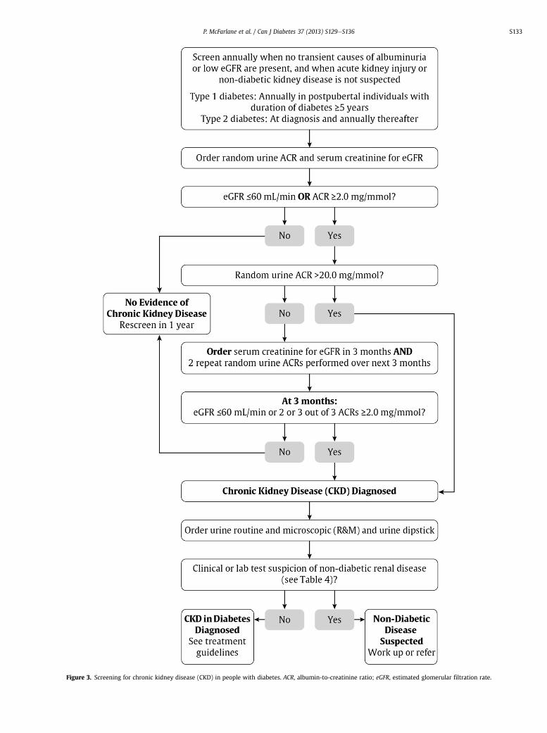
Figure 3. Screening for chronic kidney disease (CKD) in people with diabetes. ACR, albumin-to-creatinine ratio; eGFR, estimated glomerular filtration rate.
P. McFarlane et al. / Can J Diabetes 37 (2013) S129eS136 S133

Figure 4. Relative risk of chronic kidney disease (CKD). Shading shows how adjustedrelative risk is ranked for 5 outcomes from a meta-analysis of general populationcohorts: all-cause mortality, cardiovascular mortality, kidney failure treated by dialysisand transplantation, acute kidney injury, and progression of kidney disease. GFR,glomerular filtration rate.
P. McFarlane et al. / Can J Diabetes 37 (2013) S129eS136S134
As the use of RAAS blockers during pregnancy has been asso-ciated with congenital malformations, women with diabetes ofchildbearing age should avoid pregnancy if drugs from theseclasses are required (84). If a woman with diabetes receiving such
RECOMMENDATIONS
1. In adults, screening for CKD in diabetes should be conducted usinga random urine ACR and a serum creatinine converted into an eGFR [GradeD, Consensus]. Screening should commence at diagnosis of diabetes inindividuals with type 2 diabetes and 5 years after diagnosis in adults withtype 1 diabetes and repeated yearly thereafter. A diagnosis of CKD shouldbe made in patients with a random urine ACR �2.0 mg/mmol and/or aneGFR<60 mL/min on at least 2 of 3 samples over a 3-month period [GradeD, Consensus].
2. All patients with diabetes and CKD should receive a comprehensive,multifaceted approach to reduce cardiovascular risk (see VascularProtection, p. S100) [Grade A, Level 1A (51,86)].
3. Adults with diabetes and CKD with either hypertension or albuminuriashould receive an ACE inhibitor or an ARB to delay progression of CKD[Grade A, Level 1A for ACE inhibitor use in type 1 and type 2 diabetes, andfor ARB use in type 2 diabetes; Grade D, Consensus, for ARB use in type 1diabetes (59,61e65,68,69,87,88)].
4. People with diabetes on an ACE inhibitor or an ARB should have theirserum creatinine and potassium levels checked at baseline and within 1 to2 weeks of initiation or titration of therapy and during times of acuteillness [Grade D, Consensus].
5. Adults with diabetes and CKD should be given a “sick day” medication listthat outlines which medications should be held during times of acuteillness (see Appendix, 7) [Grade D, Consensus].
6. Combination of agents that block the renin-angiotensin-aldosteronesystem (ACE inhibitor, ARB, DRI) should not be routinely used in themanagement of diabetes and CKD [Grade A, Level 1(89,90)].
7. People with diabetes should be referred to a nephrologist or internist withan expertise in CKD in the following situations:a. Chronic, progressive loss of kidney functionb. ACR persistently >60 mg/mmolc. eGFR<30 mL/mind. Unable to remain on renal-protective therapies due to adverse effects
such as hyperkalemia or >30% increase in serum creatinine within 3months of starting an ACE inhibitor or ARB
e. Unable to achieve target BP (could be referred to any specialist inhypertension) [Grade D, Consensus]
Abbreviations:ACE, angiotensin-converting enzyme; ACR, albumin-to-creatinine ratio;ARB, angiotensin II receptor block; CKD, chronic kidney disease; DRI,direct renin inhibitor.
medications wishes to become pregnant, consideration should begiven to their discontinuation prior to conception.
Medication selection and dosing in CKD
Many medications need to have their dose adjusted in thepresence of low kidney function, and some are contraindicated inpeople with significant disease. Appendix 6 lists some medicationscommonly used in people with diabetes and how they should beused if kidney dysfunction is present.
Referral to a specialized renal clinic
Most people with CKD and diabetes will not require referral toa specialist in renal disease. However, specialist care may benecessary when renal dysfunction is severe, when there are diffi-culties implementing renal-protective strategies or when there areproblems managing the sequelae of renal disease (85).
Other Relevant Guidelines
Targets for Glycemic Control, p. S31Monitoring Glycemic Control, p. S35Pharmacotherapy in Type 1 Diabetes, p. S56Pharmacologic Management of Type 2 Diabetes, p. S61Type 1 Diabetes in Children and Adolescents, p. S153Type 2 Diabetes in Children and Adolescents, p. S163Diabetes and Pregnancy, p. S168Diabetes in the Elderly, p. S184
Relevant Appendices
Appendix 6: Therapeutic Considerations for Renal ImpairmentAppendix 7: Sick Day Medication List
References
1. Warram JH, Gearin G, Laffel L, et al. Effect of duration of type I diabetes on theprevalence of stages of diabetic nephropathy defined by urinary albumin/creatinine ratio. J Am Soc Nephrol 1996;7:930e7.
2. Reenders K, de Nobel E, van den Hoogen HJ, et al. Diabetes and its long-termcomplications in general practice: a survey in a well-defined population. FamPract 1993;10:169e72.
3. Weir MR. Albuminuria predicting outcome in diabetes: incidence of micro-albuminuria in Asia-Pacific Rim. Kidney Int 2004;66:S38e9.
4. Canadian Institute for Health Information. Canadian Organ ReplacementRegister Annual Report: Treatment of End-Stage Organ Failure in Canada, 2000to 2009. Ottawa, ON: Canada; 2011.
5. Foley RN, Parfrey PS, Sarnak MJ. Clinical epidemiology of cardiovascular diseasein chronic renal disease. Am J Kidney Dis 1998;32:S112e9.
6. Bell CM, Chapman RH, Stone PW, et al. An off-the-shelf help list: a compre-hensive catalog of preference scores from published cost-utility analyses. MedDecision Making 2001;21:288e94.
7. Mazzucco G, Bertani T, Fortunato M, et al. Different patterns of renal damage intype 2 diabetes mellitus: a multicentric study on 393 biopsies. Am J Kidney Dis2002;39:713e20.
8. Gambara V, Mecca G, Remuzzi G, et al. Heterogeneous nature of renal lesions intype II diabetes. J Am Soc Nephrol 1993;3:1458e66.
9. Mathiesen ER, Ronn B, Storm B, et al. The natural course of microalbuminuria ininsulin-dependent diabetes: a 10-year prospective study. Diabet Med 1995;12:482e7.
10. Lemley KV, Abdullah I, Myers BD, et al. Evolution of incipient nephropathy intype 2 diabetes mellitus. Kidney Int 2000;58:1228e37. Published erratumappears in Kidney Int. 2000;58:2257.
11. Gall MA, Nielsen FS, Smidt UM, et al. The course of kidney function in type 2(non-insulin-dependent) diabetic patients with diabetic nephropathy. Dia-betologia 1993;36:1071e8.
12. Jacobsen P, Rossing K, Tarnow L, et al. Progression of diabetic nephropathy innormotensive type 1 diabetic patients. Kidney Int Suppl 1999;71:S101e5.
13. Hasslacher C, Ritz E, Wahl P, et al. Similar risks of nephropathy in patients withtype I or type II diabetes mellitus. Nephrol Dial Transplant 1989;4:859e63.
14. Biesenbach G, Bodlaj G, Pieringer H, et al. Clinical versus histological diagnosisof diabetic nephropathy: is renal biopsy required in type 2 diabetic patientswith renal disease? QJM 2011;104:771e4.

P. McFarlane et al. / Can J Diabetes 37 (2013) S129eS136 S135
15. Middleton RJ, Foley RN, Hegarty J, et al. The unrecognized prevalence of chronickidney disease in diabetes. Nephrol Dial Transplant 2006;21:88e92.
16. Ruggenenti P, Gambara V, Perna A, et al. The nephropathy of non-insulin-dependent diabetes: predictors of outcome relative to diverse patterns ofrenal injury. J Am Soc Nephrol 1998;9:2336e43.
17. VenkataRaman TV, Knickerbocker F, Sheldon CV. Unusual causes of renalfailure in diabetics: two case studies. J Okla State Med Assoc 1990;83:164e8.
18. Anonymous. Clinical path conference. Unusual renal complications in diabetesmellitus. Minn Med 1967;50:387e93.
19. Amoah E, Glickman JL, Malchoff CD, et al. Clinical identification of nondiabeticrenal disease in diabetic patients with type I and type II disease presentingwith renal dysfunction. Am J Nephrol 1988;8:204e11.
20. El-Asrar AM, Al-Rubeaan KA, Al-Amro SA, et al. Retinopathy as a predictor ofother diabetic complications. Int Ophthalmol 2001;24:1e11.
21. Ballard DJ, Humphrey LL, Melton LJ, et al. Epidemiology of persistent protein-uria in type II diabetes mellitus. Population-based study in Rochester, Minne-sota. Diabetes 1988;37:405e12.
22. Winaver J, Teredesai P, Feldman HA, et al. Diabetic nephropathy as the mode ofpresentation of diabetes mellitus. Metab Clin Exp 1979;28:1023e30.
23. Ahn CW, Song YD, Kim JH, et al. The validity of random urine specimenalbumin measurement as a screening test for diabetic nephropathy. YonseiMed J 1999;40:40e5.
24. Kouri TT, Viikari JS, Mattila KS, et al. Microalbuminuria. Invalidity of simpleconcentration-based screening tests for early nephropathy due to urinaryvolumes of diabetic patients. Diabetes Care 1991;14:591e3.
25. Rodby RA, Rohde RD, Sharon Z, et al. The urine protein to creatinine ratio asa predictor of 24-hour urine protein excretion in type 1 diabetic patients withnephropathy. The Collaborative Study Group. Am J Kidney Dis 1995;26:904e9.
26. Chaiken RL, Khawaja R, Bard M, et al. Utility of untimed urinary albuminmeasurements in assessing albuminuria in black NIDDM subjects. DiabetesCare 1997;20:709e13.
27. Bakker AJ. Detection of microalbuminuria. Receiver operating characteristiccurve analysis favors albumin-to-creatinine ratio over albumin concentration.Diabetes Care 1999;22:307e13.
28. Huttunen NP, Kaar M, Puukka R, et al. Exercise-induced proteinuria in childrenand adolescents with type 1 (insulin dependent) diabetes. Diabetologia 1981;21:495e7.
29. Solling J, Solling K, Mogensen CE. Patterns of proteinuria and circulatingimmune complexes in febrile patients. Acta Med Scand 1982;212:167e9.
30. Ritz E. Nephropathy in type 2 diabetes. J Intern Med 1999;245:111e26.31. Wiseman M, Viberti G, Mackintosh D, et al. Glycaemia, arterial pressure and
micro-albuminuria in type 1 (insulin-dependent) diabetes mellitus. Dia-betologia 1984;26:401e5.
32. Ravid M, Savin H, Lang R, et al. Proteinuria, renal impairment, metaboliccontrol, and blood pressure in type 2 diabetes mellitus. A 14-year follow-upreport on 195 patients. Arch Intern Med 1992;152:1225e9.
33. Gault MH, Longerich LL, Harnett JD, et al. Predicting glomerular function fromadjusted serum creatinine. Nephron 1992;62:249e56.
34. Bending JJ, Keen H, Viberti GC. Creatinine is a poor marker of renal failure.Diabet Med 1985;2:65e6.
35. Shemesh O, Golbetz H, Kriss JP, et al. Limitations of creatinine as a filtrationmarker in glomerulopathic patients. Kidney Int 1985;28:830e8.
36. Levey AS, Bosch JP, Lewis JB, et al. A more accurate method to estimateglomerular filtration rate from serum creatinine: a new prediction equation.Modification of Diet in Renal Disease Study Group. Ann Intern Med 1999;130:461e70.
37. Poggio ED, Wang X, Greene T, et al. Performance of the modification of diet inrenal disease and Cockcroft-Gault equations in the estimation of GFR in healthand in chronic kidney disease. J Am Soc Nephrol 2005;16:459e66.
38. Wang PH, Lau J, Chalmers TC. Meta-analysis of effects of intensive blood-glucose control on late complications of type I diabetes. Lancet 1993;341:1306e9.
39. Anonymous. Effect of intensive therapy on the development and progression ofdiabetic nephropathy in the Diabetes Control and Complications Trial. TheDiabetes Control and Complications (DCCT) Research Group. Kidney Int 1995;47:1703e20.
40. Anonymous. Intensive blood-glucose control with sulphonylureas or insulincompared with conventional treatment and risk of complications in patientswith type 2 diabetes (UKPDS 33). UK Prospective Diabetes Study (UKPDS)Group. Lancet 1998;352:837e53.
41. Retinopathy and nephropathy in patients with type 1 diabetes four years aftera trial of intensive therapy. The Diabetes Control and Complications Trial/Epidemiology of Diabetes Interventions and Complications Research Group.N Eng J Med 2000;342:381e9.
42. Shichiri M, Kishikawa H, Ohkubo Y, et al. Long-term results of the KumamotoStudy on optimal diabetes control in type 2 diabetic patients. Diabetes Care2000;23(Suppl 2):B21e9.
43. Tight blood pressure control and risk of macrovascular and microvascularcomplications in type 2 diabetes: UKPDS 38. UK Prospective Diabetes StudyGroup. BMJ 1998;317:703e13.
44. Schrier RW, Estacio RO, Mehler PS, et al. Appropriate blood pressure control inhypertensive and normotensive type 2 diabetes mellitus: a summary of theABCD trial. Nat Clin Pract Nephrol 2007;3:428e38.
45. de Galan BE, Perkovic V, Ninomiya T, et al. Lowering blood pressure reducesrenal events in type 2 diabetes. J Am Soc Nephrol 2009;20:883e92.
46. Ruggenenti P, Fassi A, Ilieva AP, et al. Preventing microalbuminuria in type 2diabetes. N Engl J Med 2004;351:1941e51.
47. Chaturvedi N. Randomised placebo-controlled trial of lisinopril in normoten-sive patients with insulin-dependent diabetes and normoalbuminuria ormicroalbuminuria. Lancet 1997;349:1787e92.
48. Bilous R, Chaturvedi N, Sjolie AK, et al. Effect of candesartan on micro-albuminuria and albumin excretion rate in diabetes: three randomized trials.Ann Intern Med 2009;151:11e20. W13-W14.
49. Mauer M, Zinman B, Gardiner R, et al. Renal and retinal effects of enalapril andlosartan in type 1 diabetes. N Engl J Med 2009;361:40e51.
50. Gerstein HC, Mann JF, Yi Q, et al. Albuminuria and risk of cardiovascular events,death, and heart failure in diabetic and nondiabetic individuals. JAMA 2001;286:421e6.
51. Gaede P, Vedel P, Larsen N, et al. Multifactorial intervention and cardiovasculardisease in patients with type 2 diabetes. N Engl J Med 2003;348:383e93.
52. Gaede P, Vedel P, Parving HH, et al. Intensified multifactorial intervention inpatients with type 2 diabetes mellitus and microalbuminuria: the Steno type 2randomised study. Lancet 1999;353:617e22.
53. Levey AS, Coresh J. Chronic kidney disease. Lancet 2012;379:165e80.54. Levey AS, Eckardt KU, Tsukamoto Y, et al. Definition and classification of
chronic kidney disease: a position statement from Kidney Disease: ImprovingGlobal Outcomes (KDIGO). Kidney Int 2005;67:2089e100.
55. Maki DD, Ma JZ, Louis TA, et al. Long-term effects of antihypertensive agents onproteinuria and renal function. Arch Intern Med 1995;155:1073e80.
56. Kasiske BL, Kalil RS, Ma JZ, et al. Effect of antihypertensive therapy on thekidney in patients with diabetes: a meta-regression analysis. Ann Intern Med1993;118:129e38.
57. Barnett AH, Bain SC, Bouter P, et al. Angiotensin-receptor blockade versusconverting-enzyme inhibition in type 2 diabetes and nephropathy. N Engl JMed 2004;351:1952e61.
58. Yusuf S, Sleight P, Pogue J, et al. Effects of an angiotensin-converting-enzymeinhibitor, ramipril, on cardiovascular events in high-risk patients. The HeartOutcomes Prevention Evaluation Study Investigators. N Engl J Med 2000;342:145e53.
59. Lewis EJ, Hunsicker LG, Bain RP, et al. The effect of angiotensin-converting-enzyme inhibition on diabetic nephropathy. The Collaborative Study Group.N Engl J Med 1993;329:1456e62.
60. Andersen S, Tarnow L, Rossing P, et al. Renoprotective effects of angiotensin IIreceptor blockade in type 1 diabetic patients with diabetic nephropathy.Kidney Int 2000;57:601e6.
61. Strippoli GF, Craig MC, Schena FP, et al. Role of blood pressure targets andspecific antihypertensive agents used to prevent diabetic nephropathy anddelay its progression. J Am Soc Nephrol 2006;17:S153e5.
62. Lewis EJ, Hunsicker LG, Clarke WR, et al. Renoprotective effect of theangiotensin-receptor antagonist irbesartan in patients with nephropathy dueto type 2 diabetes. N Eng J Med 2001;345:851e60.
63. Brenner BM, Cooper ME, de Zeeuw D, et al. Effects of losartan on renal andcardiovascular outcomes in patients with type 2 diabetes and nephropathy.N Engl J Med 2001;345:861e9.
64. Parving HH, Lehnert H, Brochner-Mortensen J, et al. The effect of irbesartan onthe development of diabetic nephropathy in patients with type 2 diabetes.N Engl J Med 2001;345:870e8.
65. Laffel LM, McGill JB, Gans DJ. The beneficial effect of angiotensin-convertingenzyme inhibition with captopril on diabetic nephropathy in normotensiveIDDM patients with microalbuminuria. North American MicroalbuminuriaStudy Group. Am J Med 1995;99:497e504.
66. Mathiesen ER, Hommel E, Giese J, et al. Efficacy of captopril in postponingnephropathy in normotensive insulin dependent diabetic patients withmicroalbuminuria. BMJ 1991;303:81e7.
67. Jerums G, Allen TJ, Campbell DJ, et al. Long-term comparison between peri-ndopril and nifedipine in normotensive patients with type 1 diabetes andmicroalbuminuria. Am J Kidney Dis 2001;37:890e9.
68. ACE Inhibitors in Diabetic Nephropathy Trialist Group. Should all patients withtype 1 diabetes mellitus and microalbuminuria receive angiotensin-convertingenzyme inhibitors? A meta-analysis of individual patient data. Ann Intern Med2001;134:370e9.
69. Ravid M, Savin H, Jutrin I, et al. Long-term stabilizing effect of angiotensin-converting enzyme inhibition on plasma creatinine and on proteinuria innormotensive type II diabetic patients. Ann Intern Med 1993;118:577e81.
70. Ruggenenti P, Perna A, Gherardi G, et al. Renal function and requirement fordialysis in chronic nephropathy patients on long-term ramipril: REIN follow-up trial. Gruppo Italiano di Studi Epidemiologici in Nefrologia (GISEN). Ram-ipril Efficacy in Nephropathy. Lancet 1998;352:1252e6.
71. Maschio G, Alberti D, Locatelli F, et al. Angiotensin-converting enzyme inhib-itors and kidney protection: the AIPRI trial. The ACE Inhibition in ProgressiveRenal Insufficiency (AIPRI) Study Group. J Cardiovasc Pharmacol 1999;33(suppl1):S16e20. discussion S41eS43.
72. Shoda J, Kanno Y, Suzuki H. A five-year comparison of the renal protective effectsof angiotensin-converting enzyme inhibitors and angiotensin receptor blockersin patients with non-diabetic nephropathy. Intern Med 2006;45:193e8.
73. Igarashi M, Hirata A, Kadomoto Y, et al. Dual blockade of angiotensin II withenalapril and losartan reduces proteinuria in hypertensive patients with type 2diabetes. Endocr J 2006;53:493e501.
74. Jacobsen P, Parving HH. Beneficial impact on cardiovascular risk factors by dualblockade of the renin-angiotensin system in diabetic nephropathy. Kidney Int2004;66:S108e10.

P. McFarlane et al. / Can J Diabetes 37 (2013) S129eS136S136
75. Burgess E, Muirhead N, Rene de Cotret P, et al. Supramaximal dose of cande-sartan in proteinuric renal disease. J Am Soc Nephrol 2009;20:893e900.
76. Epstein M, Williams GH, Weinberger M, et al. Selective aldosterone blockadewith eplerenone reduces albuminuria in patients with type 2 diabetes. Clin JAm Soc Nephrol 2006;1:940e51.
77. Parving HH, Persson F, Lewis JB, et al. Aliskiren combined with losartan in type2 diabetes and nephropathy. N Engl J Med 2008;358:2433e46.
78. Mann JF, Schmieder RE, McQueen M, et al. Renal outcomes with telmisartan,ramipril, or both, in people at high vascular risk (the ONTARGET study): a mul-ticentre, randomised, double-blind, controlled trial. Lancet 2008;372:547e53.
79. Bakris GL, Weir MR. Angiotensin-converting enzyme inhibitor-associatedelevations in serum creatinine: is this a cause for concern? Arch Intern Med2000;160:685e93.
80. Reams GP, Bauer JH, Gaddy P. Use of the converting enzyme inhibitorenalapril in renovascular hypertension. Effect on blood pressure, renalfunction, and the renin-angiotensin-aldosterone system. Hypertension 1986;8:290e7.
81. Franklin SS, Smith RD. Comparison of effects of enalapril plus hydrochloro-thiazide versus standard triple therapy on renal function in renovascularhypertension. Am J Med 1985;79:14e23.
82. Miyamori I, Yasuhara S, Takeda Y, et al. Effects of converting enzyme inhibitionon split renal function in renovascular hypertension. Hypertension 1986;8:415e21.
83. Palmer BF. Managing hyperkalemia caused by inhibitors of the renin-angiotensin-aldosterone system. N Engl J Med 2004;351:585e92.
84. Cooper WO, Hernandez-Diaz S, Arbogast PG, et al. Major congenital malfor-mations after first-trimester exposure to ACE inhibitors. N Engl J Med 2006;354:2443e51.
85. Levin A, Mendelssohn D. Care and referral of adult patients with reducedkidney function: position paper from the Canadian Society of Nephrology.2006.
86. Gaede P, Lund-Andersen H, Parving HH, et al. Effect of a multifactorial inter-vention on mortality in type 2 diabetes. N Engl J Med 2008;358:580e91.
87. Mathiesen ER, Hommel E, Hansen HP, et al. Randomised controlled trial of longterm efficacy of captopril on preservation of kidney function in normotensivepatients with insulin dependent diabetes and microalbuminuria. BMJ 1999;319:24e5.
88. Viberti G, Wheeldon NM. Microalbuminuria reduction with valsartan inpatients with type 2 diabetes mellitus: a blood pressure-independent effect.MicroAlbuminuria Reduction With VALsartan Study Investigators. Circulation2002;106:672e8.
89. Tobe SW, Clase CM, Gao P, et al. Cardiovascular and renal outcomes with tel-misartan, ramipril, or both in people at high renal risk: results from theONTARGET and TRANSCEND studies. Circulation 2011;123:1098e107.
90. Parving H, Brenner BM, McMurray JJV, et al. Cardiorenal end points in a trial ofaliskiren for type 2 diabetes. N Engl J Med 2012;367:2204e13.

Contents lists available at SciVerse ScienceDirect
Can J Diabetes 37 (2013) S137eS141
Canadian Journal of Diabetesjournal homepage:
www.canadianjournalofdiabetes.com
Clinical Practice Guidelines
Retinopathy
Canadian Diabetes Association Clinical Practice Guideline Expert Committee
The initial draft of this chapter was prepared by Shelley R. Boyd MD, FRCSC,Andrew Advani MB ChB, PhD, FRCP(UK), Filiberto Altomare MD, FRCSC, Frank Stockl MD, FRCSC
KEY MESSAGES
� Screening is important for early detection of treatable disease. Screeningintervals for diabetic retinopathy vary according to the individual’s age andtype of diabetes.
� Tight glycemic control reduces the onset and progression of sight-threatening diabetic retinopathy.
� Laser therapy, local intraocular pharmacological therapy and surgeryreduce the risk of significant visual loss.
Introduction
Diabetic retinopathy is the most common cause of new cases oflegal blindness in people of working age (1). The Eye DiseasesPrevalence Research Group determined the crude prevalence rateof retinopathy in the adult population with diabetes of the UnitedStates to be 40.3%; sight-threatening retinopathy occurred at a rateof 8.2% (1). Previous data showed the prevalence rate of prolifera-tive retinopathy to be 23% in people with type 1 diabetes, 14% inpeople with type 2 diabetes and on insulin therapy, and 3% inpeople receiving oral antihyperglycemic therapies (2). Macularedema occurs in 11%, 15% and 4% of these groups, respectively (3).Higher prevalence rates were noted in First Nations populations inCanada (4,5).
Visual loss is associated with significant morbidity, includingincreased falls, hip fracture and a 4-fold increase in mortality (6).Among individuals with type 1 diabetes, limb amputation andvisual loss due to diabetic retinopathy are the independentpredictors of early death (7).
Definition and Pathogenesis
Diabetic retinopathy is clinically defined, diagnosed and treatedbased on the extent of retinal vascular disease exclusively. Threedistinct forms of diabetic retinopathy are described: 1) macularedema, which includes diffuse or focal vascular leakage at themacula; 2) progressive accumulation of blood vessel change thatincludes microaneurysms, intraretinal hemorrhage, vasculartortuosity and vascular malformation (together known as non-proliferative diabetic retinopathy) that ultimately leads toabnormal vessel growth (proliferative diabetic retinopathy); and 3)retinal capillary closure, a form of vascular change detected on
1499-2671/$ e see front matter � 2013 Canadian Diabetes Associationhttp://dx.doi.org/10.1016/j.jcjd.2013.01.038
fluorescein angiography, which is also well recognized as a poten-tially blinding complication of diabetes but currently has notreatment options.
Screening
Because laser therapy for sight-threatening diabetic retinopathyreduces the risk of blindness, ophthalmic screening strategies areintended to detect disease treatable by this modality (8e11). Sight-threatening diabetic retinopathy includes severe nonproliferativediabetic retinopathy, proliferative diabetic retinopathy or clinicallysignificant macular edema (CSME) (8), a strictly defined form ofdiabetic macular edema (DME) that relies on the clinical assess-ment of retinal thickening based on subjective assessment of areaand distance from the fovea (the centre of the macula responsiblefor high-acuity vision), with or without so-called hard exudates.Since the introduction of new treatments based on intravitreal(intraocular) injection of pharmacological agents and use of OpticalCoherence Tomography (OCT) to quantify macular thickness, themore general term DME, or “centre-involving” DME has come todescribe patients who could benefit from this treatment over laser,the latter of which cannot be applied to the fovea. Despite thechange in treatment modalities, screening programs remainunchanged and consider the differences in incidence and preva-lence of retinopathy observed in type 1 and type 2 diabetes, anddistinguish between children and adults (Table 1) (12e17).
Diabetic retinopathy rarely develops in children with type 1diabetes <10 years of age regardless of the duration of diabetes(16). Among patients <15 years of age, irrespective of age of onsetof diabetes, the prevalence of mild nonproliferative retinopathywas 2%, and none had sight-threatening diabetic retinopathy (9,16).However, the prevalence rate increases sharply after 5 years’duration of diabetes in postpubertal individuals with type 1 dia-betes (16). In the Wisconsin Epidemiology Study of Diabetic Reti-nopathy 4-year incidence study, no person <17 years of agedeveloped proliferative retinopathy or macular edema (14,18,19).Conversely, in people with type 2 diabetes, retinopathy may bepresent in 21% to 39% of patients soon after clinical diagnosis but issight-threatening in only about 3% (3,15,17,20). In the UnitedKingdom Prospective Diabetes Study (UKPDS), few patientswithout retinopathy at diagnosis of diabetes had disease progres-sion to the point of requiring retinal photocoagulation (lasertreatment) in the following 3 to 6 years (21). More recently,

Table 1Screening for retinopathy
When to initiate screening� Five years after diagnosis of type 1 diabetes in all individuals �15 years� In all individuals at diagnosis of type 2 diabetes
Screening methods� Seven-standard field, stereoscopic-colour fundus photography withinterpretation by a trained reader (gold standard)
� Direct ophthalmoscopy or indirect slit-lamp funduscopy through dilatedpupil
� Digital fundus photographyIf retinopathy is present
� Diagnose retinopathy severity and establish appropriate monitoringintervals (�1 year)
� Treat sight-threatening retinopathywith laser, pharmacological or surgicaltherapy
� Review glycemic, BP and lipid control, and adjust therapy to reach targetsper guidelines*
� Screen for other diabetes complicationsIf retinopathy is not present
� Type 1 diabetes: rescreen annually� Type 2 diabetes: rescreen every 1e2 years� Review glycemic, BP and lipid control, and adjust therapy to reach targetsper guidelines*
� Screen for other diabetes complications
BP, blood pressure.* See “Other Relevant Guidelines”.
S.R. Boyd et al. / Can J Diabetes 37 (2013) S137eS141S138
progression rates of diabetic retinopathy were prospectively eval-uated (12,13,22). The Liverpool Diabetic Eye Study reported the1-year cumulative incidence of sight-threatening diabetic reti-nopathy in individuals with type 1 or type 2 diabetes who, atbaseline, had no diabetic retinopathy, had background retinopathyor had mild preproliferative retinopathy. In people with type 1diabetes, the incidence in these groups was 0.3%, 3.6% and 13.5%,respectively (12), and in type 2 diabetes individuals it was 0.3%,5.0% and 15.0%, respectively (13). Although the incidence of sight-threatening diabetic retinopathy in the group without baselinediabetic retinopathy is low (12,13,21,22), there have been no studiescomparing various screening intervals in their effectiveness toreduce the risk of vision loss (23).
Telemedicine programs relying on fundus photography arewidely used in Canada and internationally for the identification andtriage of patients with diabetic retinopathy (24). Programs relyingon OCT to evaluate macular edema are under investigation.
Delay of Onset and Progression
Risk factors for the development or progression of diabeticretinopathy are longer duration of diabetes, elevated glycatedhemoglobin (A1C), increased blood pressure (BP), dyslipidemia,low hemoglobin level, pregnancy (with type 1 diabetes), protein-uria and severe retinopathy itself (14e17,19,25e31).
Glycemic control
Tight glycemic control, targeting an A1C �7%, is recommendedto slow the development and progression of diabetic retinopathy.The Diabetes Control and Complications Trial (DCCT) and theUKPDS demonstrated that intensive glycemic control (A1C �7%)reduced both the development and progression of retinopathy(32e34), with the beneficial effects of intensive glycemic controlpersisting for up to 10 years after completion of the initial trials(35,36). Two studies examined the effect of more aggressive bloodglucose lowering (A1C �6.5%) in patients with established type 2diabetes (duration 6 to 10 years). In the Action to Control Cardio-vascular Risk in Diabetes (ACCORD) Eye study, intensive glycemiccontrol was associated with a lower rate of retinopathy progressionthan standard therapy (37), while in the Action in Diabetes and
Vascular Disease: Preterax and Diamicron MR Controlled Evalua-tion (ADVANCE) Retinal Measurements study (AdRem), intensiveglycemic control did not significantly reduce development orprogression of retinopathy (38). In type 1 diabetes, rapidimprovement of glycemia may be associated with transient earlyworsening of retinopathy, but this effect is offset by long-termbenefits (39).
Blood pressure control
BP control is an important component of risk factormodificationin diabetes and reduces the risk of retinopathy progression. TheUKPDS showed that, among patients with newly diagnosed type 2diabetes, BP control (target BP <150/85 mm Hg, actual BP144/82 mm Hg) resulted in a significant reduction in retinopathyprogression as well as a decrease in significant visual loss andrequirement for laser therapy compared to less control (targetBP <180/105 mm Hg, actual mean BP 154/87 mm Hg) (40). TheACCORD and ADVANCE studies examined more aggressive BPlowering in patients with established type 2 diabetes. In both thesestudies, where mean BP was <140/80 mm Hg in both the activeintervention and control groups, active treatment did not showadditional benefit vs. standard therapy.
Although a number of trials have examined the effect of renin-angiotension system (RAS) blockade on retinopathy progressionor development among normotensive patients with diabetes, theresults generally have been conflicting or inconclusive. In theRenin-Angiotensin System Study (RASS), involving 223 normoten-sive, normoalbuminuric participants with type 1 diabetes, neitherthe angiotensin-converting enzyme (ACE) inhibitor, enalapril, northe angiotensin receptor blocker (ARB), losartan, reduced retinop-athy progression independent of BP change (41). The DiabeticRetinopathy Candesartan Trials (DIRECT) program, involving 5231participants, evaluated the effect of the angiotension II type 1 ARBcandesartan 32 mg daily on the incidence of new retinopathy inpatients with type 1 diabetes (DIRECT-Prevent 1) (42) and on theprogression of retinopathy in patients with either type 1 diabetes(DIRECT-Protect 1) (42) or type 2 diabetes (DIRECT-Protect 2) (43).The DIRECT studies did not meet their primary endpoints, althoughthere was an overall change toward less severe retinopathy withcandesartan (42,43). Thus, while BP lowering (including use of RASblockers) reduces retinopathy rates and is an important componentof vascular protection, there is insufficient evidence to recommendRAS blockade as primary prevention for retinopathy for allnormotensive patients with diabetes.
Lipid-lowering therapy
Dyslipidemia is an independent risk factor for retinal hardexudates and CSME in type 1 diabetes (28,44). While statin-basedlipid-lowering therapies are an integral part of vascular protec-tion in diabetes, the role of these agents in preventing the devel-opment or progression of retinopathy has not been established(34,45). The role of the peroxisome proliferator-activatedreceptor-alpha agonist, fenofibrate, has been assessed in 2 large-scale randomized controlled trials. In the Fenofibrate Interventionand Event Lowering in Diabetes (FIELD) study, fenofibrate 200 mgdaily reduced both the requirement for laser therapy (a pre-specified tertiary endpoint) and retinopathy progression amongpatients with pre-existing retinopathy (46). In the ACCORD Eyestudy, the addition of fenofibrate 160 mg daily to simvastatin wasassociated with a 40% reduction in the primary outcome of reti-nopathy progression over 4 years (37). From the study’s control andevent rates, the number of patients needed to treat with combi-nation statin and fenofibrate therapy to prevent 1 retinopathyprogression event is estimated at 27 over the 4-year period. The
RECOMMENDATIONS
1. In individuals �15 years of age with type 1 diabetes, screening and eval-uation for retinopathy by an expert professional should be performedannually starting 5 years after the onset of diabetes [Grade A, Level 1(14,16)].
2. In individuals with type 2 diabetes, screening and evaluation for diabeticretinopathy by an expert professional should be performed at the time ofdiagnosis of diabetes [Grade A, Level 1 (15,18)] and annually thereafter. Theinterval for follow-up assessments should be tailored to the severity of theretinopathy. In those with no or minimal retinopathy, the recommendedinterval is 1e2 years [Grade A, Level 1 (15,18)].
3. Screening for diabetic retinopathy should be performed by experiencedprofessionals, either in person or through interpretation of retinalphotographs taken through dilated pupils [Grade A, Level 1 (66)].
4. To prevent the onset and delay the progression of diabetic retinopathy,people with diabetes should be treated to achieve optimal control of bloodglucose [Grade A, Level 1A (32,33)] and BP [Grade A, Level 1A (40), for type2 diabetes].
5. Though not recommended for CVD prevention or treatment, fenofibrate, inaddition to statin therapy, may be used in patients with type 2 diabetes toslow the progression of established retinopathy [Grade A, Level 1A(37,46)].
6. Patients with sight-threatening diabetic retinopathy should be assessed bya general ophthalmologist or retina specialist [Grade D, Consensus]. Lasertherapy and/or vitrectomy [Grade A, Level 1A (8,10,57,58)] and/or phar-macological intervention [Grade A, Level 1A (51,52,55,56)] should be used.
7. Visually disabled people should be referred for low-vision evaluation andrehabilitation [Grade D, Consensus].
Abbreviations:BP, blood pressure; CVD, cardiovascular disease.
S.R. Boyd et al. / Can J Diabetes 37 (2013) S137eS141 S139
mechanism for any beneficial effect of fenofibrate in diabetic reti-nopathy has not been established, with active treatment beingassociated with an increase in high-density lipoprotein-cholesteroland decrease in serum triglycerides in ACCORD Eye (37) butappearing to be independent of plasma lipid concentrations inFIELD (46). Thus, the addition of fenofibrate to statin therapy couldbe considered in patients with type 2 diabetes to slow theprogression of established retinopathy.
Antiplatelet therapy
Systematic review suggests that acetylsalicylic acid (ASA)therapy neither decreases nor increases the incidence or progres-sion of diabetic retinopathy (47). Correspondingly, ASA use does notappear to be associated with an increase in risk of vitreoushemorrhage or DME (48,49).
Treatment
Treatment modalities for diabetic retinopathy include retinalphotocoagulation, intraocular injection of pharmacological agentsand vitreoretinal surgery.
Laser therapy
As determined in the Diabetic Retinopathy Study (DRS) and theEarly Treatment Diabetic Retinopathy Study (ETDRS), laser therapyby panretinal photocoagulation to the retinal periphery reducessevere visual loss and reduces legal blindness by 90% in people withsevere nonproliferative or proliferative retinopathy (9e11). Asdetermined by the ETDRS, focal and/or grid laser treatment to themacula for CSME reduces the incidence of moderate visual loss by50% (8). Long-term follow-up studies to the original laser photo-coagulation trials confirm its benefit over several decades (50).
Local (intraocular) pharmacological intervention
In the treatment of DME with centre-involving disease, asdefined by OCT or clinical examination, intraocular pharmaco-therapy is now available. With the knowledge that the cytokinevascular endothelial growth factor (VEGF) plays a primary role inthe development of DME, 2 anti-VEGF drugs are now widely used.Two masked phase III clinical trials, RISE and RIDE, using monthlyranibizumab, a humanized recombinant anti-VEGF antibody frag-ment, with or without prompt laser, improved visual acuitycompared against sham over the 2 years of study (51). In the RISEtrial, 44% and 39% of patients receiving 0.3 or 0.5 mg ranibizumab,respectively, gained 15 letters or more (3 lines) of acuity vs. 18% ofthose in the control arm. In the RIDE study, 33% or 45% of patientsgained 15 letters or more at doses of 0.3 or 0.5 mg, respectively.Furthermore, 1-year results of a phase III clinical trial, RESTORE,using an initial loading dose of 3 monthly injections of 0.5 mgranibizumab, and as-needed treatment thereafter, likewise showedimprovement in the primary and secondary outcome measures ofbest correct visual acuity and reduction in central macular thick-ness. In all studies, this was true when ranibizumab was used asmonotherapy or in conjunction with macular photocoagulation. Inthe RESTORE study, 37% to 43% of ranibizumab-treated patientsimproved vision by 10 letters or more compared to 16% withstandard laser therapy (52). Two-year results are pending. Similarresults were obtained by the Diabetic Retinopathy Clinical ResearchNetwork using physician-based flexible treatment algorithmsrather than a strict prescribed injection schedule (53). Intravitrealinjection with ranibizumab is approved by Health Canada.
A similar outcome was noted when comparing intraocularinjection of bevacizumab (a full-length antibody against VEGF) to
macular laser. Two-year results of a phase III clinical trial, the BOLTtrial, demonstrated a gain of at least 15 letters or more in 32% ofpatients receiving 1.25 mg bevacizumab compared to 4% in thecontrol arm (54). However, unlike ranibizumab, intraocular injec-tion of bevacizumab in diabetic retinopathy constitutes off-labeluse of the drug in Canada.
Steroids are an alternate class of drug evaluated in the treatmentof DME. Intraocular injection of steroid combined with promptmacular laser was as effective as ranibizumab in a single subgroupof patients characterized by previous cataract surgery (53).However, treatment with intraocular steroid was associated withincreased rates of glaucoma. Two phase III clinical trials investi-gating the implantation of a long-term drug delivery device con-taining fluocinolone acetonide met their primary and secondaryoutcomes (visual acuity and OCT) but showed increased rates ofglaucoma and cataract progression compared to sham (55,56). Therisk-to-benefit ratio was considered unacceptable to the UnitedStates Food and Drug Administration (FDA) where the treatmentwas not approved. By contrast, the fluocinolone insert has receivedapproval in several European countries.
Surgical intervention
The Diabetic Retinopathy Vitrectomy Study (DRVS) Groupevaluated the benefit of early vitrectomy (<6 months) in thetreatment of severe vitreous hemorrhage (57) and very severeproliferative diabetic retinopathy (58). People with type 1 diabetesof <20 years’ duration and severe vitreous hemorrhage were morelikely to achieve good vision with early vitrectomy compared toconventional management (57). Similarly, early vitrectomy was
S.R. Boyd et al. / Can J Diabetes 37 (2013) S137eS141S140
associated with higher chance of visual recovery in people witheither type 1 or 2 diabetes with very severe proliferative diabeticretinopathy (58). Surgical advances in vitrectomy since the DRVStrials have demonstrated reduced side effects with more consistentfavourable visual outcomes, thus supporting vitrectomy inadvanced proliferative diabetic retinopathy (59). Furthermore,these advances have expanded surgical indications to includevitrectomy for diffuse macular edema with or without vitre-omacular traction (60). It is worth noting that the use of peri-operative ASA (49,61,62) and warfarin therapy (63) for personsundergoing ophthalmic surgery does not appear to raise the risk ofhemorrhagic complications.
Overall, the last few years have seen significant advances insystemic, local and surgical treatments of diabetic eye disease, withsignificantly improved visual outcome. Most notably, long-termfollow-up to early laser studies confirm their sustained efficacy inpreserving vision (50). New therapies, such as intraocular phar-macological treatment, await long-term follow-up but alreadydemonstrate both preservation and recovery of vision in personswith DME. Despite these successes, it is important to encouragepatients with even moderate visual loss to seek assistance fromcommunity services that provide spectacle correction, enhancedmagnification, vision aids and measures to encourage indepen-dence and ongoing quality of life (64,65).
Other Relevant Guidelines
Targets for Glycemic Control, p. S31Dyslipidemia, p. S110Treatment of Hypertension, p. S117Type 1 Diabetes in Children and Adolescents, p. S153Type 2 Diabetes in Children and Adolescents, p. S163Diabetes and Pregnancy, p. S168
References
1. Kempen JH, O’Colmain BJ, Leske MC, et al. The prevalence of diabetic reti-nopathy among adults in the United States. Arch Ophthalmol 2004;122:552e63.
2. Klein R, Klein BE, Moss SE. Epidemiology of proliferative diabetic retinopathy.Diabetes Care 1992;15:1875e91.
3. Klein R, Klein BE, Moss SE, et al. The Wisconsin epidemiologic study of diabeticretinopathy. IV. Diabetic macular edema. Ophthalmology 1984;91:1464e74.
4. Kaur H, Maberley D, Chang A, Hay D. The current status of diabetes care, dia-betic retinopathy screening and eye-care in British Columbia’s First NationsCommunities. Int J Circumpolar Health 2004;63:277e85.
5. Maberley D, Walker H, Koushik A, Cruess A. Screening for diabetic retinopathyin James Bay, Ontario: a cost-effectiveness analysis. CMAJ 2003;168:160e4.
6. Vu HT, Keeffe JE, McCarty CA, Taylor HR. Impact of unilateral and bilateralvision loss on quality of life. Br J Ophthalmol 2005;89:360e3.
7. Cusick M, Meleth AD, Agron E, et al. Associations of mortality and diabetescomplications in patients with type 1 and type 2 diabetes: early treatmentdiabetic retinopathy study report no. 27. Diabetes Care 2005;28:617e25.
8. Photocoagulation for diabetic macular edema. Early Treatment Diabetic Reti-nopathy Study report number 1. Early Treatment Diabetic Retinopathy StudyResearch Group. Arch Ophthalmol 1985;103:1796e806.
9. Ferris 3rd FL. How effective are treatments for diabetic retinopathy? JAMA1993;269:1290e1.
10. Photocoagulation treatment of proliferative diabetic retinopathy: the secondreport of diabetic retinopathy study findings. Ophthalmology 1978;85:82e106.
11. Ferris F. Early photocoagulation in patients with either type I or type II dia-betes. Trans Am Ophthalmol Soc 1996;94:505e37.
12. Younis N, Broadbent DM, Harding SP, Vora JP. Incidence of sight-threateningretinopathy in Type 1 diabetes in a systematic screening programme. DiabetMed 2003;20:758e65.
13. Younis N, Broadbent DM, Vora JP, et al. Incidence of sight-threatening reti-nopathy in patients with type 2 diabetes in the Liverpool Diabetic Eye Study:a cohort study. Lancet 2003;361:195e200.
14. Klein R, Klein BE, Moss SE, et al. The Wisconsin Epidemiologic Study of DiabeticRetinopathy. IX. Four-year incidence and progression of diabetic retinopathywhen age at diagnosis is less than 30 years. Arch Ophthalmol 1989;107:237e43.
15. Klein R, Klein BE, Moss SE, et al. The Wisconsin Epidemiologic Study of DiabeticRetinopathy. X. Four-year incidence and progression of diabetic retinopathywhen age at diagnosis is 30 years or more. Arch Ophthalmol 1989;107:244e9.
16. Klein R, Klein BE, Moss SE, et al. The Wisconsin epidemiologic study of diabeticretinopathy. II. Prevalence and risk of diabetic retinopathy when age at diag-nosis is less than 30 years. Arch Ophthalmol 1984;102:520e6.
17. Klein R, Klein BE, Moss SE, et al. The Wisconsin epidemiologic study of diabeticretinopathy. III. Prevalence and risk of diabetic retinopathy when age atdiagnosis is 30 or more years. Arch Ophthalmol 1984;102:527e32.
18. Klein R, Klein BE, Moss SE, et al. The Wisconsin Epidemiologic Study of DiabeticRetinopathy. VII. Diabetic nonproliferative retinal lesions. Ophthalmology1987;94:1389e400.
19. Klein R, Moss SE, Klein BE, et al. The Wisconsin epidemiologic study of diabeticretinopathy. XI. The incidence of macular edema. Ophthalmology 1989;96:1501e10.
20. Kohner EM, Aldington SJ, Stratton IM, et al. United Kingdom Prospective Dia-betes Study, 30: diabetic retinopathy at diagnosis of non-insulin-dependentdiabetes mellitus and associated risk factors. Arch Ophthalmol 1998;116:297e303.
21. Kohner EM, Stratton IM, Aldington SJ, et al, UKPDS Group. Relationshipbetween the severity of retinopathy and progression to photocoagulation inpatients with Type 2 diabetes mellitus in the UKPDS (UKPDS 52). Diabet Med2001;18:178e84.
22. Maguire A, Chan A, Cusumano J, et al. The case for biennial retinopathyscreening in children and adolescents. Diabetes Care 2005;28:509e13.
23. Klein R. Screening interval for retinopathy in type 2 diabetes. Lancet 2003;361:190e1.
24. Whited JD. Accuracy and reliability of teleophthalmology for diagnosing dia-betic retinopathy and macular edema: a review of the literature. DiabetesTechnol Ther 2006;8:102e11.
25. Klein R, Klein BE, Moss SE, Cruickshanks KJ. The Wisconsin EpidemiologicStudy of Diabetic Retinopathy: XVII. The 14-year incidence and progression ofdiabetic retinopathy and associated risk factors in type 1 diabetes. Ophthal-mology 1998;105:1801e15.
26. Davis MD, Fisher MR, Gangnon RE, et al. Risk factors for high-risk proliferativediabetic retinopathy and severe visual loss: Early Treatment Diabetic Reti-nopathy Study Report #18. Invest Ophthalmol Vis Sci 1998;39:233e52.
27. Klein BE, Moss SE, Klein R. Effect of pregnancy on progression of diabeticretinopathy. Diabetes Care 1990;13:34e40.
28. Chew EY, Klein ML, Ferris 3rd FL, et al. Association of elevated serum lipidlevels with retinal hard exudate in diabetic retinopathy. Early TreatmentDiabetic Retinopathy Study (ETDRS) Report 22. Arch Ophthalmol 1996;114:1079e84.
29. Qiao Q, Keinanen-Kiukaanniemi S, Laara E. The relationship between hemo-globin levels and diabetic retinopathy. J Clin Epidemiol 1997;50:153e8.
30. Diabetes C, Complications Trial Research Group. Effect of pregnancy onmicrovascular complications in the Diabetes Control and Complications Trial.The Diabetes Control and Complications Trial Research Group. Diabetes Care2000;23:1084e91.
31. Chew EY, Mills JL, Metzger BE, et al. Metabolic control and progression ofretinopathy. The Diabetes in Early Pregnancy Study. National Institute of ChildHealth and Human Development Diabetes in Early Pregnancy Study. DiabetesCare 1995;18:631e7.
32. The Diabetes Control and Complications Trial Research Group. The effect ofintensive treatment of diabetes on the development and progression of long-term complications in insulin-dependent diabetes mellitus. N Engl J Med1993;329:977e86.
33. UK Prospective Diabetes Study (UKPDS) Group. Intensive blood-glucose controlwith sulphonylureas or insulin compared with conventional treatment and riskof complications in patients with type 2 diabetes (UKPDS 33). Lancet 1998;352:837e53.
34. Mohamed Q, Gillies MC, Wong TY. Management of diabetic retinopathy:a systematic review. JAMA 2007;298:902e16.
35. White NH, Sun W, Cleary PA, et al. Prolonged effect of intensive therapy on therisk of retinopathy complications in patients with type 1 diabetes mellitus: 10years after the Diabetes Control and Complications Trial. Arch Ophthalmol2008;126:1707e15.
36. Holman RR, Paul SK, Bethel MA, et al. 10-year follow-up of intensive glucosecontrol in type 2 diabetes. N Engl J Med 2008;359:1577e89.
37. Group AS, Group AES, Chew EY, et al. Effects of medical therapies on reti-nopathy progression in type 2 diabetes. N Engl J Med 2010;363:233e44.
38. Beulens JW, Patel A, Vingerling JR, et al. Effects of blood pressure lowering andintensive glucose control on the incidence and progression of retinopathy inpatients with type 2 diabetes mellitus: a randomised controlled trial. Dia-betologia 2009;52:2027e36.
39. Early worsening of diabetic retinopathy in the Diabetes Control and Compli-cations Trial. Arch Ophthalmol 1998;116:874e86.
40. UK Prospective Diabetes Study Group. Tight blood pressure control and risk ofmacrovascular and microvascular complications in type 2 diabetes: UKPDS 38.BMJ 1998;317:703e13.
41. Mauer M, Zinman B, Gardiner R, et al. Renal and retinal effects of enalapril andlosartan in type 1 diabetes. N Engl J Med 2009;361:40e51.
42. Chaturvedi N, Porta M, Klein R, et al. Effect of candesartan on prevention(DIRECT-Prevent 1) and progression (DIRECT-Protect 1) of retinopathy intype 1 diabetes: randomised, placebo-controlled trials. Lancet 2008;372:1394e402.
43. Sjolie AK, Klein R, Porta M, et al. Effect of candesartan on progression andregression of retinopathy in type 2 diabetes (DIRECT-Protect 2): a randomisedplacebo-controlled trial. Lancet 2008;372:1385e93.

S.R. Boyd et al. / Can J Diabetes 37 (2013) S137eS141 S141
44. Miljanovic B, Glynn RJ, Nathan DM, et al. A prospective study of serum lipidsand risk of diabetic macular edema in type 1 diabetes. Diabetes 2004;53:2883e92.
45. Colhoun HM, Betteridge DJ, Durrington PN, et al. Primary prevention ofcardiovascular disease with atorvastatin in type 2 diabetes in the CollaborativeAtorvastatin Diabetes Study (CARDS): multicentre randomised placebo-controlled trial. Lancet 2004;364:685e96.
46. Keech AC, Mitchell P, Summanen PA, et al. Effect of fenofibrate on the need forlaser treatment for diabetic retinopathy (FIELD study): a randomised controlledtrial. Lancet 2007;370:1687e97.
47. Bergerhoff K, Clar C, Richter B. Aspirin in diabetic retinopathy. A systematicreview. Endocrinol Metab Clin North Am 2002;31:779e93.
48. Aiello LP, Cahill MT, Wong JS. Systemic considerations in the management ofdiabetic retinopathy. Am J Ophthalmol 2001;132:760e76.
49. Early Treatment Diabetic Retinopathy Study Research Group. Effects of aspirintreatment on diabetic retinopathy. ETDRS report number 8. Ophthalmology1991;98(5 Suppl):757e65.
50. Chew EY, Ferris 3rd FL, Csaky KG, et al. The long-term effects of laserphotocoagulation treatment in patients with diabetic retinopathy: the earlytreatment diabetic retinopathy follow-up study. Ophthalmology 2003;110:1683e9.
51. Nguyen QD, Brown DM, Marcus DM, et al. Ranibizumab for diabetic macularedema: results from 2 phase iii randomized trials: RISE and RIDE. Ophthal-mology 2012;119:789e801.
52. Mitchell P, Bandello F, Schmidt-Erfurth U, et al. The RESTORE study: ranibi-zumab monotherapy or combined with laser versus laser monotherapy fordiabetic macular edema. Ophthalmology 2011;118:615e25.
53. Elman MJ, Bressler NM, Qin H, et al. Expanded 2-year follow-up of ranibizumabplus prompt or deferred laser or triamcinolone plus prompt laser for diabeticmacular edema. Ophthalmology 2011;118:609e14.
54. Rajendram R, Fraser-Bell S, Kaines A, et al. A 2-year prospective randomizedcontrolled trial of intravitreal bevacizumab or laser therapy (BOLT) in themanagement of diabetic macular edema. Arch Ophthalmol 2012;130:972e9.
55. Pearson PA, Comstock TL, Ip M, et al. Fluocinolone acetonide intravitrealimplant for diabetic macular edema: a 3-year multicenter, randomized,controlled clinical trial. Ophthalmology 2011;118:1580e7.
56. Campochiaro PA, Brown DM, Pearson A, et al. Long-term benefit of sustained-delivery fluocinolone acetonide vitreous inserts for diabetic macular edema.Ophthalmology 2011;118:626e35.
57. Early vitrectomy for severe vitreous hemorrhage in diabetic retinopathy. Four-year results of a randomized trial: Diabetic Retinopathy Vitrectomy StudyReport 5. Arch Ophthalmol 1990;108:958e64.
58. Early vitrectomy for severe proliferative diabetic retinopathy in eyes withuseful vision. Results of a randomized trialeDiabetic Retinopathy VitrectomyStudy Report 3. The Diabetic Retinopathy Vitrectomy Study Research Group.Ophthalmology 1988;95:1307e20.
59. Smiddy WE, Flynn Jr HW. Vitrectomy in the management of diabetic reti-nopathy. Surv Ophthalmol 1999;43:491e507.
60. El-Asrar AM, Al-Mezaine HS, Ola MS. Changing paradigms in the treatment ofdiabetic retinopathy. Curr Opin Ophthalmol 2009;20:532e8.
61. Chew EY, Klein ML, Murphy RP, et al. Effects of aspirin on vitreous/preretinalhemorrhage in patients with diabetes mellitus. Early Treatment DiabeticRetinopathy Study report no. 20. Arch Ophthalmol 1995;113:52e5.
62. Chew EY, Benson WE, Remaley NA, et al. Results after lens extraction inpatients with diabetic retinopathy: early treatment diabetic retinopathy studyreport number 25. Arch Ophthalmol 1999;117:1600e6.
63. Brown JS, Mahmoud TH. Anticoagulation and clinically significant post-operative vitreous hemorrhage in diabetic vitrectomy. Retina 2011;31:1983e7.
64. Fonda GE. Optical treatment of residual vision in diabetic retinopathy.Ophthalmology 1994;101:84e8.
65. Bernbaum M, Albert SG. Referring patients with diabetes and vision loss forrehabilitation: who is responsible? Diabetes Care 1996;19:175e7.
66. Buxton MJ, Sculpher MJ, Ferguson BA, et al. Screening for treatable dia-betic retinopathy: a comparison of different methods. Diabet Med 1991;8:371e7.

Contents lists available at SciVerse ScienceDirect
Can J Diabetes 37 (2013) S142eS144
Canadian Journal of Diabetesjournal homepage:
www.canadianjournalofdiabetes.com
Clinical Practice Guidelines
Neuropathy
Canadian Diabetes Association Clinical Practice Guidelines Expert Committee
The initial draft of this chapter was prepared by Vera Bril MD, FRCPC, Bruce Perkins MD, MPH, FRCPC,Cory Toth MD, FRCPC
14htt
KEY MESSAGES
� Elevated blood glucose levels, elevated triglycerides, high bodymass index,smoking and hypertension are risk factors for neuropathy.
� Intensive glycemic control is effective for the primary prevention orsecondary intervention of neuropathy in people with type 1 diabetes.
� In people with type 2 diabetes, lower blood glucose levels are associatedwith a reduced frequency of neuropathy.
� Simple physical examination screening tests, such as the monofilamentand vibration perception tests for neuropathy, perform reasonablywell for the identification of neuropathy and prediction of its futureonset.
Introduction
Detectable sensorimotor polyneuropathy will develop within 10years of the onset of diabetes in 40% to 50% of people with type 1 ortype 2 diabetes (1). Furthermore, up to 50% of children with type 1diabetes will have subclinical polyneuropathy (2). While clinicalneuropathy is uncommon in people with type 1 diabetes within thefirst 5 years after the onset of diabetes, people with type 2 diabetesmay have neuropathy at the time of diagnosis (3). Risk factors forneuropathy include elevated blood glucose levels, elevatedtriglycerides, high body mass index, smoking and hypertension (4).Foot ulceration, which depends on the degree of foot insensitivity(5), and amputation are important and costly sequelae of diabeticneuropathy (6). Although not all patients with neuropathy havemotor or sensory symptoms, the neuropathic pain associated withsymptomatic disease is frequently bothersome and often limitsphysical activity, quality of life and work productivity (7,8). Addi-tionally, patients with neuropathy utilize more health resourcesthan those without this complication (9). Both somatic and auto-nomic neuropathies may occur and may require referral toa specialist experienced in managing the affected body system.Mononeuropathy, particularly carpal tunnel syndrome, is commonin people with diabetes and can be difficult to diagnose (10). Theunderdiagnosis of neuropathy is a fundamental problem in theprimary care of people with diabetes and impedes the benefits ofearly identification, the management necessary to achieveimproved glycemic control and the prevention of neuropathy-related sequelae (11).
99-2671/$ e see front matter � 2013 Canadian Diabetes Associationp://dx.doi.org/10.1016/j.jcjd.2013.01.039
Screening for Peripheral Neuropathy
Screening for neuropathy can be performed rapidly and reliablyusing the 10-g Semmes-Weinstein monofilament or the 128-Hztuning fork (12e16). Methods for using the monofilament ortuning fork to detect diabetic neuropathy are explained inAppendix 8 (12,13,16). In individuals with significant earlyprogressive symptoms of neuropathy or in whom a clinical suspi-cion of nondiabetic neuropathy exists, referral for additionalneurological evaluation is indicated.
Management of Neuropathy
Intensive glycemic control is effective for the primary preven-tion and secondary intervention of neuropathy in people with type1 diabetes (8,17,18). In fact, the benefits of intensive insulin treat-ment persist for over a decade for the primary prevention ofneuropathy (19). In those with type 2 diabetes, lower blood glucoselevels are associatedwith a reduced frequency of neuropathy (7,20).No other disease-modifying treatments are currently available.Multiple treatments are available for the management of neuro-pathic pain, and detailed evidence-based guidelines on the treat-ment of painful diabetic neuropathy (PDN) have been published(21). An important observation is that few patients have completerelief of painful symptoms with any treatment, and that a 30% to50% reduction in baseline pain is considered to be a clinicallymeaningful response. There are insufficient comparative studies torecommend which oral medication should be used first, althoughmost practitioners advise against the use of opioids for PDN due tothe potential for dependency, tolerance, dose escalation anddiversion (21). Anticonvulsants (22e30) and antidepressants(31e40) are most often used as first-line therapy. Details are listedin Table 1. Opioids are effective for PDN (41e45) and are usedmostly when other treatments fail. Other effective therapeuticoptions include topical nitrate sprays (46,47), topical capsaicin(48e52) and transcutaneous electrical nerve stimulation (52,53).However, effective treatment with capsaicin involves short-termpain that limits its acceptability and generalizability in clinicalpractice. The surgical release of distal lower limb nerves is notrecommended due to lack of evidence supporting efficacy (54) andthe possible complications of foot and ankle surgery in patientswith diabetes.
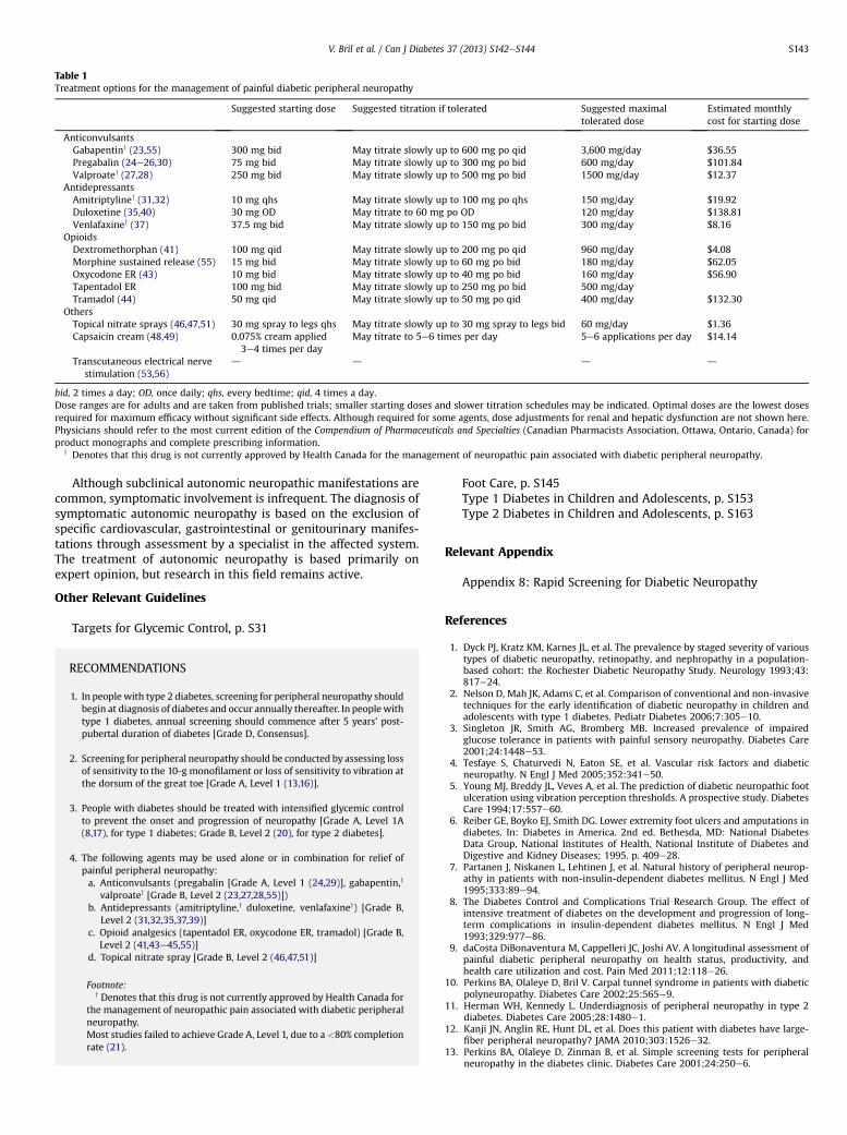
Table 1Treatment options for the management of painful diabetic peripheral neuropathy
Suggested starting dose Suggested titration if tolerated Suggested maximaltolerated dose
Estimated monthlycost for starting dose
AnticonvulsantsGabapentiny (23,55) 300 mg bid May titrate slowly up to 600 mg po qid 3,600 mg/day $36.55Pregabalin (24e26,30) 75 mg bid May titrate slowly up to 300 mg po bid 600 mg/day $101.84Valproatey (27,28) 250 mg bid May titrate slowly up to 500 mg po bid 1500 mg/day $12.37
AntidepressantsAmitriptyliney (31,32) 10 mg qhs May titrate slowly up to 100 mg po qhs 150 mg/day $19.92Duloxetine (35,40) 30 mg OD May titrate to 60 mg po OD 120 mg/day $138.81Venlafaxiney (37) 37.5 mg bid May titrate slowly up to 150 mg po bid 300 mg/day $8.16
OpioidsDextromethorphan (41) 100 mg qid May titrate slowly up to 200 mg po qid 960 mg/day $4.08Morphine sustained release (55) 15 mg bid May titrate slowly up to 60 mg po bid 180 mg/day $62.05Oxycodone ER (43) 10 mg bid May titrate slowly up to 40 mg po bid 160 mg/day $56.90Tapentadol ER 100 mg bid May titrate slowly up to 250 mg po bid 500 mg/dayTramadol (44) 50 mg qid May titrate slowly up to 50 mg po qid 400 mg/day $132.30
OthersTopical nitrate sprays (46,47,51) 30 mg spray to legs qhs May titrate slowly up to 30 mg spray to legs bid 60 mg/day $1.36Capsaicin cream (48,49) 0.075% cream applied
3e4 times per dayMay titrate to 5e6 times per day 5e6 applications per day $14.14
Transcutaneous electrical nervestimulation (53,56)
d d d d
bid, 2 times a day; OD, once daily; qhs, every bedtime; qid, 4 times a day.Dose ranges are for adults and are taken from published trials; smaller starting doses and slower titration schedules may be indicated. Optimal doses are the lowest dosesrequired for maximum efficacy without significant side effects. Although required for some agents, dose adjustments for renal and hepatic dysfunction are not shown here.Physicians should refer to the most current edition of the Compendium of Pharmaceuticals and Specialties (Canadian Pharmacists Association, Ottawa, Ontario, Canada) forproduct monographs and complete prescribing information.
y Denotes that this drug is not currently approved by Health Canada for the management of neuropathic pain associated with diabetic peripheral neuropathy.
V. Bril et al. / Can J Diabetes 37 (2013) S142eS144 S143
Although subclinical autonomic neuropathic manifestations arecommon, symptomatic involvement is infrequent. The diagnosis ofsymptomatic autonomic neuropathy is based on the exclusion ofspecific cardiovascular, gastrointestinal or genitourinary manifes-tations through assessment by a specialist in the affected system.The treatment of autonomic neuropathy is based primarily onexpert opinion, but research in this field remains active.
Other Relevant Guidelines
Targets for Glycemic Control, p. S31
RECOMMENDATIONS
1. In people with type 2 diabetes, screening for peripheral neuropathy shouldbegin at diagnosis of diabetes and occur annually thereafter. In peoplewithtype 1 diabetes, annual screening should commence after 5 years’ post-pubertal duration of diabetes [Grade D, Consensus].
2. Screening for peripheral neuropathy should be conducted by assessing lossof sensitivity to the 10-g monofilament or loss of sensitivity to vibration atthe dorsum of the great toe [Grade A, Level 1 (13,16)].
3. People with diabetes should be treated with intensified glycemic controlto prevent the onset and progression of neuropathy [Grade A, Level 1A(8,17), for type 1 diabetes; Grade B, Level 2 (20), for type 2 diabetes].
4. The following agents may be used alone or in combination for relief ofpainful peripheral neuropathy:a. Anticonvulsants (pregabalin [Grade A, Level 1 (24,29)], gabapentin,y
valproatey [Grade B, Level 2 (23,27,28,55)])b. Antidepressants (amitriptyline,y duloxetine, venlafaxiney) [Grade B,
Level 2 (31,32,35,37,39)]c. Opioid analgesics (tapentadol ER, oxycodone ER, tramadol) [Grade B,
Level 2 (41,43e45,55)]d. Topical nitrate spray [Grade B, Level 2 (46,47,51)]
Footnote:y Denotes that this drug is not currently approved by Health Canada for
the management of neuropathic pain associated with diabetic peripheralneuropathy.Most studies failed to achieve Grade A, Level 1, due to a <80% completionrate (21).
Foot Care, p. S145Type 1 Diabetes in Children and Adolescents, p. S153Type 2 Diabetes in Children and Adolescents, p. S163
Relevant Appendix
Appendix 8: Rapid Screening for Diabetic Neuropathy
References
1. Dyck PJ, Kratz KM, Karnes JL, et al. The prevalence by staged severity of varioustypes of diabetic neuropathy, retinopathy, and nephropathy in a population-based cohort: the Rochester Diabetic Neuropathy Study. Neurology 1993;43:817e24.
2. Nelson D, Mah JK, Adams C, et al. Comparison of conventional and non-invasivetechniques for the early identification of diabetic neuropathy in children andadolescents with type 1 diabetes. Pediatr Diabetes 2006;7:305e10.
3. Singleton JR, Smith AG, Bromberg MB. Increased prevalence of impairedglucose tolerance in patients with painful sensory neuropathy. Diabetes Care2001;24:1448e53.
4. Tesfaye S, Chaturvedi N, Eaton SE, et al. Vascular risk factors and diabeticneuropathy. N Engl J Med 2005;352:341e50.
5. Young MJ, Breddy JL, Veves A, et al. The prediction of diabetic neuropathic footulceration using vibration perception thresholds. A prospective study. DiabetesCare 1994;17:557e60.
6. Reiber GE, Boyko EJ, Smith DG. Lower extremity foot ulcers and amputations indiabetes. In: Diabetes in America. 2nd ed. Bethesda, MD: National DiabetesData Group, National Institutes of Health, National Institute of Diabetes andDigestive and Kidney Diseases; 1995. p. 409e28.
7. Partanen J, Niskanen L, Lehtinen J, et al. Natural history of peripheral neurop-athy in patients with non-insulin-dependent diabetes mellitus. N Engl J Med1995;333:89e94.
8. The Diabetes Control and Complications Trial Research Group. The effect ofintensive treatment of diabetes on the development and progression of long-term complications in insulin-dependent diabetes mellitus. N Engl J Med1993;329:977e86.
9. daCosta DiBonaventura M, Cappelleri JC, Joshi AV. A longitudinal assessment ofpainful diabetic peripheral neuropathy on health status, productivity, andhealth care utilization and cost. Pain Med 2011;12:118e26.
10. Perkins BA, Olaleye D, Bril V. Carpal tunnel syndrome in patients with diabeticpolyneuropathy. Diabetes Care 2002;25:565e9.
11. Herman WH, Kennedy L. Underdiagnosis of peripheral neuropathy in type 2diabetes. Diabetes Care 2005;28:1480e1.
12. Kanji JN, Anglin RE, Hunt DL, et al. Does this patient with diabetes have large-fiber peripheral neuropathy? JAMA 2010;303:1526e32.
13. Perkins BA, Olaleye D, Zinman B, et al. Simple screening tests for peripheralneuropathy in the diabetes clinic. Diabetes Care 2001;24:250e6.
V. Bril et al. / Can J Diabetes 37 (2013) S142eS144S144
14. Rith-Najarian SJ, Stolusky T, GohdesDM. Identifying diabetic patients at high riskfor lower-extremity amputation in a primary health care setting. A prospectiveevaluation of simple screening criteria. Diabetes Care 1992;15:1386e9.
15. Rahman M, Griffin SJ, Rathmann W, et al. How should peripheral neuropathybe assessed in people with diabetes in primary care? A population-basedcomparison of four measures. Diabet Med 2003;20:368e74.
16. Perkins BA, Orszag A, Olaleye D, et al. Prediction of incident diabetic neurop-athy using the monofilament exam: a 4-year prospective study. Diabetes Care2010;33:1549e54.
17. Reichard P, Berglund B, Britz A, et al. Intensified conventional insulin treatmentretards the microvascular complications of insulin-dependent diabetes melli-tus (IDDM): the Stockholm Diabetes Intervention Study (SDIS) after 5 years.J Intern Med 1991;230:101e8.
18. The Diabetes Control and Complications Trial Research Group. The effect ofintensive diabetes therapy on the development and progression of neuropathy.Ann Intern Med 1995;122:561e8.
19. Albers JW, Herman WH, Pop-Busui R, et al. Effect of prior intensive insulintreatment during the Diabetes Control and Complications Trial (DCCT) onperipheral neuropathy in type 1 diabetes during the Epidemiology of DiabetesInterventions and Complications (EDIC) Study. Diabetes Care 2010;33:1090e6.
20. UK Prospective Diabetes Study (UKPDS) Group. Intensive blood-glucose controlwith sulphonylureas or insulin compared with conventional treatment and riskof complications in patients with type 2 diabetes (UKPDS 33). Lancet 1998;352:837e53.
21. Bril V, England J, Franklin GM, et al. Evidence-based guideline: treatment ofpainful diabetic neuropathy: report of the American Academy of Neurology,the American Association of Neuromuscular and Electrodiagnostic Medicine,and the American Academy of Physical Medicine and Rehabilitation. Neurology2011;3:345e52. 352.e1-21.
22. McQuay H, Carroll D, Jadad AR, et al. Anticonvulsant drugs for management ofpain: a systematic review. BMJ 1995;311:1047e52.
23. Backonja M, Beydoun A, Edwards KR, et al. Gabapentin for the symptomatictreatment of painful neuropathy in patients with diabetes mellitus:a randomized controlled trial. JAMA 1998;280:1831e6.
24. Richter RW, Portenoy R, Sharma U, et al. Relief of painful diabetic peripheralneuropathy with pregabalin: a randomized, placebo-controlled trial. J Pain2005;6:253e60.
25. Lesser H, Sharma U, LaMoreaux L, et al. Pregabalin relieves symptoms of painfuldiabeticneuropathy: a randomizedcontrolled trial.Neurology2004;63:2104e10.
26. Rosenstock J, Tuchman M, LaMoreau L, et al. Pregabalin for the treatment ofpainful diabetic peripheral neuropathy: a double-blind, placebo-controlledtrial. Pain 2004;110:628e38.
27. Kochar DK, Jain N, Agarwal RP, et al. Sodium valproate in the management ofpainful neuropathy in type 2 diabetes: a randomized placebo controlled study.Acta Neurol Scand 2002;106:248e52.
28. Kochar DK, Rawat N, Agrawal RP, et al. Sodium valproate for painful diabeticneuropathy: a randomized double-blind placebo-controlled study. QJM 2004;97:33e8.
29. Guan Y, Ding X, Cheng Y, et al. Efficacy of pregabalin for peripheral neuropathicpain: results of an 8-week, flexible-dose, double-blind, placebo-controlledstudy conducted in China. Clin Ther 2011;33:159e66.
30. Satoh J, Yagihashi S, Baba M, et al. Efficacy and safety of pregabalin for treatingneuropathic pain associated with diabetic peripheral neuropathy: a 14 week,randomized, double-blind, placebo-controlled trial. Diabet Med 2011;28:109e16.
31. Max MB, Culnane M, Schafer SC, et al. Amitriptyline relieves diabetic neurop-athy pain in patients with normal or depressed mood. Neurology 1987;37:589e96.
32. Max MB, Lynch SA, Muir J, et al. Effects of desipramine, amitriptyline, andfluoxetine on pain in diabetic neuropathy. N Engl J Med 1992;326:1250e6.
33. Goldstein DJ, Lu Y, Detke MJ, et al. Duloxetine vs. placebo in patients withpainful diabetic neuropathy. Pain 2005;116:109e18.
34. Raskin J, Smith TR, Wong K, et al. Duloxetine versus routine care in the long-term management of diabetic peripheral neuropathic pain. J Palliat Med2006;9:29e40.
35. Raskin J, Wang F, Pritchett YL, et al. A double-blind, randomized multicentertrial comparing duloxetine with placebo in the management of diabeticperipheral neuropathic pain. Pain Med 2005;6:346e56.
36. Wernicke JF, Pritchett YL, D’Souza DN, et al. A randomized controlled trial ofduloxetine indiabeticperipheralneuropathicpain.Neurology2006;67:1411e20.
37. Rowbotham MC, Goli V, Kunz NR, et al. Venlafaxine extended release in thetreatment of painful diabetic neuropathy: a double-blind, placebo-controlledstudy. Pain 2004;110:697e706.
38. Kadiroglu AK, Sit D, Kayabasi H, et al. The effect of venlafaxine HCl on painfulperipheral diabetic neuropathy in patients with type 2 diabetes mellitus.J Diabetes Complicat 2008;22:241e5.
39. Kaur H, Hota D, Bhansali A, et al. A comparative evaluation of amitriptyline andduloxetine in painful diabetic neuropathy, a randomized, double-blind, cross-over clinical trial. Diabetes Care 2011;34:818e22.
40. Yasuda H, Hotta N, Nakao K, et al. Superiority of duloxetine to placebo inimproving diabetic neuropathic pain: results of a randomized controlled trialin Japan. J Diabetes Investig 2011;2:132e9.
41. Sang CN, Booher S, Gilron I, et al. Dextromethorphan and memantine in painfuldiabetic neuropathy and postherpetic neuralgia: efficacy and dose-responsetrials. Anesthesiology 2002;96:1053e61.
42. Nelson KA, Park KM, Robinovitz E, et al. High-dose oral dextromethorphanversus placebo in painful diabetic neuropathy and postherpetic neuralgia.Neurology 1997;48:1212e8.
43. Gimbel JS, Richards P, Portenoy RK. Controlled-release oxycodone for pain indiabetic neuropathy: a randomized controlled trial. Neurology 2003;60:927e34.
44. Harati Y, Gooch C, Swenson M, et al. Double-blind randomized trial of tramadolfor the treatment of the pain of diabetic neuropathy. Neurology 1998;50:1842e6.
45. Schwartz S, Etropolski M, Shapiro DY, et al. Safety and efficacy of tapentadol ERin patients with painful diabetic peripheral neuropathy: results of a random-ized-withdrawal, placebo-controlled trial. CMRO 2011;27:151e62.
46. Yuen KC, Baker NR, Rayman G. Treatment of chronic painful diabetic neurop-athy with isosorbide dinitrate spray: a double- blind placebo-controlled cross-over study. Diabetes Care 2002;25:1699e703.
47. Agrawal RP, Choudhary R, Sharma P, et al. Glyceryl trinitrate spray in themanagement of painful diabetic neuropathy: a randomized double blindplacebo controlled cross-over study. Diabetes Res Clin Pract 2007;77:161e7.
48. Low PA, Opfer-Gehrking TL, Dyck PJ, et al. Double-blind, placebo-controlledstudy of the application of capsaicin cream in chronic distal painful poly-neuropathy. Pain 1995;62:163e8.
49. The Capsaicin Study Group. Treatment of painful diabetic neuropathy withtopical capsaicin.A multicenter, double-blind, vehicle-controlled study. ArchIntern Med 1991;151:2225e9.
50. Tandan R, Lewis GA, Krusinski PB, et al. Topical capsaicin in painful diabeticneuropathy. Controlled study with long-term follow-up. Diabetes Care 1992;15:8e14.
51. Agrawal RP, Goswami J, Jain S, et al. Management of diabetic neuropathy bysodium valproate and glyceryl trinitrate spray: a prospective double-blindrandomized placebo-controlled study. Diabetes Res Clin Pract 2009;83:371e8.
52. Kumar D, Alvaro MS, Julka IS, et al. Diabetic peripheral neuropathy. Effective-ness of electrotherapy and amitriptyline for symptomatic relief. Diabetes Care1998;21:1322e5.
53. Bosi E, Conti M, Vermigli C, et al. Effectiveness of frequency-modulated elec-tromagnetic neural stimulation in the treatment of painful diabetic neurop-athy. Diabetologia 2005;48:817e23.
54. Chaudhry V, Stevens JC, Kincaid J, et al. Practice advisory: utility of surgicaldecompression for treatment of diabetic neuropathy: report of the Therapeu-tics and Technology Assessment Subcommittee of the American Academy ofNeurology. Neurology 2006;66:1805e8.
55. Gilron I, Bailey JM, Tu D, et al. Morphine, gabapentin, or their combination forneuropathic pain. N Engl J Med 2005;352:1324e34.
56. Hamza MA, White PF, Craig WF, et al. Percutaneous electrical nerve stimula-tion: a novel analgesic therapy for diabetic neuropathic pain. Diabetes Care2000;23:365e70.

Contents lists available at SciVerse ScienceDirect
Can J Diabetes 37 (2013) S145eS149
Canadian Journal of Diabetesjournal homepage:
www.canadianjournalofdiabetes.com
Clinical Practice Guidelines
Foot Care
Canadian Diabetes Association Clinical Practice Guidelines Expert Committee
The initial draft of this chapter was prepared by Keith Bowering MD, FRCPC, FACP,John M. Embil MD, FRCPC, FACP
KEY MESSAGES
� Foot problems are a major cause of morbidity and mortality in people withdiabetes and contribute to increased healthcare costs.
� The management of foot ulceration in people with diabetes requires aninterdisciplinary approach that addresses glycemic control, infection, off-loading of high-pressure areas, lower-extremity vascular status and localwound care.
� Antibiotic therapy is not generally required for neuropathic foot ulcerationsthat show no evidence of infection.
Introduction
Foot complications are a major cause of morbidity and mortalityin persons with diabetes and contribute to increased healthcareutilization and costs (1e3). In populations with diabetes, indivi-duals with peripheral neuropathy and peripheral arterial disease(PAD) are predisposed to foot ulceration and infection, whichultimately may lead to lower-extremity amputation (4e6).Although amputation rates for people with diabetes havedecreased in the past decade, they remain exceedingly highcompared to nondiabetic populations (7,8). Therefore, it is essentialthat every effort possible be made to prevent foot problems, and, ifthey do occur, that early and aggressive treatment be undertaken.
Risk Assessment
Characteristics that have been shown to confer a risk of footulceration in persons with diabetes include peripheral neuropathy,previous ulceration or amputation, structural deformity, limitedjoint mobility, PAD, microvascular complications, high glycatedhemoglobin (A1C) levels and onychomycosis (9e11). Loss ofsensation over the distal plantar surface to the 10-g SemmesWeinstein monofilament is a significant and independent predictorof future foot ulceration and the possibility of lower-extremityamputation (12).
In those persons with diabetes with foot ulcers, a number ofwound classification systems have been developed to provideobjective assessment of severity. Of these, the University of TexasDiabetic Wound Classification System has been validated asa predictor of serious outcomes in patients with diabetes with footulcers (Table 1) (13,14).
1499-2671/$ e see front matter � 2013 Canadian Diabetes Associationhttp://dx.doi.org/10.1016/j.jcjd.2013.01.040
In persons with diabetes with underlying ischemia, the distri-bution of PAD is greater in the arterial tree below the knee than isseen in those without diabetes (15). Noninvasive assessments forPAD in diabetes include the use of the ankle-brachial blood pressureindex (ABI), determination of systolic toe pressure by photo-plethysmography (PPG) (PPG assesses the intensity of light reflectedfrom the skin surface and the red cells below, which is indicative ofarterial pulse flow in the arterioles of the respective area), trans-cutaneous oximetry (tcPO2) andDoppler arterialflowstudies (16,17).Although the ABI is a readily available and easy-to-perform tech-nique, it may underestimate the degree of peripheral arterialobstruction in some individuals with diabetes partly due to medialarterial wall calcification in lower-extremity arteries (18). Measure-ment of systolic toe pressure by PPG may be more accurate indetermining the presence of arterial disease in this population (19).
For those persons in whom lower-limb ischemia is suspected,intra-arterial digital subtraction contrast arteriography hasprovided the most definitive assessment of PAD but may precipitaterenal failure in individuals with higher degrees of renal insuffi-ciency. Advanced magnetic resonance angiography (MRA) andcomputed tomographic angiography (CTA) do not require arterialaccess and, therefore, have gained popularity as reliable alternativesto iodinated contrast studies due to their less invasive approaches(20e22). However, caution is still necessary with MRA and CTA inpersons with renal dysfunction. The injection of intravenous radi-ocontrast dye alsomust be used in CTA; therefore, caution should beexercised (as with the use of intra-arterial iodinated contrast) inpersons with renal insufficiency so as to avoid precipitating acuterenal failure. Gadolinium-based contrast agents used in MRA havebeen associated with the development of nephrogenic systemicfibrosis in individuals with poor renal function (23,24).
The foot examination should include the assessment of skintemperature since increased warmth is the first indicator ofinflammation in an insensate foot and also may be the first sign ofacute Charcot neuroarthropathy resulting from the loss of protec-tive sensation in the foot (25e27). In addition, an acute Charcot footmay be associated with erythema and swelling, with overall clinicalcharacteristics very similar to cellulitis (28,29). The clinical andradiological differentiation between an acute Charcot foot anda foot infection can be very challenging (30). Plain radiographs havelow sensitivity and specificity in differentiating osteomyelitis fromCharcot changes. Magnetic resonance imaging (MRI) of the footmay help clarify this differential diagnosis, although no single

Table 1University of Texas Diabetic Wound Classification System (13)
K. Bowering, J.M. Embil / Can J Diabetes 37 (2013) S145eS149S146
radiological investigation to date has proven to be completelydefinitive (31).
Management and Preventative Care
The prevention of amputations has involved the use of variouspreventative measures, including regular foot examination andevaluation of amputation risk, regular callus debridement, patienteducation, professionally fitted therapeutic footwear to reduceplantar pressure and accommodate foot deformities, and earlydetection and treatment of diabetic foot ulcers (32). Many of thestudies conducted to assess interventions designed to reduce theoccurrence of and heal diabetic foot ulcers have, unfortunately,suffered from methodological problems, thereby reducing thequality of the evidence to support their use (33,34).
Generally, the management of foot ulceration should addressglycemic control, pressure relief/offloading, infection, lower-extremity vascular status and local wound care (35). This is bestachieved with an interdisciplinary approach (36,37).
Specific recommendations about dressing types cannot bemadeas there is insufficient evidence to support the use of one varietyversus another; however, the concepts that are generally acceptedas the essentials of good wound care include the provision of anoptimal wound environment, pressure offloading from the ulcersite and, in nonischemic wounds, regular debridement of nonviabletissue (38,39). In general, wound dressings that maintain a moistwound environment should be selected. There are insufficient datato support the use of specific dressing types or antimicrobialdressings in the routine management of diabetic foot wounds(40e48). There is also insufficient evidence to make any recom-mendation about the role of negative pressure wound therapy(NPWT) in the routine management of neuropathic wounds. Thereis, however, some evidence to support NPWT as a postoperativeintervention after extensive debridement (49e52). Other adjunc-tivemeasures for wound healing, such as topical growth factors anddermal substitutes, have been studied in diabetic foot ulcermanagement, but these studies have been limited in sample size,duration and follow-up. These therapies may be considered if otherconventional options already have been explored (53).
Pressure offloading may be achieved with temporary footwearuntil the ulcer heals and the character of the foot stabilizes.Removable and irremovable cast walkers and total contact castinghave demonstrated efficacy as pressure-reducing devices in plantarsurface ulcers (54e56). Although very effective in healing nonin-fected, nonischemic plantar surface neuropathic ulcers, totalcontact casting requires careful individual selection and personneltrained specifically in its application due to its potential forcomplications (57). Where bony foot deformities prevent the fitting
of appropriate footwear and/or offloading of pressure-relatedulcers, consultation with a surgeon skilled in foot surgery may beconsidered to address the deformity (58e60).
Treatment of the acute Charcot foot requires immobilization ofthe foot, typically for several months, in a total contact cast orremovable walker device until excessive foot temperatures returnto normal (61). Although bisphosphonate therapy has beenconsidered for the management of Charcot arthropathy, furtherstudies are necessary to fully evaluate the use of these agents andother medical therapies in the routine treatment of Charcotarthropathy (62e64).
Infection may complicate foot ulcers and may progress rapidlyto become limb and/or life threatening (65). When infections firstbegin, the most frequently encountered pathogens include Staph-ylococcus aureus, Streptococcus pyogenes (group A streptococcus)and Streptococcus agalactiae (group B streptococcus).With time andthe presence of devitalized tissue, gram-negative and anaerobicpathogens also can play a role in the process, leading to poly-microbial infections (66,67). Specimens for culture from the surfaceof wounds, as opposed to deeper tissues obtained by debridement,are unreliable in determining the bacterial pathogens involved(68e70). Initial antibiotic therapy is typically empiric and may bebroad spectrum, with subsequent antibiotic selection tailored tothe sensitivity results of cultured specimens. With the exception ofonly a small number of antimicrobial agents that do have a specificindication for the treatment of diabetic foot infections, the majorityof the agents available for use are selected for their antibacterialspectrum (66,71). Table 2 summarizes the different antimicrobialchoices for the empiric management of foot infections in personswith diabetes. Uncontrolled diabetes can result in immunopathywith a blunted cellular response to infection. Up to 50% of patientswith diabetes who have a significant limb infection may not havesystemic signs of fever or leukocytosis at presentation (72). Deepinfections require prompt surgical debridement in addition toappropriate antibiotic therapy (73). Granulocyte colony-stimulating factors have been used as adjunctive therapy in infec-ted diabetic wounds and, in some studies, were found to reduce theneed for surgical intervention. Data are limited and caution isadvised in interpreting these findings (74).
In medically suitable individuals with PAD, distal limb revas-cularization has potential benefit in long-term limb salvage. Certainsubpopulations with diabetes on insulin therapy have pooreroutcomes after revascularization than those on oral anithypergly-cemic therapy, perhaps reflecting a greater association of comor-bidities (75,76). Endovascular techniques with angioplasty andstenting in infrainguinal arteries are also effective in limb salvage,although the long-term results are inferior in the population withdiabetes compared to those without diabetes (77,78).

Table 2Empiric antimicrobial therapy for infection in the diabetic foot
MRSA, methicillin-resistant Staphylococcus aureus; TMP-SMX, trimethoprim-sulfamethoxazole.*Modified and used with permission from Embil JM, Trepman E. Diabetic foot infections. In: Principles and Practice of Hospital Medicine. Editors: Sylvia C. McKean, John J. Ross,Daniel D. Dressler, Daniel J. Brotman, Jeffrey S. Ginsberg. Printed in China: McGraw-Hill; 2012.
K. Bowering, J.M. Embil / Can J Diabetes 37 (2013) S145eS149 S147

K. Bowering, J.M. Embil / Can J Diabetes 37 (2013) S145eS149S148
Hyperbaric oxygen therapy (HBOT) is not considered part of theroutine management of persons with neuropathic/neuroischemicfoot ulcerations with or without underlying infection. In carefullyselected persons with nonhealing foot ulcerations for whom allpossible interventions have been attempted, HBOT may beconsidered as an adjunctive therapy (79e81). Currently, evidence-based criteria for the selection of persons with diabetes who havefoot problems and who may benefit from HBOT do not exist.
RECOMMENDATIONS
1. In people with diabetes, foot examinations by healthcare providers shouldbe an integral component of diabetes management to identify persons atrisk for ulceration and lower-extremity amputation [Grade C, Level 3(5,12)] and should be performed at least annually and at more frequentintervals in those at high risk [Grade D, Level 4 (1)]. Assessment byhealthcare providers should include the assessment of skin changes,structural abnormalities (e.g. range of motion of ankles and toe joints,callus pattern, bony deformities), skin temperature, evaluation forneuropathy and PAD, ulcerations and evidence of infection [Grade D, Level4 (1)].
2. People at high risk of foot ulceration and amputation should receive footcare education (including counselling to avoid foot trauma), professionallyfitted footwear and early referrals to a healthcare professional trained infoot care management if foot complications occur [Grade C, Level 3(33,82,83)].
3. Individuals who develop a foot ulcer should be managed by a multidisci-plinary healthcare teamwith expertise in themanagement of foot ulcers toprevent recurrent foot ulcers and amputation [Grade C, Level 3 (36)].
4. There is currently insufficient evidence to recommend any specificdressing type for diabetic foot ulcers [Grade C, Level 3 (40)]. Generalprinciples of wound management involve the provision of a moist woundenvironment, debridement of nonviable tissue (nonischemic wounds) andoffloading of pressure areas [Grade B, Level 3 (38)].
5. Evidence is currently lacking to support the routine use of adjunctivewound-healing therapies, such as topical growth factors, granulocytecolony-stimulating factors, dermal substitutes or HBOT in diabetic footulcers, but they may be considered in nonhealing, nonischemic woundswhen all other options have been exhausted [Grade D, Level 4 (53,74,80)].
Abbreviations:HBOT, hyperbaric oxygen therapy; PAD, peripheral arterial disease.
Other Relevant Guidelines
Targets for Glycemic Control, p. S31Neuropathy, p. S142
Relevant Appendices
Appendix 9: Diabetes and Foot Care: A Patient’s ChecklistAppendix 10: Diabetic Foot Ulcers: Essentials of Management
References
1. Boulton AJ, Armstrong DG, Albert SF, et al, American Diabetes Association,American Association of Clinical Endocrinologists. Comprehensive foot exam-ination and risk assessment: a report of the task force of the foot care interestgroup of the American Diabetes Association, with endorsement by the Amer-ican Association of Clinical Endocrinologists. Diabetes Care 2008;31:1679e85.
2. Davis WA, Norman PE, Bruce DG, Davis TM. Predictors, consequences and costsof diabetes-related lower extremity amputations complicating type 2 diabetes:the Freemantle Diabetes Study. Diabetologia 2006;49:2634e41.
3. O’Brien JA, Patrick AR, Caro JJ. Cost of managing complications resulting fromtype 2 diabetes mellitus in Canada. BMC Health Serv Res 2003;3:7.
4. Reiber GE, Vileikyte L, Boyko EJ, et al. Causal pathways for incident lower-extremity ulcers in patients with diabetes from two settings. Diabetes Care1999;22:157e62.
5. Crawford F, Inkster M, Kleijnen J, Fahey T. Predicting foot ulcers in patientswith diabetes: a systematic review and meta-analysis. QJM 2007;100:65e86.
6. Faglia E, Clerici G, Gabrielli L, et al. Long-term prognosis of diabetic patientswith critical limb ischemia: a population-based cohort study. Diabetes Care2009;35:822e7.
7. Fosse S, Hartemann-Heurtier A, Jacqueminet S, et al. Incidence and character-istics of lower limb amputations in people with diabetes. Diabet Med 2009;26:391e6.
8. Ikonen TS, Sund R, Venermo M, Winell K. Fewer major amputations amongindividuals with diabetes in Finland in 1997-2007: a population-based study.Diabetes Care 2010;33:2598e603.
9. Boyko EJ, Ahroni JH, Stensel V, et al. A prospective study of risk factors fordiabetic foot ulcer. The Seattle Diabetic Foot Study. Diabetes Care 1999;22:1036e42.
10. Fernando DJ, Masson EA, Veves A, et al. Relationship of limited joint mobility toabnormal foot pressures and diabetic foot ulceration. Diabetes Care 1991;14:8e11.
11. Boyko EJ, Nelson KM, Ahroni JH, et al. Prediction of diabetic foot ulcer occur-rence using commonly available clinical information. Diabetes Care 2006;29:1202.
12. Feng Y, Schlösser FJ, Bauer E, Sumpio BE. The Semmes Weinstein monofilamentexamination is a significant predictor of the risk of foot ulceration andamputation in patients with diabetes mellitus. J Vasc Surg 2011;53:220e6.
13. Armstrong DG, Lavery LA, Harkless LB. Validation of a diabetic wound classi-fication system. The contribution of depth, infection, and ischemia to risk ofamputation. Diabetes Care 1998;21:855e9.
14. Oyibo SO, Jude EB, Tarawneh I, et al. A comparison of two diabetic foot ulcerclassification systems: the Wagner and the University of Texas wound classi-fication systems. Diabetes Care 2001;24:84e8.
15. Jude EB, Oyibo SO, Chalmers N, Boulton AJ. Peripheral arterial disease indiabetic and nondiabetic patients: a comparison of severity and outcome.Diabetes Care 2001;24:1433e7.
16. Kalani M, Brismar K, Fagrell B, et al. Transcutaneous oxygen tension and toeblood pressure as predictors for outcome of diabetic foot ulcers. Diabetes Care1999;22:147e51.
17. Faglia E, Caravaggi C, Marchetti R, et al, SCAR (SCreening for ARteriopathy)Study Group. Screening for peripheral arterial disease by means of the ankle-brachial index in newly diagnosed type 2 diabetic patients. Diabet Med2005;22:1310e4.
18. Aerden D, Massaad D, von Kemp K, et al. The ankle-brachial index and thediabetic foot: a troublesome marriage. Ann Vasc Surg 2011;25:770e7.
19. Williams DT, Harding KG, Price P. An evaluation of the efficacy of methods usedin screening for lower-limb arterial disease in diabetes. Diabetes Care 2005;28:2206e10.
20. Brillet PY, Vayssairat M, Tassart M, et al. Gadolinium-enhanced MR angiog-raphy as first-line preoperative imaging in high-risk patients with lower limbischemia. J Vasc Interv Radiol 2003;14:1139e45.
21. Lapeyre M, Kobeiter H, Desgranges P, et al. Assessment of critical limb ischemiain patients with diabetes: comparison of MR angiography and digitalsubtraction angiography. AJR Am J Roentgenol 2005;185:1641e50.
22. Met R, Bipat S, Legemate DA, et al. Diagnostic performance of computedtomography angiography in peripheral arterial disease: a systematic reviewand meta-analysis. JAMA 2009;301:415e24.
23. Pedersen M. Safety update on the possible causal relationship betweengadolinium-containing MRI agents and nephrogenic systemic fibrosis. J MagnReson Imaging 2007;25:881e3.
24. Centers for Disease Control and Prevention (CDC). Nephrogenic fibrosingdermopathy associated with exposure to gadolinium-containing contrastagentseSt. Louis, Missouri, 2002-2006. MMWR Morb Mortal Wkly Rep 2007;56:137e41.
25. Lavery LA, Higgins KR, Lanctot DR, et al. Preventing diabetic foot ulcer recur-rence in high-risk patients: use of temperature monitoring as a self-assessmenttool. Diabetes Care 2007;30:14e20.
26. Armstrong DG, Lavery LA. Monitoring healing of acute Charcot’s arthropathywith infrared dermal thermometry. J Rehabil Res Dev 1997;34:317e21.
27. Yu GV, Hudson JR. Evaluation and treatment of stage 0 Charcot’s neuro-arthropathy of the foot and ankle. J Am Podiatr Med Assoc 2002;92:210e20.
28. Frykberg RG, Zgonis T, Armstrong DG, et al, American College of Foot and AnkleSurgeons. Diabetic foot disorders. A clinical practical guideline (2006 revision).J Foot Ankle Surg 2006;45(5 suppl):S1e66.
29. Ledermann HP, Morrison WB. Differential diagnosis of pedal osteomyelitis anddiabetic neuroarthropathy: MR imaging. Semin Musculoskelet Radiol 2005;9:272e83.
30. Embil JM, Trepman E. A case of diabetic Charcot arthropathy of the foot andankle. Nat Rev Endocrinol 2009;5:577e81.
31. Ahmadi ME, Morrison WB, Carrino JA, et al. Neuropathic arthropathy of thefoot with and without superimposed osteomyelitis: MR imaging characteris-tics. Radiology 2006;238:622e31.
32. Apelqvist J, Bakker K, vanHoutumWH, SchaperNC, InternationalWorkingGroupon the Diabetic Foot (IWGDF) Editorial Board. Practical guidelines on the
K. Bowering, J.M. Embil / Can J Diabetes 37 (2013) S145eS149 S149
management and prevention of the diabetic foot: based upon the InternationalConsensus on theDiabetic Foot (2007).DiabetesMetabResRev2008;24(suppl 1):S181e7.
33. Arad Y, Fonseca V, Peters A, Vinik A. Beyond the monofilament for the insen-sate diabetic foot. Diabetes Care 2011;34:1041e6.
34. Bus SA, Valk GD, van Deursen RW, et al. The effectiveness of footwear andoffloading interventions to prevent and heal foot ulcers and reduce plantarpressure in diabetes: a systematic review. Diabetes Metab Res Rev 2008;24-(suppl 1):S162e80.
35. Margolis DJ, Kantor J, Berlin JA. Healing of diabetic neuropathic foot ulcersreceiving standard treatment. A metaanalysis. Diabetes Care 1999;22:692e5.
36. Dargis V, Pantelejeva O, Jonushaite A, et al. Benefits of a multidisciplinaryapproach in the management of recurrent diabetic foot ulceration in Lithuania:a prospective study. Diabetes Care 1999;22:1428e31.
37. Aydin K, Isildak M, Karakaya J, Gürlek A. Change in amputation predictors in dia-betic footdisease: effectofmultidisciplinaryapproach.Endocrine2010;38:87e92.
38. Steed DL, Donohoe D, Webster MW, Lindsley L. Effect of extensive debridementand treatment on the healing of diabetic foot ulcers. Diabetic Ulcer StudyGroup. J Am Coll Surg 1996;183:61e4.
39. Edwards J, Stapley S. Debridement of diabetic foot ulcers. Cochrane DatabaseSyst Rev 2010;1:CD003556.
40. Vermeulen H, Ubbink D, Goossens A, et al. Dressings and topical agents forsurgical wounds healing by secondary intention. Cochrane Database Syst Rev2004;2:CD003554.
41. Vermeulen H, van Hattem JM, Storm-Versloot MN, Ubbink DT. Topical silver fortreating infected wounds. Cochrane Database Syst Rev 2007;1:CD005486.
42. Bergin SM, Wraight P. Silver based wound dressings and topical agents fortreating diabetic foot ulcers. Cochrane Database Syst Rev 2006;1:CD005082.
43. Canadian Agency for Drugs and Technologies in Health. Silver Dressings for theTreatment of Patients with Infected Wounds: A Review of Clinical and Cost-Effectiveness. July 7, 2010. Available at: http://www.cadth.ca/media/pdf/l0193_silver_dressings_htis-2.pdf. Accessed March 1, 2013.
44. Storm-Versloot MN, Vos CG, Ubbink DT, Vermeulen H. Topical silver for pre-venting wound infection. Cochrane Database Syst Rev 2010;3:CD006478.
45. Hinchliffe RJ, Valk GD, Apelqvist J, et al. A systematic review of the effective-ness of interventions to enhance the healing of chronic ulcers of the foot indiabetes. Diabetes Metab Res Rev 2008;24(suppl 1):S119e44.
46. Hilton JR, Williams DT, Beuker B, et al. Wound dressings in diabetic footdisease. Clin Infect Dis 2004;39(suppl 2):S100e3.
47. Nelson EA, O’Meara S, Golder S, et al. Systematic review of antimicrobialtreatments for diabetic foot ulcers. Diabet Med 2006;23:348e59.
48. O’Meara SM, Cullum NA, Majid M, Sheldon TA. Systematic review of antimi-crobial agents used for chronic wounds. Br J Surg 2001;88:4e21.
49. Armstrong DG, Lavery LA, Diabetic Foot Study Consortium. Negative pressurewound therapy after partial diabetic foot amputation: a multicentre, rando-mised controlled trial. Lancet 2005;366:1704e10. 12.
50. Ubbink DT, Westerbos SJ, Evans D, et al. Topical negative pressure for treatingchronic wounds. Cochrane Database Syst Rev 2008;3:CD001898.
51. Canadian Agency for Drugs and Technologies in Health. Negative PressureTherapy for Patients Infected Wounds: A Review of the Clinical and CostEffectiveness Evidence and Recommendations for Use. July 14, 2010. Availableat: http://www.cadth.ca/media/pdf/l0194_negative_pressure_therapy_htis-2.pdf. Accessed March 1, 2013.
52. Gregor S, Maegele M, Sauerland S, et al. Negative pressure wound therapy:a vacuum of evidence? Arch Surg 2008;143:189e96.
53. Buchberger B, Follmann M, Freyer D, et al. The importance of growth factors forthe treatment of chronic wounds in the case of diabetic foot ulcers. GMS HealthTechnol Assess 2010;6:Doc12. doi:10.3205/hta000090.
54. Armstrong DG, Lavery LA, Wu S, et al. Evaluation of removable and irremovablecast walkers in the healing of diabetic foot wounds: a randomized controlledtrial. Diabetes Care 2005;28:551e4.
55. Armstrong DG, Nguyen HC, Lavery LA, et al. Off-loading the diabetic footwound: a randomized clinical trial. Diabetes Care 2001;24:1019e22.
56. Katz IA, Harlan A, Miranda-Palma B, et al. A randomized trial of two irre-movable off-loading devices in the management of plantar neuropathic dia-betic foot ulcers. Diabetes Care 2005;28:555e9.
57. Nabuurs-Franssen MH, Sleegers R, Huijberts MS, et al. Total contact casting ofthe diabetic foot in daily practice: a prospective follow-up study. Diabetes Care2005;28:243e7.
58. Singh N, Armstrong DG, Lipsky BA. Preventing foot ulcers in patients withdiabetes. JAMA 2005;293:217e28.
59. Blume PA, Paragas LK, Sumpio BE, et al. Single-stage surgical treatment ofnoninfected diabetic foot ulcers. Plast Reconstr Surg 2002;109:601e9.
60. Sayner LR, Rosenblum BI, Giurini JM. Elective surgery of the diabetic foot. ClinPodiatr Med Surg 2003;20:783e92.
61. Molines L, Darmon P, Raccah D. Charcot’s foot: newest findings on its patho-physiology, diagnosis and treatment. Diabetes Metab 2010;36:251e5.
62. Trepman E, Nihal A, Pinzur MS. Current topics review: Charcot neuro-arthropathy of the foot and ankle. Foot Ankle Int 2005;26:46e63.
63. Jude EB, Selby PL, Burgess J, et al. Bisphosphonates in the treatment of Charcotneuroarthropathy: a double-blind randomised controlled trial. Diabetologia2001;44:2032e7.
64. Pitocco D, Ruotolo V, Caputo S, et al. Six-month treatment with alendronate inacute Charcot neuroarthropathy: a randomized controlled trial. Diabetes Care2005;28:1214e5.
65. Lavery LA, Armstrong DG, Wunderlich RP, et al. Risk factors for foot infectionsin individuals with diabetes. Diabetes Care 2006;29:1288e93.
66. Lipsky BA, Berendt AR, Cornia PB, et al. 2012 Infectious Diseases Society ofAmerica clinical practice guideline for the diagnosis and treatment of diabeticfoot infections. Clin Infect Dis 2012;54:e132e73.
67. Rao N, Lipsky BA. Optimizing antimicrobial therapy in diabetic foot infections.Drugs 2007;67:195e214.
68. Perry CR, Pearson RL, Miller GA. Accuracy of cultures of material from swabbingof the superficial aspect of the wound and needle biopsy in the preoperativeassessment of osteomyelitis. J Bone Joint Surg Am 1991;73:745e9.
69. Senneville E, Melliez H, Beltrand E, et al. Culture of percutaneous bone biopsyspecimens for diagnosis of diabetic foot osteomyelitis: concordance with ulcerswab cultures. Clin Infect Dis 2006;42:57e62.
70. Slater RA, Lazarovitch T, Boldur I, et al. Swab cultures accurately identifybacterial pathogens in diabetic foot wounds not involving bone. Diabet Med2004;21:705e9.
71. Embil JM, Trepman E. Diabetic infection. In: Gray J, editor. Therapeutic Choices.6th ed. Ottawa, Ontario, Canada: Canadian Pharmacists Association; 2011.
72. Eneroth M, Apelqvist J, Stenström A. Clinical characteristics and outcome in223 diabetic patients with deep foot infections. Foot Ankle Int 1997;18:716e22.
73. Tan JS, Friedman NM, Hazelton-Miller C, et al. Can aggressive treatment ofdiabetic foot infections reduce the need for above-ankle amputation? ClinInfect Dis 1996;23:286e91.
74. Cruciani M, Lipsky BA, Mengoli C, de Lalla F. Granulocyte-colony stimulatingfactors as adjunctive therapy for diabetic foot infections. Cochrane DatabaseSyst Rev 2009;3:CD006810.
75. Pomposelli FB, Kansal N, Hamdan AD, et al. A decade of experience with dor-salis pedis artery bypass: analysis of outcome in more than 1000 cases. J VascSurg 2003;37:307e15.
76. Dosluoglu HH, Lall P, Nader ND, et al. Insulin use is associated with poor limbsalvage and survival in diabetic patients with chronic limb ischemia. J VascSurg 2010;51:1178e89.
77. Abularrage CJ, Conrad MF, Hackney LA, et al. Long-term outcomes of diabeticpatients undergoing endovascular infrainguinal interventions. J Vasc Surg2010;52:314e22.
78. Dick F, Diehm N, Galimanis A, et al. Surgical or endovascular revascularizationin patients with critical limb ischemia: influence of diabetes mellitus on clinicaloutcome. J Vasc Surg 2007;45:751e61.
79. Löndahl M, Katzman P, Nilsson A, Hammarlund C. Hyperbaric oxygen therapyfacilitates healing of chronic foot ulcers in patients with diabetes. Diabetes Care2010;33:998e1003.
80. Löndahl M, Fagher K, Katzman P. What is the role of hyperbaric oxygen inthe management of diabetic foot disease? Curr Diabetes Rep 2011;11:285e93.
81. Räkel A, Huot C, Ekoé JM. Canadian Diabetes Association Technical Review:the diabetic foot and hyperbaric oxygen therapy. Can J Diabetes 2006;30:411e21.
82. Valk GD, Kriegsman DM, Assendelft WJ. Patient education for preventing dia-betic foot ulceration: a systematic review. Endocrinol Metab Clin North Am2002;31:633e58.
83. McCabe CJ, Stevenson RC, Dolan AM. Evaluation of a diabetic foot screening andprotection programme. Diabet Med 1998;15:80e4.

Contents lists available at SciVerse ScienceDirect
Can J Diabetes 37 (2013) S150eS152
Canadian Journal of Diabetesjournal homepage:
www.canadianjournalofdiabetes.com
Clinical Practice Guidelines
Erectile Dysfunction
Canadian Diabetes Association Clinical Practice Guidelines Expert Committee
The initial draft of this chapter was prepared by Gerald Brock MD, FRCSC, William Harper MD, FRCPC
KEY MESSAGES
� Erectile dysfunction (ED) affects approximately 34% to 45% of adult menwith diabetes, has been demonstrated to negatively impact quality of lifeamong those affected across all age strata and may be the earliest sign ofcardiovascular disease.
� All adult men with diabetes should be regularly screened for ED witha sexual function history. Those with ED should be investigated forhypogonadism.
� The current mainstay of therapy is phosphodiesterase type 5 inhibitors.They have been shown to have major impacts on erectile function andquality of life, with a low reported side effect profile, and should be offeredas first-line therapy to men with diabetes wishing treatment for ED.
Introduction
Erectile dysfunction (ED) affects approximately 34% to 45% ofmenwith diabetes and has been demonstrated to negatively impactquality of life among those affected across all age strata, witha greater likelihood among men with diabetes that their ED ispermanent (1). Recent reports describe up to one-third of newlydiagnosed men with diabetes have ED at presentation (2), withupward of 50% of men having ED by year 6 after diagnosis (3).Furthermore, studies indicate that 40% of men with diabetes >60years of age have complete ED (4e12). Recent studies have reportedthat alteration of the cyclic guanosinemonophosphate (cGMP(/nitricacid (NO) pathway among men with diabetes with impairedvascular relaxation is related to endothelial dysfunction (13e15).Among the populationwith diabetes, risk factors include increasingage, duration of diabetes, poor glycemic control, cigarette smoking,hypertension, dyslipidemia, androgen deficiency states (16) andcardiovascular (CV) disease (8,10,17,18). ED as a marker of potentialCV events has been reported by numerous investigators (19e26). Infact, ED has been shown to be significantly associated with all-causemortality and CV events (27,28). Diabetic retinopathy has beenshown to correlate with the presence of ED (8,10,29). Organic causesof ED include microvascular and macrovascular disease, andneuropathy. In addition, psychological or situational factors maycause or contribute to ED.
In spite of the overwhelming amount of data linking ED anddiabetes, this remains a subject often neglected by clinicianstreating the population with diabetes (30).
Compared with the general population, multiple studies havereported men with diabetes having higher rates of hypogonad-ism (16,31e34). Interestingly, a recent report describes
1499-2671/$ e see front matter � 2013 Canadian Diabetes Associationhttp://dx.doi.org/10.1016/j.jcjd.2013.01.041
a correlation between glycemic control and testosterone levels(35). Importantly, phosphodiesterase type 5 (PDE5) inhibitorsappear to be less effective in hypogonadal states (32,34,36),where treatment of nonresponders to PDE5 inhibitors withtestosterone replacement is successful in roughly 50% of indi-viduals. In addition, ED is a side effect of many drugs commonlyprescribed to men with diabetes, such as some antihypertensivesand antidepressants.
Screening
All adult men with diabetes should be regularly screened forED with a sexual function history. Screening for ED in men withtype 2 diabetes should begin at diagnosis of diabetes. Validatedquestionnaires (e.g. International Index of Erectile Function(37,38) or Sexual Health Inventory for Men (39)) have been shownto be both sensitive and specific in determining the presence ofED and providing a means of assessing response to therapy. Menwith diabetes and ED should be further investigated for hypo-gonadism. The Androgen Deficiency in Aging Males (ADAM)instrument is the most widely accepted screening questionnaire,and, while bioavailable testosterone is recognized as the goldstandard for biochemistry confirmation, total testosterone is anacceptable alternative if bioavailable testosterone is unavailable orunaffordable (40).
Treatment
While no randomized clinical trials have demonstrated thatinterventions that improve glycemic control also reduce theincidence and progression of ED, the Diabetes Control andComplications Trial (DCCT) and United Kingdom ProspectiveDiabetes Study (UKPDS) showed that intensive glycemic controlwas effective for primary prevention of and secondary interventionfor neuropathy, a condition that can impair sensory feedback fromthe penis, leading to reduced erectile function (41e43). The currentdata are controversial as it relates to diet, glycemic control and ED,with both positive and negative studies (28,44e46). Based on theseconflicting data, a prudent physician should encourage tightglycemic control as a potential factor in maintaining erectilefunction (28).
The current mainstay of treatment for ED is therapy withPDE5 inhibitors. They have been reported to have a majorimpact on erectile function and quality of life, and should be

G. Brock, W. Harper / Can J Diabetes 37 (2013) S150eS152 S151
offered as first-line therapy to men with diabetes wishingtreatment for ED (47e52). Evidence for scheduled daily therapyis effective within the population with diabetes and ED (53,54),and may improve efficacy with lower rates of side effects, mayimpact lower urinary tract symptoms and has the potential forendothelial benefits (55). Additionally, among PDE5 inhibitorfailure patients, use of a vacuum constriction device may salvagea significant percentage of erectile function and should beconsidered (56).
Contraindications for the use of PDE5 inhibitors includeunstable angina or untreated cardiac ischemia and concomitant useof nitrates (3,57,58). Interestingly, men with diabetes appear tohave lower rates of side effects with PDE5 inhibitors than thegeneral population. This is believed to be a result of altered vaso-motor tone or other factors (59).
Referral to a specialist in ED should be offered to men who donot respond to PDE5 inhibitors or for whom the use of PDE5inhibitors is contraindicated. Second-line therapies (e.g. vacuumconstriction devices, intracorporal injection therapy with prosta-glandin E1 [PGE1] alone or in combination with papaverine andphentolamine [triple therapy], or intraurethral therapy using PGE1)or third-line therapy (penile prosthesis) may be considered forthese men (60).
Ejaculatory Disorders
Ejaculatory disorders are a common disorder of sexual func-tion in men with diabetes, occurring in 32e67% of that pop-ulation (61). They range in scope from retrograde ejaculation,usually secondary to autonomic neuropathy with incompleteclosure of the bladder neck during ejaculation, to premature orretarded ejaculation. Their recognition as an important compo-nent in sexual quality of life makes inquiry about ejaculatoryfunction important.
RECOMMENDATIONS
1. All adult men with diabetes should be regularly screened for ED witha sexual function history [Grade D, Consensus].
2. Men with diabetes and ED should be investigated for hypogonadism[Grade D, Level 4 (16,31,32,34)].
3. A PDE5 inhibitor, if there are no contraindications to its use, should beoffered as first-line therapy to men with diabetes and ED in either an on-demand [Grade A, Level 1A (47-53)] or scheduled-use [Grade B, Level 2(53,54)] dosing regimen.
4. Referral to a specialist in ED should be considered for eugonadal menwhodo not respond to PDE5 inhibitors or for whom the use of PDE5 inhibitorsis contraindicated [Grade D, Consensus].
5. Men with diabetes and ejaculatory dysfunction who are interested infertility should be referred to a healthcare professional experienced in thetreatment of ejaculatory dysfunction [Grade D, Consensus].
Abbreviations:ED, erectile dysfunction; PDE5, phosphodiesterase type 5.
Other Relevant Guidelines
Screening for the Presence of Coronary Artery Disease, p. S105Diabetes in the Elderly, p. S184
References
1. Eardley I, Fisher W, Rosen RC, et al. The multinational Men’s Attitudes to LifeEvents and Sexuality study: the influence of diabetes on self-reported erectilefunction, attitudes and treatment-seeking patterns in men with erectiledysfunction. Int J Clin Pract 2007;61:1446e53.
2. Al-Hunayan A, Al-Mutar M, Kehinde EO, et al. The prevalence and predictorsof erectile dysfunction in men with newly diagnosed with type 2 diabetesmellitus. BJU Int 2007;99:130e4.
3. Aversa A, Bruzziches R, Vitale C, et al. Chronic sildenafil in men with diabetesand erectile dysfunction. Expert Opin Drug Metab Toxicol 2007;3:451e64.
4. Chew KK, Earle CM, Stuckey BG, et al. Erectile dysfunction in general medicinepractice: prevalence and clinical correlates. Int J Impot Res 2000;12:41e5.
5. Maatman TJ, Montague DK, Martin LM. Erectile dysfunction in men withdiabetes mellitus. Urology 1987;29:589e92.
6. Rubin A, Babbott D. Impotence and diabetes mellitus. JAMA 1958;168:498e500.7. Kolodny RC, Kahn CB, Goldstein HH, et al. Sexual dysfunction in diabetic men.
Diabetes 1974;23:306e9.8. McCulloch DK, Campbell IW, Wu FC, et al. The prevalence of diabetic impo-
tence. Diabetologia 1980;18:279e83.9. Zemel P. Sexual dysfunction in the diabetic patient with hypertension. Am J
Cardiol 1988;61:27He33H.10. McCulloch DK, Young RJ, Prescott RJ, et al. The natural history of impotence in
diabetic men. Diabetologia 1984;26:437e40.11. Bacon CG, Hu FB, Giovannucci E, et al. Association of type and duration of
diabetes with erectile dysfunction in a large cohort of men. Diabetes Care 2002;25:1458e63.
12. De Berardis G, Pellegrini F, Franciosi M, et al. Identifying patients with type 2diabetes with a higher likelihood of erectile dysfunction: the role of theinteraction between clinical and psychological factors. J Urol 2003;169:1422e8.
13. Angulo J, Cuevas P, Fernández A, et al. Enhanced thromboxane receptor-mediated responses and impaired endothelium-dependent relaxation inhuman corpus cavernosum from diabetic impotent men: role of protein kinaseC activity. J Pharmacol Exp Ther 2006;319:783e9.
14. Angulo J, Peiró C, Cuevas P, et al. The novel antioxidant, AC3056 (2,6-di-t-butyl-4-((dimethyl-4-methoxyphenylsilyl) methyloxy)phenol), reverses erectiledysfunction in diabetic rats and improves NO-mediated responses in peniletissue from diabetic men. J Sex Med 2009;6:373e87.
15. Angulo J, González-Corrochano R, Cuevas P, et al. Diabetes exacerbatesthe functional deficiency of NO/cGMP pathway associated with erectiledysfunction in human corpus cavernosum and penile arteries. J Sex Med 2010;7(2 pt 1):758e68.
16. Alexopoulou O, Jamart J, Maiter D, et al. Erectile dysfunction and lowerandrogenicity in type 1 diabetic patients. Diabetes Metab 2001;27:329e36.
17. Naliboff BD, Rosenthal M. Effects of age on complications in adult onsetdiabetes. J Am Geriatr Soc 1989;37:838e42.
18. Feldman HA, Goldstein I, Hatzichristou DG, et al. Impotence and its medical andpsychosocial correlates: results of the Massachusetts Male Aging Study. J Urol1994;151:54e61.
19. Grover SA, Lowensteyn I, Kaouache M, et al. The prevalence of erectiledysfunction in the primary care setting: importance of risk factors for diabetesand vascular disease. Arch Intern Med 2006;166:213e9.
20. Barrett-Connor E. Cardiovascular risk stratification and cardiovascular riskfactors associated with erectile dysfunction: assessing cardiovascular risk inmen with erectile dysfunction. Clin Cardiol 2004;27(suppl 1):I8e13.
21. Billups KL. Erectile dysfunction as an early sign of cardiovascular disease. Int JImpot Res 2005;17(suppl 1):S19e24.
22. Thompson IM, Tangen CM, Goodman PJ, et al. Erectile dysfunction andsubsequent cardiovascular disease. JAMA 2005;294:2996e3002.
23. Gazzaruso C. Erectile dysfunction and coronary atherothrombosis in diabeticpatients: pathophysiology, clinical features and treatment. Expert RevCardiovasc Ther 2006;4:173e80.
24. Barrett-Connor E. Heart disease risk factors predict erectile dysfunction25 years later (the Rancho Bernardo Study). Am J Cardiol 2005;96:3Me7M.
25. Min JK, Williams KA, Okwuosa TM, et al. Prediction of coronary heart diseaseby erectile dysfunction in men referred for nuclear stress testing. Arch InternMed 2006;166:201e6.
26. Chiurlia E, D’Amico R, Ratti C, et al. Subclinical coronary artery atherosclerosisin patients with erectile dysfunction. J Am Coll Cardiol 2005;46:1503e6.
27. Araujo AB, Travison TG, Ganz P, et al. Erectile dysfunction and mortality. J SexMed 2009;6:2445e54.
28. Giugliano F, Maiorino MI, Bellastella G, et al. Adherence to Mediterranean dietand erectile dysfunction inmenwith type 2 diabetes. J SexMed 2010;7:1911e7.
29. Klein R, Klein BEK, Lee KE, et al. Prevalence of self-reported erectile dysfunctionin people with long-term IDDM. Diabetes Care 1996;19:135e41.
30. Grant PS, Lipscomb D. How often do we ask about erectile dysfunction in thediabetes review clinic? Development of a neuropathy screening tool. ActaDiabetol 2009;46:285e90.
31. Dhindsa S, Prabhakar S, Sethi M, et al. Frequent occurrence of hypogonado-tropichypogonadism in type 2 diabetes. J Clin Endocrinol Metab 2004;89:5462e8.
32. Boyanov MA, Boneva Z, Christov VG. Testosterone supplementation in menwith type 2 diabetes, visceral obesity and partial androgen deficiency. AgingMale 2003;6:1e7.

G. Brock, W. Harper / Can J Diabetes 37 (2013) S150eS152S152
33. Shabsigh R, Rajfer J, Aversa A, et al. The evolving role of testosterone in thetreatment of erectile dysfunction. Int J Clin Pract 2006;60:1087e92.
34. Shabsigh R, Kaufman JM, Steidle C, et al. Randomized study of testosteronegel as adjunctive therapy to sildenafil in hypogonadal men with erectiledysfunction who do not respond to sildenafil alone. J Urol 2004;172:658e63.
35. El-Sakka AI, Sayed HM, Tayeb KA. Androgen pattern in patients with type 2diabetes-associated erectile dysfunction: impact of metabolic control. Urology2009;74:552e9.
36. Kalinchenko SY, Kozlov GI, Gontcharov NP, et al. Oral testosterone undecanoatereverses erectile dysfunction associated with diabetes mellitus in patientsfailing on sildenafil citrate therapy alone. Aging Male 2003;6:94e9.
37. Rosen RC, Riley A, Wagner G, et al. The International Index of Erectile Function(IIEF): a multidimensional scale for assessment of erectile dysfunction. Urology1997;49:822e30.
38. Cappelleri JC, Rosen RC, Smith MD, et al. Diagnostic evaluation of the erectilefunction domain of the International Index of Erectile Function. Urology 1999;54:346e51.
39. Ramanathan R, Mulhall J, Rao S, et al. Prospective correlation between theInternational Index of Erectile Function (IIEF) and Sexual Health Inventory forMen (SHIM): implications for calculating a derived SHIM for clinical use. J SexMed 2007;4:1334e44.
40. Morales A, Bella A, Chun S, et al. A practical guide to diagnosis, managementand treatment of testosterone deficiency for Canadian physicians. Can UrolAssoc J 2010;4:269e75.
41. Valiquette L, Montorsi F, Auerbach S. Vardenafil Study Group. First-dosesuccess with vardenafil in men with erectile dysfunction and associatedcomorbidities: RELY-I. Int J Clin Pract 2006;60:1378e85.
42. The Diabetes Control and Complications Trial Research Group. The effect ofintensive diabetes therapy on the development and progression of neuropathy.Ann Intern Med 1995;122:561e8.
43. The Diabetes Control and Complications Trial Research Group. The effect ofintensive treatment of diabetes on the development and progression oflong-term complications in insulin-dependent diabetes mellitus. N Engl J Med1993;329:977e86.
44. UK Prospective Diabetes Study (UKPDS) Group. Intensive blood-glucose controlwith sulphonylureas or insulin compared with conventional treatment and riskof complications in patients with type 2 diabetes (UKPDS 33). Lancet 1998;352:837e53.
45. Azad N, Emanuele NV, Abraira C, et al. The effects of intensive glycemic controlon neuropathy in the VA Cooperative Study on Type II Diabetes Mellitus (VACSDM). J Diabetes Complications 1990;13:307e13.
46. El-Sakka AI, Hassoba HM, Sayed HM, et al. Pattern of endocrinal changes inpatients with sexual dysfunction. J Sex Med 2005;2:551e8.
47. Fonseca V, Seftel A, Denne J, et al. Impact of diabetes mellitus on the severity oferectile dysfunction and response to treatment: analysis of data from tadalafilclinical trials. Diabetologia 2004;47:1914e23.
48. Rendell MS, Rajfer J, Wicker PA, et al. Sildenafil for treatment of erectiledysfunction in men with diabetes: a randomized controlled trial. JAMA 1999;281:421e6.
49. Boulton AJM, Selam J-L, Sweeney M, et al. Sildenafil citrate for the treatment oferectile dysfunction in men with type II diabetes mellitus. Diabetologia 2001;44:1296e301.
50. Goldstein I, Young JM, Fischer J, et al. Vardenafil, a new phosphodiesterase type5 inhibitor, in the treatment of erectile dysfunction in men with diabetes:a multicenter double-blind placebo-controlled fixed-dose study. Diabetes Care2003;26:777e83.
51. Sáenz de Tejada I, Anglin G, Knight JR, et al. Effects of tadalafil on erectiledysfunction in men with diabetes. Diabetes Care 2002;25:2159e64.
52. Carson CC, Lue TF. Phosphodiesterase type 5 inhibitors for erectile dysfunction.BJU Int 2005;96:257e80.
53. Hatzichristou D, Gambla M, Rubio-Aurioles E, et al. Efficacy of tadalafil oncedaily in men with diabetes mellitus and erectile dysfunction. Diabet Med 2008;25:138e46.
54. Buvat J, Hermann, et al. Efficacy and safety of two dosing regimens of tadalafiland patterns of sexual activity in men with diabetes mellitus and erectiledysfunction: Scheduled Use vs. On-Demand Regimen Evaluation (SURE) studyin 14 European countries. J Sex Med 2006;3:512e20.
55. Konstantinopoulos A, Giannitsas K, Athanasopoulos A, et al. The impact of dailysildenafil on levels of soluble molecular markers of endothelial function inplasma in patients with erectile dysfunction. Expert Opin Pharmacother 2009;10:155e60.
56. Canguven O, Bailen J, Fredriksson W, et al. Combination of vacuum erectiondevice and PDE5 inhibitors as salvage therapy in PDE5 inhibitor nonresponderswith erectile dysfunction. J Sex Med 2009;6:2561e7.
57. Briganti A, Salonia A, Gallina A, et al. Drug insight: oral phosphodiesterase type5 inhibitors for erectile dysfunction. Nat Clin Pract Urol 2005;2:239e47.
58. DeBusk R, Drory Y, Goldstein I, et al. Management of sexual dysfunction inpatients with cardiovascular disease: recommendations of the PrincetonConsensus Panel. Am J Cardiol 2000;86:175e81.
59. Brock G, Glina S, Moncada I, et al. Likelihood of tadalafil-associated adverseevents in integrated multiclinical trial database: classification tree analysis inmen with erectile dysfunction. Urology 2009;73:756e61.
60. Carson CC, Mulcahy JJ, Govier FE. Efficacy, safety and patient satisfactionoutcomes of the AMS 700CX inflatable penile prosthesis: results of a long-termmulticenter study. J Urol 2000;164:376e80.
61. Isidro ML. Sexual dysfunction in men with type 2 diabetes. Postgrad Med 2012;88:152e9.

Contents lists available at SciVerse ScienceDirect
Can J Diabetes 37 (2013) S153eS162
Canadian Journal of Diabetesjournal homepage:
www.canadianjournalofdiabetes.com
Clinical Practice Guidelines
Type 1 Diabetes in Children and Adolescents
Canadian Diabetes Association Clinical Practice Guidelines Expert Committee
The initial draft of this chapter was prepared by Diane Wherrett MD, FRCPC,Céline Huot MD, MSc, FRCPC, Beth Mitchell PhD, Cpsych, Danièle Pacaud MD, FRCPC
14htt
KEY MESSAGES
� Suspicion of diabetes in a child should lead to immediate confirmation ofthe diagnosis and initiation of treatment to reduce the likelihood of dia-betic ketoacidosis (DKA).
� Management of pediatric DKA differs from DKA in adults because of theincreased risk for cerebral edema. Pediatric protocols should be used.
� Children should be referred for diabetes education, ongoing care andpsychosocial support to a diabetes team with pediatric expertise.
Note: Unless otherwise specified, the term “child” or “children” is used forindividuals 0 to 18 years of age, and the term “adolescent” for those 13 to 18years of age.
Introduction
Diabetes mellitus is the most common endocrine disease andone of the most common chronic conditions in children. Type 2diabetes and other types of diabetes, including genetic defects ofbeta cell function, such asmaturity-onset diabetes of the young, arebeing increasingly recognized in children and should be consideredwhen clinical presentation is atypical for type 1 diabetes. Thissection addresses those areas of type 1 diabetes management thatare specific to children.
Education
Children with new-onset type 1 diabetes and their familiesrequire intensive diabetes education by an interdisciplinary pedi-atric diabetes healthcare (DHC) team to provide them with thenecessary skills and knowledge to manage this disease. Thecomplex physical, developmental and emotional needs of childrenand their families necessitate specialized care to ensure the bestlong-term outcomes (1,2). Education topics must include insulinaction and administration, dosage adjustment, blood glucose (BG)and ketone testing, sick-day management and prevention of dia-betic ketoacidosis (DKA), nutrition therapy, exercise, and preven-tion, detection, and treatment of hypoglycemia. Anticipatoryguidance and lifestyle counselling should be part of routine care,especially during critical developmental transitions (e.g. uponschool entry, beginning high school). Healthcare providers shouldregularly initiate discussions with children and their families about
99-2671/$ e see front matter � 2013 Canadian Diabetes Associationp://dx.doi.org/10.1016/j.jcjd.2013.01.042
school, diabetes camp, psychological issues, substance use,obtaining a driver’s license and career choices.
Children with new-onset diabetes who present with DKArequire a short period of hospitalization to stabilize the associatedmetabolic derangements and to initiate insulin therapy. Outpatienteducation for children with new-onset diabetes has been shown tobe less expensive than inpatient education and associated withsimilar or slightly better outcomes when appropriate resources areavailable (3).
Glycemic Targets
As improved metabolic control reduces both the onset andprogression of diabetes-related complications in adults andadolescents with type 1 diabetes (4,5), aggressive attempts shouldbe made to reach the recommended glycemic targets outlined inTable 1. However, clinical judgement is required to determinewhich children can reasonably and safely achieve these targets.Treatment goals and strategies must be tailored to each child, withconsideration given to individual risk factors. Young age at diabetesonset (<7 years of age) has been associated with poorer cognitivefunction in many studies (6). Episodes of severe hypoglycemia havebeen associated with poorer cognitive function in some follow-upstudies, while other studies have found chronic hyperglycemia inyoung children to be associated with poorer cognitive performance(7e10). Analysis from a large multicentre observational studyfound that knowledge of glycemic targets by patients and parents,and consistent target setting by the diabetes team, was associatedwith improved metabolic control (11).
Insulin Therapy
Insulin therapy is the mainstay of medical management of type1 diabetes. A variety of insulin regimens can be used, but few havebeen studied specifically in children with new-onset diabetes. Thechoice of insulin regimen depends on many factors, including thechild’s age, duration of diabetes, family lifestyle, socioeconomicfactors, and family, patient, and physician preferences. Regardlessof the insulin regimen used, all children should be treated to meetglycemic targets.
The honeymoon period, which can last up to 2 years afterdiagnosis, is characterized by good glycemic control and low insulinrequirements (<0.5 units/kg/day). At the end of this period, more
Table 1Recommended glycemic targets for children and adolescents with type 1 diabetes
Age (years) A1C (%) Fasting/preprandialPG (mmol/L)
Two-hourpostprandial PG* (mmol/L)
Considerations
<6 <8.0 6.0e10.0 d Caution is required to minimize hypoglycemia because of the potential association betweensevere hypoglycemia and later cognitive impairment. Consider target of <8.5% if excessivehypoglycemia occurs
6e12 �7.5 4.0e10.0 d Targets should be graduated to the child’s age. Consider target of <8.0% if excessivehypoglycemia occurs.
13e18 �7.0 4.0e7.0 5.0e10.0 Appropriate for most adolescents.y
A1C, glycated hemoglobin; PG, plasma glucose.* Postprandial monitoring is rarely done in young children except for those on pump therapy for whom targets are not available.y In adolescents in whom it can be safely achieved, consider aiming toward normal PG range (i.e. A1C�6.0%, fasting/preprandial PG 4.0e6.0mmol/L and 2-hour postprandial
PG 5.0e8.0 mmol/L).
Table 2Examples of carbohydrate for treatment of mild-to-moderate hypoglycemia
Patient weight <15 kg 15e30 kg >30 kgAmount of carbohydrate 5 g 10 g 15 gCarbohydrate sourceGlucose tablet (4 g) 1 2 or 3 4Dextrose tablet (3 g) 2 3 5Apple or orange juice, regular soft drink,
sweet beverage (cocktails)40 mL 85 mL 125 mL
D. Wherrett et al. / Can J Diabetes 37 (2013) S153eS162S154
intensive management may be required to continue meeting gly-cemic targets. Two methods of intensive diabetes managementhave been used: basal-bolus regimens (long-acting basal insulinanalogues and rapid-acting bolus insulin analogues) and contin-uous subcutaneous insulin infusion (CSII; insulin pump therapy).Basal-bolus therapy has resulted in improved control over tradi-tional twice daily NPH and rapid-acting bolus analogue therapy insome but not all studies (12,13). CSII is safe and effective and can beinitiated at any age (14). A Cochrane review found that CSII gaveslightly improved metabolic control over basal-bolus therapy (15).Some clinic-based studies of CSII in school-aged children andadolescents have shown a significant reduction in glycated hemo-globin (A1C) with reduced hypoglycemia 12 to 24 months afterinitiation of CSII when compared to pre-CSII levels (16). CSII, withuse of a continuous glucose sensor, resulted in improved controlover basal-bolus therapy alone (17). Most, but not all, pediatricstudies of the long-acting basal insulin analogues, detemir andglargine, have demonstrated improved fasting BG levels and fewerepisodes of nocturnal hypoglycemia with a reduction in A1C(12,18e20). Two large population-based observational studies havenot found improved A1C in patients using basal-bolus therapy orCSII when compared to those using NPH and rapid-acting bolusanalogues (21,22). Individualization of insulin therapy to reach A1Ctargets, minimize hypoglycemia and optimize quality of life isindicated.
Glucose Monitoring
Self-monitoring of BG is an essential part of management of type1 diabetes (23). Subcutaneous continuous glucose sensors allowdetection of asymptomatic hypoglycemia and hyperglycemia. Usehas resulted in improved diabetes control with less hypoglycemiain some studies. A randomized controlled trial did not showimproved control in children and adolescents but did in adults (24).Benefit correlated with duration of sensor use, which was muchlower in children and adolescents.
Nutrition
All children with type 1 diabetes should receive counsellingfrom a registered dietitian experienced in pediatric diabetes. Chil-dren with diabetes should follow a healthy diet as recommendedfor children without diabetes in Eating Well with Canada’s FoodGuide (25). This involves consuming a variety of foods from the 4food groups (grain products, vegetables and fruits, milk and alter-natives, and meat and alternatives). There is no evidence that 1form of nutrition therapy is superior to another in attaining age-appropriate glycemic targets. Appropriate matching of insulin tocarbohydrate content may allow increased flexibility and improvedglycemic control (26,27), but the use of insulin to carbohydrateratios is not required. The effect of protein and fat on glucose
absorption must also be considered. Nutrition therapy should beindividualized (based on the child’s nutritional needs, eating habits,lifestyle, ability and interest) and must ensure normal growth anddevelopment without compromising glycemic control. This planshould be evaluated regularly and at least annually. Featuressuggestive of eating disorders and of celiac disease should besystematically sought out (28).
Hypoglycemia
Hypoglycemia is a major obstacle for children with type 1 dia-betes and can affect their ability to achieve glycemic targets. Chil-dren with early-onset diabetes are at greatest risk for disruption ofcognitive function and neuropsychological skills, but the respectiveroles of hypoglycemia and hyperglycemia in their development arestill questioned (6,29). Significant risk of hypoglycemia oftennecessitates less stringent glycemic goals, particularly for youngerchildren. There is no evidence in children that 1 insulin regimen ormode of administration is superior to another for resolving non-severe hypoglycemia. As such, treatment must be individualized(30). Frequent use of continuous glucose monitoring in a clinicalcare setting may reduce episodes of hypoglycemia (31). Severehypoglycemia should be treated with pediatric doses of intrave-nous (IV) dextrose in the hospital setting or glucagon in the homesetting. In children, the use of mini-doses of glucagon has beenshown to be useful in the home management of mild or impendinghypoglycemia associated with inability or refusal to take oralcarbohydrate. A dose of 10 mg per year of age (minimum dose 20 mg,maximum dose 150 mg) is effective at treating and preventinghypoglycemia, with an additional doubled dose given if the BG hasnot increased in 20 minutes (32,33). See Table 2 for treatment ofmild-to-moderate hypoglycemia.
Chronic Poor Metabolic Control
Diabetes control may worsen during adolescence. Factorsresponsible for this deterioration include adolescent adjustmentissues, psychosocial distress, intentional insulin omission andphysiological insulin resistance. A careful multidisciplinaryassessment should be undertaken for every child with chronic poormetabolic control (e.g. A1C >10.0%) to identify potential causative

Table 3Risk factors for cerebral edema
� Younger age (<5 years)� New-onset diabetes� High initial serum urea� Low initial partial pressure of arterial carbon dioxide (pCO2)� Rapid administration of hypotonic fluids� IV bolus of insulin� Early IV insulin infusion (within first hour of administration of fluids)� Failure of serum sodium to rise during treatment� Use of bicarbonate
IV, intravenous.
D. Wherrett et al. / Can J Diabetes 37 (2013) S153eS162 S155
factors, such as depression and eating disorders, and to identify andaddress barriers to improved control. Multipronged interventionsthat target emotional, family and coping issues show a modestreduction in A1C with reduced rates of hospital admission (34,35).
DKA
DKA occurs in 15% to 67% of children with new-onset diabetesand at a frequency of 1 to 10 episodes per 100 patient years in thosewith established diabetes (36). As DKA is the leading cause ofmorbidity and mortality in children with diabetes, strategies arerequired to prevent the development of DKA (37). In new-onsetdiabetes, DKA can be prevented through earlier recognition andinitiation of insulin therapy. Public awareness campaigns about theearly signs of diabetes have significantly reduced the frequency ofDKA in new-onset diabetes (38). In children with established dia-betes, DKA results from failing to take insulin or poor sick-daymanagement. Risk is increased in children with poor metaboliccontrol or previous episodes of DKA, peripubertal and adolescentgirls, children on insulin pumps or long-acting basal insulinanalogues, children with psychiatric disorders and those withdifficult family circumstances (39e41). The frequency of DKA inestablished diabetes can be decreased with education, behaviouralintervention and family support (42,43), as well as access to24-hour telephone services for parents of children with diabetes(44,45).
Management of DKA
While most cases of DKA are corrected without event, 0.7% to3.0% of pediatric cases are complicated by cerebral edema (CE)(46), which is associated with significant morbidity (21% to 35%)and mortality (21% to 24%) (47). In contrast, CE has rarely beenreported in adults (39,47). Although the cause of CE is stillunknown, several factors are associated with increased risk(Table 3) (48e52). A bolus of insulin prior to infusion is not rec-ommended since it does not offer faster resolution of acidosis(53,54) and may contribute to CE (55). Recent evidence suggestsearly insulin administration (within the first hour of fluid replace-ment) may increase the risk for CE (52). Special caution should beexercised in young children with DKA and new-onset diabetes ora greater degree of acidosis and extracellular fluid volume depletionbecause of the increased risk of CE. Use of bedside criteria mayallow earlier identification of patients who require treatment for CE(56). DKA should be managed according to published protocols formanagement of pediatric DKA (Figure 1) (57).
Immunization
Historically, national guidelines have recommended influenzaand pneumococcal immunization for children with type 1 diabetes(58e60). Currently, there is no evidence supporting increasedmorbidity or mortality from influenza or pneumococcus in childrenwith type 1 diabetes (61,62). However, the management of type 1diabetes can be complicated by illness, requiring parental knowl-edge of sick-day management and increased attention duringperiods of illness. For this reason, parents may choose to immunizetheir children. Long-lasting immunogenicity to influenza vaccina-tion has been shown to be adequate in these children (63).
Smoking Prevention and Cessation
Smoking is a significant risk factor for both macrovascular andmicrovascular complications of diabetes (64) and, in adolescents, isassociated with worse metabolic control (65). Smoking preventionshould be emphasized throughout childhood and adolescence.
Contraception and Sexual Health Counselling
Adolescents with diabetes should receive regular counsellingabout sexual health and contraception. Unplanned pregnanciesshould be avoided, as pregnancy in adolescent females with type 1diabetes with suboptimal metabolic control may result in higherrisks of maternal and fetal complications than in older womenwithtype 1 diabetes who are already at increased risk compared to thegeneral population (66).
Psychological Issues
For children, and particularly adolescents, there is a need toidentify psychological disorders associated with diabetes and tointervene early to minimize the impact over the course ofdevelopment.
Psychological/psychiatric risks
Children and adolescents with diabetes have significant risks forpsychological problems, including depression, anxiety, eatingdisorders and externalizing disorders (67e69). The risks increaseexponentially during adolescence (70,71). Studies have shown thatpsychological disorders predict poor diabetes management andcontrol (72e75) and, consequently, negative medical outcomes(76e79). Conversely, as glycemic control worsens, the probabilityof psychological problems increases (80).
The presence of psychological symptoms and diabetes problemsin children and adolescents are often strongly affected by caregiver/family distress. Research has demonstrated that while parentalpsychological issues may distort perceptions of the child’s diabetescontrol (81), often, they are related to poor psychologicaladjustment and diabetes control (82e85). Maternal anxiety anddepression are associated with poor diabetes control in youngeradolescents and with reduced positive effect and motivation inolder teens (86).
Eating disorders
Ten percent of adolescent females with type 1 diabetes meet theDiagnostic and Statistical Manual of Mental Disorders (4th Edition)criteria for eating disorders compared to 4% of their age-matchedpeers without diabetes (87). Furthermore, eating disorders areassociated with poor metabolic control and earlier onset and morerapid progression of microvascular complications (88). Eatingdisorders should be suspected in those adolescent and young adultfemales who are unable to achieve and maintain metabolic targets,especially when insulin omission is suspected. It is important toidentify individuals with eating disorders because differentmanagement strategies are required to optimize metabolic controland prevent microvascular complications (87e89).

Figure 1. Immediate assessment and management of diabetic ketoacidosis (DKA) in children. BG, blood glucose; ECG, electrocardiogram; ICU, intensive care unit; IV, intravenous;PG, plasma glucose; SC, subcutaneous. Adapted with permission from 57. Wolfsdorf J, Craig ME, Daneman D, et al; for the International Society for Pediatric and Adolescent Diabetes.Diabetic ketoacidosis. Pediatr Diabetes. 2007;8:28-43.
D. Wherrett et al. / Can J Diabetes 37 (2013) S153eS162S156
Prevention and intervention
Children and adolescents with diabetes, along with theirfamilies, should be screened throughout their development forpsychological disorders (90). Given the prevalence of psychologicalissues, screening in this area can be seen as equally important as
screening for microvascular complications in children and adoles-cents with diabetes (91).
Psychological interventions with children and adolescents, aswell as families, have been shown to improve mental health(67,92), including overall well-being and perceived quality of life(93), along with depressive symptoms (94,95). In addition, there is

Table 4Recommendations for screening for comorbid conditions in children with type 1 diabetes
Condition Indications for screening Screening test Frequency
Autoimmune thyroiddisease
All children with type 1 diabetes Serum TSH level þ thyroperoxidase antibodies At diagnosis and every2 years thereafter
Positive thyroid antibodies, thyroid symptoms or goiter Serum TSH level þ thyroperoxidase antibodies Every 6e12 monthsAddison’s disease Unexplained recurrent hypoglycemia and decreasing
insulin requirements8 AM serum cortisol þ serum sodium
and potassiumAs clinically indicated
Celiac disease Recurrent gastrointestinal symptoms, poor linear growth,poor weight gain, fatigue, anemia, unexplained frequenthypoglycemia or poor metabolic control
Tissue transglutaminase þ immunoglobulin A levels As clinically indicated
TSH, thyroid-stimulating hormone.
D. Wherrett et al. / Can J Diabetes 37 (2013) S153eS162 S157
some evidence that psychosocial interventions can positively affectglycemic control (34,92,96). Most importantly, some studies havedemonstrated that psychological interventions can increase bothdiabetes treatment adherence and glycemic control, as well aspsychosocial functioning (97,98).
Comorbid Conditions
Autoimmune thyroid disease
Clinical autoimmune thyroid disease (AITD) occurs in 15% to 30%of individuals with type 1 diabetes (99). The risk for AITD during thefirst decade of diabetes is directly related to the presence orabsence of thyroid antibodies at diabetes diagnosis (100). Hypo-thyroidism is most likely to develop in girls at puberty (101). Earlydetection and treatment of hypothyroidism will prevent growthfailure and symptoms of hypothyroidism (Table 4). Hyperthy-roidism also occurs more frequently in association with type 1diabetes than in the general population.
Addison’s disease
Addison’s disease is rare, even in thosewith type1diabetes (102).Targeted screening is required in those with unexplained recurrenthypoglycemia and decreasing insulin requirements (Table 4).
Celiac disease
Celiac disease can be identified in 4% to 9% of children with type1 diabetes (99), but in 60% to 70% of these children the disease is
Table 5Screening for diabetes complications, dyslipidemia and hypertension in children with ty
Complication Indications and intervals for screening S
Nephropathy � Yearly screening commencing at 12 years of age in thosewith duration of type 1 diabetes >5 years
Retinopathy � Yearly screening commencing at 15 years of age withduration of type 1 diabetes >5 years
� Screening interval can increase to 2 years if good glycemiccontrol, duration of diabetes <10 years and noretinopathy at initial assessment
Neuropathy � Postpubertal adolescents with poor metabolic control shouldbe screened yearly after 5 years’ duration of type 1 diabetes
Dyslipidemia � Delay screening after diabetes diagnosis until metaboliccontrol has stabilized
� Screen at 12 and 17 years of age� <12 years of age: screen only those with body mass index>95th percentile, family history of hyperlipidemia orpremature cardiovascular disease
Hypertension � Screen all children with type 1 diabetes at least twice a year
ACR, albumin to creatinine ratio.
asymptomatic (silent celiac disease). Children with type 1 diabetesare at increased risk for classic or atypical celiac disease during thefirst 10 years of diabetes (103). There is good evidence that treat-ment of classic or atypical celiac disease with a gluten-free dietimproves intestinal and extraintestinal symptoms (104) andprevents the long-term sequelae of untreated classic celiac disease(105). However, there is no evidence that untreated asymptomaticceliac disease is associated with short- or long-term health risks(106) or that a gluten-free diet improves health in these individuals(107). Thus, universal screening for and treatment of asymptomaticceliac disease remains controversial (Table 4).
Diabetes Complications
There are important age-related considerations regardingsurveillance for diabetes complications and interpretation ofinvestigations (Table 5).
Nephropathy
Prepubertal children and those in the first five years of diabetesshould be considered at very low risk for microalbuminuria(108,109). A first morning urine albumin to creatinine ratio (ACR)has high sensitivity and specificity for the detection of micro-albuminuria (110,111). Although screening with a random ACR isassociated with greater compliance than with a first morningsample, its specificity may be compromised in adolescents due totheir higher frequency of exercise-induced proteinuria and benignpostural proteinuria. Abnormal random ACRs (>2.5 mg/mmol)
pe 1 diabetes
creening method
� First morning (preferred) or random ACR� Abnormal ACR requires confirmation at least 1 month later with a first
morning ACR and, if abnormal, followed by timed, overnight or 24-hour spliturine collections for albumin excretion rate
� Repeated sampling should be done every 3e4 months over a 12-monthperiod to demonstrate persistence
� Standard field, stereoscopic colour fundus photography with interpretationby a trained reader (gold standard), or
� Direct ophthalmoscopy or indirect slit-lamp funduscopy through dilatedpupil, or
� Digital fundus photography� Question and examine for symptoms of numbness, pain, cramps andparaesthesia, as well as skin sensation, vibration sense, light touchand ankle reflexes
� Fasting total cholesterol, high-density lipoprotein cholesterol, triglycerides,calculated low-density lipoprotein cholesterol
� Use appropriate cuff size

RECOMMENDATIONS
Delivery of care
1. All childrenwith diabetes should have access to an experienced pediatricDHC team and specialized care starting at diagnosis [Grade D, Level 4 (1)].
2. Childrenwith new-onset type 1 diabetes who aremedically stable shouldreceive their initial education and management in an outpatient setting,provided that appropriate personnel and daily communication with theDHC are available [Grade B, Level 1A (3)].
3. To ensure ongoing and adequate diabetes care, adolescents shouldreceive care from a specialized program aimed at creating a well-prepared and supported transition to adult care that includes a transi-tion coordinator, patient reminders, and support and education, with orwithout a joint pediatric and adult clinic [Grade C, Level 3 (132,133)].
Glycemic targets
4. Glycemic targets should be graduated with age (see Table 1):� Children <6 years of age should aim for an A1C <8.0% [Grade D,Consensus]. Caution should be used to minimize hypoglycemiabecause of the potential association in this age group between severehypoglycemia and later cognitive impairment [GradeD, Level 4 (134)].
� Children 6e12 years of age should aim for a target A1C�7.5% [GradeD,Consensus].
� Adolescents should aim for the same glycemic targets as adults[Grade A, Level 1A (5)].
5. Children with persistently poor glycemic control (e.g. A1C >10%) shouldbe assessed by a specialized pediatric diabetes team for a comprehensiveinterdisciplinary assessment and referred for psychosocial support asindicated [Grade D, Consensus]. Intensive family and individualizedpsychological interventions aimed at improving glycemic control shouldbe considered to improve chronically poor metabolic control [Grade A,Level 1A (34,35,135)].
Insulin therapy
6. Children with new-onset diabetes should be started on at least 2 dailyinjections of bolus insulin (e.g. short-acting bolus insulin or rapid-actingbolus insulin analogues) combined with basal insulin (e.g. intermediate-acting insulin or long-acting basal insulin analogue) [GradeD, Consensus].
7. Insulin therapy should be assessed at each clinical encounter to ensure itstill enables the child to meet A1C targets, minimizes the risk of hypo-glycemia and allows flexibility in carbohydrate intake, daily schedule andactivities [Grade D, Consensus]. If these goals are not being met, anintensified diabetes management approach (including increased educa-tion, monitoring and contact with diabetes team) should be used [GradeA, Level 1(4) for adolescents; Grade D, Consensus for younger children],and treatment options may include the following:� Increased frequency of injections [Grade D, Consensus].� Change in the type of basal and/or bolus insulin [Grade B, Level 2 (19),for adolescents; Grade D, Consensus, for younger children].
� Change to continuous subcutaneous insulin infusion therapy [GradeC, Level 3 (136)].
D. Wherrett et al. / Can J Diabetes 37 (2013) S153eS162S158
require confirmation with a first morning ACR or timed urineovernight collection (112).
Microalbuminuria is rare in prepubertal children, regardless ofthe duration of diabetes or metabolic control (108). Furthermore,the likelihood of transient or intermittent microalbuminuria ishigher during the early peripubertal years (109). Individuals withtransient or intermittent microalbuminuria may be at increasedrisk of progression to overt nephropathy (113). Abnormal screeningresults require confirmation and follow-up to demonstrate persis-tent abnormalities.
Treatment is indicated only for those adolescents with persis-tentmicroalbuminuria. One short-term randomized controlled trialin adolescents demonstrated that angiotensin-converting enzyme(ACE) inhibitors were effective in reducing microalbuminuriacompared to placebo (114). However, there are no long-termintervention studies assessing the effectiveness of ACE inhibitorsor angiotensin II receptor antagonists in delaying progression toovert nephropathy in adolescents with microalbuminuria. There-fore, treatment of adolescents with persistent microalbuminuria isbased on the effectiveness of treatments in adults with type 1diabetes (115).
Retinopathy
Retinopathy is rare in prepubertal children with type 1 diabetesand in postpubertal adolescents with good metabolic control(116,117).
Neuropathy
When present, neuropathy is mostly subclinical in children(118). While prospective nerve conduction studies and autonomicneuropathy assessment studies have demonstrated increasedprevalence of abnormalities over time (119), persistence of abnor-malities is an inconsistent finding (120). Vibration and mono-filament testing have suboptimal sensitivity and specificity inadolescents (121). With the exception of intensifying diabetesmanagement to achieve and maintain glycemic targets, no othertreatment modality has been studied in children and adolescents.
Dyslipidemia
Most children with type 1 diabetes should be considered at lowrisk for vascular disease associated with dyslipidemia (122,123).The exceptions are those with longer duration of disease, micro-vascular complications or other cardiovascular disease (CVD) riskfactors, including smoking, hypertension, obesity and/or familyhistory of premature CVD (124). Dyslipidemia screening should betargeted at those >12 years of age and younger children withspecific risk factors for dyslipidemia. Statin therapy has only rarelybeen studied specifically in children with diabetes, and there is noevidence linking specific low-density lipoprotein cholesterol(LDL-C) cutoffs in children with diabetes with long-term outcomes.In pubertal children without diabetes but with familial hypercho-lesterolemia, statin therapy is safe and effective at lowering LDL-Clevels and attenuating progression of surrogate markers forfuture vascular disease (125).
Hypertension
Up to 16% of adolescents with type 1 diabetes have hypertension(126). Twenty-four-hour ambulatory blood pressure (BP) moni-toring has been used to exclude white coat hypertension and toidentify loss of diurnal systolic rhythm (nondippers) with nocturnalhypertension in some normotensive adolescents with type 1
diabetes (127). These abnormalities may be predictive of futuremicroalbuminuria (127). However, the role of ambulatory BPmonitoring in routine care remains uncertain. Children with type 1diabetes and confirmed hypertension should be treated accordingto the guidelines for children without diabetes (128).
Transition to Adult Care
The change of physician or DHC team can have a major impacton disease management and metabolic control in the person withdiabetes (129). Between 25% and 65% of young adults have nomedical follow-up during the transition from pediatric to adultdiabetes care services (130,131). Those with no follow-up are morelikely to experience hospitalization for DKA during this period.Organized transition services may decrease the rate of loss offollow-up (132,133).

References
1. Glasgow AM, Weissberg-Benchell J, Tynan WD, et al. Readmissions of childrenwith diabetes mellitus to a children’s hospital. Pediatrics 1991;88:98e104.
2. von SS, Muller-Godeffroy E, Hager S, et al. Mobile diabetes education and care:intervention for children and young people with Type 1 diabetes in rural areasof northern Germany. Diabet Med 2006;23:122e7.
3. Clar C, Waugh N, Thomas S. Routine hospital admission versus out-patient orhome care in children at diagnosis of type 1 diabetes mellitus. CochraneDatabase Syst Rev 2006;2:CD004099.
Hypoglycemia
8. In children, the use of mini-doses of glucagon (10 mg per year of age withminimum dose 20 mg and maximum dose 150 mg) should be consideredin the home management of mild or impending hypoglycemia associatedwith inability or refusal to take oral carbohydrate [Grade D, Level 4 (32)].
9. In the home situation, severe hypoglycemia in an unconscious child >5years of age should be treated with 1 mg glucagon subcutaneously orintramuscularly. In children �5 years of age, a dose of 0.5 mg glucagonshould be given. The episode should be discussed with the diabeteshealthcare team as soon as possible and consideration given to reducinginsulin doses for the next 24 hours to prevent further severe hypogly-cemia [Grade D, Consensus].
10. Dextrose 0.5e1 g/kg should be given over 1e3 minutes to treat severehypoglycemiawith unconsciousness when IV access is available [Grade D,Consensus].
Diabetic ketoacidosis (DKA)
11. To prevent DKA in children with diabetes:� Targeted public awareness campaigns should be considered toeducate parents and other caregivers (e.g. teachers) about the earlysymptoms of diabetes [Grade C, Level 3 (42)].
� Comprehensive education and support services [Grade C, Level 3(43)], as well as 24-hour telephone services [Grade C, Level 3 (44)],should be available for families of children with diabetes.
12. DKA in children should be treated according to pediatric-specificprotocols [Grade D, Consensus]. If appropriate expertise/facilities arenot available locally, there should be immediate consultation witha centre with expertise in pediatric diabetes [Grade D, Consensus].
13. In children in DKA, rapid administration of hypotonic fluids should beavoided [Grade D, Level 4 (49)]. Circulatory compromise should be treatedwith only enough isotonic fluids to correct circulatory inadequacy [GradeD, Consensus]. Restoration of extracellular fluid volume should beextended over a 48-hour period with regular reassessments of fluiddeficits [Grade D, Level 4 (49)].
14. In children in DKA, IV insulin bolus should not be given; an IV infusion ofshort-acting insulin should be used at an initial dose of 0.1 units/kg/h[Grade D, Level 4 (53)]. The insulin infusion should not be started until 1hour after starting fluid replacement therapy [Grade D, Level 4 (52)].
15. In children in DKA, the insulin infusion rate should be maintained untilthe plasma anion gap normalizes. Once plasma glucose reaches 14.0e17.0mmol/L, IV glucose should be started to prevent hypoglycemia [Grade D,Consensus].
16. In children in DKA, administration of sodium bicarbonate should beavoided except in extreme circulatory compromise, as this may contributeto cerebral edema [Grade D, Level 4 (48)].
Microvascular complications
17. Screening for microalbuminuria should be performed annually,commencing at 12 years of age in children with type 1 diabetes >5 years’duration [Grade D, Consensus].
18. Children �12 years should be screened for microalbuminuria with a firstmorning urine ACR (preferred) [Grade B, Level 2 (111)] or a random ACR[Grade D, Consensus]. Abnormal results should be confirmed [Grade B,Level 2 (137)] at least 1 month later with a first morning ACR or timed,overnight urine collection for albumin excretion rate [Grade D,Consensus]. Microalbuminuria (ACR >2.5 mg/mmol) should not bediagnosed in children �12 years unless it is persistent, as demonstratedby 2 consecutive first morning ACR or timed collections obtained at 3- to4-month intervals over a 6- to 12-month period [Grade D, Consensus].
19. Children �12 years with persistent microalbuminuria should be treatedper adult guidelines (see Chronic Kidney Disease chapter, p. S129) [GradeD, Consensus].
20. In children �15 years of age with type 1 diabetes, screening and evalua-tion for retinopathy by an expert professional should be performedannually, starting 5 years after the onset of diabetes [Grade D, Consensus].The screening interval can be increased to every 2 years in children withtype 1 diabetes who have good glycemic control, duration of diabetes<10years and no significant retinopathy (as determined by an expertprofessional) [Grade D, Consensus].
21. Postpubertal childrenwith type 1 diabetes of>5 years’ duration and poormetabolic control should be questioned about symptoms of numbness,pain, cramps and paresthesia, and examined for skin sensation, vibrationsense, light touch and ankle reflexes [Grade D, Consensus].
Comorbid conditions and other complications
22. Children and adolescents with diabetes, along with their families, shouldbe screened regularly for psychosocial or psychological disorders [GradeD, Level 4 (108,109)] and should be referred to an expert in mental healthand/or psychosocial issues for intervention when required (Grade D,Consensus).
23. Adolescent females with type 1 diabetes should be regularly screenedusing nonjudgemental questions about weight and body image concerns,dieting, binge eating and insulin omission for weight loss [Grade D,Consensus].
24. Children with type 1 diabetes who are <12 years of age should bescreened for dyslipidemia if they have other risk factors, such as obesity(body mass index >95th percentile for age and gender) and/or a familyhistory of dyslipidemia or premature cardiovascular disease. Routinescreening for dyslipidemia should begin at 12 years of age, with repeatscreening after 5 years [Grade D, Consensus].
25. Once dyslipidemia is diagnosed in children with type 1 diabetes, thedyslipidemia should be treated per lipid guidelines for adults with dia-betes [Grade D, Consensus].
26. All children with type 1 diabetes should be screened for hypertension atleast twice annually [Grade D, Consensus].
27. Childrenwith type 1 diabetes and BP readings persistently above the 95thpercentile for age should receive lifestyle counselling, including weightloss if overweight [Grade D, Level 4 (138)]. If BP remains elevated,treatment should be initiated based on recommendations for childrenwithout diabetes [Grade D, Consensus].
28. Influenza immunization should be offered to children with diabetes asa way to prevent an intercurrent illness that could complicate diabetesmanagement [Grade D, Consensus].
29. Formal smoking prevention and cessation counselling should be part ofdiabetes management for children with diabetes [Grade D, Consensus].
30. Adolescent females with type 1 diabetes should receive counselling oncontraception and sexual health in order to prevent unplanned preg-nancy [Grade D, Level 4 (139)].
31. Children with type 1 diabetes who have thyroid antibodies should beconsidered high risk for autoimmune thyroid disease [Grade C, Level 3(100)]. Children with type 1 diabetes should be screened at diabetesdiagnosis with repeat screening every 2 years using a serum thyroid-stimulating hormone and thyroid peroxidase antibodies [Grade D,Consensus]. More frequent screening is indicated in the presence ofpositive thyroid antibodies, thyroid symptoms or goiter [Grade D,Consensus].
32. Children with type 1 diabetes and symptoms of classic or atypical celiacdisease (see Table 4) should undergo celiac screening [Grade D,Consensus] and, if confirmed, be treated with a gluten-free diet toimprove symptoms [Grade D, Level 4 (104)] and prevent the long-termsequelae of untreated classic celiac disease [Grade D, Level 4 (105)].Parents should be informed that the need for screening and treatment ofasymptomatic (silent) celiac disease is controversial [Grade D,Consensus].
Abbreviations:A1C, glycated hemoglobin; ACR, albumin to creatinine ratio; BP, bloodpressure; DHC, diabetes healthcare; IV, intravenous.
D. Wherrett et al. / Can J Diabetes 37 (2013) S153eS162 S159

D. Wherrett et al. / Can J Diabetes 37 (2013) S153eS162S160
4. The Diabetes Control and Complications Trial Research Group. Effect ofintensive treatment of diabetes on the development and progression of long-term complications in insulin-dependent diabetes mellitus. N Engl J Med1993;329:977e86.
5. The Diabetes Control and Complications Trial Research Group. Effect ofintensive diabetes treatment on the development and progression of long-term complications in adolescents with insulin-dependent diabetes melli-tus: Diabetes Control and Complications Trial. J Pediatr 1994;125:177e88.
6. Gaudieri PA, Chen R, Greer TF, Holmes CS. Cognitive function in children withtype 1 diabetes: a meta-analysis. Diabetes Care 2008;31:1892e7.
7. Schoenle EJ, Schoenle D, Molinari L, et al. Impaired intellectual developmentin children with Type I diabetes: association with HbA(1c), age at diagnosisand sex. Diabetologia 2002;45:108e14.
8. Ferguson SC, Blane A, Wardlaw J, et al. Influence of an early-onset age of type1 diabetes on cerebral structure and cognitive function. Diabetes Care 2005;28:1431e7.
9. Strudwick SK, Carne C, Gardiner J, et al. Cognitive functioning in children withearly onset type 1 diabetes and severe hypoglycemia. J Pediatr 2005;147:680e5.
10. Asvold BO, Sand T, Hestad K, Bjorgaas MR. Cognitive function in type 1 dia-betic adults with early exposure to severe hypoglycemia: a 16-year follow-upstudy. Diabetes Care 2010 Sep;33:1945e7.
11. Swift PG, Skinner TC, de Beaufort CE, et al, Hvidoere Study Group on Child-hood Diabetes. Target setting in intensive insulin management is associatedwith metabolic control: the Hvidoere childhood diabetes study group centredifferences study 2005. Pediatr Diabetes 2010;11:271e8.
12. Robertson KJ, Schoenle E, Gucev Z, et al. Insulin detemir compared with NPHinsulin in children and adolescents with Type 1 diabetes. Diabet Med 2007;24:27e34.
13. Chase HP, Arslanian S, White NH, Tamborlane WV. Insulin glargine versusintermediate-acting insulin as the basal component of multiple daily injectionregimens for adolescents with type 1 diabetes mellitus. J Pediatr 2008;153:547e53.
14. Phillip M, Battelino T, Rodriguez H, et al. Use of insulin pump therapy in thepediatric age-group. Consensus statement from the European Society forPaediatric Endocrinology, the Lawson Wilkins Pediatric Endocrine Society,and the International Society for Pediatric and Adolescent Diabetes, endorsedby the American Diabetes Association and the European Association for theStudy of Diabetes. Diabetes Care 2007;30:1653e62.
15. Misso ML, Egberts KJ, Page M, et al. Subcutaneous insulin infusion (CSII)versus multiple insulin injections for type 1 diabetes mellitus. CochraneDatabase Syst Rev 2010;1:CD005103.
16. Weinzimer SA, Sikes KA, Steffen AT, et al. Insulin pump treatment of child-hood type 1 diabetes. Pediatr Clin North Am 2005;52:1677e88.
17. Bergenstal RM, Tamborlane WV, Ahmann A, et al. Effectiveness of sensor-augmented insulin-pump therapy in type 1 diabetes. N Engl J Med 2010;363:311e20.
18. Alemzadeh R, Berhe T, Wyatt DT. Flexible insulin therapy with glargineinsulin improved glycemic control and reduced severe hypoglycemia amongpreschool-aged children with type 1 diabetes mellitus. Pediatrics 2005;115:1320e4.
19. Murphy NP, Keane SM, Ong KK, et al. Randomized cross-over trial of insulinglargine plus lispro or NPH insulin plus regular human insulin in adolescentswith type 1 diabetes on intensive insulin regimens. Diabetes Care 2003;26:799e804.
20. Hassan K, Rodriguez LM, Johnson SE, et al. A randomized, controlled trialcomparing twice-a-day insulin glargine mixed with rapid-acting insulinanalogs versus standard neutral protamine Hagedorn (NPH) therapy in newlydiagnosed type 1 diabetes. Pediatrics 2008;121:e466e72.
21. de Beaufort CE, Swift PG, Skinner CT, et al, Hvidoere Study Group on Child-hood Diabetes 2005. Continuing stability of center differences in pediatricdiabetes care: do advances in diabetes treatment improve outcome? DiabetesCare 2007;30:2245e50.
22. Rosenbauer J, Dost A, Karges B, et al, the DPV Initiative and the German BMBFCompetence Network Diabetes Mellitus. Improved metabolic control inchildren and adolescents with type 1 diabetes: a trend analysis usingprospective multicenter data from Germany and Austria. Diabetes Care 2012;35:80e6.
23. Nordly S, Mortensen HB, Andreasen AH, et al. Factors associated with gly-caemic outcome of childhood diabetes care in Denmark. Diabet Med 2005;22:1566e73.
24. Tamborlane WV, Beck RW, Bode BW, et al, Juvenile Diabetes ResearchFoundation Continuous Glucose Monitoring Study Group. Continuous glucosemonitoring and intensive treatment of type 1 diabetes. N Engl J Med 2008;359:1464e76.
25. Health Canada. Eating Well with Canada’s Food Guide. Ottawa, ON: HealthCanada, Health Products and Food Branch, Office of Nutrition Policy andPromotion; 2007. Publication H39e166/1990E.
26. Patton SR, Dolan LM, Powers SW. Dietary adherence and associated glycemiccontrol in families of young children with type 1 diabetes. J Am Diet Assoc2007;107:46e52.
27. Mehta SN, Quinn N, Volkening LK, Laffel LMB. Impact of carbohydratecounting on glycemic control in children with type 1 diabetes. Diabetes Care2009;32:1014e6.
28. Markowitz JT, Butler DA, Volkening LK, et al. Brief screening tool for disor-dered eating in diabetes internal consistency and external validity in
a contemporary sample of pediatric patients with type 1 diabetes. DiabetesCare 2010;33:495e500.
29. Naguib JM, Kulinskaya E, Lomax CL, Garralda ME. Neuro-cognitive perfor-mance in children with type 1 diabetes: a meta-analysis. J Pediatr Psychol2009;34:271e82.
30. Garg S, Moser E, Dain MP, Rodionova A. Clinical experience with insulinglargine in type 1 diabetes. Diabetes Technol Ther 2010;12:835e46.
31. Juvenile Diabetes Research Foundation Continuous Glucose Monitoring StudyGroup. Effectiveness of continuous glucose monitoring in a clinical careenvironment. Evidence from the Juvenile Diabetes Research FoundationContinuous Glucose Monitoring (JDRF-CGM) trial. Diabetes Care 2010;33:17e22.
32. Hartley M, Thomsett MJ, Cotterill AM. Mini-dose glucagon rescue for mildhypoglycaemia in children with type 1 diabetes: the Brisbane experience.J Paediatr Child Health 2006;42:108e11.
33. Haymond MW, Schreiner B. Mini-dose glucagon rescue for hypoglycemia inchildren with type 1 diabetes. Diabetes Care 2001;24:643e5.
34. Winkley K, Ismail K, Landau S, et al. Psychological interventions to improveglycaemic control in patients with type 1 diabetes: systematic review andmeta-analysis of randomised controlled trials. BMJ 2006;333:65.
35. Hood KK, Rohan JM, Peterson CM, Drotar D. Interventions with adherence-promoting components in pediatric type 1 diabetes: meta-analysis of theirimpact on glycemic control. Diabetes Care 2010;33:1658e64.
36. Levy-Marchal C, Patterson CC, Green A. Geographic variation of presentationat diagnosis of type 1 diabetes in children: the EURODIAB study. Diabetologia2001;44(suppl 3):B75e80.
37. Patterson CC, Dahlquist G, Harjutsalo V, et al. Early mortality in EURODIABpopulation-based cohorts of type 1 diabetes diagnosed in childhood since1989. Diabetologia 2007;50:2439e42.
38. Vanelli M, Chiari G, Ghizzoni L, et al. Effectiveness of a prevention program fordiabetic ketoacidosis in children: an 8-year study in schools and privatepractices. Diabetes Care 1999;22:7e9.
39. Keenan HT, Foster CM, Bratton SL. Social factors associated with prolongedhospitalization among diabetic children. Pediatrics 2002;109:40e4.
40. Hanas R, Lindgren F, Lindblad B. A 2-yr national population study of pediatricketoacidosis in Sweden: predisposing conditions and insulin pump use.Pediatr Diabetes 2009;10:33e7.
41. Karges B, Kapellen T, Neu A, et al, Diabetes Prospective Documentation DPVInitiative, German Federal Ministry for Education and Research BMBFCompetence Network of Diabetes Mellitus. Long-acting insulin analogs andthe risk of diabetic ketoacidosis in children and adolescents with type 1diabetes: a prospective study of 10,682 patients from 271 institutions. Dia-betes Care 2010;33:1031e3.
42. Drozda DJ, Dawson VA, Long DJ, et al. Assessment of the effect of a compre-hensive diabetes management program on hospital admission rates of chil-dren with diabetes mellitus. Diabetes Educ 1990;16:389e93.
43. Ellis D, Naar-King S, Templin T, et al. Multisystemic therapy for adolescentswith poorly controlled type 1 diabetes: reduced diabetic ketoacidosisadmissions and related costs over 24 months. Diabetes Care 2008;31:1746e67.
44. Hoffman WH, O’Neill P, Khoury C, et al. Service and education for the insulin-dependent child. Diabetes Care 1978;1:285e8.
45. Chiari G, Ghidini B, Vanelli M. Effectiveness of a toll-free telephone hotline forchildren and adolescents with type 1 diabetes. A 5-year study. Acta Biomed2003;74(suppl 1):45e8.
46. Edge JA, Hawkins MM, Winter DL, et al. The risk and outcome of cerebraloedema developing during diabetic ketoacidosis. Arch Dis Child 2001;85:16e22.
47. Rosenbloom AL. Intracerebral crises during treatment of diabetic ketoacidosis.Diabetes Care 1990;13:22e33.
48. Glaser N, Barnett P, McCaslin I, et al. Risk factors for cerebral edema in chil-dren with diabetic ketoacidosis. N Engl J Med 2001;344:264e9.
49. Harris GD, Fiordalisi I, Harris WL, et al. Minimizing the risk of brain herniationduring treatment of diabetic ketoacidemia: a retrospective and prospectivestudy. J Pediatr 1990;117:22e31.
50. Harris GD, Fiordalisi I. Physiologic management of diabetic ketoacidemia. A 5-year prospective pediatric experience in 231 episodes. Arch Pediatr AdolescMed 1994;148:1046e52.
51. Hale PM, Rezvani I, Braunstein AW, et al. Factors predicting cerebral edema inyoung children with diabetic ketoacidosis and new onset type I diabetes. ActaPaediatr 1997;86:626e31.
52. Edge JA, Jakes RW, Roy Y, et al. The UK case-control study of cerebral oedemacomplicating diabetic ketoacidosis in children. Diabetologia 2006;49:2002e9.
53. Fort P, Waters SM, Lifshitz F. Low-dose insulin infusion in the treatment ofdiabetic ketoacidosis: bolus versus no bolus. J Pediatr 1980;96:36e40.
54. Lindsay R, Bolte RG. The use of an insulin bolus in low-dose insulin infusionfor pediatric diabetic ketoacidosis. Pediatr Emerg Care 1989;5:77e9.
55. Hoorn EJ, Carlotti AP, Costa LA, et al. Preventing a drop in effective plasmaosmolality to minimize the likelihood of cerebral edema during treatment ofchildren with diabetic ketoacidosis. J Pediatr 2007;150:467e73.
56. Muir AB, Quisling RG, Yang MC, et al. Cerebral edema in childhood diabeticketoacidosis: natural history, radiographic findings, and early identification.Diabetes Care 2004;27:1541e6.
57. Wolfsdorf J, Craig ME, Daneman D, et al, International Society for Pediatricand Adolescent Diabetes. Diabetic ketoacidosis. Pediatr Diabetes 2007;8:28e43.
D. Wherrett et al. / Can J Diabetes 37 (2013) S153eS162 S161
58. National Advisory Committee on Immunization. Canadian ImmunizationGuide. 7th ed. Ottawa, ON: Public Health Agency of Canada; 2006.
59. Infectious Diseases and Immunization Committee, Canadian PaediatricSociety. Pneumococcal vaccine for children. Paediatr Child Health 2002;6:214e7.
60. Infectious Diseases and Immunization Committee, Canadian PaediatricSociety. Recommendations for the use of influenza vaccine for children.Paediatr Child Health 2004;9:283e4.
61. Davies P, Nwokoro C, Leigh M. Vaccinations against influenza and pneumo-coccus in children with diabetes: telephone questionnaire survey. BMJ 2004;328:203.
62. Irwin DE, Weatherby LB, Huang WY, et al. Impact of patient characteristics onthe risk of influenza/ILI-related complications. BMC Health Serv Res 2001;1:8.
63. Zuccotti GV, Scaramuzza A, Riboni S, et al. Long-lasting immunogenicity ofa virosomal vaccine in older children and young adults with type 1 diabetesmellitus. Vaccine 2009;27:5357e62.
64. Scott LJ, Warram JH, Hanna LS, et al. A nonlinear effect of hyperglycemia andcurrent cigarette smoking are major determinants of the onset of micro-albuminuria in type 1 diabetes. Diabetes 2001;50:2842e9.
65. Hofer SE, Rosenbauer J, Grulich-Henn J, et al, DPV-Wiss Study Group. Smokingand metabolic control in adolescents with type 1 diabetes. J Pediatr 2009;154:20e3.
66. Carmody D, Doyle A, Firth RGR, et al. Teenage pregnancy in type 1 diabetesmellitus. Pediatr Diabetes 2010;11:111e5.
67. Fogel NR, Weissberg-Benchell J. Preventing poor psychological and healthoutcomes in pediatric type 1 diabetes. Current Diab Rep 2010;10:436e43.
68. Lawrence JM, Standiford DA, Loots B, et al. Prevalence and correlates ofdepressed mood among youth with diabetes: The SEARCH for Diabetes inYouth Study. Pediatrics 2006;117:1348e58.
69. Hood KK, Huestis S, Maher A, et al. Depressive symptoms in children andadolescents with type 1 diabetes. Diabetes Care 2006;29:1389e91.
70. Northam EA, Matthews LK, Anderson PJ, et al. Psychiatric morbidity andhealth outcome in Type 1 diabetes: perspectives from a prospective longi-tudinal study. Diabet Med 2005;22:152e7.
71. Kakleas K, Kandyla B, Karayianni C, Karavanaki K. Psychosocial problems inadolescents with type 1 diabetes mellitus. Diabetes Metab 2009;35:339e50.
72. McDonnell DM, Northam EA, Donath SM, et al. Hyperglycemia and exter-nalizing behavior in children with type 1 diabetes. Diabetes Care 2007;30:2211e5.
73. Korbel DC, Wiebe DJ, Berg CA, Palmer DL. Gender differences in adherence totype 1 diabetes management across adolescence: the medicating role ofdepression. Children’s Healthcare 2007;36:83e98.
74. Bryden KS, Neil A, Mayou RA, et al. Eating habits, body weight, and insulinmisuse: a longitudinal study of teenagers and young adults with type 1 dia-betes. Diabetes Care 1999;22:1956e60.
75. Herzer M, Hood KK. Anxiety symptoms in adolescents with type 1 diabetes:association with blood glucose monitoring and glycemic control. J PediatrPsychol 2009;35:415e25.
76. Chida Y, Hamer M. An association of adverse psychosocial factors with dia-betes mellitus: a meta-analytic review of longitudinal cohort studies. Dia-betologia 2008;51:2168e78.
77. Gonzalez JS, Peyrot M, McCarl LA, et al. Depression and diabetes treatmentnonadherence: a meta-analysis. Diabetes Care 2008;31:2398e403.
78. Stewart SWM, Rao U, Emslie GJ, et al. Depressive symptoms predict hospi-talization for adolescents with type 1 diabetes mellitus. Pediatrics 2005;115:1315e9.
79. Garrison MM, Katon WJ, Richardson LP. The impact of psychiatric comor-bidities on readmissions for diabetes in youth. Diabetes Care 2005;28:2150e4.
80. Hassan K, Loar R, Anderson BJ, Heptulla RA. The role of socioeconomic status,depression, quality of life, and glycemic control in type 1 diabetes mellitus.J Pediatr 2006;149:526e31.
81. Hood KK. The influence of caregiver depressive symptoms on proxy report ofyouth depressive symptoms: a test of the depression-distortion hypothesis inpediatric type 1 diabetes. J Pediatr Psychol 2009;34:294e303.
82. Cunningham NR, Vesco AT, Dolan LM, Hood KK. From caregiver psychologicaldistress to adolescent glycemic control: the mediating role of perceivedburden around diabetes management. J Pediatc Psychol 2011;36:196e295.
83. Butler JM, Skinner M, Gelfand D, et al. Maternal parenting style and adjust-ment in adolescents with type 1 diabetes. J Pediatr Psychol 2007;32:1227e37.
84. Jaser SS, Whittemore R, Ambrosino JM, et al. Mediators of depressive symp-toms in children with type 1 diabetes and their mothers. J Pediatr Psychol2008;33:509e19.
85. Eckshtain D, Ellis DA, Kolmodin K, Naar-King S. The effects of parentaldepression and parenting practices on depressive symptoms and metaboliccontrol in urban youth with insulin dependent diabetes. J Pediatr Psychol2010;35:426e35.
86. Cameron LD, Young MJ, Wiebe DJ. Maternal trait anxiety and diabetes controlin adolescents with type 1 diabetes. J Pediatr Psychol 2007;32:733e44.
87. Jones JM, Lawson ML, Daneman D, et al. Eating disorders in adolescentfemales with and without type 1 diabetes: cross sectional study. BMJ 2000;320:1563e6.
88. Rydall AC, Rodin GM, Olmsted MP, et al. Disordered eating behavior andmicrovascular complications in young women with insulin-dependent dia-betes mellitus. N Engl J Med 1997;336:1849e54.
89. Young-Hyman DL, Davis CL. Disordered eating behavior in individuals withdiabetes. importance of context, evaluation, and classification. Diabetes Care2010;33:683e9.
90. Schwartz DD, Cline VD, Hansen JA, et al. Early risk factors for nonadherence inpediatric type 1 diabetes: a review of the recent literature. Curr Diabetes Rev2010;6:167e83.
91. Cameron FJ, Northam EA, Ambler GR, Daneman D. Routine psychologicalscreening in youth with type 1 diabetes and their parents. Diabetes Care2007;30:2716e24.
92. Harkness E, MacDonald W, Valderas J, et al. Identifying psychosocial inter-ventions that improve both physical and mental health in patients withdiabetes. Diabetes Care 2010;33:926e30.
93. De Wit M, Dellemarre-Van De Waal HA, Bokma JA, et al. Monitoring anddiscussing health-related quality of life in adolescents with type 1 diabetesimprove psychosocial well-being. Diabetes Care 2008;31:1521e6.
94. Van der Feltz-Cornelis CM, Nuyen J, Stoop C, et al. Effective of interventionsfor major depressive disorder and significant depressive symptoms inpatients with diabetes mellitus: a systematic review and meta-analysis. GenHosp Psychiatry 2010;32:380e95.
95. Rossello JM, Jimenez-Chafety MI. Cognitive-behavioral group therapy fordepression in adolescents with diabetes: a pilot study. Interam J Psychol2006;40:219e26.
96. Alam R, Sturt J, Lall R, Winkley K. An updated meta-analysis to assess theeffectiveness of psychological interventions delivered by psychologicalspecialists and generalist clinicians on glycaemic control and on psychologicalstatus. Patient Educ Couns 2009;75:25e36.
97. Delamater AM, Jacobson AM, Anderson B, et al. Psychosocial therapies indiabetes: report of the Psychosocial Therapies Working Group. Diabetes Care2001;24:1286e92.
98. Mendez FJ, Belendez M. Effects of a behavioral intervention on treatmentadherence and stress management in adolescents with IDDM. Diabetes Care1997;20:1370e5.
99. Barker JM. Clinical review: type 1 diabetes-associated autoimmunity: naturalhistory, genetic associations, and screening. J Clin Endocrinol Metab 2006;91:1210e7.
100. Glastras SJ, Craig ME, Verge CF, et al. The role of autoimmunity at diagnosis oftype 1 diabetes in the development of thyroid and celiac disease andmicrovascular complications. Diabetes Care 2005;28:2170e5.
101. Kordonouri O, Hartmann R, Deiss D, et al. Natural course of autoimmunethyroiditis in type 1 diabetes: association with gender, age, diabetes duration,and puberty. Arch Dis Child 2005;90:411e4.
102. Marks SD, Girgis R, Couch RM. Screening for adrenal antibodies in childrenwith type 1 diabetes and autoimmune thyroid disease. Diabetes Care 2003;26:3187e8.
103. Cerutti F, Bruno G, Chiarelli F, et al. Younger age at onset and sex predict celiacdisease in children and adolescents with type 1 diabetes: an Italian multi-center study. Diabetes Care 2004;27:1294e8.
104. Mayer M, Greco L, Troncone R, et al. Compliance of adolescents with celiacdisease with a gluten-free diet. Gut 1991;32:881e5.
105. Holmes GK, Prior P, Lane MR, et al. Malignancy in coeliac disease: effect ofa gluten free diet. Gut 1989;30:333e8.
106. Lang-Muritano M, Molinari L, Dommann-Scherrer C, et al. Incidence ofenteropathy-associated T-cell lymphoma in celiac disease: implicationsfor children and adolescents with type 1 diabetes. Pediatr Diabetes 2002;3:42e5.
107. Rami B, Sumnik Z, Schober E, et al. Screening detected celiac disease inchildren with type 1 diabetes mellitus: effect on the clinical course (a casecontrol study). J Pediatr Gastroenterol Nutr 2005;41:317e21.
108. Donaghue KC, Craig ME, Chan AK. Prevalence of diabetes complications 6years after diagnosis in an incident cohort of childhood diabetes. Diabet Med2005;22:711e8.
109. Schultz CJ, Konopelska-Bahu T, Dalton RN, et al. Microalbuminuria prevalencevaries with age, sex, and puberty in children with type 1 diabetes followedfrom diagnosis in a longitudinal study. Oxford Regional Prospective StudyGroup. Diabetes Care 1999;22:495e502.
110. Gatling W, Knight C, Hill RD. Screening for early diabetic nephropathy: whichsample to detect microalbuminuria? Diabet Med 1985;2:451e5.
111. Shield JP, Hunt LP, Baum JD, et al. Screening for diabetic microalbuminuria inroutine clinical care: which method? Arch Dis Child 1995;72:524e5.
112. Hogg RI, Furth S, Lemley KV, et al. National Kidney Foundation’s KidneyDisease Outcomes Quality Initiative. Clinical Practice Guidelines for ChronicKidney Disease in Children and Adolescents: Evaluation, Classification, andStratification. Pediatrics 2003;111:1416e21.
113. Stone ML, Craig ME, Chan AK, et al. Natural history and risk factors formicroalbuminuria in adolescents with type 1 diabetes: a longitudinal study.Diabetes Care 2006;29:2072e7.
114. Cook J, Daneman D, Spino M, et al. Angiotensin converting enzyme inhibitortherapy to decrease microalbuminuria in normotensive children with insulin-dependent diabetes mellitus. J Pediatr 1990;117:39e45.
115. ACE Inhibitors in Diabetic Nephropathy Trialist Group. Should all patientswith type 1 diabetes mellitus and microalbuminuria receive angiotensin-converting enzyme inhibitors? A meta-analysis of individual patient data.Ann Intern Med 2001;134:370e9.
116. Maguire A, Chan A, Cusumano J, et al. The case for biennial retinopathyscreening in children and adolescents. Diabetes Care 2005;28:509e13.

D. Wherrett et al. / Can J Diabetes 37 (2013) S153eS162S162
117. Huo B, Steffen AT, Swan K, et al. Clinical outcomes and cost-effectiveness ofretinopathy screening in youth with type 1 diabetes. Diabetes Care 2007;30:362e3.
118. Karavanaki K, Baum JD. Coexistence of impaired indices of autonomicneuropathy and diabetic nephropathy in a cohort of children with type 1diabetes mellitus. J Pediatr Endocrinol Metab 2003;16:79e90.
119. Olsen BS, Sjølie A-K, Hougaard P, et al. A 6-year nationwide cohort study ofglycaemic control in young people with type 1 diabetes. Risk markers for thedevelopment of retinopathy, nephropathy, and neuropathy. J DiabetesComplications 2000;14:295e300.
120. Donaghue KC, Fung ATW, Fairchild JM, et al. Prospective assessment ofautonomic and peripheral nerve function in adolescents with diabetes. DiabetMed 1996;13:65e71.
121. Nelson D,Mah JK, Adams C, et al. Comparison of conventional and non-invasivetechniques for the early identification of diabetic neuropathy in children andadolescents with type 1 diabetes. Pediatr Diabetes 2006;7:305e10.
122. Schwab KO, Doerfer J, Marg W, et al, DPV Science Initiative and the Compe-tence Network Diabetes mellitus. Characterization of 33 488 children andadolescents with type 1 diabetes based on the gender-specific increase ofcardiovascular risk factors. Pediatr Diabetes 2010;11:357e63.
123. Margeirsdottir HD, Larsen JR, Brunborg C, et al, Norwegian Study Group forChildhood Diabetes. High prevalence of cardiovascular risk factors in childrenand adolescents with type 1 diabetes: a population-based study. Diabetologia2008;51:554e61.
124. Celermajer DS, Ayer JGJ. Childhood risk factors for adult cardiovasculardisease and primary prevention in childhood. Heart 2006;92:1701e6.
125. Vuorio A, Kuoppala J, Kovanen PT, et al. Statins for children with familialhypercholesterolemia. Cochrane Database Syst Rev 2010;7:CD006401.
126. Eppens MC, Craig ME, Cusumano J, et al. Prevalence of diabetes complicationsin adolescents with type 2 compared with type 1 diabetes. Diabetes Care2006;29:1300e6.
127. Lurbe E, Redon J, Kesani A, et al. Increase in nocturnal blood pressure andprogression to microalbuminuria in type 1 diabetes. N Engl J Med 2002;347:797e805.
128. National Heart, Lung and Blood Institute. Blood Pressure Tables for Childrenand Adolescents from the Fourth Report on the Diagnosis, Evaluation, and
Treatment of High Blood Pressure in Children and Adolescents. 2004. Avail-able at: http://www.nhlbi.nih.gov/guidelines/hypertension/child_tbl.pdf.Accessed June 24, 2011.
129. Nakhla M, Daneman D, To T, et al. Transition to adult care for youths withdiabetes mellitus: findings from a Universal Health Care System. Pediatrics2009;124:e1134e41.
130. Frank M. Factors associated with non-compliance with a medical follow-upregimen after discharge from a pediatric diabetes clinic. Can J Diabetes1996;20:13e20.
131. Pacaud D, Yale JF, Stephure D, et al. Problems in transition from pediatriccare to adult care for individuals with diabetes. Can J Diabetes 2005;29:13e8.
132. Van Walleghem N, Macdonald CA, Dean HJ. Evaluation of a systems navigatormodel for transition from pediatric to adult care for young adults with type 1diabetes. Diabetes Care 2008;31:1529e30.
133. Holmes-Walker DJ, Llewellyn AC, Farrell K. A transition care programmewhich improves diabetes control and reduces hospital admission rates inyoung adults with Type 1 diabetes aged 15-25 years. Diabet Med 2007;24:764e9.
134. Hershey T, Perantie DC, Warren SL, et al. Frequency and timing of severehypoglycemia affects spatial memory in children with type 1 diabetes.Diabetes Care 2005;28:2372e7.
135. Armour TA, Norris SL, Jack Jr L, et al. The effectiveness of family interventionsin people with diabetes mellitus: a systematic review. Diabet Med 2005;22:1295e305.
136. McMahon SK, Airey FL, Marangou DA, et al. Insulin pump therapy in childrenand adolescents: improvements in key parameters of diabetes managementincluding quality of life. Diabet Med 2005;22:92e6.
137. Houlihan CA, Tsalamandris C, Akdeniz A, et al. Albumin to creatinine ratio:a screening test with limitations. Am J Kidney Dis 2002;39:1183e9.
138. Rocchini AP, Katch V, Anderson J, et al. Blood pressure in obese adolescents:effect of weight loss. Pediatrics 1988;82:16e23.
139. Fischl AFR, Herman WH, Sereika SM, et al. Impact of a preconception coun-seling program for teens with type 1 diabetes (READY-Girls) on patient-provider interaction, resource utilization, and cost. Diabetes Care 2010;33:701e5.

Contents lists available at SciVerse ScienceDirect
Can J Diabetes 37 (2013) S163eS167
Canadian Journal of Diabetesjournal homepage:
www.canadianjournalofdiabetes.com
Clinical Practice Guidelines
Type 2 Diabetes in Children and Adolescents
Canadian Diabetes Association Clinical Practice Guidelines Expert Committee
The initial draft of this chapter was prepared by Constadina Panagiotopoulos MD, FRCPC,Michael C. Riddell PhD, Elizabeth A.C. Sellers MD, FRCPC
14htt
KEY MESSAGES
� Anticipatory guidance regarding healthy eating and active lifestyle is rec-ommended to prevent obesity.
� Regular targeted screening for type 2 diabetes is recommended in childrenat risk.
� Children with type 2 diabetes should receive care in consultation with aninterdisciplinary pediatric diabetes healthcare team.
� Early screening, intervention and optimization of glycemic control areessential, as the onset of type 2 diabetes during childhood is associatedwith severe and early onset of microvascular complications.
Note: Unless otherwise specified, the term “child” is used for individuals0 to 18 years of age, and the term “adolescent” for those 13 to 18 years ofage.
Introduction
Type 2 diabetes in children has increased in frequency aroundthe world over the past 2 decades (1). Children from ethnic groupsat high risk for type 2 diabetes in their adult populations, namely,those of Aboriginal, African, Arabic, Hispanic or Asian descent, aredisproportionally affected. A recent Canadian national surveillancestudy demonstrated a minimum incidence of type 2 diabetes inchildren and adolescents <18 years of age of 1.54 per 100 000children per year (2). Significant regional variation was observedwith the highest minimum incidence seen in Manitoba of 12.45 per100 000 children per year. In this study, 44% of children with newonset type 2 diabetes were of Aboriginal heritage, 25% Caucasian,10.1% Asian, 10.1% African/Caribbean and the remaining of other ormixed ethnic origin (2). Recent data from the United States (US)demonstrated an incidence of 8.1 per 100 000 person years in the10- to 14-year age group and 11.8 per 100 000 person years in the15- to 19-year group. In this study, the highest rates were found inAmerican Indian, African American, Asian/Pacific Islander andHispanic youth (in descending order), and the lowest incidenceoccurred in non-Hispanic white youth (3).
Prevention
Breastfeeding has been shown to reduce the risk of youth-onsettype 2 diabetes in some populations (4).
99-2671/$ e see front matter � 2013 Canadian Diabetes Associationp://dx.doi.org/10.1016/j.jcjd.2013.01.043
Obesity is a major modifiable risk factor for the development oftype2diabetes (2). In2004,18%ofCanadianchildrenandadolescentswere overweight and8%were obese (5). Studies on theprevention ofobesity in children are limited and have generally not been demon-strated to be successful (6). In obese children, standard lifestyleinterventions in the form of dietary recommendations and regularclinic visits have been shown to have little benefit for weightreduction (6). While data are limited, family-based lifestyle inter-ventionswitha behavioural component aimedat changes in diet andphysical activity patterns have been shown to result in significantweight reduction in both children and adolescents (6). Health Can-adaeendorsed recommendations for physical activity and nutritionin children can be accessed on the Canadian Society for ExercisePhysiology (http://www.csep.ca/english/view.asp?x=804) andHealth Canada (http://www.hc-sc.gc.ca/fn-an/food-guide-aliment/choose-choix/advice-conseil/child-enfant-eng.php) websites (7,8).
The role of pharmacotherapy in the treatment of childhoodobesity is controversial, as there are few controlled trials and nolong-term safety or efficacy data (9). Several studies suggest thatlifestyle changes plus pharmacotherapy may act synergisticallywhen lifestyle intervention is aggressively pursued (10). Orlistatmay be considered to aid in weight reduction and weight mainte-nance when added to a regimen of lifestyle intervention inadolescents (11e13). Metformin has been observed to promotemodest weight loss in small, short-term trials in children andadolescents (9). However, while both metformin and orlistat havepotential for short-term positive effects on weight, glycemiccontrol, insulin sensitivity and/or lipids, no pediatric studies havebeen performed to assess the prevention of diabetes or long-termcomplications. In obese adolescents with evidence of severeinsulin resistance, pharmacological therapy with metformin ororlistat should only be considered after a comprehensive evalua-tion of the child’s metabolic status, family history and review oflifestyle interventions. Due to a lack of data in prepubertal children,the use of antiobesity drugs should only be considered in thispopulationwithin the context of a supervised clinical trial. Bariatricsurgery in adolescents should be limited to exceptional cases andbe performed only by experienced teams (14).
Screening and Diagnosis
The microvascular complications of type 2 diabetes have beenidentified at diagnosis, implying long-term, unrecognized

C. Panagiotopoulos et al. / Can J Diabetes 37 (2013) S163eS167S164
hyperglycemia (15). Children may also present with acutedecompensation in diabetic ketoacidosis (DKA) and/or hyper-osmolar coma. This argues for a systematic screening programaimed to identify children with type 2 diabetes in order toprevent acute, life-threatening presentation and to decrease thedevelopment of chronic complications. Although not proven inchildren, it is generally assumed that earlier diagnosis ofdiabetes will lead to interventions that will improve glycemiccontrol and reduce the related short- and long-termcomplications (15).
Risk factors for the development of type 2 diabetes in childreninclude a history of type 2 diabetes in a first- or second-degreerelative (16), being a member of a high-risk population (e.g.people of Aboriginal, Hispanic, South Asian, Asian or Africandescent) (1), obesity (2), impaired glucose tolerance (17), polycysticovary syndrome (PCOS) (18), exposure to diabetes in utero (19e21),acanthosis nigricans (22), hypertension and dyslipidemia (23), andnonalcoholic fatty liver disease (NAFLD) (24). Atypical antipsychoticmedications are associated with significant weight gain, insulinresistance and impaired fasting glucose/type 2 diabetes in children(25). Neuropsychiatric disorders and the use of neuropsychiatricmedications are more common in obese children at diagnosis oftype 2 diabetes compared to the general pediatric population (26).In the recent national Canadian incidence study, the mean age ofdiagnosis of type 2 diabetes in youth was 13.7 years (2). However,8% of all newly diagnosed children with type 2 diabetes were <10years of age. In children of Aboriginal, Caucasian and Asian origin,11%, 8.8% and 8.7%, respectively, presented at <10 years of age.Thus, consideration should be given for screening at a younger agein high-risk individuals (2). A fasting plasma glucose (FPG) is therecommended routine screening test for children, althoughensuring a fasting state may be a challenge. The reproducibility ofthe FPG is high (27). The oral glucose tolerance test may havea higher detection rate (28,29) in childrenwho are very obese (bodymass index [BMI] �99th percentile for age and gender) and whohave multiple risk factors for type 2 diabetes, but it has poorreproducibility (27). Glycated hemoglobin (A1C) is not recom-mended as a method to diagnose type 2 diabetes in children.Otherwise, the diagnostic criteria for diabetes in children are thesame as for adults.
Table 1Screening for diabetes complications and comorbidities in children with type 2 diabetes
Complication/comorbid condition
Indications and intervals for screening Scr
Dyslipidemia Screening should commence at diagnosis of diabetes andevery 1e3 years thereafter as clinically indicated
Fas
Hypertension At diagnosis of diabetes and every diabetes-related clinicalencounter thereafter (at least twice annually).
BP
NAFLD Yearly screening commencing at diagnosis of diabetes ALTNephropathy Yearly screening commencing at diagnosis of diabetes
Neuropathy Yearly screening commencing at diagnosis of diabetes Qu
PCOS Yearly clinical screening commencing at diagnosis ofdiabetes in pubertal females
Clinh
Retinopathy Yearly screening commencing at diagnosis of diabetes
ACR, albumin to creatinine ratio; ALT, alanine aminotransferase; BP, blood pressure; HDLNAFLD, nonalcoholic fatty liver disease; PCOS, polycystic ovary syndrome; TC, total chole
Classification
In most children, the presence of clinical risk factors, mode ofpresentation and early course of the disease indicate whether thechild has type 1 or type 2 diabetes. However, differentiation maybe difficult in some. Children with type 2 diabetes can presentwith DKA (30,31). Testing for the absence of islet autoantibodiesmay be useful (32e34). Fasting insulin levels are not helpful atdiagnosis, as levels may be low due to glucose toxicity (35). DNAdiagnostic testing for genetic defects in beta cell function shouldbe considered in children who have a strong family historysuggestive of autosomal dominant inheritance and who arelacking features of insulin resistance. This may be helpful whendiabetes classification is unclear and may lead to more appropriatemanagement (36,37).
Management
Children with type 2 diabetes should receive care inconjunction or consultation with an interdisciplinary pediatricdiabetes healthcare team. The target glycated hemoglobin (A1C)for most children with type 2 diabetes should be �7.0%. To beeffective, treatment programs for adolescents with type 2 dia-betes need to address the lifestyle and health habits of the entirefamily, emphasizing healthy eating and physical activity (38).While a recent systematic review found no good-quality studiesdirectly assessing the effects of physical activity in youth withtype 2 diabetes, it is reasonable to recommend (in the absence ofdirect evidence for this population) that children with type 2diabetes strive to achieve the same activity level recommendedfor children in general: 60 minutes daily of moderate-to-vigorous physical activity and limiting sedentary screen time tono more than 2 hours per day (39). Canadian physical activityguidelines for children and youth are available from the Cana-dian Society for Exercise Physiology (www.csep.ca). In a retro-spective review of a predominantly First Nations population ofyouth with type 2 diabetes, target A1C (�7%) was achieved andmaintained on lifestyle monotherapy for 12 months after diag-nosis in 54% of individuals who had presented with an A1C <9%
eening test
ting TC, HDL-C, TG, calculated LDL-C
measurement using appropriately sized cuff
� First morning (preferred) or random ACR� Abnormal ACR requires confirmation at least 1 month later with either a first
morning ACR or timed overnight urine collection for ACR� Repeated sampling should be done every 3 to 4 months over a 6- to 12-monthperiod to demonstrate persistence
estioned and examined for:� Symptoms of numbness, pain, cramps and paresthesia� Vibration sense� Light touch and ankle reflexesical assessment on history and physical exam for oligo/amenorrhea, acne and/orirsutism� Seven-standard field, stereoscopic colour fundus photography withinterpretation by a trained reader (gold standard); or
� Direct ophthalmoscopy or indirect slit-lamp funduscopy throughdilated pupil; or
� Digital fundus photography
-C, high-density lipoprotein cholesterol; LDL-C, low-density lipoprotein cholesterol;sterol; TG, triglycerides.

C. Panagiotopoulos et al. / Can J Diabetes 37 (2013) S163eS167 S165
at diagnosis (40). In addition, psychological issues, such asdepression, binge eating (41) and smoking cessation, need to beaddressed and interventions offered as required. In 1 retro-spective cohort of pediatric patients, the prevalence of neuro-psychiatric disorders at presentation of type 2 diabetes was19.4% (26).
Insulin is required in those with severe metabolic decompen-sation at diagnosis (e.g. DKA, glycated hemoglobin [A1C] �9.0%,symptoms of severe hyperglycemia) but may be successfullyweaned once glycemic targets are achieved, particularly if lifestylechanges are effectively adopted (42). There are limited data aboutthe safety or efficacy of oral antihyperglycemic agents in thepediatric population, and none of the oral antidiabetic agents havebeen approved by Health Canada for use in children. Metforminhas been shown to be safe in adolescents for up to 16 weeks,reducing A1C by 1.0% to 2.0% and lowering FPG with similar sideeffects as seen in adults (43). Glimepiride has also been shown tobe safe and effective in adolescents for up to 24 weeks, reducingA1C (�0.54%) to a similar extent as metformin (�0.71%) butresulting in a significant weight increase of 1.3 kg (44). TheTreatment Options for Type 2 Diabetes in Youth (TODAY) studywas a multicentre trial that randomized youth with type 2 dia-betes to metformin alone, metformin plus a lifestyle intervention,or metformin plus rosiglitazone (45). The study populationincluded youth 10 to 17 years of age with a mean diabetes dura-tion of 7.8 months and A1C <8%. In the entire study population,treatment failure (defined as A1C �8% over 6 months or sustainedmetabolic decompensation requiring insulin therapy) occurred in51.7% of the metformin group, 46.6% of the metformin plus life-style group, and 38.6% of the metformin plus rosiglitazone group(metformin-rosiglitazone vs. metformin alone; p¼0.006).However, there were important differences in response betweengenders and ethnic groups. This study demonstrated thata significant proportion of youth with type 2 diabetes requiresaggressive intervention early in the course of the disease, andtreatment failure is common. Serious adverse events thought to berelated to study medication were uncommon over mean follow-upof 3.9 years. Given the concerns raised around the long-termsafety of rosiglitazone since the start of this trial, it is prematureto recommend its routine use in children on the basis of this study.A pharmacokinetic and safety study of a single injection ofexenatide in 13 adolescents being treated with metformindemonstrated good tolerability and improved postprandialglucose levels (46).
The experience of bariatric surgery in adolescents with type 2diabetes is very limited with specific eligibility criteria (BMI >35kg/m2, Tanner stage IV or V, and skeletal maturity). A singleretrospective case series of 11 postpubertal adolescents with type 2diabetes who underwent roux-en-Y gastric bypass demonstratedsignificant improvements in BMI, glycemic control, serum lipidlevels and blood pressure (BP) compared to 67 adolescents whowere medically managed over 1 year (47). Notably, 10 of the 11surgically treated youth experienced remission of their diabeteswithout the need for medication.
Immunization
The recommendations for influenza and pneumococcalimmunization in Canada do not address the issue of type 2 dia-betes in children, and there are no studies evaluating the useful-ness of the influenza or pneumococcal vaccine in this population.There is no reason not to manage these children in a similarfashion to those with type 1 diabetes in whom influenza immu-nization is recommended to be offered as a way to avoid anintercurrent illness that could complicate diabetes management.
Some children with type 2 diabetes may also have other factors(e.g. Aboriginal heritage) that may place them at higher risk ofincreased influenza- and pneumococcal-related morbidity(48e50).
Complications
Short-term complications of type 2 diabetes in childreninclude DKA and hyperglycemic hyperosmolar state (HHS); 10%of Canadian youth present with DKA at the time of diagnosis (2).High mortality rates (up to 37% in 1 series) have been reportedin youth presenting with combined DKA and HHS at onset oftype 2 diabetes (51e53). Evidence suggests that early-onset type2 diabetes in adolescence is associated with severe and early-onset microvascular complications, including retinopathy,neuropathy and nephropathy (54e56). Although neither reti-nopathy nor neuropathy has been described in adolescents withtype 2 diabetes at diagnosis, 1 study found that 1 in 5 youth withtype 2 diabetes had peripheral nerve abnormalities, and morethan half had autonomic neuropathy after a median duration ofdiabetes of 1.3 years (56). Micro- or macroalbuminuria has beennoted in 14.2% of Canadian youth at diagnosis (2) and in up to22.2% of US youth with a mean diabetes duration of 1.9 years(57). Therefore, it is prudent to consider screening for thesecomplications at diagnosis and yearly thereafter until the naturalhistory is better understood (Table 1). Furthermore, Aboriginalyouth in Canada are at increased risk of renal diseases that arenot associated with diabetes (58). Given that the documentationof persistent albuminuria may indicate one of several possiblediagnoses, including underlying primary renal disease, diabeticnephropathy or focal sclerosing glomerulosclerosis (a comorbidcondition associated with obesity), referral to a pediatricnephrologist for assessment of etiology and treatment is rec-ommended (58).
Comorbid Conditions
Children with type 2 diabetes have an increased prevalence ofdyslipidemia (56,57,59,60), with 44.8% of Canadian children re-ported to have dyslipidemia at the time of diagnosis (2). Thus,screening for dyslipidemia at diagnosis and every 1 to 3 years asclinically indicated thereafter is recommended. In children withfamilial dyslipidemia and a positive family history of earlycardiovascular events, a statin should be started if the low-density lipoprotein cholesterol level remains >4.1 mmol/L aftera 3- to 6-month trial of dietary intervention (61). A similarapproach seems reasonable in the absence of evidence torecommend a specific intervention in children with type 2diabetes.
Similarly, screening for high BP should begin at diagnosis ofdiabetes and continue at every diabetes-related clinical encounterthereafter (62), since up to 36% of adolescents with type 2 diabeteshave hypertension (56) (see Type 1 Diabetes in Children andAdolescents chapter, p. S153, for additional discussion on thetreatment of dyslipidemia and hypertension).
Since 95% of adolescents with type 2 diabetes present withobesity and 73% have clinical evidence of insulin resistance asmanifested by acanthosis nigricans (2), surveillance shouldoccur for comorbid conditions associated with insulin resistance,including PCOS (63) and NAFLD (64) (Table 1). PCOS was re-ported in 12.1% and NAFLD (defined as alanine aminotransferase[ALT] >3� the upper limit of normal or fatty liver on ultra-sound) in 22.2% of children and youth at diagnosis of type 2diabetes (2).

RECOMMENDATIONS
1. Anticipatory guidance promoting healthy eating, maintenance ofa healthy weight and regular physical activity is recommended as part ofroutine pediatric care [Grade D, Consensus].
2. Intensive lifestyle intervention, including dietary and exercise interven-tions, family counselling and family-oriented behaviour therapy, shouldbe undertaken for obese children in order to achieve and maintaina healthy body weight [Grade D, Consensus].
3. Screening for type 2 diabetes should be performed every 2 years using anFPG test in children with any of the following:I. �3 risk factors in nonpubertal or �2 risk factors in pubertal children
[Grade D, Consensus]a. Obesity (BMI�95th percentile for age and gender) [Grade D, Level
4 (2)]b. Member of a high-risk ethnic group (e.g. Aboriginal, African,
Asian, Hispanic or South Asian descent) [Grade D, Level 4 (2)]c. Family history of type 2 diabetes and/or exposure to hypergly-
cemia in utero [Grade D, Level 4 (2)]d. Signs or symptoms of insulin resistance (including acanthosis
nigricans, hypertension, dyslipidemia, NAFLD [ALT >3X upperlimit of normal or fatty liver on ultrasound], PCOS) [Grade D, Level4 (2)]
II. Impaired fasting glucose or impaired glucose tolerance [Grade D,Consensus]
III. Use of atypical antipsychotic medications [Grade D, Consensus]
4. An oral glucose tolerance test (1.75 g/kg; maximum 75 g) may be used asa screening test in very obese children (BMI �99th percentile for age andgender) or those with multiple risk factors who meet the criteria inrecommendation 3 [Grade D, Consensus].
5. Commencing at the time of diagnosis of type 2 diabetes, all childrenshould receive ongoing intensive counselling, including lifestyle modifi-cation, from an interdisciplinary pediatric healthcare team [Grade D,Level 4 (40)].
6. The target A1C for most children with type 2 diabetes should be �7.0%[Grade D, Consensus].
7. In children with type 2 diabetes and A1C �9.0% and in those with severemetabolic decompensation (e.g. DKA), insulin therapy should be initiatedbut may be successfully weaned once glycemic targets are achieved,particularly if lifestyle changes are effectively adopted [Grade D, Level 4(42)].
8. In children with type 2 diabetes, if glycemic targets are not achievedwithin 3e6 months using lifestyle modifications alone, one of thefollowing should be initiated:� Metformin [Grade B, Level 2 (43)] OR� Glimepiride [Grade B, Level 2 (44)] OR� Insulin [Grade D, Consensus]If the decision is made to use an oral antihyperglycemic agent, metforminshould be used over glimepiride [Grade D, Consensus]. Metforminmay beused at diagnosis in those children presenting with A1C >7.0% [Grade B,Level 2 (43)].
9. Children with type 2 diabetes should be screened annually for micro-vascular complications (nephropathy, neuropathy, retinopathy) begin-ning at diagnosis of diabetes [Grade D, Level 4 (56)].
10. Children with type 2 diabetes should be screened for microalbuminuriawith a first morning urine ACR (preferred) [Grade B, Level 2 (65)] ora random ACR [Grade D, Consensus]. Abnormal results should beconfirmed [Grade B, Level 2 (66)] at least 1 month later with a firstmorning ACR and, if abnormal, followed by timed, overnight urinecollection for albumin excretion rate [Grade D, Consensus]. Micro-albuminuria [ACR > 2.5 mg/mmol (67)] should not be diagnosed inadolescents unless it is persistent as demonstrated by 2 consecutive firstmorning ACR or timed collections obtained at 3- to 4-month intervals overa 6- to 12-month period [Grade D, Consensus]. Those with persistentalbuminuria should be referred to a pediatric nephrologist for assessmentof etiology and treatment [Grade D, Level 4 (58)].
11. Childrenwith type 2 diabetes should have a fasting lipid profile measuredat diagnosis of diabetes and every 1e3 years thereafter, as clinicallyindicated [Grade D, Consensus].
12. Children with type 2 diabetes should be screened for hypertensionbeginning at diagnosis of diabetes and at every diabetes-related clinicalencounter thereafter (at least biannually) [Grade D, Consensus].
13. Children with type 2 diabetes should be screened at diagnosis forcomorbid conditions associated with insulin resistance, including NAFLD[Grade D, Level 4 (2,64)] and PCOS in pubertal females [Grade D, Level 4(2)], and yearly thereafter as clinically indicated [Grade D, Consensus].
Abbreviations:A1C, glycated hemoglobin; ACR, albumin to creatinine ratio; ALT, alanineaminotransferase; BMI, body mass index; DKA, diabetic ketoacidosis;FPG, fasting plasma glucose; NAFLD, nonalcoholic fatty liver disease;PCOS, polycystic ovary syndrome.
C. Panagiotopoulos et al. / Can J Diabetes 37 (2013) S163eS167S166
Other Relevant Guidelines
Definition, Classification and Diagnosis of Diabetes, Prediabetesand Metabolic Syndrome, p. S8
Reducing the Risk of Developing Diabetes, p. S16Hyperglycemic Emergencies in Adults, p. S72Dyslipidemia, p. S110Treatment of Hypertension, p. S117Retinopathy, p. S137Type 1 Diabetes in Children and Adolescents, p. S153Type 2 Diabetes in Aboriginal Peoples, p. S191
References
1. Nadeau K, Dabelea D. Epidemiology of type 2 diabetes in children andadolescents. Endocr Res 2008;33:35e58.
2. Amed S, Dean HJ, Panagiotopoulos C, et al. Type 2 diabetes, medication-induceddiabetes, and monogenic diabetes in Canadian children: a prospective nationalsurveillance study. Diabetes Care 2010;33:786e91.
3. Writing Group for the SEARCH for Diabetes in Youth Study Group, Dabelea D,Bell RA, D’Agostino Jr RB, et al. Incidence of diabetes in youth in the UnitedStates. JAMA 2007;297:2716e24.
4. Taylor JS, Kacmar JE, Nothnagle M, Lawrence RA. A systematic review of theliterature associating breastfeeding with type 2 diabetes and gestational dia-betes. J Am Coll Nutr 2005;24:320e6.
5. Shields M. Measured Obesity: Overweight Canadian children and adolescents.Nutrition: Findings from the Canadian Community Health Survey. Ottawa, ON:Statistics Canada; 2005:1e34. Catalogue no. 82-620-MWE2005001.
6. Oude Luttikhuis H, Baur L, Jansen H, et al. Interventions for treating obesity inchildren. Cochrane Database Syst Rev 2009;(1):CD001872.
7. Canadian Society for Exercise Physiology. Canadian Physical Activity Guidelinesand Canadian Sedentary Behaviour Guidelines. Available at: http://www.csep.ca/english/view.asp?x=804. Accessed February 25, 2013.
8. Health Canada. ChildrendCanada’s food guide. Available at: http://www.hc-sc.gc.ca/fn-an/food-guide-aliment/choose-choix/advice-conseil/child-enfant-eng.php. Accessed February 25, 2013.
9. Rogovik AL, Chanoine JP, Goldman RD. Pharmacotherapy and weight-losssupplements for treatment of paediatric obesity. Drugs 2010;70:335e46.
10. Freemark M. Pharmacotherapy of childhood obesity: an evidence-based,conceptual approach. Diabetes Care 2007;30:395e402.
11. McDuffie JR, Calis KA, Uwaifo GI, et al. Efficacy of orlistat as an adjunct tobehavioral treatment in overweight African American and Caucasian adoles-cents with obesity-related co-morbid conditions. J Pediatr Endocrinol Metab2004;17:307e19.
12. Ozkan B, Bereket A, Turan S, Keskin S. Addition of orlistat to conventionaltreatment in adolescents with severe obesity. Eur J Pediatr 2004;163:738e41.
13. Chanoine JP, Hampl S, Jensen C, et al. Effect of orlistat on weight and bodycomposition in obese adolescents: a randomized controlled trial. JAMA 2005;293:2873e83.
14. Sellers EA. Pharmacotherapy and bariatric surgery for the treatment of obesityin children and adolescents, 2006 Canadian clinical practice guidelines on themanagement and prevention of obesity in adults and children. CMAJ 2007;176:S89e91.
15. Pinhas-Hamiel O, Zeitler P. Acute and chronic complications of type 2 diabetesmellitus in children and adolescents. Lancet 2007;369:1823e31.
16. Pinhas-Hamiel O, Dolan LM, Daniels SR, et al. Increased incidence of non-insulin-dependent diabetes mellitus among adolescents. J Pediatr 1996;128(5pt 1):608e15.
17. Weiss R, Taksali SE, Tamborlane WV, et al. Predictors of changes in glucosetolerance status in obese youth. Diabetes Care 2005;28:902e9.

C. Panagiotopoulos et al. / Can J Diabetes 37 (2013) S163eS167 S167
18. Palmert MR, Gordon CM, Kartashov AI, et al. Screening for abnormal glucosetolerance in adolescents with polycystic ovary syndrome. J Clin EndocrinolMetab 2002;87:1017e23.
19. Dabelea D, Hanson RL, Lindsay RS, et al. Intrauterine exposure to diabetesconveys risks for type 2 diabetes and obesity: a study of discordant sibships.Diabetes 2000;49:2208e11.
20. Young TK, Martens PJ, Taback SP, et al. Type 2 diabetes mellitus in children:prenatal and early infancy risk factors among native Canadians. Arch PediatrAdolesc Med 2002;156:651e5.
21. Mendelson M, Cloutier J, Spence L, et al. Obesity and type 2 diabetes mellitus ina birth cohort of first nation children born to mothers with pediatric-onset type2 diabetes. Pediatr Diabetes 2011;12(3 pt 2):219e28.
22. Stoddart ML, Blevins KS, Lee ET, et al. Cherokee Diabetes Study. Association ofacanthosis nigricans with hyperinsulinemia compared with other selected riskfactors for type 2 diabetes in Cherokee Indians: the Cherokee Diabetes Study.Diabetes Care 2002;25:1009e14.
23. Weiss R, Dziura J, Burgert TS, et al. Obesity and the metabolic syndrome inchildren and adolescents. N Engl J Med 2004;350:2362e74.
24. Perseghin G, Bonfanti R, Magni S, et al. Insulin resistance and whole bodyenergy homeostasis in obese adolescents with fatty liver disease. Am J PhysiolEndocrinol Metab 2006;291:E697e703.
25. Panagiotopoulos C, Ronsley R, Davidson J. Increased prevalence of obesity andglucose intolerance in youth treated with second-generation antipsychoticmedications. Can J Psychiatry 2009;54:743e9.
26. Levitt Katz LE, Swami S, Abraham M, et al. Neuropsychiatric disorders at thepresentation of type 2 diabetes mellitus in children. Pediatr Diabetes 2005;6:84e9.
27. Libman IM, Barinas-Mitchell E, Bartucci A, et al. Reproducibility of the oralglucose tolerance test in overweight children. J Clin Endocrinol Metab 2008;93:4231e7.
28. Sinha R, Fisch G, Teague B, et al. Prevalence of impaired glucose toleranceamong children and adolescents with marked obesity. N Engl J Med 2002;346:802e10.
29. Reinehr T, Andler W, Kapellen T, et al. Clinical characteristics of type 2 diabetesmellitus in overweight European Caucasian adolescents. Exp Clin EndocrinolDiabetes 2005;113:167e70.
30. Pinhas-Hamiel O, Dolan LM, Zeitler PS. Diabetic ketoacidosis among obeseAfrican-American adolescents with NIDDM. Diabetes Care 1997;20:484e6.
31. Sellers EA, Dean HJ. Diabetic ketoacidosis: a complication of type 2 diabetes inCanadian aboriginal youth. Diabetes Care 2000;23:1202e4.
32. Dabelea D, Palmer JP, Bennett PH, et al. Absence of glutamic acid decarboxylaseantibodies in Pima Indian children with diabetes mellitus. Diabetologia 1999;42:1265e6.
33. Sellers E, Eisenbarth G, Young TK, Dean HJ. Diabetes-associated autoantibodiesin aboriginal children. Lancet 2000;355:1156.
34. Hathout EH, Thomas W, El-Shahawy M, et al. Diabetic autoimmune markers inchildren and adolescents with type 2 diabetes. Pediatrics 2001;107:E102.
35. Ferrannini E. Insulin resistance versus insulin deficiency in non-insulin-dependent diabetes mellitus: problems and prospects. Endocr Rev 1998;19:477e90.
36. Sellers EA, Triggs-Raine B, Rockman-Greenberg C, Dean HJ. The prevalence ofthe HNF-1alpha G319S mutation in Canadian aboriginal youth with type 2diabetes. Diabetes Care 2002;25:2202e6.
37. Hattersley AT. Molecular genetics goes to the diabetes clinic. Clin Med 2005;5:476e81.
38. Pinhas-Hamiel O, Standiford D, Hamiel D, et al. The type 2 family: a setting fordevelopment and treatment of adolescent type 2 diabetes mellitus. ArchPediatr Adolesc Med 1999;153:1063e7.
39. Johnson ST, Newton AS, Chopra M, et al. In search of quality evidence forlifestyle management and glycemic control in children and adolescents withtype 2 diabetes: a systematic review. BMC Pediatr 2010;10:97.
40. Wittmeier KDM, Wicklow BA, Sellers EAC, et al. Success with lifestyle mono-therapy in youth with new-onset type 2 diabetes. Paediatr Child Health 2012;17:129e32.
41. TODAY Study Group; Wilfley D, Berkowitz R, Goebbel-Fabbri A, et al. Bingeeating, mood, and quality of life in youth with type 2 diabetes: baseline datafrom the TODAY study. Diabetes Care 2011;34:858e60.
42. Sellers EA, Dean HJ. Short-term insulin therapy in adolescents with type 2diabetes mellitus. J Pediatr Endocrinol Metab 2004;17:1561e4.
43. Jones KL, Arslanian S, Peterokova VA, et al. Effect of metformin in pediatricpatients with type 2 diabetes: a randomized controlled trial. Diabetes Care2002;25:89e94.
44. Gottschalk M, Danne T, Vlajnic A, Cara JF. Glimepiride versus metformin asmonotherapy in pediatric patients with type 2 diabetes: a randomized, single-blind comparative study. Diabetes Care 2007;30:790e4.
45. TODAY Study Group, Zeitler P, Hirst K, et al. A clinical trial to maintain gly-cemic control in youth with type 2 diabetes. N Engl J Med 2012;366:2247e56.
46. Malloy J, Capparelli E, Gottschalk M, et al. Pharmacology and tolerability ofa single dose of exenatide in adolescent patients with type 2 diabetes mellitusbeing treated with metformin: a randomized, placebo-controlled, single-blind,dose-escalation, crossover study. Clin Ther 2009;31:806e15.
47. Inge TH, Miyano G, Bean J, et al. Reversal of type 2 diabetes mellitus andimprovements in cardiovascular risk factors after surgical weight loss inadolescents. Pediatrics 2009;123:214e22.
48. Advisory Committee on Immunization Practices. Preventing pneumococcaldisease among infants and young children. Recommendations of the advisorycommittee on immunization practices (ACIP). MMWR Recomm Rep 2000;49(RR-9):1e35.
49. Crighton EJ, Elliott SJ, Moineddin R, Kanaroglou P, Upshur R. A spatial analysisof the determinants of pneumonia and influenza hospitalizations in Ontario(1992-2001). Soc Sci Med 2007;64:1636e50.
50. Fanella ST, Pinto MA, Bridger NA, et al. Pandemic (H1N1) 2009 influenza inhospitalized children in Manitoba: nosocomial transmission and lessonslearned from the first wave. Infect Control Hosp Epidemiol 2011;32:435e43.
51. Rosenbloom AL. Hyperglycemic hyperosmolar state: an emerging pediatricproblem. J Pediatr 2010;156:180e4.
52. Fourtner SH, Weinzimer SA, Levitt Katz LE. Hyperglycemic hyperosmolar non-ketotic syndrome in children with type 2 diabetes. Pediatr Diabetes 2005;6:129e35.
53. Carchman RM, Dechert-Zeger M, Calikoglu AS, Harris BD. A new challenge inpediatric obesity: pediatric hyperglycemic hyperosmolar syndrome. PediatrCrit Care Med 2005;6:20e4.
54. Krakoff J, Lindsay RS, Looker HC, et al. Incidence of retinopathy and nephrop-athy in youth-onset compared with adult-onset type 2 diabetes. Diabetes Care2003;26:76e81.
55. Yokoyama H, Okudaira M, Otani T, et al. Existence of early-onset NIDDMJapanese demonstrating severe diabetic complications. Diabetes Care 1997;20:844e7.
56. Eppens MC, Craig ME, Cusumano J, et al. Prevalence of diabetes complicationsin adolescents with type 2 compared with type 1 diabetes. Diabetes Care 2006;29:1300e6.
57. Maahs DM, Snively BM, Bell RA, et al. Higher prevalence of elevated albuminexcretion in youth with type 2 than type 1 diabetes: the SEARCH for diabetes inyouth study. Diabetes Care 2007;30:2593e8.
58. Sellers EA, Blydt-Hansen TD, Dean HJ, et al. Macroalbuminuria and renalpathology in first nation youth with type 2 diabetes. Diabetes Care 2009;32:786e90.
59. Sellers EAC, Yung G, Dean HJ. Dyslipidemia and other cardiovascular riskfactors in a Canadian First Nation pediatric population with type 2 diabetesmellitus. Pediatr Diabetes 2007;8:384e90.
60. Kershnar AK, Daniels SR, Imperatore G, et al. Lipid abnormalities are prevalentin youth with type 1 and type 2 diabetes: the SEARCH for diabetes in youthstudy. J Pediatr 2006;149:314e9.
61. National Cholesterol Education Program (NCEP): highlights of the report of theexpert panel on blood cholesterol levels in children and adolescents. Pediatrics1992;89:495e501.
62. Ettinger LM, Freeman K, DiMartino-Nardi JR, Flynn JT. Microalbuminuria andabnormal ambulatory blood pressure in adolescents with type 2 diabetesmellitus. J Pediatr 2005;147:67e73.
63. Norris AW, Svoren BM. Complications and comorbidities of type 2 diabetes.Pediatr Ann 2005;34:710e8.
64. Nadeau KJ, Klingensmith G, Zeitler P. Type 2 diabetes in children is frequentlyassociated with elevated alanine aminotransferase. J Pediatr GastroenterolNutr 2005;41:94e8.
65. Shield JP, Hunt LP, Baum JD, et al. Screening for diabetic microalbuminuria inroutine clinical care: which method? Arch Dis Child 1995;72:524e5.
66. Houlihan CA, Tsalamandris C, Akdeniz A, et al. Albumin to creatinine ratio:a screening test with limitations. Am J Kidney Dis 2002;39:1183e9.
67. Hogg RI, Furth S, Lemley KV, et al. National Kidney Foundation’s Kidney DiseaseOutcomes Quality Initiative. Clinical practice guidelines for chronic kidneydisease in children and adolescents: evaluation, classification, and stratifica-tion. Pediatrics 2003;111:1416e21.

Contents lists available at SciVerse ScienceDirect
Can J Diabetes 37 (2013) S168eS183
Canadian Journal of Diabetesjournal homepage:
www.canadianjournalofdiabetes.com
Clinical Practice Guidelines
Diabetes and Pregnancy
Canadian Diabetes Association Clinical Practice Guidelines Expert Committee
The initial draft of this chapter was prepared by David Thompson MD, FRCPC, Howard Berger MD,Denice Feig MD, MSc, FRCPC, Robert Gagnon MD, FRCSC, Tina Kader MD, FRCPC, Erin Keely MD, FRCPC,Sharon Kozak BSN, Edmond Ryan MD, FRCPC, Mathew Sermer MD, FRCSC, Christina Vinokuroff PDt
KEY MESSAGES
Pregestational Diabetes� All women with pre-existing type 1 or type 2 diabetes should receivepreconception care to optimize glycemic control, assess complications,review medications and begin folate supplementation.
� Care by an interdisciplinary diabetes healthcare team composed of diabetesnurse educators, dietitians, obstetricians and diabetologists, both prior toconception and during pregnancy, has been shown to minimize maternaland fetal risks in women with pre-existing type 1 or type 2 diabetes.
Gestational Diabetes Mellitus� The diagnostic criteria for gestational diabetes mellitus (GDM) remaincontroversial; however, the committee has chosen a preferred approachand an alternate approach. The preferred approach is to begin with a 50 gglucose challenge test and, if appropriate, proceed with a 75 g oral glucosetolerance test, making the diagnosis of GDM if �1 value is abnormal(fasting �5.3 mmol/L, 1 hour �10.6 mmol/L, 2 hours �9.0 mmol/L). Thealternate approach is a 1-step approach of a 75 g oral glucose tolerance test,making the diagnosis of GDM if�1 value is abnormal (fasting�5.1 mmol/L,1 hour �10.0 mmol/L, 2 hours �8.5 mmol/L).
� Untreated GDM leads to increased maternal and perinatal morbidity, whiletreatment is associated with outcomes similar to control populations.
Introduction
This chapter discusses pregnancy in both pre-existing diabetes(pregestational diabetes) as well as gestational diabetes (GDM;diabetes diagnosed in pregnancy). Some of the management prin-ciples are common to both types of diabetes. These recommenda-tions have been created in collaboration with the Society ofObstetricians and Gynaecologists of Canada (SOGC).
Glucose Levels in Pregnancy
Elevated glucose levels have adverse effects on the fetusthroughout pregnancy. At conception and during the first trimester,hyperglycemia increases the risk of fetal malformations. Later inpregnancy, it increases the risk of macrosomia and metaboliccomplications at birth (1,2). As a result, meticulous glycemic controlis required for optimal maternal and fetal outcomes. Based ona systematic review of reports of glucose levels in non-GDMpregnancies, normal glucose levels during later pregnancy (mean
1499-2671/$ e see front matter � 2013 Canadian Diabetes Associationhttp://dx.doi.org/10.1016/j.jcjd.2013.01.044
and 1 SD above mean) were fasting 3.9 � 0.4 mmol/L, 1 hourpostprandial 6.1 � 0.7 mmol/L, and 2 hours postprandial 5.5 � 0.6mmol/L with a mean glucose of 4.9 � 0.6 mmol/L (3). The peakpostprandial glucose occurred at 69 � 24 minutes (3). However, itshould be noted that the mean fasting glucose derived from thetotal of 255 subjects in this report was 0.6 mmol/L lower than thatreported in the Hyperglycemia and Adverse Pregnancy Outcomes(HAPO) study (4). The HAPO study was the largest prospectivestudy of glycemia in pregnancy and reported amean fasting glucoseof 4.5 � 0.4 mmol/L, derived from 23316 pregnant women (4).Finally, glucose levels in obese, nondiabetic pregnant women wereslightly higher than their lean counterparts (5).
Pregestational Diabetes (Type 1 and Type 2)
The term “pregestational diabetes” refers to diabetes that waspresent before pregnancy. The prevalence of pregestational dia-betes has increased in the past decade, primarily as a result of theincrease in type 2 diabetes (6). Recent large studies of women withpregestational diabetes continue to show higher rates of compli-cations compared to the general population, including perinatalmortality, congenital malformations, hypertension, pretermdelivery, large-for-gestational-age (LGA) infants, caesarean deliveryand neonatal morbidities (7e9).
Preconception care
Preconception care for women with pregestational diabetes isassociatedwith better outcomes (10,11). Althoughmultidisciplinaryclinics improve outcomes, <50% of women receive such care.Women who are heavier, younger and smokers, and who havea lower socioeconomic status, lower health literacy and a poorrelationship with their healthcare provider, are less likely to receivepreconception care (11e14). Some, but not all, have shown thatwomen with type 2 diabetes are also less likely to receive precon-ception care (7,15). Higher glycated hemoglobin (A1C) levels areassociated with poorer outcomes, but even women who achievetight glycemic control (A1C <7.0%) have an increased risk ofcomplications, which may be caused, in part, by maternal obesity(16,17). By discussing pregnancy prior to conception, healthcareproviders may be able to improve outcomes by educating women

D. Thompson et al. / Can J Diabetes 37 (2013) S168eS183 S169
about the importance of strict glycemic control, encouraging folicacid supplementation, discontinuing potentially harmful medica-tions and reducing bodyweight. Although there are no interventiontrials to support larger doses of folic acid for women with diabetes,several factors favour recommending a largerdose. Obesity,which ismore common in women with type 2 diabetes, is associated withlower serum folate levels for the same intake, lower intake of folaterich foods and increased risk of neural tube defects independent ofglucose (18,19,20). Using a mathematical model, a 5 mg intake willbe more effective in reducing neural tube defects in this vulnerablepopulation (21).
Assessment and management of complications
Women with pre-existing vascular complications are morelikely to have poor pregnancy outcomes, and there may beprogression in the degree of vascular damage (7).
Retinopathy. Women with type 1 (22,23) and type 2 diabetes (24)should have ophthalmological assessments before conception, duringthe first trimester, as needed during pregnancy and within the firstyear postpartum (25,26). The risk of progression of retinopathy isincreased with poor glycemic control during pregnancy, and suchprogression may occur up to 1 year postpartum (23,25). Additionalrisk factors for retinopathy progression include chronic andpregnancy-induced hypertension, preeclampsia and more severepre-existing retinopathy (22,27e29). Laser photocoagulation forsevere nonproliferative or proliferative retinopathy prior topregnancy reduces the risk of visual impairment in pregnancy (30).Pregnancy does not affect the long-term outcome of mild-to-moderate retinopathy (25).
Hypertension. The incidence of hypertension complicating preg-nancy is 40% to 45% inwomenwith type 1 and type 2 diabetes (29).Type 1 diabetes is more often associated with preeclampsia andtype 2 diabetes with chronic hypertension. Other risk factors forhypertension, such as poor glycemic control in early pregnancy,are potentially modifiable. Some (31,32), but not all (33), studieshave found that increased urinary protein excretion in earlypregnancy raises the risk of developing hypertension. Any typeof hypertension is strongly associated with adverse outcomes. Anumber of antihypertensive medications are known to be safeand effective in pregnancy, including calcium channel blockers,labetalol and methyldopa.
Chronic kidney disease. Prior to conception, women should be sc-reened for chronic kidney disease. Microalbuminuria and overtnephropathy are associated with increased risk of maternal andfetal complications (34e39). An estimated glomerular filtration rate(eGFR) should be used prior to pregnancy to determine risk (40).However, during pregnancy, serum creatinine and not eGFR shouldbe used, as eGFR will underestimate GFR in pregnancy (41,42). Arandom albumin to creatinine ratio and serum creatinine should bemeasured each trimester. Proteinuria increases during pregnancy,but, in womenwith a normal GFR, pregnancy has no adverse effectson long-term renal function as long as blood pressure and bloodglucose are well controlled (34e37,43e45). In women withelevated serum creatinine, however, pregnancy can lead toa permanent deterioration in renal function (46).
There is conflicting information on whether first-trimesterexposure to angiotensin-converting enzyme (ACE) inhibitors andangiotensin II receptor blockers (ARBs) is associated with anincreased risk of congential malformations. Some (47), but not all(48), cohort studies have demonstrated an increased risk ofmalformations. A meta-analysis, limited by small study size (786exposed infants), demonstrated a significant risk ratio (relative risk[RR] 1.78, 95% confidence interval [CI] 1.07e2.94) of increased
anomalies in infants exposed to first-trimester ACE inhibitors andARBs compared to the normal population (49). However, when thegroup exposed to ACE inhibitor/ARB exposed was compared toa group of other antihypertensive pregnancies, there was nostatistically significant difference (RR 1.41, 95% confidence interval(CI) 0.66e3.04). Thus, the increased risk of malformations may bemore related to the hypertension itself rather than a direct effect ofACE inhibitors and ARBs. Fetal exposure in the second and thirdtrimesters is clearly associated with a fetal renin-angiotensinsystem blockade syndrome, which includes renal failure, oligohy-dramnios, hypotension, intrauterine growth restriction and death(50). The decision to discontinue an ACE inhibitor or ARB prior topregnancy should be discussedwith the patient andmay depend onthe indication for use/availability of an effective alternative medi-cation. Once a woman is pregnant, all ACE inhibitors and ARBsshould be discontinued.
Cardiovascular disease. Although rare, cardiovascular disease (CVD)can occur in women of reproductive age with diabetes. Myocardialinfarction in pregnancy is associated with poor maternal and fetaloutcomes (51,52).WomenwithknownCVDshouldbe evaluated andcounselled about the significant risks associated with pregnancy.
Management
Care by an interdisciplinary diabetes healthcare (DHC) teamcomposed of diabetes nurse educators, dietitians, obstetricians anddiabetologists both prior to conception and during pregnancy, hasbeen shown to minimize maternal and fetal risks in women withdiabetes (53e56). An early working relationship should be estab-lished between the woman and the DHC team to optimize care,facilitate the planning of pregnancy, ensure adequate self-carepractices and discuss the need for social support during pregnancy.
Glycemic controlAn important first step in achieving good glycemic control is to
set target glucose levels (2,54). Older studies confirm that the lowerthe mean glucose the better the outcome, with some suggestinga target mean glucose <6.7 mmol/L and others a mean <6.9 mmol/L, while a fasting target <5.9 was still associated with a 29% mac-rosomia rate (54,57,58). A prospective study in pregnant womenwith type 1 diabetes showed less preeclampsiawith glucose targetsof fasting <5.1 mmol/L, preprandial <6.0 mmol/L and 1 hourpostprandial<7.8 mmol/L (59). In the absence of specific treatmentstudies addressing this issue, use of the mean plus 2 SD glucosevalues of nondiabetic pregnant women appears appropriate givingtargets of fasting <5.3 mmol/L, I hour postprandial <7.5 and 2hours postprandial <6.7 mmol/L. Studies in GDM indicate a 1-hourpostprandial target <7.8 mmol/L is associated with good outcomes(see below); thus, harmonizing the 1-hour target <7.8 mmol/L isreasonable.
The limiting factor when seeking euglycemia in women withpregestational diabetes is the increased risk of hypoglycemiaduring pregnancy, particularly in the first trimester (60e64). Therisk of severe hypoglycemia ranged from 22% to 71%, with thelikely predictors being a history of severe hypoglycemia andhypoglycemic unawareness. The latter may relate, in part, to theloss of counterregulatory hormones reported in women with pre-gestational diabetes during pregnancy, particularly growthhormone and epinephrine (65e68). This risk of hypoglycemia maybe ameliorated if efforts are made to achieve good glycemic controlpreconception and by the use of analogue insulins (64,69,70). Therisk of hypoglycemia is also present in pregnant womenwith type 2diabetes (2). Maternal hypoglycemia does not increase the risk ofcongenital malformations in the offspring (53,71,72) or otheradverse outcomes (2). In later pregnancy, maternal hypoglycemia
D. Thompson et al. / Can J Diabetes 37 (2013) S168eS183S170
was associated with a nonsignificant increase in fetal movements(73) and had no impact on fetal heart rate (74) and no long-termconsequences for the infant (75), although repeated hypogly-cemia and associated loss of glycemic control were associated withmacrosomia (68).
MonitoringFrequent self-monitoring of blood glucose (SMBG) in pregnant
womenwith type 1 diabetes is essential during pregnancy in orderto obtain the level of glycemic control associated with betteroutcomes (57). Preprandial determinations, which are needed toguide the meal-time insulin dose adjustment and, postprandialtesting to achieve targets are associated with less macrosomia andpreeclampsia (58,59,76). Due to the increased risk of nocturnalhypoglycemia with any intensive insulin therapy, glucose moni-toring during the night is often necessary in patients receivinginsulin (77). Continuous glucose monitoring systems may helpidentify periods of hyper- or hypoglycemia (78,79) and certainlyconfirm glycemic variability (80). Whether closed loop systemswillbecome practical for use in pregnancy remains to be seen (81).Monitoring glucose 4 to 7 times per day is also needed in managingtype 2 diabetes (i.e. fasting, preprandially and 1 or 2 hours post-prandially to achieve good glycemic control).
Pharmacological therapy
Insulin. Insulin therapy must be individualized and regularlyadapted to the changing needs of pregnancy (82e85). Intensiveinsulin therapy with basal-bolus therapy or continuous subcuta-neous insulin infusion (CSII or the insulin pump) is recommendedto achieve glycemic targets prior to pregnancy. Women using CSIIshould be educated about the increased risk of diabetic ketoaci-dosis (DKA) in the event of insulin pump failure because DKA isa potentially fatal complication for the fetus (86).
Rapid-acting bolus analogues (e.g. aspart, lispro) appear safe foruse in pregnancy and show some improvement in postprandialglycemia with reduced hypoglycemia. Lispro does not cross theplacenta except at very high doses (>50 units), similar to humaninsulin (87). There is, as yet, no evidence regarding placentaltransfer of aspart. Cohort studies have shown improved A1C levelsand less hypoglycemia in women with pregestational diabetes inpregnancy taking lispro compared with human insulin, while fetaloutcomes were similar (88e90). A randomized trial of 322 womenwith type 1 diabetes, randomized to insulin aspart vs. humaninsulin, showed a trend toward reduced episodes of major hypo-glycemia, with improved postprandial glucose increments butsimilar overall glycemic control (91). Perinatal outcomes weresimilar using insulin aspart and human insulin; however, the studywas not powered to show differences in these outcomes (91).Insulin antibodies were low in both groups, in both mother andbaby (cord blood) (92). There are no published data on the use ofglulisine in pregnancy.
Glargine does not cross the placenta except at very high doses(93). There have been no studies looking at detemir placentaltransfer. A recent meta-analysis of observational studies showedno adverse fetal outcomes in women taking glargine in pregnancy,while maternal outcomes were similar (94). A randomized trial ofdetemir use compared with NPH in women with type 1 diabeteshas recently been completed, with similar maternal and fetaloutcomes in both groups (95). Detemir appears safe in pregnancy.Data on glargine are more limited (cohort and case controlstudies), and theoretical considerations make it less desirable;however, no adverse maternal or fetal effects have been found todate.
CSII. While use of CSII may be preferred by some womenwith type1 diabetes, studies have not demonstrated superiority over basal-bolus regimen (89,96e99), and, in some studies, there have beenmore adverse outcomes with CSII (89,99).
Oral antihyperglycemic agents and type 2 diabetes. A meta-analysisof first-trimester use of either glyburide or metformin and 1 meta-analysis of metformin alone did not show an increased incidence ofcongenital anomalies (100,101). Therefore, women with type 2diabetes who find themselves on metformin or glyburide whenthey conceive should continue these agents until insulin is started.One cohort study of womenwith type 2 diabetes found an increasein perinatal mortality in women taking metformin compared withinsulin; however, the circumstances surrounding these deathssuggest other confounding factors played a role (102). In anothercohort study, therewas an increase in perinatal mortality inwomentaking sulphonylureas, or sulphonylureas plus metformincompared to insulin, but not in those taking metformin alone (103).The reason for this is not known. Currently, a large randomized trialis underway to see if adding metformin to insulin will benefitmothers with type 2 diabetes and their infants (MiTy trial). In themeantime, the use of oral agents is not recommended for glycemiccontrol in women with type 2 diabetes during pregnancy.
Metformin and polycystic ovary syndrome. Considerable researchhas been done on the use of metformin in women with polycysticovary syndrome (PCOS) around the time of conception and duringpregnancy. A number of these studies have evaluated metforminfor use in ovulation induction and infertility in this population;however, there are conflicting data regarding the benefits of met-formin use in this population. Several observational studies havesuggested that metformin may decrease the rate of spontaneousabortions in women with PCOS, prompting many to advocate theuse of metformin up to the end of the first trimester or throughoutpregnancy in these women (104,105). However, in a meta-analysisof 17 randomized controlled trials (RCTs), metformin use, eitheralone or with other fertility drugs, had no significant effect on theabortion risk when used preconception (106). In each of the trials inthis meta-analysis, metformin was discontinued at the time ofdiagnosis of pregnancy. Other nonrandomized studies have notedbenefit in women who used metformin throughout pregnancy(107). Further data are needed to clarify this issue. A recentCochrane review of randomized trials found that although met-formin was effective in improving ovulation rates and pregnancyrates in women with PCOS, both alone and in combination withclomiphene, this did not translate into a significant increase in livebirths (108). The reason for this is not known. Metformin also hasbeen associated with improvement in other pregnancy outcomes,including prevention of GDM, in observational studies (109).However, in a recent, randomized, placebo-controlled trial ofmetformin treatment started in the first trimester of pregnancy inwomen with PCOS, metformin failed to reduce the rates ofpreeclampsia, GDM, preterm delivery or a composite of the 3outcomes (110). Only 1 study to date has looked at longer-termoutcomes in women with PCOS taking metformin in pregnancy.This small study found no increase in the rate of abnormal growthand motor development in infants at 18 months of age (111).
In summary, higher-level evidence has not shown metformin tobe of benefit in women with PCOS in pregnancy. The evidence,therefore, does not support the practice of continuing metforminafter conception in women with PCOS and normal glucose toler-ance. However, the considerable data available help to confirm thesafety of metformin given during pregnancy.

D. Thompson et al. / Can J Diabetes 37 (2013) S168eS183 S171
PostpartumFew studies have examined breastfeeding and the use of oral
agents. Three case series found metformin in the milk and plasmaof breastfeeding womenwhowere takingmetformin 500mg bid ortid, but infant exposure was well below the 10% “level of concern”(0.182% to 0.65%) (112e114). A study looking at weight, height andmotor-social development up to 6 months of age in children ofmothers taking metformin while breastfeeding showed normaldevelopment and no difference from formula-fed infants (111). Oneof the case series that looked at women taking glyburide or glipi-zide while breastfeeding found neither drug in the breast milk, andthe maximum theoretical infant dose again was well below 10%(<1.5%), with no hypoglycemia found in the 3 infants tested (115).There are no studies to date looking at thiazolidinedione use,glucagon-like peptide-1 agonist or dipeptidyl peptidase-4 (DPP-4)inhibitor use while breastfeeding; therefore, they should not betaken during breastfeeding. In conclusion, metformin and gly-buride can be considered for use during breastfeeding, althoughfurther long-term studies are needed to better clarify the safety ofthese drugs.
GDM
Screening and diagnosis
BackgroundIn order to justify mass screening for a medical disorder, a set of
criteria needs to be met (Table 1). For GDM, screening programsbecame widespread despite not meeting many of these traditionalcriteria and, thus, have led to numerous debates regarding theutility andmethodology of GDM screening (116,117). Recent studiesand the publication of new guidelines by the International Asso-ciation of Diabetes and Pregnancy Study Groups (IADPSG)consensus panel have given us the opportunity to revisit theevidence on screening for GDM (118).
Up until the publication of the 2 large-scale RCTs, the benefit oftreatment of varying degrees of hyperglycemia in pregnancy wasunclear (119,120). The results of these 2 trials, despite somemethodological differences, show a benefit to treatment over notreatment of diagnosed GDM with regard to select perinataloutcomes. These findings support the need for a screening strategyfor GDM, a largely asymptomatic condition, as there appears to bea beneficial intervention for patients with the disease. Worldwide,there is currently no agreement regarding the optimal screeningstrategy for GDM. Universal and selective (risk factor based)screening are the most common methods used, but only 1randomized trial has compared these 2 strategies (121). The mostcommonmethod of screening is with the stepwise 50 g oral glucosechallenge test (OGCT) at 24 to 28 weeks of gestation, followed by an
Table 1Criteria for mass screening
1. The condition sought should be a health problem for the individual andcommunity.
2. There should be an accepted treatment or useful intervention for patientswith the disease.
3. The natural history of the disease should be adequately understood.4. There should be a latent or early symptomatic stage.5. There should be a suitable and acceptable screening test or examination.6. Facilities for diagnosis and treatment should be available.7. There should be an agreed policy on whom to treat as patients.8. Treatment started at an early stage should be of more benefit than treatment
started later.9. The cost should be economically balanced in relation to possible expenditure
on medical care as a whole.10. Case finding should be a continuing process and not a once and for all project.
oral glucose tolerance test (OGTT) as the diagnostic test if a certainthreshold has been surpassed. The diagnostic test is either the75 g OGTT or the 100 g OGTT, and for each of these testsdifferent thresholds are recommended by different professionalorganizations (122e125) (see Table 2).
The HAPO study, published in 2008, was a prospective obser-vational study designed to determine if hyperglycemia duringpregnancy was associated with an increased risk of maternal orfetal complications, and whether a diagnostic threshold valuebased on adverse perinatal outcomes could be calculated (4). Thislarge study (n¼23 316) confirmed the findings from 2 previouslarge-scale, prospective, observational studies (126,127) that theincidence of select adverse maternal and fetal outcomes increasesalong a continuum of increasing maternal hyperglycemia. Unfor-tunately, no outcome-associated glycemic thresholds were iden-tified that could be used to define internationally accepted criteriafor the diagnosis of GDM. Despite this, in 2010, the IADPSGconsensus panel decided to use the HAPO data to create newdiagnostic thresholds for GDM. These recommendations aresummarized in Table 2. The thresholds for the 75 g OGTT usedwere calculated by defining glucose concentrations at which theodds ratio of the 4 HAPO primary outcomes (birthweight >90%,primary caesarean section rate, neonatal hypoglycemia and cordC-peptide levels >90%) reached 1.75. These arbitrary thresholds,when applied to the HAPO cohort, led to a GDM incidence of17.8%.
Obviously, adopting these recommendations in Canada willprofoundly impact the healthcare system, healthcare providers andour pregnant patients. We will address the issue of whether tochange the Canadian Diabetes Association (CDA) guidelines byanswering the following questions:
� Is there a need to screen for GDM?� What is the optimal method of screening?� What should the diagnostic threshold for GDM be?
Is there a need to screen for GDM?In two large RCTs comparing treatment vs. nontreatment of
pregnant women with glucose intolerance that did not meet thecriteria for overt diabetes, the incidence of select adverse perinataloutcomes was lower in the treatment group (119,120). In theAustralian Carbohydrate Intolerance Study in Pregnant Women(ACHOIS) study (119), there was a reduction in the compositeoutcome of severe perinatal complications (death, shoulderdystocia, bone fracture, nerve palsy; adjusted RR 0.33, 95% CI 0.14 to0.75), while in the National Institute of Child Health and HumanDevelopment (NICHD) study (120), there was no reduction in thecomposite primary outcome (perinatal mortality, birth trauma andneonatal hypoglycemia, hyperbilirubinemia, or hyperinsulinemia),but reductions were found in fetal overgrowth, shoulder dystocia,caesarean delivery and preeclampsia. One cannot directly inferfrom these studies that there is utility to screening for GDM as theywere not designed to assess screening vs. nonscreening. The utilityof screening will vary based on the baseline characteristics of thescreened population and the country-specific health economicsevaluation. We can, therefore, infer from the results of thesemanagement studies, along with the data confirming that theincidence of adverse perinatal outcomes increases as glucoseintolerance increases, that identification of women with hyper-glycemia in pregnancy has clinical significance. As hyperglycemiain pregnancy is an asymptomatic condition, diagnosis is dependenton some form of screening. Until a large-scale, randomized trial ofscreening vs. nonscreening for hyperglycemia in pregnancy isperformed, the recommendation to perform screening for GDMwill remain in place.

Table 2Screening and diagnosis guidelines from different associations
Organization Who is screened? Method of screening Screen positive threshold Diagnostic test Diagnostic thresholdfor GDM
CDA 2013 (Canadian DiabetesAssociation)
All women 50 g GCT (preferred)Alternative ¼ “1-step” 75 gOGTT (see IADPSG below)
�7.8 mmol/L 75 g OGTT 1. �11.1 mmol/L on50 g GCT
2. 75 g OGTTFasting �5.31 hour �10.62 hours �9.0One abnormal valueneeded for diagnosis
ADA 2013 (American DiabetesAssociation) (122)
All women “One-step” 75 g OGTT N/A N/A Fasting �5.11 hour �10.02 hours �8.5One abnormal valueneeded for diagnosis
ADIPS 1998 (Australasia) (124) 1. All women2. Only “high risk”*
50 g or 75 g GCT (nonfasting) 1. 50 g GCT: �7.8 mmol/L2. 75 g GCT: �8.0 mmol/L
75 g OGTT Fasting �5.52 hours �8.0One abnormal valueneeded for diagnosis
IADPSG 2010 (118) All women “One-step” 75 g OGTT N/A N/A Fasting �5.11 hour �10.02 hours �8.5One abnormal valueneeded for diagnosis
NICE 2008 (United Kingdom) (82) Women with riskfactors
Risk factorsy N/A 75 g OGTT Fasting �7.02 hours �7.8One abnormal valueneeded for diagnosis
WHO 1999(World HealthOrganization) (125)
1. Women with risk factors2. All women
1. Risk factorsz
2. “One-step” with 75 gOGTT
N/A 75 g OGTT Fasting �7.02 hours �7.8One abnormal valueneeded for diagnosis
GCT, Glucose challenge test; GDM, gestational diabetes mellitus; OGTT, oral glucose tolerance test.* Glycosuria, age>30 years, obesity, family history of diabetes, past history of GDMor glucose intolerance, previous adverse pregnancy outcome and belonging to a high-risk
ethnic group.y Body mass index >30 kg/m2, previous macrosomic baby weighing �4.5 kg, previous GDM, family history of diabetes (first-degree relative with diabetes), family origin
with a high prevalence of diabetes, such as South Asian (specifically women whose country of family origin is India, Pakistan or Bangladesh), black Caribbean, Middle Eastern(specifically women whose country of family origin is Saudi Arabia, United Arab Emirates, Iraq, Jordan, Syria, Oman, Qatar, Kuwait, Lebanon or Egypt).
z Older women; obese women; thosewith previous history of glucose intolerance; any pregnant womanwho has elevated fasting, or casual, blood glucose levels; thosewitha history of GDM; those with a history of large-for-gestational-age babies; women from certain high-risk ethnic groups; strong family history of diabetes mellitus.
D. Thompson et al. / Can J Diabetes 37 (2013) S168eS183S172
What is the optimal method of screening?Screening can be universal or risk factor based. The goal of risk
factorebased screening would be to ideally identify throughhistorical and clinical risk factors those patients who would benefitmost from biochemical screening while allowing those at lowerrisk to avoid the screening process. Unfortunately, traditional riskfactorebased screening has low sensitivity and specificity foridentification of GDM (128e130), and the presence of risk factorsdoes not necessarily identify those with the highest risk of adverseoutcomes (131). In populations that are older and have increasedbody mass index (BMI), selective screening ultimately leads toa majority of the pregnant population being screened; thus,universal screening is the pragmatic approach accepted in mostNorth American centers. It is possible that future analysis of theHAPO data based on GDM risk factors might allow modification ofthis recommendation (132).
Assuming universal screening, the method of screening can beeither a sequential or a 1-step process. Methods for sequentialscreening include the use of glycosuria, A1C, fasting plasma glucose(FPG), random plasma glucose and a glucose load. Aside from theglucose load, all the other methods mentioned have not beenadopted due to their poor performance as screening tests in mostpopulations (133e138).
The most common glucose test used in sequential screening isthe 50 g GCT performed between 24 to 28 weeks of gestation, and itis the screening test recommended by the CDA in the 2008guidelines. The performance of the GCT as a screening test dependson the cutoff values used, the criteria for diagnosis of GDM and theprevalence of GDM in the screened population.
The best data regarding the GCT as a screening test come fromthe Toronto Tri-Hospital study. as all participants had both a 50 gGCT and a 100 g OGTT regardless of the GCT results (127). Thethreshold for the GCT was 7.8 mmol/L, and GDM was diagnosedaccording to the National Diabetes Data Group criteria. The sensi-tivity, specificity, positive predictive value (PPV) and negativepredictive value (NPV) of the GCT in this study were 76.6%, 82.2%,14.4% and 98.9% respectively. Using the data from this study, weneed to understand that, by using the sequential 50 g GCT followedby a glucose tolerance test, some 20% of the population will screenpositive, of whom 16% will not have GDM. Due to the low sensi-tivity, almost one-fourth of the patients with GDM will not bediagnosed using this strategy; specifically, the test will not identifythose women whose only abnormality is elevated FPG. Theperformance of the 50 g GCT can be improved when slightly morecomplicated strategies are used, such as factoring in certain riskfactors, ethnic background or time from last meal (139e141).
An additional question is whether there is a GCT thresholdabove which GDM can be reliably diagnosed. The 2008 CDAguidelines recommend diagnosing GDM if the glucose level 1hour after the 50 g GCT is 10.3 mmol/L. This recommendation isbased on a retrospective cohort study in 514 women witha positive 50 g GCT who went on to have a 100 g OGTT (142).Using receiver operating curve analysis, the optimal cutoff pointfor the upper limit of the GCT was found to be 186 mg/dL (10.3mmol/L). Using a 2.7% prevalence of GDM, this cutoff point had36.1% sensitivity, 95.9% specificity, 19.6% PPV and 98.2% NPV.Approximately 21% of those with values >10.3 mmol/L hadnormal GTT results and, thus, would be wrongly classified as

Table 3Differences between selecting an OR of 1.75 vs. 2.0 for the primary outcome in theHAPO cohort (4,118)
OR 1.75 OR 2.0
Threshold glucose levels (mmol/L)Fasting 5.1 5.31 hour 10.0 10.62 hours 8.5 9.0
% of HAPO cohort that met �1 glucose threshold 16.1% 8.8%
HAPO, Hyperglycemia and Adverse Pregnancy Outcomes; OR, odds ratio.
D. Thompson et al. / Can J Diabetes 37 (2013) S168eS183 S173
having GDM. More recent studies do not support this cutoff valueand, in fact, suggested that only cutoff values >12.2 mmol/L canreliably diagnose an abnormal GTT (143e146). As with all aspectsof hyperglycemia in pregnancy, there is evidence that alonga continuum of GCT results without a diagnosis of GDM, there isan increase in certain adverse perinatal outcomes (146). At thispoint, there is no evidence supporting a specific cutoff value ofthe 50 g GCT to diagnose GDM.
One-step approachThose who subscribe to the notion that all cases of hypergly-
cemia in pregnancy need to be diagnosed and treated (i.e.increased sensitivity over specificity) will support the use of 1-stepscreening. The use of the term screening is misleading in thiscontext as this strategy entails performing the diagnostic test onthe entire population at risk. The 1-step approach includes a 75 gOGTT performed in the fasting state at 24 to 28 weeks of gestationwith plasma glucose measured at fasting and 1 and 2 hours afterthe glucose load. The IADPSG and the American Diabetes Associ-ation (ADA) have supported this option (118,122), while someEuropean guidelines recommend the 75 g OGTT only to womenwith risk factors but use the IADPSG thresholds for diagnosis ofGDM (147e149). In March 2013, the National Institutes of Health(NIH) held a consensus development conference to discuss thediagnosis of GDM. As of March 6, 2013, a draft statement waspublished online (150). This draft statement stated that, as of thattime, the NIH panel did not find sufficient evidence to supportadopting a 1-step approach, such as that proposed by IADPSG(150). Since this is only a draft NIH statement, the final statementmay differ. As mentioned above, adopting 1-stage “screening”using the IADPSG thresholds will lead to almost 18% of pregnantpatients being diagnosed with GDM. There are no data regardingthe performance of combinations of risk factorebased screeningand a 75 g OGTT or sequential 50 g GCT followed by a 75 g OGTTusing the new IADPSG criteria.
Given this lack of evidence, it is possible that the decisionregarding the recommended screening method will be deter-mined by the economic implications on the healthcare resources.An excellent review of the literature on cost effectiveness ofdifferent screening strategies for GDM can be found in HealthTechnology Assessment 2010. Canadian economic data froma prospective, randomized trial of 3 different screening strategiesoffers relevant information for the Canadian population (151). Onethousand five hundred ninety four women were randomized to 1of 3 groups: sequential screening with the 50 g GCT (cutoff 7.8mmol/L) followed by the 100 g OGTT as the diagnostic test (group1) or the 75-g OGTT (group 2); group 3 underwent a 1-step 75 gOGTT. The sequential screening strategy was found to be lessexpensive while having the same diagnostic power as there wasno difference in the incidence of GDM in all 3 groups. This, initself, is surprising as one would expect the incidence of GDM tobe higher in the universally tested group. The authors also indicatethat these results might not be applicable to higher-risk ethnicpopulations (151).
There are no economic analyses of the impact of the newlyproposed IADPSG guidelines, although the impact on workload isexpected to be substantial (152). In summary, most cost analysisevaluations support a sequential screening approach to GDM; thus,our preferred approach is to continue with this strategy.
What should be the diagnostic threshold for GDM?GDM has classically been in the unusual situation of having no
true “gold standard” for its diagnosis. Thus, all of the recent diag-nostic thresholds for GDM have been determined by consensusagreement of various national and international professionalorganizations (see Table 2).
The original criteria for diagnosis of GDMwere defined solely onthe basis of their ability to identify a prediabetic state in themother(153). Ideally, the diagnostic thresholds would be based on theirability to predict clinically relevant perinatal outcomes, such asperinatal mortality, birth trauma or birth asphyxia. The HAPO trialwas supposed to provide this missing link (4). Unfortunately, in thisstudy, no single threshold could be identified that predicted theprimary outcome. The continuous association between increasingglucose intolerance and the risk of caesarean section, birth weight>90%, neonatal hypoglycemia and cord C-peptide levels did notpermit the determination of new diagnostic criteria. The newIADPSG criteria are the result of yet another expert consensusstatement (118). Use of these new thresholds without subjectingthem to rigorous clinical evaluation will lead to a significantincrease in the number of women labeled as having GDM. Thismight prove to have a clinical benefit, but there is also the possi-bility of causing harm through unnecessary interventions,increased anxiety and an effect on women’s perceptions of theirhealth.
2013 CDA diagnostic criteria for GDMGiven the controversy that persists in the international
community about the diagnosis of gestational diabetes, there is noclear answer as to what is ideal. In the absence of a single thresholdto predict adverse outcomes in pregnancy, one can justifiably selectthresholds for the 75 g OGTT that result in an odds ratio (OR) of 1.75for development of the 4 primary outcomes in HAPO (4) or an oddsratio of 2.00 (Table 3). The IADPSG consensus committee selectedthe thresholds of OR 1.75; however, this may have implications oncost and workload. Therefore, the 2013 CDA expert committeeacknowledges the controversy and has chosen the preferredapproach of sequential screening with a 50 g GCT followed by a 75 gOGTT using the glucose thresholds that result in an OR of 2.00(fasting �5.3 mmol/L, 1 hour �10.6 mmol/L, 2 hours �9.0 mmol/L).This represents minimal change from 2008. However, it is recog-nized that the IADPSG consensus group selected a differentapproach. Therefore, an alternative approach would be 1-step 75 gOGTT using the glucose thresholds that result in an OR of 1.75(IADPSG recommended criteria) (Figures 1 and 2).
Management
LifestyleDuring pregnancy, women should be evaluated and followed by
a registered dietitian to ensure that nutrition therapy promoteseuglycemia, appropriate weight gain and adequate nutritionalintake (154e157). Meal planning should emphasize moderatecarbohydrate restriction and distribution over 3 meals and 3snacks, one of which should be at bedtime. Hypocaloric diets arenot recommended, as they result in weight loss and significantketosis and are likely inadequate in required nutrients, such asprotein and calcium. Prepregnancy body mass is a potent predictorof birth weight and should be considered when making recom-mendations about energy intake and rate of weight gain (158).

Figure 1. Preferred approach for the screening and diagnosis of gestational diabetes.1hPG, 1-hour plasma glucose; 2hPG, 2-hour plasma glucose; FPG, fasting plasmaglucose; GDM, gestational diabetes mellitus; OGTT, oral glucose tolerance test; PG,plasma glucose.
Figure 2. Alternative approach for the screening and diagnosis of gestational diabetes.1hPG, 1-hour plasma glucose; 2hPG, 2-hour plasma glucose; FPG, fasting plasmaglucose; GDM, gestational diabetes mellitus; OGTT, oral glucose tolerance test; PG,plasma glucose.
D. Thompson et al. / Can J Diabetes 37 (2013) S168eS183S174
Detailed recommendations for nutritional management of GDM areavailable (157). Physical activity should be encouraged unlessobstetrical contraindications exist or glycemic control is worsenedby the activity (159,160).
Glycemic controlFor GDM, good outcomes have been reported using targets of
fasting <5.3 mmol/L, 1 hour postprandial <7.8 and 2 hours post-prandial <6.7 mmol/L (161e164) and are close to the targets of the2 RCTs showing benefit for the treatment of GDM (119,120). Theupper therapeutic target for 1- and 2-hour postprandial, if based on2 SD above normal, would be 7.5 and 6.7 mmol/L (3), but, as notedabove, the veracity of the numbers from this systematic analysis aresuspect. Thus, until prospective studies of precise targets areavailable, using the targets in the Maternal-Fetal-Medicine-UnitNetwork study that were associated with achieving good glyce-mic control and outcomes appears reasonable (120).
MonitoringFrequent SMBG is essential to guide therapy of GDM (165,166).
Both fasting and postprandial testing are recommended to guidetherapy in order to achieve glycemic targets (164,165). Studiessupport the use of a 1-hour postprandial target, typically 7.8 mmol/L (164,167e169) or a 2-hour postprandial target, typically6.7 mmol/L (120,170,171). Although the peak for postprandial gly-cemia occurs at 69� 24minutes (3) and hence may lend support toa 1-hour target being used, in obesity, this peak appears delayed(172). Continuous glucose monitoring systems have been useful inpicking up previously undetected hyperglycemia, but it is unproven
if they are cost effective (173e175). Women with GDM, in an effortto control their glucose by diet, may put themselves and their babyat risk for starvation ketosis. Older studies raised the possibilitythat elevated ketoacids may be detrimental to the baby (75,176).While the clinical significance of these findings are doubtful, itappears prudent to check that urine ketones are negative whenfocusing on diet therapy for GDM.
Pharmacological therapy
Insulin. If womenwith GDMdo not achieve glycemic targets within2 weeks from nutritional therapy alone, insulin therapy should beinitiated (177,178). In some cases, assessment of fetal growth byearly third-trimester ultrasound can be used to guide therapy(179,180). The use of insulin to achieve glycemic targets has beenshown to reduce fetal andmaternalmorbidity (178,181). Avariety ofprotocols have been used, with multiple injections being the mosteffective (182). Insulin usually needs to be continuously adjusted toachieve glycemic targets. Although the rapid-acting bolus analoguesaspart and lispro can help achieve postprandial targets withoutcausing severe hypoglycemia (181e183), improvements in fetaloutcomes have not been demonstrated with the use of aspart orlispro compared to regular insulin (181,182). A recent analysisreveals that glargine is safe in pregnancy and can be considered anoption for pregnant patients (184). A recent Canadian review ofrapid and long-acting basal analogues in GDM for glycemic controland hypoglycemia did not shown superiority (185).
Oral antihyperglycemic agents. Glyburide is safe and effective incontrolling glucose levels in >80% of patients with GDM (186e188)and does not cross the placenta (189). Women who are older, arediagnosed earlier than 25 weeks and have higher fasting andpostprandial glucose values on their OGTT are less likely to respond

D. Thompson et al. / Can J Diabetes 37 (2013) S168eS183 S175
to glyburide (187,190). Despite the glucose levels, some earlierstudies report more adverse outcomes in women treated withglyburide compared to insulin (191,192). More recent studies haveshown glyburide to be a safe alternative with no dose-relatedincrement in neonatal hypoglycemia (193).
In 2008, Rowan et al (194) studied 751 women with GDM whowere randomly assigned to open treatment with metformin (withsupplemental insulin if required) or insulin. Of thewomen assignedto metformin, 46.3% received supplemental insulin. Metformin(alone or with supplemental insulin) was not associated withincreased perinatal complications compared with insulin. Therewas less severe hypoglycemia in neonates receiving metformin butmore spontaneous preterm delivery( i.e. <37 weeks’ gestation).Other studies have confirmed the safety of metformin with lessneonatal hypoglycemia (195). While metformin appears to be a safealternative to insulin therapy, it does cross the placenta, plusmetformin clearance is increased in pregnancy (196). Results of theoffspring follow-up of the Metformin in Gestational diabetes trial(Mig TOFU), expected in several years, will provide more data onthe long-term safety of metformin.
When comparing metformin to glyburide, there is a 2:1 failure ofcontrol of patients on metformin vs. glyburide (197). There is lesshypoglycemia with metformin and less weight gain with metformin(198). Ongoing safety data show glyburide is safe (199,200). A recentsystematic review of the literature has shown glyburide and metfor-min have similar outcomes when compared to insulin therapy (201).
Intrapartum glucose management
The primary goal of intrapartum management is to preventneonatal hypoglycemia, which is thought to occur from the fetalhyperinsulinism caused by maternal hyperglycemia (202).
Neonatal hypoglycemia. There has been much disagreement overthe definition of neonatal hypoglycemia because of the lack ofrigorous scientific studies. However, recognizing that some guide-lines must be provided for use in practice, the Canadian andAmerican Pediatric Associations suggest that plasma glucose <2.6mmol/L can result in adverse outcomes and, therefore, should betreated in symptomatic infants (203,204). Mild neonatal hypogly-cemia has been found to be associated with transient abnormalitieson physical examination (205), neurophysiological testing (206)and brain imaging (207).
Longer term follow-up found that infants with neonatal hypo-glycemia had increased rates of neurological abnormalities at 18months (208,209) and 8 years of age (210).
Risk of neonatal hypoglycemia is related to maternal glucose lev-els. Maternal hyperglycemia during labour, even when producedfor a few hours by intravenous fluids in mothers without diabetes,can cause neonatal hypoglycemia (205,211). Studies have generallybeen performed inmothers with pregestational diabetes or insulin-treated GDM. These have been observational with no randomizedtrials deliberately targeting different levels of maternal glycemiaduring labour. Most have found that there is a continuous rela-tionship between mean maternal glucose levels during labour andthe risk of neonatal hypoglycemia with no obvious threshold.Authors have often chosen 2 levels within the range and shownthat there is more hypoglycemia with the higher value, but thestudies do not arrive at a common value. For example, Miodovniket al (212) found the lowest risk if maternal glucose was<5.0 mmol/L, while Andersen et al (213) reported <7.1 mmol/L.Curet et al (214) found there was less hypoglycemia if the meanglucose was 4.6 mmol/L compared to 5.9 mmol/L (and recom-mended <5.6 mmol/L), while Lean et al (215) found that a mean of7.6 mmol/L resulted in more hypoglycemia than 4.1 mmol/L, andFeldberg et al (216) found the same result, comparing values of
7.6 mmol/L and 4.8 mmol/L. Stenninger et al (217) reported 7.8 and5.3 mmol/L, and Balsells et al (218) recommend keeping the level<7.0 mmol/L. Some authors advocate less stringent targets as beingable to prevent neonatal hypoglycemia if the maternal glucose iskept 4.0 to 8.0 mmol/L (219e221).
Intrapartum insulin management. Insulin requirements decrease in-trapartum, and some patients with type 1 diabetes even do notrequire exogenous insulin to maintain good glucose control duringlabour (219,220). There are very few studies (although manypublished protocols) as to the best method of managingglycemia during labour (221,222). Rotating intravenous fluidscompared with intravenous insulin were no different incontrolling maternal glycemia in GDM (223). Adequate glucosemust be provided during labour to meet the high glucoserequirements. Given the lack of studies, there are no specificprotocols that can be recommended to achieve the desiredmaternal glucose levels during labour.
Postpartum
Breastfeeding. Women with GDM may have more difficultybreastfeeding due to increased operative deliveries and obesity.Women with GDM should be encouraged to breastfeed immedi-ately after delivery and for at least 3 months postpartum, as thismay reduce neonatal hypoglycemia and offspring obesity, andprevent the development of metabolic syndrome and type 2 dia-betes in the mother (224e230).
Long-term maternal risks. With the diagnosis of GDM, there isevidence of impairment of both insulin secretion and action(231,232). These defects persist postpartum and increase the risk ofimpaired fasting glucose, IGT and type 2 diabetes (233,234). Thecumulative risk increases markedly in the first 5 years and moreslowly after 10 years (235,236). At 3 to 6 months postpartum, risksof dysglycemia are in the 16% to 20% range. While elevated FPGduring pregnancy is a strong predictor of early development ofdiabetes (237,238), other predictors include age at diagnosis, use ofinsulin, especially bedtime insulin or oral agents, and more than 2pregnancies (239,240). A1C at diagnosis of GDM is also a predictorof postpartum diabetes (241). Any degree of dysglycemia is asso-ciated with increased risk of postpartum diabetes (242). After 9years, 20% of women with prior GDM will develop type 2 diabetes(243). Somewomenwith GDM, especially leanwomen<30 years ofage who require insulin during pregnancy, progress to type 1 dia-betes (244,245). Women with positive antibodies (anti-glutamicacid decarboxylase (anti-GAD), anti-insulinoma antigen 2 (anti-IA2)) are more likely to have diabetes by 6 months postpartum(246). Postpartum testing is essential to identify women whocontinue to have diabetes, those who developed diabetes aftertemporary normalization and those at risk, including those withIGT. However, many women do not receive adequate postpartumfollow-up, and many believe they are not at high risk for diabetes(247e249). Only 50% return for postpartum testing (249e252). It isessential that the importance of follow-up be explicitly communi-cated with women and their caregivers who are responsible forpostpartum testing. Telephone and e-mail reminders are helpful atincreasing follow-up rates (253). Women should be screenedpostpartum to determine their glucose status. Postnatal fastingblood glucose has been the most consistently found variable indetermining women at high risk for early postpartum diabetes(254). FPG alone, however, will miss many women with somedegree of abnormal glucose tolerance (255e257); therefore, a 75 gOGTT should be done between 6 weeks and 6 months postpartum.Women should be counselled that the recurrence rate of GDM ishigh, from 30% to 84%, in subsequent pregnancies (258,259).

D. Thompson et al. / Can J Diabetes 37 (2013) S168eS183S176
Metabolic syndrome has been shown to be more prevalent inwomenwith GDM (260e262). Given the increased risk of CVDwithmetabolic syndrome, consideration should be given for screeningfor all components of metabolic syndrome in the postpartum careof women with GDM, specifically if there is a family history(263,264). High C-reactive protein, high low-density lipoprotein,fibrinogen and uric acid have been described postpartum inwomenwith a history of GDM (265). Education on lifestyle modification toprevent diabetes and CVD should begin in pregnancy and continuepostpartum. Awareness of exercise for prevention of diabetes is low(266), and emphasis on targeted strategies that incorporatewomen’s exercise beliefs may increase participation rates (267).
Long-term fetal risks. There is increasing interest in determininghow long the adverse effects of diabetes on pregnancy persist.Freinkel (268) extended the original Pedersen hypothesis of fuel-mediated teratogenesis to suggest that abnormal metabolismduring pregnancy could have long-term effects on the offspring ofdiabetic mothers (ODM) (269). Two groups pioneered work in thisarea with careful prospective studies.
Information has been collected from the Pima Indians since 1965examining the impact of maternal diabetes on children andadolescents (270). Children whose mothers had diabetes duringpregnancy had a significantly higher incidence of obesity and type 2diabetes that was detectable by age 9 and persisted into adulthood.Northwestern University enrolled women with both GDM andpregestational diabetes from 1977 to 1983 and followed theiroffspring until adolescence. Most women had good control of theirdiabetes during pregnancy. They found that aberrant maternalmetabolism in the second and third trimesters (most often beta-hydroxbutyrate levels)was associatedwith reduced intellectual andpsychomotor development on a number of tests performed up toage 11. With respect to growth, neonatal macrosomia had resolvedbyage1, andweightwasnotdifferent fromcontrols until age5. Fromage 5 through 16, the BMI of ODM (both GDM and pregestationaldiabetes) was significantly higher than in control subjects (271).
Since that time, the great majority of studies (270) continue toshow an increased risk of obesity and metabolic abnormalities inchildhood extending into adolescence and early adulthood(273e275). Some suggest GDM carries greater risk than type 1 forobesity in the offspring (276,277). Obesity in adolescence results inan increased risk of metabolic syndrome (277) and coronary arterydisease (278).
How are the long-term consequences of maternal diabetescaused and could they be prevented? Genetics, exposure toabnormal intrauterine metabolism or the family environment allcould potentially be involved. The issue was addressed in the Pima
RECOMMENDATIONS
Pregestational Diabetes
Preconception care
1. All women of reproductive age with type 1 or type 2 diabetes should receive advicthe impact of BMI on pregnancy outcomes, the need for folic acid and the need to
2. Women with type 2 diabetes and irregular menses/PCOS who are started on metfowarned about possible pregnancy [Grade D, Consensus].
3. Before attempting to become pregnant, women with type 1 or type 2 diabetes sha. Receive preconception counselling that includes optimal diabetes manageme
team to optimize maternal and neonatal outcomes [Grade C, Level 3 (10,56)]b. Strive to attain a preconception A1C �7.0% (or A1C as close to normal as can s
� Spontaneous abortion [Grade C, Level 3 (292)]� Congenital anomalies [Grade C, Level 3 (56,292e294)]
by studying nuclear families with siblings born within 3 years ofeach other, before and after themother developed diabetes. The factthat the risk of the child developing diabetes was significantlyhigher (OR 3.7) in siblings born after themother developed diabetesdemonstrated that intrauterine exposure per se conveyed theincreased risk (279). A similar studywas done in Sweden and lookedat BMI at age 18 years. After examining multiple factors, they foundthat increased BMI was mediated through an intrauterine mecha-nism (280). Studies have looked at factors that potentially could bemodified to reduce risk. Elevated maternal prepregnancy weightand excessive weight gain during pregnancy have been found bymany studies to be independent risk factors for childhood obesityand metabolic abnormalities (271e283).
LGA infants of diabetic mothers and accelerated third-trimestergrowth have widely been found to be independent risk factors foroffspring obesity and metabolic syndrome (272e283). Similarly,risk has been shown to be related to maternal glucose levels duringpregnancy (281,284,285). In a detailed study, Chandler-Laney et al(286) were able to show that the relationship between maternalglucose and childhood obesity was independent of a child’s restingenergy expenditure, time spent physically active and energy intake.Studies also have found that adequate breastfeeding is associatedwith a significant decrease in the risk of childhood obesity(223,283,287).
Firm conclusions about the benefits of modifying these riskfactors are limited by the lack of intervention studies. One studyfound that treatment of GDM did not affect obesity at age 2 (288);however, in view of the study by Silverman et al (271) and otherdata, this follow-up is too short to draw conclusions about child-hood and adolescence. In view of the known benefits of breast-feeding and of preventing maternal obesity and LGA infants, itwould not be ethical to conduct randomized trials deliberatelyexposing 1 group to suboptimal levels of 1 of these risk factors.However, it seems reasonable to assume that our current efforts attight control of maternal nutrition and diabetes during pregnancyand promoting breastfeeding will provide benefits throughoutchildhood and adolescence.
Planning future pregnancies
Women with previous GDM should plan future pregnancies inconsultation with their healthcare providers (289,290). Glucosetolerance should be assessed prior to conception to assure nor-moglycemia at the time of conception, and any glucose abnormalityshould be treated. In an effort to reduce the risk of congenitalanomalies and optimize pregnancy outcomes, all women shouldtake a folic acid supplement of 0.4 to 1.0 mg (291).
e on reliable birth control, the importance of glycemic control prior to pregnancy,stop potentially embryopathic drugs prior to pregnancy [Grade D, Level 4 (11)].
rmin or a thiazolidinedione should be advised that fertility may improve and be
ould:nt and nutrition, preferably in consultation with an interdisciplinary pregnancy
afely be achieved) to decrease the risk of:

� Preeclampsia [Grade C, Level 3 (295,296)]� Progression of retinopathy in pregnancy [Grade A, Level 1, for type 1 diabetes (23); Grade D, Consensus, for type 2 diabetes]
c. Supplement their diet with multivitamins containing 5 mg folic acid at least 3 months preconception and continuing until at least 12 weeks postconception[Grade D, Level 4 (291)]. Supplementation should continue with a multivitamin containing 0.4e1.0 mg folic acid from 12 weeks postconception to 6 weekspostpartum or as long as breastfeeding continues [Grade D, Consensus].
d. Discontinue medications that are potentially embryopathic, including any from the following classes:� ACE inhibitors and ARBs prior to conception or upon detection of pregnancy [Grade C, Level 3 (47e49)]� Statins [Grade D, Level 4 (297)]
4. Women with type 2 diabetes who are planning a pregnancy should switch from noninsulin antihyperglycemic agents to insulin for glycemic control [Grade D,Consensus]. Women with pregestational diabetes who also have PCOS may continue metformin for ovulation induction [Grade D, Consensus].
Assessment and management of complications
5. Women should undergo an ophthalmological evaluation by an eye care specialist [Grade A, Level 1, for type 1 (23,298); Grade D, Level 4, for type 2 (26)].
6. Women should be screened for chronic kidney disease prior to pregnancy (see Chronic Kidney Disease chapter, p. S129) [Grade D, Level 4, for type 1 diabetes (39);Grade D, Consensus, for type 2 diabetes]. Women with microalbuminuria or overt nephropathy are at increased risk for development of hypertension andpreeclampsia [Grade A, Level 1 (39,44)] and should be followed closely for these conditions [Grade D, Consensus].
Management in pregnancy
7. Pregnant women with type 1 or type 2 diabetes should:a. Receive an individualized insulin regimen and glycemic targets typically using intensive insulin therapy [Grade A, Level 1B, for type 1 (53,85); Grade A, Level 1,
(85) for type 2]b. Strive for target glucose values:
� Fasting PG <5.3 mmol/L� 1-hour postprandial <7.8 mmol/L� 2-hour postprandial <6.7 mmol/L [Grade D, Consensus]
c. Be prepared to raise these targets if needed because of the increased risk of severe hypoglycemia during pregnancy [Grade D, Consensus]d. Perform SMBG, both pre- and postprandially, to achieve glycemic targets and improve pregnancy outcomes [Grade C, Level 3 (56)]
8. Womenwith pregestational diabetesmay use aspart or lispro in pregnancy instead of regular insulin to improve glycemic control and reduce hypoglycemia [Grade C,Level 2, for aspart (69); Grade C, Level 3, for lispro (89,90)].
9. Detemir [Grade C, Level 2 (95)] or glargine [Grade C, Level 3 (94)] may be used in women with pregestational diabetes as an alternative to NPH.
Intrapartum glucose management
10. Women should be closelymonitored during labour and delivery, andmaternal blood glucose levels should be kept between 4.0 and 7.0 mmol/L in order tominimizethe risk of neonatal hypoglycemia [Grade D, Consensus].
11. Women should receive adequate glucose during labour in order to meet their high-energy requirements [Grade D, Consensus].
Postpartum
12. Women with pregestational diabetes should be carefully monitored postpartum as they have a high risk of hypoglycemia [Grade D, Consensus].
13. Metformin and glyburide may be used during breastfeeding [Grade C, Level 3 (109) for metformin; Grade D, Level 4, for glyburide (115)].
14. Women with type 1 diabetes in pregnancy should be screened for postpartum thyroiditis with a TSH test at 6e8 weeks postpartum [Grade D, Consensus].
15. All women should be encouraged to breastfeed since this may reduce offspring obesity, especially in the setting of maternal obesity [Grade C, Level 3 (224)].
Gestational Diabetes
Diagnosis
16. All pregnant women should be screened for GDM at 24e28 weeks of gestation [Grade C, Level 3 (121)].
17. If there is a high risk of GDM based on multiple clinical factors, screening should be offered at any stage in the pregnancy [Grade D, Consensus]. If the initialscreening is performed before 24 weeks of gestation and is negative, rescreen between 24 and 28 weeks of gestation. Risk factors include:� Previous diagnosis of GDM� Prediabetes� Member of a high-risk population (Aboriginal, Hispanic, South Asian, Asian, African)� Age �35 years� BMI �30 kg/m2
� PCOS, acanthosis nigricans� Corticosteroid use� History of macrosomic infant� Current fetal macrosomia or polyhydramnios [Grade D, Consensus]
D. Thompson et al. / Can J Diabetes 37 (2013) S168eS183 S177

18. The preferred approach for the screening and diagnosis of GDM is the following [Grade D, Consensus]:a. Screening for GDM should be conducted using the 50 g GCT administered in the nonfasting state with PG glucose measured 1 hour later [Grade D, Level 4 (299)].
PG�7.8 mmol/L at 1 hour will be considered a positive screen and will be an indication to proceed to the 75 g OGTT [Grade C, Level 2 (127)]. PG�11.1mmol/L canbe considered diagnostic of gestational diabetes and does not require a 75 g OGTT for confirmation [Grade C, Level 3 (145)].
b. If the GCT screen is positive, a 75 g OGTT should be performed as the diagnostic test for GDM using the following criteria:� �1 of the following values:
Fasting �5.3 mmol/L1 hour �10.6 mmol/L2 hours �9.0 mmol/L [Grade B, Level 1 (4)]
19. An alternative approach that may be used to screen and diagnose GDM is the 1-step approach [Grade D, Consensus]:a. A 75 g OGTT should be performed (with no prior screening 50 g GCT) as the diagnostic test for GDM using the following criteria [Grade D, Consensus]:
� �1 of the following values:Fasting �5.1 mmol/L1 hour �10.0 mmol/L2 hours �8.5 mmol/L [Grade B, Level 1 (4)]
Management during pregnancy
20. Women with GDM should:a. Strive for target glucose values:
i. Fasting PG <5.3 mmol/L [Grade B, Level 2 (164)]ii. 1-hour postprandial <7.8 mmol/L [Grade B, Level 2 (163)]iii. 2-hour postprandial <6.7 mmol/L [Grade B, Level 2 (164)]
b. Perform SMBG, both fasting and postprandially, to achieve glycemic targets and improve pregnancy outcomes [Grade B, Level 2 (163)].c. Avoid ketosis during pregnancy [Grade C, Level 3 (300)].
21. Receive nutrition counselling from a registered dietitian during pregnancy [Grade C, Level 3 (157)] and postpartum [Grade D, Consensus]. Recommendations forweight gain during pregnancy should be based on pregravid BMI [Grade D, Consensus].
22. If women with GDM do not achieve glycemic targets within 2 weeks from nutritional therapy alone, insulin therapy should be initiated [Grade D, Consensus].
23. Insulin therapy in the form of multiple injections should be used [Grade A, Level 1 (85)].
24. Rapid-acting bolus analogue insulin may be used over regular insulin for postprandial glucose control, although perinatal outcomes are similar [Grade B, Level 2(181,182)].
25. For womenwho are nonadherent to or who refuse insulin, glyburide [Grade B, Level 2 (187e192)] or metformin [Grade B, Level 2 (194)] may be used as alternativeagents for glycemic control. Use of oral agents in pregnancy is off-label and should be discussed with the patient [Grade D, Consensus].
Intrapartum glucose management
26. Women should be closelymonitored during labour and delivery, andmaternal blood glucose levels should be kept between 4.0 and 7.0mmol/L in order tominimizethe risk of neonatal hypoglycemia [Grade D, Consensus].
27. Women should receive adequate glucose during labour in order to meet their high-energy requirements [Grade D, Consensus].
Postpartum
28. Womenwith GDM should be encouraged to breastfeed immediately after delivery in order to avoid neonatal hypoglycemia [Grade D, Level 4 (227)] and to continuefor at least 3 months postpartum in order to prevent childhood obesity [Grade C, Level 3 (225)] and reduce risk of maternal hyperglycemia [Grade C, Level 3 (301)].
29. Women should be screened with a 75 g OGTT between 6 weeks and 6 months postpartum to detect prediabetes and diabetes [Grade D, Consensus].
Abbreviations:A1C, glycated hemoglobin; ACE, angiotension-converting enzyme; ARB, angiotensin II receptor blocker; BMI, body mass index; GCT, glucose challenge test;OGTT, oral glucose tolerance test; PCOS, polycystic ovarian syndrome; PG, plasma glucose; SMBG, self-monitoring of blood glucose; TSH, thyroid-stimulatingsyndrome.
D. Thompson et al. / Can J Diabetes 37 (2013) S168eS183S178
References
1. Persson M, Norman M, Hanson U. Obstetric and perinatal outcomes in type 1diabetic pregnancies: a large, population-based study. Diabetes Care 2009;32:2005e9.
2. Confidential Enquiry into Maternal and Child Health (CEMACH). Diabetes inPregnancy: Are We Providing the Best Care? Findings of a National Enquiry:England, Wales and Northern Ireland. London: CEMACH; 2007.
3. Hernandez TL, Friedman JE, Van Pelt RE, Barbour LA. Patterns of glycemia innormal pregnancy: should the current therapeutic targets be challenged?Diabetes Care 2011;34:1660e8.
4. HAPO Study Cooperative Research Group. Hyperglycemia and adverse preg-nancy outcomes. N Engl J Med 2008;35:1991e2002.
5. Harmon KA, Gerard L, Jensen DR, et al. Continuous glucose profiles in obeseand normal-weight pregnant women on a controlled diet: metabolic deter-minants of fetal growth. Diabetes Care 2011;34:2198e204.
6. Bell R, Bailey K, Cresswell T, et al. Trends in prevalence and outcomes ofpregnancy in women with pre-existing type I and type II diabetes. BJOG 2008;115:445e52.
7. Confidential Enquiry into Maternal and Child Health (CEMACH). Pregnancy inWomen with Type 2 and Type 2 Diabetes in 2002-03, England, Wales andNorthern Ireland. London: CEMACH; 2005.

D. Thompson et al. / Can J Diabetes 37 (2013) S168eS183 S179
8. Feig DS, Razzaq A, Sykora K, et al. Trends in deliveries, prenantal care,and obstetrical complications in women with pregestational diabetes. Apopulation-based study in Ontario, Canada, 1996-2001. Diabetes Care 2006;29:232e5.
9. MacIntosh MCM, Fleming KM, Bailey JA, et al. Perinatal mortality andcongenital anomalies in babies of women with type 1 or type 2 diabetes inEngland, Wales and Northern Ireland: population based study. BMJ 2006;333:177.
10. Wahabi HA, Alzeidan RA, Bawazeer GA, et al. Preconception care for diabeticwomen for improving maternal and fetal outcomes: a systematic review andmeta-analysis. BMC Pregnancy Childbirth 2010;10:63.
11. Murphy HR, Roland JM, Skinner TC, et al. Effectiveness of a regional pre-pregnancy care program in women with type 1 and type 2 diabetes: benefitsbeyond glycemic control. Diabetes Care 2010;33:2514e20.
12. Endres LK, Sharp LK, Haney E, Dooley SL. Health literacy and pregnancypreparedness in pregestational diabetes. Diabetes Care 2004;27:331e4.
13. Holing EV, Beyer CS, Brown ZA, Connell FA. Why don’t women with diabetesplan their pregnancies? Diabetes Care 1998;21:889e95.
14. Tripathi A, Rankin J, Aarvold J, et al. Preconception counseling in women withdiabetes: a population-based study in the North of England. Diabetes Care2010;33:586e8.
15. Lipscombe LL, McLaughlin HM, WuW, Feig DS. Pregnancy planning in womenwith pregestational diabetes. J Matern Fetal Neonatal Med 2011;24:1095e101.
16. Evers IM, de Valk HW, Visser GH. Risk of complications of pregnancy inwomen with type 1 diabetes: nationwide prospective study in theNetherlands. BMJ 2004;328:915.
17. Handisurya AM, Bander-Todesca D, Schober E, et al. Risk factor profile andpregnancy outcome in women with type 1 and type 2 diabetes mellitus.J Womens Health 2011;20:263e71.
18. Laraia BA, Bodnar LM, Siega-Riz AM. Pregravid body mass index is negativelyassociated with diet quality during pregnancy. Public Health Nutr 2007;10:920e6.
19. Mojtabai R. Body mass index and serum folate in childbearing age women.Eur J Epidemiol 2004;19:1029e36.
20. Watkins ML, Rasmussen SA, Honein MA, et al. Maternal obesity and risk forbirth defects. Pediatrics 2003;111(5 pt 2):1152e8.
21. Wald NJ, Law MR, Morris JK, Wald DS. Quantifying the effect of folic acid.Lancet 2001;358:2069e73. Published erratum in: Lancet. 2002;16;359:630.
22. Klein BE, Moss SE, Klein R. Effect of pregnancy on progression of diabeticretinopathy. Diabetes Care 1990;13:34e40.
23. The Diabetes Control and Complications Trial Research Group. Effect ofpregnancy on microvascular complications in the Diabetes Control andComplications Trial. Diabetes Care 2000;23:1084e91.
24. Omori Y, Minei S, Testuo T, et al. Current status of pregnancy in diabeticwomen: a comparison of pregnancy in IDDM and NIDDM mothers. DiabetesRes Clin Pract 1994;24(suppl):S273e8.
25. Chew EY, Mills JL, Metzger BE, et al. Metabolic control and progression ofretinopathy. The Diabetes in Early Pregnancy Study. National Institute of ChildHealth and Human Development Diabetes in Early Pregnancy Study. DiabetesCare 1995;18:631e7.
26. Rasmussen KL, Laugesen CS, Ringholm L, et al. Progression of diabetic reti-nopathy during pregnancy in women with type 2 diabetes. Diabetologia2010;53:1076e83.
27. Lövestam-Adrian M, Agardh CD, Aberg A, et al. Pre-eclampsia is a potent riskfactor for deterioration of retinopathy during pregnancy in type 1 diabeticpatients. Diabetic Med 1997;14:1059e65.
28. Rosenn B, Miodovnik M, Kranias G, et al. Progression of diabetic retinopathyin pregnancy: association with hypertension in pregnancy. Am J ObstetGynecol 1992;166:1214e8.
29. Cundy T, Slee F, Gamble G, et al. Hypertensive disorders of pregnancy inwomen with type 1 and type 2 diabetes. Diabet Med 2002;19:482e9.
30. Rahman W, Rahman FZ, Yassin S, et al. Progression of retinopathy duringpregnancy in type 1 diabetes. Clin Exp Ophthalmol 2007;35:231e6.
31. Sibai BM, Caritis S, Hauth J, et al. Risks of preeclampsia and adverse neonataloutcomes among women with pregestational diabetes mellitus. NationalInstitute of Child Health and Human Development Network of Maternal-FetalMedicine Units. Am J Obstet Gynecol 2000;182:364e9.
32. Schröder W, Heyl W, Hill-Grasshoff B, et al. Clinical value of detectingmicroalbuminuria as a risk factor for pregnancy induced hypertension ininsulin-treated diabetic pregnancies. Eur J Obstet Gynecol Reprod Biol 2000;91:155e8.
33. How HY, Sibai B, Lindheimer M, et al. Is early-pregnancy proteinuria associ-ated with an increased rate of preeclampsia in women with pregestationaldiabetes mellitus? Am J Obstet Gynecol 2004;190:775e8.
34. Ekbom P, Damm P, Feldt-Rasmussen B, et al. Pregnancy outcome in type 1diabetic women with microalbuminuria. Diabetes Care 2001;24:1739e44.
35. Dunne FP, Chowdhury TA, Hartland A, et al. Pregnancy outcome in womenwith insulin-dependent diabetes mellitus complicated by nephropathy. QJM1999;92:451e4.
36. BaggW, Neale L, Henley P, et al. Long-termmaternal outcome after pregnancyin women with diabetic nephropathy. N Z Med J 2003;116:U566.
37. Rossing K, Jacobsen P, Hommel E, et al. Pregnancy and progression of diabeticnephropathy. Diabetologia 2002;45:36e41.
38. Reece EA, Leguizamon G, Homko C. Stringent controls in diabetic nephropathyassociated with optimization of pregnancy outcomes. J Matern Fetal Med1998;7:213e6.
39. Jensen DM, Damm P, Ovesen P, et al. Microalbuminuria, preeclampsia, andpreterm delivery in pregnancy women with type 1 diabetes: results froma nationwide Danish study. Diabetes Care 2010;33:90e4.
40. Alsuwaida A, Mousa D, Al-Harbi A, et al. Impact of early chronic kidneydisease on maternal and fetal outcomes of pregnancy. J Matern Fetal NeonatalMed 2011;24:1432e6.
41. Smith MC, Moran P, Ward MK, Davison JM. Assessment of glomerularfiltration rate during pregnancy using the MDRD formula. BJOG 2008;115:109e12.
42. Koetje PM, Spaan JJ, Kooman JP, et al. Pregnancy reduces the accuracy of theestimated glomerular filtration rate based on Cockroft-Gault and MDRDformulas. Reprod Sci 2011;18:456e62.
43. Leguizamon G, Reece EA. Effect of medical therapy on progressive nephrop-athy: influence of pregnancy, diabetes and hypertension. J Matern Fetal Med2000;9:70e8.
44. Nielson LR, Damm P, Mathiesen ER. Improved pregnancy outcome in type 1diabetic women with microalbuminuria or diabetic nephropathy: effect ofintensified antihypertensive therapy? Diabetes Care 2009;32:38e44.
45. Biesenbach G, Grafinger P, Stöger H, et al. How pregnancy influences renalfunction in nephropathic type 1 diabetic women depends on their pre-conceptional creatinine clearance. J Nephrol 1999;12:41e6.
46. Gordon M, Landon MB, Samuels P, et al. Perinatal outcome and long-termfollow-up associated with modern management of diabetic nephropathy.Obstet Gynecol 1996;87:401e9.
47. Cooper WO, Hernandez-Diaz S, Arbogast PG, et al. Major congenital malfor-mations after first-trimester exposure to ACE inhibitors. N Engl J Med 2006;354:2443e51.
48. Li DK, Yang C, Andrade S, et al. Maternal exposure to angiotensin convertingenzyme inhibitors in the first trimester and risk of malformations in offspring:a retrospective cohort study. BMJ 2011;343:d5931.
49. Walfisch A, Al-Maawali A, Moretti ME, et al. Teratogenicity of angiotensinconverting enzyme inhibitors or receptor blockers. J Obstet Gynaecol 2011;31:465e72.
50. Bullo M, Tschumi S, Bucher B, et al. Pregnancy outcome following exposure toangiotensin-converting enzyme inhibitors or angiotensin receptor antago-nists: a systematic review. Hypertension 2012;60:444e50.
51. Silfen SL, Wapner RJ, Gabbe SG. Maternal outcome in class H diabetes mel-litus. Obstet Gynecol 1980;55:749e51.
52. Bagg W, Henley PG, Macpherson P, et al. Pregnancy in women with diabetesand ischemic heart disease. Aust N Z J Obstet Gynaecol 1999;39:99e102.
53. The Diabetes Control and Complications Trial Research Group. Pregnancyoutcomes in the Diabetes Control and Complications Trial. Am J ObstetGynecol 1996;174:1343e53.
54. Howorka K, Pumprla J, Gabriel M, et al. Normalization of pregnancyoutcome in pregestational diabetes through functional insulin treatmentand modular out-patient education adapted for pregnancy. Diabet Med2001;18:965e72.
55. Quevedo SF, Coustan DR. Diabetes and pregnancy: use of an integrated “team”
approach provides the necessary comprehensive care. R I Med J 1989;72:129e32.
56. Ray JG, O’Brien TE, Chan WS. Preconception care and the risk of congenitalanomalies in the offspring of women with diabetes mellitus: a meta-analysis.QJM 2001;94:435e44.
57. Langer O, Conway DL. Level of glycemia and perinatal outcome in pregesta-tional diabetes. J Matern Fetal Med 2000;9:35e41.
58. Combs CA, Gunderson E, Kitzmiller JL, et al. Relationship of fetal macrosomiato maternal postprandial glucose control during pregnancy. Diabetes Care1992;15:1251e7.
59. Manderson JG, Patterson CC, Hadden DR, et al. Preprandial versus post-prandial blood glucose monitoring in type 1 diabetic pregnancy: a random-ized controlled clinical trial. Am J Obstet Gynecol 2003;189:507e12.
60. Rosenn BM, Miodovnik M, Holcberg G, et al. Hypoglycemia: the price ofintensive insulin therapy for pregnant women with insulin-dependent dia-betes mellitus. Obstet Gynecol 1995;85:417e22.
61. Evers IM, ter Braak EWMT, de Valk HW, et al. Risk indicators predictive forsevere hypoglycemia during the first trimester of Type 1 diabetic pregnancy.Diabetes Care 2002;25:554e9.
62. Nielsen LR, Pedersen-Bjergaard U, Thorsteinsson B, et al. Hypoglycemia inpregnant women with type 1 diabetes: predictors and role of metaboliccontrol. Diabetes Care 2008;31:9e14.
63. Robertson H, Pearson DW, Gold AE. Severe hypoglycaemia during pregnancyin women with Type 1 diabetes is common and planning pregnancy does notdecrease the risk. Diabet Med 2009;26:824e6.
64. Heller S, Damm P, Mersebach H, et al. Hypoglycemia in type 1 diabeticpregnancy: role of preconception insulin aspart treatment in a randomizedstudy. Diabetes Care 2010;33:473e7.
65. Diamond MP, Reece EA, Caprio S, et al. Impairment of counterregulatoryhormone responses to hypoglycemia in pregnant women with insulin-dependent diabetes mellitus. Am J Obstet Gynecol 1992;166:70e7.
66. Rosenn BM, Miodovnik M, Khoury JC, Siddiqi TA. Counterregulatory hormonalresponses to hypoglycemia during pregnancy. Obstet Gynecol 1996;87:568e74.

D. Thompson et al. / Can J Diabetes 37 (2013) S168eS183S180
67. Björklund A, Adamson U, Andréasson K, et al. Hormonal counterregulationand subjective symptoms during induced hypoglycemia in insulin-dependentdiabetes mellitus patients during and after pregnancy. Acta Obstet GynecolScand 1998;77:625e34.
68. Rosenn BM, Miodovnik M, Khoury JC, Siddiqi TA. Deficient counterregulation:a possible risk factor for excessive fetal growth in IDDM pregnancies. DiabetesCare 1997;20:872e4.
69. Mathiesen ER, Kinsley B, Amiel SA, et al. Maternal glycemic control andhypoglycemia in type 1 diabetic pregnancy: a randomized trial of insulinaspart versus human insulin in 322 pregnant women. Diabetes Care 2007;30:771e6.
70. Negrato CA, Rafacho A, Negrato G, et al. Glargine vs. NPH insulin therapy inpregnancies complicated by diabetes: an observational cohort study. DiabetesRes Clin Pract 2010;89:46e51.
71. Mills JL, Knopp RH, Simpson JL, et al. Lack of relation of increased malfor-mation rates in infants of diabetic mothers to glycemic control duringorganogenesis. N Engl J Med 1988;318:671e6.
72. Steel JM, Johnstone FD, Hepburn DA, Smith AF. Can prepregnancy care ofdiabetic women reduce the risk of abnormal babies? BMJ 1990;301:1070e4.
73. Reece EA, Hagay Z, Roberts AB, et al. Fetal Doppler and behavioral responsesduring hypoglycemia induced with the insulin clamp technique in pregnantdiabetic women. Am J Obstet Gynecol 1995;172:151e5.
74. Serra-Serra V, Camara R, Sarrion P, et al. Effects of prandial glycemic changeson objective fetal heart rate parameters. Acta Obstet Gynecol Scand 2000;79:953e7.
75. Churchill JA, Berendes HW, Nemore J. Neuropsychological deficits in childrenof diabetic mothers: a report from the Collaborative Study of Cerebral Palsy.Am J Obstet Gynecol 1969;105:257e68.
76. Jovanovic-Peterson L, Peterson CM, Reed GF, et al. Maternal postprandialglucose levels and infant birth weight: the Diabetes in Early Pregnancy Study.The National Institute of Child Health and Human DevelopmenteDiabetes inEarly Pregnancy Study. Am J Obstet Gynecol 1991;164:103e11.
77. Egger M, Davey SG, Stettler C, Diem P. Risk of adverse effects of intensifiedtreatment in insulin-dependent diabetes mellitus: a meta-analysis. DiabetMed 1997;14:919e28.
78. Murphy HR, Rayman G, Lewis K, et al. Effectiveness of continuous glucosemonitoring in pregnant women with diabetes: randomised clinical trial. BMJ2008;337:a1680.
79. Murphy HR, Rayman G, Duffield K, et al. Changes in the glycemic profiles ofwomen with type 1 and type 2 diabetes during pregnancy. Diabetes Care2007;30:2785e91.
80. Kerssen A, de Valk HW, Visser GH. Day-to-day glucose variability duringpregnancy in women with Type 1 diabetes mellitus: glucose profilesmeasured with the Continuous Glucose Monitoring System. BJOG 2004;111:919e24.
81. Murphy HR, Elleri D, Allen JM, et al. Closed-loop insulin delivery duringpregnancy complicated by type 1 diabetes. Diabetes Care 2011;34:406e11.
82. National Collaborating Centre for Women’s and Children’s Health. Diabetes inPregnancy. Management of Diabetes and Its Complications from Pre-Conceptionto the Postnatal Period. London: National Institute for Health and ClinicalExcellence (NICE); March 2008 (revised reprint July 2008). Available at:http://www.nice.org.uk/nicemedia/live/11946/41320/41320.pdf.
83. Quevedo SF, Coustan DR. Diabetes and pregnancy. Use of an integrated “team”
approach provides the necessary comprehensive care. R I Med J 1989;72:129e32.
84. Jovanovic L, Druzin M, Peterson CM. Effect of euglycemia on the outcome ofpregnancy in insulin-dependent diabetic women as compared with normalcontrol subjects. Am J Med 1981;71:921e7.
85. Nachum Z, Ben-Shlomo I, Weiner E, et al. Twice daily versus four times dailyinsulin dose regimens for diabetes in pregnancy: randomised controlled trial.BMJ 1999;319:1223e7.
86. Chauhan SP, Perry KG Jr, McLaughlin BN, et al. Diabetic ketoacidosis compli-cating pregnancy. J Perinatol 1996;16:173e5.
87. Boskovic R, Feig DS, Derewlany L, et al. Transfer of insulin lispro across thehuman placenta: in vitro perfusion studies. Diabetes Care 2003;26:1390e4.
88. Banerjee M, Bhattacharya A, Hughes SM. Efficacy of insulin lispro in preg-nancies complicated with pregestational diabetes mellitus. Pract Diabetes Int2009;26:366e70.
89. Chico A, Saigi I, Garcia-Patterson A, et al. Glycemic control and perinataloutcomes of pregnancies complicated by type 1 diabetes: influence ofcontinuous subcutaneous insulin and lispro insulin. Diabetes Tech Ther 2010;12:937e45.
90. Durnwald CP, Landon MB. A comparison of lispro and regular insulin for themanagement of type 1 and type 2 diabetes in pregnancy. J Mat Fetal NeonatalMed 2008;21:309e13.
91. Hod M, Damm P, Kaaja R, et al. Fetal and perinatal outcomes in type 1 diabetespregnancy: a randomized study comparing insulin aspart with human insulinin 322 subjects. Am J Obstet Gynecol 2008;198:186.e1e7.
92. McCance DR, Damm P, Mathiesen ER, et al. Evaluation of insulin antibodiesand placental transfer of insulin aspart in pregnant women with type 1 dia-betes mellitus. Diabetologia 2008;51:2141e3.
93. Pollex EK, Feig DS, Lubetsky A, et al. Insulin glargine safety in pregnancy:a transplacental transfer study. Diabetes Care 2010;33:29e33.
94. Pollex E, Moreti ME, Koren G, Feig DS. Safety of insulin glargine use inpregnancy: a systematic review and meta-analysis. Ann Pharmacother 2011;45:9e16.
95. Mathiesen ER, Hod M, Ivanisevic M, et al. Maternal efficacy and safetyoutcomes in a randomized, controlled trial comparing insulin detemir withNPH insulin in 310 pregnant women with type 1 diabetes. Diabetes Care2012;35:2012e7.
96. Cohen O, Keidar N, Simchen M, et al. Macrosomia in well controlled CSIItreated Type I diabetic pregnancy. Gynecol Endocrinol 2008;24:611e3.
97. Farrar D, Tuffnell DJ, West J. Continuous subcutaneous insulin infusion versusmultiple daily injections of insulin for pregnant women with diabetes.Cochrane Database Syst Rev 2007;3:CD005542.
98. Kernaghan D, Farrell T, Hammond P, Owen P. Fetal growth in womenmanaged with insulin pump therapy compared to conventional insulin. Eur JObst Gynecol Reprod Biol 2008;137:47e9.
99. Chen R, Ben-Haroush A, Weissman-Brenner A, et al. Level of glycemic controland pregnancy outcome in type 1 diabetes: a comparison between multipledaily insulin injections and continuous subcutaneous insulin infusions. Am JObstet Gynecol 2007;197:404.
100. Gutzin SJ, Kozer E, Magee LA, et al. The safety of oral hypoglycemic agents inthe first trimester of pregnancy: a metaanalysis. Can J Clin Pharmacol 2003;10:179e83.
101. Gilbert C, Valois M, Koren G. Pregnancy outcome after first-trimester expo-sure to metformin: a meta-analysis. Fertil Steril 2006;86:658e63.
102. Hellmuth E, Damm P, Molsted-Pedersen L. Oral hypoglycaemic agents in 118diabetic pregnancies. Diabet Med 2000;17:507e11.
103. Ekpebegh CO, Coetzee EJ, van der Merwe L, et al. A 10-year retrospectiveanalysis of pregnancy outcome in pregestational type 2 diabetes: compar-ison of insulin and oral glucose-lowering agents. Diabet Med 2007;24:253e8.
104. Glueck CJ, Phillips H, Cameron D, et al. Continuing metformin throughoutpregnancy in women with polycystic ovary syndrome appears to safelyreduce first-trimester spontaneous abortion: a pilot study. Fertil Steril 2001;75:46e52.
105. Jakubowicz DJ, Iuorno MJ, Jakubowicz S, et al. Effects of metformin on earlypregnancy loss in the polycystic ovary syndrome. J Clin Endocrinol Metab2002;87:524e9.
106. Palomba S, Falbo A, Orio F, Zullo F. Effect of preconceptional metformin onabortion risk in polycystic ovary syndrome: a systematic review and meta-analysis of randomized controlled trials. Fertil Steril 2009;92:1646e58.
107. Khattab S, Mohsen IA, Foutouh IA, et al. Metformin reduces abortion inpregnant women with polycystic ovary syndrome. Gynecol Endoc 2006;22:680e4.
108. Tang T, Lord JM, Normal RJ, et al. Insulin-sensitizing drugs (metformin, rosi-glitzone, pioglitazone, D-chiro-inositol) for women with polycystic ovarysyndrome, oligo amenorrhoea and subfertility. Cochrane Database Syst Rev2010;11:CD003053.
109. Glueck CJ, Wang P, Kobayashi S, et al. Metformin therapy throughout preg-nancy reduces the development of gestational diabetes in women withpolycystic ovary syndrome. Fertil Steril 2002;77:520e5.
110. Vanky E, Stridsklev S, Heimstad R, et al. Metformin versus placebo from firsttrimester to delivery in polycystic ovary syndrome: a randomized, controlledmulticenter study. J Clin Endocrinol Metab 2010;95:E448eEE455.
111. Glueck CJ, Salehi M, Sieve L, et al. Growth, motor, and social development inbreast- and formula-fed infants of metformin treated women with polycysticovary syndrome. J Pediatr 2006;148:628e32.
112. Gardiner SJ, Kirkpatrick CM, Begg EJ, et al. Transfer of metformin into humanmilk. Clin Pharmacol Ther 2003;73:71e7.
113. Briggs GG, Ambrose PJ, Nageotte MP, et al. Excretion of metformin into breastmilk and the effect on nursing infants. Obstet Gynecol 2005;105:1437e41.
114. Hale TW, Kristensen JH, Hackett LP, et al. Transfer of metformin into humanmilk. Diabetologia 2002;45:1509e14.
115. Feig DS, Briggs GG, Kraemer JM, et al. Transfer of glyburide and glipizide intobreast milk. Diabetes Care 2005;28:1851e5.
116. Jarrett RJ. Gestational diabetes: a non-entity? BMJ 1993;306:37e8.117. Waugh N, Pearson D, Royle P. Screening for hyperglycaemia in pregnancy:
consensus and controversy. Best Pract Res Clin Endocrinol Metab 2010;24:553e71.
118. Metzger BE, Gabbe SG, Persson B, et al. International Association of Diabetesand Pregnancy Study Groups recommendations on the diagnosis and classi-fication of hyperglycemia in pregnancy. Diabetes Care 2010;33:676e82.
119. Crowther CA, Hiller JE, Moss JR, et al. Effect of treatment of gestational dia-betes mellitus on pregnancy outcomes. N Engl J Med 2005;352:2477e86.
120. Landon MB, Spong CY, Thom E, et al. A multicenter, randomized trial oftreatment for mild gestational diabetes. N Engl J Med 2009;361:1339e48.
121. Griffin ME, Coffey M, Johnson H, et al. Universal vs. risk factor-based screeningfor gestational diabetes mellitus: detection rates, gestation at diagnosis andoutcome. Diabet Med 2000;17:26e32.
122. American Diabetes Association. Standards of medical care in diabetesd2013.Diabetes Care 2013;36(suppl 1):S11e66.
123. Canadian Diabetes Association Clinical Practice Guidelines Expert Committee.Canadian Diabetes Association 2008 clinical practice guidelines for theprevention and management of diabetes in Canada. Can J Diabetes 2008;32(suppl 1):S1e201.

D. Thompson et al. / Can J Diabetes 37 (2013) S168eS183 S181
124. Hoffman L, Nolan C, Wilson JD, et al. Gestational diabetes mellitus: manage-ment guidelines. The Australasian Diabetes in Pregnancy Society. Med J Aust1998;169:93e7.
125. World Health Organization, International Diabetes Federation. Definition andDiagnosis of Diabetes Mellitus and Intermediate Hyperglycemia: Report ofa WHO/IDF consultation. 2006.
126. Sacks DA, Greenspoon JS, Abu-Fadil S, et al. Toward universal criteria forgestational diabetes: the 75-gram glucose tolerance test in pregnancy. Am JObstet Gynecol 1995;172(2 pt 1):607e14.
127. Sermer M, Naylor CD, Gare DJ, et al. Impact of increasing carbohydrateintolerance on maternal-fetal outcomes in 3637 women without gestationaldiabetes. The Toronto Tri-Hospital Gestational Diabetes Project. Am J ObstetGynecol 1995;173:146e56.
128. McCarthy AD, Curciarello R, Castiglione N, et al. Universal versus selectivescreening for the detection, control and prognosis of gestational diabetesmellitus in Argentina. Acta Diabetol 2010;47:97e103.
129. O’Sullivan JB, Mahan CM, Charles D, Dandrow RV. Screening criteria forhigh-risk gestational diabetic patients. Am J Obstet Gynecol 1973;116:895e900.
130. Teh WT, Teede HJ, Paul E, et al. Risk factors for gestational diabetes mellitus:implications for the application of screening guidelines. Aust N Z J ObstetGynaecol 2011;51:26e30.
131. Weeks JW, Major CA, de Veciana M, Morgan MA. Gestational diabetes: doesthe presence of risk factors influence perinatal outcome? Am J Obstet Gynecol1994;171:1003e7.
132. HAPO Study Cooperative Research Group. Hyperglycaemia and AdversePregnancy Outcome (HAPO) Study: associations with maternal body massindex. BJOG 2010;117:575-84.
133. Agarwal MM, Dhatt GS, Punnose J, Koster G. Gestational diabetes in a high-risk population: using the fasting plasma glucose to simplify the diagnosticalgorithm. Eur J Obstet Gynecol Reprod Biol 2005;120:39e44.
134. Agarwal MM, Dhatt GS. Fasting plasma glucose as a screening test forgestational diabetes mellitus. Arch Gynecol Obstet 2007;275:81e7.
135. Agarwal MM, Dhatt GS, Othman Y, Gupta R. Gestational diabetes: fastingcapillary glucose as a screening test in a multi-ethnic, high-risk population.Diabet Med 2009;26:760e5.
136. Alto WA. No need for glycosuria/proteinuria screen in pregnant women. J FamPract 2005;54:978e83.
137. Fadl H, Ostlund I, Nilsson K, Hanson U. Fasting capillary glucose as a screeningtest for gestational diabetes mellitus. BJOG 2006;113:1067e71.
138. Sacks DA, Chen W, Wolde-Tsadik G, Buchanan TA. Fasting plasma glucose testat the first prenatal visit as a screen for gestational diabetes. Obstet Gynecol2003;101:1197e203.
139. Sermer M, Naylor CD, Gare DJ, et al. Impact of time since last meal on thegestational glucose challenge test. The Toronto Tri-Hospital Gestational Dia-betes Project. Am J Obstet Gynecol 1994;171:607e16.
140. Esakoff TF, Cheng YW, Caughey AB. Screening for gestational diabetes: differentcut-offs for different ethnicities? Am J Obstet Gynecol 2005;193(3 pt 2):1040e4.
141. Naylor CD, Sermer M, Chen E, Farine D. Selective screening for gestationaldiabetes mellitus. Toronto Trihospital Gestational Diabetes Project Investi-gators. N Engl J Med 1997;337:1591e6.
142. Landy HJ, Gomez-Marin O, O’Sullivan MJ. Diagnosing gestational diabetesmellitus: use of a glucose screen without administering the glucose tolerancetest. Obstet Gynecol 1996;87:395e400.
143. Lanni S, Barrett D. The predictive value of the 1-h 50-g glucose screen fordiagnosing gestational diabetes mellitus in a high-risk population. J Mat FetalNeonatal Med 2004;15:375e9.
144. Bobrowski RA, Bottoms SF, Micallef JA, Dombrowski MP. Is the 50-gramglucose screening test ever diagnostic? J Matern Fetal Med 1996;5:317e20.
145. Cheng YW, Esakoff TF, Block-Kurbisch I, et al. Screening or diagnostic:markedly elevated glucose loading test and perinatal outcomes. J Matern FetalNeonatal Med 2006;19:729e34.
146. Dodd JM, Crowther CA, Antoniou G, et al. Screening for gestational diabetes:the effect of varying blood glucose definitions in the prediction of adversematernal and infant health outcomes. Aust N Z J Obstet Gynaecol 2007;47:307e12.
147. Health Service Executive (HSE) Office of the Nursing & Midwifery ServicesDirector. Guidelines for the Management of Pregestational and GestationalDiabetes Mellitus from Pre-conception to the Postnatal Period. Dublin: HSE;2011.
148. Scottish Intercollegiate Guidelines Network (SIGN). Management of Diabetes.A National Clinical Guideline. Edinburgh: SIGN; 2010.
149. Royal College of Obstetricians and Gynaecologists (RCOG). Diagnosis andTreatment of Gestational Diabetes. Scientific Advisory Committee OpinionPaper 23. London: RCOG; 2011.
150. National Institutes of Health. National Institutes of Health Consensus Devel-opment Conference: Diagnosing Gestational Diabetes Mellitus Conference.http://www.prevention.nih.gov/cdp/conferences/2013/gdm/resources.aspx.Accessed March 11, 2013.
151. Meltzer SJ, Snyder J, Penrod JR, et al. Gestational diabetes mellitus screeningand diagnosis: a prospective randomised controlled trial comparing costs ofone-step and two-step methods. BJOG 2010;117:407e15.
152. Flack JR, Ross GP, Ho S, McElduff A. Recommended changes to diagnosticcriteria for gestational diabetes: impact on workload. Aust N Z J ObstetGynaecol 2010;50:439e43.
153. O’Sullivan JB, Mahan CM. Criteria For The Oral Glucose Tolerance Test InPregnancy. Diabetes 1964;13:278e85.
154. Franz MJ, Bantle JP, Beebe CA, et al. Evidence-based nutrition principles andrecommendations for the treatment and prevention of diabetes and relatedcomplications. Diabetes Care 2002;25:148e98.
155. Jovanovic L. Medical nutritional therapy in pregnant women with pregesta-tional diabetes mellitus. J Matern Fetal Med 2000;9:21e8. 60,61,122,123.
156. Dornhorst A, Frost G. The principles of dietary management of gestationaldiabetes: reflection on current evidence. J Hum Nutr Diet 2002;15:145e56.
157. Anderson K, Barbeau M-C, Blagrave P, et al. Recommendations for nutritionbest practice in the management of gestational diabetes mellitus. Can J DietPract Res 2006;67:206e8.
158. Knopp RH, Magee MS, Raisys V, et al. Metabolic effects of hypocaloric diets inmanagement of gestational diabetes. Diabetes 1991;40(suppl 2):165e71.
159. Sternfeld B, Quesenberry CP Jr, Eskenazi B, et al. Exercise during pregnancyand pregnancy outcome. Med Sci Sports Exerc 1995;27:634e40.
160. Bung P, Bung C, Artal R, et al. Therapeutic exercise for insulin-requiringgestational diabetics: effects on the fetusdresults of a randomized prospec-tive longitudinal study. J Perinat Med 1993;21:125e37.
161. Langer O, Berkus M, Brustman L, et al. Rationale for insulin management ingestational diabetes mellitus. Diabetes 1991;40:186e90.
162. Langer O, Levy J, Brustman L, et al. Glycemic control in gestational diabetesmellitusdhow tight is tight enough: small for gestational age versus large forgestational age? Am J Obstet Gynecol 1989;161:646e53.
163. Rey E, Monier D, Lemonnier MC. Carbohydrate intolerance in pregnancy:incidence and neonatal outcomes. Clin Invest Med 1996;19:406e15.
164. de Veciana M, Major CA, Morgan MA, et al. Postprandial versus preprandialblood glucose monitoring in women with gestational diabetes mellitusrequiring insulin therapy. N Engl J Med 1995;333:1237e41.
165. Langer O, Yogev Y, Most O, Xenakis EM. Gestational diabetes: the conse-quences of not treating. Am J Obstet Gynecol 2005;192:989e97.
166. Hawkins JS, Casey BM, Lo JY, et al. Weekly compared with daily blood glucosemonitoring in women with diet-treated gestational diabetes. Obstet Gynecol2009;113:1307e12.
167. Jovanovic-Peterson L, Bevier W, Peterson CM. The Santa Barbara countyhealth care services program: birth weight change concomitant withscreening for and treatment of glucose-intolerance of pregnancy: a potentialcost-effective intervention? Am J Perinatol 1997;14:221e8.
168. Drexel H, Bichler A, Sailer S, et al. Prevention of perinatal morbidity by tightmetabolic control in gestational diabetes mellitus. Diabetes Care 1988;11:761e8.
169. Persson B, Stangenberg M, Hansson U, Nordlander E. Gestational diabetesmellitus (GDM). Comparative evaluation of two treatment regimens, dietversus insulin and diet. Diabetes 1985;34(suppl 2):101e5.
170. Langer O, Rodriguez DA, Xenakis EMJ, et al. Intensified versus conventionalmanagement of gestational diabetes. Am J Obstet Gynecol 1994;170:1036e47.
171. Leikin E, Jenkins JH, Graves WL. Prophylactic insulin in gestational diabetes.Obstet Gynecol 1987;70:587e92.
172. Yogev Y, Ben-Haroush A, Chen R, et al. Diurnal glycemic profile in obese andnormal weight nondiabetic pregnant women. Am J Obstet Gynecol 2004;191:949e53.
173. Kestila KK, Ekblad UU, Ronnemaa T. Continuous glucose monitoring versusself-monitoring of blood glucose in the treatment of gestational diabetesmellitus. Diabetes Res Clin Pract 2007;77:174e9.
174. Cypryk K, Pertynska-Marczewska M, Szymczak W, et al. Evaluation of meta-bolic control in women with gestational diabetes mellitus by the continuousglucose monitoring system: a pilot study. Endocr Pract 2006;12:245e50.
175. McLachlan K, Jenkins A, O’Neal D. The role of continuous glucose monitoringin clinical decision-making in diabetes in pregnancy. Aust N Z J ObstetGynaecol 2007;47:186e90.
176. Ornoy A, Ratzon N, Greenbaum C, et al. Neurobehaviour of school agechildren born to diabetic mothers. Arch Dis Child Fetal Neonatal Ed 1998;79:F94e9.
177. Langer O. Management of gestational diabetes. Clin Obstet Gynecol 1999;93:978e82.
178. Hadden D. When and how to start insulin treatment in gestational diabetes:a UK perspective. Diabet Med 2001;18:960e4.
179. Rossi G, Somigliana E, Moschetta M, et al. Adequate timing of fetal ultrasoundto guide metabolic therapy in mild gestational diabetes mellitus. Acta ObstetGynecol Scand 2000;79:649e54.
180. Kjos S, Schaefer-Graf U, Sardesi S, et al. A randomized controlled trial usingglycemic plus fetal ultrasound parameters versus glycemic parameters todetermine insulin therapy in gestational diabetes with fasting hyperglycemia.Diabetes Care 2001;24:1904e10.
181. Mecacci F, Carignani L, Cioni R, et al. Maternal metabolic control and perinataloutcome in women with gestational diabetes treated with regular or lisproinsulin: comparison with non-diabetic pregnant women. Eur J Obstet GynecolReprod Biology 2003;111:19e24.
182. Pettitt DJ. Comparison of an insulin analog, insulin aspart, and regular humaninsulin with no insulin in gestational diabetes mellitus. Diabetes Care 2003;26:183e6.
183. Pettitt DJ, Ospina P, Howard C, et al. Efficacy, safety and lack of immunoge-nicity of insulin aspart compared with regular human insulin for women withgestational diabetes mellitus. Diabet Med 2007;24:129e55.
D. Thompson et al. / Can J Diabetes 37 (2013) S168eS183S182
184. Price N, Bartlett C, Gillmer MD. Use of insulin glargine during pregnancy:a case-control pilot study. BJOG 2007;114:453e7.
185. Singh SR, Ahmad F, Lal A, et al. Efficacy and safety of insulin analogues for themanagement of diabetes mellitus: a meta-analysis. CMAJ 2009;180:385e97.
186. Kremer CJ, Duff P. Glyburide for the treatment of gestational diabetes. Am JObstet Gynecol 2004;190:1438e9.
187. Langer O, Conway DL, Berkus MD, et al. A comparison of glyburide and insulinin women with gestational diabetes mellitus. N Engl J Med 2000;343:1134e8.
188. Chmait R, Dinise T, Moore T. Prospective observational study to establishpredictors of glyburide success in women with gestational diabetes mellitus.J Perinatol 2004;24:617e22.
189. Elliott BD, Schenker S, Langer O, et al. Comparative placental transport of oralhypoglycemic agents in humans: a model of human placental drug transfer.Am J Obstet Gynecol 1994;171:653.
190. Kahn BF, Davies JK, Lynch AM, et al. Predictors of glyburide failure in thetreatment of gestational diabetes. Obstet Gynecol 2006;107:1303e9.
191. Bertini AM, Silva JC, Taborda W, et al. Perinatal outcomes and the use of oralhypoglycemic agents. J Perinat Med 2005;33:519e23.
192. Jacobson GF, Ramos GA, Ching JY, et al. Comparison of glyburide and insulinfor the management of gestational diabetes in a large managed care organi-zation. Am J Obstet Gynecol 2005;193:118e24.
193. Brustman L, Langer O, Scarpelli S, et al. Hypoglycemia in glyburide-treatedgestational diabetes: is it dose-dependent? Obstet Gynecol 2011;117:349e53.
194. Rowan JA, Hague WM, Gao W, et al. Metformin versus insulin for the treat-ment of gestational diabetes. N Engl J Med 2008;358:2003e15.
195. Tertti K, Ulla E, Tero V, Tapani R. Comparison of metformin and insulin in thetreatment of gestational diabetes: a retrospective, case-control study. RevDiabet Stud 2008;5:95e101.
196. Hughes RC, Gardiner SJ, Begg EI, Zhang M. Effect of pregnancy on the phar-macokinetics of metformin. Diabet Med 2006;23:323e6.
197. Moore LE, Clokey D, Rappaport VJ, Curet LB. Metformin compared with gly-buride in gestational diabetes: a randomized controlled trial. Obstet Gynecol2010;115:55e9.
198. Silva JC, Pacheco C, Bizato J, et al. Metformin compared with glyburide for themanagement of gestational diabetes. Int J Gynaecol Obstet 2010;111:37e40.
199. Moretti ME, Rezvani M, Koren G. Safety of glyburide for gestational diabetes:a meta-analysis of pregnancy outcomes. Ann Pharmacother 2008;42:483e90.
200. Holt RIG, Clarke P, Parry EC, Coleman MA. The effectiveness of glibenclamidein women with gestational diabetes. Diabetes Obes Metab 2008;10:906e11.
201. Nicholson W, Bolen S, Takacs Witkop C, et al. Benefits and risks of oral agentscompared with insulin in women with gestational diabetes: a systematicreview. Obstet Gynecol 2009;113:193e205.
202. Obenshain SS, Adam PAJ, King KC, et al. Human fetal insulin response tosustained maternal hyperglycemia. N Engl J Med 1970;283:566e70.
203. Canadian Pediatric Society Position Statement. Screening guidelines fornewborns at risk for low blood glucose. Pediatr Child Health 2004;9:723e9.
204. Adamkin DH, Committee on Fetus and Newborn. Clinical report-postnatalglucose homeostasis in late-preterm and term infants. Pediatrics 2011;127:575e9.
205. Singhi S. Effect of intrapartum glucose therapy on neonatal blood glucoselevels and neurobehavioral status of hypoglycemic term newborn infants.J Perinat Med 1988;16:217e24.
206. Koh THHG, Aynsley-Green A, Tarbit M, Eyre JA. Neural dysfunction duringhypoglycemia. Arch Dis Child 1988;63:1353e8.
207. Kinnala A, Rikalainen H, Lapinleimu H, et al. Cerebral magnetic resonanceimaging and ultrasonography findings after neonatal hypoglycemia. Pediat-rics 1999;103:724e9.
208. Lucas A, Morley R, Cole TJ. Adverse neurodevelopmental outcome ofmoderate neonatal hypoglycemia. BMJ 1988;297:1304e8.
209. Koivisto M, Blanco-Sequeiros, Krause U. Neonatal symptomatic and asymp-tomatic hypoglycemia: a follow-up study of 151 children. Dev Med ChildNeurol 1972;14:603e14.
210. Stenninger E, Flink R, Eriksson B, Sahlen C. Long term neurological dysfunc-tion and neonatal hypoglycaemia after diabetic pregnancy. Arch Dis ChildFetal Neonatal Ed 1998;79:F174e5.
211. Kenepp NB, Shelley WC, Gabbe S, et al. Fetal and neonatal hazards of maternalhydration with 5% dextrose before caesarean section. Lancet 1982;1:1150e2.
212. Miodovnik M, Mimouni F, Tsang RC, et al. Management of the insulin-dependent diabetic during labour and delivery. Am J Perinatol 1987;4:106e14.
213. Andersen O, Hertel J, Schmolker L, Kuhl C. Influence of the maternal plasmaglucose concentration at delivery on the risk of hypoglycemia in infants ofinsulin-dependent diabetic mothers. Acta Paediatr Scand 1985;74:268e73.
214. Curet LB, Izquierdo LA, GilsonGJ, et al. Relative effects of antepartum andintrapartum management maternal blood glucose levels on incidence ofneonatal hypoglycemia. J Perinatol 1997;17:113e5.
215. Lean MEJ, Pearson DWM, Sutherland HW. Insulin management during labourand delivery in mothers with diabetes. Diabet Med 1990;7:162e4.
216. Feldberg D, Dicker D, Samuel N, et al. Intrapartum management of insulin-dependent diabetes mellitus (IDDM) gestants. A comparative study ofconstant intravenous infusion and continuous subcutaneous infusion pump(CSIIP). Acta Obstet Gynecol Scand 1988;67:333e8.
217. Stenninger E, Lindqvist A, Aman J, et al. Continuous glucose monitoringsystem in diabetic mothers during labour and postnatal glucose adaptation oftheir infants. Diabet Med 2008;25:450e4.
218. Balsells M, Corcoy R, Adelantado JM, et al. Gestational diabetes mellitus:metabolic control during labour. Diabetes Nutri Metab Clin Exp 2000;13:257e62.
219. Golde SH, Good-Anderson B, Montoro M, Artal R. Insulin requirements duringlabor: a reappraisal. Am J Obstet Gynecol 1982;144:556e9.
220. Jovanovic L, Peterson CM. Insulin and glucose requirements during the firststage of labor in insulin-dependent women. Am J Med 1983;75:607e12.
221. Brown S, Kyne-Grzebalski D, Mwangi B, Taylor R. Effect of management policyupon 120 type 1 diabetic pregnancies: policy decisions in practice. DiabetMed 1999;16:573e8.
222. Barrett HL, Morris J, McElduff A. Watchful waiting: a management protocolfor maternal glycemia in the peripartum period. Aust N Z J Obstet Gynecol2009;49:162e7.
223. Rosenberg VA, Eglinton GS, Rauch ER, et al. Intrapartum maternal glycemiccontrol in women with insulin requiring diabetes: a randomized clinical trialof rotating fluids versus insulin drip. Am J Obstet Gynecol 2006;195:1095e9.
224. Mayer-Davis EJ, Rifas-Shiman SL, Zhou L, et al. Breast-feeding and risk forchildhood obesity: does maternal diabetes or obesity status matter? DiabetesCare 2006;29:2231e7.
225. Schaefer-Graf UM, Hartmann R, Pawliczak J, et al. Association of breast-feeding and early childhood overweight in children from mothers withgestational diabetes mellitus. Diabetes Care 2006;29:1105e7.
226. Stuebe AM, Rich-Edwards JW, Willett WC, et al. Duration of lactation andincidence of type 2 diabetes. JAMA 2005;294:2601e10.
227. ChertokI RA, Raz I, Shoham I, et al. Effects of early breastfeeding on neonatalglucose levels of term infants born to women with gestational diabetes. J HumNutri Diet 2009;22:166e9.
228. Gunderson EP, Jacobs DR, Chiang V, et al. Duration of lactation and incidenceof the metabolic syndrome in women of reproductive age according togestational diabetes mellitus status: a 20-year prospective study in CARDIA(Coronary Artery Risk Development in Young Adults). Diabetes 2010;59:495e504.
229. Liu B, Jorm L, Banks E. Parity, breastfeeding and the subsequent risk ofmaternal type 2 diabetes. Diabetes Care 2010;33:1239e41.
230. Feig DS, Lipscombe LL, Tomlinson G, Blumer I. Breastfeeding predicts the riskof childhood obesity in a multi-ethnic cohort of women with diabetes.J Matern Fetal Neonatal Med 2011;24:511e5.
231. Catalano PM, Drago NM, Amini SB. Longitudinal changes in pancreatic beta-cell function and metabolic clearance rate of insulin in pregnant womenwith normal and abnormal glucose tolerance. Diabetes Care 1998;21:403e8.
232. Ergin T, Lembet A, Duran H, et al. Does insulin secretion in patients with oneabnormal glucose tolerance test value mimic gestational diabetes mellitus?Am J Obstet Gynecol 2002;186:204e9.
233. Kjos SL, Peters RK, Xiang A, et al. Predicting future diabetes in Latino womenwith gestational diabetes. Utility of early postpartum glucose tolerancetesting. Diabetes 1995;44:586e91.
234. Pallardo F, Herranz L, Garcia-Ingelmo T, et al. Early postpartum metabolicassessment in women with prior gestational diabetes. Diabetes Care 1999;22:1053e8.
235. O’Sullivan JB. Diabetes mellitus after GDM. Diabetes 1991;40(suppl 2):131e5.236. Kim C, Newton KM, Knopp RH. Gestational diabetes and the incidence of type
2 diabetes: a systematic review. Diabetes Care 2002;25:1862e8.237. Kaufmann RC, Schleyhahn FT, Huffman DG, et al. Gestational diabetes diag-
nostic criteria: long-term maternal follow-up. Am J Obstet Gynecol 1995;172:621e5.
238. Schaefer-Graf UM, Buchanan TA, Xiang AH, et al. Clinical predictors for a highrisk for the development of diabetes mellitus in the early puerperium inwomen with recent gestational diabetes mellitus. Am J Obstet Gynecol 2002;186:751e6.
239. Ekelund M, Shaat N, Almgren P, et al. Prediction of postpartum diabetes inwomen with gestational diabetes mellitus. Diabetologia 2010;53:452e7.
240. Cheung NW, Helmink D. Gestational diabetes: the significance of persistentfasting hyperglycemia for the subsequent development of diabetes mellitus.J Diabetes Complications 2006;20:21e5.
241. Oldfield MD, Donley P, Walwyn L, et al. Long term prognosis of women withgestational diabetes in a multiethnic population. Postgrad Med J 2007;83:426e30.
242. Retnakaran R, Shah BR. Abnormal screening glucose challenge test in preg-nancy and future risk of diabetes in young women. Diabet Med 2009;26:474e7.
243. Feig D, Zinman B, Wang X, Hux JE. Risk of development of diabetes mellitusafter diagnosis of GDM. CMAJ 2008;179:229e34.
244. Järvelä IY, Juutinen J, Koskela P, et al. Gestational diabetes identifies women atrisk for permanent type 1 and type 2 diabetes in fertile age: predictive role ofautoantibodies. Diabetes Care 2006;29:607e12.
245. Dozio N, Beretta A, Belloni C, et al. Low prevalence of islet autoantibodies inpatients with gestational diabetes mellitus. Diabetes Care 1997;20:81.
246. Löbner K, Knopff A, Baumgarten A, et al. Predictors of postpartum diabetes inwomen with gestational diabetes mellitus. Diabetes 2006;55:792e7.
247. Smirnakis KV, Chasan-Taber L, Wolf M, et al. Postpartum diabetes screening inwomen with a history of gestational diabetes. Obstet Gynecol 2005;106:1297e303.
248. Clark HD, van Walraven C, Code C, et al. Did publication of a clinical practiceguideline recommendation to screen for type 2 diabetes in women withgestational diabetes change practice? Diabetes Care 2003;26:265e8.
D. Thompson et al. / Can J Diabetes 37 (2013) S168eS183 S183
249. Morrison MK, Lowe JM, Collins CE. Perceived risk of type 2 diabetes inAustralian women with a recent history of GDM. Diabet Med 2010;8:882e6.
250. Kerimoglu OS, Yalvac S, Karcaaltincaba D, et al. Early postpartum diabetesmellitus screening rates in patients with a history of gestational diabetes.Arch Gynecol Obstet 2010;282:613e6.
251. Kim C. Missed opportunities for type 2 diabetes screening among womenwith a history of GDM. Am J Public Health 2006;96:1643e8.
252. Russell M, Phipps MG, Olson C, et al. Rates of postpartum glucose testing aftergestational diabetes mellitus. Obstet Gynecol 2006;108:1456e62.
253. Lawrence GM. Prevalence and timing of postpartum glucose testing andsustained glucose dysregulation after GDM. Diabetes Care 2010;33:569e76.
254. Holt RI, Goddard JR, Clarke P, et al. A postnatal fasting plasma glucose is usefulin determining which women with gestational diabetes should undergoa postnatal oral glucose tolerance test. Diabet Med 2003;20:594e8.
255. Reinblatt SL, Morin L, Meltzer SJ. The importance of a postpartum 75 g oralglucose tolerance test in women with gestational diabetes. J Obstet GynaecolCan 2006;28:690e4.
256. Ferrara A, Peng T, Kim C. Trends in postpartum diabetes screening andsubsequent diabetes and impaired fasting glucose among women with histo-ries of gestational diabetes mellitus: a report from the Translating ResearchInto Action for Diabetes (TRIAD) Study. Diabetes Care 2009;32:269e74.
257. McClean S, Farrar D, Kelly CA, et al. The importance of postpartum glucosetolerance testing after pregnancies complicated by gestational diabetes. Dia-bet Med 2010;27:650e4.
258. Nohira T. Recurrence of gestational diabetes rates and risk factors. DiabetesRes Clin Pract 2006;71:75e81.
259. Kim C. Recurrence of GDM: a systematic review. Diabetes Care 2007;30:1314e9.
260. Kousta E, Efstathiadou Z, Lawrence NJ, et al. The impact of ethnicity onglucose regulation and the metabolic syndrome following gestational dia-betes. Diabetologia 2006;49:36e40.
261. Bo S, Monge L, Macchetta C, et al. Prior gestational hyperglycemia: a long-term predictor of the metabolic syndrome. J Endocrinol Invest 2004;27:629e35.
262. Lauenborg J, Mathiesen E, Hansen T, et al. The prevalence of the metabolicsyndrome in a Danish population of women with previous gestational dia-betes mellitus is three-fold higher than in the general population. J ClinEndocrinol Metab 2005;90:4004e10.
263. Rivas AM. High frequency of diabetes in early postpartum assessment ofwomen with GDM, diabetes and metabolic syndrome. Clin Res Rev 2007;1:159e65.
264. Carr DB, Utzschneider KM, Hull RL, et al. Gestational diabetes increase the riskof cardiovascular disease in women with a family history of type 2 diabetes.Diabetes Care 2006;9:2078e83.
265. Di CG, Lencioni C, Volpe L, et al. C reactive protein: and metabolic syndrome inwomen with previous gestational diabetes. Diabetes Metab Res Rev 2007;23:135e40.
266. Graco M, Garrard J, Jasper AE. Participation in physical activity: perceptions ofwomen with a previous history of gestational diabetes. Health Promot J Austr2009;20:20e5.
267. Symons Downs D, Ulbrecht JS. Understanding exercise beliefs and behaviorsin women with gestational diabetes mellitus. Diabetes Care 2006;29:236e40.
268. Freinkel N. Banting Lecture 1980: of pregnancy and progeny. Diabetes 1980;29:1023e35.
269. Pedersen J. Weight and length at birth of infants of diabetic mothers. ActaEndocrinol 1954;16:330e42.
270. Dabelea D, Knowler WC, Petitt DJ. Effect of diabetes in pregnancy on offspring:follow-up research in the Pima Indians. J Matern Fetal Med 2000;9:83e8.
271. Silverman BL, Tizzo TA, Cho NH, Metzger BE. Long-term effects of the intra-uterine environment. Diabetes Care 1998;21(suppl 2):B142e9.
272. Vohr BR, Boney CM. Gestational diabetes: the forerunner for the developmentof maternal and childhood obesity and metabolic syndrome? J Matern FetalMed 2008;21:149e57.
273. Huang R-C, De Klerk NH, Smith A, et al. Life course childhood adipositytrajectories associated with adolescent insulin resistance. Diabetes Care 2011;34:1019e25.
274. Clausen TD, Mathiesen ER, Hansen T, et al. High prevalence of type 2 diabetesand pre-diabetes in adult offspring of women with gestational diabetesmellitus or type 1 diabetes. Diabetes Care 2008;31:340e6.
275. Lawlor DA, Lichenstein P, Langstrom N. Association of maternal diabetesmellitus in pregnancy with offspring adiposity into early adulthood. Circu-lation 2011;123:258e65.
276. Boerschmann H, Oger MP, Henneberger L, et al. Prevalence and predictors ofoverweight and insulin resistance in offspring of mothers with gestationaldiabetes mellitus. Diabetes Care 2010;33:1845e9.
277. Weiss R, Dziura J, Burgert TS, et al. Obesity and the metabolic syndrome inchildren and adolescents. N Engl J Med 2004;350:2362e74.
278. Tirosh A, Shai I, Afek A, et al. Adolescent BMI trajectory and risk of diabetesversus coronary disease. N Engl J Med 2011;364:1315e25.
279. Dabelea D, Hanson RL, Lindsay RS, et al. Intrauterine exposure to diabetesconveys risks for type 2 diabetes and obesity. A study of discordant sibships.Diabetes 2000;49:2208e10.
280. Schaefer-Graf UM, Pawliczak J, Passow D, et al. Birth weight and parental BMIpredict overweight in children from mothers with gestational diabetes. Dia-betes Care 2005;28:1745e50.
281. Boney CM, Verma A, Tucker R, Vohr BR. Metabolic syndrome in childhood:association with birth weight, maternal obesity and gestational diabetesmellitus. Pediatrics 2005;115:e290e6.
282. Rijpert M, Evers IM, De Vroede M, et al. Risk factors for childhood overweightin offspring of type 1 diabetic women with adequate glycemic control duringpregnancy. Diabetes Care 2009;32:2099e104.
283. Hummel S, Pfluger M, Kreichauf S, et al. Predictors of overweight duringchildhood in offspring of parents with type 1 diabetes. Diabetes Care 2009;32:921e5.
284. Weiss PAM, Scholz HS, Haas J, et al. Long-term follow-up of infants of motherswith type 1 diabetes. Diabetes Care 2000;23:905e11.
285. Hillier TA, Pedula KL, Schmidt MM, et al. Childhood obesity and metabolicimprinting. Diabetes Care 2007;30:2287e92.
286. Chandler-Laney PC, Bush NC, Rouse DJ, et al. Maternal glucose concentrationduring pregnancy predicts fat and lean mass of prepubertal offspring. Dia-betes Care 2011;34:741e5.
287. Crume TL, Ogden L, Maligie M, et al. Long-term impact of neonatal breast-feeding on childhood adiposity and fat distribution among children exposedto diabetes in utero. Diabetes Care 2011;34:641e5.
288. Gillman MW, Oakey H, Baghurst PA, et al. Effect of treatment of gestational dia-betes mellitus on obesity in the next generation. Diabetes Care 2010;33:964e8.
289. MacNeill S, Dodds L, Hamilton DC, et al. Rates and risk factors for recurrenceof gestational diabetes. Diabetes Care 2001;24:659e62.
290. Gaudier FL, Hauth JC, Poist M, et al. Recurrence of gestational diabetes mel-litus. Obstet Gynecol 1992;80:755e8.
291. Wilson RD. Pre-conception vitamin/folic acid supplementation 2007: the useof folic acid in combination with a multivitamin supplement for theprevention of neural tube defects and other congenital anomalies. J ObstetGynaecol Can 2007;29:1003e26.
292. Inkster ME, Fahey TP, Donnan PT, et al. Poor glycated hemoglobin control andadverse pregnancy outcomes in type 1 and type 2 diabetes mellitus:systematic review of observational studies. BMC Pregnancy Childbirth 2006;6:30.
293. Suhonen L, Hiilesmaa V, Teramo K. Glycemic control during early pregnancyand fetal malformations in women with type 1 diabetes mellitus. Diabetologia2000;43:79e82.
294. Guerin A, Nisenbaum R, Ray JG. Use of maternal GHb concentration toestimate the risk of congenital anomalies in the offspring of women withpre-pregnancy diabetes. Diabetes Care 2007;30:1920e5.
295. Hiilesmaa V, Suhonen L, Teramo K. Glycaemic control is associated with pre-eclampsia but not with pregnancy-induced hypertension in women with type1 diabetes mellitus. Diabetologia 2000;43:1534e9.
296. Hsu CD, Tan HY, Hong SF, et al. Strategies for reducing the frequency ofpreeclampsia in pregnancies with insulin-dependent diabetes mellitus. Am JPerinatol 1996;13:265e8.
297. Edison RJ, Muenke M. Central nervous system and limb anomalies in casereports of first-trimester statin exposure. N Engl J Med 2004;350:1579e82.
298. The Diabetes Control and Complications Trial Research Group. Early wors-ening of diabetic retinopathy in the Diabetes Control and Complications Trial.Arch Ophthalmol 1998;116:874e86.
299. Rey E, Hudon L, Michon N, et al. Fasting plasma glucose versus glucosechallenge test: screening for gestational diabetes and cost effectiveness. ClinBiochem 2004;37:780e4.
300. Rizzo T, Metzger BE, Burns WJ, et al. Correlations between antepartummaternal metabolism and child intelligence. N Engl J Med 1991;325:911e6.
301. Gunderson EP, Hedderson MM, Chiang V, et al. Lactation intensity and post-partum maternal glucose tolerance and insulin resistance in women withrecent GDM: the SWIFT cohort. Diabetes Care 2012;35:50e6.

Contents lists available at SciVerse ScienceDirect
Can J Diabetes 37 (2013) S184eS190
Canadian Journal of Diabetesjournal homepage:
www.canadianjournalofdiabetes.com
Clinical Practice Guidelines
Diabetes in the Elderly
Canadian Diabetes Association Clinical Practice Guidelines Expert Committee
The initial draft of this chapter was prepared by Graydon S. Meneilly MD, FRCPC, FACP,Aileen Knip RN, MN, CDE, Daniel Tessier MD, MSc, FRCPC
KEY MESSAGES
� Diabetes in the elderly is metabolically distinct from diabetes in youngerpeople and the approach to therapy should be different.
� Sulphonylureas should be used with caution because the risk of hypogly-cemia increases exponentially with age.
� Long-acting basal analogues are associated with a lower frequency ofhypoglycemia than conventional insulins in this age group.
� In elderly people, if mixture of insulin is required, the use of premixedinsulins as an alternative to mixing insulins minimizes dose errors.
Introduction
The definition of “elderly” varies, with some studies defining theelderly population as �60 years of age. Administrative guidelinesfrequently classify people >65 years of age as elderly. Althoughthere is no uniformly agreed-upon definition of elderly, it isgenerally accepted that this is a concept that reflects an agecontinuum starting sometime after age 65 and is characterized bya slow, progressive impairment in function that continues until theend of life (1).
Diagnosis
As noted in the Definition, Classification and Diagnosis of Dia-betes, Prediabetes and Metabolic Syndrome chapter (p. S8), gly-cated hemoglobin (A1C) can be used as 1 of the diagnostic tests fortype 2 diabetes in adults. Unfortunately, normal aging is associatedwith a progressive increase in A1C, and there is a significantdiscordance between fasting plasma glucoseebased and A1C-baseddiagnosis of diabetes in this age group, a difference that is accen-tuated by race and gender (2). Pending further studies to define therole of A1C in the diagnosis of diabetes in the elderly, otherscreening tests may need to be considered in some patients.Screening for diabetes may be warranted in select individuals. Inthe absence of positive intervention studies on morbidity ormortality in this population, the decision about screening for dia-betes should be made on an individual basis.
Reducing the Risk of Developing Diabetes
Lifestyle interventions are effective in reducing the risk ofdeveloping diabetes in elderly people at high risk for the
1499-2671/$ e see front matter � 2013 Canadian Diabetes Associationhttp://dx.doi.org/10.1016/j.jcjd.2013.01.045
development of the disease (3). Acarbose (4), rosiglitazone (5) andpioglitazone (6) also are effective in preventing diabetes in elderlypeople at high risk. Metformin may not be effective (3).
Management
Glycemic control
As interdisciplinary interventions, especially those that havebeen specifically designed for this age group, have been shown toimprove glycemic control in elderly individuals with diabetes,these people should be referred to a diabetes healthcare team(7e9). Pay-for-performance programs improve a number of qualityindicators in this age group (10,11). Telemedicine case managementand web-based interventions can improve glycemic control, lipids,blood pressure (BP), psychosocial well-being and physical activity;reduce hypoglycemia and ethnic disparities in care; and allow fordetection and remediation of medically urgent situations, as well asreduce hospitalizations (12e21). A pharmaceutical care programcan significantly improve medication compliance, as well as thecontrol of diabetes and its associated risk factors (22).
The same glycemic targets apply to otherwise healthy elderly asto younger people with diabetes. In older patients with diabetes ofseveral years’ duration and established complications, intensivecontrol reduces the risk of microvascular events but does notreducemacrovascular events or mortality (23e25). However, betterglycemic control appears to be associated with less disability andbetter function (26,27). It is known that postprandial glucose valuesare a better predictor of outcome in elderly patients with diabetesthan A1C or preprandial glucose values. Recently, it has beendemonstrated that older patients with type 2 diabetes who havesurvived an acute myocardial infarction may have a lower risk fora subsequent cardiovascular (CV) event with targeting of post-prandial vs. fasting/preprandial glycemia (28). In patients withequivalent glycemic control, greater variability of glucose values isassociated with worse cognition (29).
Unfortunately, aging is a risk factor for severe hypoglycemiawith efforts to intensify therapy (30). Asymptomatic hypoglycemia,as assessed by continuous glucose monitoring, is frequent in thispopulation (31). This increased risk of hypoglycemia appears to bedue to an age-related reduction in glucagon secretion, impairedawareness of hypoglycemic warning symptoms and alteredpsychomotor performance, which prevents the patient from

Figure 1. Clinical frailty scale. Adapted with permission from Moorhouse P, Rockwood K. Frailty and its quantitative clinical evaluation. J R Coll Physicians Edinb. 2012;42:333-340.
G.S. Meneilly et al. / Can J Diabetes 37 (2013) S184eS190 S185
taking steps to treat hypoglycemia (32,33). Episodes of severehypoglycemia may increase the risk of dementia (34), although thisis controversial. Cognitive dysfunction in elderly subjects has beenidentified as a significant risk factor for the development of severehypoglycemia (35,36). Therefore, the most important issue toaddress when attempting to achieve treatment guidelines inelderly patients is to prevent hypoglycemia as much as possible,even if that means that higher glycemic targets must be used.
“Frailty” is a widely used term associated with aging thatdenotes a multidimensional syndrome that gives rise to increasedvulnerability. Frailty may have a biological basis and appears to bea distinct clinical syndrome. Many definitions of frailty have beenproposed. The most commonly applied definition (Fried’s FrailtyPhenotype) suggests that a patient is frail when 3 or more of thefollowing criteria are present: unintentional weight loss (>10pounds in the past year), self-reported exhaustion, weakness (gripstrength), slow walking speed and low physical activity (37).Progressive frailty has been associated with reduced function andincreased mortality, and older patients with diabetes are morelikely to be frail (38). When frailty occurs, it is a better predictor ofcomplications and death in elderly patients with diabetes than ischronological age or burden of comorbidity (39). The Clinical FrailtyScale, developed by Rockwood et al (40), has demonstrated validityas a 7-point frailty scale that has since been modified to a 9-pointfrailty scale from 1 (very fit) to 9 (terminally ill), which can helpto determine which subjects are frail (41) (Figure 1). In people withmultiple comorbidities, a high level of functional dependency and
limited life expectancy (i.e. frail patients), decision analysissuggests that the benefit of intensive control is likely to be minimal(42). From a clinical perspective, the decision to offer more or lessstringent glycemic control should be based on the degree of frailty.Patients with moderate or more advanced frailty (Figure 1) havea reduced life expectancy and should not undergo stringent gly-cemic control. When attempts are made to improve glycemiccontrol in these patients, there are fewer episodes of significanthyperglycemia but also more episodes of severe hypoglycemia (43).
Nutrition and physical activity
Nutrition education programs can improve metabolic control inambulatory older people with diabetes (44). Amino acid supple-mentation may improve glycemic control and insulin sensitivity inthese patients, although this is controversial (45,46). Physicaltraining programs can be successfully implemented in older peoplewith diabetes, although comorbid conditions may prevent aerobicphysical training in many patients, and increased activity levelsmay be difficult to sustain. Prior to instituting an exercise program,elderly subjects should be carefully evaluated for underlying CV ormusculoskeletal problems that may preclude such programs.Aerobic exercise improves arterial stiffness and baroreflex sensi-tivity, both surrogate markers of increased CV morbidity andmortality (47,48). While the effects of aerobic exercise programs onglucose and lipid metabolism are inconsistent (49e51), resistancetraining has been shown to result in modest improvements in

G.S. Meneilly et al. / Can J Diabetes 37 (2013) S184eS190S186
glycemic control, as well as improvements in strength, bodycomposition, and mobility (52e56). Exercise programs may reducethe risk of falls and improve balance in patients with neuropathy(57,58). However, it appears difficult to maintain these lifestylechanges outside of a supervised setting (59).
Oral antihyperglycemic agents
In lean elderly people with type 2 diabetes, the principalmetabolic defect is impairment in glucose-induced insulin secre-tion (60). Therefore, initial therapy for these individuals shouldinvolve agents that stimulate insulin secretion. In obese elderlypeople with type 2 diabetes, the principal metabolic defect isresistance to insulin-mediated glucose disposal, with insulinsecretion being relatively preserved (61e63). Initial therapy forobese older people with diabetes should involve agents thatimprove insulin resistance. There have been no randomized trials ofmetformin in the elderly, although clinical experience suggests it isan effective agent. Metformin may reduce the risk of cancer inelderly patients with diabetes (64,65). Alpha-glucosidase inhibitorsare modestly effective in older people with diabetes, buta substantial percentage of individuals cannot tolerate thembecause of gastrointestinal side effects (66e69). Thiazolidinedionesare effective agents but are associated with an increased incidenceof edema and congestive heart failure (CHF) in older people(70e73). Rosiglitazone, but not pioglitazone, may increase the riskof CV events and death (74e77). These agents also increase the riskof fractures inwomen (77,78). When used asmonotherapy, they areless likely to fail than metformin or glyburide (73). Interestingly,drugs that increase insulin sensitivity, such as thiazolidinedionesand metformin, may attenuate the progressive loss in musclemass that occurs in older people with diabetes and contributes tofrailty (79).
Sulphonylureas should be used with caution because the riskof severe or fatal hypoglycemia increases exponentially with age(80,81) and appears to be higher with glyburide (82e84). Gli-clazide and glimepiride are preferred over glyburide in theelderly because they are associated with a lower frequency ofhypoglycemia and CV events (85e90). A long-acting formulationof gliclazide resulted in equivalent glycemic control and thesame frequency of hypoglycemic events as regular gliclazide inthe elderly (87), and appears to result in a lower frequency ofhypoglycemic events than glimepiride (88). Meglitinides (repa-glinide and nateglinide) are associated with a lower frequencyof hypoglycemia in the elderly compared to glyburide (91e93)and would be preferred in individuals with irregular eatinghabits.
Dipeptidyl peptidase (DPP)-4 inhibitors (linagliptin, saxagliptinand sitagliptin) are similarly effective in young and old patients,cause minimal hypoglycemia when used alone and do not result inweight gain (94e97). The efficacy of liraglutide with respect to A1Candweight is independent of age and is well tolerated in the elderlywith a low risk of hypoglycemia (98).
Insulin therapy
Insulin regimens in the elderly should be individualized andselected to promote patient safety. The clock drawing test can beused to predict which elderly subjects are likely to have problemswith insulin therapy (99). In elderly people, the use of premixedinsulins as an alternative to mixing insulins (100) and prefilledinsulin pens as an alternative to conventional syringes (101,102)minimizes dose errors and may improve glycemic control. Pre-mixed insulin analogues can be administered after meals(103e105) and may be associated with better control than basalinsulins, but at the expense of more hypoglycemia and greater
weight gain (106). Basal-bolus regimens may be associated withgreater improvements in glycemic control, health status and moodthan twice-daily injections of long-acting insulin (107), althoughpremixed insulin analogues can result in equivalent glycemiccontrol to basal-bolus regimens (108). In older people with poorlycontrolled type 2 diabetes requiring insulin, both continuoussubcutaneous insulin infusion and basal-bolus regimens can resultin excellent glycemic control with reduced glycemic variability, aswell as good safety and patient satisfaction (109,110). One studydemonstrated equivalent glycemic control in older people treatedwith either twice-daily insulin injections or a combination ofa single injection of NPH insulin with an oral antihyperglycemicagent (111). The addition of glargine to oral agents results inimproved control and a reduced frequency of hypoglycemia whencompared to escalation of oral agents (112). Both detemir andglargine resulted in a reduced rate of hypoglycemia whencompared to 30/70 insulin or NPH (113,114). Finally, elderly patientswith diabetes are at increased risk for falls and fractures, andinsulin therapy increases this risk, although the mechanism for thiseffect is unclear (115).
Prevention and Treatment of Complications
Hypertension
Treatment of isolated systolic hypertension or combinedsystolic and diastolic hypertension in elderly people with diabetesis associated with a significant reduction in CV morbidity andmortality and microvascular events. Also, the number needed totreat (NNT) reduces with increasing age (116e120). Treatment ofisolated systolic hypertension may also preserve renal function inelderly people with diabetes (121). Several different classes ofantihypertensive agents have been shown to be effective inreducing the risk of CV events and end stage renal disease,including thiazide-like diuretics, long-acting calcium channelblockers, angiotensin-converting enzyme (ACE) inhibitors, andangiotensin II receptor blockers (116e126). Any of these agents isa reasonable first choice (122e124). Although the calcium channelblocker amlodipine may be associated with an increased risk ofCHF (124), the combination of ACE inhibitor and amlodipineappears to reduce CV events more than the combination of an ACEinhibitor and hydrochlorothiazide (127). Cardioselective betablockers and alpha-adrenergic blockers are less likely to reduceCV risk than the above agents (122e125). ACE inhibitors maybe particularly valuable for people with diabetes and �1other CV risk factor (128). More intensive control of BP(systolic<140 vs. <120) does not improve outcomes and results inmore side effects (129). As a result, there has been discussionabout altering the systolic BP target for the elderly to 140 mm Hg;however, the Canadian Hypertension Education Panel (CHEP), incollaboration with the Canadian Diabetes Association, havemaintained the target BP of <130/80 mm Hg in diabetes. Therehas been a significant improvement in the last decade in thenumber of older people treated for hypertension, and therapiesbeing used are more consistent with current clinical practiceguidelines (130).
Dyslipidemia
The treatment of dyslipidemia with statins for both primary andsecondary prevention of CV events has been shown in most,although not all, studies to significantly reduce CV morbidity andmortality in older people with diabetes (131e139). The data on theuse of fibrates in this patient population are equivocal (140,141),although they may reduce albuminuria and slow glomerularfiltration rate loss (142).

� Meglitinides may be used instead of glyburide to reduce the risk ofhypoglycemia [Grade C Level 2 (92) for repaglinide; Grade C, Level 3(93) for nateglinide], particularly in patients with irregular eatinghabits [Grade D Consensus].
G.S. Meneilly et al. / Can J Diabetes 37 (2013) S184eS190 S187
Erectile dysfunction
Type 5 phosphodiesterase inhibitors appear to be effective forthe treatment of erectile dysfunction in carefully selected elderlypeople with diabetes (143e145).
6. In elderly people, thiazolidinediones should be used with caution due tothe increased risk of fractures and heart failure [Grade D Consensus].
7. Detemir and glargine may be used instead of NPH or human 30/70insulin to lower the frequency of hypoglycemic events [Grade B, Level 2(113,114)].
8. In elderly people, if insulin mixture is required, premixed insulins andprefilled insulin pens should be used instead of mixing insulins to
Depression
Depression is common in elderly patients with diabetes, anda systematic approach to the treatment of this illness not onlyimproves quality of life but reduces mortality (146).
reduce dosing errors and to potentially improve glycemic control [GradeB, Level 2 (100e102)].
9. The clock drawing test may be used to predict which elderly subjectswill have difficulty learning to inject insulin [Grade D, Level 4 (99)].
10. In elderly nursing home residents, regular diets may be used instead of“diabetic diets” or nutritional formulas [Grade D, Level 4 (155e157)].
Diabetes in Nursing Homes
Diabetes is often undiagnosed in nursing home patients(147e150). The prevalence of diabetes is high in institutions, andindividuals frequently have established macro- and microvascularcomplications, as well as substantial comorbidity (150e153).Antipsychotic drug use is a risk factor for the development of dia-betes in patients in institutions (154). In observational studies, thedegree of glycemic control varies widely between different centres(147,152), adherence to clinical practice guidelines is poor andinsulin sliding scales are used frequently despite lack of evidencefor their effectiveness (150). Undernutrition is a major problem inpeople with diabetes living in nursing homes (152).
There are very few intervention studies on diabetes in nursinghomes. The short-term substitution of a regular diet or a standardnutritional formula for a “diabetic diet” or a diabetic nutritionalformula did not modify the level of glycemic control (150,155e157).For selected nursing home residents with type 2 diabetes, substi-tution of regular insulin by lispro insulin (bolus analogue) mayimprove glycemic control and A1C levels with a reduced number ofhypoglycemic episodes (158).
RECOMMENDATIONS
1. Healthy elderly people with diabetes should be treated to achieve thesame glycemic, blood pressure and lipid targets as younger people withdiabetes [Grade D, Consensus].
2. In the frail elderly, while avoiding symptomatic hyperglycemia, glyce-mic targets should be A1C �8.5% and fasting plasma glucose orpreprandial PG 5.0e12.0 mmol/L, depending on the level of frailty.Prevention of hypoglycemia should take priority over attainment ofglycemic targets because the risks of hypoglycemia are magnified in thispatient population [Grade D, Consensus].
3. In elderly people with cognitive impairment, strategies should be usedto strictly prevent hypoglycemia, which include the choice of anti-hyperglycemic therapy and less stringent A1C target [Grade D,Consensus].
4. Elderly people with type 2 diabetes should perform aerobic exerciseand/or resistance training, if not contraindicated, to improve glycemiccontrol [Grade B, Level 2 (49e53)].
5. In elderly people with type 2 diabetes, sulphonylureas should be usedwith caution because the risk of hypoglycemia increases exponentiallywith age [Grade D, Level 4 (80)].� In general, initial doses of sulphonylureas in the elderly should behalf of those used for younger people, and doses should be increasedmore slowly [Grade D, Consensus].
� Gliclazide and gliclazide MR [Grade B, Level 2 (85,87)] and glime-piride [Grade C, Level 3 (86)] should be used instead of glyburide, asthey are associated with a reduced frequency of hypoglycemicevents.
Other Relevant Guidelines
Screening for Type 1 and Type 2 Diabetes, p. S12Reducing the Risk of Developing Diabetes, p. S16Organization of Diabetes Care, p. S20Self-Management Education, p. S26Targets for Glycemic Control, p. S31Pharmacotherapy in Type 1 Diabetes, p. S56Pharmacologic Management of Type 2 Diabetes, p. S61Hypoglycemia, p. S69Screening for the Presence of Coronary Artery Disease, p. S105Dyslipidemia, p. S110Treatment of Hypertension, p. S117Erectile Dysfunction, p. S150
References
1. Tessier D, Meneilly GS. Diabetes management in the elderly. In: Gerstein HC,Haynes RB, editors. Evidence-based Diabetes Care. Hamilton, ON: BC DeckerInc; 2001. p. 370e9.
2. Lipska KJ, De Rekeneire N, Van Ness PH, et al. Identifying dysglycemic states inolder adults: implications of the emerging use of hemoglobin A1C. J ClinEndrocinol Metab 2010;95:5289e95.
3. Diabetes Prevention Program Research Group; Crandall J, Schade D, Ma Y,et al. The influence of age on the effects of lifestyle modification and met-formin in prevention of diabetes. J Gerontol A Biol Med Sci 2006;61:1075e81.
4. Chiasson JL, Josse RG, Gomis R, et al, STOP-NIDDM Trial Research Group.Acarbose for prevention of type 2 diabetes mellitus: the STOP-NIDDM rand-omised trial. Lancet 2002;359:2072e7.
5. DREAM (Diabetes REduction Assessment with ramipril and rosiglitazoneMedication) Trial InvestigatorsGerstein HC, Yusuf S, Bosch J, et al. Effect ofrosiglitazone on the frequency of diabetes in patients with impaired glucosetolerance or impaired fasting glucose: a randomized controlled trial. Lancet2006;368:1096e105.
6. DeFronzo RA, Tripathy D, Schwenke DC, et al. Pioglitazone for diabetesprevention in impaired glucose tolerance. N Engl J Med 2011;364:1104e15.
7. Kronsbein P, Jörgens V, Mühlhauser I, et al. Evaluation of a structured treat-ment and teaching programme on non-insulin-dependent diabetes. Lancet1988;2:1407e11.
8. Wilson W, Pratt C. The impact of diabetes education and peer support uponweight and glycemic control of elderly persons with noninsulin dependentdiabetes mellitus (NIDDM). Am J Public Health 1987;77:634e5.
9. Braun AK, Kubiak T, Kuntsche J, et al. SGS: a structured treatment andteaching programme for older patients with diabetes mellitus: a prospectiverandomized controlled multi-centre trial. Age Ageing 2009;38:390e6.
10. Fagan PJ, Schuster AB, Boyd C. Chronic care improvement in primary care:evaluation of an integrated pay-for-performance and practice-based carecoordination program among elderly patients with diabetes. Health Serv Res2010;45:1763e82.
11. McGovernMP,WilliamsDJ, Hannaford PC, et al. Introduction of a new incentiveand target-based contract for family physicians in the UK: good for olderpatients with diabetes but less good for women? Diabet Med 2008;25:1083e9.
G.S. Meneilly et al. / Can J Diabetes 37 (2013) S184eS190S188
12. Shea S, Weinstock RS, Teresi JA, et al. A randomized trial comparing tele-medicine case management with usual care in older, ethnically diverse,medically underserved patients with diabetes mellitus: 5 year results of theIDEATel Study. J Am Med Inform Assoc 2009;16:446e56.
13. Weinstock RS, Teresi JA, Goland R, et al. Glycemic control and healthdisparities in older ethnically diverse underserved adults with diabetes.Diabetes Care 2011;34:274e9.
14. Trief PM, Teresi JA, Eimcke JP, et al. Improvement in diabetes self-efficacy andglycaemic control using telemedicine in a sample of older, ethnically diverseindividuals who have diabetes: the IDEATel project. Age Ageing 2009;38:219e25.
15. Weinstock RS, Brooks G, Palmas W, et al. Lessened decline in physical activityand impairment of older adults with diabetes with telemedicine andpedometer use: results from the IDEATel study. Age Ageing 2011;40:98e105.
16. Izquierdo R, Meyer S, Starren J, et al. Detection and remediation of medicallyurgent situations using telemedicine case management for older patientswith diabetes mellitus. Ther Clin Risk Manag 2007;3:485e9.
17. Lim S, Kang SM, Shin H, et al. Improved glycemic control without hypogly-cemia in elderly diabetic patients using the ubiquitous health care service,a new medical information system. Diabetes Care 2011;34:308e13.
18. Bond GE, Burr RL, Wolf FM, et al. The effects of a web-based intervention onpsychosocial well being among adults aged 60 and older with diabetes:a randomized trial. Diabetes Educ 2010;36:446.
19. Bond GE, Burr R, Wolf FM, et al. The effects of a web-based intervention on thephysical outcomes associated with diabetes among adults age 60 and older:a randomized trial. Diabetes Technol Ther 2007;9:52e9.
20. Berg GD, Wadhwa S. Health services outcomes for a diabetes diseasemanagement program for the elderly. Dis Manag 2007;10:226e35.
21. Rosenweig JL, Taitel MS, Norman GK, et al. Diabetes disease management inMedicare advantage reduces hospitalizations and costs. Am J Manag Care2010;16:e157e62.
22. Obreli-Neto PR, Guidoni CM, Baldoni AO, et al. Effect of a 36-month phar-maceutical care program on pharmacotherapy adherence in elderly diabeticand hypertensive patients. Int J Clin Pharm 2011;33:642e9.
23. Turnbull FM, Abraira C, Anderson RJ, et al. Intensive glucose control andmacrovascular outcomes in type 2 diabetes. Diabetologia 2009;52:2288e98.
24. The ADVANCE collaborative group. Intensive blood glucose control andvascular outcomes in patients with type 2 diabetes. N Engl J Med 2008;358:2560e72.
25. The ACCORD study group. Effects of intensive glucose lowering in type 2diabetes. N Engl J Med 2008;358:2545e59.
26. Wang C-P, Hazuda HP. Better glycemic control is associated with maintenanceof lower-extremity function over time in Mexican American and EuropeanAmerican older adults with diabetes. Diabetes Care 2011;34:268e73.
27. Kalyani RR, Sauder CD, Brancati FL, Selvin E. Association of diabetes, comor-bidities and A1c with functional disability in older adults. Diabetes Care 2010;33:1055e60.
28. Raz I, Ceriello A, Wilson PW, et al. Post hoc subgroup analysis of the HEART2DTrial demonstrates lower cardiovascular risk in older patients targetingpostprandial versus fasting/premeal glycemia. Diabetes Care 2011;34:1511e3.
29. Rizzo MR, Marfella R, Barbieri M, et al. Relationships between daily acuteglucose fluctuations and cognitive performance among aged type 2 diabeticpatients. Diabetes Care 2010;33:2169e74.
30. Miller ME, Bonds DE, Gerstein H, et al. The effects of baseline characteristics,glycaemia treatment approach, and glycated haemoglobin concentration onthe risk of severe hypoglycaemia: post hoc epidemiological analysis of theACCORD study. BMJ 2010;340:b5444.
31. Munshi MN, Segal AR, Emmy Suhl E, et al. Frequent hypoglycemia amongelderly patients with poor glycemic control. Arch Intern Med 2011;171:362e4.
32. Meneilly GS, Cheung E, Tuokko H. Counterregulatory hormone responses tohypoglycemia in the elderly patient with diabetes. Diabetes 1994;43:403e10.
33. Bremer JP, Jauch-Chara K, Hallschmid M, et al. Hypoglycemia unawareness inolder compared with middle-aged patients with type 2 diabetes. DiabetesCare 2009;32:1513e7.
34. Whitmer RA, Karter AJ, Yaffe K, et al. Hypoglycemic episodes and risk ofdementia in older patients with type 2 diabetes mellitus. JAMA 2009;301:1565e72.
35. De Galan BE, Zoungas S, Chalmers J, et al. Cognitive function and risks ofcardiovascular disease and hypoglycaemia in patients with type 2 diabetes:the action in diabetes and vascular disease: preterax and diamicron modifiedrelease controlled evaluation (ADVANCE) trial. Diabetologia 2009;52:2328e36.
36. Bruce DG, Davis WA, Casey GP, et al. Severe hypoglycemia and cognitiveimpairment in older patients with diabetes: the Freemantle Diabetes Study.Diabetologia 2009;52:1808e15.
37. Fried LP, Tangen CM, Walston J, et al. Frailty in older adults: evidence fora phenotype. J Gerontol Med Sci 2001;56A:M146e56.
38. Blaum CS, Xue QL, Tian J, et al. Is hyperglycemia associated with frailty statusin older women? J Am Geriatr Soc 2009;57:840e7.
39. Hubbard RE, Andrew MK, Fallah H, Rockwood K. Comparison of the prog-nostic importance of diagnosed diabetes, co-morbidity and frailty in olderpeople. Diabet Med 2010;27:603e6.
40. Rockwood K, Song X, MacKnight C, et al. A global clinical measure of fitnessand frailty in elderly people. CMAJ 2005;173:489e95.
41. Moorhouse P, Rockwood K. Frailty and its quantitative clinical evaluation. J RColl Physicians Edinb 2012;42:333e40.
42. Huang ES, Zhang Q, Gandra N, et al. The effect of comorbid illness and func-tional status on the expected benefits of intensive glucose control in olderpatients with type 2 diabetes: a decision analysis. Ann Intern Med 2008;149:11e9.
43. Lee SJ, Boscardin WJ, Cenzer IS, et al. The risks and benefits of implementingglycemic control guidelines in frail older adults with diabetes mellitus. J AmGeriatr Soc 2011;59:666e72.
44. Miller CK, Edwards L, Kissling G, et al. Nutrition education improves metabolicoutcomes among older adults with diabetes mellitus: results froma randomized controlled trial. Prev Med 2002;34:252e9.
45. Solerte SB, Fioravanti M, Locatelli E, et al. Improvement of blood glucosecontrol and insulin sensitivity during a long-term (60 weeks) randomizedstudy with amino acid dietary supplements in elderly subjects with type 2diabetes mellitus. Am J Cardiol 2008;101(suppl):82Ee8E.
46. Leenders M, Verdijk LB, van der Hoeven L. Prolonged leucine supplementationdoes not augment muscle mass or affect glycemic control in elderly type 2diabetic men. J Nutr 2011;141:1070e6.
47. Madden KM, Lockhart C, Cuff D, et al. Short-term aerobic exercise reducesarterial stiffness in older adults with type 2 diabetes, hypertension andhypercholesterolemia. Diabetes Care 2009;32:1531e5.
48. Madden KM, Lockhart C, Potter TF, Cuff D. Aerobic training restores arterialbaroreflex sensitivity in older adults with 2 type diabetes, hypertension andhypercholesterolemia. Clin J Sport Med 2010;20:312e7.
49. Tessier D, Ménard J, Fülöp T, et al. Effects of aerobic physical exercise in theelderly with type 2 diabetes mellitus. Arch Gerontol Geriatr 2000;31:121e32.
50. Ligtenberg PC, Godaert GLR, Hillenaar EF, et al. Influence of a physical trainingprogram on psychological well being in elderly type 2 diabetes patients.Psychological well being, physical training, and type 2 diabetes [letter]. Dia-betes Care 1998;21:2196e7.
51. Ligtenberg PC, Hoekstra JBL, Bol E, et al. Effects of physical training onmetabolic control in elderly type 2 diabetes mellitus patients. Clin Sci (Lond)1997;93:127e35.
52. Dunstan DW, Daly RM, Owen N, et al. High-intensity resistance trainingimproves glycemic control in older patients with type 2 diabetes. DiabetesCare 2002;25:1729e36.
53. Castaneda C, Layne JE, Munoz-Orians L, et al. A randomized controlled trial ofresistance exercise training to improve glycemic control in older adults withtype 2 diabetes. Diabetes Care 2002;25:2335e41.
54. Brandon LJ, Gaasch DA, Boyette LW, et al. Effects of long-term resistivetraining on mobility and strength in older adults with diabetes. J Gerontol ABiol Sci Med Sci 2003;58:740e5.
55. CuffDJ,MeneillyGS,MartinA, et al. Effective exercisemodality to reduce insulinresistance in women with type 2 diabetes. Diabetes Care 2003;26:2977e82.
56. Ibanez J, Izquierdo M, Argüelles I, et al. Twice-weekly progressive resistancetraining decreases abdominal fat and improves insulin sensitivity in oldermen with type 2 diabetes. Diabetes Care 2005;28:662e7.
57. Morrison S, Colberg SR, Mariano M, et al. Balance training reduces falls risk inolder individuals with type 2 diabetes. Diabetes Care 2010;33:748e50.
58. Song CH, Petrofsky JS, Lee SW, et al. Effects of an exercise program on balanceand trunk proprioception in older adults with diabetic neuropathies. DiabetTechnol Ther 2011;13:803e11.
59. Dunstan DW, Daly RM, Owen N, et al. Home-based resistance training is notsufficient to maintain improved glycemic control following supervisedtraining in older individuals with type 2 diabetes. Diabetes Care 2005;28:3e9.
60. Meneilly GS, Elahi D. Metabolic alterations in middle-aged and elderly leanpatients with type 2 diabetes. Diabetes Care 2005;28:1498e9.
61. Meneilly GS, Elliott T. Metabolic alterations in middle-aged and elderly obesepatients with type 2 diabetes. Diabetes Care 1999;22:112e8.
62. Meneilly GS, Elliott T, Tessier D, et al. NIDDM in the elderly. Diabetes Care1996;19:1320e5.
63. Arner P, Pollare T, Lithell H. Different aetiologies of type 2 (non-insulin-dependent) diabetes mellitus in obese and non-obese subjects. Diabetologia1991;34:483e7.
64. Libby G, Donnelly LA, Donnan PT, et al. New users of metformin are at low riskof incident cancer. Diabetes Care 2009;32:1620e5.
65. Baur DM, Klotsche J, Ole-Petter R, Hamnvik OPR. Type 2 diabetes mellitus andmedications for type 2 diabetes mellitus are associated with risk for andmortality from cancer in a German primary care cohort. Metabolism 2011;60:1363e71.
66. Johnston PS, Lebovitz HE, Coniff RF, et al. Advantages of alpha-glucosidaseinhibition as monotherapy in elderly type 2 diabetic patients. J Clin Endo-crinol Metab 1998;83:1515e22.
67. Orimo H, Akiguchi I, Shiraki M. Usefulness of acarbose in the management ofnon-insulin-dependent diabetes in the aged. In: Creutzfeldt W, editor. Acar-bose. New York: Excerpta Medica; 1982. p. 348e52.
68. Johansen K. Acarbose treatment of sulfonylurea-treated non-insulin depen-dent diabetics: a double-blind cross-over comparison of an alpha-glucosidaseinhibitor with metformin. Diabet Metab 1984;10:219e23.
69. Josse RG, Chiasson JL, Ryan EA, et al. Acarbose in the treatment of elderlypatients with type 2 diabetes. Diabetes Res Clin Pract 2003;59:37e42.
70. Chilcott J, Tappenden P, Jones ML, et al. A systematic review of the clinicaleffectiveness of pioglitazone in the treatment of type 2 diabetes mellitus. ClinTher 2001;23:1792e823.
G.S. Meneilly et al. / Can J Diabetes 37 (2013) S184eS190 S189
71. Rajagopalan R, Perez A, Ye Z, et al. Pioglitazone is effective therapy for elderlypatients with type 2 diabetes mellitus. Drugs Aging 2004;21:259e71.
72. Kreider M, Heise M. Rosiglitazone in the management of older patients withtype 2 diabetes mellitus. Int J Clin Pract 2002;56:538e41.
73. Kahn SE, Haffner SM, Heise MA, et al, ADOPT Study Group. Glycemic durabilityof rosiglitazone, metformin, or glyburide monotherapy. N Engl J Med 2006;355:2427e43.
74. Winkelmayer WC, Setoguchi S, Levin R, Soloman DH. Comparison of cardio-vascular outcomes in elderly patients with diabetes who initiated rosiglita-zone vs pioglitazone therapy. Arch Intern Med 2008;168:2368e75.
75. Graham DJ, Ouellet-Hellstrom R, MaCurdy TE, et al. Risk of acute myocardialinfarction, stroke, heart failure and death in elderly Medicare patients treatedwith rosiglitazone or pioglitazone. JAMA 2010;304:411e8.
76. Lipscombe LL, Gomes T, Lévesque LE, et al. Thiazolidinediones and cardio-vascular outcomes in older patients with diabetes. JAMA 2007;298:2634e43.
77. Home PD, Pocock SJ, Beck-Nielsen H, et al, RECORD Study Team. Rosiglitazoneevaluated for cardiovascular outcomes in oral agent combination therapy fortype 2 diabetes (RECORD): a multicentre, randomized, open-label trial. Lancet2009;373:2125e35.
78. Loke YK, Singh S, Furberg CD. Long-term use of thiazolidinediones and frac-tures in type 2 diabetes: a meta-analysis. CMAJ 2009;180:32e9.
79. Lee CG, Boyko EJ, Barrett-Connor E, et al. Insulin sensitizers may attenuatelean mass loss in older men with diabetes. Diabetes Care 2011;34:2381e6.
80. Asplund K, Wiholm BE, Lithner F. Glibenclamide-associated hypoglycaemia:a report on 57 cases. Diabetologia 1983;24:412e7.
81. Shorr RI, Ray WA, Daugherty JR, et al. Individual sulfonylureas and serioushypoglycemia in older people. J Am Geriatr Soc 1996;44:751e5.
82. Shorr RI, Ray WA, Daugherty JR, et al. Incidence and risk factors for serioushypoglycemia in older persons using insulin or sulfonylureas. Arch InternMed 1997;157:1681e6.
83. Johnsen SP, Monster TB, Olsen ML, et al. Risk and short term prognosis ofmyocardial infarction among users of antidiabetic drugs. Am J Ther 2006;13:134e40.
84. Greco D, Pisciotta M, Gambina F, Maggio F. Severe hypoglycaemia leading tohospital admission in type 2 diabetic patients aged 80 years or older. Exp ClinEndocrinol Diabetes 2010;118:215e9.
85. Tessier D, Dawson K, Tétrault JP, et al. Glibenclamide vs gliclazide in type 2diabetes of the elderly. Diabet Med 1994;11:974e80.
86. Holstein A, Plaschke A, Egberts EH. Lower incidence of severe hypoglycaemiain patients with type 2 diabetes treated with glimepiride versus glibencla-mide. Diabetes Metab Res Rev 2001;17:467e73.
87. Diamicron MR Study Group. Diamicron MR once daily is effective and welltolerated in type 2 diabetes: a double-blind, randomized, multinational study.J Diabetes Complications 2000;14:185e91.
88. Schernthaner G, Grimaldi A, Di Mario U, et al. GUIDE study: double-blindcomparison of once-daily gliclazide MR and glimepiride in type 2 diabeticpatients. Eur J Clin Invest 2004;34:535e42.
89. Zeller M, Danchin N, Simon D, et al, French Registry of Acute ST-Elevation andNon-ST-Elevation Myocardial Infarction Investigators. Impact of type ofpreadmission sulfonylureas on mortality and cardiovascular outcomes indiabetic patients with acute myocardial infarction. J Clin Endocrinol Metab2010;95:4993e5002.
90. Pantalone KM, Kattan MW, Yu C, et al. The risk of overall mortality in patientswith type 2 diabetes receiving glipizide, glyburide, or glimepiride mono-therapy. Diabetes Care 2010;33:1224e9.
91. Del Prato S, Heine RJ, Keilson L, et al. Treatment of patients over 64 years ofage with type 2 diabetes: experience from nateglinide pooled databaseretrospective analysis. Diabetes Care 2003;26:2075e80.
92. Papa G, Fedele V, Rizzo MR, et al. Safety of type 2 diabetes treatment withrepaglinide compared with glibenclamide in elderly people: a randomized,open-label, two-period, cross-over trial. Diabetes Care 2006;29:1918e20.
93. Schwartz SL, Gerich JE, Marcellari A, et al. Nateglinide, alone or in combinationwith metformin, is effective and well tolerated in treatment-naïve elderlypatients with type 2 diabetes. Diabetes Obes Metab 2008;10:652e60.
94. Schwartz SL. Treatment of elderly patients with type 2 diabetes mellitus:a systematic review of the benefits and risks of dipeptidyl peptidase-4inhibitors. Am J Geriatr Pharmacother 2010;8:405e18.
95. Doucet J, Chacra A, Maheux P, et al. Efficacy and safety of saxagliptin inolder patients with type 2 diabetes mellitus. Curr Med Res Opin 2011;27:863e9.
96. Barzilai N, Guo H, Mahoney EM, et al. Efficacy and tolerability of sitagliptinmonotherapy in elderly patients with type 2 diabetes: a randomized, double-blind, placebo-controlled trial. Curr Med Res Opin 2011;27:1049e58.
97. Chien MN, Lee CC, Chen WC, et al. Effect of sitagliptin as add-on therapy inelderly type 2 diabetes patients with inadequate glycemic control in Taiwan.Int J Gerontol 2011;5:103e6.
98. Bode BW, Brett J, Falahati A, Pratley RE. Comparison of the efficacy andtolerability profile of liraglutide, a once daily human GLP-1 analog in patientswith type 2 diabetes �65 and <65 years of age: a pooled analysis from phaseIII studies. Am J Geriatr Pharmacother 2011;9:423e33.
99. Trimble LA, Sundberg S, Markham L, et al. Value of the clock drawing test topredict problems with insulin skills in older adults. Can J Diabetes 2005;29:102e4.
100. Coscelli C, Calabrese G, Fedele D, et al. Use of premixed insulin among theelderly: reduction of errors in patient preparation of mixtures. Diabetes Care1992;15:1628e30.
101. Corsi A, Torre E, Coronel GA, et al. Pre-filled insulin pen in newly insulin-treated diabetic patients over 60 years old. Diabetes Nutr Metab 1997;10:78e81.
102. Coscelli C, Lostia S, Lunetta M, et al. Safety, efficacy, acceptability of a pre-filled insulin pen in diabetic patients over 60 years old. Diabetes Res ClinPract 1995;28:173e7.
103. Herz M, Sun B, Milicevic Z, et al. Comparative efficacy of preprandial orpostprandial Humalog Mix75/25 versus glyburide in patients 60 to 80 years ofage with type 2 diabetes mellitus. Clin Ther 2002;24:73e86.
104. Warren ML, Conway MJ, Klaff LJ, et al. Postprandial versus preprandial dosingof biphasic insulin aspart in elderly type 2 diabetes patients. Diabetes Res ClinPract 2004;66:23e9.
105. Galic E, Vrtovec M, Bozikov V, et al. The impact of the timing of HumalogMix25 injections on blood glucose fluctuations in the postprandial period inelderly patients with type 2 diabetes. Med Sci Monit 2005;11:P187e92.
106. Wolffenbuttel BHR, Klaff LJ, Bhushant R, et al. Initiating insulin therapy inelderly patients with type 2 diabetes: efficacy and safety of lispro mix 25 vs.basal insulin combined with oral glucose lowering agents. Diabet Med 2009;26:1147e55.
107. Hendra TJ, Taylor CD. A randomized trial of insulin on well being and carrierstrain in elderly type 2 diabetic subjects. J Diabetes Complications 2004;18:148e54.
108. Aria K, Hirao K, Yamauchi M, et al, Japanese Diabetes Clinical Data Manage-ment Study Group. Influence of BMI, age and duration of diabetes mellitus onglycaemic control with twice-daily injections of biphasic insulin aspart 30versus multiple daily injections of insulin aspart (JDDM 18): retrospectivereanalysis of a 6-month, randomized, open-label, multicentre trial in Japan.Clin Drug Invest 2010;30:35e40.
109. Herman WH, Ilag LL, Johnson SL, et al. A clinical trial of continuous subcu-taneous insulin infusion versus multiple daily injections in older adults withtype 2 diabetes. Diabetes Care 2005;28:1568e73.
110. Johnson SL, McEwan LN, Newton CA, et al. The impact of continuous subcu-taneous insulin infusion and multiple daily injections of insulin on glucosevariability in older adults with type 2 diabetes. J Diabetes Complications2011;25:211e5.
111. Wolffenbuttel BH, Sels JP, Rondas-Colbers GJ, et al. Comparison of differentinsulin regimens in elderly patients with NIDDM. Diabetes Care 1996;19:1326e32.
112. Papa G, Fedele V, Chiavetta A, et al. Therapeutic options for elderly diabeticsubjects: open label, randomized clinical trial of insulin glargine added to oralantidiabetic drugs versus increased dosage of oral antidiabetic drugs. ActaDiabetol 2008;45:53e9.
113. Janka HU, Plewe G, Busch K. Combination of oral antidiabetic agents withbasal insulin versus premixed insulin alone in randomized elderly patientswith type 2 diabetes mellitus. J Am Geriatr Soc 2007;55:182e8.
114. Garber AJ, Clauson P, Pedersen CB, Kolendorf K. Lower risk of hypoglycemiawith insulin detemir than with neutral protamine Hagedorn insulin in olderpersons with type 2 diabetes: a pooled analysis of Phase III trials. J Am GeriatrSoc 2007;55:1735e40.
115. Berlie HD, Garwood CL. Diabetes medications related to an increased risk offalls and fall-related morbidity in the elderly. Ann Pharmacother 2010;44:712e7.
116. Curb JD, Pressel SL, Cutler JA, et al. Effect of diuretic-based antihypertensivetreatment on cardiovascular disease risk in older diabetic patients with iso-lated systolic hypertension. Systolic Hypertension in the Elderly ProgramCooperative Research Group. JAMA 1996;276:1886e92.
117. Wang JG, Staessen JA, Gong L, et al. Chinese trial on isolated systolic hyper-tension in the elderly. Systolic Hypertension in China (Syst-China) Collabo-rative Group. Arch Intern Med 2000;160:211e20.
118. Tuomilehto J, Rastenyte D, Birkenhäger WH, et al. Effects of calcium-channelblockade in older patients with diabetes and systolic hypertension. SystolicHypertension in Europe Trial Investigators. N Engl J Med 1999;340:677e84.
119. Lindholm LH, Hansson L, Ekbom T, et al. Comparison of antihypertensivetreatments in preventing cardiovascular events in elderly diabetic patients:results from the Swedish Trial in Old Patients with Hypertension-2. STOPHypertension-2 Study Group. J Hypertens 2000;18:1671e5.
120. Toshiharu N, Zoungas S, Neal B, et al. Efficacy and safety of routine bloodpressure lowering in older patients with diabetes: results from the ADVANCECollaborative Group. J Hypertens 2010;28:1141e9.
121. Voyaki SM, Staessen JA, Thijs L, et al, Systolic Hypertension in Europe (Syst-Eur) Trial Investigators. Follow-up of renal function in treated and untreatedolder patients with isolated systolic hypertension. J Hypertens 2001;19:511e9.
122. Barzilay JI, Davis BR, Bettencourt J, et al, ALLHAT Collaborative ResearchGroup. Cardiovascular outcomes using doxazosin vs. chlorthalidone for thetreatment of hypertension in older adults with and without glucosedisorders: a report from the ALLHAT study. J ClinHypertens 2004;6:116e25.
123. Rahman M, Pressel S, Davis BR, et al. Renal outcomes in high-risk hyperten-sive patients treated with an angiotensin-converting enzyme inhibitor ora calcium channel blocker vs a diuretic: a report from the Antihypertensiveand Lipid Lowering Treatment to Prevent Heart Attack Trial (ALLHAT). ArchIntern Med 2005;165:936e46.
124. Whelton PK, Barzilay J, Cushman WC, et al, ALLHAT Collaborative ResearchGroup. Clinical outcomes in antihypertensive treatment of type 2 diabetes,impaired fasting glucose concentration, and normoglycemia: Antihypertensive

G.S. Meneilly et al. / Can J Diabetes 37 (2013) S184eS190S190
and Lipid Lowering Treatment to Prevent Heart Attack Trial (ALL-HAT). ArchIntern Med 2005;165:1401e9.
125. Kjeldsen SE, Dahlöf B, Devereux RB, et al, LIFE (Losartan Intervention forEndpoint Reduction) Study Group. Effects of losartan on cardiovascularmorbidity and mortality in patients with isolated systolic hypertension andleft ventricular hypertrophy: a Losartan Intervention for Endpoint Reduction(LIFE) substudy. JAMA 2002;288:1491e8.
126. Ferrari R, Perindopril and Remodeling in Elderly with Acute MyocardialInfarction Investigators. Effects of angiotensin-converting enzyme inhibitionwith perindopril on left ventricular remodeling and clinical outcome: resultsof the randomized Perindopril and Remodeling in Elderly with AcuteMyocardial Infarction (PREAMI) Study. Arch Intern Med 2006;166:659e66.
127. Weber MA, Bakris GL, Jamerson K, et al, ACCOMPLISH Investigators. Cardio-vascular events during differing hypertension therapies in patients withdiabetes. J Am Coll Cardiol 2010;56:77e85.
128. Heart Outcomes Prevention Evaluation Study Investigators. Effects of ramiprilon cardiovascular and microvascular outcomes in people with diabetesmellitus: results of the HOPE study and MICRO-HOPE substudy. Lancet 2000;355:253e9.
129. Cushman WC, Evans GW, Byington RP, et al. Effects of intensive blood pres-sure control in type 2 diabetes mellitus. N Engl J Med 2010;362:1575e85.
130. McAlister FA, Campbell NR, Duong-Hua M, et al. Antihypertensive medicationprescribing in 27,822 elderly Canadians with diabetes over the past decade.Diabetes Care 2006;29:836e41.
131. Baigent C, Keech A, Kearney PM, et al, Cholesterol Treatment Trialists’ (CTT)Collaborators. Efficacy and safety of cholesterol lowering treatment:prospective meta-analysis of data from 90,056 participants in 14 randomizedtrials of statins. Lancet 2005;366:1267e78.
132. Collins R, Armitage J, Parish S, et al, Heart Protection Study CollaborativeGroup. Effects of cholesterol lowering with simvastatin on stroke and othermajor vascular events in 20536 people with cerebrovascular disease or otherhigh risk conditions. Lancet 2004;363:757e67.
133. Collins R, Armitage J, Parish S, et al, Heart Protection Study CollaborativeGroup. MRC/BHF Heart Protection Study of cholesterol lowering with sim-vastatin in 5963 people with diabetes: a randomized placebo-controlled trial.Lancet 2003;361:2005e16.
134. Heart Protection Study Collaborative Group. The effects of cholesterollowering with simvastatin on cause-specific mortality and on cancer inci-dence in 20,536 high-risk people: a randomized placebo-controlled trial(ISRCTN48489393). BMC Med 2005;3:6.
135. Heart Protection Study Collaborative Group. MRC/BHF Heart Protection Studyof cholesterol lowering with simvastatin in 20,536 high-risk individuals:a randomized placebo-controlled trial. Lancet 2002;360:7e22.
136. Keech A, Colquhoun D, Best J, et al, LIPID Study Group. Secondary preventionof cardiovascular events with long-term pravastatin in patients with diabetesor impaired fasting glucose: results from the LIPID trial. Diabetes Care 2003;26:2713e21.
137. Neil HA, DeMicco DA, Luo D, et al, CARDS Study Investigators. Analysis ofefficacy and safety in patients aged 65-75 years at randomization: Collab-orative Atorvastatin Diabetes Study (CARDS). Diabetes Care 2006;29:2378e84.
138. ALLHAT Officers and Coordinators for the ALLHAT Collaborative ResearchGroup. Major outcomes in moderately hypercholesterolemic, hypertensivepatients randomized to pravastatin vs usual care: the Antihypertensive andLipid Lowering Treatment to Prevent Heart Attack Trial (ALLHAT-LLT). JAMA2002;288:2998e3007.
139. Sever PS, Poulter NR, Dahlöf B, et al. Reduction in cardiovascular events withatorvastatin in 2,532 patients with type 2 diabetes: Anglo-Scandinavian
Cardiac Outcomes Trialelipid lowering arm (ASCOT-LLA). Diabetes Care2005;28:1151e7.
140. Rubins HB, Robins SJ, Collins D, et al. Diabetes, plasma insulin, and cardio-vascular disease: subgroup analysis from the Department of Veterans Affairshigh-density lipoprotein intervention trial (VA-HIT). Arch Intern Med 2002;162:2597e604.
141. Ginsberg HN, Elam MB, Lovato LC, et al. Effects of combination lipid therapy intype 2 diabetes mellitus. N Engl J Med 2010;362:1563e74.
142. Davis TME, Ting R, Best JD, et al, the FIELD Study Investigators. Effects offenofibrate on renal function in patients with type 2 diabetes mellitus: thefenofibrate intervention and event lowering in diabetes (FIELD) study. Dia-betologia 2011;54:280e90.
143. Wagner G, Montorsi F, Auerbach S, et al. Sildenafil citrate (VIAGRA) improveserectile function in elderly patients with erectile dysfunction: a subgroupanalysis. J Gerontol A Biol Sci Med Sci 2001;56A:M113e9.
144. Sáenz de Tejada I, Anglin G, Knight JR, et al. Effects of tadalafil on erectiledysfunction in men with diabetes. Diabetes Care 2002;25:2159e64.
145. Goldstein I, Young JM, Fischer J, et al, Vardenafil Diabetes Study Group. Var-denafil, a new phosphodiesterase type 5 inhibitor, in the treatment of erectiledysfunction in men with diabetes: a multicenter double-blind placebo-controlled fixed-dose study. Diabetes Care 2003;26:777e83.
146. Bogner HR, Morales KH, Post EP, Bruce ML. A randomized controlled trial ofa depression treatment program for older adults based in primary care(PROSPECT). Diabetes Care 2007;30:3005e10.
147. Hauner H, Kurnaz AA, Haastert B, et al. Undiagnosed diabetes mellitus andmetabolic control assessed by HbA(1c) among residents of nursing homes.Exp Clin Endocrinol Diabetes 2001;109:326e9.
148. Sinclair AJ, Gadsby R, Penfold S, et al. Prevalence of diabetes in care homeresidents. Diabetes Care 2001;24:1066e8.
149. Aspray TJ, Nesbit K, Cassidy TP, et al. Diabetes in British nursing andresidential homes: a pragmatic screening study. Diabetes Care 2006;29:707e8.
150. Garcia TJ, Brown SA. Diabetes management in the nursing home: a systematicreview of the literature. Diabetes Educ 2011;37:167e87.
151. Wolffenbuttel BH, vanVliet S, Knols A, et al. Clinical characteristics andmanagement of diabetic patients residing in a nursing home. Diabetes ResClin Pract 1991;13:199e206.
152. Mooradian AD, Osterweil D, Petrasek D, et al. Diabetes mellitus in elderlynursing home patients: a survey of clinical characteristics and management.J Am Geriatr Soc 1988;36:391e6.
153. Dybicz SB, Thompson S, Molotsky S, Stuart B. Prevalence of diabetes and theburden of comorbid conditions among elderly nursing home residents. Am JGeriatr Pharmacother 2011;9:212e23.
154. Jalbert JJ, Daiellon LA, Eaton CB, et al. Antipsychotic use and the risk of dia-betes in nursing home residents with dementia. Am J Geriatr Pharmacother2011;9:153e63.
155. Coulston AM, Mandelbaum D, Reaven GM. Dietary management of nursinghome residents with non-insulin-dependent diabetes mellitus. Am J Clin Nutr1990;51:67e71.
156. Tariq SH, Karcic E, Thomas DR, et al. The use of a no-concentrated-sweets dietin the management of type 2 diabetes in nursing homes. J Am Diet Assoc2001;101:1463e6.
157. Levinson Y, Epstein A, Adler B, et al. Successful use of a sucrose-containingenteral formula in diabetic nursing home elderly. Diabetes Care 2006;29:698e700.
158. Velussi M. Lispro insulin treatment in comparison with regular human insulinin type 2 diabetic patients living in nursing homes. Diabetes Nutri Metab2002;15:96e100.

Contents lists available at SciVerse ScienceDirect
Can J Diabetes 37 (2013) S191eS196
Canadian Journal of Diabetesjournal homepage:
www.canadianjournalofdiabetes.com
Clinical Practice Guidelines
Type 2 Diabetes in Aboriginal Peoples
Canadian Diabetes Association Clinical Practice Guidelines Expert Committee
The initial draft of this chapter was prepared by Stewart B. Harris MD, MPH, FCFP, FACPM,Onil Bhattacharyya PhD, MD, CCFP, Roland Dyck MD, FRCPC, Mariam Naqshbandi Hayward BA, MSc,Ellen L. Toth MD, FRCPC
KEY MESSAGES
� Aboriginal peoples living in Canada are among the highest risk populationsfor diabetes and related complications. Community-based and culturallyappropriate prevention strategies and surveillance of diabetes indicatorsamong this high risk population are essential to reducing health disparities.
� Efforts to prevent diabetes should focus on diabetes risk factors, includingprevention of childhood, adolescent, adult, and pregravid obesity; preven-tion and optimal management of gestational diabetes; and prevention ofmodifiable risk factors, such as smoking, inactivity, stress, and unhealthyeating habits.
� Screening for diabetes in adults should be considered every 1 to 2 years inAboriginal individuals with �1 additional risk factor(s). Screening every 2years also should be considered from age 10 years or established puberty inAboriginal childrenwith �1 additional risk factor(s), including exposure todiabetes in utero.
� Early identification of diabetes in pregnancy should be emphasized andpost-partum screening for diabetes in those with gestational diabetesshould be instituted with appropriate follow-up.
� Treatment of diabetes in Aboriginal peoples should follow current clinicalpractice guidelines using community-specific diabetes managementprograms developed and delivered in partnership with the targetcommunities.
� Improvements in systematic care and medical management are needed tohelp close the substantial care gap between Aboriginal and non-Aboriginalpeoples to mitigate diabetes-related morbidity and premature mortality.
Introduction
Around the globe, diabetes incidence and prevalence rates areseveral times higher among Indigenous peoples compared to thegeneral population (1). In Canada, Aboriginal peoples are a hetero-geneous population comprised of individuals of First Nations, Inuit,and Métis heritage living in a range of environments from largecities to small, isolated communities. National survey data haveconsistently shown that the national age-adjusted prevalence ofdiabetes is 3 to 5 times higher in First Nations than in the generalpopulation (2e5) and population screening has shown rates as highas 26% in individual communities (6). As inmost populations whereincidence and prevalence rates are higher, age of diagnosis isyounger in First Nations peoples (7,8). These rates are similar inother countries where Indigenous populations have been subject tocolonization (1). In a recent profile of health status, Métis, aged 19years and older, in Manitoba, were found to have an age and sex
1499-2671/$ e see front matter � 2013 Canadian Diabetes Associationhttp://dx.doi.org/10.1016/j.jcjd.2013.01.046
adjusted diabetes rate of 11.8% compared to the provincial rate of8.8% (9). In 2006, 7% of Métis were reported to have been diagnosedwith diabetes while the national prevalence during the same timeperiod was reported at 4% (10). Among the Inuit and Alaska Natives,it has recently been shown that the diabetes prevalence rate hassubstantially increased and is now comparable with the generalCanadian population (11,12).
The higher rate of adverse health outcomes in Aboriginalpeoples is associated with a number of factors, including lifestyle(diet and physical activity), genetic susceptibility, and historic-political and psychosocial factors, stemming from a history ofcolonization that severely undermined Aboriginal values, culture,and spiritual practices (13). Barriers to care that are unique toAboriginal settings also exacerbate the problem with fragmentedhealthcare, poor chronic disease management, high healthcarestaff turnover, and limited or non-existent surveillance (14). Inaddition, social determinants of health, including low income,lack of education, high unemployment, poor living conditions,lack of social support, negative stereotyping and stigmatization,and poor access to health services compound the problem (14).Different understandings of the etiology of health and illnessfrom the holistic, collective social experience adopted by manyIndigenous peoples to the traditional biomedical model whichcenters the disease within the individual may also influencecare (15).
Among First Nations peoples, a gender difference exists withmore females impacted by type 2 diabetes than males (7,16). This ismost striking during reproductive years, resulting in recent agestandardized prevalence rates of over 20% among First Nationswomen compared to about 16% among First Nations men. Inaddition, diabetes prevalence rates have more than tripled from1980 to 2005 among First Nations children (8,17). Similarly, inci-dence rates of type 2 diabetes among Indigenous youth in Australiahave been documented to be 6.1 times that of non-Indigenousyouth (18). Métis men and women are reported to have a similarprevalence of diabetes (10).
Aboriginal women in Canada also experience gestational dia-betes mellitus (GDM) rates 2 to 3 times higher than others (19,20),in part related to an interaction of Aboriginal ethnicity with pre-gravid adiposity (19,21). High GDM rates preceded the appearanceof the type 2 diabetes epidemic in remote communities surveyed inthe early 1990s (22) and increasing GDM rates (20) have paralleled

S.B. Harris et al. / Can J Diabetes 37 (2013) S191eS196S192
increases in high birth weight rates over several decades. Bothmaternal GDM (23) and high birth weight (24) are predictors fortype 2 diabetes in the offspring (25) and likely contribute to thehigher type 2 diabetes rates in First Nations women compared tomen (7).
While genetic factors are important in the epidemic of type 2diabetes among Indigenous peoples (26), its rapid appearanceover a few decades in genetically diverse populations is likely theresult of an interaction of local genetic mutations with numeroussocial stressors and lifestyle factors (27e32). Recent researchsuggests that epigenetic factors play a key role in the interactionbetween genes and the environment, influencing the develop-ment of diabetes complications (33,34). Inequities in the socialdeterminants of health brought about through colonization (14)contribute to the main risk factors for type 2 diabetes inAboriginal peoples, such as decreased rates of physical activity,stress, dietary acculturation and an unhealthy diet, food insecu-rity, obesity/metabolic syndrome, and high rates of diabetesduring pregnancy.
Complications and Mortality Due to Diabetes
Indigenous peoples with diabetes also experience disparities indiabetes-related complications and mortality. Higher prevalencerates of microvascular disease, including chronic kidney disease(CKD) (35), lower limb amputation (9,36), foot abnormalities(37,38), and more severe retinopathy (39), are found in Aboriginalpeoples with diabetes than in the general populationwith diabetes.Aboriginal peoples also are burdened by higher rates of macro-vascular disease (9,15) and exhibit higher rates of cardiometabolicrisk factors, including smoking, obesity, and hypertension (9,35,40),that may indicate a future increase in cardiovascular morbidity andmortality.
As in other Indigenous populations, First Nations people withdiabetes have high rates of albuminuria (41) and are more likelythan others to progress to end-stage renal disease (ESRD) (42).Potentially modifiable risk factors for kidney disease progressioninclude poor glycemic control, systolic hypertension, smoking, andinsufficient use of angiotensin-converting-enzyme (ACE) inhibitors(41,43) as well as periodontal disease (44). Likely relevant for otherchronic diabetic complications, longer duration of diabetes (41,45)related to younger adult onset (45) is associated with higher ESRDrates and differential mortality and highlights the urgent need forprimary diabetes prevention. The provincial dialysis initiation rateis higher for Métis than other Manitobans (0.46% vs. 0.34%) (9). Ona positive note, ESRD incidence among Aboriginal peoples hasstabilized since the early 1990s in both the United States (46) andCanada (42), and is probably due to the introduction of ACEinhibitors and application of interdisciplinary chronic disease caremodels (46).
The prevalence of metabolic syndrome is elevated among bothFirst Nations adults (47) and children (48,49) and, like type 2 dia-betes, disproportionately affects females with rates as high as 45%in Oji-Cree women. Increased adiposity and dysglycemia are morecommon components than hypertension (47), and non-traditionalrisk factors, such as elevated C-reactive protein are also elevated(48). There is a strong relationship between metabolic syndromeand later type 2 diabetes (50,51). Thus, Aboriginal peoples withmetabolic syndrome should be targeted by programs designed toprevent type 2 diabetes since interventions, such as increasedphysical activity (52) and consumption of long chain omega 3 fattyacids (53), have been shown to improve glucose tolerance inAboriginal peoples.
A reversal in long-term trends for decreasing mortality amongAmerican Indians since the mid-1980s appears primarily due tothe direct and indirect effects of type 2 diabetes (54). Surveillance
data from Alberta indicate that Aboriginal peoples with diabeteshave mortality rates 2 to 3 times higher than the general pop-ulation with diabetes (8). Provincially, Métis with diabetes aresignificantly more likely to die within a 5-year period than otherManitobans with diabetes (20.8% vs. 18.6%) (9). In BritishColumbia, First Nations peoples with diabetes have nearly twicethe mortality rate than First Nations peoples without diabetes(55). Additionally, administrative data have demonstratedincreased hospitalizations for heart disease among First Nationspeople in Ontario, despite decreases in the general population(56). Healthcare costs for Aboriginal peoples with diabetes havebeen shown to be considerably higher than costs in the generalpopulation with diabetes due to higher use of physician andhospital services (57). Increased morbidity and mortality amongFirst Nations people are at least partly due to poorer quality ofdiabetes care (35,58,59).
Screening
Routine medical care for Aboriginal peoples of all ages shouldinclude identification of modifiable risk factors, such as obesity,abnormal waist circumference (WC) or body mass index (BMI),physical inactivity, smoking, and unhealthy eating habits. Screeningfor diabetes with a fasting plasma glucose (FPG) test, an A1C, or anoral glucose tolerance test (OGTT) should be considered every 1 to 2years in individuals with �1 additional risk factor(s). Screeningevery 2 years also should be considered from age 10 or establishedpuberty (60) in Aboriginal children with �1 additional riskfactor(s), including exposure to diabetes in utero (see Screening forType 1 and Type 2 Diabetes chapter, p. S12). Regular screening andfollow-up should be done in children who are very obese (BMI�99.5 percentile) (see Type 1 Diabetes in Children and Adolescents,p. S153; Type 2 Diabetes in Children and Adolescents, p. S163).While an OGTT remains the standard for the diagnosis of diabetes,the A1C has a distinct appeal for testing in this population as it isrelatively inexpensive and does not require fasting.
Systematic screening for diabetes and related complicationshas taken place in several Aboriginal community settings acrossNorth America. Screening has proved possible in both rural andremote communities through appropriate dialogue, respect andplanning, the provision of concomitant health education andcare, and the promotion of follow-up (58,61e64). In the UnitedStates, a kidney evaluation program screened 89,552 participantsin 49 states, 4.5% of whom were Native Americans (63). InAlberta, substantial numbers of Aboriginal individuals withabnormalities have been identified through community-basedscreening (64), particularly First Nations people with docu-mented risk factors.
Regular screening, follow-up, and surveillance in individualswith prediabetes (IFG and/or IGT), history of GDM, or polycysticovary syndrome (PCOS) should be encouraged, as 20 to 50% of highrisk individuals with IFG may have a 2-hour plasma glucose �11.1mmol/L (65). Lifestyle or metformin should be initiated as treat-ment of prediabetes and ongoing monitoring should be instituted.
Primary Prevention
Efforts to prevent diabetes should focus on all diabetes riskfactors, including prevention of childhood, adolescent, adult, andpregravid obesity; and prevention and optimal management ofdiabetes in pregnancies to reduce macrosomia and diabetes risk inoffspring. Prevention strategies in communities should be imple-mented in collaboration with community leaders, healthcareprofessionals, and funding agencies to engage entire communities,promote environmental changes, and prevent increased risk ofdiabetes (66,67). Such partnerships are important in incorporating
S.B. Harris et al. / Can J Diabetes 37 (2013) S191eS196 S193
traditions and local culture, building both trusting relationshipsand community capacity, and increasing diabetes-related knowl-edge (68). Programs should be developed in collaboration withcommunities and implemented within the framework of availablehealth resources and infrastructure of each community andpromote traditional activities and foods (provided they are safe,acceptable, and accessible).
Prevention of childhood obesity through moderate interven-tions, starting in infancy, has shown promise (69). In Zuni FirstNations children in the United States, an educational componenttargeting decreased consumption of sugared beverages, knowledgeof diabetes risk factors, and a youth-oriented fitness centre signif-icantly decreased insulin resistance (70). These types of interven-tions aimed at decreasing childhood obesity, as well as efforts topromote breast-feeding in the first year of life (23), may help toreduce the risk for diabetes. As well, strategies aimed at theprevention of pregravid obesity prior to first conception or subse-quent pregnancy may be important tools to decrease the incidenceof GDM and type 2 diabetes in pregnancy, thereby potentiallydecreasing the incidence of diabetes in subsequent generations ofAboriginal peoples (71e73).
Management
Lifestyle intervention programs targeted towards Aboriginalpeople with diabetes show modest results. Targeted programs toimprove diet and increase exercise have been effective inimproving glycemic control (74,75), reducing caloric intake (76),reducing weight (74), reducing WC and diastolic blood pressure(77), and increasing folate intake (78). A key component to allsuccessful programs is cultural appropriateness.
Similar to prevention strategies, treatment of diabetes inAboriginal peoples should be in the context of local traditions,language, and culture, while also adhering to current clinical prac-tice guidelines.Whilemost diabetes educationprogramsworkmosteffectivelywhen delivered bymultidisciplinary teams, in Aboriginalcommunities, where access to physicians is often limited, strategiesto improve care should focus on building capacity of existing front-line staff (community health care providers, nurses) to implementclinical practice guidelines (58,79,80).
Working with community healthcare providers and commu-nity leaders assures that local resources and challenges, such asaccess to healthy foods, geographic location, and isolation level,are acknowledged and considered and that programs developedare community-directed (81e84). A diabetes managementprogram incorporating self-management and patient educationaddressing diet and exercise within a Hawaiian/Samoan Indige-nous population utilized community health workers in theapplication of clinical practice guidelines and a chronic diseasemanagement model. The study demonstrated a significantimprovement in A1C levels and important changes in patientknowledge of reducing consumption of non-healthy foods (82).Maori and Pacific Islander adults with type 2 diabetes and CKDreceived community care provided by local healthcare assistantsto manage hypertension and demonstrated a reduction in systolicblood pressure and in 24-hour urine protein, and a greaternumber of prescribed antihypertensives. Left ventricular mass andleft atrial volume progressed in the usual care group, but not inthe intervention group (85).
Systems Intervention
Comprehensive management of diabetes in small remotecommunities (where many Aboriginal people live) remainsdifficult due to discontinuities in staffing, lack of work-practice
support, and services not adapted to individual’s needs (86).Existing intervention studies have assessed impact on clinicaloutcomes, process measures of care, lifestyle changes, andpatient satisfaction. The main types of interventions that havebeen tested include: expanding the scope of practice for nursesand allied care (82,87e89), increasing access to care andscreening (90,91), multifaceted interventions designed toimprove quality of care, and targeting patients through lifestyleprograms (92).
Expanding the scope of practice for nurses and allied healthprofessionals in diabetes care is an effective strategy, and particu-larly important where doctors are scarce. The DREAM 3 study usedhome and community care workers to implement a nurse-ledalgorithm-driven hypertension management program whichproduced sustained reductions in blood pressure in a SaskatchewanFirst Nations community through a randomized controlled trial(87e89). Algorithm-based screening and management of renal andcardiovascular abnormalities by local health workers supported bynurses and physicians reduced renal failure (93,94). Algorithm-based, nurse-led management showed improvement in hyperten-sion and cholesterol (95e97). Nurse case management has shownbenefit in urban and rural settings, increasing screening rates andcompliance (98,99). Multidisciplinary teams, occasionally includingAboriginal health workers, also have shown benefit (100e102). TheSANDS study demonstrated that aggressive lipid targets could besafely maintained in Indigenous peoples with diabetes with thehelp of standardized algorithms, point-of-care lipid testing, andnon-physician providers (103).
For mitigation of geographic access to diabetes care, mobilescreening and treatment units that target Aboriginal communitieshave been found to be effective in Western Canada. Mobile unitsequipped with staff, lab, and diagnostic equipment showedsignificant improvements in BMI, blood pressure, A1C, and lipidlevels (90,91). An outreach team conducting small group academicdetailing with clinicians improved blood pressure and clientsatisfaction (104). Retinal photography has been shown to be aneffective strategy to increase access to screening for diabetic reti-nopathy in remote communities (105).
Given the multiple barriers to high quality care, multifacetedinterventions also have shown benefit. These include: diabetesregistries, recall systems, care plans, training for community healthworkers, and an outreach service. These have been found to beeffective in Australia, but it is not clear which elements are key(86,106e108).
There is an urgent need for systematic and validated surveil-lance of prevalence, incidence, and morbidity and mortality ratesdue to type 2 diabetes in First Nations communities (35).Surveillance systems in Australia monitoring diabetes rates intheir Aboriginal peoples have shown improvements in quality ofcare (109). In the United States, federally funded on-reserveprograms include diabetes registries, use of flow charts, annualchart audits with continuous quality assurance, full-time dedi-cated diabetes clinical staff, and funding for community initia-tives. These programs have been associated with consistentimprovements in diabetes quality measures (110). The James BayCree in Quebec have instituted a regional diabetes surveillancesystem that tracks clinical outcomes, including complications(111,112). A registry program also has been developed forQueensland in Australia (113). Surveillance systems incorporatingdiabetes registries would allow organizations and providers todocument clinical care, monitor trends in care, identifycommunity needs, evaluate programs, and facilitate policydevelopment (8,55,109,114). A national surveillance programshould be considered in Canada for on- and off-reserve Aborig-inal communities.

RECOMMENDATIONS
1. Starting in early childhood, Aboriginal people should be evaluated formodifiable risk factors of diabetes (e.g. obesity, lack of physical activity,unhealthy eating habits), prediabetes, or metabolic syndrome [Grade D,Consensus, see Type 2 Diabetes in Children and Adolescents, p. S163].
2. Screening for diabetes in Aboriginal children and adults should followguidelines for high risk populations (i.e. earlier and at more frequentintervals depending on presence of additional risk factors) [Grade D,Consensus, see Screening for Type 1 and Type 2 Diabetes, p. S12; Type 2Diabetes in Children and Adolescents, p. S163].
3. Culturally appropriate primary prevention programs for children andadults should be initiated in and by Aboriginal communities with supportfrom the relevant health system(s) and agencies to assess and mitigate theenvironmental risk factors, such as:� geographic and cultural barriers� food insecurity� psychological stress� insufficient infrastructure� settings that are not conducive to physical activity
[Grade D, Consensus].
4. Management of prediabetes and diabetes in Aboriginal peoples shouldfollow the same clinical practice guidelines as those for the general pop-ulation with respect for, and sensitivity to, particular language, culturalhistory, traditional beliefs and medicines, and geographic issues as theyrelate to diabetes care and education in Aboriginal communities acrossCanada. Programs should adopt a holistic approach to health thataddresses a broad range of stressors shared by Aboriginal peoples [GradeD, Consensus].
5. Aboriginal peoples in Canada should have access in their communities toa diabetes management program that would include an interprofessionalnurse-led team, diabetes registries, and ongoing quality assurance andsurveillance programs [Grade D, Level 4 (35,80,87)].
6. Aboriginal women should attempt to reach a healthy body weight prior toconception to reduce their risk for gestational diabetes [Grade D, Level 4(6,19)].
7. Programs to detect pre-gestational and gestational diabetes, provideoptimal management of diabetes in pregnancy, and timely post-partumfollow-up should be instituted for all Aboriginal women to improve peri-natal outcomes, manage persistent maternal dysglycemia, and reduce type2 diabetes rates in their children [Grade D, Level 4 (115,116), see Diabetesand Pregnancy, p. S168].
S.B. Harris et al. / Can J Diabetes 37 (2013) S191eS196S194
Other Relevant Guidelines
Screening for Type 1 and Type 2 Diabetes, p. S12Reducing the Risk of Developing Diabetes, p. S16Weight Management in Diabetes, p. S82Type 2 Diabetes in Children and Adolescents, p. S163
Related Websites
First Nation, Inuit and Aboriginal Health. Available at: http://www.hc-sc.gc.ca/fniah-spnia/diseases-maladies/diabete/index-eng.php. Accessed March 21, 2013.
National Aboriginal Diabetes Association. Available at: http://www.nada.ca. Accessed March 21, 2013.
References
1. Yu CH, Zinman B. Type 2 diabetes and impaired glucose tolerance inAboriginal populations: A global perspective. Diabetes Res Clin Pract 2007;78:159e70.
2. Green C, Blanchard J, Young TK, et al. The epidemiology of diabetes in theManitoba-registered First Nation population: Current patterns and compar-ative trends. Diabetes Care 2003;26:1993e8.
3. First Nations Centre, National Aboriginal Health Organization. First Nationsregional longitudinal health survey (RHS) 2002/03-results for adults, youthand children living in First Nations communities. Ottawa: First Nations Centre,National Aboriginal Health Organization; 2005.
4. First Nations Information Governance Centre. First Nations regional longitu-dinal health survey (RHS). RHS phase 2 (2008/2010) preliminary results.Adult, youth, child. Ottawa: First Nations Information Governance Centre;2011.
5. Young TK, Reading J, Elias B, et al. Type 2 diabetes mellitus in Canada’s FirstNations: Status of an epidemic in progress. CMAJ 2000;163:561e6.
6. Harris SB, Gittelsohn J, Hanley A, et al. The prevalence of NIDDM and asso-ciated risk factors in Native Canadians. Diabetes Care 1997;20:185e7.
7. Dyck RF, Osgood N, Lin TH, et al. Epidemiology of diabetes mellitus among FirstNations and non-First Nations adults. CMAJ 2010;182(3):249e56.
8. Oster RT, Johnson JA, Hemmelgarn BR, et al. Recent epidemiologic trends ofdiabetes mellitus among status Aboriginal adults. CMAJ 2011;183:E803eE808.
9. Martens PJ, Bartlett J, Burland E, et al. Profile of Metis Health Status andHealthcare Utilization in Manitoba: A population-based study. Winnipeg, MB:Manitoba Centre for Health Policy; June 2010.
10. Janz T, Seto J, Turner A. Aboriginal peoples survey, 2006, no. 4. An overviewof the health of the Métis population. Statistics Canada. 2009. http://www.statcan.gc.ca/pub/89-637-x/2009006/art/art1-eng.htm. Accessed July 7,2012.
11. Egeland GM, Cao Z, Young TK. Hypertriglyceridemic-waist phenotype andglucose intolerance among Canadian Inuit: The international polar year inuithealth survey for adults 2007-2008. CMAJ 2011;183:E553e8.
12. Narayanan ML, Schraer CD, Bulkow LR, et al. Diabetes prevalence, incidence,complications and mortality among Alaska Native people 1985-2006. Int JCircumpolar Health 2010;69(3):236e52.
13. Nettleton C, et al. Symposium on the social determinants of Indigenoushealth. In: An overview of current knowledge of the social determinants ofIndigenous health. Geneva, Switzerland: World Health Organization; 2007.
14. Gracey M, King M. Indigenous health part 1: Determinants and diseasepatterns. Lancet 2009;374:65e75.
15. Naqshbandi M, Harris SB, Esler JG, et al. Global complication rates of type 2diabetes in Indigenous peoples: A comprehensive review. Diabetes Res Clin-Pract 2008;82(1):1e17.
16. Fagot-Campagna A, Pettit DJ, Engelgau MM, et al. Type 2 diabetes amongNorth American children and adolescents: An epidemiologic review anda public health perspective. J Pediatr 2000;136(5):664e72.
17. Dyck RF, Osgood ND, Gao A, et al. The epidemiology of diabetes mellitus amongFirst Nations and non-First Nations children in Saskatchewan. Canadian Journalof Diabetes 2012;36:19e24.
18. Craig ME, Femia G, Broyda V, et al. Type 2 diabetes in Indigenous and non-Indigenous children and adolescents in New South Wales. Med J Aust 2007;186(10):497e9.
19. Dyck R, Klomp H, Tan LK, et al. A comparison of rates, risk factors, andoutcomes of gestational diabetes between Aboriginal and non-Aboriginalwomen in the Saskatoon health district. Diabetes Care 2002;25:487e93.
20. Aljohani N, Rempel BM, Ludwig S, et al. Gestational diabetes in Manitobaduring a twenty-year period. Clin Invest Med 2008;31:E131e7.
21. Harris SB, Caulfield LE, Sugamori ME, et al. The epidemiology of diabetes inpregnant Native Canadians. Diabetes Care 1997;20(9):1422e5.
22. Dyck RF, Tan L, Hoeppner VH. Body mass index, gestational diabetes anddiabetes mellitus in three northern Saskatchewan Aboriginal communities.Chronic Diseases Canada 1995;16(1):24e6.
23. Young TK, Martens PJ, Taback SP, et al. Type 2 diabetes mellitus in children:Prenatal and early infancy risk factors among native Canadians. Arch PediatrAdolesc Med 2002;156:651e5.
24. Dyck RF, Klomp H, Tan L. From “thrifty genotype” to “hefty fetal phenotype”:The relationship between high birth weight and diabetes in Saskatchewanregistered Indians. Can J Public Health 2001;92(5):340e4.
25. Dyck RF, Cascagnette PJ, Klomp H. The importance of older maternal age andother birth-related factors for diabetes in the offspring: Particular implica-tions for First Nations women? Can J Diabetes 2010;34(1):41e9.
26. Hanson RL, Bogardus C, Duggan D, et al. A search for variants associated withyoung-onset type 2 diabetes in American Indians in a 100K genotyping array.Diabetes 2007;56(12):3045e52.
27. Bian L, Hanson RL, Ossowski V, et al. Variants in ASK1 are associated withskeletal muscle ASK1 expression, in vivo insulin resistance, and type 2 dia-betes in Pima Indians. Diabetes 2010;59(5):1276e82.
28. Bian L, Hanson RL, Muller YL, et al. Variants in ACAD10 are associated withtype 2 diabetes, insulin resistance and lipid oxidation in Pima Indians. Dia-betologia 2010;53(7):1349e53.
29. Degaffe GH, Vander Jagt DL, Bobelu A, et al. Distribution of glyoxalase Ipolymorphism among Zuni Indians: The Zuni Kidney Project. Journal of Dia-betes and its Complications 2008;22(4):267e72.
30. Hegele RA, Cao H, Harris SB, et al. The hepatocyte nuclear factor-1alphaG319S. A private mutation in Oji-Cree associated with type 2 diabetes. Dia-betes Care 1999;22:524.
31. Voruganti VS, Cole SA, Ebbesson SO, et al. Genetic variation in APOJ, LPL, andTNFRSF10B affects plasma fatty acid distribution in Alaskan Eskimos. Amer-ican Journal of Clinical Nutrition 2010;91(6):1574e83.
32. Iwasaki Y, Bartlett J, O’Neil J. An examination of stress among Aboriginalwomen and men with diabetes in Manitoba, Canada. Ethn Health 2004;9(2):189e212.

S.B. Harris et al. / Can J Diabetes 37 (2013) S191eS196 S195
33. Pirola L, Balcerczyk A, Okabe J, et al. Epigenetic phenomena linked to diabeticcomplications. Nature Reviews Endocrinology 2010;6(12):665e75.
34. Reddy MA, Natarajan R. Epigenetic mechanisms in diabetic vascular compli-cations. Cardiovascular Research 2010;90(3):421e9.
35. Harris SB, Naqshbandi M, Bhattacharyya O, et al. Major gaps in diabetesclinical care among Canada’s First Nations: Results of the CIRCLE study. Dia-betes Res Clin Pract 2011;92(2):272e9.
36. Martens PJ, Martin BD, O’Neil JD, et al. Diabetes and adverse outcomes ina First Nations population: Associations with healthcare access, andsocioeconomic and geographical factors. Can J Diabetes 2007;31(3):223e32.
37. Chuback J, Embil JM, Sellers E, et al. Foot abnormalities in Canadian Aboriginaladolescents with type 2 diabetes. Diabet Med 2007;24:747e52.
38. Rose G, Duerksen F, Trepman E, et al. Multidisciplinary treatment of diabeticfoot ulcers in Canadian Aboriginal and non-Aboriginal people. Foot and AnkleSurgery 2008;14:74e81.
39. Ross SA, McKenna A, Mozejko S, et al. Diabetic retinopathy in Native andnonnative Canadians. Experimental Diabetes Research 2007;2007:76271.
40. Oster RT, Toth EL. Differences in the prevalence of diabetes risk-factors amongFirst Nation, Métis and non-Aboriginal adults attending screening clinics inrural Alberta, Canada. Rural Remote Health 2009;9(2):1170.
41. Dyck RF, Sidhu N, Klomp H, et al. Differences in glycemic control and survivalpredict higher ESRD rates in diabetic First Nations adults. Clin Invest Med2010;33(6):E390e7.
42. Dyck RF, Osgood ND, Lin TH, et al. End Stage Renal Disease among people withdiabetes: A comparison of First Nations people and other Saskatchewanresidents from 1981-2005. Can J Diabetes 2010;34(4):324e33.
43. Xu J, Lee ET, Devereux RB, et al. A longitudinal study of risk factors for incidentalbuminuria in diabetic American Indians. Am J Kidney Dis 2008;51(3):415e24.
44. Shultis WA, Weil EJ, Looker HC, et al. Effect of periodontitis on overtnephropathy and ESRD in type 2 diabetes. Diabetes Care 2007;30(2):306e11.
45. Pavkov ME, Bennett PH, Knowler WC, et al. Effect of youth-onset type 2diabetes mellitus on end-stage renal disease and mortality in young andmiddle-aged Pima Indians. JAMA 2010;296(4):421e6.
46. Narva AS, Sequist TD. Reducing health disparities in American Indians withchronic kidney disease. Seminars Neph 2010;30(1):19e25.
47. Liu J, Hanley AJG, Young TK, et al. Characteristics and prevalence of themetabolic syndrome among three ethnic groups in Canada. InternationalJournal of Obesity 2006;30:669e76.
48. Retnakaran R, Zinman B, Connelly PW, et al. Nontraditional cardiovascularrisk factors in pediatric metabolic syndrome. J Pediatr 2006;148:176e82.
49. Kaler S, Ralph-Campbell K, Pohar S, et al. High rates of the metabolicsyndrome in a First Nations community in Alberta: Prevalence and determi-nants in adults and children. Int J Circumpolar Health 2006;65(5):389e402.
50. Ley SH, Harris SB, Mamakeesick M, et al. Metabolic syndrome and itscomponents as predictors of incident type 2 diabetes mellitus in an Aboriginalcommunity. CMAJ 2009;180(6):617e24.
51. Wang H, Shara NM, Umans JG, et al. Incidence rates and predictors of diabetesin those with pre-diabetes: The Strong Heart Study. Diabetes Metab Res Rev2010;26:378e85.
52. Mitchell MS, Gaul CA, Naylor PJ, et al. Habitual moderate-to-vigorous physicalactivity is inversely associated with insulin resistance in Canadian FirstNations youth. Pediatric Exercise Science 2010;22:254e65.
53. Ebbesson SOE, Tejero ME, Nobmann ED, et al. Fatty acid consumption andmetabolic syndrome components: The GOCADAN study. JCMS 2007;2(4):244e9.
54. Kunitz SJ. Ethics in public health research: Changing patterns of mortalityamong American Indians. Am J of Public Health 2008;98(3):404e11.
55. Public Health Agency of Canada. Report from the National Diabetes Surveil-lance System: Diabetes in Canada, 2009. Available at http://www.phac-aspc.gc.ca/publicat/2009/ndssdic-snsddac-09/1-eng.php. Accessed June 10, 2011.
56. Shah BR, Hux JE, Zinman B. Increasing rates of ischemic heart disease in theNative population of Ontario, Canada. Arch Intern Med 2000;160:1862e6.
57. Pohar SL, Johnson JA. Health care utilization and costs in Saskatchewan’sregistered Indian population with diabetes. BMC Health Services Research2007;7:126.
58. Oster RT, Virani S, Strong D, et al. Diabetes care and health status of FirstNations individuals with type 2 diabetes in Alberta. Can Fam Physician 2009;55:386e93.
59. Klomp H, Dyck RF, Sidhu N, et al. Measuring quality of diabetes care by linkinghealth care system databases with laboratory data. BMC Research Notes 2010;3:233. http://dx.doi.org/10.1186/1756-0500-3-233.
60. Gahagan S, Silverstein J. American Academy of Pediatrics Committee onNative American Child Health. Prevention and treatment of type 2 diabetesmellitus in children, with special emphasis on American Indian and AlaskaNative children. Pediatrics 2003;112:328e47.
61. Jin AJ, Martin D, Maberley D, et al. Evaluation of a mobile diabetes caretelemedicine clinic serving Aboriginal communities in Northern BritishColumbia, Canada. Int J Circumpolar Health 2004;63(suppl 2):124e8.
62. Panagiotopoulos C, Rozmus J, Gagnon RE, et al. Diabetes screening of childrenin a remote First Nations community on the west coast of Canada: Challengesand solutions. Rural Remote Health 2007;7(3):771.
63. Vassalotti JA, Li S, McCullough PA, et al. Kidney early evaluation program: ACommunity-Based Screening Approach to Address Disparities in ChronicKidney Disease. Semin Nephrol 2010;30(1):66e73.
64. Oster RT, Ralph-Campbell K, Connor T, et al. What happens after community-based screening for diabetes in rural and Indigenous individuals? DiabetesRes ClinPract 2010;88(3):e28e31.
65. Perry RC, Shankar RR, Fineberg N, et al. HbA1c measurement improves thedetection of type 2 diabetes in high-risk individuals with nondiagnostic levelsof fasting plasma glucose. Diabetes Care 2001;24:465e71.
66. Ho LS, Gittelsohn J, Rimal R, et al. An integrated multi-institutional diabetesprevention program improves knowledge and healthy food acquisition innorthwestern Ontario First Nations. Health Educ Behav 2008;35(4):561e73.
67. Coppell KJ, Tipene-Leach DC, Pahau HL, et al. Two-year results froma community-wide diabetes prevention intervention in a high risk indigenouscommunity: The Ngati and healthy project. Diabetes Res Clin Pract 2009;85(2):220e7.
68. Roussos ST, Fawcett SB. A review of collaborative partnerships as a strategyfor improving community health. Annu Rev Public Health 2000;21:369e402.
69. Hakanen M, Langstrom H, Kaitosaari T, et al. Development of overweight in anatherosclerosis prevention trial started in early childhood. The STRIP study.Int J Obes (Lond) 2006;30:618e26.
70. Ritenbaugh C, Teufel-Shone NI, Aickin MG, et al. A lifestyle interventionimproves plasma insulin levels among Native American high school youth.Prev Med 2003;36:309e19.
71. Brennand EA, Dannenbaum D, Willows ND. Pregnancy outcomes of FirstNation women in relation to pregravid weight and pregnancy weight gain.J Obstet Gynaecol Can 2005;27:936e44.
72. Dean H, Flett B. Natural history of type 2 diabetes diagnosed in childhood:Long-term follow-up in young adult years [abstract]. Diabetes 2002;51(suppl2):A24. Abstract 99-OR.
73. Dyck RF, Sheppard MS, Cassidy H, et al. Preventing NIDDM among Aboriginalpeople: Is exercise the answer? Description of a pilot project using exercise toprevent gestational diabetes. Int J Circumpolar Health 1998;57(suppl 1):375e8.
74. Gilliland SS, Azen SP, Perez GE, et al. Strong in body and spirit: Lifestyleintervention for Native American adults with diabetes in New Mexico. Dia-betes Care 2002;25(1):78e83.
75. Robertson C, Kattelmann K, Ren C. Control of type 2 diabetes mellitus usinginteractive internet-based support on a Northern Plains Indian reservation: Apilot study. Topics in Clinical Nutrition 2007;22(2):185e93.
76. Mayer-Davis EJ, Sparks KC, Hirst K, et al. Diabetes Prevention ProgramResearch Group. Dietary intake in the Diabetes Prevention Program cohort:Baseline and 1-year post-randomization. Annals of Epidemiology 2004;14(10):763e72.
77. Chan LCK, Ware R, Kesting J, et al. Short term efficacy of a lifestyle inter-vention programme on cardiovascular health outcome in overweight Indig-enous Australians with and without type 2 diabetes mellitus. The healthylifestyle programme (HELP). Diabetes Research and Clinical Practice 2007;75(1):65e71.
78. Gray-Donald K, Robinson E, Collier A, et al. Intervening to reduce weight gainin pregnancy and gestational diabetes mellitus in Cree communities: Anevaluation. CMAJ 2000;163(10):1247e51.
79. Curtis J, Lipke S, Effland S, et al. Effectiveness and safety of medicationadjustment by nurse case managers to control hyperglycemia. The DiabetesEducator 2009;35(5):851e6.
80. Pylypchuk G, Vincent L, Wentworth J, et al. Diabetes Risk Evaluation andMicroalbuminuria (DREAM) Studies: Ten Years of Participatory Research WithA First Nation’s Home and Community Model for Type 2 Diabetes Care inNorthern Saskatchewan. Int J Circumpolar Health 2008;67(2-3):191e202.
81. Bartlett JG, Iwasaki Y, Gottlieb B, et al. Framework for Aboriginal-guideddecolonizing research involving Métis and First Nations persons with dia-betes. Social Science & Medicine 2007;65:2371e82.
82. Beckham S, Bradley S, Washburn A, et al. Diabetes Management: Utilizingcommunity health care workers in a Hawaiian/Samoan population. J of HealthCare for the Poor and Underserved 2008;19:416e42.
83. Cargo M, Delormier T, Lévesque L, et al. Can the democratic ideal of partici-patory research be achieved? An inside look at an academic-indigenouscommunity partnership. Health Education Research 2008;23(5):904e14.
84. Newman SD, Andrews JO, Magwood GS, et al. Community advisory boards incommunity-based participatory research: A synthesis of best processes. PrevChronic Dis 2011;8(3):1e12.
85. Hotu C, Bagg W, Collins J, et al. A community-based model of care improvesblood pressure control and delays progression of proteinuria, left ventricularhypertrophy and diastolic dysfunction in Maori and Pacific patients with type2 diabetes and chronic kidney disease: A randomized controlled trial. NephrolDial Transplant 2010;25(10):3260e6.
86. Bailie RS, Si D, Robinson GW, et al. A multifaceted health service interventionin remote Aboriginal communities: 3-year follow-up of the impact on dia-betes care. Med J Aust 2004;181:195e200.
87. Tobe SW, Pylypchuk G, Wentworth J, et al. Effect of nurse-directed hyper-tension treatment among First Nations people with existing hypertension anddiabetes mellitus: The Diabetes Risk Evaluation and Microalbuminuria(DREAM 3) randomized controlled trial. CMAJ 2006;174(9):1267e71.
88. Tobe SW, Lum-Kwong MM, Perkins N, et al. Heart and stroke foundation ofOntario (HSFO) high blood pressure strategy’s hypertension managementinitiative study protocol. BMC Health Services Research 2008;8(1):251.
89. Tobe S, Vincent L, Wentworth J, et al. Blood pressure 2 years after a chronicdisease management intervention study. Int J Circumpolar Health 2010;69(1):50e60.

S.B. Harris et al. / Can J Diabetes 37 (2013) S191eS196S196
90. Oster RT, Shade S, Strong D, et al. Improvements in indicators of diabetes-related health status among First Nations individuals enrolled in a commu-nity-driven diabetes complications mobile screening program in Alberta,Canada. Can J Public Health 2010;101(5):410e4.
91. Virani S, Strong D, Tennant M, et al. Rationale and implementation of theSLICK project: Screening for Limb, I-eye, Cardiovascular and Kidney (SLICK)complications in individuals with type 2 diabetes in Alberta’s first nationscommunities. Canadian Journal of Public Health 2006;97(3):241e7.
92. Ralph-Campbell K, Pohar SL, Guirguis LM, et al. Aboriginal Participation in theDOVE study. Can J Public Health 2006;97(4):305e9.
93. HoyWE, Baker PR, Kelly AM, et al. Reducing premature death and renal failurein Australian Aboriginals: A community-based cardiovascular and renalprotective program. Medical Journal of Australia 2000;172(10):473e8.
94. Hoy WE, Kondalsamy-Chennakesavan SN, Nicol JL. Clinical outcomes associ-ated with changes in a chronic disease treatment program in an Australianaboriginal community. Medical Journal of Australia 2005;183(6):305e9.
95. Russell M, Fleg JL, Galloway WJ, et al. Examination of lower targets for low-density lipoprotein cholesterol and blood pressure in diabetesethe StopAtherosclerosis in Native Diabetics Study (SANDS). Am Heart J 2006;152(5):867e75.
96. Weir MR, Yeh F, Silverman A, et al. Safety and feasibility of achieving lowersystolic blood pressure goals in persons with type 2 diabetes: The SANDS trial.Journal of Clinical Hypertension 2009;11(10):540e8.
97. Wilson C, Huang CC, Shara N, et al. Cost-effectiveness of lower targets for bloodpressure and low-density lipoprotein cholesterol in diabetes: the Stop Athero-sclerosis in Native Diabetics Study (SANDS). J Clin Lipidol 2010;4(3):165e72.
98. Wilson C, Curtis J, Lipke S, et al. Nurse case manager effectiveness and caseload in a large clinical practice: Implications for workforce development.Diabet Med 2005;22:1116e20.
99. Kenealy TW, Eggleton KS, Robinson EM, et al. Systematic care to reduce ethnicdisparities in diabetes care. Diabetes Research and Clinical Practice 2010;89(3):256e61.
100. Rith-Najarian S, Branchaud C, Beaulieu O, et al. Reducing lower-extremityamputations due to diabetes: application of the Staged Diabetes Manage-ment approach in a primary care setting. Journal of Family Practice 1998;47(2):127e32.
101. Simmons D. Impact of an integrated approach to diabetes care at the Rum-balara Aboriginal Health Service. Internal Medicine Journal 2003;33(12):581e5.
102. Cooper J, Moore S, Palmer L, et al. Partnership approach to indigenous primaryhealth care and diabetes: A case study from regional New South Wales.Australian Journal of Rural Health 2007;15(1):67e70.
103. Russell M, Silverman A, Fleg JL, et al. Achieving lipid targets in adults withtype 2 diabetes: The Stop Atherosclerosis in Native Diabetics Study. Journal ofClinical Lipidology 2010;4(5):435e43.
104. Majumdar SR, Guirguis LM, Toth EL, et al. Controlled Trial of a MultifacetedIntervention for Improving Quality of Care for Rural Patients With Type 2Diabetes. Diabetes Care 2003;26(11):3061e6.
105. Maberley D, Walker H, Koushik A, et al. Screening for diabetic retinopathy inJames Bay, Ontario: A cost-effectiveness analysis. CMAJ 2003;168:160e4.
106. McDermott RA, Schmidt BA, Sinha A, et al. Improving diabetes care in theprimary healthcare setting: A randomised cluster trial in remote indigenouscommunities. Medical Journal of Australia 2001;174(10):497e502.
107. McDermott R, Tulip F, Schmidt B, et al. Sustaining better diabetes care inremote indigenous Australian communities. British Medical Journal 2003;327(7412):428e30.
108. Bailie R, Si D, Dowden M, et al. Improving organisational systems for diabetescare in Australian Indigenous communities. BMC Health Services Research2007;7:67.
109. Si D, Baille R, Dowden M, et al. Assessing quality of diabetes care and itsvariation in Aboriginal community health centres in Australia. Diabetes MetabRes Rev 2010;26:464e73.
110. Indian Health Service National Diabetes Program. Special Diabetes Programfor Indians. 2011 Report to Congress. “Making Progress Toward a HealthierFuture”. 2011.
111. Dannenbaum D, Verronneau M, Torrie J, et al. Comprehensive computerizeddiabetes registry. Serving the Cree of EeyouIstchee (easter James Bay). CanFam Physician 1999;45:364e70.
112. Dannenbaum D, Kuzmina E, Lejeune P, et al. Prevalence of Diabetes andDiabetes-related Complications in First Nations Communities in NorthernQuebec (EeyouIstchee), Canada. Can J Diabetes 2008;32(1):46e52.
113. McDermott RA, McCulloch BG, Campbell SK, et al. Diabetes in the Torres StraitIslands of Australia: better clinical systems but significant increase in weightand other risk conditions among adults, 1999e2005. Med J Aust 2007;186:505e8.
114. Birtwhistle R, Keshavjee K, Lambert-Lanning A, et al. Building a Pan-CanadianPrimary Care Sentinel Surveillance Network: Initial Development and MovingForward. JABFM 2009;22(4):412e22.
115. Dabelea D, Knowler W, Pettitt D. Effect of diabetes in pregnancy on offspring:Follow-up research in the Pima Indians. The Journal of Maternal-FetalMedicine 2000;9:83e8.
116. Osgood N, Dyck R, Grassmann W. The inter-and intragenerational impact ofgestational diabetes on the epidemic of type 2 diabetes. American Journal ofPublic Health 2011;101:173e9.
Appendix 1
Etiologic Classification of Diabetes Mellitus
Can J Diabetes 37 (2013) S197–S212
S198
Appendix 2
Sample Diabetes Patient Care Flow Sheet for Adults
Appendix / Can J Diabetes 37 (2013) S197–S212

S199Appendix / Can J Diabetes 37 (2013) S197–S212

S200
Appendix 3
Examples of Insulin Initiation and Titration Regimens in People with Type 2 Diabetes
Appendix / Can J Diabetes 37 (2013) S197–S212

S201Appendix / Can J Diabetes 37 (2013) S197–S212

S202
Appendix 4
Self-Monitoring of Blood Glucose (SMBG) Recommendation Tool for Healthcare Providers
Appendix / Can J Diabetes 37 (2013) S197–S212

S203Appendix / Can J Diabetes 37 (2013) S197–S212

S204
Appendix 5
Approximate Cost Reference List for Antihyperglycemic Agents
Appendix / Can J Diabetes 37 (2013) S197–S212

S205Appendix / Can J Diabetes 37 (2013) S197–S212
S206 Appendix / Can J Diabetes 37 (2013) S197–S212

S207
Appendix 6
Therapeutic Considerations for Renal Impairment
Appendix / Can J Diabetes 37 (2013) S197–S212
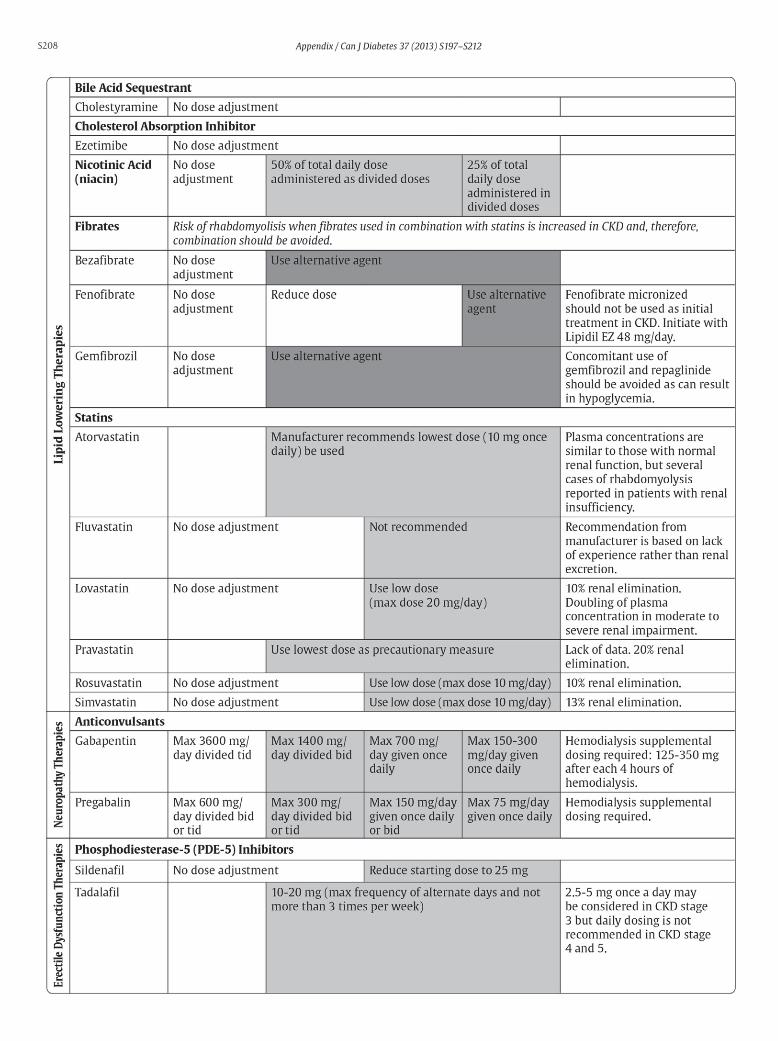
S208 Appendix / Can J Diabetes 37 (2013) S197–S212

S209
Appendix 7
Sick Day Medication List
Appendix / Can J Diabetes 37 (2013) S197–S212

S210
Appendix 8
Rapid Screening for Diabetic Neuropathy
Multiple screening methods are published. These methods (1) are designed to screen for the presence or absence of diabetic neuropathy, as opposed to screening for specific sites on the feet that are at risk of ulceration (multisite testing). If neuropathy is identified by either of these methods, other sites may be tested to identify high-risk areas for ulceration.
1. Perkins BA, Olaleye D, Zinman B, et al. Simple screening tests for peripheral neuropathy in the diabetes clinic. Diabetes Care 2001;24:250-6.
Appendix / Can J Diabetes 37 (2013) S197–S212
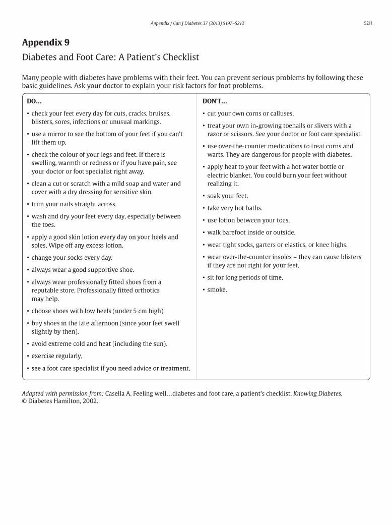
S211
Appendix 9
Diabetes and Foot Care: A Patient’s Checklist
Many people with diabetes have problems with their feet. You can prevent serious problems by following these basic guidelines. Ask your doctor to explain your risk factors for foot problems.
Adapted with permission from: Casella A. Feeling well…diabetes and foot care, a patient’s checklist. Knowing Diabetes.© Diabetes Hamilton, 2002.
Appendix / Can J Diabetes 37 (2013) S197–S212

S212
Appendix 10
Diabetic Foot Ulcers: Essentials of Management
Appendix 11
A1C Conversion Chart
Appendix / Can J Diabetes 37 (2013) S197–S212