the cardiac exam
TRANSCRIPT

THE CARDIAC EXAM
Caroline Ball, MD, FACC
Stritch School of Medicine
Medicine Clerkship

LEARNING OBJECTIVES
• Define the Mechanism of Generation, Clinical Significance and Best Listening Areas for:
• S1/S2
• S2 Splitting Patterns
• S3/S4
• Ejection Clicks (Early and Mid)
• Opening Snap
• Describe the Grading System for Murmurs
• Compare and contrast the location, pattern of radiation, timing, pitch, shape, quality, and
response to common physiologic maneuvers of several murmurs

KEYS TO THE CARDIOVASCULAR EXAM
• Stand on the right side of the patient
• Only auscultate over skin, never over clothing
• Quiet room

RULES OF THUMB
• “Benign Murmurs” = S’s: short, soft, systolic
• Diastolic Murmurs = Bad
• ARMS (Aortic Regurgitation, Mitral Stenosis)
• Left-Sided Sounds are Louder than right-sided murmurs
• Right-Sided Sounds increase with inspiration (except PS)
• Intensity does not = severity of valve lesions (ex VSD), but timing does!
• Remember the company the murmur keeps

GENERATING HEART SOUNDS

GENERATING HEART SOUNDS
• S1 = T1+M1
• Closure of tricuspid and mitral valves
• S2 = P2+A2
• Closure of pulmonic and aortic valves.

GENERATING HEART SOUNDS, CONTINUED
• Gallops can be right or left-sided
• Best heard with the bell
• Auscultate over PMI for L-sided gallops
• Auscultate over Xiphoid process for R-sided gallops
• “Summation” Gallop = in tachycardia when S3/S4 are
heard together, may be louder than S2.

S3
• Blood being “sucked” into the ventricle
• Heard in Sys-Tol-Ic dysfunction
• Acute MI
• Dilated Cardiomyopathy
• Increased volume of ventricular flow (ex MR, AI, VSD, PDA)
• May be normal in young people, athletes, third trimester of pregnancy
• “Sloshing In”
• “Kentucky”
• May be heard every few beats
• Cannot be heard with mitral stenosis (due to tight mitral valve)

S4
• Atrial Kick
• Heard in Di-A-Stol-Ic dysfunction
• “A Stiff Wall”
• “Tennessee”
• Heard post-MI
• May be heard in acute MR (not chronic, with LA enlargement)
• May be heard with first degree AV block

EJECTION SOUNDS
• Rarely the valves can be heard opening (onset of ventricular systole)
• Sounds close to physiologic splitting of S1
• Due to “doming” of the aortic valve
• Aortic Opening Sound:
• Best heard over the apex or over the aortic area
• Pulmonic Opening Sound:
• Best heard over L second intercostal area
• Respiratory variation (decrease with inspiration)

MITRAL VALVE PROLAPSE
• Mid-systolic prolapse of the mitral (+/- tricuspid) valve leaflets
into the left atrium
• Often accompanied with mid-late systolic murmur
• Systolic Click can also be heard with:
• Marfan Syndrome
• Ostium secundum ASD
• Papillary Muscle dysfunction

OPENING SNAP OF MITRAL STENOSIS
• Similar to aortic ejection sounds: occurs due to “doming”
of the MV leaflets
• The closer the opening snap is to S2 = delayed opening of
the MV = more severe mitral stenosis.

DETERMINING S1
• Usually louder over the apex

S1 IS LOUDER WHEN
• Increased ventricular contraction
• Increased rate of pressure generation in the ventricle
• Heart is closer to the chest wall.
• Hyperdynamic states
• Rheumatic Mitral Stenosis = Loud S1
• Due to elevated LAP
• Short PR interval on ECG = Loud S1
• Due to the MV leaflets being wide apart when the QRS hits
• Mitral Valve Prolapse

S1 IS SOFTER WHEN
• Prolonged PR
• Leaflets are already almost closed when QRS hits
• Acute, Severe AI
• LV diastolic pressure rises quickly -> premature closure of the mitral valve
leaflets
• MR due to lack of leaflet mobility
• Chest configuration reduces auscultation:
• Emphysema
• Obesity
• Large breasts
• Pericardial Effusion

VARIABLE S1: AV DISSOCIATION
•Atrial Fibrillation
•Complete Heart Block
•VT

PHYSIOLOGIC SPLIT HEART SOUNDS
• With Expiration A2 and P2 occur at the same time
• With Inspiration there is increased filling on the right side
of the heart, and thus a delay in closure of P2.
• In patients over age 50 the split sound may be
indistinguishable due to LV dysfunction and some degree of
AS


FIXED SPLIT HEART SOUNDS
• ASD: Right to left shunting
• Higher cardiac output through the R sided valves
• Persistent delay in P2


PARADOXICAL SPLITTING
• A2 is slow to occur, P2 remains stable
• LV is slower to contract (as in LBBB)
• Increased resistance for the LV (AS, HTN)


PERSISTENT SPLITTING
• P2 is always delayed, but more delayed with
inspiration
• RBBB (LV is delayed)
• Pulmonic Stenosis


LOUD P2
• Pulmonary Hypertension

LOUD A2
• Systemic Hypertension

FAINT A2
• Aortic Stenosis
• Faint P2 with pulmonic stenosis

GRADING MURMURS
Grade Description
1 Faintest murmur, heard only with special effort
2 Faint murmur, but heard immediately
3 Moderately loud murmur
4 Loud murmur associated with palpable thrill
5 Very loud murmur heard with part of stethoscope touching
chest wall
6 Loudest murmur heard with stethoscope removed from chest
wall.

PHYSICAL EXAM MANEUVERS
Maneuver Physiologic Effect Murmurs Affected
Passive Leg Raise Increase venous return -> Increase Preload Decreases HCM murmur
Squatting Compresses leg veins -> increased venous
return, increased afterload
Delays MVP click and shortens murmur
Decreases HCM murmur (“Squashes MVP”)
Increases AS murmur intensity
Valsalva Increases intrathoracic pressure ->
decreased preload
Moves MVP earlier in systole
Increases HCM
Standing Blood pools in legs -> Decreases preload Increases HCM
Decreases AS murmur intensity
Isometric Hand
Grip
Increases peripheral vascular resistance Increases MR murmur
Increases VSD murmur
Increases AI murmur

SYSTOLIC MURMURS

SYSTOLIC EJECTION MURMUR
• Early-peaking

AORTIC STENOSIS
• Systolic
• Radiates to carotids (especially R Supraclavicular Region)
• Louder with increased flow:
• Following a pause after a premature beat (“post-extrasystolic”)
• Passive leg raise
• Squatting
• Associated with “pulsus parvus et tardus” (Slow Rising, Late Peaking)
• Gallavardin Phenomenon = musical quality radiation of the AS murmur to the apex.

GRADING AS SEVERITY
• Mild AS: systolic murmur ends before
S2
• Moderate AS: Murmur peaks LATER in
systole. Delay in A2
• Severe AS: Paradoxical splitting of S2
(versus absent S2).
Mild Aortic Stenosis. C = opening click
Severe Aortic Stenosis

HYPERTROPHIC CARDIOMYOPATHY
• Maneuvers:
• Lounder with standing
• Louder during Valsalva
• Softer with squatting
• Softer with passive leg raise
• Associated with “Triple Ripple” on palpation over the apex
• “Spike and dome” pulse (Quick rising, twin-peaking)

HOLOSYSTOLIC MURMUR?
• Chronic MR
• TR
• VSD
• And it is usually chronic MR…
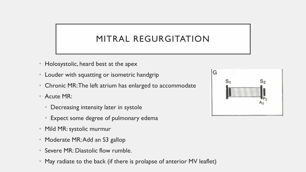
MITRAL REGURGITATION
• Holosystolic, heard best at the apex
• Louder with squatting or isometric handgrip
• Chronic MR: The left atrium has enlarged to accommodate
• Acute MR:
• Decreasing intensity later in systole
• Expect some degree of pulmonary edema
• Mild MR: systolic murmur
• Moderate MR: Add an S3 gallop
• Severe MR: Diastolic flow rumble.
• May radiate to the back (if there is prolapse of anterior MV leaflet)

TRICUSPID REGURGITATION
• Holosystolic murmur
• Heard over LLSB
• Caravello’s Sign: Louder with inspiration

VENTRICULAR SEPTAL DEFECT
• Holosystolic murmur
• Heard best along left sternal border
• Louder with isometric handgrip.
Slightly delayed P2

MITRAL VALVE PROLAPSE
• Midsystolic Click moves towards
S1 and late systolic murmur
starts earlier with standing
• Click moves earlier on
inspiration
• Murmur starts later and click
moves towards S2 with squatting

DIASTOLIC MURMURS
Always bad.

ARMS
• Early Diastolic Murmurs
• Aortic and pulmonic regurgitation
• Mid/Late Diastolic Murmurs
• Mitral and tricuspid stenosis
• Combined Systolic and Diastolic Murmurs:
• PDA
• Coronary AV fistula
• Pulmonary AV fistula
• Ruptured Sinus of Valsalva aneurysm

AORTIC REGURGITATION
• High frequency
• Decrescendo
• “Blowing” in character
• Easily disguised by ambient noise
• Press the stethoscope in tight to the patient’s skin
• Louder with sitting upright and leaning forward
• Louder with sudden squatting and isometric handgrip

AUSTIN-FLINT MURMUR
• Severe AI
• “Low-pitched rumble at the apex in mid-late diastole”
• Occurs due to the severe AI jet affecting the anterior
mitral valve leaflet.
• Severe AI causing mitral stenosis
• Board question favorite!!!!

ACUTE AORTIC DISSECTION
• Unlike acute valvular disease, this AI murmur is heard best
over the third right intercostal space (vaLvuLar AI is heard
over the L sternal border)
• Alternatively: AI due to the aortic Root occurs on the R
sternal border

PULMONARY REGURGITATION
• Usually seen in the setting of congenital
heart disease vs pulmonary hypertension

MITRAL STENOSIS
• Louder with inspiration
• May require patient being in L lateral decubitus position.
• A2 -> P2 -> Opening Snap
• Earlier opening snap = higher left atrial pressure

CONTINUOUS MURMURS

PDA
• “Machine-like”
• Best heard at the firsta nd second L intercostal
space
• Diastolic phase is louder with isometric handgrip

VENOUS HUM
• Heard over R IJ
• Heard in:
• Children
• Young adults
• Pregnancy
• Thyrotoxicosis
• Due to low RA pressure

NECK VEIN ASSESSMENT
• Internal Jugular Vein does not have valves
• Officially: RAP (in cm water) = 5 + the
height from sternal angle to top of water
column.
• Patients with very high RAP need to be
more upright
• Patients with low RAP need to be more
supine

NECK VEIN ASSESSMENT
• Venous and Arterial Pulsations will be next to each
other.
• Venous will be lateral
• In sinus rhythm the venous pulsation will be triphasic
• The “A” wave will occur before carotid palpation
• The “V” wave will occur with carotid palpation

VENOUS WAVEFORMS
• Normal = A > V wave
• “Giant A” = Pulmonary Hypertension from any
cause
** ABSENT IN AF **
“Cannon” A waves with AV dissociation
• “CV” = “Large V Waves” = SeVere TR.
May also represent ASD
• Rapid X/Y descent = constriction

ASSESSMENT OF CHF
• Peripheral Edema and Rales are the least sensitive markers
of CHF:
• Peripheral edema can also be due to lymphatic dysfunction, renal,
hepatic dysfunction
• Chronic CHF Increased pulmonary lymphatics fewer rales
• Elevated JVP and the presence of an S3 = the most specific
signs for heart failure

HEPATOJUGULAR REFLEX
• Press over the upper abdomen for 10 seconds and watch
JVP
• Normal = brief rise and fall in the JVP.
• Abnormal = sustained rise in JVP Indicates R heart
failure, most commonly 2/2 elevated LV filling pressures,
but could also be RV infarct.

KUSSMAUL’S SIGN
• Normally: Inspiration Increased RV filling, but the RV can
accommodate
• Constriction: The RV cannot accommodate the increased
RV filling, and therefore there is a rise in JVP with
inspiration
• May also be seen in decreased RV compliance

REFERENCES
• ”Systolic Murmurs.” Chapter 26. Clinical Methods: The
History, Physical, and Laboratory Examinations. 3rd Edition.
Walker HK, Hall WD, Hurst JW, editors. Boston:
Butterworths; 1990.
• Chapter 12: The History and Physical Exam. Braunwald’s
Heart Disease. Bonnow RW, Mann DL, Zipes DP, and Libby
P, editors. 9th edition. Elsevier.