the cashew allergens: a molecular and serological
TRANSCRIPT

The cashew allergens:
a molecular and serological characterisation
Marit Reitsma

Thesis committee
Promotors
Prof. Dr H. J. Wichers
Professor of Immunomodulation by Food
Wageningen University & Research
Prof. Dr H. F. J. Savelkoul
Professor of Cell Biology and Immunology
Wageningen University & Research
Co-promotor
Dr N. W. de Jong,
Assistant professor, Department of Internal Medicine, section Allergology
Erasmus MC, Rotterdam
Other members
Prof. Dr M. A. J. S. van Boekel, Wageningen University & Research
Prof. Dr E. N. C. Mills, University of Manchester, UK
Dr K. Adel-Patient, INRA Food Allergy Laboratory, Gif-sur-Yvette, France
Dr P. V. Jeurink, Danone Nutricia Research, Utrecht
This research has been conducted under the auspices of the Graduate School VLAG.
(Advanced studies in Food technology, Agrobiotechnology, Nutrition and Health Sciences)

The cashew allergens:
a molecular and serological characterisation
Marit Reitsma
Thesis
submitted in fulfilment of the requirements for the degree of doctor
at Wageningen University
by the authority of the Rector Magnificus
Prof. Dr A. P. J. Mol
in the presence of the
Thesis Committee appointed by the Academic Board
to be defended in public
on Monday April 10 2017
at 11 a.m. in the Aula.

Marit Reitsma
“The cashew allergens: a molecular and serological characterization”
186 pages.
PhD thesis, Wageningen University, Wageningen, the Netherlands (2017)
With references, with summary in English
ISBN: 978-94-6343-078-4
DOI: 10.18174/403658

1
Table of Contents
Chapter 1 General introduction 3 Chapter 2 Protein transport across the small intestine in food allergy 23 Chapter 3 Purification and characterization of Anacardium occidentale (cashew) 45
allergens Ana o 1, Ana o 2 and Ana o 3
Chapter 4 Origin and processing methods slightly affect allergenic characteristics 75 of cashew nuts (Anacardium occidentale)
Chapter 5 Pichia pastoris recombinantly expressed Anacardum occidentale 107 (cashew nut) allergens Ana o 1, Ana o 2 and Ana o 3
Chapter 6 General discussion 141 Summary 171 Acknowledgements 175 About the author 179

2

3
Chapter 1
General introduction

Chapter 1
4
Cashew allergy
Cashew allergy is a severe allergy [1-4] of which the prevalence appears to be increasing [6].
As information is scarce on prevalence, allergen characteristics, and cross-reactivity, the
IDEAL project was initiated: Improvement of Diagnostic mEthods for ALlergy assessment of
cashew allergy in children. In this project the clinical side of cashew allergy was combined
with molecular studies in order to get a broad picture of cashew nut allergy and its
responsible allergens. This was realised by involving three tertiary care centres for food
allergy (Erasmus MC Rotterdam, University Medical Centre Groningen, and Reinier de Graaf
Gasthuis) to perform Double Blind Placebo Controlled Food Challenges (DBPCFC) in children
with suspected cashew allergy.
This thesis research was performed as part of this IDEAL project and focussed on the
molecular biological studies. This thesis describes the study of cashew nut (Anacardium
occidentale) proteins, focussed on its known allergens: Ana o 1, Ana o 2 and Ana o 3. These
proteins are studied with regards to purification, effects of heat treatments, cross-reactivity,
and IgE binding.
Allergy
The self-reported prevalence of food allergy in Europe is approximately 6%. However, the
prevalence of food challenge-confirmed food allergy is below 1% [7]. Type I hypersensitivity,
or IgE-mediated allergy, can be divided into three phases: the sensitization, stimulation, and
effector phase [8], see Figure 1.1. During the sensitization phase an allergen (protein) enters
the body, for example via ingestion, and this allergen is taken up and processed by an
Antigen Presenting Cell (APC). The APC presents the allergen to CD4+-T cells through their
MHCII molecule and T Cell Receptor (TCR). Once these cells differentiate into Th2 cells, they
can activate B cells by a combination of cytokines (IL4, IL13) and co-stimulatory receptor
binding (CD40-CD40Ligand, MHCII-B-cell receptor). The activated B cells start to produce and
release IgE, which binds to mast cells and basophils due to the presence of high affinity IgE
receptors (FcɛRI) expressed on these cells [8, 9]. During the second phase, the allergen again
enters the body and binds and crosslinks two receptor-bound IgE molecules, thereby
inducing the third phase. In this third phase, allergic mediators such as histamine and other
inflammatory mediators are released from the mast cells and basophils, inducing the
symptoms typical for an allergic reaction (e.g. itchiness, bronchial spasm, etc.) [10].

General introduction
5
Figure 1.2 Cashew apple and cashew nut, based on [5].
Figure 1.1 Food allergy: sensitization, stimulation and effector phase. Based on [9].
As explained above, in order to develop an allergy, a protein or peptide must come into
contact with an antigen presenting cell. For food allergy the most obvious contact between
food proteins and cells is through the digestive system. Proteins can be transported over the
gut by several methods: for example paracellular transport [12], or endocytosis via M-cells
[12] or enterocytes [13] could explain the ability of food proteins/peptides to reach immune
cells and initiating the sensitization or stimulation phase. The 2S albumins of brazil nut and
sesame seed, digested and undigested, can pass the
epithelial cell layer intact as shown in a Caco-2 setup,
indicating both the resistance to digestion and the
capability of intestinal transport of these 2S albumins
[14]. Also peanut protein was shown to be transported
across the intestinal epithelial layer in an in vivo mouse
model [15].
Cashew nut
Cashew (Anacardium occidentale) originates from Brazil
and has been distributed to Mozambique and India by

Chapter 1
6
Figure 1.3 Overview of the steps of
cashew nut processing, based on [5].
the Portuguese in the 16th century, after which these nuts were further spread by nature
[16]. Cashew trees are easily cultivated, yielding 7 to 11 kilos of cashew nuts per year. The
cashew tree is often cultivated by smallholder farms, and cross-pollination occurs freely
between trees, leading to high variability
between trees with little characterisation
regarding to cashew tree varieties [5].
The cashew nut is a kidney-shaped seed
which forms below a cashew apple, also
named the false fruit of the cashew tree, see
Figure 1.2. The edible cashew nut kernel is
protected by a peel (testa) and is sheltered
inside a shell containing a corrosive liquid
named cashew nut shell liquid [5]. In recent
years the global production of cashew nuts
has been rising with a production of 4.7
million tonnes of raw cashew in 2011 with
Vietnam as largest producer [16]. This
production is mostly focussed on the cashew
nut, but value-added side-products can be
obtained from the cashew apple (alcoholic
beverages, juice, candy, chutney, jam), testa (poultry feed), and cashew nut shell liquid
(ingredient for paint, varnish) [17].
As explained by Azam-Ali and Judge [5], cashew nuts require many processing steps to be
prepared for consumption. Picked or fallen cashew nuts are soaked to increase the moisture
content of the kernel to 9% in order to avoid blackening during the subsequent heating step.
This heating step, where the nut is roasted or fried, makes the shell brittle and simplifies
taking out the cashew kernel. The shell is most often manually removed, yielding more of
the highly desired whole cashew kernels compared to machine cutting of the shell. The
removed kernel at this point is still covered by the testa, which is removed after a heating
step of 6hr at 70˚C. After subsequent sorting of the cashews based on size, the moisture
content is adjusted to 5% by humidification, after which the cashews are shipped and
processed (e.g. roasted, fried) for consumption [5], see Figure 1.3.
Clinical aspects
As mentioned by van der Valk et al. [18]; allergy to cashew has gained increasing attention in
recent years, and is often categorized as a severe allergy compared to other (tree-) nut
allergies [1-4]. A retrospective study in a hospital in Sweden showed an increasing number of

General introduction
7
cashew-allergic reactions over a period of 10 years (2001-2010) [6]. Another study in Sweden
mentioned that in children over 3 years of age, allergic reactions to peanut and tree nuts
were the most common food allergies resulting in emergency hospital visits. In total 5% of
these food allergic reactions were due to a cashew allergy [19]. In children, most (74%, 58
out of 78) cashew allergic reactions occur below 6 years of age [3].
The prevalence of cashew allergy itself has not been clearly described in literature.
Prevalence of tree nut allergy as a group was determined at 1.3% based on self-reported
lifetime allergy and 0.5% based on oral food challenges [20]. Within this group of tree nut
allergy the percentage of cashew allergic individuals lies somewhere between 5% [21] and
55% [22] (see Table 1.1). The prevalence of tree nut allergy as a group seems to be
increasing in children as based on self-reported allergies [23].
Table 1.1 Cashew allergy prevalence as calculated from several studies.
Year Region Population % cashew allergic of tree nut allergic
Methods Source
1995-1997
USA Children with acute reactions to peanut and or tree nuts
55% (11/20) Questionnaire upon hospital allergy treatment
[22]
1997 USA Members of the Food Allergy and Anaphylaxis Network, patients of allergists (89% <18 year old)
20% Questionnaire [24]
1997 USA General US population 7% (8/118) Telephone survey [25] 2002 USA General US population 44% (36/82) Telephone survey [26] 2003-2006
The Netherlands
Adults with suspected food allergy
20% Questionnaire upon suspected food allergy
[27]
2005 USA 3-21 years old with tree nut allergy
30% (34/115) Diagnosed by history, skin prick test or IgE levels
[4]
2005 France Schoolchildren 5% (1/19) Questionnaire [21] 2005 UK 7-10 year old children 19% (8/43) Skin prick test [28] 2008 USA General US population 35% (29/84) Telephone survey [29] 2016 Europe 0- <18 year old children 30% (78/256) Medical history [3]
Considering the symptoms upon an allergic reaction to cashew, certain dissimilarities were
found between multiple studies. A study on 42 cashew-allergic children in France showed
that most suffered from cutaneous symptoms (56%), 25% displayed respiratory reactions
and 7% displayed asthmatic reactions [30]. Hourihane et al. showed that 14 out of 29 (48%)
cashew allergic patients experienced wheezing and 11 out of 29 (38%) experienced collapse
or feeling faint [31]. Maloney et al. show 84% of cashew allergic reactions result in
cutaneous symptoms, 70% in respiratory symptoms and about 65% in gastrointestinal
symptoms [32]. On the other hand in the UK, from 47 cashew-allergic children 98% showed
cutaneous symptoms, 32% gastrointestinal and 40% suffered from wheezing [2]. These data

Chapter 1
8
show high variability in the perceived symptoms. An explanation for these differences might
be found in a difference in study population or different methods of observation.
Despite the difference in symptoms observed between different studies, all studies agree on
the severity of this allergy. The severity of cashew allergy can be explained by two factors:
the high risk of a severe allergic reaction and the low amount of cashew required in order to
develop this reaction. The high risk associated with cashew allergy has been revealed by
Davoren et al. In a retrospective study in Australia it was shown that although peanut allergy
was more prevalent, incidence of anaphylaxis upon cashew ingestion (20 out of 27 cashew
allergic reactions developed into anaphylaxis, 74%) was higher than peanut induced
anaphylaxis (54 out of 177, 31%) [1]. In another study, cashew-allergic children were
matched and compared to peanut-allergic children. This comparison showed that cashew
allergy resulted more often in severe reactions, such as lower airway narrowing, than peanut
allergy. Next to this, upon emergency treatment, adrenalin was more often administered to
cashew allergic than peanut allergic children [2].
The low threshold required for an allergic reaction was described by Hourihane et al. stating
that 14 out of 29 (48%) cashew-allergic patients experienced an allergic reaction after
exposure to cashew via smelling/touching or tasting but not eating [31].
Known allergens in cashew: Ana o 1, Ana o 2 and Ana o 3
The majority of a cashew nut is composed of lipids (44%), but the second largest component
in cashew is protein (19%) [33]. In this protein fraction the three known allergens from
cashew are present: Ana o 1, Ana o 2 and Ana o 3. The nucleotide and amino acid sequences
of these three major allergens are depicted in Figure 1.4.
Ana o 1
Ana o 1 is a 7S globulin of 50kDa. The cDNA coding sequence of Ana o 1 has been described
by Wang et al. after preparing a cDNA library from cashew in late maturation, subsequent
expression in E. coli and screening by serum from cashew allergic patients. For Ana o 1, two
cDNA sequences were identified: Ana o1.0101 and Ana o1.0102, which differ by a single
nucleotide [34]. As a 7S globulin, Ana o 1 is a seed storage protein that is part of the cupin
protein family. Based on other cupins, Ana o 1 is expected to contain a double stranded α-
helix [10] and, like other 7S globulins, is expected to form a trimer of 150kDa in native state
[38].
Ana o 1 has been noted as a major allergen when recombinantly produced Ana o 1 bound
50% of patient sera (10/20). Upon subsequent epitope studies, 11 linear epitopes [34] and
one conformational epitope [39] were observed.

General introduction
9
Ana o 2
Ana o 2, also named anacardein or cashew major protein, is an 11S globulin that also
belongs to the cupin protein family [10]. In general, 11S globulins are the most prevalent
proteins in many seeds [38] and when examining cashew protein extracts this also seems the
case for cashew [40]. 11S globulins are commonly hexameric proteins composed of 60kDa
subunits. These subunits each contain an acidic large subunit of about 40kDa and a basic
small subunit of about 20kDa linked together by a single disulphide bond [41]. In Ana o 2,
these size descriptions are correct as the large subunit has been determined at 33kDa [35]
and the small subunit at 20kDa [42, 43]. On denaturing SDS-PAGE also a minor band of
53kDa was tentatively identified as Ana o 2 [35].
Ana o 1: Ana o 1.0101, vicilin-like protein [Anacardium occidentale] GenBank:
AAM73730.2 [34]
1 mgpptkfsfs lflvsvlvlc lgfalakidp elkqckhqck vqrqydeqqk eqcvkeceky
61 ykekkgrere heeeeeewgt ggvdepsthe paekhlsqcm rqcerqeggq qkqlcrfrcq
121 erykkergqh nykreddede dedeaeeede npyvfededf ttkvkteqgk vvllpkftqk
181 skllhaleky rlavlvanpq afvvpshmda dsiffvswgr gtitkilenk resinvrqgd
241 ivsissgtpf yianndenek lylvqflrpv nlpghfevfh gpggenpesf yrafsweile
301 aalktskdtl eklfekqdqg timkaskeqi ramsrrgegp kiwpfteest gsfklfkkdp
361 sqsnkygqlf eaeridyppl ekldmvvsya nitkggmsvp fynsratkia ivvsgegcve
421 iacphlsssk sshpsykklr arirkdtvfi vpaghpfatv asgnenleiv cfevnaegni
481 rytlagkkni ikvmekeake lafkmegeev dkvfgkqdee fffqgpewrk ekegrade
Ana o 1: Ana o 1.0102, vicilin-like protein, partial [Anacardium occidentale] GenBank:
AAM73729.1 [34]
1 pptkfsfslf lvsvlvlclg falakidpel kqckhqckvq rqydeqqkeq cvkecekyyk
61 ekkgrerehe eeeeewgtgg vdepsthepa ekhlsqcmrq cerqeggqqk qlcrfrcqer
121 ykkergqhny kreddedede deaeeedenp yvfededftt kvkteqgkvv llpkftqksk
181 llhalekyrl avlvanpqaf vvpshmdads iffvswgrgt itkilenkre sinvrqgdiv
241 sissgtpfyi anndenekly lvqflrpvnl pghfevfhgp ggenpesfyr afsweileaa
301 lktskdtlek lfekqdqgti mkaskeqvra msrrgegpki wpfteestgs fklfkkdpsq
361 snkygqlfea eridypplek ldmvvsyani tkggmsvpfy nsratkiaiv vsgegcveia
421 cphlssskss hpsykklrar irkdtvfivp aghpfatvas gnenleivcf evnaegniry
481 tlagkkniik vmekeakela fkmegeevdk vfgkqdeeff fqgpewrkek egrade
Ana o 2: Ana o 2, partial [Anacardium occidentale] GenBank: AAN76862.1 [35]
1 lsvcflilfh gclasrqewq qqdecqidrl dalepdnrve yeagtveawd pnheqfrcag
61 valvrhtiqp nglllpqysn apqliyvvqg egmtgisypg cpetyqapqq grqqgqsgrf
121 qdrhqkirrf rrgdiiaipa gvahwcyneg nspvvtvtll dvsnsqnqld rtprkfhlag
181 npkdvfqqqq qhqsrgrnlf sgfdtellae afqvderlik qlksednrgg ivkvkddelr
241 virpsrsqse rgseseeese dekrrwgqrd ngieetictm rlkenindpa radiytpevg
301 rlttlnslnl pilkwlqlsv ekgvlyknal vlphwnlnsh siiygckgkg qvqvvdnfgn
361 rvfdgevreg qmlvvpqnfa vvkrareerf ewisfktndr amtsplagrt svlggmpeev
421 lanafqisre darkikfnnq qttltsgess hhmrdda
Ana o 3: 2s albumin [Anacardium occidentale] GenBank: AAL91665.1 [36]
1 makfllllsa favlllvana siyraiveve edsgreqscq rqfeeqqrfr ncqryvkqev
61 qrggrynqrq eslreccqel qevdrrcrcq nleqmvrqlq qqeqikgeev relyetasel
121 pricsispsq gcqfqssy
Figure 1.4 Protein sequence of Ana o 1, Ana o 2 and Ana o 3 with the difference between Ana o
1.0101 and Ana o 1 1.0102 indicated in bold and italic, IgE-binding epitopes [34, 35, 37] underlined.

Chapter 1
10
To the recombinant version of Ana o 2, 13 out of 21 (62%) cashew-allergic patient IgE could
bind, indicating Ana o 2 to be a major allergen. Ana o 2 contains at least 22 linear epitopes
which are spread out over the entire protein [35]. When these linear epitopes were
compared to linear epitopes on other 11S globulin proteins, four “hot spots” were identified,
IgE binding epitopes at overlapping positions in aligned protein sequences. One of these
hotspots in Ana o 2 was identified to be shielded within the monomeric subunits of Ana o 2,
requiring denaturation of the protein before exposure of this epitope [44]. Next to these
linear epitopes at least one conformational epitope on Ana o 2 has been discovered [43, 45].
This conformational epitope was studied using a monoclonal mouse antibody that has been
shown to inhibit human IgE binding to a conformational epitope on Ana o 2. This
conformational epitope consists of a protein segment of 24 amino acids containing β-strands
and a short helical segment on the large subunit of Ana o 2, which connects to the small
subunit of Ana o 2 [43, 45].
Ana o 3
Ana o 3 belongs, as a 2S albumin, to the prolamin superfamily of proteins. These proteins are
mostly seed storage proteins and are commonly small (7-16kDa) with multiple inter-chain
disulphide bonds and four α-helices [10]. Also the cDNA sequence of Ana o 3 has been
determined by Robotham et al. and the recombinant Ana o 3 has been used to produce goat
anti-Ana o 3 antibodies that can be used to purify native Ana o 3. This showed native Ana o 3
to be a 12.598kDa 2S albumin which probably undergoes posttranslational modification as
the protein size based on the cDNA sequence was predicted to be 16.335kDa [37]. Based on
literature this protein is most probably proteolytically cleaved at the C-terminus into a small
and a large subunit that stay associated by four disulphide bonds [41, 46]. The large subunit
of this protein is present in three isoforms of 6, 8 and 10kDa [37]. The small subunit is not
mentioned in experimental studies, perhaps because it is too small to be observed on SDS-
PAGE or simply because it was never looked for. On western blot 21 out of 26 (81%) patient
sera bound to rAna o 3, confirming Ana o 3 to be a major allergen. In Ana o 3, 8 linear
epitopes have been identified, of which some show high similarity with Jug r 1 from walnut
and sesame seed 2S albumin [37].
Purified and recombinantly produced cashew allergens
The cashew nut allergens Ana o 1, 2 and 3 have been recombinantly produced in E. coli by
Wang and Robotham et al. [34, 35, 37]. Cashew-derived Ana o 1, 2, and 3 have been
identified using these recombinant allergens by inhibition blotting. Inhibition blotting was
done by pre-incubating serum from a cashew-allergic person with e.g. recombinant Ana o 1,
and observing on western blot which protein bands from cashew are no longer bound by IgE,
thereby identifying native Ana o 1. E. coli derived recombinant Ana o 1 is a protein of 55 and

General introduction
11
65kDa (two clones, different in start site, differing 73 amino acids) [34], rAna o 2 is 52kDa
and also forms a dimer of 120kDa [35], rAna o 3 is a 14kDa protein [37].
Of the three allergens, only Ana o 3 has been purified from the cashew nut [42]. In this
protocol Mattison et al. used a sodium phosphate gradient on a ceramic hydroxyapatite
column after defatting of the cashews and precipitation of the protein extract. In this article
neither the yield nor the purity of the purified Ana o 3 allergen was mentioned. For Ana o 2 a
purification protocol has been described [47], however, as mentioned by Teuber et al. this
fraction is not immunologically pure [48]. No protocol for the purification of Ana o 1 has
been described in literature.
Allergen protein purification starting from the food source or after generating recombinant
proteins, warrants a discussion on subsequent application. The recombinant production of
proteins is, in general, easier to standardise, often yields higher amounts of pure protein
(depending on the expression system used), and contamination of one allergen with the
other is much less likely to occur compared to protein purification from the food source.
Purification from the food source directly, on the other hand, results in a native protein with
correct post-translational modifications like protein folding, disulphide bridges,
glycosylation, etc. When multiple isoforms of a single protein are present in the food source
purification might extract multiple (but not necessarily all) isoforms, while recombinant
protein expression only produces one isoform unless multiple colonies of the different
protein isoforms are prepared. Also some technical specifications can prompt different
choices as protein purification and recombinant expression both require different specialist
equipment, and besides, for recombinant protein expression the protein/cDNA sequence of
the protein of interest should be known, otherwise first a cDNA library should be prepared.
Lastly the effect of the food matrix when studying heat treatment effects, protein digestion
or protein transport characteristics is missed for recombinantly produces proteins.
Heat treatments and in vitro digestion
Cashews are eaten in processed form: most commonly the raw nuts are heated for 20-35min
at 150°C in order to remove the shell, afterwards they can be roasted or fried. Roasted
cashews (120°C or 160°C for 20min) are most often eaten in the US while in the Netherlands
mostly fried cashews (93°C increased to 135°C in 35-40min or 150°C -160°C for 1-3min) are
consumed [49]. Because cashews are always heat-treated before consumption, and as this
might affect the structure and function of the proteins, several studies have examined the
effect of processing on cashew proteins. Most of these studies have been performed using
SDS-PAGE and western blotting techniques, using either patient IgE or polyclonal anti-
cashew antibodies, thereby mostly focussing on (linear) epitopes.

Chapter 1
12
Studies focussing on roasted cashew show varying results fluctuating from no difference in
antibody binding [50], to decreased [51] or even increased [52] antibody binding (patient IgE
[50, 51] or polyclonal IgG [51, 52]) upon roasting of cashew nuts. The discrepancies between
these studies can to some extent be caused by the increased solubility of Ana o 3 in roasted
cashew [51]. When the antibodies used, bind specifically to Ana o 3, this binding will be
increased in extracts from roasted cashew nuts simply due to the higher amount of Ana o 3
present in the roasted cashew sample.
Besides roasting, also more harsh heat treatments like γ-irradiation and autoclaving have
been applied to cashew. γ-Irradiation followed by frying or blanching did not affect binding
of a polyclonal antibody, while γ-irradiation followed by autoclaving or roasting did reduce
binding of anti-cashew IgG as observed on western blot [53]. The required dose of γ-
irradiation will, however, lead to inedible cashews [54].
A study by Venkatachalam et al. showed a thorough comparison of cashew subjected to
many different heat treatments by using rabbit, goat and mouse antibodies. For Ana o 1, a
decrease in antibody binding after roasting, blanching, microwaving and autoclaving was
observed but not after γ-irradiation, indicating the probable loss of a conformational epitope
within Ana o 1 upon heating. Another antibody showed only decreased binding to Ana o 1
after prolonged autoclaving of the cashew nuts [55]. Ana o 2 remained immunologically
stable despite microwaving, roasting, blanching or γ-irradiation. Only autoclaving for 20min
could decrease antibody binding slightly [55]. The immunoreactivity of Ana o 2 towards two
monoclonal mouse antibodies could chemically be reduced by SDS but not by guanidinium
HCl or urea despite that the protein conformation of Ana o 2 was changed by all three
chemicals [56]. Ana o 3 appeared more sensitive to processing steps such as roasting,
autoclaving and blanching [55]. However, for Ana o 3 this was assessed using a monoclonal
antibody that potentially binds a conformational epitope [55], while for Ana o 2 the
antibodies used in the various studies probably target linear epitopes [56], making it difficult
to compare the stability of these two allergens. It is expected that Ana o 3, like other 2S
albumins, is highly resistant to heat processing [46]. Considering pH stability, all three
allergens were stable in the middle-pH range but were unstable at the extreme pH of 1 and
13 [55].
Lastly, the effect of sulphite, a chemical that has been shown to disrupt disulphide bonds,
has been tested on cashew extracts [52]. On both western blot and dot blot, a decrease in
patient-IgE binding was observed after sulphite treatment of both raw and roasted cashew
[52]. However, such a treatment cannot be applied to whole cashews, and, as already
mentioned by the authors, sodium sulphite itself can cause allergic reactions.

General introduction
13
Besides the effect of heat and processing treatments on cashew nut proteins, also the in
vitro digestibility of these proteins has been studied. This in vitro digestion showed Ana o 1
and the 30kDa subunit of Ana o 2 to be partially susceptible to both pepsin and trypsin
digestion. However, Ana o 3 and the 20kDa subunit of Ana o 2 were not completely digested
by either pepsin (0.8U 2hr) or trypsin (20U 2hr) alone, nor by pepsin (0.8U 30min) followed
by trypsin (2U 30min). Ana o 3 pre-treated with DTT to break disulphide bonds was more
susceptible to pepsin digestion, as based on reduced visibility on SDS-PAGE and reduced (but
not completely obstructed) IgE binding in inhibition ELISA testing [42]. The effect of in vitro
digested cashew on the in vivo allergic response has also been studied. Mice sensitized to
native cashew showed reduced allergic reactions upon intraperitoneal injected pepsin-
digested cashew compared to exposure to non-digested cashew protein. Next to this,
immunotherapy using a pepsin-digested cashew protein extract could reduce the allergic
reaction upon cashew challenges in mice, indicating a possible role for pepsin-digested
cashew in immunotherapy [57].
To summarise, as could be expected from 7S globulins, 11S globulins, and 2S albumins [10]
Ana o 1, Ana o 2 and Ana o 3 are highly resistant to heat treatments and quite resistant to in
vitro digestion. The IgE-immunoreactivity of Ana o 2 and 3 can be decreased by sulphite
treatments and intense γ-irradiation. However, applying these treatments in the cashew nut
industry is not feasible as sodium sulphite itself might cause allergic reactions and cannot be
applied to whole cashew nuts, and as the use of high γ-irradiation would result in non-edible
cashews [54].
Cross-allergenicity with other Anacardiaceae and other tree nuts
Cashew is a tree nut belonging to the family of Anacardiaceae together with other edible
plants and trees like sumac, mango, pistachio, and pink pepper [58]. Cross-allergenicity
between cashew and other Anacardiaceae and tree nuts has been studied to some extent.
Especially the possibility of cross reactivity between cashew and pistachio has been
presented by several groups [59-63] and part of this cross reactivity might be explained by
cross reactivity between Pis v 3 and Ana o 1 [62], and sequence homology between Pis v 2
and Ana o 2 and between Pis v 1 and Ana o 3 [63]. Cashew allergen Ana o 3 even showed a
higher specificity in correctly identifying pistachio allergy by serum IgE, compared to using a
pistachio extract [64]. Also in vivo cross-reactivity between cashew and pistachio has been
noted. In this study, cashew-immunotherapy in cashew-sensitized mice resulted in
decreased allergic reaction towards both cashew and pistachio [65]. Also the role of walnut,
a tree nut but not a member of the Anacardiaceae, was studied here. Mice sensitized to
both cashew and walnut showed a diminished allergic reaction towards cashew after
receiving walnut immunotherapy [65]. The other way around, when walnut was introduced

Chapter 1
14
into the diet of cashew-sensitized mice, the mice showed elevated walnut-specific IgE levels
and developed an allergic reaction towards walnut [66].
Besides walnut and pistachio, also studies with mango have been performed. Mango as
member of the Anacardiaceae is suspected to cross-react with both cashew and pistachio as
based on inhibition RAST results [67-69].
Studies on the possibility of cross reactivity between cashew and peanut are not unexpected
as 20-30% of peanut allergic individuals are also allergic to one or more tree nuts [23].
However, studies using cashew and peanut inhibition ELISAs [50, 70], basophil activation test
(BAT) [70], and studies on epitope homology of Ana o 1 and Ara h 1 [34] did not substantiate
a claim of cross allergenicity between peanut and cashew. No cross-reacivity was found by
inhibition ELISA and inhibition western blot between roasted cashew and rAra h 2 [71].
Epitope homology between the 11S globulins Ara h 3 (peanut), Cor a 9 (hazelnut), Jug r 4
(walnut) and Ana o 2, however, has been suggested [72]. Overall, cross reactivity between
cashew and peanut is not confirmed, which is not unexpected as peanut is neither a tree nut
nor part of the Anacardiaceae family. This notion is further substantiated by the finding that
in vivo it has been shown that peanut-allergic persons, who are also tree nut allergic, are less
likely to be allergic to cashew than peanut-tolerant persons who are tree nut allergic [32].
Cashew apple and cashew pollen
Besides allergens in the cashew itself and cross-reactive allergens in related plants, also
some research has been performed on allergenic proteins in other parts of the cashew tree:
the cashew apple, pollen and the shell. Despite the limited exposure to these products
outside cashew cultivation areas, it is still worth mentioning.
Presence of allergens in the cashew apple similar to the allergens present in cashew nuts has
been described by Comstock et al. In this study cashew-allergic patients’ sera showed IgE
binding to proteins from cashew apple juice concentrate on western blot, a reaction that
could be inhibited (7.5, 20, 25, 50, 60kDa bands) for some sera when pre-incubated with
cashew nut extract [73]. Upon western blotting with anti-Ana o 1 and anti-Ana o 2
monoclonal antibodies, several proteins from cashew apple juice could be detected (45kDa
anti-Ana o 1; 37 and 46kDa anti-Ana o 2) [73]. Besides cashew apple allergy, one can also be
allergic to cashew tree pollen. In India, a country with many cashew-plantations, 65 patients
with allergic bronchial asthma were tested for cashew-pollen sensitisation by a bronchial
provocation test and a skin prick test. Of these 65, patients 20 were found to be allergic to
pollen of the Anacardium occidentale tree by both tests [74]. The last study deals with the
shell around the cashew kernel. A defatted protein extract was made from cashew nut shell,
showing presence of multiple proteins of 14–97kDa as visualised by SDS-PAGE [75]. In
addition, the oil present in this shell can cause contact dermatitis [76].

General introduction
15
Detection by immunoassay
As for now, cashew allergy cannot be cured, cashew allergic patients should avoid cashews,
even trace amounts. Several methods for the detection of cashew nut allergens in food
products have been developed, e.g. ELISA, immunoblotting, (RT-)PCR, dipstick, and mass
spectrometry [77]. As described by van Hengel [77] each of these methods has it’s pro’s and
con’s. For example, PCR analysis detects DNA or RNA sensitively, serving as an indicator but
does not directly detect the proteins. The dipstick method is a fast method to detect
proteins but is not quantitative, and mass spectrometry is very specific but requires highly
specialised equipment and trained personnel [77].
Specifically for the detection of cashew nut in food products, methods have been developed.
First of all an ELISA assay has been developed for the detection of the so-called “cashew
major protein” which mainly exists of Ana o 2. The assay is able to detect levels of 0.02ppm
cashew major protein, and is suitable for use in most food matrixes (e.g. wheat flour, rice
cereal, chocolate cookies) and compatible with several spices (e.g. salt, brown sugar,
cardamom). The assay did show diminished sensitivity when combined with milk chocolate,
raisin bran cereal, cinnamon and nutmeg. Upon heat-processing of cashew the detection
was diminished as well, especially roasting of the cashew at 170°C for 20min diminished the
sensitivity of this ELISA assay [78]. Gaskin and Taylor also developed a cashew specific ELISA
with high sensitivity (1μg cashew/g of product) but with significant cross reactivity towards
pistachio and, to a certain degree, also towards hazelnut [79]. Secondly, a mass
spectrometry method has been developed, detecting Ana o 2 and Ana o 3 in the sub-ppm
range, thereby being declared as more sensitive than ELISA assays [80]. Lastly, also PCR
assays detecting DNA, have been developed targeting Ana o 3, detecting 2mg/kg [81] and
0.005% of the total food weight [82].
Aim and outline of the thesis
Diagnostic procedures in allergy are partly based on the detection of serum-IgE for the
offending allergen. Very important drawbacks of this type of testing, are the often limited
clinical relevance of its outcomes with often high numbers of false-positive or false-negative
test results. The aim of this thesis is a better chemical identification and functional
characterisation of the major cashew nut allergens Ana o 1, 2 and 3. This characterisation
will contribute to the development of more sensitive and reliable diagnostic procedures for
the early detection of cashew allergy in young children. This will allow improved disease
monitoring and future therapy for this ’vulnerable’ group.

Chapter 1
16
In the second chapter of this thesis a review is provided on intestinal protein transport,
focussing on the difference between sensitised versus non-sensitised persons. This
difference in intestinal protein transport between sensitised and non-sensitised persons
could indicate a possible mechanism of how allergens could come into contact with immune
cells and cause the deveopment of a food allergy. The experimental chapters of this thesis
are focussed on cashew nut allergy, studying the cashew nut allergens Ana o 1, Ana o 2 and
Ana o 3. In chapter 3, a purification method is described for Ana o 1, Ana o 2 and Ana o 3
from blanched cashew nuts, and their protein characteristics such as IgE-binding and
glycosylation are described. For Ana o 3 epitope mapping experiments have been
performed, and mass spectrometry was done to study the N- and C-termini of Ana o 3. Next,
in chapter 4, a comparison was made between cashews of different origins which have been
subjected to different heat treatments. This was done as all cashew studies described in
literature have been performed using store-bought cashews, from unknown origin and with
doubtfull information on pre-treatments (e.g. heating steps applied in order to remove the
shell) while being sold and used as “ raw” cashew. Therefore, in chapter 4, 8 different origins
of cashew were compared for their protein composition. This allowed us to assess whether
or not data from literature can be universally used in cashew protein research or whether it
is specific to a cashew of a particular origin. Furthermore, the difference between different
origins and the influence of heat treamtens on the cashews’ protein composition, protein
glycation/glycosylation, digestibility, and IgE binding was studied. In Chapter 5, the
recombinant production of Ana o 1, 2 and 3 from Pichia pastoris is described. This was done
as recombinantly expressed proteins are usually more easily purified in greater quantity and
with no chance of allergen contamination between the purified Ana o 1, Ana o 2 and Ana o
3. The cloning and purification of these three allergens, as well as characterization on 1D and
2D electrophoresis, western blot, and inhibition blot, is described in this chapter.
The results of these 5 chapters are discussed in chapter 6. The newly obtained knowledge on
Ana o 1, 2 and 3 is summarized. Also, extra information regarding the importance of cross-
reactivity with other nuts and Anacardiaceae family members is provided, and the relevance
of these results for the clinical field is discussed.

General introduction
17
References [1] Davoren, M., Peake, J., Cashew nut allergy is associated with a high risk of anaphylaxis. Arch Dis Child
2005, 90, 1084-1085.
[2] Clark, A. T., Anagnostou, K., Ewan, P. W., Cashew nut causes more severe reactions than peanut: case-matched comparison in 141 children. Allergy 2007, 62, 913-916.
[3] Grabenhenrich, L. B., Dolle, S., Moneret-Vautrin, A., Kohli, A., et al., Anaphylaxis in children and adolescents: The European Anaphylaxis Registry. J. Allergy Clin. Immun. 2016, 137, 1128-1137 e1121.
[4] Fleischer, D. M., Conover-Walker, M. K., Matsui, E. C., Wood, R. A., The natural history of tree nut allergy. J. Allergy Clin. Immun. 2005, 116, 1087-1093.
[5] Azam-Ali, S. H., Judge, E. C., Food and Agriculture Organization of the United Nations 2001.
[6] Johnson, J., Malinovschi, A., Alving, K., Lidholm, J., et al., Ten-year review reveals changing trends and severity of allergic reactions to nuts and other foods. Acta paediatrica 2014, 103, 862-867.
[7] Nwaru, B. I., Hickstein, L., Panesar, S. S., Muraro, A., et al., The epidemiology of food allergy in Europe: a systematic review and meta-analysis. Allergy 2014, 69, 62-75.
[8] Multidisciplinary approaches to allergies, Springer 2012.
[9] Larche, M., Akdis, C. A., Valenta, R., Immunological mechanisms of allergen-specific immunotherapy. Nat. Rev. Immunology 2006, 6, 761-771.
[10] Mills, E. N. C., Madsen, C., Shewry, P. R., Wicher, H. J., Food allergens of plant origin - their molecular and evolutionary relationships. Trends Food Sci Tech 2003, 14, 145-156.
[11] Lack, G., Update on risk factors for food allergy. J. Allergy Clin. Immun. 2012, 129, 1187-1197.
[12] Price, D., Ackland, L., Suphioglu, C., Nuts 'n' guts: transport of food allergens across the intestinal epithelium. Asia Pacific allergy 2013, 3, 257-265.
[13] Yu, L. C., The epithelial gatekeeper against food allergy. Pediatrics and neonatology 2009, 50, 247-254.
[14] Moreno, F. J., Rubio, L. A., Olano, A., Clemente, A., Uptake of 2S albumin allergens, Ber e 1 and Ses i 1, across human intestinal epithelial Caco-2 cell monolayers. J. Agr. Food Chem. 2006, 54, 8631-8639.
[15] Chambers, S. J., Wickham, M. S., Regoli, M., Bertelli, E., et al., Rapid in vivo transport of proteins from digested allergen across pre-sensitized gut. Biochem Biophys Res Commun 2004, 325, 1258-1263.
[16] Dendena, B., Corsi, S., Cashew, from seed to market: a review. Agron Sustain Dev 2014, 34, 753-772.
[17] Nair, K. P., The agronomy and economy of important tree crops of the developing world, Elsevier 2010.
[18] van der Valk, J. P., Dubois, A. E., Gerth van Wijk, R., Wichers, H. J., de Jong, N. W., Systematic review on cashew nut allergy. Allergy 2014, 69, 692-698.
[19] Vetander, M., Helander, D., Flodstrom, C., Ostblom, E., et al., Anaphylaxis and reactions to foods in children--a population-based case study of emergency department visits. Clin. Exp. Allergy 2012, 42, 568-577.
[20] Nwaru, B. I., Hickstein, L., Panesar, S. S., Roberts, G., et al., Prevalence of common food allergies in Europe: a systematic review and meta-analysis. Allergy 2014, 69, 992-1007.
[21] Rance, F., Grandmottet, X., Grandjean, H., Prevalence and main characteristics of schoolchildren diagnosed with food allergies in France. Clin. Exp. Allergy 2005, 35, 167-172.
[22] Sicherer, S. H., Burks, A. W., Sampson, H. A., Clinical features of acute allergic reactions to peanut and tree nuts in children. Pediatrics 1998, 102, e6.
[23] McWilliam, V., Koplin, J., Lodge, C., Tang, M., et al., The Prevalence of Tree Nut Allergy: A Systematic Review. Curr. Allergy Asthma R. 2015, 15, 54.
[24] Sicherer, S. H., Furlong, T. J., Munoz-Furlong, A., Burks, A. W., Sampson, H. A., A voluntary registry for peanut and tree nut allergy: characteristics of the first 5149 registrants. J. Allergy Clin. Immun. 2001, 108, 128-132.
[25] Sicherer, S. H., Munoz-Furlong, A., Burks, A. W., Sampson, H. A., Prevalence of peanut and tree nut allergy in the US determined by a random digit dial telephone survey. J. Allergy Clin. Immun. 1999, 103, 559-562.
[26] Sicherer, S. H., Munoz-Furlong, A., Sampson, H. A., Prevalence of peanut and tree nut allergy in the United States determined by means of a random digit dial telephone survey: a 5-year follow-up study. J. Allergy Clin. Immun. 2003, 112, 1203-1207.

Chapter 1
18
[27] Le, T. M., Lindner, T. M., Pasmans, S. G., Guikers, C. L., et al., Reported food allergy to peanut, tree nuts and fruit: comparison of clinical manifestations, prescription of medication and impact on daily life. Allergy 2008, 63, 910-916.
[28] Roberts, G., Peckitt, C., Northstone, K., Strachan, D., et al., Relationship between aeroallergen and food allergen sensitization in childhood. Clin. Exp. Allergy 2005, 35, 933-940.
[29] Sicherer, S. H., Munoz-Furlong, A., Godbold, J. H., Sampson, H. A., US prevalence of self-reported peanut, tree nut, and sesame allergy: 11-year follow-up. J. Allergy Clin. Immun. 2010, 125, 1322-1326.
[30] Rance, F., Bidat, E., Bourrier, T., Sabouraud, D., Cashew allergy: observations of 42 children without associated peanut allergy. Allergy 2003, 58, 1311-1314.
[31] Hourihane, J. O., Harris, H., Langton-Hewer, S., Kilburn, S. A., Warner, J. O., Clinical features of cashew allergy. Allergy 2001, 56, 252-253.
[32] Maloney, J. M., Rudengren, M., Ahlstedt, S., Bock, S. A., Sampson, H. A., The use of serum-specific IgE measurements for the diagnosis of peanut, tree nut, and seed allergy. J. Allergy Clin. Immun. 2008, 122, 145-151.
[33] Venkatachalam, M., Sathe, S. K., Chemical composition of selected edible nut seeds. J. Agr. Food Chem. 2006, 54, 4705-4714.
[34] Wang, F., Robotham, J. M., Teuber, S. S., Tawde, P., et al., Ana o 1, a cashew (Anacardium occidental) allergen of the vicilin seed storage protein family. J. Allergy Clin. Immun. 2002, 110, 160-166.
[35] Wang, F., Robotham, J. M., Teuber, S. S., Sathe, S. K., Roux, K. H., Ana o 2, a major cashew (Anacardium occidentale L.) nut allergen of the legumin family. Int. Arch. Allergy A. Imm. 2003, 132, 27-39.
[36] Robotham, J. M., Wang, F., Seamon, V., Teuber, S. S., et al., Ana o 3, an important cashew nut (Anacardium occidentale L.) allergen of the 2S albumin family. J. Allergy Clin. Immun. 2005, 115, 1284-1290.
[37] Robotham, J. M., Wang, F., Seamon, V., Teuber, S. S., et al., Ana o 3, an important cashew nut (Anacardium occidentale L.) allergen of the 2S albumin family. J. Allergy Clin. Immun. 2005, 115, 1284-1290.
[38] Astwood, J. D., Silvanovich, A., Bannon, G. A., Vicilins: a case study in allergen pedigrees. The J. Allergy Clin. Immun. 2002, 110, 26-27.
[39] Guan, X., Noble, K. A., Tao, Y., Roux, K. H., Sathe, S. K., Young, N. L., Marshall, A. G., Epitope mapping of 7S cashew antigen in complex with antibody by solution-phase H/D exchange monitored by FT-ICR mass spectrometry. J. Mass Spectrometry 2015, 50, 812-819.
[40] Hummel, M., Wigger, T., Hoper, T., Westkamp, I., Brockmeyer, J., Simple, Rapid, and Selective Isolation of 2S Albumins from Allergenic Seeds and Nuts. J. Agr. Food Chem. 2015.
[41] Mills, E. N. C., Shewry, P. R., Plant food allergens, Blackwell science Ltd 2004. [42] Mattison, C. P., Grimm, C. C., Wasserman, R. L., In vitro digestion of soluble cashew proteins and
characterization of surviving IgE-reactive peptides. Mol. Nutr. Food Res. 2014, 58, 884-893.
[43] Xia, L., Willison, L. N., Porter, L., Robotham, J. M., et al., Mapping of a conformational epitope on the cashew allergen Ana o 2: a discontinuous large subunit epitope dependent upon homologous or heterologous small subunit association. Mol Immunol 2010, 47, 1808-1816.
[44] Robotham, J. M., Hoffman, G. G., Teuber, S. S., Beyer, K., Sampson, H. A., Sathe, S. K., Roux, K. H., Line ar IgE-epitope mapping and comparative structural homology modeling of hazelnut and English walnut 11S globulins. Mol. Immunol. 2009, 46, 2975-2984.
[45] Robotham, J. M., Xia, L., Willison, L. N., Teuber, S. S., et al., Characterization of a cashew allergen, 11S globulin (Ana o 2), conformational epitope. Mol Immunol 2010, 47, 1830-1838.
[46] Moreno, F. J., Clemente, A., 2S Albumin Storage Proteins: What Makes them Food Allergens? The open biochemistry journal 2008, 2, 16-28.
[47] Sathe, S. K., SzeTao, K. W. C., Wolf, W. J., Hamaker, B. R., Biochemical characterization and in vitro digestibility of the major globulin in cashew nut (Anacardium occidentale). J. Agr. Food Chem. 1997, 45, 2854-2860.
[48] Teuber, S. S., Sathe, S. K., Peterson, W. R., Roux, K. H., Characterization of the soluble allergenic proteins of cashew nut (Anacardium occidentale L.). J. Agr. Food Chem. 2002, 50, 6543-6549.
[49] Masthoff, L. J., Hoff, R., Verhoeckx, K. C., van Os-Medendorp, H., et al., A systematic review of the effect of thermal processing on the allergenicity of tree nuts. Allergy 2013, 68, 983-993.

General introduction
19
[50] de Leon, M. P., Glaspole, I. N., Drew, A. C., Rolland, J. M., et al., Immunological analysis of allergenic cross-reactivity between peanut and tree nuts. J. Clin. Exp. Allergy 2003, 33, 1273-1280.
[51] C. P. Mattison, Y. B.-M., B. Vant-Hullc, A. M. Vargasa, R. L. Wassermand, C. C. Grimm, Heat-induced alterations in cashew allergen solubility and IgE binding. Tox. Reports 2016, 244-251.
[52] Mattison, C. P., Desormeaux, W. A., Wasserman, R. L., Yoshioka-Tarver, M., et al., Decreased Immunoglobulin E (IgE) Binding to Cashew Allergens following Sodium Sulfite Treatment and Heating. J. Agr. Food Chem. 2014, 62, 6746-6755.
[53] Su, M., Venkatachalam, M., Teuber, S. S., Roux, K. H., Sathe, S. K., Impact of gamma-irradiation and thermal processing on the antigenicity of almond, cashew nut and walnut proteins. J Sci Food Agr 2004, 84, 1119-1125.
[54] Mexis, S. F., Kontominas, M. G., Effect of gamma-irradiation on the physicochemical and sensory properties of cashew nuts (Anacardium occidentale L.). Lwt-Food Sci Technol 2009, 42, 1501-1507.
[55] Venkatachalam, M., Monaghan, E. K., Kshirsagar, H. H., Robotham, J. M., et al., Effects of processing on immunoreactivity of cashew nut (Anacardium occidentale L.) seed flour proteins. J. Agr. Food Chem. 2008, 56, 8998-9005.
[56] Kshirsagar, H. H., Fajer, P., Sharma, G. M., Roux, K. H., Sathe, S. K., Biochemical and spectroscopic characterization of almond and cashew nut seed 11S legumins, amandin and anacardein. J. Agr. Food Chem. 2011, 59, 386-393.
[57] Kulis, M., Macqueen, I., Li, Y., Guo, R., et al., Pepsinized cashew proteins are hypoallergenic and immunogenic and provide effective immunotherapy in mice with cashew allergy. J. Agr. Food Chem. 2012, 130, 716-723.
[58] USDA, 2016, https://www.usda.gov/
[59] Noorbakhsh, R., Mortazavi, S. A., Sankian, M., Shahidi, F., et al., Pistachio allergy-prevalence and in vitro cross-reactivity with other nuts. Allergology international 2011, 60, 425-432.
[60] Parra, F. M., Cuevas, M., Lezaun, A., Alonso, M. D., et al., Pistachio nut hypersensitivity: identification of pistachio nut allergens. Clni. Exp. Allergy 1993, 23, 996-1001.
[61] Goetz, D. W., Whisman, B. A., Goetz, A. D., Cross-reactivity among edible nuts: double immunodiffusion, crossed immunoelectrophoresis, and human specific igE serologic surveys. Ann. Allerg. Asthma Im. 2005, 95, 45-52.
[62] Willison, L. N., Tawde, P., Robotham, J. M., Penney, R. M. t., et al., Pistachio vicilin, Pis v 3, is immunoglobulin E-reactive and cross-reacts with the homologous cashew allergen, Ana o 1. Clin. Exp. Allergy 2008, 38, 1229-1238.
[63] Ahn, K., Bardina, L., Grishina, G., Beyer, K., Sampson, H. A., Identification of two pistachio allergens, Pis v 1 and Pis v 2, belonging to the 2S albumin and 11S globulin family. Clin. Exp. Allergy 2009, 39, 926-934.
[64] Savvatianos, S., Konstantinopoulos, A. P., Borga, A., Stavroulakis, G., et al., Sensitization to cashew nut 2S albumin, Ana o 3, is highly predictive of cashew and pistachio allergy in Greek children. J. Allergy Clin. Immun. 2015, 136, 192-194.
[65] Kulis, M., Li, Y., Lane, H., Pons, L., Burks, W., Single-tree nut immunotherapy attenuates allergic reactions in mice with hypersensitivity to multiple tree nuts. J. Allergy Clin. Immun. 2011, 127, 81-88.
[66] Kulis, M., Burks, A. W., Effects of a pre-existing food allergy on the oral introduction of food proteins: findings from a murine model. Allergy 2015, 70, 120-123.
[67] Fernandez, C., Fiandor, A., Martinez-Garate, A., Martinez Quesada, J., Allergy to pistachio: crossreactivity between pistachio nut and other Anacardiaceae. Clin. Exp. Allergy 1995, 25, 1254-1259.
[68] Funes, E., Milán, J. M., Pagán, J. A., López, J. D., Garcia, F. J., Negro, J. M., Hernández, J., Polo, F., Rico, P., Allergy to Anacardiaceae. Identification of allergens. Alergol. Immunol. Clin 1999, 14, 82-89.
[69] Besler, M., Paschke, A., Rodriguez, J., Internet Symposium on Food Allergens, www.food-allergens.de 2001.
[70] de Leon, M. P., Drew, A. C., Glaspole, I. N., Suphioglu, C., et al., Functional analysis of cross-reactive immunoglobulin E antibodies: peanut-specific immunoglobulin E sensitizes basophils to tree nut allergens. Clin. Exp. Allergy 2005, 35, 1056-1064.
[71] de Leon, M. P., Drew, A. C., Glaspole, I. N., Suphioglu, C., et al., IgE cross-reactivity between the major peanut allergen Ara h 2 and tree nut allergens. Mol Immunol 2007, 44, 463-471.

Chapter 1
20
[72] Barre, A., Jacquet, G., Sordet, C., Culerrier, R., Rouge, P., Homology modelling and conformational analysis of IgE-binding epitopes of Ara h 3 and other legumin allergens with a cupin fold from tree nuts. Mol Immunol 2007, 44, 3243-3255.
[73] Comstock, S. S., Robotham, J. M., Tawde, P., Kshirsagar, H., et al., Immunoglobulin E-reactive proteins in cashew (Anacardium occidentale) apple juice concentrate. J. Agr Ffood Chem. 2008, 56, 5977-5982.
[74] Fernandes, L., Mesquita, A. M., Anacardium occidentale (cashew) pollen allergy in patients with allergic bronchial asthma. J. Allergy Clin. Immun. 1995, 95, 501-504.
[75] Yuliana, M., Truong, C. T., Huynh, L. H., Ho, Q. P., Ju, Y. H., Isolation and characterization of protein isolated from defatted cashew nut shell: Influence of pH and NaCl on solubility and functional properties. Lwt-Food Sci Technol 2014, 55, 621-626.
[76] Lampe, K. F., Dermatitis-producing Anacardiaceae of the Caribbean area. Clinics in dermatology 1986, 4, 171-182.
[77] van Hengel, A. J., Food allergen detection methods and the challenge to protect food-allergic consumers. Anal Bioanal Chem 2007, 389, 111-118.
[78] Wei, Y., Sathe, S. K., Teuber, S. S., Roux, K. H., A sensitive sandwich ELISA for the detection of trace amounts of cashew (Anacardium occidentale L.) nut in foods. J. Agr. Food Chem. 2003, 51, 3215-3221.
[79] Gaskin, F. E., Taylor, S. L., Sandwich Enzyme-Linked Immunosorbent Assay (ELISA) for Detecti on of Cashew Nut in Foods. J Food Sci 2011, 76, T218-T226.
[80] Sealey-Voyksner, J., Zweigenbaum, J., Voyksner, R., Discovery of highly conserved unique peanut and tree nut peptides by LC-MS/MS for multi-allergen detection. Food chemistry 2016, 194, 201-211.
[81] Ehlert, A., Hupfer, C., Demmel, A., Engel, K. H., Busch, U., Detection of Cashew Nut in Foods by a Specific Real-time PCR Method. Food Anal Method 2008, 1, 136-143.
[82] Cheng, F., Wu, J., Zhang, J., Pan, A., et al., Development and inter-laboratory transfer of a decaplex polymerase chain reaction assay combined with capillary electrophoresis for the simultaneous detection of ten food allergens. Food Chem. 2016, 199, 799-808.

21

22

23
Chapter 2
Protein transport across the small intestine in food
allergy
Marit Reitsma, Joost Westerhout, Harry J. Wichers, Heleen M. Wortelboer, Kitty C. M.
Verhoeckx
Published in Molecular Nutrition and Food Research, 2014, 58, 194–205

Chapter 2
24
Abstract
In view of the imminent deficiency of protein sources for human consumption in the near
future, new protein sources need to be identified. However, safety issues such as the risk of
allergenicity are often a bottleneck, due to the absence of predictive, validated and accepted
methods for risk assessment. The current strategy to assess the allergenic potential of
proteins focuses mainly on homology, stability and cross-reactivity, although other factors
such as intestinal transport might be of added value too. In this review, we present an
overview of the knowledge of protein transport across the intestinal wall and the methods
currently being used to measure this. A literature study reveals that protein transport in
sensitised persons occurs para-cellularly with the involvement of mast cells, and trans-
cellularly via enterocytes, while in non-sensitised persons micro-fold cells and enterocytes
are considered most important. However, there is a lack of comparable systematic studies
on transport of allergenic proteins. Knowledge of the multiple protein transport pathways
and which model system can be useful to study these processes may be of added value in
the risk assessment of food allergenicity.

Protein transport across the small intestine in food allergy
25
Introduction
In the near future, a shortage of protein sources for human consumption is foreseen and,
therefore, alternative and sustainable protein sources (e.g. insects and algae) are now being
explored for the production of food and feed. However, safety issues, such as the risk of
allergenicity of novel proteins are often a bottleneck in bringing these products to the food
market due to the absence of predictive, validated and accepted methods for risk
assessment. New proteins or genetically modified foods are currently assessed for their
allergenic potential using an allergenicity assessment strategy advised by the Food and
agriculture Organization and the WorldHealth Organization. This strategy is based on a
weight of evidence approach that recognises that no single endpoint can be used to predict
human allergenic potential [1, 2] and focuses on characterising the protein/gene source,
amino acid sequence homology to known allergens, in vitro cross-reactivity with known
allergens and protein stability in a static pepsin digestion model [3]. However, these
methods are mostly subjective and no guidance on procedures and interpretation of the
outcome is available as yet. For example, several reviews and studies have indicated that in
vitro digestion using the simulated gastric fluid method, protein digestion by pepsin in acidic
conditions, is not always a good predictor of allergenicity [4,5]. A comparative study by Fu et
al. found no correlation between simulated gastric and intestinal fluid stability of allergenic
and non-allergenic protein and between major and minor allergens [6]. Furthermore, it was
shown (own experience) in a dynamic digestion model (TNO’s intestinal model for the
gastrointestinal tract) that even less stable proteins might reach the intestinal tract in an
intact, at least immunogenic, form. We hypothesised that transport of food proteins and
peptides across the gastrointestinal barrier is needed to induce sensitisation or to elicit an
allergic reaction, making it an important parameter in allergy research, next to digestion of
these proteins. Therefore, we collected information on how proteins are transported in both
sensitised persons and non-sensitised persons. This information can be used to estimate
whether intestinal passage of protein or immune-reactive protein fragments can be
incorporated into a new method for risk assessment of allergenicity of a protein. For this
reason a comprehensive literature study was performed using the open-access databases
NCBI-PubMed, Scopus and Web of Science to review the current knowledge on the
mechanisms of protein transport across the small intestine and which methods are currently
being used to study the intestinal absorbance of proteins.
Transport of allergens across the intestinal tract
The gastrointestinal barrier has different functions to fulfil, e.g. absorb nutrients and exclude
“unwanted” compounds, such as bacteria and allergens. The intestinal tract is composed of
different layers, glycocalyx, luminal mucosa, which are composed of a monolayer of

Chapter 2
26
epithelial cells, lamina propria, which is connective tissue scaffold containing the blood and
lymphatic vessels, and the muscularis mucosa (muscle layer) [7]. The lamina propria is highly
folded to form villi, increasing the intestinal surface. This surface is covered by a
heterogeneous population of epithelial cells, including absorptive intestinal epithelial cells
(enterocytes), enteroendocrine cells (L-cells), mucin-secreting goblet cells and microfold cells
(M cells) [8].
The secretive and absorptive cells have a highly folded apical membrane, forming uniform
micro-villi, express brush border enzymes and membrane-embedded transporter proteins.
The epithelial cells are joined at their apical side by tight junctions and other integral
membrane proteins (e.g. claudins and occludins) that prevent the passage of
macromolecular compounds larger than 600Da [9, 10]. In addition, secretory Ig A (IgA) and
mucus (secreted by goblet cells) also restrict the absorption of dietary antigens [11]. Mucus,
a mixture consisting mainly of water, glycoproteins (e.g. mucin), lipids and proteins, forms a
protective layer of 50–450μm on top of the intestinal epithelial cell layer [12]. Although no
reports on the relation between protein absorption and intestinal mucus were found,
thickness of the mucus layer can influence the absorption rate of drug compounds [12],
making it acceptable to reason that also food protein absorption can be limited or facilitated
by the mucus.
Intestinal micro-biota is pivotal to health homeostasis. Although a clear relation between gut
micro-biota and allergy has been established many times, the mechanism behind this is not
clear yet. As summarised by Gigante et al., a difference in gut micro-biota can be seen
between atopic (prone to develop allergies) and healthy persons. Furthermore, the gut
micro-biota plays a crucial role in oral tolerance induction as germ-free mice do not develop
tolerance [13]. Moreover, lack of intestinal bacteria in germ-free mice leads to a decreased
intestinal surface, decreased intestinal cell renewal, and a thinner mucus layer [14].
However, no evidence was found on the role of micro-biota in protein transport, but due to
the date present, it can be envisioned that micro-biota will play a relevant role.
Para-cellular and trans-cellular transport routes of proteins
Transport of proteins across the intestinal tract depends on size, polarity, shape, aggregation
status and 3D structure of the protein and may occur either via the para-cellular route or via
trans-cellular routes (Figure 2.1). Para-cellular transport refers to the transfer of compounds
through the inter-cellular space between the cells and is regulated by the integrity of tight
junctions [19,20]. Para-cellular transport is only considered to occur for small hydrophilic
compounds (up to 600Da [9, 10]), but Pauletti et al. and Rubas et al. described that the
maximal radius of a protein, which could pass the intestinal barrier via the para-cellular
mechanism was estimated at 15A° (± 3.5kDa) [19, 21]. Proteins transported via the para-
cellular route are not exposed to lysosomes in the enterocyte and are therefore not

Protein transport across the small intestine in food allergy
27
degraded [15]. In healthy adults, however, paracellular protein/antigen transport is not
considered of much importance [22–25]. This is in contrast to already sensitised persons,
where the integrity of the tight junctions is decreased due to the presence of mast cells,
enhancing the amount of non-degraded protein entering the human body [26] as will be
more extensively explained in the section concerning mast cells.
Figure 2.1 Different passage routes across the intestines.
The trans-cellular transport route comprises the absorption of compounds via passive
diffusion, carrier-mediated transport, or endocytosis (Figure 2.1). As proteins are mostly
hydrophilic macro-molecules, passive diffusion across the lipid bi-layer of the cell membrane
is considered minimal and will therefore not be discussed in this review. Carrier-mediated
transport routes are present in the human intestine for amino-acids [27] and di- and tri-
peptides [22], but literature on protein carrier-mediated transport was not found. The main
route of trans-cellular protein transport is endocytosis, which is known to occur in different
cell types and will be described in the following sections.
Enterocytes
Enterocytes or absorptive intestinal cells are epithelial cells and are the most abundant cells
in the intestinal barrier. Due to the formation of tight junctions between two adjacent
enterocytes, they are involved in para-cellular transport, but additionally they are also

Chapter 2
28
involved in protein and peptide absorption via the trans-cellular route. According to So et al.
[28], soluble particles such as proteins can be endocytosed by enterocytes, after which they
are transported in small vesicles or larger phagosomes and are either digested in lysosomes
or left intact (trans-cytosis) [29], but the fragments as well as the intact proteins may still be
allergenic, as was shown by Terpend et al. In this study, HT29–9A cells (an enterocytic cell
line) were incubated with HRP; 1% HRP was endocytosed by the cells, of which 90% was
subsequently hydrolysed intra-cellularly. However, the authors showed that 40% of the
hydrolysed protein had a molecular mass of at least 1100Da, which is still large enough to
bind MHC-II molecules [30]. In another study, it was estimated that 2% of intact proteins can
reach the intestinal lymph and portal circulation under physiological conditions [31].
Belut et al. described another trans-cellular pathway of antigen uptake by enterocytic cells,
which is facilitated by IgE and only occurs in sensitised persons, when luminal antigen-
specific IgE is present [32]. This IgE forms a complex with the antigen and binds to the CD23
receptor, which is over-expressed on the apical membrane of enterocytes in sensitised
persons. The CD23 receptor transports the IgE-antigen complex trans-cellularly without
lysosomal degradation across the intestinal membrane [9, 33]. The importance of CD23 in
this transport system was shown by Bevilacqua et al. who exposed the intestine of HRP
sensitised and non-sensitised mice to HRP. The HRP sensitised mice showed increased
transport of intact HRP, which was eliminated after the addition of anti-CD23 antibodies,
whereas the transport of partly degraded HRP was not changed [33]. The involvement of IgE
was further demonstrated by Yu et al. who passively immunised naive mice by injecting
immune serum from a mouse that had been actively sensitised to HRP. IgE depletion of this
serum eliminated the induced increase in intestinal trans-epithelial antigen transport [34].
O’Brien et al. showed that after antigen uptake with or without subsequent lysosomal
degradation, antigens with a size of at least 18–20 amino acids long can be presented on
enterocytic MHC-II molecules [35], or secreted into exosomes. Thereafter, the antigen can
be presented to T cells by MHCII molecules on the enterocyte, but this rarely happens [36]. It
is more likely that antigen-containing exosomes or tolerosomes will present the antigens on
their MHC-II molecules to T cells [9, 36, 37], possibly reaching them via pores in the intestinal
basement membrane or via the blood circulation. Inflammatory conditions in the intestine
may increase exosome secretion from intestinal epithelial cells as was shown in vitro when
intestinal epithelial cells increased secretion of exosomes after the addition of the pro-
inflammatory cytokine IFN-γ [36]. Exosomes are thought to induce tolerance toward the
antigen it contains. However, not all studies confirm this effect [38]. It has been suggested
that intestinal epithelial derived MHC-II containing exosomes are tolerogenic in the absence
of co-stimulatory molecules, but can activate T cells in presence of co-stimulatory molecules
[25].

Protein transport across the small intestine in food allergy
29
Besides IgE, also IgA and IgG are involved in enterocytic protein transport. Polymeric IgA and
IgG reach the intestinal lumen trans-cellularly via, respectively, the polymeric IgA receptor
and the neonatal Fc receptor on enterocytes [39, 40]. However, in contrast to IgE, binding of
luminal antigens to IgA prevents their uptake, and binding of IgA to antigens that have
already crossed the intestinal tract leads to secretion back into the intestinal lumen.
Therefore, antigen-specific IgA is considered to be protective against sensitisation and
allergic reactions. Transport of IgG–antigen complexes across the intestines in the neonate
seems to be tolerogenic, but in adults this role is less clear [40].
M cells
M cells are specialised epithelial cells that are part of the Peyer’s patches in the intestinal
tract (Figure 2.2), but they can also be found in the gut independently of these patches [42].
Since M cells contain fewer lysosomes, have a thinner glycocalyx layer compared to
enterocytes and do not present membrane-associated enzymes, the chance of compounds
to be degraded during trans-cellular transport is low. M cells have been shown to transport
proteins, bacteria, viruses and other particles of up to 1μm [43], either via phagocytosis [44],
endocytosis [45] or pinocytosis [43]. For example, HRP has been shown to be absorbed by
rabbit and piglet tonsil and intestinal Peyer’s patch cells [45, 46]. Another study showed that
in vivo exposure of murine intestine to IgA and IgG against mouse mammary tumour virus
revealed binding and internalisation of both Igs by M cells [47].
The mechanism by which M cells transport proteins and/or antigens has been suggested to
occur via a basal pocket, such as invagination in M cells creating “M cell pockets” where B
and T cells, macrophages and dendritic cells (DC) appear to be present [48, 49]. The exact
function of these pockets is unknown but it is thought that these pockets may shorten the
intra-cellular distance for antigens to travel before being displayed to antigen-presenting
cells, which then migrate to antigen-specific lymphocytes in underlying lymphoid follicles
inducing T-cell proliferation. This process results in the development of IgA-producing B cells,
some of which move into the vasculature and then back to the mucosal surfaces, efficiently
seeding specific mucosal immunity [43, 50, 51]. Considering antigen transport, most
research has been focused on the uptake of antigens by either M cells or enterocytes. The
current perception is that particulate or aggregated antigens are taken up by M cells,
inducing a local or systemic immune response towards that antigen and inducing the
production of IgA. Soluble antigens, on the other hand, are thought to be predominantly
absorbed by epithelial cells, leading to suppression of the immune system, induction of
tolerance, towards this antigen [11, 52]. A comparison between soluble and aggregated milk
proteins (α-lactalbumin and β-lactoglobulin) revealed both in vitro and in vivo an uptake of
aggregated milk proteins by Peyer’s patches, containing mostly M cells, inducing a stronger

Chapter 2
30
immune reaction than the soluble milk proteins that were predominantly transported into
intestinal epithelial cells. However severity of orally induced anaphylactic reactions were
greatly impaired by aggregation of the milk proteins, implying that aggregated antigens can
induce sensitisation, while allergic reactions following this sensitisation are mainly caused by
soluble antigens [53]. However, M cells have been proven to transport not only insoluble,
but also soluble peptides and the onset of tolerance versus sensitisation also seems to be
influenced by particle size [54, 55].
Figure 2.2 The intestine at a cellular level, based on Spahn and Kucharzik [41].
Mast cells
Shea-Donohue et al. estimated that about 2% of resident intestinal cells are mast cells and
during immune responses this amount increases [56]. Mast cells are involved in eliciting
symptoms during allergic reactions, but they are also indirectly involved in the transport of
allergens. In this respect, food hypersensitivity reactions are known to occur in two phases.
The first phase is mast cell independent and antigen-specific with antigen uptake occurring
via trans-cytosis by intestinal epithelial cells, which is increased by sensitisation to the
allergen. The next phase is mast cell-dependent, increased after sensitisation, and during
this phase intestinal permeability to non-specific bystander proteins is also increased [26].
Using colonocytes this increased intestinal permeability was shown to be triggered by IgE-
induced tryptase release by mast cells, which activates the protease-activated receptor 2 on
colonic epithelial cells, causing redistribution of several tight junction proteins and inducing
increased para-cellular permeability to macromolecules [57]. As small intestinal enterocytes

Protein transport across the small intestine in food allergy
31
also contain this protease-activated receptor [58], we expect a similar interaction between
activated mast cell components and enterocytes in the small intestine.
DCs
DCs constitute a major cellular component of the intestinal lamina propria, and play a pivotal
role in the balance between tolerance and sensitisation in the intestine. DCs are antigen-
presenting cells and are able to intercalate between intestinal epithelial cells, by virtue of
extended dendrites without disrupting the epithelial barrier by opening tight junctions and
forming new tight junctional complexes with adjacent epithelial cells, a process that is up-
regulated in inflammatory conditions [59]. In this way DCs are able to take up soluble
compounds from the lumen, although protein up-take by dendrites is controversial [60] and
no literature data of direct protein or peptide sampling by DCs from the intestinal lumen was
found.
In addition, Chambers et al. showed that intestinal (Peyer’s patch) DCs obtained from
sensitised and subsequently challenged mice can induce allergen-specific IgE in naive mice in
the absence of an allergen challenge [61]. However, in general, intestinal DCs are thought to
be more tolerogenic compared to DCs from other organs (e.g. the spleen) as they activate
relatively more regulatory T cells [62]. Both the level of DC maturation and the type of DC
could be of influence in this “tolerogenic” role; immature DCs are more likely to induce
tolerance [36] and plasmacytoid DCs have been shown to decrease sensitisation [63].
Macro-phages
Besides DCs, macro-phages have been shown capable of transporting antigens across the
intestinal tract using pseudopodia [64]. After antigen uptake, the antigen-containing macro-
phages enter the systemic circulation, leading to systemic reactions towards this antigen.
Whether or not Peyer’s patch macro-phages transport compounds out of Peyer’s patches
depends on the compound size; (latex) particles with a diameter <5μm can be transported
via macro-phages, while particles with a diameter of 5–10μm mostly remain inside the
Peyer’s patches, eliciting local mucosal effects [18, 65]. Similar to intestinal DCs, intestinal
lamina propria macro-phages display a non-inflammatory phenotype compared to macro-
phages from other tissues, e.g. blood monocytes, from which they stem. Unlike other macro-
phages, intestinal macro-phages do not express receptors for, among others, LPS, IgG and IL-
2, and express low levels of all major pro-inflammatory cytokines, even after phagocytosis,
an action which they perform avidly [66]. On top of this non-inflammatory phenotype of
intestinal macro-phages, these cells have also been described as critical actors in intestinal
tolerance induction to food protein antigens [67].

Chapter 2
32
Goblet cells
The last epithelial cell type that must be discussed is the goblet cell, whose main function is
the production of mucin. Recently McDole et al. showed in vivo, in healthy persons, that
goblet cells transported 10kDa peptides from the intestinal lumen to underlying lamina
propria DCs. However, this transport is probably size limited as it was shown that large
particles (0.02–1μm) cannot enter via this pathway [68].
From such literature data it can be concluded that protein transport in non-sensitised
persons occurs via the M cells and enterocytes, while in sensitised persons the para-cellular
route with involvement of mast cells, and trans-cellular route via enterocytes are considered
the most important.
Measurement of allergen transport across the intestinal tract
Food proteins need to cross the gastrointestinal tract in order to induce sensitisation or elicit
an allergic reaction. Therefore, we assume that the assessment of allergen or protein
transport across the intestinal barrier might be a relevant, additional parameter in
allergenicity risk assessment. The different techniques to assess intestinal protein transport
will be discussed in the following sections. An overview of reviewed transport studies and
allergens used is given in Table 2.1.
In vitro assays
Several in vitro methods have been used to study intestinal protein absorption, and an
overview is presented in Table 2.1. Various cell lines (e.g. Caco-2 and HT-29) can be used to
study the epithelial transport of proteins in vitro. These cell lines are from different origins
and species and all have their advantages and disadvantages. The Caco-2 cells, a cell line of
human colonic origin, exhibit many properties of small intestinal epithelium as they form a
polarised monolayer of well-differentiated columnar absorptive cells expressing a brush
border on their apical surface with typical small intestinal enzymes and transporters. The
cells form very dense tight junctions, whose tightness resembles more the colonic than small
intestinal tissue [8]. HT-29 cells stem from a human colon adenocarcinoma cell line that
contains both absorptive and mucus secretive cells [30]. Compared to the Caco-2 cell line or
the human intestine, the HT-29 expresses few carriers and is mostly used to study the effect
of mucin on transport [80]. One of the major advantages of using cell lines to study the
intestinal transport of proteins is the relatively high throughput at which different proteins
can be studied. On the other hand, due to the lack of a physiologically relevant environment
(e.g. interactions with other cell-types, intestinal fluid composition and mucus presence)
[81], prediction of human oral exposure to allergens on the basis of in vitro studies with cell

Protein transport across the small intestine in food allergy
33
lines might be prone to errors. Other in vitro methods for measuring intestinal transport of
proteins include the Ussing chamber technique and the everted sac technique [75–79, 82].
Table 2.1 In vitro absorption studies using different techniques, proteins and analysis methods. For
each study the transport of intact protein is presented. FITC; Fluorescein isothiocyanate.
Technique Species Protein Food-related
Intact
protein
Analysis Reference
Caco-2, everted sac
Human, Wistar rat
γ-Conglutin Lupin Yes Immunoblotting [69]
Caco-2, HT-29 Human FITC-tetanus toxoid, FITC-ovalbumin
No Assumed Laser scanning confocal microscopy for fluorescence intensities
[28]
HT-29 Human FITC-tetanus toxoid
No Assumed Fluorescence microscopy
[70]
Caco-2 Human Ber e 1, Ses i 1 Brazil nut Sesame seed
Yes HPLC, ELISA [98]
Caco-2 Human β-Lactoglobulin, albumin
Milk Assumed 125
I-radioactive labeling
[72]
HT29-19A Human Horseradish peroxidise
No Yes HPLC, 3H-labeling [30]
Caco-2 Human β-Lactoglobulin, α-lactalbumin
Milk Yes HPLC, 14
C-labeling [73]
Ussing chamber
Rabbit β-Lactoglobulin Milk Yes Elisa, HPLC, SDS-PAGE, 14
C-labeling [74]
Ussing chamber
Rabbit Horseradish peroxidise
No Yes Enzymatic activity, 3H-
radioactive labeling [75]
Ussing chamber
Rabbit Bovine Serum Albumin
No Yes Immunoblotting, ELISA,
125I-radioactive
labeling
[76]
Ussing chamber
Human Horseradish peroxidise
No Yes Enzymatic activity, 3H-
radioactive labeling [77]
Ussing chamber
Pig Horseradish peroxidise
No Yes Enzymatic activity [78]
Ussing chamber
Wistar rat pups
Horseradish peroxidise
No Yes 125
I-radioactive labeling combined with HPLC gel filtration
[79]
Ussing chamber
Human Horseradish peroxidise
No Yes Enzymatic activity, 125
I-radioactive labeling
[77]
Introduction of material into the
gut lumen (ileal loop)
BALB/c mouse
Ara h 1, Ara h 2, Ara h 3
Peanut Unknown Transmission electron microscopy, fluorescence microscopy, detection of protein bodies
[54]

Chapter 2
34
Both techniques have a higher resemblance to the in vivo situation as they require small
sections of ex vivo intestinal tissue rather than cell mono-layers. In both the Ussing chamber
technique and everted sac technique, a section of the gut is used. In the everted sac method,
the intestinal section is everted and both ends are tied after filling the sac with buffer. The
‘sausage’ is placed into buffer with the compound of interest. With the Ussing chamber
technique, the intestinal segment is mounted in an Ussing chamber where one side will be
exposed to buffer with the compound of interest (apical or lumen side) and the other side to
buffer without the compound of interest (basolateral or mucosal side).
Heyman et al. and Majamaa et al. both used the Ussing technique with jejunal biopsies from
children to study the transport of HRP [77, 82]. However, due to the limited availability of
healthy human tissue, intestinal tissue from rabbit [75], piglets [78] or rats [79] is often used.
No literature could be found on intestinal protein absorption using the everted sac
technique with human tissue. However, the everted sac technique has been applied for
investigating the absorption of human γ-globulin and ovalbumin across the ileum of rabbits
and guinea pigs [83], as well as γ-conglutin absorption in rats [69]. A disadvantage of everted
sac method against the Ussing chamber technique is that with everted sac the muscle layer
is still present and this may lead to the underestimation of the protein transport value. A
major disadvantage of using ex vivo human tissue is its relatively limited viability and low
throughput. Normally, ex vivo tissue is used within 2–3 h, because intestinal oedema and
disruption of the villi will occur when tissue is incubated in vitro [84]. The disadvantage of
using animal tissue may be the inter-species differences in anatomy, physiology,
metabolism, diet and micro-biota, which complicates the extrapolation of data to humans
[80, 85]. But an advantage of using animal ex vivo tissue is the possibility to investigate the
effect of sensitisation on the intestinal absorption of proteins by immunising the tissue in
vivo prior to the in vitro studies [29]. Pigs share more physiological and immunological
similarities to humans than rodents, and the use of (mini)pigs is becoming increasingly
common in nutritional research [86]. Therefore, we recently developed InTESTineTM, a
medium throughput alternative for the Ussing chamber technique using intestinal waste
tissue from pigs, incubated on a rocker platform in a high oxygen incubator. In this system,
viability of tissue could be retained for 2h and the para-cellular absorption transport
resembles that of human intestinal tissue in Ussing chambers (Wortelboer et al.,
unpublished results) and transport of macro-molecular proteins was studied using
radioactive labelled proteins (Verhoeckx et al., unpublished results).
In vivo assays
The most physiologically relevant way to study intestinal protein absorption is by oral
exposure to the protein in vivo, as all types of intestinal cells, blood circulation and mucus
are present. Preferably, to elucidate the protein absorption in human, in vivo experiments

Protein transport across the small intestine in food allergy
35
are performed in humans, as this is the target species. However, the majority of
experimental studies on protein absorption described in literature have been performed in
animals (Table 2.2).Consequently, the interpretation of the findings could be difficult due to
inter-species differences. Also, the possibility of separating variables that influence protein
absorption is limited in in vivo studies [81].
Table 2.2 In vivo absorption studies using different techniques, proteins and analysis methods. For
each studie the transport of intact protein is presented.
Technique Species Protein Food-related
Intact protein
Analysis Ref-erence
Oral exposure Pig IgG, β-lactoglobulin, albumin, trypsin
Colostrum Yes Immunoblotting [87]
Oral exposure Human α-Lactalbumin Milk Assumed 125
I-radioactive labeling
[88]
Oral exposure Human β-Lactoglobulin Milk Assumed Radioimmunoassay [89]
Oral exposure Human Ovalbumin Egg Yes Gel permeation chromatography, ELISA
[16]
Oral exposure Brown Norway rat
β-Lactoglobulin Milk Assumed ELISA [90]
Oral exposure BALB/c mouse human serum albumin
No No 125
I-radioactive labeling
[91]
Oral exposure ddY mouse ovalbumin Egg Yes ELISA, Immunoblotting
[71]
Oral exposure, everted sac
Guinea pig, hamster, rabbit
albumin, γ-globulin, β2- γ-globulin
Egg/milk Assumed Fluorescence microscopy
[83]
Oral exposure BALB/c mouse, CD rat rabbit
IgA No Assumed
Colloidal gold-conjugation electron microscopy
[47]
Oral exposure ddY mouse 14-16kDa rice allergen
Rice Yes ELISA, Immunoblotting
[92]
The number of in vivo studies of intestinal protein absorption in humans is limited, though
we were able to find some examples. Paganelli and Levinsky measured β-lactoglobulin by
RIA in the serum of healthy human subjects, indicating that β-lactoglobulin was present in
the blood after 0.5 – 3h of milk consumption [89]. Husby and colleagues investigated the
absorption of ovalbumin and β-lactoglobulin into the blood of healthy adults by HPLC
combined with ELISA. The study revealed that intact ovalbumin was absorbed by seven of
eight individuals, whereas β-lactoglobulin was not detected in any of the subjects [93].
Jakobsson et al. measured α-lactalbumin from human milk in the serum of breast fed infants
by RIA [88]. In the same project, these researchers discovered that the absorption of α-

Chapter 2
36
lactalbumin decreased as age increased, indicating that the intestinal wall becomes less
permeable, also mentioned as closure of the gut. A comparable observation was made by
Roberton et al., who measured the absorption of cows’ milk protein β-lactoglobulin into the
blood of preterm and term neonates by RIA [94]. This observation appears to be applicable
to all species, although the rate at which the decrease in protein absorption occurs is
different between species [95].
It is clear that in in vivo protein transport studies many different animal species are used,
e.g. rats, mice, guinea pigs, dogs, pigs, calves, cows, steers and sheep [22, 96]. In the absence
of a so-called golden standard in predicting or identifying food allergens by use of intestinal
transport studies, the use of larger animals, particularly pigs, might be better comparable to
the human situation [96].
Protein analysis
A crucial step in protein transport studies is the analytical method to be used to detect the
intact protein. In protein transport studies, proteins are often labelled radioactively [30, 72,
75, 76, 79, 91, 97] or fluorescently [28, 70, 83] for relatively easy detection. However, the
detection of fluorescent or radioactive labelled proteins in plasma does not necessarily
indicate that the protein is absorbed intact [91, 97]. The same is true for using transmission
electron microscopy [46, 47, 61, 83], fluorescence microscopy [28, 61, 70, 83], light
microscopy [46, 54], ELISA [16, 71, 76, 90, 92, 97, 98] and/or a RIA [88, 89]. A positive result
in an ELISA or RIA could be caused by only a specific part of the protein, so even if the
protein is degraded intra-cellularly it may still be recognised by the antibody used in these
assays. Immunoblotting [69, 71, 87, 92] and high-performance liquid (gel permeation)
chromatography [16, 30, 79, 99] are good alternatives, since these techniques separate the
proteins by their molecular weight, thus proving the presence of intact protein. Another
appropriate method, but unfortunately not always possible, is measuring the functional
activity of the protein. For example, Heyman et al. used an HRP enzyme activity assay to
measure intact HRP, since only intact HRP shows enzyme activity [100]. A more quantitative
method, however not applied in the screened literature, is the use of LC coupled to MS.
However, the use of these techniques requires special skills and equipment, fragments and
intact proteins cannot always be measured simultaneously and the concentration of
transported proteins and fragments is often at the lower LOD. Furthermore, the MS spectra
of intact proteins are very complex and are difficult to recognise in biological samples
containing more than one protein.

Protein transport across the small intestine in food allergy
37
Figure 2.3 Different mechanisms and cells involved in intestinal protein transport. Boxes indicate they
have no (dashed lines), or a possible (no filling) role in intestinal protein transport in sensitised (light
grey) or non-sensitised (dark grey) persons.
Conclusion and discussion
New proteins or genetically modified foods are currently assessed for their allergenic
potential using an allergenicity assessment strategy advised by the Food and Agriculture
Organization and World Health Organization. This strategy is based on a weight of evidence
approach that recognises that no single endpoint can be used to predict human allergenic
potential [1, 2].
The goal of this review was to study how protein and immune-reactive protein fragments
are transported across the intestinal wall, and whether it can be advised to develop a new
model for allergenicity risk assessment based on this protein transport.
Proteins are transported across the intestinal barrier via many different routes and these
routes appear to depend on the health status of the individual. The different routes and cells
involved are summarised in Figure 2.3. The data indicate that in non-sensitised persons,
absorption of proteins via M cells and trans-cellular transport via enterocytes are considered
very important, whereas in sensitised persons, proteins can also be transported via the para-
cellular transport route with involvement of mast cells, and trans-cellular via IgE-mediated
transport via the CD23 receptor on enterocytes. The current perception is that particulate or

Chapter 2
38
aggregated antigens are taken up by M cells, inducing a local or systemic immune response
towards that antigen and inducing the production of IgA. Soluble antigens on the other hand
are thought to be predominantly absorbed by epithelial cells, leading to suppression of the
immune system, induction of tolerance, towards this antigen [11, 52]. For this reason,
transport route (M cells or epithelial cells) and/or transported protein size (intact or
fragmented) could be possible parameters to predict the allergenic potential of proteins.
However, at the moment more research is needed since no comparative study on proteins
with different allergenicity was found in the literature and the studies performed on
individual allergens cannot be compared due to inter-laboratory differences, differences in
transport methods and the analytical methods used.
Enough evidence was found that intact protein can cross the intestinal wall, which indicates
that there is a potential risk of immunological sensitisation. This urges the need to develop a
good protocol to determine the route of transport. In our opinion, the use of ex vivo
intestinal segments or in vivo experiments is crucial next to a reliable instrumental analytical
method (e.g. LC-MS) to analyse intact and fragmented proteins preferably simultaneously.
Acknowledgements
The authors thank Marleen Hoozemans for her help with Figure 2.1 and 2.2. The authors are
member of Cost-Action INFOGEST.

Protein transport across the small intestine in food allergy
39
References [1] Codex Alimentarius Commission, Alinorm 03/34: Joint FAO/WHO Food Standard Programme, Codex
Alimentarius Commission, Twenty-Fifth Session, Rome, Italy 30 June–5 July, 2003. Appendix III, guideline for the conduct of food safety assessment of foods derived from recombinant-DNA plants and Appendix IV, Annex on the assessment of possible allergenicity. 2003, 47–60.
[2] EFSA, EFSA Panel on Genetically Modified Organisms (GMO), Draft Scientific Opinion on the assessment of allergenicity of GM plants and microorganisms and derived food and feed. EFSA J. 2010, 8, 1–168.
[3] FAO/WHO, Evaluation of Allergenicity of Genetically Modified Foods. Report of a Joint FAO/WHO Expert Consultation on Allergenicity of Foods Derived from Biotechnology, Rome, Italy 2001, 22–25 January, 1–18.
[4] Herman, R. A., Woolhiser, M. M., Ladics, G. S., Korjagin, V. A., et al., Stability of a set of allergens and non-allergens in simulated gastric fluid. Int. J. Food Sci. Nutr. 2007, 58, 125–141.
[5] Untersmayr, E., Jensen-Jarolim, E., The role of protein digestibility and antacids on food allergy outcomes. J. Allergy Clin. Immunol. 2008, 121, 1301–1308.
[6] Fu, T., Abbott, U. R., Hatzos, C., Digestibility of food allergens and nonallergenic proteins in simulated gastric fluid and simulated intestinal fluid—a comparative study. J. Agric. Food Chem. 2002, 50, 7154–7160.
[7] Reitsma, S., Slaaf, D. W., Vink, H., van Zandvoort, M. A., oude Egbrink, M. G., The endothelial glycocalyx: composition, functions, and visualization. Pflugers Arch. Eur. J. Phys. 2007, 454, 345–359.
[8] Hillgren, K. M., Kato, A., Borchardt, R. T., In vitro systems for studying intestinal drug absorption. Med. Res. Rev. 1995, 15, 83–109.
[9] Heyman, M., Gut barrier dysfunction in food allergy. Eur. J. Gastroenterol. Hepatol. 2005, 17, 1279–1285.
[10] Kondoh, M., Takahashi, A., Yagi, K., Spiral progression in the development of absorption enhancers based on the biology of tight junctions. Adv. Drug Deliv. Rev. 2012, 64, 515–522.
[11] Sampson, H. A., Food allergy. Part 1: immunopathogenesis and clinical disorders. J. Allergy Clin. Immunol. 1999, 103, 717–728.
[12] Khanvilkar, K., Donovan, M. D., Flanagan, D. R., Drug transfer trough mucus. Adv. Drug. Deliv. Rev. 2001, 48, 173–193.
[13] Gigante, G., Tortora, A., Ianiro, G., Ojetti, V. et al., Role of gut microbiota in food tolerance and allergies. Dig. Dis. 2011, 9, 40–549.
[14] Natividad, J. M. M., Verdu, E. F., Modulation of intestinal barrier by intestinal microbiota: pathological and therapeutic implications. Pharmacol. Res. 2013, 69, 42–51.
[15] Shimizu, M., Food-derived peptides and intestinal functions. Bio. Factors 2004, 21, 43–47. [16] Husby, S., Jensenius, J. C., Svehag, S. E., Passage of undegraded dietary antigen into the blood of
healthy adults. Further characterization of the kinetics of uptake and the size distribution of the antigen. Scand. J. Immunol. 1986, 24, 447–455.
[17] van Ness van Alstyne, E., The Absorption of Protein Without Digestion. Arch. Intern. Med. (chic) 1913, 12, 372–394.
[18] Smith, P. L., Wall, D. A., Gochoco, C. H., Wilson, G., Routes of delivery: case studies. Adv. Drug Deliv. Rev. 1992, 8, 253– 290.
[19] Rubas, W., Cromwell, M. E., Shahrokh, Z., Villagran, J., et al., Flux measurements across Caco-2 monolayers may predict transport in human large intestinal tissue. J. Pharm. Sci. 1996, 85, 165–169.
[20] Morishita, M., Peppas, N. A., Is the oral route possible for peptide and protein drug delivery? Drug Discov. Today 2006, 11, 905–910.
[21] Pauletti, G. M., Gangwar, S., Knipp, G. T., Nerurkar, M. M., et al., Structural requirements for intestinal absorption of peptide drugs. J. Control. Release 1996, 41, 3–17.
[22] Gilbert, E. R., Wong, E. A., Webb, K. E., Jr., Board-invited review: peptide absorption and utilization—implications for animal nutrition and health. J. Anim. Sci. 2008, 86, 2135– 2155.
[23] Heyman, M., Symposium on ‘dietary influences on mucosal immunity’. How dietary antigens access the mucosal immune system. Proc. Nutr. Soc. 2001, 60, 419–426.
[24] Keita, A. V., Soderholm, J. D., The intestinal barrier and its regulation by neuroimmune factors. Neurogastroenterol. Motil. 2010, 22, 718–733.
[25] Mallegol, J., van Niel, G., Heyman, M., Phenotypic and functional characterization of intestinal epithelial exosomes. Blood Cell. Mol. Dis. 2005, 35, 11–16.

Chapter 2
40
[26] Berin, M. C., Kiliaan, A. J., Yang, P. C., Groot, J. A., et al., The influence of mast cells on pathways of transepithelial antigen transport in rat intestine. J. Immunol. 1998, 161, 2561–2566.
[27] Thwaites, D. T., Anderson, C. M., The SLC36 family of proton-coupled amino acid transporters and their potential role in drug transport. Br. J. Pharmacol. 2011, 164, 1802– 1816.
[28] So, A. L., Small, G., Sperber, K., Becker, K., et al., Factors affecting antigen uptake by human intestinal epithelial cell lines. Dig. Dis. Sci. 2000, 45, 1130–1137.
[29] Walker, W. A., Isselbacher, K. J., Bloch, K. J., Immunologic control of soluble protein absorption from the small intestine: a gut-surface phenomenon. Am. J. Clin. Nutr. 1974, 27, 1434–1440.
[30] Terpend, K., Boisgerault, F., Blaton, M. A., Desjeux, J. F., et al., Protein transport and processing by human HT29–19A intestinal cells: effect of interferon gamma. Gut 1998, 42, 538–545.
[31] Warshaw, A. L., Walker, W. A., Isselbacher, K. J., Protein uptake by the intestine: evidence for absorption of intact macromolecules. Gastroenterology 1974, 66, 987–992.
[32] Belut, D., Moneret-Vautrin, D. A., Nicolas, J. P., Grilliat, J. P., IgE levels in intestinal juice. Dig. Dis. Sci. 1980, 25, 323–332.
[33] Bevilacqua, C., Montagnac, G., Benmerah, A., Candalh, C., et al., Food allergens are protected from degradation during CD23-mediated transepithelial transport. Int. Arch. Allergy Immunol. 2004, 135, 108–116.
[34] Yu, L. C., Yang, P. C., Berin, M. C., Di Leo, V., et al., Enhanced transepithelial antigen transport in intestine of allergic mice is mediated by IgE/CD23 and regulated by interleukin-4. Gastroenterology 2001, 121, 370–381.
[35] O’Brien, C., Flower, D. R., Feighery, C., Peptide length significantly influences in vitro affinity forMHC class II molecules. Immunome Res. 2008, 4, 6.
[36] Telemo, E., Korotkova, M., Hanson, L. A., Antigen presentation and processing in the intestinal mucosa and lymphocyte homing. Ann. Allergy Asthma Immunol. 2003, 90, 28–33.
[37] van Niel, G., Raposo, G., Candalh, C., Boussac, M. et al., Intestinal epithelial cells secrete exosome-like vesicles. Gastroenterology 2001, 121, 337–349.
[38] Van Niel, G., Mallegol, J., Bevilacqua, C., Candalh, C. et al., Intestinal epithelial exosomes carry MHC class II/peptides able to inform the immune system in mice. Gut 2003, 52, 1690–1697.
[39] Mavris, M., Sansonetti, P., Epithelial cell responses. Best Pract. Res Clin. Gastroenterol. 2004, 18, 373–386.
[40] Berin, M. C., Mucosal antibodies in the regulation of tolerance and allergy to foods. Semin. Immunopathol. 2012. 34, 633–642.
[41] Spahn, T. W., Kucharzik, T., Modulating the intestinal immune system: the role of lymphotoxin and GALT organs. Gut 2004, 53, 456–465.
[42] Jang, M. H., Kweon, M. N., Iwatani, K., Yamamoto, M. et al., Intestinal villous M cells: an antigen entry site in the mucosal epithelium. Proc. Natl. Acad. Sci. USA 2004, 101, 6110–6115.
[43] Miller, H., Zhang, J., Kuolee, R., Patel, G. B. et al., Intestinal M cells: the fallible sentinels? World J. Gastroenterol. 2007, 13, 1477–1486.
[44] Fujimura, Y., Functional morphology of microfold cells (M cells) in Peyer’s patches—phagocytosis and transport of BCG by M cells into rabbit Peyer’s patches. Gastroenterol. Jpn. 1986, 21, 325–335.
[45] Neutra, M. R., Phillips, T. L., Mayer, E. L., Fishkind, D. J., Transport of membrane-bound macromolecules by M cells in follicle-associated epithelium of rabbit Peyer’s patch. Cell Tissue Res. 1987, 247, 537–546.
[46] Gebert, A., Identification of M-cells in the rabbit tonsil by vimentin immunohistochemistry and in vivo protein transport. Histochem. Cell Biol. 1995, 104, 211–220.
[47] Weltzin, R., Lucia-Jandris, P., Michetti, P., Fields, B. N. et al., Binding and transepithelial transport of immunoglobulins by intestinal M cells: demonstration using monoclonal IgA antibodies against enteric viral proteins. J. Cell. Biol. 1989, 108, 1673–1685.
[48] Farstad, I. N., Halstensen, T. S., Fausa, O., Brandtzaeg, P., Heterogeneity of M-cell-associated B and T cells in human Peyer’s patches. Immunology 1994, 83, 457–464.
[49] Iwasaki, A., Kelsall, B. L., Unique functions of CD11b+, CD8 alpha+, and double-negative Peyer’s patch dendritic cells. J. Immunol. 2001, 166, 4884–4890.
[50] Yamanaka, T., Straumfors, A., Morton, H., Fausa, O. et al., M cell pockets of human Peyer’s patches are specialized extensions of germinal centers. Eur. J. Immunol. 2001, 31, 107–117.
[51] Neutra, M. R., Mantis, N. J., Kraehenbuhl, J. P., Collaboration of epithelial cells with organized mucosal lymphoid tissues. Nat. Immunol. 2001, 2, 1004–1009.

Protein transport across the small intestine in food allergy
41
[52] Brandtzaeg, P. E., Current understanding of gastrointestinal immunoregulation and its relation to food allergy. Ann. N Y Acad. Sci. 2002, 964, 13–45.
[53] Roth-Walter, F., Berin, M. C., Anaboldi, P., Escalante, C. R. et al., Pasteurization of milk proteins promotes allegic sensitization by enhancing uptake through Peyer’s patches. Allergy 2008, 63, 882–890.
[54] Chambers, S. J., Wickham, M. S., Regoli, M., Bertelli, E. et al., Rapid in vivo transport of proteins from digested allergen across pre-sensitized gut. Biochem. Biophys. Res. Commun. 2004, 325, 1258–1263.
[55] Matsunaga, Y., Wakatsuki, Y., Tabata, Y., Kawasaki, H. et al., Oral immunization with size-purified microsphere beads as a vehicle selectively induces systemic tolerance and sensitization. Vaccine 2000, 19, 579–588.
[56] Shea-Donohue, T., Stiltz, J., Zhao, A., Notari, L., Mast cells. Curr. Gastroenterol. Rep. 2010, 12, 349–357.
[57] Jacobs, C., Yang, P., Darmoul, D., Amadesi, S. et al., Mast cell tryptase controls paracellular permeability of the intestine. J. Biol. Chem. 2005, 280, 36, 31936–31948.
[58] Kong, W., McConalogue, K., Khitin, L. M., Hollenberg, M. D. et al., Luminal trypsin may regulate enterocytes through proteinase-activated receptor 2. Proc. Natl. Acad. Sci. USA 1997, 94, 8884–8889.
[59] Rescigno, M., Urbano, M., Valzasina, B., Francolini, M. et al., Dendritic cells express tight junction proteins and penetrate gut epithelialmonolayers to sample bacteria. Nat. Immunol. 2001, 2, 361–367.
[60] Schulz, O., Jaensson, E., Persson, E. K., Liu, X. et al., Intestinal CD103+, but not CX3CR1+, antigen sampling cells migrate in lymph and serve classical dendritic cell functions. J. Exp. Med. 2009, 206, 3101–3114.
[61] Chambers, S. J., Bertelli, E., Winterbone, M. S., Regoli, M. et al., Adoptive transfer of dendritic cells from allergic mice induces specific immunoglobulin E antibody in naive recipients in absence of antigen challenge without altering the T helper 1/T helper 2 balance. Immunology 2004, 112, 72–79.
[62] Manicassamy, S., Reizis, B., Ravindran, R., Nakaya, H. et al., Activation of beta-catenin in dendritic cells regulates immunity versus tolerance in the intestine. Science 2010, 329, 849–853.
[63] Smit, J. J., Bol-Schoenmakers, M., Hassing, I., Fiechter, D. et al., The role of intestinal dendritic cells subsets in the establishment of food allergy. Clin. Exp. Allergy 2011, 41, 890–898.
[64] Rubas, W., Grass, G. M., Gastrointestinal lymphatic absorption of peptides and proteins. Adv. Drug Deliv. Rev. 1991, 7, 15–69.
[65] Ebel, J. P., A method for quantifying particle absorption from the small intestine of the mouse. Pharm. Res. 1990, 7, 848–851.
[66] Smythies, L. E., Sellers, M., Clements, R. H., Mosteller- Barnum, M. et al., Human intestinal macrophages display profound inflammatory anergy despite avid phagocytic and bacteriocidal activity. J. Clin. Invest. 2005, 115, 66–75.
[67] Mcl Mowat, A., Bain, C. C., Mucosal macrophages in intestinal homeostasis and inflammation. Innate Immun. 2011, 3, 550–564.
[68] McDole, J. R., Wheeler, L. W., McDonald, K. G., Wang, B. et al., Goblet cells deliver luminal antigen to CD103+ dendritic cells in the small intestine. Nature 2012, 483, 345–349.
[69] Capraro, J., Clemente, A., Rubio, L. A., Magni, C. et al., Assessment of the lupin seed glucose-lowering protein intestinal absorption by using in vitro and ex vivo models. Food Chem. 2011, 125, 1279–1283.
[70] Mayer, L., So, L. P., Yio, X. Y., Small, G., Antigen trafficking in the intestine. Ann. N Y Acad. Sci. 1996, 778, 28–35.
[71] Matsubara, T., Aoki, N., Honjoh, T., Mizumachi, K. et al., Absorption, migration and kinetics in peripheral blood of orally administered ovalbumin in a mouse model. Biosci. Biotechnol. Biochem. 2008, 72, 2555–2565.
[72] Puyol, P., Perez, M. D., Sanchez, L., Ena, J. M. et al., Uptake and passage of beta-lactoglobulin, palmitic acid and retinol across the Caco-2monolayer. Biochim. Biophys. Acta 1995, 1236, 149–154.
[73] Caillard, I., Tome, D., Transport of beta-lactoglobulin and alpha-lactalbumin in enterocyte-like Caco-2 cells. Reprod. Nutr. Dev. 1995, 35, 179–188.
[74] Marcon-Genty, D., Tome, D., Kheroua, O., Dumontier, A. M. et al., Transport of beta-lactoglobulin across rabbit ileum in vitro. Am. J. Physiol. 1989, 256, G943–G948.
[75] Ducroc, R., Heyman, M., Beaufrere, B., Morgat, J. L. et al., Horseradish peroxidase transport across rabbit jejunum and Peyer’s patches in vitro. Am. J. Physiol. 1983, 245, G54–G58.
[76] Hardin, J. A., Kimm, M. H., Wirasinghe, M., Gall, D. G., Macromolecular transport across the rabbit proximal and distal colon. Gut 1999, 44, 218–225.

Chapter 2
42
[77] Majamaa, H., Isolauri, E., Evaluation of the gut mucosal barrier: evidence for increased antigen transfer in children with atopic eczema. J. Allergy Clin. Immunol. 1996, 97, 985–990.
[78] Keljo, D. J., Hamilton, J. R., Quantitative determination of macromolecular transport rate across intestinal Peyer’s patches. Am. J. Physiol. 1983, 244, G637–G644.
[79] Sutas, Y., Autio, S., Rantala, I., Isolauri, E., IFN-gamma enhances macromolecular transport across Peyer’s patches in suckling rats: implications for natural immune responses to dietary antigens early in life. J. Pediatr. Gastroenterol. Nutr. 1997, 24, 162–169.
[80] Deferme, S., Annaert, P., Augustijns, P., in: Ehrhardt, C., Kim, K.-J. (Eds.), Drug Absorption Studies, Springer, New York, USA, 2008, pp. 182–215.
[81] Le Ferrec, E., Chesne, C., Artusson, P., Brayden, D. et al., In vitromodels of the intestinal barrier. The report and recommendations of ECVAM Workshop 46. European Centre for the Validation of Alternative methods. Altern. Lab. Anim. 2001, 29, 649–668.
[82] Heyman, M., Boudraa, G., Sarrut, S., Giraud, M. et al., Macromolecular transport in jejunal mucosa of children with severe malnutrition: a quantitative study. J. Pediatr. Gastroenterol. Nutr. 1984, 3, 357–363.
[83] Lecce, J. G., Broughton, C. W., Cessation of uptake of macromolecules by neonatal guinea pig, hamster and rabbit intestinal epithelium (closure) and transport into blood. J. Nutr. 1973, 103, 744–750.
[84] Plumb, J. A., Burston, D., Baker, T. G., Gardner, M. L., A comparison of the structural integrity of several commonly used preparations of rat small intestine in vitro. Clin. Sci. 1987, 73, 53–59.
[85] Nejdfors, P., Ekelund, M., Jeppsson, B.,Westrom, B. R., Mucosal in vitro permeability in the intestinal tract of the pig, the rat, and man: species- and region-related differences. Scand. J. Gastroenterol. 2000, 35, 501–507.
[86] Patterson, J. K., Lei, X. G., Miller, D. D., The pig as an experimental model for elucidating the mechanisms governing dietary influence on mineral absorption. Exp. Biol. Med. (Maywood) 2008, 233, 651–664.
[87] Carlsson, L. C., Westrom, B. R., Karlsson, B. W., Intestinal absorption of proteins by the neonatal piglet fed on sow’s colostrum with either natural or experimentally eliminated trypsin-inhibiting activity. Biol. Neonate 1980, 38, 309–320.
[88] Jakobsson, I., Axelsson, I., Juvonen, P., Lindberg, T. et al., Human α-lactalbumin as a marker of macromolecular absorption in early infancy. Acta Paediatr. 1989, 78, 42–47.
[89] Paganelli, R., Levinsky, R. J., Solid phase radioimmunoassay for detection of circulating food protein antigens in human serum. J. Immunol. Methods 1980, 37, 333–341.
[90] Sakamoto, Y., Ohtsuka, T., Yoshida, H., Ohto, K., et al., Time course of changes in the intestinal permeability of foodsensitized rats after oral allergen challenge. Pediatr. Allergy Immunol. 1998, 9, 20–24.
[91] Skogh, T., Overestimate of 125I-protein uptake from the adult mouse gut. Gut 1982, 23, 1077–1080. [92] Yamada, C., Yamashita, Y., Seki, R., Izumi, H., et al., Digestion and gastrointestinal absorption of the
14–16-kDa rice allergens. Biosci. Biotechnol. Biochem. 2006, 70, 1890–1897. [93] Husby, S., Jensenius, J. C., Svehag, S. E., Passage of undegraded dietary antigen into the blood of
healthy adults. Quantification, estimation of size distribution, and relation of uptake to levels of specific antibodies. Scand. J. Immunol. 1985, 22, 83–92.
[94] Roberton, D. M., Paganelli, R., Dinwiddie, R., Levinsky, R. J., Milk antigen absorption in the preterm and term neonate. Arch Dis. Child. 1982, 57, 369–372.
[95] Lecce, J. G., Absorption of macromolecules by neonatal intestine. Biol. Neonat. 1965, 9, 50–61. [96] Bischoff, S., Crowe, S. E., Gastrointestinal food allergy: new insights into pathophysiology and clinical
perspectives. Gastroenterology 2005, 128, 1089–1113. [97] Bohe, M., Borgstrom, A., Genell, S., Ohlsson, K., Characterization of radioactivity in plasma after
intraduodenal administration of 131I-labelled human cationic trypsin. Scand. J. Gastroenterol. Suppl. 1986, 126, 21–24.
[98] Moreno, F. J., Rubio, L. A., Olano, A., Clemente, A., Uptake of 2S albumin allergens, Ber e 1 and Ses i 1, across human intestinal epithelial Caco-2 cell monolayers. J. Agric. Food Chem. 2006, 54, 8631–8639.
[99] Moreno, F. J., Gastrointestinal digestion of food allergens: effect on their allergenicity. Biomed. Pharmacother. 2007, 61, 50–60.
[100] Heyman, M., Desjeux, J. F., Significance of intestinal food protein transport. J. Pediatr. Gastroenterol. Nutr. 1992, 15, 48–57.

43

36

45
Chapter 3
Purification and characterization of Anacardium
occidentale (cashew) allergens Ana o 1, Ana o 2, and
Ana o 3
Marit Reitsma Shanna Bastiaan-Net, Stefano Sforza, Johanna P. M. van der Valk, Roy van
Gerth van Wijk, Huub F. J. Savelkoul, Nicolette W. de Jong, and Harry J. Wichers
J. Agric. Food Chem. 2016, 64, 1191−1201

Chapter 3
46
Abstract
In this study a fast and simple purification procedure for the three known allergens from
cashew (7S globulin Ana o 1, 11S globulin Ana o 2, and 2S albumin Ana o 3) is described. The
purified allergens are characterized by sodium dodecyl sulfate polyacrylamide gel
electrophoresis (SDS-PAGE), Western blot, glycoprotein stain, and protein identification. The
purified proteins still bind IgE, and this IgE binding varied between different pools of patient
serum. Ana o 1 was found to be a glycoprotein. Ana o 3 has been studied more in detail to
identify both the small and large subunits, both displaying micro-heterogeneity, and epitope
mapping of Ana o 3 has been performed.

Purification and characterisation of Ana o 1, 2 and 3
47
Introduction
The prevalence of allergy to tree nuts in Europe is 0.5% (based on oral food challenges) to
1.3% (based on self-reported allergy) [1]. Within this group, the percentage of cashew
(Anacardium occidentale) allergy is between 7% [2] and 55% [3]. Individuals suffering from
cashew allergy can experience cutaneous, respiratory, and gastrointestinal symptoms and, in
the most severe case, anaphylactic shock [4−6]. Administration of adrenaline is required
more often [7], and the risk of developing an anaphylactic reaction seems higher than in
peanut allergic patients [4].
Currently, three major cashew allergens are known: Ana o 1, Ana o 2, and Ana o 3 [8]. Ana o
1 is a 7S vicilin seed storage protein of 50kDa that is expected to form a trimer in the native
state [9-10]. The 11S globulin in cashew, Ana o 2, consists of a 33kDa large subunit and a
20kDa small subunit, bound together by disulfide bonds, forming a 53kDa protein [11-12]. In
native conditions 11S globulins form hexameric structures of ~360kDa [10]. In literature Ana
o 2 is also referred to as anacardein and cashew major protein [13-14]. The third major
cashew allergen is Ana o 3, a 2S albumin of 12.6kDa [15]. On the basis of similarity to other
2S albumins, this protein is most probably proteolytically cleaved into an N-terminal small
subunit and a C-terminal large subunit that remain associated by four disulfide bonds [10,
16]. Robotham et al. reported denatured and reduced Ana o 3 to contain three isoforms of
6, 8, and 10kDa. Each isoform had an N-terminal sequence that corresponded to the large
subunit, but no small subunit was described [15].
Reports on isolation and characterization of the cashew allergens are scarce. Ana o 3 has
been purified before [17, 18], but neither purity nor yield were mentioned. Occasionally,
references to isolated forms of Ana o 2 (anacardein) are made [19, 20]; however, as already
indicated by the authors themselves, these fractions are not “immunochemically pure” [20].
When this protocol was repeated in 2010, a purity of >90% was stated, but no protein
characterization data like SDS-PAGE or Western blot were shown [13].
In this article we describe a fast and simple method to obtain Ana o 1, Ana o 2, and Ana o 3
with high purity. While purification of Ana o 1 has never been described, the purification of
Ana o 2 has never been fully described or characterized. We describe a simple method that
allows simultaneous purification of Ana o 1 and Ana o 3. Biochemical and immunochemical
analysis of the isolated proteins revealed that the proteins retain their IgE-binding capacity
and showed Ana o 1, but not Ana o 2 or Ana o 3, to be a glycoprotein. Ana o 3 has been
examined further to identify the large subunit, as well as the so-far unidentified small
subunit. Both the small and large subunits display micro-heterogeneity.

Chapter 3
48
Materials and methods
Materials and reagents
Blanched cashews (Anacardium occidentale) from Brazil were kindly provided by Intersnack
(Doetinchem, The Netherlands). Acetone, ammonium sulfate, and glycine were purchased
from Merck (Darmstadt, Germany). Filter paper (595 1/2) was purchased from Whatman
(Dassel, Germany). Ammonium bicarbonate, NaCl, NaH2PO4, β-mercaptoethanol, sodium
dodecyl sulfate (SDS), Tween-20, bovine serum albumin (BSA), alkaline-phosphatase
conjugated goat antirabbit antibody, Sigmafast 5-bromo-4- chloro-3-indolyl phosphate/nitro
blue tetrazolium (BCIP/NBT), and dithiothreitol (DTT) were purchased from Sigma-Aldrich
(St. Louis, MO). Amicon centrifugal filters were purchased from Merck Millipore (Tullagreen,
Ireland). Minisart syringe filters (0.45μm) were purchased from Sartorius Stedim
(Goettingen, Germany). Ethylenediaminetetraacetic acid (EDTA), Halt protease inhibitor
cocktail (no. 78438), Pierce Coomassie (Bradford) Protein Assay Kit, and Pierce Glycoprotein
Staining Kit were purchased from Thermo Scientific (Rockford, IL). NuPAGE lithium dodecyl
sulfate (LDS) sample buffer, NuPAGE 1 mm 10% Bis-Tris mini gels, NuPAGE 2-(N-orpholino)
ethanesulfonic acid (MES) SDS running buffer, and SimplyBlue SafeStain were purchased
from Invitrogen (Carlsbad, CA). Protein molecular weight marker and nitrocellulose
membrane were purchased from Bio-Rad (Hercules, CA). Tris was purchased from USB
(Cleveland, OH). Methanol was purchased from Actu-All (Randmeer, The Netherlands).
Rabbit antihuman IgE was purchased from Dako (Glostrup, Denmark). Eight serum samples
were purchased from Plasmalab International (Everett, WA). Ana o 3 peptides for epitope
mapping were purchased from Synpeptide (Shanghai, China). The polyester backbone was
purchased from GL Precision (San Jose, CA). IgE was purchased from Enzo (Farmingdale, NY).
Trypsin was purchased from Promega (Madison, WI).
Obtaining defatted cashew and extraction of total soluble protein
Blanched cashews were ground by mortar and pestle. Ground cashews were defatted twice
with acetone, 1:5w/v for 2h at 4°C while stirring, followed by filtration using filter paper and
drying for 2h (first defatting step) or overnight (second defatting step). Defatted cashew was
suspended in an ammonium bicarbonate buffer, pH7.9 (0.1M ammonium bicarbonate, 0.5M
NaCl), 1:5w/v and stirred overnight at 4°C. Subsequently, the samples were centrifuged for
90min at 4000g at 4°C, and the obtained supernatant was used for the purification of Ana o
1 and Ana o 3.

Purification and characterisation of Ana o 1, 2 and 3
49
Protein precipitation
Proteins were precipitated in a stepwise fashion. In the first step, 15% w/v ammonium
sulfate was added and mixed by stirring for 1h at 4°C; subsequently this was centrifuged for
1h at 4°C in a swinging bucket centrifuge. The process was repeated three more times to
yield final ammonium sulfate concentrations of 30%, 45%, and 52.5%w/v. The final
supernatant, obtained after the last step of precipitation (52.5%), was diluted four times in
Milli-Q water (MQ).
Purification of Ana o 1 using ultrafiltration
Four times 4mL of the diluted final supernatant of ammonium sulfate precipitation was
passed through a 30kDa Amicon centrifugal filter by centrifugation, followed by two times
5mL of MQ. Subsequently again four times 4mL of the diluted final supernatant was passed
through the same filter. Five mL of MQ was added to the retentate; this retentate was
passed through a new 30kDa Amicon centrifugal filter and purified in this filter by 10 times
5min shaking in 5mL of MQ followed by centrifugation. All centrifugation steps were
performed for 5min at 4000g at 20°C. The final retentate containing Ana o 1 was suspended
in a final volume of 3 mL of MQ, concentrated using a 0.5mL 3Da Amicon centrifugal filter,
and stored at −20°C.
Purification of Ana o 3 using ultrafiltration
The filtrate from Ana o 1 purification was concentrated using a 50mL 3kDa Amicon
centrifugal filter and stored at −20°C.
Purification of Ana o 2 using gel filtration chromatography
Ground blanched cashew was stirred in ammonium bicarbonate buffer (1:5w/v) overnight at
4°C and centrifuged for 30min at 4000g at 20°C. From the supernatant the top white layer
and lipid layer were removed and the remaining supernatant was filtered (0.45μm). Of this
filtered supernatant, 300μL was loaded onto a Superdex 200 column (10/300GL, bed volume
24mL) on an Akta purifier (both Amersham Pharmacia Biotech (Uppsala, Sweden)). The
proteins were eluted from the column using 15mL elution buffer (50mM NaH2PO4, 0.15M
NaCl, pH7) at 0.5mL/min; 0.5mL fractions were collected. The fractions were concentrated
using a 0.5mL 3kDa Amicon centrifugal filter and stored at −20°C.
Purification of Ana o 3 in the presence of protease inhibitors
Ground blanched cashew was stirred in ammonium bicarbonate buffer (1:5w/v) with added
protease inhibitor cocktail and 5mM EDTA for 1h at 4°C and centrifuged for 90min at 4000g

Chapter 3
50
at 4°C. Protein precipitation and purification of Ana o 3 was performed as described above
with the adaptation of performing all steps at 4°C.
Protein concentration
Protein concentration was determined using a Coomassie protein assay according to the
manufacturer’s instructions.
SDS-PAGE
On the basis of a Coomassie protein assay, 20μg of denatured and reduced samples (10% β-
mercaptoethanol in NuPAGE LDS sample buffer, 5min, 100°C) were loaded onto NuPAGE
1mm 10% Bis-Tris mini gels, alongside a molecular weight marker (Precision Plus Protein
Dual Xtra Standard). Electrophoresis was performed at 160V for 55min in NuPAGE MES SDS
running buffer. For SDS-PAGE, the gels were stained with SimplyBlue SafeStain. Imaging and
analysis were performed using a Universal Hood III and Image Lab 4.1 software (both Bio-
Rad). Nonreducing denaturing SDS-PAGE was performed as described above, but during the
sample preparation no β-mercaptoethanol was added.
Western blotting
Transfer of proteins from reducing denaturing SDS-PAGE gel to a nitrocellulose membrane
(0.2μm) was performed using a Criterion blotter (Bio-Rad) in a cold tris-glycine buffer (25mM
tris, 190mM glycine, 0.1% SDS, 20% methanol) for 36min at 70V. Membranes were washed
with TBST (50mM tris, 150mM NaCl, 0.1% Tween-20, pH7.4) and blocked with 3% BSA in TBS
(TBST without Tween-20) for 1h at 4°C. After washing with TBST and TBS, the blots were
incubated overnight with a pool (to ascertain sufficient coverage of epitopes) of patient
serum (group 1) or plasma (group 2) 1:5 diluted in TBS. Subsequently, the blots were washed
with TBST and TBS and incubated with rabbit antihuman IgE diluted 1:15.000 in TBS. Again,
the blots were washed with TBST and TBS, incubated with alkaline phosphatase conjugated
goat anti-rabbit diluted 1:20.000 in TBS, followed by washing with TBST and TBS. Blots were
stained with Sigmafast BCIP/NBT for 10min (group 1 and antibody control blot) or 2.5min
(group 2). Imaging and analysis were performed using a Universal Hood III and Image Lab 4.1
software. The antibody control blot was performed as described earlier, but instead of
serum/plasma the membrane was incubated overnight with TBS. Inhibition Western blotting
was performed as described earlier with the exception that the plasma was incubated with
either 1mg/mL purified Ana o 1 or Ana o 3 for 4h at room temperature.
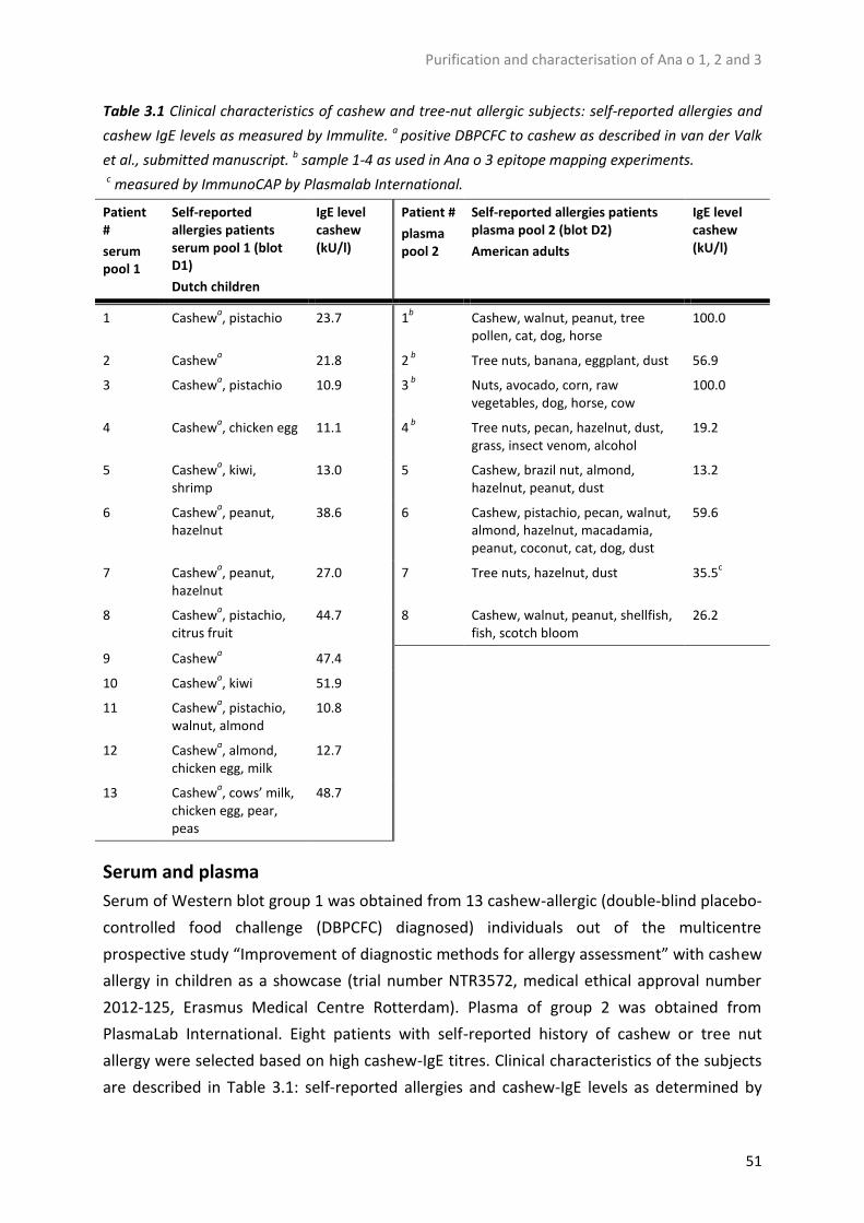
Purification and characterisation of Ana o 1, 2 and 3
51
Table 3.1 Clinical characteristics of cashew and tree-nut allergic subjects: self-reported allergies and
cashew IgE levels as measured by Immulite. a positive DBPCFC to cashew as described in van der Valk
et al., submitted manuscript. b sample 1-4 as used in Ana o 3 epitope mapping experiments.
c measured by ImmunoCAP by Plasmalab International.
Patient #
serum pool 1
Self-reported allergies patients serum pool 1 (blot D1)
Dutch children
IgE level cashew (kU/l)
Patient #
plasma pool 2
Self-reported allergies patients plasma pool 2 (blot D2)
American adults
IgE level cashew (kU/l)
1 Cashewa, pistachio 23.7 1
b Cashew, walnut, peanut, tree pollen, cat, dog, horse
100.0
2 Cashewa 21.8 2
b Tree nuts, banana, eggplant, dust 56.9
3 Cashewa, pistachio 10.9 3
b Nuts, avocado, corn, raw
vegetables, dog, horse, cow 100.0
4 Cashewa, chicken egg 11.1 4
b Tree nuts, pecan, hazelnut, dust,
grass, insect venom, alcohol 19.2
5 Cashewa, kiwi,
shrimp 13.0 5 Cashew, brazil nut, almond,
hazelnut, peanut, dust 13.2
6 Cashewa, peanut,
hazelnut 38.6 6 Cashew, pistachio, pecan, walnut,
almond, hazelnut, macadamia, peanut, coconut, cat, dog, dust
59.6
7 Cashewa, peanut,
hazelnut 27.0 7 Tree nuts, hazelnut, dust 35.5
c
8 Cashewa, pistachio,
citrus fruit 44.7 8 Cashew, walnut, peanut, shellfish,
fish, scotch bloom 26.2
9 Cashewa 47.4
10 Cashewa, kiwi 51.9
11 Cashewa, pistachio,
walnut, almond 10.8
12 Cashewa, almond,
chicken egg, milk 12.7
13 Cashewa, cows’ milk,
chicken egg, pear, peas
48.7
Serum and plasma
Serum of Western blot group 1 was obtained from 13 cashew-allergic (double-blind placebo-
controlled food challenge (DBPCFC) diagnosed) individuals out of the multicentre
prospective study “Improvement of diagnostic methods for allergy assessment” with cashew
allergy in children as a showcase (trial number NTR3572, medical ethical approval number
2012-125, Erasmus Medical Centre Rotterdam). Plasma of group 2 was obtained from
PlasmaLab International. Eight patients with self-reported history of cashew or tree nut
allergy were selected based on high cashew-IgE titres. Clinical characteristics of the subjects
are described in Table 3.1: self-reported allergies and cashew-IgE levels as determined by

Chapter 3
52
Immulite 2000, F202. Glycoprotein staining. Glycosylation of proteins was checked using a
Glycoprotein Staining kit. Samples were run on SDS-PAGE as described above alongside a
positive control (horseradish peroxidase, kit content) and a negative control (soybean trypsin
inhibitor, kit content). Staining was performed according to the manufacturer’s instructions.
Imaging and analysis were performed using a Universal Hood III and Image Lab 4.1 software.
Protein identification
Protein identification by MALDIMS/MS was performed by Alphalyse Denmark. Protein
samples for protein identification were excised from denaturing reducing Coomassie-stained
SDS-PAGE gels. Trypsin-digested protein bands were analysed on a Bruker Autoflex Speed
MALDI TOF/TOF. The MS data were blasted against the NCBI and UniProt database.
Additional protein identification analysis of the 10kDa protein band of Ana o 3 was
performed by LC-MS/MS analysis. On the basis of the results of Shevchenko et al. [21], bands
cut from SDS-PAGE gel were washed in 0.2M NH4HCO3 in acetonitrile (1:1) followed by
addition of 10mM DTT in 0.1M NH4HCO3 at 56°C for 45min. The gel pieces were incubated
in 55mM iodoacetamide (IAA) in 0.1M NH4HCO3 and washed in 0.1M NH4HCO3 in
acetonitrile (1:1), and after drying in a SpeedVac, the gel pieces were digested overnight at
37°C in 0.1M NH4HCO3 with sequencing grade modified porcine trypsin (Promega). Gel
pieces were incubated with 25mM NH4HCO3 for 15min at 37°C. After addition of
acetonitrile the samples were dried and dissolved in 0.1% formic acid. Trypsin-digested
peptides were separated on a Thermo Scientific EASY column (3μm bead-packed 10cm C18
column) connected to an Easy-nLC 1000 ultrahigh-pressure system (Thermo Scientific).
Peptides were loaded in solvent A (0.1% formic acid in MQ) onto a pre-column (5μm bead-
packed 2cm C18 column) prior to separation on the analytical column using a 40min 5−50%
linear gradient of solvent B (100% acetonitrile (ACN) in 0.1% formic acid) at a flow rate of
300nL/min. During the gradient, online MS analysis of peptides was performed with a
QExactive Plus (Thermo Scientific) mass spectrometer using a nanoelectrospray source.
Ionization (2.4kV) was performed using a stainless steel emitter and a heated capillary
temperature of 250°C. Full MS scans were acquired over the m/z range 400−1.500 with a
mass resolution of 70.000 (at m/z 200). Full scan target was set at 1 × 10⁶ with a maximum
fill time of 100ms. The five most intense peaks with charge states 1−4 were fragmented in
the higher-energy collisional dissociation (HCD) collision cell with a normalized collision
energy of 30%. The mass range was set to 200−2000 with a mass resolution of 17.500 (at
m/z 200). The target value for fragment scans was set at 1 × 10⁵, the intensity threshold was
kept at 4 × 10⁴, and the maximum allowed accumulation times were 50ms. The peptide
match was set to preferred, isolation width was set at 4 and isotope exclusion was on, and
the dynamic exclusion was set to 30s. LC-MS/MS data acquired by the Q-Exactive were
processed using ProteomeDiscoverer software 1.4 (Thermo Scientific). The fragmentation

Purification and characterisation of Ana o 1, 2 and 3
53
spectra were searched against an Anacardiaceae database downloaded from Uniprot using
Sequest HT with precursor mass tolerance of 10ppm and fragment mass tolerance of
20mDa. Minimum peptide length was 6 amino acids, and maximum of miscleavages was set
to 2. Carbamidomethylation of cysteine was set as fixed modification, and oxidation of
methionine was set as variable modification. The percolator was used for false-discovery
rate (FDR) estimation using reversed decoy database filtering on q-value below 1% for strict
and 5% for relaxed stringency.
Analysis of Ana o 3 by UPLC/PDA/ESI-MS
Identification of the heterogeneity of the N- and C-termini was executed by ultra-
performance liquid chromatography UPLC)/photodiode array (PDA)/electrospray ion source
(ESI)-MS. Purified native Ana o 3, denatured and reduced Ana o 3 (10mM DTT, 1% SDS, 5min
at 100°C), and proteins eluted from de-stained SDS-PAGE (50mM Tris, 0.1mM EDTA, 0.15M
NaCl, 0.1% SDS, 1mM DTT, 4h) were filtered over a 0.5 mL 3kDa Amicon centrifugal filter and
suspended in 200μL of eluent A (0.1% trifluoroacetyl (TFA) and 1% ACN in water). The
solution was analyzed by LC/PDA/ESI-MS by injecting 2μL in an Acquity UPLC separation
module equipped with a BEH C4 300Å column (1.7μm, 2.1 × 100mm), interfaced with an
Acquity PDA detector and a Synapt G2-Si HD mass spectrometer. Gradient elution was
performed between eluent A and B. Eluent A was 1% ACN and 0.1% TFA in water; eluent B
was 0.1% TFA in ACN. The gradient ramp was 0−2min isocratic 90% A, 2−12min linear
gradient from 90% A to 25% A, 12−15min linear gradient from 25% A to 0.1% A, 15−20min
isocratic 0.1% A, plus reconditioning. The flow rate was 0.35 mL/min. PDA detection was
performed in the range 200−400nm, with a resolution of 1.2nm and a sampling rate of 40
points/s. MS detection was performed in the full scan mode with the following parameters:
positive ion mode, capillary voltage 3kV, source temperature 150°C, cone voltage 40V,
desolvation temperature 500°C, cone gas flow (N2) 200L/h, desolvation gas flow (N2)
800L/h, aquisition range 150−4000m/z, scan time 0.3s, and interscan time 0.015s. The UV
chromatogram was obtained by extracting the trace measured at 214nm from the PDA full
scan trace.
Analysis of Ana o 3 tryptic peptides by UPLC/PDA/ESI-MS
Purified Ana o 3 was denatured and reduced (1% SDS, 10mM DTT, 5min at 100°C) and
digested with trypsin for 1h at 37°C. Samples were analysed by LC/PDA/ESI-MS by injecting
2μL in an Acquity UPLC separation module equipped with a Peptide BEH C18 column, 300Å
(1.7μm, 2.1mm × 150), interfaced with an Acquity PDA detector and a Synapt G2-Si HD mass
spectrometer. Gradient elution was performed between eluent A and B. Eluent A was 1%
ACN and 0.1% TFA in water; eluent B was 0.1% TFA in ACN. The gradient ramp was 0−2min

Chapter 3
54
isocratic 97% A, 2−10min linear gradient from 97% A to 78% A, 12−16min linear gradient
from 78% A to 70% A, 16−19 min from 70% A to 0.1% A, 19−24min isocratic 0.1% A, plus
reconditioning. The flow rate was 0.35mL/min. PDA detection was performed in the range
200−400nm, with a resolution of 1.2nm and a sampling rate of 40 points/s. MS detection
was performed in the full scan mode with the following parameters: positive ion mode,
capillary voltage 3kV, source temperature 150°C, cone voltage 30V, desolvation temperature
500°C, cone gas flow (N2) 200L/h, desolvation gas flow (N2) 800L/h, acquisition range 100−
3000m/z, scan time 0.3s, and interscan time 0.015s. Survey MSe mode was applied with the
following parameters: acquisition range 100−3000m/z, ramp high energy from 20.0 to 30.0,
TIC threshold 5.0, survey scan time 0.3s. Lock spray mass correction was applied by
continuously perfusing Angiotensin II as calibrant into the source during the scan. The data
were analysed using BioPharmaLynx 1.3.3 software. Identified peptides were cross-checked
manually.
Epitope mapping of Ana o 3
Forty-two peptides of 15 amino acids, with an offset of 3 amino acids, were synthesized by
Synpeptide, covering the entire length of Ana o 3 (amino acids 1−138). Peptides were
dissolved, and 5μmol was spotted in duplicate on nitrocellulose (0.2μm, placed on a
polyester backbone) alongside two positive controls (0.6μg total protein extract and 5μg IgE)
and a negative solvent control. The same procedure was followed as explained above for
Western blotting with the only difference being that the blots were incubated with plasma
from individual patients (group 2) and one blot was incubated with serum from a non-
allergic person. The four plasma samples were chosen based on the highest Ana o 3 binding
capacity out of the eight plasma samples, as determined by Western blotting (data not
shown), for clinical characteristics see Table 3.1. Staining was performed for 30min. After
washing in TBS, the average pixel intensity of each duplicate spot was calculated using the
Image Lab 4.1 software, and the values for the negative serum were deducted per duplicate
spot. Strong binding epitopes were defined as the 30% strongest IgE-binding epitopes.
Results
Protein purification
Purification of Ana o 1 and Ana o 3 was achieved by protein extraction from defatted cashew
followed by stepwise ammonium sulfate precipitation. The final supernatant, after 52.5%
ammonium sulfate precipitation, was used to purify Ana o 1 and Ana o 3. Ana o 2 was
purified by gel filtration chromatography of a non-defatted cashew extract. A flow sheet for
the purification of Ana o 1, Ana o 2, and Ana o 3 is shown in Figure 3.1.

Purification and characterisation of Ana o 1, 2 and 3
55
Figure 3.1 Flow sheet for the purification of Ana o 1, Ana o 2, and Ana o 3. AHC, ammonium
bicarbonate buffer; o/n, overnight 16h; rt, room temperature; final sup, final supernatant.
In Figure 3.2A, various fractions collected during the purification process of cashew allergens
are shown on denaturing and reducing SDS-PAGE gel. The two total protein extracts
(defatted and non-defatted) contain several proteins next to Ana o 1, Ana o 2, and Ana o 3,
with the 11S globulin Ana o 2 as the most prominent protein. Upon stepwise addition of
ammonium sulfate to the total protein extract (defatted), to a final concentration of 15, 30,
and 45%, generally Ana o 2 together with various high molecular weight proteins
precipitated; see Supplementary Information, Figure 3.S1. After the last step of precipitation
(52.5%), the final supernatant contains only Ana o 1 and Ana o 3 (Figure 3.2A, lane 3); this
last fraction was used to purify these two proteins. Omitting the stepwise precipitation steps
by directly adding 52.5% ammonium sulfate resulted in a supernatant containing more
impurities (data not shown).
The gel filtration chromatogram from the purification of Ana o 2 showed three peaks (Figure
3.3): peak 1 contains a mixture of several proteins (among others, Ana o 1, Ana o 2, and Ana
o 3), peak 2 contains Ana o 1 and Ana o 2, and peak 3 contains the purified Ana o 2 fraction
as shown in Figure 3.2A (lane 6).

Chapter 3
56
Protein identification
As specified in Figure 3.2A and Figure 3.S2, on the basis of MALDI-MS/MS, Ana o 1 was
confirmed in the 100kDa band (b) and the 50kDa band (c). For Ana o 2 the 53kDa (a), 30kDa
(d), and 21kDa bands (f) were confirmed as the complete protein, large subunit, and small
subunit, respectively. The 22kDa band (e) was identified as an 11S globulin from pistachio;
however, we interpret this to be Ana o 2 from cashew, as 11S from pistachio and Ana o 2
display 49% sequence identity and 65% sequence similarity (EMBOSS Needle, EMBL-EBI).
The three bands in lane 7 (10, 8, 6kDa; bands g, h, i, respectively) were identified as Ana o 3.
Purity and yield of the proteins
Purities of the Ana o 1, Ana o 2, and Ana o 3 fractions were determined by applying
increased protein concentrations (20, 30, and 40μg) on SDSPAGE (data not shown). Purity of
these fractions was calculated as the relative percentage of the desired protein bands versus
visible impurities on SDS-PAGE. Purity of Ana o 1 was 95.6% ± 3.3%, purity of Ana o 2 was
92.6% ± 4.6%, and purity of Ana o 3 was 98.5% ± 1.3%. Protein yield of the purified fractions
was calculated based on the Coomassie protein assay and SDS-PAGE analysis of several
purified fractions. Overall yield is 1% for Ana o 1 (0.33mg per run), 34% for Ana o 2 (1.99mg
per run), and 3% for Ana o 3 (5.52 mg per run).
Western blot
In Figure 3.2D, two Western blots performed with two different pools of patient
serum/plasma are shown; blot D1 was performed using 13 sera from cashew-allergic
children as determined by DBPCFC, while blot D2 was performed using 8 plasma samples
from cashew- or tree-nut allergic adults. Less strong IgE-binding to cashew allergens is
observed using serum from group 1 (blot D1), compared to plasma from group 2 (blot D2).
Blot D1 shows binding of IgE to Ana o 2 (lane 4; 30, 22, and 21kDa), Ana o 3 (lane 5; 10 and
8kDa, not 6kDa), and slightly to Ana o 1 (lane 3; 50kDa). In the total protein extract, the
same proteins as well as additional proteins of 80, 70, 53 (full length Ana o 2), 40, and 16kDa
appear IgE-reactive. In blot D2 binding of IgE to Ana o 1 (lane 3; 100 and 50kDa), Ana o 2
(lane 4; 30, 22, and 21kDa), and Ana o 3 (lane 5; 10 and 8kDa, not 6kDa) is visible. In the total
protein extract, the same proteins as well as additional proteins of 80, 65, 53kDa (Ana o 2),
40, 16, and 12kDa bind IgE. In blot D2, lane 3, a small impurity of 30kDa (Ana o 2) can be
observed in the purified Ana o 1 extract. In the third blot, D3, the antibody control blot is
shown, demonstrating marginal non-specific binding of the antibodies to the 22−21kDa
subunits of Ana o 2. These Western blots indicate that all three purified protein fractions are
able to bind IgE.

Purification and characterisation of Ana o 1, 2 and 3
57
Figure 3.2 All lanes: mwm, molecular weight marker; Total protein, total protein extract from
defatted cashew used for purification of Ana o 1 and Ana o 3; Final sup, final supernatant after 52.5%
ammonium sulfate precipitation; Total protein nd, total protein extract from non-defatted cashew
used for purification of Ana o 2; neg. control, negative control glycoprotein stain (soybean trypsin
inhibitor); pos. control, positive control glycoprotein stain (horseradish peroxidase). (A) Reducing
denaturing SDS-PAGE of purified cashew extracts. Letters a−i indicate bands cut for protein
identification: (a) 53kDa Ana o 2, (b) 100kDa Ana o 1, (c) 50kDa Ana o 1, (d) 30kDa Ana o 2, (e) 22kDa
11S globulin, (f) 21kDa Ana o 2, (g) 10kDa Ana o 3, (h) 8kDa Ana o 3, (i) 6kDa Ana o 3. (B) Non-
reducing denaturing SDS-PAGE of purified cashew extracts, no β-mercaptoethanol added. (C)
Glycoprotein stain of purified cashew extracts. (D) Western blots of purified cashew extracts. Blot D1
has been incubated with the serum pool of group 1, blot D2 has been incubated with a plasma pool
from group 2, and blot D3 is the antibody control blot that has not been incubated with patient
plasma or serum.

Chapter 3
58
Inhibition blotting, presented in Figure 3.S3, shows that purified Ana o 1 completely
inhibited binding to Ana o 1, and Ana o 3 completely inhibited binding to Ana o 3 in the total
protein extract. Besides inhibition of binding to Ana o 1, purified Ana o 1 also diminished IgE
binding to Ana o 2 and Ana o 3.
Figure 3.3 Gel filtration chromatogram of total protein extract of non-defatted cashew used for the
purification of Ana o 2.
Non-reducing denaturing SDS-PAGE
To check the presence of disulfide bridges in the purified allergen fractions, a non-reducing
denaturing SDS-PAGE, in the absence of β-mercaptoethanol, was performed. In the Ana o 1
fraction (Figure 3.2B, lane 3) protein bands of 160, 100, and 50kDa can be observed. In lane
4, containing Ana o 2, diffuse bands of 53, 42, 39, 30, 20, and 10kDa are present. Ana o 3
(lane 5) shows an abundant protein band of 12kDa and minor bands of 20 and 5kDa.
Glycosylation of cashew proteins
On the basis of prediction models (NetNGlyc 1.0, Denmark, http://www.cbs.dtu.dk/
services/NetNGlyc/), the amino acid sequences of Ana o 1 (amino acid 391, asparagine) and
Ana o 3 (amino acid 19, asparagine), but not that of Ana o 2, contain putative N-glycosylation
motifs. The predicted glycosylation of Ana o 1 was confirmed by glycoprotein staining (see
Figure 3.2C). Both Ana o 2 and Ana o 3 do not seem to be glycosylated, neither in the
purified nor in the total protein fraction. In addition an unknown protein of 3kDa, present in
the full protein extract, also seems to be glycosylated.

Purification and characterisation of Ana o 1, 2 and 3
59
Analysis of Ana o 3
Protein characterization and protein identification of Ana o 3 revealed some discrepancies
with data from literature. It was previously reported that all three denatured protein bands
from SDS-PAGE represented the large subunit of Ana o 3.15 However, our results identify
one of the bands as the small subunit from Ana o 3. Besides, the IgE binding on Western blot
to two out of three bands of denatured Ana o 3 indicated the possibility of one of these
bands possibly being the small subunit of Ana o 3. To analyse this possibility, both denatured
reduced and native Ana o 3 samples were analysed for peptide mass, purity, and annotation
of protein sequences using UPLC/PDA/ESI-MS.
Table 3.2 Molecular masses of denatured Ana o 3 derived from UPLC/PDA/ESI-MS analysis and their
tentative sequence identification. aAccording to Moreno and Clemente [16], bAccording to our data.
Tentative sequence annotation within the range 0.5 Da from the theoretical calculated mass
Experimental molecular mass (Da)
Leader peptide b
Small subunit b
Large subunit b
Leader peptide 1-20a 1-20/28
b
Small subunit 21-65a 29-65
b
Large subunit 66-138a 69-137
b
Chromatographic Peak 1 (Rt = 5.61)
34-62 / 35-63 3742.7 X
34-63 / 35-64 / 36-65 3799.7 X
33-63 3886.8 X
33-65 4099.9 X
Chromatographic Peak 2 (Rt = 5.82)
37-64 3514.6 X
37-65 3670.7 X
22-53 3959.8 X
39-69 4016.8 X X
33-66 4263.1 X
31-66 4507.1 X
Chromatographic Peak 3 (Rt = 7.60)
2-70 or 56-122 or unknown isoform
8161.7 X X X
1-71 or unknown isoform 8421.8 X X X
Chromatographic Peak 4 (Rt= 7.91)
69-136 8084.7 X
69-137 8171.7 X

Chapter 3
60
Fig
ure
3.4
Ch
rom
ato
gra
ph
ic a
na
lysi
s (U
V t
race
at
21
4n
m)
of
den
atu
red
(A
) a
nd
na
tive
(B
) A
na
o 3
. MS
spec
tru
m a
sso
cia
ted
wit
h t
he
ma
jor
pea
k in
na
tive
An
a o
3 (
full
spec
trum
fro
m 2
00
to
40
00
m/z
) sh
ow
ing
th
e cl
ust
ers
corr
esp
on
din
g t
o t
he
7+
-ch
arg
ed, 6
+-c
ha
rged
, a
nd
5+
-ch
arg
ed i
on
s (C
), a
nd
ch
arg
e-st
ate
dec
on
volu
tio
n o
f th
is s
pec
tru
m s
ho
wn
in
(C
), p
erfo
rmed
by
the
Ma
xEn
t so
ftw
are
, in
dic
ati
ng
th
e a
ctu
al m
ole
cula
r m
ass
es o
f th
e m
ain
iso
form
s p
rese
nt
(D).

Purification and characterisation of Ana o 1, 2 and 3
61
Denatured and reduced Ana o 3 shows four peaks (Figure 3.4A). Peaks 1 and 2,
corresponding to the 6kDa protein band on SDS-PAGE, were compatible with the small
subunit of Ana o 3 with molecular weights of 3.7−4.5kDa. Peak 3 (10kDa band) and peak 4
(8kDa band) were both compatible with the large subunit of Ana o 3 with molecular weights
of 8.1−8.4kDa. Using both trypsin-digested and nondigested denatured reduced Ana o 3, the
peptide sequences from the four peaks were annotated (see Table 3.2). The range of protein
molecular weights in the four peaks was found to originate from N- and C-terminal micro-
heterogeneity, also called N- or C-terminal clipping (see Table 3.3 and Figure 3.5). Tryptic
peptides clearly showed that the small subunit displayed both N- and C-terminal micro-
heterogeneity, with its N-terminus starting at amino acid 29−36 and its C-terminus
terminating at amino acid 63−65. The large subunit displayed only minimal C-terminal micro-
heterogeneity, starting at amino acid 69 and ending at amino acid 136−137. When Ana o 3
was purified using protease inhibitors, the same degree of micro-heterogeneity was
obtained (data not shown).
Native Ana o 3 eluted as one major peak of 11.8−12.8kDa (Figure 3.4C and D) with a purity of
88% (Figure 3.4B, purity calculated by UV trace at 214nm). The observed masses fitted with
the molecular masses of the small subunit (peaks 1 and 2) linked to the large subunit (peak
4) by two disulfide bridges. Together with the two other predicted disulfide bridges within
the large subunit,16 a total of 4 disulfide bridges can be found within native Ana o 3. Besides
demonstrating micro-heterogeneity in Ana o 3, the obtained data also indicate the presence
of isoforms of the large subunit.

Chapter 3
62
Table 3.3 Sequence annotation of Ana o 3 tryptic peptides including termini based on molecular mass
(<10 ppm error) and fragment identification.
Figure 3.5 Amino acid sequence (coding region) of Ana o 3, with the small and large subunits
indicated as based on UPLC/PDA/ESI-MS analysis. The leader peptide is underlined, the black bars
indicate the constant regions, and the light gray bars indicate regions of micro-heterogeneity. A
dotted box is placed around the small subunit (first box) and the large subunit (second box) of Ana o
3.
Epitope mapping of Ana o 3
Using 42 overlapping linear peptides, 12 IgE-binding peptides were detected in Ana o 3 (see
Table 3.4). Two IgE-binding peptide regions were identified, with the first consisting of
amino acids 10−30 and the second, more strongly IgE-bound region consisting of amino acids
85−108. Besides 4 mildly binding epitopes described in Table 3.4, 2 strongly binding peptides
were identified at amino acids 55−69 and 121−135. All strongly binding epitopes were
detected in the peptides of the large subunit or containing part of the large subunit.
Peptide m/z (charge state)
Identified peptide sequence
565.9 (3+) 36-48 N-termini small subunit until trypsin cleavage site (amino acid 48)
618.0 (3+) 35-48
637.0 (3+) 34-48
666.0 (3+) 33-48
704.3 (3+) 32-48
747.3 (3+) 31-48
790.3 (3+) 30-48
823.4 (3+) 29-48
660.4 (2+) 55-65 Trypsin cleavage site (amino acid 55) until
C-termini small subunit 582.3 (2+) 55-64
553.8 (2+) 55-63
759.7 (3+) 69-86 N-terminus large subunit until trypsin cleavage site (amino acid 86)
786.4 (2+) 123-137 Trypsin cleavage site (amino acid 123) until
C-termini large subunit 742.8 (2+) 123-136

Purification and characterisation of Ana o 1, 2 and 3
63
Table 3.4 Linear epitope mapping of Ana o 3 using plasma of four individuals (no. 1−4) from group 2;
bold peptide numbers indicate a strong IgE-binding epitope, bold amino acid numbers indicate an IgE-
binding peptide region; underlined amino acid sequences indicate the small subunit of Ana o 3 (amino
acids 29−65), between the leader sequence (1−20) and the large subunit (69−137) of Ana o 3.
IgE-binding intensity Peptide Amino acid # Amino acid sequence 1 2 3 4
1 1-15 MAKFLLLLSAFAVLL 2 4-18 FLLLLSAFAVLLLVA 3 7-21 LLSAFAVLLLVANAS 4 10-24 AFAVLLLVANASIYR + + + ++ 5 13-27 VLLLVANASIYRAIV + + + 6 16-30 LVANASIYRAIVEVE + + + 7 19-33 NASIYRAIVEVEEDS 8 22-36 IYRAIVEVEEDSGRE 9 25-39 AIVEVEEDSGREQSC 10 28-42 EVEEDSGREQSCQRQ 11 31-45 EDSGREQSCQRQFEE 12 34-48 GREQSCQRQFEEQQR 13 37-51 QSCQRQFEEQQRFRN 14 40-54 QRQFEEQQRFRNCQR ++ 15 43-57 FEEQQRFRNCQRYVK 16 46-60 QQRFRNCQRYVKQEV 17 49-63 FRNCQRYVKQEVQRG + ++ 18 52-66 CQRYVKQEVQRGGRY 19 55-69 YVKQEVQRGGRYNQR ++ +++ + 20 58-72 QEVQRGGRYNQRQES 21 61-75 QRGGRYNQRQESLRE 22 64-78 GRYNQRQESLRECCQ 23 67-81 NQRQESLRECCQELQ 24 70-84 QESLRECCQELQEVD 25 73-87 LRECCQELQEVDRRC 26 76-90 CCQELQEVDRRCRCQ + ++ + 27 79-93 ELQEVDRRCRCQNLE 28 82-96 EVDRRCRCQNLEQMV 29 85-99 RRCRCQNLEQMVRQL ++ +++ + + 30 88-102 RCQNLEQMVRQLQQQ ++ ++ ++ ++ 31 91-105 NLEQMVRQLQQQEQI 32 94-108 QMVRQLQQQEQIKGE ++ ++ ++ +++ 33 97-111 RQLQQQEQIKGEEVR 34 100-114 QQQEQIKGEEVRELY 35 103-117 EQIKGEEVRELYETA 36 106-120 KGEEVRELYETASEL 37 109-123 EVRELYETASELPRI 38 112-126 ELYETASELPRICSI + + 39 115-129 ETASELPRICSISPS 40 118-132 SELPRICSISPSQGC 41 121-135 PRICSISPSQGCQFQ +++ +++ + + 42 124-138 CSISPSQGCQFQSSY Total cashew positive control +++ +++ +++ +++

Chapter 3
64
Discussion
In this study we have purified and characterized the three major cashew allergens Ana o 1,
Ana o 2, and Ana o 3. Our method enables the simultaneous purification of Ana o 1 and Ana
o 3.
Using precipitation, ultrafiltration, and gel filtration chromatography, we have isolated three
cashew allergens with high purity. The purities of the single allergen fractions are
approximately 96% for Ana o 1, 93% for Ana o 2, and 99% for Ana o 3 as based on SDS-PAGE
and 88% for Ana o 3 based on high-performance liquid chromatography (HPLC) analysis. The
only IgE-reactive impurity we observed, as established by Western blotting, was a protein of
30kDa in the Ana o 1 fraction (Figure 3.2, D1). A substantial difference in IgE-binding to Ana o
1 is apparent between the two patient groups (blots D1 and D2), which might be explained
by a population difference of the sera. Group 1 (blot D1) consists of Dutch children, and
group 2 (blot D2) consists of American adults. Geographical and age differences have been
described before to explain differences in allergen binding [22, 23].
For Ana o 3 we observed only IgE binding to the 10 and 8kDa proteins and not to the 6kDa
protein. This is consistent with other Western blots performed by us using both various
patient pools and individual patient sera. Teuber et al., however, did detect IgE binding to
the 6kDa protein [20]. Our analyses show that the 6kDa protein is the small subunit of Ana o
3 while both the 10 and 8kDa proteins represent the large subunit. Epitope mapping of Ana
o 3 by Robotham et al. [15] showed more strongly binding epitopes on the small subunit
compared to our data. Table 3.4 shows that our patient IgE binds strongest to the peptides
of the large subunit. One mildly IgE-binding peptide region was identified between the
presumptive leader sequence15 and the start of the small subunit as determined by
UPLC/PDA/ESI-MS analysis. It is unknown if this peptide is present in cashew when eaten;
however, IgE binding to leader peptides that are not attached to the mature allergen has
been observed before for Ana o [19] and Ana o 2 [11].
On Western blot, besides binding of IgE to Ana o 1, Ana o 2, and Ana o 3, several other
proteins were bound in the total protein extract (Figure 3.2, D1 and D2). These allergens of
12, 16, 40, 65, 70, and 80kDa have been observed before [20, 24], but have not been
identified.
The yield of purification for Ana o 1 is 1%, for Ana o 2 is 34%, and for Ana o 3 is 3%. The low
yield can be explained by the precipitation steps of the total protein extract where Ana o 1
and Ana o 3 are discarded along with the undesirable proteins. However, as the protein
content of cashew is 18.8% [25], the amount of cashew needed is relatively low. No isoform
selection is expected as inhibition blotting clearly shows complete inhibition of IgE binding to
Ana o 1 and Ana o 3 in the total protein extract upon inhibition with, respectively, purified
Ana o 1 or Ana o 3. Besides inhibition of IgE binding to Ana o 1, purified Ana o 1 also
inhibited binding of IgE to Ana o 3 and partially to Ana o 2. Cross-reactivity between Ana o 1

Purification and characterisation of Ana o 1, 2 and 3
65
and Ana o 3 was unexpected, although some cross-reactivity between recombinant Ana o 1
and Ana o 2 has been shown before [11]. Cross-reactivity between 7S globulins, 11S
globulins, and 2S albumins has been observed before for peanut [26].
Glycoprotein staining of the cashew protein fractions showed Ana o 1 and an unknown
protein of 3kDa to be glycosylated. Both Ana o 2 and Ana o 3 are not glycosylated.
Glycosylation of cashew proteins has been studied once before where only one glycosylated
protein of ~3kDa was indicated [27]. In their study Sathe et al. used a total protein extract
that, as can also be observed from our glycoprotein stained gel, possibly did not contain
enough Ana o 1 to show a clear band in glycoprotein staining. Another explanation could be
a difference in cashew variety used.
Under non-reducing denaturing conditions, Ana o 1 is present as both a dimer of 100kDa and
a single protein of 50kDa. Non-reduced denatured Ana o 2 displays some protein bands that
are also visible in reducing denaturing SDS-PAGE: the 53kDa full protein and 30 and
21kDasubunits. Ana o 3 seems to form one complex of ∼12kDa linked by disulfide bridges,
which was confirmed by LC/ESI-MS.
Purified Ana o 3 manifested as a triplet of 10, 8, and 6kDa proteins on SDS-PAGE under
denaturing reducing conditions. Native 2S-albumins are post-translationally processed into
an N-terminal small subunit of ~5.5kDa and a C-terminal large subunit of ~8.8kDa, held
together via cysteine linkages [16]. In an attempt to further identify the protein triplet, we
subjected both native and denatured reduced Ana o 3 to LC-MS analysis. This indicated the
10 and 8kDa proteins to be the large subunit of 8.2−8.4 and 8.1−8.2kDa, respectively, and
the 6kDa protein to be the small subunit of 3.7−4.5kDa. Tryptic digestion of Ana o 3
indicated the small subunit to span amino acids 29−36 until 63−65, including N- and C-
terminal micro-heterogeneity, and the large subunit to span amino acid 69 until 136−137,
including C-terminal micro-heterogeneity, differing slightly from the proposed amino acid
stretches proposed by Moreno et al. [16]. The same degree of micro-heterogeneity was
observed when Ana o 3 was purified in the presence of protease inhibitors, indicating this
micro-heterogeneity is not an artefact from the method of extraction but is due to
proteolytic processes, the presence of different precursors, or the shift in cleavage sites in
the cashew itself [16]. Micro-heterogeneity of 2S albumins has been described before for
castor bean [28], Brazil nut [29], and sesame [30].
This data differs from the data reported by Robotham et al., who described all three protein
bands to be isoforms of the large subunit of Ana o 3 with very similar N-terminal sequences,
differing in only 1−2 amino acids out of 10 [15]. An explanation for this discrepancy could lay
in a difference in cashew variety or storage conditions.
As food allergy is highly prevalent and the incidence of cashew allergy seems to be rising
[31], it is relevant to study which proteins are involved. Ana o 1, Ana o 2, and Ana o 3 are the
three major allergens in cashew. Using pure allergen fractions allows for determining to

Chapter 3
66
which specific cashew allergen a patient reacts, and they can be used in cross-reactivity
studies, where pure allergens can be used for inhibition blotting. Besides, these allergen
fractions can also be used for analysis of allergen structure, comparing cashew varieties, and
cashew protein processing stability.
The advantage of using purified allergens over the use of recombinant allergens is clear for
Ana o 1 and Ana o 3. Recombinant expression of a glycosylated allergen like Ana o 1 is
difficult as both prokaryotic (e.g., E. coli) and eukaryotic (e.g., P. Pastoris) expression systems
may not be capable of reproducing the natural glycation pattern as in cashew [32,33].
Considering Ana o 3, recombinant expression will not properly display the micro-
heterogeneity of the native protein; however, recombinant proteins are required to
compare micro-heterogenic variations of Ana o 3. Besides, other post-translational
processing modifications such as disulfide bridges (Ana o 2, Ana o 3) might prove more
difficult in E. coli expression systems [32].
Funding
This project was funded by Technology Foundation STW, Stichting Voedselallergie, Siemens
Healthcare Diagnostics, HAL Allergy, Intersnack Nederland B.V., ALK-Abello B.V., and the
Nederlands Anafylaxis Netwerk.
Acknowledgements
The authors thank Jack Yang and Yvonne Veltman for their contribution in the first steps of
allergen purification. For her valuable advice for the epitope mapping of Ana o 3, we thank
Dr. Lisa Steinbrueck from Pepperprint Heidelberg, Germany.

Purification and characterisation of Ana o 1, 2 and 3
67
References [1] Nwaru, B. I.; Hickstein, L.; Panesar, S. S.; Roberts, G.; Muraro, A.; Sheikh, A. Prevalence of common
food allergies in Europe: a systematic review and meta-analysis. Allergy 2014, 69, 992−1007. [2] Sicherer, S. H.; Munoz-Furlong, A.; Burks, A. W.; Sampson, H. A. Prevalence of peanut and tree nut
allergy in the US determined by a random digit dial telephone survey. J. Allergy Clin. Immunol. 1999,
103, 559−62. [3] Sicherer, S. H.; Burks, A. W.; Sampson, H. A. Clinical features of acute allergic reactions to peanut and
tree nuts in children. Pediatrics 1998, 102, e6. [4] Davoren, M.; Peake, J. Cashew nut allergy is associated with a high risk of anaphylaxis. Arch. Dis. Child.
2005, 90, 1084−1085. [5] Hasegawa, M.; Inomata, N.; Yamazaki, H.; Morita, A.; Kirino, M.; Ikezawa, Z. Clinical features of four
cases with cashew nut allergy and cross-reactivity between cashew nut and pistachio. Allergol. Int.
2009, 58, 209−15. [6] Rance, F.; Bidat, E.; Bourrier, T.; Sabouraud, D. Cashew allergy: observations of 42 children without
associated peanut allergy. Allergy 2003, 58, 1311−4. [7] Clark, A. T.; Anagnostou, K.; Ewan, P. W. Cashew nut causes more severe reactions than peanut: case-
matched comparison in 141 children. Allergy 2007, 62, 913−6. [8] van der Valk, J. P.; Dubois, A. E. J.; Gerth van Wijk, R.; Wichers, H. J.; de Jong, N. W. Systematic review
on cashew nut allergy. Allergy 2014, 69, 692−698. [9] Wang, F.; Robotham, J. M.; Teuber, S. S.; Tawde, P.; Sathe, S. K.; Roux, K. H. Ana o 1, a cashew
(Anacardium occidental) allergen of the vicilin seed storage protein family. J. Allergy Clin. Immunol.
2002, 110, 160−6. [10] Mills, E. N. C.; Shewry, P. R. Plant food allergens; Blackwell Science Ltd.: 2004; p 219. [11] Wang, F.; Robotham, J. M.; Teuber, S. S.; Sathe, S. K.; Roux, K. H. Ana o 2, a major cashew (Anacardium
occidentale L.) nut allergen of the legumin family. Int. Arch. Allergy Immunol. 2003, 132, 27−39. [12] Xia, L.; Willison, L. N.; Porter, L.; Robotham, J. M.; Teuber, S. S.; Sathe, S. K.; Roux, K. H. Mapping of a
conformational epitope on the cashew allergen Ana o 2: a discontinuous large subunit epitope dependent upon homologous or heterologous small subunit association. Mol. Immunol. 2010, 47,
1808−16. [13] Robotham, J. M.; Xia, L.; Willison, L. N.; Teuber, S. S.; Sathe, S. K.; Roux, K. H. Characterization of a
cashew allergen, 11S globulin (Ana o 2), conformational epitope. Mol. Immunol. 2010, 47, 1830−8. [14] Venkatachalam, M.; Monaghan, E. K.; Kshirsagar, H. H.; Robotham, J. M.; O’Donnell, S. E.; Gerber, M.
S.; Roux, K. H.; Sathe, S. K. Effects of processing on immunoreactivity of cashew nut (Anacardium
occidentale L.) seed flour proteins. J. Agric. Food Chem. 2008, 56, 8998−9005. [15] Robotham, J. M.; Wang, F.; Seamon, V.; Teuber, S. S.; Sathe, S. K.; Sampson, H. A.; Beyer, K.; Seavy, M.;
Roux, K. H. Ana o 3, an important cashew nut (Anacardium occidentale L.) allergen of the 2S albumin
family. J. Allergy Clin. Immunol. 2005, 115, 1284−90. [16] Moreno, F. J.; Clemente, A. 2S Albumin Storage Proteins: What Makes them Food Allergens? Open
Biochem. J. 2008, 2, 16−28. [17] Mattison, C. P.; Grimm, C. C.; Wasserman, R. L. In vitro digestion of soluble cashew proteins and
characterization of surviving IgE-reactive peptides. Mol. Nutr. Food Res. 2014, 58, 884−93. [18] Hummel, M.; Wigger, T.; Hoper, T.; Westkamp, I.; Brockmeyer, J. Simple, Rapid, and Selective Isolation
of 2S Albumins from Allergenic Seeds and Nuts. J. Agric. Food Chem. 2015, 63, 6035−6040. [19] Sathe, S. K.; SzeTao, K. W. C.; Wolf, W. J.; Hamaker, B. R. Biochemical characterization and in vitro
digestibility of the major globulin in cashew nut (Anacardium occidentale). J. Agric. Food Chem. 1997,
45, 2854−2860. [20] Teuber, S. S.; Sathe, S. K.; Peterson, W. R.; Roux, K. H. Characterization of the soluble allergenic
proteins of cashew nut (Anacardium occidentale L.). J. Agric. Food Chem. 2002, 50, 6543− 6549.
[21] Shevchenko, A.; Tomas, H.; Havli, J.; Olsen, J. V.; Mann, M. Ingel digestion for mass spectrometric
characterization of proteins and proteomes. Nat. Protoc. 2007, 1, 2856−2860. [22] Vereda, A.; van Hage, M.; Ahlstedt, S.; Ibanez, M. D.; Cuesta- Herranz, J.; van Odijk, J.; Wickman, M.;
Sampson, H. A. Peanut allergy: Clinical and immunologic differences among patients from 3 different
geographic regions. J. Allergy Clin. Immunol. 2011, 127, 603− 607. [23] Ballmer-Weber, B. K.; Lidholm, J.; Fernandez-Rivas, M.; Seneviratne, S.; Hanschmann, K. M.; Vogel, L.;
Bures, P.; Fritsche, P.; Summers, C.; Knulst, A. C.; Le, T. M.; Reig, I.; Papadopoulos, N. G.; Sinaniotis, A.;

Chapter 3
68
Belohlavkova, S.; Popov, T.; Kralimarkova, T.; de Blay, F.; Purohit, A.; Clausen, M.; Jedrzejczak-Czechowcz, M.; Kowalski, M. L.; Asero, R.; Dubakiene, R.; Barreales, L.; Clare Mills, E. N.; van Ree, R.; Vieths, S. IgE recognition patterns in peanut allergy are age dependent: perspectives of the EuroPrevall
study. Allergy 2015, 70, 391−407. [24] Mattison, C. P.; Desormeaux, W. A.; Wasserman, R. L.; Yoshioka-Tarver, M.; Condon, B.; Grimm, C. C.
Decreased immunoglobulin E (IgE) binding to cashew allergens following sodium sulfite treatment and
heating. J. Agric. Food Chem. 2014, 62, 6746−55. [25] Venkatachalam, M.; Sathe, S. K. Chemical composition of selected edible nut seeds. J. Agric. Food
Chem. 2006, 54, 4705−14. [26] Bublin, M.; Kostadinova, M.; Radauer, C.; Hafner, C.; Szepfalusi, Z.; Varga, E. M.; Maleki, S. J.;
Hoffmann-Sommergruber, K.; Breiteneder, H. IgE cross-reactivity between the major peanut allergen Ara h 2 and the nonhomologous allergens Ara h 1 and Ara h 3. J. Allergy Clin. Immunol. 2013, 132, 118
−24. [27] Sathe, S. K.; Venkatachalam, M.; Sharma, G. M.; Kshirsagar, H. H.; Teuber, S. S.; Roux, K. H.
Solubilization and electrophoretic characterization of select edible nut seed proteins. J. Agric. Food
Chem. 2009, 57, 7846−56. [28] da Siva, J. G., Jr.; Machado, O. L.; Izumi, C.; Padovan, J. C.; Chait, B. T.; Mirza, U. A.; Greene, L. J. Amino
acid sequence of a new 2S albumin from Ricinus communis which is part of a 29-kDa precursor
protein. Arch. Biochem. Biophys. 1996, 336, 10−18. [29] Moreno, F. J.; Jenkins, J. A.; Mellon, F. A.; Rigby, N. M.; Robertson, J. A.; Wellner, N.; Clare Mills, E. N.
Mass spectrometry and structural characterization of 2S albumin isoforms from Brazil nuts
(Bertholletia excelsa). Biochim. Biophys. Acta, Proteins Proteomics 2004, 1698, 175−86. [30] Moreno, F. J.; Maldonado, B. M.; Wellner, N.; Mills, E. N. Thermostability and in vitro digestibility of a
purified major allergen 2S albumin (Ses i 1) from white sesame seeds (Sesamum indicum L.). Biochim.
Biophys. Acta, Proteins Proteomics 2005, 1752, 142−53. [31] Sicherer, S. H.; Munoz-Furlong, A.; Godbold, J. H.; Sampson, H. A. US prevalence of self-reported
peanut, tree nut, and sesame allergy: 11-year follow-up. J. Allergy Clin. Immunol. 2010, 125, 1322−6. [32] Willison, L. N.; Sathe, S. K.; Roux, K. H. Production and analysis of recombinant tree nut allergens.
Methods 2014, 66, 34−43. [33] Lorence, A. Recombinant Gene Expression, Reviews and Protocols, 3rd ed.; Springer Science + Business
Media: 2012.

Purification and characterisation of Ana o 1, 2 and 3
69
Supplementary information
Figure 3.S1 Reducing denaturing SDS-PAGE of ammonium sulphate precipitation. Depicted are (left to
right): total protein extract from defatted cashew used for purification of Ana o 1 and Ana o 3, 15%,
30%, 45%, and 52.5% pellet of ammonium sulphate precipitation, final supernatant after 52.5%
ammonium sulphate precipitation, molecular weight marker.

Chapter 3
70

Purification and characterisation of Ana o 1, 2 and 3
71
Figure 3.S2 Protein identification, analysis of protein bands from SDS-PAGE indicated in Figure 3.2.
Results based on MALDI-MS/MS analysis unless stated otherwise. Indicated are the identified protein,
GenBank number, sequence coverage, Mascot score, and the protein sequence with matched
peptides underlined. For the proteins identified as Ana o 3 the large subunit (10 and 8kDa protein
band) or the small subunit (6kDa protein band) as based on Figure 3.5 are indicated in bold. For the
proteins identified as Ana o 2 the large subunit [11] (30kDa protein band) or the small subunit [11]
(21kDa protein band) are indicated in bold.

Chapter 3
72
Figure 3.S3 Inhibition western blot of Ana o 1 and Ana o 3, using plasma pool of group 2. Molecular
weight marker followed by 3 lanes containing a total protein extract from defatted cashew used for
purification of Ana o 1 and Ana o 3. Lane 2 is not inhibited, the plasma used in lane 3 was inhibited
with 1mg/ml purified Ana o 1, the plasma used in lane 4 is inhibited with 1mg/ml purified Ana o 3.

73

74

75
Chapter 4
Origin and processing methods slightly affect
allergenic characteristics of cashew nuts (Anacardium
occidentale)
Marit Reitsma, Shanna Bastiaan-Net, Lutske Sijbrandij, Evelien de Weert, Stefano Sforza,
Huub F. J. Savelkoul, Nicolette W. de Jong, and Harry J. Wichers
Manuscript in preparation

Chapter 4
76
Abstract
The protein content and allergen composition was studied of cashew nuts from 8 different
origins (Benin, Brazil, Ghana, India, Ivory Coast, Mozambique, Tanzania, Vietnam), subjected
to different in-shell heat treatments (steamed, fried, drum-roasted). On 2D electrophoresis,
9 isoforms of Ana o 1, 29 isoforms of Ana o 2 (11 of the acidic subunit, 18 of the basic
subunit), and 8 isoforms of the large subunit of Ana o 3 were tentatively identified. Based on
1D and 2D electrophoresis, no difference in allergen content (Ana o 1, 2, 3) was detected
between the cashew nuts of different origins (p>0.5), some small but significant differences
were detected in allergen solubility between differently heated cashew nuts. No major
differences in N- and C-terminal micro-heterogeneity of Ana o 3 were detected between
cashew nuts of different origins. Between the different heat treatments, no difference was
detected in glycation, pepsin digestibility, or IgE binding of the cashew nut proteins.

Origin and processing of cashew nuts
77
Introduction
Cashew nuts have been studied regarding their three major allergens [1], Ana o 1 [2], Ana o
2 [3-5] and Ana o 3 [6], as well as regarding their nutritional content [7], clinical reactivity [8-
11], in vitro digestibility [12, 13], and their detection in food items [14, 15]. However, hardly
any information is available on potential differences in allergen content between cashew
nuts of different origins. Only a few studies were found describing a comparison of cashew
nuts of different origins and varieties; one study counting the amount of chromosomes
present in cashew nuts of two origins [16] and two other studies reporting no differences in
protein content in cashew nuts of different varieties or origins [17, 18]. One paper stated in
their unpublished results not to have detected differences in protein pattern on SDS-PAGE
between cashew nut protein extracts from Florida and Mumbai, however no results were
shown [19].
In some nuts and legumes differences in allergen composition were reported for different
origins. For instance, in Balinese peanuts, very low levels of the 7S globulin Ara h 1 were
detected compared to other varieties [20]. However, this lower level of Ara h 1 did not result
in a decrease in immunoreactivity as determined using an RBL (rat basophilic leukemia cell
line) model [21]. Other, commercial, peanut varieties do not seem to differ in major allergen
content (Ara h 1, 2, 3 and 6) [22, 23], just as different pistachio nut varieties seem to be
similar in protein content and IgE binding capacity [24].
With respect to the effects of thermal processing on cashew nuts, most studies have been
performed on related nuts like pistachio, and legumes like peanut. For example, (steam-)
roasting of peanut [25] and pistachio [24] decreased protein solubility, with steam-roasting
of pistachio also inducing a decrease in patient IgE binding and a decrease in pepsin
digestibility [24]. Using an RBL model, raw hazelnut appeared to be more allergenic than
roasted hazelnut [26]. Roasting of total peanut protein has been shown to induce increased
IgE binding in an ELISA assay [27], while no difference between raw and roasted peanut Ara
h 2 and 6 was detected in an RBL assay [28]. Using purified peanut allergens, roasting of Ara
h 1 (7S globulin) increased IgE cross-linking of RBL cells, while for Ara h 2/6 (2S albumins) the
cross-linking capacity was decreased [29]. In a basophil activation test (BAT) model it was
shown that the effect of thermal processing on peanut differed between peanut varieties
and patients [30].
The effects of thermal processing on the three major allergens of cashew nuts has been
studied by Venkatachalam et al. [31], demonstrating the stability of monoclonal antibody
(mAb)-binding by these allergens. IgE binding of Ana o 1, 2, and 3 was stable (as assessed by
ELISA, western blot and dot blot) between a pH range of 2 to 12. IgG-binding of Ana o 1 and

Chapter 4
78
2 was heat stable, as only autoclaving, but not microwaving, roasting or y-irradiation,
resulted in a decrease in mouse mAb binding as shown with western blotting [31].
Immunoreactivity of Ana o 3 was decreased by roasting and autoclaving, however this was
measured by western blot using a mAb that is directed at a conformational epitope [31]. It is
unclear if these heat treatments affected the multiple linear epitopes of Ana o 3. Roasting of
cashew nuts was reported to increase the solubility of Ana o 3 [32] while the total cashew
nut protein solubility decreased [33]. Blanching of the cashew nuts on the other hand,
causes leaking of all three allergens from the cashew nut into the blanching water [31].
However, the effects of frying were not studied by Venkatachalam et al.
In the present study we compared cashews nuts of 8 different origins (Benin, Brazil, Ghana,
India, Ivory Coast, Mozambique, Tanzania, and Vietnam) to analyze possible differences in
protein and allergen content. Next, we compared the effect of different heat treatments
(raw, steaming, frying, and drum roasting) on the protein and allergen content of the cashew
nuts. We have studied the proteins by DUMAS method, SDS-PAGE, 2D electrophoresis,
glycoprotein staining, in vitro digestion, and western blotting. Ana o 3 was also analyzed for
N- and C-terminal micro-heterogeneity using UPLC/PDA/ESI-MS.
The cashew nuts used in this study were heat-treated (steamed, fried, drum-roasted) in the
shell. Each of these three heat treatments is routinely applied in industry to cashew nuts
before de-shelling. The effects of such heat treatments on the cashew nut allergens Ana o 1,
2 and 3, has not been studied before.
Materials and methods
Chemicals
Urea, thiourea, CHAPS, DTT, SDS, β-mercaptoethanol, NaCl, NH4HCO3, NaHCO3, (NH4)2CO3,
Tris, glycerol, porcine pepsin, Sigmafast BCIP/NBT and alkaline phosphatase conjugated goat
anti-rabbit (A3687) were obtained from Sigma-Aldrich, St. Louis, MO. KCl, KH2PO4,
ammonium sulfate, glycine, Coomassie R-250 and bromophenol blue were purchased from
Merck, Darmstadt, Germany. 3kDa Amicon centrifugal filter were purchased from Merck
Millipore, Tullagreen, Ireland. Precision Plus Protein Dual Xtra Standard molecular weight
marker was purchased from Bio-Rad, Hercules, CA. The 2-D Quant Kit, IPG strips (pH 3-11
non-linear), IPG buffer (pH 3-11), and iodoacetamide were obtained from GE Healthcare,
Piscataway, NJ. NuPAGE LDS sample buffer, Mark12 unstained standard molecular weight
marker, NuPAGE 1mm 4-12% Bis-Tris ZOOM gel, NuPAGE MES SDS running buffer, and
SYPRO Ruby Protein Gel Stain were purchased from Invitrogen, Carlsbad, CA. CandyCane

Origin and processing of cashew nuts
79
glycoprotein molecular weight marker, MemCode Reversible Protein Stain Kit, and Pro-Q
Emerald 300 Glycoprotein gel and blot stain kit were obtained from Thermo Scientific,
Rockford, IL. Methanol and acetic acid were both purchased from Actu-All, Randmeer, the
Netherlands. 0.45μm filters were obtained from Minisart, Satorius Stedim, Goettingen,
Germany. MgCl2(H2O)6 was obtained from Boom BV, Meppel, the Netherlands. Polyclonal
rabbit anti-human IgE (A0094) was obtained from Dako, Glostrup, Denmark.
Cashew nut samples
Raw, sundried cashew nuts (Anacardium occidentale) originating from Benin, Brazil, Ghana,
India, Ivory Coast, Mozambique, Tanzania and Vietnam, were kindly provided by Intersnack
B.V. (Doetinchem, the Netherlands) as raw cashew nuts in the shell. These raw cashew nuts
were soaked in water for 8hr at room temperature, after which all floating cashew nuts were
discarded. Heat-treatments of these raw in-shell cashew nuts were performed by Intersnack
Nederland B.V.: steaming (20min 100˚C), frying (2min 180˚C), or drum roasting (8min 150˚C).
Due to limited cashews from Tanzania, no Tanzania steamed sample was available. All heat-
treated cashew nuts were subsequently de-shelled, and incubated for 6hr at 70˚C, after
which the skin (testa) was removed. For samples that are designated as raw cashew nuts,
the raw untreated cashew nuts were de-shelled and peeled directly after the 8hr soaking
step. Shelled cashew nuts were stored at 4˚C, de-shelled and peeled cashew nuts were
stored at -80˚C.
Protein extraction
Ten de-shelled and peeled cashew nuts were cut into small pieces, frozen by liquid nitrogen,
and ground by an analytic mill (IKA A11, Staufen, Germany). Ground cashew nuts were
stored at -80˚C. Protein was extracted o/n 1:25 w/v in an urea extraction buffer (7M urea,
2M thiourea, 2% (w/v) CHAPS, 50mM DTT, pH 8.8) at 4˚C while rotating. The samples were
centrifuged for 20min 4˚C 10.000g and the supernatants were stored at -20˚C.
Protein content
The protein content in the extracts was determined by the 2-D Quant Kit according to the
manufacturers’ instructions. The protein content of the ground de-shelled and peeled
cashew nuts was determined by DUMAS method. In duplicate, 10mg ground cashew nuts
was analyzed in an N Analyser (Flash EA 1112, Thermo Scientific). A conversion factor of 5.3
[7] was used to calculate the percentage of protein in the cashew nuts.

Chapter 4
80
In vitro gastric digestion
For a non-denaturing non-reducing protein extract, protein from Vietnam ground cashew
nut was extracted 1:13 w/v in an ammonium bicarbonate buffer (0.5M NaCl, 0.1M
NH4HCO3, pH 7.9) for 1hr while shaking at room temperature, followed by centrifugation for
5min at 10.000g, and 0.45μm filtering. For a denaturing reducing protein extract, protein
from Vietnam ground cashew nut was extracted 1:13 w/v into the urea extraction buffer
(7M urea, 2M thiourea, 2% (w/v) CHAPS, 50mM DTT, pH8.8) for 1hr while shaking at room
temperature, followed by centrifugation for 5min at 10.000g, and 0.45μm filtering. Buffer
exchange was performed for the urea protein extract by washing the extract five times with
0.5ml simulated gastric fluid (SGF)[34]: 6.9mM KCl, 0.9mM KH2PO4, 25mM NaHCO3,
47.2mM NaCl, 0.1mM MgCl2(H2O)6, 0.5mM (NH4)2CO3) in a 3kDa Amicon centrifugal filter.
To 400μg protein, diluted in 400μl SGF buffer with pH3, 76μg porcine pepsin (equal to 61-
190U) was added. After 1hr incubation at 37˚C while shaking, the reaction was stopped by
adding LDS sample buffer and β-mercaptoethanol as described below for SDS-PAGE.
SDS-PAGE
15μg of sample was denatured and reduced (10% β-mercaptoethanol in NuPAGE LDS sample
buffer, 5min 100˚C) and run on SDS-PAGE as described earlier [1]. The gels were Coomassie
stained by incubation in fix solution (40% methanol, 10% acetic acid) for 45s 1000W in a
microwave, followed by 15min incubation at room temperature. Subsequently, the gels
were stained in 30% methanol, 10% acetic acid, 0.02% w/v Coomassie R-250 for 45s 1000W
in a microwave, followed by 15min incubation at room temperature. The gels were de-
stained in 8% acetic acid for 45sec 1000W in a microwave followed by shaking o/n at room
temperature. Imaging and analysis were performed using a Universal Hood III and Image Lab
4.1 software (both Bio-Rad).
Glycoprotein stain
Samples were run on SDS-PAGE as described above, alongside a CandyCane glycoprotein
molecular weight standard. Gels were stained using the Pro-Q Emerald 300 Glycoprotein gel
and blot stain kit. Imaging and analysis were performed using a Universal Hood III and Image
Lab 4.1 software.
2D Electrophoresis
IPG strips (pH 3-11 non-linear) were rehydrated overnight with either 40μg total cashew nut
protein (urea protein extract), or 5μg purified Ana o 1, 2 or 3 [1] diluted with rehydration
buffer (5.6M urea, 1.6M thiourea, 20mM DTT, 1.6% w/v chaps, a few grains of bromophenol
blue, 200x diluted IPG buffer pH 3-11 NL) to a final volume of 125μl. Isoelectric focusing was

Origin and processing of cashew nuts
81
performed in a protean IEF cell (Bio- Rad): 300V 0.2kvhr linear, 1000V 0.3kVhr rapid, 5000V
4kVhr rapid, 5000V linear 2kVhr. The focused strips were reduced for 15min in 50mM Tris,
20mg/ml SDS, 10mg/ml DTT, 30% glycerol and a few grains of bromophenol blue.
Subsequently, the strips were alkylated for 15min in 50mM Tris, 20mg/ml SDS, 25mg/ml
iodoacetamide, 30% glycerol and a few grains of bromophenol blue. The strips were run on a
NuPAGE 1mm 4-12% Bis-Tris ZOOM gel alongside a molecular weight standard (Mark12
unstained standard). Electrophoresis was performed at 160V for 55min in NuPAGE MES SDS
running buffer. Gels were stained with SYPRO Ruby Protein Gel Stain according to the
manufacturers’ instructions.
For all gels the staining time and light exposure time for the picture were equal.
Western blotting
Western blotting of 1D and 2D SDS-PAGE was performed as described earlier [1] with the
addition of a reversible protein stain step (MemCode Reversible Protein Stain Kit) directly
after the protein transfer. IgE binding to the proteins was visualized by Sigmafast BCIP/NBT
staining for 6 minutes.
Plasma samples
Plasma samples were obtained from PlasmaLab International (Everett, WA). Three patients
with self-reported tree nut or cashew nut allergy and high cashew nut IgE titers were
selected for western blotting experiments and pooled in equal volumes. Patient
characteristics are presented in Table 4.1.
Table 4.1 Clinical characteristics of cashew nut and tree nut allergic subjects. IgE levels specific for
total cashew nut protein and Ana o 1, 2, 3 were measured by Immulite as described by van der Valk et
al. [53]. Self-reported allergies per patient were provided by PlasmaLab International.
Patient # plasma
Self-reported allergies IgE level cashew nut (kU/l)
IgE level Ana o 1 (kU/l)
IgE level Ana o 2 (kU/l)
IgE level Ana o 3 (kU/l)
1 Tree nuts, hazelnut, pecan, dust mites, grass, insect venom
19.2 2.5 12.4 12.3
2 Cashew nut, peanut, walnut, tree pollen, cat, dog, horse
>100 >100 >100 >100
3 Tree nuts, banana, eggplant 61.7 23.0 58.2 >100
Analysis of Ana o 3 micro-heterogeneity
Proteins were extracted from raw cashew nuts of eight origins by an ammonium bicarbonate
buffer (0.1M ammonium bicarbonate, 0.5M NaCl, pH 7.9) 1:5w/v at 4˚C o/n while rotating.
Proteins in the supernatant (20min, 10.000g, 4˚C) were roughly precipitated by adding 52.5%
w/w ammonium sulfate and mixing for 2hr while rotating. Subsequently, the samples were

Chapter 4
82
centrifuged for 10min, 10.000g at 4˚C. Both native and reduced (1mg protein/ml in 10mM
DTT, 1% SDS, 5min 100˚C) supernatants containing Ana o 3, were filtered over a 0.5ml 3kDa
Amicon centrifugal filter and suspended in 200μl eluent A (0.1% trifluoracetic acid and 1%
acetonitrile). The samples were analyzed by LC/PDA/ESI-MS as described before [1].
Statistical analysis
One-Way ANOVA analyses were executed with SPSS-22 (LSD). Groups were compared either
by origin (comparing 8 groups of 4 samples each), or by heat treatment (comparing 4 groups
of 8 samples each). Background coloring of the gels was subtracted for all analyzed spots.
Significance levels were set at 0.05%. PCA plot analysis was performed in Canoco 5 version
5.04.
Results and discussion
Protein content
The average protein content for all cashew nuts, as measured by DUMAS assay, was 16.8%
(±1.8%). Averages per country of origin and per heat treatment are presented in Table 4.2.
On average, the highest protein concentration was measured in cashew nuts from Vietnam,
and the lowest protein concentration in cashew nuts from India. Table 4.2 also shows the
significant differences in protein content of cashew nuts of different origins. These
differences might be related to differences in moisture or fat content of the nuts. Small
differences in protein content for cashew nuts of different origins have been detected
before [17]. When comparing the average protein content of raw cashew nuts, we measured
a lower value (16.9±1.0%) than Rico et al. (18.06 ±0.8% when re-calculated with a conversion
factor of 5.3) [17]. Variation in moisture content between these two results might explain
this difference, as we did not dry the nuts before analysis while Rico et al. did.
In this study, no significant differences were detected in the total protein content of cashew
nuts subjected to different heat treatments.
1D electrophoresis
Figure 4.1 shows the electrophoresis profiles of extracted proteins from cashew nuts of 8
different origins subjected to 4 different heat treatments. The allergens Ana o 1, 2 and 3 are
indicated in Figure 4.1A. In Table 4.3, the percentages of Ana o 1, 2 and 3 are indicated as a
percentage of all protein bands on SDS-PAGE from Figure 4.1. The extractable protein
content of the cashew nuts, visualized on SDS-PAGE, demonstrates the presence of 4.1±0.9%
Ana o 1, 51.5±4.9% Ana o 2, and 25.4±3.9% Ana o 3 (average of all 31 samples). There were
no significant differences in allergen content (p>0.5, One-Way ANOVA) for cashew nuts of

Origin and processing of cashew nuts
83
Table 4.2 Protein content (%) of cashew nuts from different origins and subjected to different heat
treatments as measured by the DUMAS assay. Average (Avg.) and standard deviation (Stdev.) of
duplicates are shown. Average compared: protein content of vertically depicted cashew nut origins is
significantly (p≤0.05, One-Way ANOVA) lower than cashew nuts from Ghana (a), Ivory Coast (b),
Mozambique (c), Tanzania (d), Vietnam (e).
Raw Steamed Fried
Drum-roasted
Average
Avg. Stdev. Avg. Stdev. Avg. Stdev. Avg. Stdev. Avg. Stdev. Avg. compared
Benin 17.3 0.2 17.7 0.5 13.7 0.5 16.1 1.6 16.2
1.8 c, e Brazil 16.1 1.5 16.6 0.2 16.2 0.1 16.2 0.5 16.3 0.3 c, e Ghana 17.6 0.4 17.1 1.4 15.1 0.3 17.8 0.5 16.9 1.3 e India 16.3 0.6 14.7 0.8 15.8 0.1 14.5 0.6 15.3 0.9 a, b, c, d, e Ivory Coast 17.4 0.2 15.7 0.4 16.2 0.3 16.7 0.1 16.5 0.8 c, e Mozambique 17.3 1.7 19.4 0.2 16.2 0.1 18.7 0.6 17.9 1.4 Tanzania 15.8 0.2 17.7 0.1 16.3 0.8 16.6 1.0 c, e Vietnam 17.5 0.6 19.2 0.5 18.2 0.4 18.8 0.1 18.4 0.8 Average 16.9 1.0 17.2 1.7 16.1 1.4 16.9 1.5 16.8 1.8
different origins, meaning that Ana o 1, 2 and 3 were present in the same quantities in all
eight cashew nut origins. In the electrophoresis profile of proteins from cashew nuts
subjected to different heat treatments however, small but significant differences could be
detected (One-Way ANOVA). Levels of Ana o 1 were significantly lower in steamed cashew
nuts compared to raw, fried and drum-roasted cashew nuts (all p=0.00). Also, levels of Ana o
1 were significantly lower (p=0.02) in fried cashew nuts compared to drum-roasted cashew
nuts. Levels of Ana o 2 were significantly lower in raw cashew nuts compared to steamed
(p=0.00), fried (p=0.00) and drum-roasted (p=0.02) cashew nuts. Also levels of Ana o 2 were
significantly lower in drum-roasted cashew nuts compared to steamed cashew nuts (p=0.02).
Levels of Ana o 3 were significantly lower in fried cashew nuts compared to raw (p=0.04) and
drum-roasted (p=0.01) cashew nuts. Table 4.3 also shows the percentage increase or
decrease in detected allergen in the heat-treated cashew nut compared to the raw cashew
nut.
Besides these differences in the relative quantities of Ana o 1, 2 and 3 in the protein
extracts, the presence of >70kDa proteins was clearly lower in the steamed cashew nut
protein extracts (Figure 4.1). It is expected that the differences in protein composition as
visualized on SDS-PAGE, result from differences in protein extractability, or from heat-
induced effects on the protein (e.g. aggregation). As seen before, heating causes cashew nut
[32], peanut [35] and walnut [36] proteins to become insoluble. Prolonged heating (20 and
especially 24min roasting at 149˚C) can induce a decrease in solubility into a borate buffered
saline buffer of Ana o 1, 2 and other proteins except Ana o 3 [32, 33]. In accordance with
these results, in our study, the quantity of Ana o 3 was the highest in the drum-roasted

Chapter 4
84
samples. The differences in Ana o 3 solubility upon roasting, are relatively small (3%
increase) compared to the effects described by Mattison et al. (40% increase) [32]. This can
be explained by the short heat exposure (roasting 8min 150˚C), and the moderating effect of
the surrounding shell, of the cashew nuts used in our study. Mattison et al. applied a dark
roasting heat treatment of 24min at 149˚C to de-shelled cashew nuts.
Figure 4.1 SDS-PAGE of urea protein extracts of (A) raw, (B) steamed, (C) fried, (D) drum-roasted
cashew nuts of 8 origins. All four gels, from left to right: molecular weight standard (in kDa), Benin
(1), Brazil (2), Ghana (3), India (4), Ivory Coast (5), Mozambique (6), Tanzania (7, not in steamed, gel
B), Vietnam (8). The position of Ana o 1, 2 and 3 are indicated by arrows in SDS-PAGE A.

Origin and processing of cashew nuts
85
Table 4.3 Percentage of Ana o 1, 2 and 3 in the urea protein extracts on 1D SDS-PAGE depicted in
Figure 4.1. Percentages were calculated by Image Lab 4.1. Arrows indicate an increase () or decrease
() in detected allergen in the heat-treated cashew nut compared to the raw cashew nut.
Be
nin
Bra
zil
Gh
ana
Ind
ia
Ivo
ry
Co
ast
Mo
zam
-
biq
ue
Tan
zan
ia
Vie
tnam
Ave
rag
e
Std
ev.
com
par
ed
to r
aw
Raw Ana o 1 5.5 4.1 5.2 4.8 4.4 3.7 4.9 4.6 4.6 0.6 Ana o 2 45.6 48.7 48.2 44.0 45.2 46.6 45.4 47.5 46.4 1.6 Ana o 3 25.3 27.2 24.2 28.1 27.0 26.4 25.7 26.2 26.3 1.2
Steamed Ana o 1 3.1 2.9 2.9 2.1 3.7 2.2 2.9 2.8 0.5 39% Ana o 2 59.8 54.8 52.5 55.1 53.8 53.6 57.6 55.3 2.5 19% Ana o 3 25.8 25.6 27.8 27.1 26.6 25.2 22.3 25.8 1.8 2%
Fried Ana o 1 4.5 4.0 4.4 3.8 4.4 3.6 4.4 3.9 4.1 0.3 11% Ana o 2 48.1 53.2 59.6 62.2 51.5 51.9 51.6 54.4 54.1 4.6 17% Ana o 3 28.4 23.2 14.1 15.8 26.1 24.7 23.6 23.1 22.4 4.9 15%
Drum-roasted Ana o 1 4.9 4.7 5.0 5.5 5.2 4.1 5.0 3.9 4.8 0.5 4% Ana o 2 51.6 60.2 48.5 52.0 46.8 51.5 46.2 50.1 50.9 4.4 10% Ana o 3 26.0 16.1 29.4 27.9 31.0 27.4 30.1 29.1 27.1 4.7 3%
The IgE binding to heat-treated cashew nuts from Vietnam is shown in Figure 4.2. The
control western blot showed minor non-specific binding of the antibodies to the 21kDa basic
subunit of Ana o 2 as observed before [1] (data not shown).
Figure 4.2 Reversible total protein stain on nitrocellulose membrane (A) and western blot (B) of raw
(1), steamed (2), fried (3) and drum-roasted (4) cashew nut proteins from Vietnam. The molecular
weight standard (in kDa) is indicated on the left, arrows indicate the cashew nut allergens Ana o 1, 2
and 3.

Chapter 4
86
The western blot in Figure 4.2B shows, based on band intensity calculations of Figure 4.2A
and B, the same IgE binding to the three known allergens in these four heat-treated cashew
nut protein extracts. IgE binding to Ana o 1 (50kDa), Ana o 2 (53, 30, 21kDa), and Ana o 3
(10, 8kDa), as well as IgE binding to 70, 39 and 12kDa protein bands is similar between the
four heat treatments. For Ana o 3, no IgE binding occurred to the 6kDa small subunit,
possibly because the strongest IgE-binding epitopes are present in the large subunit for at
least two out of three plasma samples [1]. IgE binding to the small subunit has been
detected before [19] and seems to be more variable between persons than IgE binding to
the large subunit.
Figure 4.3 Glycoprotein stain of raw (1), steamed (2), fried (3) and
drum-roasted (4) Benin cashew nut protein extracts. The molecular
weight standard (in kDa) is indicated on the left of the gels,
*indicates the position of the 3kDa glycoprotein.
Glycoprotein stain
Heating of nuts can potentially induce differences in glycation [25]. The Maillard reaction is a
non-enzymatic browning reaction between proteins and non-reducing sugars which occurs
faster at higher temperatures [37, 38]. Levels of glycated proteins, or Advanced Glycation
End products, the products of the Maillard reaction, are therefore higher in roasted cashew
nuts compared to raw cashew nuts [39].
A glycoprotein stain was performed on 1D SDS-PAGE of cashew nut proteins from the Benin
origin subjected to four different heat treatments (Figure 4.3). This revealed that in all
protein extracts only one protein band (3kDa) was clearly glycated/glycosylated, but not Ana
o 1, 2 or 3. No differences were detected in the protein extracts of differently heat-treated
cashew nuts. Glycation of heat-treated cashew nut proteins has not been described before,
but was expected based on peanut studies [25]. Besides, purified cashew nut protein Ana o
1, when run at high quantity on SDS-PAGE, has shown presence of light
glycosylation/glycation [1]. The relatively low levels of Ana o 1 in the protein extracts studied
here, are likely too small to observe the minimal glycosylation/glycation of this protein in a
total protein extract.
Based on glycoprotein staining on SDS-PAGE we do not observe differences in glycation
between proteins due to the heat treatments applied to these cashew nuts. Also no

Origin and processing of cashew nuts
87
difference in the colour of the cashew nuts was observed after de-shelling and peeling. It is
likely that the cashew nuts, heat-treated in the shell, were to some extent protected from
the heat and the Maillard reaction by this shell. Indeed, heating in a shell decreases the level
of AGEs as shown in peanut [39], an effect that is likely even stronger in cashew nuts as the
cashew nut shell is rather thick and solid.
2D electrophoresis
2D electrophoresis of each of the 31 protein extracts was performed to obtain detailed
information on the presence of possible isoforms of Ana o 1, 2 and 3, and to detect
differences between the cashew nut origins and heat treatments. The terminology regarding
isoforms is often inconsistently applied but is used here to refer to different forms of the
same protein, differing in for example glycosylation or a few amino acids. The presence of
multiple isoforms might be caused by allelic differences, the occurrence of alternative
splicing, or the occurrence of different post-translational modifications like glycosylation,
phosphorylation, deamidation, and N- or C-terminal truncation [40]. Existence of multiple
isoforms for 7S and 11S globulins as well as for 2S albumins has been documented before in
peanut [41].
Based on the 2D electrophoresis and subsequent western blots of purified Ana o 1, 2 and 3
proteins, and one total protein extract of drum-roasted cashew nuts from Vietnam (Figure
4.4), the allergens could be tentatively identified on all 2D electrophoresis gels
(Supplementary information 1). As indicated in Figure 4.5, presumed Ana o 1 isoforms are
indicated by spot number 1-9, presumed Ana o 2 isoforms are indicated by spot 10-38 with
the acidic (large) subunit at 33-37kDa (spot 10-20) and the basic (small) subunit at 21-23kDa
(spot 21-38), and finally presumed Ana o 3 isoforms are indicated by spot number 39-46 at a
molecular weight of 10 and 8kDa.
When analyzing the results in more detail it appears that purified Ana o 1 (Figure 4.4A)
consists of four clear protein spots and five less intense protein spots. After western blotting
(Figure 4.4E), IgE bound to protein spots in the same region on the blot. The individual spots
can, however, not so clearly be distinguished. Besides IgE binding to these 50kDa protein
spots, IgE also bound to a few minor protein spots of 30kDa, possibly indicating presence of
residual Ana o 2 in this purified extract of Ana o 1. For Ana o 1, two isoforms (Ana o 1.0101
and Ana o 1.0102) are known, differing in length and displaying one amino acid substitution
[2]. As these two isoforms do not differ in theoretical isoelectric point (5.6, calculated by the
pI calculator, ExPASy), the differences in isoelectric points of the 9 isoforms of Ana o 1 are
likely to have been caused by differences in posttranslational modifications of the isoforms.

Chapter 4
88
Figure 4.4 Total protein stain (A-D) and western blot (E-H) of 2D electrophoresis of purified Ana o 1
(A, E), Ana o 2 (B, F), Ana o 3 (C, G), and a total protein extract of Vietnam drum-roasted cashew nuts
(D, H). The molecular weight standard (in kDa) is indicated on the left of the gels. The 2D
electrophoresis profile of Ana o 1 and Ana o 3 (dashed box) are enlarged (solid line box) in the corner
of the gel (A, C) and blot (G). Protein spots and IgE binding spots and regions are indicated by
numbers corresponding to Figure 4.5. *Indicates IgE binding to proteins of 55kDa.

Origin and processing of cashew nuts
89
As the isoforms of Ana o 1 have a pI of 5.2-5.8 on 2D electrophoresis, only minor
(posttranslational) differences are expected between the isoforms. Ana o 1 has 15 predicted
phosphorylation sites, one N-glycosylation site, and one amidation site [42], which could
cause such differences in pI between the protein isoforms. On the other hand, also the
occurrence of additional alleles could explain the different isoforms of Ana o 1 observed
here.
For purified Ana o 2 (Figure 4.4B), multiple isoforms were present after 2D electrophoresis;
11 forms of the 33-37kDa acidic (large) subunit and 18 forms of the 21-23kDa basic (small)
subunit. The pI range of the acidic subunit isoforms (5.2-6.3) was broader than expected
(5.7). Such basic shifts on 2D electrophoresis might be caused by dethiomethyl methionine
modifications [43]. The acidic subunit has 3 predicted acetylation (N-myristolyation) sites, as
well as 10 phosphorylation sites. However, these posttranslational modifications would
cause an acidic shift instead of the observed basic shift [44-46]. In contrast, the basic subunit
of Ana o 2 showed an acidic shift on 2D electrophoresis: the protein spots migrated at a pH
range of 5.2-9.3 instead of the calculated pI of 8.9. In this basic subunit of Ana o 2, 4
phosphorylation sites, as well as 1 acetylation (N-myristolyation) site were predicted [42].
Both these posttranslational modifications can induce an acidic shift in pI [44-46]. Such an
acidic shift can be extensive in small basic proteins [46], such as the basic subunit of Ana o 2.
Also other posttranslational modifications or a combination of different posttranslational
modifications may have contributed to the large variation in protein isoelectric point.
Western blotting of this 2D electrophoresis sample (Figure 4.4F) showed binding of IgE to all
protein spots of the acidic subunit, but not to all protein spots of the basic subunit: IgE of the
plasma pool used in this study, did not bind to the proteins in spot 21-23, and spot 34-38
while these proteins were clearly present. Possibly, these isoforms of the Ana o 2 basic
subunit differ in their amino acid sequence in the epitope region. For different
recombinantly produced protein isoforms from hazelnut and birch pollen (Cor a 1, Bet v 1),
patient-specific variation in IgE binding patterns were reported [47, 48]. Perhaps, also in this
experiment, specificity in IgE binding from different plasma samples, for specific isoforms of
the protein as represented in spot 21-23, 34, and 35-38, can explain the observations. Since
multiple epitopes are present on Ana o 2 [3], the difference between the protein isoforms
might reflect a combination of differences in primary protein structure and variation in
posttranslational processing. Alternatively, it is also possible that during the purification of
Ana o 2, non-allergenic proteins of the same molecular weight were co-purified, showing up
on the 2D electrophoresis gel at spot 21-23 and spot 34-38.
Ana o 3 (Figure 4.4C) appeared as 8 protein spots, 4 of 10kDa and 4 of 8kDa. The small
subunit of 6kDa could not be detected on either the 2D gel nor on the western blot of
purified Ana o 3 while being present in the purified extract as visualized on 1D SDS-PAGE [1].

Chapter 4
90
The pI range of the large subunit of Ana o 3 is relatively small: pH 4.4-5.4 and matches with
the calculated pI of 4.9. Hence, not much posttranslational or sequential diversity within this
large subunit is likely. Based on the protein sequence, Ana o 3 has 5 potential
phosphorylation sites, of which three are present in the protein sequence of the large
subunit [42]. The small subunit of Ana o 3 has a wide diversity in isoelectric points (pI range
of 6.4-10.1) based on calculation of the differently truncated isoforms [1]. This wide range of
isoelectric points might cause that this subunit could not be visualized on 2D gel. On western
blot (Figure 4.4G) only 4 of the 8 protein spots (spot 40, 41, 44, and 45) bound IgE, this might
be due to a difference in epitopes of the Ana o 3 isoforms, specificity of the plasma samples,
or impurities in the protein sample.
Figure 4.5 Total protein stain of 2D electrophoresis gel of the Ghana raw cashew nut protein extract.
Indicated are areas and spots with presumed isoforms of Ana o 1, 2 and 3. The molecular weight
standard (in kDa) is indicated on the left of the gel.
On the 2D electrophoresis gel in Figure 4.4D and western blot in Figure 4.4H, a total protein
extract of drum-roasted cashew nut proteins from Vietnam is shown. This blot shows the
same protein spots as the blots of the purified allergens, albeit that no IgE bound to Ana o 3

Origin and processing of cashew nuts
91
(Figure 4.4H), probably, as too little of this protein was present on this immunoblot and gel
(Figure 4.4D, H). On 2D electrophoresis gel, drum-roasted cashew nuts from Vietnam have
relatively low levels of Ana o 3 compared to drum-roasted cashew nuts of other origins, see
Supplementary information 1D.
IgE clearly bound the different isoforms of Ana o 1 and Ana o 2. Besides the previously
mentioned protein spots, IgE also displayed binding to multiple protein spots at ±55kDa
(Figure 4.4D, H), which is likely the complete Ana o 2 monomer of 53kDa that is lost in the
purification of Ana o 2 as it is not visible on either 1D [1] or 2D electrophoresis (Figure 4.4B,
3F).
When comparing all 31 2D electrophoresis gels to each other (Supplementary information 1)
all tentatively identified isoforms of Ana o 1, 2 and 3 were present in each of the protein
extracts, indicating no difference in presence of the isoforms of Ana o 1, 2 or 3 in cashew
nuts of different origins or after exposure to different heat treatments. The spot intensity
was calculated for the protein isoforms of Ana o 1, 2 and 3. In line with the results of 1D
electrophoresis, no significant differences were detected between the different cashew nut
origins (p>0.5). However, for the different heat treatments the quantities of the three
allergens did differ; levels of Ana o 1 were significantly lower in steamed cashew nut protein
extracts compared to fried cashew nut protein extracts (p=0.01). This difference in quantity
of Ana o 1, that was also detected on 1D electrophoresis (Figure 4.1, Table 4.3), is due to
differences in color intensity of spot 6-9. Fried cashew nut protein extracts showed higher
levels of Ana o 2 than raw (p=0.02) and steamed (p=0.04) cashew nut protein extracts on 2D
electrophoresis. Levels of Ana o 3 were significantly higher in fried cashew nut protein
extracts compared to raw (p=0.00), steamed (p=0.00), and drum-roasted (p=0.04) cashew
nut protein extracts. This is opposite of what was determined based on 1D electrophoresis.
This discrepancy may be explained by the absence of the small subunit in the 2D
electrophoresis gels while it is present on 1D electrophoresis.
Cashew nut protein profiles could be clustered per heat treatment and not per origin by
means of PCA plot (Figure 4.6). Clustering of the groups was caused by the spots of Ana o 2
for the raw and the drum-roasted cashew nuts, while the combined protein spots of Ana o 1,
2 and 3 divided the steamed from the fried cashew nut group. Especially the raw cashew nut
group stands out with minimal overlap from the other heat treatment groups. This PCA plot
supports the statistical analysis of the 2D SDS-PAGE described above.
The differences in allergen quantity between the differently heated cashew nuts, as
detected by 2D electrophoresis, can have multiple explanations. As described above, the
heating steps may have caused changes in protein structure (e.g. aggregation) and protein
solubility or extractability. Additionally, the tentative identification of the Ana o 1, 2 and 3
isoforms was based on the 2D electrophoresis of the purified allergens. The purification

Chapter 4
92
process could have been selective for certain allergen isoforms or could still contain protein
impurities that might influence the current protein isoform identifications. However, as the
2D electrophoresis patterns are highly similar between the purified allergens and the total
cashew nut protein extracts on both gel and western blot, it is expected that no isoforms
were missed during protein purification. Still, protein sequencing should confirm these
tentative protein isoform identifications.
Figure 4.6 PCA plot of spot intensities of Ana o 1, 2 and 3 on all 31 2D electrophoresis gels of cashew
nut proteins from cashew nuts of diverse origins and subjected to different heat treatments. Raw (R,
green diamonds), steamed (S, yellow circles), fried (F, red squares) and drum-roasted (D, blue
triangles) cashew nut samples originating from Benin (Benin), Brazil (Brazi), Ghana (Ghana), India
(India), Ivory Coast (Ivory), Mozambique (Mozam), Tanzania (Tanza) and Vietnam (Vietn) are shown.
Grouping per heat treatment (green line raw, yellow line steamed, red line fried blue line drum-
roasted cashew nut) by Canoco5. Arrows correlate to the position of the cashew nut values in the PCA
plot. The arrows are derived from the data of spot 1-9 (Ana o 1, arrow 1), spot 10-20 (acidic subunit
of Ana o 2, arrow 2), spot 21-38 (basic subunit of Ana o 2, arrow 5-15), and spot 39-46 (Ana o 3,
arrow 4). 81.7% of the total variation can be explained by this PCA plot.

Origin and processing of cashew nuts
93
In vitro gastric digestion
A protein extract (ammonium bicarbonate extraction buffer, prepared under non-denaturing
and non-reducing conditions) from cashew nuts from Vietnam was digested by pepsin for 60
minutes as shown in Figure 4.7A. Before addition of the pepsin (Figure 4.7A, t=0, lane 1-4),
Ana o 1 (50kDa), Ana o 2 (53, 30, 21kDa) and Ana o 3 (10, 8, 6kDa) were all present in the
protein extracts. In the higher molecular weight range some differences were detected
between the four heat treatments of the cashew nut protein extracts (Figure 4.7A lane 1-4).
Multiple high molecular weight proteins (>60kDa) were present in the raw cashew nut
protein extract but not in the steamed, fried or drum-roasted cashew nut protein extracts.
One protein band of 72kDa seems to be absent only in the steamed cashew nut protein
extract.
These differences in the protein extracts were observed repeatedly for multiple extractions
and for cashew nuts of different origins. The protein electrophoresis profiles (Figure 4.7A,
lane 1-4) are different from the 1D electrophoresis results of urea-extracted protein (Figure
4.1 A-D, lane 8). The increased solubility of the high molecular weight (>60kDa) proteins in
the urea buffer is probably caused by the higher pH and the denaturing effect of the urea
buffer [35]. The decreased solubility of high molecular weight proteins after heat treatments
is similar to what has been observed before in roasted walnuts [36].
After 60 minutes of pepsin digestion (Figure 4.7A, lane 5-8), protein bands of 35kDa (pepsin),
10, 8 and 6kDa (molecular weights corresponding to Ana o 3) were visible, as well as a 5kDa
band that was not detected on SDS-PAGE before digestion. This 5kDa protein likely consists
of a mixture of peptides from multiple pepsin-digested proteins. Ana o 1 and 2 were
degraded after 60min incubation with pepsin. This result indicates that Ana o 3 is more
resistant to pepsin proteolysis than Ana o 1 and 2, as described before by Mattison et al.
[13]. Also in other nuts and seeds the resistance of 2S albumin proteins to pepsin digestion
has been shown [49-51]. The heat treatments do not seem to influence the digestibility of
Ana o 3.
Besides digestion of non-reduced non-denatured heat-treated cashew nut proteins, a small
experiment was performed digesting urea-extracted cashew nut proteins. These reduced
denatured proteins were washed in an SGF (simulated gastric fluid) buffer to remove the
urea and DTT, which interfere with pepsin activity. As shown in Figure 4.7B, Ana o 1 and 2
were again fully degraded after 60minutes digestion with pepsin, similarly as seen for the
native protein extracts. In contrast to native Ana o 3, reduced and denatured Ana o 3 was
largely degraded by pepsin. Only a 7kDa protein remains. As DTT reduces disulfide bridges
and has been shown to thereby disrupt the alpha helical conformation of Ana o 3 [13] this

Chapter 4
94
difference in digestibility between native and reduced Ana o 3 highlights the importance of
the disulfide bonds and protein conformation in pepsin resistance.
Figure 4.7 Total protein stain on SDS-PAGE of raw (1, 5, 9, 10), steamed (2, 6), fried (3, 7) and drum-
roasted (4, 8) non-reduced (A) and reduced (B) cashew nut proteins from Vietnam before (T=0, lane 1-
4, lane 9) and after (t=60, lane 5-8, lane 10) 60min pepsin digestion. The molecular weight standard
(in kDa) indicated on the left for SDS-PAGE A and on the right for SDS-PAGE B. [ indicates the high
molecular weight region (>60kDa) varying between the different heat treatments, * indicates the
72kDa protein that is absent in the protein extract of steamed Vietnam cashew nuts, arrows indicate
the cashew nut allergens Ana o 1, 2 and 3.
Micro-heterogeneity of Ana o 3
No major difference in micro-heterogeneity was detected between the raw cashew nuts of 8
different origins (Supplementary information 2). As reported earlier [1], Ana o 3 consists of a
large subunit of 10 and 8kDa, and a small subunit of 6kda when visualized on reducing SDS-
PAGE. The large subunit displays minimal C-terminal micro-heterogeneity while the small
subunit displays both N- and C-terminal micro-heterogeneity. The highly similar pattern of
Ana o 3 micro-heterogeneity in cashew nuts of 8 different origins indicates a high degree of
specificity of this N- and C-terminal micro-heterogeneity. This high degree of similarity in the
cashew nut genome, post-translational modification process or shift in cleavage site matches
with the overall high degree of similarity between the different origins of the cashew nuts
studied here.
In the present study, we have compared cashew nuts of 8 origins, and subjected (in-shell) to
three different heat treatments. This is the first study on the electrophoresis profile of
cashew nuts that have been heat-treated in-shell, a treatment that is always applied
industrially to cashew nuts prior to de-shelling. A summary of the presented data regarding
Ana o 1, 2 and 3 is shown in Table 4.4. This table shows the protein characteristics of Ana o

Origin and processing of cashew nuts
95
1, 2 and 3 after 1D and 2D electrophoresis of protein extracts from cashew nuts from
different origins and subjected to different heat treatments. Minimal differences (maximum
3.1%) were detected in total protein content between cashews from different origins. Based
on 1D and 2D electrophoresis no difference in the Ana o 1, 2 and 3 content of cashew nuts
from these 8 different origins could be detected. Even a very specific protein characteristic,
namely the micro-heterogeneity of Ana o 3, was highly similar between the cashew nuts of
different origins. Cashew nut trees were distributed from Brazil to India and Mozambique in
the 16th century [52]. In this short time period of 500 years limited to no variation in cashew
nut allergens has occurred between these populations of cashew nut trees, as based on SDS-
PAGE and 2D electrophoresis. Apparently, possible differences in soil or climate also did not
strongly affect the quantities of Ana o 1, 2 and 3 in these cashew nuts.
Table 4.4 Overview of protein and isoform characteristics of Ana o 1, 2 and 3 as based on 1D and 2D
electrophoresis. Significant effects (*p<0.05, One-Way ANOVA) of heat treatments are indicated for
raw (R), steamed (S), fried (F), and drum-roasted (D) cashew nut protein extracts. The small subunit of
Ana o 3 is not detectable on 2D electrophoresis gel (-).
Characteristics Ana o 1 Ana o 2 Ana o 3
Acidic subunit
Basic subunit
Large subunit
Small subunit
1D
electro
ph
oresis
Molecular weight, reduced (kDa) 50 30 21 10, 8 6
Percentage present in urea protein extract
4.1±0.9% 51.5±4.9% 25.4±3.9%
Present after heat treatment* S<R, F, D
F<D R<S, F, D
D<S F<R, D
Glycated/glycosylated Only visible when
purified [1] No No
Pepsin digestion resistance, 60min, 400μg native protein + 76μg pepsin
Degraded Degraded Resistant
Pepsin digestion resistance, 60min, 400μg denatured reduced protein + 76μg pepsin
Degraded Degraded Degraded
2D
electro
ph
oresis
pI calculated 5.6 5.7 8.9 4.9 6.4-10.1
pI based on 2D gel electrophoresis 5.2-5.8 5.2-6.3
5.2-9.3 4.4-5.4
-
Number of isoforms 9 11 18 8 -
Number of IgE reactive isoforms 9 11 11 4 -
Present after heat treatment* S<F F>R, S F>R, S, D
-
Only small differences were detected between cashew nut proteins of cashew nuts
subjected to different heat treatments. On both 1D and 2D electrophoresis, the quantity of
extracted Ana o 1 was lower in steamed cashew nuts compared to fried cashew nuts. The
quantity of extracted Ana o 3 was, for both 1D and 2D electrophoresis, lower in fried cashew
nuts compared to raw and drum-roasted cashew nuts. No difference in glycation,

Chapter 4
96
digestibility or IgE binding was detected between cashew nuts subjected to different heat
treatments. As the cashew nuts studied here were heat-treated within the shell, a step that
occurs prior to the final roasting/frying step, most likely more extreme differences can be
obtained when focusing on these secondary heat treatment steps as shown by Mattison et
al. [32]. In further research, cellular studies such as a basophil activation test or RBL cell
assay might be useful in order to detect possible effects of heat treatments on cashew nut
proteins on cross-linking of receptor-bound IgE.
Acknowledgements
The authors thank Rebekah Sayers for her help with 2D electrophoresis. The authors would
also like to thank H. Overkamp from Intersnack Nederland B.V. for providing the cashew nuts
and applying the heat treatments to these cashew nuts.
This article has been supported by COST Action FA 1402.
Funding
This project was funded by Technology Foundation STW, Stichting Voedselallergie, Siemens
Healthcare Diagnostics, HAL Allergy, Intersnack Nederland B.V., ALK-Abello B.V., and the
Nederlands Anafylaxis Netwerk.
Conflict of interest
The authors declare no competing financial interest.

Origin and processing of cashew nuts
97
References [1] Reitsma, M., et al., Purification and Characterization of Anacardium occidentale (Cashew) Allergens
Ana o 1, Ana o 2, and Ana o 3. J Agric Food Chem, 2016. 64(5): p. 1191-201. [2] Wang, F., et al., Ana o 1, a cashew (Anacardium occidental) allergen of the vicilin seed storage protein
family. J Allergy Clin Immunol, 2002. 110(1): p. 160-6. [3] Wang, F., et al., Ana o 2, a major cashew (Anacardium occidentale L.) nut allergen of the legumin
family. Int Arch Allergy Immunol, 2003. 132(1): p. 27-39. [4] Xia, L., et al., Mapping of a conformational epitope on the cashew allergen Ana o 2: a discontinuous
large subunit epitope dependent upon homologous or heterologous small subunit association. Mol Immunol, 2010. 47(9): p. 1808-16.
[5] Robotham, J.M., et al., Characterization of a cashew allergen, 11S globulin (Ana o 2), conformational epitope. Mol Immunol, 2010. 47(9): p. 1830-8.
[6] Robotham, J.M., et al., Ana o 3, an important cashew nut (Anacardium occidentale L.) allergen of the 2S albumin family. J Allergy Clin Immunol, 2005. 115(6): p. 1284-90.
[7] Venkatachalam, M. and S.K. Sathe, Chemical composition of selected edible nut seeds. J Agric Food Chem, 2006. 54(13): p. 4705-14.
[8] Davoren, M. and J. Peake, Cashew nut allergy is associated with a high risk of anaphylaxis. Arch Dis Child, 2005. 90(10): p. 1084-5.
[9] Clark, A.T., K. Anagnostou, and P.W. Ewan, Cashew nut causes more severe reactions than peanut: case-matched comparison in 141 children. Allergy, 2007. 62(8): p. 913-6.
[10] Hourihane, J.O., et al., Clinical features of cashew allergy. Allergy, 2001. 56(3): p. 252-3. [11] van der Valk, J.P., et al., Multicentre Double-Blind Placebo-Controlled Food Challenge Study in Children
Sensitised to Cashew Nut. PLoS One, 2016. 11(3): p. e0151055. [12] Sathe, S.K., et al., Biochemical characterization and in vitro digestibility of the major globulin in cashew
nut (Anacardium occidentale). Journal of Agricultural and Food Chemistry, 1997. 45(8): p. 2854-2860. [13] Mattison, C.P., C.C. Grimm, and R.L. Wasserman, In vitro digestion of soluble cashew proteins and
characterization of surviving IgE-reactive peptides. Mol Nutr Food Res, 2014. 58(4): p. 884-93. [14] Cheng, F., et al., Development and inter-laboratory transfer of a decaplex polymerase chain reaction
assay combined with capillary electrophoresis for the simultaneous detection of ten food allergens. Food Chem, 2016. 199: p. 799-808.
[15] Gaskin, F.E. and S.L. Taylor, Sandwich enzyme-linked immunosorbent assay (ELISA) for detection of cashew nut in foods. J Food Sci, 2011. 76(9): p. T218-26.
[16] Aliyu, O.M. and J.A. Awopetu, Chromosome studies in cashew (Anacardium occidentale L.). African Journal of Biotechnology, 2007. 6(2): p. 131-136.
[17] Rico, R., M. Bulló, and J. Salas-Salvadó Nutritional composition of raw fresh cashew (Anacardium occidentale L.) kernels from different origin Food Science & Nutrition Early View. Food Science & Nutrition, 2015. n/a.
[18] Nagaraja, K.V. and V.M.K. Nampoothiri, Chemical Characterization of High-Yielding Varieties of Cashew (Anacardium-Occidentale). Qualitas Plantarum-Plant Foods for Human Nutrition, 1986. 36(3): p. 201- 206.
[19] Teuber, S.S., et al., Characterization of the soluble allergenic proteins of cashew nut (Anacardium occidentale L.). Journal of Agricultural and Food Chemistry, 2002. 50(22): p. 6543-6549.
[20] Schmidt,H. C.G., T. Latendorf, M. Nebendahl, A. Petersen, S. Krause, M. Leippe, W. Becker, O. Janssen, 2-D DIGE analysis of the proteome of extracts from peanut variants reveals striking differences in major allergen contents. Proteomics, 2009. 9: p. 3507-3521.
[21] Krause, S., et al., Peanut varieties with reduced Ara h 1 content indicating no reduced allergenicity. Mol Nutr Food Res, 2010. 54(3): p. 381-7.
[22] Koppelman, S.J., et al., Quantification of major peanut allergens Ara h 1 and Ara h 2 in the peanut varieties Runner, Spanish, Virginia, and Valencia, bred in different parts of the world. Allergy, 2001. 56(2): p. 132-7.
[23] Koppelman, S.J., et al., Allergenicity Attributes of Different Peanut Market Types. Food Chem Toxicol, 2016.
[24] Noorbakhsh, R., et al., Influence of processing on the allergenic properties of pistachio nut assessed in vitro. J Agric Food Chem, 2010. 58(18): p. 10231-5.
[25] Hebling, C.M., et al., Global proteomic screening of protein allergens and advanced glycation endproducts in thermally processed peanuts. J Agric Food Chem, 2013. 61(24): p. 5638-48.

Chapter 4
98
[26] Vogel, L. T.H., S. Vieths, Development of a biological assay to determine the allergenic potential of foods. Journal für verbraucherschutz und Lebensmittelsicherheit, 2006. 1: p. 317-324.
[27] Maleki, S.J., et al., The effects of roasting on the allergenic properties of peanut proteins. Journal of Allergy and Clinical Immunology, 2000. 106(4): p. 763-768.
[28] Vissers, Y.M., et al., Effect of heating and glycation on the allergenicity of 2S albumins (Ara h 2/6) from peanut. PLoS One, 2011. 6(8): p. e23998.
[29] Vissers, Y.M., et al., Effect of roasting on the allergenicity of major peanut allergens Ara h 1 and Ara h 2/6: the necessity of degranulation assays. Clin Exp Allergy, 2011. 41(11): p. 1631-42.
[30] Sabato, V., et al., Basophil activation reveals divergent patient-specific responses to thermally processed peanuts. J Investig Allergol Clin Immunol, 2011. 21(7): p. 527-31.
[31] Venkatachalam, M., et al., Effects of processing on immunoreactivity of cashew nut (Anacardium occidentale L.) seed flour proteins. J Agric Food Chem, 2008. 56(19): p. 8998-9005.
[32] Mattison, C.P., B. Vant-Hullc, A. M. Vargasa, R. L. Wassermand, C. C. Grimm, Heat-induced alterations in cashew allergen solubility and IgE binding. Toxicology Reports, 2016(3): p. 244-251.
[33] Sealey-Voyksner, J., J. Zweigenbaum, and R. Voyksner, Discovery of highly conserved unique peanut and tree nut peptides by LC-MS/MS for multi-allergen detection. Food Chem, 2016. 194: p. 201-11.
[34] Minekus, M., et al., A standardised static in vitro digestion method suitable for food - an international consensus. Food Funct, 2014. 5(6): p. 1113-24.
[35] Poms, R.E., C. Capelletti, and E. Anklam, Effect of roasting history and buffer composition on peanut protein extraction efficiency. Mol Nutr Food Res, 2004. 48(6): p. 459-64.
[36] Downs, M.L., et al., Insoluble and soluble roasted walnut proteins retain antibody reactivity. Food Chem, 2016. 194: p. 1013-21.
[37] Chung, S.Y. and E.T. Champagne, Allergenicity of Maillard reaction products from peanut proteins. J Agric Food Chem, 1999. 47(12): p. 5227-31.
[38] Teodorowicz, M., et al., Effect of Maillard reaction on biochemical properties of peanut 7S globulin (Ara h 1) and its interaction with a human colon cancer cell line (Caco-2). Eur J Nutr, 2013. 52(8): p. 1927- 38.
[39] Uribarri, J., et al., Advanced glycation end products in foods and a practical guide to their reduction in the diet. J Am Diet Assoc, 2010. 110(6): p. 911-16 e12.
[40] Agrawal, P., S. Kumar, and H.R. Das, Mass spectrometric characterization of isoform variants of peanut (Arachis hypogaea) stem lectin (SL-I). Journal of Proteomics, 2010. 73(8): p. 1573-1586.
[41] Chassaigne, H., et al., Resolution and identification of major peanut allergens using a combination of fluorescence two-dimensional differential gel electrophoresis, Western blotting and Q-TOF mass spectrometry. J Proteomics, 2009. 72(3): p. 511-26.
[42] de Castro, E., et al., ScanProsite: detection of PROSITE signature matches and ProRule-associated functional and structural residues in proteins. Nucleic Acids Res, 2006. 34(Web Server issue): p. W362-5.
[43] Lengqvist, J., et al., Observed peptide pI and retention time shifts as a result of post-translational modifications in multidimensional separations using narrow-range IPG-IEF. Amino Acids, 2011. 40(2): p. 697-711.
[44] Khaldi, N. and D.C. Shields, Shift in the isoelectric-point of milk proteins as a consequence of adaptive divergence between the milks of mammalian species. Biol Direct, 2011. 6: p. 40.
[45] Zhu, K., et al., Protein pI shifts due to posttranslational modifications in the separation and characterization of proteins. Anal Chem, 2005. 77(9): p. 2745-55.
[46] Kumar, Y., et al., ProteoMod: A new tool to quantitate protein post-translational modifications. Proteomics, 2004. 4(6): p. 1672-83.
[47] Breiteneder, H., et al., Four recombinant isoforms of Cor a I, the major allergen of hazel pollen, show different IgE-binding properties. Eur J Biochem, 1993. 212(2): p. 355-62.
[48] Ferreira, F., et al., Dissection of immunoglobulin E and T lymphocyte reactivity of isoforms of the major birch pollen allergen Bet v 1: potential use of hypoallergenic isoforms for immunotherapy. J Exp Med, 1996. 183(2): p. 599-609.
[49] Pfeifer, S., et al., Cor a 14, the allergenic 2S albumin from hazelnut, is highly thermostable and resistant to gastrointestinal digestion. Mol Nutr Food Res, 2015. 59(10): p. 2077-86.
[50] Moreno, F.J., et al., Stability of the major allergen Brazil nut 2S albumin (Ber e 1) to physiologically relevant in vitro gastrointestinal digestion. FEBS J, 2005. 272(2): p. 341-52.

Origin and processing of cashew nuts
99
[51] Moreno, F.J., et al., Thermostability and in vitro digestibility of a purified major allergen 2S albumin (Ses i 1) from white sesame seeds (Sesamum indicum L.). Biochim Biophys Acta, 2005. 1752(2): p. 142-53.
[52] Dendena, B. and S. Corsi, Cashew, from seed to market: a review. Agronomy for Sustainable Development, 2014. 34(4): p. 753-772.
[53] van der Valk, J.P., et al., sIgE Ana o 1, 2 and 3 accurately distinguish tolerant from allergic children sensitised to cashew nuts. Clin Exp Allergy, 2016.

Chapter 4
100
Supplementary information
Figure 4.S1A Total protein stain of 2D electrophoresis of raw cashew nut protein extracts from Benin
(A), Brazil (B), Ghana (C), India (D), Ivory Coast (E), Mozambique (F), Tanzania (G), Vietnam (H). The
molecular weight standard (in kDa) is indicated on the left of the gels.

Origin and processing of cashew nuts
101
Figure 4.S1B Total protein stain of 2D electrophoresis of steamed cashew nut protein extracts from
Benin (A), Brazil (B), Ghana (C), India (D), Ivory Coast (E), Mozambique (F), Vietnam (H). The molecular
weight standard (in kDa) is indicated on the left of the gels.

Chapter 4
102
Figure 4.S1C Total protein stain of 2D electrophoresis of fried cashew nut protein extracts from Benin
(A), Brazil (B), Ghana (C), India (D), Ivory Coast (E), Mozambique (F), Tanzania (G), Vietnam (H). The
molecular weight standard (in kDa) is indicated on the left of the gels.

Origin and processing of cashew nuts
103
Figure 4.S1D Total protein stain of 2D electrophoresis of drum-roasted cashew nut protein extracts
from Benin (A), Brazil (B), Ghana (C), India (D), Ivory Coast (E), Mozambique (F), Tanzania (G),
Vietnam (H). The molecular weight standard (in kDa) is indicated on the left of the gels.

Chapter 4
104
Fig
ure
4.S
-2 C
hro
ma
tog
raph
ic a
na
lysi
s o
f p
reci
pit
ate
d a
nd
den
atu
red
An
a o
3 (
sma
ll su
bu
nit
) fr
om
ra
w c
ash
ew n
uts
of
dif
fere
nt
ori
gin
.

105

106

107
Chapter 5
Pichia pastoris recombinantly expressed Anacardium
occidentale (cashew nut) allergens Ana o 1, Ana o 2
and Ana o 3
Marit Reitsma, Shanna Bastiaan-Net, Johanna P. M. van der Valk, Huub F. J. Savelkoul,
Nicolette W. de Jong, Harry J. Wichers
Manuscript in preparation

Chapter 5
108
Abstract
This paper describes the recombinant expression of rAna o 1, 2 and 3 by P. pastoris. Like the
native allergens, rAna o 1 is glycosylated, rAna o 2 and 3 are not. All three recombinant
proteins are produced as a mixture of complete and truncated or post-translationally
cleaved polypeptides, of which some N-terminal polypeptides are lost during the purification
process. Despite the truncation of some of the proteins, all three recombinant allergens
were IgE reactive as shown by inhibition western blot. 2D electrophoresis showed that rAna
o 1 and 3 are subjected to only minor posttranslational modifications, while rAna o 2 is more
extensively modified by P. pastoris.

Recombinantly expressed Ana o 1, 2 and 3
109
Introduction
Native, and recombinantly produced allergens have been proven valuable tools in allergy
diagnostics. The usefulness of pure allergens has been well recognised in studies focussed on
cross-allergenicity of related allergens [1], patient population characterization via IgE binding
[2, 3], protein biochemical characterization (glycation [4], heat stability [5], digestibility[6]),
and the study of (conformational) epitopes [7]. Allergens can either be purified directly from
the food source or recombinantly produced in expression hosts like bacterial, yeast, insect or
human cells. Recently, we have described the purification of the three major cashew
allergens Ana o 1, 2 and 3 [8]. The purity of these allergens was high (96%, 93%, 99%
respectively as based on SDS-PAGE analysis), however, impurities can contain other allergens
(e.g. contamination of Ana o 1 with Ana o 2) or other cashew nut proteins. Using a
recombinant production system this potential contamination is avoided.
Ana o 1, 2, and 3 have been recombinantly produced in BL21 (DE3) E. coli by Wang and
Robotham et al. [9-11]. The resulting 55 and 65kDa rAna o 1 (obtained from 2 colonies,
transformants differing in start site), 52kDa (and 120kDa dimer) rAna o 2, and 14kDa rAna o
3 were used to identify the native cashew allergens. The choice for recombinant allergen
production in E. coli is common in the tree nut allergen field. As described by Willison et al.
[12], most (25/27) tree nut allergens are recombinantly produced by E. coli, while only two
allergens (Brazil nut Ber e 1, and hazelnut Cor a 8) were produced in Pichia pastoris.
However, E. coli is known for its inability to perform post-translational modifications like
disulphide bond formation and glycosylation, except when specific strains are used [13, 14].
P. pastoris on the other hand is capable of disulphide bond formation and glycosylation,
albeit that the glycosylation pattern can differ from that of the native protein [13, 15]. As the
major allergens from cashew nuts undergo post-translational modifications such as
glycosylation (Ana o 1 [8]), and disulphide bond formation (Ana o 2 [6], Ana o 3 [8]), we
chose to produce these three allergens in a P. pastoris expression system.
In this study we describe the protein characterization of P. pastoris expressed Ana o 1, 2, and
3. We compare the recombinant allergens to the purified native allergens. The three
allergens were studied on reducing and non-reducing SDS-PAGE, (inhibition) western blot,
and by 2D electrophoresis.
Materials and methods
Chemicals
The AccuPrime pfx polymerase, TOP 10 E. coli cells, pPICzα-A vector, the Fermentas enzymes
EcoRI, XbaI, Sac I, and T4 DNA ligase, Pichia EasyCompTM kit, Pichia Transformation kit,
biotin, HisPur Cobalt Spin Columns, MemCodeTM Reversible Protein Stain Kit, anti-c-Myc

Chapter 5
110
antibody (R950-25), Mark12TM Unstained Standard, CandyCaneTM glycoprotein molecular
weight standard, NuPAGE LDS sample buffer, NuPAGE MES SDS running buffer, 10% Bis-Tris
mini gels, NuPAGE 1mm 4-12% Bis-Tris ZOOM gels, Sypro Ruby Protein Gel Stain, Pro-Q
Emerald 300 Glycoprotein stain, Pierce™ ECL western blotting substrate were obtained from
Thermo Scientific, Rockford, IL. The QIAquick PCR purification kit, and the Plasmid Mini and
Midi kit were purchased from Qiagen, Hilden, Germany. Yeast nitrogen base, potassium
phosphate, glycerol, sodium phosphate, NaCl, imidazole, β-mercaptoethanol, urea, thiourea,
DTT, SDS, chaps, Tris, Tween-20, BSA, alkaline phosphatase conjugated goat anti-rabbit
antibody (A3687), and Sigmafast BCIP/NBT were obtained from Sigma-Aldrich, St. Louis, MO.
YPD, micro agar, yeast extract, peptone, and sorbitol were obtained from Duchefa
Biochemie b.v., Haarlem, the Netherlands. The IPG buffer pH 3-11 NL, IPG strips (pH 3-11
nonlinear), iodoacetamide, and the 2-D Quant Kit were obtained from GE Healthcare,
Piscataway, NJ. The Precision Plus Protein Dual Xtra Standard and the nitrocellulose
membrane (0.2μm) were obtained from Bio-Rad, Hercules, CA. Bromophenol blue and
glycine were purchased from Merck, Darmstadt, Germany. Methanol was purchased from
Actu-All, Randmeer, the Netherlands, and 30kDa and 3kDa Amicon centrifugal filters were
obtained from Merck Millipore, Tullagreen, Ireland. Goat anti-mouse HRP (P0447) and
polyclonal rabbit anti-human IgE (A0094) antibodies were obtained from Dako, Glostrup,
Denmark.
Gene construction
The nucleotide sequence of the three allergens were codon optimised for expression in P.
pastoris and synthesised by BaseClear B.V. (Leiden the Netherlands), based on the
sequences identified by Wang and Robotham et al.; GenBank number Ana o 1.0101
AAM73730.2 ; Ana o 2.0101, AAN76862.1; and Ana o 3.0101, AAL91665.1 [9-11]. Start and
end of the sequences were based on Wang and Robotham et al. [9-11]. For Ana o 1 and 3,
restriction sites were added, and leader peptides [9-11] were removed by PCR, using the
primers mentioned below. Ana o 2 cDNA was synthesized without the predicted leader
sequence. Figure 5.1 and Supplementary information 1 show the synthesised nucleotide
sequences, the nucleotide sequences obtained by PCR and inserted into a piczα-A vector,
and the deduced amino acid sequences as expressed by P. pastoris.

Recombinantly expressed Ana o 1, 2 and 3
111
Figure 5.1. Graphic representation of the Piczα-A vector with inserted DNA, and the recombinant
proteins as produced and secreted by P. pastoris.
PCR
For Ana o 1 and 3, synthesised nucleotide sequences were amplified and leader peptides
were removed by PCR. The following primers were used (Biolegio, Nijmegen, the
Netherlands): Ana o 1 forward 5’GGGGGAATTCAAGATTGACCCGGAGTTG-3’, Ana o 1 reverse
5’GGGGTCTAGAAATTCATCAGCACGCC-3’, Ana o 3 forward
5’GGGGGAATTCTCCATTTACCGAGCCATTG-3’, Ana o 3 reverse
5’GGGGTCTAGAAAATAAGATGACTGAAACTGACAGC-3’ with EcoRI and XbaI restriction sites
underlined and GGGG inserted for primer stability. Using an AccuPrime pfx polymerase,
cDNA was amplified in a Primus 96 plus Thermal Cycler (MWG AG Biotech, Ebersberg,
Germany), for 2min 95˚C, followed by 30 cycles of 15sec 95˚C, 30sec 57.5˚C, and 2min 68˚C
for Ana o 1; 2min 95˚C, followed by 5 cycles of 15sec 95˚C, 30sec 56˚C, 1min 68˚C, followed
by 30 cycles of 15sec 95˚C and 90sec 68˚C for Ana o 3. Amplified cDNA was purified from a
1% agarose gel by the QIAquick PCR purification kit.
Transformation of Pichia pastoris
The nucleotide sequence of Ana o 2 was synthesised in a pUC57 vector by BaseClear B.V. and
by heat-shock inserted into TOP 10 E. coli cells. After overnight growth of the Ana o 2-pUC57
transformed E. coli, cDNA was isolated using a Plasmid Mini Kit.
cDNA of Ana o 1, 2 and 3 was cut (EcoRI and XbaI) and ligated (T4 DNA ligase) into a pPICzα-
A vector. TOP 10 E. coli cells were transformed with the plasmid by heat-shock, and zeocin
resistant transformants were verified by sequencing (BaseClear B.V.). In accordance to the
protocol of the Pichia EasyCompTM Kit, midiprepped plasmid (Plasmid Midi Kit) was treated
with restriction enzyme SacI, and, after a heat inactivation step of 20min 65˚C, used for
transformation into competent P. pastoris cells of strain X33 by use of the EasyCompTM
Pichia Transformation kit. Transformed cells were plated onto YPDS plates (4% YPD, 2% agar,
16% sorbitol) containing increasing Zeocin concentrations (100, 250, 500, 750, and

Chapter 5
112
1000ug/ml) and three colonies from the 500 and 750ug/ml plates were verified by colony
PCR.
Expression and purification of recombinant proteins
The colony expressing most protein out of three colonies was picked for Ana o 1, Ana o 2
and Ana o 3 and expression was optimized for time and methanol induction. Single colonies
were grown in 25ml BMGY (BMY (1% yeast extract, 2% peptone, 1.34% yeast nitrogen base,
4 × 10⁻⁵%biotin, 100mM potassium phosphate pH 6) with 1% glycerol) with Zeocin selection,
at 30˚C for 24hr 230rpm. Subsequently, the cultures were centrifuged (5min 3000g, room
temperature) and the pellet was re-suspended in 100ml BMMY at an OD600 of 1.0. The
cultures were grown in BMMY (BMY supplemented with methanol) containing 1% methanol
for expression during 96hr (rAna o 1, rAna o 2) or 24hr (rAna o 3) at 30˚C 230rpm. During the
expression period methanol was added to the desirable concentration every 24hr. A control
colony of empty P. pastoris strain X33 was grown for either 24hr in BMGY+24hr in BMMY or
24hr in BMGY+96hr in BMMY (the BMMY contained 0.5% methanol as carbon source, higher
levels than 0.5% was toxic for empty P. pastoris), at 30˚C 230rpm. Supernatant was collected
by centrifugation at 5000g for 30min at 4˚C, and concentrated in a 50ml 30kDa (rAna o 1,
rAna o 2) or 3kDa (rAna o 3) Amicon centrifugal filter.
Recombinantly expressed proteins were purified in 3ml HisPur Cobalt Spin Columns
according to the manufacturers’ instructions. In short maximally 20mg protein was applied
onto an equilibrated column (50mM sodium phosphate, 300mM NaCl, 10mM imidazole, pH
7.4), followed by 30min mixing, washing with equilibration buffer, and elution of the His-
tagged proteins (50mM sodium phosphate, 300mM NaCl, 150mM imidazole, pH 7.4). All
steps were performed by gravity flow. Eluted His-affinity-purified proteins were
concentrated in a 0.5ml 3kDa Amicon centrifugal filter.
Protein quantification
The protein content of the P. pastoris supernatant and the purified recombinant proteins
was determined by the 2-D Quant Kit according to the manufacturers’ instructions.
1D SDS-PAGE
Denatured samples containing 5μg protein (10% β-mercaptoethanol in NuPAGE LDS sample
buffer, 5min 100˚C) were run on SDS-PAGE as described earlier [8]. Mark12TM Unstained
Standard was used for Sypro Ruby stained gels, CandyCaneTM glycoprotein molecular
weight standard for Pro-Q Emerald stained gels, and the Precision Plus Protein Dual Xtra
Standard was used for western blots. Gels were stained o/n using a Sypro Ruby Protein Gel
Stain for a total protein stain or a Pro-Q Emerald 300 Glycoprotein stain to stain

Recombinantly expressed Ana o 1, 2 and 3
113
glycoproteins. Imaging and analysis were performed using a Universal Hood III and Image
Lab 4.1 software (both Bio-Rad).
Non-Reducing SDS-PAGE was performed as described above, but during the sample
preparation no β- mercaptoethanol was added.
2D electrophoresis
IPG strips (pH 3-11 nonlinear) were rehydrated overnight with 8μg protein mixed with 125μl
rehydration buffer (5.6M urea, 1.6M thiourea, 20mM DTT, 1.6% chaps w/v, a few grains of
bromophenol blue, and 200x diluted IPG buffer pH 3-11 NL). Isoelectric focussing was
performed on a protean IEF cell (Bio- Rad): 300V 0.2kvhr linear, 1000V 0.3kVhr rapid, 5000V
4kVhr rapid, 5000V linear 2kVhr. The focussed strips were reduced for 15min in 50mM Tris,
20mg/ml SDS, 10mg/ml DTT, 30% glycerol, and a few grains of bromophenol blue.
Subsequently, the strips were alkylated for 15min in 50mM Tris, 20mg/ml SDS, 25mg/ml
iodoacetamide, 30% glycerol, and a few grains of bromophenol blue. The strips were run on
a NuPAGE 1mm 4-12% Bis-Tris ZOOM gel alongside a molecular weight marker (Mark12TM
Unstained Standard). Electrophoresis was performed at 160V for 55min in NuPAGE MES SDS
running buffer. Gels were stained o/n with Sypro Ruby Protein Gel Stain according to the
manufacturers’ instructions.
Western blotting
Transfer of proteins from 1D and 2D SDS-PAGE gel to a nitrocellulose membrane (0.2μm),
was performed using a criterion blotter (Bio-Rad) in a cold Tris-glycine buffer (25mM Tris,
190mM glycine, 0.1% SDS, 20% methanol) for 36min at 70V. Transferred proteins were
visualised and destained using the MemCodeTM Reversible Protein Stain Kit according to the
manufacturers’ instructions. Membranes were washed with TBS-T (50mM Tris, 150mM NaCl,
0.1% Tween-20, pH 7.4), and blocked with 3% BSA in TBS (TBS-T without Tween-20) for 1hr
at 4 degrees.
For the c-Myc western blots, after washing with TBST and TBS, the blots were incubated with
mouse anti-c-Myc antibody, 1:5000 in TBS o/n. Subsequently, the blots were washed with
TBST and TBS and incubated for 1hr with goat anti-mouse HRP antibody diluted 1:1000 in
TBS. Next, the blots were washed with TBST and TBS, covered with ECL western blotting
substrate and images were taken by a Universal Hood III. The antibody control blot was
performed as described above but with omission of the first antibody, instead incubation
was performed with TBS.
For the serum western blots, the blocked membranes were incubated o/n using a serum
pool diluted 1:5 in TBS, followed by washing in TBST and TBS, and incubating for 1hr using
polyclonal rabbit anti-human IgE antibody diluted 1:15000. This was followed by washing in
TBST and TBS and incubating for 1hr using alkaline phosphatase conjugated goat anti-rabbit

Chapter 5
114
antibody diluted 1:20.000 in TBS, washing in TBST and TBS, and staining by Sigmafast
BCIP/NBT for 6min.
Inhibition western blotting was performed as stated above for serum western blots,
however, the plasma pool was pre-incubated with 40μg (40μl) His-affinity-purified Ana o 1,
40μg (100μl) His-affinity-purified Ana o 2, 25μg (50μl) His-affinity-purified Ana o 3, or 40μl
(equal to the volume of 40μg rAna o 1) His affinity-purified X33 control. Pre-incubation of a
100μl serum pool with 25-40μg recombinant protein, was performed for 2hr shaking at
room temperature.
Analysis of all blots was performed using the Image Lab 4.1 software.
Serum
The serum pool used for western blotting was composed of 13 sera from cashew-allergic
patients as diagnosed by a double-blind placebo-controlled food challenge, as described in
the multicentre prospective study “Improvement of diagnostic methods for allergy
assessment” with cashew allergy in children as a showcase (trial number NTR3572, medical
ethical approval number 2012-125, Erasmus Medical Centre Rotterdam) [16]. The plasma
pool was composed from 3 patients with self-reported cashew nut or tree nut allergy and
high cashew nut IgE titers. The plasma samples were obtained from PlasmaLab International
(Everett, WA). Both the serum pool and the plasma pool were obtained by pooling the
samples in equal volume. For patient characteristics, see Table 5.1.
Protein identification
Protein identification by MALDI-MS/MS was performed by Alphalyse Denmark as described
earlier [8].
Table 5.1 Clinical characteristics of the serum and plasma pool used for western blotting. Serum
samples are from cashew nut allergic subjects diagnosed by double blind placebo controlled food
challenges [3]. Plasma samples of cashew* and tree nut** allergic subjects obtained from PlasmaLab
International.
Serum pool Plasma pool patient # sIgE cashew (kU/L) patient # sIgE cashew (kU/L)
1 58.3 1**
19.2 2 100 2
* >100
3 10 3**
61.7 4 32
5 100 6 32.6 7 22.5 8 21.1 9 11.2
10 7.9 11 5.83

Recombinantly expressed Ana o 1, 2 and 3
115
Results and discussion
Expression and purification of recombinant Ana o 1, 2, 3
Recombinant Ana o 1, 2 and 3 (rAna o 1, 2, 3) were produced without their leader peptide,
following the example of Wang and Robotham et al. [9-11]. This was done as previous
attempts of cloning and protein expression of rAna o 1 and rAna o 3 by P. pastoris failed
when the leader peptide was present (data not shown). The inability of P. pastoris to
produce recombinant proteins when the leader peptide is present has been shown before
[17], and might be explained by hindrance of transcription/translation or instability of the
protein after production.
rAna o 1, 2 and 3 were produced in Pichia pastoris and concentrated using a 30kDa (rAna o
1, 2) or 3kDa (rAna o 3) molecular weight cut-off. The cut-off of 30kDa was chosen for rAna o
1 and 2 in order to speed up the concentration process and to already remove small proteins
originating from P. pastoris. The recombinant proteins were purified from the concentrated
P. pastoris supernatant by His-affinity-purification. The purified proteins were studied on
reducing and non-reducing SDS-PAGE, (inhibition) western blot, and 2D electrophoresis. The
expressed proteins will be discussed one by one. An overview of all the results is shown in
Table 5.2.
Controls
The P. pastoris control colony, not containing any inserted cashew DNA, was used in order to
check the purification procedure, and was used as a negative control for the different
western blots. Figure 5.2A shows the presence of multiple protein bands of 14kDa to
>200kDa in the P. pastoris supernatant before His-affinity purification. His-affinity
purification removes all proteins from this extract (lane ctrl). Therefore it is expected that for
the rAna o 1, 2 and 3 colonies no significant amounts of P. pastoris-specific proteins will
elute along with the His-tagged rAna o 1, 2 and 3 proteins. As expected [18], the larger
molecular weight proteins from the P. pastoris supernatant, 50kDa to >180kDa proteins, are
glycosylated (Figure 5.2B). Figure 5.2C shows no binding of the c-Myc antibody to the control
samples as no recombinant His- and c-Myc-tagged proteins are produced by this control
colony. Serum IgE (Figure 5.2D) does bind to a 230kDa P. pastoris protein, however, this only
occurs in the supernatant and not in the His-affinity purified samples, confirming the
specificity of the His-affinity purification. As a positive control for the western blots of rAna o
1, 2 and 3, a total cashew nut protein extract was applied on western blot (Figure 5.2D). In
this western blot, serum IgE binding to native Ana o 1, 2 and 3 is confirmed: 53kDa
monomeric Ana o 2, 50kDa Ana o 1, the 20-22kDa basic (small) subunit of Ana o 2, the 33kDa
acidic (large) subunit of Ana o 2, and the 10 and 8kDa large subunit of Ana o 3 [8]. Finally, in

Chapter 5
116
Figure 5.2E an inhibition western blot is shown. A positive total cashew nut protein control is
included, indicating binding of the plasma IgE to native Ana o 1, 2 and 3. Pre-incubation of
the plasma IgE with the His-affinity-purified control sample results in a decreased, but not
completely inhibited, IgE binding to 53kDa Ana o 2 (the complete monomer), 50kDa Ana o 1,
and a 39kDa protein. No decrease in IgE binding was detected to the acidic and basic
subunits of Ana o 2 or to Ana o 3. Overall, Figure 5.2 shows by SDS-PAGE and western blot,
that during the His-affinity purification no significant amount of P. pastoris proteins
contaminate the His-affinity-purified proteins, and that although some P. pastoris proteins
(230kDa) bind IgE, these do not contaminate the His-affinity-purified samples.

Recombinantly expressed Ana o 1, 2 and 3
117
Figure 5.2 Sypro Ruby total protein stain (A), Emerald glycoprotein stain (B), c-Myc western blot (C),
and serum western blot (D) of 3kDa concentrated P. pastoris supernatant of control colony (P. sup
ctrl), and His-affinity-purified control (ctrl). Inhibition western blot (F) with IgE binding to a total
cashew nut protein extract either not inhibited (cashew lane), or inhibited by pre-incubation of the
plasma pool with an equal volume of His-affinity-purified control as used for inhibition with rAna o 1
and 2 (Inhibit ctrl lane). The molecular weight in kDa is indicated on the left of the figures, the bold
numbers on the right of Figure D indicates the protein bands of native Ana o 1, 2 and 3, * in Figure E
indicates the inhibited proteins.

Chapter 5
118
Ana o 1
Native Ana o 1 is a 7S globulin of 50kDa [8], forming trimers in non-reduced state [19].
Recombinantly produced Ana o 1 from E. coli has a molecular weight of 65 and 55kDa,
depending on the location of the start site [9]. The P. pastoris recombinantly produced Ana o
1 described in this paper is produced from the same nucleotide sequence as the 65kDa Ana
o 1 that was expressed in E. coli. When calculated based on the amino acid sequence of the
expected protein, rAna o 1 is expected to be 62kDa including c- Myc epitope and Histidine
tag.
Figure 5.3A shows on reducing SDS-PAGE, that P. pastoris colony rAna o 1 produces a protein
of 51kDa besides P. pastoris proteins of 14kDa to >200kDa. His-affinity-purified rAna o 1
contains the same 51kDa protein, as well as several less prevalent proteins of 105 and
30kDa. MALDI-MS/MS protein identification confirmed the 51kDa protein as Ana o 1,
matching amino acids from E111 up to the C-terminus, including the Histidine-tag. The
30kDa protein was also identified as Ana o 1, however, only C-terminal peptides (I202-H537)
were identified. A 17kDa protein from the P. pastoris supernatant was also identified as Ana
o 1, however, only N-terminal peptides were identified (Q19-R98). The protein identification
results are shown in Supplementary information 2. The 51kDa protein was identified as a
glycoprotein as determined by a glycoprotein stain (Figure 5.3B). Native Ana o 1 is also
glycosylated [8], however, P. pastoris might glycosylate this protein in a different manner
[20].
Non-reduced (Figure 5.3C) rAna o 1 clearly showed higher quantities of the 105kDa protein
in addition to the 51kDa protein. This 105kDa protein is expected to be a dimeric form of
rAna o 1, aggregated under non-reducing conditions. Next to these proteins, smaller
proteins of 25-30kDa are visible, indicating that the 30kDa C-terminal rAna o 1 polypeptide is
not covalently attached by disulphide bridges, to an N-terminus in non-reducing state.
On western blot (Figure 5.3D) the anti c-Myc antibody binds to the 105kDa rAna o 1 dimer,
the 51kDa rAna o 1, and the 30kDa C-terminal rAna o 1, confirming these are either full-
length rAna o 1, or C-terminal rAna o 1. The 17kDa protein does not bind to the anti c-Myc
antibody, confirming this protein does not contain the C-terminus of rAna o 1.
On western blot (Figure 5.3E), serum IgE binds to the 51 and 17kDa proteins, confirming the
immunoreactivity of these proteins. The 105kDa rAna o 1 dimer on the other hand does not
appear on the serum western blot, likely due to the relatively low abundance of this protein,
combined with the lower sensitivity of the serum western blot compared to the c-Myc
western blot. Also the 30kDa C-terminal rAna o 1 is not visible on this serum western blot.
The relatively weak binding of IgE to the 30kDa C-terminal protein, compared to the stronger
IgE binding to the 17kDa protein, while both proteins are more or less equally present on
SDS-PAGE, indicates that, for the sera used, most IgE epitopes are present on the N-terminus
of rAna o 1.

Recombinantly expressed Ana o 1, 2 and 3
119
Figure 5.3 Sypro Ruby total protein stain (A), Emerald glycoprotein stain (B), non-reducing Sypro Ruby
total protein stain (C), c-Myc western blot (D), and serum western blot (E) of 30kDa concentrated P.
pastoris supernatant of colony rAna o 1 (P. sup rAna o 1), and His-affinity-purified rAna o 1 (rAna o 1).
Inhibition western blot (F) with IgE binding to a total cashew nut protein extract either not inhibited
(cashew lane), or inhibited by pre-incubation of the plasma pool with 40μg His-affinity-purified rAna o
1 (Inhibit rAna o 1 lane). The molecular weight in kDa is indicated on the left of the figures, the bold
numbers on the right of the figures indicate the protein bands described in Table 5.2, * indicates the
inhibited protein binding after pre-incubation of the plasma with rAna o 1.

Chapter 5
120
As a last experiment the immunoreactivity of rAna o 1 and native Ana o 1 were compared on
a plasma inhibition western blot, where plasma IgE was inhibited with rAna o 1. Figure 5.3F
shows that IgE binding to native Ana o 1 (50kDa) is completely inhibited by rAna o 1. This
inhibition western blot confirms the immunoreactivity of the linear epitopes of rAna o 1,
matching that of native Ana o 1.
We have shown that rAna o 1 is glycosylated, and is, on western blot, comparable in
immunoreactivity to native Ana o 1. However, these results do raise some questions. First of
all, rAna o 1 is expressed as a 51kDa protein instead of the expected 62kDa. MALDI-MS/MS
analysis of the 51kDa protein detected mostly C-terminal peptides: all matched peptides
were found between amino acid E111 and H537, covering 74% of the sequence. Such a
protein sequence would result in a 48.5kDa protein, very close to the 51kDa detected on
SDS-PAGE. Protein identification of the 17kDa protein resulted in a matched protein
sequence (Q19-R98) of only 9.6kDa. Hence, it is likely that not all the peptides were
identified. This N-terminal 17kDa protein detected in the P. pastoris supernatant might be
the N-terminus belonging to the C-terminal 51kDa protein. A fraction of these two subunits
are bound to one another by P. pastoris, as the 17kDa would otherwise be discarded during
the 30kDa concentration step, as well as during the His-affinity purification of rAna o 1.
It can be seen on reducing SDS-PAGE (Figure 5.3A) and serum western blot (Figure 5.3E),
that a large amount of the 17kDa protein is lost during purification, indicating that the
principal fraction of the 51 and 17kDa proteins were not attached to each other during His-
affinity purification. Like other 7S globulins, native Ana o 1 is not processed into subunits,
but is produced as one 50kDa protein. E. coli-produced Cor a 11, a 7S vicilin from hazelnut
was produced as one full-length protein [21].
At this point it is still unclear where the 30kDa C-terminal polypeptide fits in. Possibly, P.
pastoris does not only cleave the 62kDa protein into two subunits of 51 and 17kDa, but also
cleaves the 51kDa C-terminal protein at another site, resulting in a 30 and 25kDa protein. It
is also possible that these proteins are the product of proteolytic cleavage during the
production or purification process.
Overall it can be stated that the production of rAna o 1 in P. pastoris leads to a different
product, when compared molecularly, as can be obtained from cashew nuts. No comparison
can be made with the E. coli-produced rAna o 1 as no SDS-PAGE results were shown in the
paper by Wang et al., only a total cashew nut protein western blot in which the serum was
pre-incubated (inhibited) with rAna o 1 [9]. Truncated, or proteolytically cleaved expression
of 7S globulins (E.coli-produced rAha h 1), has been described before [22, 23] and was
suggested to have been caused by inefficient translation of the protein due to rare codons,
or due to numerous cysteine residues [23].

Recombinantly expressed Ana o 1, 2 and 3
121
A graphic representation of the various recombinantly produced protein polypeptides, as
well as of the native proteins, are presented in Figure 5.6. As based on SDS-PAGE results, in
P. pastoris, rAna o 1, is produced as a two connected 51 + 17kDa subunits, from which the
51kDa subunit is partly cleaved into 30 + 25kDa polypeptides. The occurrence of this 7S
globulin in the form of subunits is different from native Ana o 1. However, despite these
differences between the cashew native and the P. pastoris recombinant Ana o 1 proteins, IgE
binding did occur to both proteins. It should be kept in mind that the 17kDa protein did bind
the serum IgE very strongly, and that a considerable amount of this specific polypeptide is
lost during either the concentration or the purification step.
Ana o 2
Ana o 2, the 11S globulin from cashew nut, is a 53kDa protein that is composed of a small
(basic) subunit of about 21kDa, and a large (acidic) subunit of about 33kDa [8, 10]. In native
state this protein forms hexameric structures [19]. Cloning of Ana o 2 cDNA into an E. coli
expression system yielded a 52kDa protein, although no SDS-PAGE data was shown to
confirm the protein size [10]. Here, recombinant Ana o 2 was produced in a P. pastoris
expression system.
In our experiments, the expected molecular weight of the recombinant Ana o 2 protein is
53kDa, with possible 30 and 24kDa subunits (calculated from [10]). Figure 5.4A shows the
proteins produced by P. pastoris, and the purified fraction after His-affinity purification.
Again it can be seen that P. pastoris produced proteins of 14kDa to >200kDa, with the 51-
55kDa protein bands being most pronounced. His-affinity purification selectively purifies
these 51-53kDa proteins, as well as proteins of 41, 39, 36, and 25-30kDa, from the P. pastoris
supernatant. The 55, 51, and 36kDa proteins were analysed by MALDI-MS/MS. Both the 55
and 51kDa proteins were identified as rAna o 2, matching peptides from D171- R416. The
36kDa protein was also identified as rAna o 2, matching peptides from C45-R416. However,
this 371 amino acid-long polypeptide sequence would result in a 42kDa protein (C45-R416),
or a 48kDa protein including the complete N-terminus (C45-H467). As most sequence
matches were found in the C-terminus of the protein sequence, it is expected that this
36kDa protein is a C-terminal polypeptide of rAna o 2.
Native Ana o 2 is not glycosylated as shown by glycoprotein staining [8]. P. pastoris
recombinant Ana o 2 is also not glycosylated as shown in Figure 5.4B. The glycoprotein stain
does show presence of 150 and >180kDa glycosylated proteins, in both the P. pastoris
supernatant and the His-affinity-purified sample. As these proteins are present in low
quantity on the SDS-PAGE of Figure 5.4A, these proteins must be highly glycosylated to be
stained so dark in Figure 5.4B. These proteins are likely impurities in the His-affinity-purified
rAna o 2 sample. The 55, 51, 36, and 25-30kDa proteins are not glycosylated.

Chapter 5
122
Figure 5.4 Sypro Ruby total protein stain (A), Emerald glycoprotein stain (B), non-reducing Sypro Ruby
total protein stain (C), c-Myc western blot (D), and serum western blot (E) of 30kDa concentrated P.
pastoris supernatant of colony rAna o 2 (P. sup rAna o 2), and His-affinity-purified rAna o 2 (rAna o 2).
Inhibition western blot (F) with IgE binding to a total cashew nut protein extract either not inhibited
(cashew lane), or inhibited by pre-incubation of the plasma pool with 40μg His-affinity-purified rAna o
2 (Inhibit rAna o 2 lane). The molecular weight in kDa is indicated on the left of the figures, the bold
numbers on the right of the figures indicate the protein bands described in Table 5.2. >180 indicates
the >180kDa impurities in the His-affinity-purified rAna o 2 sample * indicates the inhibited protein
binding after pre-incubation of the plasma with rAna o 2.

Recombinantly expressed Ana o 1, 2 and 3
123
Non-reduced (Figure 5.4C) rAna o 2 shows a higher abundance of a 110kDa protein, which is
most likely an aggregated form (dimer) of rAna o 2. Beside this possible dimer, the 51-55kDa
proteins are still clearly visible, as well as proteins of 35-38kDa, 25, 20, and 17kDa.
Several western blots were performed with rAna o 2. First of all a western blot using an anti
c-Myc antibody was done (Figure 5.4D). In both the P. pastoris supernatant and the His-
affinity-purified rAna o 2 sample, the same protein bands are bound by the anti c-Myc
antibody: 110, 55, 36, 30, 25, 23kDa proteins. Subsequently, a serum western blot (Figure
5.4E) shows IgE binding to a 230kDa P. pastoris protein, as already shown for the control
sample of Figure 5.2D. In the His-affinity-purified sample this 230kDa protein is not present
and therefore does not bind the serum IgE. The serum IgE does bind to the 55 and 51kDa
rAna o 2 proteins, as well as to the 36, 30, 25 and 23kDa proteins that were also detected on
the c-Myc western blot. This western blot confirms the IgE reactivity of the recombinantly
produced Ana o 2.
Lastly, in Figure 5.4F an inhibition western blot is shown, in which a plasma pool was pre-
incubated with rAna o 2 before western blotting with a total cashew nut protein extract.
Compared to the non-inhibited cashew nut lane, the rAna o 2-inhibited lane shows strongly
reduced IgE binding to the 21 and 33kDa Ana o 2 subunits, as well as complete inhibition of
IgE binding to the 53kDa monomeric Ana o 2. Also decreased IgE binding to a 39kDa protein
and to Ana o 1 was detected, similar as observed in the control sample of Figure 5.2E, and
therefore likely caused by the impurities present in the rAna o 2 sample. The inhibition
western blot indicates that the IgE reactivity of rAna o 2 and native Ana o 2 are highly
comparable on western blot.
The characterisation of rAna o 2 from P. pastoris brings about some confusion due to the
proteins of different polypeptides produced by P. pastoris. First of all it is clear that the
55kDa protein is the complete rAna o 2 protein. Besides this complete protein several
truncated rAna o 2 proteins are produced by P. pastoris (Figure 5.6B): 51, 36, 30, 25, and
23kDa proteins, of which the 36kDa protein was identified as a C-terminal polypeptide of
rAna o 2. All these proteins contain the C-terminus of rAna o 2, as they are purified via their
Histidine-tag, and contain the c-Myc epitope as shown by western blotting. In cashew nut,
the 11S globulin Ana o 2 is produced as a single polypeptide, which is subsequently linked by
disulphide bridges, and cleaved by asparaginyl endopeptidase [24-26]. The cleavage site of
this enzyme in native Ana o 2 is after the amino acids D257, N258. In the protein sequence
of rAna o 2 four sites are present in which an asparagine is preceded by an aspartic acid.
Possibly, proteolytic enzymes from P. pastoris not only cleave the protein at the designated
site (N258), but also at these other sites (N24, N214, N344). The C-terminal protein sizes of
such possibly asparaginyl endopeptidase truncated proteins would be 51, 29, 24 and 14kDa,
matching with the detected C-terminal polypeptides of 51, 30, and 25/23kDa. Asparaginyl
endopeptidase cleaves specifically after an asparagine but is not specific towards the amino

Chapter 5
124
acid following this asparagine amino acid [27]. Therefore the 36kDa protein might originate
from a cleavage site at N154 (36kDa C-terminal polypeptide). Similar truncation of a
recombinantly produced 11S globulin, walnut Jug r 4 in E. coli, has been described before
[28]. It is clear that the C-terminal polypeptides have not been connected to their N-terminal
counterpart by disulphide bridges, as occurs in native Ana o 2. This can be concluded as
these N-terminal polypeptides were not detected on the serum western blot, nor on (non-
reducing) SDS-PAGE. It is unclear why P. pastoris does not connect the N- and C-terminal
polypeptides, while it is capable of doing so [13, 15].
All the C-terminal polypeptides are bound by serum IgE on western blot, indicating presence
of epitopes on these truncated C-terminal rAna o 2 polypeptides. However, only the 55kDa
protein represents the complete rAna o 2 protein, and as several different N-terminal
polypeptides are missed during the His-affinity purification of the P. pastoris supernatant,
the N-terminal epitopes are underrepresented. Next to this, also the conformational folding
of the proteins is expected to differ from native Ana o 2, thereby possibly affecting the
conformational epitopes. While native Ana o 2 is composed of two subunits, linked together
by disulphide bonds [29], a part of the native protein is produced as a 53kDa monomeric
protein. However, rAna o 2 is produced mostly in monomeric form, and only a part is
processed into subunits. This difference in subunit structure will change the conformational
folding of this protein, potentially affecting the conformational epitope present on both
native and E. coli or P. pastoris recombinantly produced Ana o 2 [30].
Ana o 3
As shown in Figure 5.5A, the recombinant expression of Ana o 3 in P. pastoris results in the
production of proteins of 12kDa to >200kDa. Of these proteins, only an 18kDa protein and a
16kDa protein are purified by His-affinity purification. The expected molecular weight of
rAna o 3 is 17.3kDa, with possible subunits of 6 and 11kDa (based on [8], and calculated
from the protein sequence cloned into P. pastoris as described in Supplementary
information 1). Both the 18kDa protein and the 16kDa protein were identified as Ana o 3 by
MALDI-MS/MS (A7-R104), see Supplementary information 2.
The high molecular weight proteins (>50kDa) from P. pastoris are glycosylated (Figure 5.5B).
The 18 and 16kDa recombinant proteins are not glycosylated. The nucleotide sequence of
Ana o 3 does contain one putative N-glycosylation motif [8], however, this N-glycosylation
motif is present in the leader peptide, which is not included in to the recombinant protein.
Native Ana o 3 is also not glycosylated [8].
In order to verify the presence of disulphide bridges in the recombinant Ana o 3 protein, a
non-reducing SDS-PAGE was performed (Figure 5.5C). Native Ana o 3 has a large and a small
subunit, bound together by disulphide bridges to form a protein of 12.6kDa. The molecular
weight of recombinant Ana o 3 does not differ between a reducing and non-reducing SDS-

Recombinantly expressed Ana o 1, 2 and 3
125
PAGE, indicating that rAna o 3 is not proteolytically cleaved into a small and a large subunit,
and that no disulphide bonds are present in rAna o 3.
Figure 5.5D shows a western blot using a c-Myc antibody, targeting the c-Myc epitope
located C-terminally in the recombinant proteins. In the P. pastoris supernatant, the c-Myc
antibody only binds the 18 and 16kDa rAna o 3 proteins, indicating that no recombinant
proteins containing a c-Myc epitope are discarded during the purification step. In the His-
affinity-purified rAna o 3 sample, the c-Myc antibody does not only bind to the expected 18
and 16kDa proteins, but also to a 12kDa protein. This 12kDa protein did not appear before
on SDS-PAGE, however, the c-Myc western blot is more sensitive than SDS-PAGE. It is
possible this C-terminal protein is produced by P. pastoris at relatively low levels. This
protein might be produced without (part of) the N-terminus like described for rAna o 2, or it
might be proteolytically cleaved after production of the complete protein. However, due to
the low quantity on SDS-PAGE, the 12kDa protein it could not be analysed by MALDI-MS/MS.
Also the relative amount of this protein, compared to the amount of 18 or 16kDa proteins,
could not be calculated as it is not visible on SDS-PAGE. Further analysis by mass
spectrometry could give more information.
Figure 5.5E shows IgE binding of a serum pool of cashew allergic patients to both the 18 and
the 16kDa protein of rAna o 3, confirming the immunoreactivity of these recombinant Ana o
3 proteins. The 12kDa protein that is visible in the c-Myc western blot, is not visible in the
serum western blot. This protein either does not bind IgE, or is present in too small quantity
to be visualised on this western blot. Besides binding of IgE to recombinant Ana o 3 in a
standard serum western blot, also an inhibition western blot was performed. For this
inhibition western blot, a plasma pool from cashew nut and tree nut allergic patients was
pre-incubated with His-affinity-purified rAna o 3, in order to analyse if rAna o 3 could bind all
IgE in this serum pool, and therefore shows the same immunoreactivity on western blot as
native Ana o 3. Indeed Figure 5.5F shows that, after inhibition, no IgE binds to native Ana o 3
(10, 8kDa). This indicates that not only does rAna o 3 bind IgE, but also that rAna o 3
contains the same linear epitopes and has comparable immunoreactivity on western blot as
native Ana o 3.
It can be concluded that P. pastoris expresses rAna o 3 as 18 and 16kDa proteins (Figure
5.6C), that can be His-affinity purified, and are, on western blot, immunologically
comparable to native Ana o 3. It is unclear why rAna o 3 is expressed as both an 18kDa
protein and as a 16kDa protein. Possibly two different start sites for DNA transcription are
used by P. pastoris. This has been reported for the expression of the 2S albumin from brazil
nut, Ber e 1, in P. pastoris [31]. For Ber e 1, approximately 20% of the recombinant protein
produced was a ±4kDa smaller protein than the expected 14kDa protein [31]. For rAna o 3,

Chapter 5
126
Figure 5.5 Sypro Ruby total protein stain (A), Emerald glycoprotein stain (B), non-reducing Sypro Ruby
total protein stain (C), c-Myc western blot (D), and serum western blot (E) of 3kDa concentrated P.
pastoris supernatant of colony rAna o 3 (P. sup rAna o 3), and His-affinity-purified rAna o 3 (rAna o 3).
Inhibition western blot (F) with IgE binding to a total cashew nut protein extract either not inhibited
(cashew lane), or inhibited by pre-incubation of the plasma pool with 25μg His-affinity-purified rAna o
3 (Inhibit rAna o 3 lane). The molecular weight in kDa is indicated on the left of the figures, the bold
numbers on the right of the figures indicate the protein bands described in Table 5.2, * indicates the
inhibited protein binding after pre-incubation of the plasma with rAna o 3.

Recombinantly expressed Ana o 1, 2 and 3
127
P. pastoris produces ±15% 18kDa protein and ±85% of the 16kDa protein. Confirmation of a
difference in start site between the two proteins could be provided by N-terminal
sequencing of the proteins. No comparison can be made to the E.coli-produced rAna o 3 as
in this study by Robotham et al. no SDS-PAGE or western blot data are shown besides an
inhibition western blot [11]. Native Ana o 3 is produced as a large and a small subunit, linked
together by disulphide bonds [8, 11]. Here, P. pastoris-produces rAna o 3 as the complete
protein, and as an N-terminally truncated protein. rAna o 3 is apparently not processed into
subunits as it would be by the cashew tree. Production of a complete 2S albumin, not
processed into subunits, has been shown before for walnut Jug r 1 and a hazelnut 2S
albumin, produced in E. coli [32, 33]. It is unclear if E.coli-produced rAna o 3 was cleaved into
subunits or not [11].
Table 5.2 Overview of protein characteristics of recombinant Ana o 1, 2, and 3 in P. pastoris
supernatant and His affinity-purified samples. Molecular weight indicated in kDa, - indicates that the
method was not applied to that sample, (..) indicates the number of spots detected on 2D gel.
Methods rAna o 1 rAna o 2 rAna o 3
Supernatant His-purified Supernatant His-purified Supernatant His-purified
Calculated 62 53 (30+24kDa subunits) 17 (6+11Da subunits)
Reducing SDS-PAGE
51, 14->200
105, 51, 30 51-53, 14->200
51-53, 41, 39, 36, 25-30
16, 10, 14->200
18, 16
Glycosylation 50->180 51 50->180 Impurities, not 51-53
50->180 No bands
Non-reducing SDS-PAGE
- 105, 51, 25-30
- 110, 51-53, 35-38, 25, 20, 17
- 16-18
c-Myc western
51, 30 105, 51, 30 110, 53, 36, 25, 23
110, 53, 36, 30, 25, 23
18, 16 18, 16, 12
Serum western
230, 51, 17 51, 17 230, 53, 51, 36, 30, 25, 23
53, 51, 36, 30, 25, 23
230, 18, 16 18, 16
2D gel
- 51 (1), 50 (3), 30 (5), 25-30 (5), 17 (1)
ND 54 (5), 53 (7), 51 (7), 50 (4), 39 (2), 36 (4), 25 (5), 23 (6), 20 (1), 15 (1), 13 (1), 7 (1)
- 18 (2), 16 (2)
2D serum western
-
- - 54 (5), 53 (7), 51 (7), 50 (4), 39 (2), 36 (4), 25 (5), 24 (4)
- -
Protein identification
17 (N-terminal)
51, 30 (C-terminal)
- 53, 51, 36 (C-terminal)
- 18, 16

Chapter 5
128
Figure 5.6 Representation of rAna o 1, 2 and 3 as produced by P. pastoris. The top grey bar is the
native cashew nut protein, the second grey bar is the expected P. pastoris-produced protein with the
c-Myc and His-tag indicated. The black bars below are the recombinant proteins produced by P.
pastoris and detected on SDS-PAGE and western blot.
2D electrophoresis
As 2D electrophoresis of the native cashew nut allergens revealed presence of multiple
isoforms for both Ana o 1, 2 and 3 (chapter 4 of this thesis), 2D electrophoresis of His-
affinity-purified rAna o 1, 2 and 3 was performed. 2D electrophoresis of rAna o 1 (Figure
5.7A) shows an abundant spot at 51kDa as well as a few minor spots at 50, 30, 25 and 17kDa.

Recombinantly expressed Ana o 1, 2 and 3
129
The isoelectric point (pI) of the abundant 51kDa protein spot is 5.4, in line with the expected
pI of 5.6 [34]. The occurrence of one protein spot for the 51 protein, suggests that P. pastoris
does not produce many different isoforms of the rAna o 1 protein. The multiple spots for the
low abundance 30 and 25kDa proteins indicates that these proteins are either different
forms of incorrectly produced rAna o 1, or that they are post-translationally processed
forms, which are e.g. glycosylated or phosphorylated.
rAna o 2 has by far the most complicated spot pattern on 2D electrophoresis of the three
recombinant proteins, see Figure 5.7B. At a molecular weight of 55kDa 7 spots are present
(pI 5.4-6.1), 5 spots at 54kDa (pI 5.5-6.0), 7 spots at 51kDa (pI 5.4-6.1), 4 spots at 50kDa (pI
5.4-5.8), 2 spots at 39kDa (pI 5.0-5.2), 4 pots at 36kDa (pI 5.2-5.4), 5 spots at 25kDa (pI 5.2-
6.1), 6 spots at 23kDa (pI 5.2-6.2), and several separate spots are present at 20 (pI 6.0, 6.2,
6.8), 15 (pI 4.3), 13 (pI 4.0) and 7kDa (pI 10.5). The spots of 50-54kDa represent most likely
the same proteins as the 55 and 51kDa bands on SDS-PAGE, differing slightly in
electrophoresis profile due to the difference in polyacrylamide concentrations between the
1D and 2D gels. Apparently the full length rAna o 2 protein is subjected to several post-
translational modifications, leading to a change in isoelectric point. On 2D electrophoresis of
native Ana o 2, 11 spots can be detected for the large acidic subunit (pI 5.4-6.3), and 18
spots can be detected for the small basic subunit (pI 5.4-9.3, chapter 4 of this thesis). It is
expected that native Ana o 2 is subjected to several post-translational modifications, and
this is likely to be the case as well for rAna o 2. However, the pI range of the native protein
isoforms is much broader (5.4-9.3) compared to the pI range of the most abundant protein
spots of rAna o 2 (pI 5.0-6.2). This might be related to a difference in protein translation,
post-translational modifications, or due to the truncation of the recombinant protein. For
the 2D electrophoresis gel of rAna o 2 also a western blot was performed, in order to
determine if the isoforms are immuno-reactive (Figure 5.7D). Serum IgE binds to all
recombinant Ana o 2 protein isoforms mentioned above, except the single protein spots of
20, 15, 13 and 7kDa, suggesting these single protein spots might be impurities in the His-
affinity-purified protein sample.
Finally, also 2D electrophoresis was performed for rAna o 3. 2D electrophoresis of native Ana
o 3 shows that the large subunit (10 and 8kDa on reducing SDS-PAGE) has 8 isoforms, 4 of
10kDa and 4 of 8kDa (Chapter 4 of this thesis). The small subunit of 6kDa was not detected
on 2D SDS-PAGE. 2D electrophoresis of recombinant Ana o 3 (Figure 5.7C) shows four
distinct protein spots: two of 18kDa (pI 5.5 and 5.7) and two of 16kDa (pI 5.5 and 5.7). The
isoelectric points of 5.5 and 5.7 are in line with the expected pI of 5.4 [34]. It is likely that
rAna o 3 is post-translationally processed by P. pastoris, for example by phosphorylation as
possible phosphorylation sites are present in the amino acid sequence [35], and P. pastoris is
capable of phosphorylation [36].

Chapter 5
130
Figure 5.7 2D electrophoresis of rAna o 1 (A), rAna o 2 (B), rAna o 3 (C), and a 2D electrophoresis
western blot of rAna o 2 (D) using a serum pool. The molecular weight in kDa is indicated on the left
of the figures.
Protein yield
The protein yield of the three recombinant allergens was calculated as mg His-affinity-
purified protein per litre of P. pastoris supernatant: 10.5mg/L (±1.9mg/L) for rAna o 1,
4.8mg/L (±3.2mg/L) for rAna o 2 and 11.1mg/L (±7.6mg/L) for rAna o 3. The low protein yield
of rAna o 2 is probably related to the lower total protein levels present in the P. pastoris
supernatant of 194.5mg/L (±34.6mg/L) for rAna o 2 compared to 282.5mg/L (±27.6mg/L) for
Ana o 1 and 267.5mg/L (±34.6mg/L) for rAna o 3. Furthermore, as many truncated C-
terminal rAna o 2 polypeptides were produced by P. pastoris, these proteins lack the N-
terminal α-signal peptide that induces secretion of the recombinant proteins into the P.
pastoris supernatant. Furthermore, the protein yield might be low as several N-terminal
polypeptides are discarded during the protein purification step.
Overall the three recombinant proteins are produced at relatively low quantity when
compared to the 4- 12g recombinant protein per litre P. pastoris supernatant [15] that P.
pastoris is capable of. Other tree nut proteins (2S albumin Ber e 1) have been produced at
200mg/L in E. coli [31].The relatively low yield of all three allergens might be improved by

Recombinantly expressed Ana o 1, 2 and 3
131
the addition of a Maltose-binding fusion tag to increase protein solubility [12]. Also, the
recombinant protein production process was optimised for expression time and methanol
induction, but not for temperature, pH of the growth medium, time of the glycerol growth
phase, or production system (shake-flask VS bioreactor) [37, 38].
Summary and recommendations
It can be stated that P. pastoris does produce immunologically reactive cashew nut allergens,
however, all three proteins are cleaved into polypeptides in a different manner than the
cashew tree does. As is shown in Figure 5.6A, the primary translation product of rAna o 1 is a
62kDa protein. The protein is however, produced as, or cleaved into, two polypeptides of 51
and 17kDa. A small fraction of these two polypeptides are connected by disulphide bridges,
but most are not. Besides this unexpected cleavage, also the 51kDa protein is for a small
fraction of the total protein production, cleaved into polypeptides of 21 and 30kDa.
The production of rAna o 2 is shown in Figure 5.6B. The coding sequence of rAna o 2
encodes a 53kDa protein, that is potentially split into two subunits of 30 and 24kDa, linked
via disulphide bridges. However, only a part of the recombinant protein is produced as a
55kDa protein, which is likely this 53kDa complete protein. rAna o 2 is post-translationally
cleaved into C-terminal polypeptides of 51, 36, 30, 25 and 23kDa. These C-terminal
polypeptides are not connected to their N-terminal counterparts, causing these N-terminal
polypeptides to be discarded during the purification process.
Last but not least, the peptide structure of rAna o 3 is shown in Figure 5.6C. rAna o 3
encodes a 17kDa protein, that is potentially split into two subunits of 6 and 11kDa. rAna o 3
however, is produced as an 18 and 16kDa protein, and minor amount is produced as a 12kDa
C-terminal polypeptide. It is unclear if the N-terminal polypeptides are attached to their C-
terminal polypeptides, as they are not visible on SDS-PAGE or western blot. However, the N-
terminal polypeptides are theoretically rather small (2-6kDa), and can therefore be difficult
to detect on SDS-PAGE. It is unclear if there are epitopes present on these N-terminal
polypeptides.
Similar truncation or cleavage by P. pastoris as observed here for rAna o 1, 2 and 3, has been
reported before [22, 23, 28, 31]. It is possible that the polypeptides are formed due to the
occurrence of different start sites for protein transcription/translation, or due to post
translational processing. The recombinant allergens are IgE reactive, despite the differences
between with the native allergens.
If these P. pastoris-produced recombinant proteins will be used for further experiments, the
exact transcription/translation start sites or post-translational cleavage sites should be
studied by N-terminal amino acid sequencing and mass spectrometry. Also, the protein
production should be optimized. Furthermore a more thorough inhibition blotting
comparison should be made. Western blotting with native purified protein inhibition should

Chapter 5
132
be compared to western blotting with the recombinant proteins. The native and
recombinant proteins should inhibit IgE binding comparably at similar protein
concentrations. Also cell experiments could be performed, using for example the rat
basophil leukemia (RBL) cell line, to compare cross-linking activity of the native proteins to
the recombinant proteins.
Acknowledgements
The authors would like to thank Ying Deng, Renata Ariens, Gracian Camps Ramon, and
Eveline de Jong for their help with the cloning of rAna o 1, 2 3.
Funding
This work was supported by Technology Foundation STW, Stichting Voedselallergie, Siemens
Healthcare Diagnostics, HAL Allergy, Intersnack Nederland B.V., ALK-Abello B.V., and the
Nederlands Anafylaxis Netwerk.

Recombinantly expressed Ana o 1, 2 and 3
133
References [1] Willison, L. N., Tawde, P., Robotham, J. M., Penney, R. M. t., et al., Pistachio vicilin, Pis v 3, is
immunoglobulin E-reactive and cross-reacts with the homologous cashew allergen, Ana o 1. Clinical and experimental allergy : journal of the British Society for Allergy and Clinical Immunology 2008, 38, 1229-1238.
[2] Savvatianos, S., Konstantinopoulos, A. P., Borga, A., Stavroulakis, G., et al., Sensitization to cashew nut 2S albumin, Ana o 3, is highly predictive of cashew and pistachio allergy in Greek children. The Journal of allergy and clinical immunology 2015, 136, 192-194.
[3] van der Valk, J. P., Gerth van Wijk, R., Vergouwe, Y., Steyerberg, E. W., et al., sIgE Ana o 1, 2 and 3 accurately distinguish tolerant from allergic children sensitised to cashew nuts. Clinical and experimental allergy : journal of the British Society for Allergy and Clinical Immunology 2016.
[4] Teodorowicz, M., Fiedorowicz, E., Kostyra, H., Wichers, H., Kostyra, E., Effect of Maillard reaction on biochemical properties of peanut 7S globulin (Ara h 1) and its interaction with a human colon cancer cell line (Caco-2). European journal of nutrition 2013, 52, 1927-1938.
[5] Blanc, F., Vissers, Y. M., Adel-Patient, K., Rigby, N. M., et al., Boiling peanut Ara h 1 results in the formation of aggregates with reduced allergenicity. Molecular nutrition & food research 2011, 55, 1887-1894.
[6] Mattison, C. P., Grimm, C. C., Wasserman, R. L., In vitro digestion of soluble cashew proteins and characterization of surviving IgE-reactive peptides. Molecular nutrition & food research 2014, 58, 884-893.
[7] Guan, X., Noble, K. A., Tao, Y., Roux, K. H., Sathe, S. K., Young, N. L., Marshall, A. G., Epitope mapping of 7S cashew antigen in complex with antibody by solution-phase H/D exchange monitored by FT-ICR mass spectrometry. Journal of Mass Spectrometry 2015, 50, 812-819.
[8] Reitsma, M., Bastiaan-Net, S., Sforza, S., van der Valk, J. P., et al., Purification and Characterization of Anacardium occidentale (Cashew) Allergens Ana o 1, Ana o 2, and Ana o 3. Journal of agricultural and food chemistry 2016, 64, 1191-1201.
[9] Wang, F., Robotham, J. M., Teuber, S. S., Tawde, P., et al., Ana o 1, a cashew (Anacardium occidental) allergen of the vicilin seed storage protein family. The Journal of allergy and clinical immunology 2002, 110, 160-166.
[10] Wang, F., Robotham, J. M., Teuber, S. S., Sathe, S. K., Roux, K. H., Ana o 2, a major cashew (Anacardium occidentale L.) nut allergen of the legumin family. International archives of allergy and immunology 2003, 132, 27-39.
[11] Robotham, J. M., Wang, F., Seamon, V., Teuber, S. S., et al., Ana o 3, an important cashew nut (Anacardium occidentale L.) allergen of the 2S albumin family. The Journal of allergy and clinical immunology 2005, 115, 1284-1290.
[12] Willison, L. N., Sathe, S. K., Roux, K. H., Production and analysis of recombinant tree nut allergens. Methods 2014, 66, 34-43.
[13] Mattanovich, D., Branduardi, P., Dato, L., Gasser, B., et al., Recombinant protein production in yeasts. Methods in molecular biology 2012, 824, 329-358.
[14] Makino, T., Skretas, G., Georgiou, G., Strain engineering for improved expression of recombinant proteins in bacteria. Microbial cell factories 2011, 10, 32.
[15] Demain, A. L., Vaishnav, P., Production of recombinant proteins by microbes and higher organisms. Biotechnology advances 2009, 27, 297-306.
[16] van der Valk, J. P., Gerth van Wijk, R., Dubois, A. E., de Groot, H., et al., Multicentre Double-Blind Placebo-Controlled Food Challenge Study in Children Sensitised to Cashew Nut. PloS one 2016, 11, e0151055.
[17] Brocca, S., Schmidt-Dannert, C., Lotti, M., Alberghina, L., Schmid, R. D., Design, total synthesis, and functional overexpression of the Candida rugosa lip1 gene coding for a major industrial lipase. Protein science : a publication of the Protein Society 1998, 7, 1415-1422.
[18] Huang, C. J., Damasceno, L. M., Anderson, K. A., Zhang, S., et al., A proteomic analysis of the Pichia pastoris secretome in methanol-induced cultures. Applied microbiology and biotechnology 2011, 90, 235-247.
[19] Mills, E. N. C., Shewry, P. R., Plant food allergens, Blackwell science Ltd 2004. [20] Cregg, J. M., Introduction: distinctions between Pichia pastoris and other expression systems. Methods
in molecular biology 2007, 389, 1-10.
Chapter 5
134
[21] Lauer, I., Foetisch, K., Kolarich, D., Ballmer-Weber, B. K., et al., Hazelnut (Corylus avellana) vicilin Cor a 11: molecular characterization of a glycoprotein and its allergenic activity. The Biochemical journal 2004, 383, 327-334.
[22] Kleber-Janke, T., Becker, W. M., Use of modified BL21(DE3) Escherichia coli cells for high-level expression of recombinant peanut allergens affected by poor codon usage. Protein expression and purification 2000, 19, 419-424.
[23] Burks, A. W., Cockrell, G., Stanley, J. S., Helm, R. M., Bannon, G. A., Recombinant peanut allergen Ara h I expression and IgE binding in patients with peanut hypersensitivity. The Journal of clinical investigation 1995, 96, 1715-1721.
[24] Tai, S. S., Wu, L. S., Chen, E. C., Tzen, J. T., Molecular cloning of 11S globulin and 2S albumin, the two major seed storage proteins in sesame. Journal of agricultural and food chemistry 1999, 47, 4932-4938.
[25] Beyer, K., Grishina, G., Bardina, L., Grishin, A., Sampson, H. A., Identification of an 11S globulin as a major hazelnut food allergen in hazelnut-induced systemic reactions. The Journal of allergy and clinical immunology 2002, 110, 517-523.
[26] Chua, A. C., Hsiao, E. S., Yang, Y. C., Lin, L. J., et al., Gene families encoding 11S globulin and 2S albumin isoforms of jelly fig (Ficus awkeotsang) Achenes. Bioscience, biotechnology, and biochemistry 2008, 72, 506- 513.
[27] Nielsen, N. C., in: Vasil, B. A. L. I. K. (Ed.), Springer Science 1997. [28] Wallowitz, M., Peterson, W. R., Uratsu, S., Comstock, S. S., et al., Jug r 4, a legumin group food allergen
from walnut (Juglans regia Cv. Chandler). Journal of agricultural and food chemistry 2006, 54, 8369-8375.
[29] Xia, L., Willison, L. N., Porter, L., Robotham, J. M., et al., Mapping of a conformational epitope on the cashew allergen Ana o 2: a discontinuous large subunit epitope dependent upon homologous or heterologous small subunit association. Mol Immunol 2010, 47, 1808-1816.
[30] Robotham, J. M., Xia, L., Willison, L. N., Teuber, S. S., et al., Characterization of a cashew allergen, 11S globulin (Ana o 2), conformational epitope. Mol Immunol 2010, 47, 1830-1838.
[31] Alcocer, M. J., Murtagh, G. J., Bailey, K., Dumoulin, M., et al., The disulphide mapping, folding and characterisation of recombinant Ber e 1, an allergenic protein, and SFA8, two sulphur-rich 2S plant albumins. Journal of molecular biology 2002, 324, 165-175.
[32] Sordet, C., Culerrier, R., Granier, C., Rance, F., et al., Expression of Jug r 1, the 2S albumin allergen from walnut (Juglans regia), as a correctly folded and functional recombinant protein. Peptides 2009, 30, 1213-1221.
[33] Garino, C., Zuidmeer, L., Marsh, J., Lovegrove, A., et al., Isolation, cloning, and characterization of the 2S albumin: a new allergen from hazelnut. Molecular nutrition & food research 2010, 54, 1257-1265.
[34] http://web.expasy.org/compute_pi/ [35] Blom, N., Gammeltoft, S., Brunak, S., Sequence and structure-based prediction of eukaryotic protein
phosphorylation sites. Journal of molecular biology 1999, 294, 1351-1362. [36] Montesino, R., Nimtz, M., Quintero, O., Garcia, R., et al., Characterization of the oligosaccharides
assembled on the Pichia pastoris-expressed recombinant aspartic protease. Glycobiology 1999, 9, 1037-1043.
[37] Macauley-Patrick, S., Fazenda, M. L., McNeil, B., Harvey, L. M., Heterologous protein production using the Pichia pastoris expression system. Yeast 2005, 22, 249-270.
[38] Anasontzis, G. E., Salazar Pena, M., Spadiut, O., Brumer, H., Olsson, L., Effects of temperature and glycerol and methanol-feeding profiles on the production of recombinant galactose oxidase in Pichia pastoris. Biotechnology progress 2014, 30, 728-735.

Recombinantly expressed Ana o 1, 2 and 3
135
Supplementary information
Synthesized Ana o 1 nucleotide sequence: CTCGAGAAAAGAGAGGCTGAAGCTGGTCCTCCTACAAAGTTCTCTTTTTCTCTGTTTTTAGTTTCTGTTTTGGTCCTGTGTTTAGGTTTTGCTTTGGCTAAGATTGACCCGGAGTTGAAACAGTGCAAGCACCAGTGTAAAGTGCAGAGGCAGTATGATGAGCAACAGAAGGAGCAATGTGTGAAAGAGTGTGAAAAGTACTACAAAGAGAAGAAGGGACGGGAACGAGAGCATGAAGAGGAAGAAGAGGAATGGGGTACTGGTGGCGTTGATGAACCCAGCACTCATGAGCCAGCTGAAAAGCATCTCTCCCAGTGTATGAGACAGTGCGAGAGACAAGAAGGAGGTCAACAAAAGCAATTATGTCGTTTTAGGTGTCAAGAGCGTTATAAGAAAGAGAGAGGACAACATAATTACAAGAGAGAAGACGATGAAGACGAGGACGAAGATGAAGCCGAGGAGGAAGATGAGAATCCCTATGTATTCGAGGATGAAGATTTCACCACCAAAGTCAAGACTGAGCAAGGTAAAGTAGTTTTGTTGCCCAAGTTTACTCAAAAATCGAAGCTTCTTCACGCCCTGGAGAAATACCGTCTAGCCGTTTTGGTTGCAAATCCTCAGGCTTTTGTAGTTCCAAGTCACATGGATGCTGACAGTATTTTCTTTGTTTCTTGGGGACGCGGAACTATCACCAAGATCTTAGAGAACAAACGAGAGAGCATTAATGTCAGACAGGGAGATATCGTCAGCATTAGTTCTGGTACTCCTTTTTATATCGCCAATAACGATGAAAACGAGAAGCTTTACCTCGTCCAATTCCTAAGACCTGTAAATTTACCAGGTCATTTCGAAGTGTTTCATGGTCCAGGCGGTGAAAATCCAGAGTCTTTCTATAGAGCTTTCTCGTGGGAAATATTGGAAGCCGCTCTGAAGACCTCAAAAGACACACTTGAGAAACTTTTCGAGAAACAGGACCAAGGTACTATCATGAAAGCCTCCAAAGAACAAATTAGAGCTATGTCCAGGAGAGGCGAAGGTCCTAAAATTTGGCCATTTACAGAGGAATCAACGGGTTCATTCAAACTGTTCAAAAAGGATCCCTCTCAATCCAATAAATACGGCCAATTGTTTGAAGCTGAACGTATTGATTATCCGCCACTTGAAAAGTTGGACATGGTTGTCTCCTATGCGAACATCACCAAGGGAGGAATGTCTGTTCCTTTCTACAACTCAAGAGCAACGAAAATAGCCATTGTGGTTTCTGGAGAAGGTTGCGTTGAGATAGCATGTCCTCATCTATCCTCTTCGAAAAGTTCACACCCAAGTTACAAGAAATTGAGGGCAAGAATTAGAAAGGACACAGTGTTTATTGTCCCAGCGGGTCACCCTTTCGCAACTGTTGCTTCAGGAAATGAAAACTTGGAAATCGTGTGCTTTGAAGTAAACGCAGAAGGCAACATAAGGTACACATTAGCCGGGAAGAAGAACATTATTAAGGTCATGGAGAAGGAAGCTAAAGAGTTGGCATTCAAGATGGAAGGAGAAGAAGTGGACAAAGTGTTTGGAAAACAAGATGAAGAGTTTTTCTTCCAGGGGCCAGAATGGCGAAAGGAAAAAGAAGGGCGTGCTGATGAATTTCTAGA
Ana o 1 nucleotide sequence as expressed in P. pastoris: ATGAGATTTCCTTCAATTTTTACTGCTGTTTTATTCGCAGCATCCTCCGCATTAGCTGCTCCAGTCAACACTACAACAGAAGATGAAACGGCACAAATTCCGGCTGAAGCTGTCATCGGTTACTCAGATTTAGAAGGGGATTTCGATGTTGCTGTTTTGCCATTTTCCAACAGCACAAATAACGGGTTATTGTTTATAAATACTACTATTGCCAGCATTGCTGCTAAAGAAGAAGGGGTATCTCTCGAGAAAAGAGAGGCTGAAGCTGAATTCAAGATTGACCCGGAGTTGAAACAGTGCAAGCACCAGTGTAAAGTGCAGAGGCAGTATGATGAGCAACAGAAGGAGCAATGTGTGAAAGAGTGTGAAAAGTACTACAAAGAGAAGAAGGGACGGGAACGAGAGCATGAAGAGGAAGAAGAGGAATGGGGTACTGGTGGCGTTGATGAACCCAGCACTCATGAGCCAGCTGAAAAGCATCTCTCCCAGTGTATGAGACAGTGCGAGAGACAAGAAGGAGGTCAACAAAAGCAATTATGTCGTTTTAGGTGTCAAGAGCGTTATAAGAAAGAGAGAGGACAACATAATTACAAGAGAGAAGACGATGAAGACGAGGACGAAGATGAAGCCGAGGAGGAAGATGAGAATCCCTATGTATTCGAGGATGAAGATTTCACCACCAAAGTCAAGACTGAGCAAGGTAAAGTAGTTTTGTTGCCCAAGTTTACTCAAAAATCGAAGCTTCTTCACGCCCTGGAGAAATACCGTCTAGCCGTTTTGGTTGCAAATCCTCAGGCTTTTGTAGTTCCAAGTCACATGGATGCTGACAGTATTTTCTTTGTTTCTTGGGGACGCGGAACTATCACCAAGATCTTAGAGAACAAACGAGAGAGCATTAATGTCAGACAGGGAGATATCGTCAGCATTAGTTCTGGTACTCCTTTTTATATCGCCAATAACGATGAAAACGAGAAGCTTTACCTCGTCCAATTCCTAAGACCTGTAAATTTACCAGGTCATTTCGAAGTGTTTCATGGTCCAGGCGGTGAAAATCCAGAGTCTTTCTATAGAGCTTTCTCGTGGGAAATATTGGAAGCCGCTCTGAAGACCTCAAAAGACACACTTGAGAAACTTTTCGAGAAACAGGACCAAGGTACTATCATGAAAGCCTCCAAAGAACAAATTAGAGCTATGTCCAGGAGAGGCGAAGGTCCTAAAATTTGGCCATTTACAGAGGAATCAACGGGTTCATTCAAACTGTTCAAAAAGGATCCCTCTCAATCCAATAAATACGGCCAATTGTTTGAAGCTGAACGTATTGATTATCCGCCACTTGAAAAGTTGGACATGGTTGTCTCCTATGCGAACATCACCAAGGGAGGAATGTCTGTTCCTTTCTACAACTCAAGAGCAACGAAAATAGCCATTGTGGTTTCTGGAGAAGGTTGCGTTGAGATAGCATGTCCTCATCTATCCTCTTCGAAAAGTTCACACCCAAGTTACAAGAAATTGAGGGCAAGAATTAGAAAGGACACAGTGTTTATTGTCCCAGCGGGTCACCCTTTCGCAACTGTTGCTTCAGGAAATGAAAACTTGGAAATCGTGTGCTTTGAAGTAAACGCAGAAGGCAACATAAGGTACACATTAGCCGGGAAGAAGAACATTATTAAGGTCATGGAGAAGGAAGCTAAAGAGTTGGCATTCAAGATGGAAGGAGAAGAAGTGGACAAAGTGTTTGGAAAACAAGATGAAGAGTTTTTCTTCCAGGGGCCAGAATGGCGAAAGGAAAAAGAAGGGCGTGCTGATGAATTTCTAGAACAAAAACTCATCTCAGAAGAGGATCTGAATAGCGCCGTCGACCATCATCATCATCATCAT
Ana o 1 amino acid sequence as obtained from P. pastoris: 62.1kDa EFKIDPELKQCKHQCKVQRQYDEQQKEQCVKECEKYYKEKKGREREHEEEEEEWGTGGVDEPSTHEPAEKHLSQCMRQCERQEGGQQKQLCRFRCQERYKKERGQHNYKREDDEDEDEDEAEEEDENPYVFEDEDFTTKVKTEQGKVVLLPKFTQKSKLLHALEKYRLAVLVANPQAFVVPSHMDADSIFFVSWGRGTITKILENKRESINVRQGDIVSISSGTPFYIANNDENEKLYLVQFLRPVNLPGHFEVFHGPGGENPESFYRAFSWEILEAALKTSKDTLEKLFEKQDQGTIMKASKEQIRAMSRRGEGPKIWPFTEESTGSFKLFKKDPS

Chapter 5
136
QSNKYGQLFEAERIDYPPLEKLDMVVSYANITKGGMSVPFYNSRATKIAIVVSGEGCVEIACPHLSSSKSSHPSYKKLRARIRKDTVFIVPAGHPFATVASGNENLEIVCFEVNAEGNIRYTLAGKKNIIKVMEKEAKELAFKMEGEEVDKVFGKQDEEFFFQGPEWRKEKEGRADEFLEQKLISEEDLNSAVDHHHHHH
Synthesized Ana o 2 nucleotide sequence: GAATTCCGTCAGGAATGGCAACAACAAGATGAGTGCCAAATCGATAGGCTGGATGCCTTGGAACCCGATAACCGAGTTGAGTATGAAGCCGGTACGGTTGAAGCCTGGGACCCTAACCATGAGCAATTCCGATGTGCTGGTGTTGCATTGGTTAGGCATACCATACAACCTAATGGCCTGTTGTTGCCTCAATATTCTAATGCTCCTCAATTGATTTACGTTGTCCAGGGTGAGGGTATGACAGGTATTTCATATCCAGGATGTCCAGAAACTTACCAAGCACCCCAACAGGGAAGACAGCAGGGACAGTCTGGTAGATTCCAGGACCGGCATCAAAAGATCCGAAGATTTAGACGAGGCGATATTATCGCAATCCCCGCCGGAGTAGCACACTGGTGTTACAACGAGGGCAATTCCCCGGTCGTCACTGTTACTCTTCTGGACGTCTCAAACTCGCAAAATCAGCTTGATAGGACGCCTAGAAAATTTCATCTGGCTGGTAACCCTAAAGATGTATTTCAGCAACAGCAACAACACCAATCCAGAGGGAGAAACCTTTTTTCTGGTTTTGATACAGAGTTATTGGCTGAGGCTTTCCAAGTGGACGAACGTCTGATAAAGCAGTTGAAAAGCGAGGACAACAGGGGTGGAATTGTTAAGGTTAAGGATGACGAACTTAGAGTGATCAGACCATCAAGATCCCAATCCGAGCGTGGAAGTGAAAGTGAAGAGGAAAGTGAGGATGAAAAAAGAAGATGGGGACAACGTGACAATGGGATTGAAGAAACCATTTGCACTATGAGATTAAAAGAGAATATCAATGATCCTGCTCGCGCTGACATTTACACCCCAGAAGTTGGTCGTCTTACTACATTAAACTCCCTCAACCTCCCAATTTTGAAATGGCTTCAACTCAGTGTTGAAAAGGGTGTGCTATACAAAAATGCTCTAGTCCTGCCACACTGGAACTTGAACTCGCATTCCATAATATATGGATGTAAGGGTAAAGGTCAGGTTCAAGTAGTAGACAACTTCGGCAACAGAGTGTTCGACGGCGAAGTCAGAGAGGGACAGATGTTGGTCGTGCCACAAAACTTTGCAGTAGTTAAGCGTGCAAGAGAGGAAAGATTCGAATGGATTTCTTTCAAGACCAATGATCGGGCCATGACTTCTCCGTTAGCTGGACGCACCTCTGTTTTAGGTGGGATGCCAGAGGAAGTGTTAGCCAATGCGTTCCAGATCTCAAGAGAAGATGCTAGGAAGATCAAGTTTAATAATCAGCAGACAACTTTGACATCTGGAGAGTCAAGCCACCATATGAGGGATGATGCTGGTCTAGA
Ana o 2 nucleotide sequence as expressed in P. pastoris: ATGAGATTTCCTTCAATTTTTACTGCTGTTTTATTCGCAGCATCCTCCGCATTAGCTGCTCCAGTCAACACTACAACAGAAGATGAAACGGCACAAATTCCGGCTGAAGCTGTCATCGGTTACTCAGATTTAGAAGGGGATTTCGATGTTGCTGTTTTGCCATTTTCCAACAGCACAAATAACGGGTTATTGTTTATAAATACTACTATTGCCAGCATTGCTGCTAAAGAAGAAGGGGTATCTCTCGAGAAAAGAGAGGCTGAAGCTGAATTCCGTCAGGAATGGCAACAACAAGATGAGTGCCAAATCGATAGGCTGGATGCCTTGGAACCCGATAACCGAGTTGAGTATGAAGCCGGTACGGTTGAAGCCTGGGACCCTAACCATGAGCAATTCCGATGTGCTGGTGTTGCATTGGTTAGGCATACCATACAACCTAATGGCCTGTTGTTGCCTCAATATTCTAATGCTCCTCAATTGATTTACGTTGTCCAGGGTGAGGGTATGACAGGTATTTCATATCCAGGATGTCCAGAAACTTACCAAGCACCCCAACAGGGAAGACAGCAGGGACAGTCTGGTAGATTCCAGGACCGGCATCAAAAGATCCGAAGATTTAGACGAGGCGATATTATCGCAATCCCCGCCGGAGTAGCACACTGGTGTTACAACGAGGGCAATTCCCCGGTCGTCACTGTTACTCTTCTGGACGTCTCAAACTCGCAAAATCAGCTTGATAGGACGCCTAGAAAATTTCATCTGGCTGGTAACCCTAAAGATGTATTTCAGCAACAGCAACAACACCAATCCAGAGGGAGAAACCTTTTTTCTGGTTTTGATACAGAGTTATTGGCTGAGGCTTTCCAAGTGGACGAACGTCTGATAAAGCAGTTGAAAAGCGAGGACAACAGGGGTGGAATTGTTAAGGTTAAGGATGACGAACTTAGAGTGATCAGACCATCAAGATCCCAATCCGAGCGTGGAAGTGAAAGTGAAGAGGAAAGTGAGGATGAAAAAAGAAGATGGGGACAACGTGACAATGGGATTGAAGAAACCATTTGCACTATGAGATTAAAAGAGAATATCAATGATCCTGCTCGCGCTGACATTTACACCCCAGAAGTTGGTCGTCTTACTACATTAAACTCCCTCAACCTCCCAATTTTGAAATGGCTTCAACTCAGTGTTGAAAAGGGTGTGCTATACAAAAATGCTCTAGTCCTGCCACACTGGAACTTGAACTCGCATTCCATAATATATGGATGTAAGGGTAAAGGTCAGGTTCAAGTAGTAGACAACTTCGGCAACAGAGTGTTCGACGGCGAAGTCAGAGAGGGACAGATGTTGGTCGTGCCACAAAACTTTGCAGTAGTTAAGCGTGCAAGAGAGGAAAGATTCGAATGGATTTCTTTCAAGACCAATGATCGGGCCATGACTTCTCCGTTAGCTGGACGCACCTCTGTTTTAGGTGGGATGCCAGAGGAAGTGTTAGCCAATGCGTTCCAGATCTCAAGAGAAGATGCTAGGAAGATCAAGTTTAATAATCAGCAGACAACTTTGACATCTGGAGAGTCAAGCCACCATATGAGGGATGATGCTGGTCTAGAACAAAAACTCATCTCAGAAGAGGATCTGAATAGCGCCGTCGACCATCATCATCATCATCAT
Ana o 2 amino acid sequence as obtained from P. pastoris: 53.3kDa EFRQEWQQQDECQIDRLDALEPDNRVEYEAGTVEAWDPNHEQFRCAGVALVRHTIQPNGLLLPQYSNAPQLIYVVQGEGMTGISYPGCPETYQAPQQGRQQGQSGRFQDRHQKIRRFRRGDIIAIPAGVAHWCYNEGNSPVVTVTLLDVSNSQNQLDRTPRKFHLAGNPKDVFQQQQQHQSRGRNLFSGFDTELLAEAFQVDERLIKQLKSEDNRGGIVKVKDDELRVIRPSRSQSERGSESEEESEDEKRRWGQRDNGIEETICTMRLKENINDPARADIYTPEVGRLTTLNSLNLPILKWLQLSVEKGVLYKNALVLPHWNLNSHSIIYGCKGKGQVQVVDNFGNRVFDGEVREGQMLVVPQNFAVVKRAREERFEWISFKTNDRAMTSPLAGRTSVLGGMPEEVLANAFQISREDARKIKFNNQQTTLTSGESSHHMRDDAGLEQKLISEEDLNSAVDHHHHHH

Recombinantly expressed Ana o 1, 2 and 3
137
Synthesized Ana o 3 nucleotide sequence: CTCGAGAAAAGAGAGGCTGAAGCTGCAAAGTTCTTACTCCTCCTATCTGCCTTCGCAGTCCTCCTCCTGGTGGCTAACGCCGAATTCTCCATTTACCGAGCCATTGTGGAGGTTGAAGAAGACTCGGGCCGTGAGCAGAGTTGCCAACGGCAGTTCGAAGAGCAGCAGCGATTCCGGAACTGTCAAAGGTACGTGAAGCAGGAGGTCCAGAGGGGAGGACGCTATAACCAGAGACAAGAAAGCTTGAGGGAATGCTGCCAGGAGTTGCAGGAAGTAGACAGAAGGTGCCGCTGCCAGAACCTAGAGCAAATGGTGAGGCAGCTGCAGCAACAGGAACAAATAAAGGGTGAGGAGGTTCGGGAACTTTATGAAACAGCCAGTGAATTGCCTCGCATTTGCAGTATTTCACCCAGCCAGGGCTGTCAGTTTCAGTCATCTTATTTTCTAGA
Ana o 3 nucleotide sequence as expressed in P. pastoris:
ATGAGATTTCCTTCAATTTTTACTGCTGTTTTATTCGCAGCATCCTCCGCATTAGCTGCTCCAGTCAACACTACAACAGAAGATGAAACGGCACAAATTCCGGCTGAAGCTGTCATCGGTTACTCAGATTTAGAAGGGGATTTCGATGTTGCTGTTTTGCCATTTTCCAACAGCACAAATAACGGGTTATTGTTTATAAATACTACTATTGCCAGCATTGCTGCTAAAGAAGAAGGGGTATCTCTCGAGAAAAGAGAGGCTGAAGCTGAATTCTCCATTTACCGAGCCATTGTGGAGGTTGAAGAAGACTCGGGCCGTGAGCAGAGTTGCCAACGGCAGTTCGAAGAGCAGCAGCGATTCCGGAACTGTCAAAGGTACGTGAAGCAGGAGGTCCAGAGGGGAGGACGCTATAACCAGAGACAAGAAAGCTTGAGGGAATGCTGCCAGGAGTTGCAGGAAGTAGACAGAAGGTGCCGCTGCCAGAACCTAGAGCAAATGGTGAGGCAGCTGCAGCAACAGGAACAAATAAAGGGTGAGGAGGTTCGGGAACTTTATGAAACAGCCAGTGAATTGCCTCGCATTTGCAGTATTTCACCCAGCCAGGGCTGTCAGTTTCAGTCATCTTATTTTCTAGAACAAAAACTCATCTCAGAAGAGGATCTGAATAGCGCCGTCGACCATCATCATCATCATCAT
Ana o 3 amino acid sequence as obtained from P. pastoris: 17.3kDa EFSIYRAIVEVEEDSGREQSCQRQFEEQQRFRNCQRYVKQEVQRGGRYNQRQESLRECCQELQEVDRRCRCQNLEQMVRQLQQQEQIKGEEVRELYETASELPRICSISPSQGCQFQSSYFLEQKLISEEDLNSAVDHHHHHH
Figure 5.S1 Nucleotide and protein sequences of rAna o 1, rAna o 2 and rAna o 3. Restriction sites are
indicated in bold, leader peptides are double struck through, a-factor signal sequence and kex2 signal
cleavage sequence are single struck through, c-Myc epitope and His-tag are underlined.

Chapter 5
138
rAna o 1: 51kDa protein band, 58% sequence coverage, Mascot score 855. Amino acid E111 and H537 are indicated in bold. EFKIDPELKQCKHQCKVQRQYDEQQKEQCVKECEKYYKEKKGREREHEEEEEEWGTGGVDEPSTHEPAEKHLSQCMRQCERQEGGQQKQLCRFRCQERYKKERGQHNYKREDDEDEDEDEAEEEDENPYVFEDEDFTTKVKTEQGKVVLLPKFTQKSKLLHALEKYRLAVLVANPQAFVVPSHMDADSIFFVSWGRGTITKILENKRESINVRQGDIVSISSGTPFYIANNDENEKLYLVQFLRPVNLPGHFEVFHGPGGENPESFYRAFSWEILEAALKTSKDTLEKLFEKQDQGTIMKASKEQIRAMSRRGEGPKIWPFTEESTGSFKLFKKDPSQSNKYGQLFEAERIDYPPLEKLDMVVSYANITKGGMSVPFYNSRATKIAIVVSGEGCVEIACPHLSSSKSSHPSYKKLRARIRKDTVFIVPAGHPFATVASGNENLEIVCFEVNAEGNIRYTLAGKKNIIKVMEKEAKELAFKMEGEEVDKVFGKQDEEFFFQGPEWRKEKEGRADEFLEQKLISEEDLNSAVDHHHHHH
rAna o 1 30kDa protein band, 30% sequence coverage, Mascot score 636. Amino acid I202 and H537 are indicated in bold. EFKIDPELKQCKHQCKVQRQYDEQQKEQCVKECEKYYKEKKGREREHEEEEEEWGTGGVDEPSTHEPAEKHLSQCMRQCERQEGGQQKQLCRFRCQERYKKERGQHNYKREDDEDEDEDEAEEEDENPYVFEDEDFTTKVKTEQGKVVLLPKFTQKSKLLHALEKYRLAVLVANPQAFVVPSHMDADSIFFVSWGRGTITKILENKRESINVRQGDIVSISSGTPFYIANNDENEKLYLVQFLRPVNLPGHFEVFHGPGGENPESFYRAFSWEILEAALKTSKDTLEKLFEKQDQGTIMKASKEQIRAMSRRGEGPKIWPFTEESTGSFKLFKKDPSQSNKYGQLFEAERIDYPPLEKLDMVVSYANITKGGMSVPFYNSRATKIAIVVSGEGCVEIACPHLSSSKSSHPSYKKLRARIRKDTVFIVPAGHPFATVASGNENLEIVCFEVNAEGNIRYTLAGKKNIIKVMEKEAKELAFKMEGEEVDKVFGKQDEEFFFQGPEWRKEKEGRADEFLEQKLISEEDLNSAVDHHHHHH
rAna o 1 17kDa protein band, 13% sequence coverage, Mascot score 113. Amino acid Q19 and R98 are indicated in bold. EFKIDPELKQCKHQCKVQRQYDEQQKEQCVKECEKYYKEKKGREREHEEEEEEWGTGGVDEPSTHEPAEKHLSQCMRQCERQEGGQQKQLCRFRCQERYKKERGQHNYKREDDEDEDEDEAEEEDENPYVFEDEDFTTKVKTEQGKVVLLPKFTQKSKLLHALEKYRLAVLVANPQAFVVPSHMDADSIFFVSWGRGTITKILENKRESINVRQGDIVSISSGTPFYIANNDENEKLYLVQFLRPVNLPGHFEVFHGPGGENPESFYRAFSWEILEAALKTSKDTLEKLFEKQDQGTIMKASKEQIRAMSRRGEGPKIWPFTEESTGSFKLFKKDPSQSNKYGQLFEAERIDYPPLEKLDMVVSYANITKGGMSVPFYNSRATKIAIVVSGEGCVEIACPHLSSSKSSHPSYKKLRARIRKDTVFIVPAGHPFATVASGNENLEIVCFEVNAEGNIRYTLAGKKNIIKVMEKEAKELAFKMEGEEVDKVFGKQDEEFFFQGPEWRKEKEGRADEFLEQKLISEEDLNSAVDHHHHHH
rAna o 2: 53kDa protein band, 24% sequence coverage, Mascot score 241. Amino acid D171 and R416 are indicated in bold. EFRQEWQQQDECQIDRLDALEPDNRVEYEAGTVEAWDPNHEQFRCAGVALVRHTIQPNGLLLPQYSNAPQLIYVVQGEGMTGISYPGCPETYQAPQQGRQQGQSGRFQDRHQKIRRFRRGDIIAIPAGVAHWCYNEGNSPVVTVTLLDVSNSQNQLDRTPRKFHLAGNPKDVFQQQQQHQSRGRNLFSGFDTELLAEAFQVDERLIKQLKSEDNRGGIVKVKDDELRVIRPSRSQSERGSESEEESEDEKRRWGQRDNGIEETICTMRLKENINDPARADIYTPEVGRLTTLNSLNLPILKWLQLSVEKGVLYKNALVLPHWNLNSHSIIYGCKGKGQVQVVDNFGNRVFDGEVREGQMLVVPQNFAVVKRAREERFEWISFKTNDRAMTSPLAGRTSVLGGMPEEVLANAFQISREDARKIKFNNQQTTLTSGESSHHMRDDAGLEQKLISEEDLNSAVDHHHHHH
rAna o 2: 51kDa protein band, 32% sequence coverage, Mascot score 263. Amino acid D171 and R416 are indicated in bold. EFRQEWQQQDECQIDRLDALEPDNRVEYEAGTVEAWDPNHEQFRCAGVALVRHTIQPNGLLLPQYSNAPQLIYVVQGEGMTGISYPGCPETYQAPQQGRQQGQSGRFQDRHQKIRRFRRGDIIAIPAGVAHWCYNEGNSPVVTVTLLDVSNSQNQLDRTPRKFHLAGNPKDVFQQQQQHQSRGRNLFSGFDTELLAEAFQVDERLIKQLKSEDNRGGIVKVKDDELRVIRPSRSQSERGSESEEESEDEKRRWGQRDNGIEETICTMRLKENINDPARADIYTPEVGRLTTLNSLNLPILKWLQLSVEKGVLYKNALVLPHWNLNSHSIIYGCKGKGQVQVVDNFGNRVFDGEVREGQMLVVPQNFAVVKRAREERFEWISFKTNDRAMTSPLAGRTSVLGGMPEEVLANAFQISREDARKIKFNNQQTTLTSGESSHHMRDDAGLEQKLISEEDLNSAVDHHHHHH

Recombinantly expressed Ana o 1, 2 and 3
139
rAna o 2: 36kDa protein band, 45% sequence coverage, Mascot score 423. Amino acid C45 and R416 are indicated in bold. EFRQEWQQQDECQIDRLDALEPDNRVEYEAGTVEAWDPNHEQFRCAGVALVRHTIQPNGLLLPQYSNAPQLIYVVQGEGMTGISYPGCPETYQAPQQGRQQGQSGRFQDRHQKIRRFRRGDIIAIPAGVAHWCYNEGNSPVVTVTLLDVSNSQNQLDRTPRKFHLAGNPKDVFQQQQQHQSRGRNLFSGFDTELLAEAFQVDERLIKQLKSEDNRGGIVKVKDDELRVIRPSRSQSERGSESEEESEDEKRRWGQRDNGIEETICTMRLKENINDPARADIYTPEVGRLTTLNSLNLPILKWLQLSVEKGVLYKNALVLPHWNLNSHSIIYGCKGKGQVQVVDNFGNRVFDGEVREGQMLVVPQNFAVVKRAREERFEWISFKTNDRAMTSPLAGRTSVLGGMPEEVLANAFQISREDARKIKFNNQQTTLTSGESSHHMRDDAGLEQKLISEEDLNSAVDHHHHHH
rAna o 3: 18kDa protein band, 69% sequence coverage, Mascot score 452. Amino acid A7 and R104 are indicated in bold. EFSIYRAIVEVEEDSGREQSCQRQFEEQQRFRNCQRYVKQEVQRGGRYNQRQESLRECCQELQEVDRRCRCQNLEQMVRQLQQQEQIKGEEVRELYETASELPRICSISPSQGCQFQSSYFLEQKLISEEDLNSAVDHHHHHH
rAna o 3: 16Da protein band, 69% sequence coverage, Mascot score 513. Amino acid A7 and R104 are indicated in bold. EFSIYRAIVEVEEDSGREQSCQRQFEEQQRFRNCQRYVKQEVQRGGRYNQRQESLRECCQELQEVDRRCRCQNLEQMVRQLQQQEQIKGEEVRELYETASELPRICSISPSQGCQFQSSYFLEQKLISEEDLNSAVDHHHHHH
Figure 5.S2 Protein identification of P. pastoris recombinantly produced proteins, matched peptides
are underlined.

140

141
Chapter 6
General discussion

Chapter 6
142
This thesis focussed on the cashew nut (Anacardium occidentale) allergens, Ana o 1, Ana o 2
and Ana o 3. This research was carried out within a larger project, the IDEAL project
(Improvement of Diagnostic mEthods for ALlergy assessment, cashew allergy in children as a
showcase), a collaborative project integrating molecular and clinical aspects of cashew nut
allergy. While this thesis is focussed mostly on cashew nut allergen characterisation, the
clinical research in Rotterdam, Delft and Groningen focused on the diagnosis of cashew nut
allergy in children using double blind placebo controlled food challenges (DBPCFC), skin prick
tests (SPT), serum IgE (sIgE) measurements, and quality of life assessments in cashew nut
sensitized children. Merging different disciplines in this way, led to a collaboration in which
molecular and clinical data could be used to deepen our understanding of this type of
allergy, and thoroughly characterised allergen samples could be biologically characterised
making use of sera of patients included in this study. This collaboration led to new insights
into cashew nut allergy.
Cashew nut allergy
In the IDEAL project, the cashew nut proteins were not only studied at laboratory scale, as
will be discussed later in this chapter, the cashew nut proteins were also studied regarding
their clinical profile [1-4]. The study was initiated because at the children’s hospital
“Kinderhaven”, in Rotterdam, a high number of children were reported having possible
cashew nut allergy (290 children). Of these 290 children, 90 were tested by SPT, of which 69
(73%) were sensitized to cashew nuts. In the IDEAL study, cashew nut sensitized children
were diagnosed for cashew nut allergy by a double blind placebo controlled food challenge
(DBPCFC) [1]. In 137 (76.5%) of the 179 tested children, an IgE mediated cashew nut allergy
could be confirmed by a positive DBPCFC. Symptoms experienced during these positive
challenges were gastro-intestinal (64% oral allergy symptoms, 72% nausea/stomach
pain/vomiting/diarrhoea), symptoms of the skin (21% urticarial, 28% redness and itchiness,
27% angioedema), 19% symptoms of the eye, 15% symptoms of the upper airways, and 7%
symptoms of the lower airways. No cardiovascular symptoms were observed [1]. The
remarkably low dose of cashew nut protein required for the induction of an allergic reaction
was already specified by Blom et al. (7.4mg cashew nut causing an allergic reaction in 5% of
the allergic population) [5] and by Davoren et al. (anaphylaxis after contact via skin or oral
mucosa) [6]. This was confirmed by the study of van der Valk et al., as 17% of the children
experienced anaphylaxis upon ingestion of only 1mg cashew nut protein [1]. This lowest
observed adverse effect level (LOAEL) of cashew nut protein is comparable to the LOAEL of
peanut protein. Ingestion of 100μg peanut protein can already result in subjective symptoms
in peanut allergic patients. Objective symptoms have been observed from 5mg peanut
protein onwards [7].

General discussion
143
The age of the cashew nut allergic patients studied here was between 2 and 17 years old,
with an average of 9 years old [1]. It is known that food allergy is more prevalent in children
than in adults [8]. This might be caused by the low gastric acid and pepsin secretion early in
life [9, 10], the overall immaturity of the gastro-intestinal tract in new-borns [11], and
possible sensitization to allergens via the umbilical cord during pregnancy [12], or via breast
milk [13]. Of the cashew nut sensitized children, 38% reported not having consumed cashew
nut [2]. Sensitization in-utero, or via the breast milk, might explain this phenomenon. Of
course it is also possible that children or their parents are unaware of previous consumption
of, or skin contact with, (traces of) cashew nuts.
Based on the review presented in Chapter 2, intestinal exposure to soluble monomeric Ana
o 1, 2, and 3 should induce tolerance. However, in cashew nut also trimeric (Ana o 1) and
hexameric (Ana o 2) allergens are present, which have a higher probability of transport by
M-cells, inducing a local immune response.
No improvement in quality of life was gained by the patients participating in the IDEAL study
after the food challenge or diagnosis (either positive or negative) [4]. This was explained by
the relative ease of avoiding cashew nuts in the diet, which is not as burdensome as avoiding
for example dairy- or egg-based products [4]. A negative food challenge also did not often
result in the introduction of cashew nuts into the diet on regular basis. Only 43% of the
negatively diagnosed children successfully introduced cashew nuts into their diet [3].
Allergy diagnostics
At the moment, the golden standard of food allergy diagnostics is the DBPCFC [14].
However, a DBPCFC is costly and time consuming, as well as invasive for the patients. Other
diagnostic tools are the patient history, SPT, mediator release assays, specific IgE
measurements, and elimination diets [15]. Many of these tests require well-characterised
allergen extracts or purified allergens. In component resolved diagnostics (CRD), purified
natural or recombinant allergens are used for serologic patient characterisation; enhancing
the knowledge regarding clinical diagnostics and possible cross-reactivity towards other
allergens, and allowing the classification of patients into different clinical phenotypes [16,
17]. In the IDEAL study the clinical centres have performed the DBPCFC, while we have
purified native allergens and produced recombinant allergens for specific IgE measurements.
This was done in order to possibly add more diagnostic tools for the diagnosis of cashew nut
allergy, and to avoid the invasive and costly DBPCFC wherever possible.
The purification process for native Ana o 1, 2 and 3, has been described in Chapter 3. The
purified allergens have subsequently been applied for clinical diagnostics: serum IgE

Chapter 6
144
measurements of Ana o 1, 2 and 3 [2]. Cashew allergic children had (median values) sIgE
levels of 2.0, 6.3, and 13.0 kU/l for Ana o 1, 2 and 3 respectively. Sensitized, but not allergic,
children had median sIgE levels of 0.2, 1.2, and 0.6 kU/l for Ana o 1, 2 and 3 respectively [2].
The serum levels of Ana o 1, 2 and 3 could better distinct between allergic and sensitized-
only patients, than sIgE or SPT of total cashew nut protein. However, based on allergen
specific sIgE levels, no distinction could be made between mild and severe allergic reactions
[2].
Based on the sIgE data, a predictive model was developed by van der Valk et al. that should
help to decide whether or not a DBPCFC is required for the diagnosis of a patient [25]. This
model includes gender, SPT results, and Ana o 3 sIgE levels.
Besides the use of native purified allergens, also recombinant allergens can be used in sIgE
testing. As mentioned in Chapter 1, recombinant protein production can give higher yields, is
easier to standardise, and the produced allergen will not be contaminated with the presence
of other allergens. Purification of allergens directly from the food, as the allergens described
in Chapter 3, yields allergens with correct post-translational modifications, and the
possibility to purify multiple isoforms at once. In Chapter 5 we described the recombinant
production of Ana o 1, 2 and 3 in a yeast (P. pastoris) system, and compared them to the
native allergens. As described in Chapter 5, P. pastoris-produced Ana o 1, 2 and 3 were IgE
reactive, but differed from the native allergens. Like the native allergen, rAna o 1 was
glycosylated, but it was cleaved into two subunits that were mostly not bound to one
another, and therefore significant amounts of the N-terminal rAna o 1 polypeptide was lost
during purification. Ana o 2 was cleaved into at least 5 subunits, probably by endo
peptidases that should cleave Ana o 2 only once to obtain the acidic and basic subunit. Also
for Ana o 2 the N-terminal polypeptides were removed during purification as they were not
bound to their C-terminal counterparts. Finally, Ana o 3 was also produced differently than it
should have been, with the production of two proteins of which one misses its N-terminal
polypeptide. Furthermore, Ana o 3 was not cleaved into two subunits as occurs in the
cashew nut. All in all the recombinantly-produced cashew nut allergens either need to be
further studied to better understand why the proteins were produced as they were, or
cloning and production of these allergens should be done again. However, it is difficult to
decide whether or not our recombinant allergens are exceptional, or if such differences
between native and recombinant allergens occur more often. For example the E.coli-
produced allergens from Wang and Robotham et al. [18-20] were never shown on SDS-
PAGE, so if truncation or endopeptidase cleavage of the proteins has occurred remains
unclear to the reader. Furthermore, papers that do note such discrepancies between native
and recombinant allergens [21-24], only mention such observations briefly, and do not relate
to the effect this might have on subsequent applications of such a recombinant allergen.

General discussion
145
The cashew nut allergens
Ana o 1, 2 and 3 are the only characterised allergens from cashew nut so far. These three
major allergens have been studied regarding their nucleotide sequence [18-20],
electrophoretic profile on 1D [18-20, 26], state of glycosylation [26], heat and pepsin/trypsin
digestibility [27-29], the identification of linear [18-20] and conformational epitopes (Ana o 2
only) [30, 31], and they have been recombinantly expressed in E.coli [18-20]. In this thesis
we have characterised purified native allergens further by 2D-electrophoresis (Chapter 4)
and have expressed recombinant proteins also in the P. pastoris expression system (Chapter
5). The protein characteristics of these three allergens are summarized in Table 6.1.
As this thesis focusses mostly on the allergens Ana o 1, 2 and 3, a visual representation is
provided in Figure 6.1. This Figure shows an SDS-PAGE of a total cashew nut protein extract
and the purified allergens Ana o 1, 2 and 3 which are described in Chapter 3 of this thesis.
Ana o 1 is a 50kDa vicilin (7S globulin), Ana o 2 is a 53kDa 11S globulin with acidic (30kDa)
and basic (21kDa) subunits, Ana o 3 is a 2S albumin with large subunits of 8.1-8.4kDa
(molecular weight on SDS-PAGE 10 and 8kDa) and a small subunit of 3.7-4.5kDa (molecular
weight on SDS-PAGE 6kDa). A 53kDa band is indicated containing Ana o 2 protein [26].
Figure 6.1 SDS-PAGE of a total cashew nut protein
extract, purified Ana o 1, purified Ana o 2, and
purified Ana o 3. The molecular weights are
indicated on the left. In the figure Ana o 1 is
indicated (1, 50kDa), Ana o 2 acidic subunit (2A,
±30kDa), Ana o 2 basic subunit (2B, ±21kDa), Ana o
3 large subunits (3L, 10 and 8kDa), Ana o 3 small
subunit (3S, 6kDa). *indicates a 53kDa protein band
containing Ana o 2.

Chapter 6
146
Table 6.1 Protein characteristics of cashew nut allergens Ana o 1, Ana o 2 and Ana o 3. Data obtained
from Chapter 3 [26], Chapter 4 [27], and Chapter 5 [32] from this thesis, and from literature.
Protein characteristics Ana o 1 Ana o 2 Ana o 3
General protein characteristics
Genbank number Ana o 1.0101 AAM73730.2, Ana o 1.0102 AAM73729.1
AAN76862.1 AAL91665.1
Protein family Vicilin, 7S globulin [20]
Legumin, 11S globulin [19]
2S albumin [18]
Known by other names - Cashew major protein [29], Anacardein [31]
-
Leader peptide Yes, M3-A28 [20] Yes, L1-A14 [19] Yes, M1-A20 [18]
Recombinant expression E.coli
Yes [20] Yes [19] Yes [18]
Recombinant expression P. pastoris
Yes [32] Yes [32] Yes [32]
Protocol for purification available
Yes [26] Yes [26] Yes [26, 33, 34]
1D electrophoresis Molecular weight reduced
50kDa [20, 26] 21, 30kDa [19, 26, 30]
10, 8, 6kDa [18, 26]
Molecular weight native
50kDa [20, 26] 53kDa [19, 26, 30] 12.6kDa [18]
Subunits on SDS-PAGE No [20, 26] Yes, 30kDa acidic subunit, 21kDa basic subunit [19, 26, 30]
Yes, 8-10kDa large subunit, 6kDa small subunit [26]
Quaternary structure Homo-trimer, 150kDa [35]
Homo-hexamer, ±360kDa [35]
Homo-monomer, 12.6kDa [35]
Disulphide bridges No Yes, 1 [33, 35] Yes, 4 [35, 36]
Predicted N-glycosylation
Yes, N391 [26] No [26] Yes, N19 [26]
Glycosylation (native) Yes [26] No [26] No [26]
Mass spectrometry N-terminal micro-heterogeneity
Unknown, not expected
Unknown, not expected
Yes, large and small subunit [26]
C-terminal micro-heterogeneity
Unknown, not expected
Unknown, not expected
Yes, small subunit [26]
Size of subunits No subunits Not done Large subunit 8.1-8.4kDa, small subunit 3.7-4.5kDa [26]
2D electrophoresis
Isoelectric point calculated
5.6 [27] 5.7 (acidic subunit), 8.9 (basic subunit) [27]
4.9 (large subunit) 6.4-10.1 (small subunit) [27]
Isoelectric point 5.4-5.8 [27] 5.4-6.3 (acidic subunit), 5.4-9.3 (basic subunit) [27]
4.4-5.4 (large subunit), small subunit not visible [27]
Number of isoforms (spots)
9 [27] 11 (acidic subunit) 18 (basic subunit) [27]
8 (large subunit) [27]
Stability Pepsin digestibility (400μg protein, 126U
Degraded [27] Degraded [27] Resistant [27], degraded after

General discussion
147
pepsin, 60min) 60min when reduced [27]
Trypsin digestibility (25μg protein, 20U trypsin, 5min)
Degraded [33] Resistant [33] Resistant [33]
Pepsin (25μg protein, 0.8U pepsin, 30min) + trypsin (2U trypsin, 30min)
Degraded [33] Degraded [33] Partially degraded [33]
Heat stability in-shell: steaming, frying, drum roasting
Decreased solubility after steaming [27]
Increased solubility after frying [27]
Decreased solubility after frying [27]
Heat stability de-shelled: roasting
Decreased solubility [28]
Decreased solubility [28]
Increased solubility [28]
Heat stability de-shelled: Blanching, γ -irradiation, microwaving, dry roasting
No effect on linear epitopes (antibody binding) [29]
No effect on linear epitopes (antibody binding) [29]
Decreased IgG binding to a conformational epitope [29]
Heat stability: autoclaving
Decreased mAb binding to linear epitopes [29]
Decreased mAb binding to linear epitopes [29]
Decreased IgG binding to a conformational epitope [29]
pH stability Stable except pH 1, 13 [29]
Stable except pH 1, 13 [29]
Stable except pH 1, 12, 13 [29]
Difference between multiple cashew nut origins
1D electrophoresis No significant differences [27]
No significant differences [27]
No significant differences [27]
2D electrophoresis No significant differences [27]
No significant differences [27]
No significant differences [27]
IgE binding characteristics
Number of linear epitopes
11 [20] 22 [19] 16 [18], 12 [26]
Conformational epitopes
Unknown 1 [30, 31] Unknown
IgE reactive isoforms 2D electrophoresis
9/9 [27] 11/11 (acidic subunit) 11/18 (basic subunit) [27]
4/8 [27]
Clinical characteristics
sIgE cashew nut allergic children
2.0 kU/l [2] 6.3 kU/l [2] 13.0 kU/l [2]
sIgE cashew nut sensitized children (not allergic)
0.2 kU/l [2] 1.2 kU/l [2] 0.6 kU/l [2]
Predictive value for cashew nut allergy
Predictive [2] Predictive [2] Predictive [2, 37, 38]
Cross-reactivity Homology to other proteins
80% identical to Pis v 3 [39], 27% identical to Ara h 1 [20]
48% identical to Pis v 2 [40], 43% identical to Ara h 3, 42% identical to Ara h 4 [19]
64% identical to Pis v 1 [40]
In vitro cross-reactivity Pis v 3 [39] Pis v 2 [40] Pis v 1 [40]
Clinical cross-reactivity Pistachio [38-43], mango [44-46]

Chapter 6
148
The development of allergies towards cashew nut proteins is not unexpected since vicilin
proteins (Ana o 1), 11S globulins (Ana o 2), and 2S albumins (Ana o 3) have been identified as
allergens in a multitude of plants, including most tree nuts. Vicilin proteins, like Ana o 1,
have been identified in, among others, chickpea (Cic a 1), coconut (Coc n 1), hazelnut (Cor a
11), lentil (Len c 1), peanut (Ara h 1), pea (Pis s 1, Pis s 2), pecan (Car i 2), pistachio (Pis v 3),
sesame (Ses i 3), soy (Gly m 5), and walnut (Jug r 2) [48]. 11S Globulins, such as Ana o 2, have
been identified in almond (Pru du 6), Brazil nut (Ber e 2), chickpea (Cic a 6), coconut (Coc n
4), hazelnut (Cor a 9), kiwi (Act d 12), peanut (Ara h 3, Ara h 4), pecan (Car i 4), pistachio (Pis
v 2, Pis v 5), sesame (Ses i 6, Ses i 7), soy (Gly m 6), walnut (Jug r 4), and white mustard (Sin a
2) [48]. 2S Albumins, like Ana o 3, have been identified in Brazil nut (Ber e 1), butternut (Jug
ca 1), castor bean (Ric c 1, Ric c 3), hazelnut (Cor a 14), kiwi (Act d 13), oriental mustard (Bra j
1), peanut (Ara h 2, Ara h 6, Ara h 7), pecan (Car i 1), pistachio (Pis v 1), rapeseed (Bra n 1),
sesame (Ses i 1, Ses i 2), soy (Gly m 8), walnut (Jug n 1, Jug r 1), and white mustard (Sin a 1)
[48]. Not only do these three allergens have much in common with other allergenic proteins,
but also many other common characteristics of allergenic proteins can be noticed for these
three allergens (see Table 6.2):
1. First of all, a crucial requirement for proteins to act as allergens is to contain at least
two IgE-binding epitopes. Without these two epitopes, the required cross-linking of
two IgE molecules cannot occur, and no allergic reaction will take place. Ana o 1
contains 11 linear epitopes [20], Ana o 2 contains 22 linear epitopes [19], as well as at
least one conformational epitope [30, 31], and Ana o 3 contains at least 12 [26] or 16
[18] epitopes. The difference in number of epitopes detected in our study (12
epitopes, Chapter 3) and in the paper of Robotham et al. (16 epitopes [18]) might
have been caused by the different patient serum used (derived from American adults
with self-reported cashew nut allergy), different method of peptide production,
different manner of IgE detection on blot, or it could be caused by a difference in cut-
off point of positive IgE binding. In our study, not only did we detect 9 epitopes
within the large and small subunit of Ana o 3, we also detected 3 mildly IgE-reactive
epitopes in the leader peptide of Ana o 3. This leader peptide was not included in the
epitope analysis of Robotham et al. A leader peptide (signal peptide) directs the
transport of the protein towards the endoplasmic reticulum, and is cleaved from the
pre-protein, after which the leader peptide is expected to be degraded [49]. As it is
not certain that all leader peptides of Ana o 3 are degraded in the edible nut, we
choose to take along the leader peptide in our epitope analysis.
2. Secondly, Ana o 1, 2 and 3 all three function as seed storage proteins. In general,
allergens are not enzymatic or structural proteins that are metabolically required in
the plant [50].

General discussion
149
3. Next, most allergens have a molecular mass between 10 and 60kDa [51, 52], a
molecular weight range that includes Ana o 1 (50kDa), 2 (53kDa) and 3 (12.6kDa).
4. Also, high abundance of a protein (>1% of the total protein content [53] is an often
occurring characteristic of allergenic proteins that is also observed for Ana o 1 (±4%),
Ana o 2 (±52%), and Ana o 3 (±25%) (Chapter 4). It is likely that this high abundance
increases the likelihood of (part of) the protein surviving the digestive tract, and
coming into contact with the immune system, possibly setting off a sensitization
reaction.
5. Besides the high abundance of allergenic proteins within the total protein content,
also the total protein content is often relatively high (>20%) in allergenic foods [54].
Many allergenic foods with a protein content <30% often have high levels of fat,
while non-allergenic foods with a protein content <30% often have high levels of
carbohydrates. The food matrix might interfere with the digestion process by
competition (protein) [55], and by protecting the proteins inside lipid structures [56].
Cashew nuts have both a high protein content (±16.8%) (Chapter 4), and a high lipid
content (±50%) [57-60].
6. This before-mentioned characteristic (high protein content) is also related with
higher levels of glycation (Maillard reaction) [61]. Roasting of cashew nuts increases
the presence of advanced glycation end products (AGEs) from 6730kU/100g to
9807kU/100g cashew nuts. The formation of AGEs is stronger during high heat low
moisture processing steps (dry roasting, frying), compared to heating steps in the
presence of water (e.g. steaming, boiling) [61]. In Chapter 3 we described that, of the
cashew nut allergens, only Ana o 1 is glycosylated. Upon heating, no glycation of the
cashew nut proteins was observed in our study (Chapter 4), however, the heating
was applied to cashew nuts within the shell, shielding the nuts from the heat. Indeed,
upon removal of the shell, there was no difference in colour between raw and heated
(20min 100˚C steamed, 2min 180˚C fried, 8min 150˚C drum roasted) cashew nuts
[27]. In the raw, as well in as the heated cashew nuts, a strongly glycated 3kDa
cashew nut protein was detected [26, 62]. This 3kDa protein has not been recognised
as an allergen by western blotting, possibly this 3kDa protein is too small to contain
multiple epitopes [56]. Glycation may also influence a proteins’ allergenicity. Huby et
al. stated that many allergens are glycosylated [63]. The effect of glycation on the
allergenic reaction is not yet fully determined. For example, in peanut, glycation of
raw Ara h 1 (7S vicilin) did not alter its’ immunoreactivity (reverse EAST inhibition
assay) [64], while glycation of Ara h 2 and Ara h 6 (2S albumins) reduced IgE binding
(ELISA) [65], glycated rAra h 2 increased IgE binding (ELISA, EAST assay) [66], and
glycation of a total peanut protein extract increased IgE binding (western blot) [67].

Chapter 6
150
7. A second to last characteristic that is often observed in protein allergens, is stability
of their immunological properties to (in vitro) digestion [56, 68, 69]. For example
peanut allergens have been shown to be still immunologically active after gastric
digestion [70]. However, resistance to digestibility does not always provide a clear
distinction between allergenic and non-allergenic proteins [71], and results can differ
between studies [72]. Overall, 2S albumins (Brazil nut, mustard, soy, sunflower seed)
seem to be consistently stable against pepsin digestion [72]. In Chapter 4, we
reported that, of the three allergens, the 2S albumin Ana o 3 is most resistant to
pepsin digestion. After 60min of pepsin digestion, using physiologically relevant
levels of pepsin [56], Ana o 3 was still detectable on SDS-PAGE, while Ana o 1 and 2
were not. No difference in pepsin digestibility was observed between proteins
obtained from cashew nuts that had been exposed to different heat treatments in-
shell. When the cashew nut proteins were reduced by urea and DTT before pepsin
digestion, disrupting the disulphide bridges and thereby the protein conformation,
Ana o 3 was more easily digested. With this experiment the critical role of the
disulphide bridges for digestion stability is demonstrated.
Mattison et al. [33], also observed a higher pepsin digestion stability of Ana o 3
compared to Ana o 1 and 2. In their study mass spectrometric analysis revealed the
presence of 5-15kDa polypeptides from both Ana o 2 and Ana o 3 after pepsin
digestion [33]. As we have not performed such an analysis on our digested proteins,
we cannot confirm if protein fragments of Ana o 2 were obscured by the protein
bands of Ana o 3. However, it should be noted that Mattison et al. used about ten
times less pepsin in this analysis. Also, we did not observe protein bands above
12kDa (Chapter 4), and in our data the protein pattern on SDS-PAGE in the 6- 10kDa
range of the digested proteins, is identical to the non-digested protein pattern of Ana
o 3 in this same 6-10kDa range. Mattison et al. also studied cashew nut protein
digestion, using both pepsin and trypsin, each for 30 minutes [33]. Only 10kDa
proteins were visible after this digestion step, containing mostly Ana o 3 and minor
amounts of Ana o 2 polypeptides [33].
It is important to realise that a protein that is more easily digested, is expected to be
less allergenic as the chance of sensitization is less than for digestion-stable proteins.
Once a person is allergic, however, the allergic reaction often occurs quickly after
consumption of the allergen [73], before the proteins reach the digestive enzymes.
For peanut it was shown that uptake of immune-reactive peanut polypeptides,
occurred already 10 minutes after ingestion of the peanuts [74]. The absorption of
immune-reactive peanut polypeptides seemed to already start in the mouth, no
swallowing of the peanuts seems necessary [74].

General discussion
151
8. Lastly, stability of their immunological properties upon (heat-) processing, is a
frequently observed characteristic of allergenic proteins [70]. As summarized by
Rahaman et al. [75], processing may enhance, reduce, or have no effect on protein
allergenicity [75]. Processing-induced modifications, such as unfolding, aggregation,
glycosylation, and enzymatic hydrolysis, can lead to the disruption and the exposure
of linear and conformational epitopes [75]. For example, roasting of peanut can
increase IgE binding (ELISA) to the total protein [76]. Roasting of purified Ara h 1 (7s
globulin) did not change its IgE binding capacity (EAST assay) [64], while boiling
purified Ara h 1 induced the formation of large aggregates and decreased its IgE
binding capacity (EAST assay, histamine release by RBL cells) [64]. For cashew nut
proteins, dark roasting strongly increased the solubility of Ana o 3, and decreased the
solubility of Ana o 1 and 2 as shown on SDS-PAGE and confirmed by ELISA (human
IgE, polyclonal rabbit anti-cashew) [28]. Blanching, γ -irradiation, microwaving, and
dry roasting did not affect Ana o 1 and 2 binding by a mAb [29]. IgG binding to Ana o
1 and 2 was however affected after 20-30minutes autoclaving, and after incubation
at pH 1 and 13. IgG binding of the mAb to Ana o 3 was reduced after blanching,
roasting, and autoclaving [29]. However, the specific mAb used in that study targets a
conformational epitope of Ana o 3, so no conclusion can be drawn on the effect of
these heat treatments on the linear epitopes of Ana o 3. When studying the effect of
blanching on the protein composition of food items, it should be kept in mind that
proteins can leak into the boiling water. In Figure 6.2, both a raw and a blanched
(10min 100˚C) cashew nut protein extract, as well as the concentrated blanching
water are presented. Multiple cashew nut proteins elute into the cooking water (lane
blanching water), but as Ana o 2 is the most prevalent protein, this is also the most
prevalent protein in the cooking water. About 0.2% of the proteins eluted from the
cooking water in this experiment. Venkatachalam et al. indicated that Ana o 1, 2 and
3 all elute into the cooking water (western blotting, mAb) [29]. Elution of allergens
into the cooking water has also been described for peanut [77].
Cashew nuts always undergo multiple heating steps before they are ready for
consumption. As described in Chapter 1, the nut is first heated within its shell, and a
second heat treatment is later applied to the de-shelled cashew nuts [78]. In Chapter
4 we have described the effect of this first (in-shell) heat treatment on the cashew
nut proteins. Only small differences in protein composition (1D and 2D
electrophoresis) were detected between cashews subjected, in-shell, to steaming,
frying, and drum roasting. For example the quantity of extracted Ana o 1 was lower in
steamed cashew nuts than in fried cashew nuts, the quantity of extracted Ana o 2
was higher in fried cashew nuts than in raw cashew nuts, and the quantity of Ana o 3
was lower in fried cashew nuts than in raw or drum roasted cashew nuts [27]. No

Chapter 6
152
difference in IgE binding was noted between the differently heat-treated cashew
nuts. The higher level of soluble Ana o 3 in the drum roasted cashews was seen
before [28], however, the increase observed by Mattison et al. was much higher (40%
[28]) compared to our results (3%). This difference can be explained by the shorter
roasting time applied in our study, and by the moderating effect of the thick cashew
nut shell in our study, as we applied 8min 150˚C to in-shell cashew nuts [27], and
Mattison et al. applied 24min 149˚C to de-shelled cashew nuts [28].
Table 6.2 Allergenic characteristics of Ana o 1, Ana o 2, and Ana o 3. Data obtained from literature,
and from Chapter 3 [26] and Chapter 4 [27] from this thesis. Symbols indicate high (),intermediate
(±), and no () correlation with allergenicity.
Characteristic Ana o 1 Ana o 2 Ana o 3
Homology to other allergens
80% identical to Pis v 3 [39], 27% identical to Ara h 1 [20]
48% identical to Pis v 2 [40], 43% identical to Ara h 3, 42$ identical to Ara h 4 [19]
64% identical to Pis v 1 [40]
Multiple epitopes 11 linear [20] 22 linear [19] 1 conformational [30, 31]
12-16 linear [18, 26]
No daily used protein 7S globulin, seed storage protein
7S globulin, seed storage protein
2S albumin, seed storage protein
Protein 10-60kDa 50kDa [20, 26] 53kDa (30+21kDa subunits) [19, 26]
12.6kDa [18, 26]
High abundance specific protein (>1%)
4% [27] 52% [27] 25% [27]
High total protein levels, high fat levels
±16.8% protein [27], ±50% lipid [57-60]
±16.8% protein [27], ±50% lipid [57-60]
±16.8% protein [27], ±50% lipid [57-60]
Glycation Yes [26] No [26] No [26]
Digestion stability Low [27, 33] Middle [27, 33] ± High [27, 33]
In-shell processing stability (steam, fry, drum roast)
Only minor effects on protein solubility [27]
Only minor effects on protein solubility [27]
Only minor effects on protein solubility [27]
De-shelled processing stability (Blanching, γ -irradiation, microwaving, dry roasting)
No effect on linear epitopes (antibody binding) [29]
No effect on linear epitopes (antibody binding) [29]
Decreased IgG binding to a conformational epitope [29]
±
De-shelled processing stability (autoclaving, pH 1 or 13)
Decreased mAb binding to linear epitopes [29]
Decreased mAb binding to linear epitopes [29]
Decreased IgG binding to a conformational epitope [29]
±

General discussion
153
Figure 6.2 Total protein extract of raw and blanched (10min
100˚C) acetone-defatted cashew nuts (0.1 M ammonium
bicarbonate, 0.5M NaCl, 1:5 w/v o/n), 15µg protein loading.
Blanching water is 20 times concentrated by evaporation. The
molecular weights are indicated on the left.
Taken together, Ana o 1, 2 and 3 are important and highly stable cashew nut allergens. In
Chapter 4 we have proven that cashew nuts, derived from different origins, all contain Ana o
1, 2 and 3 in similar quantities (as judged from 1D and 2D electrophoresis). Also, no
differences were observed in the allergenic isoforms present in the cashew nuts of different
origins, as determined by 2D electrophoresis [27]. These results indicate that for the
available literature on cashew nuts, each study using different cashew nuts, these
parameters are not likely to explain differences in characteristics of cashew nut allergens
between different studies.
Both the cDNA sequences for the recombinant production of Ana o 1, 2 and 3, as well as the
molecular weight of the three allergens were based on publications by the group of dr
Kenneth Roux (Florida State University). However, some conflicting results between our
results and their publications were obtained and described here:
First of all, Ana o 1 is labelled as a major allergen as IgE from 50% (10/20) of a group of
cashew allergic patients bound recombinant Ana o 1 [20]. In our studies, we have not
performed dot blotting or western blotting experiments with purified or recombinant Ana o
1 with single sera. We have always used serum pools in order to minimize the amount of
serum needed for the experiments. The patients in this study were children, and therefore
limited amount of blood was drawn at the medical centres. When pooling sera, specific IgE is
diluted resulting in a less strong IgE binding pattern, but high levels of specific IgE from one
person can skew the IgE binding pattern from the pool towards a single allergen. In our study
we have had difficulties with some serum pools to detect IgE binding to Ana o 1, while IgE
binding to Ana o 2 and Ana o 3, both also major allergens [18, 19] was almost always
detected. A difference in population or age of the patients might explain these differences in

Chapter 6
154
IgE binding patterns [79, 80]. Furthermore, the IgE levels of the cashew allergic children
(positive DBPCFC) to Ana o 1 are lower (2.0 kU/l) than the IgE levels to Ana o 2 (6.3 kU/l) and
Ana o 3 (13.0 kU/l) [2]. Possibly the pooling of the sera, combined with the low quantity of
Ana o 1 in the protein extracts (4% Chapter 4), lead to the occasional difficulty in detecting
Ana o 1 on western blot.
Secondly, in Chapter 3, we have tentatively identified a 53kDa protein from reducing SDS-
PAGE as the monomeric form of Ana o 2. Under non-reducing conditions, more of this 53kDa
protein is detected on SDS-PAGE. However, it was expected that reduced Ana o 2 would only
be present in the form of acidic and basic subunits, and not as a 53kDa primary translation
product. Wang et al. also detected Ana o 2 in this 53kDa protein band, as inhibition western
blotting, using E.coli-expressed rAna o 2 as inhibitor, decreased IgE binding to this 53kDa
protein [19]. Possibly not enough β-mercaptoethanol was added to reduce Ana o 2 fully. On
the other hand, since as much as 10% β-mercaptoethanol is added to reduce the proteins,
and as purified Ana o 2 does not display this 53kDa protein band, it is not expected that this
is the reason. Most likely not all Ana o 2 is cleaved into subunits during the formation of this
protein. This also implies that this 53kDa monomeric Ana o 2 is not present in the purified
Ana o 2 since it is not detected on SDS-PAGE.
Thirdly, the study of Ana o 3 resulted in some results that conflicted with data from
literature. The first puzzle that needed to be solved was the molecular weight of Ana o 3.
Ana o 3 is reported as a 12.6kDa protein [18], while on reducing SDS-PAGE Ana o 3 is
detected as three protein bands of 10, 8 and 6kDa (Figure 6.1). A 2S albumin is usually
synthesized as a precursor polypeptide, which is subsequently cleaved into a large (8-10kDa)
and a small subunit (3-4kDa) [36]. We were curious what subunits of Ana o 3 are present at
what molecular weight on SDS-PAGE, since 8 and 4kDa subunits were expected, while 10, 8
and 6kDa subunits were detected. Furthermore, Robotham et al. reported that all three
protein bands were large subunit polypeptides of Ana o 3, as all three had a similar N-
terminal amino acid sequence. The small subunit was not mentioned in this study. As
described in Chapter 3, we detected polypeptides of the large subunit in the 10 and 8kDa
protein bands, while small subunit polypeptides were detected in the 6kDa protein band.
Furthermore, N- and C-terminal micro-heterogeneity (terminal clipping) was detected in the
small subunit of Ana o 3, only minimal C-terminal micro-heterogeneity was detected in the
large subunit. These results are in contrast with the data from Robotham et al. as they
observed large subunit N-terminal amino acids in the 6kDa protein band. As the three
protein bands are very close together on SDS-PAGE, it might be that proteins from the 8kDa
protein band, but not the 6kDa protein band was analysed by Robotham et al. No N-terminal
clipping was reported by Robotham et al., matching our results that no N-terminal clipping
occurs in the large subunit.

General discussion
155
For major fish allergen parvalbumin it was suggested, based on protein sequence
comparisons and an allergen database, that protein sequence micro-heterogeneity is related
to increased protein isoform allergenicity [81]. Of course, micro-heterogeneity might also
reduce IgE binding [82], for example when occurring in the epitopes of the allergen. As
reported in Chapter 4, the N- and C-terminal micro-heterogeneity of Ana o 3 in cashew nuts
from 8 different origins was highly similar. Therefore no difference in allergenicity is
expected between Ana o 3 proteins from different origins. Also it should be noted that most
micro-heterogeneity for Ana o 3 was detected in the N-terminus of the small subunit. We
have not detected any epitopes in that region (Chapter 3), but Robotham et al. [18] did.
The second puzzle concerning Ana o 3, concerns the IgE binding to the small subunit (6kDa
protein band) of Ana o 3. Despite the presence of 3 (Chapter 3) to 7 [18] epitopes on the
small subunit of Ana o 3, no IgE binding was observed in any of the multiple western blots
we have performed. In literature, only Teuber et al. depicts a western blot showing clear IgE
binding to all three protein bands of Ana o 3 [83]. This difference in IgE binding can be
explained by a difference in serum used on the western blot, our study population
apparently binds stronger to the large subunit of Ana o 3 than to the small subunit.
In Chapter 4, we have reported that Ana o 1, Ana o 2, and Ana o 3, all contain multiple
isoforms. Based on 2D electrophoresis and western blot of both the purified allergens and a
total cashew nut protein extract, the isoforms of Ana o 1, 2 and 3 were tentatively identified.
We detected 9 isoforms of Ana o 1, 29 isoforms of Ana o 2 (11 isoforms of the acidic subunit,
18 spots of the basic subunit), and 8 isoforms of Ana o 3 (4 isoforms of the 10kDa large
subunit, 4 isoforms of the 8kDa large subunit, the small subunit was not observed on 2D
electrophoresis). Occurrence of these different isoforms is expected to be due to post-
translational modifications such as phosphorylation, glycosylation (Ana o 1), acetylation, etc.
Upon western blotting IgE was bound by all isoforms of Ana o 1, all isoforms of the large
subunit of Ana o 2, 11 out of 18 isoforms of the small subunit of Ana o 2, and 4 out of the 8
isoforms of the large subunit of Ana o 3. The absence of IgE binding to some of the isoforms
of Ana o 2 and 3 might be patient-specific as only one plasma pool was used for these
western blots, but it is also possible that the epitopes of these specific isoforms are different
from the other isoforms. A last reason for the lack of IgE binding to some of the allergen
isoforms is that these might be different proteins, which were not detected on 1D SDS-PAGE
because of the similarity in molecular weight with the purified allergen. Future experiments
should include protein sequencing experiments in order to confirm the identity of these
tentatively identified allergen isoforms.
The allergens Ana o 1, 2 and 3 were purified in Chapter 3. The purity of these samples was
96% for Ana o 1, 93% for Ana o 2, and 99% for Ana o 3, as based on SDS-PAGE analysis, and

Chapter 6
156
88% for Ana o 3 as based on HPLC analysis. Based on 2D electrophoresis and 2D western
blot, Ana o 1 seems indeed of high purity; all 50kDa protein spots bound IgE, indicating, but
not yet confirming, all protein spots are isoforms of Ana o 1. A minor impurity of 30kDa was
detected both on 1D (Chapter 3) and 2D electrophoresis (Chapter 4). This impurity is most
likely the acidic subunit of Ana o 2 as it is bound by IgE upon western blotting. On inhibition
western blot (total cashew nut protein western blot with plasma pre-incubated with purified
Ana o 1), IgE binding to Ana o 1, but also partially to Ana o 2 and 3, was inhibited by purified
Ana o 1. The impurity of Ana o 2 in the purified Ana o 1 extract explains the inhibition of IgE
binding to Ana o 2. The impurity might be only a small percentage, but for inhibition a large
quantity of protein was used (1mg/ml). Next, the inhibition of Ana o 3 can be caused by
possible cross-reactivity between 7S globulins and 2S albumins [84].
The purity of Ana o 2 is high based on 1D SDS-PAGE (93%), however, it should be noted that
the 53kDa monomeric Ana o 2 is not detected in the purified protein sample (Chapter 3, 4).
Furthermore, western blotting of a 2D electrophoresis gel of purified Ana o 2 indicates that
not all protein spots bind IgE (Chapter 4). These non-IgE reactive proteins could be Ana o 2
with modifications in the epitope region, otherwise these proteins might be impurities with
the same molecular weight as Ana o 2. The purity of this sample should be confirmed by
HPLC analysis, and protein identification analysis should be performed on the 2D
electrophoresis protein spots.
The highest purity was observed for Ana o 3 (99%). On 1D SDS-PAGE no impurities were
observed, while on 2D electrophoresis not every protein spot was bound by IgE. Again, as for
Ana o 2, either these protein spots are isoforms of Ana o 3 with a difference in the epitopes,
or these protein spots are impurities of the same molecular weight as Ana o 3.
Besides the known allergens Ana o 1, 2 and 3, it is highly likely that also other allergens are
present in cashew nuts. In peanut for example, 11 allergens have been detected: 7S vicilin
Ara h 1, 11S globulin Ara h 3 and 4, 2S albumin Ara h 2, 6, and 7, profilin Ara h 5,
pathogenesis related (PR) Ara h 8, nonspecific lipid transfer protein (nsLTP) Ara h 9, and
oleosin Ara h 10 and 11 [85]. In tree nuts, such as almond, chestnut, hazelnut, and walnut
also similar allergens (nsLTP, oleosin, profilin, PR) have been identified [86, 87]. On our
western blots (Chapter 3, 4) several IgE-binding protein bands are observed that are not Ana
o 1, 2 or 3: E.g. 80, 60, 40, 17, and 13kDa protein bands. Based on the protein size
comparison with the peanut allergens, the 13kDa cashew nut protein might be a profilin, and
the 17kDa protein could be a PR protein. The nsLTP proteins have a similar molecular weight
as Ana o 3 (7-9kDa [35]), which makes the identification of this protein difficult. The 60kDa
IgE-binding cashew nut protein is of comparable molecular weight as Ara h 1 (7S vicilin) and
Ara h 3 and 4 (11S globulins). Oleosins are fat-soluble proteins, and as cashew nuts are often
defatted prior to protein extraction, such proteins are likely not to be detected on western

General discussion
157
blot. Also in the experiments in this thesis the cashew nuts were defatted prior to protein
extraction, therefore it is expected that no oleosins were present on SDS-PAGE or western
blot. It is highly recommended that further protein identification studies should be
performed concerning these unknown allergens from cashew nut.
Co-sensitization
Of the children that participated in the IDEAL study, 50% reported having experienced an
allergic reaction towards pistachio, 7% to mango, 22% to hazelnut, and 34% to peanut, after
consumption of these foods. Based on serum IgE (sIgE) measurements in all cashew nut sIgE-
sensitized children, 98% children were co-sensitized to pistachio, 21% to mango, 69% to
hazelnut, and 62% to peanut. Co-sensitization of children when measured by skin prick test
(SPT) in cashew nut sensitized children (positive cashew nut SPT), resulted in 92% co-
sensitization to pistachio, 19% to mango, 44% to hazelnut, and 59% to peanut [1]. As can be
observed from Figure 6.3, mango (Mangifera indica, fruit) and pistachio (Pistacia vera,
nut/seed) belong, together with cashew nut (Anacardium occidentale), sumac (Rhus coriaria,
spice), and pink pepper (Schinus molle and Schinis terebinthifolius, berry used as spice), to
the Anacardiaceae family. Besides the Anacardiaceae family, also other tree nuts beside
cashew nut and pistachio are indicated in this figure. Peanut is not included in this figure as
it is not a tree nut.
Table 6.3 Allergens known from cashew nut, pistachio, mango and pink pepper. Allergen with
molecular weight in native (N) or reduced (R) state. Besides these allergens the protein sequence of
several other proteins, e.g. rubisco and energy metabolism related proteins, are known for all four
mentioned foods (NCBI, protein index).
7S
globulin 11S
globulin 2S
albumin Profillin Chitinase Other
Cashew Ana o 1 (N+R 50kDa) [20, 26]
Ana o 2 (N 53kDa, R 21,30kDa) [19, 26, 33]
Ana o 3 (N 13kDa, R 10, 8, 6kDa) [26, 96]
Pistachio Pis v 3 (R 45kDa) [39]
Pis v 2 (R 32kDa) [40] Pis v 5 (R 36kDa) [48]
Pis v 1 (R 7kDa) [40]
Pis v 4 (R 26kDa, manganese superoxide dismutase,[48, 97])
Mango Man i 3.01, Man i 3.02 [48]
Unnamed chitinase (R 46kDa) [48, 98]
Unnamed Bet v 1-like protein (R 14kDa) [48]
Man i 1 (R 40kDa) [48, 99]
Man i 2 (R 30kDa) [48, 99]
Pink pepper
Unknown

Chapter 6
158
Fig
ure
6.3
. Ta
xon
om
ic c
lass
ific
ati
on
of
An
aca
rdia
cea
e fa
mily
mem
ber
s a
nd
tre
e n
uts.
Ta
xon
om
y d
ata
ad
ap
ted
fro
m d
e Le
on
et
al.
[47
],
mo
d3
fied
usi
ng
da
ta f
rom
pla
nts
.usd
a.g
ov.

General discussion
159
Because of the high co-sensitization rate of cashew nut allergic patients to pistachio and
hazelnut that was observed in the children in our study, we initiated a sub-study into co-
sensitization towards multiple tree nuts and Anacardiaceae family members: cashew nut,
mango, pink pepper, pistachio, sumac, pine nut, Brazil nut, chestnut, hazelnut, pecan,
walnut, macadamia, and almond.
The chance of having an allergy to more than one tree nut is considerable: 35-37% based on
clinical history [88, 89], or 14% (2/14) based on food challenges [90]. Allergic cross-reactivity
between cashew nut and pistachio has been well-described [41-43, 91], and avoidance of
both nuts is advised when diagnosed with cashew nut allergy [92]. More specifically, the
cashew nut allergen Ana o 1 and the pistachio allergen Pis v 3, both vicilin proteins, display
substantial cross-reactivity [39]. Considering cross-reactivity within the Anacardiaceae
family, less research has been done compared to tree nut cross-reactivity. A few cases have
been reported where mango allergic individuals were also allergic to pistachio [93], cashew
[94], or cashew apple [95]. Other studies on cross-reactivity between cashew and other
members of the Anacardiaceae family, such as pink pepper or sumac, could not be found. An
overview of all allergens described from cashew, pistachio and mango is presented in Table
6.3.
For this cross-allergenicity study, protein was extracted from raw nuts and fruits (cashew
nut, mango, pink pepper, pistachio, sumac, pine nut, Brazil nut, chestnut, hazelnut, pecan,
walnut, macadamia, almond) into an urea buffer (8M Urea, 20mM Sodium Phosphate pH7,
1mM NaCl), and into a tris buffer (20mM Tris pH7.6, 150mM NaCl, 1mM EDTA). The tris and
urea protein extracts were mixed 1:1 based on equal protein concentrations. Dot blots were
spotted in duplicate with 250ng protein from the mixed tris/urea protein extracts. The sera
of 57 IDEAL-study patients were studied on dot blot. Of these 57 patients, 14 tested negative
for cashew nut allergy in the DBPCFC [1], 41 tested positive, and 2 were undecided. Based on
the dot blots, the 57 patients could be categorized into four groups:
Group 1, Anacardiaceae sensitized: positive for cashew nut, positive for at least one
other member of the Anacardiaceae family, positive for up to one tree nut outside
the Anacardiaceae family; 7 patients (7/7 positive DBPCFC): serum pool 1.
Group 2, cashew nut sensitized: positive for cashew nut, not positive for other
members of the Anacardiaceae family, positive for up to two tree nuts outside the
Anacardiaceae family; 18 patients (14/18 positive DBPCFC).
Group 3, Anacardiaceae and tree nut sensitized: positive for cashew nut, positive for
at least one other members of the Anacardiaceae family, positive for more than two
tree nuts outside the Anacardiaceae family; 11 patients (8/11 positive DBPCFC):
serum pool 3.

Chapter 6
160
Group 4, not cashew nut sensitized: not positive for cashew nut; 21 patients (12/21
DBPCFC positive, 2/21 undecided DBPCFC).
In group 4, false negative results were obtained in 12 out of 20 patients that did not show
IgE binding to cashew nut proteins on dot blot but are allergic to cashew nuts as diagnosed
by DBPCFC. These results might be caused by the use of raw cashew nuts for the dot blots,
while in the DBPCFC roasted cashew nuts were incorporated into muffins. Furthermore
these muffins contained sugar and were heated, quite likely causing Maillard reactions,
possibly forming glycated cashew nut proteins, which may increase allergic reactivity [66,
67]. Also it is possible that during the protein extraction certain allergens, such as fat soluble
oleosins, are missed, and are therefore not present on dot blot while having been present in
the muffin. Lastly, the possibility was considered that these patients experienced mainly oral
or abdominal complaints during the DBPCFC. Experience of mostly oral complaints could be
an indication that these patients responded to unstable proteins, which were denatured by
the low pH of the stomach, and which were then possibly also denatured by the high levels
of urea in one of the extraction buffers. Experience of mostly abdominal complaints could be
an indication that these patients responded to epitopes which are hidden inside the
proteins, exposed only after the denaturing effect of the stomach, which might not occur in
the protein extraction buffer for the dot blot. However, no significant difference was
detected in the occurrence of oral or abdominal complaints (nausea, vomiting, and
diarrhoea) between the patients with false negative dot blot results and the patients
responding positive on both dot bot and DBPCFC. The false positive results obtained in this
experiment (7/36 patients) are expected to be due to the presence of IgE in sensitized, but
not allergic, individuals.
Western blotting (Figure 6.4) was performed on cashew nut, pistachio, mango, pink pepper,
and sumac proteins, using two serum pools; serum pool 1 (patient group 1, Anacardiaceae
sensitized) and serum pool 2 (patient group 3, Anacardiaceae and tree nut sensitized).

General discussion
161
Figure 6.4 SDS-PAGE (A) and western blot (B, C) of cashew (1), pistachio (2), mango (3), pink pepper
(4), and sumac (5) proteins. For western blot B serum pool 1 was used, for western blot C serum pool
3 was used. The protein bands are indicated on the left in the five lanes of SDS-PAGE and western
blot.
The IgE of the two groups of cashew-sensitized patients bound the cashew nut, pistachio,
mango, pink pepper and sumac proteins in a distinct pattern. IgE from serum pool 1 binds
larger molecular weight proteins (>50kDa), while the IgE from serum pool 3 binds more to
proteins in the 15-50kDa range. Both serum pools bind equally to the small molecular weight
proteins of 8-10kDa (presumed 2S albumins; Ana o 3 and Pis v 1). The proteins from sumac
did not separate well on SDS-PAGE. It is unsure if the lack of IgE binding to sumac (Figure 6.4,
B and C, lane 5) on western blot is caused by the lack of sumac protein-specific IgE or by the
lack of proper separation of the sumac proteins.
Based on Table 6.3, some of the protein bands, detected on SDS-PAGE and western blot,
were tentatively named. Lane 1, cashew nut, shows IgE binding to Ana o 2 (protein band 2
the 53kDa monomeric protein, protein band 4-5 the basic and acidic subunits) and Ana o 3
(8kDa, band 6 the large subunit). Lane 2, pistachio, shows IgE binding to 7S globulin Pis v 3
(41kDa, band 9), 11S globulin Pis v 5 (34kDa, band 10), 11S globulin Pis v 2 (32kDa, band 11),
Pis v 4 (26kDa, band 13), and 2S albumin Pis v 1 (9kDa, band 16). IgE binding to mango
proteins, lane 3, occurred to Man i 1 (42kDa, band 17), and Man I 2 (33kDa, band 19). As no
allergenic proteins are known from pink pepper, no bands could be identified from western
blot, however, the molecular weight of band 22 and band 28 could possibly indicate these to
be a 11S globulin (52kDa, band 22) and a 2S albumin (8kDa, band 28). The occurence of seed
storage proteins such as 11S and 2S albumins in the pink pepper berry is possible as these
seed storage proteins are recognised as allergens in various plants such as tomato, kiwi, soy,

Chapter 6
162
lupine, rapeseed, and chickpea [48]. It is likely that the high levels of co-sensitization
between cashew nut and pistachio, observed in the children participating in the IDEAL study,
is due to the presence of 7S, 11S and 2S albumins in both nuts. Whether or not pink pepper
should also be avoided by these children is not yet fully determined. First SPT and sIgE
measurements, and possibly DBPCFC tests should be performed to confirm our results in
vivo.
The presence of IgE against mango proteins in the serum of cashew nut allergic children is
interesting, however the question does remain how clinically relevant these allergens are, as
serum IgE levels and skin prick test results for mango were significantly lower than for
cashew in both patient groups (p <0.05, ANOVA, LSD, SPSS-22). Besides, in a sub-group of
the IDEAL patient population (11 children), no children reacted positive during an open food
challenge with mango, 17 other children indicated to be able to eat mango at home without
any allergic reaction. Of these 28 children, 12 were sensitized to mango as determined by
sIgE levels and SPT.
The observed difference in IgE-protein binding profile might indicate a possible difference in
primary food allergen (e.g. cashew or pistachio), or a possible difference in primal allergen
family (e.g. 2S albumin or 11S globulin). In our study no difference in severity of the allergic
reaction towards cashew nut was noted between the two groups. These data should be
extended with mass spectrometric protein identification methods to better understand the
difference in IgE binding patters between patient groups, and the western blots should be
repeated with either more patient serum pools or, preferably, unpooled sera.
Subdivision of patients into groups based on sensitization profile, might, in the future, lead
to a better distinction between patients with different co-sensitization and co-allergenicity
profiles. The need for more invasive and costly diagnostic methods such as the DBPCFC
might decrease when co-allergenicity profiles can be predicted based on serum IgE profile
(by western blot or sIgE measurement).
Future directions
In the IDEAL study both the clinical as well as the molecular characteristics of cashew nut
allergy was studied. Based on the DBPCFC, SPT results, and the sIgE measurements (using
our purified cashew nut allergens), a predictive model was established by van der Valk et al.
[25]. This model provides an indication whether or not a person should be diagnosed by
DBPCFC, or that sIgE and SPT measurements alone can be used for the diagnosis. Perhaps in
a later stage other determinants, such as a mediator release assay, can be incorporated into
this model. The addition of a mediator release assay might discriminate better between the
levels of severity of the allergy. However, first experiments should be performed using
various patient sera to see if the level of cell degranulation (using e.g. RBL or LAD2 cells)

General discussion
163
correlates with the severity of the patients’ allergy. It might also be useful to measure sIgE of
the separate pistachio allergens of all cashew nut allergic patients. This might give more
information regarding cross-allergenicity between the separate allergens of cashew nut and
pistachio. As described in this chapter, different co-sensitization profiles towards cashew,
pistachio, pink pepper and mango, were observed for different patient groups. Possibly
these sensitization profiles can be linked to true co-allergenicity between cashew nut and
other nuts. If such a diagnostic profile can be developed, better advice might be provided
concerning the avoidance of certain foods in the diet. However the DBPCFC will still need to
be used as the golden standard for allergy diagnostics.
The current study used cashew nut allergy as a showcase allergy. The results from this study
might also be used as a basis for other food allergens. A model, similar to the model
produced by van de Valk et al. was described in 2012 by Klemans et al.for peanut allergy
[100]. It can be envisioned that such a model might also be prepared for other tree nuts such
as pistachio, hazelnut, etc. For such a model again DBPCFC, SPT and sIgE measurements to
specific allergens should be performed. sIgE measurements might be performed with native
purified allergens as was done in this study, or with recombinantly produced allergens.
The patients in this study were children of 2 to 17 years old. Tree nut allergy is less prevalent
among adults than among children [101]. It is unclear if the same sIgE levels to Ana o 1, 2
and 3 can be expected in adults as all studies have been performed in children [1, 37, 38]
Furthermore it is unknown if the same co-sensitization and co-allergenicity can be expected
in this different age group. Additionally it is unknown if the same patient characteristics such
as sIgE levels are similar in children from a different geographic location.
Regarding the cashew nut allergens Ana o 1, 2 and 3 several opportunities for more research
remain. First of all two isoforms of Ana o 1 and one isoform of both Ana o 2 and Ana o 3
have been identified, while for the same types of allergens in peanut multiple isoforms have
been detected [48], therefore it can be expected that also multiple isoforms can still be
detected in cashew nut, especially when keeping in mind the 2D electrophoresis results
described in Chapter 4. Furthermore it highly likely that beside Ana o 1, 2 and 3, also other
allergens (e.g. Ana o 4, 5, 6), such as profilins, pathogenesis related proteins, and oleosins
are present in the cashew nut. Further identification of the allergens in cashew nuts would
improve the possibilities of diagnostic measurements in cashew nut sensitized and allergic
patients.
Several attempts have been made to decrease the allergenicity of cashew nuts [102, 103].
This is a difficult tasks for multiple reasons: first of all, as described in Chapter 4, no
difference in soluble allergen content was detected between cashew nuts of 8 different

Chapter 6
164
origins. Besides, since Ana o 1, 2 and 3 are present at high levels in the cashew nut, it would
be difficult to remove them while maintaining viable and tasty cashew nuts.
The in-shell heat treatments described in Chapter 4 indicated that no large effect on allergen
solubility occurs in the pre-treatment of cashew nuts prior to the removal of the shell. Heat
treatments performed after removal of the shell, showed that dark roasting decreased the
solubility of Ana o 1 and 2 [28], and that autoclaving can decrease monoclonal antibody
binding to linear epitopes of Ana o 1 and 2 [29]. Treatment of cashew nut protein extracts
with sodium oleate and sodium sulphite could also reduce IgE binding to the cashew nut
allergens [102, 103]. However it is questionable how tasty such autoclaved or dark roasted
cashew nuts are, or what the effect is of sodium oleate or sodium sulphite on whole cashew
nuts. It might, however, be relevant to study the combined effect of pre-shelling heat
treatments with subsequent roasting steps under conditions generally applied to cashew
nuts meant for consumption.
Conclusions
In this thesis we have characterised the cashew nut allergens Ana o 1, 2 and 3, both as native
purified proteins (Chapter 3), but also as the P. pastoris recombinantly produced proteins
(Chapter 5). Furthermore we have compared the cashew nut allergens from cashew nuts of
different origins (Chapter 4), and subjected to different heat treatments in-shell (Chapter 4).
Furthermore, we have provided a background on the cashew nut proteins (Chapter 1),
cashew nut allergy (Chapter 1), and the pathway of (allergenic) protein transport in
sensitized and non-sensitized persons (Chapter 2).
The results from this thesis have already been applied to expand the clinical patient
characterisation with measurements of IgE levels to purified cashew nut allergens. These
results have subsequently been used in a predictive model that can be used to evaluate the
need for DBPCFC in children with possible cashew nut allergy. This model, when confirmed
to work in other (geographic) patient populations, might be used to avoid unnecessary food
provocations, and perhaps similar models should be composed for other food allergens or
patient populations. In this thesis the current knowledge regarding the cashew nut allergens
has been expanded with purification protocols and protocols for the recombinant
purification of the allergens from P. pastoris, identification of N- and C-terminal micro-
heterogeneity of native Ana o 3, 2D electrophoretic profiles for Ana o 1, 2 and 3, and
information regarding the presence of the three allergens in multiple cashew nut origins.

General discussion
165
References [1] van der Valk, J.P., et al., Multicentre Double-Blind Placebo-Controlled Food Challenge Study in Children
Sensitised to Cashew Nut. PLoS One, 2016. 11(3): p. e0151055. [2] van der Valk, J.P., et al., sIgE Ana o 1, 2 and 3 accurately distinguish tolerant from allergic children
sensitised to cashew nuts. Clin Exp Allergy, 2016. [3] van der Valk, J.P., et al., Failure of introduction of cashew nut after a negative oral food challenge test
in children. Pediatr Allergy Immunol, 2016. 27(6): p. 654-8. [4] van der Valk, J.P., et al., No difference in health-related quality of life, after a food challenge with
[cashew nut in children participating in a clinical trial. Pediatr Allergy Immunol, 2016. [5] Blom, W.M., et al., Threshold dose distributions for 5 major allergenic foods in children. J Allergy Clin
Immunol, 2013. 131(1): p. 172-9. [6] Davoren, M. and J. Peake, Cashew nut allergy is associated with a high risk of anaphylaxis. Archives of Disease in Childhood, 2005. 90(10): p. 1084-1085. [7] Hourihane, J.O., et al., An evaluation of the sensitivity of subjects with peanut allergy to very low doses
of peanut protein: A randomized, double-blind, placebo-controlled food challenge study. Journal of Allergy and Clinical Immunology, 1997. 100(5): p. 596-600.
[8] Sampson, H.A., Food allergy. Part 1: immunopathogenesis and clinical disorders. J Allergy Clin Immunol, 1999. 103(5 Pt 1): p. 717-28.
[9] Agunod, M., et al., Correlative study of hydrochloric acid, pepsin, and intrinsic factor secretion in newborns and infants. Am J Dig Dis, 1969. 14(6): p. 400-14.
[10] Mason, S., Some Aspects of Gastrif Function in Newborn. Archives of Disease in Childhood, 1962. 37(194): p. 387-&.
[11] Xu, R.J., Development of the newborn GI tract and its relation to colostrum milk intake: A review. Reproduction Fertility and Development, 1996. 8(1): p. 35-48.
[12] Vance, G.H.S., et al., Exposure of the fetus and infant to hens' egg ovalbumin via the placenta and breast milk in relation to maternal intake of dietary egg. Clinical and Experimental Allergy, 2005. 35(10): p. 1318-1326.
[13] Vadas, P., et al., Detection of peanut allergens in breast milk of lactating women. JAMA, 2001. 285(13): p. 1746-8.
[14] Flokstra-de Blok, B.M. and A.E. Dubois, Quality of life measures for food allergy. Clin Exp Allergy, 2012. 42(7): p. 1014-20.
[15] Turnbull, J.L., H.N. Adams, and D.A. Gorard, Review article: the diagnosis and management of food allergy and food intolerances. Aliment Pharmacol Ther, 2015. 41(1): p. 3-25.
[16] Luengo, O. and V. Cardona, Component resolved diagnosis: when should it be used? Clin Transl Allergy, 2014. 4: p. 28.
[17] Borres, M.P., et al., Recent advances in component resolved diagnosis in food allergy. Allergol Int, 2016. 65(4): p. 378-387.
[18] Robotham, J.M., et al., Ana o 3, an important cashew nut (Anacardium occidentale L.) allergen of the 2S albumin family. Journal of Allergy and Clinical Immunology, 2005. 115(6): p. 1284-1290.
[19] Wang, F., et al., Ana o 2, a major cashew (Anacardium occidentale L.) nut allergen of the legumin family. Int Arch Allergy Immunol, 2003. 132(1): p. 27-39.
[20] Wang, F., et al., Ana o 1, a cashew (Anacardium occidental) allergen of the vicilin seed storage protein family. J Allergy Clin Immunol, 2002. 110(1): p. 160-6.
[21] Alcocer, M.J., et al., The disulphide mapping, folding and characterisation of recombinant Ber e 1, an allergenic protein, and SFA8, two sulphur-rich 2S plant albumins. J Mol Biol, 2002. 324(1): p. 165-75.
[22] Wallowitz, M., et al., Jug r 4, a legumin group food allergen from walnut (Juglans regia Cv. Chandler). J Agric Food Chem, 2006. 54(21): p. 8369-75.
[23] Kleber-Janke, T. and W.M. Becker, Use of modified BL21(DE3) Escherichia coli cells for high-level expression of recombinant peanut allergens affected by poor codon usage. Protein Expr Purif, 2000. 19(3): p. 419-24.
[24] Burks, A.W., et al., Recombinant peanut allergen Ara h I expression and IgE binding in patients with peanut hypersensitivity. J Clin Invest, 1995. 96(4): p. 1715-21.
[25] van der Valk, J.P.G.v.W., R.; Vergouwe, Y.; Steyerberg, E. W.; Reitsma, M.; Wichers, H. J.; Savelkoul, H. F.; Vlieg-Boerstra, B.; de Groot, H.; Dubois, A. E.; de Jong, N. W., Prediction of cashew nut allergy in sensitised children. PhD thesis, J. P. van der Valk, Erasmus Universiteit Rotterdam, , 2016.

Chapter 6
166
[26] Reitsma, M., et al., Purification and Characterization of Anacardium occidentale (Cashew) Allergens Ana o 1, Ana o 2, and Ana o 3. J Agric Food Chem, 2016. 64(5): p. 1191-201.
[27] M. Reitsma, S.B.-N., L. Sijbrandij, E. de Weert, S. Sforza, H. F. J. Savelkoul, N. W. de Jong, H. J. Wichers, Protein characterization of cashew nuts (Anacardium occidentale) from diverse origins and subjected to various processing methods. Unpublished.
[28] Mattison, Cp. P. M., B. Vant-Hullc, A. M. Vargasa, R. L. Wassermand, C. C. Grimm, Heat-induced alterations in cashew allergen solubility and IgE binding. Toxicology Reports, 2016(3): p. 244-251.
[29] Venkatachalam, M., et al., Effects of processing on immunoreactivity of cashew nut (Anacardium occidentale L.) seed flour proteins. J Agric Food Chem, 2008. 56(19): p. 8998-9005.
[30] Xia, L., et al., Mapping of a conformational epitope on the cashew allergen Ana o 2: a discontinuous large subunit epitope dependent upon homologous or heterologous small subunit association. Mol Immunol, 2010. 47(9): p. 1808-16.
[31] Robotham, J.M., et al., Characterization of a cashew, 11S globulin (Ana o 2), conformational epitope. Mol Immunol, 2010. 47(9): p. 1830-8.
[32] Reitsma, M., Bastiaan-Net, S., J. P. M. van der Valk, H. F. J. Savelkoul, N. W. de Jong, H. J. Wichers, Pichia pastoris recombinantly expressed Anacardium occidentale (cashew nut) allergens Ana o 1, Ana o 2 and Ana o 3. Unpublished.
[33] Mattison, C.P., C.C. Grimm, and R.L. Wasserman, In vitro digestion of soluble cashew proteins and characterization of surviving IgE-reactive peptides. Mol Nutr Food Res, 2014. 58(4): p. 884-93.
[34] Hummel, M., et al., Simple, Rapid, and Selective Isolation of 2S Albumins from Allergenic Seeds and Nuts. J Agric Food Chem, 2015.
[35] Mills, E.N.C., Shewry, P. R., Plant food allergens. 2004: Blackwell science Ltd. 219. [36] Moreno, F.J. and A. Clemente, 2S Albumin Storage Proteins: What Makes them Food Allergens? Open
Biochem J, 2008. 2: p. 16-28. [37] Lange, L., et al., Ana o 3-specific IgE is a good predictor for clinically relevant cashew allergy in children.
Allergy, 2016. [38] Savvatianos, S., et al., Sensitization to cashew nut 2S albumin, Ana o 3, is highly predictive of cashew
and pistachio allergy in Greek children. J Allergy Clin Immunol, 2015. 136(1): p. 192-4. [39] Willison, L.N., et al., Pistachio vicilin, Pis v 3, is immunoglobulin E-reactive and cross-reacts with the
homologous cashew allergen, Ana o 1. Clin Exp Allergy, 2008. 38(7): p. 1229-38. [40] Ahn, K., et al., Identification of two pistachio allergens, Pis v 1 and Pis v 2, belonging to the 2S albumin
and 11S globulin family. Clin Exp Allergy, 2009. 39(6): p. 926-34. [41] Noorbakhsh, R., et al., Pistachio allergy-prevalence and in vitro cross-reactivity with other nuts.
Allergol Int, 2011. 60(4): p. 425-32. [42] Parra, F.M., et al., Pistachio nut hypersensitivity: identification of pistachio nut allergens. Clin Exp
Allergy, 1993. 23(12): p. 996-1001. [43] Goetz, D.W., B.A. Whisman, and A.D. Goetz, Cross-reactivity among edible nuts: double
immunodiffusion, crossed immunoelectrophoresis, and human specific igE serologic surveys. Ann Allergy Asthma Immunol, 2005. 95(1): p. 45-52.
[44] Fernandez, C., et al., Allergy to pistachio: crossreactivity between pistachio nut and other Anacardiaceae. Clin Exp Allergy, 1995. 25(12): p. 1254-9.
[45] Funes, E., Milán, J. M., Pagán, J. A., López, J. D., Garcia, F. J., Negro, J. M., Hernández, J., Polo, F., Rico, P., Allergy to Anacardiaceae. Identification of allergens. Alergol. Immunol. Clin, 1999. 14(2): p. 82-89.
[46] Besler, M., Paschke, A., Rodriguez, J. Allergen data collection: Mango (Mangifera indica). in Internet Symposium on Food Allergens. 2001. www.food-allergens.de.
[47] de Leon, M.P., J.M. Rolland, and R.E. O'Hehir, The peanut allergy epidemic: allergen molecular characterisation and prospects for specific therapy. Expert Rev Mol Med, 2007. 9(1): p. 1-18.
[48] www.allergome.org. [49] Martoglio, B., Intramembrane proteolysis and post-targeting functions of signal peptides. Biochem Soc
Trans, 2003. 31(Pt 6): p. 1243-7. [50] Mills, E.N.C., et al., Food allergens of plant origin - their molecular and evolutionary relationships.
Trends in Food Science & Technology, 2003. 14(4): p. 145-156. [51] Samartin, S., A. Marcos, and R.K. Chandra, Food hypersensitivity. Nutrition Research, 2001. 21(3): p.
473-497. [52] Sicherer, S.H., Food allergy. Lancet, 2002. 360(9334): p. 701-10. [53] Metcalfe, D.D., et al., Assessment of the allergenic potential of foods derived from genetically
engineered crop plants. Crit Rev Food Sci Nutr, 1996. 36 Suppl: p. S165-86.

General discussion
167
[54] Lehrer, S.B., R. Ayuso, and G. Reese, Current understanding of food allergens. Ann N Y Acad Sci, 2002. 964: p. 69-85.
[55] Schulten, V., et al., A food matrix reduces digestion and absorption of food allergens in vivo. Mol Nutr Food Res, 2011. 55(10): p. 1484-91.
[56] Wickham, M., R. Faulks, and C. Mills, In vitro digestion methods for assessing the effect of food structure on allergen breakdown. Mol Nutr Food Res, 2009. 53(8): p. 952-8.
[57] Fetuga, B., G. Babatunde, and V. Oyenuga, Composition and nutritive value of cashew nut to the rat. J Agric Food Chem, 1974. 22(4): p. 678-82.
[58] Toschi, T.G., et al., A Study on Cashew Nut Oil Composition. Journal of the American Oil Chemists Society, 1993. 70(10): p. 1017-1020.
[59] Nagaraja, K.V., Lipids of high-yielding varieties of cashew (Anacardium occidentale L.). Plant Foods Hum Nutr, 1987. 37(4): p. 307-11.
[60] Brufau, G., J. Boatella, and M. Rafecas, Nuts: source of energy and macronutrients. Br J Nutr, 2006. 96 Suppl 2: p. S24-8.
[61] Uribarri, J., et al., Advanced glycation end products in foods and a practical guide to their reduction in the diet. J Am Diet Assoc, 2010. 110(6): p. 911-16 e12.
[62] Sathe, S.K., et al., Solubilization and electrophoretic characterization of select edible nut seed proteins. J Agric Food Chem, 2009. 57(17): p. 7846-56.
[63] Huby, R.D.J., R.J. Dearman, and I. Kimber, Why are some proteins allergens? Toxicological Sciences, 2000. 55(2): p. 235-246.
[64] Blanc, F., et al., Boiling peanut Ara h 1 results in the formation of aggregates with reduced allergenicity. Mol Nutr Food Res, 2011. 55(12): p. 1887-94.
[65] Vissers, Y.M., et al., Effect of heating and glycation on the allergenicity of 2S albumins (Ara h 2/6) from peanut. PLoS One, 2011. 6(8): p. e23998.
[66] Gruber, P., W.M. Becker, and T. Hofmann, Influence of the maillard reaction on the allergenicity of rAra h 2, a recombinant major allergen from peanut (Arachis hypogaea), its major epitopes, and peanut agglutinin. J Agric Food Chem, 2005. 53(6): p. 2289-96.
[67] Chung, S.Y. and E.T. Champagne, Allergenicity of Maillard reaction products from peanut proteins. J Agric Food Chem, 1999. 47(12): p. 5227-31.
[68] Astwood, J.D., J.N. Leach, and R.L. Fuchs, Stability of food allergens to digestion in vitro. Nat Biotechnol, 1996. 14(10): p. 1269-73.
[69] Sampson, H.A., Update on food allergy. J Allergy Clin Immunol, 2004. 113(5): p. 805-19; quiz 820. [70] Verhoeckx, K.C., et al., Food processing and allergenicity. Food Chem Toxicol, 2015. 80: p. 223-40. [71] Fu, T.J., U.R. Abbott, and C. Hatzos, Digestibility of food allergens and nonallergenic proteins in
simulated gastric fluid and simulated intestinal fluid-a comparative study. J Agric Food Chem, 2002. 50(24): p. 7154-60.
[72] Bogh, K.L. and C.B. Madsen, Food Allergens: Is There a Correlation between Stability to Digestion and Allergenicity? Crit Rev Food Sci Nutr, 2016. 56(9): p. 1545-67.
[73] Pumphrey, R.S., Lessons for management of anaphylaxis from a study of fatal reactions. Clin Exp Allergy, 2000. 30(8): p. 1144-50.
[74] Dirks, C.G., et al., Does absorption across the buccal mucosa explain early onset of food-induced allergic systemic reactions? Journal of Allergy and Clinical Immunology, 2005. 115(6): p. 1321-1323.
[75] Rahaman, T., T. Vasiljevic, and L. Ramchandran, Effect of processing on conformational changes of food proteins related to allergenicity. Trends in Food Science & Technology, 2016. 49: p. 24-34.
[76] Maleki, S.J., et al., The effects of roasting on the allergenic properties of peanut proteins. Journal of Allergy and Clinical Immunology, 2000. 106(4): p. 763-768.
[77] Mondoulet, L., et al., Influence of thermal processing on the allergenicity of peanut proteins. J Agric Food Chem, 2005. 53(11): p. 4547-53.
[78] Azam-Ali, S.H., Judge, E. C. Small-scale cashew nut processing. 2001 2001; Available from: http://www.fao.org/3/a-ac306e.pdf.
[79] Vereda, A., et al., Peanut allergy: Clinical and immunologic differences among patients from 3 different geographic regions. Journal of Allergy and Clinical Immunology, 2011. 127(3): p. 603-607.
[80] Ballmer-Weber, B.K., et al., IgE recognition patterns in peanut allergy are age dependent: perspectives of the EuroPrevall study. Allergy, 2015. 70(4): p. 391-407.
[81] Lapteva, Y.S., V.N. Uversky, and S.E. Permyakov, Sequence microheterogeneity of parvalbumin, the major fish allergen. Biochim Biophys Acta, 2013. 1834(8): p. 1607-14.

Chapter 6
168
[82] Breiteneder, H., et al., Four recombinant isoforms of Cor a I, the major allergen of hazel pollen, show different IgE-binding properties. Eur J Biochem, 1993. 212(2): p. 355-62.
[83] Teuber, S.S., et al., Characterization of the soluble allergenic proteins of cashew nut (Anacardium occidentale L.). J Agric Food Chem, 2002. 50(22): p. 6543-9.
[84] Bublin, M., et al., IgE cross-reactivity between the major peanut allergen Ara h 2 and the nonhomologous allergens Ara h 1 and Ara h 3. J Allergy Clin Immunol, 2013. 132(1): p. 118-24.
[85] Saiz, J., et al., Peanut allergens: an overview. Crit Rev Food Sci Nutr, 2013. 53(7): p. 722-37. [86] Willison, L.N., S.K. Sathe, and K.H. Roux, Production and analysis of recombinant tree nut allergens.
Methods, 2014. 66(1): p. 34-43. [87] Roux, K.H., S.S. Teuber, and S.K. Sathe, Tree nut allergens. Int Arch Allergy Immunol, 2003. 131(4): p.
234-44. [88] Sicherer, S.H., A.W. Burks, and H.A. Sampson, Clinical features of acute allergic reactions to peanut and
tree nuts in children. Pediatrics, 1998. 102(1): p. e6. [89] Ewan, P.W., Clinical study of peanut and nut allergy in 62 consecutive patients: new features and
associations. BMJ, 1996. 312(7038): p. 1074-8. [90] Bock, S.A. and F.M. Atkins, The natural history of peanut allergy. J Allergy Clin Immunol, 1989. 83(5): p.
900-4. [91] Hasegawa, M., et al., Clinical features of four cases with cashew nut allergy and cross-reactivity
between cashew nut and pistachio. Allergol Int, 2009. 58(2): p. 209-15. [92] van der Valk, J.P., et al., Systematic review on cashew nut allergy. Allergy, 2014. 69(6): p. 692-8. [93] Renner, R., et al., Identification of a 27 kDa protein in patients with anaphylactic reactions to mango. J
Investig Allergol Clin Immunol, 2008. 18(6): p. 476-81. [94] Quercia, O., et al., Unexpected anaphylaxis to cashew nut. Allergy, 1999. 54(8): p. 895-7. [95] Hegde, V.L. and Y.P. Venkatesh, Anaphylaxis following ingestion of mango fruit. J Investig Allergol Clin
Immunol, 2007. 17(5): p. 341-4. [96] Robotham, J.M., et al., Ana o 3, an important cashew nut (Anacardium occidentale L.) allergen of the
2S albumin family. J Allergy Clin Immunol, 2005. 115(6): p. 1284-90. [97] Noorbakhsh, R., et al., Cloning, expression, characterization, and computational approach for cross-
reactivity prediction of manganese superoxide dismutase allergen from pistachio nut. Allergol Int, 2010. 59(3): p. 295-304.
[98] Diaz-Perales, A., et al., Cross-reactions in the latex-fruit syndrome: A relevant role of chitinases but not of complex asparagine-linked glycans. J Allergy Clin Immunol, 1999. 104(3 Pt 1): p. 681-7.
[99] Paschke, A., et al., Characterization of allergens in mango fruit and ripening dependence of the allergenic potency. Food and Agricultural Immunology, 2001. 13(1): p. 51-61.
[100] Klemans, R.J., et al., The diagnostic value of specific IgE to Ara h 2 to predict peanut allergy in children is comparable to a validated and updated diagnostic prediction model. J Allergy Clin Immunol, 2013. 131(1): p. 157-63.
[101] Sicherer, S.H., et al., US prevalence of self-reported peanut, tree nut, and sesame allergy: 11-year follow-up. J Allergy Clin Immunol, 2010. 125(6): p. 1322-6.
[102] Mattison, C.P., et al., Decreased immunoglobulin E (IgE) binding to cashew allergens following sodium sulfite treatment and heating. J Agric Food Chem, 2014. 62(28): p. 6746-55.
[103] Chung, S.Y., et al., Treatment with oleic acid reduces IgE binding to peanut and cashew allergens. Food Chem, 2015. 180: p. 295-300.

169

170

171
Summary

172

Summary
173
Cashew nut allergy can be a severe food allergy of which the prevalence appears to be
increasing. The aim of this thesis was a comprehensive molecular and serological
characterisation of the cashew nut allergens Ana o 1, 2 and 3 for improved diagnosis and
characterisation of patient populations.
Chapter 1 in this thesis provides background information on cashew nuts, allergy, the
allergens Ana o 1, 2 and 3, the effect of heat treatments on cashew nut proteins, the
digestibility of cashew nut proteins, cross-reactivity between cashew nut proteins and other
tree nuts, and the detection of cashew nut in food products. Subsequently, in Chapter 2, a
review is presented on the topic of epithelial protein and allergen transport. This review
describes multiple pathways of intestinal protein transport, sums up existing experimental
data concerning protein and peptide transport, and presents different methods to study this.
Interestingly, the pathway of (allergenic) protein transport can differ between sensitized and
non-sensitized persons. In sensitized persons, protein transport occurs transcellularly via
enterocytes, and paracellularly with the involvement of mast cells, while in non-sensitized
persons microfold cells and enterocytes are considered most important.
In the next three chapters, cashew nut allergens were studied. Cashew nut allergy and
cashew nut allergens were chosen because of a high number of undiagnosed cashew nut
allergic children reported at the children’s hospital “Kinderhaven”, in Rotterdam, an
outpatient clinic that is involved in this study. Chapter 3 describes a protocol for the
purification of Ana o 1, 2 and 3 from cashew nuts. Ana o 1 and 3 were purified by protein
extraction, salt precipitation and filtering over a 30kDa molecular weight membrane. Ana o 2
was purified by protein extraction followed by gel filtration chromatography. These purified
proteins were characterised by SDS-PAGE, western blot, glycoprotein stain, and protein
identification. In this chapter also more in-depth analysis was performed on the N- and C-
termini of the large and small subunits of Ana o 3. These N- and C-termini of Ana o 3, as well
as the SDS-PAGE protein profiles were compared between cashew nuts of different origins in
Chapter 4. In this chapter also the effects of different heat treatments on the
electrophoretic behaviour of cashew nut allergens from various origins were studied, using
both 1D and 2D electrophoresis. In these data no significant differences were detected
between the electrophoresis patterns of Ana o 1, 2 or 3 in the various origins of cashew nuts.
Some small but significant differences in Ana o 1, 2 and 3 content, however, were detected
between the differently heated cashew nuts. No major differences in N- and C-terminal
micro-heterogeneity were detected between cashew nuts of different origins.
Next, in Chapter 5, the cashew nut allergens Ana o 1, 2 and 3 were produced as recombinant
proteins using a yeast (P. pastoris) production system. This procedure was used as
recombinant allergens often produce higher yields of higher purity compared to native
purified allergens. The recombinant proteins were compared to the native cashew nut

Summary
174
proteins for their glycosylation pattern, IgE binding capacity, and 2D electrophoresis profile.
In Chapter 6, the major findings of this thesis are discussed. An overview of the protein
characteristics (e.g. 1D and 2D electrophoresis profile, glycosylation, IgE binding, pepsin-
digestibility) was provided, as well as a discussion on the clinical benefits that can be derived
from the results obtained in this thesis. Also some additional results are presented, studying
the serologic cross-reactivity between cashew nuts and other tree nuts and Anacardiaceae
nuts and fruits.
This thesis provides an in-depth study regarding the protein characteristics of the cashew
nut allergens Ana o 1, 2 and 3. Using the allergens that were purified in this thesis project,
the serum IgE levels of Ana o 1, 2 and 3 could be measured in cashew nut-allergic children.
The allergens were also recombinantly produced to obtain higher quantity of allergens for
regular use in diagnostics of cashew nut allergy. The results from this thesis can potentially
expand clinical patient characterisation with measurements of IgE levels to purified and
recombinantly produced major cashew nut allergens. These results might have applications
for other food allergens or patient populations.

175
Acknowledgements

176

Acknowledgements
177
Toen ik mijn PhD traject meer dan 4 jaar geleden begon, waren mijn twee hoofddoelen het
ontwikkelen van mezelf van student naar werknemer en het aanleren van nieuwe
technieken in het lab. Ondertussen heb ik niet alleen deze doelen volbracht, maar ook vele
andere dingen geleerd: data interpretatie, artikelen schrijven, presenteren, plannen,
studenten begeleiding, omgaan met stress en lastige resultaten, en uiteraard nog veel meer.
Uiteraard was dit allemaal niet mogelijk zonder de steun van iedereen om mij heen. Daarom
zou ik hier graag mijn dank willen uitspreken.
Allereerst natuurlijk een woord van dank richting mijn promotoren en begeleiders: Harry,
Nicolette, Huub en Shanna. Dank voor de kans die jullie me gaven om aan dit project te
beginnen. Nog veel meer zou ik jullie willen bedanken voor jullie hulp, tijd, begeleiding,
inzichten, en denkwerk van de afgelopen 4 jaar. Harry bedankt dat je me gefocust hield op
het duidelijk overbrengen van de boodschap binnen de artikelen. Nicolette, bedankt voor je
enorme berg positieve energie, dit vond ik altijd erg motiverend. Huub bedankt voor je
begeleiding tijdens het doorhakken van knopen richting het einde van mijn PhD traject. Als
laatste zou ik Shanna willen bedanken voor alle tijd en energie die je, vaak naast je
“gewone” werk, in mijn onderzoek hebt gestoken. Zonder jou zou ik nooit mijn weg hebben
gevonden in het lab met alle klonerings experimenten. Daarnaast heb je veel oog voor detail,
wat mij scherp hield tijdens het opschrijven van mijn data in de vorm van artikelen. Hierbij
ook meteen een woord van dank aan mijn mede-promovendus Hanna voor de vele serum
samples waar ik mee aan de slag kon. Bedankt ook aan de vele bedrijven die bij dit IDEAL
project betrokken waren, bedankt voor jullie interesse en enthousiasme.
I’ve had a great time at FBR, with all colleagues and the many (PhD) students that came by
over the years. The cheerful and positive atmosphere helped me through the less happy
moments in my PhD. I would also like to thank Book for her positive energy and support
during my master thesis, making me enjoy lab work while I was never a lab-person
beforehand! Lonneke, jij verdient wat extra waardering. Ik vond het erg gezellig om jou als
bureau-buurvrouw te hebben en ik ben blij dat we samen de leuke en minder leuke dingen
van het leven/PhD traject konden delen. Bedankt dat je mijn paranimf wil zijn, en bedankt
voor je vriendschap! Also a big thanks to Food Chemistry, I may not have been very involved
in the group, but I really did enjoy the PhD trips, the occasional hallway talks, and the social
part of supervising FRAAI.
During my PhD I’ve supervised a number of students, each of you taught me skills in
supervision and guidance, and each of you made my efforts worthwhile with their hard work
and good results: Jack Yang, Jean-Francois Faivre, Yvonne Veltman, Eveline de Jong, Gracian
Camps Ramon, Danny (Ying Deng), Mauro Wijnands, Lutske Sijbrandij, and Evelien de Weert.

Acknowledgements
178
Bedankt ook aan alle meiden om me heen: vanuit de middelbare/basisschool Verena en
Elise; de chickies Lirije, Karlijn, Martine, Anneke en Marijke; en mijn mede-voedingsmiepjes
Willeke en Annelies. Daarnaast heb ik ook vele nieuwe vriendschappen gevonden de
afgelopen jaren: Edwin, Stefan en Wolf met jullie mannen avonden en jullie prinsessenspel.
Elise, bedankt dat je mijn paranimph wil zijn!
Wolf en Marieke, het was enorm gezellig met jullie als buren en als goede vrienden.
Ondertussen zijn we dan geen buren meer, maar gelukkig zijn we, als rasechte stalkers, een
stukje jullie richting op verhuisd. En tot slot natuurlijk Ilse en Gerard, ik vind het altijd erg
gezellig met z’n allen, deze vriendschap gaan we na het afronden van alle PhD’s zeker
voortzetten! Dank ook voor jullie veelvuldige hulp bij het oppassen op Elsa.
Uiteraard ook dank aan mijn familie (pap, mam, Arnout) en schoonfamilie (Jelle, Martje,
Judy, Willem) voor jullie interesse, steun en geduld tijdens deze 4 drukke jaren. Ik vond het
altijd erg leuk om in “ Jip en Janneke taal” uit te leggen hoe het nu zit met die cashew noten.
Pap, mam, bedankt voor het goede voorbeeld wat jullie me altijd gegeven hebben.
En dan tot slot natuurlijk Jan. Jan, bedankt voor je steun en liefde. Ik denk dat terwijl ik mijn
lab- en schrijf-skills ontwikkelde, jij je opvrolijk-skills enorm hebt ontwikkeld. Bedankt voor je
luisterend oor, het gedachteloos bankhangen, en alle lol. Dankzij jouw invloed (een overload
aan puppy foto’s) hebben we eindelijk de stap durven om een hond in huis te nemen: Elsa.
Wat een prachtig iets is het om samen een hond op te voeden, en wat doet ze het goed! Ons
altijd-enthousiaste-tikkie-eigenzinnige-mormel houdt me altijd vrolijk en is een heerlijke
afleiding na een lange dag werken.

179
About the author

180

About the author
181
Curiculum vitae Marit Reitsma
I was born in Capelle aan den IJssel on December 26 1988. After completing secondary
school at the Emmauscollege in Rotterdam (VWO, profiel natuur gezondheid), I started my
bachelor Nutrition and Health at the Wageningen university in 2007, which I finished in
2010. I continued my education with the master Nutrition and Health, specialisation
molecular nutrition and toxicology at the Wageningen university. During my master I did a
thesis on the subject of the immunomodulating effects of β-glucans on THP-1 macrophages
at the Cell Biology and Immunology group of the Wageningen university. Next I studied the
endocrine disrupting effects of thioxanthone photoinitiators at the research institute RIKILT
in Wageningen. I finished my master education with an internship at knowledge institute
TNO in Zeist on the subject of protein transport across the small intestine. I graduated my
master cum laude in 2012.
Directly after graduating I started as a PhD candidate at the Wageningen university in a
combined project of the Food Quality and Health Effects group of Wageningen University
Food and Biobased Research, the chairgroup Food Chemistry of the Wageningen University,
and the chairgroup Cell Biology and Immunology of the Wageningen University. My research
focussed on cashew nut allergy; purifying the three main cashew nut allergens, producing
these allergens in a recombinant yeast expression system, and characterising these proteins
in a molecular and serological manner. I have also studied mediator release assays, and
tested co-sensitization of cashew nut allergic patients to nuts and fruits related to the
cashew nut. During my PhD I have joined the educational programme of the VLAG graduate
school, supervised the theses of several bachelor and master students, supervised
Wageningen University laboratory practicals, and attended multiple (inter)national
conferences. Moreover, I obtained a grant to learn 2D electrophoresis at the Manchester
University of Biotechnology, and published a technical note on the epitope mapping of Ana o
3 in collaboration with the company PEPperPRINT.
November 2016 I started working as a project assistant at the contract research organisation
PPD in Bennekom.
Contact information:
Email: [email protected]
Linkedin: https://www.linkedin.com/in/maritreitsma

About the author
182
List of publications
Peer reviewed publications
van der Valk JP, Gerth van Wijk R, Vergouwe Y, Steyerberg EW, Reitsma M, Wichers HJ,
Savelkoul HF, Vlieg-Boerstra B, de Groot H, Dubois AE, de Jong NW. sIgE Ana o 1, 2 and 3
accurately distinguish tolerant from allergic children sensitized to cashew nuts. Clin Exp
Allergy. 2017, 47(1), 113-120.
Reitsma M, Bastiaan-Net S, Sforza S, van der Valk JP, van Gerth van Wijk R, Savelkoul HF, de
Jong NW, Wichers HJ. Purification and Characterization of Anacardium occidentale (Cashew)
Allergens Ana o 1, Ana o 2, and Ana o 3. JAFC. 2016, 64, 1191-1201.
van der Valk JP, Gerth van Wijk R, Dubois AE, de Groot H, Reitsma M, Vlieg-Boerstra B,
Savelkoul HF, Wichers HJ, de Jong NW. Multicentre Double-Blind Placebo-Controlled Food
Challenge Study in Children Sensitised to Cashew Nut. PLoS One. 2016, 11 (3).
Reitsma M, Westerhout J, Wichers HJ, Wortelboer HM, Verhoeckx KC. Protein transport
across the small intestine in food allergy. Mol. Nutr. Food Res. 2014, 58, 194–205.
Reitsma M, Bovee TF, Peijnenburg AA, Hendriksen PJ, Hoogenboom RL, Rijk JC.
Endocrine-Disrupting Effects of Thioxanthone Photoinitiators. J. Toxicol. Sci. 2013, 132 (1),
64–74.
Chanput W, Reitsma M, Kleinjans L, Mes JJ, Savelkoul HF, Wichers HJ. β-Glucans are involved
in immune-modulation of THP-1 macrophages. Mol. Nutr. Food Res. 2012, 56, 822–833.
Application note
Reitsma M, Bastiaan-Net S. IgE Epitope Mapping of the Cashew Nut Allergen Ana o 3.
Application note in collaboration with Pepperprint Germany, 2016.

About the author
183
Overview of completed training activities
Discipline specific courses and conferences year
Meeting, IDEAL project half-yearly meeting, The Netherlands b 2012-2016
Conference, INFOGEST, London, UK b 2012
Conference, EMBRN, Berlin, Germany 2012
Conference, EMBRN, Udine, Italy 2013
Conference, ISMA, Vienna, Austria 2013
Conference, INFOGEST a, c 2014
Conference, EMBRN, Marseille, France 2015
Symposium immunomodulating effects of food compounds, a 2012
Symposium mucosal factors regulating allergy (WIAS) a 2013
Course, bioinformatics (EPS) a 2013
Course, flow cytometry, BD Bioscience, Erembodegem, Belgium 2015
Course, BD Accuri flow cytometry, BD Bioscience, Leiden, The Netherlands 2015
Experimental work abroad: 2D electrophoresis at the
Manchester University of Biotechnology, Manchester, UK b 2016
General courses and activities year
VLAG PhD week, Baarlo, The Netherlands 2012
Course, Information Literacy for PhD Including Endnote (WGS) a 2013
Course, competence assessment (WGS) a 2013
Course, teaching and supervising thesis students (WGS) a 2013
Course, Techniques for writing and presenting a scientific paper (WGS) a 2014
Course, career assessment (WGS) a 2015
Course, reviewing a scientific paper (WGS) a 2015
Course, Adobe InDesign essential training (WGS) a 2015
Course, Career orientation (WGS) a 2015
Mini symposium, how to write a world-class paper a 2013
Mini symposium, mobilising your network (young AFSG) a 2014
Workshop, workshop carousel (WGS) a 2014
Optional courses and activities year
Preparation of research proposal 2012
PhD excursion FCH, Singapore and Malaysia b, c 2012
PhD excursion FCH, Germany, Denmark, Sweden and Finland b, c 2014
Research group meetings, FQHE-FBR group meeting a, b 2012-2016
Research group meetings, CBI group meeting a, b 2012-2016

About the author
184
Teaching obligation, courses year
Food Related allergies and Intolerances (FCH-21806) a 2012-2014
Immunomodulation by food and feed (CBI-50806) a 2012-2014
Teaching obligation, students year
Bachelor students, 4 months each, 3 students 2012-2016
Master students, 6 months each, 3 students 2012-2016
Internship students, 3-6 months each, 3 students 2012-2016
High school research project (“profielwerkstuk”), 2 students 2014
VLAG: graduate school for food, nutrition, biobased, and biomolecular sciences
WGS: Wageningen graduate school
WIAS: graduate school, Wageningen institute of animal sciences
EPS: graduate school, experimental plant sciences
FCH: chairgroup food chemistry
CBI: chairgroup cell biology and immunology
FQHE: Food Quality Health Effects group
FBR: Food and Biobased Research
a Wageningen, The Netherlands
b oral presentation c poster presentation

185

186
Colophon
The work in this thesis was performed at Wageningen UR FBR (FQHE group), the laboratory
of Food Chemistry, and the laboratory of Cell Biology and Immunology. This research was
financially supported by Technology Foundation STW, Stichting Voedselallergie, Siemens
Healthcare Diagnostics, HAL Allergy, Intersnack Nederland B.V., ALK-Abello B.V., and the
Nederlands Anafylaxis Netwerk. Financial support from Wageningen University and Research
for printing this thesis is gratefully acknowledged.
The cashew allergens: a molecular and serological characterisation
Copyright: Marit Reitsma, 2017