the causes and consequences of transient epileptic amnesia
TRANSCRIPT

Behavioural Neurology 24 (2011) 299–305 299DOI 10.3233/BEN-2011-0340IOS Press
The causes and consequences of transientepileptic amnesia
Christopher R. Butlera,∗ and Adam Zemanb
aDepartment of Clinical Neurology, University of Oxford, Oxford, UKbDepartment of Neurology, Peninsula Medical School, University of Exeter, UK
Abstract. Transient epileptic amnesia (TEA) is a recently recognised syndrome of epilepsy in which the principle manifestationof seizures is recurrent episodes of isolated memory loss. In this article, we describe the clinical and cognitive profile of thisemerging syndrome, and present new data that provide at most weak support for its proposed relationship to cerebrovasculardisease. TEA is often associated with two unusual forms of interictal memory impairment: accelerated long-term forgetting andremote memory impairment. We discuss the clinical and theoretical implications of these relatively novel cognitive deficits.
1. Introduction
Transient amnesia is a striking clinical phenomenon.However, in the majority of cases its cause remainsa mystery. Over recent years, it has been recognisedthat a proportion of patients who experience recur-rent episodes of isolated memory loss have a form ofepilepsy that has been called transient epileptic amne-sia (TEA). In this article, we describe the clinical andcognitive profile of this emerging syndrome, includ-ing new data that provide at most weak support for itsproposed relationship to cerebrovascular disease. Wealso discuss the clinical and theoretical implications ofthe unusual forms of interictal memory impairment thatoften accompany TEA.
2. TEA: development of the concept
Perhaps the earliest description of epilepsy-relatedtransient amnesia is to be found in Hughlings-Jackson’sreport [1] of Dr. Z, a physician with focal epilepsywho, whilst at work, experienced the onset of his typ-ical epileptic aura. He subsequently examined, diag-nosed and treated a child with pneumonia, yet later had
∗Corresponding author: Dr. Christopher Butler, Department ofClinical Neurology, Level 6, West Wing, John Radcliffe Hospital,Oxford OX3 9DU, UK. Tel.: +44 01865 234632; E-mail: [email protected].
no recollection of the consultation. Some years later,Z’s brain came to autopsy, and a single, circumscribedlesion in the left uncus was discovered [2].
Over subsequent decades, a steady stream of case re-ports and short series of patients with epileptic amnesiaappeared in the literature. These are reviewed in But-ler and Zeman [3]. The term ‘transient epileptic am-nesia’ was introduced by Kapur [4,5], who highlight-ed that amnesic attacks caused by epilepsy are simi-lar to the well-recognised syndrome of transient glob-al amnesia (TGA), but may have certain distinguish-ing features, including brevity and recurrence. Zemanand colleagues reported the clinical profile of 10 pa-tients with TEA and proposed diagnostic criteria (seebelow) [6]. Most recently, Butler and colleagues havedescribed the clinical, neuropsychological and radio-logical features of 50 patients with TEA [7], as partof the UK-wide TIME (The Impairment of Memory inEpilepsy) Project.
The diagnostic criteria for TEA are as follows:
1. A history of recurrent witnessed episodes of tran-sient amnesia
2. Cognitive functions other than memory judgedto be intact during typical episodes by a reliablewitness
3. Evidence for a diagnosis of epilepsy based on oneor more of the following:
a. epileptiform abnormalities on electroenceph-alography
ISSN 0953-4180/11/$27.50 2011 – IOS Press and the authors. All rights reserved

300 C.R. Butler and A. Zeman / The causes and consequences of transient epileptic amnesia
b. the concurrent onset of other clinical featuresof epilepsy (e.g. lip-smacking, olfactory hal-lucinations)
c. a clear-cut response to anticonvulsant therapy.
3. Clinical features of TEA
The amnesic attacks of TEA typically begin in latemiddle age, with a mean age of onset of 57 years. Twothirds of patients are male. The episodes of amnesiaare generally briefer that those of TGA, lasting a me-dian of 30 to 60 minutes, although longer episodes arenot uncommon. As with most forms of epilepsy, thefrequency of attacks is highly variable but, on average,they occur about once per month. In contrast, TGArecurs at a rate of only 6–10% per year [8]. Anotherhelpful clue to the diagnosis is that episodes of TEAcharacteristically occur uponwaking, with around 70%of patients experiencing at least some attacks in thiscontext. During the amnesic episode, patients have dif-ficulty laying down new memories (anterograde am-nesia) and retrieving memories for past events (retro-grade amnesia). The anterograde component is oftenpartial: 44% of patients later say they can “remembernot having been able to remember”.
Whilst amnesia is the predominant feature of TEAattacks, careful enquiry can, in some instances, revealother signs suggestive of epilepsy. The most commonof these is olfactory or gustatory hallucinosis, experi-enced by up to 50% of patients with TEA. Subtle oralautomatisms (lip smacking or chewing) or brief periodsof unresponsivenessmay also accompany some attacks.A few patients develop more clear-cut ‘complex partialseizures’, but generalised tonic-clonic convulsions arerare. Interictal EEG reveals epileptiform abnormalitiesin only about one third of cases, although sensitivi-ty may be significantly enhanced by sleep-deprivation.Magnetic resonance imaging (MRI) of the brain is usu-ally clinically unremarkable. The amnesic attacks ofTEA typically respond well to low dose monotherapywith an antiepileptic drug. However, treatment is of-ten delayed as patients may be misdiagnosed as havingTGA, ‘psychogenic attacks’ or dementia [7].
Despite effective treatment of the amnesic episodes,81% of patients with TEA complain of significant per-sistent – i.e. interictal – memory difficulties [3]. Theproblems they describe are unusual. Of the 50 TEApatients in the TIME Project, i) 70% reported loss ofmemories for salient, personally experienced eventsfrom the remote past; ii) 44% described the excessively
rapid fading of newly acquired memories over a peri-od of days to weeks; iii) 36% reported new difficultieswith spatial navigation, even around previously famil-iar environments. These atypical memory deficits arediscussed in more detail below.
4. Pathophysiology of TEA
Several clinical features of TEA – the mixed an-terograde and retrograde amnesia, associated olfactoryhallucinations and the localisation of epileptiform ab-normalities on scalp EEG – point to the medial tem-poral lobes as being the seizure source. In one case,FDG-PET imaging during a prolonged episode of ictalamnesia revealed isolated hypermetabolism in the lefthippocampus, a finding that had resolved three monthslater [9]. Furthermore, the series of 50 TEA patientsstudied by Butler et al. [7] had localised but subtlevolume loss in the medial temporal lobes bilaterally.
Ten published case reports of TEA have includedEEG data gathered during an amnesic attack [3]. Ofthese, six demonstrated the amnesia as an ictal phe-nomenon, whereas in four it was found to be postictal.Ictal epileptiform EEG changes were bilateral in eightof the ten patients. Interictal epileptiform abnormali-ties, when present, are also more often than not bilater-al – 56% bilateral, 32% left-sided, 12% right-sided [3].
5. Aetiology of TEA
Focal brain lesions, always in the temporal region,have been reported in a small minority of TEA casesand include tumours (meningioma [10], glioblastomamultiforme [11], metastasis [12]), haemosiderin depo-sition [13] and hippocampal sclerosis [9]. Occasion-ally, transient episodes of dense amnesia and wander-ing may occur as a result of seizures in patients withneurodegenerative dementia [14]. However, in mostcases of TEA, no clear cause for the epilepsy is iden-tified and, as noted above, brain imaging is usuallyunremarkable. There is no evidence of an increasedprevalence of recognised risk factors for epilepsy suchas birth injury, febrile seizures, head injury, intracranialinfection or family history of epilepsy [7]. Zeman andcolleagues [6] noticed a high prevalence of cardiac dis-ease amongst the ten patients described in their seriesand postulated a role for cerebrovascular disease in theaetiology of TEA. We investigated this possibility inthe 50 patients from the TIME Project by quantifyingrisk factors and radiological signs of cerebrovasculardisease.

C.R. Butler and A. Zeman / The causes and consequences of transient epileptic amnesia 301
5.1. Cerebrovascular disease risk factors
Using a series of standard questions, data were col-lected from patients regarding: 1) previous events po-tentially associated with cerebral hypoxia includingcardiac arrhythmias, cardiac valve disease, myocardialinfarction or other possible hypotensive episodes and2) cerebrovascular disease risk factors (CVRFs) com-prising hypertension, diabetes mellitus, smoking, pre-vious TIA or stroke, ischaemic heart disease or periph-eral vascular disease and any family history of stroke,ischaemic heart disease or peripheral vascular disease.
Control data were obtained from the Lothian BirthCohort 1936 Study (LBC1936). This ongoing study,run by the University of EdinburghDepartment of Psy-chology, collects clinical and neuropsychological datafrom people who were born in 1936 and underwentcognitive testing at the age of 11 as part of the “ScottishMental Survey”. This healthy control dataset was cho-sen because of its large size and mean age of 67 years,close to that of the TIME patients. In clinical studies,CVRFs tend to have a relatively small effect on theoutcomes of interest so having a large sample size iscritical. For the purposes of the TIME Project, partic-ipant records from LBC1936 were examined and in-formation about CVRFs was extracted using a pro for-ma identical to that used for TEA patients. Informa-tion about hyperlipidaemia in the control cohort wasunfortunately not available and could therefore not beincluded in the analysis.
The cerebrovascular risk factor data are shown inTable 1. The patient and control groups were wellmatched for age although the variance differed (pa-tients: mean= 68.3 years (SD=8.6),controls: mean=67.0 years (SD = 1.1), p = 0.320). The proportionof males was greater in the patient group (odds ratio(OR) = 2.08, 95% confidence interval (CI) = 1.11 to3.90, p = 0.02). The CVRFs significantly overrepre-sented in the patient group were: cardiac arrhythmias(OR = 4.62, CI = 1.63 to 13.12, p = 0.008), cardiacvalve disease (OR = 4.09, CI = 1.18 to 14.10, p =0.038) and arterial aneurysm (OR = 15.96, CI = 1.4to 179.31, p = 0.003). Of the six TEA patients witha history of cardiac arrhythmia, four had paroxysmalatrial fibrillation and two had been diagnosed with in-termittent complete heart block following collapses andfitted with a permanent pacemaker. Of the four patientswith cardiac valve disease, two had mild aortic steno-sis, one aortic incompetence, and one had had an aorticvalve replacement. Two patients and one control sub-ject had undergone elective repair of an abdominal aor-
tic aneurysm. The number of patients in these CVRFcategorieswas low and the lower limit of the confidenceintervalswas close to 1 in each case. Cigarette smokingwas underrepresented in the patient group (OR = 0.32,CI = 0.20 to 0.65, p < 0.001). There was thereforeno thoroughly compelling evidence for an increasedprevalence of CVRFs amongst TEA patients.
5.2. Radiological markers of cerebrovascular disease
Besides revealing previous infarction and haemor-rhagic stroke, MRI can disclose signs of covert small-vessel disease. White matter hyperintensities (WMH),seen on T2-weighted and fluid-attenuated inversion re-covery (FLAIR) MRI sequences, represent regions ofscattered white matter loss with increased tissue watercontent. They are common in older adults and cor-respond to ischaemia from hypoperfusion and small-vessel disease [15]. Their extent correlates with cer-tain cerebrovascular risk factors including hyperten-sion [16] and diabetes [17]. Moreover, their presence isassociated with cognitive decline [18,19]. These imag-ing abnormalities may, therefore, provide a robust ob-jective indication of the presence and extent of small-vessel disease.
The 50 TEA patients in the TIME Project all under-went MR brain imaging as described in Butler et al. [7].Control imaging data were obtained from 70 healthy,age-matched control subjects. Axial T2-weighted andFLAIR images were rated for WMH using the Fazekasscale [20]. This widely used scale distinguishes be-tween periventricular and deep white matter hyperin-tensities.
The WMH data are shown in Table 2. Patients andcontrols were well matched for age (patient mean =68.2 (SD = 8.8), control mean = 68.5 (SD = 5.1), p =0.859) and sex (patients: 31 males; controls: 48 males;χ2 = 0.001; p = 0.976). For comparison of propor-tions, the datasets were dichotomised into mild (WMHscore 0 or 1) or moderate to severe (WMH score 2 or 3).WMH scores were not significantly different betweenthe patient and control group for either periventricularWMH (χ2 = 1.195; p = 0.274) or deep WMH (χ2 =0.345, p = 0.557).
While the elevated levels of cardiac dysrrhythmiaand valve disease may provide a clue, the aetiology ofTEA remains unclear. These new data do not supportthe hypothesis that TEA is caused by cerebrovasculardisease. However, it is important to note the possibili-ty that subtle but strategically located vascular lesionsmay act as a seizure focus but not be detectable with

302 C.R. Butler and A. Zeman / The causes and consequences of transient epileptic amnesia
Table 1Cerebrovascular disease risk factors in TEA patients and LBC1936 healthy control subjects
Patients Controls Odds 95% confidence p(n = 50) (n = 434) ratio interval
mean age (SD) (yrs) 67.0 (8.7) 67.0 (1.1) n/a n/a n/asex (M/F) 34 /16 194 /190 2.08 1.11–3.90 0.020previous TIA/stroke 3 (6%) 13 (3%) 1.82 0.50–6.63 0.276†hypertension 23 (46%) 135 (31%) 1.57 0.87–2.85 0.134diabetes mellitus 2 (4%) 33 (8%) 0.43 0.13–1.91 0.204†IHD (MI or angina) 6 (12%) 54 (12%) 0.83 0.34–2.05 0.691PVD 1 (2%) 4 (1%) 1.94 0.21–17.70 0.459†cardiac arrhythmia 6 (12%) 11 (3%) 4.62 1.63–13.12 0.008†cardiac valve disease 4 (8%) 8 (2%) 4.09 1.18–14.10 0.038†arterial aneurysm 2 (4%) 1 (< 1%) 15.96 1.4–179.31 0.003†cigarette smoking (ever) 13 (26%) 202 (47%) 0.32 0.20–0.65 < 0.001alcohol excess 9 (18%) 62 (14%) 1.14 0.53–2.47 0.739FHx of TIA/stroke/IHD/PVD 17 (34%) 157 (36%) 0.75 0.40–1.38 0.35
†Fisher’s Exact Test.FHx = family history; IHD = ischaemic heart disease; MI = myocardial infarction; PVD = peripheralvascular disease; TIA = transient ischaemic attack.
the imaging techniques used in this study. Alternativepossible aetiologies include neurodegenerative diseaseor immune-mediated limbic encephalitis. A recent sin-gle case study [21] reported an improvement in neu-ropsychological profile after a two year period of treat-ment with anticonvulsant medication, leading the au-thors to argue against a neurodegenerative aetiology inthis patient. Further studies with more sensitive imag-ing, longitudinal follow-up,autoantibodymeasurementand pathological examination are needed.
6. Neuropsychology of TEA
Despite the widespread complaints of poor memoryamongst patients with TEA, performance on standardneuropsychological tests is usually intact. In the 50 pa-tients described by Butler et al. [7], scores on tests ofgeneral intelligence, language, executive function andvisuospatial perception were not significantly differentfrom closely matched controls. Tests of anterogradememory did reveal a subtle decrement in performanceacross the group (about 1 standard deviation below thecontrol group mean), and scores were positively corre-lated with the volume of medial temporal lobe struc-tures [22]. Nevertheless, the patients’ performance onthese tests did not correlatewith their subjectivememo-ry complaints and was well within the range consideredto be clinically normal. The discrepancy between sub-jective and objectivememoryperformancemay, in part,reflect the limited scope of standard neuropsychologi-cal tests which, for understandable reasons, do not ex-amine memory retention over extended delays of more
Table 2White matter hyperintensities in TEA patients and healthy controls
Score Patients Controls(n = 47) (n = 70)
Age 68.2 (8.8) 68.5 (5.1)Sex (M/F) 31 /16 48 /22White matter hyperintensities
PVH 0 5 (10%) 2 (3%)1 22 (46%) 31 (44%)2 15 (32%) 33 (47%)3 5 (12%) 4 (6%)
DMWH 0 7 (15%) 4 (6%)1 36 (76%) 62 (89%)2 1 (2%) 4 (6%)3 3 (7%) 0 (0%)
PVH = periventricular hyperintensities, DWMH = deep white mat-ter hyperintensities.
than about 30 minutes, and rarely probe autobiograph-ical recollection. However, as we discuss in the fol-lowing sections, accelerated long-term forgetting andremote memory impairment are widespread in TEA,have also been demonstrated in other forms of epilepsy,and offer new arenas to explore the underpinnings ofhuman memory function.
7. Accelerated long-term forgetting
Approximately 50% of patients with TEA describethe excessively rapid fading of newly acquired memo-ries over a period of days to weeks [7]. For example, auniversity professor visited Italy for a conference and,on his return, presented a slide show of the trip, witha detailed narrative, to his family. However, 1 monthlater he had no recollection of the trip at all [9]. Pa-
C.R. Butler and A. Zeman / The causes and consequences of transient epileptic amnesia 303
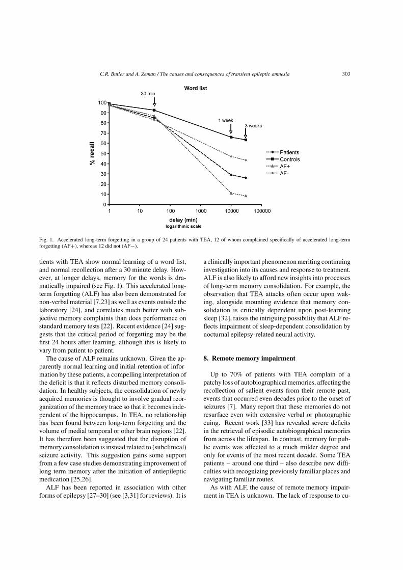
Fig. 1. Accelerated long-term forgetting in a group of 24 patients with TEA, 12 of whom complained specifically of accelerated long-termforgetting (AF+), whereas 12 did not (AF−).
tients with TEA show normal learning of a word list,and normal recollection after a 30 minute delay. How-ever, at longer delays, memory for the words is dra-matically impaired (see Fig. 1). This accelerated long-term forgetting (ALF) has also been demonstrated fornon-verbal material [7,23] as well as events outside thelaboratory [24], and correlates much better with sub-jective memory complaints than does performance onstandard memory tests [22]. Recent evidence [24] sug-gests that the critical period of forgetting may be thefirst 24 hours after learning, although this is likely tovary from patient to patient.
The cause of ALF remains unknown. Given the ap-parently normal learning and initial retention of infor-mation by these patients, a compelling interpretation ofthe deficit is that it reflects disturbed memory consoli-dation. In healthy subjects, the consolidation of newlyacquired memories is thought to involve gradual reor-ganization of the memory trace so that it becomes inde-pendent of the hippocampus. In TEA, no relationshiphas been found between long-term forgetting and thevolume of medial temporal or other brain regions [22].It has therefore been suggested that the disruption ofmemory consolidation is instead related to (subclinical)seizure activity. This suggestion gains some supportfrom a few case studies demonstrating improvement oflong term memory after the initiation of antiepilepticmedication [25,26].
ALF has been reported in association with otherforms of epilepsy [27–30] (see [3,31] for reviews). It is
a clinically important phenomenonmeriting continuinginvestigation into its causes and response to treatment.ALF is also likely to afford new insights into processesof long-term memory consolidation. For example, theobservation that TEA attacks often occur upon wak-ing, alongside mounting evidence that memory con-solidation is critically dependent upon post-learningsleep [32], raises the intriguing possibility that ALF re-flects impairment of sleep-dependent consolidation bynocturnal epilepsy-related neural activity.
8. Remote memory impairment
Up to 70% of patients with TEA complain of apatchy loss of autobiographicalmemories, affecting therecollection of salient events from their remote past,events that occurred even decades prior to the onset ofseizures [7]. Many report that these memories do notresurface even with extensive verbal or photographiccuing. Recent work [33] has revealed severe deficitsin the retrieval of episodic autobiographical memoriesfrom across the lifespan. In contrast, memory for pub-lic events was affected to a much milder degree andonly for events of the most recent decade. Some TEApatients – around one third – also describe new diffi-culties with recognizing previously familiar places andnavigating familiar routes.
As with ALF, the cause of remote memory impair-ment in TEA is unknown. The lack of response to cu-
304 C.R. Butler and A. Zeman / The causes and consequences of transient epileptic amnesia
ing, and the apparent patchiness of the memory loss,suggests a problem with the storage of memory traces,rather than a general impairment of retrieval mecha-nisms. However, we have recently encountered a pa-tient in whom long lost memories were recovered fol-lowing a series of episodes of deja vu [34], indicatingthat in some cases the deficit may indeed lie at the levelof retrieval. No relationship has been found betweenthe extent of autobiographicalmemory impairment andregional brain volumes in TEA [22]. Nevertheless, thepossibility remains that subtle structural damage, per-haps in the medial temporal lobes but beyond the res-olution of imaging techniques used to date, is respon-sible. An alternative explanation is that epileptic ac-tivity, spreading through the autobiographical memorynetwork, somehow causes irreparable damage to estab-lished memory traces. Whatever the underlying mech-anism, it is not immediately clear that current neuro-scientific models of memory can account for such anextensive loss of remote autobiographical memory inthe context of relatively preserved new learning (see [3]for further discussion).
9. Borderlands of TEA
As we have discussed in this article, the syndrome ofTEA is characterised by recurrent episodes of isolatedamnesia that begin in middle to old age, often occurupon waking, typically last about 30 minutes and re-spond well to anticonvulsant medication. Patients arefrequentlymisdiagnosed as having transient global am-nesia. The amnesic attacks often go hand in hand withpersistent but difficult to measure cognitive deficits in-cluding ALF and remote memory impairment. As withany syndrome, TEA has somewhat indistinct bound-aries. A notable example is the overlap with what Gal-lassi and colleagues have termed the Epileptic AmnesicSyndrome (EAS) [35,36]. Patients with EAS have lateonset (mean age = 63 years) persistent memory diffi-culties, often detectable on standard neuropsycholog-ical instruments, in association with subtle temporallobe seizures. In other patients (e.g. [37]), complaintsof ALF and remote memory impairment can precedethe onset of amnesic seizures by several years. Fur-ther research is necessary to clarify the relationship be-tween TEA and other forms of late-onset temporal lobeepilepsy. In the meantime, the concept serves to high-light this distinctive presentation of epilepsy, and facil-itate novel explorations of human memory function.
Acknowledgements
The authors are grateful to Dr J Penge for assistancewith gathering data on cerebrovascular disease risk fac-tors and white matter hyperintensities on brain imag-ing. We also thank Professor Ian Deary for permittingour use of data from the LBC1936 study.
References
[1] J. Hughlings-Jackson, On a particular variety of epilepsy (in-tellectual aura), one case with symptoms of organic brain dis-ease, Brain 11 (1888), 179–207.
[2] J. Hughlings-Jackson and W.S. Colman, Case of epilepsy withtasting movements and dreamy state: very small patch ofsoftening in the left uncinate gyrus, Brain 21 (1898), 580–590.
[3] C.R. Butler and A.Z. Zeman, Recent insights into the im-pairment of memory in epilepsy: transient epileptic amnesia,accelerated long-term forgetting and remote memory impair-ment, Brain 131(9) (2008), 2243–2263.
[4] N. Kapur, Transient epileptic amnesia: a clinically distinctform of neurological memory disorder, inL Transient glob-al amnesia and related disorders, H.J. Markowitsch, ed.,Hogrefe and Huber: New York, 1990, pp. 140–151.
[5] N. Kapur, Transient epileptic amnesia–a clinical update and areformulation, Journal of Neurology, Neurosurgery & Psychi-atry 56(11) (1993), 1184–1190.
[6] A.Z.J. Zeman, S.J. Boniface and J.R. Hodges, Transientepileptic amnesia: A description of the clinical and neuropsy-chological features in 10 cases and a review of the litera-ture, Journal of Neurology, Neurosurgery & Psychiatry 64(4)(1998), 435–443.
[7] C.R. Butler et al., The syndrome of transient epileptic amnesia,Ann Neurol 61(6) (2007), 587–598.
[8] P. Quinette et al., What does transient global amnesia reallymean? Review of the literature and thorough study of 142 cas-es, Brain 129(Pt 7) (2006), 1640–1658.
[9] C.R. Butler and A. Zeman, A case of transient epileptic amne-sia with radiological localization, Nature Reviews Neurology4(9) (2008), 516–521.
[10] K.J. Meador, R.J. Adams and H.F. Flanigin, Transient globalamnesia and meningioma, Neurology 35(5) (1985), 769–771.
[11] J.R. Shuping, J.F. Toole and E. Alexander, Jr., Transient globalamnesia due to glioma in the dominant hemisphere, Neurology30(1) (1980), 88–90.
[12] C.-F. Huang and M.-C. Pai, Transient amnesia in a patient withleft temporal tumor: symptomatic transient global amnesia oran epileptic amnesia? Neurologist 14(3) (2008), 196–200.
[13] G. Maheu et al., A Case of Postictal Transient Anterograde andRetrograde Amnesia, Epilepsia 45(11) (2004), 1459–1460.
[14] A.L. Rabinowicz et al., Transient epileptic amnesia in de-mentia: a treatable unrecognized cause of episodic amnesticwandering, Alzheimer Disease & Associated Disorders 14(4)(2000), 231–233.
[15] F. Fazekas et al., Pathologic correlates of incidental MRI whitematter signal hyperintensities, Neurology 43(9) (1993), 1683–1689.
[16] C. Dufouil et al., Longitudinal study of blood pressure andwhite matter hyperintensities: The EVA MRI Cohort, Neurol-ogy 56(7) (2001), 921–926.

C.R. Butler and A. Zeman / The causes and consequences of transient epileptic amnesia 305
[17] R. Schmidt et al., Magnetic resonance imaging signal hyper-intensities in the deep and subcortical white matter. A com-parative study between stroke patients and normal volunteers,Arch Neurol 49(8) (1992), 825–827.
[18] M.M.B. Breteler et al., Cerebral white matter lesions, vascu-lar risk factors, and cognitive function in a population-basedstudy: The Rotterdam Study, Neurology 44(7) (1994), 1246–1252.
[19] J.C. De Groot et al., Cerebral white matter lesions and cogni-tive function: The Rotterdam scan study, Annals of Neurology47(2) (2000), 145–151.
[20] F. Fazekas et al., MR signal abnormalities at 1.5 T inAlzheimer’s dementia and normal aging, AJRAmJRoentgenol149(2) (1987), 351–356.
[21] M. Razavi, J. Barrash and S. Paradiso, A Longitudinal Studyof Transient Epileptic Amnesia, Cognitive and BehavioralNeurology 23(2) (2010), 142.
[22] C.R. Butler et al., Transient epileptic amnesia: regional brainatrophy and its relationship to memory deficits, Brain 132(2)(2009), 357–368.
[23] F. Manes et al., Autobiographical amnesia and acceleratedforgetting in transient epileptic amnesia, J Neurol NeurosurgPsychiatry 76(10) (2005), 1387–1391.
[24] N. Muhlert et al., Accelerated forgetting of real-life eventsin Transient Epileptic Amnesia, Neuropsychologia 48(11)(2010), 3235–3244.
[25] M. O’Connor et al., Accelerated forgetting in association withtemporal lobe epilepsy and paraneoplastic encephalitis, Brain& Cognition 35(1) (1997), 71–84.
[26] A. Midorikawa and M. Kawamura, Recovery of Long-TermAnterograde Amnesia, but Not Retrograde Amnesia, after Ini-
tiation of an Anti-Epileptic Drug in a Case of Transient Epilep-tic Amnesia, Neurocase 13(5) (2007), 385–389.
[27] R.V. Blake et al., Accelerated forgetting in patients withepilepsy: evidence for an impairment in memory consolida-tion, Brain 123(Pt 3) (2000), 472–483.
[28] R. Mameniskiene et al., The decay of memory between de-layed and long-term recall in patients with temporal lobeepilepsy, Epilepsy Behav 8(1) (2006), 278–288.
[29] R.C. Martin et al., Impaired Long-Term Retention DespiteNormal Verbal Learning in Patients With Temporal Lobe Dys-function, Neuropsychology 5(1) (1991), 3–12.
[30] M. Davidson et al., Memory consolidation and acceleratedforgetting in children with idiopathic generalized epilepsy,Epilepsy & Behavior 11(3) (2007), 394–400.
[31] B.D. Bell and A.R. Giovagnoli, Recent innovative studies ofmemory in temporal lobe epilepsy, Neuropsychol Rev 17(4)(2007), 455–476.
[32] S. Diekelmann and J. Born, The memory function of sleep,Nat Rev Neurosci 11(2) (2010), 114–126.
[33] F. Milton et al., Remote memory deficits in transient epilepticamnesia, Brain 133(5) (2010), 1368–1379.
[34] F. Milton, C.R. Butler and A.Z.J. Zeman, Transient epilepticamnesia: deja vu heralding recovery of lost memories, Journalof Neurology, Neurosurgery & Psychiatry (2010).
[35] R. Gallassi, Epileptic amnesic syndrome: an update and fur-ther considerations, Epilepsia 47(Suppl 2) (2006), 103–105.
[36] R. Gallassi et al., Epileptic amnesic syndrome, Epilepsia33(Suppl 6) (1992), S21–S25.
[37] M. Hornberger et al., Focal retrograde amnesia: Extendingthe clinical syndrome of transient epileptic amnesia, Journalof Clinical Neuroscience 17(10), 1319–1321.

Submit your manuscripts athttp://www.hindawi.com
Stem CellsInternational
Hindawi Publishing Corporationhttp://www.hindawi.com Volume 2014
Hindawi Publishing Corporationhttp://www.hindawi.com Volume 2014
MEDIATORSINFLAMMATION
of
Hindawi Publishing Corporationhttp://www.hindawi.com Volume 2014
Behavioural Neurology
EndocrinologyInternational Journal of
Hindawi Publishing Corporationhttp://www.hindawi.com Volume 2014
Hindawi Publishing Corporationhttp://www.hindawi.com Volume 2014
Disease Markers
Hindawi Publishing Corporationhttp://www.hindawi.com Volume 2014
BioMed Research International
OncologyJournal of
Hindawi Publishing Corporationhttp://www.hindawi.com Volume 2014
Hindawi Publishing Corporationhttp://www.hindawi.com Volume 2014
Oxidative Medicine and Cellular Longevity
Hindawi Publishing Corporationhttp://www.hindawi.com Volume 2014
PPAR Research
The Scientific World JournalHindawi Publishing Corporation http://www.hindawi.com Volume 2014
Immunology ResearchHindawi Publishing Corporationhttp://www.hindawi.com Volume 2014
Journal of
ObesityJournal of
Hindawi Publishing Corporationhttp://www.hindawi.com Volume 2014
Hindawi Publishing Corporationhttp://www.hindawi.com Volume 2014
Computational and Mathematical Methods in Medicine
OphthalmologyJournal of
Hindawi Publishing Corporationhttp://www.hindawi.com Volume 2014
Diabetes ResearchJournal of
Hindawi Publishing Corporationhttp://www.hindawi.com Volume 2014
Hindawi Publishing Corporationhttp://www.hindawi.com Volume 2014
Research and TreatmentAIDS
Hindawi Publishing Corporationhttp://www.hindawi.com Volume 2014
Gastroenterology Research and Practice
Hindawi Publishing Corporationhttp://www.hindawi.com Volume 2014
Parkinson’s Disease
Evidence-Based Complementary and Alternative Medicine
Volume 2014Hindawi Publishing Corporationhttp://www.hindawi.com