the cell nucleus
DESCRIPTION
Chapter 9. The cell Nucleus. Biology Department of the Basic Teaching Colledge. Xiamixinuer · Yilike. The May of 2012. Chapter 9 Nucleus. Learning Objectives - PowerPoint PPT PresentationTRANSCRIPT

1
The cell NucleusThe cell NucleusThe cell NucleusThe cell Nucleus
XiamixinuerXiamixinuer··Yilike Yilike
Chapter 9
2
Chapter 9 Nucleus
Learning ObjectivesLearning Objectives1. Mastering: ultrastructure of nuclear envelop; 1. Mastering: ultrastructure of nuclear envelop;
nuclear pore complex; composition and four levels nuclear pore complex; composition and four levels organization of chromatin; packaging of chromatin; organization of chromatin; packaging of chromatin; types of chromatin.types of chromatin.
2. Comprehending: structure and function of 2. Comprehending: structure and function of nucleolus; function of nuclear pore complex; nucleolus; function of nuclear pore complex; process of RNA processing.process of RNA processing.
3. Understanding: basic function of nucleus; nuclear 3. Understanding: basic function of nucleus; nuclear matrix.matrix.

3
1. The nucleus: Nuclear envelope and NPC
A. Structure: Double-membrane nuclear envelope surrounds the nucleus
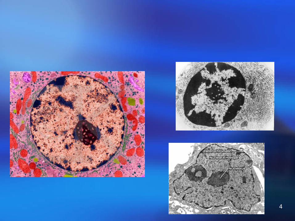
Structure of the interphase nucleus
4

5
The main functions of nucleusThe main functions of nucleus
1. Carry genetic information ( DNA ) ;
2.Duplicate,transcript of genetic information and control protein synthesis;
3.Regulation and control centre of living action of cells.

6
NP(nucleoplasmic index)
NP= Vn/(Vc - Vn)
Usually size of nucleus can be estimated by calculate the NP.
Normal cell NP≈0.5, Dividing cell NP>0.5, Aging cell NP<0.5。
7
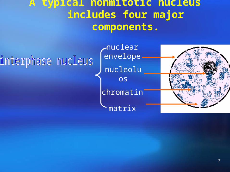
A typical nonmitotic nucleus includes four major components.
nuclear envelope
nucleoluos
chromatin
matrix

8
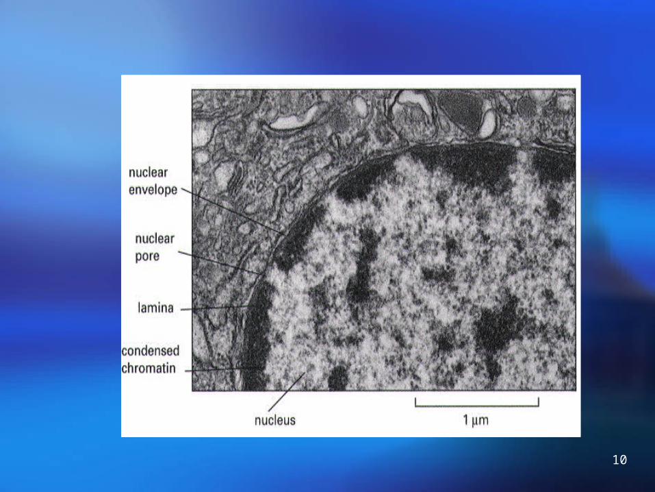
B. The nuclear envelope consists of two membranes by a perinuclear space.

9
10

11
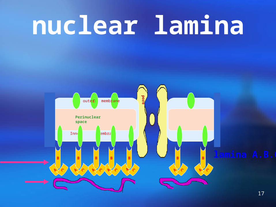
The inner surface of the nuclear envelope is lined by the nuclear lamina
The nuclear lamina supports the nuclear envelope: Gives shape and stability of nuclear envelope;
Provides a structure link between chromatin and nuclear envelope;
The nuclear lamina is composed of lamins.
The integrity of the nuclear lamina is regulated by phosphorylation and dephosphorylation.
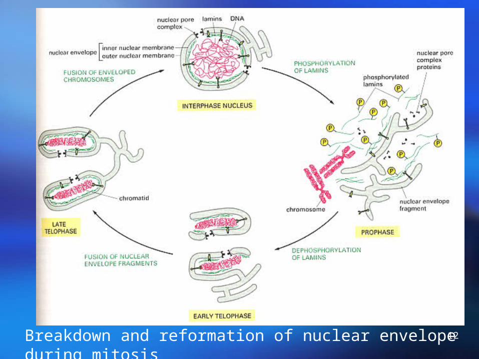
12Breakdown and reformation of nuclear envelope during mitosis

13
The structures of nuclear envelope
outer nuclear membrane
inner nuclear membrane
perinuclear space
nuclear pores
nuclear lamina

14
C. Nuclear pore complex (NPC)

15
The structure of
Nuclear pore
complex (NPC)

16
Old model structure
Ring subunit: 8 pair
Annular subunit: 8
Central plug : 1
fibril:
17
nuclear lamina
pore蛋outer membrane
Inner membrane
Perinuclear space
A C A CA CA CA C A C A C
B B B B B B B lamina A.B.C
18
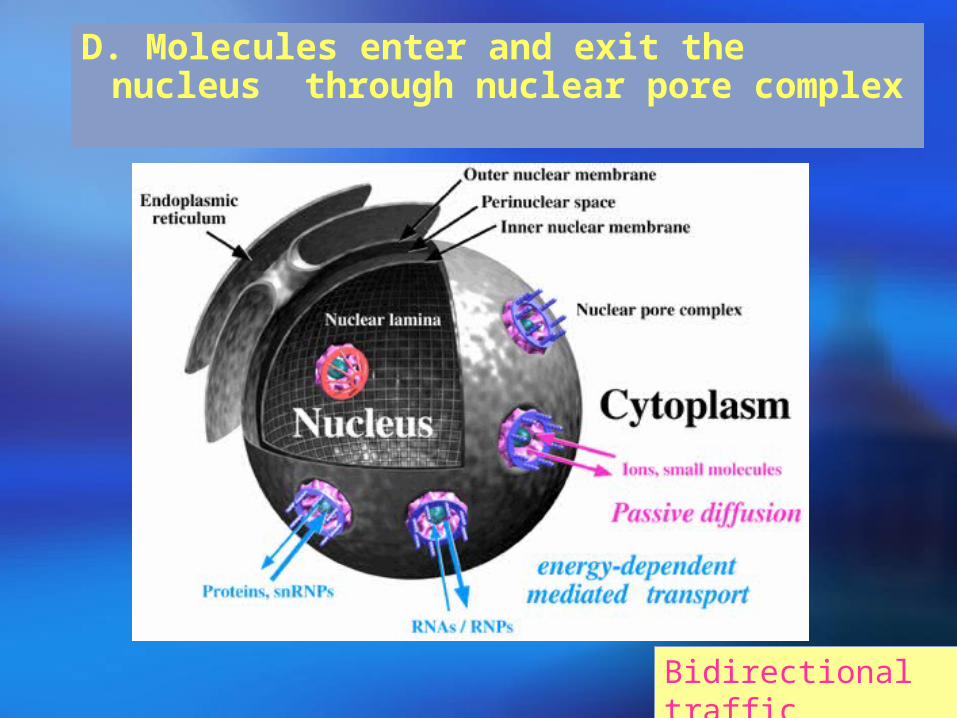
D. Molecules enter and exit the nucleus through nuclear pore complex
Bidirectional traffic

19
Molecules enter and exit the nucleus through nuclear pore complex

20
Passive transport—passively diffuse
3000-4000 NPC/cell(mammalian);
To import about 106 histone/3 mins.(DNA-sythesizing cell) = 100 histone/ min/NPC
Each NPC contains one or more open aqueous channels: 9 nm in diameter and 15 nm long
The effective size of these channels has been determined by injecting various sizes of colloidal gold particles and examined by electron microscopy.
<10 nm in diameter
<60kd globular proteinAble to enter the nucleus
21
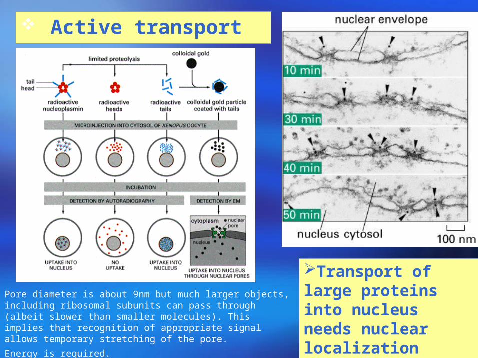
Active transport
Transport of large proteins into nucleus needs nuclear localization signal (NLS)
Pore diameter is about 9nm but much larger objects, including ribosomal subunits can pass through (albeit slower than smaller molecules). This implies that recognition of appropriate signal allows temporary stretching of the pore.
Energy is required.
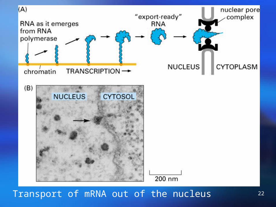
22Transport of mRNA out of the nucleus

23
Nucleolus and ChromosomesChapter 9 section 2
Learning Objectives
(1) The components of chromatin and packaging of
chromosome
(2) Nucleolus.

24human nucleus under LM
human lymphocyte nucleus under SEM
Chromatin fiber

25
Each human cell contains about 2 m of DNA within nucleus if stretched end-to-end, yet the nucleus of a human cell itself is only about 6 μm in diameter.
Compaction ratio=nearly 10000-fold. (Chromosome 22: DNA 1.5cm2 μ m)
Eukaryotes package DNA in Chromatin and chromosomes
26

27
Chromosomes exist in different states throughout the life of a cell.
Chromatin: (Interphase)
Fibers, 10-30nm in diameter,dispersed through the nucleus.
DNA+Proteins+non-Proteins+RNA.
Chromosomes: (M phase)
Cell division, these fibers condense and fold into larger, compact structure.

28
Levels of organizatio
n of
chromatin
29
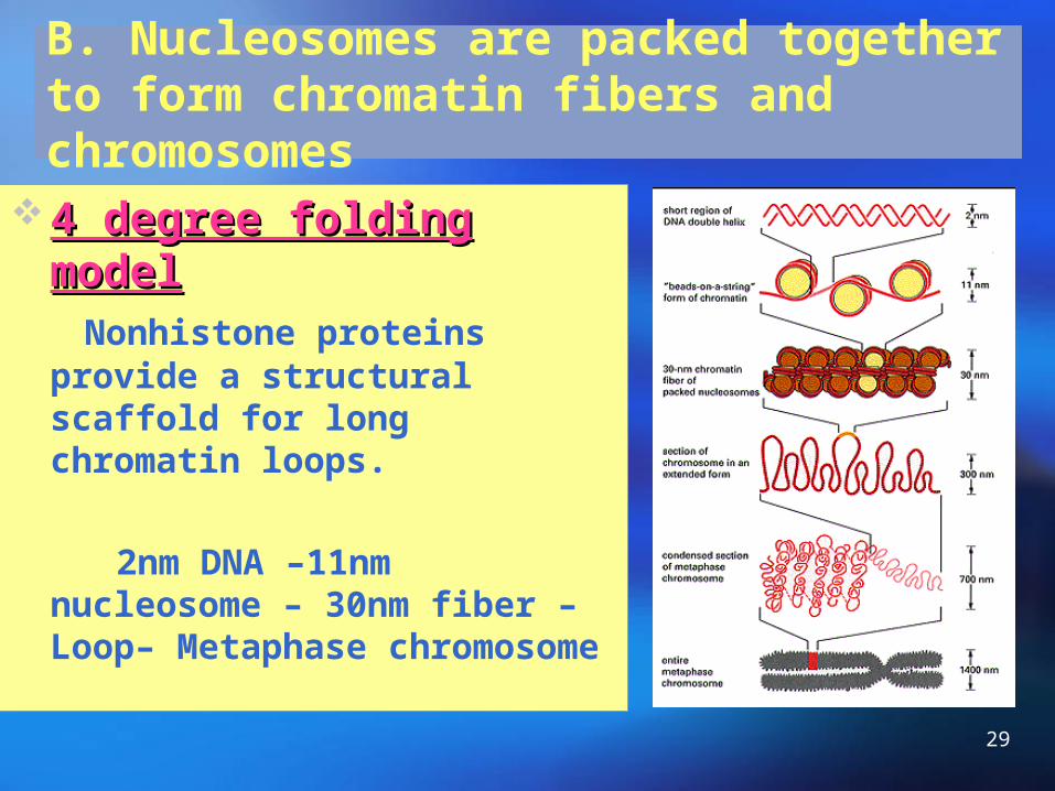
B. Nucleosomes are packed together to form chromatin fibers and chromosomes
4 degree folding model4 degree folding model Nonhistone proteins provide a
structural scaffold for long chromatin loops.
2nm DNA –11nm nucleosome – 30nm fiber – Loop– Metaphase chromosome

30
4 degree folding model4 degree folding model
First degree:
Nucleosomes Second degree:
Filament 10nm in diameter Third level:
Fiber,30nm in diameter Fourth level:
Chromosome
31
A. DNA packaging:First degree of packaging is Nucleosomes
Nucleosomes are the basic unit of chromatin structure.

32
Evidence:
(1)Electron micrographs of chromatin fibers
Isolated from interphase nucleus: 30nm thick
Chromatin unpacked, show the nuclesome

33
Evidence:
(3)X-ray diffraction studies
3-D of nucleosome Nature 389:251, 1997

34
Structure of nucleosome
Digested briefly:
H1+Octamer+200bpDNA
Digested longer:
Octamer+146bp
H1 is released
Core particle

35
Structural Structural
organization of organization of
the nucleosomethe nucleosome

36
CORE OF PERIPHERAL HISTON
H4
DNA (146bp 、 1.75 ROUND)
H3
H4H3
H2AH2A
H2B
H2B
H4H3
H3H4
H2AH2A
H2B
H2B
10nm
LINKER DNA ( 60b
p)
H1
H1
NUCLEOSOME
CORE
LINKER
DNA MOLECULE :146bp 、 1.75 ROUND
HISTONE :2 ( H2A 、 H2B 、 H3 、 H4 ) OCTAMER
HISTONE : H1
DNA MOLECULE : 60bp
NUCLEOSOME

37
A histone octamer forms the nucleosome core
Histone octamer:
(H2A-H2B)-(H3-H4)-(H3-H4)-(H2A-H2B)
Where is the histone H1?
H1 molecules are associated with the linker region. 146+15~50bp linker DNA
200bpDNA:
Linker DNA:15-50bp
Nucleosomal DNA:146bp to wrap 1.75 times around the histone core.
38
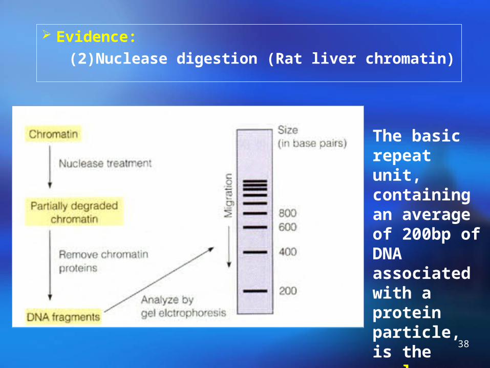
Evidence:
(2)Nuclease digestion (Rat liver chromatin)
The basic repeat unit, containing an average of 200bp of DNA associated with a protein particle, is the nucleosome
39
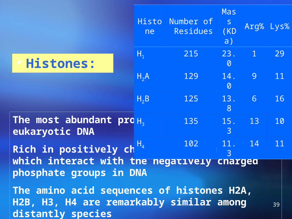
Histones:
The most abundant proteins associated with eukaryotic DNA
Rich in positively charged basic amino acids, which interact with the negatively charged phosphate groups in DNA
The amino acid sequences of histones H2A, H2B, H3, H4 are remarkably similar among distantly species
HistoneNumber of Residues
Mass(KDa
)Arg% Lys%
H1 215 23.0 1 29
H2A 129 14.0 9 11
H2B 125 13.8 6 16
H3 135 15.3 13 10
H4 102 11.3 14 11

40
Second level: nucleosome filament10nm in diameter
H1

41
Third level:
fiber,30nm in diameter
Fourth level: chromosome

42
Summary of 4 degree folding model

43
44
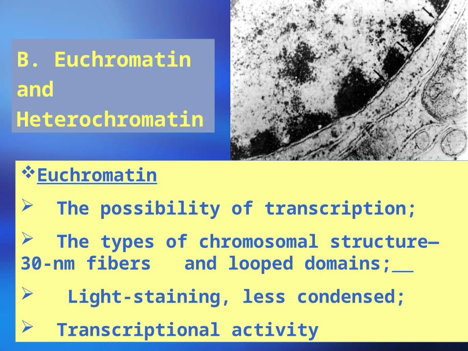
B. Euchromatin
and
Heterochromatin
Euchromatin
The possibility of transcription;
The types of chromosomal structure—30-nm fibers and looped domains;
Light-staining, less condensed;
Transcriptional activity
45
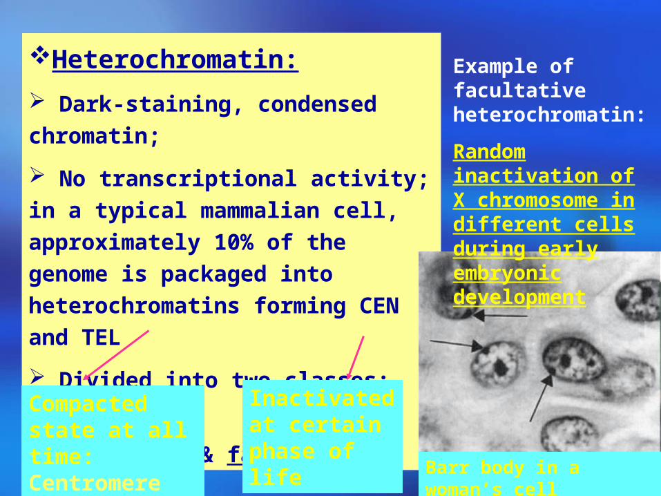
Heterochromatin:
Dark-staining, condensed chromatin;
No transcriptional activity; in a typical
mammalian cell, approximately 10% of
the genome is packaged into
heterochromatins forming CEN and TEL
Divided into two classes:
Constitutive & facultative
Compacted state at all time: Centromere
Inactivated at certain phase of life
Example of facultative heterochromatin:
Random inactivation of X chromosome in different cells during early embryonic development
Barr body in a woman’s cell

46
3. Chromosome number,size,and shape at metaphase are species specific
ChromatidsKaryotype Banding

47
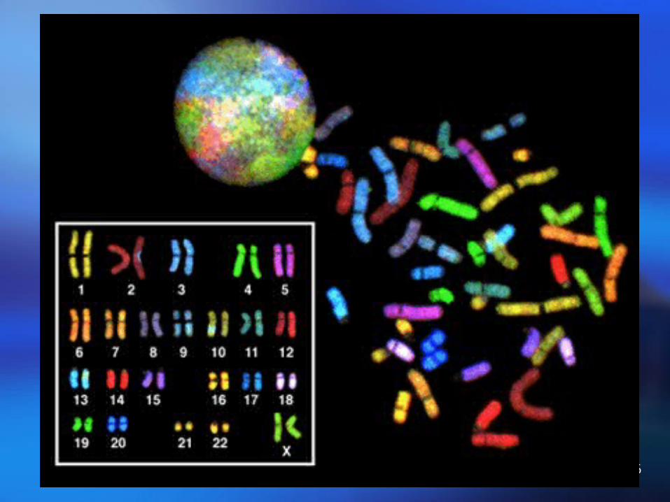
Human mitotic chromosomes and karyotype
48
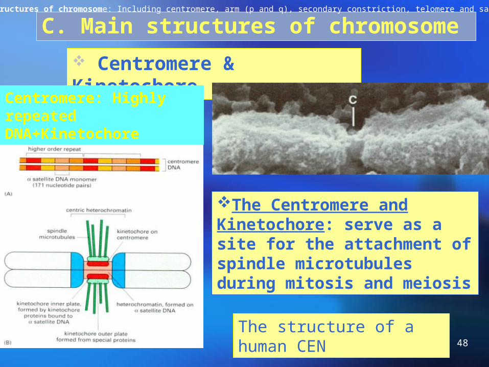
C. Main structures of chromosome
Centromere & Kinetochore
Centromere: Highly repeated DNA+Kinetochore
The Centromere and Kinetochore: serve as a site for the attachment of spindle microtubules during mitosis and meiosis
The structure of a human CEN
Main structures of chromosome: Including centromere, arm (p and q), secondary constriction, telomere and satellite.
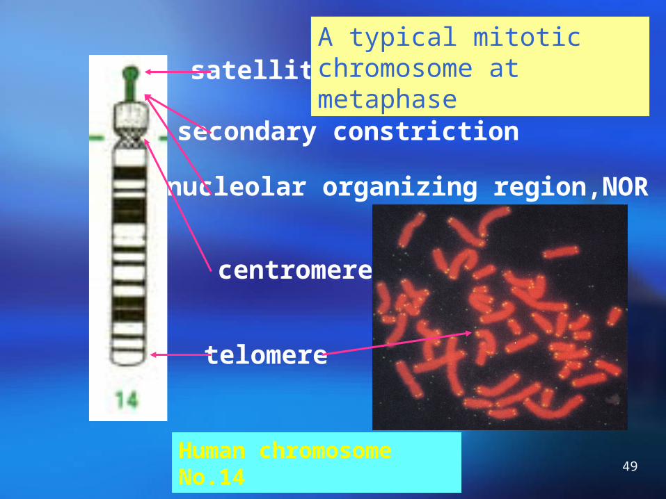
49Human chromosome No.14
secondary constriction
nucleolar organizing region,NOR
satellite
telomere
centromere
A typical mitotic chromosome at metaphase
50
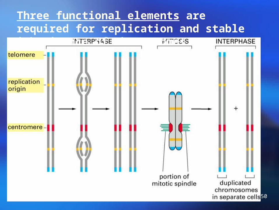
Three functional elements are required for replication and stable inheritance of chromosomes

51
Functions
Protect the chromosomes from nuclease influence
For the complete replication of chromosome
Prevent the ends of chromosomes from fusing
Telomeres:

52
Telomere, telomerase and cellular aging, cancer cell
Telomerase is found in germ cells, not in somatic cells.
The telomere length of adult is shorter than that of younger.
Telomere shortening is thought to activate a suicide program.
So, telomere shortening plays a key role in protecting the body from cancer.
90% of human tumors contain an active telomerase.
53
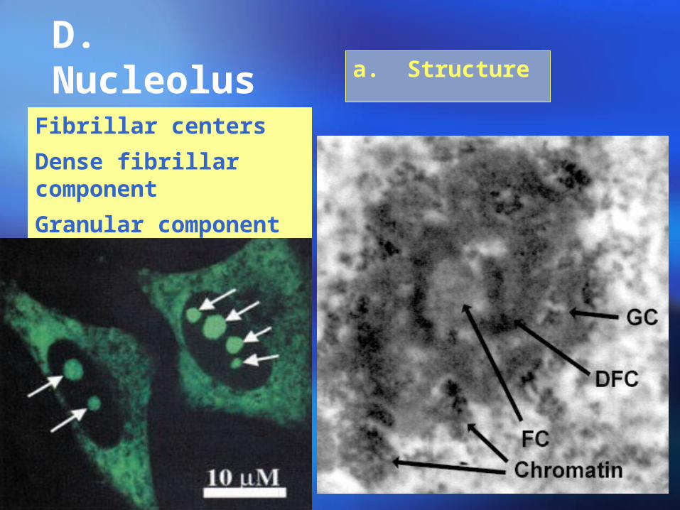
D. Nucleolus a. Structure
Fibrillar centers
Dense fibrillar component
Granular component

54
b. Functions of nucleolus: Ribosomal Biogenesis
Ribosomal Biogenesis:Ribosomal Biogenesis:
pre-rRNA synthesispre-rRNA synthesis ProcessProcess AssemblyAssembly
FC. DFC DFC.GC Cytosol
Directional processDirectional process

55
Characteristics of RNA transcription
NORs in human chromosomes:
13\14\15\21\22
Code for (in eukaryotes):
18s, 28s, 5.8s rRNA
56
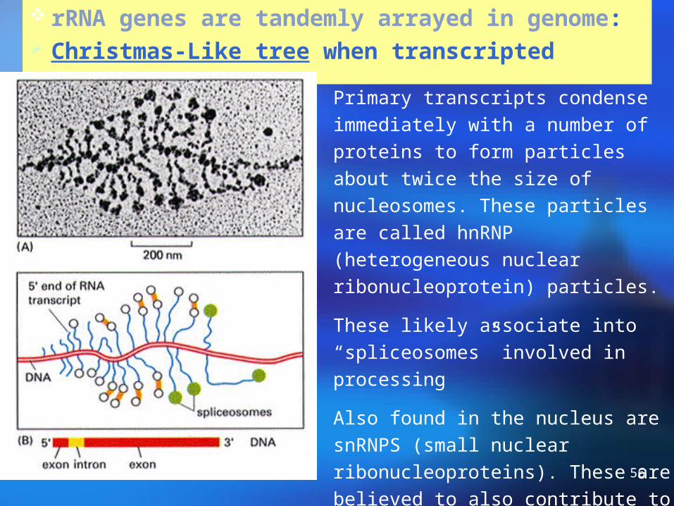
rRNA genes are tandemly arrayed in genome: Christmas-Like tree when transcripted
Primary transcripts condense immediately with a number of proteins to form particles about twice the size of nucleosomes. These particles are called hnRNP (heterogeneous nuclear ribonucleoprotein) particles.
These likely associate into “spliceosomes” involved in processing
Also found in the nucleus are snRNPS (small nuclear ribonucleoproteins). These are believed to also contribute to RNA splicing and to histone mRNA synthesis

57
Cleavage of pre-rRNA as RNP particles
Mammalia

58
Synthesis and processing of 5s rRNA
5s rRNAs are encoded by a large number genes (Human, 2000)
5s rRNA gene are located outside the nucleolus.
5s rRNAs are transcibed by RNA poly III.
The 3’ end of 5s rRNA is removed during processing.
59
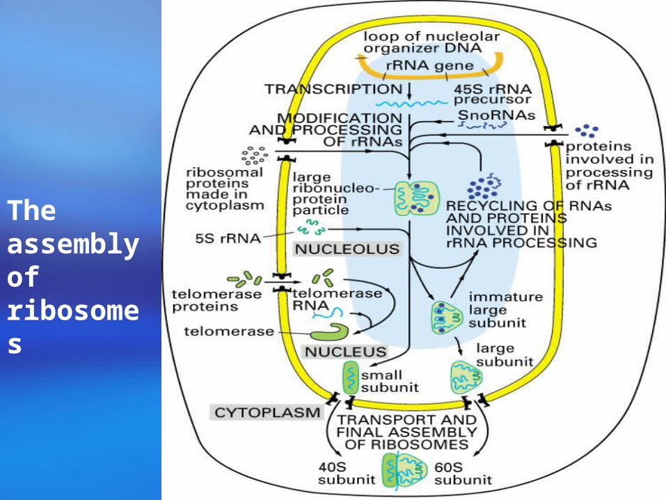
The assembly of ribosomes

60
The nucleolus disappears during mitosis
Nucleolar fusion of human fibroblast in culture
61
Summary
(1). The components of the typical nonmitotic nucleus
(2). The components of chromatin and packaging of chromosome: Scaffold radial loop structure model.
(3). Nucleolus components and functions.
Homework
Nucleolus Structure includes which parts? What are the functions of nucleolus? Describe briefly the difference between
euchromatin and heterochromatin. Describe briefly structure of nucleosome? Any Questions??
62