the child with musculoskeletal and neuromuscular...
TRANSCRIPT

The Child with
Musculoskeletal and
Neuromuscular Dysfunction

Overview of A&P
Osteoblasts- immature bone cells that
replace cartilage cells as bone grows
Child’s bones are less dense and
more porous
Ossification progresses outwardly
from the diaphysis
Essential for adequate calcium intake
to prevent osteoporosis

Club Foot
Foot twisted and fixed in abnormal position Plantar flexion
Dorsiflexion
Varus deviation
Valgus deviation
May be unilateral or bilateral; bone deformity and soft tissue contracture
Etiology unknown Familial tendency
Intrauterine position
NM or vascular problems

Treatment and Nursing Care
Manipulation/ serial
casting
Parents perform ROM
Denis Browne Splints
or corrective shoes for
1 yr
Surgery- 4- 12
months, casted 6-12
weeks
Neurovascular checks
Elevate on pillow
Monitor for
welling/drainage
Pain Management
Distraction/Age
appropriate play

Osteogenesis Imperfecta (OI)
Pathologic fractures- “brittle bone disease”
Bones are fragile that fractures result from trauma but also from pressure or birth
Autosomal dominant inheritance
Normal Ca and Ph, abnormal pre collagen type I
Often misdiagnosed as child abuse

Clinical Manifestations/Assessment
Blue sclera/cataracts
Transparent skin
Weak muscles, short stature, fractures
Hearing problems
H/O fractures delayed growth
Discolored teeth
Normal or above average intelligence

Nursing Care
Mobility: gentle turning, passive ROM,
skin care
Light weight leg braces, splinting, PT
Meds
Calcitonin- aids bone healing
Biphosphonates to increase bone mass
Growth hormone to stimulate growth

Developmental Dysplasia of the Hip
Head and acetabulum improperly aligned
Occurs 1-2/1000 births; unilateral
Females more than males
Etiology
Family history
Maternal hormones-estrogen causing laxity of
the hip joint and capsule
Breech, twins, large infant
sociocultural

Pathophysiology
Preluxation
Delay in acetabular
development
Femoral head in the
acetabulum
Subluxation
Incomplete dislocation
Femoral head in
contact with
acetabulum
Stretched capsule and
ligament
Dislocation
Femoral head loses
contact with
acetabulum
Displaced posteriorly
and superiorly over the
rim
Uncorrected
subluxation of
dislocation can lead to
permanent disability

Clinical Assessment Findings
Infants
Ortolani’s sign
Barlow’s sign
Asymmetric gluteal
folds
Limited abduction
of affected hip
Older Children
Limp/toe walking
Delayed walking
Trendelenburg’s
sign
Telescoping of
affected leg
Lordosis and
waddling gait with
bilateral dislocation
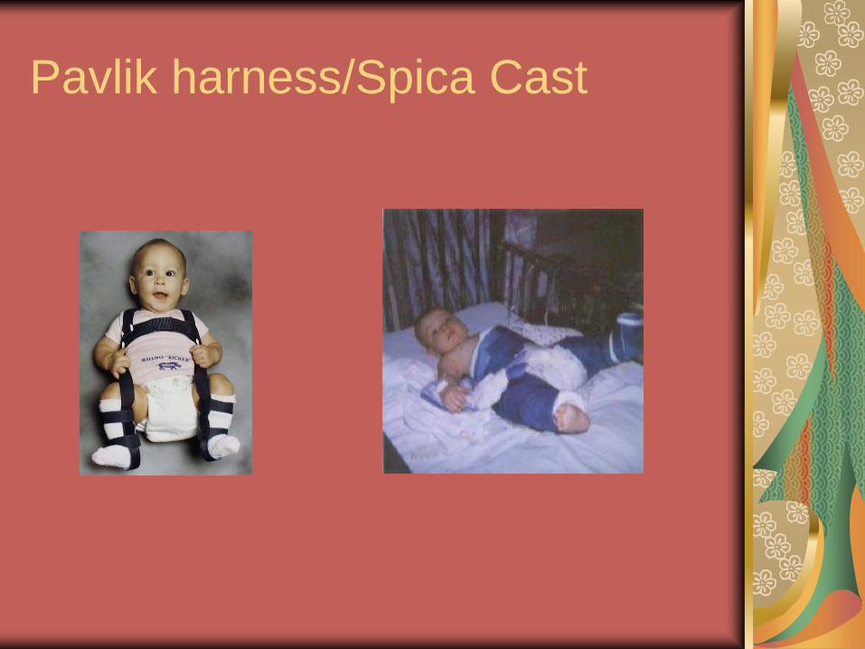
Pavlik harness/Spica Cast

Family Education
Pavlik Harness Proper application and assessment of skin
Wear t shirt and socks worn under brace to prevent skin irritation
Diaper placed under straps
Don not remove for diaper change only bath
Modification of car seat, positioning for nursing and eating
Ensure adequate stimulation with toys and activities to continue development

Spinal Deformities
Scoliosis
Lateral curvature of the spine
Females 11-14yrs
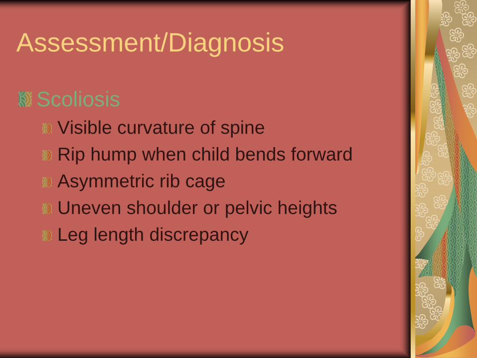
Assessment/Diagnosis
Scoliosis
Visible curvature of spine
Rip hump when child bends forward
Asymmetric rib cage
Uneven shoulder or pelvic heights
Leg length discrepancy

Milwaukee brace

Patient Teaching
Brace worn 23
hours/day
Brace off to shower,
bathe swim
Wear t shirt under
brace to protect skin
Exercises (pelvic tilt
and lateral
strengthening
Spinal Fusion – Post
op
Turn deep breathing
cough
Pain medication
ROM

Other Common Muscular Skeletal
Problems in Childhood
Duchenne
Muscular
Dystrophy
JRA
Spina bifida
Cerebral Palsy
Osteomyelitis
Fractures
Greenstick
Spiral
Open/closed
Complete
(transverse)
Slipped Capitol
Femoral Epiphysis