the chinese herbal medicine formula mkg … · international journal of molecular sciences article...
TRANSCRIPT

International Journal of
Molecular Sciences
Article
The Chinese Herbal Medicine Formula mKGSuppresses Pulmonary Fibrosis of Mice Inducedby Bleomycin
Ying Gao 1,2,†, Li-Fu Yao 2,†, Yang Zhao 3, Li-Man Wei 1, Peng Guo 2, Meng Yu 1, Bo Cao 2,Tan Li 2, Hong Chen 2,* and Zhong-Mei Zou 1,*
1 Institute of Medicinal Plant Development, Chinese Academy of Medical Sciences andPeking Union Medical College, Beijing 100193, China; [email protected] (Y.G.);[email protected] (L.-M.W.); [email protected] (M.Y.)
2 Department of Pharmacy, Logistics College of Chinese People’s Armed Police Force, Tianjin 300309, China;[email protected] (L.-F.Y.); [email protected] (P.G.); [email protected] (B.C.);[email protected] (T.L.)
3 Tianjin University of Traditional Chinese Medicine, Tianjin 300193, China; [email protected]* Correspondence: [email protected] (H.C.); [email protected] (Z.-M.Z.);
Tel.: +86-22-8487-6771 (H.C.); +86-10-5783-3290 (Z.-M.Z.); Fax: +86-10-5783-3290 (Z.-M.Z.)† These authors contributed equally to this work.
Academic Editor: Marcello IritiReceived: 6 January 2016; Accepted: 4 February 2016; Published: 15 February 2016
Abstract: Pulmonary fibrosis (PF) is a serious progressive lung disease and it originates frominflammation-induced parenchymal injury with excessive extracellular matrix deposition to result inthe destruction of the normal lung architecture. Modified Kushen Gancao Formula (mKG), derivedfrom traditional Chinese herbal medicine, has a prominent anti-inflammatory effect. The presentstudy is to explore the inhibitory effects of mKG on bleomycin (BLM)-induced pulmonary fibrosisin mice. mKG significantly decreased pulmonary alveolitis, fibrosis scores, and interleukin-6 (IL-6),interleukin-17 (IL-17), transforming growth factor-β (TGF-β) and hydroxyproline (HYP) levels inlung tissue of mice compared with BLM treatment. It markedly alleviated the increase of HYPcontent in the lung tissues and pathologic changes of pulmonary fibrosis caused by BLM instillation.In conclusion, mKG has an anti-fibrotic effect and might be employed as a therapeutic candidateagent for attenuating pulmonary fibrosis.
Keywords: pulmonary fibrosis; bleomycin; mKG (Modified Kushen Gancao Formula); mice
1. Introduction
Pulmonary fibrosis (PF) is a serious chronic lung disease with unknown pathogenesis. It is aprogressive and lethal lung disease, characterized by the accumulation of extracellular matrix inthe alveolar septum to result in the destruction of the normal lung architecture [1,2]. The currenttreatment for PF includes corticosteroids and immunosuppressive agents, but the outcomes of therapyare not satisfying. Traditional Chinese medicines (TCMs) are gaining more attention all over the worlddue to their holistic concept of TCM theory and long historical clinical practice. In Chinese herbaltherapy, the most widely used medicines are combined by many herbs and prepared according toTCM formulation concepts.
We have optimized a three-herb TCM formula, mKG, derived from a famous traditional Chinesemedicinal formula, Kushen Gancao Tang, which consists of the roots of Angelica sinensis (Oliv.) Diels.(Dang-gui), the roots of Sophora flavescens Ait. (Kushen) and the rhizome of Glycyrrhiza uralensis Fisch(Gancao). Kushen Gancao Tang has been recorded in Zhong Miao Xian Fang, a classic medical book
Int. J. Mol. Sci. 2016, 17, 238; doi:10.3390/ijms17020238 www.mdpi.com/journal/ijms

Int. J. Mol. Sci. 2016, 17, 238 2 of 12
published in the Ming dynasty of China. It is comprised of two medicinal herbs, Kushen and Gancao,which have been used to treat choleplania, infection, esoenteritis and dysentery for a long time. NowKushen Gaocao Formula has been used in the clinic to treat hepatitis and immune system diseases [3].In addition, it is reported to have anti-liver fibrosis activity [4]. Kushen has been used in China forits cleaning heat, drying humidity and diuresis for a long time. Pharmacological studies have shownits anti-angiogenic, anti-inflammatory and anti-nociceptive activities. So Kushen has been widelyused for treating various inflammatory and infectious diseases in the clinic, such as eczema andupper respiratory tract infections. It has also been used for treating asthma by suppressing airwayresponses, decreasing inflammation and down-regulating Th2 levels in asthmatic-model mice [5].Its active component oxymatrine can attenuate bleomycin-induced pulmonary fibrosis in mice byregulating the TGF-β/Smad signaling pathway [6,7]. Gancao has been used as an herbal medicinefor centuries in China to treat various diseases such as gastric or duodenal ulcers, hepatitis, sorethroats, coughs, bronchitis, arthritis, allergies, and cardiovascular disease [8,9]. Gancao and its activecompounds including glycyrrhizin, isoliquiritigenin and β-glycyhrritinic acid have been shown topossess prominent anti-inflammatory activities and can inhibit reactive oxygen species (ROS) [10–12].Glycyrrhizic acid can alleviate carbon tetrachloride-induced liver fibrosis and bleomycin-inducedpulmonary fibrosis in rats [13,14]. Modern research showed that Dang-Gui has broad pharmacologicaleffects on cardiovascular diseases, gynecological diseases and immune system disorders [15,16].The combination of Dang-Gui and Kushen is extensively used in traditional Chinese medicine to treatinflammatory diseases, such as acne, heart disease, and hepatitis [17].
Considering the properties of those herbs, the Kushen Gancao Tang has been modified inexpectation of the synergistic enhancement of its anti-inflammatory and anti-fibrotic effects. In addition,Modified Kushen Gancao Formula has been used to treat bronchial asthma and chronic obstructivepulmonary disease (COPD) in Pingjin Hospital, Tianjin, China, whereas there are no reports about theeffects of Modified Kushen Gancao Formula on PF. Additionally, the mechanisms by which ModifiedKushen Gancao Formula suppresses inflammatory and fibrotic effects have not been fully elucidated.
Although the mechanisms of PF are not understood fully, transforming growth factor-β (TGF-β)is recognized as a critical factor for inducing fibrosis. In the early period of PF, inflammatory factorssuch as interleukin-17 (IL-17) and interleukin-6 (IL-6) play an important role in autoimmunity. IL-17 isa novel family cytokine that consists of six protein members (from IL-17A to IL-17F), which play animportant role in many chronic inflammatory diseases [18–21]. Bleomycin (BLM), an antineoplasticdrug, has been reported to cause fibrosis in different species of animals [22–24]. The intratrachealinstillation of BLM was widely accepted as the method to develop the model of lung fibrosis. Here wefocused on exploring whether the mKG decreases the expression of TGF-β and inflammatory factorsso that it can attenuate BLM-induced PF. In this study we evaluate the antifibrotic properties of mKG.
2. Results
2.1. Histological Analysis
Numerous inflammatory cell infiltrations were observed in the alveolar and interstitial space ofthe lung by HE staining. From Figure 1 we can learn that mild edema became serious after inducingby BLM, with the structural confusion of the lung tissue and obvious swelling in the alveolar septum.Apparent alleviation of pulmonary alveolitis was observed in lung tissue of the pirfenidone group andhigh-dose mKG group, and the alveolar septum was thinner. Additionally, from the result of Massonstaining (Figure 2), it showed that blue-stained collagen massively increased in the untreated group.Treatment with mKG alleviated the collagen deposit according to the result of the Masson staining.The effect was more significant when treated with pirfenidone and the high-dose Ganshen formula.

Int. J. Mol. Sci. 2016, 17, 238 3 of 12
Int. J. Mol. Sci. 2016, 17, 238 3 of 12
Figure 1. Effect of mKG on histopathologic changes in BLM-induced pulmonary fibrosis mice. (A) Normal group; (B) BLM group; (C) Pirfenidone group; (D) Dexamethasone group; (E) mKG-H group; (F) mKG-M group; (G) mKG-L group. All sections were stained with HE and representative sections are shown at the same magnification (200×).
Figure 2. Effect of mKG on histopathologic changes in BLM-induced pulmonary fibrosis mice. (A) Normal group; (B) BLM group; (C) Pirfenidone group; (D) Dexamethasone group; (E) mKG-H group; (F) mKG-M group; (G) mKG-L group. All sections were stained with Masson trichrome and representative sections are shown at the same magnification (200×).
Figure 1. Effect of mKG on histopathologic changes in BLM-induced pulmonary fibrosis mice.(A) Normal group; (B) BLM group; (C) Pirfenidone group; (D) Dexamethasone group; (E) mKG-Hgroup; (F) mKG-M group; (G) mKG-L group. All sections were stained with HE and representativesections are shown at the same magnification (200ˆ).
Int. J. Mol. Sci. 2016, 17, 238 3 of 12
Figure 1. Effect of mKG on histopathologic changes in BLM-induced pulmonary fibrosis mice. (A) Normal group; (B) BLM group; (C) Pirfenidone group; (D) Dexamethasone group; (E) mKG-H group; (F) mKG-M group; (G) mKG-L group. All sections were stained with HE and representative sections are shown at the same magnification (200×).
Figure 2. Effect of mKG on histopathologic changes in BLM-induced pulmonary fibrosis mice. (A) Normal group; (B) BLM group; (C) Pirfenidone group; (D) Dexamethasone group; (E) mKG-H group; (F) mKG-M group; (G) mKG-L group. All sections were stained with Masson trichrome and representative sections are shown at the same magnification (200×).
Figure 2. Effect of mKG on histopathologic changes in BLM-induced pulmonary fibrosis mice.(A) Normal group; (B) BLM group; (C) Pirfenidone group; (D) Dexamethasone group; (E) mKG-Hgroup; (F) mKG-M group; (G) mKG-L group. All sections were stained with Masson trichrome andrepresentative sections are shown at the same magnification (200ˆ).

Int. J. Mol. Sci. 2016, 17, 238 4 of 12
2.2. Expression of IL-6, IL-17 mRNAs in Lung Tissue
From Figures 3 and 4 it was found that IL-6 and IL-17 mRNA expression increased in the lungtissue of mice after BLM administration. Treatment with pirfenidone and mKG attenuated IL-6 andIL-17 mRNA expression, and there was statistical significance with high-dose mKG.
Int. J. Mol. Sci. 2016, 17, 238 4 of 12
2.2. Expression of IL-6, IL-17 mRNAs in Lung Tissue
From Figures 3 and 4 it was found that IL-6 and IL-17 mRNA expression increased in the lung tissue of mice after BLM administration. Treatment with pirfenidone and mKG attenuated IL-6 and IL-17 mRNA expression, and there was statistical significance with high-dose mKG.
Figure 3. Effect of mKG on the mRNA Expression of IL-6 in the lung tissues of BLM-induced pulmonary fibrosis mice. (a) The mRNA expressions of IL-6 in the lung tissues were detected by RT-PCR; (b) The semiquantitative result of the IL-6 mRNA expression. Data were expressed as mean ± S.D. ## p < 0.01 compared with the control group. * p < 0.05, ** p < 0.01 compared with the BLM group. (A) Normal group; (B) BLM group; (C) Pirfenidone group; (D) Dexamethasone group; (E) mKG-H group; (F) mKG-M group; (G) mKG-L group.
Figure 4. Effect of mKG on the mRNA Expression of IL-17 in the lung tissues of BLM-induced pulmonary fibrosis mice. (a) The mRNA expressions of IL-17 in the lung tissues were detected by RT-PCR; (b) The semiquantitative result of the IL-17 mRNA expression. Data were expressed as mean ± S.D. ## p < 0.01 compared with the control group. * p < 0.05, ** p < 0.01 compared with the BLM group. (A) Normal group; (B) BLM group; (C) Pirfenidone group; (D) Dexamethasone group; (E) mKG-H group; (F) mKG-M group; (G) mKG-L group.
2.3. Results of Immunohistochemistry Staining for Col-1 and Col-3
In Figure 5, the expression of Col-1 and Col-3 was markedly increased in the BLM group when compared with the normal group, and the expression of Col-1 and Col-3 could be inhibited in groups that were treated with mKG, pirfenidone and dexamethasone by immunohistochemistry testing. Especially in the mKG-H group and positive control (pirfenidone and dexamethasone) groups, Col-1- and Col-3-stained area fractions were about half of that in the BLM group or even less.
Figure 3. Effect of mKG on the mRNA Expression of IL-6 in the lung tissues of BLM-induced pulmonaryfibrosis mice. (a) The mRNA expressions of IL-6 in the lung tissues were detected by RT-PCR;(b) The semiquantitative result of the IL-6 mRNA expression. Data were expressed as mean ˘ S.D.## p < 0.01 compared with the control group. * p < 0.05, ** p < 0.01 compared with the BLM group.(A) Normal group; (B) BLM group; (C) Pirfenidone group; (D) Dexamethasone group; (E) mKG-Hgroup; (F) mKG-M group; (G) mKG-L group.
Int. J. Mol. Sci. 2016, 17, 238 4 of 12
2.2. Expression of IL-6, IL-17 mRNAs in Lung Tissue
From Figures 3 and 4 it was found that IL-6 and IL-17 mRNA expression increased in the lung tissue of mice after BLM administration. Treatment with pirfenidone and mKG attenuated IL-6 and IL-17 mRNA expression, and there was statistical significance with high-dose mKG.
Figure 3. Effect of mKG on the mRNA Expression of IL-6 in the lung tissues of BLM-induced pulmonary fibrosis mice. (a) The mRNA expressions of IL-6 in the lung tissues were detected by RT-PCR; (b) The semiquantitative result of the IL-6 mRNA expression. Data were expressed as mean ± S.D. ## p < 0.01 compared with the control group. * p < 0.05, ** p < 0.01 compared with the BLM group. (A) Normal group; (B) BLM group; (C) Pirfenidone group; (D) Dexamethasone group; (E) mKG-H group; (F) mKG-M group; (G) mKG-L group.
Figure 4. Effect of mKG on the mRNA Expression of IL-17 in the lung tissues of BLM-induced pulmonary fibrosis mice. (a) The mRNA expressions of IL-17 in the lung tissues were detected by RT-PCR; (b) The semiquantitative result of the IL-17 mRNA expression. Data were expressed as mean ± S.D. ## p < 0.01 compared with the control group. * p < 0.05, ** p < 0.01 compared with the BLM group. (A) Normal group; (B) BLM group; (C) Pirfenidone group; (D) Dexamethasone group; (E) mKG-H group; (F) mKG-M group; (G) mKG-L group.
2.3. Results of Immunohistochemistry Staining for Col-1 and Col-3
In Figure 5, the expression of Col-1 and Col-3 was markedly increased in the BLM group when compared with the normal group, and the expression of Col-1 and Col-3 could be inhibited in groups that were treated with mKG, pirfenidone and dexamethasone by immunohistochemistry testing. Especially in the mKG-H group and positive control (pirfenidone and dexamethasone) groups, Col-1- and Col-3-stained area fractions were about half of that in the BLM group or even less.
Figure 4. Effect of mKG on the mRNA Expression of IL-17 in the lung tissues of BLM-inducedpulmonary fibrosis mice. (a) The mRNA expressions of IL-17 in the lung tissues were detected byRT-PCR; (b) The semiquantitative result of the IL-17 mRNA expression. Data were expressed asmean ˘ S.D. ## p < 0.01 compared with the control group. * p < 0.05, ** p < 0.01 compared with theBLM group. (A) Normal group; (B) BLM group; (C) Pirfenidone group; (D) Dexamethasone group;(E) mKG-H group; (F) mKG-M group; (G) mKG-L group.
2.3. Results of Immunohistochemistry Staining for Col-1 and Col-3
In Figure 5, the expression of Col-1 and Col-3 was markedly increased in the BLM group whencompared with the normal group, and the expression of Col-1 and Col-3 could be inhibited in groupsthat were treated with mKG, pirfenidone and dexamethasone by immunohistochemistry testing.Especially in the mKG-H group and positive control (pirfenidone and dexamethasone) groups, Col-1-and Col-3-stained area fractions were about half of that in the BLM group or even less.

Int. J. Mol. Sci. 2016, 17, 238 5 of 12Int. J. Mol. Sci. 2016, 17, 238 5 of 12
Figure 5. Effect of mKG on the mRNA Expression of Col-1, Col-3 in the lung tissues of BLM-induced pulmonary fibrosis mice by immunohistochemical analysis. (a) The Col-1, Col-3 expressions in the lung tissues were detected by immunohistochemical analysis (Col-1: upper panels; Col-3: lower panels). (Magnification, 200×); (b) The quantitative result of Col-1 expression; (c) The quantitative result of Col-3 expression. Data were expressed as mean ± S.D. ## p < 0.01 compared with the control group. * p < 0.05, ** p < 0.01 compared with the BLM group. (A) Normal group; (B) BLM group; (C) Pirfenidone group; (D) Dexamethasone group; (E) mKG-H group; (F) mKG-M group; (G) mKG-L group.
2.4. Effect of mKG on Expression of TGF-β1, Hydroxyproline (HYP), IL-6 and IL-17
The result of ELISA showed that TGF-β1 and HYP levels were increased in the BLM group compared with the normal group, while they were decreased in the positive control group and mKG treatment group, especially in the high-dose mKG group, when compared with BLM group. IL-6 and IL-17 concentrations in the BLM group were obviously increased compared with the normal group, whereas the administration of mKG significantly reduced these two cytokines levels in the lung tissues (Figure 6).
Figure 6. Cont.
Figure 5. Effect of mKG on the mRNA Expression of Col-1, Col-3 in the lung tissues of BLM-inducedpulmonary fibrosis mice by immunohistochemical analysis. (a) The Col-1, Col-3 expressions in the lungtissues were detected by immunohistochemical analysis (Col-1: upper panels; Col-3: lower panels).(Magnification, 200ˆ); (b) The quantitative result of Col-1 expression; (c) The quantitative result ofCol-3 expression. Data were expressed as mean ˘ S.D. ## p < 0.01 compared with the control group.* p < 0.05, ** p < 0.01 compared with the BLM group. (A) Normal group; (B) BLM group; (C) Pirfenidonegroup; (D) Dexamethasone group; (E) mKG-H group; (F) mKG-M group; (G) mKG-L group.
2.4. Effect of mKG on Expression of TGF-β1, Hydroxyproline (HYP), IL-6 and IL-17
The result of ELISA showed that TGF-β1 and HYP levels were increased in the BLM groupcompared with the normal group, while they were decreased in the positive control group and mKGtreatment group, especially in the high-dose mKG group, when compared with BLM group. IL-6 andIL-17 concentrations in the BLM group were obviously increased compared with the normal group,whereas the administration of mKG significantly reduced these two cytokines levels in the lung tissues(Figure 6).
Int. J. Mol. Sci. 2016, 17, 238 5 of 12
Figure 5. Effect of mKG on the mRNA Expression of Col-1, Col-3 in the lung tissues of BLM-induced pulmonary fibrosis mice by immunohistochemical analysis. (a) The Col-1, Col-3 expressions in the lung tissues were detected by immunohistochemical analysis (Col-1: upper panels; Col-3: lower panels). (Magnification, 200×); (b) The quantitative result of Col-1 expression; (c) The quantitative result of Col-3 expression. Data were expressed as mean ± S.D. ## p < 0.01 compared with the control group. * p < 0.05, ** p < 0.01 compared with the BLM group. (A) Normal group; (B) BLM group; (C) Pirfenidone group; (D) Dexamethasone group; (E) mKG-H group; (F) mKG-M group; (G) mKG-L group.
2.4. Effect of mKG on Expression of TGF-β1, Hydroxyproline (HYP), IL-6 and IL-17
The result of ELISA showed that TGF-β1 and HYP levels were increased in the BLM group compared with the normal group, while they were decreased in the positive control group and mKG treatment group, especially in the high-dose mKG group, when compared with BLM group. IL-6 and IL-17 concentrations in the BLM group were obviously increased compared with the normal group, whereas the administration of mKG significantly reduced these two cytokines levels in the lung tissues (Figure 6).
Figure 6. Cont. Figure 6. Cont.
Int. J. Mol. Sci. 2016, 17, 238 6 of 12Int. J. Mol. Sci. 2016, 17, 238 6 of 12
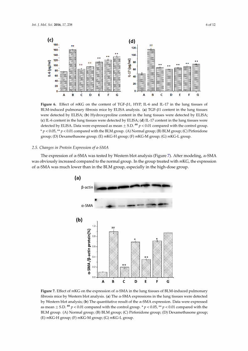
Figure 6. Effect of mKG on the content of TGF-β1, HYP, IL-6 and IL-17 in the lung tissues of BLM-induced pulmonary fibrosis mice by ELISA analysis. (a) TGF-β1 content in the lung tissues were detected by ELISA; (b) Hydroxyproline content in the lung tissues were detected by ELISA; (c) IL-6 content in the lung tissues were detected by ELISA; (d) IL-17 content in the lung tissues were detected by ELISA. Data were expressed as mean ± S.D. ## p < 0.01 compared with the control group. * p < 0.05, ** p < 0.01 compared with the BLM group. (A) Normal group; (B) BLM group; (C) Pirfenidone group; (D) Dexamethasone group; (E) mKG-H group; (F) mKG-M group; (G) mKG-L group.
2.5. Changes in Protein Expression of a-SMA
The expression of α-SMA was tested by Western blot analysis (Figure 7). After modeling, α-SMA was obviously increased compared to the normal group. In the group treated with mKG, the expression of α-SMA was much lower than in the BLM group, especially in the high-dose group.
Figure 7. Effect of mKG on the expression of α-SMA in the lung tissues of BLM-induced pulmonary fibrosis mice by Western blot analysis. (a) The α-SMA expressions in the lung tissues were detected by Western blot analysis; (b) The quantitative result of the α-SMA expression. Data were expressed as mean ± S.D. ## p < 0.01 compared with the control group. * p < 0.05, ** p < 0.01 compared with the BLM group. (A) Normal group; (B) BLM group; (C) Pirfenidone group; (D) Dexamethasone group; (E) mKG-H group; (F) mKG-M group; (G) mKG-L group.
Figure 6. Effect of mKG on the content of TGF-β1, HYP, IL-6 and IL-17 in the lung tissues ofBLM-induced pulmonary fibrosis mice by ELISA analysis. (a) TGF-β1 content in the lung tissueswere detected by ELISA; (b) Hydroxyproline content in the lung tissues were detected by ELISA;(c) IL-6 content in the lung tissues were detected by ELISA; (d) IL-17 content in the lung tissues weredetected by ELISA. Data were expressed as mean ˘ S.D. ## p < 0.01 compared with the control group.* p < 0.05, ** p < 0.01 compared with the BLM group. (A) Normal group; (B) BLM group; (C) Pirfenidonegroup; (D) Dexamethasone group; (E) mKG-H group; (F) mKG-M group; (G) mKG-L group.
2.5. Changes in Protein Expression of a-SMA
The expression of α-SMA was tested by Western blot analysis (Figure 7). After modeling, α-SMAwas obviously increased compared to the normal group. In the group treated with mKG, the expressionof α-SMA was much lower than in the BLM group, especially in the high-dose group.
Int. J. Mol. Sci. 2016, 17, 238 6 of 12
Figure 6. Effect of mKG on the content of TGF-β1, HYP, IL-6 and IL-17 in the lung tissues of BLM-induced pulmonary fibrosis mice by ELISA analysis. (a) TGF-β1 content in the lung tissues were detected by ELISA; (b) Hydroxyproline content in the lung tissues were detected by ELISA; (c) IL-6 content in the lung tissues were detected by ELISA; (d) IL-17 content in the lung tissues were detected by ELISA. Data were expressed as mean ± S.D. ## p < 0.01 compared with the control group. * p < 0.05, ** p < 0.01 compared with the BLM group. (A) Normal group; (B) BLM group; (C) Pirfenidone group; (D) Dexamethasone group; (E) mKG-H group; (F) mKG-M group; (G) mKG-L group.
2.5. Changes in Protein Expression of a-SMA
The expression of α-SMA was tested by Western blot analysis (Figure 7). After modeling, α-SMA was obviously increased compared to the normal group. In the group treated with mKG, the expression of α-SMA was much lower than in the BLM group, especially in the high-dose group.
Figure 7. Effect of mKG on the expression of α-SMA in the lung tissues of BLM-induced pulmonary fibrosis mice by Western blot analysis. (a) The α-SMA expressions in the lung tissues were detected by Western blot analysis; (b) The quantitative result of the α-SMA expression. Data were expressed as mean ± S.D. ## p < 0.01 compared with the control group. * p < 0.05, ** p < 0.01 compared with the BLM group. (A) Normal group; (B) BLM group; (C) Pirfenidone group; (D) Dexamethasone group; (E) mKG-H group; (F) mKG-M group; (G) mKG-L group.
Figure 7. Effect of mKG on the expression of α-SMA in the lung tissues of BLM-induced pulmonaryfibrosis mice by Western blot analysis. (a) The α-SMA expressions in the lung tissues were detectedby Western blot analysis; (b) The quantitative result of the α-SMA expression. Data were expressedas mean ˘ S.D. ## p < 0.01 compared with the control group. * p < 0.05, ** p < 0.01 compared with theBLM group. (A) Normal group; (B) BLM group; (C) Pirfenidone group; (D) Dexamethasone group;(E) mKG-H group; (F) mKG-M group; (G) mKG-L group.

Int. J. Mol. Sci. 2016, 17, 238 7 of 12
3. Discussion
PF is a chronic lung disease characterized by excessive accumulation of extracellular matrix(ECM) deposition to mesenchymal transition (EMT), which finally leads to the decline of lungfunction. Historically, pulmonary fibrosis was believed to result mainly from chronic inflammation [25].The persistent immune response to injury led to inflammation, tissue remodeling and repair processestaking place constantly. Because environmental pollution is more and more serious, the incidence ofPF has been on the rise in China recently, and it is a progressive and fatal pulmonary disease withoutproven drug therapies. Although the mechanisms of PF are not understood fully, transforming growthfactor-β1 (TGF-β1) is recognized as a critical factor in inducing fibrosis [26–28]. Furthermore, studieshave also confirmed that inflammation factors such as IL-6 and IL-17 contribute to the process ofPF [29,30].
The pulmonary fibrosis model induced by BLM is widely used to study the anti-fibrotic effects ofnumerous drugs. BLM causes alveolar cell damage, inflammatory response, fibroblast proliferationand subsequent collagen content deposition [31,32]. This study was undertaken to demonstratethe antifibrotic properties of mKG. In our in vivo study, the body weight of rats decreased, thehydroxyproline content of the lungs increased, and pathologic changes of pulmonary fibrosis markedlyappeared after BLM instillation. However, this situation got relieved in the mKG treatment groups.According to the results of the histological analysis, we can prove the inhibition effect of mKG oninflammation and pulmonary fibrosis. The upregulation of TGF-β1 expression is a consistent featureof most fibrotic diseases. Many studies have created a strong rationale for an antifibrotic strategyin which the principal objective of treatment is blocking TGF-β1. The inhibition effect of mKG onTGF-β1 could be shown from the results of the ELISA analysis and the expression of α-SMA wasdownregulated according to the results of the Western blot. Additionally, the results of the analysis ofIL-6, IL-17A mRNA expression and the contents of these two cytokines in the lung tissues confirmedthe anti-inflammatory effect of mKG.
PF is characterized by the remodeling of lung tissue and over-deposition of ECM [33,34].Our previous studies suggested that mKG attenuated BLM-induced PF in rats via the inhibitionof ECM. A main pathological cause of PF is the imbalance between the synthesis and degradation ofECM which increases the production of type I and type III collagen (Col-1, Col-3) [35]. Compared withthe model group, mKG significantly reduced the content of HYP, Col-1 and Col-3, especially for thehigh-dose group. The α-smooth muscle actin (α-SMA) is an important downstream protein of TGF-β1.Myofibroblasts are highly synthetic for collagen, featured by the presence of α-SMAs, which are theprimary effector cells during the progression of fibrosis [36,37]. The downregulation of the expressionof α-SMA also confirmed the fact that mKG could inhibit the production of ECM.
4. Experimental Section
4.1. Reagents and Antibodies
All raw herbs were purchased from Beijing Tongren Tang Pharmaceutical Co., Ltd. (Beijing, China)and identified as the roots of Angelica sinensis (Oliv.) Diels., the roots of Sophora flavescens Ait., therhizome of Glycyrrhiza uralensis Fisch. by Professor Yulin Lin of the Institute of Medicinal PlantDevelopment (IMPLAD), Chinese Academy of Medical Sciences and Peking Union Medical College.The voucher specimen is deposited in our laboratory of IMPLAD.
Prestained protein marker was purchased from New England Biolabs (Beijing, China).Anti-albumin (Alb) (ab8940) sheep antibody, anti-a-smooth muscle actin (a-SMA) (ab5694) rabbitantibody were purchased from Abcam (Cambridge, MA, USA) and prepared in 1:3000, and 1:2000dilution, respectively. The BCA Protein Assay Kit and the SuperSignal West Pico ChemiluminescentSubstrate (ECL) were obtained from Pierce Chemical Company (Rockford, IL, USA). Trizol wasobtained from Invitrogen (Carlsbad, CA, USA). The First-Strand cDNA Synthesis Kit (K1622) was

Int. J. Mol. Sci. 2016, 17, 238 8 of 12
purchased from Fermentas (St Leon-Roth, Germany). ELISA kits were obtained from Nanjing JianchengBioengineering Institute (Nanjing, China).
4.2. Animals
A total of 70 BALB/c healthy male mice, weighing 20–22 g, were obtained from the Academyof Military Medical Experimental Animal Center. The mice were kept in community cages with12 h periods of light and dark cycles and maintained on standard rodent chow with access to waterad libitum. All animal care and experimentation was approved by the principles and guidelines of theNational Institutes of Health Guide for the Care and Use of Laboratory Animals.
4.3. Extract of mKS Preparation
Modified Kushen Gancao Formula (mKS) consists of Keushen Gancao Tang and Angelica oil.The mKS extract was prepared in our laboratory. Briefly, Gancao (the rhizome of Glycyrrhiza uralensis)60 g and Kushen (the roots of Sophora flavescens) 60 g were soaked together in 1200 mL of 60% ethanolfor 1 h at room temperature and thereafter refluxed for 1 h. The filtrate was collected and the residueswere then refluxed twice in 1200 mL of 60% ethanol for 1 h. The three filtrates were combined andconcentrated under vacuum to give the extract of Keushen Gancao Tang (yield 28.5%). Dang-gui (theroots of Angelica sinensis) 160 g was subjected to extraction separately in 1600 mL of water by thesteam generator for 8 h at room temperature to extract the Angelica oil. The oil is collected, dried withanhydrous sodium sulphate and stored at 4 ˝C until use (yield 0.4%). Before the experiments, Angelicaoil was added to the dry powder of Kushen Gancao Tang. Quality control of the final mKG extractwas performed using UPLC-MS/MS and GC-MS (Supplementary Materials). Samples of mKG areavailable from the authors.
4.4. Groups of Animals and Treatments
Seventy BALB/c mice were randomly divided into seven groups (n = 10 per group): Normalcontrol group, BLM-treated group, pirfenidone group, dexamethasone group and three mKG groups(mKG-L, mKG-M, mKG-H) at the dose of 0.41, 1.64 and 6.56 g/kg/day, respectively. Bleomycin wasdissolved by sterile normal saline, and then mice in groups except in the normal control group wereintratracheally injected with prepared bleomycin at a dose of 5 mg/kg. The normal control group wasgiven equal amounts of normal saline. From the second day to the 21st day after modeling, mice in mKGgroups or pirfenidone group were intragastrically administrated with prepared different dose mKGsolution or prednisone (50 mg/kg), while mice in dexamethasone group were intraperitoneally injectedwith dexamethasone from the second day to the fourth day. Mice in normal control and BLM-treatedgroup received equal amounts of normal saline. The body weight of mice was monitored every day.On the 21st day, the blood of the mice was drawn from the abdominal aorta under pentobarbitalanesthesia and the lungs were removed. The left lungs were used for histological examination, and theright lungs were frozen in liquid nitrogen.
4.5. Histological Examination
The samples of left lung were fixed in 10% neutral formalin, embedded in paraffin and sectioned.The sections were stained with hematoxylin and eosin (HE) and Massons’s trichrome according toconventional methods. The slides were evaluated under a light microscope.
4.6. Measurement of Content of Hydroxyproline,TGF-β1, IL-6 and IL-17 in Lung Tissues
Hydroxyproline (HYP) content of lung tissue was measured by using hydroxyproline kits (NanjingJiancheng Biochemical Institute, Nanjing, China). TGF-β1 in lung tissue was tested by ELISA kit(Nanjing Jiancheng Biochemical Institute, Nanjing, China). IL-6 and IL-17 were tested by ELISA

Int. J. Mol. Sci. 2016, 17, 238 9 of 12
kit (Nanjing Jiancheng Biochemical Institute, Nanjing, China). It was performed according to theinstruction of the manufacturer.
4.7. Immunohistochemistry Analysis
Paraffin-embedded tissue sections were rehydrated in xylene and graded ethanol solutions.The slides were exposed to methanol-hydrogen peroxide for 10 min [19], washed in phosphate bufferedsaline (PBS), and then antigen retrieval was made. After blocking with serum for 20 min, sectionswere then immunostained with type 1 collagen (Col-1) and type 3 collagen (Col-3) primary antibodiesand incubated overnight at 4 ˝C. After washing the slides thrice with PBS, the sections were thenincubated with secondary antibody for 20 min at room temperature. Sections were then washed withPBS and incubated with streptavidin-biotin-peroxidase complex for 10 min. After washing with PBS,diaminobenzidine was added as a visualizing agent. Nuclei were counterstained with hematoxylin.Positive staining for Col-1 and Col-3 was brown. Expression of Col-1 and Col-3 was compared betweengroups by calculating the ratio of positive staining area. A computer-aided morphometric analysis wasused to quantitatively determine the positive staining area. Five fields of view were selected randomlyfrom every slice, and the even integration optic density (IOD) value of the positive staining area wascorrected by the blank in the same visual field.
4.8. Analysis of IL-6 and IL-17A mRNA Expression
Total RNA of lung tissue was isolated using Trizol reagent (Invitrogen, Carlsbad, CA, USA)according to manufacturer’s instructions. Two micrograms of total RNA from each sample werereverse-transcribed to cDNA which was used for PCR. GAPDH: sense GGGTGGTCCAGGGTTTCTTACT,antisense AGGTTGTCTCCTGCGACTTCA; IL-6: CAAAGCCAGAGTCCTTCAGAG, antisenseGCCACTCCTTCTGTGACTCC; IL-17A: sense GCAAGAGATCCTGGTCCTGA, antisense AGCATCTTCTCGACCCTGAA. The optical density was recorded to evaluate the ratio of IL-6/GAPDH orIL-17A/GAPDH to determine the mRNA expression.
4.9. Western Blotting Analysis
Lung tissue samples were lysed by RIPA buffer containing 50 mM Tris-HCl (pH 7.2), 150 mMNaCl, 1% NP-40, 0.1% SDS, 1 mM EDTA, and 1 mM PMSF and homogenized in ice water for 30 min.After centrifugation (12,000 r/min, 10 min at 4 ˝C), the supernatant was collected, packed, and stored in´80 ˝C. Just before using, the loading buffer was mixed into the supernatant at a ratio of 1:4 and cookedin boiling water for 10 min. Then, the total protein concentration was determined by Bio-Rad Dcprotein Assay Reagent (San Diego, CA, USA). Proteins in the supernatant were separated by SDS-PAGEon a 12% SDS polyacrylamide gel and then transferred to polyvinylidene fluoride (PVDF) membranes.The blotted membranes were blocked with 5% non-fat dairy milk (w/v). Blotted membranes werewashed by 0.1% Tween-TBS (TBST) three times (10 min per time) after 2 h. Then blotted membraneswere incubated at 4 ˝C overnight with primary anti- α -SMA antibodies (1:2000, Abcam, Cambridge,UK) and with secondary antibodies at room temperature for 1 h. After being washed with TBST threetimes (10 min per time), the blots were visualized with ECL reagent (Thermo Scientific, Rockford,IL, USA).
4.10. Statistical Analysis
The data are expressed as mean ˘ SD. Statistical testing was performed with SPSS softwareversion 12.0 (SPSS Inc., Chicago, IL, USA). Comparison between groups was performed with testof homogeneity of variances and one-way analysis of variance (ANOVA), and post hoc analysis wasperformed using Bonferroni or Dunnett’s multiple comparison tests. Values of p < 0.05 and p < 0.01were considered to be statistically significant.

Int. J. Mol. Sci. 2016, 17, 238 10 of 12
5. Conclusions
This study demonstrated the anti-inflammatory properties of mKG and it also attenuatedBLM-induced pulmonary fibrosis. Additionally, mKG showed some effects of regulating the ECMdeposition and inhibiting the PF progression, and it may become an important method for preventingand treating PF.
Supplementary Materials: Supplementary materials can be found at http://www.mdpi.com/1422-0067/17/2/238/s1.
Acknowledgments: This work has been financially supported by National S&T Major Special Project of China onMajor New Drug Innovation (2012ZX09103201-29), Beijing Natural Science Foundation (No. 7132136), and theChina Postdoctoral Science Foundation (2012M520204).
Author Contributions: Zhong-Mei Zou and Hong Chen conceived and designed the research; Peng Guo, Bo Cao,Tan Li and Li-Fu Yao performed the experiments; Ying Gao and Li-Fu Yao analyzed the data; Yang Zhao,Li-Man Wei and Bo Cao contributed reagents/materials/analysis tools; Li-Man Wei and Meng Yu performedquality control of Mkg; Ying Gao and Li-Fu Yao wrote the first draft of the manuscript and other authorsparticipated in revision.
Conflicts of Interest: The authors declare no conflict of interest.
Abbreviations
PF Pulmonary fibrosisBLM BleomycinmKG Modified Gancao Kushen FormulaTGF-β Transforming growth factor-βHYP HydroxyprolineROS Reactive oxygen speciesIL-17 Interleukin-17IL-6 Interleukin-6ECM Extracellular matrixα-SMA α smooth muscle actinSFE Supercritical Fluid ExtractionmKG-L Modified Gancao Kushen formula-Low dosemKG-M Modified Gancao Kushen formula-Middle dosemKG-H Modified Gancao Kushen formula-High doseHE Hematoxylin and eosinPBS Phosphate buffered salineCol-1 Type 1 collagenCol-3 Type 3 collagenIOD Integration optic densityANOVA One-way analysis of varianceEMT Mesenchymal transition
References
1. Noble, P.W.; Barkauskas, C.E.; Jiang, D. Pulmonary fibrosis: Patterns and perpetrators. J. Clin. Investig. 2012,122, 2756–2762. [CrossRef] [PubMed]
2. Wynn, T.A. Integrating mechanisms of pulmonary fibrosis. J. Exp. Med. 2011, 208, 1339–1350. [CrossRef][PubMed]
3. Wan, X.Y.; Luo, M.; Li, X.D.; He, P. Hepatoprotective and anti-hepatocarcinogenic effects of glycyrrhizin andmatrine. Chem. Biol. Interact. 2009, 181, 15–19. [CrossRef] [PubMed]

Int. J. Mol. Sci. 2016, 17, 238 11 of 12
4. Huang, L.; Qi, L.; Chen, Z.; Li, Y.; Wen, Z. Optimization of a compound prescription for treating liver fibrosis(in Chinese). Nan fang yi ke da xue xue bao = J. South. Med. Univ. 2012, 32, 106–108.
5. Wen, M.C.; Huang, C.K.; Srivastava, K.D.; Zhang, T.F.; Schofield, B. Kushen (Sophora flavescens Ait), a SingleChinese Herb, Abrogates Airway Hyperreactivity in a Murine Model of Asthma. J. Allergy Clin. Immunol.2004, 113, S218–S221. [CrossRef]
6. Liu, L.; Lu, W.; Ma, Z.; Li, Z. Oxymatrine attenuates bleomycin-induced pulmonary fibrosis in mice via theinhibition of inducible nitric oxide synthase expression and the TGF-beta/Smad signaling pathway. Int. J.Mol. Med. 2012, 29, 815–822. [PubMed]
7. Chen, X.; Sun, R.; Hu, J.; Mo, Z.; Yang, Z.; Liao, D.; Zhong, N. Attenuation of bleomycin-induced lungfibrosis by oxymatrine is associated with regulation of fibroblast proliferation and collagen production inprimary culture. Basic Clin. Pharmacol. Toxicol. 2008, 103, 278–286. [CrossRef] [PubMed]
8. Kim, J.Y.; Park, S.J.; Yun, K.J.; Cho, Y.W.; Park, H.J.; Lee, K.T. Isoliquiritigenin isolated from the roots ofGlycyrrhiza uralensis inhibits LPS-induced iNOS and COX-2 expression via the attenuation of NF-kappaBin RAW 264.7 macrophages. Eur. J. Pharmacol. 2008, 584, 175–184. [CrossRef] [PubMed]
9. Asl, M.N.; Hosseinzadeh, H. Review of pharmacological effects of Glycyrrhiza sp. and its bioactivecompounds. Phytother. Res. 2008, 22, 709–724. [CrossRef] [PubMed]
10. Kim, S.N.; Kim, M.H.; Min, Y.K.; Kim, S.H. Licochalcone A inhibits the formation and bone resorptiveactivity of osteoclasts. Cell Biol. Int. 2008, 32, 1064–1072. [CrossRef] [PubMed]
11. Lee, C.K.; Son, S.H.; Park, K.K.; Park, J.H.; Lim, S.S.; Kim, S.H.; Chung, W.Y. Licochalcone A inhibits thegrowth of colon carcinoma and attenuates cisplatin-induced toxicity without a loss of chemotherapeuticefficacy in mice. Basic Clin. Pharmacol. Toxicol. 2008, 103, 48–54. [CrossRef] [PubMed]
12. Fu, Y.; Hsieh, T.C.; Guo, J.; Kunicki, J.; Lee, M.Y.; Darzynkiewicz, Z.; Wu, J.M. Licochalcone-A,a novel flavonoid isolated from licorice root (Glycyrrhiza glabra), causes G2 and late-G1 arrests inandrogen-independent PC-3 prostate cancer cells. Biochem. Biophys. Res. Commun. 2004, 322, 263–270.[CrossRef] [PubMed]
13. Liang, B.; Guo, X.L.; Jin, J.; Ma, Y.C.; Feng, Z.Q. Glycyrrhizic acid inhibits apoptosis and fibrosis incarbon-tetrachloride-induced rat liver injury. World J. Gastroenterol. 2015, 21, 5271–5280. [CrossRef] [PubMed]
14. Gao, L.; Tang, H.; He, H.; Liu, J.; Mao, J.; Ji, H.; Lin, H.; Wu, T. Glycyrrhizic acid alleviates bleomycin-inducedpulmonary fibrosis in rats. Front. Pharmacol. 2015, 6, 215. [CrossRef] [PubMed]
15. Wu, Y.C.; Hsieh, C.L. Pharmacological effects of Radix Angelica Sinensis (Danggui) on cerebral infarction.Chin. Med. 2011, 6, 32. [CrossRef] [PubMed]
16. Wilasrusmee, C.; Kittur, S.; Siddiqui, J.; Bruch, D.; Wilasrusmee, S.; Kittur, D.S. In vitro immunomodulatoryeffects of ten commonly used herbs on murine lymphocytes. J. Altern. Complement. Med. 2002, 8, 467–475.[CrossRef] [PubMed]
17. Yuan, X.; Sun, Y.; Miao, N.; Sun, S.; Wang, Y.; Hu, Z.; Yuan, J.; Xu, M.; Liu, Z. The synergistic anti-inflammatoryeffect of the combination of sodium ferulate and oxymatrine and its modulation on inflammation-associatedmediators in RAW 264.7 cells. Ethnopharmacology 2011, 137, 1477–1485. [CrossRef] [PubMed]
18. Wilson, M.S.; Madala, S.K.; Ramalingam, T.R.; Gochuico, B.R.; Rosas, I.O.; Cheever, A.W.; Wynn, T.A.Bleomycin and IL-1beta-mediated pulmonary fibrosis is IL-17A dependent. J. Exp. Med. 2010, 207, 535–552.[CrossRef] [PubMed]
19. Isailovic, N.; Daigo, K.; Mantovani, A.; Selmi, C. Interleukin-17 and innate immunity in infections andchronic inflammation. J. Autoimmun. 2015, 60, 1–11. [CrossRef] [PubMed]
20. Pei, F.; Han, Y.; Zhang, X.; Yan, C.; Huang, M.; Deng, J.; Kang, J. Association analysis of the IL-17F His161Argpolymorphism in myocardial infarction. Coron. Artery Dis. 2009, 20, 513–517. [CrossRef] [PubMed]
21. Catana, C.S.; Berindan Neagoe, I.; Cozma, V.; Magdas, C.; Tabaran, F.; Dumitrascu, D.L. Contribution ofthe IL-17/IL-23 axis to the pathogenesis of inflammatory bowel disease. World J. Gastroenterol. 2015, 21,5823–5830. [PubMed]
22. Gao, Y.; Lu, J.; Zhang, Y.; Chen, Y.; Gu, Z.; Jiang, X. Baicalein attenuates bleomycin-induced pulmonaryfibrosis in rats through inhibition of miR-21. Pulm. Pharmacol. Ther. 2013, 26, 649–654. [CrossRef] [PubMed]
23. Ji, Y.; Wang, T.; Wei, Z.F.; Lu, G.X.; Jiang, S.D.; Xia, Y.F.; Dai, Y. Paeoniflorin, the main active constituentof Paeonia lactiflora roots, attenuates bleomycin-induced pulmonary fibrosis in mice by suppressing thesynthesis of type I collagen. J. Ethnopharmacol. 2013, 149, 825–832. [CrossRef] [PubMed]

Int. J. Mol. Sci. 2016, 17, 238 12 of 12
24. Pan, Y.; Fu, H.; Kong, Q.; Xiao, Y.; Shou, Q.; Chen, H.; Ke, Y.; Chen, M. Prevention of pulmonary fibrosis withsalvianolic acid a by inducing fibroblast cell cycle arrest and promoting apoptosis. J. Ethnopharmacol. 2014,155, 1589–1596. [CrossRef] [PubMed]
25. Weber, C.; Hristov, M. Atherogenesis and inflammation. From cellular mediators to regulatory mechanismsof inflammation in atherosclerosis. Hamostaseologie 2015, 35, 99–101. [PubMed]
26. Chung, E.J.; Hudak, K.; Horton, J.A.; White, A.; Scroggins, B.T.; Vaswani, S.; Citrin, D. Transforming growthfactor alpha is a critical mediator of radiation lung injury. Radiat. Res. 2014, 182, 350–362. [CrossRef][PubMed]
27. Li, Y.; Foster, W.; Deasy, B.M.; Chan, Y.; Prisk, V.; Tang, Y.; Cummins, J.; Huard, J. Transforming growthfactor-beta1 induces the differentiation of myogenic cells into fibrotic cells in injured skeletal muscle: A keyevent in muscle fibrogenesis. Am. J. Pathol. 2004, 164, 1007–1019. [CrossRef]
28. Burger, A.; Loffler, H.; Bamberg, M.; Rodemann, H.P. Molecular and cellular basis of radiation fibrosis. Int. J.Radiat. Biol. 1998, 73, 401–408. [CrossRef] [PubMed]
29. Foran, E.; Garrity-Park, M.M.; Mureau, C.; Newell, J.; Smyrk, T.C.; Limburg, P.J.; Egan, L.J. Upregulation ofDNA methyltransferase-mediated gene silencing, anchorage-independent growth, and migration of coloncancer cells by interleukin-6. Mol. Cancer Res. 2010, 8, 471–481. [CrossRef] [PubMed]
30. O’Hagan, H.M.; Wang, W.; Sen, S.; Destefano Shields, C.; Lee, S.S.; Zhang, Y.W.; Clements, E.G.; Cai, Y.;van Neste, L.; Easwaran, H.; et al. Oxidative damage targets complexes containing DNA methyltransferases,SIRT1, and polycomb members to promoter CpG Islands. Cancer Cell 2011, 20, 606–619. [CrossRef] [PubMed]
31. Izbicki, G.; Segel, M.J.; Christensen, T.G.; Conner, M.W.; Breuer, R. Time course of bleomycin-induced lungfibrosis. Int. J. Exp. Pathol. 2002, 83, 111–119. [CrossRef] [PubMed]
32. Serrano-Mollar, A.; Closa, D.; Prats, N.; Blesa, S.; Martinez-Losa, M.; Cortijo, J.; Estrela, J.M.; Morcillo, E.J.;Bulbena, O. In vivo antioxidant treatment protects against bleomycin-induced lung damage in rats.Br. J. Pharmacol. 2003, 138, 1037–1048. [CrossRef] [PubMed]
33. Royce, S.G.; Moodley, Y.; Samuel, C.S. Novel therapeutic strategies for lung disorders associated with airwayremodelling and fibrosis. Pharmacol. Ther. 2014, 141, 250–260. [CrossRef] [PubMed]
34. Loomis-King, H.; Flaherty, K.R.; Moore, B.B. Pathogenesis, current treatments and future directions foridiopathic pulmonary fibrosis. Curr. Opin. Pharmacol. 2013, 13, 377–385. [CrossRef] [PubMed]
35. Parker, M.W.; Rossi, D.; Peterson, M.; Smith, K.; Sikstrom, K.; White, E.S.; Connett, J.E.; Henke, C.A.;Larsson, O.; Bitterman, P.B. Fibrotic extracellular matrix activates a profibrotic positive feedback loop.J. Clin. Investig. 2014, 124, 1622–1635. [CrossRef] [PubMed]
36. Saw, V.P.; Schmidt, E.; Offiah, I.; Galatowicz, G.; Zillikens, D.; Dart, J.K.; Calder, V.L.; Daniels, J.T. Profibroticphenotype of conjunctival fibroblasts from mucous membrane pemphigoid. Am. J. Pathol. 2011, 178, 187–197.[CrossRef] [PubMed]
37. Wynn, T.A.; Ramalingam, T.R. Mechanisms of fibrosis: Therapeutic translation for fibrotic disease. Nat. Med.2012, 18, 1028–1040. [CrossRef] [PubMed]
© 2016 by the authors; licensee MDPI, Basel, Switzerland. This article is an open accessarticle distributed under the terms and conditions of the Creative Commons by Attribution(CC-BY) license (http://creativecommons.org/licenses/by/4.0/).