the circulatory system booklet
DESCRIPTION
qTRANSCRIPT

the circulatory
system
Name: _________________________ Class: __________________
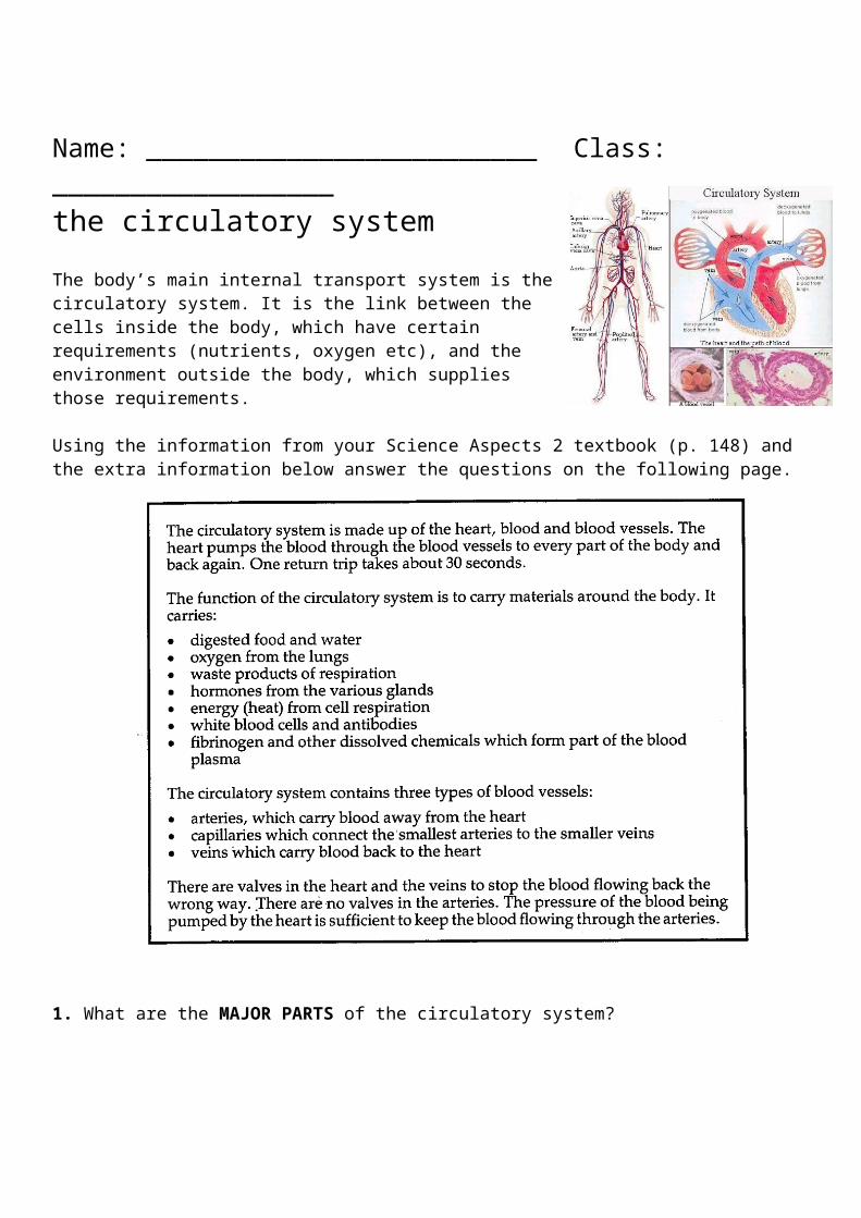
the circulatory system
The body’s main internal transport system is the circulatory system. It is the link between the cells inside the body, which have certain requirements (nutrients, oxygen etc), and the environment outside the body, which supplies those requirements.
Using the information from your Science Aspects 2 textbook (p. 148) and the extra information below answer the questions on the following page.
1. What are the MAJOR PARTS of the circulatory system?
2. What are the MAJOR FUNCTIONS of the circulatory system?

The Heart
The heart is the pump that pushes the blood around tho body. It is about the size of a clenched fist and sits in the centre of your chest behind your rib cage and sternum (the bone that runs down the middle joining the two sides of your rib cage).
Follow the link below and watch the short video called From the Heart.http://www.teachersdomain.org/resource/tdc02.sci.life.stru.circulator/
Go to the Portal and open the PowerPoint called The Heart and complete the activities below all about …. the HEART!
EXTERIOR VIEW of the heart
The outside of the heart is surrounded by a membranous sac. What is this sac called and what is its function?
Label the parts of the heart that you can see just by looking at the outside of it.
OR
The heart you see drawn on the average Valentine is only a rough representation of the actual structure of the heart. Your heart is actually shaped more like an upside-down pear.
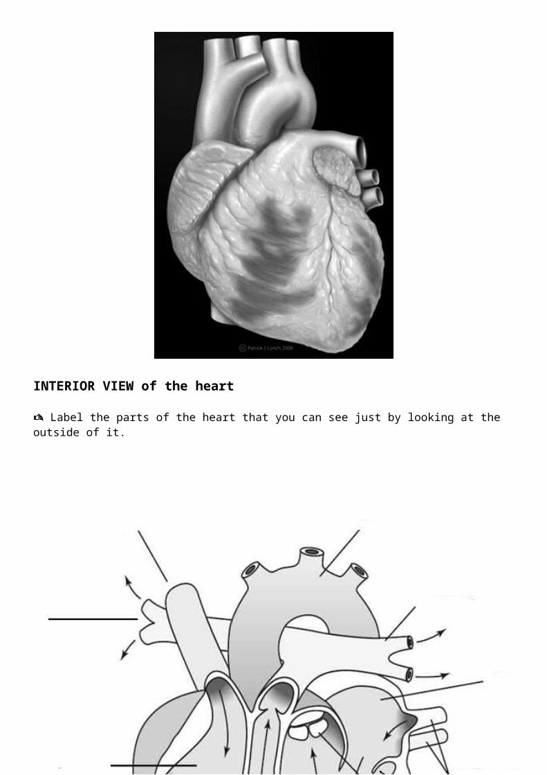
INTERIOR VIEW of the heart
Label the parts of the heart that you can see just by looking at the outside of it.
Your heart is about the size of your clenched fist.
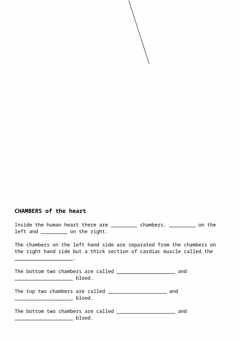
CHAMBERS of the heart
Inside the human heart there are _________ chambers. _________ on the left and _________ on the right.
The chambers on the left hand side are separated from the chambers on the right hand side but a thick section of cardiac muscle called the ____________________.
The bottom two chambers are called ____________________ and ____________________ blood.
The top two chambers are called ____________________ and ____________________ blood.
The bottom two chambers are called ____________________ and ____________________ blood.
Label the chambers of the heart on the diagram below.

VESSELS in the heart
The heart is surrounded by a number of vessels that carry blood either: TO the heart, or AWAY from the heart
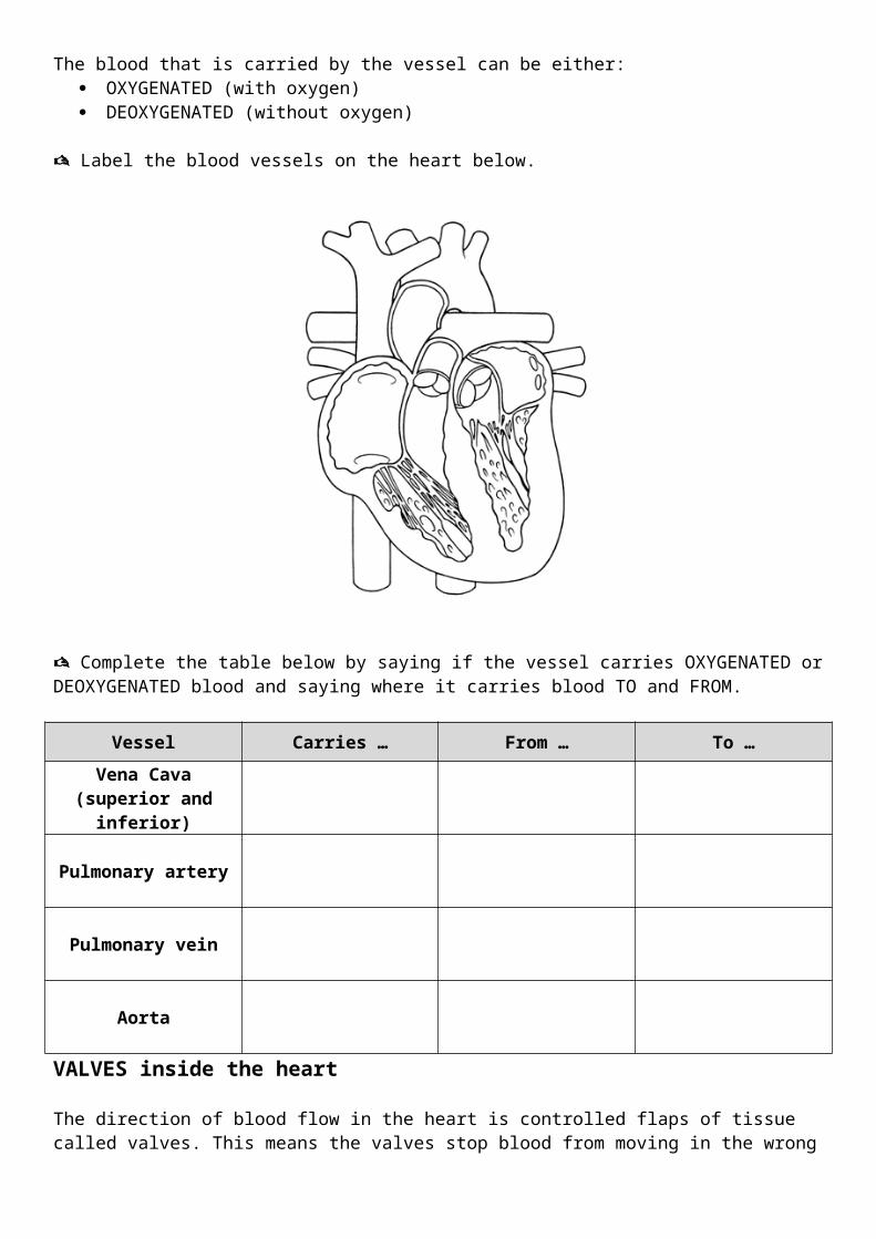
The blood that is carried by the vessel can be either: OXYGENATED (with oxygen) DEOXYGENATED (without oxygen)
Label the blood vessels on the heart below.
Complete the table below by saying if the vessel carries OXYGENATED or DEOXYGENATED blood and saying where it carries blood TO and FROM.
Vessel Carries … From … To …
Vena Cava(superior and inferior)
Pulmonary artery
Pulmonary vein
Aorta
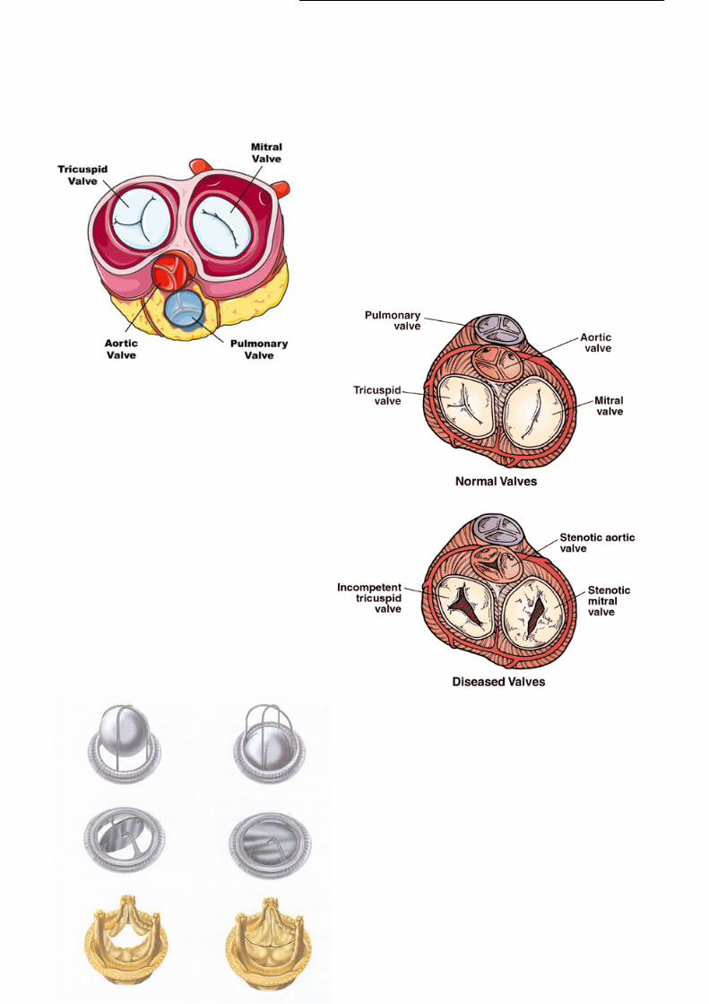
VALVES inside the heart
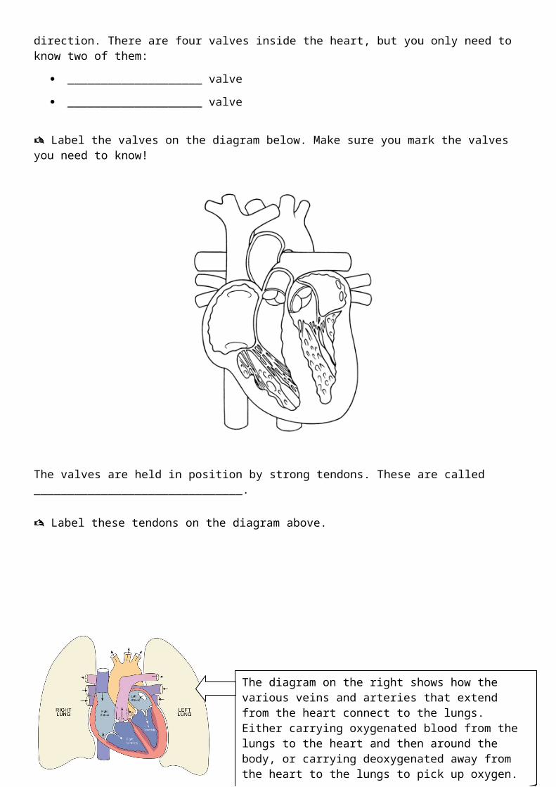
The direction of blood flow in the heart is controlled flaps of tissue called valves. This means the valves stop blood from moving in the wrong direction. There are four valves inside the heart, but you only need to know two of them:
____________________ valve
____________________ valve
Label the valves on the diagram below. Make sure you mark the valves you need to know!
The valves are held in position by strong tendons. These are called _______________________________.
Label these tendons on the diagram above.
The diagram on the right shows how the various veins and arteries that extend from the heart connect to the lungs. Either carrying oxygenated blood from the lungs to the heart and then around the body, or carrying deoxygenated away from the heart to the lungs to pick up oxygen.
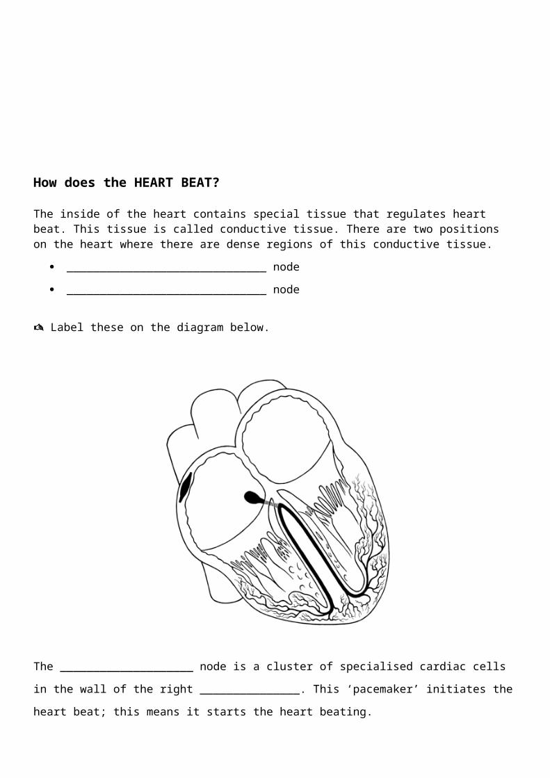
How does the HEART BEAT?
The inside of the heart contains special tissue that regulates heart beat. This tissue is called conductive tissue. There are two positions on the heart where there are dense regions of this conductive tissue.
______________________________ node
______________________________ node
Label these on the diagram below.
The ____________________ node is a cluster of specialised cardiac cells in the wall of the right
_______________. This ‘pacemaker’ initiates the heart beat; this means it starts the heart beating.
The ____________________ node is another cluster of specialised cardiac cells. It is found at the base of
the right atrium, just above the right _______________. It is the secondary pacemaker and regulates the
beating o the _______________.
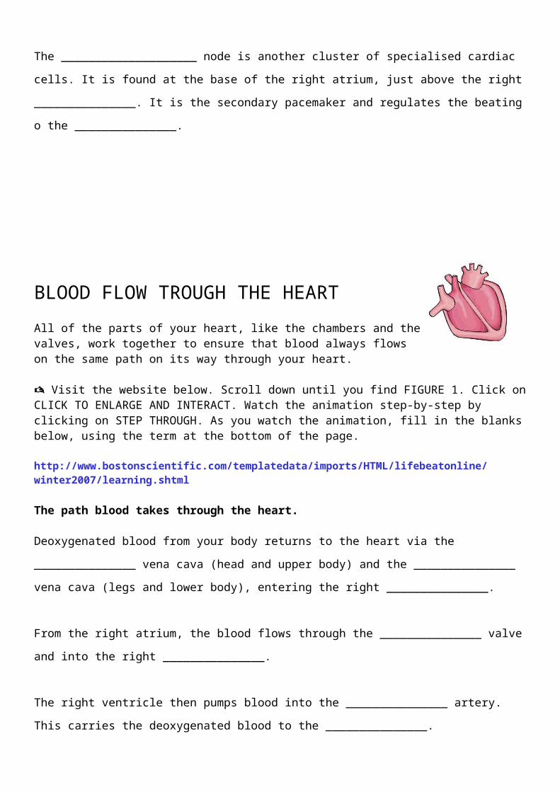
BLOOD FLOW TROUGH THE HEART
All of the parts of your heart, like the chambers and the valves, work together to ensure that blood always flows on the same path on its way through your heart.
Visit the website below. Scroll down until you find FIGURE 1. Click on CLICK TO ENLARGE AND INTERACT. Watch the animation step-by-step by clicking on STEP THROUGH. As you watch the animation, fill in the blanks below, using the term at the bottom of the page.
http://www.bostonscientific.com/templatedata/imports/HTML/lifebeatonline/winter2007/learning.shtml
The path blood takes through the heart.
Deoxygenated blood from your body returns to the heart via the _______________ vena cava (head and
upper body) and the _______________ vena cava (legs and lower body), entering the right
_______________.
From the right atrium, the blood flows through the _______________ valve and into the right
_______________.
The right ventricle then pumps blood into the _______________ artery. This carries the deoxygenated
blood to the _______________.
At the lungs, the blood’s _______________ supply is replenished. The blood is now referred to as
_______________.
Oxygenated blood flows back to the heart via the _______________ veins, entering the left
_______________.
From the left atrium, the blood flows through the _______________ valve and into the left
_______________.
The left ventricle then pumps blood into the _______________. This carries the oxygenated blood to the
rest of the _______________.
inferior lungs ventricle atrium ventricle tricuspid
oxygen pulmonary bicuspid atrium
aorta body Superior pulmonary oxygenated
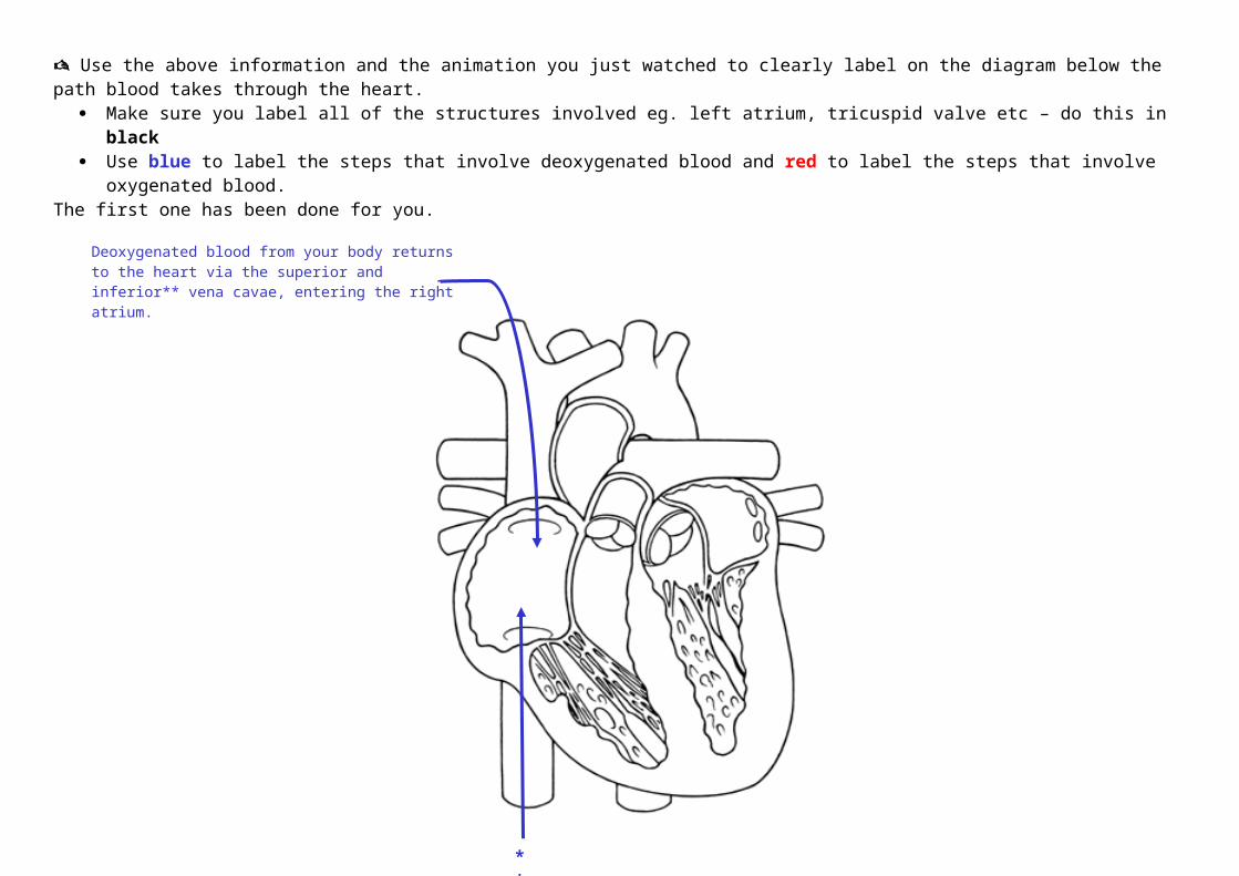
Use the above information and the animation you just watched to clearly label on the diagram below the path blood takes through the heart. Make sure you label all of the structures involved eg. left atrium, tricuspid valve etc – do this in black Use blue to label the steps that involve deoxygenated blood and red to label the steps that involve oxygenated blood.
The first one has been done for you.
Deoxygenated blood from your body returns to the heart via the superior and inferior** vena cavae, entering the right atrium.
**

Follow-up Questions
Answer the following questions about the flow of blood through the heart, using the website below to help you – you will need to watch the animation and read the text information.http://www.nhlbi.nih.gov/health/dci/Diseases/hhw/hhw_pumping.html
1. What is the function of the four valves inside the heart? What are their names?
2. Fill in the blanks.
The right side of the heart pumps blood from the _______________ to the ______________, through the
______________ artery.
The left side of the heart pumps blood from the ______________ to the ______________, through the
______________.
3. Blood that is low in oxygen is called: ___________________
Blood that is high in oxygen is called: ___________________
4. What causes the right atrium to contract? What happens when the right atrium does contract? What is this process called?
5. What causes the tricuspid valve to close? Why does this valve need to close?
6. What happens during ventricular systole?
7. As deoxygenated blood enters the right atrium causing the right atrium to contract, what is happening in the left side of the heart?
8. What causes the bicuspid (mitral) valve to close? Why does this valve need to close?
9. What happens during ventricular systole (ventricle contracts) in the left side of the heart?
10. After the blood leaves the ventricles (both right and left) what is happening in the two atria?
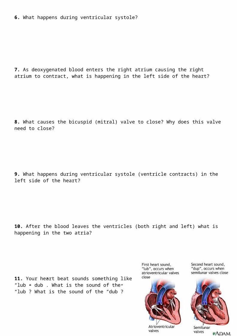
11. Your heart beat sounds something like “lub – dub”. What is the sound of the “lub”? What is the sound of the “dub”?
12. Each heartbeat has two basic stages: diastole and systole. What happens during diastole? What happens during systole?

Heart Dissection
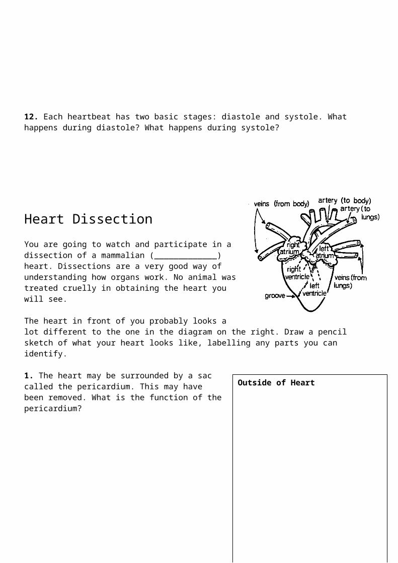
You are going to watch and participate in a dissection of a mammalian (_____________) heart. Dissections are a very good way of understanding how organs work. No animal was treated cruelly in obtaining the heart you will see.
The heart in front of you probably looks a lot different to the one in the diagram on the right. Draw a pencil sketch of what your heart looks like, labelling any parts you can identify.
1. The heart may be surrounded by a sac called the pericardium. This may have been removed. What is the function of the pericardium?__________________________________________________
_______________________________________________________________________________________
_____________
2. How do you know which blood vessel is the aorta?__________________________________________________
_______________________________________________________________________________________
_____________
3. What is the function of the blood vessels that run over the surface of the heart?_______________________________________________________________________________________
_____________
__________________________________________________
4. What is the white material around the top of the ventricles?
Outside of Heart
_______________________________________________________________________________________
Your teacher, with assistance from students, will now cut the heart in half, exposing the inside of the heart. Draw what you see on the following page.
5. How many chambers can you see? __________________. What are they called? ___________________________________
___________________________________
___________________________________
___________________________________
Label these chambers on your diagram.
Inside of Heart
If you get confused which is the right side and left side of the heart, label it clearly on your diagram above.6. There is a notable difference in the thickness between the walls of the atria and the ventricles. Which are thicker? Why is there this difference?_______________________________________________________________________________________
_______________________________________________________________________________________
_______________________________________________________________________________________
_______________________________________________________________________________________
7. There is also a difference in the thickness between the walls of the two ventricles. Why is this so?_______________________________________________________________________________________
_______________________________________________________________________________________
_______________________________________________________________________________________
_______________________________________________________________________________________
8. Why do some blood vessels (the arteries) have thick walls and some (the veins) have thin walls?_______________________________________________________________________________________
_______________________________________________________________________________________
_______________________________________________________________________________________
_______________________________________________________________________________________
9. How do you think the flaps of tissue (the valves) between the atria and ventricles work? Label the valves on your diagram above. _______________________________________________________________________________________
_______________________________________________________________________________________
_______________________________________________________________________________________
_______________________________________________________________________________________
10. What do you think is the function of the cords that attach the valve edges to the ventricle walls?
_______________________________________________________________________________________
_______________________________________________________________________________________
_______________________________________________________________________________________
_______________________________________________________________________________________
11. Which way do you think the blood flows through the heart? Show the movement of blood on the diagram below. Show oxygenated blood in red and deoxygenated blood in blue.
12. Why does the heart pump twice during each beat? What sound does this make?_______________________________________________________________________________________
_______________________________________________________________________________________
_______________________________________________________________________________________
_______________________________________________________________________________________
_______________________________________________________________________________________
_______________________________________________________________________________________

Heart Transplant
The first human heart transplant was performed in South Africa in 1967, and Australia’s first heart transplant took place in Sydney in 1968. On average 70 people receive a heart transplant per year in Australia. Heart transplant is major surgery and is the last resort for people suffering from chronic heart diseases and illnesses. Generally the heart transplant should significantly improve the patient’s life expectancy and quality of life. Approximate survival values for heart transplantation are 80%, 70% and 55% at 1, 5 and 10 years respectively.
To be able to perform heart transplants you need to be a fully qualified cardiologist and surgeon, this means YEARS or University and on-the-job training. However, you can step into the shows of a heart transplant surgeon by visiting the website below
and perform a simulated heart transplant. http://www.pbs.org/wgbh/nova/eheart/transplantwave.html
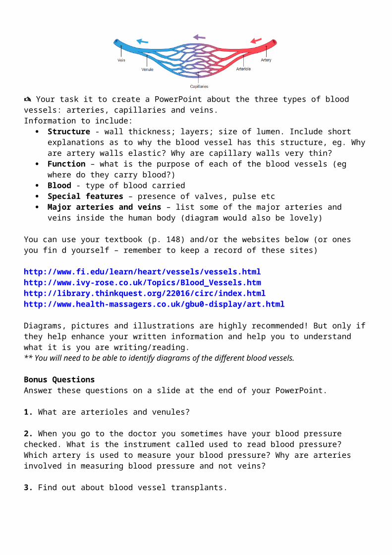
Blood Vessels
The blood is carried in a series of continuous tubes of different sizes celled blood vessels. They carry the blood to each organ in the body and take it back to the heart. In each organ blood vessels allow chemicals to move into and out of cells.
The direction of blood flow is kept up by the pumping of the heart. When the powerful muscles of your heart contract, they force blood out into tubes called arteries. The arteries branch into tiny little tubes called capillaries. The capillaries join together to form veins. The veins carry blood back to the heart.
Your task it to create a PowerPoint about the three types of blood vessels: arteries, capillaries and veins. Information to include:
Structure - wall thickness; layers; size of lumen. Include short explanations as to why the blood vessel has this structure, eg. Why are artery walls elastic? Why are capillary walls very thin?
Function – what is the purpose of each of the blood vessels (eg where do they carry blood?) Blood - type of blood carried Special features – presence of valves, pulse etc Major arteries and veins – list some of the major arteries and veins inside the human body
(diagram would also be lovely)
You can use your textbook (p. 148) and/or the websites below (or ones you fin d yourself – remember to keep a record of these sites)
http://www.fi.edu/learn/heart/vessels/vessels.htmlhttp://www.ivy-rose.co.uk/Topics/Blood_Vessels.htmhttp://library.thinkquest.org/22016/circ/index.htmlhttp://www.health-massagers.co.uk/gbu0-display/art.html

Diagrams, pictures and illustrations are highly recommended! But only if they help enhance your written information and help you to understand what it is you are writing/reading. ** You will need to be able to identify diagrams of the different blood vessels.
Bonus QuestionsAnswer these questions on a slide at the end of your PowerPoint.
1. What are arterioles and venules?
2. When you go to the doctor you sometimes have your blood pressure checked. What is the instrument called used to read blood pressure? Which artery is used to measure your blood pressure? Why are arteries involved in measuring blood pressure and not veins?
3. Find out about blood vessel transplants.

Blood
Who needs blood? We do. Without blood, our bodies would stop working. Blood is our body’s liquid messenger. Even though you cannot see it, blood is everywhere in your body. It tastes salty, like tears. Blood’s job is to carry oxygen and nutrients to all parts of your body, and carry carbon dioxide and other waste products back to the lungs, kidneys and liver to get rid of. It fights against infection and helps heal wounds, so we can stay healthy. There is no substitute for blood.
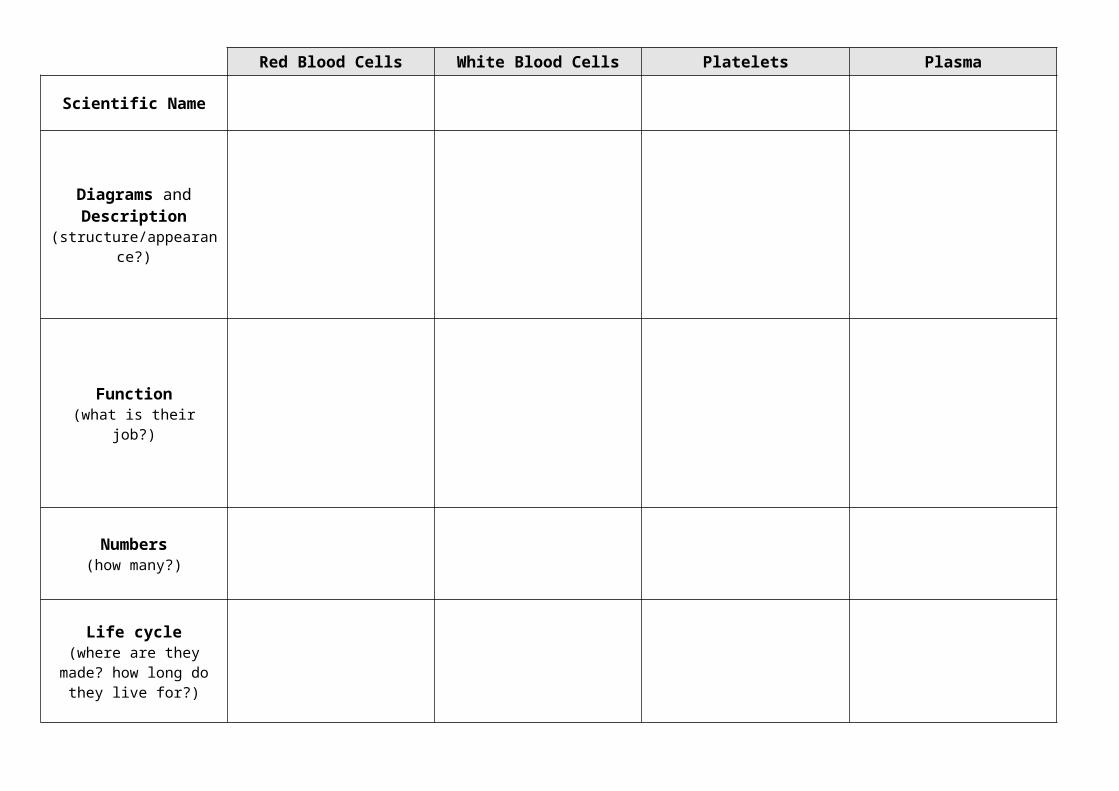
Read the section titled BLOOD on pages 149-150 of your Science Aspects 2 textbook. Complete the table about the components of blood on the back of this page. The website below will be useful:http://www.fi.edu/learn/heart/blood/blood.html
When you have finished your table, read the article called KEEPING US WORKING, and answer the questions below. The article may also be useful in adding to your table.
1. Where are blood cells made? Give two examples of places in your body where they are made? What is the name of the ‘parent’ cell that all blood cells develop from?
2. Why does blood sometimes appear to be red and at other times it is blue? What is haemoglobin and what role does it play in the ‘colour’ of blood?

The average person has about 5 litres of blood moving through their circulatory system.
3. The brain receives 15%-20% of the body’s blood supply. Why is this so important?
Red Blood Cells White Blood Cells Platelets Plasma
Scientific Name
Diagrams and Description (structure/appearance?)
Function(what is their job?)
Numbers(how many?)
Life cycle(where are they made? how
long do they live for?)
Development of Blood Cells
BLOOD TYPES
The Australian Federal Government has declared 2009 as the Year of the Blood Donor. Blood donations are extremely important as this blood can be used to save lives following a traumatic accident where lots of blood is lost, during surgery and to help with the treatment of cancers and other illnesses.
Everybody’s blood is not the same. There are different types or groups of blood. These types/groups determine who can use your blood if you chose to donate and whose blood you can use if you are in need of a blood transfusion. Read the section titled BLOOD GROUPINS on page 150 of your Science Aspects 2 textbook. Then visit the Red Cross Website below and answer the following questions:http://www.donateblood.com.au/page.aspx?IDDataTreeMenu=42&parent=30
1. There are four main blood types – A, B, O and AB. Fill in the table below.
A B O AB
% of Australians with this blood type
How in demand is this blood type?
Who can use this blood type?
In an emergency, what blood type could be given to this person?
2. Looking at the graph at the top of the webpage, answer the following:
a. Most common blood type: __________
b. Least common blood type: __________
3. Find out what the + and – (Rhesus Factor) means. How was it discovered?

Where does the blood go?
Note-taking: PowerPoint – Where does the blood go?___________________________________________________________________________
___________________________________________________________________________
___________________________________________________________________________
___________________________________________________________________________
___________________________________________________________________________
___________________________________________________________________________
___________________________________________________________________________
___________________________________________________________________________
___________________________________________________________________________
___________________________________________________________________________
___________________________________________________________________________
___________________________________________________________________________
___________________________________________________________________________
___________________________________________________________________________
___________________________________________________________________________
___________________________________________________________________________
___________________________________________________________________________
___________________________________________________________________________
___________________________________________________________________________
___________________________________________________________________________
___________________________________________________________________________
___________________________________________________________________________
___________________________________________________________________________
___________________________________________________________________________
___________________________________________________________________________
___________________________________________________________________________
___________________________________________________________________________
__________________________________________________________________________
Questions:
The diagram above shows the passage of blood through the body of a human. Use it and the information you learned from the PowerPoint to answer the following questions.
1. Consider a red blood cell at position X. List the structures (in order) that it will pass through before it returns to X.
2. How many times did it pass through the heart before it returned to its original position?
3. Does blood pass first into the atrium or ventricle of the heart?
4. You observed from your dissection that the wall of the left ventricle was much thicker than that of the right ventricle. Explain why.
5. The tubes which carry blood to the body organs are called _______________ ______________.
Those which carry blood back from the organs to the heart are called _______________. In the organs the
arteries branch to form _______________ .
6. Deoxygenated blood contains much carbon dioxide and little oxygen. Oxygenated blood contains
_______________________________________________________________________________________
7. Blood is oxygenated in the _______________________________________________________________
Carbon dioxide in the blood comes from the ___________________________________________________
It is removed from the body in the ___________________________________________________________
8. Which side of the heart pumps deoxygenated blood? _________________________________________
9. What type of blood travels in the pulmonary artery? __________________________________________

Revision
