the clinical impact of calcium-suppressed images …...the clinical impact of calcium-suppressed...
TRANSCRIPT
The clinical impact of calcium-suppressed images generated from IQon Spectral CT
The Philips IQon Spectral CT delivers conventional and spectral clinical information in a single scan utilizing the IQon dual layer detectors. The IQon spectral detector simultaneously, both in time and space, distinguishes between X-ray photons of high and low energies necessary for generating spectral images. The scanner is capable of generating prospective and retrospective spectral image types reconstructed through advanced spectral algorithms. Spectral image data have the potential for additional clinical information to conventional CT imaging.
The Calcium Suppression (CaSupp) image is a newly introduced IQon HU-based spectral image type. In this image, voxels containing calcium are suppressed and replaced by virtual HU values as similar as possible to the expected HU without calcium contribution to the attenuation.
Case studies
Computed tomography
The following is a collection
of case studies demonstrating
the improved imaging capabilities
of the IQon Spectral CT when using
CaSupp images.
Visualizing bone marrow in the presence of bone lesions with calcium-suppressed CT images
Case study 1 Nuran Abdullayev MD, University Hospital Cologne, Uniklinik Köln; Zimam Romman, Gregor Pahn, Nadav Shapira, Galit Kafri, Philips Healthcare
Underlying bone marrow abnormalities in the presence
of bone lesions can be masked in conventional CT imaging,
making MRI the modality of choice as it is more accurate
and sensitive in the diagnosis of bone lesions.1, 2 The Calcium
Suppression (CaSupp) image may help the clinician in the
visualization of bone marrow abnormalities that otherwise
are obscured by bone while comparing to conventional
CT imaging.
In this case review study, we are showing the clinical
advantage of CaSupp in helping the clinician in visualization
of bone marrow abnormality after bone suppression in
ConclusionVisualization of bone marrow is limited in conventional
CT imaging and is favored by MRI due to its increased
accuracy and sensitivity. The illustrated example showed
that calcium-suppressed imaging using IQon Spectral
CT provides additional information to the clinician that
may help them in the visualization of bone marrow
involvement when bone metastasis lesions are presented.
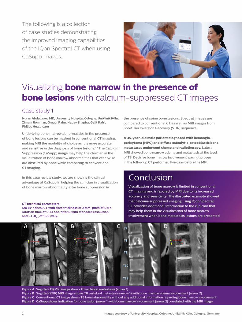
Figure A Sagittal (T1) MRI image shows T8 vertebral metastasis (arrow 1).Figure B Sagittal (STIR) MRI image shows T8 vertebral metastasis (arrow 1) with bone marrow edema involvement (arrow 2).Figure C Conventional CT image shows T8 bone abnormality without any additional information regarding bone marrow involvement.Figure D CaSupp shows indication for bone lesion (arrow 1) with bone marrow involvement (arrow 2) correlated with the MRI image.
CT technical parameters 120 kV helical CT with slice thickness of 2 mm, pitch of 0.67, rotation time of 0.33 sec, filter B with standard resolution, and CTDIvol of 16.9 mGy.
2
the presence of spine bone lesions. Spectral images are
compared to conventional CT as well as MRI images from
Short Tau Inversion Recovery (STIR) sequence.
A 35-year-old male patient diagnosed with hemangio-pericytoma (HPC) and diffuse osteolytic-osteoblastic bone metastases underwent chemo and radiotherapy. Latest
MRI showed bone marrow edema and metastasis at the level
of T8. Decisive bone marrow involvement was not proven
in the follow up CT performed five days before the MRI.
Images courtesy of University Hospital Cologne, Uniklinik Köln, Cologne, Germany.2
CT technical parameters 120 kV helical CT with slice thickness of 0.8 mm, pitch of 1.17, rotation time of 0.4 sec, filter B with standard resolution, and CTDIvol of 13.5 mGy.
A B
C D
CT technical parameters 120 kV helical CT with slice thickness of 0.8 mm, pitch of 1.17, rotation time of 0.4 sec, filter B with standard resolution, and CTDIvol of 13.5 mGy.
Visualizing bone marrow in the presence of bone fractures with calcium-suppressed CT images
ConclusionVisualization of bone marrow is limited in conventional
CT imaging and MRI is the modality of choice for diagnosis
of bone marrow edema. The illustrated example showed
that calcium-suppressed imaging using IQon Spectral CT
provides additional information to the clinician that may
help in the visualization of bone marrow involvement
when bone fractures are presented.
Case study 2 Victor Neuhaus MD, University Hospital Cologne, Uniklinik Köln; Zimam Romman, Gregor Pahn, Nadav Shapira, Galit Kafri, Philips Healthcare
Underlying bone marrow abnormalities in the presence of
fractures can be masked in conventional CT imaging by the
dense trabecular bone, making MRI the modality of choice
to determine the age and etiology of vertebral fractures.3,4
The Calcium Suppression (CaSupp) image may help the
clinician in visualization of bone marrow abnormalities
that otherwise are obscured by the dense trabecular
bone while comparing to conventional CT imaging.
In this case review study, we are showing the clinical
utility of CaSupp in helping the clinician with the
visualization of bone marrow abnormality after bone
suppression in lumbar vertebral fracture. Spectral images
are compared to conventional CT as well as MRI images
from T1-weighted and Short Tau Inversion Recovery
(STIR) sequences.
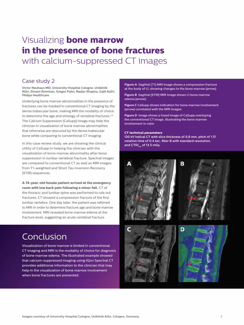
A 74-year-old female patient arrived at the emergency room with low back pain following a minor fall. CT of
the thoracic and lumbar spine was performed to rule out
fractures. CT showed a compression fracture of the first
lumbar vertebra. One day later, the patient was referred
to MRI in order to determine fracture age and bone marrow
involvement. MRI revealed bone marrow edema at the
fracture level, suggesting an acute vertebral fracture.
3
Figure A Sagittal (T1) MRI image shows a compression fracture at the body of L1, showing changes to the bone marrow (arrow).
Figure B Sagittal (STIR) MRI image shows L1 bone marrow edema (arrow).
Figure C CaSupp shows indication for bone marrow involvement (arrow) correlated with the MRI images.
Figure D Image shows a fused image of CaSupp overlaying the conventional CT image, illustrating the bone marrow involvement in color.
Images courtesy of University Hospital Cologne, Uniklinik Köln, Cologne, Germany.

Figure A Sagittal (T2) MRI image shows L2-L3 disc herniation (arrow).
Figure B Sagittal conventional CT image indicates bone abnormality at the level of L2-L3 with limited information regarding disc herniation extent (arrow).
Figure C CaSupp reveals disc herniation (arrow) which can be correlated with the MRI image.
©2017 Koninklijke Philips N.V. All rights are reserved.Philips reserves the right to make changes in specifications and/or to discontinue any product at any time without notice or obligation and will not be liable for any consequences resulting from the use of this publication. Trademarks are the property of Koninklijke Philips N.V. or their respective owners.
www.philips.com
Printed in The Netherlands.4522 991 31761 * NOV 2017
Visualizing intervertebral disc herniation with calcium-suppressed CT images
A 75-year-old male patient with history of multiple intervertebral disc herniation lesions. The patient presented
with acute low back pain and was referred to MRI. Among
other findings, MRI showed disc herniation at the level of L2-
L3. Eigthteen weeks later, CT was performed to plan surgery
for decompression of multi-segmental spinal stenosis.
Case study 3 Simon Lennartz MD, University Hospital Cologne, Uniklinik Köln; Zimam Romman, Gregor Pahn, Nadav Shapira, Galit Kafri, Philips Healthcare
Visualization of intervertebral disc pathologies such as disc
herniation is limited in conventional CT imaging, making MRI
the modality of choice as it is more accurate and sensitive
in the diagnosis of soft tissue differentiation.5,6 Calcium
Suppression (CaSupp) images may help a clinician to better
visualize those pathologies by improving delineation to
adjacent bony structures, such as neural foramen.
In this case review study, a clinical advantage of the CaSupp
image type is demonstrated. Calcium suppression helped
the clinician in improved visualization of intervertebral disc
herniation. The spectral images were compared to their
conventional CT counterparts. MRI images of same region
are also included as reference.
ConclusionVisualization of intervertebral disc herniation is limited
in conventional CT imaging and usually performed
using MRI. The illustrated example shows that calcium-
suppressed imaging using IQon Spectral CT provides
additional information to the clinician that may help
in better assessment of intervertebral disc herniation.
CT technical parameters 120 kV helical CT with slice thickness of 1 mm, pitch of 0.98, rotation time of 0.5 sec, filter B with standard resolution, and CTDIvol of 28.5 mGy.
References1 Hui-Lin Yang, et al. Diagnosis of bone metastases: a meta-analysis comparing 18FDG PET, CT, MRI and bone scintigraphy. Eur Radiol. 2011;21:2604–2617.2 Heindel W, et al. The Diagnostic Imaging of Bone Metastases. Deutsches Ärzteblatt International. 2014;111:741–7.3 Ananya P, et al. Imaging of vertebral fractures. Indian J Endocrinol Metab. 2014 May-Jun;18(3):295–303.4 Eriksen, Erik F. Treatment of bone marrow lesions (bone marrow edema). 2015 International Bone & Mineral Society,
BoneKEy Reports 4, Article number 755 (2015). doi:10.1038/bonekey.2015.1245 Lurie JD, et al. Reliability of Magnetic Resonance Imaging Readings for Lumbar Disc Herniation in the Spine Patient Outcomes Research Trial (SPORT). NIH-PA Author Manuscript, Spine (Phila Pa 1976). 2008 April 20;33(9):991–998. doi:10.1097/BRS.0b013e31816c83796 Nadja A, Farshad-Amacker, et al. MR imaging of degenerative disc disease. European Journal of Radiology 84 (2015);1768–1776.
Results from case studies are not predictive of results in other cases. Results in other cases may vary.
Images courtesy of University Hospital Cologne, Uniklinik Köln, Cologne, Germany.