the cognitive neuroscience of number sense - inserm-cea
TRANSCRIPT

1
Chapter for “The Cognitive Neuroscience, 3rd Ed” by Gazzaniga et al.
FROM NUMBER NEURONS TO MENTAL ARITHMETIC: THE
COGNITIVE NEUROSCIENCE OF NUMBER SENSE
Manuela Piazza and Stanislas Dehaene
INSERM U562 Cognitive Neuroimaging
Service Hospitalier Frédéric Joliot, CEA/DRM/DSV
4 place du Général Leclerc, 91401 Orsay cedex, France
Phone: +33 1 69 86 78 40
E-mail: [email protected]

2
1. INTRODUCTION
Consider a lioness in her pride in the Serengeti National park, Tanzania. One night she is all alone and
hears a roar from an intruder lioness. Should she try to drive the intruder off? That would be an even
match, thus ending in a possibly fatal fight. She decides not to act. The following night she is with four
sisters, when they hear the roars of three intruder lionesses. This time it is three versus five. The
lionesses peer into each others eyes and then launch the attack. But by the time they reach the expected
location, they find no intruder, and that is because the sounds were actually coming from loudspeakers
set up by a researcher investigating the numerical capacity of animals. This research shows that
generally, animals decide to attack back only when the number of defenders is superior to the number
of intruders (McComb, Packer, & Pusey, 1994). The process of “decision making” of these animals
seems to be based on a multimodal comparison between the number of the number of defenders,
which is perceived visually, and the number of defenders, which is perceived auditorily. This suggests
hat the internal representation of number in animals can be quite abstract.
Numbers might be thought to be a very recent cultural invention in the evolution of the human
species. Indeed, number words and digits arise from the specifically human and evolutionary recent
ability to create and mentally manipulate complex symbols. However, the sense of numbers is older. A
sensitivity to numerical properties of the world is present in numerous non-human species as well as in
babies, and its strong adaptive value is easily seen. Such a sensitivity to numbers also seems to be
rooted on a distinct neural circuitry, which has been reproducibly identified in different subjects and
species with convergent methods. These observations lead to the hypothesis that an elementary
number system is present very early in life in both humans and animals, and constitutes the start-up-
tool for the development of symbolic numerical thinking that permeates so deeply our western
technological societies (Dehaene, 1998).
In this chapter we present an overview of the most relevant findings on the cognitive neuroscience
of number sense, showing how data from different domains, from cognitive psychology to
electrophysiology, are providing us with complementary information on how our brain represents and
manipulates numbers. We first consider how behavioural data have shed light on such number sense in

3
non-verbal organisms, either in animals or in human infants. Then, we show how functional imaging
and neuropsychology clarify the cerebral substrates of numerical ability. Finally, we explore the nature
of the neural coding of numbers, as clarified by recent electrophysiological studies in monkeys.
2. BEHAVIOURAL EVIDENCE FOR ANALOGICAL REPRESENTATION OF
NUMBER
The ability to make numerical judgments has been tested in many different species of animals
(from pigeons to rats, racoons, dolphins and monkeys) both in the wild and in more controlled
experimental settings in laboratory, with very different types of paradigms. Typically, animals are
trained to respond differentially to a variety of numerically defined stimuli: the number of visual
stimuli, of tones, of motor responses or of reinforcements. However, as number normally co-varies
with some of the physical attributes of the stimuli (such as brightness, density, time, hedonic value)
reports of numerical abilities in animals are often met with scepticism: how can one be sure that
animals are processing number rather than any other parameters of the stimulus? Two arguments have
been used to demonstrate genuine numerical competence: first, animals can transfer numerosity
between different modalities. For example, rats initially trained on distinct auditory and visual
numerosity discrimination tasks could later generalise to novel sequences in which auditory and visual
sequences were mixed (Church & Meck, 1984). This suggests that animals possess an abstract, amodal
representation of number. Second, animals are able to generalise numerically relevant behaviour to
novel, non-differentially rewarded stimuli (Brannon & Terrace, 2000; Church & Meck, 1983;
Sawamura, Shima, & Tanji, 2002; Nieder, Freedman, & Miller, 2002). This indicates that they are able
to bring to the task more knowledge of numerical invariance than the training alone could provide. For
example, rats were trained to press one lever in response to a short two-tone sequence and another in
response to a long eight-tone sequence. Although duration discrimination was sufficient for that initial
performance, subsequently, the rats generalised their behaviour to novel, non-differentially rewarded
sequences in which duration was fixed and only number varied. This suggests that the animals were
representing number during the initial training phase (Church & Meck, 1983).

4
An ability to discriminate sets on the basis of their number is also present in pre-verbal human
infants. With the classic method of habituation-recovery of looking time, both new-borns and pre-
verbal infants have been shown to discriminate sets of visual objects, as well as tones or words that
differed in the number of syllables, on the unique basis of their numerosity (e.g. Xu & Spelke, 2000).
Again, there is suggestive evidence of cross-modal numerosity matching (Starkey, Spelke, & Gelman,
1983), as well as of processing of the numerosity of abstract entities such as collections, whose
physical attributes can be well controlled (Wynn, Bloom, & Chiang, 2002). Such animal and infant
data, taken together, suggest that the sensitivity to the numerical aspect of the word does not depend
on an acquired ability to manipulate symbols, but is based on a non-verbal amodal representation of
numerosity.
This representation can also be evidenced in human adults. When prevented from using
language and our counting procedures, adults can make approximate numerosity judgements similar to
those of animals and infants. They too can transfer numerosity from different modalities (visual and
auditory) and modes of stimulus presentation (sequential and simultaneous) and they do this without
cost relative to a unimodal stimulus presentation (Barth, Kanwisher, & Spelke, 2003).
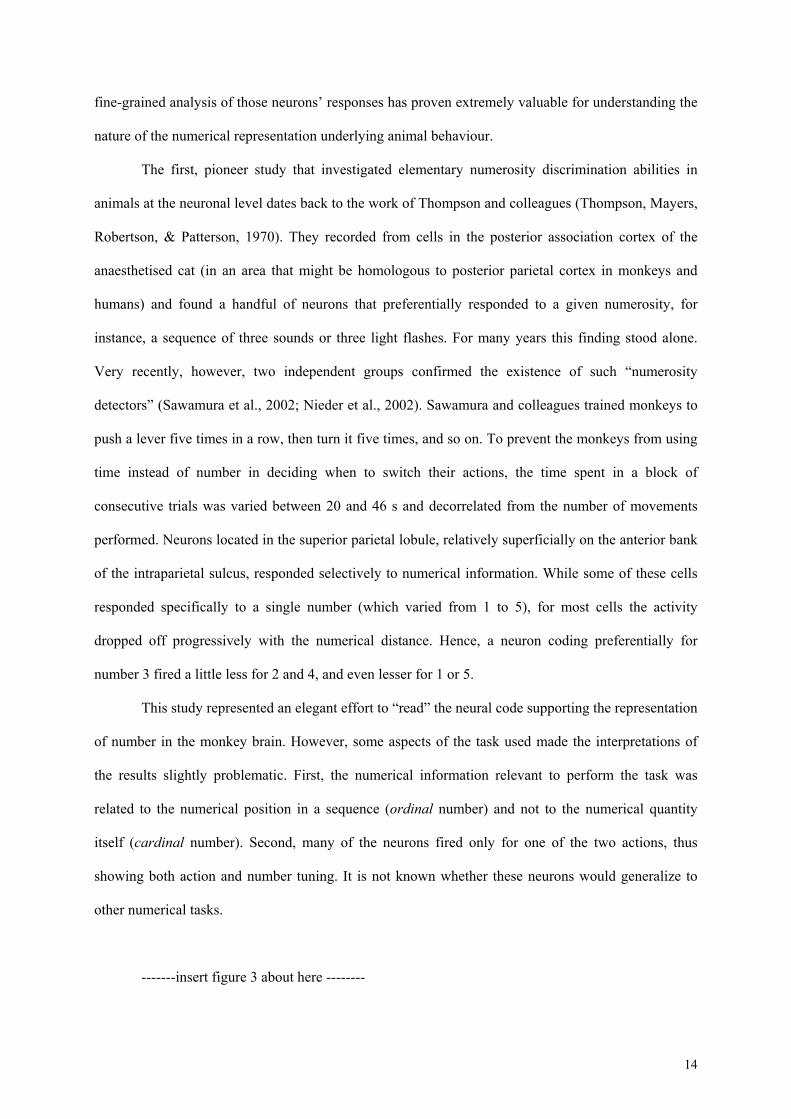
-------insert figure 1 about here --------
The parallels between human and animal behavior in number-related tasks are therefore
numerous, but the most striking one is probably that they both seem to be governed by the very same
metric (Gallistel & Gelman, 2000)(see figure 1). Number estimation performance in both humans and
non-human animals is approximate, and becomes less and less accurate as the numbers increase.
Furthermore, the variability in performance increases linearly with the size of the number involved (a
property called scalar variability or “Weber’s law”, where the proportionality constant is called the
Weber fraction). For example, rats instructed to press a lever a certain number of time, will break off
the sequence of lever presses with a probability that is roughly proportional to the percent deviation of
the actual number of presses from the number of presses required to get the reward (Mechner,
1958)(figure 1A). Likewise, monkeys instructed to judge whether successive visual displays have the

5
same number of items, make errors in direct proportion to the ratio of the two numbers. Thus, for
larger numerical quantities, the two numerosities have to be numerically more distant for performance
to reach the same level as the one obtained with smaller quantities (Nieder & Miller, 2003)(figure 1B).
In humans, studies directly inspired by animal experiments, show exactly the same type of
metric. For example, Whalen et al. (1999) presented numbers between 7 and 25 on a computer screen,
and asked subjects to press a button as fast as they could until they had felt they had made
approximately the indicated number of button presses (Whalen, Gallistel, & Gelman, 1999; verbal
counting was prevented by asking subjects to recite words while performing the task). Results showed
that both the mean estimate and its variability were proportional to target value, and that therefore the
coefficient of variation (the ratio of the standard deviation and the mean) was constant across target
size (figure 1C). Very similar results are found when humans are asked to estimate prices for different
items: again, the standard deviation of price estimates is directly proportional to the mean price
(Dehaene & Marques, 2002)(figure 1D).
Moreover, Weber’s law holds even when numerical judgements are not estimates, but exact
computations made over abstract numerical symbols like digits and number words. Such symbolic
numerical judgments (like, for example, choosing the larger number between 49 and 72) show
magnitude effects exactly like non symbolic numerosity judgments: they are influenced by the
numerical difference between two values (“numerical distance effect”), and by their absolute
magnitude (“magnitude effect”), such that ultimately performance can be predicted by the ratio of the
two numbers involved (Moyer & Landauer, 1967; Dehaene, 1992).
It has been a matter of debate whether Weber’s law is better described by a linear continuum
with increasing variability, or by a non linear, maybe logarithmic scale with constant variability. Some
have argued that the linear model should be preferred, because it allows a simpler calculation of sums
and differences (Gallistel & Gelman, 1992). Contrariwise, others have proposed a logarithmic coding
because this compressive scheme avoids an explosion in the size of the internal representation as the
range of represented numerosities increases (Dehaene & Changeux, 1993). In fact, both assumptions
accurately predict performance, and for long it has been thought that the linear and the logarithmic

6
hypotheses could not be disentangled. However, recently, detailed analyses of the exact shape of
response distributions in both humans and animals have suggested that the internal scale is not linear,
but logarithmic. When plotted on a linear scale, performance curves are asymmetrical and are best
fitted by a log-normal distribution (figure 1D). However, they become symmetric when plotted on a
logarithmic scale, and are then best fitted by a simple Gaussian with fixed variability (Dehaene et al.,
2002; Nieder & Miller, 2003). Thus, the behaviour of both humans and monkeys can be described in a
more compact way by assuming a logarithmic than a linear internal scale for number.
3. IMAGING NUMBER SENSE IN HUMANS
Given those behavioral observations, it has been proposed that animals and humans share a
common and evolutionary ancient mechanism for representing numerical quantities, and that this
mechanism serves as a foundational core of numerical knowledge, providing humans with a start-up
tool that permits the acquisition of numerical symbols (Butterworth, 1999; Dehaene, 1997; however,
see (Simon, 1999) for an opposite view). The analogical representation of number would ground our
intuition of what a given numerical size means, and of the proximity relations between numbers. It
would be crucial in tasks that put strong emphasis on the quantitative aspects of numbers, like for
example the estimation of a price (Dehaene et al., 2002) (figure 1D), the approximation of complex
arithmetical problems (Dehaene, Spelke, Pinel, Stanescu, & Tsivkin, 1999, Spelke & Tsivkin, 2001),
or the rough estimation of the number of elements in a set (Whalen et al., 1999). In support of this
view, recent neuroimaging data show that an area of the brain is systematically activated whenever
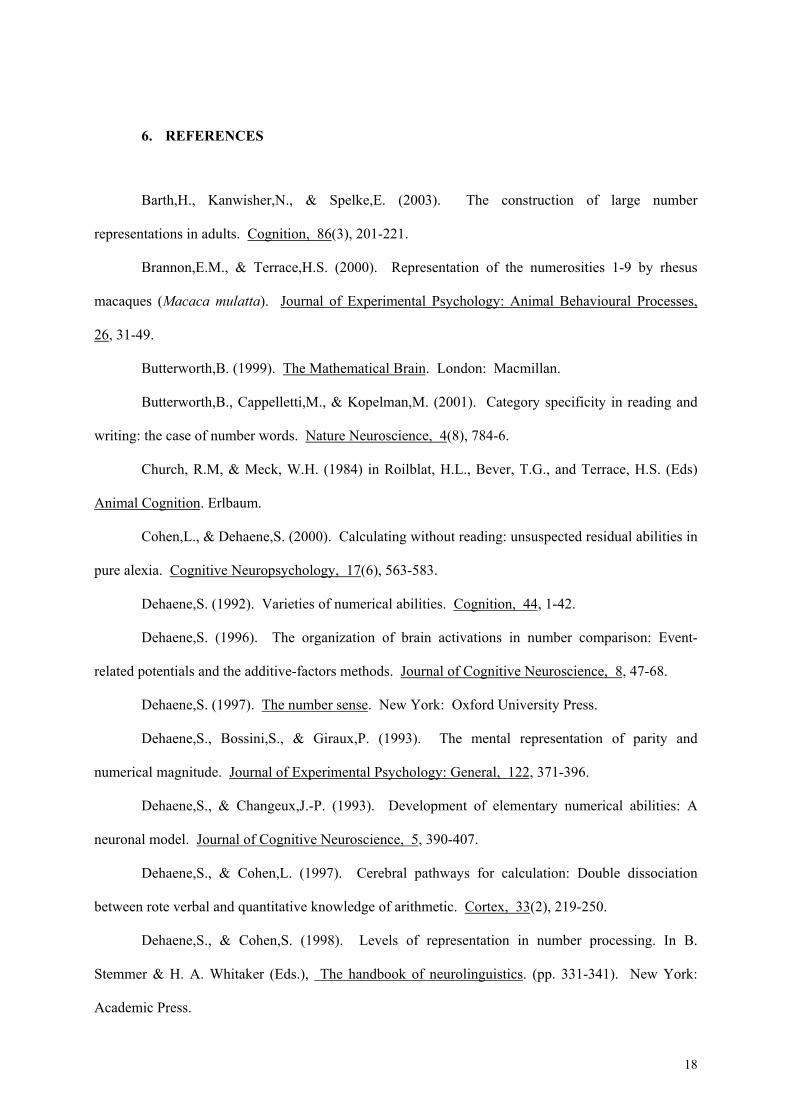
this putative core system for numerical quantities is called for: the horizontal segment of the
intraparietal sulcus in the parietal lobes.
The first investigation of the neural correlates of human numerical abilities showed increased
metabolism in both parietal and frontal regions during complex calculation, using a single-photon
emission (Roland & Friberg, 1985). This result was in agreement with earlier studies of brain-lesioned
patients that had shown a crucial role of parietal lobes in number processing. Since then, many studies
using more refined functional imaging techniques like PET or fMRI have brought converging
evidence for the recruitment of parietal regions in different number-related tasks. On the basis of a

7
detailed review of the recent literature, and of a meta-analaysis of some of the available activation
images, we proposed that parietal activation in number-related tasks can be segregated into three
distinct sites, each associated with a distinct process (Dehaene, Piazza, Pinel, & Cohen, 2002): the
posterior superior parietal lobule associated with visuo-spatial processing, the angular gyrus of the left
hemisphere, associated with verbal processing of numbers, and the horizontal segment of the
intraparietal sulcus, the most plausible candidate for a domain-specific locus where numerical quantity
is represented.
-------insert figure 2 about here --------
In the present review we focus mostly on the latter site, the intraparietal sulcus (hereafter
HIPS) (see figure 2A). Several features of its responsiveness to experimental conditions suggest that
this region encodes the analogical representation of numerical magnitude that grounds our intuition of
what a given numerical size means, and of the proximity relations between numbers.
First, in calculation, the HIPS is more active when subject estimate the approximate result of
an addition problem than when they compute its exact solution, even when the task difficulty is strictly
controlled (Dehaene et al., 1999). This fits well with the approximate nature of the representation of
numerical quantities. Within exact calculation, this region is also more active for operations that
require a genuine manipulation of numerical quantities, such as subtraction, than for those that can be
stored in rote verbal memory, such as multiplication (Lee, 2000). Moreover, its activation is
modulated by semantic parameters such as the absolute magnitude of the numbers. It is larger and lasts
longer during operations with large numbers than with small numbers (Dehaene et al., 1999; Stanescu-
Cosson et al., 2000).
Second, the HIPS is also active whenever a comparative operation that needs access to a
numerical scale is called for. For instance, its activation is higher when comparing the magnitudes of
two numbers than when simply reading them. Furthermore its activation is modulated by the
numerical distance separating the numbers (Pinel, Dehaene, Riviere, & LeBihan, 2001). The
systematic contribution of this region to number comparison processes has also been replicated using

8
scalp recordings of event-related potentials (Dehaene, 1996). The typical scalp signature of a
numerical distance effect, moreover, has been observed in 5-year-old children, with a topography
similar to adults for numbers presented either as Arabic numerals or as sets of dots (Temple & Posner,
1998).
Third, the HIPS shows relatively robust category-specificity for numbers when directly
contrasted with different categories of objects or concepts. For example, in comparative judgements, it
is more active when comparing numbers than when processing other categories of objects on non-
numerical scales (such as comparing the ferocity of animals, the relative positions of body parts, or the
orientation of two visually presented characters) (Pesenti, Thioux, Seron, & DeVolder, 2000; Thioux,
Pesenti, Costes, De Volder, & Seron, 2002). Even in tasks that do not directly require a numerical
judgement, such as simple detection tasks, the HIPS is the only region that shows higher activation
when processing numbers than when processing letters of the alphabet or colors (Eger, Sterzer, Russ,
Giraud, & Kleinschmidt, 2003). Both control continua (letters and colors) were chosen because, like
numbers, they show a distance effect (e.g. when detecting the letter M, it takes longer to reject the
letter L than the letter C). Moreover, the alphabet also shares with numbers a strong serial component.
However, both letters and colors lack quantitative meaning. Therefore, this experiment suggests that
the HIPS activation for numbers relates to the processing of their quantitative meaning.
Fourth, the activation of the HIPS is independent of the particular modality of input used to
present numbers. Arabic numerals, spelled-out number words, and even non-symbolic stimuli like sets
of dots or tones, can activate this region if subjects attend to the corresponding number. In one study,
subjects attended either to the numerosity or to the physical characteristics (color, pitch) of series of
auditory and visual events. The right HIPS was active whenever the subjects attended to number,
regardless of the modality of the stimuli (Piazza, Mechelli, Price, & Butterworth, 2003). In another
study, the activation of the bilateral HIPS was found to correlate directly with the numerical distance
between two numbers in a comparison task, and this effect was observed whether the numbers were
presented as words or as digits (Pinel et al., 2001). Finally, in a simple detection task, the HIPS was
activated for numbers relative to letters and colours, irrespective of the visual or auditory modality of

9
presentation (Eger et al., 2003). These results are consistent with the hypothesis that the HIPS encodes
the abstract quantity meaning of numbers rather the numerical symbols themselves.
Fifth, quantity processing and HIPS activation was demonstrated even when the subjects were
not aware of having seen a numerical symbol (Naccache & Dehaene, 2001). In this experiment,
subjects were asked to compare target numbers to a fixed reference of 5. Unbeknownst to them, just
prior to the target, another number, the prime, was briefly present in a subliminal manner. FMRI
revealed that the left and right intraparietal regions were sensitive to the unconscious repetition of the
same number. When the prime and target corresponded to the same quantity (possibly in two different
notations, such as ONE and 1), less parietal activation was observed than when the prime and target
corresponded to two distinct quantities (e.g. FOUR and 1). This result suggests that this region
comprises distinct neural assemblies for different numerical quantities, so that more activation can be
observed when two such neural assemblies are activated than when only one is. It also indicates that
this region can contribute to number processing in a subliminal fashion.
Finally, convincing evidence for the crucial role of the HIPS in numerical quantity
representation comes from neuropsychological studies of impairments of number processing and
calculation in adults as well as in developmental cases. Several single-case studies indicate that
numbers doubly dissociate from other categories of concepts at the semantic level. On the one hand,
spared calculation and number comprehension abilities have been described in patients with grossly
deteriorated semantic processing (“semantic dementia”: Butterworth, Cappelletti, & Kopelman, 2001;
Remond-Besuchet et al., 1998). In both cases, the lesions broadly affected the left temporo-frontal
cortices while sparing the intraparietal regions. On the other hand, several cases demonstrate that the
understanding of numbers and their relations can be specifically impaired in the context of otherwise
preserved language and semantics (e.g. Dehaene & Cohen, 1998; Delazer & Benke, 1997). The
majority of such cases result from parietal regions, particularly in the left hemisphere.
Developmental studies confirm that impairments of arithmetical abilities correlate with
abnormalities in the functional or anatomical organization of the HIPS. Levy et al. (1999) report the
case of an adult with lifelong isolated dyscalculia together with superior intelligence and reading
ability, in whom the standard anatomical MRI appeared normal, yet MR spectroscopy techniques

10
revealed a metabolic abnormality in the left inferior parietal area. Similarly, Isaacs et al. (2001) used
voxel-based morphometry to compare grey matter density in adolescents born at equally severe grades
of prematurity, half of whom suffered from dyscalculia. The only brain region that showed reduced
grey matter associated with dyscalculia was the left intraparietal sulcus (see figure 2C).
More recently, Molko and colleagues studied a population of females affected by Turner
syndrome, a syndrome characterised by a congenital abnormality of the X chromosome, and often
associated with defective development of number skills, in the context of a normal general intelligence
(Molko et al., submitted). They used voxel-based morphometry to compare the grey matter density of
such dyscalculic Turner population with an age, education, and sex matched group of controls. Results
showed a region of reduced grey matter in the depth of the right HIPS. Morphometric analysis
restricted to the cortical sulci showed that the length and depth of the right intraparietal sulcus was
reduced in Turner patients. Finally, the fMRI activation of this region differed significantly with
respect to the control group in number-related tasks such as simple additions.
Other parietal regions involved in number processing
These data, taken together suggest that the HIPS is the most crucial cortical region for the
correct development of numerical skills. Clearly, however, it is not the only system involved in
number processing. Mental arithmetic relies on a highly composite set of processes, many of which
are probably not specific to the number domain. For instance, studies of language interference in
normal subjects suggest that language-based processes play an important role in exact but not
approximate calculation (Spelke et al., 2001). Likewise, concurrent performance of a spatial task
interferes with subtraction, but not multiplication, while concurrent performance of a language task
interferes with multiplication, but not subtraction (Lee & Kang, 2002). Such behavioral dissociations
suggest that the neural bases of calculation must be heterogeneous.
A recent re-analysis of brain activation studies allowed us to individuate two satellite parietal
systems which are often involved in numerical tasks, even if their primary functions is not specific to
numbers. We isolated a language-related region in the left angular gyrus, associated with verbal
processing of numbers, and a visuo-spatial region in the posterior superior parietal lobe, presumably
associated with spatial and non-spatial attention. In arithmetic tests, the left angular gyrus shows

11
increasingly greater activation as the task puts greater requirement on verbal processing. For example,
this region is more active in exact calculation than in approximation (Dehaene et al., 1999). This fits
with behavioural data which indicate that exact arithmetic facts are stored in a language-specific
format in bilinguals, while approximate knowledge is language-independent (Spelke et al., 2001).
Moreover, within exact calculation, the left angular gyrus shows greater activation for operations that
require access to a rote verbal memory of arithmetic facts, such as multiplication, than for operations
that are not stored and require some form of quantity manipulation like subtraction.
The posterior superior parietal regions is also frequently active, usually in synergy with the
HIPS, for instance in number processing during number comparison (Pinel et al., 2001; Pesenti et al.,
2000), approximation (Dehaene et al., 1999), some subtraction tasks (Lee, 2000), and counting
(Piazza, Mechelli, Butterworth, & Price, 2002; Piazza, Giacomini, Le Bihan, & Dehaene, 2003).
However, this region is clearly not specific to the number domain as it plays a central role in
attentional selection in space and time (Wojciulik & Kanwisher, 1999). Psychological experiments
indicate that numbers have a strong serial and therefore spatial component, to the point that it has been
metaphorically proposed that numbers are represented internally on a “number line”, a quasi-spatial
representation on which numbers are organized by their proximity (Moyer et al., 1967; Dehaene,
Bossini, & Giraux, 1993). It is therefore conceivable that the same process of covert attention that
operates to select locations in space can also be engaged when attending to specific quantities on the
number line.
According to the proposed tripartite organization of parietal activation during arithmetic
suggested by our meta analysis of existing neuroimaging studies, dissociations are expected between
the types of arithmetic tasks that patients should or should not be able to perform according to the
lesion site. For example, a lesion to the HIPS should affect all tasks requiring a genuine manipulation
of both symbolic and non-symbolic numerical quantities, including approximation, subtraction,
comparison, or estimation of numerosity. Lesions of the angular gyrus or other language-related
regions of the left hemisphere should result in an impairment in retrieving arithmetical facts which are
stored in verbal format, such as multiplication facts. Finally, lesions extending to the most posterior
portion of the intraparietal sulcus and/or the superior parietal lobe should impair tasks that put strong

12
emphasis on the visuo-spatial layout of number, like, for example, bisection tasks (e.g. what number
lies between 11 and 19?). Indeed such dissociations have been reported in the literature. For example,
some patients are much more impaired in subtraction than in multiplication (Dehaene & Cohen, 1997;
Delazer et al., 1997; van Harskamp & Cipolotti, 2001). Such patients typically do not show language
impairments, but, in the rare case in which this has been investigated, may show difficulties in
estimating the numerosity of arrays (Lemer, Dehaene, Spelke, & Cohen, 2003). Lesions are often
reported around the left intraparietal sulcus. Other patients show the reverse dissociation, being more
severely impaired in multiplication than in subtraction. They almost always have associated aphasia
(Cohen & Dehaene, 2000; Dehaene & Cohen, 1997; Sandrini, Miozzo, Cotelli, & Cappa, 2003).
Furthermore, the lesions in such patients often spare the intraparietal cortex and can affect multiple
regions known to be engaged in language processing, such as the left perisylvian cortices including the
inferior angular gyrus, the left parieto-temporal carrefour or the left basal ganglia.
A recent study directly compared two acalculic patients, one with a focal lesion of the left
parietal lobe and Gerstmann’s syndrome and another with semantic dementia with predominantly left
temporal hypometabolism. As predicted by a numerical quantity deficit, the first patient was more
impaired in subtraction than in multiplication, showed a severe slowness in approximation of
calculation, and exhibited associated impairments in estimation and numerical comparison tasks, both
with Arabic digits and with sets of dots. As predicted by a verbal deficit, the second patient was more
impaired in multiplication than in subtraction, had intact approximation abilities, and showed
preserved processing of non-symbolic numerosities (Lemer et al., 2003).
Support for the dissociation between quantity representation and the spatial scanning of the
mental number line is provided by a study with unilateral neglect patients (Zorzi, Priftis, & Umilta,
2002). It is a well-known, indeed almost a defining feature of those patients that they perform poorly
in spatial bisection tests. When asked to locate the middle of a line segment, neglect patients with right
parietal lesions tend to indicate a location further to the right, consistent with their failure to attend to
the left side of space. Zorzi et al. tested their performance in a numerical bisection task, where they
were asked to find the middle of two orally presented numbers. Strikingly, the patients erred
systematically, often selecting a number far larger than the correct answer (e.g. what number falls in

13
between 11 and 19? 17). This suggests that spatial attention can be oriented on the left-to-right
oriented number line, and that this attention-orienting process contributes to the resolution of simple
arithmetic problems such as the bisection test. Interestingly, these patients were not acalculic and did
not show deficits in other numerical tasks such as simple arithmetic fact retrieval.
In summary, a review of neuropsychological dissociations between numerical operations
indicates that most if not all cases so far can be accommodated by the postulated dissociation between
a quantity circuit (supporting subtraction and other quantity-manipulation operations) a verbal circuit
(supporting multiplication and other rote memory-based operations), and a visuo-spatial circuit
(supporting number bisection and other tasks where the spatial sense of numbers is particularly
relevant).
One intriguing discrepancy between the lesion and imaging studies relates to the lateralization
of the core quantity circuit. In quantity-related dyscalculia, lesions are often restricted to the left
hemisphere. However, imaging studies show bilateral parietal activation in quantity tasks. The exact
role of the right hemisphere in number processing is therefore still unclear. Some neuropsychological
studies have suggested a superiority of the right hemisphere, and in particular of the parietal region, in
numerosity estimation tasks, like estimating the number of dots in briefly presented arrays of elements
(Warrington & James, 1967). Such a right-hemisphere superiority in non-symbolic numerical tasks
was replicated in an imaging study of numerosity estimation, where parietal activation was indeed
restricted to the right hemisphere (Piazza et al., 2003). One possibility is that our core numerical
system is initially bilateral and progressively becomes biased for symbolic manipulations of numbers
in the left hemisphere and for non-symbolic tasks in the right hemisphere. Such speculation, however,
awaits confirmation.
4. THE NEURAL CODING OF NUMEROSITY
Recent electrophysiological studies have considerably improved our understanding of the neural bases
of number sense. In 1993, Dehaene and Changeux presented a theoretical model of how numerical
quantity could be represented at the single neuron level. They proposed the existence of “numerosity
detectors”, neurons coarsely tuned to an approximate quantity. In 2002, two electrophysiological
studies reported the first clear evidence for the existence of such neurons. As we shall see below, the
14
fine-grained analysis of those neurons’ responses has proven extremely valuable for understanding the
nature of the numerical representation underlying animal behaviour.
The first, pioneer study that investigated elementary numerosity discrimination abilities in
animals at the neuronal level dates back to the work of Thompson and colleagues (Thompson, Mayers,
Robertson, & Patterson, 1970). They recorded from cells in the posterior association cortex of the
anaesthetised cat (in an area that might be homologous to posterior parietal cortex in monkeys and
humans) and found a handful of neurons that preferentially responded to a given numerosity, for
instance, a sequence of three sounds or three light flashes. For many years this finding stood alone.
Very recently, however, two independent groups confirmed the existence of such “numerosity
detectors” (Sawamura et al., 2002; Nieder et al., 2002). Sawamura and colleagues trained monkeys to
push a lever five times in a row, then turn it five times, and so on. To prevent the monkeys from using
time instead of number in deciding when to switch their actions, the time spent in a block of
consecutive trials was varied between 20 and 46 s and decorrelated from the number of movements
performed. Neurons located in the superior parietal lobule, relatively superficially on the anterior bank
of the intraparietal sulcus, responded selectively to numerical information. While some of these cells
responded specifically to a single number (which varied from 1 to 5), for most cells the activity
dropped off progressively with the numerical distance. Hence, a neuron coding preferentially for
number 3 fired a little less for 2 and 4, and even lesser for 1 or 5.
This study represented an elegant effort to “read” the neural code supporting the representation
of number in the monkey brain. However, some aspects of the task used made the interpretations of
the results slightly problematic. First, the numerical information relevant to perform the task was
related to the numerical position in a sequence (ordinal number) and not to the numerical quantity
itself (cardinal number). Second, many of the neurons fired only for one of the two actions, thus
showing both action and number tuning. It is not known whether these neurons would generalize to
other numerical tasks.
-------insert figure 3 about here --------

15
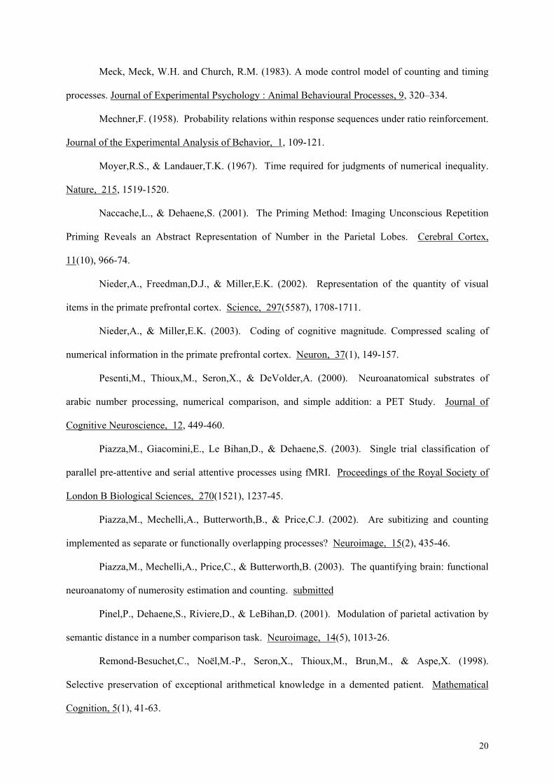
A more recent study, however, reported neurons tuned to cardinal number, and performed
fine-grained analysis of the neurons’ tuning curves in parallel with a thorough analysis of the
monkeys’ behavior. (Nieder et al., 2002; Nieder & Miller, 2003). Macaque monkeys were trained to
perform a match-to-sample task on successively presented visual displays containing between 1 and 5
randomly arranged items (see figure 3A). During training, certain visual features inevitably co-varied
with numerosity. However, after training, the monkeys spontaneously generalised to novel displays in
which all of the relevant non-numerical variables were controlled, suggesting that they were attending
to number.
Neurons were then recorded, initially mainly in the dorsolateral prefrontal cortex (Nieder et
al., 2002). About one-third of the neurons there were tuned to a specific numerosity between 1 and 5
(the maximal numerosity that was tested). This finding was very similar to the Sawamura et al. study,
but because the stimulation was visual it was possible to extensively vary the stimulus and verify to
what extent it could be explained by other non-numerical parameters. The results indicated a
remarkable degree of invariance to non-numerical parameters: a given neuron remained tuned to the
same number across a broad variation of stimuli that controlled for object size, density, spacing, and
spatial layout. Nevertheless, further tests will be required to assess generalization to ordinal
numerosity as well as to other modalities of auditory or motor stimulation.
One intriguing feature was the localization of such number-sensitive neurons in the prefrontal
cortex. Intensive training may explain why many prefrontal neurons became sensitive to number in
Nieder and Miller’s study. However, human data suggested the intraparietal cortex, not the prefrontal
cortex, as a crucial site for number processing (see session 2 and 4). While Sawamura et al. (2002)
reported many number-tuned neurons in the superior parietal lobule, Nieder et al.(2002) reported
finding only a small proportion of number neurons in area 7a of the inferior parietal lobule (about 7%).
More recently, however, by recording in the depth of the intraparietal sulcus, at a location that might
correspond to area VIP, Nieder and Miller observed a much higher proportion of neurons tuned to
numerosity (Andreas Nieder and Earl Miller, personal communication)(see figure 3B). Crucially,
those neurons showed differential firing as a function of numerosity at a latency of about 80 ms, which
is shorter than the value of 120 ms observed for prefrontal neurons. This is compatible with the

16
hypothesis that numerosity is first extracted and represented in the IPS, and later transmitted to
prefrontal circuits as needed for the requested task (Dehaene & Changeux, 1993). Furthermore, their
localization in the depth of the IPS is a plausible monkey homolog to the site of activation observed in
humans. Indeed, this localization was predicted by a human brain imaging study in which the
activation during subtraction was found to be inserted within a network of visuo-motor areas plausibly
corresponding to areas AIP, MIP, V6A and LIP in monkeys (Simon et al., 2002).
Several features of the response of those visual numerosity neurons illuminate the mechanisms
of extraction and representation of number. First, their firing latency is independent of the number
being represented (see figure 3C). This result is not compatible with models that postulate a serial
accumulator mechanism, and which would therefore an linear increase in firing latency with
numerosity (Gallistel & Gelman, 1992). It fits, however, with a parallel mechanism of numerosity
abstraction, as predicted by the Dehaene and Changeux (1993) model. A second important feature of
number-coding neurons is that their tuning curves are broad, again suggesting approximate coding.
Furthermore, this breadth is proportional to the neuron’s preferred number. Thus, evidence for
Weber’s law can be observed at the single neuron level, mirroring behavior. Finally, the neural tuning
curves are asymmetrical on a linear scale but assume a Gaussian shape when plotted on a logarithmic
scale (Nieder & Miller 2003)(see figure 3D). Exactly like behavioural data, this suggests a
compressive logarithmic neural encoding of numerical quantity.
5. CONCLUSION
Altogether, these neurophysiological results indicate that, down to a rather minute level of
detail, the visual numerosity neurons observed by Nieder and Miller (2003) provide a plausible
neuronal basis for the sense of number that is known to be present in animals and humans.
Nevertheless, several experimental steps will be needed to confirm this hypothesis. More extensive
studies in animals are needed to evaluate the abstractness and invariance of the neurons to multimodal
stimuli. Investigating a greater range of numerosities, and examining the presence of those neurons in
naïve untrained animals, is crucial. Finally, reversible lesion or microstimulation studies are necessary
to demonstrate that those neuronal representations play a causal role in the monkeys’ numerical
abilities. In parallel, experiments in humans, with both non-symbolic and symbolic numerical stimuli,

17
should attempt to demonstrate that a similar code for number is present in the human brain. This could
be done with intracranial recordings, but also non-invasively using the “priming method” in fMRI
(Naccache et al., 2001).
The number domain represents one of the very few domains of cognitive neuroscience where
experimental studies can range from recordings of single neurons in the monkey to response-time
studies of verbal symbols in humans. Such parallel studies in various species and using a range of
experimental methods are helping making sense of the human ability for mathematics through an
integrated and progressively more coherent picture of the working brain.
18
6. REFERENCES
Barth,H., Kanwisher,N., & Spelke,E. (2003). The construction of large number
representations in adults. Cognition, 86(3), 201-221.
Brannon,E.M., & Terrace,H.S. (2000). Representation of the numerosities 1-9 by rhesus
macaques (Macaca mulatta). Journal of Experimental Psychology: Animal Behavioural Processes,
26, 31-49.
Butterworth,B. (1999). The Mathematical Brain. London: Macmillan.
Butterworth,B., Cappelletti,M., & Kopelman,M. (2001). Category specificity in reading and
writing: the case of number words. Nature Neuroscience, 4(8), 784-6.
Church, R.M, & Meck, W.H. (1984) in Roilblat, H.L., Bever, T.G., and Terrace, H.S. (Eds)
Animal Cognition. Erlbaum.
Cohen,L., & Dehaene,S. (2000). Calculating without reading: unsuspected residual abilities in
pure alexia. Cognitive Neuropsychology, 17(6), 563-583.
Dehaene,S. (1992). Varieties of numerical abilities. Cognition, 44, 1-42.
Dehaene,S. (1996). The organization of brain activations in number comparison: Event-
related potentials and the additive-factors methods. Journal of Cognitive Neuroscience, 8, 47-68.
Dehaene,S. (1997). The number sense. New York: Oxford University Press.
Dehaene,S., Bossini,S., & Giraux,P. (1993). The mental representation of parity and
numerical magnitude. Journal of Experimental Psychology: General, 122, 371-396.
Dehaene,S., & Changeux,J.-P. (1993). Development of elementary numerical abilities: A
neuronal model. Journal of Cognitive Neuroscience, 5, 390-407.
Dehaene,S., & Cohen,L. (1997). Cerebral pathways for calculation: Double dissociation
between rote verbal and quantitative knowledge of arithmetic. Cortex, 33(2), 219-250.
Dehaene,S., & Cohen,S. (1998). Levels of representation in number processing. In B.
Stemmer & H. A. Whitaker (Eds.), The handbook of neurolinguistics. (pp. 331-341). New York:
Academic Press.

19
Dehaene,S., & Marques,J.F. (2002). Cognitive Euroscience: Scalar variability in price
estimation and the cognitive consequences of switching to the Euro. Quarterly Journal of
Experimental Psychology, 55(3), 705-31.
Dehaene,S., Piazza,M., Pinel,P., & Cohen,L. (2002). Three parietal circuits for number
processing. Cognitive Neuropsychology, In press.
Dehaene,S., Spelke,E., Pinel,P., Stanescu,R., & Tsivkin,S. (1999). Sources of mathematical
thinking: Behavioral and brain-imaging evidence. Science, 284(5416), 970-974.
Delazer,M., & Benke,T. (1997). Arithmetic facts without meaning. Cortex, 33(4), 697-710.
Eger,E., Sterzer,P., Russ,M.O., Giraud,A.L., & Kleinschmidt,A. (2003). A supramodal
number representation in human intraparietal cortex. Neuron, 37(4), 719-725.
Gallistel,C.R., & Gelman,I., I. (2000). Non-verbal numerical cognition: from reals to integers.
Trends in Cognitive Science, 4(2), 59-65.
Gallistel,C.R., & Gelman,R. (1992). Preverbal and verbal counting and computation.
Cognition, 44, 43-74.
Isaacs,E.B., Edmonds,C.J., Lucas,A., & Gadian,D.G. (2001). Calculation difficulties in
children of very low birthweight: A neural correlate. Brain, 124(9), 1701-7.
Lee,K.M. (2000). Cortical areas differentially involved in multiplication and subtraction: A
functional magnetic resonance imaging study and correlation with a case of selective acalculia.
Annals of Neurology, 48, 657-661.
Lee,K.M., & Kang,S.Y. (2002). Arithmetic operation and working memory: Differential
suppression in dual tasks. Cognition, 83(3), B63-B68.
Lemer,C., Dehaene,S., Spelke,E., & Cohen,L. (2003). Approximate quantities and exact
number words:Dissociable systems. Neuropsychologia, In press.
Levy,L.M., Reis,I.L., & Grafman,J. (1999). Metabolic abnormalities detected by H-MRS in
dyscalculia and dysgraphia. Neurology, 53, 639-641.
McComb,K., Packer,C., & Pusey,A. (1994). Roaring and numerical assessment in contests
between groups of female lions, Panthera leo. Animal Behaviour, 47, 379-387.
20
Meck, Meck, W.H. and Church, R.M. (1983). A mode control model of counting and timing
processes. Journal of Experimental Psychology : Animal Behavioural Processes, 9, 320–334.
Mechner,F. (1958). Probability relations within response sequences under ratio reinforcement.
Journal of the Experimental Analysis of Behavior, 1, 109-121.
Moyer,R.S., & Landauer,T.K. (1967). Time required for judgments of numerical inequality.
Nature, 215, 1519-1520.
Naccache,L., & Dehaene,S. (2001). The Priming Method: Imaging Unconscious Repetition
Priming Reveals an Abstract Representation of Number in the Parietal Lobes. Cerebral Cortex,
11(10), 966-74.
Nieder,A., Freedman,D.J., & Miller,E.K. (2002). Representation of the quantity of visual
items in the primate prefrontal cortex. Science, 297(5587), 1708-1711.
Nieder,A., & Miller,E.K. (2003). Coding of cognitive magnitude. Compressed scaling of
numerical information in the primate prefrontal cortex. Neuron, 37(1), 149-157.
Pesenti,M., Thioux,M., Seron,X., & DeVolder,A. (2000). Neuroanatomical substrates of
arabic number processing, numerical comparison, and simple addition: a PET Study. Journal of
Cognitive Neuroscience, 12, 449-460.
Piazza,M., Giacomini,E., Le Bihan,D., & Dehaene,S. (2003). Single trial classification of
parallel pre-attentive and serial attentive processes using fMRI. Proceedings of the Royal Society of
London B Biological Sciences, 270(1521), 1237-45.
Piazza,M., Mechelli,A., Butterworth,B., & Price,C.J. (2002). Are subitizing and counting
implemented as separate or functionally overlapping processes? Neuroimage, 15(2), 435-46.
Piazza,M., Mechelli,A., Price,C., & Butterworth,B. (2003). The quantifying brain: functional
neuroanatomy of numerosity estimation and counting. submitted
Pinel,P., Dehaene,S., Riviere,D., & LeBihan,D. (2001). Modulation of parietal activation by
semantic distance in a number comparison task. Neuroimage, 14(5), 1013-26.
Remond-Besuchet,C., Noël,M.-P., Seron,X., Thioux,M., Brun,M., & Aspe,X. (1998).
Selective preservation of exceptional arithmetical knowledge in a demented patient. Mathematical
Cognition, 5(1), 41-63.

21
Roland,P.E., & Friberg,L. (1985). Localization of cortical areas activated by thinking.
Journal of Neurophysiology, 53, 1219-1243.
Sandrini,M., Miozzo,A., Cotelli,M., & Cappa,S.F. (2003). The residual calculation abilities of
a patient with severe aphasia: evidence for a selective deficit of subtraction procedures. Cortex,
39(1), 85-96.
Sawamura,H., Shima,K., & Tanji,J. (2002). Numerical representation for action in the parietal
cortex of the monkey. Nature, 415(6874), 918-922.
Simon, O., Mangin, J.F., Cohen, L., Le Bihan, D., and Dehaene, S. (2002). Topographical
layout of hand, eye, calculation, and language-related areas in the human parietal lobe. Neuron, 33(3),
475-87.
Simon,T. (1999). Finding the foundations of numerical thinking in a brain without numbers.
Trends in Cognitive Science, 3(10), 363-365.
Spelke,E.S., & Tsivkin,S. (2001). Language and number: a bilingual training study.
Cognition, 78(1), 45-88.
Stanescu-Cosson, R., Pinel, P., van de Moortele, P.-F., Le Bihan, Cohen, L., and Dehaene, S.
(2000). Cerebral bases of calculation processes : impact of number size on the cerebral circuits for
exact and approximate calculation. Brain, 123, 2240-2255.
Temple,E., & Posner,M.I. (1998). Brain mechanisms of quantity are similar in 5-year-olds
and adults. Proceedings of the National Academy of Sciences USA, 95, 7836-7841.
Thioux,M., Pesenti,M., Costes,N., De Volder,A., & Seron,X. (2002). Dissociating category-
specific and task related cerebral activation during semantic processing. submitted
Thompson,R.F., Mayers,K.S., Robertson,R.T., & Patterson,C.J. (1970). Number coding in
association cortex of the cat. Science, 168, 271-273.
van Harskamp,N.J., & Cipolotti,L. (2001). Selective impairments for addition, subtraction
and multiplication. implications for the organisation of arithmetical facts. Cortex, 37(3), 363-88.
Warrington,E.K., & James,M. (1967). Tachistoscopic number estimation in patients with
unilateral cerebral lesions. Journal of Neurology, 30, 468-474.

22
Whalen,J., Gallistel,C.R., & Gelman,R. (1999). Non-verbal counting in humans: The
psychophysics of number representation. Psychological Science, 10, 130-137.
Wojciulik, E., Kanwisher, N. (1999). The generality of parietal involvement in visual
attention. Neuron, 23(4), 747-64.
Wynn,K., Bloom,P., & Chiang,W.C. (2002). Enumeration of collective entities by 5-month-
old infants. Cognition, 83(3), B55-B62.
Xu,F., & Spelke,E.S. (2000). Large number discrimination in 6-month-old infants.
Cognition, 74(1), B1-B11.
Zorzi,M., Priftis,K., & Umilta,C. (2002). Brain damage: neglect disrupts the mental number
line. Nature, 417(6885), 138-139.

23
FIGURE LEGENDS
FIGURE 1. Evidence for Weber’s law in animal and human numerical behavior
(A) The probability of rats breaking off a sequence of lever presses as a function of the number of
presses in the sequence and the number required to get the reward. The inset shows the mean number
of lever presses (circles), standard deviation (squares). The coefficient of variation (CV), which is the
ratio between the mean and the standard deviation, is constant, indicating Weber’s law (redrawn from
Mechner, 1958 and from Gallistel & Gelman, 2000). (B) Behavioral performance of two monkeys in a
same-different task where they judged whether a test stimulus contained the same or a different
number of items as the sample display. Each curve represents the percentage of “same” response as a
function of test numerosity, for a given sample numerosity (modified from Nieder, 2003). (C)
Behavioral performance of human adults that were asked to produce a given number of key presses.
The mean number of presses (circles), standard deviation (squares), and the coefficient of variation are
striking similar to the rats’ performance drawn above (redrawn from Gallistel & Gelman, 2000). (D)
Distribution of human adults’ estimates of prices of items, after normalization by the mean price. The
distribution is consistently skewed and is better fitted by a log-normal than by a normal curve (from
Dehaene & Marques, 2002).
FIGURE 2. Brain imaging of number sense
(A) Three-dimensional representation of the three parietal sites of major activation in number
processing individuated by a recent meta-analysis of fMRI studies of number processing. CS, central
sulcus; IPS, intraparietal sulcus. (from Dehaene et al., 2003). (B) Regions whose activation increases
with number size during calculation (from Stanescu et al., 2001), including left HIPS, left premotor,
and left inferior prefrontal areas. (C) Region of reduced grey matter in a population of subjects with
developmental dyscalculia (from Isaacs et al., 2000). The location of impairment coincides with the
left HIPS.

24
FIGURE 3. Evidence for Weber’s law in neural coding (courtesy of Andreas Nieder and Earl
Miller).
(A) Example of stimuli presented to monkeys instructed to perform a delayed match-to-sample
number task on visual displays containing from 1 to 5 items. (B) Recording sites in parietal and
prefrontal cortex, with the percentage of numerosity-sensitive neurons observed in each subregion. (C)
Responses (spike density functions) of two sample neurons, one preferentially responding to 3 and the
other to 4 items. Each colored line shows the time course of activity for the five tested numbers. Grey
shading represents the sample period (800 ms). (D) Neural representation of number by a bank of
visual numerosity detectors in the monkey prefrontal cortex. The different graphs represent the
average activity of neurons tuned to different specific number (1 to 5 from top to bottom)(Modified
from Nieder et al., 2002 and Nieder & Miller, 2003). This population code for number coincides with
the theoretical code proposed in Dehaene and Changeux’s (1993) neuronal network model.

25
FIGURE 1. Evidence for Weber’s law in animal and human numerical behavior
A
Freq
uenc
y (%
)
0
2
4
6
8
10
12
14
16
18
20
0 1 2 3
Normal distribution.
Log-normal distribution.
Normalized price (linear scale)
C D
B
0
0,1
0,2
0,3
1 3 5 7 9 11 13 15 17 19 21 23 25
N = 4N = 8N = 12N = 16
Number of level presses (linear scale)
CV
N
00.5
1
Mea
n
102030
0
SD
04812
0
0.15
0,30
CV
10
20
30
0
Mea
n
SD
0
2
4
Number of level presses
1 2 3 4 5 6 7 8 9 10 11
23456
Number of items (linear scale)
Perfo
rman
ce(%
sam
eas
sam
ple)
A
Freq
uenc
y (%
)
0
2
4
6
8
10
12
14
16
18
20
0 1 2 3
Normal distribution.
Log-normal distribution.
Normalized price (linear scale)
Freq
uenc
y (%
)
0
2
4
6
8
10
12
14
16
18
20
0 1 2 3
Normal distribution.
Log-normal distribution.
Normalized price (linear scale)
C D
B
0
0,1
0,2
0,3
1 3 5 7 9 11 13 15 17 19 21 23 25
N = 4N = 8N = 12N = 16
N = 4N = 8N = 12N = 16
Number of level presses (linear scale)
CV
N
00.5
1
Mea
n
102030
0
SD
04812
CV
N
00.5
1
CV
N
00.5
1
N
00.5
1
Mea
n
102030
0
SD
04812
Mea
n
102030
0
SD
04812
0
0.15
0,30
CV
10
20
30
0
Mea
n
SD
0
2
4
10
20
30
0
Mea
n
SD
0
2
4
Number of level presses
1 2 3 4 5 6 7 8 9 10 11
23456
Number of items (linear scale)
Perfo
rman
ce(%
sam
eas
sam
ple)

26
FIGURE 2. Brain imaging of number sense
CS
IPS
Right hemisphereLeft hemisphere
left angular gyrus (AG)
bilateral posterior superior parietal lobe (PSPL)
bilateral horizontal segment of intraparietal sulcus (HIPS)
Top view
A
L
C
BCS
IPS
CS
IPS
Right hemisphereLeft hemisphere
left angular gyrus (AG)
bilateral posterior superior parietal lobe (PSPL)
bilateral horizontal segment of intraparietal sulcus (HIPS)left angular gyrus (AG)
bilateral posterior superior parietal lobe (PSPL)
bilateral horizontal segment of intraparietal sulcus (HIPS)
Top view
A
L
C
B

27
FIGURE 3. Evidence for Weber’s law in neural coding (courtesy of Andreas Nieder and Earl
Miller).
BTime
N u m b e r o f ite m s
( lo g s c a le )
0
2 5
5 0
7 5
10 0
0
2 5
5 0
7 5
1 0 0
0
2 5
5 0
7 5
1 0 0
No
rma
lize
dre
spo
ns
e(%
)
0
2 5
5 0
7 5
1 0 0
0
2 5
5 0
7 5
1 0 0
1 2 3 4 5
A C DFixation500 ms
Sample800 ms
Delay1000 ms
Test1200 msMatch
Test1200 msMatch
Test1200 msNon-Match
P=0.25
P=0.25
P=0.50
0 500 1000 1500 20000
10
20
30
40
5012345
Time
Spik
era
te (H
z)
0 500 1000 1500 20000
10
2012345
Spik
era
te (H
z)B
Time
N u m b e r o f ite m s
( lo g s c a le )
0
2 5
5 0
7 5
10 0
0
2 5
5 0
7 5
1 0 0
0
2 5
5 0
7 5
1 0 0
No
rma
lize
dre
spo
ns
e(%
)
0
2 5
5 0
7 5
1 0 0
0
2 5
5 0
7 5
1 0 0
1 2 3 4 5
A C DFixation500 ms
Sample800 ms
Delay1000 ms
Test1200 msMatch
Test1200 msMatch
Test1200 msNon-Match
P=0.25
P=0.25
P=0.50
0 500 1000 1500 20000
10
20
30
40
5012345
Time
Spik
era
te (H
z)
0 500 1000 1500 20000
10
2012345
Spik
era
te (H
z)
N u m b e r o f ite m s
( lo g s c a le )
0
2 5
5 0
7 5
10 0
0
2 5
5 0
7 5
1 0 0
0
2 5
5 0
7 5
1 0 0
No
rma
lize
dre
spo
ns
e(%
)
0
2 5
5 0
7 5
1 0 0
0
2 5
5 0
7 5
1 0 0
1 2 3 4 5
N u m b e r o f ite m s
( lo g s c a le )
0
2 5
5 0
7 5
10 0
0
2 5
5 0
7 5
1 0 0
0
2 5
5 0
7 5
1 0 0
No
rma
lize
dre
spo
ns
e(%
)
0
2 5
5 0
7 5
1 0 0
0
2 5
5 0
7 5
1 0 0
1 2 3 4 5
N u m b e r o f ite m s
( lo g s c a le )
0
2 5
5 0
7 5
10 0
0
2 5
5 0
7 5
1 0 0
0
2 5
5 0
7 5
1 0 0
0
2 5
5 0
7 5
1 0 0
0
2 5
5 0
7 5
1 0 0
No
rma
lize
dre
spo
ns
e(%
)
0
2 5
5 0
7 5
1 0 0
0
2
No
rma
lize
dre
spo
ns
e(%
)
0
2 5
5 0
7 5
1 0 0
0
2 5
5 0
7 5
1 0 0
1 2 3 4 5
A C DFixation500 ms
Sample800 ms
Delay1000 ms
Test1200 msMatch
Test1200 msMatch
Test1200 msNon-Match
P=0.25
P=0.25
P=0.50Fixation500 ms
Sample800 ms
Delay1000 ms
Test1200 msMatch
Test1200 msMatch
Test1200 msNon-Match
P=0.25
P=0.25
P=0.50
0 500 1000 1500 20000
10
20
30
40
5012345
Time
Spik
era
te (H
z)
0 500 1000 1500 20000
10
20
30
40
5012345
Time0 500 1000 1500 2000
0
10
20
30
40
5012345
Time
Spik
era
te (H
z)
0 500 1000 1500 20000
10
2012345
Spik
era
te (H
z)
0 500 1000 1500 20000
10
2012345
Spik
era
te (H
z)