the comparative effects of a six-week balance training
TRANSCRIPT

Graduate Theses, Dissertations, and Problem Reports
2006
The comparative effects of a six-week balance training program, The comparative effects of a six-week balance training program,
gluteus medius strength training program, and combined balance gluteus medius strength training program, and combined balance
training/gluteus medius strength training program on dynamic training/gluteus medius strength training program on dynamic
postural control postural control
Vincent J. Leavey West Virginia University
Follow this and additional works at: https://researchrepository.wvu.edu/etd
Recommended Citation Recommended Citation Leavey, Vincent J., "The comparative effects of a six-week balance training program, gluteus medius strength training program, and combined balance training/gluteus medius strength training program on dynamic postural control" (2006). Graduate Theses, Dissertations, and Problem Reports. 2457. https://researchrepository.wvu.edu/etd/2457
This Thesis is protected by copyright and/or related rights. It has been brought to you by the The Research Repository @ WVU with permission from the rights-holder(s). You are free to use this Thesis in any way that is permitted by the copyright and related rights legislation that applies to your use. For other uses you must obtain permission from the rights-holder(s) directly, unless additional rights are indicated by a Creative Commons license in the record and/ or on the work itself. This Thesis has been accepted for inclusion in WVU Graduate Theses, Dissertations, and Problem Reports collection by an authorized administrator of The Research Repository @ WVU. For more information, please contact [email protected].

The Comparative Effects of a Six-Week Balance Training Program, Gluteus Medius Strength Training Program, and Combined Balance Training/Gluteus Medius Strength Training Program
on Dynamic Postural Control
Vincent J. Leavey, BA, ATC, PES
Thesis submitted to the School of Physical Education at West Virginia University
in partial fulfillment of the requirements for the degree of
Master of Science in
Athletic Training
Michelle A. Sandrey, PhD, ATC, Chair Linda Carson, PhD
Gregory Dahmer, MA, ATC
School of Physical Education
Morgantown, West Virginia 2006
Keywords: ankle, proprioception, hip, neuromuscular, postural control

ABSTRACT
The Comparative Effects of a Six-Week Balance Training Program, Gluteus Medius Strength Training Program, and Combined Balance Training/Gluteus Medius Strength Training Program
on Dynamic Postural Control
Vincent J. Leavey
Little is known about proprioception, gluteus medius strength, or combination training programs for improving dynamic balance. The purpose of this study was to assess the outcome of six weeks of gluteus medius strength training, proprioception training, and a combination of the two on dynamic balance. The study was a 2x4x8 and a 2x4 factorial design with three experimental groups and a control group. This study included 48 healthy, physically active, college-aged subjects. The six-week protocol for the experimental groups was conducted following a specific program three times a week for an average of 30-minutes. The proprioception training program included a battery of exercises that advanced from week to week. The gluteus medius training program utilized exercises that focused on strengthening the gluteus medius. Pre and post-test measurements of dynamic balance were conducted using the Star Excursion Balance Test (SEBT) and the Manual Muscle Testing System for gluteus medius (GM). Testing was conducted one week prior to and following the six-week exercise protocol. Significant differences were found for test, direction and test by group on the SEBT for all eight reach directions and for test and test by group for GM. There was no difference between groups. In conclusion, gluteus medius strength training and/or proprioception training may be used as a supplement to improve dynamic balance among healthy subjects.

iii
ACKNOWLEDGEMENTS
I would like to extend a huge thank you out to my family: Dad, Mom, Mandy, and Jim. I cannot thank you enough for always encouraging me to do my best without adding any additional pressure. There is no way for me to tell you just how much I appreciate you being there for me and supporting me financially and emotionally throughout the past 24 years. I’m doing my best to keep you proud. I would like to thank all of my fellow graduate athletic trainers from the past two years. We really had some great times together and I wish you all the best in the future. In addition, thank you very much for taking part in my research, because without you this would not have been possible. I would like to thank my club sports and intramural athletes. Your assistance with this study was much appreciated. Dr. Sandrey, thank you for everything. Your encouragement and direction have helped me complete this study with only a few small bumps along the way. I cannot express how much help you were to me in developing an idea and making it a reality. The great amount of research I have read in the past two years has helped me immensely during this thesis study and will only benefit me in the future. It is for that reason that I thank you for making me a better researcher and athletic trainer. I would like to thank Gregory Dahmer for being a member of my committee. You cannot understand how much the knowledge you have imparted upon me has helped me in writing this thesis and in valuing the importance of human biomechanics. Thank you for your encouragement and assistance in making this thesis better. I would also like to thank Dr. Carson for being a member of my committee. Your expertise has made this paper more consistent and fluent. I wish I could explain how much I appreciate your time and effort in helping me enhance the quality of this paper. Mike Curley and Jack Brautigam, thank you very much. Without having the equipment and facilities available at HealthWorks Rehab and Fitness, this research never would have been possible. Thank you very much for your generosity. Last but never least, Crystal. Thank you so much for everything. You are an incredible person with great spirit, and I am extremely lucky for having you in my life. Having someone to call when times got rough has helped me immensely through the past two years. You will never know how much I appreciate everything you do for me. I wish there were a way to repay you for everything. We have had great times together, and your constant encouragement makes me want to achieve more and more. I am absolutely indebted to you and cannot thank you enough. I cannot wait to spend the rest of my life with you. I love you, always and forever.

iv
TABLE OF CONTENTS
ACKNOWLEDGEMENTS........................................................................................................... iii
LIST OF TABLES.......................................................................................................................... v
LIST OF FIGURES ...................................................................................................................... vii
INTRODUCTION .......................................................................................................................... 1
METHODS ..................................................................................................................................... 2
RESULTS ..................................................................................................................................... 12
DISCUSSION............................................................................................................................... 13
CONCLUSION............................................................................................................................. 23
REFERENCES ............................................................................................................................. 26
APPENDICES .............................................................................................................................. 31
APPENDIX A: THE PROBLEM...................................................................................... 32
APPENDIX B: LITERATURE REVIEW ........................................................................ 38
APPENDIX C: ADDITIONAL METHODS .................................................................... 60
APPENDIX D: ADDITIONAL RESULTS ...................................................................... 85
APPENDIX E: RECOMMENDATIONS FOR FUTURE RESEARCH........................ 104
ADDITIONAL REFERENCES.................................................................................................. 105

v
LIST OF TABLES
Table Page B1. Cavities of the Subtalar Joint ..................................................................................................39 B2. Ligaments of the Ankle ...........................................................................................................40 B3. Muscles Acting on the Subtalar Joint......................................................................................41 B4. Muscles Acting on the Talocrural Joint ..................................................................................42 B5. Specific Cutaneous Mechanoreceptors ...................................................................................51 B6. Specific Muscle Mechanoreceptors ........................................................................................52 B7. Specific Joint Mechanoreceptors ............................................................................................52 B8. Proprioception Training Studies..............................................................................................59 C1. Informed Consent....................................................................................................................60 C2. Authorization to Use or Disclose Protected Health Information ............................................65 C3. Subject Demographics.............................................................................................................67 C4. Proprioceptive Training Program............................................................................................68 C5. Gluteus Medius Strength Training Program ...........................................................................68 C6. Star Excursion Balance Test Instructions (Pre- and Post-Test) ..............................................69 C7. Pre-Test Data Collection Sheet for the Star Excursion Balance Test .....................................70 C8. Post-Test Data Collection Sheet for the Star Excursion Balance Test....................................71 C9. Gluteus Medius Strength Testing Instructions (Pre- and Post-Test).......................................72 C10. Pre-Test Data Collection Sheet for Gluteus Medius Strength...............................................72 C11. Post-Test Data Collection Sheet for Gluteus Medius Strength .............................................73 D1. Descriptive Statistics for Subject Demographics by Group, Gender, and Sport ....................85 D2. Descriptive Statistics of Subject Demographics by Age, Height, and Weight .......................85
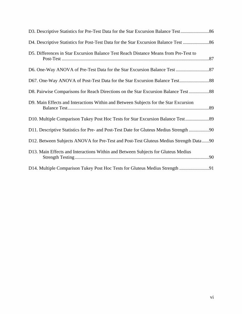
vi
D3. Descriptive Statistics for Pre-Test Data for the Star Excursion Balance Test ........................86 D4. Descriptive Statistics for Post-Test Data for the Star Excursion Balance Test ......................86 D5. Differences in Star Excursion Balance Test Reach Distance Means from Pre-Test to Post-Test ............................................................................................................................87 D6. One-Way ANOVA of Pre-Test Data for the Star Excursion Balance Test ............................87 D67. One-Way ANOVA of Post-Test Data for the Star Excursion Balance Test.........................88 D8. Pairwise Comparisons for Reach Directions on the Star Excursion Balance Test .................88 D9. Main Effects and Interactions Within and Between Subjects for the Star Excursion
Balance Test.......................................................................................................................89 D10. Multiple Comparison Tukey Post Hoc Tests for Star Excursion Balance Test ....................89 D11. Descriptive Statistics for Pre- and Post-Test Date for Gluteus Medius Strength .................90 D12. Between Subjects ANOVA for Pre-Test and Post-Test Gluteus Medius Strength Data ......90 D13. Main Effects and Interactions Within and Between Subjects for Gluteus Medius
Strength Testing .................................................................................................................90 D14. Multiple Comparison Tukey Post Hoc Tests for Gluteus Medius Strength .........................91

vii
LIST OF FIGURES
Figure Page C1. Subject Performing Fixed Surface Balancing .........................................................................74 C2. Subject Performing Tilt Board Balancing in the Dorsiflexion/Plantar Flexion Direction......74 C3. Subject Performing Tilt Board Balancing in the Inversion/Eversion Direction .....................75 C4. Subject Performing Tilt Board Balancing with a Diagonal Placement...................................75 C5. Subject Performing Wobble Board Balancing ........................................................................76 C6. Subject Performing Functional Hop Exercises .......................................................................76 C7. Functional Hop Pattern............................................................................................................77 C8. Subject Performing Side-Lying Hip Abduction with Elastic Resistance................................77 C9. Subject Walking with Weight in Non-Dominant Hand ..........................................................78 C10. Subject Performing Gorilla Walk with Elastic Resistance....................................................78 C11. Subject Performing Hip Abduction on Multi-hip Machine ..................................................79 C12. Subject Performing Single-Leg Squat...................................................................................80 C13. Subject Performing Lateral Step-Downs...............................................................................80 C14. Star Excursion Balance Test .................................................................................................81 C15. Subject Performing Static Quadriceps Stretch ......................................................................81 C16. Subject Performing Static Hamstrings Stretch......................................................................82 C17. Subject Performing Static Calf Stretch .................................................................................82 C18. Subject Performing Star Excursion Balance Test into Anterior Direction ...........................83 C19. Subject Performing Star Excursion Balance Test into Medial Direction..............................83 C20. Subject Performing Star Excursion Balance Test into Posterior Direction ..........................84 C21. Subject Performing Gluteus Medius Strength Test...............................................................84
viii
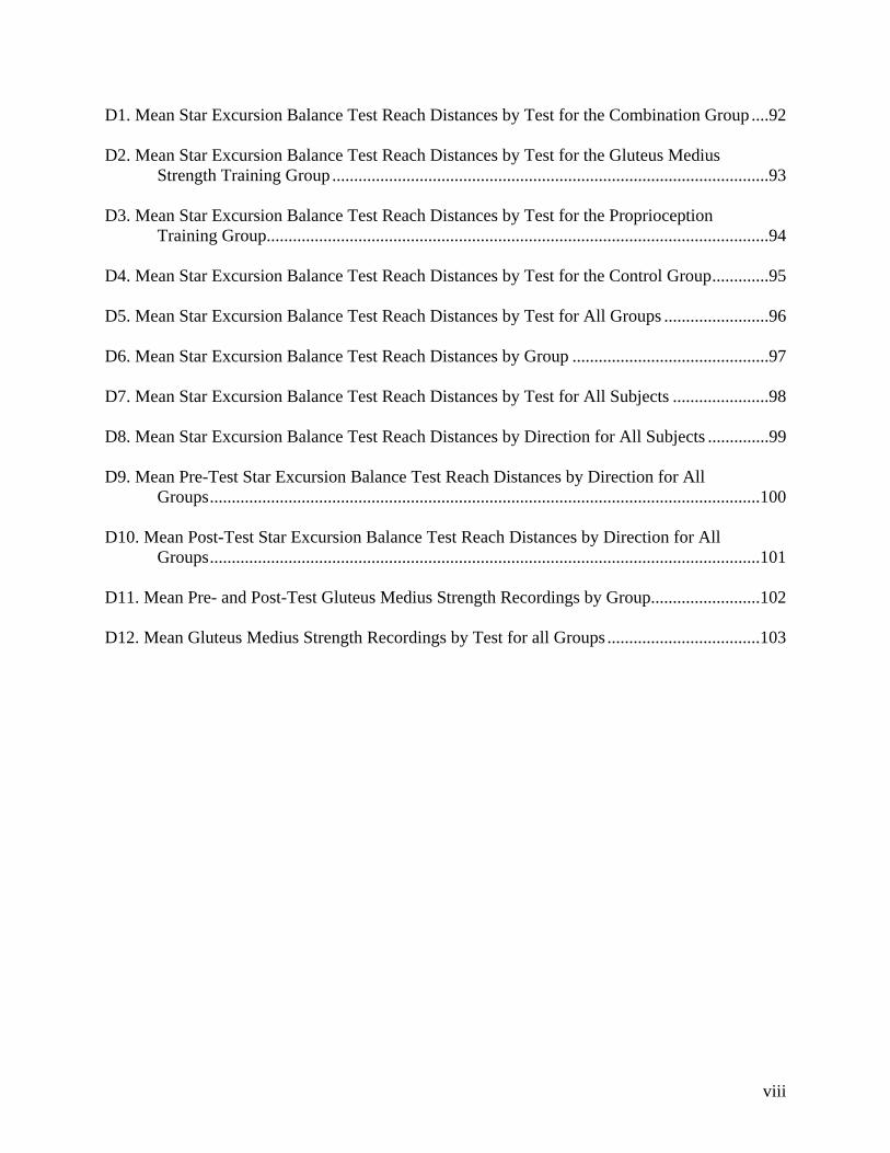
D1. Mean Star Excursion Balance Test Reach Distances by Test for the Combination Group ....92 D2. Mean Star Excursion Balance Test Reach Distances by Test for the Gluteus Medius Strength Training Group ....................................................................................................93 D3. Mean Star Excursion Balance Test Reach Distances by Test for the Proprioception Training Group...................................................................................................................94 D4. Mean Star Excursion Balance Test Reach Distances by Test for the Control Group.............95 D5. Mean Star Excursion Balance Test Reach Distances by Test for All Groups ........................96 D6. Mean Star Excursion Balance Test Reach Distances by Group .............................................97 D7. Mean Star Excursion Balance Test Reach Distances by Test for All Subjects ......................98 D8. Mean Star Excursion Balance Test Reach Distances by Direction for All Subjects ..............99 D9. Mean Pre-Test Star Excursion Balance Test Reach Distances by Direction for All
Groups..............................................................................................................................100 D10. Mean Post-Test Star Excursion Balance Test Reach Distances by Direction for All
Groups..............................................................................................................................101 D11. Mean Pre- and Post-Test Gluteus Medius Strength Recordings by Group.........................102 D12. Mean Gluteus Medius Strength Recordings by Test for all Groups ...................................103

INTRODUCTION
Ankle sprains are one of the most common injuries in sports today.1-26 Eighty-five to
ninety-five percent of all ankle ligament injuries are lateral ankle sprains (LAS) caused by an
inversion mechanism.1,2,5,6,15,18,22,24,27 An estimated 10 to 45 percent of all injuries in running and
jumping sports are ankle sprains.1,2,9,11,15, There are about 27,000 ankle sprains per day in the
United States, or one per every 10,000 people.1,11 Of those ankle sprains, recurrent ankle sprains
can lead to chronic ankle instability creating mechanical and functional deficits.1,2,4,5,7,21,26,28-33
Both can create long term complications with proprioceptive deficits affecting the ability of both
the ankle and hip strategy in maintaining dynamic postural control.28-33
There are two strategies for maintaining dynamic postural control: a hip strategy and an
ankle strategy.34,35 Generally, the ankle strategy, in which the peroneal muscles maintain
balance, is the method most often used by healthy individuals. The hip strategy, although most
often seen in the elderly, is adopted by previously-healthy individuals who sustain an ankle
sprain. In this strategy, the gluteus medius is used to correct posture and keep an individual
balanced and erect.23,34-36 Recent studies have shown that the gluteus medius muscle is weak
following ankle sprains.2,20,35 Usually an individual does not train to strengthen the gluteus
medius muscle when attempting to increase dynamic postural control following a lateral ankle
sprain. Specifically, in rehabilitating the sprained ankle, ankle strength training and
proprioception training is utilized to regain losses in balance that may have occurred.21,37-44
The Star Excursion Balance Test (SEBT) is often used as an assessment of an
individual’s dynamic postural control. The Star Excursion Balance Test is a functional balance
test that uses a unilateral stance on the center of an asterisk (star) and a maximal reach down
each of the asterisk’s eight lines.45-47 The SEBT has been used to evaluate dynamic postural
1

control following a lateral ankle sprain, but has not been used to evaluate gluteus medius
weakness. Several studies10,19,21,25,35 have evaluated proprioceptive training programs on
semidynamic and dynamic balance. To the authors knowledge, there are no known studies in the
literature that have evaluated the effect of strength training the gluteus medius on dynamic
postural control in healthy or injured subjects. Nor have these studies evaluated the efficacy of
gluteus medius strength training, ankle neuromuscular training (proprioception, dynamic postural
control, and joint position sense training exercises), or a combination of the two, for their effects
on dynamic postural control.20,35,46,48 Therefore, the purpose of this study was to determine if
gluteus medius muscle strength training, proprioception training, or a combination of the two
will have an effect on dynamic postural control.
METHODS
There were two separate designs for this study. This study was a 2 x 4 x 8 factorial
design as well as a 2 x 4. Independent variables of both designs were test and group. Test
existed on two levels (pre and post). The group existed on four levels (proprioception training
exercises, gluteus medius strength training exercises, combination, and control). An additional
independent variable for the first design was the eight reach directions for the SEBT. The
dependent variable in the first design was direction (anterior, anterolateral, lateral, posterolateral,
posterior, posteromedial, medial, and anteromedial). The dependent variable of the second
design was the gluteus medius strength measurement.
Subjects
This study began with 60 subjects in total, all of whom were students at West Virginia
University. Each of the subjects was a healthy individual with an unremarkable history of lower
extremity injury in the six months leading up to the study as well as no lower extremity surgery
2

in the past year. Subjects in experimental groups attended at least 14 of the 18 training periods
(approximately 77 percent attendance) and returned for post-testing in order for their results to be
included in this study. Twelve of the subjects dropped out or were removed from the study by
the primary investigator because they either missed too many training sessions or did not return
for one or both post-testing sessions.
Fifteen subjects were randomly assigned to each of the three experimental groups or to
the control group using a stratified randomization for gender and activity. Forty-eight subjects
(25 males and 23 females) completed the entire study. The mean age of the subjects was 22.06
± 1.58 years. The mean height of all subjects was 171.87 ± 9.52 centimeters, while the mean
weight was 75.72 ± 15.79 kilograms The gluteus medius strength training group completed the
study with thirteen subjects (n=13) while the combination and proprioception training groups
each completed the study with twelve subjects (n=12). The control group completed the study
with eleven subjects (n=11). Complete descriptive statistics for subject demographics can be
seen in Tables D1 and D2.
Instrumentation Using balance tasks to assess dynamic postural control has recently become more
commonplace in the clinical physical therapy and traditional athletic training settings.13
Dynamic postural control tests such as excursion and functional reach tests are superior to basic
single leg stance (SLS) tests.49 The Star Excursion Balance Test is a simple method of testing an
individual’s dynamic postural control. The SEBT, which was first introduced by Gary Gray in
1995, is a functional balance test that uses a unilateral stance on the center of an asterisk (star)
and a maximal reach down each of the asterisk’s eight lines.45-47 The SEBT offers a simple,
reliable, low-cost alternative to more expensive, refined instruments available today.20 The lines
3

extend out from the center of the asterisk at 45 degree increments on a grid. Each line has a
specific name in relation to which leg is being tested (the leg the participant is standing on is the
leg being tested). The eight directions are named anterolateral (AL), anterior (A), anteromedial
(AM), medial (M), posteromedial (PM), posterior (P), posterolateral (PL), and lateral (L)
respective to the foot that is weight-bearing.45-47,49-51 The SEBT is generally placed directly on a
non-slick floor using tape, eight tape measures, and a protractor. The participant unilaterally
stands in the center of the asterisk and maximally reaches with the contralateral leg down each
line.20 The test requires six practice reaches in each of the eight directions and three recorded
test reaches. Each reach, or trial, should be held for one second for recording of measurements.
Each trial is measured from the center of the asterisk and the three trials are averaged and
compared to the participant’s height or true leg length. Another way to compare SEBT
excursions is to add the averages from each of the eight directions and then multiply them by the
participant’s height. This does not allow the examiner to specifically point out directions in
which the participant is lacking in dynamic postural control, but it does provide a simple method
of comparing one participant’s overall dynamic postural control to that of others’.19 The further
the reach, the greater the demand is on the dynamic postural control of the weight-bearing leg.
28,45-47,49 The participant returns to the static unilateral stance position following each reach and
remains there for 10-15 seconds before the next trial. The reaches are completed in one direction
before moving on to another direction and should be completed in sequential order either
clockwise (CW) or counterclockwise (CCW). During the SEBT, the individual’s dynamic
postural control constantly corrects as his or her center of mass migrates from over the base of
support in all directions.52 Some factors to be considered when testing an individual using the
SEBT are balance disorders, foot type, past injuries/surgeries, flexibility, and shoe condition.51-52
4
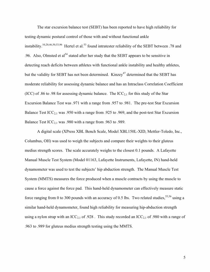
The star excursion balance test (SEBT) has been reported to have high reliability for
testing dynamic postural control of those with and without functional ankle
instability.10,20,46,50,53,96 Hertel et al.53 found intratester reliability of the SEBT between .78 and
.96. Also, Olmsted et al54 stated after her study that the SEBT appears to be sensitive in
detecting reach deficits between athletes with functional ankle instability and healthy athletes,
but the validity for SEBT has not been determined. Kinzey47 determined that the SEBT has
moderate reliability for assessing dynamic balance and has an Intraclass Correlation Coefficient
(ICC) of .86 to .98 for assessing dynamic balance. The ICC2,1 for this study of the Star
Excursion Balance Test was .971 with a range from .957 to .981. The pre-test Star Excursion
Balance Test ICC2,1 was .950 with a range from .925 to .969, and the post-test Star Excursion
Balance Test ICC2,1 was .980 with a range from .963 to .989.
A digital scale (XPress XBL Bench Scale, Model XBL150L-XID, Mettler-Toledo, Inc.,
Columbus, OH) was used to weigh the subjects and compare their weights to their gluteus
medius strength scores. The scale accurately weighs to the closest 0.1 pounds. A Lafayette
Manual Muscle Test System (Model 01163, Lafayette Instruments, Lafayette, IN) hand-held
dynamometer was used to test the subjects’ hip abduction strength. The Manual Muscle Test
System (MMTS) measures the force produced when a muscle contracts by using the muscle to
cause a force against the force pad. This hand-held dynamometer can effectively measure static
force ranging from 0 to 300 pounds with an accuracy of 0.5 lbs. Two related studies,55,56 using a
similar hand-held dynamometer, found high reliability for measuring hip-abduction strength
using a nylon strap with an ICC2,1 of .928 . This study recorded an ICC2,1 of .980 with a range of
.963 to .989 for gluteus medius strength testing using the MMTS.
5
Procedures
Subjects were contacted and asked to attend a meeting where they were provided with an
Informed Consent Form, an Authorization to Use or Disclose Protected Health Information
Form, and a Subject Demographics Questionnaire. The study was described to the potential
subjects to make an informed decision about whether to participate or not. Any questions from
the potential subject pool were answered and explained. The potential subjects were then asked
to fill out the Informed Consent Form (Table C1), the Authorization to Use or Disclose Protected
Health Information Form (Table C2), and Subject Demographics Questionnaire (Table C3)
truthfully and to the best of their ability. The researcher then reviewed the forms for
completeness and noted if subjects fit the inclusion criteria, and not the exclusion criteria. If
subjects fit the inclusion criteria, they were contacted by the researcher to scheduled a time to
perform the pre-test Star Excursion Balance Test and gluteus medius strength testing.
Times were established for subjects to meet with the researcher three times a week over
six weeks for approximately 20 minutes per session to perform their proprioceptive exercises,
gluteus medius strengthening exercises, or a combination of the two. The dominant leg was used
for the training sessions. Leg dominance was determined by the leg the subjects would have
used to kick a ball. All exercises were performed at HealthWorks Rehab and Fitness in
Morgantown, WV to serve as an environmental control. The primary researcher administered
and supervised all testing and exercising sessions. At the conclusion of the last exercise session,
the subjects performed their post-test for the SEBT and gluteus medius strength. The post-test
was performed to the exact specifications as the pre-test, and was completed within the week
following the final training session of the sixth week.
6
Control group: The control group was not required to attend any sessions for the training
exercises. The researcher was in contact with the subjects of this group weekly to obtain
information on any activities they were performing, such as weightlifting, running, or swimming,
and to see that they were adhering to the guidelines.
Proprioceptive training group: Proprioceptive exercises are those exercises that increase
the body’s ability to detect motion in the foot and make postural adjustments accordingly.
Proprioceptive training is most often performed using static or dynamic balance exercises.48 The
subjects within the proprioception group performed a battery of exercises that changed from
week to week.48 Exercises one through three included fixed surface balancing with eyes open
(Figure C1), fixed surface balancing with eyes closed, and fixed surface balancing while picking
up objects. A tilt board (ECO Pro Rocker Board, Model 80314, Power Systems, Inc., Knoxville,
TN), was added for additional balance training for exercises four through nine. Tilt board
balancing occurred with the board in a dorsiflexion/plantar flexion pattern with eyes open
(Figure C2) as well as closed. Next the subject performed tilt board balancing with the board
positioned in an inversion/eversion pattern with eyes open (Figure C3) and then closed. The
subject performed tilt board balancing with the board placed on a diagonal with the eyes open
(Figure C4) and then closed. Wobble board (ECO Pro Wobble Board, Model 80312, Power
Systems, Inc., Knoxville, TN) balancing was performed with eyes open and then closed for
exercises ten and eleven (Figure E5). The last exercises performed were the functional hops with
eyes open and then with eyes closed upon landing (Figures C6 and C7). These exercises were
performed for 15 seconds with 45 second breaks between exercises in the first week. Time was
then added to the exercises and taken away from the rest periods until the two were equal at 30
seconds by week four. A detailed description of the weekly progression can be seen in Table C4.
7

Gluteus medius strength training group: This group performed six exercises for the six
week training period (Table C5). These exercises have not been validated by other studies to
increase gluteus medius strength, but they were selected from typical rehabilitation programs
performed in sports medicine clinics and athletic training facilities that focus on strengthening
the hip abductors. The first exercise (Figure 8) was a side-lying hip abduction exercise using a
blue (“Extra Heavy”) Thera-Band (Model 20050, The Hygenic Corporation, Akron, OH). The
subjects performed three sets of 10 repetitions of this exercise for the first two weeks, increasing
to three sets of 15 repetitions the third and fourth weeks, and three sets of 20 the final two weeks.
Next the subject walked for three minutes around an 80 meter track at HealthWorks Rehab and
Fitness. The subject carried a dumbbell in the hand opposite the dominant leg. The weight was
started at five percent of body weight for the first two weeks, 10 percent of body weight for the
third and fourth weeks, and 15 percent of body weight for the final two weeks (Figure C9).
Gorilla walking, or lateral walking with a Thera-Band wrapped around both legs and placed just
above the knees was next (Figure C10). The subject walked for three sets of 20 seconds using a
blue (“Extra Heavy”) Thera-Band for the first two weeks, three sets of 30 seconds the third and
fourth weeks, and three sets of 40 seconds for the final two weeks. The fourth exercise (Figure
C11) was standing hip abduction using a multi-hip machine (Maximus MX506, Model MX506,
CYBEX International, Inc., Fairfield, CT). The moving arm pad was placed just above the
lateral knee on the lateral femoral condyle while completing three sets of 10 repetitions. The
weight used was adjusted according to the pre-test strength values recorded for each subject. For
the first two weeks, 50 percent of the tested value was used. The force was increased to 60
percent of the tested value for the third and fourth weeks and 70 percent for the final two weeks.
The next exercise was a single leg squat (Figure C12). The subject performed a squat
8
approximately 45 degrees while standing on the dominant leg. Each subject was cued when to
stop and return to the starting position by the primary investigator. Two sets of five squats were
completed the first two weeks, three sets of five squats were completed the third and fourth
weeks, and four sets of five squats were completed the final two weeks. Finally, the subject
completed lateral step-downs (Figure C13). The subject stood on a six inch step on the dominant
leg. The subjects were instructed to bend the knee until the contralateral foot barely touched the
ground next to the step, and then return to the starting position. Two sets of five step-downs for
the first two weeks, three sets of five step-downs for the third and fourth weeks, and four sets of
five step-downs for the final two weeks were performed.
Combination group: This group performed all of the exercises for the proprioception
training group and the gluteus medius strength training group. The proprioception and gluteus
medius strength exercises were performed in a random order as decided by convenience of
training equipment availability.
Pre- and Post-Test
Initially, every subject was oriented to the Star Excursion Balance Test (Figure C14). A
detailed description of Star Excursion Balance Test procedures can be found in Table C6. The
subjects had their true leg length measured prior to testing in order to normalize the excursion
measurements.45,46 They underwent a warm-up session of five minutes on a stationary bicycle at
120 revolutions per minute (RPM) followed by five minutes of static quadriceps, hamstrings, and
calf stretching (Figures C15, C16, and C17). The subject took a five minute break and then
started with six trials of excursions for each of the eight directions on the SEBT. A five minute
rest was given before the subject underwent the pre-test. Subjects were asked to randomly select
index cards with different directions on the back of each to determine the starting excursion
9

direction. Right-leg dominant subjects started with the direction they selected on the card and
continued in a counterclockwise direction until all eight directions were completed. Left-leg
dominant subjects started with the direction they selected on the card and continued in a
clockwise direction until all eight directions were completed (Figures C18, 19, and 20). Each
subject then underwent the three trials which were recorded on a data sheet (Tables C7 and C8)
for each reach direction on the SEBT. Subjects were instructed to touch down very gently at the
furthest reach point possible along each of the eight lines. Each trial was held for at least one
second in order for the primary investigator to make a recording. If the subject touched down
with the non-dominant leg to provide considerable support, that trial was nullified.45,46,49,52
Trials were also nullified if the dominant leg was lifted off the ground or if the subject could not
maintain balance. The subject returned to the static unilateral stance position following each
reach trial and remained in that position for at least 10 seconds.45,46 The three trials in each reach
direction were averaged and normalized to the individual’s leg length. To avoid a possibility for
fatigue and/or a learning effect, there was a rest period before starting the actual trials.
On a separate testing day, the subjects were scheduled to have their gluteus medius
strength measured. A detailed description of gluteus medius strength testing procedures can be
seen in Table C9. First, the subject’s were weighed using a digital scale to be used to normalize
the strength values to the subject’s body weight. The scale had an accuracy of 0.1 pounds. A
Lafayette Manual Muscle Test System (Model 01163, Lafayette Instruments, Lafayette, IN)
hand-held dynamometer was used to test the subjects’ gluteus medius (hip abduction) strength.
The subject was side-lying on the non-dominant side. The dominant hip was at approximately 10
degrees of abduction prior to testing.55 The force pad of the hand-held dynamometer was placed
just above the knee joint on the lateral femoral condyle (Figure C21).56 The subject took three
10

practice trials and three trials were recorded on a data sheet (Tables C10 and C11).55,56 The
dynamometer was zeroed, and then the subject was instructed to abduct the dominant leg by
building up force for two seconds and then applying a maximal effort for four seconds.56 The
value on the dynamometer was recorded and then the dynamometer was zeroed out again before
the next trial. The subject had 15 seconds of rest between trials. The three trials were averaged
and were normalized to the individual’s body weight.
Data Analysis On coded sheets, the anterior, anterolateral, lateral, posterolateral, posterior,
posteromedial, medial, and anteromedial reach distances were recorded. An average of these
excursions was calculated from the three trials of each dependent variable for each subject and
recorded. Upon test completion of all subjects, values were entered into a spreadsheet on SPSS.
To normalize to leg length for the SEBT, the primary investigator divided the average excursion
length for each direction by the leg length and then multiplied by 100.
On separate coded sheets, the gluteus medius strength values were recorded (Tables C10
and C11). To normalize to body weight for gluteus medius strength, the primary investigator
divided the average gluteus medius strength by the subject’s body weight and then multiplied by
100.
Statistical Analysis Descriptive analysis consisted of means and standard deviations of all subjects for the
SEBT and gluteus medius strength. A one-way Multivariate Analysis of Variance (ANOVA)
was calculated for pre-test scores for each the SEBT and gluteus medius strength to determine if
there were differences between the groups. A three-way Repeated Measures ANOVA was used
to determine main effects and interactions for the SEBT. A two-way Repeated Measures
11
ANOVA was used to determine the main effects and interactions of gluteus medius strength.
Tukey post-hoc tests and pairwise comparisons were run to determine if differences existed
between pre-test and post-test results. A P value of 0.05 was used for all analyses.
RESULTS
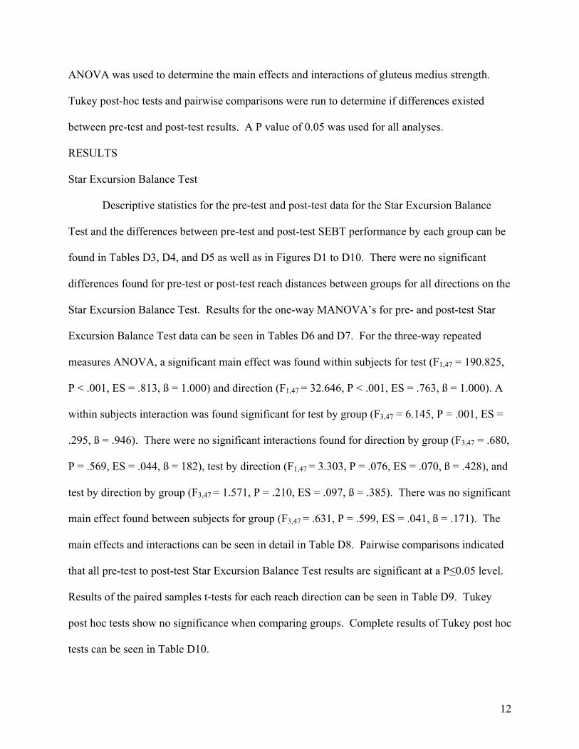
Star Excursion Balance Test
Descriptive statistics for the pre-test and post-test data for the Star Excursion Balance
Test and the differences between pre-test and post-test SEBT performance by each group can be
found in Tables D3, D4, and D5 as well as in Figures D1 to D10. There were no significant
differences found for pre-test or post-test reach distances between groups for all directions on the
Star Excursion Balance Test. Results for the one-way MANOVA’s for pre- and post-test Star
Excursion Balance Test data can be seen in Tables D6 and D7. For the three-way repeated
measures ANOVA, a significant main effect was found within subjects for test (F1,47 = 190.825,
P < .001, ES = .813, ß = 1.000) and direction (F1,47 = 32.646, P < .001, ES = .763, ß = 1.000). A
within subjects interaction was found significant for test by group (F3,47 = 6.145, P = .001, ES =
.295, ß = .946). There were no significant interactions found for direction by group (F3,47 = .680,
P = .569, ES = .044, ß = 182), test by direction (F1,47 = 3.303, P = .076, ES = .070, ß = .428), and
test by direction by group (F3,47 = 1.571, P = .210, ES = .097, ß = .385). There was no significant
main effect found between subjects for group (F3,47 = .631, P = .599, ES = .041, ß = .171). The
main effects and interactions can be seen in detail in Table D8. Pairwise comparisons indicated
that all pre-test to post-test Star Excursion Balance Test results are significant at a P≤0.05 level.
Results of the paired samples t-tests for each reach direction can be seen in Table D9. Tukey
post hoc tests show no significance when comparing groups. Complete results of Tukey post hoc
tests can be seen in Table D10.
12
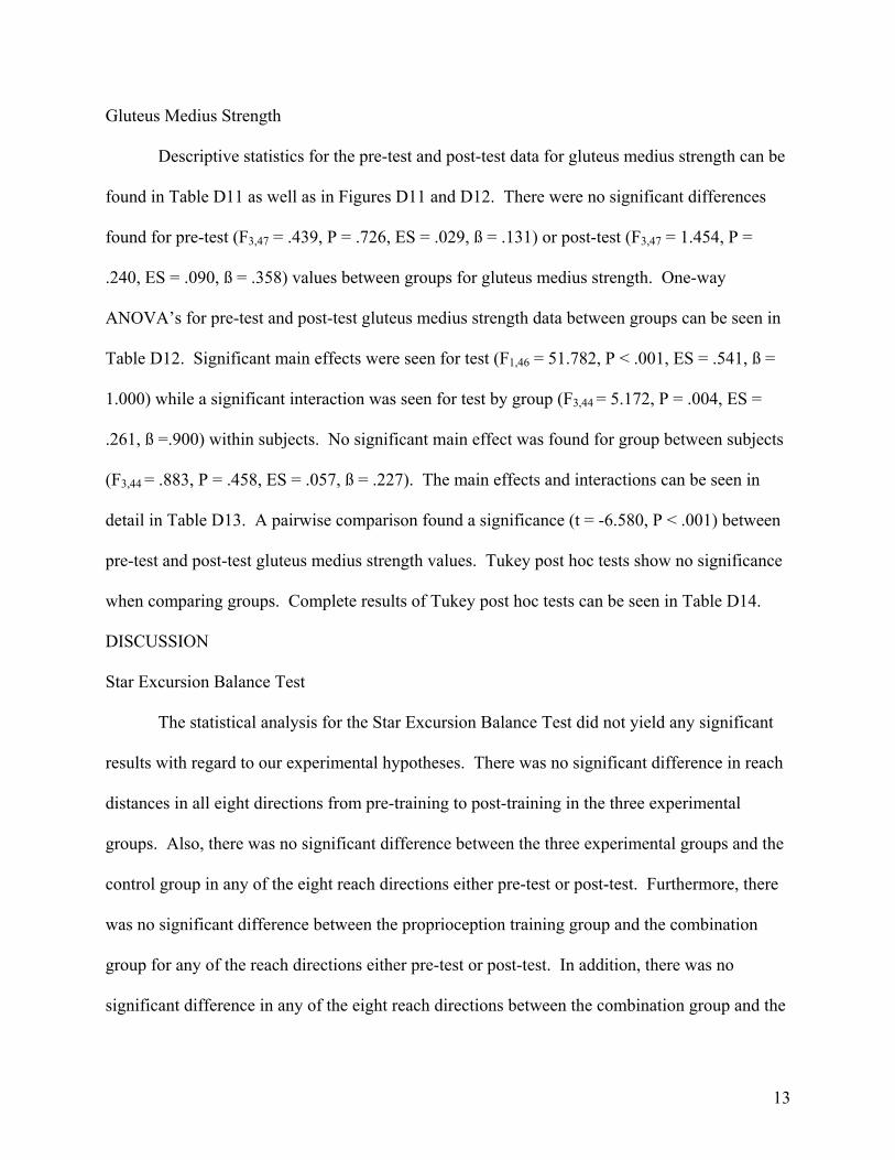
Gluteus Medius Strength
Descriptive statistics for the pre-test and post-test data for gluteus medius strength can be
found in Table D11 as well as in Figures D11 and D12. There were no significant differences
found for pre-test (F3,47 = .439, P = .726, ES = .029, ß = .131) or post-test (F3,47 = 1.454, P =
.240, ES = .090, ß = .358) values between groups for gluteus medius strength. One-way
ANOVA’s for pre-test and post-test gluteus medius strength data between groups can be seen in
Table D12. Significant main effects were seen for test (F1,46 = 51.782, P < .001, ES = .541, ß =
1.000) while a significant interaction was seen for test by group (F3,44 = 5.172, P = .004, ES =
.261, ß =.900) within subjects. No significant main effect was found for group between subjects
(F3,44 = .883, P = .458, ES = .057, ß = .227). The main effects and interactions can be seen in
detail in Table D13. A pairwise comparison found a significance (t = -6.580, P < .001) between
pre-test and post-test gluteus medius strength values. Tukey post hoc tests show no significance
when comparing groups. Complete results of Tukey post hoc tests can be seen in Table D14.
DISCUSSION
Star Excursion Balance Test
The statistical analysis for the Star Excursion Balance Test did not yield any significant
results with regard to our experimental hypotheses. There was no significant difference in reach
distances in all eight directions from pre-training to post-training in the three experimental
groups. Also, there was no significant difference between the three experimental groups and the
control group in any of the eight reach directions either pre-test or post-test. Furthermore, there
was no significant difference between the proprioception training group and the combination
group for any of the reach directions either pre-test or post-test. In addition, there was no
significant difference in any of the eight reach directions between the combination group and the
13

control group. Therefore, the four experimental hypotheses for the Star Excursion Balance Test
were rejected.
For this study, results for within subjects found significant main effects for both test and
direction. Despite there being no significance between groups, overall pre-test to post-test reach
distances were notably different. With all groups, the posterior (P) reach direction was always
the greatest while the lateral (L) and anterolateral (AL) reach directions were the least. In
addition, a within and between subjects interaction was found significant for test by group. This
interaction indicates that the specific test (either pre- or post-test) influenced the reach distances
recorded by the groups. On the contrary, pre- and post-test pairwise comparisons of each of the
eight SEBT reach directions were significant at the P≤0.05 level. No significant interactions
were found for direction by group, test by direction, and test by direction by group.
Despite no significant differences, it is apparent that all groups improved their reach
distances in three of the directions. The posteromedial, posterior, and posterolateral reach
directions noted significant gains across all groups except for the control group. The control
group had the least improvement from pre-test to post-test, while the combination group and
gluteus medius strength training group showed the most improvement. The combination and
gluteus medius strength training groups were able to show improved SEBT reach distances
because the gluteus medius strength training exercises they performed was able to train thehip
strategy of dynamic postural control. The combination group experienced the greatest mean
improvements in the posteromedial (5.75 ± 3.84), posterior (6.26 ± 3.19), and posterolateral
(6.14 ± 7.30) reach directions, while the gluteus medius strength training group’s improvements
were slightly less (5.13 ± 4.94, 5.65 ± 3.91, and 5.46 ± 4.39, respectively). The proprioception
training group’s mean improvements were notably less than the combination and gluteus medius
14

strength training groups (3.38 ± 3.00, 4.77 ± 5.00, and 6.24 ± 5.66, respectively), on those same
directions. The control group showed the least mean improvements of 2.49 ± 2.36, 3.24 ± 3.63,
and 2.97 ± 5.33, respectively. The results of this study and others like Piegaro,57 Samson,58 and
Gribble and Hertel,45 noted changes in reach distances pre-test to post-test for the posteromedial,
posterior, and posterolateral directions. They hypothesized that the posterior reach directions
were the easiest of the reach directions, and it is for this reason that even the control group
showed improvements from pre-test to post-test. Group pre-test and post-test performance
means from each reach direction can be seen in Figures D1, D2, D3, D4, and D5. Figure D6
indicates that despite having the lowest overall Star Excursion Balance Test pre-test means, the
combination group showed the greatest improvement between pre-test and post-test. The
anterior, anteromedial and medial reach directions were not the easiest to perform, however they
were easier than lateral and anterolatereal and indicated a moderate change between pre-test and
post-test.
Among all subjects, the lateral and anterolateral reach directions indicated the least
improvements for pre-test and post-test than any of the other directions. One reason for this may
be that the two directions appeared to be the most difficult. This is apparent by the graph in
Figure D7 which indicates both pre-test and post-test means for each reach direction for each
group. Studies have alluded to the difficulty of each of the reach directions based on their
results. Gribble and Hertel,45 Samson,58 and Piegaro57 all indicated that the lateral and
anterolateral directions were among the lowest excursions recorded. A shorter reach direction
would imply more difficulty and/or a lack of dynamic postural control for that particular reach
direction. The differences between pre-test and post-test SEBT reach distances can be seen for
all groups in Figure D8, while separate pre-test and post-test reach distances for each group can
15
be seen in Figures D9 and D10. Although there were no significant differences found within
these results, the graphs indicate differences that may be clinically relevant.
Although there were differences pre-test and post-test with reach directions following the
training programs, this study also expected to see significant difference between the groups
performing the proprioception exercises (proprioception and combination) and the groups
performing only gluteus medius strength training or no training (control). However, no such
significance was found. Perhaps this suggests that either gluteus medius strength training,
proprioception training, or a combination of both will be effective in improving SEBT reach
directions. However, the combination group trained both hip and ankle strategy with the training
programs included in this study.
Why significant results wee not evident could be that all of the subjects were healthy.
Healthy individuals should have no dynamic balance or ankle proprioception abnormalities that
would typically be seen in individuals with chronic ankle instability or following an acute ankle
injury. Because of no history of injury among healthy subjects, neuromotor systems are intact
and require no re-training. In this case, when healthy individuals were trained for lower
extremity proprioception, they had no significant difference in pre-test to post-test dynamic
postural control. It is proposed that healthy individuals should undergo more than a six-week
classic rehabilitation-type program for proprioception in order to achieve gains in dynamic
postural control.
There are other training studies that reported group results similar to this study using
healthy subjects. Blackburn et al.59 and Riemann et al.,60 also conducted studies using active,
healthy subjects. The study by Blackburn et al. tested 32 healthy, physically active subjects on
the Bass Test, a hop and land test for dynamic balance, to find a significant difference between
16
the control group and proprioception group. The Bass Test is performed by having subjects
complete a pattern of hopping and landing on a series of 10 marks that have been placed on the
floor in a standardized manner. Subjects were required to completely cover each mark with their
metatarsal heads while maintaining their balance for as long as possible with the calcaneus lifted
off of the ground for up to five seconds. In their study, the proprioception group used exercises
such as resistance band kicks, single-leg standing on a foam surface, single-leg hops, and BAPS
board single-leg standing for six weeks. The study also had an ankle strength group and a
combined proprioception/strength group, however, this study found significant differences
between groups.59 The study by Riemann et al.60 was performed on 26 recreationally active and
tested ankle kinesthesia and postural control. Unlike this study, the one by Riemann et al.60 used
simply BAPS board proprioceptive exercises. However, much like this study, there was no
significant difference between experimental and control groups for static or functional postural
control.
Piegaro57 used a proprioceptive training program very similar to the one used for this
study, however, the training period in the Piegaro study lasted only four weeks. Piegaro also
tested his subjects using the Star Excursion Balance Test on healthy college students (N=39). He
noted significant differences between pre-test and post-test reach distances in the medial,
posterior, and lateral directions, and a time by group interaction for posteromedial and
anterolateral.57 He also found no significant difference between groups. To our knowledge, the
study by Piegaro is the only other study that has used the Star Excursion Balance Test to
determine dynamic postural control before and after a training program.
In contrast, two other training studies, one by Powers et al.41 and one by Bernier and
Perrin,48 were conducted on individuals with a history of functional ankle instability. Powers et
17

al.41 used a proprioception training program consisting solely of resistance band kicks on 38
college-aged individuals. The study determined that the six-week program caused no significant
change from pre-test to post-test in static balance on a force plate. Bernier and Perrin,48 on the
other hand, used a combination of a s balance training on fixed and unstable surfaces as well as
hopping exercises. Using the Balance System by Chattanooga, their six-week program achieved
significant differences for semi-dynamic postural stability on anterior/posterior and
medial/lateral modified equilibrium scores between groups. Although this study used the same
proprioceptive training program it was hypothesized that the reason why there were no
significant differences between groups was that it used healthy subjects rather than those with
functional ankle instability.
Why this occurred may be related to the ankle and hip strategies that are used to maintain
balance in healthy subjects. Generally, young, healthy, active individuals, like the ones in this
study use the ankle strategy before the hip strategy for altering/correcting center of mass. Using
the ankle strategy, an individual maintains postural control by firing the peroneal muscles.13,61 In
older individuals or those with ankle injuries, the gluteus medius and other hip abductors
compensate for weakened ankle musculature and maintain postural control.13,62,65 This proximal
compensation to postural sway of center of mass is often seen in individuals with lateral ankle
sprains and those with functional instability in the ankle.2,10,48,65 An increase in gluteus medius
activity during sudden ankle inversion in healthy subjects as well as those with functionally
unstable ankles was noted in an EMG study by Schmitz et al.64 It is for this reason that it
becomes very difficult to train individuals, healthy or injured, for purely ankle strategy
balancing. Some of the individuals in the proprioception group may have used the hip strategy
to maintain balance during their training, and therefore, were able to improve strength in the
18

gluteus medius without training, which may have affected the results of this study. It is proposed
that the subjects performing the gluteus medius strength training exercises (gluteus medius
strength and combination) had the greatest gains in SEBT reach distances because they trained
the gluteus medius, the primary muscle involved with the hip strategy of dynamic postural
control.
The randomized stratification used to divide subjects into four groups guaranteed that all
active subjects were evenly distributed into the control and experimental groups. The descriptive
statistics for the groups can be seen in Tables D1 and D2, which show similarities between all
groups in many demographics including gender, age, height, and weight. Also, all of the
subjects were relatively active individuals, but none would be classified as “high level” athletes.
It is also suggested from the results that moderately active, healthy subjects will achieve gains in
dynamic postural control in spite of the group into which they were placed. In addition, healthy
subjects may require a training period in excess of the six-week program developed for this
study. A training period of eight or ten weeks may have achieved more statistically significant
results. The healthy subjects had no neuromotor dysfunction or other abnormality that may have
predisposed them to poor dynamic postural stability. Therefore, the subjects in this study might
not have acquired the benefits from any of the training programs that would be noted in subjects
with functional ankle instability.
Despite the effect size for some of this study’s data being moderate to large, the small
sample size used for this study undoubtedly affected the significance and power. The groups
started at 15 subjects each and finished with the gluteus medius strength group (n=13) having the
most, the combination and proprioception groups (n=12) in the middle, and the control group
19
having the fewest (n=11). If the groups each had 20 (N=80) or 30 (N=120) subjects finish the
study, the results may have been much different.
Gluteus Medius Strength
The statistical analysis for gluteus medius strength testing did not yield any significant
results with regard to our experimental hypothesis. There was no significant difference in
gluteus medius strength measurements from pre-training to post-training between the two
experimental groups performing six weeks of gluteus medius strength training and the control
group. Also, there was no significant difference between the gluteus medius strength training
groups and the proprioception and control groups for gluteus medius strength values. Therefore,
the two experimental hypotheses for gluteus medius strength testing were rejected.
For this study, results for within subjects found significant main effects for test. Despite
there being no significant differences for pre-test data among the subjects, overall pre-test to
post-test gluteus medius strength values were notably different. The gluteus medius strength
group showed the greatest improvement in gluteus medius strength (2.05 ± 1.31) followed by the
combination group (1.70 ± 1.55) and the proprioception group (0.70 ± 0.71). The control group
showed the least improvement (0.45 ± 0.91). Even though the between subjects factor for group
was not significant, the graphs in Figures D11 and D12 show increases in gluteus medius
strength among those who performed gluteus medius strengthening. In addition, a within
subjects interaction was found significant for test by group. This interaction indicates that the
specific test (either pre- or post-test) influenced the gluteus medius strength values recorded for
the groups.
As this is the first study to evaluate a strengthening program of the gluteus medius for
dynamic postural control, there are no comparison studies. This study focused on determining
20

the effect of a combination of gluteus medius strength training and proprioception training on
dynamic postural control. It is for this reason why an increase in gluteus medius strength was
not the primary focus but rather whether the training programs improved dynamic postural
control.
The exercises developed as a training program for strengthening the gluteus medius have
not been evaluated collectively as being reliable or valid in the literature. They were typically
used in a rehabilitation setting when attempting to strengthen a weak gluteus medius, and it is
assumed from past studies that the gluteus medius muscle was involved while performing the
exercises.62,65,66 Wilson65 used the functional method of strengthening the gluteus medius also
employed in this study. He used healthy subjects to strengthen the gluteus medius by carrying a
dumbbell in the contralateral hand. The subjects used a dumbbell in the range of five to 15
percent of the individual’s body weight by walking while carrying the weight. The study
determined through side-lying manual muscle testing as well as a standing Trendelenburg sign
(dropping in contralateral hip during a unilateral stance) that this effectively strengthened the
gluteus medius.65 “Arc walking” is another strengthening method that Wilson65 used in his
study. In arc walking, the individual walks while attached to a secure structure by a resistance
band. The individual then walks in an arc pattern around the origin of the resistance band while
keeping his or her toes pointed at the band. The lateral sidestepping against a resistance was
shown to strengthen the gluteus medius also using the side-lying manual muscle test and
Trendelenburg sign.65 The arc walking exercise utilized by Wilson65 was very similar to the
gorilla walking exercise used in this study. Both use hip abduction while in a standing position
against an elastic resistance. Gorilla walking was chosen for this study because it requires hip
21
abduction against direct resistance as opposed to resistance applied to the waist, as in arc
walking.
Open kinetic chain exercises are most often employed for strengthening the GM in the
clinical setting. Generally, the GM is strengthened using a side-lying straight leg raise as the hip
is moved into abduction.65,66 This is usually performed against an elastic resistance or an ankle
cuff weight. Earl62 suggests using the open kinetic chain exercise called hip hikes, or standing
hip abduction.62 This study utilized open kinetic chain exercises in both the side-lying and
standing positions. The side-lying hip abduction exercise used in this study used a blue, or Extra
Heavy, latex rubber resistance band while the standing hip abduction was performed on the
multi-hip machine. Two other closed kinetic chain exercises normally performed as part of
typical lower extremity rehabilitation and/or strengthening program were also included. Single-
leg squats and lateral step-downs, although not found in rehabilitation or strengthening literature
for strengthening the gluteus medius, are often used in the clinical setting. These exercises use
an individual’s body weight as resistance to strengthen the gluteus medius.
Most of the studies conducted on testing the strength of the gluteus medius have been
performed on injured subjects with ankle sprains, iliotibial band pain, or patellofemoral pain
syndrome and do not give any strengthening suggestions.55,65,67 Another study recommends
strengthening the gluteus medius when it is found to be in a weakened state because of its
involvement in dynamic postural control and locomotion during gait.66 This study by Ogiwara
and Sugiura66 suggested the use of progressive resistance exercises using a ten-repetition-
maximum. Progressive resistance exercises have become the most commonly employed method
for regaining the strength of the gluteus medius in the clinical setting, but there is not a simple
way to determine an individual’s ten-repetition-maximum to ensure adequate resistance for
22
strengthening. Although ten-repetition-maximum is the suggestion by Ogiwara and Sugiura66 for
rehabilitating injured athletes, it may not have been the most appropriate choice for training
healthy subjects when the goal is strengthening. The weight increments used did not place an
overload force on the muscles, thus additional weight or exercises that are purely strength related
are needed. Following the National Academy of Sports Medicine’s71 guidelines for strength
training (four to six sets of one to five repetitions that are between 85 and 100 percent of an
individual’s one repetition maximum four times per week) for all gluteus medius strength
training group exercises may have yielded significant results. A suggestion would be to perform
exercises using the overload principle to stress the gluteus medius by performing exercises using
the subject’s body weight or a load in excess of body weight in place of resistance band training.
This study has indicated some clinical significance in successfully improving gluteus
medius strength in all groups, especially among those in the combination and gluteus medius
strength groups. It is quite possible that the groups who performed these exercises did not
perform enough sets and reps of these exercises or did not have enough resistance while
performing them to maximize strength. Despite lack of significant differences between groups
the subjects in the proprioception group were still able to benefit from a strengthened gluteus
medius because of the proprioception training exercises being performed on one foot. Every
exercise, including hopping and balancing on unstable surfaces, was performed in a unilateral
dominant leg stance. However, there was no statistical significance between the groups which
suggests the gluteus medius may require more than the exercises performed to increase strength.
Clinical Implications
Although this study presented statistical significance, the true benefits of this study lies in
its clinical relevance. This study indicated that six weeks of proprioception training, gluteus
23

medius strengthening, or a combination program will increase SEBT reach distances in all
directions as well as slightly increase gluteus medius strength. The two groups that performed
gluteus medius strengthening completed the study with greater increase in gluteus medius
strength than did the proprioception and control groups. In addition the SEBT reach distances
among the gluteus medius strengthening and combination groups demonstrated greater
improvements in most SEBT reach directions when compared to the control and proprioception
training groups.
This study will be beneficial in the clinical setting for increasing the dynamic postural
control of healthy individuals. Improvements were seen in both Star Excursion Balance Test
reach distances as well as gluteus medius strength recordings regardless of group. The
combination group demonstrated the most improvements in SEBT reach distances while the
gluteus medius strength training group demonstrated the most improvement in gluteus medius
strength. The results indicate that in spite of the training performed for the six-week period,
improvements were seen among all groups in both SEBT reach distances and gluteus medius
strength. In summary, the use of proprioception exercises, gluteus medius strength exercises, or
a combination of the two will help improve dynamic postural control among healthy, active
individuals.
CONCLUSION
This study has indicated that a combination of gluteus medius strength training and
proprioception training does not demonstrate an improvement in dynamic postural control more
so than proprioception training alone. However, the enhancement of dynamic postural control
recognized following six weeks of either gluteus medius strength training or proprioception
training is clinically relevant. Although there was no significant difference between pre-test and
24
post-test values of gluteus medius strength, it is apparent that the groups performing gluteus
medius strengthening exercises showed improved gluteus medius strength. In the clinical
setting, these training programs would be performed on individuals with lower extremity
injuries, which should result in greater improvement in dynamic postural control using hip and
ankle strategy. However, it is rare that an individual with a lower extremity injury will perform
gluteus medius strengthening to increase dynamic postural control. This study indicates that,
although not statistically significant, gluteus medius strengthening used as a supplement to a
lower extremity rehabilitation program may improve dynamic balance greater than
proprioception training alone.
25
REFERENCES
1. Baumhauer JF, Alosa DM, Renstrom PAFH, Trevino S, Benynnon B. A prospective study of ankle injury risk factors. Am J Sports Med. 1995;23(5):564-570.
2. Beckman SM, Buchanan TS. Ankle inversion injury and hypermobility: effect on hip and
ankle muscle electromyography onset latency. Arch Phys Med Rehabil. 1995;76(12):1138-1143.
3. Beynnon BD, Renstrom PA, Alosa DM, Baumhauer JF, Vacek PM. Ankle ligament
injury risk factors: a prospective study of college athletes. J Orthop Res. 2001;19(2):213-20.
4. Beynnon BD, Murphy DF, Alosa DM. Predictive factors for lateral ankle sprains: a
literature review. J Athl Train. 2002;37(4):376-380.
5. Brown C, Ross S, Mynark R, Guskiewicz K. Assessing functional ankle instability with joint position sense, time to stabilization, and electromyography. J Sport Rehabil. 2004;3(2):122-34.
6. Demeritt KM, Shultz SJ, Docherty CL, Gansneder BM, Perrin DH. Chronic ankle
instability does not affect lower extremity functional performance. J Athl Train. 2002;37(4):507-511.
7. Denegar CR, Miller SJ. Can chronic ankle instability be prevented? Rethinking
management of lateral ankle sprains. J Athl Train. 2002; 37(4):430-435.
8. Denegar CR, Hertel J, Fonseca J. The effect of lateral ankle sprain on dorsiflexion range of motion, posterior talar glide, and joint laxity. J Orthop Sports Phys Ther. 2002;32(4):166-173.
9. DiGiovanni BF, Partal G, Baumhauer JF. Acute ankle injury and chronic lateral
instability in the athlete. Clin Sports Med. 2004;23(1):1-19.
10. Gribble PA, Hertel J, Denegar CR, Buckley WE. The effects of fatigue and chronic ankle instability on dynamic postural control. J Athl Train. 2004;39(4):321-329.
11. Hale SA, Hertel J. Reliability and sensitivity of the foot and ankle disability index in
subjects with chronic ankle instability. J Athl Train. 2005;40(1):35-40.
12. Hertel J, Guskiewicz K, Kahler D, Perrin D. Effect of lateral ankle and joint anesthesia on center of balance, postural sway, and joint position sense. J Sport Rehabil. 1996;5:111-119.
13. Hertel J. Functional anatomy, pathomechanics, and pathophysiology of lateral ankle
instability. J Athl Train. 2002;37:364-375.
26

14. Hertel J, Denegar CR, Monroe MM, Stokes WL. Talocrural and subtalar joint instability
after lateral ankle sprain. Med Sci Sports Exerc. 1999;31(11):1501-1508.
15. Hubbard TJ, Kaminski TW. Kinesthesia is not affected by functional ankle instability status. J Athl Train. 2002;37(4):481-486.
16. Lynch SA, Eklund U, Gottlieb D, Renstrom PAFH, Beynnon B. Electromyographic
latency changes in the ankle musculature during inversion moments. Am J Sports Med. 1996;24(3):362-369.
17. Madras D, Barr B. Rehabilitation for functional ankle instability. J Sport Rehabil.
2003;12(2):133-142.
18. Munn J, Beard DJ, Refshauge KM, Lee RWY. Do functional-performance tests detect impairment in subjects with ankle instability?. J Sport Rehabil. 2002;11(1):40-50.
19. Nakagawa L. Performance in static, dynamic, and clinical tests of postural control in
individuals with recurrent ankle sprains. J Sport Rehabil. 2004;13(3):255-268.
20. Olmsted LC, Carcia CR, Hertel J, Shultz SJ. Efficacy of the Star Excursion Balance Tests in detecting reach deficits in subjects with chronic ankle instability. J Athl Train. 2002;37(4):501-506.
21. Olmsted-Kramer LC, Hertel J. Preventing recurrent lateral ankle sprains: an evidence-
based approach. Athl Ther Today. 2004;9(6):19-22,34-35,68.
22. Richie DH Jr. Functional instability of the ankle and the role of neuromuscular control: a comprehensive review. J Foot Ank Surg. 2001;40(4):240-251,265-267.
23. Uh BS, Beynnon B, Helie BV, Alosa DM, Renstrom PA. The benefit of a single-leg
strength training program for the muscles around the untrained ankle: a prospective, randomized, controlled study. Am J Sports Med. 2000;28(4):568-573.
24. Vela L, Tourville TW, Hertel J. Physical examination of acutely injured ankles: an
evidence-based approach. Athl Ther Today. 2003;8(5):13-19,36-37,72.
25. Wikstrom EA, Tillman MD, Borsa PA. Detection of dynamic stability deficits in subjects with functional ankle instability. Med Sci Sports Exerc. 2005;37:169-175.
26. Willems T, Witvrouw E, Verstuyft J, Vaes P, De Clercq D. Proprioception and muscle
strength in subjects with a history of ankle sprains and chronic instability. J Athl Train. 2002;37(4):487-493.
27. Guskiewicz KM, Perrin DH. Effect of orthotics on postural sway following inversion
ankle sprain. J Orthop Sports Phys Ther. 1996;23(5):326-331.
27

28. Hertel J. Functional instability following lateral ankle sprain. Sports Med.
2000;29(5):359-369.
29. Hanney WJ. Proprioceptive training for ankle instability. Strength Condit J. 2000;22(5):63-68.
30. Hubbard TJ, Kaminski TW, Vander Griend RA, Kovaleski JE. Quantitative assessment
of mechanical laxity in the functionally unstable ankle. Med Sci Sports Exerc. 2004;36(5):760-766.
31. Kaminski TW, Hartsell HD. Factors contributing to chronic ankle instability: a strength
perspective. J Athl Train. 2002;37(4):394-405.
32. Konradsen L. Factors contributing to chronic ankle instability: kinesthesia and joint position sense. J Athl Train. 2002;37(4):381-385.
33. Hiller CE, Refshauge KM, Beard DJ. Sensorimotor control is impaired in dancers with
functional ankle instability. Am J Sports Med. 2004;32(1):216-223.
34. Mok NW. Hip strategy for balance control in quiet standing is reduced in people with low back pain. Spine. 2004;29(6):E107-E112.
35. Fujisawa N. Human standing posture control system depending on adopted strategies.
Med Biol Eng Comput. 2005;43(1):107-114.
36. Livengood AL, DiMattia MA, Uhl TL, Mattacola CG. Clinical evaluation & testing. "Dynamic Trendelenburg": single-leg-squat test for gluteus medius strength. Athl Ther Today. 2004;9(1):24-25.
37. Abfall MK, Bruce SL. Improving proprioception and neuromuscular control following
lower extremity injury. Athl Ther Today. 1998;3(5):37-41.
38. Hertel J, Denegar CR. A rehabilitation paradigm for restoring neuromuscular control following athletic injury. Athl Ther Today. 1998;3(5):12-16.
39. Forkin DM, Koczur C, Battle R, Newton RA. Evaluation of kinesthetic deficits indicative
of balance control in gymnasts with unilateral chronic ankle sprains. J Orthop Sports Phys Ther. 1996;23(4):245-250.
40. Lephart SM, Pincivero DM, Giraldo JL, Fu FH. The role of proprioception in the
management and rehabilitation of athletic injuries. Am J Sports Med. 1997;25(1):130-137.
28
41. Powers ME, Buckley BD, Kaminski TW, Hubbard TJ, Ortiz C. Six weeks of strength and proprioception training does not affect muscle fatigue and static balance in functional ankle instability. J Sport Rehabil. 2004;13(3):201-227.
42. Riemann BL, Lephart SM. The sensorimotor system, part i: the physiologic basis of
functional joint stability. J Athl Train. 2002;37(1):71-79.
43. Riemann BL, Lephart SM. The sensorimotor system, part ii: the physiologic basis of functional joint stability. J Athl Train. 2002;37(1):80-84.
44. Runge CF, Shupert CL, Horak FB, Zajak FE. Role of vestibular information in inititation
of rapid postural responses. Exp Brain Res. 1998;122:403-412.
45. Gribble PA, Hertel J. Considerations for normalizing measures of the star excursion balance test. Measure Phys Ed Exer Sci. 2003;7(2):89-100.
46. Gribble P, Kaminski TW. Research digest. The star excursion balance test as a
measurement tool. Athl Ther Today. 2003;8(2):46-47.
47. Kinzey SJ. The reliability of the star-excursion test in assessing dynamic balance. J Orthop Sports Phys Ther. 1998;27(5):356-360.
48. Bernier JN, Perrin DH. Effect of coordination training on proprioception of the
functionally unstable ankle. J Orthop Sports Phys Ther. 1998;27(4):264-275.
49. Earl JE, Hertel J. Lower-extremity muscle activation during the star excursion balance tests. J Sport Rehabil. 2001;10(2):93-104.
50. Gribble P, Hertel J, Denegar C, Buckley W. Reliability and validity of a 2-D video
digitizing system during a static and a dynamic task. J Sport Rehabil. 2005;14:137-149. 51. Cote KP, Brunet ME, Gansneder BM, Shultz SJ. Effects of pronated and supinated foot
postures on static and dynamic postural stability. J Athl Train. 2005;40(1):41-46.
52. Hertel J, Denegar CR, Buckley WE, Sharkey NA, Stokes WL. Effect of rear-foot orthotics on postural control in healthy subjects. J Sport Rehabil. 2001;10:36-47.
53. Hertel J, Miller SJ, Denegar CR. Intratester and intertester reliability during the Star
Excursion Balance Tests. J Sport Rehabil. 2000;9(2):104-116.
54. Olmsted LC, Carcia CR, Hertel J, Shultz SJ. Efficacy of the Star Excursion Balance Tests in detecting reach deficits in subjects with chronic ankle instability. J Athl Train. 2002;37(4):501-506.
55. Ireland ML, Wilson JD, Ballantyne BT, Davis IM. Hip strength in females with and
without patellofemoral pain. J Orthop Sports Phys Ther. 2003;33(11):671-676.
29

56. DiMattia MA, Livengood AL, Uhl TL, Mattacola CG, Malone TR. What are the validity
of the single-leg-squat test and its relationship to hip-abduction strength. J Sport Rehabil. 2005;14:108-123.
57. Piegaro AD. The Comparative Effects of Four-Week Core Stabilization & Balance-
Training Programs in Semidynamic & Dynamic Balance. Masters Thesis, Morgantown, WV: West Virginia University. 2003.
58. Samson KM. The Effects of a Five-Week Core Stabilization-Training Program on
Dynamic Balance in Tennis Athletes. Masters Thesis, Morgantown, WV: West Virginia University. 2005.
59. Blackburn JT, Riemann BL, Myers JB, Lephart SM. Kinematic analysis of the hip and
trunk during bilateral stance on firm, foam, and multiaxial support surfaces. Clin Biomech. 2003;18(7):655-661.
60. Riemann BL, Tray NC, Lephart SM. Unilateral multiaxial coordination training and
ankle kinesthesia, muscle strength, and posture control. J Sport Rehabil. 2003;12:13-30.
61. Raty HP, Impivaara O, Karppi SL. Dynamic balance in former elite male athletes and in community control subjects. Scand J Med Sci Sports. 2002;12:111-116.
62. Earl JE. Gluteus medius activity during 3 variations of isometric single-leg stance. J
Sport Rehabil. 2004;13:1-11.
63. Friel K, McLean N, Myers C, Caceras M. Ipsilateral hip abductor weakness after inversion ankle sprain. J Athl Train. 2006;41(1):74-78.
64. Schmitz RJ, Riemann BL, Thompson T. Gluteus medius activity during isometric closed-
chain hip rotation. J Sport Rehabil. 2002;11:179-188.
65. Wilson E. Core stability: assessment and functional strengthening of the hip abductors. Strength Condit J. 2005;27(2):21-23.
66. Ogiwara S, Sugiura K. Determination of ten-repetition-maximum for gluteus medius
muscle. J Phys Ther Sci. 2001;13(1):53-57.
67. Fredericson M, Cookingham CL, Chaudhari AM, Dowdell BC, Oestreicher N, Sahrmann SA. Hip abductor weakness in distance runners with iliotibial band syndrome. Clin J Sports Med. 2000;10(3):169-175.
30

APPENDICES
31
APPENDIX A
THE PROBLEM
Research Question There have been few studies conducted on gluteus medius strength and its effect on
dynamic postural control of the lower extremity.4,27,39,65-66 The studies that have been completed
have found that individuals with weak hip abductors (the gluteus medius muscle in particular)
are at risk to experience chronic ankle sprains. Although it is postulated that chronic ankle
sprains are caused by faulty dynamic postural control of the lower extremity, these past studies
have not attempted to verify if the weakness within the gluteus medius is connected to the
changes that occur in dynamic postural control.27,65,71 Nor have these studies evaluated the
efficacy of gluteus medius strength training versus the efficacy of ankle neuromuscular training
(proprioception, dynamic postural control, and joint position sense training exercises), which are
typically included in a rehabilitation program following an ankle injury, for their effects on
dynamic postural control.20,45,49
Using observations like the ones developed by the National Academy of Sports
Medicine’s Performance Enhancement Specialist Certification, ATC’s will find malalignments,
abnormalities, and/or tight or weak muscles.71 Generally, ATC’s do their best to correct any and
all of these findings in their athletes. Occasionally, however, some of the imbalances within the
athletes are not recognized and are therefore not modified until an injury occurs. The weakness
of the gluteus medius is one of those imbalances often not recognized or treated.
It is very rare that an individual trains to strengthen the gluteus medius muscle to increase
dynamic postural control while using the hip strategy. The hip strategy, although most often
seen in the elderly, is one of the two methods adopted by individuals for dynamic postural
32

control. In this strategy, the gluteus medius is used to correct posture and keep an individual
balanced and erect.34,35,44 In the studies conducted thus far, it is apparent that a weak gluteus
medius can lead to chronic ankle sprains.71,72 Taking this analogy one step further, I would like
to determine whether a change in the strength of the gluteus medius following a training
program will help decrease the number of ankle sprains athletes may suffer.
Therefore, the following research question is being proposed: Will a six-week gluteus
medius strength training program, when used in addition to typical proprioception training
exercises performed as rehabilitation for lower extremity injuries, increase Star Excursion
Balance Test reach directions in healthy subjects?
Experimental Hypotheses
1. There will be a significant difference in Star Excursion Balance Test reach distances in all eight directions from pre-training to post-training in the three experimental groups.
2. There will be a significant difference between the three experimental groups and the
control group reach distances in all eight directions of the Star Excursion Balance Test.
3. There will be a significant difference between the proprioception training group and
the combination group for Star Excursion Balance Test reach distances in all eight directions.
4. There will be a significant difference in Star Excursion Balance Test reach distances
in all eight directions between the combination experimental group and the control group.
5. There will be a significant difference in pre-training to post-training gluteus medius
strength values between the two experimental groups performing six-weeks of gluteus medius strength training and the control group.
6. There will be a significant difference between the gluteus medius strength training
groups and the proprioception/control groups for gluteus medius strength values.
33
Assumptions
1. All participants in this study will meet all of the inclusion criteria while no participant shall be incorporated if (s)he meets any exclusion criteria. The inclusion criteria will be all individuals involved are healthy, college-aged subjects (18-25) with no lower extremity injuries in the past six months and no history of surgery on the dominant leg.
2. Participants will give maximum effort and perform to the best of his/her ability
during training periods and pre/post testing.
3. The Star Excursion Balance Test will challenge the dynamic postural control of the lower extremity of the subject on the dominant leg.
4. The Star Excursion Balance Test is valid and reliable for assessing dynamic postural
control.
5. The participants will be compliant with their designated training programs and will return for post-testing.
6. Six weeks of dynamic postural control training will cause a change in overall
dynamic postural control.
7. Six weeks of gluteus medius strength training will cause a change in the strength of the gluteus medius muscle for hip abduction.
8. The principle investigator will be reliable in recording measurements for the Star
Excursion Balance Test as well as gluteus medius strength.
9. The Lafayette MMT system is a valid and reliable method in assessing gluteus medius strength.
Delimitations
1. The small sample size may not be generalizable to the population as a whole. 2. Participants for the study are healthy college students from a single university.
Operational Definitions
1. Ankle Injury – Any harm caused to the distal tibia, distal fibula, tarsal bones, metatarsal bones, or ligaments, muscles, or tendons surrounding these bones when the ankle is placed in extremes of physiological ROM.
2. Ankle Instability – A decreased ability to stabilize oneself during any activity or
during stationary standing caused by the ankle ligaments, musculature, or bones.20,22
34

3. Contralateral - Referring to the opposite side of the body.
4. Dominant Leg – Preferred leg used when kicking a soccer ball.
5. Dynamic Postural Control – The ability to maintain equilibrium when not in a static
state (movement is occurring through jumping, walking, kicking, or some other activity).19,47
6. Gluteus Medius – A hip abductor that originates just below the iliac crest on the ilium
and inserts into the greater trochanter.66,71
7. Golgi Tendon Organ – Receptors found at the junction of the tendons and muscle fibers that respond to both stretch and contraction of the muscle.
8. Hip Abduction – Moving the lower extremity at the hip joint away from the midline
of the body (away from the contralateral lower extremity).
9. Hip Strategy – A method of maintaining dynamic postural control by concentrically contracting the gluteus medius instead of the peroneals.34,44
10. Joint Position Sense - The body’s conscious awareness of joint position and
movement resulting from the proprioceptive input to the central nervous system.40
11. Mechanoreceptor – A sensory receptor that responds to mechanical energy such as touch or pressure.
12. Muscle Spindle – Sensory receptors within the muscles that responds to stretch only.
13. One Repetition Maximum – Performing a muscle contraction using a resistance or
weight at which the individual will only be able to perform one repetition.
14. Proprioception - The specialized variation of the sensory modality of touch that encompasses the sensation of movement (kinesthesia) and joint position (joint position sense).12,59
15. Rehabilitation – Exercises and modalities used in order to help promote healing and
return a person with an injury to pre-injury state.
16. Stability – The ability to stabilize oneself while stationary or during movement on a flat or odd surface.25
17. Star Excursion Balance Test – A grid placed on a level floor that has eight lines
extending at 45 degree increments from the center of the grid (anterior, anterolateral, lateral, posterolateral, posterior, posteromedial, medial, anteromedial) on which a
35

subject maintains a single-leg stance while reaching with the other leg to touch as far as possible along each line.45,47,49,53
18. Strength Training – Performing exercises (four to six sets of one to five repetitions)
that are between 85 and 100 percent of an individual’s one repetition maximum four times per week.65
19. Unilateral – Referring to one side of the body only (a unilateral stance is standing on
one leg only). Limitations
1. Participants may drop out of the study at any time. 2. Participants may not be compliant with the training programs.
3. Participants may not return to complete post-testing.
Significance of the Study
To the author’s knowledge, this study will be the first in determining whether or not
strength training of the gluteus medius muscle (which has been found to be weak in chronic
ankle sprain athletes) will help increase dynamic postural control and strength of the gluteus
medius muscle. This study is suggesting that chronic ankle sprains are caused by functional
instability related to faulty neuromotor control within and surrounding the ankle. If this training
program can show an increase in Star Excursion Balance Test scores following a six-week
gluteus medius strength training program combined with a six-week dynamic postural control
training program versus a six-week dynamic postural control training program alone, then the
next step in the protocol would be to evaluate gluteus medius strength training following a
chronic ankle sprain.
This study will hopefully bring new knowledge to ATC’s as well as other allied health
professionals and those that practice sports medicine. If this study is able to determine that
performing gluteus medius strength training for six weeks (when added to dynamic postural
36

control training) is more effective than a six-week dynamic postural control training program
alone to increase Star Excursion Balance reach directions, then ATC’s will be able to incorporate
additional preventative measures for lateral ankle sprains. In an age where technology is
advancing and in a setting where preventative techniques encompass new bracing and taping
styles, ATC’s could benefit from having knowledge of a simple prevention technique that will
not cost them any additional time or money.
This study will provide alternative prevention techniques for ankle sprains. With better
prevention comes less occurrence of injury and less days away from participation. I believe that
if I am able to determine that gluteus medius training is as effective as dynamic postural control
training in healthy subjects (or as a supplement to dynamic postural control training), this study
can be repeated with subjects who experience chronic ankle sprains. If gluteus medius strength
training can in fact increase Star Excursion Balance Test reaches (and therefore dynamic postural
control), it is a simple way for the ATC to prevent chronic ankle injuries among his or her
athletes. This will save the ATC hours a day by not having to spend valuable time taping ankles
as well as being cost effective.
Finally, I hope to build upon the current knowledge base that exists in sports medicine in
preventing chronic ankle sprains. If there is a more effective way to prevent injuries by
increasing dynamic postural control in the lower extremity, then many people will benefit from
this knowledge. ATC’s will be able to more effectively treat athletes and keep them healthy, and
the athletes will simply be able to participate in sports with less occurrence of chronic ankle
sprains.
37

APPENDIX B
LITERATURE REVIEW
Introduction Dynamic postural control training is often used in the clinical setting by both Certified
Athletic Trainers (ATC’s) and Physical Therapists (PT’s) when rehabilitating ankle
injuries.17,21,37-43 The dynamic postural control and proprioception training has been shown to
increase postural balance within the ankle that a sprain or strain may have caused.37,38,48,76,77
However, strength training of the gluteus medius (GM) following an ankle injury has not been
well established. Recent studies2,36,20 have shown that the GM muscle is often temporarily
weakened immediately following ankle sprains, and to the same extent, the GM was also found
to be permanently weak in those individuals that suffer from chronic unilateral ankle sprains.
The weakened GM may increase faulty dynamic postural control, alter the biomechanics of the
entire lower extremity, and lead to faulty movements rendering individuals to be prone to future
ankle injuries.68,78,79 In order to understand the theory behind strength and/or dynamic postural
control training programs, the anatomy of the ankle as well as that of the GM must first be
understood. This review of the literature will address the following areas: the ankle; the GM;
proprioception and neuromuscular control; dynamic postural control; and studies performed on
proprioceptive training.
The Ankle
Ankle ligament injuries (sprains) are one of the most common injuries in sports today.1-
26,41,53 Eighty-five to ninety-five percent of all ankle ligament injuries are lateral ankle sprains
caused by an inversion mechanism.1,2,5,6,15,18,22,24,27 An estimated 10 to 45 percent of all injuries
in running and jumping sports are ankle sprains.1,2, 9,11,15,22,23 There are about 27,000 ankle
38

sprains per day in the United States, or one per every 10,000 people.1,11 Overall, about two
million ankles are sprained each year.80 Ten percent of all emergency room visits are deemed
necessary because of ankle injuries.9
Subtalar joint anatomy: The subtalar joint is a plane synovial joint that encompasses the
inferior surface of the talus and the superior surfaces of the calcaneus and navicular. The
subtalar joint converts torque between the lower leg (internal and external rotation) and the foot
(pronation and supination) with eversion and inversion motions.13 There are two separate
cavities within the subtalar joint: the posterior and the anterior. The floor of the anterior cavity is
formed by the plantar calcaneo-navicular (“spring”) ligament. The cavities are separated by the
sinus tarsi and the canalis tarsi and have separate ligaments and joint capsules.13 Despite their
separation, the two cavities share an axis of rotation which is usually 42 degrees upwardly tilted
and 23 degrees medially angled.13,14 These cavities are more thoroughly described in detail in
Table B1.
Table B1. Cavities of the Subtalar Joint13 Cavity Surrounding Structures Posterior Subtalar Joint Inferior posterior facet of the talus and the superior posterior facet of the
calcaneus
Anterior Subtalar Joint (A.K.A. talocalcaneonavicular)
Head of the talus, the anterior-superior facets, the sustentaculum tali of the calcaneus, and the concave proximal surface of the tarsal navicular
The talus is the primary weight-bearing bone in the ankle. It has been referred to as the
mechanical keystone at the apex of the foot.81 The talus has a body, a neck, and a head, and the
superior portion, or the trochlea, articulates with the mortise formed by the tibia and fibula.
Subtalar neutral is considered the most stable position for the talus to be in anatomically and
biomechanically. Subtalar neutral, or a position in which joint measurements are taken occurs
when the joint “locks in” while palpating the head and tail of the talus of a subject in a prone
39

position. The foot should be moved into abduction and adduction until the head and tail are felt
evenly under both the thumb and forefinger.3
The ankle ligaments can be divided into three groups: lateral, medial, and syndesmosis.
The lateral ligaments are the most commonly sprained of the ankle ligaments. These ligaments
are responsible for about 87 percent of the resistance to inversion of the talus when the ankle is
non-weight-bearing.27 During weight-bearing activities, however, the ligaments are the sole
source of subtalar joint stability. The ligaments are described in detail in Table B2.
B2. Ligaments of the Ankle9,13,82 Ligament Distal Attachment Proximal Attachment Function Anterior Talofibular* Neck of the talus Lateral malleolus Primary restraint against plantar flexion
and internal rotation of foot and inversion
Calcaneofibular* Lateral surface of calcaneus
Tip of lateral malleolus Prevents inversion
Posterior Talofibular* Lateral tubercle of the talus
Lateral malleolar fossa Prevents inversion
Lateral Talocalcaneal* Posterolateral calcaneus Posterolateral talus Prevents excessive inversion and supination
Cervical* Cervical tubercle of calcaneus
Neck of the talus
Assists in prevention of inversion
Anterior Tibiotalar# Superior posterior portion of navicular
Inferior medial malleolus Prevention of eversion
Tibionavicular# Sustenaculum tali of calcaneus & navicular bone
Inferior medial malleolus Prevention of eversion
Tibiocalcaneal# Sustentaculum tali of calcaneus
Inferior medial malleolus Prevention of eversion
Posterior Tibiotalar# Medial tubercle of talus and sustentaculum tali of calcaneus
Inferior medial malleolus Prevention of eversion
* Lateral ligament # Medial (deltoid) ligament
The motions of inversion and eversion occur at the subtalar joint. All of the muscles that
act on the subtalar joint receive their vascular supply from the posterior tibial artery or the
peroneal artery, a branch of the posterior tibial artery.82 The inverters of the subtalar joint are the
40
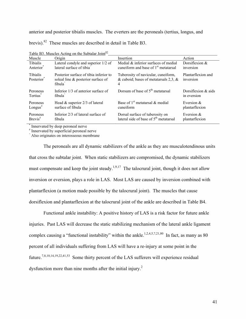
anterior and posterior tibialis muscles. The everters are the peroneals (tertius, longus, and
brevis).82 These muscles are described in detail in Table B3.
Table B3. Muscles Acting on the Subtalar Joint82 Muscle Origin Insertion Action Tibialis Anterior*
Lateral condyle and superior 1/2 of lateral surface of tibia^
Medial & inferior surfaces of medial cuneiform and base of 1st metatarsal
Dorsiflexion & inversion
Tibialis Posterior*
Posterior surface of tibia inferior to soleal line & posterior surface of fibula^
Tuberosity of navicular, cuneiform, & cuboid; bases of metatarsals 2,3, & 4
Plantarflexion and inversion
Peroneus Tertius*
Inferior 1/3 of anterior surface of fibula^
Dorsum of base of 5th metatarsal Dorsiflexion & aids in eversion
Peroneus Longus#
Head & superior 2/3 of lateral surface of fibula
Base of 1st metatarsal & medial cuneiform
Eversion & plantarflexion
Peroneus Brevis#
Inferior 2/3 of lateral surface of fibula
Dorsal surface of tuberosity on lateral side of base of 5th metatarsal
Eversion & plantarflexion
* Innervated by deep peroneal nerve
# Innervated by superficial peroneal nerve ^ Also originates on interosseous membrane
The peroneals are all dynamic stabilizers of the ankle as they are musculotendinous units
that cross the subtalar joint. When static stabilizers are compromised, the dynamic stabilizers
must compensate and keep the joint steady.1,9,17 The talocrural joint, though it does not allow
inversion or eversion, plays a role in LAS. Most LAS are caused by inversion combined with
plantarflexion (a motion made possible by the talocrural joint). The muscles that cause
dorsiflexion and plantarflexion at the talocrural joint of the ankle are described in Table B4.
Functional ankle instability: A positive history of LAS is a risk factor for future ankle
injuries. Past LAS will decrease the static stabilizing mechanism of the lateral ankle ligament
complex causing a “functional instability” within the ankle.1,2,4,5,7,21,80 In fact, as many as 80
percent of all individuals suffering from LAS will have a re-injury at some point in the
future.7,8,10,14,19,22,41,53 Some thirty percent of the LAS sufferers will experience residual
dysfunction more than nine months after the initial injury.2
41

Functional ankle instability is defined as recurrent ankle sprains over a period of time or a
feeling of “giving way” in the foot and/or ankle as a result of articular deafferentation.19,26,41
Table B4. Muscles Acting on the Talocrural Joint82 Muscle Origin Insertion Action Gastrocnemius* Lateral aspect of lateral femoral condyle
& popliteal surface of femur above medial condyle
Posterior surface of calcaneus via calcaneal (Achilles) tendon
Plantarflexion with an extended knee
Soleus* Posterior aspect of head of fibula, superior ¼ of posterior surface of soleal line of tibia & medial border of tibia
Posterior surface of calcaneus via calcaneal (Achilles) tendon
Plantarflexion independent of knee position
Plantaris* Inferior end of lateral supracondylar line of femur & oblique popliteal ligament
Posterior surface of calcaneus via calcaneal (Achilles) tendon
Weakly assists plantarflexion
Flexor Hallucis Longus*
Inferior 2/3 of posterior surface of fibula^
Base of distal phalanx of hallux
Flexion of great toe & weak plantarflexion of ankle
Flexor Digitorum Longus*
Medial part of posterior surface of tibia inferior to soleal line & via a broad tendon to fibula
Base of distal phalanges of lateral 4 digits
Flexion of lateral 4 digits & plantarflexion of ankle
Extensor Hallucis Longus
Middle part of anterior surface of fibula^ Dorsal aspect of base of distal phalanx of great toes
Extension of great toe and dorsiflexion
Extensor Digitorum Longus#
Lateral condyle of tibia & superior 3/4 of medial surface of fibula^
Middle and distal phalanges of lateral 4 digits
Extension of lateral 4 digits & dorsiflexion of ankle
* Innervated by tibial nerve # Innervated by deep peroneal nerve ^ Also originates on interosseous membrane
Functional instability of the ankle, or the most common and serious residual disability
after LAS, is defined as a motion beyond voluntary control but not exceeding the physiologic
range of motion.83 This is seen in individuals that have had a history of three or more LAS
within the past five years.84 It is recognized in 10 to 42 percent of those individuals suffering
from an acute ankle sprain and results from altered proprioceptive
sense.5,6,27,11,12,14,18,20,22,27,30,48,76,84,85 Chronic ankle dysfunction and residual symptoms (pain,
swelling, decreased range of motion) will be seen in 40-75 percent of LAS sufferers.25
Mechanical instability, however, is an objective measurement of laxity within a joint. A
42
mechanical instability of the ankle would be considered motion within the subtalar joint beyond
the physiologic range of motion.83 Although important, functional instability will be addressed
more than with mechanical instability of the ankle.
Biomechanics: There are two areas of biomechanics: kinetics and kinematics. Kinetics is
the study of forces that produce motion or maintain equilibrium. Kinematics, on the other hand,
is the area that describes motion (through location, direction, and magnitude) without regard to
the forces causing the motion.38,86 Lower body, starting at the toes, forms a kinematic chain, or a
series of joints linked together. Motion at one of the joints will cause motion at an adjacent joint,
just as a malalignment at one joint may cause a compensation at a joint above or below.86
Movement of the ankle cannot occur around only one axis. Ankle motion is a combination of
dorsiflexion or plantarflexion along with slight internal or external rotation and anterior or
posterior translation of the talus on the tibia.87
Ankle motion can be described in regard to the planes in which the motion is occurring
and also the axes around which that movement is taking place. Dorsiflexion and plantar flexion
of the ankle occur in the sagittal plane around a coronal. Also, some rotation occurs in the ankle
mortise in both the transverse plane (abduction/adduction) about a vertical axis and the frontal
plane (inversion/eversion) about an anteroposterior axis.85 In neutral, the talocrural joint’s true
axis is considered to run through the fibular malleolus and the body of the talus. It then runs
through or just inferior to the tibial malleolus. Considering the positioning of the malleoli, the
axis is laterally rotated 20 to 30 degrees in the transverse plane and slanted about 10 degrees
down on the lateral side.85 Subtalar joint motion is a complex twisting that is the result of
triplanar motion of the talus around a single oblique axis. Therefore the subtalar joint has only
one degree of freedom into supination and pronation.85 Ankle range of motion will vary between
43

individuals, but generally, 20 degrees of dorsiflexion from neutral is considered normal. Plantar
flexion measures will vary usually between 30 and 50 degrees from neutral.85,88 Inversion and
eversion values usually range between 30 to 35 and 25 to 20 degrees, respectively.88
Foot position may be abnormal in individuals with altered or deficient proprioceptive
ability rendering the foot as an unstable base of support for the proximal lower extremity.5 This
instability may cause future ankle sprains or proximal problems at the knees, hips, pelvis, or low
back. The proper biomechanical alignment of the lower extremity, when viewed from an
anterior angle, shows a knee that is directly over its base of support and is neither in varus nor
valgus. From the lateral view, the knee should not be more anterior than the toes during a squat.
Also, the lower leg and back should be parallel during a squat and when landing from a height.78
During landing, the body’s weight is decelerated using trunk, hip, and leg musculature.89 Joints
of the lower extremity all work together and are linked by the kinematic chain. A dysfunction at
one level will have an effect at multiple other levels throughout the chain.39
During weight acceptance, the subtalar joint moves into pronation in order to absorb
shock. While pronated, the foot becomes more flexible (by “unlocking” at the midtarsal joint)
for adjusting to uneven ground surfaces.52 This pronation is better suited to help the foot
maintain balance. When the foot is preparing for push off, it is supinated and much more rigid.
In the supinated position, dynamic postural control is more difficult to maintain because of the
smaller base of support provided. In individuals with functional ankle instability, the foot tends
to supinate more easily and is then at risk for LAS.52,86
Compressive forces will vary throughout phases of gait as well as position of foot and
ankle. In normal bilateral weight bearing, 77 to 90 percent of the force from the body’s weight is
transmitted through the tibial plafond to the talar dome. The remaining 10 to 23 percent is
44

placed on the medial, or tibial, and lateral, or fibular, talar facets.85 When inversion is increased,
the force upon the medial facet is increased as is the force on the lateral facet during eversion. In
the early stance phase of gait, the compressive forces transmitted to the ankle are less than 20
percent of body weight. The compressive force can be as high as five times body weight during
the late stance phase of normal gait. In addition , a shear force of eight times body weight can
act upon the ankle during heel off.90
Structural foot type may affect postural sway during static and dynamic postural
control.52 Hyperpronated and hypersupinated subtalar joints may be less able to control postural
sway. Orthotics have been shown to significantly reduce postural sway in those with LAS. They
provided an increased structural support to both the medial and lateral sides of the foot. This
support does not allow the increased subtalar joint movement that occurs in LAS patients.27
The Gluteus Medius
The gluteus medius plays many important roles. It has been shown to become
significantly active during midstance and terminal stance of gait in order to provide pelvic
stabilization during a normal gait cycle.64,65 The GM may also be a very important dynamic
stabilizer of the hip and pelvis as well as the core during a sudden loss of balance.64,65
Gluteus medius anatomy: The gluteus medius is the primary hip abductor that also
assists in hip internal rotation. Another important function of the GM is to keep the pelvis level
when the contralateral lower extremity is raised.82 Also, posterior fibers of the GM have been
shown to aid in hip extension and external rotation while the anterior fibers assist internal
rotation.65
Although the gluteus medius is found deep to the gluteus maximus, it can be palpated
inferior to the iliac crest and posterior to the tensor fascia lata.75,82 It is a “fan-shaped” muscle
45
that originates on the external surface of the ilium between the anterior and posterior gluteal lines
and inserts on the lateral surface of the greater trochanter of the femur. The GM gets its vascular
supply from the superior gluteal artery and is innervated by the superior gluteal nerve.82
Biomechanics of the gluteus medius: The GM is a very important muscle of the lower
extremity. It is the primary abductor of the hip as well as an important “anti-gravity” muscle of
the lower extremity that is vital to postural control. It stabilizes the trunk and pelvis laterally
during gait and single-leg standing.66 When an injury occurs distal to the hip on the lower
extremity, the kinematic chain above and below that site are altered. Generally, when an injury
occurs at the ankle and the peroneal muscles cannot stabilize posture as they would prior to the
injury, the GM will compensate. This theory will be further discussed in the Dynamic Postural
Control section of this literature review.
Gluteus medius weakness: There have been reports of peroneal (evertor) muscle
weakness following LAS as well as a weak peroneal muscle group being a positive risk factor for
future LAS. 1, 2,16,48 Functional ankle instability has been shown to cause peroneal muscle
weakness and increased peroneal reaction time.2,48 However, little research has been conducted
on the hip abductor group (the GM in particular) following LAS in the presence of functional
ankle instability. Gluteus medius (GM) strength has, for the most part, been ignored when it
comes to distal lower extremity injuries.89 However, one study62 noted an altered activation of
the hip abductors associated with lower extremity injuries including LAS.
Generally, GM weakness has been associated with the elderly and those individuals with
nervous system disorders. It is not often that the young, athletic population with no history of
injury is evaluated for GM weakness.65 It has been stated, however, that “the possibility that a
localized joint injury could influence the strength of muscle remote from the site has implications
46
for both preventative and therapeutic approaches.”91 One study2 found a premature recruitment
of hip musculature used to achieve dynamic postural control in individuals with unilateral poor
functional ankle instability following a history of LAS. Both local sensation and motor function
were shown to be altered on the ipsilateral hip muscles in patients with unilateral ankle sprains.92
Another study64 found a general weakness of ipsilateral hip musculature in individuals with
unilateral LAS.
Gluteus medius strength has been found to be decreased in individuals with other lower
extremity injuries as well. Subjects with patellofemoral pain have been shown to have
significantly lower (about 26 percent) isometric hip abduction strength values than their healthy,
age-matched counterparts.55 Runners with iliotibial band syndrome have also demonstrated a
weakness in hip abduction.67 When weak, the gluteals cannot decelerate the landing
appropriately by slowing the movements of internal rotation and adduction.89
There have been various measurement techniques developed for use in determining the
strength of the GM. Performing isometric contractions of the GM has been performed in studies
evaluating strength. One way is to have the subject in a side-lying position with a pillow
between his or her legs to allow the superior leg (leg being tested) to be in about 10 degrees of
abduction. A strap is then placed just proximal to the iliac crest and secured around the
underside of the table to stabilize the subject’s trunk. A hand-held dynamometer is then placed
directly over the lateral knee joint and is held in place by another strap secured around the table.
The subject is then asked to perform one practice trial of hip abduction then three experimental
trials with 15 seconds of rest between each. The peak value is recorded from each of the three
experimental trials.55
47

Other ways to test the GM strength are to look at EMG activity to note muscle activity or
the Trendelenburg test for muscle weakness. Single leg squat and lateral stepdown exercises
were performed in one study13 that identified the GM as firing stronger when the participant was
wearing foot orthotics as opposed to not wearing orthotics. Though it does not correlate well to
isometric GM strength, the Trendelenburg test can be used to functionally examine the GM. The
Trendelenburg test uses a single leg stance (SLS) as the level of the pelvis and hips is observed
from a distance by the examiner. If the pelvis drops on the non-weight-bearing side, the GM
(and other hip abductors) on the weight-bearing side are said to be weak.36,56,65
A single leg squat test further assesses the strength of the GM by having the participant
perform a Trendelenburg test and then perform a squat once in a SLS position. The examiner
again watches from a distance and assesses the GM strength. If the participant allows his or her
knee to move medially (genu valgum) the GM is weak.36,56 A manual muscle test of the GM
with the individual side-lying is another appropriate method for assessing GM weakness. If the
individual’s GM is weak, he or she will compensate by also contracting the tensor fascia lata
(flexing the hip), externally rotating, or contracting the quadratus lumborum (“hiking” the hip).65
Finally, functional tests, such as jumping, hopping, and running, can be used as indirect
measurements of muscular strength.56
Strength training: Rehabilitation exercises for LAS have traditionally included strength
training of the peroneal and other dynamic stabilizing muscles of the ankle.1,11 Six weeks of
strength training of the peroneal muscles performed 3 times per week using Thera-Band latex
tubing (increasing in intensity throughout the training period) showed improvements in joint
position sense among individuals with functionally unstable ankles.69 The GM however is
rarely, if ever, strengthened following LAS despite its involvement in dynamic postural control.
48
Very often the GM is overlooked following LAS, and the peroneal muscles are instead the focal
point of strengthening.41 Most of the studies conducted on strengthening the GM have been
prescriptive studies performed on injured subjects with iliotibial band pain or patellofemoral pain
syndrome.55,67 However, there has been little research establishing a link between strength
training the gluteus medius following LAS.
Because of its involvement in dynamic postural control and locomotion during gait, it
is essential to strengthen the GM when it is found to be in a weakened state.66 One implication
for strengthening the GM, as well as testing the strength of the GM, is progressive resistance
exercises. Progressive resistance exercises, or PRE, were developed by DeLorme and are based
on the principle of the ten-repetition-maximum, or 10 RM. The 10 RM is the “maximum load an
individual can exert against gravity no more or less than 10 times with his/her maximum
effort.”66 PRE has become the most commonly employed method for regaining the strength of
the GM, but there still appears to be no simple way to calculate the 10 RM of the GM.66
Open kinetic chain (OKC) exercises, in which the distal segment (the foot in this case) is
free-moving, are most often employed for strengthening the GM. Generally, the GM is
strengthened using a side-lying straight leg raise as the hip is moved into abduction.65,66 This can
be performed against a resistance using a Thera-Band or an ankle cuff weight or against gravity
alone. Another OKC exercise for GM strengthening is standing hip hikes (for abduction on the
frontal plane).62 A novel idea discussed by Wilson65 shows a more functional method of
strengthening the GM. It states that by carrying a weight in the contralateral hand somewhere
between 5 and 15 percent of the individual’s body weight, the GM will be strengthened.65
For general strengthening following an injury, Hertel and Denegar38 suggests using high
repetition, low resistance exercises prior to using high resistance exercises and power training. A
49
six week rehabilitation protocol focused on GM strengthening in runners with iliotibial band
syndrome indicated that 90 percent were pain free and returned to pre-injury activity level.
Fredericson et al. hypothesized that the increase in hip abduction strength causes greater hip
adduction and internal rotation during running and therefore minimized the valgus vector at the
knee.67 As hip flexion increases from 0 to 90 degrees, the moment arm of the GM increases.
This puts the GM at a mechanical advantage to cause hip internal rotation.64
“Arc walking” is another strengthening method in which the individual walks with a
resistance band around his or her waist that is tied off to a secure structure. The individual is
instructed to walk in an arc around the origin of the resistance band while keeping his or her toes
pointed at the band. The lateral sidestepping against a resistance will ensure that strengthening
and postural control gains occur.65 Overall, the GM is largely involved in lower extremity
injuries and should therefore be included in all lower extremity rehabilitation programs.64,96
Proprioception and Neuromuscular Control
Proprioception is the special variance of the sense of touch that encompasses the
sensation of joint movement, or kinesthesia, and joint position sense.5,15,17,26,37,40,48,74 Joint
position sense is a result from afferent input being sent to the central nervous system (CNS) from
various sensory receptors and allowing precise movements and reflexes to occur.37,74,92 If the
afferent input is accurate, the efferent response will be a controlled, coordinated motion.5
Proprioception is also defined as “the reception of stimuli produced within an
organism.”84 Proprioception uses both the peripheral nerves and the central nervous system
integrated together to produce an awareness to one’s surroundings.84 Proprioception occurs
when the Golgi tendon organs (receptors within the muscle that are sensitive to tension during
muscle contraction) and muscle spindles (receptors within the muscle that sense muscle length
50

and rate of length change) within the muscles along with the receptors in the skin, ligaments, and
joint capsules sense movement and convey it to the CNS through neural afferent
pathways.42,74,76,84,92
Mechanoreceptors are specialized neuroepithelial structures found in skin, ligaments,
muscular, and tendons that surround joints.93 Mechanoreceptors receive and send information
about functional and mechanical deformation to the CNS. The information is translated into
afferent neural signals that will increase when an increase in deformation occurs.74,93 Specific
cutaneous mechanorecptors are described in Tables B5.
Table B5. Specific Cutaneous Mechanoreceptors74,75 Type of Mechanoreceptor Location Sensory Information Adaptation
Rate Pacinian Corpuscles Deep dermis or
hypodermis Deep cutaneous pressure, vibration, & proprioception
Rapid
Meissner’s Corpuscles Dermal papillae of hairless skin
Touch, pressure, & slow vibrations Rapid
Ruffini Ends Dermis Continuous touch or pressure & depression or stretch of skin
Slow
Hair Follicles Along the axis of strands of hair
Light touch & bending of hair Rapid
There are two types of muscle mechanoreceptors: muscle spindles and Golgi tendon
organs (GTO’s). Muscle spindles are sensititve to stretch while GTO’s are sensititve to tension.
Afferent neural fibers carry information from the muscle spindles to the CNS and then divide
into branches that choose from one of several different pathways. One pathway completes the
reflex arc by directly stimulating efferent motor neurons that lead back to the muscle(s) that was
stretched. This reflex will cause a muscle contraction in response to the muscle being stretched.
Golgi tendon organs (GTO’s), however, will fire when a muscle/tendon is contracted and
becomes tense. The firing activity of the GTO’s afferent neuron supplies the motor control
systems, in the brain and locally, with constant information regarding muscle tension. The basic
51
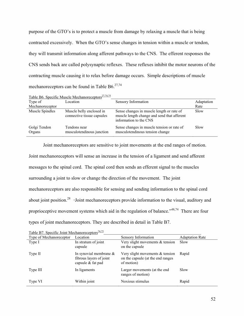
purpose of the GTO’s is to protect a muscle from damage by relaxing a muscle that is being
contracted excessively. When the GTO’s sense changes in tension within a muscle or tendon,
they will transmit information along afferent pathways to the CNS. The efferent responses the
CNS sends back are called polysynaptic reflexes. These reflexes inhibit the motor neurons of the
contracting muscle causing it to relax before damage occurs. Simple descriptions of muscle
mechanoreceptors can be found in Table B6.27,74
Table B6. Specific Muscle Mechanoreceptors27,74,75 Type of Mechanoreceptor
Location Sensory Information Adaptation Rate
Muscle Spindles Muscle belly enclosed in connective tissue capsules
Sense changes in muscle length or rate of muscle length change and send that afferent information to the CNS
Slow
Golgi Tendon Organs
Tendons near musculotendinous junction
Sense changes in muscle tension or rate of musculotendinous tension change
Slow
Joint mechanoreceptors are sensitive to joint movements at the end ranges of motion.
Joint mechanoreceptors will sense an increase in the tension of a ligament and send afferent
messages to the spinal cord. The spinal cord then sends an efferent signal to the muscles
surrounding a joint to slow or change the direction of the movement. The joint
mechanoreceptors are also responsible for sensing and sending information to the spinal cord
about joint position.28 “Joint mechanoreceptors provide information to the visual, auditory and
proprioceptive movement systems which aid in the regulation of balance.”48,74 There are four
types of joint mechanoreceptors. They are described in detail in Table B7.
Table B7. Specific Joint Mechanoreceptors74,75 Type of Mechanoreceptor Location Sensory Information Adaptation Rate Type I In stratum of joint
capsule Very slight movements & tension on the capsule
Slow
Type II In synovial membrane & fibrous layers of joint capsule & fat pad
Very slight movements & tension on the capsule (at the end ranges of motion)
Rapid
Type III In ligaments Larger movements (at the end ranges of motion)
Slow
Type VI Within joint Noxious stimulus Rapid
52
Once afferent information is sent upward, there are three levels of motor control within
the CNS that can respond with an efferent motor response. The spinal cord is the first level of
motor control. It is responsible for reflexes, direct motor responses to peripheral sensory
information and simple patterns of motor coordination. The second level, the brain stem, helps
control postural equilibrium through excitatory or inhibitory synapses. Finally, the third level is
the cerebral cortex. The cortex encodes the muscles to be contracted, the magnitude of
contraction, and the direction of movement. The cerebral cortex is also in charge of planning
movements that will require activating more than one muscle.42
Proprioceptive sensation includes both the position sense of a static limb as well as the
position of that limb as it moves through space.84 Specifically at the ankle, it is the ability to
detect motion of the foot and make postural adjustments in response to the motion detected.48
The ability to sense the position of the foot prior to heel strike is of utmost importance when
attempting to prevent LAS.5,48 When an injury occurs in a joint, the joint loses its ability to
gauge stresses placed upon it and react to it appropriately.37,84 The ankle sprain itself might
cause damage not only to the ligaments and the joint capsule but also to the neural structures
within the capsule causing decreased neural afferent input and, therefore, neuromuscular
control.12,53,84 In fact, neural fibers have less tensile strength than do ligamentous fibers, and
they are therefore damaged quite easily during joint sprains.12 This decrease in neuromuscular
control and so-called “joint deafferentation” following an injury may be the reason for re-
injury.12,37 Injuries may also cause a reduction in the velocity of nerve conduction.53
Injuries to the ankle will almost always cause an overall decrease in neuromuscular
control and proprioception within the ankle.53,84 Pain and swelling will inhibit normal
neuromuscular control patterns by blocking the alpha efferent pathways while facilitating gamma
53

efferent pathways. This will cause an inability of the individual to volitionally contract certain
muscles around the injured joint while other muscles will reflexively spasm accompanied by
hypersensitivity to joint movement and muscle stretching.38
Dynamic Postural Control
Dynamic postural control has been called “the single most important component of
athletic ability” because it is required for all movements.76 Dynamic postural control is needed
for activities of daily living such as walking, stair climbing, and running.47 Tools for
determining dynamic postural control ability have been used to quantify the amount of functional
impairment within the injured area, especially following LAS.13 Nakagawa19 states that
assessing postural control with balance testing during closed-chained functional tasks provides
information on the overall function of the joints and their proprioceptive and neuromuscular-
control abilities.
It has been noted that “physical activity appears to be an important determinant of
dynamic balance.”61 Raty61 was able to show that continued involvement in athletics and light
leisure activities lends itself to better dynamic postural control. Despite the fact of the
importance of physical activity, there have been few studies performed to determine the effect of
LAS on the performance of dynamic postural control tasks. It is hypothesized that individuals
with LAS and/or functional ankle instability will exhibit decreased postural control statically and
dynamically.19 Riemann and Lephart42 have stated that functional ankle instability or LAS will
cause a disruption of postural control.
There are three levels of dynamic postural control: spinal, brainstem, and cognitive
awareness.37 The spinal level causes reflexes to occur with short, quick movements. Altered
sensory receptors will impede the execution of reflex responses that are meant to occur for
54
protective reasons.38 The brainstem level receives information from the mechanorecptors located
within the joints (somatosensory), the vestibular centers in the semicircular canals for
equilibrium, and visual inputs from the ocular centers.12,13,47,48,52,76,84 These three “balance
senses” work together to maintain postural control at all times, and if one is impaired, the other
two must compensate.17,19,39,48 The final level, cognitive awareness, is associated with joint and
body position. This level determines fine motor movements and muscle memory from repetition
of motions.37,52
Postural control, often more simply referred to as balance, is often measured by having an
individual stand on one foot for as long as possible.20,52,84 Variations can be added to challenge
the individual performing the test. Closing the eyes, varying the trunk position or standing
surface, and adding arm or leg movements are simple additions to make balancing more
challenging.26,37,60 Balance, in essence, is maintaining the body’s center of mass (COM) over the
body’s base of support (the feet) or a “physical equilibrium” in order to stay in an upright
position.13,47,52,84,96 The body uses a “reflex mechanism” to maintain the body’s COM over the
feet.22
There are two strategies for maintaining dynamic postural control. The younger, healthy,
active population generally uses the ankle strategy for altering/correcting COM. In the ankle
strategy, the postural control is preserved by firing the lateral peroneal (everting) muscles of the
ankle.13,61 In the older population or in other cases when the ankle provides inadequate sensory
information regarding kinesthesia and joint position sense, the hip musculature (the GM in
particular) will compensate and maintain postural control. This proximal compensation to
postural sway of COM is often seen in LAS individuals and those with functional instability in
the ankle.2,10,48 A study discussed by Schmitz et al.64 showed an increase in GM activity during
55

sudden ankle inversion in healthy and in functionally unstable ankle subjects. Further, the GM
controls the lateral weight transfer during forward oriented whole body movements.61 Runge44
states “postural tasks that require ‘hip strategy’ to maintain equilibrium may be more dependent
upon vestibular input for response selection and control than other tasks.” It has been shown that
patients with vestibular deficiencies will fail to initiate the hip strategy while healthy individuals
generally switch to a hip strategy from an ankle strategy and will instead fall or take a step.44
Individuals with functional ankle instability were shown to take a significantly longer
amount of time to restore their single leg postural control following sudden inversion
perturbations than did healthy control subjects. Functional ankle instability will allow the COM
to traverse outside the base of support caused by decreased proprioceptive input.10,39,76,92 It is
suggested that an individual with a functional ankle instability will make postural corrections at
the hip more often than will those healthy individuals without ankle instability.51 One study95
found female soccer players with LAS were more likely to use the hip strategy during SLS on
the injured leg while they used the ankle strategy for postural corrections on the uninjured side.
The examiner stated that the change in postural control strategy can be attributed to the alteration
in weight-bearing forces at the subtalar joint following LAS. Another author suggests the use of
orthotics to correct the subtalar joint will return the individuals to the ankle strategy or postural
control.51 These balance corrections are triggered by the proprioceptive capabilities of both the
lower extremity and the trunk.94
It has been suggested that the increased postural sway and, therefore, decreased postural
control and balance can be attributed to the disturbance of proprioceptive sensory receptors.19
One study95 found that postural sway increases even if a few sensory inputs have been disrupted.
56

Also, dynamic postural control, like joint position sense and kinesthetic awareness, may also be
decreased by fatigue.10
Studies on Proprioceptive Training
Proprioceptive training is often less addressed during rehabilitation than are restoring full
range of motion (ROM) and functional strength.84 Proprioceptive training is very important
following an injury because it allows for dynamic joint stability and prevents excessive strain on
the joints and muscles, especially in the physically active.15,27,37,38,41,48,52 The goal of
proprioceptive training following an injury is to retrain the afferent pathways to enhance the
sensation of joint movement.40 Closed kinetic chain (CKC) exercises have been shown to
redevelop lower extremity neuromuscular control following an injury.12,37
There have been a large number of proprioception training programs developed in
prescriptive studies following lower extremity injuries as well as others that are merely
attempting to increase the lower extremity’s proprioceptive ability. They recommend using
single leg standing and then progressing to wobble boards and ankle discs.17,84 One study76
suggests using T-Band kicks with a heavy resistance while unilaterally standing, also called
“steamboats.” The study used 50 kicks in each of four directions (medial, lateral, anterior, and
posterior). The study also had the subjects perform square hopping 4 times for 20 seconds. In
addition, single-leg standing was included in the study on a foam surface for 3 sets of 20
seconds. Finally, the subjects were placed in a single-leg stance on a BAPS board (between level
3 and 5) for 3 sets of 20 seconds.76
Also, fatigue, much like injury, may impair the kinesthetic and joint position sensation
properties of joint structures.10,74 Because “functional (ankle) instability stemming from
neuromuscular and proprioceptive deficits is hypothesized to be a major contributing factor to
57

chronic ankle instability,” it is vital to re-train the ankle for proprioception and neuromuscular
control following LAS.53
Postural awareness should be stressed throughout the rehabilitation of all injuries in order
to avoid undo stress on the body, especially injured areas.38 It has been reported that six weeks
of disk balance training and coordination balance training greatly improved dynamic postural
control in most subjects.48 Dynamic postural control is often accomplished through a functional
task while maintaining one’s base of support.45,46 Dynamic postural control exercises must be
included in the rehabilitation of an injured joint.10-12,17,21,46,53,77,84
Five studies have been performed on proprioception training on healthy subjects as well
as following an injury and in those with a chronic instability. Studies by Blackburn et al.59 and
Powers et al.41 used Thera-Band kicks (tying an elastic resistance to the contralateral ankle and
performing quick kicks while maintaining a single leg stance) for six-weeks for proprioception
training. The Blackburn et al. study showed significant difference between the proprioception
training group and the control group for dynamic balance. However, the Powers et al. study
found no significant difference between pre-test and post-test results for static balance in any
group. Riemann et al.,60 using a BAPS board for four-weeks to train proprioception, found no
significant differences for postural control but did find significant differences in ankle
kinesthesia between the experimental and control groups. Finally, studies by Piegaro57 and
Bernier and Perrin48 both used a combination of balance training on fixed and unstable surfaces
as well as hopping exercises. Bernier and Perrin’s six-week program achieved significant
differences on anterior/posterior and medial/lateral modified equilibrium scores between groups.
Piegaro57 used the same program as Bernier and Perrin but shortened it to four-weeks. He found
significant differences in pre-test to post-test anterior reach direction distances among those in
58

the experimental group. These studies are briefly described in Table B8 below. This study uses
a proprioceptive program to the exact specifications of the Bernier and Perrin study.
Table B8. Proprioception Training Studies Author Subjects Training
Period Results
Piegaro57 39 healthy college students 4 weeks Significant differences from pre-test to post-test reaches into anterior direction of SEBT
Bernier and Perrin48
48 individuals between 18 and 32 with a history of chronic ankle instability
6 weeks Significant differences from pre-test to post-test in anterior/posterior and medial/lateral directions of modified equilibrium score
Blackburn et al.59
32 physically active healthy individuals between 18 and 25
6 weeks Significant difference in dynamic balance between control group and proprioception experimental group
Powers et al.41
38 individuals with a mean age of 21.6 with functional ankle instability
6 weeks No significant differences in any group pre-test to post-test regarding static balance
Riemann et al.60
26 healthy recreationally active individuals
4 weeks Significantly increased ankle kinesthesia, no significant difference in postural control
Summary
The ankle is one of the most commonly injured joints in the body. The ankle is a distal
link of the kinematic chain. When injured or unstable, other structures must compensate for the
ankle. The gluteus medius is often found to be weak in individuals with lower extremity injuries.
Lower extremity injuries, including lateral ankle sprains, will cause disturbances of the normal
proprioceptive receptors. With decreased proprioceptive ability and neuromuscular control,
dynamic postural control is diminished. Dynamic postural control is a vital part of the
rehabilitation of any injury and can be assessed using the star excursion balance test.
59

APPENDIX C
ADDITIONAL METHODS
Table C1. Informed Consent
THE COMPARATIVE EFFECTS OF A SIX-WEEK BALANCE TRAINING PROGRAM, GLUTEUS MEDIUS STRENGTH TRAINING PROGRAM, AND
COMBINED BALANCE TRAINING/GLUTEUS MEDIUS STRENGTH TRAINING PROGRAM ON DYNAMIC POSTURAL CONTROL
Introduction I, ________________________, have been invited to participate in this research study, which has been explained to me by Vincent J. Leavey, ATC, PES. He is conducting this research under the supervision of Michelle A. Sandrey, PhD, ATC to fulfill the requirements for a master’s thesis in Athletic Training in the School of Physical Education at West Virginia University. Purpose of the Study The purpose of this study is to determine if gluteus medius muscle strength training, proprioception training, or a combination of the two will have an affect on dynamic postural control. Description of Procedures This study will be conducted at HealthWorks Rehab & Fitness, 943 Maple Drive, Morgantown, WV 26505. Orientation Procedures At an orientation meeting the purpose of this study will be explained to me. I will be given an informed consent form explaining my rights as a research subject and a demographic/inclusion criteria questionnaire. If I am one of sixty eligible subjects, I will be contacted by the principal investigator and will schedule a time for my testing. I will have a one in four chance of being randomly assigned to either of the training groups or the control group. I will be asked for my full cooperation and to work to the best of my ability. My involvement in this will initially take sixty minutes for a pre-testing session. This will be followed by meeting with the researcher three times a week for twenty minutes over a six week period. A final sixty minutes of post-testing will conclude my participation. Interventions I will be tested using the Star Excursion Balance Test. Testing will be administered and supervised by the principal investigator at HealthWorks Rehab & Fitness, 943 Maple Drive, Morgantown, WV 26505. Prior to testing, the device and procedure will be explained to my satisfaction.
60

THE COMPARATIVE EFFECTS OF A SIX-WEEK BALANCE TRAINING PROGRAM, GLUTEUS MEDIUS STRENGTH TRAINING PROGRAM, AND
COMBINED BALANCE TRAINING/GLUTEUS MEDIUS STRENGTH TRAINING PROGRAM ON DYNAMIC POSTURAL CONTROL
I will have my leg length measured prior to testing in order to normalize my scores to my height. A tape measure will be used to measure the distance between the top of my hip bone to the center of the ankle on the same leg. I will warm up on a stationary bike for 10 minutes at a self-selected pace and then stretch my quadriceps, hamstrings, and calves for 5 minutes. I will then take a five minute break. Next I will be instructed to stand in the center of the Star Excursion Balance Test on my dominant leg (leg I would use to kick a ball). I will be instructed to reach with my non-dominant leg as far as possible down one of the eight lines which will be randomly selected using a series of index cards. The lines are to the following directions: front, front right corner, right, back right corner, back, back left corner, left, and front left corner. If I am reaching with my right leg, I will start with the direction on the index card I choose randomly and then go around the star in a counterclockwise manner and clockwise if I am reaching with my left leg. I will be instructed to hold the position of furthest reach for two seconds. I will reach as far down each line as I possibly can without losing my balance six times as practice to become familiar with the test. I will then take a five minute break and then will complete three test trials down each of the eight lines with fifteen seconds of rest between each trial. The average of my three trials will be recorded. Trials will be discarded and repeated if I am unable to maintain my balance at any point in the trial and place my non-dominant foot on the floor. While performing on the Star Excursion Balance Test, it is likely that I will not lose my balance because I will be performing the test with my eyes open. However, I will be instructed to touch down with my non-dominant leg if I feel I am losing my balance. The principal investigator will also be standing beside me if I am unable to touch down with my non-dominant leg. Following each test, the primary investigator will record the distances of each trial on a data sheet. If all data has been correctly recorded, my testing session will be completed. At this time, I will be asked by the primary researcher for any questions or comments. A time will then be set for my gluteus medius strength to be tested. On a separate date, I will have my hip abduction (moving one leg away from the other leg) strength tested using a Lafayette Manual Muscle Test System (Model 01163, Lafayette Industries, Lafayette, IN). Testing will be administered and supervised by the principal investigator at HealthWorks Rehab & Fitness, 943 Maple Drive, Morgantown, WV 26505. Prior to testing, the device and procedure will be explained to my satisfaction. First I will be weighed using a digital scale to normalize my scores to my body weight. I will lie on my side (opposite of my dominant leg) on a table. I will slightly abduct my dominant leg. A strap will then be wrapped under the table and over my dominant leg just above my knee. A Manual Muscle Test System will then be placed under the strap to measure the force I produce into hip abduction. I will then complete 3 trials of hip abduction. During each trial, I will abduct
61
THE COMPARATIVE EFFECTS OF A SIX-WEEK BALANCE TRAINING PROGRAM, GLUTEUS MEDIUS STRENGTH TRAINING PROGRAM, AND
COMBINED BALANCE TRAINING/GLUTEUS MEDIUS STRENGTH TRAINING PROGRAM ON DYNAMIC POSTURAL CONTROL
my hip by taking 2 seconds to build up force and then hold that maximal effort for 4 seconds. I will rest for 15 seconds between each trial. Following each test, the primary investigator will record the distances of each SEBT trial and each trial of gluteus medius strength outputs on data sheets. If all data has been correctly recorded, my testing session will be completed. At this time, I will be asked by the primary researcher for any questions or comments. The data from my testing session will be averaged with the data from the rest of the subjects for analysis. This entire process for SEBT and gluteus medius strength will be repeated at the conclusion of my six week training period. After the initial testing, I will perform a six week training period. According to the group I am randomly assigned to, I will perform those exercises with the researcher three times a week for twenty minutes per session. If I am randomly assigned to the control group, I will contact the researcher once a week to let them know which other activities outside daily activities I have been participating. If I am in the control group, I will perform the pre- and post-testing procedures. I will not perform any of the training exercises of the other groups. If I am in the proprioception training group, I will perform exercises that involve balancing exercises and exercises that will train my joint position sense in my dominant leg. I will perform a battery of exercises that have been proven to improve my proprioception. The exercises I will perform will change from week to week but will include balancing on a fixed surface, balancing on a tilt board, balancing on a wobble board, and functionally hopping. If I am in the gluteus medius strength training group, I will perform exercises to strengthen hip abduction on my dominant leg. I will perform 6 exercises including side-lying hip abduction with an elastic band, walking with a dumbbell in my non-dominant hand at a weight adjusted for my body weight, sideways walking with an elastic band tied around my knees, hip abduction on a machine, and single leg squatting. If I am in the combination group, I will perform the exercises from both the gluteus medius strength training group as well as the proprioception training group.
62

THE COMPARATIVE EFFECTS OF A SIX-WEEK BALANCE TRAINING PROGRAM, GLUTEUS MEDIUS STRENGTH TRAINING PROGRAM, AND
COMBINED BALANCE TRAINING/GLUTEUS MEDIUS STRENGTH TRAINING PROGRAM ON DYNAMIC POSTURAL CONTROL
Risks and Discomforts There are no known or expected risks from participating in this study. The only known or expected discomfort may be mild muscle soreness in my dominant lower extremity with performing the proprioceptive and/or strength exercises. While performing on the Star Excursion Balance Test, it is likely that I will not lose my balance because I will be performing the test with my eyes open. However, I will be instructed to touch down with my non-dominant leg if I feel I am losing my balance. The principal investigator will also be standing beside me if I am unable to touch down with my non-dominant leg. Some soreness may be experienced in my hip following gluteus medius strengthening. There may be some soreness experienced in my lower leg following the balance training exercises, but it should be minimal at worst. Should any injury occur, I understand that Vincent J. Leavey, ATC, PES will provide first aid and make any necessary medical referral at my expense. Alternative I understand that I do not have to participate in this study. No negative actions will be taken against me if I choose not to participate. Benefits I understand that this study is not expected to be of direct benefit to me, but the knowledge gained may be of benefit to others. Financial Considerations I understand that I will receive no monetary compensation for completing this study. Contact Persons For more information about this research, I can contact Vincent J. Leavey, ATC, PES at (609) 680-4981 or at [email protected] or, Michelle A. Sandrey, PhD, ATC at (304) 293-3295 Ext. 5220 or at [email protected]. For information regarding my rights as a research subject, I may contact the Executive Secretary of the Review Board at (304) 293-7073. Confidentiality I understand that any information about me obtained as a result of my participation in this research will be kept as confidential as legally possible. Identifying information on the informed consent form and demographic/injury history questionnaire will be kept confidential by
63

THE COMPARATIVE EFFECTS OF A SIX-WEEK BALANCE TRAINING PROGRAM, GLUTEUS MEDIUS STRENGTH TRAINING PROGRAM, AND
COMBINED BALANCE TRAINING/GLUTEUS MEDIUS STRENGTH TRAINING PROGRAM ON DYNAMIC POSTURAL CONTROL
assigning a code number to each informed consent form and demographic/injury history questionnaire. I understand that my research records and test results, just like hospital records, may be subpoenaed by court order. In any publications that result from this research, neither my name nor any information from which I might be identified will be published without my consent. Voluntary Participation Participation in this study is voluntary. I understand that I am free to withdraw my consent to participate in this study at any time and that such refusal to participate will not affect my future care, my employee status at West Virginia University, or my class standing or grades. Refusal to participate or withdrawal will involve no penalty to me. I have been given the opportunity to ask questions about the research, and I have received answers concerning areas I did not understand. In the event new information becomes available that may affect my willingness to continue to participate in this study, this information will be given to me so I may make an informed decision about my participation. Upon signing this form I will receive a copy. I willingly consent to participate in this research. Signature of Subject Date/Time Signature of Principal Investigator Date/Time
64

Table C2. Authorization to Use or Disclose Protected Health Information
Authorization to Use or Disclose Protected Health Information (PHI)
West Virginia University
I hereby voluntarily authorize the use or disclosure of my individually identifiable health information as described below. Patient Name: ID Number: Date of Birth: IRB Protocol #: 16727 Persons/organizations providing the protected health information (e.g. hospitals): Individual subjects affiliated with West Virginia University Athletic Training and West Virginia University Club Sports & Intramurals
Persons/organizations receiving the information (e.g. investigators, clinical coordinators, sponsor, FDA): Vincent J. Leavey, ATC, PES Michelle A. Sandrey, PhD, ATC
The following information will be used: A self-report injury history questionnaire (lower extremity injuries within the past six months, surgery to the lower extremity in the past year, visual disorders, nerve problems, medications being taken that may affect balance, or chronic ankle injuries). The information is being disclosed for the following purposes (Start with the Title of the study and include additional information e.g. screening and recruiting subjects; analyzing research data, or other specified purposes): The Comparative Effects of a Six-Week Balance Training Program, Gluteus Medius Strength Training Program, and Combined Balance Training/Gluteus Medius Strength Training Program on Dynamic Postural Control. Information will be used to screen subjects for inclusion and exclusion criteria of the study. I may revoke this authorization at any time by notifying the Principal Investigator in writing at: Vincent J. Leavey , ATC, PES School of Physical Education P.O. Box 6116 Morgantown, WV 26505 If I do revoke my authorization, any information previously disclosed cannot be withdrawn. Once information about me is disclosed in accordance with this authorization, the recipient may redisclose it and the information may no longer be protected by federal privacy regulations.
65

Authorization to Use or Disclose Protected Health Information (Contd.) I may refuse to sign this authorization form. My clinical treatment may not be affected by whether or not I sign this form. I may not be allowed to participate in the research if I do not sign the form. This authorization will expire on the date that the research study ends. (Other options for expiration include an actual date of expiration, occurrence of a particular event, or “none” if the authorization will have no expiration date.)
Expiration date: I will be given a copy of this authorization form.
Signature of subject or subject’s legal representative (Form MUST be completed before signing)
Date
Printed name of subject’s legal representative Relationship to the subject Initials
Parent Medical power of attorney/representative Legal guardian
Health care surrogate
______________________________________________________________________________
66
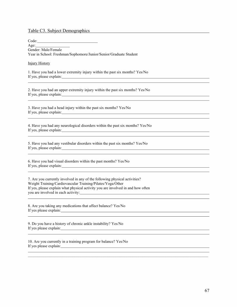
Table C3. Subject Demographics Code: Age: Gender: Male/Female Year in School: Freshman/Sophomore/Junior/Senior/Graduate Student Injury History 1. Have you had a lower extremity injury within the past six months? Yes/No If yes, please explain: 2. Have you had an upper extremity injury within the past six months? Yes/No If yes, please explain: 3. Have you had a head injury within the past six months? Yes/No If yes, please explain: 4. Have you had any neurological disorders within the past six months? Yes/No If yes, please explain: 5. Have you had any vestibular disorders within the past six months? Yes/No If yes, please explain: 6. Have you had visual disorders within the past months? Yes/No If yes, please explain: 7. Are you currently involved in any of the following physical activities? Weight Training/Cardiovascular Training/Pilates/Yoga/Other If yes, please explain what physical activity you are involved in and how often you are involved in each activity: 8. Are you taking any medications that affect balance? Yes/No If yes please explain: 9. Do you have a history of chronic ankle instability? Yes/No If yes please explain: 10. Are you currently in a training program for balance? Yes/No If yes please explain: _____________________________________________________________________________________________
67
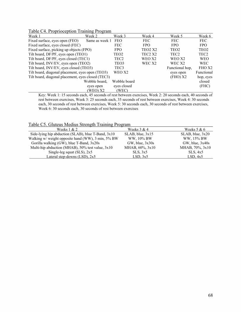
Table C4. Proprioception Training Program Week 1 Week 2 Week 3 Week 4 Week 5 Week 6 Fixed surface, eyes open (FEO) Same as week 1 FEO FEC FEC FEC Fixed surface, eyes closed (FEC) FEC FPO FPO FPO Fixed surface, picking up objects (FPO) FPO TEO2 X2 TEO2 TEO2 Tilt board, DF/PF, eyes open (TEO1) TEO2 TEC2 X2 TEC2 TEC2 Tilt board, DF/PF, eyes closed (TEC1) TEC2 WEO X2 WEO X2 WEO Tilt board, INV/EV, eyes open (TEO2) TEO3 WEC X2 WEC X2 WEC Tilt board, INV/EV, eyes closed (TEO3) TEC3 Functional hop, FHO X2 Tilt board, diagonal placement, eyes open (TEO3) WEO X2 eyes open Functional Tilt board, diagonal placement, eyes closed (TEC3) (FHO) X2 hop, eyes Wobble board, Wobble board closed
eyes open eyes closed (FHC) (WEO) X2 (WEC)
Key: Week 1: 15 seconds each, 45 seconds of rest between exercises, Week 2: 20 seconds each, 40 seconds of rest between exercises, Week 3: 25 seconds each, 35 seconds of rest between exercises, Week 4: 30 seconds each, 30 seconds of rest between exercises, Week 5: 30 seconds each, 30 seconds of rest between exercises, Week 6: 30 seconds each, 30 seconds of rest between exercises
Table C5. Gluteus Medius Strength Training Program Weeks 1 & 2 Weeks 3 & 4 Weeks 5 & 6 Side-lying hip abduction (SLAB), blue T-Band, 3x10 SLAB, blue, 3x15 SLAB, blue, 3x20 Walking w/ weight opposite hand (WW), 3 min, 5% BW WW, 10% BW WW, 15% BW Gorilla walking (GW), blue T-Band, 3x20s GW, blue, 3x30s GW, blue, 3x40s Multi-hip abduction (MHAB), 50% test value, 3x10 MHAB, 60%, 3x10 MHAB, 70%, 3x10
Single-leg squat (SLS), 2x5 SLS, 3x5 SLS, 4x5 Lateral step-downs (LSD), 2x5 LSD, 3x5 LSD, 4x5
68

Table C6. Star Excursion Balance Test Instructions (Pre- and Post-Test) 1. Subjects had their leg length measured in inches and recorded to normalize their
excursions to their height. 2. They underwent a warm-up session of five minutes on a stationary bicycle at 120
revolutions per minute.
3. The subjects took a five minute break.
4. Subjects selected an index card to randomly determine in which direction they will start.
5. Subjects were instructed to stand in the center of the star grid and maintain a single-leg stance while reaching with the opposite leg to touch as far as possible along the randomly chosen reach direction.
6. Subjects were instructed to touch the farthest point possible as light as possible along a
chosen reach direction with the most distal part of their reach foot.
7. Subjects whose reaching leg is the right went around the star grid in a counterclockwise fashion while subjects whose reaching leg is the left went around the star grid in a clockwise fashion.
8. Subjects were instructed to perform six practice trials in each of the eight excursions with
a 15-second rest between each excursion.
9. After a five minute rest following the last practice trial, testing began.
10. Three trials were performed in each of the eight excursions with a 15-second rest between each reach.
11. Trials were discarded and repeated if the reach foot was used to provide considerable
support when touching the ground, if the subjects’ stance foot was lifted from the center of the star grid, or if the subjects were not able to maintain their balance at any point in the trial.
12. The average excursions for each reach direction were calculated and recorded.
13. The average excursions for each reach direction were divided by leg length and multiplied
by 100 to determine each subject’s dynamic balance score in each of the eight reach directions.
______________________________________________________________________________
69

Table C7. Pre-Test Data Collection Sheet for the Star Excursion Balance Test Pre-Test Data Collection Sheet for the Star Excursion Balance Test
Subject Number: Age: Gender: Male/Female Height: Weight: Right Leg Length: Left Leg Length: Dominant Lower Extremity: Right/Left Excursion Trial 1 Trial 2 Trial 3 Average Anterior Anteromedial Medial Posteromedial Posterior Posterolateral Lateral Anterolateral
70
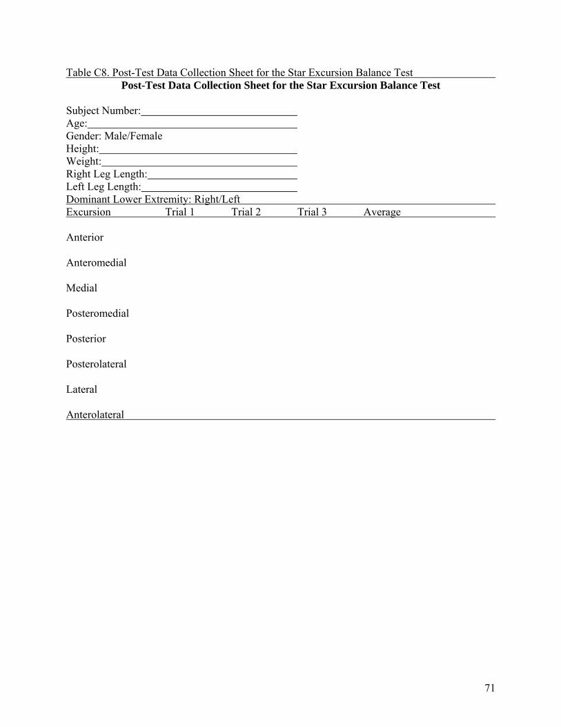
Table C8. Post-Test Data Collection Sheet for the Star Excursion Balance Test Post-Test Data Collection Sheet for the Star Excursion Balance Test
Subject Number: Age: Gender: Male/Female Height: Weight: Right Leg Length: Left Leg Length: Dominant Lower Extremity: Right/Left Excursion Trial 1 Trial 2 Trial 3 Average Anterior Anteromedial Medial Posteromedial Posterior Posterolateral Lateral Anterolateral
71

Table C9. Gluteus Medius Strength Testing Instructions (Pre- and Post-Test) 1. Subjects were weighed using a digital scale. 2. Subjects were positioned side-lying on a treatment table. The hip was at approximately
10 degrees of abduction prior to testing.
3. The force pad of the manual muscle testing system was placed just above the knee joint on the lateral femoral condyle.
4. The subjects took three practice trials and three trials to be recorded.
5. The dynamometer was zeroed, and then the subject was instructed to push the leg upward
gradually building up force for two seconds and then a maximal effort for four seconds.
6. The value on the dynamometer was recorded and then the dynamometer was zeroed out again before the next trial.
7. The subjects had 15-seconds of rest between trials.
______________________________________________________________________________ Table C10. Pre-Test Data Collection Sheet for Gluteus Medius Strength
Pre-Test Data Collection Sheet for Gluteus Medius Strength Subject Number: Age: Gender: Male/Female Height: Weight: Dominant Lower Extremity: Right/Left Trial 1 Trial 2 Trial 3 Average ______________________________________________________________________________
72

Table C11. Post-Test Data Collection Sheet for Gluteus Medius Strength Post-Test Data Collection Sheet for Gluteus Medius Strength
Subject Number: Age: Gender: Male/Female Height: Weight: Dominant Lower Extremity: Right/Left Trial 1 Trial 2 Trial 3 Average ______________________________________________________________________________
73

Figure C1. Subject Performing Fixed Surface Balancing
1. Subject unilaterally stands on dominant leg on a flat, fixed surface.
2. Subject maintains balance unilaterally on flat
surface for specified period of time corresponding to week of training.
Figure C2. Subject Performing Tilt Board Balancing in the Dorsiflexion/Plantar Flexion Direction
1. Subject unilaterally stands on dominant leg on a
tilt board that is set for dorsiflexion/plantar flexion.
2. Subject maintains balance unilaterally on tilt
board for specified period of time corresponding to week of training.
74
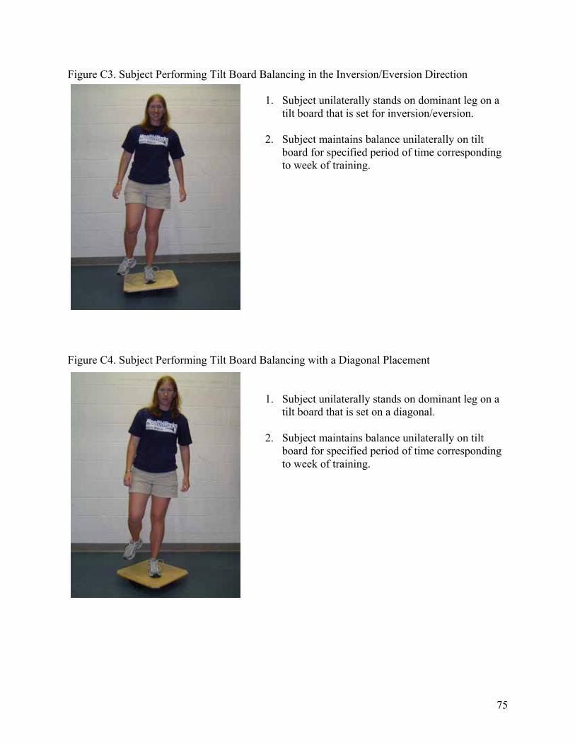
Figure C3. Subject Performing Tilt Board Balancing in the Inversion/Eversion Direction
1. Subject unilaterally stands on dominant leg on a tilt board that is set for inversion/eversion.
2. Subject maintains balance unilaterally on tilt
board for specified period of time corresponding to week of training.
Figure C4. Subject Performing Tilt Board Balancing with a Diagonal Placement
1. Subject unilaterally stands on dominant leg on a tilt board that is set on a diagonal.
2. Subject maintains balance unilaterally on tilt
board for specified period of time corresponding to week of training.
75

Figure C5. Subject Performing Wobble Board Balancing
1. Subject unilaterally stands on dominant leg on a wobble board.
2. Subject maintains balance unilaterally on wobble
board for specified period of time corresponding to week of training.
Figure C6. Subject Performing Functional Hop Exercise
A B
1. Subject unilaterally stands on dominant leg on a flat surface in box marked “Start.” (Figure A)
2. Subject performs a single-leg hop from “Start” box to box marked “1.”
3. Subject achieves balance in box “1.” (Figure B)
4. Subject repeats steps 2 and 3 along the functional hop pattern until box “6” is reached.
5. Subject begins once again at box marked “Start” until 30 seconds has passed.
76

Figure C7. Functional Hop Pattern
6
1
4
3
2
5
Start
Figure C8. Subject Performing Side-Lying Hip Abduction with Elastic Resistance
A A
1. Subject lies on his/her side on a table with dominant leg toward the ceiling. 2. He/she will start with hip in a neutral position. (Figure A)
3. Subject moves top dominant leg into abduction as high as possible. (Figure B)
4. Subject then returns dominant leg to starting position and repeats steps 2 and 3 for
number of repetitions that corresponds to week of training.
B A
77

Figure C9. Subject Walking with Weight in Non-Dominant Hand
1. Subject carries a dumbbell in the hand on the opposite side of the body from the dominant leg.
2. Subject walks for three minutes while holding the
dumbbell of a designated weight that corresponds to week of training.
Figure C10. Subject Performing Gorilla Walking with Elastic Resistance
A B
1. Subject stands on both legs and has an elastic/rubber band around both legs (just above the knees) until snug. (Figure A)
2. Subject walks sideways with the dominant leg leading for the designated time that
corresponds with the week of training. (Figure B)
78

Figure C11. Subject Performing Hip Abduction on Multi-Hip Machine
B A
1. Subject stands on a multi-hip machine with non-dominant leg weight-bearing in neutral
and dominant leg against the leg pad. (Figure A) 2. Subject selects weight according to pre-test values and week of training.
3. Subject then moves dominant leg away into abduction as far as possible and then returns
to the starting position. (Figure B)
4. Subject repeats until three sets of ten are completed.
79

Figure C12. Subject Performing Single Leg Squat A B A
1. Subject stands on dominant leg and then squats down using only the dominant leg. 2. Subject squats until the knee is bent between 45 and 60 degrees and returns to the starting
position.
3. Subject repeats squats corresponding to week of training. Figure C13. Subject Performing Lateral Step-Downs
A B
1. Subject stands on a 6” step on dominant leg with non-dominant leg off of step. (Figure A) 2. Subject bends knee on dominant leg until the foot on non-dominant leg touches ground
next to the step. (Figure B)
3. Subject repeats step-downs corresponding to week of training.
80

FigureC14. Star Excursion Balance Test
Anterior
Anterolateral Anteromedial
Posterior
Lateral Medial
Posterolateral Posteromedial
Right-Leg Stance
Anterior
AnteromedialAnterolateral
Posterior
MedialLateral
PosteromedialPosterolateral
Left-Leg Stance
Figure C15. Subject Performing Static Quadriceps Stretch
1. Subject stands unilaterally and grabs ankle that is non weight-bearing. 2. Subject pulls heel toward buttocks until a stretch is felt in the quadriceps (anterior thigh).
3. Subject holds stretch for 20 seconds and then performs stretch on opposite leg.
4. Subject repeats stretch two times on each leg.
81
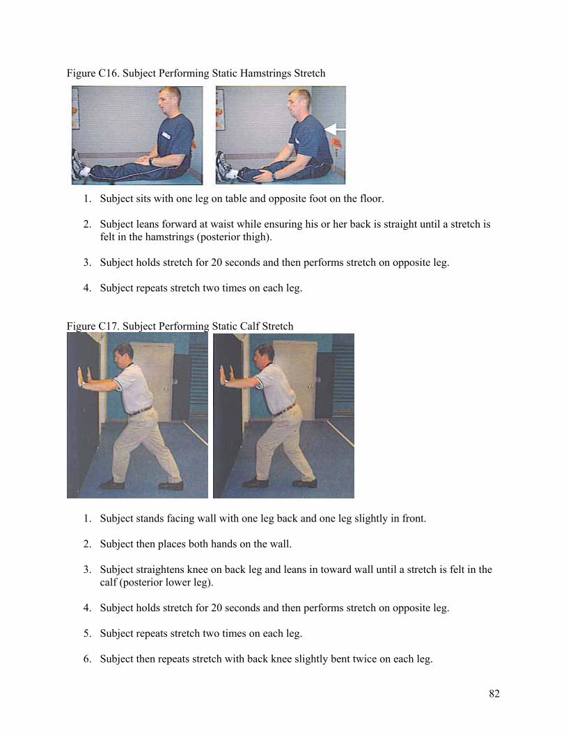
Figure C16. Subject Performing Static Hamstrings Stretch
1. Subject sits with one leg on table and opposite foot on the floor. 2. Subject leans forward at waist while ensuring his or her back is straight until a stretch is
felt in the hamstrings (posterior thigh).
3. Subject holds stretch for 20 seconds and then performs stretch on opposite leg.
4. Subject repeats stretch two times on each leg. Figure C17. Subject Performing Static Calf Stretch
1. Subject stands facing wall with one leg back and one leg slightly in front. 2. Subject then places both hands on the wall.
3. Subject straightens knee on back leg and leans in toward wall until a stretch is felt in the
calf (posterior lower leg).
4. Subject holds stretch for 20 seconds and then performs stretch on opposite leg.
5. Subject repeats stretch two times on each leg.
6. Subject then repeats stretch with back knee slightly bent twice on each leg.
82
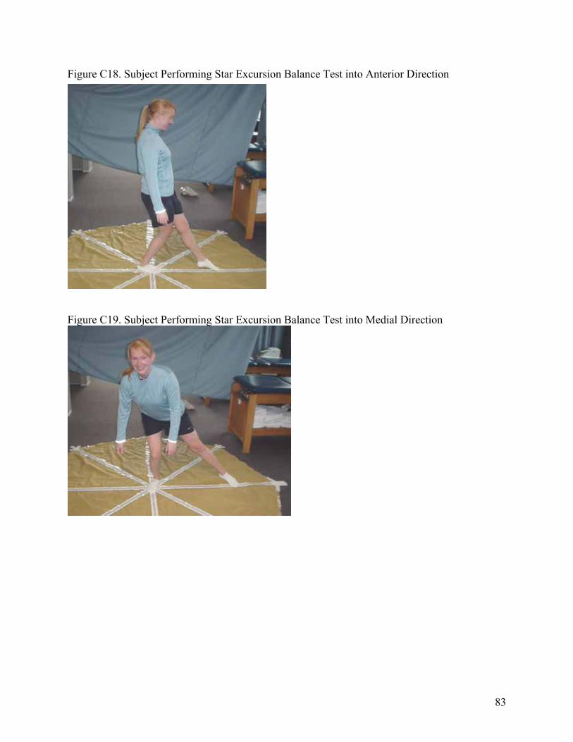
Figure C18. Subject Performing Star Excursion Balance Test into Anterior Direction
Figure C19. Subject Performing Star Excursion Balance Test into Medial Direction
83

Figure C20. Subject Performing Star Excursion Balance Test into Posteromedial Direction
Figure C21. Subject Performing Gluteus Medius Strength Test
84
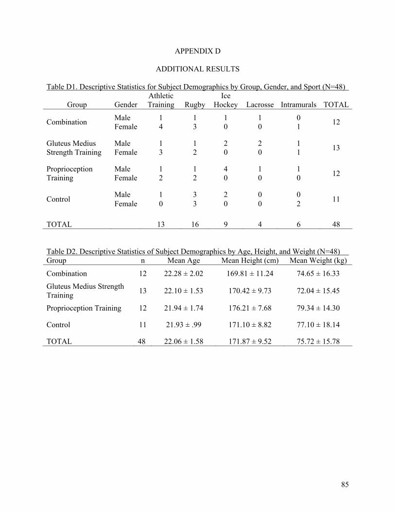
APPENDIX D
ADDITIONAL RESULTS
Table D1. Descriptive Statistics for Subject Demographics by Group, Gender, and Sport (N=48)
Group Gender Athletic Training Rugby
Ice Hockey Lacrosse Intramurals TOTAL
Male 1 1 1 1 0 Combination Female 4 3 0 0 1 12
Male 1 1 2 2 1 Gluteus Medius Strength Training Female 3 2 0 0 1 13
Male 1 1 4 1 1 Proprioception Training Female 2 2 0 0 0 12
Male 1 3 2 0 0 Control Female 0 3 0 0 2 11
TOTAL 13 16 9 4 6 48 Table D2. Descriptive Statistics of Subject Demographics by Age, Height, and Weight (N=48) Group n Mean Age Mean Height (cm) Mean Weight (kg)
Combination 12 22.28 ± 2.02 169.81 ± 11.24 74.65 ± 16.33
Gluteus Medius Strength Training 13 22.10 ± 1.53 170.42 ± 9.73 72.04 ± 15.45
Proprioception Training 12 21.94 ± 1.74 176.21 ± 7.68 79.34 ± 14.30
Control 11 21.93 ± .99 171.10 ± 8.82 77.10 ± 18.14
TOTAL 48 22.06 ± 1.58 171.87 ± 9.52 75.72 ± 15.78
85
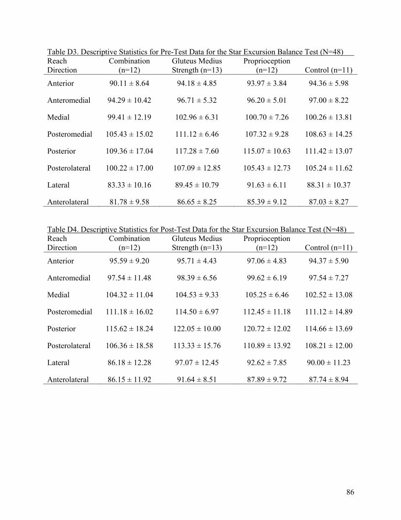
Table D3. Descriptive Statistics for Pre-Test Data for the Star Excursion Balance Test (N=48) Reach Direction
Combination (n=12)
Gluteus Medius Strength (n=13)
Proprioception (n=12) Control (n=11)
Anterior 90.11 ± 8.64 94.18 ± 4.85 93.97 ± 3.84 94.36 ± 5.98
Anteromedial 94.29 ± 10.42 96.71 ± 5.32 96.20 ± 5.01 97.00 ± 8.22
Medial 99.41 ± 12.19 102.96 ± 6.31 100.70 ± 7.26 100.26 ± 13.81
Posteromedial 105.43 ± 15.02 111.12 ± 6.46 107.32 ± 9.28 108.63 ± 14.25
Posterior 109.36 ± 17.04 117.28 ± 7.60 115.07 ± 10.63 111.42 ± 13.07
Posterolateral 100.22 ± 17.00 107.09 ± 12.85 105.43 ± 12.73 105.24 ± 11.62
Lateral 83.33 ± 10.16 89.45 ± 10.79 91.63 ± 6.11 88.31 ± 10.37
Anterolateral 81.78 ± 9.58 86.65 ± 8.25 85.39 ± 9.12 87.03 ± 8.27 Table D4. Descriptive Statistics for Post-Test Data for the Star Excursion Balance Test (N=48) Reach Direction
Combination (n=12)
Gluteus Medius Strength (n=13)
Proprioception (n=12) Control (n=11)
Anterior 95.59 ± 9.20 95.71 ± 4.43 97.06 ± 4.83 94.37 ± 5.90
Anteromedial 97.54 ± 11.48 98.39 ± 6.56 99.62 ± 6.19 97.54 ± 7.27
Medial 104.32 ± 11.04 104.53 ± 9.33 105.25 ± 6.46 102.52 ± 13.08
Posteromedial 111.18 ± 16.02 114.50 ± 6.97 112.45 ± 11.18 111.12 ± 14.89
Posterior 115.62 ± 18.24 122.05 ± 10.00 120.72 ± 12.02 114.66 ± 13.69
Posterolateral 106.36 ± 18.58 113.33 ± 15.76 110.89 ± 13.92 108.21 ± 12.00
Lateral 86.18 ± 12.28 97.07 ± 12.45 92.62 ± 7.85 90.00 ± 11.23
Anterolateral 86.15 ± 11.92 91.64 ± 8.51 87.89 ± 9.72 87.74 ± 8.94
86

Table D5. Mean Differences in SEBT Reach Distance Means from Pre-Test to Post-Test (N=48) Reach Direction
Combination (n=12)
Gluteus Medius Strength (n=13)
Proprioception (n=12) Control (n=11)
Anterior 5.48 ± 3.67 1.53 ± 2.14 3.09 ± 2.72 0.00 ± 0.79
Anteromedial 3.25 ± 4.09 1.68 ± 3.22 3.42 ± 3.13 0.54 ± 1.83
Medial 4.91 ± 2.65 1.57 ± 6.04 4.55 ± 3.42 2.26 ± 3.72
Posteromedial 5.75 ± 3.84 3.38 ± 3.00 5.13 ± 4.94 2.49 ± 2.36
Posterior 6.26 ± 3.19 4.77 ± 5.00 5.65 ± 3.91 3.24 ± 3.63
Posterolateral 6.14 ± 7.30 6.24 ± 5.66 5.46 ± 4.39 2.97 ± 5.33
Lateral 2.85 ± 6.22 7.62 ± 7.96 0.99 ± 2.19 1.69 ± 3.28
Anterolateral 4.37 ± 7.00 4.99 ± 3.65 2.5 ± 2.12 0.71 ± 1.40 Table D6. One-Way MANOVA of Pre-Test Data for the Star Excursion Balance Test (N=48) Reach Direction F P ES ß
Anterior 1.341 0.273 0.084 0.332
Anteromedial 0.313 0.816 0.021 0.106
Medial 0.276 0.842 0.018 0.099
Posteromedial 0.527 0.666 0.035 0.149
Posterior 1.004 0.400 0.064 0.254
Posterolateral 0.572 0.637 0.038 0.158
Lateral 1.628 0.196 0.100 0.398
Anterolateral 0.880 0.459 0.057 0.226 Key: F = F value, P = significance, ES = Effect Size, ß = Power
87

Table D7. One-Way MANOVA of Post-Test Data for the Star Excursion Balance Test (N=48) Reach Direction F P ES ß
Anterior 0.347 0.791 0.023 0.112
Anteromedial 0.171 0.915 0.012 0.079
Medial 0.148 0.930 0.010 0.075
Posteromedial 0.196 0.898 0.013 0.084
Posterior 0.854 0.472 0.055 0.221
Posterolateral 0.491 0.690 0.032 0.141
Lateral 2.103 0.113 0.125 0.501
Anterolateral 0.705 0.554 0.046 0.187 Key: F = F value, P = significance, ES = Effect Size, ß = Power Table D8. Main Effects and Interactions Within and Between Subjects for the Star Excursion Balance Test (N=48) F P ES ß
TEST 190.825 <0.001* 0.813 1.000
DIRECTION 141.825 <0.001* 0.763 1.000
GROUP 0.631 0.599 0.041 0.171
TEST X GROUP 6.145 0.001* 0.295 0.946
DIRECTION X GROUP 0.680 0.569 0.044 0.182
TEST X DIRECTION 3.303 0.076 0.070 0.428
TEST X DIRECTION X GROUP 1.571 0.210 0.097 0.385 Key: F = F value, P = significance, ES = Effect Size, ß = Power, * Significant at the P≤0.05 level
88

Table D9. Pairwise Comparisons for Reach Directions on the Star Excursion Balance Test (N=48) Reach Direction T P
Pre- and Post-Test Anterior -5.538 <0.001*
Pre- and Post-Test Anteromedial -4.698 <0.001*
Pre- and Post-Test Medial -5.287 <0.001*
Pre- and Post-Test Posteromedial -7.690 <0.001*
Pre- and Post-Test Posterior -8.579 <0.001*
Pre- and Post-Test Posterolateral -6.363 <0.001*
Pre- and Post-Test Lateral -3.947 <0.001*
Pre- and Post-Test Anterolateral -5.116 <0.001* Key: T = t value, P = Significance, * Significant at the P≤0.05 level Table D10. Multiple Comparison Tukey Post Hoc Tests for Star Excursion Balance Test (N=48) Group Comparisons P
COMBINATION : GLUTEUS MEDIUS STRENGTH 0.559
COMBINATION : PROPRIOCEPTION 0.783
COMBINATION : CONTROL 0.953
GLUTEUS MEDIUS STRENGTH : PROPRIOCEPTION 0.984
GLUTEUS MEDIUS STRENGTH : CONTROL 0.876
PROPRIOCEPTION : CONTROL 0.979 Key: P = Significance
89
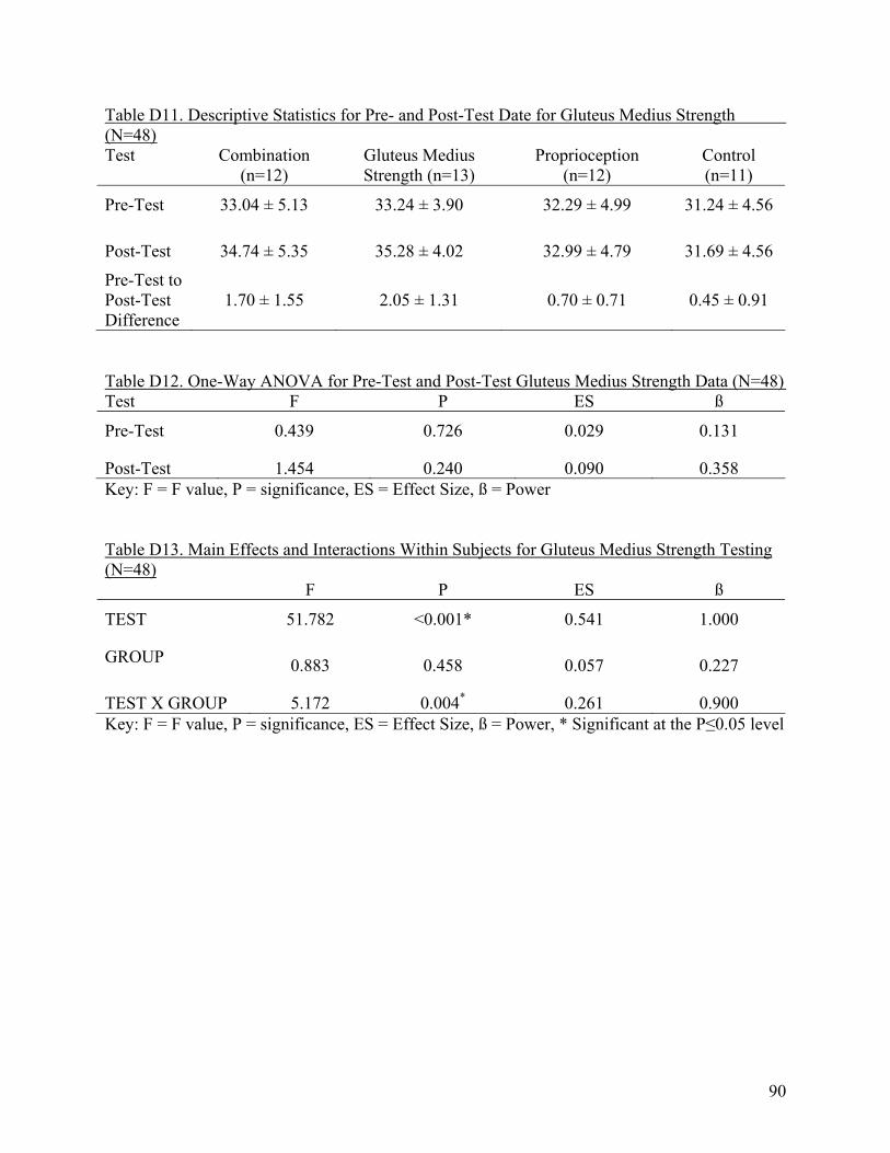
Table D11. Descriptive Statistics for Pre- and Post-Test Date for Gluteus Medius Strength (N=48) Test Combination
(n=12) Gluteus Medius Strength (n=13)
Proprioception (n=12)
Control (n=11)
Pre-Test 33.04 ± 5.13 33.24 ± 3.90 32.29 ± 4.99 31.24 ± 4.56
Post-Test 34.74 ± 5.35 35.28 ± 4.02 32.99 ± 4.79 31.69 ± 4.56
Pre-Test to Post-Test Difference
1.70 ± 1.55 2.05 ± 1.31 0.70 ± 0.71 0.45 ± 0.91
Table D12. One-Way ANOVA for Pre-Test and Post-Test Gluteus Medius Strength Data (N=48) Test F P ES ß
Pre-Test 0.439 0.726 0.029 0.131
Post-Test 1.454 0.240 0.090 0.358 Key: F = F value, P = significance, ES = Effect Size, ß = Power Table D13. Main Effects and Interactions Within Subjects for Gluteus Medius Strength Testing (N=48) F P ES ß
TEST 51.782 <0.001* 0.541 1.000
GROUP 0.883 0.458 0.057 0.227
TEST X GROUP 5.172 0.004* 0.261 0.900 Key: F = F value, P = significance, ES = Effect Size, ß = Power, * Significant at the P≤0.05 level
90

Table D14. Multiple Comparison Tukey Post Hoc Tests for Gluteus Medius Strength (N=48) Group Comparisons P
COMBINATION : GLUTEUS MEDIUS STRENGTH 0.997
COMBINATION : PROPRIOCEPTION 0.911
COMBINATION : CONTROL 0.597
GLUTEUS MEDIUS STRENGTH : PROPRIOCEPTION 0.819
GLUTEUS MEDIUS STRENGTH : CONTROL 0.463
PROPRIOCEPTION : CONTROL 0.929 Key: P = Significance
91

Figure D1.Mean SEBT Reach Distances by Test for the Combination Group
8090100
110
120
130
12
Test
Mean Reach Distances Compared to Leg Length
AM PM ALPLMA LP
Pr
e-test
Post-
test
92

Figure D2. Mean SEBT Reach Distances by Test for the Gluteus Medius Strength Training Group
8090100
110
120
130
12
Test
Mean Reach Distances Compared to Leg Length
AM PM ALPLMA LP
P
re-tes
t
P
ost-t
est
93
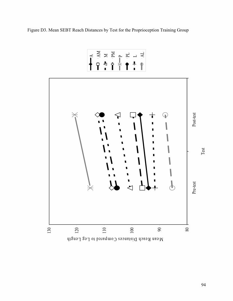
Figure D3. Mean SEBT Reach Distances by Test for the Proprioception Training Group
8090100
110
120
130
12
Test
Mean Reach Distances Compared to Leg Length
AM PM ALPLMA LP
Pr
e-tes
t
P
ost-t
est
94

Figure D4. Mean SEBT Reach Distances by Test for the Control Group
8090100
110
120
130
12
Test
Mean Reach Distances Compared to Leg Length
AM PM ALPLMA LP
Pr
e-tes
t
Post-
test
95

Figure D5. Mean SEBT Reach Distances by Test for All Subjects
8090100
110
120
130
12
Test
Mean Reach Distances Compared to Leg Length
AM PM ALPLMA LP
Pr
e-test
Post-
test
96

Figure D6. Mean SEBT Reach Distances by Test for All Groups
9092949698100
102
104
106
12
34
Grou
p
Mean Reach Distances Compared to Leg Length
Pre-t
estPo
st-tes
t
Contr
olPr
oprio
cepti
onGl
uteus
M
edius
Co
mbina
tion
97

Figure D7. Mean SEBT Reach Distances by Group
8090100
110
120
130
12
34
Grou
p
Mean Reach Distances Compared to Leg Length
AMA M PM ALP PL L
Contr
olPr
oprio
cepti
onGl
uteus
Med
iusStr
ength
Comb
inatio
n
98
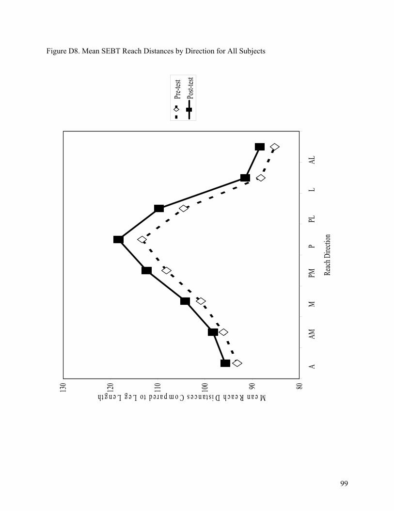
Figure D8. Mean SEBT Reach Distances by Direction for All Subjects
8090100
110
120
130
12
34
56
78
Reach
Dire
ction
Mean Reach Distances Compared to Leg Length
Pre-te
stPo
st-tes
t
A
AM
M
P
M
P
PL
L
AL
99

Figure D9. Mean Pre-Test SEBT Reach Distances by Direction for All Groups
8090100
110
120
130
12
34
56
78
Reach
Dire
ction
Mean Reach Distances Compared to Leg Length
Comb
inatio
nGl
uteus
Mediu
s Stre
ngth
Propri
ocep
tion
Contr
ol
A
AM
M
P
M
P
PL
L
AL
100
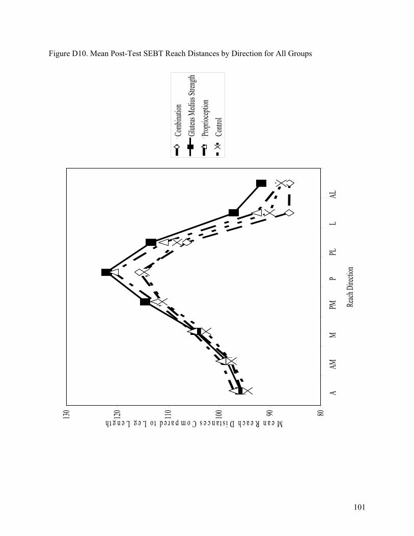
Figure D10. Mean Post-Test SEBT Reach Distances by Direction for All Groups
8090100
110
120
130
12
34
56
78
Reach
Direc
tion
Mean Reach Distances Compared to Leg Length
Comb
inatio
nGl
uteus
Mediu
s Stre
ngth
Propri
ocepti
onCo
ntrol
A
AM
M
P
M
P
PL
L
AL
101
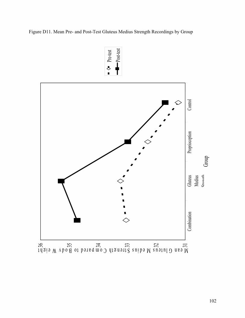
Figure D11. Mean Pre- and Post-Test Gluteus Medius Strength Recordings by Group
313233343536
12
34
Grou
p
Mean Gluteus Medius Strength Compared to Body Weight
Pre-te
stPo
st-tes
t
Contr
olPro
prioce
ption
Glute
us Me
dius
Streng
th
Comb
inatio
n
102

Figure D12. Mean Gluteus Medius Strength Recordings by Test for all Groups
313233343536
12
Test
Mean Gluteus Strength Compared to Body Weight
Comb
ination
Gluteu
s Medi
us Str
ength
Propri
oceptio
nCo
ntrol
Pre-tes
t
Post
-test
103
APPENDIX E
RECOMMENDATIONS FOR FUTURE RESEARCH
1. Increase the length of the study from six weeks to ten weeks. 2. Increase the training sessions per week from three to four.
3. Conduct the study using injured subjects with acute lower extremity injuries.
4. Conduct the study using non-physically active subjects.
5. Conduct the study using subjects with chronic ankle instability. 6. Conduct the study using an older population who utilizes the hip strategy. 7. Conduct the study using intercollegiate athletes as opposed to club sports athletes,
intramurals athletes, and athletic trainers.
8. Increase the number of subjects from between 11-13 to at least 20 subjects per group.
9. Conduct the study using more purely strengthening exercises (85 % of one repetition maximum, four to six sets of one to five repetitions, four times per week) for the gluteus medius strengthening and combination training groups.
10. Train the combination group for balance and gluteus medius strength on separate days
to avoid fatigue.
11. Conduct a longitudinal study to examine the differences in pre- and post-training injury rates.
12. Conduct the study without allowing any compensatory arm movement during the
SEBT.
104
ADDITIONAL REFERENCES
68. Cornwall MW. Postural sway following inversion sprain of the ankle. J Am Podiatr Med Assoc. 1991;81(5):243-247.
69. Wilkerson GB. Dynamic ankle stability: mechanical and neuromuscular
interrelationships. J Sport Rehabil. 1994;3(1):43-57.
70. Hertel J. Effect of foot orthotics on quadriceps and gluteus medius electromyographic activity during selected exercises. Arch Phys Med Rehabil. 2005;86(1):26-30.
71. Clark MA, Russell AM. Optimum Performance Training for the Performance
Enhancement Specialist. 1st ed. Calabasas, CA: National Academy of Sports Medicine; 2002.
72. Neumann DA. Effect of load and carrying position on the electromyographic activity of
the gluteus medius muscle during walking. Phys Ther. 1985;65(3):305-311.
73. Frese E. Clinical reliability of manual muscle testing: middle trapezius and gluteus medius muscles. Phys Ther. 1987;67:1072-1076.
74. Huston JL, Sandrey MA, Lively MW, Kotsko K. The effects of calf-muscle fatigue on
sagittal-plane joint-position sense in the ankle. J Sport Rehabil. 2005;14:168-184.
75. Tortora GJ, Grabowski SR. Principles of Anatomy and Physiology. 9th ed. New York, NY: John Wiley & Sons, Inc.; 2000:263,494.
76. Blackburn T, Guskiewicz KM, Petschauer MA, Prentice WE. Balance and joint stability:
the relative contributions of proprioception and muscular strength. J Sport Rehabil. 2000;9:315-328.
77. Weiner DK, Bongiorni DR, Studenski SA, Duncan PW, Kochersberger GG. Does
functional reach improve with rehabilitation?. Arch Phys Med Rehabil. 1993;74:796-800.
78. Ferris CM, Freedman AD. Neuromuscular and biomechanical lower extremity training for female athletes. Athl Ther Today. 2001;6(4):54-62.
79. Krebs DE, Robbins CE, Lavine L, Mann RW. Hip biomechanics during gait. J Orthop
Sports Phys Ther. 1998;28(1):51-9.
80. Baumhauer JF, Alosa DM, Renstrom PAFH, Trevino S, Benynnon B. Test-retest reliability of ankle injury risk factors. Am J Sports Med. 1995;23(5):571-574.
81. Rockar PA. The subtalar joint: anatomy and joint motion. J Orthop Sports Phys Ther.
1995;21(6):361-372.
105

82. Moore KL, Dalley AF. Clinically Oriented Anatomy. 4th ed. Baltimore, MD: Lippincott, Williams, & Wilkins; 1999: 551-555,577,583,588,589.
83. Mangwani J. Chronic lateral ankle instability: review of anatomy, biomechanics,
pathology, diagnosis and treatment. Foot: Int J Clin Foot Sci. 2001;11(2):76-84.
84. Hanney WJ. Proprioceptive training for ankle instability. Strength Condit J. 2000;22(5):63-68.
85. Levangie PK, Norkin CC. Joint Structure and Function: A Comprehensive Analysis. 3rd
ed. Philadelphia, PA.: F.A. Davis; 2001:2,372-376.
86. Dahmer G. Biomechanics. ATTR 627. Class notes. West Virginia University. School of Physical Education.
87. Hinterman B. Biomechanics of the unstable ankle joint and clinical implications. Med Sci
Sports Exerc. 1999;31(7):S459-S469.
88. Norkin CC, White J. Measurement of Joint Motion. 2nd ed. Phildelphia, PA: F.A. Davis; 1995:150.
89. Garrsion JG, Hart JM, Palmieri RM, Kerrigan DC, Ingersoll CD. Lower extremity EMG
in male and female college soccer players during single-leg landing. J Sport Rehabil. 2005;14:48-57.
90. Nordin M, Frankel VH. Basic Biomechanics of the Musculoskeletal System. 3rd ed.
Baltimore, MD: Lippincott Williams & Wilkins; 2001:251.
91. Bullock-Saxton JE. Local sensation changes and altered hip muscle function following severe ankle sprain. Phys Ther. 1994;74(1):17-28.
92. Bullock-Saxton JE, Janda V, Bullock MI. The influence of ankle sprain injury on muscle
activation during hip extension. Int J Sports Med. 1994;15(6):330-334.
93. Myers JB, Guskiewicz KM, Schneider RA, Prentice WE. Proprioception and neuromuscular control of the shoulder after muscle fatigue. J Athl Train. 1999;34(4):362-367.
94. Bloem BR, Allum JHJ, Carpenter MG, Verschuuren JJGM, Honeggar F. Triggering of
balance corrections and compensatory strategies in a patient with total leg proprioceptive loss. Exp Brain Res. 2002;142:91-107.
95. Kuo AD, Speers RA, Peterka RJ, Horak FB. Effect of altered sensory conditions on
multivariate descriptors of human postural sway. Exp Brain Res. 1998;122:185-195.
106

96. Olmsted LC, Hertel J. Influence of foot type and orthotics on static and dynamic postural control. J Sport Rehabil. 2004;13(1):54-66.
107