the density code for the development of a vaccine?chenglab/pdf/cheng2016jps.pdf · hepatitis a, and...
TRANSCRIPT
lable at ScienceDirect
Journal of Pharmaceutical Sciences 105 (2016) 3223-3232
Contents lists avai
Journal of Pharmaceutical Sciences
journal homepage: www.jpharmsci .org
Review
The Density Code for the Development of a Vaccine?
Wei Cheng 1, 2, 3, *
1 Department of Pharmaceutical Sciences, College of Pharmacy, University of Michigan, Ann Arbor, Michigan 481092 Department of Biological Chemistry, University of Michigan Medical School, Ann Arbor, Michigan 481093 Department of Biophysics, University of Michigan, Ann Arbor, Michigan 48109
a r t i c l e i n f o
Article history:Received 19 June 2016Revised 17 July 2016Accepted 26 July 2016Available online 17 September 2016
Keywords:vaccinesvaccine deliveryimmune responseimmunologyprotein densityB-cell receptorB-cell activationparticulate antigennanoparticles
Conflicts of interest: The author declares no competin* Correspondence to: Wei Cheng (Telephone: 734-76
E-mail address: [email protected] (W. Cheng).
http://dx.doi.org/10.1016/j.xphs.2016.07.0200022-3549/© 2016 American Pharmacists Association
a b s t r a c t
The development of prophylactic vaccines remains largely empirical in nature and rarely have generalrules been applied in the strategic decision and the formulation of a viral vaccine. Currently, there are atotal of 15 virus agents from 12 unique virus families with vaccines licensed by the U.S. Food and DrugAdministration. Extensive structural information on these viral particles and potential mechanisms ofprotection are available for the majority of these virus pathogens and their respective vaccines. Here Ireview the quantitative features of these viral surface antigens in relation to the molecular mechanismsof B-cell activation and point out a potential correlation between the density of immunogenic proteinsdisplayed on the surface of the vaccine antigen carrier and the success of a vaccine. These features help usunderstand the humoral immunity induced by viral vaccines on a quantitative ground and re-emphasizethe importance of antigen density on the activation of the immune system. Although the detailedmechanisms behind this phenomenon remain to be explored, it implies that both the size of antigencarriers and the density of immunogenic proteins displayed on these carriers are important parametersthat may need to be optimized for the formulation of a vaccine.
© 2016 American Pharmacists Association®. Published by Elsevier Inc. All rights reserved.
The Impact of Vaccination on Human Health
The recent outbreak of zika virus in Latin America and theCaribbean1 is growing rapidly. Up to 1.3 million suspected caseshave been reported in Brazil by December 2015. By March 2016, 33countries and territories in the Americas have reported confirmedcases of zika virus infection.2 Less than 2 years ago, the devastatingEbola virus outbreak occurred in West Africa.3 With more than11,000 deaths reported, this Ebola outbreak has become the mostextensive Ebola outbreak in human history.3
Besides the necessary social and administrative measures thatmust take place in the events of these infectious disease outbreaks,effective vaccination against these infectious diseases,4,5 if feasible,might offer one of the long-term solutions to these public healththreats. Indeed, vaccination has made a major impact on globalpopulation health and has controlled more than a dozen of humandiseases. In looking through the human history, perhaps few sci-entific discoveries can rival vaccination in its efficacy and magni-tude in the elimination of disease burden and suffering. In this
g financial interests.3-3709; Fax: 734-615-6162).
®. Published by Elsevier Inc. All rig
context, antibiotic does not even compete with vaccination,because only safe water, which is considered to be a basic humanright by the World Health Organization,6 performs better.7
Based on the statistics from the Centers for Disease Control andPrevention in the United States, vaccination has eliminated severaldiseases from the Americas. Notably, the percent reduction in theannual morbidity before and after routine vaccination is 97% ormore for smallpox, diphtheria, measles, mumps, polio, rubella,congenital rubella syndrome, tetanus, and Haemophilus influenza.8
More than 88% reduction in annual morbidity is also achieved forpertussis (i.e., 100-day cough), hepatitis A, and varicella.8 Amongthese different diseases, smallpox, measles, mumps, polio, rubella,hepatitis A, and varicella are all caused by viral infections.
A Short List of Licensed Viral Vaccines
A survey of vaccines currently licensed for immunization anddistribution in the United States identified over 90 pharmaceuticalproducts (http://www.fda.gov/BiologicsBloodVaccines/Vaccines/).Among these, 15 virus agents from a total of 12 unique virus fam-ilies have been targeted. These virus agents are listed in Table 1.9-22
As seen from Table 1, these virus agents include viruses that usedifferent forms of nucleic acids as their genetic materials. Ofpharmaceutical scientists' interests, 9 virus agents of 15 have live
hts reserved.
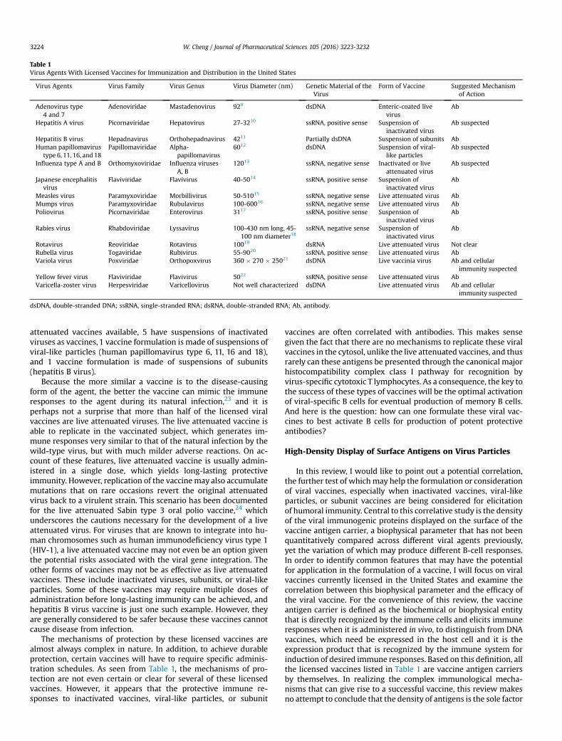
Table 1Virus Agents With Licensed Vaccines for Immunization and Distribution in the United States
Virus Agents Virus Family Virus Genus Virus Diameter (nm) Genetic Material of theVirus
Form of Vaccine Suggested Mechanismof Action
Adenovirus type4 and 7
Adenoviridae Mastadenovirus 929 dsDNA Enteric-coated livevirus
Ab
Hepatitis A virus Picornaviridae Hepatovirus 27-3210 ssRNA, positive sense Suspension ofinactivated virus
Ab suspected
Hepatitis B virus Hepadnavirus Orthohepadnavirus 4211 Partially dsDNA Suspension of subunits AbHuman papillomavirus
type 6, 11, 16, and 18Papillomaviridae Alpha-
papillomavirus6012 dsDNA Suspension of viral-
like particlesAb suspected
Influenza type A and B Orthomyxoviridae Influenza virusesA, B
12013 ssRNA, negative sense Inactivated or liveattenuated virus
Ab suspected
Japanese encephalitisvirus
Flaviviridae Flavivirus 40-5014 ssRNA, positive sense Suspension ofinactivated virus
Ab
Measles virus Paramyxoviridae Morbillivirus 50-51015 ssRNA, negative sense Live attenuated virus AbMumps virus Paramyxoviridae Rubulavirus 100-60016 ssRNA, negative sense Live attenuated virus AbPoliovirus Picornaviridae Enterovirus 3117 ssRNA, positive sense Suspension of
inactivated virusAb
Rabies virus Rhabdoviridae Lyssavirus 100-430 nm long, 45-100 nm diameter18
ssRNA, negative sense Suspension ofinactivated virus
Ab
Rotavirus Reoviridae Rotavirus 10019 dsRNA Live attenuated virus Not clearRubella virus Togaviridae Rubivirus 55-9020 ssRNA, positive sense Live attenuated virus AbVariola virus Poxviridae Orthopoxvirus 360 � 270 � 25021 dsDNA Live vaccinia virus Ab and cellular
immunity suspectedYellow fever virus Flaviviridae Flavivirus 5022 ssRNA, positive sense Live attenuated virus AbVaricella-zoster virus Herpesviridae Varicellovirus Not well characterized dsDNA Live attenuated virus Ab and cellular
immunity suspected
dsDNA, double-stranded DNA; ssRNA, single-stranded RNA; dsRNA, double-stranded RNA; Ab, antibody.
W. Cheng / Journal of Pharmaceutical Sciences 105 (2016) 3223-32323224
attenuated vaccines available, 5 have suspensions of inactivatedviruses as vaccines, 1 vaccine formulation is made of suspensions ofviral-like particles (human papillomavirus type 6, 11, 16 and 18),and 1 vaccine formulation is made of suspensions of subunits(hepatitis B virus).
Because the more similar a vaccine is to the disease-causingform of the agent, the better the vaccine can mimic the immuneresponses to the agent during its natural infection,23 and it isperhaps not a surprise that more than half of the licensed viralvaccines are live attenuated viruses. The live attenuated vaccine isable to replicate in the vaccinated subject, which generates im-mune responses very similar to that of the natural infection by thewild-type virus, but with much milder adverse reactions. On ac-count of these features, live attenuated vaccine is usually admin-istered in a single dose, which yields long-lasting protectiveimmunity. However, replication of the vaccinemay also accumulatemutations that on rare occasions revert the original attenuatedvirus back to a virulent strain. This scenario has been documentedfor the live attenuated Sabin type 3 oral polio vaccine,24 whichunderscores the cautions necessary for the development of a liveattenuated virus. For viruses that are known to integrate into hu-man chromosomes such as human immunodeficiency virus type 1(HIV-1), a live attenuated vaccine may not even be an option giventhe potential risks associated with the viral gene integration. Theother forms of vaccines may not be as effective as live attenuatedvaccines. These include inactivated viruses, subunits, or viral-likeparticles. Some of these vaccines may require multiple doses ofadministration before long-lasting immunity can be achieved, andhepatitis B virus vaccine is just one such example. However, theyare generally considered to be safer because these vaccines cannotcause disease from infection.
The mechanisms of protection by these licensed vaccines arealmost always complex in nature. In addition, to achieve durableprotection, certain vaccines will have to require specific adminis-tration schedules. As seen from Table 1, the mechanisms of pro-tection are not even certain or clear for several of these licensedvaccines. However, it appears that the protective immune re-sponses to inactivated vaccines, viral-like particles, or subunit
vaccines are often correlated with antibodies. This makes sensegiven the fact that there are no mechanisms to replicate these viralvaccines in the cytosol, unlike the live attenuated vaccines, and thusrarely can these antigens be presented through the canonical majorhistocompatibility complex class I pathway for recognition byvirus-specific cytotoxic T lymphocytes. As a consequence, the key tothe success of these types of vaccines will be the optimal activationof viral-specific B cells for eventual production of memory B cells.And here is the question: how can one formulate these viral vac-cines to best activate B cells for production of potent protectiveantibodies?
High-Density Display of Surface Antigens on Virus Particles
In this review, I would like to point out a potential correlation,the further test of whichmay help the formulation or considerationof viral vaccines, especially when inactivated vaccines, viral-likeparticles, or subunit vaccines are being considered for elicitationof humoral immunity. Central to this correlative study is the densityof the viral immunogenic proteins displayed on the surface of thevaccine antigen carrier, a biophysical parameter that has not beenquantitatively compared across different viral agents previously,yet the variation of which may produce different B-cell responses.In order to identify common features that may have the potentialfor application in the formulation of a vaccine, I will focus on viralvaccines currently licensed in the United States and examine thecorrelation between this biophysical parameter and the efficacy ofthe viral vaccine. For the convenience of this review, the vaccineantigen carrier is defined as the biochemical or biophysical entitythat is directly recognized by the immune cells and elicits immuneresponses when it is administered in vivo, to distinguish from DNAvaccines, which need be expressed in the host cell and it is theexpression product that is recognized by the immune system forinduction of desired immune responses. Based on this definition, allthe licensed vaccines listed in Table 1 are vaccine antigen carriersby themselves. In realizing the complex immunological mecha-nisms that can give rise to a successful vaccine, this review makesno attempt to conclude that the density of antigens is the sole factor
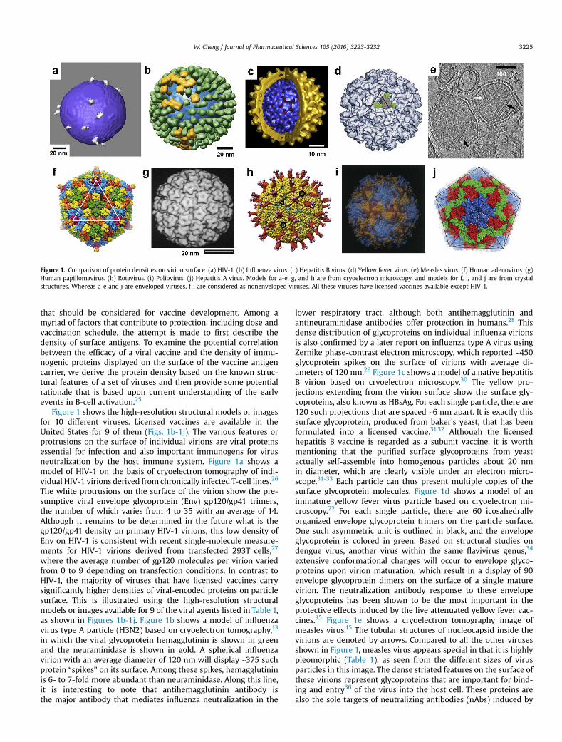
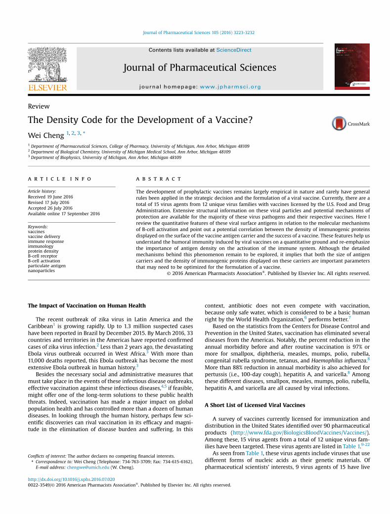
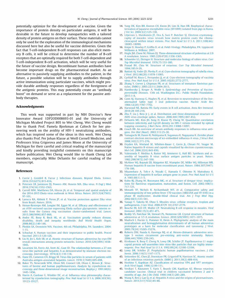
Figure 1. Comparison of protein densities on virion surface. (a) HIV-1. (b) Influenza virus. (c) Hepatitis B virus. (d) Yellow fever virus. (e) Measles virus. (f) Human adenovirus. (g)Human papillomavirus. (h) Rotavirus. (i) Poliovirus. (j) Hepatitis A virus. Models for a-e, g, and h are from cryoelectron microscopy, and models for f, i, and j are from crystalstructures. Whereas a-e and j are enveloped viruses, f-i are considered as nonenveloped viruses. All these viruses have licensed vaccines available except HIV-1.
W. Cheng / Journal of Pharmaceutical Sciences 105 (2016) 3223-3232 3225
that should be considered for vaccine development. Among amyriad of factors that contribute to protection, including dose andvaccination schedule, the attempt is made to first describe thedensity of surface antigens. To examine the potential correlationbetween the efficacy of a viral vaccine and the density of immu-nogenic proteins displayed on the surface of the vaccine antigencarrier, we derive the protein density based on the known struc-tural features of a set of viruses and then provide some potentialrationale that is based upon current understanding of the earlyevents in B-cell activation.25
Figure 1 shows the high-resolution structural models or imagesfor 10 different viruses. Licensed vaccines are available in theUnited States for 9 of them (Figs. 1b-1j). The various features orprotrusions on the surface of individual virions are viral proteinsessential for infection and also important immunogens for virusneutralization by the host immune system. Figure 1a shows amodel of HIV-1 on the basis of cryoelectron tomography of indi-vidual HIV-1 virions derived from chronically infected T-cell lines.26
The white protrusions on the surface of the virion show the pre-sumptive viral envelope glycoprotein (Env) gp120/gp41 trimers,the number of which varies from 4 to 35 with an average of 14.Although it remains to be determined in the future what is thegp120/gp41 density on primary HIV-1 virions, this low density ofEnv on HIV-1 is consistent with recent single-molecule measure-ments for HIV-1 virions derived from transfected 293T cells,27
where the average number of gp120 molecules per virion variedfrom 0 to 9 depending on transfection conditions. In contrast toHIV-1, the majority of viruses that have licensed vaccines carrysignificantly higher densities of viral-encoded proteins on particlesurface. This is illustrated using the high-resolution structuralmodels or images available for 9 of the viral agents listed in Table 1,as shown in Figures 1b-1j. Figure 1b shows a model of influenzavirus type A particle (H3N2) based on cryoelectron tomography,13
in which the viral glycoprotein hemagglutinin is shown in greenand the neuraminidase is shown in gold. A spherical influenzavirion with an average diameter of 120 nm will display ~375 suchprotein “spikes” on its surface. Among these spikes, hemagglutininis 6- to 7-fold more abundant than neuraminidase. Along this line,it is interesting to note that antihemagglutinin antibody isthe major antibody that mediates influenza neutralization in the
lower respiratory tract, although both antihemagglutinin andantineuraminidase antibodies offer protection in humans.28 Thisdense distribution of glycoproteins on individual influenza virionsis also confirmed by a later report on influenza type A virus usingZernike phase-contrast electron microscopy, which reported ~450glycoprotein spikes on the surface of virions with average di-ameters of 120 nm.29 Figure 1c shows a model of a native hepatitisB virion based on cryoelectron microscopy.30 The yellow pro-jections extending from the virion surface show the surface gly-coproteins, also known as HBsAg. For each single particle, there are120 such projections that are spaced ~6 nm apart. It is exactly thissurface glycoprotein, produced from baker's yeast, that has beenformulated into a licensed vaccine.31,32 Although the licensedhepatitis B vaccine is regarded as a subunit vaccine, it is worthmentioning that the purified surface glycoproteins from yeastactually self-assemble into homogenous particles about 20 nmin diameter, which are clearly visible under an electron micro-scope.31-33 Each particle can thus present multiple copies of thesurface glycoprotein molecules. Figure 1d shows a model of animmature yellow fever virus particle based on cryoelectron mi-croscopy.22 For each single particle, there are 60 icosahedrallyorganized envelope glycoprotein trimers on the particle surface.One such asymmetric unit is outlined in black, and the envelopeglycoprotein is colored in green. Based on structural studies ondengue virus, another virus within the same flavivirus genus,34
extensive conformational changes will occur to envelope glyco-proteins upon virion maturation, which result in a display of 90envelope glycoprotein dimers on the surface of a single maturevirion. The neutralization antibody response to these envelopeglycoproteins has been shown to be the most important in theprotective effects induced by the live attenuated yellow fever vac-cines.35 Figure 1e shows a cryoelectron tomography image ofmeasles virus.15 The tubular structures of nucleocapsid inside thevirions are denoted by arrows. Compared to all the other virusesshown in Figure 1, measles virus appears special in that it is highlypleomorphic (Table 1), as seen from the different sizes of virusparticles in this image. The dense striated features on the surface ofthese virions represent glycoproteins that are important for bind-ing and entry36 of the virus into the host cell. These proteins arealso the sole targets of neutralizing antibodies (nAbs) induced by
W. Cheng / Journal of Pharmaceutical Sciences 105 (2016) 3223-32323226
measles virus vaccines.37 Figure 1f shows a model of a humanadenovirus based on the high-resolution crystal structures of anadenovirus capsid.38 One of the 20 facets on this icosahedral virus isoutlined in white. Each facet contains 12 hexon trimers and apenton at each vortex (pink), and there are a total of 240 hexontrimers on a single particle. Indeed, the hexon protein containsmost of the epitopes recognized by nAbs against human adeno-virus.39,40 Figure 1g shows a model of a human papillomavirus type1 virion based on a cryoelectron microscopy study.12 This particlehas a diameter of 60 nm. The outer shell of the virus particle ismade of 72 capsomers, and each capsomer is a pentamer of themajor capsid protein L1. The human papillomavirus vaccine is madeof viral-like particles that are self-assembled from this L1 protein,which induce high titers of neutralizing antibody and offer pro-tection.41,42 Figure 1h shows the structure of a rotavirus particledetermined by cryoelectron microscopy and single-particle anal-ysis.43 In each particle, there are a total of 60 “spikes” (red stemwith purple top) protruding from the surface of the virion, which ismade of viral protein VP4. In addition, the extended shell (yellow)below these spikes is made of viral VP7 protein. Although themechanism of protection by the live rotavirus vaccine is not yetclear, both VP4 and VP7 induce nAbs and these antibodies arebelieved to contribute to protection.44,45 Figure 1i shows a model ofa poliovirus based on the high-resolution crystal structures of theMahoney strain of type 1 poliovirus.17 The viral proteins VP1, VP2,and VP3 are colored in blue, yellow, and red, respectively. A singlepoliovirus particle is approximately 31 nm in diameter, and eachparticle has 60 copies each of the VP1, VP2, and VP3 proteins. Basedon the crystal structures, the neutralization epitopes have beenmapped to all of these 3 proteins. Figure 1j shows the overallstructure of the hepatitis A virus obtained from high-resolution X-ray crystallography,46 where the black lines depict the facets of thisicosahedral virus. The viral-encoded proteins VP1, VP2, and VP3 arecolored in blue, green, and red, respectively. nAbs, likely thoseagainst VP1 and VP3, are correlated with the protection offered bythe inactivated hepatitis A virus vaccine.47,48
Cryoelectron tomography studies of purified rubella virus werealso published recently.20 The envelope glycoproteins of the viruswere shown to form extended rows of density that were separated9 nm apart on virion surface. By the time this article is written, thereare no published high-resolution structural models available toderive the density of surface antigens for Japanese encephalitis vi-rus, mumps virus, rabies virus, smallpox virus, or varicella-zostervirus. However, structural models of similar viruses or viruseswithin the same family or genus are available. For example,Japanese encephalitis virus and dengue virus belong to the sameflavivirus genus in the flavivirus family. The organization of theirenvelope glycoproteins might be similar. Indeed, Luca et al.49 havesolved the crystal structure for the ectodomain of the Japaneseencephalitis virus envelope protein, which shows high similarity tothat of the dengue virus. As mentioned above, mature dengue virushas 90 envelope glycoprotein dimers on a single virion surface.34
Cryoelectron tomography studies of rabies virus revealed discon-tinuous spike-like objects on the surface of virions that arecompatible with viral glycoproteins.50 Although it is difficult toderive glycoprotein densities from these images, studies of thevesicular stomatitis virus (VSV), which belongs to the same familyof rhabdoviridae as rabies virus, might offer some hints. Electronmicroscopy studies of VSV revealed a layer of density outside theviral membrane51 that is clearly made of proteins, which can beremoved upon trypsin digestion.52 Ensemble estimations based onRNA and proteins present in viral particles yielded approximately500 glycoproteins per VSV particle.53 High-resolution structuralmodels of varicella-zoster virus also lag behind. However, high-resolution structural models are available for the herpes simplex
virus, which is another human virus within the same herpesviridaefamily as varicella-zoster virus. Based on cryoelectron tomography,herpes simplex virus particles are pleomorphic. Their diametersrange from 170 to 200 nm, and each virion carries 600 to 750glycoprotein molecules on the virion surface.54
Molecular Mechanisms That Relate Density of SurfaceAntigens to Antibody Neutralization
From the analysis above, it is clear that a majority of the virusesthat have licensed vaccines display high densities of viral proteinson the surface of individual virions. Most of these proteins are thetargets of nAbs induced upon vaccination, which offer protection tothe vaccinated subjects. At themolecular level, there are 2 potentialmechanisms that can relate the density of surface antigens to nAbs.First, high-density display of viral antigens facilitates bivalentbinding by the immunoglobulin G (IgG) antibody, which is impor-tant for the potency of the antibody to neutralize viral infectivity.Second, high-density display of viral antigens facilitates B-cellactivation, which is the prerequisite for antibody secretion andmemory B-cell formation. I will elucidate these 2 aspects, especiallythe second aspect, in detail below.
High Density of Surface Antigens Facilitates Bivalent Binding by IgG
Among different classes of antibodies in the body, IgG is themajor class of antibodies that binds various pathogens. An IgGmolecule has 2 identical “arms” of Fab, and antigens bind to the tipof each Fab. The distance that can be reached or bridged by the 2antigen-binding sites in each of the IgG is typically 15 nm.55 As aresult, low-density display of viral antigens on the virion surface, ifthe distance between antigenic molecules is greater than 15 nm,will impede the bivalent binding of IgG. This indeed occurs on HIV-1 virions as demonstrated recently by Galimidi et al.55,56 Failure tobind antigens bivalently will increase the off rate of antibodies fromthe bound target and leads to low efficiency in neutralizing virusinfectivity. In contrast, as shown in Figure 1b for influenza virus, theaverage center-to-center spacing between the densely packedglycoproteins on virion surface is about 11 nm,13 which is shortenough to promote bivalent binding of IgG. Experimental data onneutralization of influenza viruses support this conclusion.57
Similarly, based on the published protein data bank structuresavailable for human adenovirus (protein data bank entry 1VSZ and3IYN), the center-to-center distances between 2 neighboring hexontrimers that this author calculated range from 89 to 98 Å, which isagain sufficient to promote bivalent binding of IgG, and consistentwith the fact that the hexon protein is the major neutralizationtarget in human adenovirus.39,40 In summary, low-density displayof viral antigens can actually be used by the virus to evade hostneutralizing antibody responses.
High Density of Surface Antigens Facilitates B-Cell Activation
The second mechanism that might operate in those licensedvaccines is that the high-density display of surface antigens on avaccine antigen carrier facilitates B-cell activation. The sensitivity ofthe immune system to the density of protein antigens has long beenappreciated. Back in 1993, Bachmann et al.58 demonstrated that thebiophysical organization of protein antigens has a direct influenceonB-cell responsiveness andantibodyproduction. In thiswork, theyused the envelope glycoprotein of VSV as amodel immunogen (VSV-G) and presented this antigen in different biophysical forms forimmunization in mice followed by testing the IgM and/or IgG re-sponses from the animal. As mentioned above, individual VSV par-ticles have a high density of envelope glycoproteins on their surface.
W. Cheng / Journal of Pharmaceutical Sciences 105 (2016) 3223-3232 3227
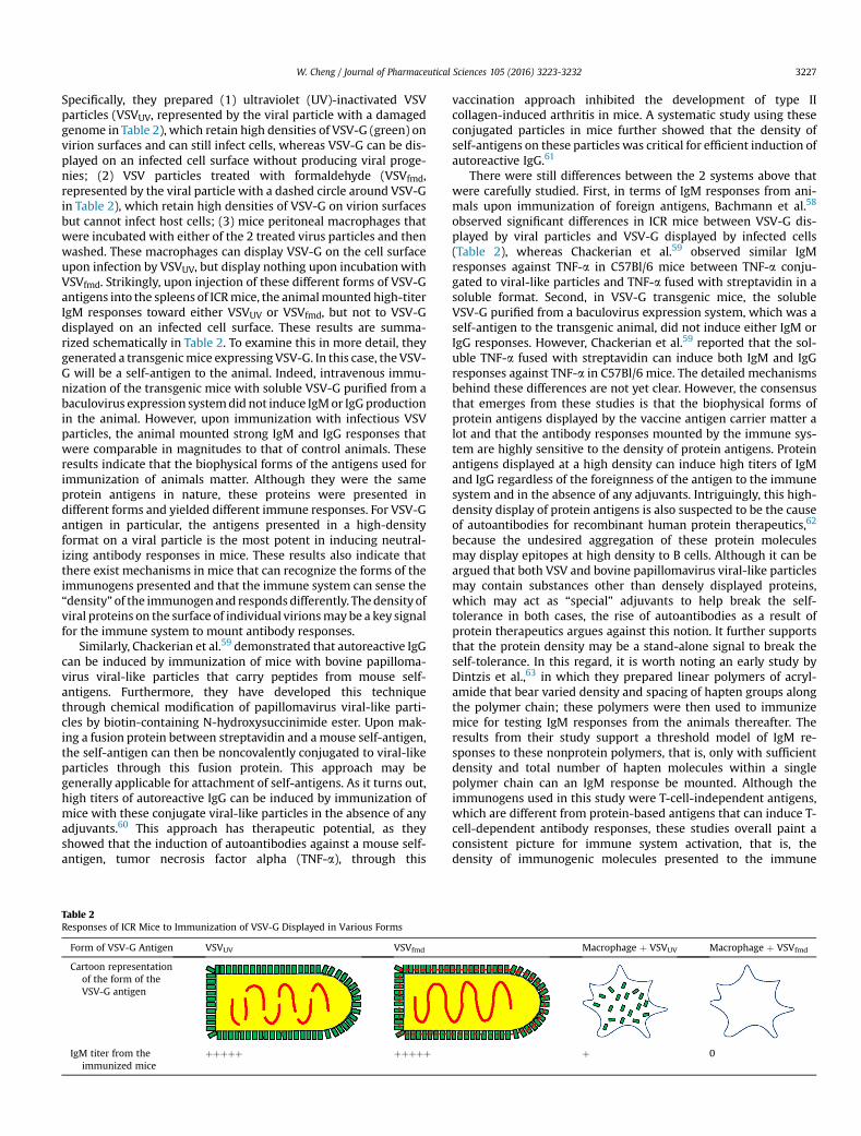
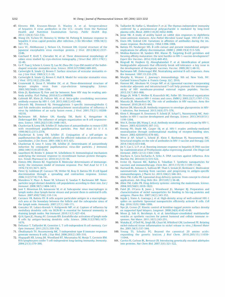
Specifically, they prepared (1) ultraviolet (UV)-inactivated VSVparticles (VSVUV, represented by the viral particle with a damagedgenome in Table 2), which retain high densities of VSV-G (green) onvirion surfaces and can still infect cells, whereas VSV-G can be dis-played on an infected cell surface without producing viral proge-nies; (2) VSV particles treated with formaldehyde (VSVfmd,represented by the viral particle with a dashed circle around VSV-Gin Table 2), which retain high densities of VSV-G on virion surfacesbut cannot infect host cells; (3) mice peritoneal macrophages thatwere incubated with either of the 2 treated virus particles and thenwashed. These macrophages can display VSV-G on the cell surfaceupon infection by VSVUV, but display nothing upon incubationwithVSVfmd. Strikingly, upon injection of these different forms of VSV-Gantigens into the spleens of ICRmice, the animalmounted high-titerIgM responses toward either VSVUV or VSVfmd, but not to VSV-Gdisplayed on an infected cell surface. These results are summa-rized schematically in Table 2. To examine this in more detail, theygenerated a transgenicmice expressing VSV-G. In this case, the VSV-G will be a self-antigen to the animal. Indeed, intravenous immu-nization of the transgenic mice with soluble VSV-G purified from abaculovirus expression systemdid not induce IgMor IgGproductionin the animal. However, upon immunization with infectious VSVparticles, the animal mounted strong IgM and IgG responses thatwere comparable in magnitudes to that of control animals. Theseresults indicate that the biophysical forms of the antigens used forimmunization of animals matter. Although they were the sameprotein antigens in nature, these proteins were presented indifferent forms and yielded different immune responses. For VSV-Gantigen in particular, the antigens presented in a high-densityformat on a viral particle is the most potent in inducing neutral-izing antibody responses in mice. These results also indicate thatthere exist mechanisms in mice that can recognize the forms of theimmunogens presented and that the immune system can sense the“density”of the immunogenand respondsdifferently. Thedensityofviral proteins on the surface of individual virionsmay be a key signalfor the immune system to mount antibody responses.
Similarly, Chackerian et al.59 demonstrated that autoreactive IgGcan be induced by immunization of mice with bovine papilloma-virus viral-like particles that carry peptides from mouse self-antigens. Furthermore, they have developed this techniquethrough chemical modification of papillomavirus viral-like parti-cles by biotin-containing N-hydroxysuccinimide ester. Upon mak-ing a fusion protein between streptavidin and amouse self-antigen,the self-antigen can then be noncovalently conjugated to viral-likeparticles through this fusion protein. This approach may begenerally applicable for attachment of self-antigens. As it turns out,high titers of autoreactive IgG can be induced by immunization ofmice with these conjugate viral-like particles in the absence of anyadjuvants.60 This approach has therapeutic potential, as theyshowed that the induction of autoantibodies against a mouse self-antigen, tumor necrosis factor alpha (TNF-a), through this
Table 2Responses of ICR Mice to Immunization of VSV-G Displayed in Various Forms
Form of VSV-G Antigen VSVUV VSVfmd
Cartoon representationof the form of theVSV-G antigen
IgM titer from theimmunized mice
þþþþþ þþþþþ
vaccination approach inhibited the development of type IIcollagen-induced arthritis in mice. A systematic study using theseconjugated particles in mice further showed that the density ofself-antigens on these particles was critical for efficient induction ofautoreactive IgG.61
There were still differences between the 2 systems above thatwere carefully studied. First, in terms of IgM responses from ani-mals upon immunization of foreign antigens, Bachmann et al.58
observed significant differences in ICR mice between VSV-G dis-played by viral particles and VSV-G displayed by infected cells(Table 2), whereas Chackerian et al.59 observed similar IgMresponses against TNF-a in C57Bl/6 mice between TNF-a conju-gated to viral-like particles and TNF-a fused with streptavidin in asoluble format. Second, in VSV-G transgenic mice, the solubleVSV-G purified from a baculovirus expression system, which was aself-antigen to the transgenic animal, did not induce either IgM orIgG responses. However, Chackerian et al.59 reported that the sol-uble TNF-a fused with streptavidin can induce both IgM and IgGresponses against TNF-a in C57Bl/6 mice. The detailed mechanismsbehind these differences are not yet clear. However, the consensusthat emerges from these studies is that the biophysical forms ofprotein antigens displayed by the vaccine antigen carrier matter alot and that the antibody responses mounted by the immune sys-tem are highly sensitive to the density of protein antigens. Proteinantigens displayed at a high density can induce high titers of IgMand IgG regardless of the foreignness of the antigen to the immunesystem and in the absence of any adjuvants. Intriguingly, this high-density display of protein antigens is also suspected to be the causeof autoantibodies for recombinant human protein therapeutics,62
because the undesired aggregation of these protein moleculesmay display epitopes at high density to B cells. Although it can beargued that both VSV and bovine papillomavirus viral-like particlesmay contain substances other than densely displayed proteins,which may act as “special” adjuvants to help break the self-tolerance in both cases, the rise of autoantibodies as a result ofprotein therapeutics argues against this notion. It further supportsthat the protein density may be a stand-alone signal to break theself-tolerance. In this regard, it is worth noting an early study byDintzis et al.,63 in which they prepared linear polymers of acryl-amide that bear varied density and spacing of hapten groups alongthe polymer chain; these polymers were then used to immunizemice for testing IgM responses from the animals thereafter. Theresults from their study support a threshold model of IgM re-sponses to these nonprotein polymers, that is, only with sufficientdensity and total number of hapten molecules within a singlepolymer chain can an IgM response be mounted. Although theimmunogens used in this study were T-cell-independent antigens,which are different from protein-based antigens that can induce T-cell-dependent antibody responses, these studies overall paint aconsistent picture for immune system activation, that is, thedensity of immunogenic molecules presented to the immune
Macrophage þ VSVUV Macrophage þ VSVfmd
þ 0
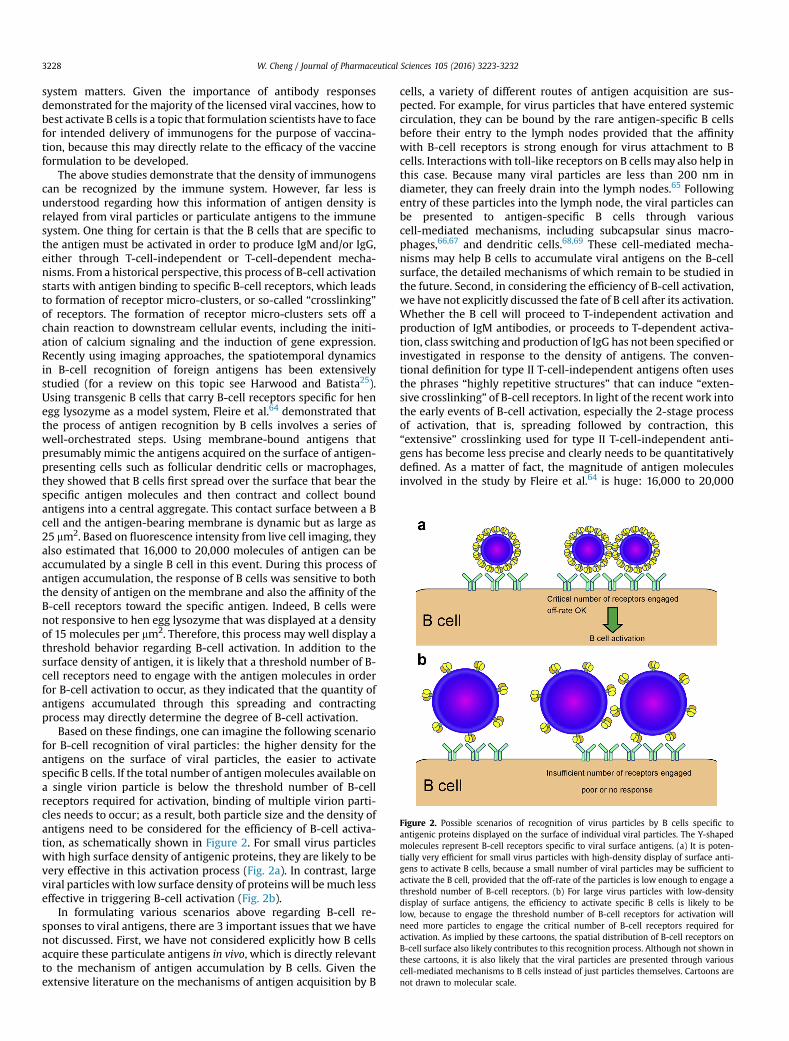
Figure 2. Possible scenarios of recognition of virus particles by B cells specific toantigenic proteins displayed on the surface of individual viral particles. The Y-shapedmolecules represent B-cell receptors specific to viral surface antigens. (a) It is poten-tially very efficient for small virus particles with high-density display of surface anti-gens to activate B cells, because a small number of viral particles may be sufficient toactivate the B cell, provided that the off-rate of the particles is low enough to engage athreshold number of B-cell receptors. (b) For large virus particles with low-densitydisplay of surface antigens, the efficiency to activate specific B cells is likely to below, because to engage the threshold number of B-cell receptors for activation willneed more particles to engage the critical number of B-cell receptors required foractivation. As implied by these cartoons, the spatial distribution of B-cell receptors onB-cell surface also likely contributes to this recognition process. Although not shown inthese cartoons, it is also likely that the viral particles are presented through variouscell-mediated mechanisms to B cells instead of just particles themselves. Cartoons arenot drawn to molecular scale.
W. Cheng / Journal of Pharmaceutical Sciences 105 (2016) 3223-32323228
system matters. Given the importance of antibody responsesdemonstrated for the majority of the licensed viral vaccines, how tobest activate B cells is a topic that formulation scientists have to facefor intended delivery of immunogens for the purpose of vaccina-tion, because this may directly relate to the efficacy of the vaccineformulation to be developed.
The above studies demonstrate that the density of immunogenscan be recognized by the immune system. However, far less isunderstood regarding how this information of antigen density isrelayed from viral particles or particulate antigens to the immunesystem. One thing for certain is that the B cells that are specific tothe antigen must be activated in order to produce IgM and/or IgG,either through T-cell-independent or T-cell-dependent mecha-nisms. From a historical perspective, this process of B-cell activationstarts with antigen binding to specific B-cell receptors, which leadsto formation of receptor micro-clusters, or so-called “crosslinking”of receptors. The formation of receptor micro-clusters sets off achain reaction to downstream cellular events, including the initi-ation of calcium signaling and the induction of gene expression.Recently using imaging approaches, the spatiotemporal dynamicsin B-cell recognition of foreign antigens has been extensivelystudied (for a review on this topic see Harwood and Batista25).Using transgenic B cells that carry B-cell receptors specific for henegg lysozyme as a model system, Fleire et al.64 demonstrated thatthe process of antigen recognition by B cells involves a series ofwell-orchestrated steps. Using membrane-bound antigens thatpresumably mimic the antigens acquired on the surface of antigen-presenting cells such as follicular dendritic cells or macrophages,they showed that B cells first spread over the surface that bear thespecific antigen molecules and then contract and collect boundantigens into a central aggregate. This contact surface between a Bcell and the antigen-bearing membrane is dynamic but as large as25 mm2. Based on fluorescence intensity from live cell imaging, theyalso estimated that 16,000 to 20,000 molecules of antigen can beaccumulated by a single B cell in this event. During this process ofantigen accumulation, the response of B cells was sensitive to boththe density of antigen on the membrane and also the affinity of theB-cell receptors toward the specific antigen. Indeed, B cells werenot responsive to hen egg lysozyme that was displayed at a densityof 15 molecules per mm2. Therefore, this process may well display athreshold behavior regarding B-cell activation. In addition to thesurface density of antigen, it is likely that a threshold number of B-cell receptors need to engage with the antigen molecules in orderfor B-cell activation to occur, as they indicated that the quantity ofantigens accumulated through this spreading and contractingprocess may directly determine the degree of B-cell activation.
Based on these findings, one can imagine the following scenariofor B-cell recognition of viral particles: the higher density for theantigens on the surface of viral particles, the easier to activatespecific B cells. If the total number of antigenmolecules available ona single virion particle is below the threshold number of B-cellreceptors required for activation, binding of multiple virion parti-cles needs to occur; as a result, both particle size and the density ofantigens need to be considered for the efficiency of B-cell activa-tion, as schematically shown in Figure 2. For small virus particleswith high surface density of antigenic proteins, they are likely to bevery effective in this activation process (Fig. 2a). In contrast, largeviral particles with low surface density of proteins will bemuch lesseffective in triggering B-cell activation (Fig. 2b).
In formulating various scenarios above regarding B-cell re-sponses to viral antigens, there are 3 important issues that we havenot discussed. First, we have not considered explicitly how B cellsacquire these particulate antigens in vivo, which is directly relevantto the mechanism of antigen accumulation by B cells. Given theextensive literature on the mechanisms of antigen acquisition by B
cells, a variety of different routes of antigen acquisition are sus-pected. For example, for virus particles that have entered systemiccirculation, they can be bound by the rare antigen-specific B cellsbefore their entry to the lymph nodes provided that the affinitywith B-cell receptors is strong enough for virus attachment to Bcells. Interactions with toll-like receptors on B cells may also help inthis case. Because many viral particles are less than 200 nm indiameter, they can freely drain into the lymph nodes.65 Followingentry of these particles into the lymph node, the viral particles canbe presented to antigen-specific B cells through variouscell-mediated mechanisms, including subcapsular sinus macro-phages,66,67 and dendritic cells.68,69 These cell-mediated mecha-nisms may help B cells to accumulate viral antigens on the B-cellsurface, the detailed mechanisms of which remain to be studied inthe future. Second, in considering the efficiency of B-cell activation,we have not explicitly discussed the fate of B cell after its activation.Whether the B cell will proceed to T-independent activation andproduction of IgM antibodies, or proceeds to T-dependent activa-tion, class switching and production of IgG has not been specified orinvestigated in response to the density of antigens. The conven-tional definition for type II T-cell-independent antigens often usesthe phrases “highly repetitive structures” that can induce “exten-sive crosslinking” of B-cell receptors. In light of the recent work intothe early events of B-cell activation, especially the 2-stage processof activation, that is, spreading followed by contraction, this“extensive” crosslinking used for type II T-cell-independent anti-gens has become less precise and clearly needs to be quantitativelydefined. As a matter of fact, the magnitude of antigen moleculesinvolved in the study by Fleire et al.64 is huge: 16,000 to 20,000
W. Cheng / Journal of Pharmaceutical Sciences 105 (2016) 3223-3232 3229
molecules of antigen are involved at the level of single B cells. It willbe critical in the future to determine the threshold numbers of B-cell receptors required for both T-cell-dependent and T-cell-inde-pendent activation, and quantitatively how these 2 processes differ.
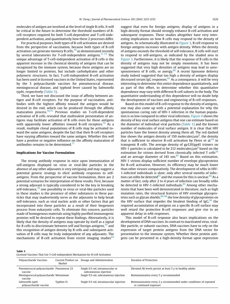
For practical purposes, both types of B-cell activation are desiredfrom the perspective of vaccination, because both types of B-cellactivation can generatememory B cells,70 as demonstrated recentlyby several laboratories for T-cell-independent antigens.71-73 Theunique advantage of T-cell-independent activation of B cells is theapparent increase in the chemical identity of antigens that can berecognized by the immune system, because these antigens are nolonger limited to proteins, but include carbohydrates and otherpolymeric structures. In fact, T-cell-independent B-cell activationhas been used in licensed vaccines in the United States, representedby the 3 polysaccharide vaccines for pneumococcal disease,meningococcal disease, and typhoid fever caused by Salmonellatyphi, respectively (Table 3).
Third, we have not discussed the issue of affinity between an-tigen and B-cell receptors. For the purpose of vaccination, anti-bodies with the highest affinity toward the antigen would bedesired in the end, which can be produced through the affinitymaturation process.74,75 Previous studies on T-cell-dependentactivation of B cells revealed that multivalent presentation of an-tigens may facilitate activation of B cells even for those antigenswith apparently lower affinities toward B-cell receptors.76 As aresult, multiple clonal populations of B cells may be activated to-ward the same antigens, despite the fact that their B-cell receptorshave varying affinities toward the same antigen. Whether this willhave a positive or negative influence on the affinity maturation ofantibodies remains to be determined.77
Implications for Vaccine Formulation
The strong antibody response in mice upon immunization ofself-antigens displayed on virus or viral-like particles in theabsence of any other adjuvants is very intriguing andmay suggest apotential generic strategy to elicit antibody responses to self-antigens. From the perspective of vaccine formulation, there are 2potential scenarios for interpretation of these results. First, becausea strong adjuvant is typically considered to be the key in breakingself-tolerance,78 one possibility in virus or viral-like particles usedin these studies is the presence of other substances in these par-ticles that may inadvertently serve as the adjuvants to help breakself-tolerance, such as viral nucleic acids or other factors that getincorporated into these particles as a result of their biogenesisprocess from eukaryotic cells. To eliminate this concern, particlesmade of homogenousmaterials using highly purified immunogenicproteins will be desired to repeat these findings. Alternatively, it islikely that the density of antigens may operate by itself as a signalfor B cells to discriminate between self and foreign antigens,79 andthis recognition of antigen density by B cells and subsequent acti-vation of B cells may be truly independent of any adjuvants. Themechanisms of B-cell activation from recent imaging studies64
Table 3Licensed Vaccines That Use T-Cell-independent Mechanism for B-Cell Activation
Polysaccharide Vaccines Current Product onMarket
Dosage and Administration
Pneumococcal polysaccharidevaccine
Pneumovax 23 Single 0.5-mL intramuscular orsubcutaneous injection
Meningococcal polysaccharidevaccine
Menomune Single 0.5-mL subcutaneous injec
Salmonella typhipolysaccharide vaccine
Typhim Vi Single 0.5-mL intramuscular injec
suggest that even for foreign antigens, display of antigens in ahigh-density format should strongly enhance B-cell activation andsubsequent responses. These studies altogether have very inter-esting implications on how B cells may respond to the density ofantigens. As schematically illustrated in Figure 3, B cell response toforeign antigens increases with antigen density. When the densityof antigens exceeds the threshold of self-tolerance, B cells will startto respond to self-antigens, as indicated by the shaded area inFigure 3. Furthermore, it is likely that the response of B cells to thedensity of antigens may not be simply monotonic. It has beensuggested that very high densities of antigens may lead to unre-sponsiveness of B cells, or anergy.80 A recent liposome deliverystudy indeed suggested that too high a density of antigen displaydecreased serum IgG responses.81 As a consequence, it will be veryinteresting to determine this entire dependence quantitatively, andas part of this effort, to determine whether this quantitativedependence may vary with different B-cell subsets in the body. Thequantitative understanding of this dependence will no doubt helpthe development of vaccines and engineering of B-cell responses.
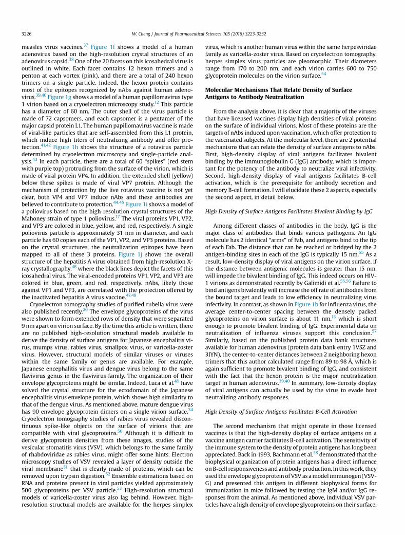
Based on thismodel of B-cell response to the density of antigens,one may also come up with a potential explanation for why thespontaneous curing rate of HIV-1 infection in the human popula-tion is so low compared to other viral infections. Figure 4 shows thedensity of key viral surface antigens that one can estimate based onthe diameter of individual viral particles (Table 1) and the knownnumber of molecules of viral surface antigen. It is clear that HIVparticles have the lowest density among them all. The red dashedline shows the antigen density of 150 molecules/mm2, the densitythat is sufficient to observe B-cell activation by Fleire et al.64 intransgenic B cells. The average density of gp120/gp41 trimers onHIV-1 particles is calculated to be 212 molecules/mm2 based on theestimation for virions derived from chronically infected T cells26
and an average diameter of 145 nm.82 Based on this estimation,HIV-1 virions display sufficient number of envelope glycoproteinsfor B-cell activation. However, its efficiency may be much lowerthan other viruses comparatively. The development of nAbs in HIV-1-infected individuals is slow; only after several months of infec-tion can nAbs be detected83 and the reason for this is unclear.84 As amatter of fact, only after 2 to 4 years of infection can broadly nAbsbe detected in HIV-1-infected individuals.85 Among other mecha-nisms that have been well demonstrated in literature, such as highmutation rates, the structural features of HIV envelope glycopro-teins such as glycan shields,86-88 the lowdensity of glycoproteins onthe HIV surface that impedes the bivalent binding of IgG,56 therequired accumulation of antigens on a specific B-cell surface maywell retard the protective B-cell responses and give rise to anapparent delay in nAb responses.
This model of B-cell response also bears implications on thedevelopment of DNAvaccines. In contrast to inactivated virus, viral-like particle, or subunit vaccines, DNA vaccines have to rely on theexpression of target protein antigens from the DNA vector forpresentation to the immune system. Whether these protein anti-gens can be presented in a high-density format upon expression
Duration of Protection
Elevated Ab levels persist at least 5 y in healthy adults
tion Reimmunization every 5 y recommended
tion Reimmunization every 2 y recommended under conditions of repeatedor continued exposure
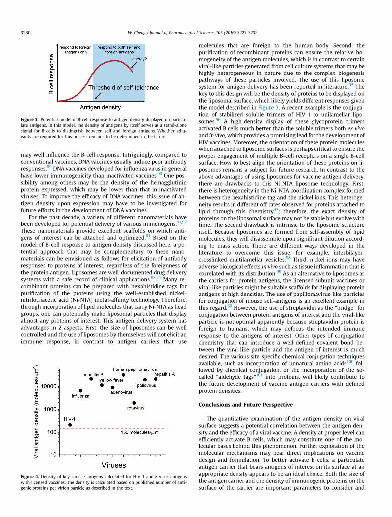
Figure 3. Potential model of B-cell response to antigen density displayed on particu-late antigens. In this model, the density of antigens by itself serves as a stand-alonesignal for B cells to distinguish between self and foreign antigens. Whether adju-vants are required for this process remains to be determined in the future.
W. Cheng / Journal of Pharmaceutical Sciences 105 (2016) 3223-32323230
may well influence the B-cell response. Intriguingly, compared toconventional vaccines, DNA vaccines usually induce poor antibodyresponses.89 DNA vaccines developed for influenza virus in generalhave lower immunogenicity than inactivated vaccines.90 One pos-sibility among others may be the density of the hemagglutininprotein expressed, which may be lower than that in inactivatedviruses. To improve the efficacy of DNA vaccines, this issue of an-tigen density upon expression may have to be investigated forfuture efforts in the development of DNA vaccines.
For the past decade, a variety of different nanomaterials havebeen developed for potential delivery of various immunogens.91,92
These nanomaterials provide excellent scaffolds on which anti-gens of interest can be attached and optimized.81 Based on themodel of B-cell response to antigen density discussed here, a po-tential approach that may be complementary to these nano-materials can be envisioned as follows for elicitation of antibodyresponses to proteins of interest, regardless of the foreignness ofthe protein antigen. Liposomes are well-documented drug deliverysystems with a safe record of clinical applications.93,94 Many re-combinant proteins can be prepared with hexahistidine tags forpurification of the proteins using the well-established nickel-nitrilotriacetic acid (Ni-NTA) metal-affinity technology. Therefore,through incorporation of lipid molecules that carry Ni-NTA as headgroups, one can potentially make liposomal particles that displayalmost any proteins of interest. This antigen delivery system hasadvantages in 2 aspects. First, the size of liposomes can be wellcontrolled and the use of liposomes by themselves will not elicit animmune response, in contrast to antigen carriers that use
Figure 4. Density of key surface antigens calculated for HIV-1 and 8 virus antigenswith licensed vaccines. The density is calculated based on published number of anti-genic proteins per virion particle as described in the text.
molecules that are foreign to the human body. Second, thepurification of recombinant proteins can ensure the relative ho-mogeneity of the antigen molecules, which is in contrast to certainviral-like particles generated from cell culture systems that may behighly heterogeneous in nature due to the complex biogenesispathways of these particles involved. The use of this liposomesystem for antigen delivery has been reported in literature.95 Thekey to this design will be the density of proteins to be displayed onthe liposomal surface, which likely yields different responses giventhe model described in Figure 3. A recent example is the conjuga-tion of stabilized soluble trimers of HIV-1 to unilamellar lipo-somes.96 A high-density display of these glycoprotein trimersactivated B cells much better than the soluble trimers both ex vivoand in vivo, which provides a promising lead for the development ofHIV vaccines. Moreover, the orientation of these protein moleculeswhen attached to liposome surfaces is perhaps critical to ensure theproper engagement of multiple B-cell receptors on a single B-cellsurface. How to best align the orientation of these proteins on li-posomes remains a subject for future research. In contrast to theabove advantages of using liposomes for vaccine antigen delivery,there are drawbacks to this Ni-NTA liposome technology. First,there is heterogeneity in the Ni-NTA coordination complex formedbetween the hexahistidine tag and the nickel ions. This heteroge-neity results in different off rates observed for proteins attached tolipid through this chemistry97; therefore, the exact density ofproteins on the liposomal surface may not be stable but evolvewithtime. The second drawback is intrinsic to the liposome structureitself. Because liposomes are formed from self-assembly of lipidmolecules, they will disassemble upon significant dilution accord-ing to mass action. There are different ways developed in theliterature to overcome this issue, for example, interbilayer-crosslinked multilamellar vesicles.98 Third, nickel ions may haveadverse biological effects in vivo such as tissue inflammation that iscorrelated with its distribution.99 As an alternative to liposomes asthe carriers for protein antigens, the licensed subunit vaccines orviral-like particles might be suitable scaffolds for displaying proteinantigens at high densities. The use of papillomavirus-like particlesfor conjugation of mouse self-antigens is an excellent example inthis regard.60 However, the use of streptavidin as the “bridge” forconjugation between protein antigens of interest and the viral-likeparticle is not optimal apparently because streptavidin protein isforeign to humans, which may defocus the intended immuneresponse to the antigens of interest. Other types of conjugationchemistry that can introduce a well-defined covalent bond be-tween the viral-like particle and the antigen of interest is muchdesired. The various site-specific chemical conjugation techniquesavailable, such as incorporation of unnatural amino acids100 fol-lowed by chemical conjugation, or the incorporation of the so-called “aldehyde tags”101 into proteins, will likely contribute tothe future development of vaccine antigen carriers with definedprotein densities.
Conclusions and Future Perspective
The quantitative examination of the antigen density on viralsurface suggests a potential correlation between the antigen den-sity and the efficacy of a viral vaccine. A density at proper level canefficiently activate B cells, which may constitute one of the mo-lecular bases behind this phenomenon. Further exploration of themolecular mechanisms may bear direct implications on vaccinedesign and formulation. To better activate B cells, a particulateantigen carrier that bears antigens of interest on its surface at anappropriate density appears to be an ideal choice. Both the size ofthe antigen carrier and the density of immunogenic proteins on thesurface of the carrier are important parameters to consider and
W. Cheng / Journal of Pharmaceutical Sciences 105 (2016) 3223-3232 3231
potentially optimize for the development of a vaccine. Given theimportance of protein density on particulate antigens, it will bedesirable in the future to develop nanoparticles with a tailoreddensity of protein antigens on their surface. These materials cannotonly be used for the exploration of the immunological mechanismsdiscussed here but also be useful for vaccine deliveries. Given thefact that T-cell-independent B-cell responses can also elicit mem-ory B cells, it will be critical to determine the number of B-cellreceptors that are optimally required for both T-cell-dependent andT-cell-independent B-cell activation, which will be very useful forthe future of vaccine design. Recombinant human antibodies havebecome important drugs on the pharmaceutical market. As analternative to passively supplying antibodies to the patient, in thefuture, a possible solution will be to supply antibodies throughactive immunization using particulate vaccines, which might pro-vide durable antibody responses regardless of the foreignness ofthe antigenic proteins. This may potentially create an “antibodyboost” on demand or serve as a replacement for the passive anti-body therapies.
Acknowledgments
This work was supported in part by NIH Director's NewInnovator Award 1DP2OD008693-01 and the University ofMichigan Mcubed Project 805 to Wei Cheng. Wei Cheng wouldlike to thank Prof. Pamela Bjorkman at Caltech for her pio-neering work on the avidity of HIV-1 neutralizing antibodies,which has inspired some of the ideas in this work. Wei Chengalso thanks Prof. Per Johan Klasse at Weill Cornell Medicine, andProfessors Irina Grigorova and James Moon at the University ofMichigan for their careful and critical reading of the manuscriptand kindly providing insightful comments on this manuscriptbefore publication. Wei Cheng would like to thank Cheng Labmembers, especially Mike DeSantis for careful reading of themanuscript.
References
1. Currie J, Grenfell B, Farrar J. Infectious diseases. Beyond Ebola. Science.2016;351(6275):815-816.
2. Petersen LR, Jamieson DJ, Powers AM, Honein MA. Zika virus. N Engl J Med.2016;374(16):1552-1563.
3. Carroll MW, Matthews DA, Hiscox JA, et al. Temporal and spatial analysis ofthe 2014-2015 Ebola virus outbreak in West Africa. Nature. 2015;524(7563):97-101.
4. Larocca RA, Abbink P, Peron JP, et al. Vaccine protection against Zika virusfrom Brazil. Nature. 2016.
5. Henao-Restrepo AM, Longini IM, Egger M, et al. Efficacy and effectiveness ofan rVSV-vectored vaccine expressing Ebola surface glycoprotein: interim re-sults from the Guinea ring vaccination cluster-randomised trial. Lancet.2015;386(9996):857-866.
6. Andre FE, Booy R, Bock HL, et al. Vaccination greatly reduces disease,disability, death and inequity worldwide. Bull World Health Organ.2008;86(2):140-146.
7. Plotkin SA, Orenstein WA. Vaccines. 4th ed. Philadelphia, PA: Saunders; 2004:1-15.
8. Schuchat A. Human vaccines and their importance to public health. ProcedVaccinol. 2011;5:120-126.
9. Liu H, Jin L, Koh SB, et al. Atomic structure of human adenovirus by cryo-EMreveals interactions among protein networks. Science. 2010;329(5995):1038-1043.
10. Locarnini SA, Ferris AA, Stott AC, Gust ID. The relationship between a 27-nmvirus-like particle and hepatitis A as demonstrated by immune electron mi-croscopy. Intervirology. 1974;4(2):110-118.
11. Dane DS, Cameron CH, Briggs M. Virus-like particles in serum of patients withAustralia-antigen-associated hepatitis. Lancet. 1970;1(7649):695-698.
12. Baker TS, Newcomb WW, Olson NH, Cowsert LM, Olson C, Brown JC. Struc-tures of bovine and human papillomaviruses. Analysis by cryoelectron mi-croscopy and three-dimensional image reconstruction. Biophys J. 1991;60(6):1445-1456.
13. Harris A, Cardone G, Winkler DC, et al. Influenza virus pleiomorphy charac-terized by cryoelectron tomography. Proc Natl Acad Sci U S A. 2006;103(50):19123-19127.
14. Yang DK, Kim BH, Kweon CH, Kwon JH, Lim SI, Han HR. Biophysical charac-terization of Japanese encephalitis virus (KV1899) isolated from pigs in Korea.J Vet Sci. 2004;5(2):125-130.
15. Liljeroos L, Huiskonen JT, Ora A, Susi P, Butcher SJ. Electron cryotomog-raphy of measles virus reveals how matrix protein coats the ribonu-cleocapsid within intact virions. Proc Natl Acad Sci U S A. 2011;108(44):18085-18090.
16. Knipe D, Howley P, Griffin D, et al. Fields Virology. Philadelphia, PA: LippincottWilliams & Wilkins; 2007.
17. Hogle JM, ChowM, Filman DJ. Three-dimensional structure of poliovirus at 2.9A resolution. Science. 1985;229(4720):1358-1365.
18. Schneider LG, Diringer H. Structure and molecular biology of rabies virus. CurrTop Microbiol Immunol. 1976;75:153-180.
19. Prasad BV, Chiu W. Structure of rotavirus. Curr Top Microbiol Immunol.1994;185:9-29.
20. Battisti AJ, Yoder JD, Plevka P, et al. Cryo-electron tomography of rubella virus.J Virol. 2012;86(20):11078-11085.
21. Cyrklaff M, Risco C, Fernandez JJ, et al. Cryo-electron tomography of vacciniavirus. Proc Natl Acad Sci U S A. 2005;102(8):2772-2777.
22. Zhang Y, Corver J, Chipman PR, et al. Structures of immature flavivirus par-ticles. EMBO J. 2003;22(11):2604-2613.
23. Hamborsky J, Kroger A, Wolfe S. Epidemiology and Prevention of Vaccine-preventable Diseases. 13th ed. Washington D.C.: Public Health Foundation;2015:1-8.
24. Cann AJ, Stanway G, Hughes PJ, et al. Reversion to neurovirulence of the live-attenuated Sabin type 3 oral poliovirus vaccine. Nucleic Acids Res.1984;12(20):7787-7792.
25. Harwood NE, Batista FD. Early events in B cell activation. Annu Rev Immunol.2010;28:185-210.
26. Zhu P, Liu J, Bess Jr J, et al. Distribution and three-dimensional structure ofAIDS virus envelope spikes. Nature. 2006;441(7095):847-852.
27. DeSantis MC, Kim JH, Song H, Klasse PJ, Cheng W. Quantitative correlationbetween infectivity and Gp120 density on HIV-1 virions revealed by opticaltrapping virometry. J Biol Chem. 2016;291:13088-13097.
28. Couch RB. An overview of serum antibody responses to influenza virus anti-gens. Dev Biol (Basel). 2003;115:25-30.
29. Yamaguchi M, Danev R, Nishiyama K, Sugawara K, Nagayama K. Zernike phasecontrast electron microscopy of ice-embedded influenza A virus. J Struct Biol.2008;162(2):271-276.
30. Dryden KA, Wieland SF, Whitten-Bauer C, Gerin JL, Chisari FV, Yeager M.Native hepatitis B virions and capsids visualized by electron cryomicroscopy.Mol Cell. 2006;22(6):843-850.
31. Valenzuela P, Medina A, Rutter WJ, Ammerer G, Hall BD. Synthesis and as-sembly of hepatitis B virus surface antigen particles in yeast. Nature.1982;298(5872):347-350.
32. McAleer WJ, Buynak EB, Maigetter RZ, Wampler DE, Miller WJ, Hilleman MR.Human hepatitis B vaccine from recombinant yeast. Nature. 1984;307(5947):178-180.
33. Miyanohara A, Toh-e A, Nozaki C, Hamada F, Ohtomo N, Matsubara K.Expression of hepatitis B surface antigen gene in yeast. Proc Natl Acad Sci U SA. 1983;80(1):1-5.
34. Kuhn RJ, Zhang W, Rossmann MG, et al. Structure of dengue virus: implica-tions for flavivirus organization, maturation, and fusion. Cell. 2002;108(5):717-725.
35. Monath TP, Nichols R, Archambault WT, et al. Comparative safety andimmunogenicity of two yellow fever 17D vaccines (ARILVAX and YF-VAX) in aphase III multicenter, double-blind clinical trial. Am J Trop Med Hyg.2002;66(5):533-541.
36. Yanagi Y, Takeda M, Ohno S. Measles virus: cellular receptors, tropism andpathogenesis. J Gen Virol. 2006;87(Pt 10):2767-2779.
37. Bouche FB, Ertl OT, Muller CP. Neutralizing B cell response in measles. ViralImmunol. 2002;15(3):451-471.
38. Reddy VS, Natchiar SK, Stewart PL, Nemerow GR. Crystal structure of humanadenovirus at 3.5 A resolution. Science. 2010;329(5995):1071-1075.
39. Madisch I, Harste G, Pommer H, Heim A. Phylogenetic analysis of the mainneutralization and hemagglutination determinants of all human adenovirusprototypes as a basis for molecular classification and taxonomy. J Virol.2005;79(24):15265-15276.
40. Roberts DM, Nanda A, Havenga MJ, et al. Hexon-chimaeric adenovirus sero-type 5 vectors circumvent pre-existing anti-vector immunity. Nature.2006;441(7090):239-243.
41. Kirnbauer R, Booy F, Cheng N, Lowy DR, Schiller JT. Papillomavirus L1 majorcapsid protein self-assembles into virus-like particles that are highly immu-nogenic. Proc Natl Acad Sci U S A. 1992;89(24):12180-12184.
42. Lowy DR, Schiller JT. Prophylactic human papillomavirus vaccines. J ClinInvest. 2006;116(5):1167-1173.
43. Settembre EC, Chen JZ, Dormitzer PR, Grigorieff N, Harrison SC. Atomic modelof an infectious rotavirus particle. EMBO J. 2011;30(2):408-416.
44. Hoshino Y, Kapikian AZ. Classification of rotavirus VP4 and VP7 serotypes.Arch Virol Suppl. 1996;12:99-111.
45. Vesikari T, Rautanen T, Varis T, Beards GM, Kapikian AZ. Rhesus rotaviruscandidate vaccine. Clinical trial in children vaccinated between 2 and 5months of age. Am J Dis Child. 1990;144(3):285-289.
46. Wang X, Ren J, Gao Q, et al. Hepatitis A virus and the origins of picornaviruses.Nature. 2015;517(7532):85-88.
W. Cheng / Journal of Pharmaceutical Sciences 105 (2016) 3223-32323232
47. Klevens RM, Kruszon-Moran D, Wasley A, et al. Seroprevalenceof hepatitis A virus antibodies in the U.S.: results from the NationalHealth and Nutrition Examination Survey. Public Health Rep.2011;126(4):522-532.
48. Wang CH, Tschen SY, Heinricy U, Weber M, Flehmig B. Immune response tohepatitis A virus capsid proteins after infection. J Clin Microbiol. 1996;34(3):707-713.
49. Luca VC, AbiMansour J, Nelson CA, Fremont DH. Crystal structure of theJapanese encephalitis virus envelope protein. J Virol. 2012;86(4):2337-2346.
50. Guichard P, Krell T, Chevalier M, et al. Three dimensional morphology ofrabies virus studied by cryo-electron tomography. J Struct Biol. 2011;176(1):32-40.
51. Ge P, Tsao J, Schein S, Green TJ, Luo M, Zhou ZH. Cryo-EM model of the bullet-shaped vesicular stomatitis virus. Science. 2010;327(5966):689-693.
52. Cartwright B, Smale CJ, Brown F. Surface structure of vesicular stomatitis vi-rus. J Gen Virol. 1969;5(1):1-10.
53. Cartwright B, Smale CJ, Brown F, Hull R. Model for vesicular stomatitis virus.J Virol. 1972;10(2):256-260.
54. Grunewald K, Desai P, Winkler DC, et al. Three-dimensional structure ofherpes simplex virus from cryo-electron tomography. Science.2003;302(5649):1396-1398.
55. Klein JS, Bjorkman PJ. Few and far between: how HIV may be evading anti-body avidity. PLoS Pathog. 2010;6(5):e1000908.
56. Galimidi RP, Klein JS, Politzer MS, et al. Intra-spike crosslinking overcomesantibody evasion by HIV-1. Cell. 2015;160(3):433-446.
57. Edwards MJ, Dimmock NJ. Hemagglutinin 1-specific immunoglobulin Gand Fab molecules mediate postattachment neutralization of influenza Avirus by inhibition of an early fusion event. J Virol. 2001;75(21):10208-10218.
58. Bachmann MF, Rohrer UH, Kundig TM, Burki K, Hengartner H,Zinkernagel RM. The influence of antigen organization on B cell responsive-ness. Science. 1993;262(5138):1448-1451.
59. Chackerian B, Lowy DR, Schiller JT. Induction of autoantibodies to mouse CCR5with recombinant papillomavirus particles. Proc Natl Acad Sci U S A.1999;96(5):2373-2378.
60. Chackerian B, Lowy DR, Schiller JT. Conjugation of a self-antigen topapillomavirus-like particles allows for efficient induction of protective au-toantibodies. J Clin Invest. 2001;108(3):415-423.
61. Chackerian B, Lenz P, Lowy DR, Schiller JT. Determinants of autoantibodyinduction by conjugated papillomavirus virus-like particles. J Immunol.2002;169(11):6120-6126.
62. Sauerborn M, Brinks V, Jiskoot W, Schellekens H. Immunological mechanismunderlying the immune response to recombinant human protein therapeu-tics. Trends Pharmacol Sci. 2010;31(2):53-59.
63. Dintzis HM, Dintzis RZ, Vogelstein B. Molecular determinants of immunoge-nicity: the immunon model of immune response. Proc Natl Acad Sci U S A.1976;73(10):3671-3675.
64. Fleire SJ, Goldman JP, Carrasco YR, Weber M, Bray D, Batista FD. B cell liganddiscrimination through a spreading and contraction response. Science.2006;312(5774):738-741.
65. Manolova V, Flace A, Bauer M, Schwarz K, Saudan P, Bachmann MF. Nano-particles target distinct dendritic cell populations according to their size. Eur JImmunol. 2008;38(5):1404-1413.
66. Junt T, Moseman EA, Iannacone M, et al. Subcapsular sinus macrophages inlymph nodes clear lymph-borne viruses and present them to antiviral B cells.Nature. 2007;450(7166):110-114.
67. Carrasco YR, Batista FD. B cells acquire particulate antigen in a macrophage-rich area at the boundary between the follicle and the subcapsular sinus ofthe lymph node. Immunity. 2007;27(1):160-171.
68. Gonzalez SF, Lukacs-Kornek V, Kuligowski MP, et al. Capture of influenza bymedullary dendritic cells via SIGN-R1 is essential for humoral immunity indraining lymph nodes. Nat Immunol. 2010;11(5):427-434.
69. Qi H, Egen JG, Huang AY, Germain RN. Extrafollicular activation of lymph nodeB cells by antigen-bearing dendritic cells. Science. 2006;312(5780):1672-1676.
70. Defrance T, Taillardet M, Genestier L. T cell-independent B cell memory. CurrOpin Immunol. 2011;23(3):330-336.
71. Obukhanych TV, Nussenzweig MC. T-independent type II immune responsesgenerate memory B cells. J Exp Med. 2006;203(2):305-310.
72. Alugupalli KR, Leong JM, Woodland RT, Muramatsu M, Honjo T, Gerstein RM.B1b lymphocytes confer T cell-independent long-lasting immunity. Immunity.2004;21(3):379-390.
73. Taillardet M, Haffar G, Mondiere P, et al. The thymus-independent immunityconferred by a pneumococcal polysaccharide is mediated by long-livedplasma cells. Blood. 2009;114(20):4432-4440.
74. Jerne NK. A study of avidity based on rabbit skin responses to diphtheriatoxin-antitoxin mixtures. Acta Pathol Microbiol Scand Suppl. 1951;87:1-183.
75. Eisen HN, Siskind GW. Variations in affinities of antibodies during the im-mune response. Biochemistry. 1964;3:996-1008.
76. Batista FD, Neuberger MS. B cells extract and present immobilized antigen:implications for affinity discrimination. EMBO J. 2000;19(4):513-520.
77. Medina-Ramirez M, Sanders RW, Klasse PJ. Targeting B-cell germlines andfocusing affinity maturation: the next hurdles in HIV-1-vaccine development?Expert Rev Vaccines. 2014;13(4):449-452.
78. Ringvall M, Huijbers EJ, Ahooghalandari P, et al. Identification of potentbiodegradable adjuvants that efficiently break self-toleranceea key issue inthe development of therapeutic vaccines. Vaccine. 2009;28(1):48-52.
79. Bachmann MF, Zinkernagel RM. Neutralizing antiviral B cell responses. AnnuRev Immunol. 1997;15:235-270.
80. Murphy K, Weaver C. Janeway's Immunobiology. 9th ed. New York, NY:Garland Science/Taylor & Francis Group, LLC; 2016.
81. Hanson MC, Abraham W, Crespo MP, et al. Liposomal vaccines incorporatingmolecular adjuvants and intrastructural T-cell help promote the immunoge-nicity of HIV membrane-proximal external region peptides. Vaccine.2015;33(7):861-868.
82. Briggs JA, Wilk T, Welker R, Krausslich HG, Fuller SD. Structural organizationof authentic, mature HIV-1 virions and cores. EMBO J. 2003;22(7):1707-1715.
83. Mascola JR, Montefiori DC. The role of antibodies in HIV vaccines. Annu RevImmunol. 2010;28:413-444.
84. Burton DR, Mascola JR. Antibody responses to envelope glycoproteins in HIV-1 infection. Nat Immunol. 2015;16(6):571-576.
85. Klein F, Mouquet H, Dosenovic P, Scheid JF, Scharf L, Nussenzweig MC. Anti-bodies in HIV-1 vaccine development and therapy. Science. 2013;341(6151):1199-1204.
86. Wei X, Decker JM, Wang S, et al. Antibody neutralization and escape by HIV-1.Nature. 2003;422(6929):307-312.
87. Kwong PD, Doyle ML, Casper DJ, et al. HIV-1 evades antibody-mediatedneutralization through conformational masking of receptor-binding sites.Nature. 2002;420(6916):678-682.
88. West Jr AP, Scharf L, Scheid JF, Klein F, Bjorkman PJ, Nussenzweig MC.Structural insights on the role of antibodies in HIV-1 vaccine and therapy. Cell.2014;156(4):633-648.
89. Jin Y, Cao C, Li P, et al. Boosting immune response to hepatitis B DNA vaccineby coadministration of Prothymosin alpha-expressing plasmid. Clin Diagn LabImmunol. 2005;12(12):1364-1369.
90. Stachyra A, Gora-Sochacka A, Sirko A. DNA vaccines against influenza. ActaBiochim Pol. 2014;61(3):515-522.
91. Irvine DJ, Hanson MC, Rakhra K, Tokatlian T. Synthetic nanoparticles forvaccines and immunotherapy. Chem Rev. 2015;115(19):11109-11146.
92. Hartwell BL, Antunez L, Sullivan BP, Thati S, Sestak JO, Berkland C. Multivalentnanomaterials: learning from vaccines and progressing to antigen-specificimmunotherapies. J Pharm Sci. 2015;104(2):346-361.
93. Allen TM, Cullis PR. Liposomal drug delivery systems: from concept to clinicalapplications. Adv Drug Deliv Rev. 2013;65(1):36-48.
94. Allen TM, Cullis PR. Drug delivery systems: entering the mainstream. Science.2004;303(5665):1818-1822.
95. Patel JD, O'Carra R, Jones J, Woodward JG, Mumper RJ. Preparation andcharacterization of nickel nanoparticles for binding to his-tag proteins andantigens. Pharm Res. 2007;24(2):343-352.
96. Ingale J, Stano A, Guenaga J, et al. High-density array of well-ordered HIV-1spikes on synthetic liposomal nanoparticles efficiently activate B cells. CellRep. 2016;15(9):1986-1999.
97. Nye JA, Groves JT. Kinetic control of histidine-tagged protein surface densityon supported lipid bilayers. Langmuir. 2008;24(8):4145-4149.
98. Moon JJ, Suh H, Bershteyn A, et al. Interbilayer-crosslinked multilamellarvesicles as synthetic vaccines for potent humoral and cellular immune re-sponses. Nat Mater. 2011;10(3):243-251.
99. Wataha JC, O'Dell NL, Singh BB, Ghazi M, Whitford GM, Lockwood PE. Relatingnickel-induced tissue inflammation to nickel release in vivo. J Biomed MaterRes. 2001;58(5):537-544.
100. Young TS, Schultz PG. Beyond the canonical 20 amino acids:expanding the genetic lexicon. J Biol Chem. 2010;285(15):11039-11044.
101. Carrico IS, Carlson BL, Bertozzi CR. Introducing genetically encoded aldehydesinto proteins. Nat Chem Biol. 2007;3(6):321-322.