the diagnosis-driven physical exam of the knee library/sgim/meetings/annual meeting... · the...
TRANSCRIPT

4/1/2014
1
The Diagnosis-Driven Physical Exam
of the Knee April 25, 2014
Carlin Senter MD, Natalie Voskanian MD, Veronica Jow MD
Carlin Senter, MD Assistant Clinical Professor
UCSF Sports Medicine

4/1/2014
2
Natalie Voskanian, MD Assistant Clinical Professor
UCSD Sports Medicine
Veronica Jow, MD Associate Team Physician
UC Berkeley

4/1/2014
3
Small group experts: UCSF residents and faculty
Outline
• Knee anatomy
• Knee exam
• Knee hands on exam practice
• Cases
• Questions

4/1/2014
4
Knee: top 3 diagnoses in primary care referrals to ortho (at UCSF)
1. Osteoarthritis (OA)
2. Meniscus tear
3. Patellofemoral pain
1. Patellofemoral pain syndrome
2. Patellofemoral chondromalacia
Musculoskeletal work-up
• History
• Inspection
• Palpation
• Range of motion
• Other Tests
• Strength • Neurovascular

4/1/2014
5
Knee Anatomy
The quadriceps muscles extend the
knee
http://thefitcoach.wordpress.com/2012/04/07/267/ http://scientia.wikispaces.com/Thigh+and+Leg+-+Lecture+Notes

4/1/2014
6
The quadriceps muscles merge to form the quadriceps tendon… patellar tendon
The hamstrings flex the knee
www.hep2go.com

4/1/2014
7
Pes anserine bursa
http://meded.ucsd.edu/clinicalmed/joints.htm
Common cause of medial knee pain (hurts with side to side or twisting maneuvers): +TTP below the jointline; can extend from posteromedial pes tendons to anteromedial bursa on medial tibia. Tx = PT, nsaids, injection
There are 4 main ligaments in the knee
MCL: resists valgus - medial femoral condyle to medial tibia (crosses medial jointline) LCL: resists varus -Lateral femoral condyle to fibular head (crosses lateral jointline) - resists varus ACL: resists anterior tibial translation PCL: resists posterior tibial translation

4/1/2014
8
Meniscus
• Medial • Lateral
• Cushion the knee joint and protect the articular cartilage
• Pain and tenderness in medial or lateral jointline • +/- effusion • May or may not have h/o trauma
• OA pts get degenerative tears (XR is sufficient in these pts)
Knee history: MOI
• Acute vs sub-acute or chronic
• Direct fall onto patella – Patellar fracture or
cartilage damage or prepatellar bursitis
• Varus or valgus force to the knee – MCL or LCL
• Noncontact with a pop • ACL
http://www.ski-injury.com/kneeanat.gif, Accessed 10/04/05. Accessed 10/4/05

4/1/2014
9
3 Key Q’s to Ask in a Knee Injury
1. Locking or catching = displaced meniscus flap (bucket handle tear) or intra-articular loose body (loose piece of cartilage or bone)
2. Instability = ligament tear or muscle inhibition (quad, hamstring)
3. Effusion = intra-articular derangement
1. Immediate: due to blood/hemarthrosis (ACL, fracture, cartilage injury, patellar dislocation)
2. Subacute: 8-24 hours, due to synovial fluid buildup (meniscus, OA)
Where is the knee pain?
• Anterior:
- Patellofemoral syndrome
- Quadriceps tendinitis
- Patellar tendinitis
• Lateral:
- Lateral jointline: meniscus tear
or OA
- IT band syndrome
- LCL sprain (rare)
- Fibular head: fracture (rare)
• Medial
- Medial joint-line: meniscus
tear or OA
- MCL sprain
- Pes anserine bursitis
• Posterior
- Hamstring tendinitis
- Gastrocnemius strain
- OA
- Meniscus tear
- Effusion, popliteal cyst

4/1/2014
10
Knee exam
Inspection
Permission for use provided by Dr. Charles Goldberg, UCSD
VALGUS
http://doctorhoang.wordpress.com/2010/09/06/valgus-knee-and-bunion/

4/1/2014
11
Significance of inspection
http://www.stritch.luc.edu/lumen/MedEd/Radio/curriculum/Family%20Medicine/Degenerative_joint_disease.htm
Evaluating for an Effusion
Milking & Ballotment
Permission for use provided by Dr. Charles Goldberg, UCSD
4/1/2014
12
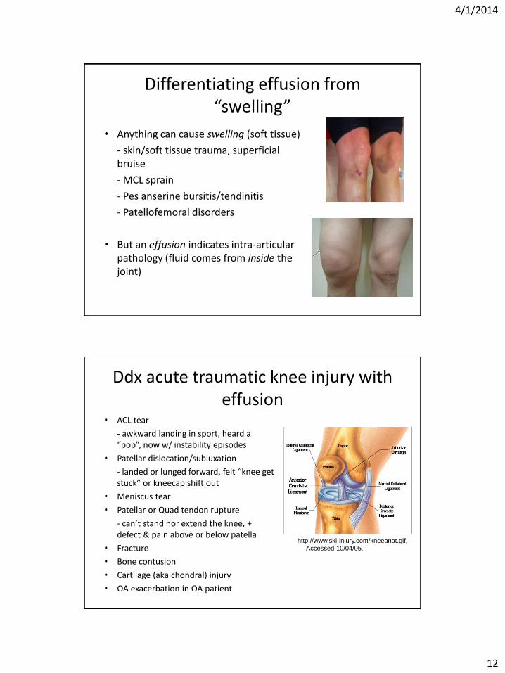
Differentiating effusion from “swelling”
• Anything can cause swelling (soft tissue)
- skin/soft tissue trauma, superficial bruise
- MCL sprain
- Pes anserine bursitis/tendinitis
- Patellofemoral disorders
• But an effusion indicates intra-articular pathology (fluid comes from inside the joint)
Ddx acute traumatic knee injury with effusion
• ACL tear
- awkward landing in sport, heard a “pop”, now w/ instability episodes
• Patellar dislocation/subluxation
- landed or lunged forward, felt “knee get stuck” or kneecap shift out
• Meniscus tear
• Patellar or Quad tendon rupture
- can’t stand nor extend the knee, + defect & pain above or below patella
• Fracture
• Bone contusion
• Cartilage (aka chondral) injury
• OA exacerbation in OA patient
http://www.ski-injury.com/kneeanat.gif, Accessed 10/04/05. Accessed 10/4/05

4/1/2014
13
Ddx of atraumatic knee effusion
• Meniscus tear
• Osteoarthritis
• Crystal arthropathy (pseudogout, gout)
• Inflammatory arthritis
• Septic arthritis
• Benign or malignant tumor
Palpation of joint line seated or supine
Permission for use provided by Dr. Charles Goldberg, UCSD
4/1/2014
14
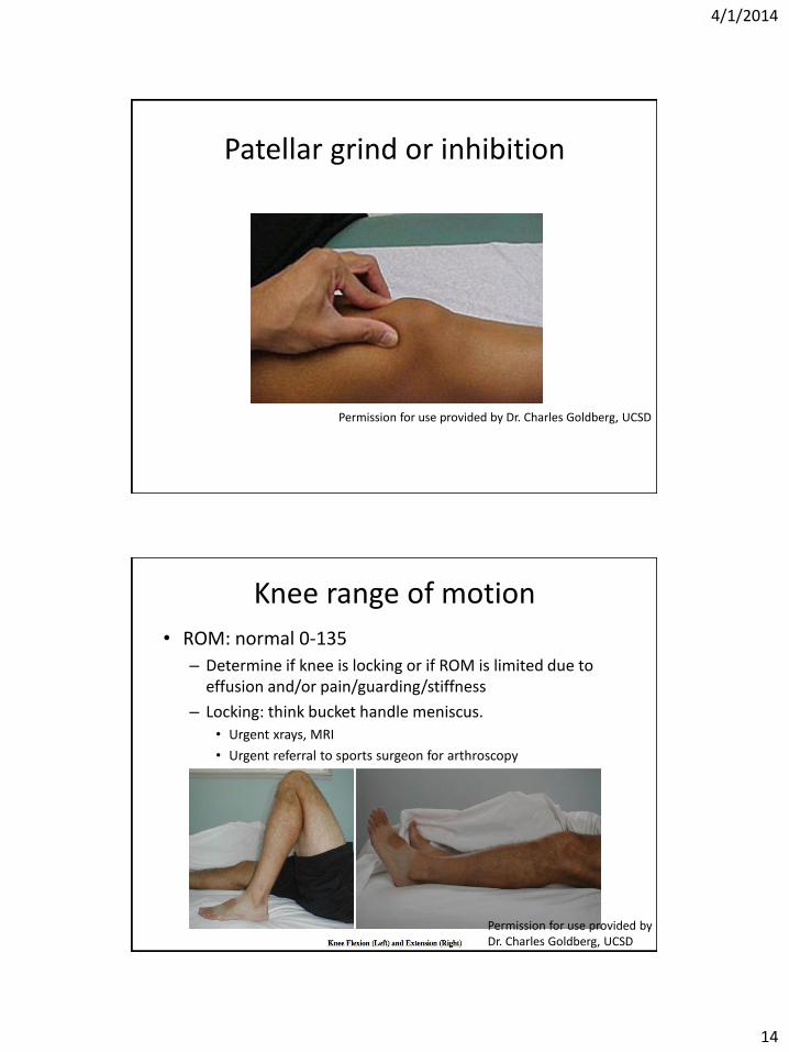
Patellar grind or inhibition
Permission for use provided by Dr. Charles Goldberg, UCSD
Knee range of motion
• ROM: normal 0-135
– Determine if knee is locking or if ROM is limited due to effusion and/or pain/guarding/stiffness
– Locking: think bucket handle meniscus. • Urgent xrays, MRI
• Urgent referral to sports surgeon for arthroscopy
Permission for use provided by Dr. Charles Goldberg, UCSD

4/1/2014
15
Other Tests: Lachman to evaluate ACL Sensitivity 75-100% Specificity 95-100%
ACL: Anterior Drawer Sensitivity 22-41%, Specificity 97%
Permission for use provided by Dr. Charles Goldberg, UCSD

4/1/2014
16
PCL: Posterior Drawer
Video used with permission from Anthony Luke, MD.
PCL: Sag sign
4/1/2014
17
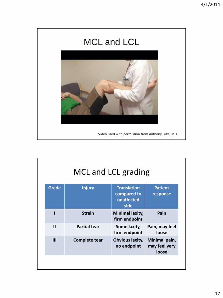
MCL and LCL
Video used with permission from Anthony Luke, MD.
MCL and LCL grading
Grade Injury Translation compared to unaffected
side
Patient response
I Strain Minimal laxity, firm endpoint
Pain
II Partial tear Some laxity, firm endpoint
Pain, may feel loose
III Complete tear Obvious laxity, no endpoint
Minimal pain, may feel very
loose

4/1/2014
18
Why test MCL and LCL at 30°flexion and full extension?
• Laxity at 30°flexion, no laxity at full extension = isolated MCL or LCL injury
• Laxity at 30°flexion and laxity at full extension = MCL or LCL + possible ACL or PCL injury
4 tests for meniscus tear
1. Isolated joint line tenderness
2. McMurray
3. Thessaly
4. Squat

4/1/2014
19
Meniscus: McMurray
Sensitivity medial 65%, Specificity medial 93% Magee, DJ. Orthopaedic Physical Assessment, 5th ed. 2008.
Meniscus: Thessaly
Video used with permission from Anthony Luke, MD.

4/1/2014
20
Meniscus: squat
Chronic anterior knee pain
• DDx
– Patellofemoral pain syndrome
– Patellofemoral chondromalacia
• Inspect and assess the kinetic chain
• Identify tight structures and/or weak structures
– Iliotibial band tightness
– Gluteus medius (hip abduction) strength

4/1/2014
21
Patellofemoral pain syndrome: miserable malalignment syndrome
• Femoral anteversion (inward rotation of femur)
• Squinting patella (inward patellar rotation)
• Patella alta
• Increased Q-angle
• Excessive outward tibial rotation
http://www.gla.ac.uk/ibls/US/fab/tutorial/biomech/akp3.html
Ober’s Test for tight IT Band

4/1/2014
22
Hip abduction (gluts) strength
http://www.youtube.com/watch?v=9Iy-QrcuGno&feature=player_detailpage
One-legged standing squat
• Patient standing on unaffected leg
• Do 3 slow 1-legged squats
• Watch for stability, valgus angulation of knee, ask about pain
• Switch and perform on affected leg
• Sign of weak hip abductors, weak core
• Can bring out pain of patellofemoral pain

4/1/2014
23
One-legged standing squat
• Will insert video
One-legged standing squat
• Will insert video

4/1/2014
24
Knee Exam Hands On
• Standing: inspection – Genu Varum or valgum – Look for Quad atrophy – Abnormal gait, limping – Look for effusion when sitting
• Sitting: palpation – Joint line – Femoral condyles – Tibial plateau – Fibular head – Quad tendon, patellar tendon – Pes anserine bursa/tendons
• Supine: test ROM, strength, & begin provocative tests
– Knee flexion (130 deg) & extension (0 deg) – Patellar facets – Patellar grind
• Special tests • Milking (effusion) • Ballotment (effusion) • Patellar grind (patellofemoral
syndrome) • Anterior drawer (ACL) • Lachmans (ACL) • Posterior drawer (PCL) • Valgus (MCL) • Varus (LCL) • Mcmurray’s (meniscus) • Ober’s (IT Band) • Thessaly’s (meniscus) • Squat test (meniscus) • Single leg squat • Hip abduction strength
Knee cases

4/1/2014
25
Case #1: House of Air
• 35 y/o woman on trampoline half-pipe. Jumped down and felt a pop with immediate knee pain and swelling.
• Went to ER: placed in knee immobilizer and given Vicodin for pain relief.
• Now, 3d later, has posterior pain, tightness with bending.
• Knee feels unstable if not in the brace.
Ddx acute traumatic knee injury with effusion
• ACL tear
- awkward landing in sport, heard a “pop”, now w/ instability episodes
• Patellar dislocation/subluxation
- landed or lunged forward, felt “knee get stuck” or kneecap shift out
• Meniscus tear
• Patellar or Quad tendon rupture
- can’t stand nor extend the knee, + defect & pain above or below patella
• Fracture
• Bone contusion
• Cartilage (aka chondral) injury
• OA exacerbation in OA patient
http://www.ski-injury.com/kneeanat.gif, Accessed 10/04/05. Accessed 10/4/05
4/1/2014
26
Knee exam case #1
• Tender medial joint line
• Able to perform active straight leg raise
• ROM: 5-90, limited due to pain
– Determine if knee is locking or if ROM is limited due to effusion
– Locking: think bucket handle meniscus.
• Urgent xrays, MRI
• Urgent referral to sports surgeon for arthroscopy
Knee exam case #1
• Strength 5/5 hip flexion, knee extension, PF, DF.
• 2+ dorsalis pedis pulses bilaterally
• Sensation intact to light touch over legs bilaterally
• Reflexes 2+ at patella and achilles bilaterally

4/1/2014
27
Case #1 special tests
• (+) pain with medial McMurray, (-) lateral
• Unable to do thessaly and squat 2/2 medial knee pain, instability
• (-) laxity to varus or valgus at 0 and 30
• (+) Lachman
• (+) Anterior drawer
• (-) Posterior drawer
Case #1 diagnosis
1. Patellar tendon rupture
2. PCL tear
3. ACL tear
4. MCL tear
5. Meniscus tear
6. ACL tear + meniscus tear http://www.ski-injury.com/kneeanat.gif,
Accessed 10/04/05. Accessed 10/4/05
4/1/2014
28
Case #2: Sketcher Shape-Ups
40 y/o woman with sharp anterior knee pain x 1 month. No injury. Might have some swelling. No locking but the knee is popping. Feels unstable when walking down stairs. Pain worse up/down stairs, prolonged walking. Painful when gets up from sitting. Hasn’t been doing squats/lunges. Hurts to kneel when cleaning under the bed.
Exercise: started a walking program for New Year’s resolution, wearing new Sketcher Shape-Up shoes.
Ddx subacute-chronic anterior knee pain
1. Patellofemoral pain syndrome
2. Patellar chondromalacia
3. Osteoarthritis of patellofemoral joint

4/1/2014
29
Case #2: Sketcher Shape-Ups Physical exam
• Valgus angulation of the knees
• No effusion
• +Tender medial and lateral patellar facets
• ROM 0-135, crepitus
• No laxity with lachman, posterior drawer, varus or valgus at 0 and 30 degrees
• (+) Patellar grind
• (+) Ober bilaterally
• 4/5 hip abductor strength bilaterally
• Unstable 1-legged squat with valgus knee angulation
Case #2 diagnosis
1. Patellofemoral pain syndrome
2. Patellar chondromalacia
3. Osteoarthritis

4/1/2014
30
Case #3
• 65 y/o man with lateral knee pain and swelling of the R knee since hiking last week
• h/o lateral meniscus surgery 8yrs ago • No locking, no instability • Knee feels stiff particularly when has been resting
for a while • Exam: +effusion, +tender lateral joint line, (+)
lateral knee irritation with lateral McMurray, (+) lateral pain with squat and Thessaly, no ligamentous laxity
Diagnosis?
A. Lateral meniscus tear
B. ACL tear
C. Osteoarthritis
D. Patellar dislocation
E. Septic arthritis
F. Osteoarthritis with degenerative lateral meniscus tear

4/1/2014
31
Meniscus Tear: Treatment
• Individualized to the patient, age, type of tear, associated pathology
• Asymptomatic -> no intervention
• Pain/swelling without mechanical symptoms (not bucket handle
tear) -> trial of non-surgical treatment often is tried
- PT, rest, cortisone injection, nsaids
- if fail non-surg then can consider elective surgery
• Catching/locking, bucket handle tear -> surgery
• Degenerative meniscus tears are part of OA, surgery is not effective
and does not treat the OA which is the primary problem (so don’t get
an MRI if XR shows OA… unless suspect malignancy, etc)