the digestive system 2.04 understand the digestive … bones taste buds on the surface sweet sour...
TRANSCRIPT

BELLWORK
DEFINE:
PERISTALSIS
CHYME
RUGAE
2.07 Remember the structures of the
digestive system 1

STANDARD
8) Outline basic concepts of normal structure
and function of all body systems, and explain
how homeostasis is maintained.
(the digestive system)

OBJECTIVES
IDENTIFY AND LABEL THE STRUCTURES
OF THE DIGESTIVE SYSTEM
DESCRIBE THE FUNCTION OF EACH
STRUCTURE OF THE DIGESTIVE SYSTEM

Structures
Digestive system
Also known as:
Alimentary Canal
Digestive Tract
Gastrointestinal Tract Upper GI
Lower GI
Approximately 30’ in length from mouth to anus
2.07 Remember the structures of the
digestive system 5

Primary structures Mouth
Esophagus
Stomach
Small intestines
Large intestines
Accessory structures Tongue
Teeth
Salivary glands
Pancreas
Liver
Gall bladder
2.07 Remember the structures of the
digestive system 6
Structures of the digestive
system


FUNCTIONS OF THE
DIGESTIVE SYSTEM Physical breakdown of food: involves the
processes of ingestion and mastication
Chemical digestion of food into the end products
of fat, carbohydrates and protein: involves the
processes of digestion and secretion
Absorption of nutrients into blood capillaries of
the small intestines
Eliminate waste products of digestion: involves
the processes of excretion or defecation
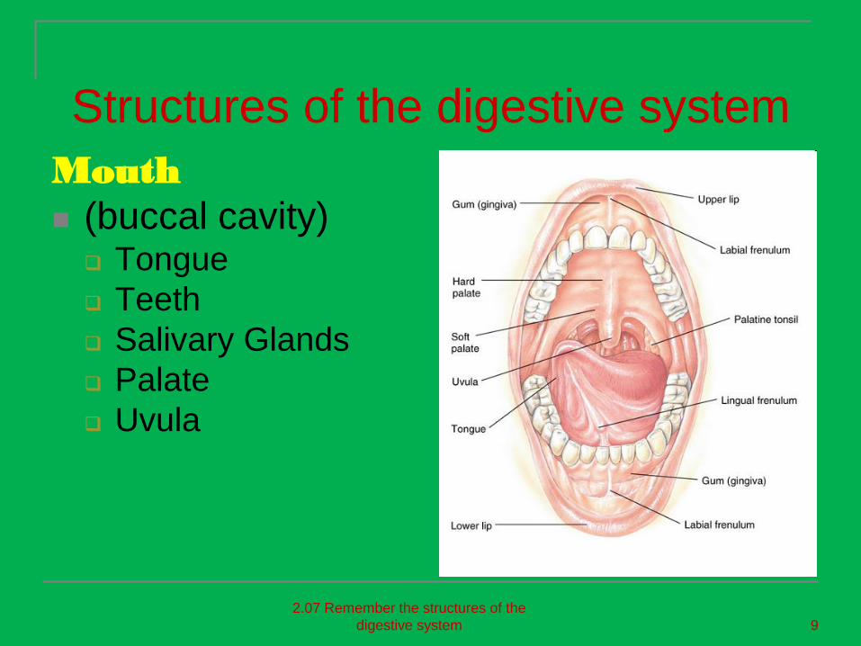
Structures of the digestive system
Mouth
(buccal cavity) Tongue
Teeth
Salivary Glands
Palate
Uvula
2.07 Remember the structures of the
digestive system 9

Structures of the digestive
systemTongue
Attached to floor of mouth
Made of skeletal muscle attached to four bones
Taste buds on the surface
Sweet
Sour
Bitter
Salty2.07 Remember the structures of the
digestive system 10
Tasting is one of our special senses.
What other body system have we discussed
That relates to the tongue and our taste buds?

Structures of the digestive
system
Teeth
Primary (deciduous)
20 “baby teeth”
Secondary
(permanent)
32 “adult teeth”
The process of
chewing is called
mastication.
2.07 Remember the structures of the
digestive system 11

Structures of the digestive
system
Salivary glands
Produce enzymes
that break down
carbohydrates and
convert starches to
sugar
2.07 Remember the structures of the
digestive system 12

Structures of the digestive
systemPalate
Hard
Soft
Uvula
Flap of skin
hanging in the
back of the throat
This keeps food from
going up our nose
when we swallow!
2.07 Remember the structures of the
digestive system 13
HOW MANY OF YOU LIKE
SUSHI?
EVER WONDER WHY THEY
SERVE GINGER WITH YOUR
MEAL?

Structures of the digestive
system
Pharynx (throat)
Nasopharynx
Oropharynx
Hypopharynx
When we swallow
this closes off the
trachea
2.07 Remember the structures of the
digestive system 15

Structures of the digestive
system
Esophagus Muscular tube, 10”
long
Connects the pharynx and stomach
Peristalsis occurs here
Esophageal wall layers
– Mucosa
– Submucosa
– Muscular
– External serous
2.07 Remember the structures of the
digestive system 16
Consider the process of ingestion.
Are there any health concerns related to eating
and the esophagus?

Structures of the digestive
system
Stomach Upper left
quadrant of the of the abdominal cavity
Fundus
Body
Pylorus
Cardiac Sphincter
Pyloric Sphincter
Rugae- expanding folds
2.07 Remember the structures of the
digestive system 17
The stomach secretes gastric
acids and enzymes causing
chemical digestion.
How long does it take the
stomach to empty?

Structures of the digestive
system
Small intestines
Duodenum
First segment
12” long
Jejunum
Second segment
8’ long
Ileum
Third segment
10-12’ long
2.07 Remember the structures of the
digestive system 19

Small Intestines (small bowels)
The major organ of DIGESTION, where most
food is broken down
ABSORPTION occurs in small intestine ---
absorption of 80% of usable nutrients ---
digested food passes into bloodstream and
on to body cells, indigestible passes on to
large intestine
Fatty acids and fat absorbed into lymphatic
vessels from the small intestine

Structures of the digestive system
Large intestines
Approximately 2” in
diameter
5’ long
2.07 Remember the structures of the
digestive system 21

Large Intestines
(large bowel or colon) Responsible for water reabsorption
Absorption of vitamins produced in the large
intestine (B complex and K vitamins)
packaging and compacting the waste
products for elimination

Activity: Make a poster!
Break into four groups and divide tasks:
Draw/Label/Color the structures of the digestive
system: mouth, esophagus, stomach, small
intestines, and large intestines.
Write a brief description of the function of each
structure on your poster.
Include the accessory organs: liver, pancreas,
and gall bladder.
Research and add the function of the accessory
organs to your poster.

Liver, Pancreas, and Gall Bladder

Liver
Located below the diaphragm, upper right
quadrant
Largest glandular organ and largest organ in
the abdominopelvic cavity
Receives 1.5 quarts of blood a minute from
the hepatic portal vein and artery
Connected to gallbladder and small intestine
by ducts
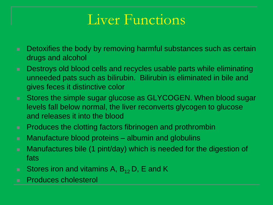
Liver Functions
Detoxifies the body by removing harmful substances such as certain
drugs and alcohol
Destroys old blood cells and recycles usable parts while eliminating
unneeded pats such as bilirubin. Bilirubin is eliminated in bile and
gives feces it distinctive color
Stores the simple sugar glucose as GLYCOGEN. When blood sugar
levels fall below normal, the liver reconverts glycogen to glucose
and releases it into the blood
Produces the clotting factors fibrinogen and prothrombin
Manufacture blood proteins – albumin and globulins
Manufactures bile (1 pint/day) which is needed for the digestion of
fats
Stores iron and vitamins A, B12 D, E and K
Produces cholesterol
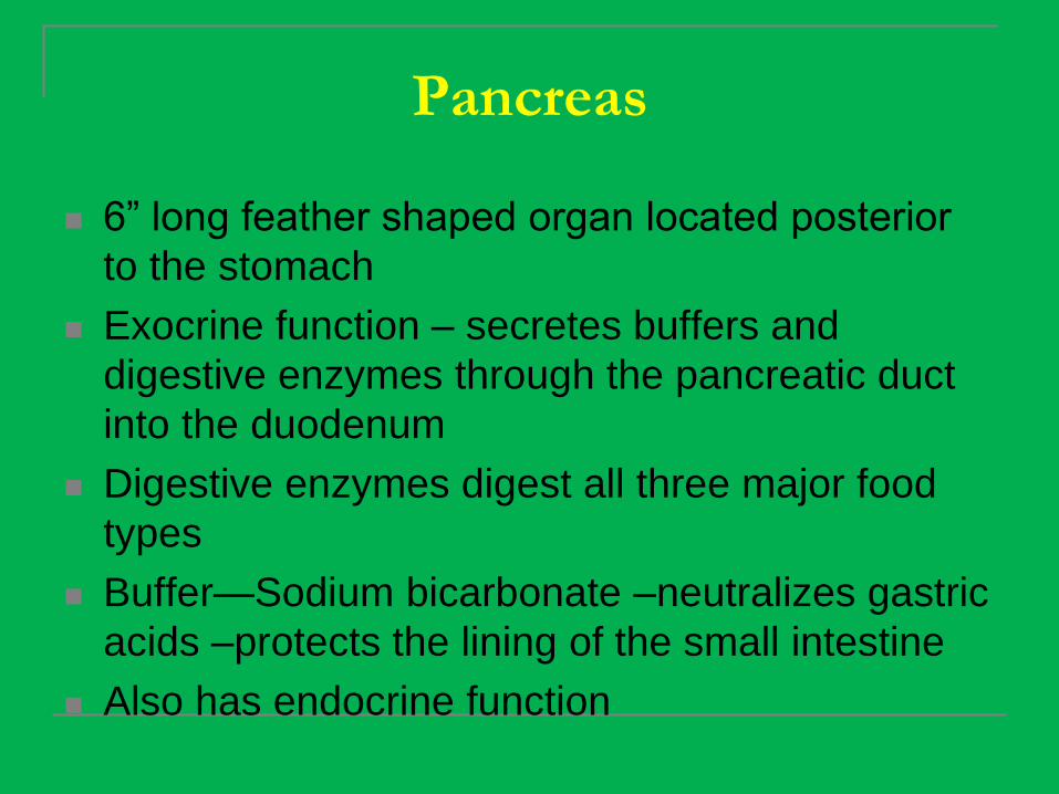
Pancreas
6” long feather shaped organ located posterior
to the stomach
Exocrine function – secretes buffers and
digestive enzymes through the pancreatic duct
into the duodenum
Digestive enzymes digest all three major food
types
Buffer—Sodium bicarbonate –neutralizes gastric
acids –protects the lining of the small intestine
Also has endocrine function

Gall bladder
Small, sac-shaped, muscular organ, green in
color, located at the inferior surface of the liver
Stores and concentrates bile -- reabsorbs water
content of bile received from liver
Bile enters the gallbladder by way of the hepatic
duct
When fatty foods enters the duodenum, bile is
released by gallbladder into the cystic duct and
then into the COMMON BILE DUCT which
connects to the duodenum
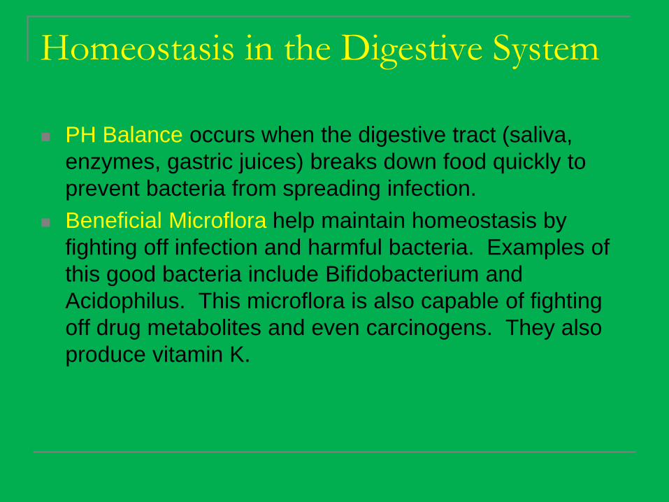
Homeostasis in the Digestive System
PH Balance occurs when the digestive tract (saliva,
enzymes, gastric juices) breaks down food quickly to
prevent bacteria from spreading infection.
Beneficial Microflora help maintain homeostasis by
fighting off infection and harmful bacteria. Examples of
this good bacteria include Bifidobacterium and
Acidophilus. This microflora is also capable of fighting
off drug metabolites and even carcinogens. They also
produce vitamin K.

Bellwork
INCONTINENCE – involuntary urination, often seem in older
persons, or due to illness and disease
GLYCOSURIA – sugar in urine, seen in DM, seriously ill individuals
with metabolic diseases or conditions
PYURIA – pus in urine, due to infection in the urine, or other part of
urinary system
ANURIA – no urine produced, acute and end stage kidney disease
HEMATURIA – blood in urine injury, disease or injury to kidney
DIURETIC – drug or substance to increase urine production

Standard
8) Outline basic concepts of normal structure
and function of all body systems, and explain
how homeostasis is maintained.
(particularly the urinary system)
Objectives
Label the structures of the urinary system
and describe each function
Explain how homeostasis is maintained in the
urinary system
Describe what dialysis is and who is a
candidate for this kind of treatment.
Identify what a urinalysis test is used for and
possible explanations of its results.

Urinary System
(also called the Excretory System)

Functions of the Kidney
remove waste products from the body
remove drugs from the body
balance the body's fluids
release hormones that regulate blood pressure
produce an active form of vitamin D that
promotes strong, healthy bones
control the production of red blood cells
Both the prefix “nephro” or the term
“renal” refer to the kidney.

The Ureters
One from each kidney
Can a person have more than two ureters?
Connect the kidneys to the bladder , carry
urine from kidney to bladder
Smooth muscle tube with mucous membrane
lining
Peristalsis pushes urine down ureters
Based on previous lessons, what is peristalsis?

The Bladder
Hollow, elastic muscular organ, capable of great
expansion, elastic fibers and involuntary muscle
Located the pelvic cavity
Stores and aids in the elimination of urine –
usually about 500cc
Emptying urine (voiding) is involuntary but
controlled through nervous system (voluntary)
Urine leaves the body through the URETHRA

The Urethra
The urethra is a tube that conveys urine from the
urinary bladder to the outside of the body.
Its wall is lined with mucous membranes and
contains a relatively thick layer of smooth
muscle tissue.
Female urethra: 4 cm; functions only as urinary
canal
Male urethra: functions both as a urinary canal
and a passageway for cells and secretions from
various reproductive organs. 20 cm.
URINALYSIS
The average output = 1500 ml/day
URINALYSIS – examination of urine to
determine presence of blood cells, bacteria,
acidity level, specific gravity and physical
characteristics (color, clarity and odor)
What are examples of a time a urinalysis might
be ordered?

THINK.PAIR.SHARE.

URINALYSIS
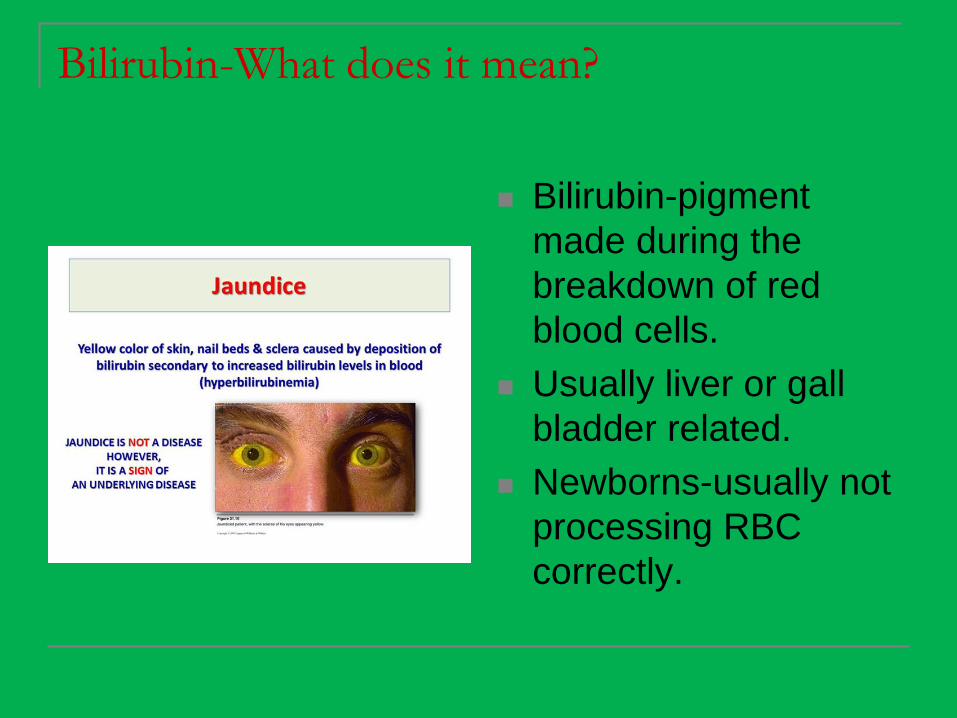
Bilirubin-What does it mean?
Bilirubin-pigment
made during the
breakdown of red
blood cells.
Usually liver or gall
bladder related.
Newborns-usually not
processing RBC
correctly.

Other chemicals found in urine.
Food,
dehydration,
and drugs
can also affect
these tests.
Usually, further
tests are done
to confirm any
questionable
results.
PKU is an amino acid deficiency that can cause mental
or physical disabilities. Tested as infants.

Chronic Renal Failure
Gradual loss of nephron
function lasting three months
or more
What does the nephron do?
Risk factors – diabetes,
hypertension, age, obesity,
ethnicity
Treatment for hypertension,
cholesterol, diet, fluid on heart
and lungs, itching
As the disease progresses
dialysis or kidney transplant
may be required

Dialysis (Watch Video)
Dialysis artificially removes waste products and extra
fluid from your blood when your kidneys can no longer
do this.
Hemodialysis- A machine filters wastes, salts and fluid from
your blood when your kidneys are no longer healthy
enough to do this work adequately.
Hemodialysis requires you to follow a strict treatment
schedule, take medications regularly and, usually, make
changes in your diet.
Peritoneal dialysis-During peritoneal dialysis, blood vessels
in your abdominal lining (peritoneum) fill in for your kidneys,
with the help of a fluid (dialysate) that flows into and out of
the peritoneal space.

Activity
Draw and color the
various possibilities of
urine samples. (Think
specimen cup)!!!
Label each and
describe what might
be the cause for the
color, clarity, or odor.
Circle any areas of
concern.