the digestive tract of drosophila melanogaster
TRANSCRIPT

GE47CH17-Lemaitre ARI 29 October 2013 14:38
The Digestive Tractof Drosophila melanogasterBruno Lemaitre1 and Irene Miguel-Aliaga2
1Global Health Institute, School of Life Sciences, Ecole Polytechnique Federale Lausanne(EPFL), CH-1015 Lausanne, Switzerland; email: [email protected] Clinical Sciences Center, Imperial College London, London W12 0NN,United Kingdom; email: [email protected]
Annu. Rev. Genet. 2013. 47:377–404
The Annual Review of Genetics is online atgenet.annualreviews.org
This article’s doi:10.1146/annurev-genet-111212-133343
Copyright c© 2013 by Annual Reviews.All rights reserved
Keywords
gut, Drosophila, digestion, absorption, enteroendocrine cells, entericnervous system, intestine, immunity
Abstract
The digestive tract plays a central role in the digestion and absorptionof nutrients. Far from being a passive tube, it provides the first lineof defense against pathogens and maintains energy homeostasis by ex-changing neuronal and endocrine signals with other organs. Historicallyneglected, the gut of the fruit fly Drosophila melanogaster has recentlycome to the forefront of Drosophila research. Areas as diverse as stemcell biology, neurobiology, metabolism, and immunity are benefittingfrom the ability to study the genetics of development, growth regula-tion, and physiology in the same organ. In this review, we summarizeour knowledge of the Drosophila digestive tract, with an emphasis on theadult midgut and its functional underpinnings.
377
Ann
u. R
ev. G
enet
. 201
3.47
:377
-404
. Dow
nloa
ded
from
ww
w.a
nnua
lrev
iew
s.or
gby
Im
peri
al C
olle
ge L
ondo
n on
01/
30/1
4. F
or p
erso
nal u
se o
nly.
GE47CH17-Lemaitre ARI 29 October 2013 14:38
Peritrophic matrix(PM): a noncellularmatrix composed ofchitin andglycoprotein that linesthe invertebratemidgut and separatesthe food bolus fromthe epithelium
Proventriculus: theforegut portion of thecardia. Proventriculusis often used todescribe the entirecardia
INTRODUCTION
The gut is one of the largest organs in the bodycavity. Aside from its central role in digestingand absorbing nutrients, the inner lining of thedigestive tract must also serve as the first line ofdefense against a wide variety of pathogens. Thegut is also a major source of neuronal and en-docrine signals able to modulate nutrient stor-age or food intake by regulating the activity ofother organs, such as the pancreas and the brainin mammals. Hence, far from being a passivetube exclusively concerned with digestion, thegut is emerging as a major regulator of multi-ple biological processes. As a result, what hadhistorically been a relatively obscure organ isnow coming to the forefront of research in ar-eas as diverse as stem cell biology, neurobiol-ogy, metabolism, and immunity.
The gut has also been a relatively understud-ied organ in Drosophila melanogaster, given that,since the advent of the genetic revolution, thismodel system has played such a central role inthe investigation of developmental processes.Consequently, physiological studies have typi-cally been relegated to less genetically amenableinsects, possibly because of their larger size (50).The potential to combine genetic and func-tional approaches in Drosophila has only beenrealized in recent years, and concurrent withthis, there has been a surge of interest in the flygut and its functions. In this review, we sum-marize our knowledge—at times fragmented orsketchy—of the Drosophila digestive tract, withan emphasis on the adult midgut and its func-tional underpinnings.
STRUCTURE OF THEDROSOPHILA DIGESTIVE TRACT
The emergence of the gastrointestinal tractwithin the body cavity was a major innovationin animal evolution, allowing the transitionfrom an intracellular to an extracellular modeof digestion (169). In bilaterians, the diges-tive tract further evolved into a successionof histologically distinct regions tailored tospecific digestive needs. Hence, the dietary
requirements of Drosophila (consisting pri-marily of fermenting fruit) resulted in analimentary canal similar to that of otherdipterans feeding on decaying matter, such asthe housefly Musca domestica (49, 52, 102, 126,182). Two prominent features can be discernedfrom an examination of the anatomical andcellular architecture of the Drosophila gut: itscompartmentalization and plasticity, both ofwhich have been best characterized in the adultmidgut.
Compartmentalization of theDigestive Tract
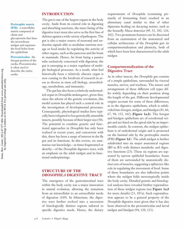
As in other insects, the Drosophila gut consistsof a simple epithelium, surrounded by visceralmuscles, nerves, and tracheae. The nature andarrangement of these different cell types dif-fer widely depending on their position alongthe length of the gut. Different developmentalorigins account for some of these differences,as in the digestive epithelium, which is subdi-vided into foregut, midgut, and hindgut (43, 66,67, 94, 152, 162) (Figure 1a,b). The foregutand hindgut epithelium are of ectodermal ori-gin and are lined on the apical side by an imper-meable cuticle. In contrast, the midgut epithe-lium is of endodermal origin and is protectedon the luminal side by the peritrophic matrix(PM) (Figure 1d ). The adult midgut is furthersubdivided into six major anatomical regions(R0 to R5) with distinct metabolic and diges-tive functions (23). These six regions are sep-arated by narrow epithelial boundaries. Someof them are surrounded by anatomically dis-tinct sets of muscles, suggesting a sphincter-likerole in regulating the movement of food. Someof these boundaries are also inflection pointswhere the midgut folds stereotypically insidethe body cavity. Detailed genetic and histolog-ical analyses have revealed further regionaliza-tion of these midgut regions (see Figure 1a,bfor more details) (23, 107a). Such regionaliza-tion appears to be a general property of theDrosophila digestive tract given that it has alsobeen observed in the proventriculus and larvalmidgut and hindgut (94, 120, 121).
378 Lemaitre · Miguel-Aliaga
Ann
u. R
ev. G
enet
. 201
3.47
:377
-404
. Dow
nloa
ded
from
ww
w.a
nnua
lrev
iew
s.or
gby
Im
peri
al C
olle
ge L
ondo
n on
01/
30/1
4. F
or p
erso
nal u
se o
nly.

GE47CH17-Lemaitre ARI 29 October 2013 14:38
Crop
Proventriculus Hindgut
Peritrophicmatrix
Enterocytes
MusclesBasement
membraneEnteroendocrine
cell
EnteroblastStem cell
Microvilli
Malpighian tubules
a
b
c d BacteriaBacteria
Midgut
Foregut
Figure 1Schematic organization of the Drosophila digestive tract. (a) A 3D reconstruction of the digestive tract withinthe body cavity (23). (b) The digestive tract is divided into three discrete domains of different developmentalorigin: foregut, midgut, and hindgut. Each of these domains is further subdivided into genetically distinctcompartments (illustrated by different colors in the case of the midgut). (c) The midgut is composed of anepithelium surrounded by two layers of visceral muscles. The midgut epithelium consists of enterocytes,enteroendocrine cells (EEC), and progenitor cells. (d) Electron microscopy sections of a third-instar larvalgut following infection with Erwinia carotovora 15 (2). The peritrophic matrix establishes a physical barrierthat prevents contact between bacteria and the epithelial cell.
Compartmentalization is achieved andmaintained through the combinatorial action ofpan-gut and region-specific transcription fac-tors. For instance, in the middle midgut, theso-called copper-cell region relies on the activ-ity of two region-specific homeobox proteins,Labial and Ptx1, and a pan-midgut transcrip-tion factor, GATAe (23, 56, 83, 135). Gene ex-pression boundaries are not always sharp, espe-
cially in the vicinity of boundaries where gradedgene expression appears to be widespread andrelies in part on the Wnt pathway (23). Thissuggests that these boundaries are gut tissue–organizing centers where Wingless may act asa morphogen, akin to its function in patterningembryos or imaginal discs.
Finally, it is noteworthy that larvae andadults differ in the anatomical and genetic
www.annualreviews.org • The Digestive Tract of Drosophila melanogaster 379
Ann
u. R
ev. G
enet
. 201
3.47
:377
-404
. Dow
nloa
ded
from
ww
w.a
nnua
lrev
iew
s.or
gby
Im
peri
al C
olle
ge L
ondo
n on
01/
30/1
4. F
or p
erso
nal u
se o
nly.

GE47CH17-Lemaitre ARI 29 October 2013 14:38
Crop: an enlargeddiverticulum of theforegut used to storefood before it entersthe midgut
organization of their digestive tract, plausi-bly as a result of their different dietary habits.Whereas larvae feed continuously to sustaintheir growth and are able to ingest solid foodthanks to their mouth hooks, adult flies feed lessfrequently, ingesting liquid via their proboscis.This intermittent ingestion of liquid may ac-count for the presence of the crop, a storage or-gan found only in adults (170). Features specificto larval guts include the presence of four gas-tric ceca in the anterior midgut, which are ma-jor sites of digestion and absorption, at least inother insects (126). Thus, the adaptive changesof these gut regions during the fly’s life cyclefurther illustrate the gut’s structural complex-ity and highlight another notable feature of thisorgan: its plasticity.
The Drosophila Gut: A Plastic Organ
As in other holometabolous insects, the adultDrosophila gut is generated de novo duringmetamorphosis. In fact, three distinct gastroin-testinal tissues supersede one another duringdevelopment: the larval gut (arising from theembryonic gut), the transient pupal gut, and theadult gut (see References 109, 124, 176, 178 fordetails). After metamorphosis, both the larvaland pupal midgut degenerate to form meco-nium, which is expelled soon after eclosion.
The adult midgut epithelium contains twomain types of differentiated cells: enterocytesand enteroendocrine cells (EECs) (Figure 1c).Enterocytes (also called columnar cells in otherinsects) are large polyploid cells that secretedigestive enzymes and absorb nutrients. Al-though enterocytes differ markedly in mor-phology along the gut, most of them are pos-itive for the MyoIA-Gal4 driver (inserted inthe Myosin 31DF gene). Interspersed amongthe enterocyte monolayer are small nucle-ated prospero-positive EECs (113, 134, 175).Whereas the larval gut is postmitotic and un-dergoes massive growth by increases in cell sizecoupled with polyploidization, the adult gut iscapable of regeneration. As in other insects (49,102, 126), the adult midgut is constantly re-placed by new cells derived from small regen-
erative cells [named intestinal stem cells (ISCs)in Drosophila] scattered across the basal surfaceof the gut epithelium (113, 134). Midgut ISCsundergo either asymmetric or symmetric divi-sion(s). In asymmetric divisions, one daughtercell maintains ISC characteristics and remainsmitotic, whereas the other daughter cell (alsoknown as an enteroblast) exits the cell cycleand differentiates into either an absorptive en-terocyte or an EEC, an outcome determined byNotch signaling activity (133). Differentiatingenterocytes endoreplicate their genome two tothree times to increase their size and develop abrush border on their apical side.
All midgut ISCs and enteroblasts (referredto together as progenitor cells) express themarker gene escargot. Nevertheless, distinctpopulations of self-renewing stem cells havebeen identified along the digestive tract. Forinstance, the copper-cell region is renewed bya specific population of ISCs that, unlike othermidgut ISCs, are largely quiescent (172). Sim-ilarly, another subpopulation of self-renewingcells is found in the foregut-midgut junctionand may contribute to the maintenance of theproventriculus (167). In the hindgut, stem cellsare restricted to a narrow segment known asthe hindgut proliferation zone located at themidgut-hindgut boundary (177). The existenceof different stem cell populations may con-tribute to the establishment of different re-gional identities. Alternatively, or in paral-lel, surrounding tissues, such as visceral mus-cles and tracheae, could provide region-specificniche signals to differentiating ISC lineages toimpart regional identity to their progeny (23,107a).
The midgut epithelium is renewed withinone to two weeks under steady-state conditions(113, 134). Stem cell activity is influenced by themetabolic state of the host and by environmen-tal factors (reviewed in 89, 176). In the fed state,localized insulin signaling increases ISC activ-ity and drives tissue growth (131). Increasedstem cell activity is also observed upon intesti-nal damage by corrosive agents or pathogenicbacteria. This allows the rapid regenerationof the compromised gut and maintains barrier
380 Lemaitre · Miguel-Aliaga
Ann
u. R
ev. G
enet
. 201
3.47
:377
-404
. Dow
nloa
ded
from
ww
w.a
nnua
lrev
iew
s.or
gby
Im
peri
al C
olle
ge L
ondo
n on
01/
30/1
4. F
or p
erso
nal u
se o
nly.

GE47CH17-Lemaitre ARI 29 October 2013 14:38
integrity (4, 22, 90). The activity of a numberof pathways, namely the JAK-STAT, epider-mal growth factor (EGF) receptor, decapen-taplegic (DPP), and Wingless/Myc pathways,stimulates the proliferation and differentiationof progenitor cells into enterocytes (40, 72,90, 105; see References 89, 176 for reviews).Upon damage, these pathways are activated bythe release of EGFs, Wingless, and JAK-STATligands (Upd3, Upd2) from damaged entero-cytes or progenitors, thus establishing a com-pensatory homeostatic loop. Interestingly, thevisceral muscles that surround the epitheliumcontribute to epithelium renewal by producingthe EGF ligand Vein (89, 90, 176). An outstand-ing question in the regeneration field is to iden-tify the cells and signals that initiate the complexhomeostatic loop that leads to epithelial regen-eration. One possible trigger may be the releaseof Upd2 and Upd3 by enterocytes, either in re-sponse to stress or following rupture of the ad-jacent epithelium and consequent activation ofthe Hippo pathway (reviewed in 89, 176).
Senescence of the Adult Gut
The plasticity of the gut is further high-lighted by the changes it undergoes during adultlife. Ultrastructural observations in the 1970sand 1980s revealed that aging alters the adultmidgut and is associated with increased lipiddeposits, autophagic vacuoles, and the appear-ance of ballooned mitochondria (6, 65). Morerecently, other studies have shown that oldDrosophila guts display defective compartmen-talization (23) and become leaky (148) and dys-plastic, as defined by a higher rate of stem cellproliferation and accumulation of undifferenti-ated progenitors (12, 35). This dysplasia is per-haps not surprising given that, as one of themost highly mitotic adult organs, the gut muststrike a balance with regard to stem cell activ-ity, allowing regeneration while minimizing therisk of hyperplasia. Interestingly, a link betweenlife span and stem cell activity was recentlyreported by Biteau et al. (13), who observedthat maximal life span is achieved when intesti-
nal proliferation is reduced rather than totallyinhibited.
Mechanistically, little is known about themolecular effectors of these aging hallmarks,but there are some intriguing correlations. In-deed, gut senescence is associated with thechronic activation of the stress-responsive JNKpathway and with elevated reactive oxygenspecies (ROS) levels in the gut (12). A link be-tween ROS, mitochondria, gut senescence, andaging is further supported by the observationthat life span can be extended upon overexpres-sion of dPGC-1, a regulator of mitochondrialbiogenesis in the gut (147). Interestingly, agedflies also have higher bacterial loads in theirgut (21, 146), and axenic flies display attenu-ated and delayed intestinal dysplasia, suggestingthat gut-associated bacteria contribute to thedisorganization of the gut in aged flies, possiblyby modulating epithelium turnover (21). Bar-rier dysfunction in old guts also correlates withshorter life span and higher expression of im-mune genes by the fat body (148). An emergingtheme from these somewhat disparate observa-tions is that the intestine may be an importantmodulator of life span at the organismal level.
THE DROSOPHILA GUT:A SELECTIVE BARRIER
The digestive tract forms a selective barrier thatallows absorption of nutrients, ions, and wa-ter but limits contact with potentially damagingagents, such as toxins and pathogens. This se-lectivity is enabled by specialized physical bar-riers and a potent mucosal immune system.
Peritrophic Matrix and Mucus
Most metazoans, including vertebrates, isolatetheir intestinal epithelium from the external en-vironment with one or more carbohydrate-richbarriers (e.g., mucus). In insects, the foregut andhindgut are lined by a relatively impermeablecuticle, whereas the midgut is protected by theperitrophic matrix (PM) (Figure 1d ). Almostnothing is known about the composition, orga-nization, and role of these barriers in Drosophila.
www.annualreviews.org • The Digestive Tract of Drosophila melanogaster 381
Ann
u. R
ev. G
enet
. 201
3.47
:377
-404
. Dow
nloa
ded
from
ww
w.a
nnua
lrev
iew
s.or
gby
Im
peri
al C
olle
ge L
ondo
n on
01/
30/1
4. F
or p
erso
nal u
se o
nly.

GE47CH17-Lemaitre ARI 29 October 2013 14:38
Endoperitrophicspace: part of thelumen located insidethe peritrophic matrix
Ectoperitrophicspace: part of thelumen located betweenthe peritrophic matrixand the midgutepithelium
Antimicrobialpeptides: smallcationic peptides thatare able to kill bacteria
Peptidoglycanrecognition protein(PGRP): a family ofproteins that can bindto peptidoglycan, aspecific component ofbacterial cell walls
The PM is believed to be an organized lat-tice composed of chitin fibrils held together bysecreted chitin-binding proteins, notably per-itrophins (79, 101). Electron microscopy in-dicates that the adult PM is secreted as fourlayers in the proventriculus, which are proba-bly compressed by muscular contraction of theproventricular walls to coalesce into two lay-ers as they enter the midgut (94, 152). Peristal-sis may propel the PM as far as the hindgut.Consistent with this, transcripts encoding PMcomponents (e.g., peritrophin) are strongly en-riched in the proventriculus (23). The observa-tion that peritrophin genes are also expressedin a more distal part of the midgut suggests thatthis barrier is remodeled along the gut (23).To date, no fly devoid of PM has been gen-erated, suggesting that this matrix is essentialfor viability. Nevertheless, a mutation in thedrosocrystallin gene, a structural element of thePM, results in reduced PM thickness and higherpermeability and is associated with higher sus-ceptibility to ingested entomopathogenic bac-teria or pore-forming toxins (96). A protectiverole for the PM against abrasive food particlesand pathogens as well as in sequestering in-gested toxins has already been described (79,101). Studies in other insects further suggestthat the PM plays an essential role in diges-tion by partitioning the lumen into two dis-tinct compartments, the endoperitrophic andthe ectoperitrophic spaces, which contain dif-ferent digestive enzymes (183).
In addition to the PM, the existence of a mu-cous layer is suggested by the fact that the apicalsurface of midgut epithelial cells is positive forperiodic acid-Schiff (23): a staining methodused to detect mucosubstances. Although morethan 30 Drosophila genes have been annotatedas mucin-like proteins (174), the functionalrelevance of these genes or, more generally, ofmucus in the gut has not been investigated.
Drosophila Gut Immunity
The Drosophila gut is proving to be a goodmodel to study interactions between stress, re-pair, and immune responses/tolerance to com-
mensal bacteria (see References 21a, 42, 61 forreviews).
Ingestion of gram-negative bacteria triggersthe expression of several antimicrobial pep-tide genes in specific domains along the di-gestive tract (187). Although most of thesegenes are under the control of the Imd path-way, some antifungal Drosomycin-like peptidegenes are also induced in the anterior midgutby the JAK-STAT pathway, probably in re-sponse to epithelial damage (22, 136, 198).In the gut, the Imd pathway is activated bytwo pattern-recognition receptors of the pep-tidoglycan recognition protein (PGRP) fam-ily: the transmembrane PGRP-LC receptor(which plays a predominant role in the foregutand the hindgut) and the intracellular PGRP-LE sensor (the role of which is restricted tothe midgut) (15, 127). Both receptors are acti-vated upon recognition of diaminopimelic acid(DAP)-type peptidoglycan, a cell wall com-ponent of gram-negative bacteria and certaingram-positive bacterium (e.g., Bacillus). Severalnegative regulators downregulate the Imd path-way to prevent its activation by indigenous bac-teria or ingested bacteria and to tailor its activityto the severity of infection. Negative regulatorsinclude secreted amidases that scavenge pepti-doglycans (the bacterial ligand of the Imd path-way) and Pirk, which disrupts signaling betweenthe PGRP-LC receptor and its downstreamadaptor, Imd (104, 139, 198). Although the Imdpathway is activated all along the gut, transcrip-tion factors, such as the homeobox Caudal, re-strict the expression of a subset of Imd targetgenes (157). This additional level of regula-tion explains why genes encoding antimicro-bial peptides are expressed in specific domains(157).
A complementary line of defense involvesthe production of ROS by the NADPH oxi-dase Duox (74). Duox is thought to be presenton the apical surface of the gut epithelium and isenriched in the foregut and hindgut. Duox ac-tivity is stimulated upon recognition by an as yetuncharacterized G-coupled receptor of uracile,a microbially derived ligand that is proposed tobe released by pathogenic bacteria (100). Duox
382 Lemaitre · Miguel-Aliaga
Ann
u. R
ev. G
enet
. 201
3.47
:377
-404
. Dow
nloa
ded
from
ww
w.a
nnua
lrev
iew
s.or
gby
Im
peri
al C
olle
ge L
ondo
n on
01/
30/1
4. F
or p
erso
nal u
se o
nly.

GE47CH17-Lemaitre ARI 29 October 2013 14:38
Plasmatocytes:Drosophila hemocyteswith macrophageactivity
is also regulated at the transcriptional level bythe p38 MAPK pathway and the ATF2 tran-scription factor (73). Excessive production ofROS is circumvented by an inducible catalase(75) and by negative regulators of Duox activity(73).
In addition to these signaling pathways,acidity, peristalsis, and the PM (96) mayall contribute to bacterial clearance. Indeed,most ingested bacteria do not survive passagethrough the acidic zone. The crop also producesmany detoxifying enzymes (e.g., glutathion S-transferase), which may detoxify the bolus priorto its entry into the midgut. To date, thereis no evidence of any blood cells intimatelyassociated with the gut, although a popula-tion of plasmatocytes with macrophage capacityresides within the folds of the larval proven-triculus (31, 152, 199). Gut infection can lead tointestinal damage, which can be caused by bac-terial toxins or by an overaggressive, and thusdeleterious, immune response. Stress responseprograms and increased epithelial renewal canthen be deployed to repair the intestinal ep-ithelium and maintain the integrity of the gutbarrier (21, 32, 33, 90).
DIGESTIVE ENZYMESAND DIGESTION
In the intestine, the complex mix of dietarycomponents ingested by flies is broken downby digestive enzymes before it is absorbed bythe intestinal epithelium. Digestion is also in-fluenced by the physicochemical conditions ofthe gut (notably pH) and the enzymatic activityof microbes (49). Although digestion in Dipteramay also occur in the crop or extra-orally, bothdigestion and absorption are predominantly ac-complished in the midgut (102).
Digestive Enzymes
The Drosophila genome encodes a vast array ofputative digestive enzymes involved in the pro-cessing of carbohydrates, proteins, and lipids—as many as 349 based on bioinformatic predic-tions (Figure 2a). Endopeptidases and exopep-
tidases [involved in the initial and final steps ofprotein digestion, respectively] are particularlyabundant (28, 155, 180). There are also 52 and29 proteins with predicted carbohydrase and li-pase activity, respectively (84, 85) (Figure 2a).This diverse enzymatic repertoire may be op-timized to deal with the nature and complexityof the diet of Drosophila, which is rich in decay-ing fruits. In this regard, it is interesting to notethe presence of 15 genes encoding lysozymes,which hydrolyze peptidoglycan, a major com-ponent of bacterial cell walls. Given that noimmune role has been ascribed to Drosophilalysozymes, it is possible that they are involvedin digestion (97). Thus, their high gene copynumber strongly suggests that bacteria make upa significant proportion of the Drosophila dietin the wild. In addition, the Drosophila genomealso encodes chitinases and glucanases that mayparticipate in the digestion of yeasts that are alsofound in rotting fruits.
Most digestive gene families are organizedin tight genomic clusters, with each member ofthe cluster being differentially expressed alongthe digestive tract (23) (Figure 2b). This appliesto Jonah proteases, trypsins, α-esterases, man-nosidases, and lipases (23). These clusters mayhave arisen by gene duplication followed by di-vergence in order to optimize enzymatic activ-ities in each portion of the digestive tract—anevolutionary process suggested by the extensivestudies of the Drosophila α-amylase gene family(41, 201).
Expression analyses of several digestive en-zyme genes suggest sequential processing of nu-trients along the intestine. For instance, amy-lases (catalyzing the breakdown of starches) arefound in midgut regions R2 and R4, whereasenzymes involved in the processing of simplecarbohydrates are mostly found in R4, R5, andthe hindgut (1, 23). However, it remains to beestablished whether this restricted expression ismaintained, given that digestive enzymes maydiffuse to a considerable distance in the gut lu-men. The peritrophic membrane may be animportant tissue to consider in this regard. InRhynchosciara americana larvae, initial digestionis carried out by polymer hydrolases within the
www.annualreviews.org • The Digestive Tract of Drosophila melanogaster 383
Ann
u. R
ev. G
enet
. 201
3.47
:377
-404
. Dow
nloa
ded
from
ww
w.a
nnua
lrev
iew
s.or
gby
Im
peri
al C
olle
ge L
ondo
n on
01/
30/1
4. F
or p
erso
nal u
se o
nly.

GE47CH17-Lemaitre ARI 29 October 2013 14:38
Polysaccharide(e.g., starch)
Dextrins or Disaccharides(e.g., sucrose/maltose)
Monosaccharide (e.g., glucose/fructose)
Amylases Glucosidase
Glucose repression
2RGene span
LambdaTry EtaTry AlphaTry
KappaTry ThetaTry EpsilonTry
ZetaTry BetaTry IotaTry
DeltaTry
GammaTry
7,230k 7,240k
R1 R2 R3 R4 R5Delta/gammaTry
AlphaTry
BetaTry
EpsilonTry
ThetaTry
KappaTry
LambdaTry
IotaTry
EtaTry
ZetaTry
b a
c
Carbohydrase 52Amylase 3
Mannosidase 7
Glucosidase 10
Trehalase 2
Beta-galactosidase 2
Lysozyme 15
Chitinase 10
Amidase PGRP 3
Proteinasae 234Aminopeptidase 12
Carboxypeptidase 22
Cysteine-type endopeptidase 9
Aspartic-type endopeptidase 12
Serine-type endopeptidase 132
Metalloendopeptidase 53
Peptidyl-dipeptidase 5
Lipase and esterase 63Lipase 29
Carboxylesterase 16
Phospholipase 13
Sphingomyelin phosphodiesterase 3
Phosphodiesterase 2
Figure 2Digestive enzymes. (a) List of putative Drosophila digestive enzyme genes. Genes were identified based on the following criteria: abilityto encode an enzyme (gene ontology) with a signal peptide and significant gene expression enrichment in one of the following organs:salivary gland, crop, midgut, and hindgut. See complete table and details at http://lemaitrelab.epfl.ch/resources. (b) (Top) Eleventrypsin genes are clustered together on chromosome II. (Bottom) The expression of each gene of this cluster is enriched in differentregions of the midgut (see color code corresponding to R1–5) (23). (c) Digestion of carbohydrates by the sequential action of amylaseand glucosidases. The presence of glucose inhibits the expression of amylase. Abbreviation: PGRP, peptidoglycan recognition protein.
Paracrine: a form ofcell signaling in whichthe target cell is nearthe signal-releasingcell
Prandial: relating tofood ingestion
peritrophic membrane, giving rise to productsof lower molecular weight, which then diffuseout of the peritrophic membrane. The secondphase of digestion occurs in the ectoperitrophicfluid, followed by a final digestion step primar-ily confined to the microvilli of the midgut,where enzymes are trapped in the glycocalyx(49, 184). Although the existence of such a de-gree of spatial organization with regard to di-gestion has not been investigated in Drosophila,vesicles containing the lipase Magro are se-
creted into the larval gut lumen in the sameregion of the proventriculus as the PM (164),raising the possibility that vesicular traffickingin this region may function to load the PM withdigestive enzymes.
Regulation of Digestive Enzymes
Digestive enzymes are subject to complex regu-lation by neural, systemic, paracrine, and pran-dial mechanisms (102, 103, 194). Modulation
384 Lemaitre · Miguel-Aliaga
Ann
u. R
ev. G
enet
. 201
3.47
:377
-404
. Dow
nloa
ded
from
ww
w.a
nnua
lrev
iew
s.or
gby
Im
peri
al C
olle
ge L
ondo
n on
01/
30/1
4. F
or p
erso
nal u
se o
nly.

GE47CH17-Lemaitre ARI 29 October 2013 14:38
Vacuolar-typeH+-ATPase(V-ATPase):a multi-subunittransporter thatcouples the energy ofATP hydrolysis toproton transportacross the intracellularor plasma membranesof eukaryotic cells
of enzyme expression by nutrient quantity orquality has been observed in several arthro-pods (36, 194). One of the rare character-ized examples of digestive enzyme regulationin Drosophila concerns carbohydrate digestionand involves repression of enzymes importantin the initial phase of digestion by digestiveend products. Indeed, sucrose and its prod-ucts, glucose and fructose, have been shownto repress amylase gene expression—an effectdescribed as glucose repression (1, 10, 185)(Figure 2c). This reduction in digestive ca-pacity may well be a metabolic adaptation tolimit dietary sugar absorption during periodsof nutritional abundance, given that Drosophilais poorly adapted to nutritional excess (122).Mechanistically, reduced amylase activity re-sults from downregulation of gene transcrip-tion. Although the mechanism of transcrip-tional repression remains to be established, thisfinding suggests that digestive genes receive in-puts from regional transcription factors that de-fine their site of expression and from metabolictranscription factors that adjust their expressionlevels to match organismal requirements. Twoother factors known to influence digestion arestarvation and bacterial infection, which typi-cally downregulate the transcription of diges-tive enzymes, perhaps to spare resources (22,203). The existence of additional levels of regu-lation at the posttranscriptional level (e.g., pro-tein release) has not been investigated.
Functional Studies
With rare exceptions, the precise roles of diges-tive enzymes have not been established. Usingan inversion mutant in which both amylase pand d are affected (amynull) (82), it was shownthat amylase-deficient flies perish on a starch-only diet. However, lethality can be rescuedby dietary supplementation with simple sugars,which are the end products of amylase diges-tion. Interestingly, a different study reportedthat amylase-deficient flies were able to surviveon a starch-only medium only when they werehoused with wild-type flies (76). This findingled the authors to posit the existence of extra-
oral digestion, with wild-type flies regurgitat-ing amylases onto the medium, which wouldpredigest carbohydrates on the food surface.Another functional study concerned the lipaseMagro, expressed in the proventriculus underthe control of the transcription factor DHR96(86, 164). Downregulation of either magro orDHR96 by RNAi rendered larvae unable tobreak down dietary triglyceride in their gutlumen, leading to reduced triglyceride stores(164). This phenotype was rescued by feedinglarvae pancreatin, a mixture of pancreatic en-zymes that includes triglyceride lipase.
The Drosophila Midgut pH
Nutrient breakdown and enzymatic activity arestrongly affected by the physicochemical prop-erties of the lumen. Whereas mammalian di-gestion takes place in acidic conditions, insectdigestion occurs at neutral or basic pHs. InDrosophila adults, the initially neutral pH of theanterior midgut is followed by an acidic zone(pH < 4) that corresponds to the copper-cellregion. The posterior midgut is mildly alkaline(pH = 7–9), whereas the hindgut is slightlyacidic (pH = 5), although its pH can fluctuatedepending on the fly’s diet (37, 53, 162). Akinto the mammalian stomach, the midgut acidiczone denatures proteins and provides an opti-mal pH for the activity of some peptidases. Itprobably also kills microorganisms that are in-gested with the food and facilitates the absorp-tion of lipids and metals. The role of the cop-per cells [a pool of highly differentiated cells inR3 (53, 162)] in acid production has been es-tablished in larvae that lack the labial or alpha-spectrin genes, which affect the differentiationor shape of these copper cells, respectively (55).Acid secretion is thought to involve the po-larized activity of an apical multi-subunit V-ATPase (vacuolar-type H+-ATPase) pump thatexports H+ in the lumen in exchange for en-ergy in the form of ATP (51, 161). Several V-ATPases enriched in the copper-cell region ofthe adult gut (such as vha100-4) are good candi-dates to mediate the acidification of this midgutregion (23).
www.annualreviews.org • The Digestive Tract of Drosophila melanogaster 385
Ann
u. R
ev. G
enet
. 201
3.47
:377
-404
. Dow
nloa
ded
from
ww
w.a
nnua
lrev
iew
s.or
gby
Im
peri
al C
olle
ge L
ondo
n on
01/
30/1
4. F
or p
erso
nal u
se o
nly.

GE47CH17-Lemaitre ARI 29 October 2013 14:38
Hemolymph: insectblood
Transcellulartransport: transportof solutes by a cellthrough a cell
Drosophila Gut-Associated Microbesand Their Role in Digestion
Drosophila feed on decaying fruit, and their di-gestive tract is therefore constantly exposed todietary microbes, which may themselves con-tribute to digestion. The Drosophila gut lumenis an environment with relatively low bacterialdiversity and numbers (1–30 species, 103–105
CFU/fly) (30, 156, 191, 192; reviewed in 17,48, 59). The most prevalent bacterial speciesare members of the genera Lactobacillus andAcetobacter. The existence of a stable popula-tion of dividing bacteria residing in the gut hasnot yet been formally demonstrated, as bacte-ria found in the Drosophila gut are also found inthe substratum, suggesting constant contami-nation. Experiments with germ-free flies (raisedin the absence of both internal and external bac-teria) have shown that gut-associated bacteriacan be beneficial to Drosophila by promotinggrowth in poor dietary conditions (151, 163,171). How bacteria contribute to growth hasyet to be established, but they are not merely afood source given that growth promotion wasonly observed in the presence of live bacteria.Live bacteria may predigest food, change its nu-tritional content, and/or improve digestion in-side the gut. In addition to their contributionto Drosophila growth, gut bacteria also stimu-late intestinal epithelial turnover and influencethe basal level of antimicrobial peptide gene ex-pression in the gut (21, 156, 163).
Less studied than gut-associated bacteria,yeasts play an important role in Drosophila nu-trition by providing many essential nutrients,such as amino acids, sterols, B vitamins, andfatty acids: compounds not generally present indecaying plant material (see 17 and referencestherein).
ABSORPTION AND STORAGE
Absorption refers to the uptake of luminalcontents across the intestine to the hemolymph(49). Most absorption is via transcellular trans-port, requiring specific transporters at boththe apical and basal sides of enterocytes, andis likely to be regulated. Hence, far from being
a passive barrier, the gut may play active rolesin the transport, conversion, and storage ofmetabolites. In spite of their potential impacton energy homeostasis, such roles are largelyuncharacterized.
Absorption of Carbohydratesand Proteins
Following the breakdown of complex carbo-hydrates and proteins by digestive enzymes, adiverse array of transporters internalizes simplesugars and amino acids into the enterocytefor further digestion and/or absorption (see16, 114 for reviews). Many of the mammaliantransporter families are present in Drosophila,including the facilitative glucose transportersof the GLUT family (60), cationic aminoacid transporters (38), ion-dependent and-independent amino acid transporters forneutral amino acids (70, 108, 117, 149), anddipeptide transporters (154). However, intesti-nal expression has only been reported for theamino acid transporters pathetic, minidiscs, andNAT1 and the dipeptide transporter yin (opt1)(70, 117, 154), and the roles of these transportsystems in the intestine remain to be estab-lished. Although many of them may handledietary nutrients, some may be involved in ab-sorption of fermented and symbiotic products.A case in point is the broad neutral amino acidtransporter NAT1, which is expressed in thelarval posterior midgut and is able to transportboth L- and D-isomers of several amino acids(117). D-isomers are particularly abundant inthe cell walls of bacteria and can substituteessential L-amino acids in the Drosophila diet(68), suggesting that NAT1 may normallyfunction to absorb substrates from bacteria-enriched fermented diets (16). Of note, AAT1,the closest relative of Drosophila NAT1 in Aedesaegypti, has limited capacity for transporting D-isomers (16). This suggests that the absorptionmachinery is optimized to the specific diet.
Absorption of Lipids
Dietary triglyceride is broken down to yieldmonoglycerides and fatty acids, which can
386 Lemaitre · Miguel-Aliaga
Ann
u. R
ev. G
enet
. 201
3.47
:377
-404
. Dow
nloa
ded
from
ww
w.a
nnua
lrev
iew
s.or
gby
Im
peri
al C
olle
ge L
ondo
n on
01/
30/1
4. F
or p
erso
nal u
se o
nly.

GE47CH17-Lemaitre ARI 29 October 2013 14:38
Malpighian tubules:branching tubulesextending from thealimentary canal at thelevel of the midgut-hindgut junction withexcretory andosmoregulatoryfunctions
Aquaporins: integralmembrane proteinsthat form pores in themembranes ofbiological cells. Someare involved in watertransport
then be absorbed, along with dietary sterols,by intestinal cells. In the intestine, triglyc-eride is resynthesized and packaged togetherwith cholesterol and fat body–derived car-rier proteins to form lipoprotein particles,which are trafficked throughout the body (137).Our knowledge of intestinal lipid transport inDrosophila is still rudimentary, but 14 Drosophilahomologs of the mammalian cluster of differ-entiation 36 (CD36)/scavenger receptor class Btype 1 gene family have recently been identified(81). The intestinal expression of 12 of these14 Drosophila CD36-like genes (81) pointsto their possible function in lipid uptake ormetabolism, given that these proteins can medi-ate the transport of lipoproteins and fatty acidsin mammals.
Unlike mammals, insects are unable to syn-thesize sterols from acetate and thus requirea dietary source of sterol for the synthe-sis of the steroid-molting hormone ecdysone.Niemann-pick C1 (NPC1) proteins are 13-transmembrane proteins possessing a sterol-sensing domain that play a key role in intestinalabsorption and intracellular trafficking of sterolin mammals (88). The Drosophila genome en-codes eight NPC2 and two NPC1 homologs.Although NPC1a and NPC2a are required forintracellular sterol trafficking (87), NPC1b isexpressed in a specific midgut compartment andis required for intestinal sterol absorption. AnNPC1b mutation causes early larval lethality,possibly because of a defect in ecdysone synthe-sis that results from a sterol deficit (129, 189).Interestingly, NPC genes are targets of the tran-scription factor DHR96, the activity of which isenhanced upon cholesterol scarcity, providinga homeostatic link between dietary cholesteroland its transport machinery (25, 129).
Absorption of Ions and Water
In insects, specialized structures in or associatedwith the adult digestive tract play essential rolesin maintaining the ionic concentration of thehemolymph: the Malpighian tubules, anal pads,and the hindgut. The maintenance of ion gradi-ents is also essential to support secondary active
transport. This is exemplified by the sodium-glucose linked transporter (SGLT), which usesthe energy from a sodium gradient created byan ATP-ase pump to transport glucose acrossthe apical membrane.
In Drosophila, the nature of intestinal iongradients has been characterized by Naikkhwah& O’Donnell (123), who used the scanning ion-selective electrode technique in isolated guts ofthird instar larvae. This revealed that K+ andNa+ absorption occurs in specific portions ofthe midgut and hindgut and is subject to di-etary regulation. Indeed, reduced cation ab-sorption and increased secretion are observedin response to salt stress. The molecular ma-chinery involved in shuttling ions and watermolecules across the intestinal epithelium hasonly been partly characterized. Five genes en-coding homologs of the cation-chloride SLC12cotransporters are expressed in relevant tis-sues (gut, anal pads, and Malpighian tubules).However, single deletions of two of thesegenes (CG4357 and CG10413) did not resultin any obvious phenotypes (62, 173). Similarly,knockdown of an abundant anion exchangerof the SLC4 family (CG8177) throughout themidgut had no effect on viability (54). TheDrosophila genome also encodes six water aqua-porins, five of which are enriched in regionsR2 and R3 of the midgut (93), but their rolein maintaining fluid balance remains to beestablished.
Incorporation of Metals
Intestinal metal absorption has been the fo-cus of several in-depth studies that have re-vealed evolutionarily conserved mechanisms ofuptake, storage, and export. Metals such as iron,zinc, and copper are essential micronutrients.However, their toxicity at high concentrationsrequires sophisticated mechanisms of uptakeand storage. The middle midgut, notably thecopper-cell region, is devoted to the absorptionof these metals (63, 118). This region is stainedby Prussian blue because of the presence of ironand becomes bright luminescent orange upon
www.annualreviews.org • The Digestive Tract of Drosophila melanogaster 387
Ann
u. R
ev. G
enet
. 201
3.47
:377
-404
. Dow
nloa
ded
from
ww
w.a
nnua
lrev
iew
s.or
gby
Im
peri
al C
olle
ge L
ondo
n on
01/
30/1
4. F
or p
erso
nal u
se o
nly.

GE47CH17-Lemaitre ARI 29 October 2013 14:38
copper ingestion as a result of the fixation ofcopper by metallothionein (58, 111).
Copper is internalized by three intesti-nal Ctr1 family importers: Ctr1A, Ctr1B, andCtr1C. Ctr1B is expressed at the brush borderand is upregulated under low copper condi-tions (202). Ctr1B mutants die at the larval stagewhen copper is scarce, whereas heterozygotesare viable but show melanization defects, re-flecting the requirement for copper as a co-factor for enzymes involved in pigmentation(202). Copper export from the intestine to thehemolymph is mediated by the P-type ATPasedATP7, which is localized at the basal mem-brane (26). Iron uptake is, in part, mediatedby the divalent transporter ion transporter 1(DMT1) homolog Malvolio (11). In addition totheir role in iron storage, ferritins also mediateiron export from the midgut to the hemolymph(107, 181). Finally, zinc absorption is mediatedby ZIP transporters, whereas zinc efflux fromthe cytoplasm to the hemolymph is mediatedby ZnT transporters. The Drosophila genomeencodes eight Zip genes and six ZnT genes withdifferent expression patterns along the gut (106,197). Silencing of one of them, ZnT1, a trans-porter expressed at the basal membrane, resultsin developmental arrest upon dietary zinc re-striction, whereas its overexpression causes hy-persensitivity to zinc (190).
Many of the genes involved in the ab-sorption and storage of metals are subject tohomeostatic regulation. The conserved metaltranscription factor MTF-1 is an importantmediator that, strikingly, appears to be ableto regulate different target genes dependingon whether a specific metal is scarce or tooabundant (160, 166). For example, it promotesexpression of the copper transporter Ctr1Bwhen copper is scarce (200), but it activates theexpression of four metallothionein proteins(cysteine-rich proteins able to sequester metals)in intestines exposed to high copper diets (8,58, 130).
In addition to transport, metal storage isanother important target of homeostatic reg-ulation. Indeed, larvae are able to accumulatecopper to overcome a period of copper scarcity
(8). Finally, behavioral adaptations (e.g., avoid-ance of high copper food) may also contributeto maintaining metal homeostasis (8).
INTESTINAL TRANSITAND EXCRETION
The differential availability of digestive en-zymes and transporters provides one possiblemechanism by which digestive and absorptiveprocesses can be adjusted to nutritional de-mands. The nutritional state is also known toregulate processes such as the transit of foodalong the alimentary canal as well as food excre-tion. These modes of regulation, summarizedbelow, may in turn affect the extent to whichthis food is utilized.
Pulse-chase experiments using food dyeshave revealed that food can travel the entirelength of the digestive tract in less than an hour(193), but this process is actively regulated bynutrient availability at the levels of intestinalcapacity and excretion. Indeed, crop size ismuch larger in diet-restricted or sugar-fed fliesthan in fully fed flies (57, 193) (Figure 3a), andstarvation has been shown to reduce the rateof defecation long before the gut is emptied(37). Further evidence for active regulationof intestinal transit and excretion is providedby the finding that, in spite of their increasedfood intake, female flies actively engaged inreproduction reduce their defecation rateand concentrate their intestinal contents andexcreta (37). This response does not resultfrom passive allocation of fluid/nutrients to eggproduction or from changes in renal function.Instead, it is mediated by a sex hormone thatacts on a subset of hindgut-innervating neurons(37) (see below for details). Changes in intesti-nal fluid retention are likely to involve the distalpart of the hindgut (the rectum and/or rectalglands), given its role in water reabsorption inother insects (49), and may help maximize ab-sorption at a time of high nutritional demand.However, the precise link between fluid re-tention, absorption, and intestinal peristalsis iscurrently unknown. An important checkpointin this regard may occur at the level of the crop,
388 Lemaitre · Miguel-Aliaga
Ann
u. R
ev. G
enet
. 201
3.47
:377
-404
. Dow
nloa
ded
from
ww
w.a
nnua
lrev
iew
s.or
gby
Im
peri
al C
olle
ge L
ondo
n on
01/
30/1
4. F
or p
erso
nal u
se o
nly.
GE47CH17-Lemaitre ARI 29 October 2013 14:38
Stomatogastricnervous system(SNS): discreteclusters (or ganglia) ofneuronal cell bodiesresiding on andinnervating theanterior portion of thedigestive tract
Pyloric valve (orpylorus):constriction thatseparates the midgutand hindgut, and fromwhich the Malpighiansystems extend
Rectal ampulla (orrectal sac):enlargement of theposterior portion ofthe adult hindgut,consisting of athin-walled epitheliumand four rectal glandsor pads
Submucosal plexus(or Meissner’splexus): the nervebranches and gangliaof the submucous coatof the mammalianintestine that innervatethe epithelium andsmooth muscles
where differential peristalsis and engorgementcan determine whether food is temporarilystored in this organ or released into the midgutfor digestion and absorption (170).
Finally, it is noteworthy that, in additionto intestinal transit and defecation rate, thenature of excreta is also subject to complexhomeostatic regulation. Indeed, the hindguthas been shown to differentially adjust the finalpH of intestinal contents prior to excretionin response to both external (nutrient scarcityor imbalance) and internal (reproductive)challenges, presumably offsetting the excessacid produced by these metabolic stressors(37). In addition to modulating absorption,the aforementioned differences in intestinaltransit and excretion might impact otheraspects of intestinal homeostasis, such as stemcell renewal, immunity, and senescence. Thishypothesis awaits further investigation.
REGULATION OF INTESTINALFUNCTIONS BY THE VISCERALNERVOUS SYSTEM ANDENDOCRINE SIGNALS
Coordination of gut functions, such as diges-tion, absorption, transit, and excretion, requiresthe existence of a system capable of sensingintestinal state and regulating intestinal func-tions. Recent data point to a central role forboth enteric neurons and endocrine signals inthese processes.
Enteric Neurons
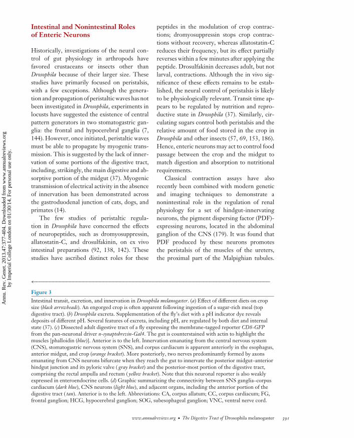
The adult digestive tract receives innervationfrom three sources (Figure 3c,d): (a) the stom-atogastric nervous system (SNS) (39, 78, 168);(b) the corpus cardiacum, a neurosecretorystructure that, in adult flies, is fused with oneof the stomatogastric ganglia (the hypocerebralganglion) (99); and (c) neurons located in thecentral nervous system (CNS), which extendtheir axons toward three sections of the diges-tive tract (37, 115, 116, 179).
Unlike the mammalian enteric nervous sys-tem, which populates the entire length of the
gastrointestinal tract, the fly’s digestive tract isonly innervated in three distinct regions: theesophagus-crop-anterior midgut, the midgut-hindgut junction, and the posterior hindgut(37) (Figure 3c). Muscle valves are present inall three regions, consistent with the idea thatperistaltic regulation is one of the main func-tions of these intestinal neurons. Although mostneuronal fibers terminate on the visceral mus-cles, some reach the underlying epithelium (37).Epithelial innervation is particularly prominentin the esophagus, pyloric valve, and rectal am-pulla (all of which are ectodermally derivedepithelia). This is reminiscent of the organi-zation of the mammalian submucosal plexusand, as such, is suggestive of neuronal regu-lation of epithelial properties, such as secre-tion and absorption (see below for one suchexample).
Not all innervation is efferent. Dendritesemanating from peripheral sensory neurons areapparent in the anterior- and posterior-mostregions of the digestive tract (37), and theyappear most abundant in the esophagus andanterior midgut (P. Cognigni & I. Miguel-Aliaga, unpublished data). Very few neuronsmay contribute to this sensory innervationgiven that few peripheral cell bodies are foundin close proximity to the digestive tract (P.Cognigni & I. Miguel-Aliaga, unpublisheddata), and many of them are likely to be effer-ent cell bodies residing in the stomatogastricganglia (Figure 3d ).
Although there have been few neu-roanatomical studies of the larval or adultdigestive tract, they have nonetheless shownenteric innervation to be chemically and neu-roanatomically diverse. Indeed, neuronal fiberspositive for serotonin and several neuropeptideshave been described in its anterior portion,including dromyosuppressin, adipokinetichormone, and, possibly, allatostatin-C andFMRFamide (or an FMRFamide-like peptide,such as sNPF) (24, 99, 110, 128). Fibers positivefor pigment-dispersing hormone, ion transportpeptide, and proctolin have also been observedin both the larval and/or adult hindgut (5, 46,47, 115, 125). It is noteworthy that all three
www.annualreviews.org • The Digestive Tract of Drosophila melanogaster 389
Ann
u. R
ev. G
enet
. 201
3.47
:377
-404
. Dow
nloa
ded
from
ww
w.a
nnua
lrev
iew
s.or
gby
Im
peri
al C
olle
ge L
ondo
n on
01/
30/1
4. F
or p
erso
nal u
se o
nly.

GE47CH17-Lemaitre ARI 29 October 2013 14:38
Pars intercerebralis:a neurosecretorycenter of the insectbrain, located alongthe anterior midline
innervated regions receive insulinergic in-nervation, whether emanating from the parsintercerebralis insulin-producing cells (theaxons of which extend beyond the ring glandtoward the anterior midgut and crop in adultflies) or the insulin-like peptide 7 (Ilp7)-producing neurons of the abdominal ganglion(which innervate the midgut-hindgut junction
and rectal ampulla) (27, 116). Intriguingly, thetwo populations of insulin-producing neuronsappear to make direct synaptic contacts in theCNS (37), suggesting that they may functionas an interconnected circuit to regulate thedelivery of insulin to different portions ofthe digestive tract (see below for functionalanalyses).
ba
c
nsyb>CD8-GFPPhalloidin
Esophagus, anteriormidgut, and crop Midgut-hindgut junction
Rectal ampullaand rectum
d
Brain
Aorta
Midgut
CA
FG
SOG
VNC
Crop stalk
EsophagusHCG/
CC
390 Lemaitre · Miguel-Aliaga
Ann
u. R
ev. G
enet
. 201
3.47
:377
-404
. Dow
nloa
ded
from
ww
w.a
nnua
lrev
iew
s.or
gby
Im
peri
al C
olle
ge L
ondo
n on
01/
30/1
4. F
or p
erso
nal u
se o
nly.
GE47CH17-Lemaitre ARI 29 October 2013 14:38
Intestinal and Nonintestinal Rolesof Enteric Neurons
Historically, investigations of the neural con-trol of gut physiology in arthropods havefavored crustaceans or insects other thanDrosophila because of their larger size. Thesestudies have primarily focused on peristalsis,with a few exceptions. Although the genera-tion and propagation of peristaltic waves has notbeen investigated in Drosophila, experiments inlocusts have suggested the existence of centralpattern generators in two stomatogastric gan-glia: the frontal and hypocerebral ganglia (7,144). However, once initiated, peristaltic wavesmust be able to propagate by myogenic trans-mission. This is suggested by the lack of inner-vation of some portions of the digestive tract,including, strikingly, the main digestive and ab-sorptive portion of the midgut (37). Myogenictransmission of electrical activity in the absenceof innervation has been demonstrated acrossthe gastroduodenal junction of cats, dogs, andprimates (14).
The few studies of peristaltic regula-tion in Drosophila have concerned the effectsof neuropeptides, such as dromyosuppressin,allatostatin-C, and drosulfakinin, on ex vivointestinal preparations (92, 138, 142). Thesestudies have ascribed distinct roles for these
peptides in the modulation of crop contrac-tions; dromyosuppressin stops crop contrac-tions without recovery, whereas allatostatin-Creduces their frequency, but its effect partiallyreverses within a few minutes after applying thepeptide. Drosulfakinin decreases adult, but notlarval, contractions. Although the in vivo sig-nificance of these effects remains to be estab-lished, the neural control of peristalsis is likelyto be physiologically relevant. Transit time ap-pears to be regulated by nutrition and repro-ductive state in Drosophila (37). Similarly, cir-culating sugars control both peristalsis and therelative amount of food stored in the crop inDrosophila and other insects (57, 69, 153, 186).Hence, enteric neurons may act to control foodpassage between the crop and the midgut tomatch digestion and absorption to nutritionalrequirements.
Classical contraction assays have alsorecently been combined with modern geneticand imaging techniques to demonstrate anonintestinal role in the regulation of renalphysiology for a set of hindgut-innervatingneurons, the pigment dispersing factor (PDF)-expressing neurons, located in the abdominalganglion of the CNS (179). It was found thatPDF produced by these neurons promotesthe peristalsis of the muscles of the ureters,the proximal part of the Malpighian tubules.
←−−−−−−−−−−−−−−−−−−−−−−−−−−−−−−−−−−−−−−−−−−−−−−−−−−−−−−−−−−−−−−−−−−−−−−Figure 3Intestinal transit, excretion, and innervation in Drosophila melanogaster. (a) Effect of different diets on cropsize (black arrowheads). An engorged crop is often apparent following ingestion of a sugar-rich meal (topdigestive tract). (b) Drosophila excreta. Supplementation of the fly’s diet with a pH indicator dye revealsdeposits of different pH. Several features of excreta, including pH, are regulated by both diet and internalstate (37). (c) Dissected adult digestive tract of a fly expressing the membrane-tagged reporter CD8-GFPfrom the pan-neuronal driver n-synaptobrevin-Gal4. The gut is counterstained with actin to highlight themuscles [phalloidin (blue)]. Anterior is to the left. Innervation emanating from the central nervous system(CNS), stomatogastric nervous system (SNS), and corpus cardiacum is apparent anteriorly in the esophagus,anterior midgut, and crop (orange bracket). More posteriorly, two nerves predominantly formed by axonsemanating from CNS neurons bifurcate when they reach the gut to innervate the posterior midgut–anteriorhindgut junction and its pyloric valve ( gray bracket) and the posterior-most portion of the digestive tract,comprising the rectal ampulla and rectum ( yellow bracket). Note that this neuronal reporter is also weaklyexpressed in enteroendocrine cells. (d) Graphic summarizing the connectivity between SNS ganglia–corpuscardiacum (dark blue), CNS neurons (light blue), and adjacent organs, including the anterior portion of thedigestive tract ( tan). Anterior is to the left. Abbreviations: CA, corpus allatum; CC, corpus cardiacum; FG,frontal ganglion; HCG, hypocerebral ganglion; SOG, subesophageal ganglion; VNC, ventral nerve cord.
www.annualreviews.org • The Digestive Tract of Drosophila melanogaster 391
Ann
u. R
ev. G
enet
. 201
3.47
:377
-404
. Dow
nloa
ded
from
ww
w.a
nnua
lrev
iew
s.or
gby
Im
peri
al C
olle
ge L
ondo
n on
01/
30/1
4. F
or p
erso
nal u
se o
nly.

GE47CH17-Lemaitre ARI 29 October 2013 14:38
Hence, some enteric neurons may use the di-gestive tract as a docking site to exert their func-tions on other internal organs at some distance.
More integrative in vivo approaches are re-vealing neuronal regulation of features otherthan muscle contractions. Indeed, a newmethod based on the semiautomated analysisof defecation behavior (Figure 3b), provid-ing quantitative readouts for food intake, fluid-ion balance, and intestinal transit, has uncov-ered the first enteric neurons to regulate fluidbalance in invertebrates. The HGN1 neuronscomprise a group of two to five CNS neuronslocated in the posterior segments of the ab-dominal ganglion, the axons of which reachthe hindgut and project through the visceralmuscles to reach the underlying epithelium(37). Neuronal silencing experiments have re-vealed that HGN1 neurons, consistent withtheir epithelial innervation, are required forthe postmating changes in intestinal fluid re-tention described above. Interestingly, GRASP[GFP (green fluorescent protein) reconstitu-tion across synaptic partners] analysis (71) hassuggested direct synaptic contact between theaxonal terminals of the sex peptide–receivingsensory neurons in the reproductive tract(known to relay mating status to affect post-mating behavior) with HGN1 dendrites in theabdominal ganglion (P. Cognigni & I. Miguel-Aliaga, unpublished data). Hence, this findingprovides the first example of a simple circuit thatcouples the reproductive and digestive systems,bypassing the brain to effect visceral changes inresponse to the reproductive state.
Endocrine Regulation of IntestinalFunctions by Systemic Signals andEnteroendocrine Cell Hormones
In addition to being subject to direct neuralcontrol, the Drosophila gut may also be regulatedby extrinsic hormonal signals (emanating fromendocrine glands or neuroendocrine organs)and by its own peptides, which are produced bythe EECs. The former kind of regulation is sug-gested by the intestinal expression of receptorsfor neurotransmitters or peptides not produced
by gut-innervating neurons or EECs (188). Onesuch example is the leucokinin receptor, whichis expressed by both the Malpighian tubules andthe digestive tract, and binds to leucokinin: adiuretic peptide secreted systemically from theCNS-derived nerves that terminate at the ab-dominal wall (3, 37, 132, 143). Downregulationof either this peptide or its receptor leads to ab-normal excreta and extreme fluid retention that,on occasion, ruptures the abdominal wall, withboth visceral muscles and the stellate cells ofthe tubule contributing to this phenotype (37;P. Cognigni & I. Miguel-Aliaga, unpublishedresults). The finding of such a severe pheno-type for a CNS peptide on visceral physiologyhighlights the importance of this systemic modeof regulation.
EECs are abundant and diverse in themidgut (see Table 1 for a list of EEC peptides)(134, 145, 188). Given that this is the portionof the digestive tract least profusely innervated(37), midgut EECs may carry out some neural-like functions in regulating intestinal physiol-ogy and/or conveying the intestinal/nutritionalstate to other internal organs. In this regard, it isperhaps not surprising that the developmentalprogram of EECs shares similarities with thatof neurons, probably reflecting a common phy-logenetic origin of these cell types (77, 175).Consistent with this idea, all the peptides pro-duced by EECs are also present in the CNS,hence their brain-gut peptide denomination.
Currently, very little is known about thefunctions and modes of action of the EECs andtheir peptides. In mammals, secreted EEC pep-tides can enter the bloodstream to affect tis-sues at a considerable distance, ranging fromother portions of the digestive tract to the braincenters that regulate appetite (91). It is unclearwhether any of the Drosophila EEC peptides arereleased into the circulation. EEC peptides canalso act more locally, and such a local role wasrecently suggested by the finding that a subset ofDH31-producing EECs regulates midgut peri-stalsis in Drosophila larvae, pointing to the vis-ceral muscle as one site of EEC peptide action(98). Although tantalizing, these findings alsoillustrate the difficulties of ascribing functions
392 Lemaitre · Miguel-Aliaga
Ann
u. R
ev. G
enet
. 201
3.47
:377
-404
. Dow
nloa
ded
from
ww
w.a
nnua
lrev
iew
s.or
gby
Im
peri
al C
olle
ge L
ondo
n on
01/
30/1
4. F
or p
erso
nal u
se o
nly.

GE47CH17-Lemaitre ARI 29 October 2013 14:38
Table 1 Peptide hormones present in the adult midguta
Gene LC-MS Microarray In situ/immunohistochemistryAkh (CG1171) − ? axons (ant) (99)Ast (CG13633) + + EECs (post)Ast-C (CG14919) + + EECs (ant, mid, post)burs (CG13419) − + N/ICCHa1 (CG14358) + + N/ICCHa2 (CG14375) + + N/IDh31 (CG13094) + + EECs (post)Ilp3 (CG14167) − + muscle subset (19, 188)itp (CG13586) − + axons (post) (47)Mip (CG6456) + + EECs (post)?npf (CG10342) − + EECs (ant, mid) (18)Pdf (CG6496) + − axons (post) (80, 125, 179)Ptth (CG13687) − + N/IsNPF (CG13968) + ? axons (ant)Tk (CG14734) + + EECs (ant, mid, post)
aOnly peptide hormones detected by mass spectrometry (145), microarray analyses (34), or immunohistochemistry (188) arelisted (unless otherwise indicated). Note that the source of these peptides may be the EECs or axons innervating the anterior-or posterior-most portions of the midgut. Discrepancies between mass spectrometry and RNA-protein data result from thefact that large peptides are not readily detectable by mass spectrometry. Abbreviations: ant, anterior; EECs, enteroendocrinecells; LC-MS, liquid chromatography–mass spectrometry; mid, middle; N/I, not investigated; post, posterior.
to EEC peptides; given the similarities betweenneurons and EECs, it remains difficult to silencea gene in EECs without affecting its expressionin neurons.
REACHING OUT: INTESTINALSENSORS, APPETITEREGULATION, AND ENERGYHOMEOSTASIS
The findings described above indicate thatthe fly’s digestive tract is equipped with theanatomical substrates for interorgan signaling:sensory neurons and/or EECs. Such signalingmight be used exclusively to relay informationback to the CNS with the overall goal ofmonitoring, maintaining, and temporally orga-nizing intestinal functions. However, intestinalsignaling could be involved in more integrativefunctions by, for example, coupling intestinalstate with behavioral and metabolic adaptationsto nutrient availability. Flies are known to adapttheir food intake and preference to both exter-
nal nutrient availability and their internal state(29, 44, 57, 64, 150). Given that the nutritionalrequirements of Drosophila are fairly complex[including proteins, carbohydrates, cholesterol,choline/lecithin, salts, and a subset of vitamins(158)] and that the digestive tract is the first in-ternal organ to come in contact with nutrients,this organ is particularly well placed to senseand relay nutritional signals to modulate behav-ior and metabolism. The following two sectionssummarize its possible role in these processes.
Intestinal Nutrient Sensing:Molecular Sensors
Although the sensory innervation of the diges-tive tract is sparse, it may be significant. In thecontext of nutrient sensing, the recent report offour peripheral neurons on the proventriculusthat express the gustatory receptor Gr43a is in-triguing (119). These neurons extend dendriticprocesses in the foregut lumen, and their ax-ons innervate either the midgut muscles or the
www.annualreviews.org • The Digestive Tract of Drosophila melanogaster 393
Ann
u. R
ev. G
enet
. 201
3.47
:377
-404
. Dow
nloa
ded
from
ww
w.a
nnua
lrev
iew
s.or
gby
Im
peri
al C
olle
ge L
ondo
n on
01/
30/1
4. F
or p
erso
nal u
se o
nly.
GE47CH17-Lemaitre ARI 29 October 2013 14:38
CNS at the level of the subesophageal ganglion;hence, they may both relay nutritional informa-tion back to the CNS and act locally. Moreover,the ability of the gut to sense nutrients may notbe confined to its neurons: At least 12 gustatoryreceptors have been recently reported to be ex-pressed in subsets of EECs (140), raising thepossibility that, as in mammals, EECs modu-late their secretion of hormones in response tonutrient quality/quantity. The functional rel-evance of the taste receptors in intestinal sen-sory neurons and EECs requires further geneticanalyses that can distinguish between their rolesin the intestine versus those in more traditionalexternal taste structures, such as the proboscis.
The existence of neuronal stretch receptorson the crop and anterior midgut that moni-tor the volume of ingested food is well sup-ported by both neurophysiological and anatom-ical data in several other insects (reviewed in165). These experiments have established theirfunctional importance locally, in regulating in-testinal physiology, and remotely, in the con-trol of food intake. However, the presenceand molecular nature of these receptors inDrosophila remain to be elucidated.
Relaying Nutritional Information:Intestinal Regulation of Appetiteand Nutrient Storage
The digestive tract may make use of the nu-tritional information acquired by the sensorslisted above to control its own functions. Fur-thermore, tantalizing findings suggest that thegut may also be able to relay nutritional infor-mation to the brain or other internal organs. In-deed, effects on food intake in response to nutri-ent scarcity have been observed when crop- orhindgut-innervating, insulin-producing neu-rons are inactivated (37). It is conceivable thatthese neurons use the gut merely as a dockingpoint to release insulin systemically and affectother tissues. However, more local targets ofaction are suggested by the findings that dif-ferent insulin-producing neurons have differenteffects on food intake when inactivated and thatthey innervate different portions of the diges-
tive tract (37). Similar effects on appetite havealso been reported upon mutation of genes ex-pressed in the anterior portion of the digestivetract. Indeed, mutants lacking the putative juve-nile hormone-binding protein termed Takeout,normally expressed in the proventriculus andcrop and induced in additional portions of thedigestive tract by starvation, also fail to adapttheir food intake according to food availability(112, 159). Although the expression of Takeoutin tissues other than the gut (such as taste neu-rons) may partly account for this phenotype,the role of intestinal Takeout deserves furtherinvestigation.
Further studies are needed to determinewhether the behavioral changes caused by theabove genetic manipulations result from a gut-to-brain signal or from more indirect effects onmetabolism, which might be sensed elsewhere.Experiments in other insect species, however,provide some evidence for direct gut-to-braincommunication emanating from the crop andinvolving peripheral stretch receptors, whichwould relay information about crop engorge-ment back to the CNS to limit food intakein the blowfly (9, 45, 170). The link betweencrop physiology, diet, and sensory activity ismuch more tenuous in Drosophila, but, intrigu-ingly, mutations in the dropdead gene, whichlead to neurodegeneration (20), are associatedwith increased crop size and reduced transferof ingested food from the crop to the midgut(141).
Whether relayed by neuronal or hormonalmeans, the possible neural targets of intestinalsignaling in the CNS are also unknown. In-triguingly, the brain-gut peptide NPF (the flyortholog of mammalian NPY) has been shownto promote feeding and make starved larvaemore willing to take in noxious food (195, 196).More recently, NPF has been shown to bind toits receptor in central dopaminergic neurons toregulate appetitive memory in adult flies (95).It will be of interest to determine whether thedopaminergic neurons are a target of systemicNPF emanating from the gut. Together, theabove studies point to the existence of complexinteractions between the gut and other tissues
394 Lemaitre · Miguel-Aliaga
Ann
u. R
ev. G
enet
. 201
3.47
:377
-404
. Dow
nloa
ded
from
ww
w.a
nnua
lrev
iew
s.or
gby
Im
peri
al C
olle
ge L
ondo
n on
01/
30/1
4. F
or p
erso
nal u
se o
nly.

GE47CH17-Lemaitre ARI 29 October 2013 14:38
but highlight the need to identify the signalingmechanisms involved.
CONCLUSIONS
Organs such as the fly wing or brain have provento be instrumental in uncovering fundamen-tal principles of development. The functionalcomplexity, plasticity, and genetic amenabil-ity of the Drosophila gut are allowing the studyof not only developmental but also physiolog-ical questions in this fascinating organ. How-ever, our knowledge of its functional intrica-cies is still rudimentary. The shift in focus inDrosophila research from “how to make a fly”to “how a fly works” is timely in this regard,and we anticipate interactions between the ge-netically minded Drosophila community and themore physiology-oriented entomologists to befruitful in the coming years. Many features ofdigestion and absorption appear to be con-served between flies and mammals, which is alikely legacy of the ancestral origin of the ali-mentary canal. Investigation of these processesin this simpler model system may contributeto the identification of metabolically relevant
molecules, which could become drug targets forthe treatment of metabolic disorders.
On a more speculative note, it is intriguingthat the genes and signaling pathways that theadult gut uses to maintain its homeostasis haveall been involved in developmental processes.Given that the gut was one of the first organsto arise during metazoan evolution, it isconceivable that the ancestral function of manysignaling modules traditionally regarded asdevelopmental (such as transcription factorsor morphogens) was to maintain the structuralcompartmentalization of the gut or even toorchestrate digestion, and that these signalingmodules were later co-opted into playing a roleduring embryonic development. Thus, revisit-ing the function of developmental pathways inthe context of gut immunity or stem cell main-tenance may cast a new light on such pathways.Similarly, the already apparent similarities be-tween EECs and neurons suggest that the studyof EECs may shed light on conserved principlesof neuronal development. Hence, we anticipatefields as diverse as metabolism, immunity, andneurobiology to benefit from future geneticanalyses of intestinal homeostasis.
SUMMARY POINTS
1. The compartmentalized and dynamic natures of the adult gut make it an excellent modelorgan to both revisit basic developmental processes and investigate homeostatic tissueinteractions or mechanisms of tissue maintenance.
2. The gut is a complex barrier where the internal and the external environments interactthrough metabolic, repair, and immune pathways.
3. The Drosophila genome encodes a vast repertoire of digestive enzymes and transporters.Sophisticated regulation of digestive and absorptive processes may be crucial to thesuccess of this species.
4. As with the mammalian gastrointestinal tract, the fly’s digestive tract is equipped witha diverse neuronal and hormonal repertoire. Sensory neurons and EEC hormones maymediate interorgan signaling to effect metabolic and behavioral adaptations.
FUTURE ISSUES
1. It will be important to investigate the establishment and maintenance of gut compart-mentalization during the Drosophila life cycle and upon intestinal damage.
www.annualreviews.org • The Digestive Tract of Drosophila melanogaster 395
Ann
u. R
ev. G
enet
. 201
3.47
:377
-404
. Dow
nloa
ded
from
ww
w.a
nnua
lrev
iew
s.or
gby
Im
peri
al C
olle
ge L
ondo
n on
01/
30/1
4. F
or p
erso
nal u
se o
nly.

GE47CH17-Lemaitre ARI 29 October 2013 14:38
2. Characterizing the mechanisms underlying gut senescence and determining their con-tribution to aging will be vital.
3. Another step will be to determine how digestive enzymes and transporters are regulatedto coordinate and modulate digestion.
4. Characterizing the neuronal and hormonal repertoire of the adult gut and establish-ing their homeostatic roles, both in the intestine and systemically, will be key to ourunderstanding.
DISCLOSURE STATEMENT
The authors are not aware of any affiliations, memberships, funding, or financial holdings thatmight be perceived as affecting the objectivity of this review.
ACKNOWLEDGMENTS
The authors apologize for their inability to cite many other substantial contributions, notablyon stem cell regulation, because of space limitations. We thank Wen Bin (Alfred) Chng, PaolaCognigni, Dani Osman, Veronique Dijkstra-Bulliard, and Maroun Bou Sleiman for their assistancewith the preparation of this manuscript and figures, and we are grateful to Ronald Dubreuil,Marie Meister, Steve Perlman, William Schaffner, and Christian Wegener for providing insightfulcomments.
LITERATURE CITED
1. Abraham I, Doane WW. 1978. Genetic regulation of tissue-specific expression of amylase structuralgenes in Drosophila melanogaster. Proc. Natl. Acad. Sci. USA 75:4446–50
2. Acosta Muniz C, Jaillard D, Lemaitre B, Boccard F. 2007. Erwinia carotovora Evf antagonizes theelimination of bacteria in the gut of Drosophila larvae. Cell Microbiol. 9:106–19
3. Al-Anzi B, Armand E, Nagamei P, Olszewski M, Sapin V, et al. 2010. The leucokinin pathway and itsneurons regulate meal size in Drosophila. Curr. Biol. 20:969–78
4. Amcheslavsky A, Jiang J, Ip YT. 2009. Tissue damage–induced intestinal stem cell division in Drosophila.Cell Stem Cell 4:49–61
5. Anderson MS, Halpern ME, Keshishian H. 1988. Identification of the neuropeptide transmitter proc-tolin in Drosophila larvae: characterization of muscle fiber–specific neuromuscular endings. J. Neurosci.8:242–55
6. Anton-Erxleben F, Miquel J, Philpott DE. 1983. Fine-structural changes in the midgut of old Drosophilamelanogaster. Mech. Ageing Dev. 23:265–76
7. Ayali A, Zilberstein Y, Cohen N. 2002. The locust frontal ganglion: a central pattern generator networkcontrolling foregut rhythmic motor patterns. J. Exp. Biol. 205:2825–32
8. Balamurugan K, Egli D, Hua H, Rajaram R, Seisenbacher G, et al. 2007. Copper homeostasis inDrosophila by complex interplay of import, storage and behavioral avoidance. EMBO J. 26:1035–44
9. Belzer WR. 1978. Recurrent nerve inhibition of protein feeding in the blowfly Phormia regina. Physiol.Entomol. 3:259–63
10. Benkel BF, Hickey DA. 1986. Glucose repression of amylase gene expression in Drosophila melanogaster.Genetics 114:137–44
11. Bettedi L, Aslam MF, Szular J, Mandilaras K, Missirlis F. 2011. Iron depletion in the intestines ofMalvolio mutant flies does not occur in the absence of a multicopper oxidase. J. Exp. Biol. 214:971–78
396 Lemaitre · Miguel-Aliaga
Ann
u. R
ev. G
enet
. 201
3.47
:377
-404
. Dow
nloa
ded
from
ww
w.a
nnua
lrev
iew
s.or
gby
Im
peri
al C
olle
ge L
ondo
n on
01/
30/1
4. F
or p
erso
nal u
se o
nly.

GE47CH17-Lemaitre ARI 29 October 2013 14:38
12. Biteau B, Hochmuth CE, Jasper H. 2008. JNK activity in somatic stem cells causes loss of tissuehomeostasis in the aging Drosophila gut. Cell Stem Cell 3:442–55
13. Biteau B, Karpac J, Supoyo S, Degennaro M, Lehmann R, Jasper H. 2010. Lifespan extension bypreserving proliferative homeostasis in Drosophila. PLoS Genet. 6:e1001159
14. Bortoff A, Davis RS. 1968. Myogenic transmission of antral slow waves across the gastroduodenaljunction in situ. Am. J. Physiol. 215:889–97
15. Bosco-Drayon V, Poidevin M, Boneca IG, Narbonne-Reveau K, Royet J, Charroux B. 2012. Peptido-glycan sensing by the receptor PGRP-LE in the Drosophila gut induces immune responses to infectiousbacteria and tolerance to microbiota. Cell Host Microbe 16:153–65
16. Boudko DY. 2012. Molecular basis of essential amino acid transport from studies of insect nutrientamino acid transporters of the SLC6 family (NAT-SLC6). J. Insect Physiol. 58:433–49
17. Broderick NA, Lemaitre B. 2012. Gut-associated microbes of Drosophila melanogaster. Gut Microbes3:307–21
18. Brown MR, Crim JW, Arata RC, Cai HN, Chun C, Shen P. 1999. Identification of a Drosophila brain-gutpeptide related to the neuropeptide Y family. Peptides 20:1035–42
19. Buch S, Melcher C, Bauer M, Katzenberger J, Pankratz MJ. 2008. Opposing effects of dietary proteinand sugar regulate a transcriptional target of Drosophila insulin-like peptide signaling. Cell Metab. 7:321–32
20. Buchanan RL, Benzer S. 1993. Defective glia in the Drosophila brain degeneration mutant drop-dead.Neuron 10:839–50
21. Buchon N, Broderick NA, Chakrabarti S, Lemaitre B. 2009. Invasive and indigenous microbiota impactintestinal stem cell activity through multiple pathways in Drosophila. Genes Dev. 23:2333–44
21a. Buchon N, Broderick NA, Lemaitre B. 2013. Gut homeostasis in a microbial world: insights fromDrosophila melanogaster. Nat. Rev. Microbiol. 16:615–26
22. Buchon N, Broderick NA, Poidevin M, Pradervand S, Lemaitre B. 2009. Drosophila intestinal responseto bacterial infection: activation of host defense and stem cell proliferation. Cell Host Microbe 5:200–11
23. Buchon N, Osman D, David FPA, Yu Fang H, Boquete JP, Deplancke B, Lemaitre B. 2013. Morpho-logical and molecular characterization of adult midgut compartmentalization in Drosophila. Cell Rep.3:1725–38
24. Budnik V, Wu CF, White K. 1989. Altered branching of serotonin-containing neurons in Drosophilamutants unable to synthesize serotonin and dopamine. J. Neurosci. 9:2866–77
25. Bujold M, Gopalakrishnan A, Nally E, King-Jones K. 2010. Nuclear receptor DHR96 acts as a sentinelfor low cholesterol concentrations in Drosophila melanogaster. Mol. Cell. Biol. 30:793–805
26. Burke R, Commons E, Camakaris J. 2008. Expression and localisation of the essential copper transporterDmATP7 in Drosophila neuronal and intestinal tissues. Int. J. Biochem. Cell Biol. 40:1850–60
27. Cao C, Brown MR. 2001. Localization of an insulin-like peptide in brains of two flies. Cell Tissue Res.304:317–21
28. Carlson JR, Hogness DS. 1985. The Jonah genes: a new multigene family in Drosophila melanogaster.Dev. Biol. 108:341–54
29. Carvalho GB, Kapahi P, Benzer S. 2005. Compensatory ingestion upon dietary restriction in Drosophilamelanogaster. Nat. Methods 2:813–15
30. Chandler JA, Lang JM, Bhatnagar S, Eisen JA, Kopp A. 2011. Bacterial communities of diverseDrosophila species: ecological context of a host-microbe model system. PLoS Genet. 7:e1002272
31. Charroux B, Royet J. 2009. Elimination of plasmatocytes by targeted apoptosis reveals their role inmultiple aspects of the Drosophila immune response. Proc. Natl. Acad. Sci. USA 106:9797–802
32. Chatterjee M, Ip YT. 2009. Pathogenic stimulation of intestinal stem cell response in Drosophila. J.Cell. Physiol. 220:664–71
33. Chen J, Xie C, Tian L, Hong L, Wu X, Han J. 2010. Participation of the p38 pathway in Drosophilahost defense against pathogenic bacteria and fungi. Proc. Natl. Acad. Sci. USA 107:20774–79
34. Chintapalli VR, Wang J, Dow JAT. 2007. Using FlyAtlas to identify better Drosophila melanogastermodels of human disease. Nat. Genet. 39:715–20
35. Choi Y-J, Hwang M-S, Park J-S, Bae S-K, Kim YS, Yoo M-A. 2008. Age-related upregulation ofDrosophila caudal gene via NF-κB in the adult posterior midgut. Biochim. Biophys. Acta 1780:1093–100
www.annualreviews.org • The Digestive Tract of Drosophila melanogaster 397
Ann
u. R
ev. G
enet
. 201
3.47
:377
-404
. Dow
nloa
ded
from
ww
w.a
nnua
lrev
iew
s.or
gby
Im
peri
al C
olle
ge L
ondo
n on
01/
30/1
4. F
or p
erso
nal u
se o
nly.

GE47CH17-Lemaitre ARI 29 October 2013 14:38
36. Clissold FJ, Tedder BJ, Conigrave AD, Simpson SJ. 2010. The gastrointestinal tract as a nutrient-balancing organ. Proc. R. Soc. B 277:1751–59
37. Cognigni P, Bailey AP, Miguel-Aliaga I. 2011. Enteric neurons and systemic signals couple nutritionaland reproductive status with intestinal homeostasis. Cell Metab. 13:92–104
38. Colombani J, Raisin S, Pantalacci S, Radimerski T, Montagne J, Leopold P. 2003. A nutrient sensormechanism controls Drosophila growth. Cell 114:739–49
39. Copenhaver PF. 2007. How to innervate a simple gut: familiar themes and unique aspects in theformation of the insect enteric nervous system. Dev. Dyn. 236:1841–64
40. Cordero JB, Stefanatos RK, Scopelliti A, Vidal M, Sansom OJ. 2012. Inducible progenitor-derivedWingless regulates adult midgut regeneration in Drosophila. EMBO J. 31:3901–17
41. Da Lage JL, Maczkowiak F, Cariou ML. 2000. Molecular characterization and evolution of the amylasemultigene family of Drosophila ananassae. J. Mol. Evol. 51:391–403
42. Davis MM, Engstrom Y. 2012. Immune response in the barrier epithelia: lessons from the fruit flyDrosophila melanogaster. J. Innate Immun. 4:273–83
43. Demerec M, ed. 1950. Biology of Drosophila. New York: John Wiley & Sons44. Dethier VG. 1976. The Hungry Fly: A Physiological Study of Behaviour Associated with Feeding. Boston:
Harvard Univ. Press45. Dethier VG, Gelperin A. 1967. Hyperphagia in the blowfly. J. Exp. Biol. 47:191–20046. Dircksen H. 2009. Insect ion transport peptides are derived from alternatively spliced genes and dif-
ferentially expressed in the central and peripheral nervous system. J. Exp. Biol. 212:401–1247. Dircksen H, Tesfai LK, Albus C, Nassel DR. 2008. Ion transport peptide splice forms in central and
peripheral neurons throughout postembryogenesis of Drosophila melanogaster. J. Comp. Neurol. 509:23–41
48. Douglas AE. 2011. Lessons from studying insect symbioses. Cell Host Microbe 10:359–6749. Douglas AE. 2013. The alimentary canal. In The Insects: Structure and Function, ed. RF Chapman,
SJ Simpson, AE Douglas, pp. 46–80. Cambridge, UK: Cambridge Univ. Press50. Dow JA. 2003. The Drosophila phenotype gap: and how to close it. Brief Funct. Genomic Proteomic
2:121–2751. Dow JA. 1999. The multifunctional Drosophila melanogaster V-ATPase is encoded by a multigene family.
J. Bioenerg. Biomembr. 31:75–8352. Dow JA, Evans PD, Wigglesworth VB. 1987. Insect midgut function. Adv. Insect Physiol. 19:187–32853. Dubreuil RR. 2004. Copper cells and stomach acid secretion in the Drosophila midgut. Int. J. Biochem.
Cell Biol. 36:745–5254. Dubreuil RR, Das A, Base C, Mazock GH. 2010. The Drosophila anion exchanger (DAE) lacks a
detectable interaction with the spectrin cytoskeleton. J. Negat. Results Biomed. 9:555. Dubreuil RR, Frankel J, Wang P, Howrylak J, Kappil M, Grushko TA. 1998. Mutations of alpha
spectrin and labial block cuprophilic cell differentiation and acid secretion in the middle midgut ofDrosophila larvae. Dev. Biol. 194:1–11
56. Dubreuil RR, Grushko T, Baumann O. 2001. Differential effects of a labial mutation on the devel-opment, structure, and function of stomach acid–secreting cells in Drosophila melanogaster larvae andadults. Cell Tissue Res. 306:167–78
57. Edgecomb RS, Harth CE, Schneiderman AM. 1994. Regulation of feeding behavior in adult Drosophilamelanogaster varies with feeding regime and nutritional state. J. Exp. Biol. 197:215–35
58. Egli D, Yepiskoposyan H, Selvaraj A, Balamurugan K, Rajaram R, et al. 2006. A family knockout of allfour Drosophila metallothioneins reveals a central role in copper homeostasis and detoxification. Mol.Cell. Biol. 26:2286–96
59. Erkosar B, Storelli G, Defaye A, Leulier F. 2013. Host-intestinal microbiota mutualism: “learning onthe fly.” Cell Host Microbe 13:8–14
60. Escher SA, Rasmuson-Lestander A. 1999. The Drosophila glucose transporter gene: cDNA sequence,phylogenetic comparisons, analysis of functional sites and secondary structures. Hereditas 130:95–103
61. Ferrandon D. 2012. The complementary facets of epithelial host defenses in the genetic model organismDrosophila melanogaster: from resistance to resilience. Curr. Opin. Immunol. 25:59–70
398 Lemaitre · Miguel-Aliaga
Ann
u. R
ev. G
enet
. 201
3.47
:377
-404
. Dow
nloa
ded
from
ww
w.a
nnua
lrev
iew
s.or
gby
Im
peri
al C
olle
ge L
ondo
n on
01/
30/1
4. F
or p
erso
nal u
se o
nly.

GE47CH17-Lemaitre ARI 29 October 2013 14:38
62. Filippov V, Aimanova K, Gill SS. 2003. Expression of an Aedes aegypti cation-chloride cotransporterand its Drosophila homologues. Insect Mol. Biol. 12:319–31
63. Filshie BK, Poulson DF, Waterhouse DF. 1971. Ultrastructure of the copper-accumulating region ofthe Drosophila larval midgut. Tissue Cell 3:77–102
64. Fujita M, Tanimura T. 2011. Drosophila evaluates and learns the nutritional value of sugars. Curr. Biol.21:751–55
65. Gartner LP. 1973. Ultrastructural examination of ageing and radiation-induced life-span shorteningin adult Drosophila melanogaster. Int. J. Radiat. Biol. Relat. Stud. Phys. Chem. Med. 23:23–39
66. Gartner LP. 1976. Fine-structure of adult Drosophila midgut musculature. J. Submicroscop. Cytol. Pathol.8:131–36
67. Gartner LP. 1985. The fine structural morphology of the midgut of adult Drosophila: a morphometricanalysis. Tissue Cell 17:883–88
68. Geer BW. 1966. Utilization of D-amino acids for growth by Drosophila melanogaster larvae. J. Nutr.90:31–39
69. Gelperin A. 1966. Investigations of a foregut receptor essential to taste threshold regulation in theblowfly. J. Insect Physiol. 12:829–41
70. Goberdhan DC, Meredith D, Boyd CA, Wilson C. 2005. PAT-related amino acid transporters reg-ulate growth via a novel mechanism that does not require bulk transport of amino acids. Development132:2365–75
71. Gordon MD, Scott K. 2009. Motor control in a Drosophila taste circuit. Neuron 61:373–8472. Guo Z, Driver I, Ohlstein B. 2013. Injury-induced BMP signaling negatively regulates Drosophila midgut
homeostasis. J. Cell Biol. 201:945–6173. Ha E-M, Lee K-A, Park SH, Kim S-H, Nam H-J, et al. 2009. Regulation of DUOX by the galphaq-
phospholipase Cβ-Ca2+ pathway in Drosophila gut immunity. Dev. Cell 16:386–9774. Ha E-M, Oh C-T, Bae Y-S, Lee WJ. 2005. A direct role for dual oxidase in Drosophila gut immunity.
Science 310:847–5075. Ha E-M, Oh C-T, Ryu JH, Bae Y-S, Kang S-W, et al. 2005. An antioxidant system required for host
protection against gut infection in Drosophila. Dev. Cell 8:125–3276. Haj-Ahmad Y, Hickey DA. 1982. A molecular explanation of frequency-dependent selection in
Drosophila. Nature 299:350–5277. Hartenstein V, Takashima S, Adams KL. 2010. Conserved genetic pathways controlling the develop-
ment of the diffuse endocrine system in vertebrates and Drosophila. Gen. Comp. Endocrinol. 166:462–6978. Hartenstein V, Tepass U, Gruszynski-Defeo E. 1994. Embryonic development of the stomatogastric
nervous system in Drosophila. J. Comp. Neurol. 350:367–8179. Hegedus D, Erlandson M, Gillott C, Toprak U. 2009. New insights into peritrophic matrix synthesis,
architecture, and function. Annu. Rev. Entomol. 54:285–30280. Helfrichforster C, Homberg U. 1993. Pigment-dispersing hormone-immunoreactive neurons in the
nervous system of wild-type Drosophila melanogaster and of several mutants with altered circadian rhyth-micity. J. Comp. Neurol. 337:177–90
81. Herboso L, Talamillo A, Perez C, Barrio R. 2011. Expression of the scavenger receptor class B type I(SR-BI) family in Drosophila melanogaster. Int. J. Dev. Biol. 55:603–11
82. Hickey DA, Benkel BF, Abukashawa S, Haus S. 1988. DNA rearrangement causes multiple changes ingene expression at the amylase locus in Drosophila melanogaster. Biochem. Genet. 26:757–68
83. Hoppler S, Bienz M. 1994. Specification of a single cell type by a Drosophila homeotic gene. Cell76:689–702
84. Horne I, Haritos VS. 2008. Multiple tandem gene duplications in a neutral lipase gene cluster inDrosophila. Gene 411:27–37
85. Horne I, Haritos VS, Oakeshott JG. 2009. Comparative and functional genomics of lipases inholometabolous insects. Insect Biochem. Mol. Biol. 39:547–67
86. Horner MA, Pardee K, Liu S, King-Jones K, Lajoie G, et al. 2009. The Drosophila DHR96 nuclearreceptor binds cholesterol and regulates cholesterol homeostasis. Genes Dev. 23:2711–16
www.annualreviews.org • The Digestive Tract of Drosophila melanogaster 399
Ann
u. R
ev. G
enet
. 201
3.47
:377
-404
. Dow
nloa
ded
from
ww
w.a
nnua
lrev
iew
s.or
gby
Im
peri
al C
olle
ge L
ondo
n on
01/
30/1
4. F
or p
erso
nal u
se o
nly.

GE47CH17-Lemaitre ARI 29 October 2013 14:38
87. Huang X, Warren JT, Buchanan J, Gilbert LI, Scott MP. 2007. Drosophila Niemann-Pick type C-2genes control sterol homeostasis and steroid biosynthesis: a model of human neurodegenerative disease.Development 134:3733–42
88. Jia L, Betters JL, Yu L. 2011. Niemann-pick C1-like 1 (NPC1L1) protein in intestinal and hepaticcholesterol transport. Annu. Rev. Physiol. 73:239–59
89. Jiang H, Edgar BA. 2011. Intestinal stem cells in the adult Drosophila midgut. Exp. Cell Res. 317:2780–8890. Jiang H, Patel PH, Kohlmaier A, Grenley MO, McEwen DG, Edgar BA. 2009. Cytokine/Jak/Stat
signaling mediates regeneration and homeostasis in the Drosophila midgut. Cell 137:1343–5591. Johnson LR, ed. 2006. Physiology of the Gastrointestinal Tract. San Diego, CA: Elsevier92. Kaminski S, Orlowski E, Berry K, Nichols R. 2002. The effects of three Drosophila melanogaster my-
otropins on the frequency of foregut contractions differ. J. Neurogenet. 16:125–3493. Kaufmann N, Mathai JC, Hill WG, Dow JA, Zeidel ML, Brodsky JL. 2005. Developmental expression
and biophysical characterization of a Drosophila melanogaster aquaporin. Am. J. Physiol. Cell Physiol.289:C397–407
94. King D. 1988. Cellular organization and peritrophic membrane formation in the cardia (proventriculus)of Drosophila melanogaster. J. Morphol. 196:253–82
95. Krashes MJ, DasGupta S, Vreede A, White B, Armstrong JD, Waddell S. 2009. A neural circuitmechanism integrating motivational state with memory expression in Drosophila. Cell 139:416–27
96. Kuraishi T, Binggeli O, Opota O, Buchon N, Lemaitre B. 2011. Genetic evidence for a protectiverole of the peritrophic matrix against intestinal bacterial infection in Drosophila melanogaster. Proc. Natl.Acad. Sci. USA 108:15966–71
97. Kylsten P, Kimbrell DA, Daffre S, Samakovlis C, Hultmark D. 1992. The lysozyme locus in Drosophilamelanogaster: Different genes are expressed in midgut and salivary glands. Mol. Gen. Genet. 232:335–43
98. LaJeunesse DR, Johnson B, Presnell JS, Catignas KK, Zapotoczny G. 2010. Peristalsis in the junctionregion of the Drosophila larval midgut is modulated by DH31 expressing enteroendocrine cells. BMCPhysiol. 10:14
99. Lee G, Park JH. 2004. Hemolymph sugar homeostasis and starvation-induced hyperactivity affected bygenetic manipulations of the adipokinetic hormone-encoding gene in Drosophila melanogaster. Genetics167:311–23
100. Lee KA, Kim SH, Kim EK, Ha EM, You H, et al. 2013. Bacterial-derived uracil as a modulator ofmucosal immunity and gut-microbe homeostasis in Drosophila. Cell 153:797–811
101. Lehane MJ. 1997. Peritrophic matrix structure and function. Annu. Rev. Entomol. 42:525–50102. Lehane MJ, Billingsley PF. 1996. Biology of the Insect Midgut. Heidelberg, Ger.: Springer103. Lehane MJ, Blakemore D, Williams S, Moffatt MR. 1995. Regulation of digestive enzyme levels in
insects. Comp. Biochem. Physiol. B 110:285–89104. Lhocine N, Ribeiro PS, Buchon N, Wepf A, Wilson R, et al. 2008. PIMS modulates immune tolerance
by negatively regulating Drosophila innate immune signaling. Cell Host Microbe 4:147–58105. Li Z, Zhang Y, Han L, Shi L, Lin X. 2013. Trachea-derived DPP controls adult midgut homeostasis
in Drosophila. Dev. Cell 24:133–43106. Lye JC, Richards CD, Dechen K, Paterson D, de Jonge MD, et al. 2012. Systematic functional charac-
terization of putative zinc transport genes and identification of zinc toxicosis phenotypes in Drosophilamelanogaster. J. Exp. Biol. 215:3254–65
107. Mandilaras K, Pathmanathan T, Missirlis F. 2013. Iron absorption in Drosophila melanogaster. Nutrients5:1622–47
107a. Marianes A, Spradling AC. 2013. Physiological and stem cell compartmentalization within the Drosophilamidgut. eLife 2:e00886
108. Martin JF, Hersperger E, Simcox A, Shearn A. 2000. minidiscs encodes a putative amino acid transportersubunit required non-autonomously for imaginal cell proliferation. Mech. Dev. 92:155–67
109. Mathur D, Bost A, Driver I, Ohlstein B. 2010. A transient niche regulates the specification of Drosophilaintestinal stem cells. Science 327:210
110. McCormick J, Nichols R. 1993. Spatial and temporal expression identify dromyosuppressin as a brain-gut peptide in Drosophila melanogaster. J. Comp. Neurol. 338:278–88
400 Lemaitre · Miguel-Aliaga
Ann
u. R
ev. G
enet
. 201
3.47
:377
-404
. Dow
nloa
ded
from
ww
w.a
nnua
lrev
iew
s.or
gby
Im
peri
al C
olle
ge L
ondo
n on
01/
30/1
4. F
or p
erso
nal u
se o
nly.

GE47CH17-Lemaitre ARI 29 October 2013 14:38
111. McNulty M, Puljung M, Jefford G, Dubreuil RR. 2001. Evidence that a copper-metallothionein com-plex is responsible for fluorescence in acid-secreting cells of the Drosophila stomach. Cell Tissue Res.304:383–89
112. Meunier N, Belgacem YH, Martin JR. 2007. Regulation of feeding behaviour and locomotor activityby takeout in Drosophila. J. Exp. Biol. 210:1424–34
113. Micchelli CA, Perrimon N. 2006. Evidence that stem cells reside in the adult Drosophila midgut epithe-lium. Nature 439:475–79
114. Miguel-Aliaga I. 2012. Nerveless and gutsy: intestinal nutrient sensing from invertebrates to humans.Semin. Cell Dev. Biol. 23:614–20
115. Miguel-Aliaga I, Thor S. 2004. Segment-specific prevention of pioneer neuron apoptosis by cell-autonomous, postmitotic Hox gene activity. Development 131:6093–105
116. Miguel-Aliaga I, Thor S, Gould AP. 2008. Postmitotic specification of Drosophila insulinergic neuronsfrom pioneer neurons. PLoS Biol. 6:e58
117. Miller MM, Popova LB, Meleshkevitch EA, Tran PV, Boudko DY. 2008. The invertebrate B(0) systemtransporter, D. melanogaster NAT1, has unique D-amino acid affinity and mediates gut and brainfunctions. Insect Biochem. Mol. Biol. 38:923–31
118. Missirlis F, Kosmidis S, Brody T, Mavrakis M, Holmberg S, et al. 2007. Homeostatic mechanisms foriron storage revealed by genetic manipulations and live imaging of Drosophila ferritin. Genetics 177:89–100
119. Miyamoto T, Slone J, Song X, Amrein H. 2012. A fructose receptor functions as a nutrient sensor inthe Drosophila brain. Cell 151:1113–25
120. Murakami R, Shigenaga A, Kawano E, Matsumoto A, Yamaoka I, Tanimura T. 1994. Novel tissueunits of regional differentiation in the gut epithelium of Drosophila, as revealed by P-element-mediateddetection of enhancer. Rouxs Arch. Dev. Biol. 203:243–49
121. Murakami R, Shiotsuki Y. 2001. Ultrastructure of the hindgut of Drosophila larvae, with special referenceto the domains identified by specific gene expression patterns. J. Morphol. 248:144–50
122. Musselman LP, Fink JL, Narzinski K, Ramachandran PV, Hathiramani SS, et al. 2011. A high-sugardiet produces obesity and insulin resistance in wild-type Drosophila. Dis. Models Mech. 4:842–49
123. Naikkhwah W, O’Donnell MJ. 2012. Phenotypic plasticity in response to dietary salt stress: Na+ andK+ transport by the gut of Drosophila melanogaster larvae. J. Exp. Biol. 215:461–70
124. Nakagoshi H. 2005. Functional specification in the Drosophila endoderm. Dev. Growth Differ. 47:383–92125. Nassel DR, Shiga S, Mohrherr CJ, Rao KR. 1993. Pigment-dispersing hormone-like peptide in the
nervous system of the flies Phormia and Drosophila: immunocytochemistry and partial characterization.J. Comp. Neurol. 331:183–98
126. Nation JL. 2002. Insect Physiology and Biochemistry. Boca Raton, FL: CRC Press. 485 pp.127. Neyen C, Poidevin M, Roussel A, Lemaitre B. 2012. Tissue- and ligand-specific sensing of gram-
negative infection in Drosophila by PGRP-LC isoforms and PGRP-LE. J. Immunol. 189:1886–97128. Nichols R, Bendena WG, Tobe SS. 2002. Myotropic peptides in Drosophila melanogaster and the genes
that encode them. J. Neurogenet. 16:1–28129. Niwa R, Niwa YS. 2011. The fruit fly Drosophila melanogaster as a model system to study cholesterol
metabolism and homeostasis. Cholesterol 2011:176802130. Norgate M, Southon A, Zou S, Zhan M, Sun Y, et al. 2007. Copper homeostasis gene discovery in
Drosophila melanogaster. Biometals 20:683–97131. O’Brien LE, Soliman SS, Li X, Bilder D. 2011. Altered modes of stem cell division drive adaptive
intestinal growth. Cell 147:603–14132. O’Donnell MJ, Dow JA, Huesmann GR, Tublitz NJ, Maddrell SH. 1996. Separate control of anion
and cation transport in Malpighian tubules of Drosophila melanogaster. J. Exp. Biol. 199:1163–75133. Ohlstein B, Spradling A. 2007. Multipotent Drosophila intestinal stem cells specify daughter cell fates
by differential notch signaling. Science 315:988–92134. Ohlstein B, Spradling AC. 2006. The adult Drosophila posterior midgut is maintained by pluripotent
stem cells. Nature 439:470–74135. Okumura T, Tajiri R, Kojima T, Saigo K, Murakami R. 2007. GATAe-dependent and -independent
expressions of genes in the differentiated endodermal midgut of Drosophila. Gene Expr. Patterns 7:178–86
www.annualreviews.org • The Digestive Tract of Drosophila melanogaster 401
Ann
u. R
ev. G
enet
. 201
3.47
:377
-404
. Dow
nloa
ded
from
ww
w.a
nnua
lrev
iew
s.or
gby
Im
peri
al C
olle
ge L
ondo
n on
01/
30/1
4. F
or p
erso
nal u
se o
nly.

GE47CH17-Lemaitre ARI 29 October 2013 14:38
136. Osman D, Buchon N, Chakrabarti S, Huang YT, Su WC, et al. 2012. Autocrine and paracrine unpairedsignaling regulate intestinal stem cell maintenance and division. J. Cell Sci. 125(Pt. 4):5944–49
137. Palm W, Sampaio JL, Brankatschk M, Carvalho M, Mahmoud A, et al. 2012. Lipoproteins in Drosophilamelanogaster: assembly, function, and influence on tissue lipid composition. PLoS Genet. 8:e1002828
138. Palmer GC, Tran T, Duttlinger A, Nichols R. 2007. The drosulfakinin 0 (DSK 0) peptide encoded in theconserved Dsk gene affects adult Drosophila melanogaster crop contractions. J. Insect Physiol. 53:1125–33
139. Paredes JC, Welchman DP, Poidevin M, Lemaitre B. 2011. Negative regulation by amidase PGRPsshapes the Drosophila antibacterial response and protects the fly from innocuous infection. Immunity35:770–79
140. Park JH, Kwon JY. 2011. Heterogeneous expression of Drosophila gustatory receptors in enteroen-docrine cells. PLoS ONE 6:e29022
141. Peller CR, Bacon EM, Bucheger JA, Blumenthal EM. 2009. Defective gut function in drop-dead mutantDrosophila. J. Insect Physiol. 55:834–39
142. Price MD, Merte J, Nichols R, Koladich PM, Tobe SS, Bendena WG. 2002. Drosophila melanogasterflatline encodes a myotropin orthologue to Manduca sexta allatostatin. Peptides 23:787–94
143. Radford JC, Davies SA, Dow JA. 2002. Systematic G-protein-coupled receptor analysis in Drosophilamelanogaster identifies a leucokinin receptor with novel roles. J. Biol. Chem. 277:38810–17
144. Rand D, Ayali A. 2010. Neuroanatomy and neurophysiology of the locust hypocerebral ganglion.J. Insect Physiol. 56:884–92
145. Reiher W, Shirras C, Kahnt J, Baumeister S, Isaac RE, Wegener C. 2011. Peptidomics and peptidehormone processing in the Drosophila midgut. J. Proteome Res. 10:1881–92
146. Ren C, Webster P, Finkel SE, Tower J. 2007. Increased internal and external bacterial load duringDrosophila aging without life-span trade-off. Cell Metab. 6:144–52
147. Rera M, Bahadorani S, Cho J, Koehler CL, Ulgherait M, et al. 2011. Modulation of longevity and tissuehomeostasis by the Drosophila PGC-1 homolog. Cell Metab. 14:623–34
148. Rera M, Clark RI, Walker DW. 2012. Intestinal barrier dysfunction links metabolic and inflammatorymarkers of aging to death in Drosophila. Proc. Natl. Acad. Sci. USA 109:21528–33
149. Reynolds B, Roversi P, Laynes R, Kazi S, Boyd CA, Goberdhan DC. 2009. Drosophila expresses a CD98transporter with an evolutionarily conserved structure and amino acid–transport properties. Biochem.J. 420:363–72
150. Ribeiro C, Dickson BJ. 2010. Sex peptide receptor and neuronal TORS6K signaling modulate nutrientbalancing in Drosophila. Curr. Biol. 20:1000–5
151. Ridley EV, Wong AC, Westmiller S, Douglas AE. 2012. Impact of the resident microbiota on thenutritional phenotype of Drosophila melanogaster. PLoS ONE 7:e36765
152. Rizki TM. 1956. The secretory activity of the proventriculus of Drosophila melanogaster. J. Exp. Zool.131:203–21
153. Roces F, Blatt J. 1999. Haemolymph sugars and the control of the proventriculus in the honey bee Apismellifera. J. Insect Physiol. 45:221–29
154. Roman G, Meller V, Wu KH, Davis RL. 1998. The opt1 gene of Drosophila melanogaster encodes aproton-dependent dipeptide transporter. Am. J. Physiol. 275:C857–69
155. Ross J, Jiang H, Kanost MR, Wang Y. 2003. Serine proteases and their homologs in the Drosophilamelanogaster genome: an initial analysis of sequence conservation and phylogenetic relationships. Gene304:117–31
156. Ryu JH, Kim SH, Lee HY, Bai JY, Nam YD, et al. 2008. Innate immune homeostasis by the homeoboxgene caudal and commensal-gut mutualism in Drosophila. Science 319:777–82
157. Ryu JH, Nam K-B, Oh C-T, Nam H-J, Kim S-H, et al. 2004. The homeobox gene Caudal regu-lates constitutive local expression of antimicrobial peptide genes in Drosophila epithelia. Mol. Cell. Biol.24:172–85
158. Sang JH. 1956. The quantitative nutritional requirements of Drosophila melanogaster. J. Exp. Biol. 33:45–72
159. Sarov-Blat L, So WV, Liu L, Rosbash M. 2000. The Drosophila takeout gene is a novel molecular linkbetween circadian rhythms and feeding behavior. Cell 101:647–56
402 Lemaitre · Miguel-Aliaga
Ann
u. R
ev. G
enet
. 201
3.47
:377
-404
. Dow
nloa
ded
from
ww
w.a
nnua
lrev
iew
s.or
gby
Im
peri
al C
olle
ge L
ondo
n on
01/
30/1
4. F
or p
erso
nal u
se o
nly.

GE47CH17-Lemaitre ARI 29 October 2013 14:38
160. Selvaraj A, Balamurugan K, Yepiskoposyan H, Zhou H, Egli D, et al. 2005. Metal-responsive transcrip-tion factor (MTF-1) handles both extremes, copper load and copper starvation, by activating differentgenes. Genes Dev. 19:891–6
161. Shanbhag S, Tripathi S. 2005. Electrogenic H+ transport and pH gradients generated by a V-H+-ATPase in the isolated perfused larval Drosophila midgut. J. Membr. Biol. 206:61–72
162. Shanbhag S, Tripathi S. 2009. Epithelial ultrastructure and cellular mechanisms of acid and base trans-port in the Drosophila midgut. J. Exp. Biol. 212:1731–44
163. Shin SC, Kim SH, You H, Kim B, Kim AC, et al. 2011. Drosophila microbiome modulates host devel-opmental and metabolic homeostasis via insulin signaling. Science 334:670–4
164. Sieber MH, Thummel CS. 2012. Coordination of triacylglycerol and cholesterol homeostasis byDHR96 and the Drosophila LipA homolog magro. Cell Metab. 15:122–7
165. Simpson SJ, Douglas AE 2013. Nutrition. In The Insects: Structure and Function, ed. RF Chapman,SJ Simpson, AE Douglas, pp. 81–106 Cambridge, UK: Cambridge Univ. Press
166. Sims HI, Chirn GW, Marr MT 2nd. 2012. Single nucleotide in the MTF-1 binding site can determinemetal-specific transcription activation. Proc. Natl. Acad. Sci. USA 109:16516–21
167. Singh SR, Zeng X, Zheng Z, Hou SX. 2011. The adult Drosophila gastric and stomach organs aremaintained by a multipotent stem cell pool at the foregut/midgut junction in the cardia (proventriculus).Cell Cycle 10:1109–20
168. Spiess R, Schoofs A, Heinzel HG. 2008. Anatomy of the stomatogastric nervous system associated withthe foregut in Drosophila melanogaster and Calliphora vicina third instar larvae. J. Morphol. 269:272–82
169. Stainier DY. 2005. No organ left behind: tales of gut development and evolution. Science 307:1902–4170. Stoffolano JG Jr, Haselton AT. 2013. The adult dipteran crop: a unique and overlooked organ. Annu.
Rev. Entomol. 58:205–25171. Storelli G, Defaye A, Erkosar B, Hols P, Royet J, Leulier F. 2011. Lactobacillus plantarum promotes
Drosophila systemic growth by modulating hormonal signals through TOR-dependent nutrient sensing.Cell Metab. 14:403–14
172. Strand M, Micchelli CA. 2011. Quiescent gastric stem cells maintain the adult Drosophila stomach. Proc.Natl. Acad. Sci. USA 108:17696–701
173. Sun Q, Tian E, Turner RJ, Ten Hagen KG. 2010. Developmental and functional studies of the SLC12gene family members from Drosophila melanogaster. Am. J. Physiol. Cell Physiol. 298:C26–37
174. Syed ZA, Hard T, Uv A, van Dijk-Hard IF. 2008. A potential role for Drosophila mucins in developmentand physiology. PLoS ONE 3:e3041
175. Takashima S, Adams KL, Ortiz PA, Ying CT, Moridzadeh R, et al. 2011. Development of the Drosophilaentero-endocrine lineage and its specification by the Notch signaling pathway. Dev. Biol. 353:161–72
176. Takashima S, Hartenstein V. 2012. Genetic control of intestinal stem cell specification and develop-ment: a comparative view. Stem Cell Rev. 8:597–608
177. Takashima S, Mkrtchyan M, Younossi-Hartenstein A, Merriam JR, Hartenstein V. 2008. The behaviourof Drosophila adult hindgut stem cells is controlled by Wnt and Hh signalling. Nature 454:651–55
178. Takashima S, Younossi-Hartenstein A, Ortiz PA, Hartenstein V. 2011. A novel tissue in an establishedmodel system: the Drosophila pupal midgut. Dev. Genes Evol. 221:69–81
179. Talsma AD, Christov CP, Terriente-Felix A, Linneweber GA, Perea D, et al. 2012. Remote controlof renal physiology by the intestinal neuropeptide pigment-dispersing factor in Drosophila. Proc. Natl.Acad. Sci. USA 109:12177–82
180. Tamaki FK, Padilha MH, Pimentel AC, Ribeiro AF, Terra WR. 2012. Properties and secretory mech-anism of Musca domestica digestive chymotrypsin and its relation with Drosophila melanogaster homologs.Insect Biochem. Mol. Biol. 42:482–90
181. Tang X, Zhou B. 2013. Ferritin is the key to dietary iron absorption and tissue iron detoxification inDrosophila melanogaster. FASEB J. 27:288–98
182. Terra WR. 1990. Evolution of digestive systems of insects. Annu. Rev. Entomol. 35:181–200183. Terra WR. 2001. The origin and functions of the insect peritrophic membrane and peritrophic gel.
Arch. Insect Biochem. Physiol. 47:47–61
www.annualreviews.org • The Digestive Tract of Drosophila melanogaster 403
Ann
u. R
ev. G
enet
. 201
3.47
:377
-404
. Dow
nloa
ded
from
ww
w.a
nnua
lrev
iew
s.or
gby
Im
peri
al C
olle
ge L
ondo
n on
01/
30/1
4. F
or p
erso
nal u
se o
nly.

GE47CH17-Lemaitre ARI 29 October 2013 14:38
184. Terra WR, Ferreira AC, de Bianchi AG. 1979. Distribution of digestive enzymes among the endo-and ectoperitrophic spaces and midgut cells of Rhynchosciara and its physiological significance. J. InsectPhysiol. 25:487–94
185. Thompson DB, Treat-Clemons LG, Doane WW. 1992. Tissue-specific and dietary control of alpha-amylase gene expression in the adult midgut of Drosophila melanogaster. J. Exp. Zool. 262:122–34
186. Treherne JE. 1957. Glucose absorption in the cockroach. J. Exp. Biol. 34:478–85187. Tzou P, Ohresser S, Ferrandon D, Capovilla M, Reichhart JM, et al. 2000. Tissue-specific inducible
expression of antimicrobial peptide genes in Drosophila surface epithelia. Immunity 13:737–48188. Veenstra JA, Agricola HJ, Sellami A. 2008. Regulatory peptides in fruit fly midgut. Cell Tissue Res.
334:499–516189. Voght SP, Fluegel ML, Andrews LA, Pallanck LJ. 2007. Drosophila NPC1b promotes an early step in
sterol absorption from the midgut epithelium. Cell Metab. 5:195–205190. Wang X, Wu Y, Zhou B. 2009. Dietary zinc absorption is mediated by ZnT1 in Drosophila melanogaster.
FASEB J. 23:2650–61191. Wong AC, Chaston JM, Douglas AE. 2013. The inconstant gut microbiota of Drosophila species revealed
by 16S rRNA gene analysis. ISME J. 7:1922–32192. Wong CN, Ng P, Douglas AE. 2011. Low-diversity bacterial community in the gut of the fruitfly
Drosophila melanogaster. Environ. Microbiol. 13:1889–900193. Wong R, Piper MD, Blanc E, Partridge L. 2008. Pitfalls of measuring feeding rate in the fruit fly
Drosophila melanogaster. Nat. Methods 5:214–15; author reply 5194. Woodring J, Diersch S, Lwalaba D, Hoffmann KH, Meyering-Vos M. 2009. Control of the release of
digestive enzymes in the caeca of the cricket Gryllus bimaculatus. Physiol. Entomol. 34:144–51195. Wu Q, Wen T, Lee G, Park JH, Cai HN, Shen P. 2003. Developmental control of foraging and social
behavior by the Drosophila neuropeptide Y-like system. Neuron 39:147–61196. Wu Q, Zhao Z, Shen P. 2005. Regulation of aversion to noxious food by Drosophila neuropeptide Y-
and insulin-like systems. Nat. Neurosci. 8:1350–55197. Yepiskoposyan H, Egli D, Fergestad T, Selvaraj A, Treiber C, et al. 2006. Transcriptome response to
heavy metal stress in Drosophila reveals a new zinc transporter that confers resistance to zinc. NucleicAcids Res. 34:4866–77
198. Zaidman-Remy A, Herve M, Poidevin M, Pili-Floury S, Kim MS, et al. 2006. The Drosophila amidasePGRP-LB modulates the immune response to bacterial infection. Immunity 24:463–73
199. Zaidman-Remy A, Regan JC, Brandao AS, Jacinto A. 2012. The Drosophila larva as a tool to studygut-associated macrophages: PI3K regulates a discrete hemocyte population at the proventriculus. Dev.Comp. Immunol. 36:638–47
200. Zhang B, Egli D, Georgiev O, Schaffner W. 2001. The Drosophila homolog of mammalian zinc fingerfactor MTF-1 activates transcription in response to heavy metals. Mol. Cell. Biol. 21:4505–14
201. Zhang Z, Inomata N, Yamazaki T, Kishino H. 2003. Evolutionary history and mode of the amylasemultigene family in Drosophila. J. Mol. Evol. 57:702–9
202. Zhou H, Cadigan KM, Thiele DJ. 2003. A copper-regulated transporter required for copper acquisition,pigmentation, and specific stages of development in Drosophila melanogaster. J. Biol. Chem. 278:48210–18
203. Zinke I, Schutz CS, Katzenberger JD, Bauer M, Pankratz MJ. 2002. Nutrient control of gene expressionin Drosophila: microarray analysis of starvation and sugar-dependent response. EMBO J. 21:6162–73
404 Lemaitre · Miguel-Aliaga
Ann
u. R
ev. G
enet
. 201
3.47
:377
-404
. Dow
nloa
ded
from
ww
w.a
nnua
lrev
iew
s.or
gby
Im
peri
al C
olle
ge L
ondo
n on
01/
30/1
4. F
or p
erso
nal u
se o
nly.

GE47-FrontMatter ARI 2 November 2013 9:9
Annual Review ofGenetics
Volume 47, 2013
Contents
Causes of Genome InstabilityAndres Aguilera and Tatiana Garcıa-Muse � � � � � � � � � � � � � � � � � � � � � � � � � � � � � � � � � � � � � � � � � � � � � � � � � 1
Radiation Effects on Human HeredityNori Nakamura, Akihiko Suyama, Asao Noda, and Yoshiaki Kodama � � � � � � � � � � � � � � � � � � �33
Dissecting Social Cell Biology and Tumors Using Drosophila GeneticsJose Carlos Pastor-Pareja and Tian Xu � � � � � � � � � � � � � � � � � � � � � � � � � � � � � � � � � � � � � � � � � � � � � � � � � � � � �51
Estimation and Partition of Heritability in Human Populations UsingWhole-Genome Analysis MethodsAnna A.E. Vinkhuyzen, Naomi R. Wray, Jian Yang, Michael E. Goddard,
and Peter M. Visscher � � � � � � � � � � � � � � � � � � � � � � � � � � � � � � � � � � � � � � � � � � � � � � � � � � � � � � � � � � � � � � � � � � � � � �75
Detecting Natural Selection in Genomic DataJoseph J. Vitti, Sharon R. Grossman, and Pardis C. Sabeti � � � � � � � � � � � � � � � � � � � � � � � � � � � � � � � �97
Adaptive Translation as a Mechanism of Stress Responseand AdaptationTao Pan � � � � � � � � � � � � � � � � � � � � � � � � � � � � � � � � � � � � � � � � � � � � � � � � � � � � � � � � � � � � � � � � � � � � � � � � � � � � � � � � � � � � � 121
Organizing Principles of Mammalian Nonsense-MediatedmRNA DecayMaximilian Wei-Lin Popp and Lynne E. Maquat � � � � � � � � � � � � � � � � � � � � � � � � � � � � � � � � � � � � � � � 139
Control of Nuclear Activities by Substrate-Selectiveand Protein-Group SUMOylationStefan Jentsch and Ivan Psakhye � � � � � � � � � � � � � � � � � � � � � � � � � � � � � � � � � � � � � � � � � � � � � � � � � � � � � � � � � � � 167
Genomic Imprinting: Insights From PlantsMary Gehring � � � � � � � � � � � � � � � � � � � � � � � � � � � � � � � � � � � � � � � � � � � � � � � � � � � � � � � � � � � � � � � � � � � � � � � � � � � � � � 187
Regulation of Bacterial Metabolism by Small RNAsUsing Diverse MechanismsMaksym Bobrovskyy and Carin K. Vanderpool � � � � � � � � � � � � � � � � � � � � � � � � � � � � � � � � � � � � � � � � � � � 209
Bacteria and the Aging and Longevity of Caenorhabditis elegansDennis H. Kim � � � � � � � � � � � � � � � � � � � � � � � � � � � � � � � � � � � � � � � � � � � � � � � � � � � � � � � � � � � � � � � � � � � � � � � � � � � � � � 233
The Genotypic View of Social Interactions in Microbial CommunitiesSara Mitri and Kevin Richard Foster � � � � � � � � � � � � � � � � � � � � � � � � � � � � � � � � � � � � � � � � � � � � � � � � � � � � � 247
SIR Proteins and the Assembly of Silent Chromatin in Budding YeastStephanie Kueng, Mariano Oppikofer, and Susan M. Gasser � � � � � � � � � � � � � � � � � � � � � � � � � � � � 275
v
Ann
u. R
ev. G
enet
. 201
3.47
:377
-404
. Dow
nloa
ded
from
ww
w.a
nnua
lrev
iew
s.or
gby
Im
peri
al C
olle
ge L
ondo
n on
01/
30/1
4. F
or p
erso
nal u
se o
nly.

GE47-FrontMatter ARI 2 November 2013 9:9
New Gene Evolution: Little Did We KnowManyuan Long, Nicholas W. VanKuren, Sidi Chen, Maria D. Vibranovski � � � � � � � � � � � 307
RNA Editing in Plants and Its EvolutionMizuki Takenaka, Anja Zehrmann, Daniil Verbitskiy, Barbara Hartel,
and Axel Brennicke � � � � � � � � � � � � � � � � � � � � � � � � � � � � � � � � � � � � � � � � � � � � � � � � � � � � � � � � � � � � � � � � � � � � � � � � 335
Expanding Horizons: Ciliary Proteins Reach Beyond CiliaShiaulou Yuan and Zhaoxia Sun � � � � � � � � � � � � � � � � � � � � � � � � � � � � � � � � � � � � � � � � � � � � � � � � � � � � � � � � � � � 353
The Digestive Tract of Drosophila melanogasterBruno Lemaitre and Irene Miguel-Aliaga � � � � � � � � � � � � � � � � � � � � � � � � � � � � � � � � � � � � � � � � � � � � � � � � � 377
RNase III: Genetics and Function; Structure and MechanismDonald L. Court, Jianhua Gan, Yu-He Liang, Gary X. Shaw, Joseph E. Tropea,
Nina Costantino, David S. Waugh, and Xinhua Ji � � � � � � � � � � � � � � � � � � � � � � � � � � � � � � � � � � � � 405
Modernizing the Nonhomologous End-Joining Repertoire:Alternative and Classical NHEJ Share the StageLudovic Deriano and David B. Roth � � � � � � � � � � � � � � � � � � � � � � � � � � � � � � � � � � � � � � � � � � � � � � � � � � � � � � � 433
Enterococcal Sex Pheromones: Signaling, Social Behavior,and EvolutionGary M. Dunny � � � � � � � � � � � � � � � � � � � � � � � � � � � � � � � � � � � � � � � � � � � � � � � � � � � � � � � � � � � � � � � � � � � � � � � � � � � � 457
Control of Transcriptional ElongationHojoong Kwak and John T. Lis � � � � � � � � � � � � � � � � � � � � � � � � � � � � � � � � � � � � � � � � � � � � � � � � � � � � � � � � � � � � � 483
The Genomic and Cellular Foundations of Animal OriginsDaniel J. Richter and Nicole King � � � � � � � � � � � � � � � � � � � � � � � � � � � � � � � � � � � � � � � � � � � � � � � � � � � � � � � � � 509
Genetic Techniques for the ArchaeaJoel A. Farkas, Jonathan W. Picking, and Thomas J. Santangelo � � � � � � � � � � � � � � � � � � � � � � � 539
Initation of Meiotic Recombination: How and Where? Conservationand Specificities Among EukaryotesBernard de Massy � � � � � � � � � � � � � � � � � � � � � � � � � � � � � � � � � � � � � � � � � � � � � � � � � � � � � � � � � � � � � � � � � � � � � � � � � � � � 563
Biology and Genetics of Prions Causing NeurodegenerationStanley B. Prusiner � � � � � � � � � � � � � � � � � � � � � � � � � � � � � � � � � � � � � � � � � � � � � � � � � � � � � � � � � � � � � � � � � � � � � � � � � 601
Bacterial Mg2+ Homeostasis, Transport, and VirulenceEduardo A. Groisman, Kerry Hollands, Michelle A. Kriner, Eun-Jin Lee,
Sun-Yang Park, and Mauricio H. Pontes � � � � � � � � � � � � � � � � � � � � � � � � � � � � � � � � � � � � � � � � � � � � � � � 625
Errata
An online log of corrections to Annual Review of Genetics articles may be found athttp://genet.annualreviews.org/errata.shtml
vi Contents
Ann
u. R
ev. G
enet
. 201
3.47
:377
-404
. Dow
nloa
ded
from
ww
w.a
nnua
lrev
iew
s.or
gby
Im
peri
al C
olle
ge L
ondo
n on
01/
30/1
4. F
or p
erso
nal u
se o
nly.

ANNUAL REVIEWSIt’s about time. Your time. It’s time well spent.
ANNUAL REVIEWS | Connect With Our ExpertsTel: 800.523.8635 (US/CAN) | Tel: 650.493.4400 | Fax: 650.424.0910 | Email: [email protected]
New From Annual Reviews:
Annual Review of Statistics and Its ApplicationVolume 1 • Online January 2014 • http://statistics.annualreviews.org
Editor: Stephen E. Fienberg, Carnegie Mellon UniversityAssociate Editors: Nancy Reid, University of Toronto
Stephen M. Stigler, University of ChicagoThe Annual Review of Statistics and Its Application aims to inform statisticians and quantitative methodologists, as well as all scientists and users of statistics about major methodological advances and the computational tools that allow for their implementation. It will include developments in the fi eld of statistics, including theoretical statistical underpinnings of new methodology, as well as developments in specifi c application domains such as biostatistics and bioinformatics, economics, machine learning, psychology, sociology, and aspects of the physical sciences.
Complimentary online access to the fi rst volume will be available until January 2015. TABLE OF CONTENTS:• What Is Statistics? Stephen E. Fienberg• A Systematic Statistical Approach to Evaluating Evidence
from Observational Studies, David Madigan, Paul E. Stang, Jesse A. Berlin, Martijn Schuemie, J. Marc Overhage, Marc A. Suchard, Bill Dumouchel, Abraham G. Hartzema, Patrick B. Ryan
• The Role of Statistics in the Discovery of a Higgs Boson, David A. van Dyk
• Brain Imaging Analysis, F. DuBois Bowman• Statistics and Climate, Peter Guttorp• Climate Simulators and Climate Projections,
Jonathan Rougier, Michael Goldstein• Probabilistic Forecasting, Tilmann Gneiting,
Matthias Katzfuss• Bayesian Computational Tools, Christian P. Robert• Bayesian Computation Via Markov Chain Monte Carlo,
Radu V. Craiu, Jeff rey S. Rosenthal• Build, Compute, Critique, Repeat: Data Analysis with Latent
Variable Models, David M. Blei• Structured Regularizers for High-Dimensional Problems:
Statistical and Computational Issues, Martin J. Wainwright
• High-Dimensional Statistics with a View Toward Applications in Biology, Peter Bühlmann, Markus Kalisch, Lukas Meier
• Next-Generation Statistical Genetics: Modeling, Penalization, and Optimization in High-Dimensional Data, Kenneth Lange, Jeanette C. Papp, Janet S. Sinsheimer, Eric M. Sobel
• Breaking Bad: Two Decades of Life-Course Data Analysis in Criminology, Developmental Psychology, and Beyond, Elena A. Erosheva, Ross L. Matsueda, Donatello Telesca
• Event History Analysis, Niels Keiding• Statistical Evaluation of Forensic DNA Profi le Evidence,
Christopher D. Steele, David J. Balding• Using League Table Rankings in Public Policy Formation:
Statistical Issues, Harvey Goldstein• Statistical Ecology, Ruth King• Estimating the Number of Species in Microbial Diversity
Studies, John Bunge, Amy Willis, Fiona Walsh• Dynamic Treatment Regimes, Bibhas Chakraborty,
Susan A. Murphy• Statistics and Related Topics in Single-Molecule Biophysics,
Hong Qian, S.C. Kou• Statistics and Quantitative Risk Management for Banking
and Insurance, Paul Embrechts, Marius Hofert
Access this and all other Annual Reviews journals via your institution at www.annualreviews.org.
Ann
u. R
ev. G
enet
. 201
3.47
:377
-404
. Dow
nloa
ded
from
ww
w.a
nnua
lrev
iew
s.or
gby
Im
peri
al C
olle
ge L
ondo
n on
01/
30/1
4. F
or p
erso
nal u
se o
nly.