the diverged trypanosome micos complex as a hub for...
TRANSCRIPT
Article
The Diverged Trypanosom
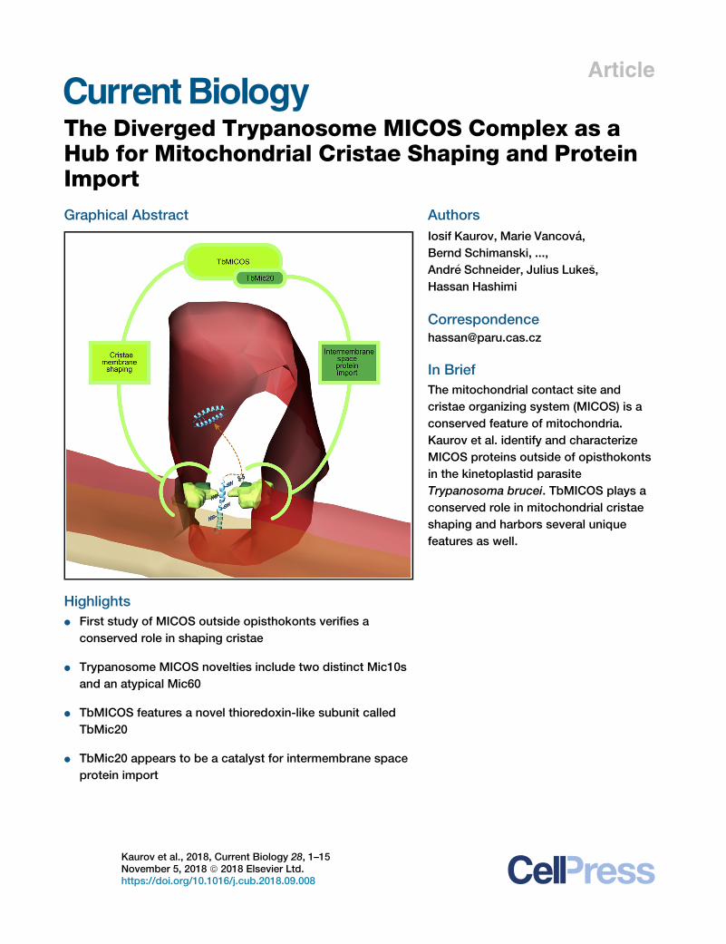
e MICOS Complex as aHub for Mitochondrial Cristae Shaping and ProteinImportGraphical Abstract
Highlights
d First study of MICOS outside opisthokonts verifies a
conserved role in shaping cristae
d Trypanosome MICOS novelties include two distinct Mic10s
and an atypical Mic60
d TbMICOS features a novel thioredoxin-like subunit called
TbMic20
d TbMic20 appears to be a catalyst for intermembrane space
protein import
Kaurov et al., 2018, Current Biology 28, 1–15November 5, 2018 ª 2018 Elsevier Ltd.https://doi.org/10.1016/j.cub.2018.09.008
Authors
Iosif Kaurov, Marie Vancova,
Bernd Schimanski, ...,
Andr�e Schneider, Julius Luke�s,
Hassan Hashimi
In Brief
The mitochondrial contact site and
cristae organizing system (MICOS) is a
conserved feature of mitochondria.
Kaurov et al. identify and characterize
MICOS proteins outside of opisthokonts
in the kinetoplastid parasite
Trypanosoma brucei. TbMICOS plays a
conserved role in mitochondrial cristae
shaping and harbors several unique
features as well.
Please cite this article in press as: Kaurov et al., The Diverged Trypanosome MICOS Complex as a Hub for Mitochondrial Cristae Shaping and ProteinImport, Current Biology (2018), https://doi.org/10.1016/j.cub.2018.09.008
Current Biology
Article
The Diverged Trypanosome MICOS Complexas a Hub for Mitochondrial Cristae Shapingand Protein ImportIosif Kaurov,1,2 Marie Vancova,1,2 Bernd Schimanski,3 Lawrence Rudy Cadena,2 Ji�rı Heller,1 Toma�s Bıly,1,2 David Pot�e�sil,4
Claudia Eichenberger,3 Hannah Bruce,2,7 Silke Oeljeklaus,5 Bettina Warscheid,5,6 Zbyn�ek Zdrahal,4 Andr�e Schneider,3
Julius Luke�s,1,2 and Hassan Hashimi1,2,8,*1Institute of Parasitology, Biology Center, Czech Academy of Sciences, 37005 �Cesk�e Bud�ejovice, Czech Republic2Faculty of Science, University of South Bohemia, 37005 �Cesk�e Bud�ejovice, Czech Republic3Department of Chemistry and Biochemistry, University of Bern, 3012 Bern, Switzerland4Central European Institute of Technology, Masaryk University, 62500 Brno, Czech Republic5Faculty of Biology, Biochemistry and Functional Proteomics, Institute of Biology II, University of Freiburg, 79104 Freiburg, Germany6BIOSS Centre for Biological Signaling Studies, University of Freiburg, 79104 Freiburg, Germany7Present address: Genome Research, Ltd., Wellcome Sanger Institute, Wellcome Genome Campus, Hinxton, Cambridge, UK8Lead Contact
*Correspondence: [email protected]://doi.org/10.1016/j.cub.2018.09.008
SUMMARY
The mitochondrial contact site and cristae organiza-tion system (MICOS) is a multiprotein complexresponsible for cristae formation. Even thoughcristae are found in all mitochondria capable ofoxidative phosphorylation, only Mic10 and Mic60appear to be conserved throughout eukaryotes.The remaining 4 or 5 knownMICOS subunits are spe-cific to the supergroup Opisthokonta, which includesyeast and mammals that are the only organisms inwhich this complex has been analyzed experimen-tally. We have isolated the MICOS from Trypano-soma brucei, a member of the supergroup Excavatathat is profoundly diverged from opisthokonts. Weshow that it is required for the maintenance of theunique discoidal cristae that typify excavates, suchas euglenids and kinetoplastids, the latter of whichinclude trypanosomes. The trypanosome MICOSconsists of 9 subunits, most of which are essentialfor normal growth. Unlike in opisthokonts, it containstwo distinct Mic10 orthologs and an unconventionalputative Mic60 that lacks amitofilin domain. Interest-ingly, one of the essential trypanosomatid-specificMICOS subunits called TbMic20 is a thioredoxin-like protein that appears to be involved in import ofintermembrane space proteins, including respiratorychain complex assembly factors. This result points totrypanosome MICOS coordinating cristae shapingand population of its membrane with proteinsinvolved in respiration, the latter via the catalytic ac-tivity of TbMic20. Thus, trypanosome MICOS allowsus to define which of its features are conserved inall eukaryotes and decipher those that represent line-age-specific adaptations.
C
INTRODUCTION
Cristae are mitochondrial inner membrane (MIM) invaginations
that are the organelle’s morphological hallmark. They are distrib-
uted throughout eukaryotes, inherited from the last eukaryotic
common ancestor (LECA) [1, 2]. Cristae assume different forms,
with lamellar ones found in the best-studied supergroup Opis-
thokonta, which encompasses yeast and animals. Tubular-
shaped cristae typify the unicellular supergroup SAR, containing
stramenopiles, alveolates, and rhizarians. Euglenid members of
the protistan supergroup Excavata generally have discoidal
cristae, which exhibit a paddle-like morphology. Thus, cristae
have evolved different morphologies that may represent adap-
tions to the multifarious cellular milieus of diverse eukaryotes.
Cristae occurrence correlates with an aerobic lifestyle medi-
ated by mitochondria. This is because cristae membranes are
enriched with respiratory chain complexes that perform oxida-
tive phosphorylation (OXPHOS) [3–5]. Such a configuration
boosts OXPHOS capacity by increasing the surface area for
this process and concentrating the soluble electron carrier cyto-
chrome c in the lumen enclosed by the cristae membrane [6, 7].
Pioneering studies in yeast identified a key multiprotein com-
plex for cristae biogenesis called the mitochondrial contact site
and cristae organizing system (MICOS) [8–10]. In this and other
opisthokonts, the MICOS complex has been shown to maintain
cristae junctions (CJs), narrow necks attaching cristae to the
MIM [11–13]. The evolutionarily conserved core subunits Mic10
and Mic60 catalyze formation of negative curvature at CJs
[14–16]. MICOS also forms contacts with mitochondrial outer
membrane (MOM) proteins via Mic60’s conserved C-terminal
mitofilin domain. This domain is an intermembrane space (IMS)
extension that interacts with the b-barrel protein sorting and as-
sembly machinery subunit Sam50 and the protein translocase of
the outer membrane (TOM) [17–19]. Furthermore, the mitofilin
domain has been reported to augment the mitochondrial IMS
import and assembly (MIA) pathway in yeast by interaction
with the central MIA catalyst Mia40 [10].
urrent Biology 28, 1–15, November 5, 2018 ª 2018 Elsevier Ltd. 1

Please cite this article in press as: Kaurov et al., The Diverged Trypanosome MICOS Complex as a Hub for Mitochondrial Cristae Shaping and ProteinImport, Current Biology (2018), https://doi.org/10.1016/j.cub.2018.09.008
By an oxidative folding mechanism, MIA sequesters small
cysteine-rich proteins bearing twin CX3C or CX9C motifs
[20, 21]. Mia40 catalyzes this folding through its reactive CPC
motif, forming an intermolecular disulfide bond with a thiol group
of a reduced and unfolded IMS precursor [22]. With Erv1, an IMS
sulfhydryl oxidase, two intramolecular disulfide bridges linking
each of the dual CX3,9C motifs are formed, creating a hairpin
fold that traps substrate proteins within the IMS. During this pro-
cess, Erv1 reoxidizes Mia40 for another round of translocation.
The mechanistic model of MICOS function has been proposed
based on data gained solely from opisthokonts [11–13]. However,
the majority of identified MICOS subunits are restricted to this
clade. Only the Mic10 and Mic60 core subunits have a wide dis-
tribution overlapping with the occurrence of cristae [1, 2]. This
strongly indicates that the ancestor of MICOS was already pre-
sent in LECA.Mic60may even pre-dateMICOS, supportedby ev-
idence that it originates from the proteobacterial endosymbiont
giving rise to mitochondria [1, 2]. Hitherto unidentified supernu-
merary MICOS subunits interacting with the ancient core may
contribute to the diverse cristae morphologies of eukaryotes.
Thus, true insight into how the ancient and ubiquitous MICOS
shapes cristae in eukaryotes requires mechanistic studies
outside of the single opisthokont clade. The discipline of evolu-
tionary cell biology postulates that a comparative approach to
examining a biological system in different organisms can reveal
chemical and physical constraints to their evolution [23].
Conversely, this approach can also identify a system’s more
flexible attributes, from which novelties may potentially emerge
in certain lineages. The in silico approach to address the evolu-
tionary cell biology of MICOS has reached saturation, necessi-
tating functional data from other eukaryotic groups.
To this end, we have undertaken the first study of MICOS
composition and function outside of opisthokonts. We have cho-
sen the pathogen Trypanosoma brucei as our model, not only for
its robust genetic toolkit but also because of its suitability for tack-
ling the evolutionary cell biology of MICOS. T. brucei is a kineto-
plastid belonging to the supergroup Excavata and therefore has
an extended independent evolutionary history [24]. Its singlemito-
chondrion undergoes massive remodeling during its life cycle
[25]. The procyclic stage (PS) infecting the tsetse fly midgut has
a mitochondrion with extensive discoidal cristae and OXPHOS
capacity and thus is the focus here. The long slender bloodstream
stage (BS) infecting mammalian hosts lacks both cristae and
OXPHOS. Furthermore, Mic10 was the only MICOS ortholog
found in T. brucei by bioinformatics methods alone [1, 2], hinting
at the tantalizing possibility of itsMICOSbearing novel properties.
RESULTS
Trypanosomatids Have Two Mic10 ParalogsTrypanosomatid Mic10 is made up of two well-supported clades
[1] (Figure 1A). In T. brucei, these paralogs are represented by
TbMic10-1 and TbMic10-2 (TbMic10-1/2) (Figure 1B). Mic10
duplication does not pre-date the emergence of trypanosoma-
tids, because there is only one Mic10 ortholog found in the
Euglena gracilis [26] and Bodo saltans genomes [27], represent-
ing their free-living sister groups. Although B. saltans Mic10
clearly belongs to the TbMic10-1 clade, the E. gracilis homolog
cannot be assigned to either with high confidence.
2 Current Biology 28, 1–15, November 5, 2018
As Mic10 family members, each T. brucei paralog contains
two transmembrane domains (TMDs) (Figure 1B). The C-prox-
imal TMD2 has a highly conserved GxGxGxGmotif, which medi-
ates Mic10 oligomerization required for membrane bending in
yeast [14, 16]. The motif is conserved in the TbMic10-1 clade,
whereas it is abbreviated to 3 alternating Gs in the TbMic10-2
clade (Figures 1B and S1A).
Both paralogs share some features that distinguish them
from the best-characterized Saccharomyces cerevisiae Mic10
(Figure S1A). The TMD1 glycine-rich motif essential for Mic10
oligomerization is dramatically reduced in TbMic10-1/2. Further-
more, the T. brucei paralogs lack the positively charged KRR
loop between the TMDs needed for MIM targeting of the yeast
homolog [16]. Surprisingly, the KKR loop is absent in Mic10 or-
thologs of all eukaryotes outside the opisthokonts (Figure S1A).
This fact underscores the need to study MICOS in other organ-
isms like T. brucei.
TbMic10 Paralogs Shape CristaeC-terminal V5-epitope-tagged TbMic10 paralogs show a patchy
mitochondrial localization by indirect immunofluorescence (Fig-
ure 1C). To further investigate whether TbMic10-1/2 associate
with cristae, immunogold labeling of the V5 epitopes on
Tokuyasu cryosections were viewed by transmission electron
microscopy (TEM) or electron tomography (ET). Nanoparticles
(NPs) immunodecorating TbMic10-1-V5 and TbMic10-2-V5
were mainly observed at cristae membranes (Figure 1D; Video
S1).
Next, we addressed whether TbMic10-1/2 are involved in
shaping cristae. Initial attempts to delete either TbMic10-1 or
TbMic10-2 by gene knockout did not yield any obvious pheno-
type, suggesting their functional redundancy. Thus, we gener-
ated cells in which both paralogs were ablated simultaneously
by tetracycline-inducible RNAi silencing of TbMic10-2 in a
TbMic10-1 deletion background (Figure S2). Cryosections
derived fromDTbMic10-1:TbMic10-2Y 1 and 4 days post-induc-
tion (dpi) of RNAi were examined by TEM. At 4 dpi, cristae were
significantly elongated compared to the parental cell line, some-
times as stacked semi-circles or arcs (Figure 2A). The severity of
this phenotype correlates with time of induction, as it is less pro-
nounced yet present at 1 dpi (Figure 6C).
Stacking of elongated cristae was also observed in yeast
Mic10 deletion mutants in conjunction with CJ loss [8–10].
Because the slender CJs of discoidal cristae are difficult to
observe on ultrathin cryosections, a 3D reconstruction of serial
TEM cryosections was performed on DTbMic10-1:TbMic10-2Y
4 dpi, which allows better assessment of CJ loss thanks to a
broad z axis (Figures 2B and 2C; Video S2). The aberrant cristae
were barrel shaped with finger-like projections extending from
one end. CJs were not observed along the examined
�700 nm. In conclusion, the TbMic10 paralogs function syner-
gistically in shaping of cristae and CJ biogenesis in PS
T. brucei, thus confirming its hypothetical role outside of
opisthokonts.
TbMic10 Paralogs Are Part of a Multi-protein ComplexBlue native PAGE of digitonin-solubilized mitochondrial mem-
branes resolved a >1-MDa complex containing TbMic10-1, likely
corresponding to T. brucei MICOS, with potential intermediates

Figure 1. T. brucei Has Two Mic10 Paralogs that Localize to Cristae Membranes
(A) Maximum-likelihood tree of Mic10 orthologs from Excavata, SAR, and Opisthokonta. Branches well supported by bootstrap/posterior probabilities are
indicated, and ‘‘n.s.’’ signifies no support. Scale bar, substitutions/site. Alignment used for the tree is shown in Figure S1A.
(B) Scheme of TbMic10-1 and TbMic10-2. Each transmembrane domain (TMD) is shown in pink with conserved G residues demarked by orange bars. Scale bars
indicate LC-MS/MS peptide coverage as in Figure 4B.
(C) Indirect immunofluorescence of TbMic10-1/2. k, kDNA (i.e., mitochondrial genome); n, nucleus. Scale bar, 5 mm.
(D) TbMic10-1-V5 and TbMic10-2-V5 immunogold labeling. Inset, tomography model. Colored arrow key is on right. Scale bars, 50 nm.
See also Video S1.
Please cite this article in press as: Kaurov et al., The Diverged Trypanosome MICOS Complex as a Hub for Mitochondrial Cristae Shaping and ProteinImport, Current Biology (2018), https://doi.org/10.1016/j.cub.2018.09.008
and/or subcomplexes migrating at �440 and �200 kDa (Fig-
ure 3A). To immunocapture proteins interacting with TbMic10-
1/2, we used TbMic10-1/2-V5 as affinity handles in the
T. brucei 927 strain. Solubilized hypotonically isolated mitochon-
dria (Figure 3B) were incubatedwithmouse a-V5 antibody cross-
linked to Protein G Dynabeads to immunoprecipitate (IP) the
tags. After extensive washing, eluted proteins that coIP with
TbMic10-1/2 were trypsinized and identified by liquid chroma-
tography-tandem mass spectroscopy (LC-MS/MS). A mock IP
was done with mitochondria from the parental cell line without
V5 epitope to discriminate any non-specific binding to the anti-
body-bead adduct. TbMic10-1/2-V5 IPs performed in triplicate
invariably isolated the same complement of 11 proteins (named
using the established MICOS nomenclature) [28], none of which
were detected in the mock control (Figure 3C; Data S1A and
S1B). All the identified subunits exhibited high peptide coverage
(Figure 4B).
These interactions were verified by IP of the subunit we desig-
nated TbMic60 (explained in the next section), which was recip-
rocally tagged with a C-terminal hemagglutinin (HA) epitope. LC-
MS/MS analysis revealed the same complement of TbMICOS
subunits in triplicate TbMic60 IPs (Figure 3C; Data S1C). The
Current Biology 28, 1–15, November 5, 2018 3
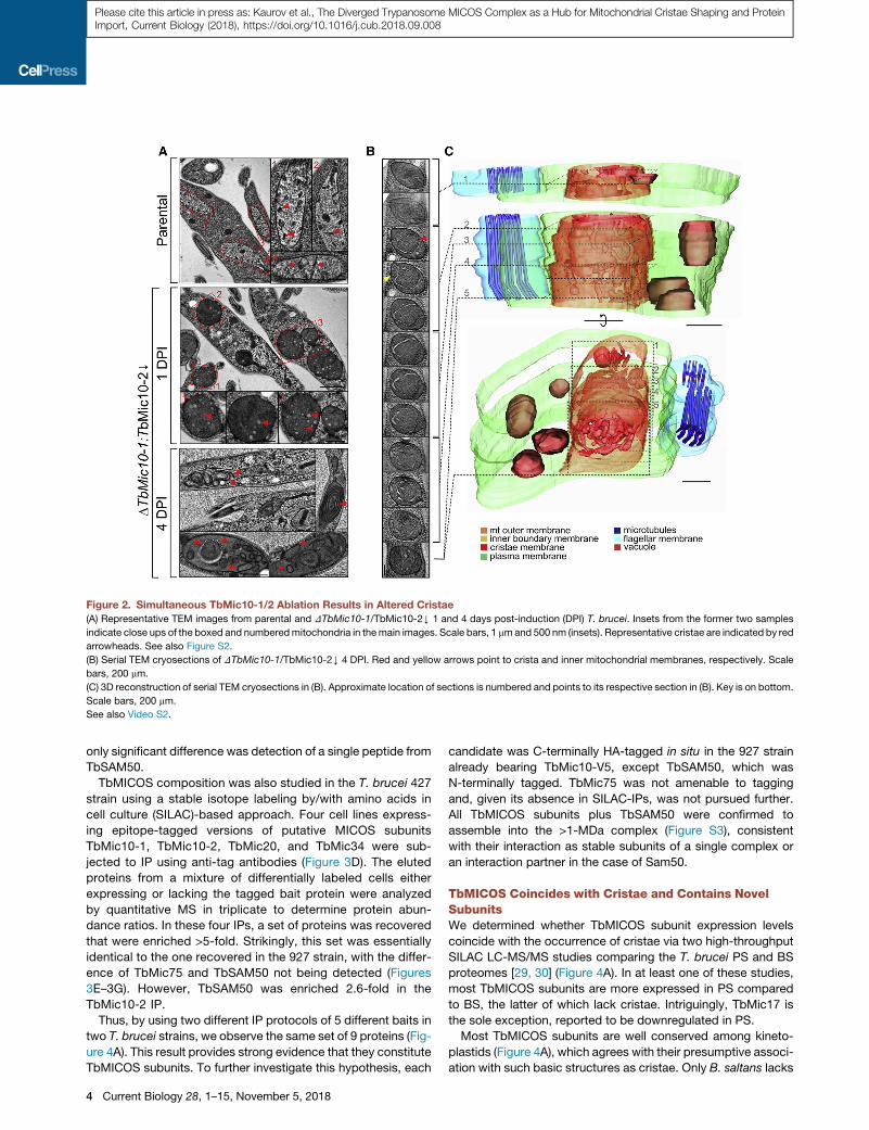
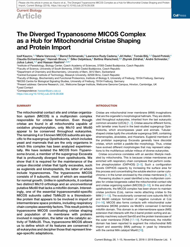
Figure 2. Simultaneous TbMic10-1/2 Ablation Results in Altered Cristae
(A) Representative TEM images from parental and DTbMic10-1/TbMic10-2Y 1 and 4 days post-induction (DPI) T. brucei. Insets from the former two samples
indicate close ups of the boxed and numberedmitochondria in themain images. Scale bars, 1 mmand 500 nm (insets). Representative cristae are indicated by red
arrowheads. See also Figure S2.
(B) Serial TEM cryosections of DTbMic10-1/TbMic10-2Y 4 DPI. Red and yellow arrows point to crista and inner mitochondrial membranes, respectively. Scale
bars, 200 mm.
(C) 3D reconstruction of serial TEM cryosections in (B). Approximate location of sections is numbered and points to its respective section in (B). Key is on bottom.
Scale bars, 200 mm.
See also Video S2.
Please cite this article in press as: Kaurov et al., The Diverged Trypanosome MICOS Complex as a Hub for Mitochondrial Cristae Shaping and ProteinImport, Current Biology (2018), https://doi.org/10.1016/j.cub.2018.09.008
only significant difference was detection of a single peptide from
TbSAM50.
TbMICOS composition was also studied in the T. brucei 427
strain using a stable isotope labeling by/with amino acids in
cell culture (SILAC)-based approach. Four cell lines express-
ing epitope-tagged versions of putative MICOS subunits
TbMic10-1, TbMic10-2, TbMic20, and TbMic34 were sub-
jected to IP using anti-tag antibodies (Figure 3D). The eluted
proteins from a mixture of differentially labeled cells either
expressing or lacking the tagged bait protein were analyzed
by quantitative MS in triplicate to determine protein abun-
dance ratios. In these four IPs, a set of proteins was recovered
that were enriched >5-fold. Strikingly, this set was essentially
identical to the one recovered in the 927 strain, with the differ-
ence of TbMic75 and TbSAM50 not being detected (Figures
3E–3G). However, TbSAM50 was enriched 2.6-fold in the
TbMic10-2 IP.
Thus, by using two different IP protocols of 5 different baits in
two T. brucei strains, we observe the same set of 9 proteins (Fig-
ure 4A). This result provides strong evidence that they constitute
TbMICOS subunits. To further investigate this hypothesis, each
4 Current Biology 28, 1–15, November 5, 2018
candidate was C-terminally HA-tagged in situ in the 927 strain
already bearing TbMic10-V5, except TbSAM50, which was
N-terminally tagged. TbMic75 was not amenable to tagging
and, given its absence in SILAC-IPs, was not pursued further.
All TbMICOS subunits plus TbSAM50 were confirmed to
assemble into the >1-MDa complex (Figure S3), consistent
with their interaction as stable subunits of a single complex or
an interaction partner in the case of Sam50.
TbMICOS Coincides with Cristae and Contains NovelSubunitsWe determined whether TbMICOS subunit expression levels
coincide with the occurrence of cristae via two high-throughput
SILAC LC-MS/MS studies comparing the T. brucei PS and BS
proteomes [29, 30] (Figure 4A). In at least one of these studies,
most TbMICOS subunits are more expressed in PS compared
to BS, the latter of which lack cristae. Intriguingly, TbMic17 is
the sole exception, reported to be downregulated in PS.
Most TbMICOS subunits are well conserved among kineto-
plastids (Figure 4A), which agrees with their presumptive associ-
ation with such basic structures as cristae. Only B. saltans lacks

Figure 3. TbMICOS Subunit Composition
(A) Blue native (BN)-PAGE resolution of TbMICOS visualized on awestern blot (WB) with anti-TbMic10-1 antibody. *, non-specific band. (Lower panel) Coomassie
staining of F1FO-ATP synthase as a loading control is shown. Native protein size markers are on left.
(B) Scheme of 927 strain IPmitochondrial isolation and solubilization. (Top) Genetic background of parental cell line (mock) and tagged cell lines (bait) is shown. P,
pellet; S, supernatant.
(C) Summary of 927 IPs. Affinity handles for TbMic10-1/2 (V5) and TbMic60 (HA) bait proteins point to co-IP proteins. Key is on right; N/D, sub-organellar
localization not determined. Mass spectroscopy data used to make scheme are in Data S1.
(D) Scheme of 427 SILAC-IP as in (B), with genetic background of overexpression (O/E) and in situ tagged cell lines depicted at the top.
(E–H) SILAC-IP of epitope-tagged TbMic10-1 (E), TbMic10-2 (F), TbMic34 (G), and TbMic20 (H). x axis, the mean Log10 enrichment ratios of replicate bait IPs
(n = 3), whose data point is marked in red; y axis, the corresponding Log10 p values. Blue quadrant highlights proteins that achieved the threshold of both values.
Key to data point shape and fold enrichment for each protein is shown at the bottom.
See also Data S2 and Figure S3.
Current Biology 28, 1–15, November 5, 2018 5
Please cite this article in press as: Kaurov et al., The Diverged Trypanosome MICOS Complex as a Hub for Mitochondrial Cristae Shaping and ProteinImport, Current Biology (2018), https://doi.org/10.1016/j.cub.2018.09.008
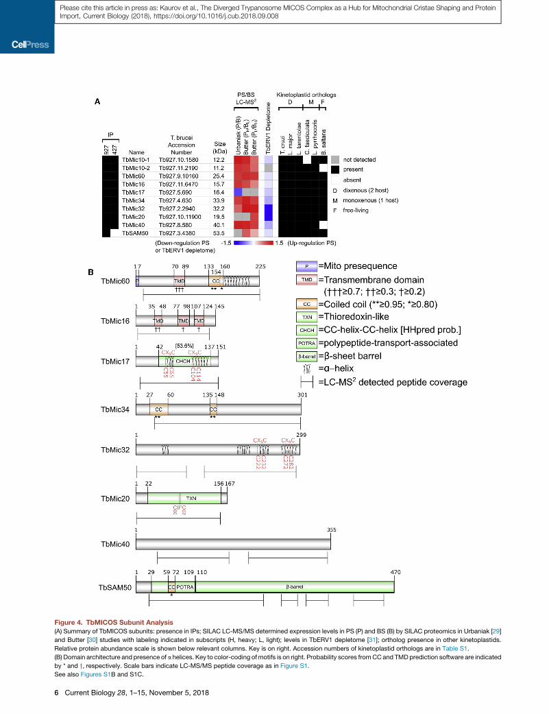
Figure 4. TbMICOS Subunit Analysis
(A) Summary of TbMICOS subunits: presence in IPs; SILAC LC-MS/MS determined expression levels in PS (P) and BS (B) by SILAC proteomics in Urbaniak [29]
and Butter [30] studies with labeling indicated in subscripts (H, heavy; L, light); levels in TbERV1 depletome [31]; ortholog presence in other kinetoplastids.
Relative protein abundance scale is shown below relevant columns. Key is on right. Accession numbers of kinetoplastid orthologs are in Table S1.
(B) Domain architecture and presence of a helices. Key to color-coding ofmotifs is on right. Probability scores fromCCand TMDprediction software are indicated
by * and y, respectively. Scale bars indicate LC-MS/MS peptide coverage as in Figure S1.
See also Figures S1B and S1C.
6 Current Biology 28, 1–15, November 5, 2018
Please cite this article in press as: Kaurov et al., The Diverged Trypanosome MICOS Complex as a Hub for Mitochondrial Cristae Shaping and ProteinImport, Current Biology (2018), https://doi.org/10.1016/j.cub.2018.09.008
Please cite this article in press as: Kaurov et al., The Diverged Trypanosome MICOS Complex as a Hub for Mitochondrial Cristae Shaping and ProteinImport, Current Biology (2018), https://doi.org/10.1016/j.cub.2018.09.008
a homolog of TbMic40. Interestingly, the monoxenous trypano-
somatid Crithidia fasciculata appears to have lost TbMic10-2.
Because these subunits are restricted to kinetoplastids, we
investigated their domain architecture in silico to gain insight
into their possible function. We designate a 25-kDa protein
TbMic60 despite it missing the C-terminal mitofilin domain,
which has been thus far this protein family’s defining character
[1]. However, TbMic60 has the same domain architecture of
the N-terminal half of other Mic60 orthologs: a predicted mito-
chondrial presequence [32] that is consistent with observed
LC-MS/MS peptide coverage (Figure S1B); a single TMD; a
coiled coil (CC) domain; an a helix interspersed with conserved
charged; and aromatic amino acids [33] (Figure S1C).
TbMic16 has 3 predicted TMDs, and TbMic34 has 2 putative
CC domains. TbMic17 and TbMic32 have a pair of CX9C motifs
within predicted a helices, an IMS protein signature. TbMic17
has a degenerated CHCH domain that is detected by HHpred
structure prediction [34], a feature also found in opisthokont
Mic19. Interestingly, TbMic20 is a thioredoxin-like protein, com-
plete with a CIPC motif that may catalyze redox reactions.
Association of TbMICOS with Cristae MembranesThe sub-organellar localization of TbMICOS was undertaken
next. To facilitate comparison of all TbMICOS subunits,
TbMic10-1/2 were also in situ HA tagged. The molecular weight
of each tagged protein, with an extra �5 kDa from the epitope,
was confirmed by western blot analysis. The tagged proteins
also served as proxies to estimate steady-state levels of
TbMICOS subunits (Figure S4A). Consistent with aforemen-
tioned high-throughput proteomics data, TbMic17-HA was the
least expressed subunit. Also, TbMic60-HA and TbMic34-HA
exhibit similar levels (Figure S4B).
Next, hypotonically isolated mitochondria from the tagged
TbMICOS cell lines were fractionated into matrix and membrane
parts, the latter being further separated into peripheral and inte-
gralMIM fractions using an establishedpipeline [35] (FigureS4C).
These fractions were probed with mitochondrial HSP70 [36] as a
marker for matrix and peripheral MIM fractions. Expectedly,
TbMic10-1-V5 and TbMic10-2-HA ended up in fractions having
the integral MIM component prohibitin [37], confirming these
are membrane proteins (Figure S4D). As each cell line contained
TbMic10-1-V5, it was subsequently used as an integral mem-
brane protein marker. TbMICOS subunit localization agreed
with in silico predictions: TbMic60 and TbMic16 are integral pro-
teins and the rest are in the MIM periphery (Figure S4D).
The sub-organellar localization of the soluble epitope-tagged
TbMICOS subunits and integral subunit topology were subse-
quently addressed. Mitoplasts derived from these cell lines
were subjected to a proteinase K protection assay (Figure S4E).
Persistence of the C-terminal epitope indicates its localization in
the matrix face of the intact MIM. The presence of matrix HSP70
and degradation of IMS TbERV1 confirmed the functionality of
the assay. TbMic10-1-V5 served as a control after its topology
had been verified (Figure S4F). In all cases, the C-terminal tag
was degraded, indicating presence of the corresponding protein
in the IMS.
Then, we asked whether the MICOS subunits are enriched
within cristae in vivo. To address this question qualitatively, im-
munogold labeling of HA epitopes was employed in all TbMICOS
cell lines, viewed by ET for integral subunits or TEM for the rest.
As seen in Figures 5A–5T and Video S1, NPs mostly decorate
cristae membranes, with some signal observed at the inner
boundary membrane. Quantitative immunogold TEM revealed
that HA-binding NPs were concentrated in mitochondria in com-
parison with the parental cell line lacking the epitope (Figure 5U).
In all cases except TbMic20, more than half of NPs label cristae
and all show significant inner boundary membrane localization
(Figure 5V). This pattern is in contrast to the parental cell line,
in whichmost NPs associate with undefinedmitochondrial struc-
tures. Interestingly, HA-binding NPs often appear in clusters
(Figures 5A–5D), suggesting a multimeric stoichiometry of the
tagged protein within TbMICOS. This phenomenon was quanti-
fied by NP pairs that were <30 nm apart, revealing that most
tagged subunits formed clusters with median�10-nm distances
(Figure 5W). Such clustering was vastly underrepresented in the
negative control parental cells. In summary, we have shown that
the TbMICOS subunits localize to cristae membranes in a way
suggesting their multimerization.
Depletion of TbMICOS Subunits Alters CristaeAfter establishing that TbMICOS localizes to cristaemembranes,
we asked whether some subunits have a role in shaping cristae,
as was shown by concurrent ablation of TbMic10-1/2. Each cell
line was transfected with a construct for inducible RNAi targeting
a specific TbMICOS subunit or TbSAM50. An HA-tagged copy of
each subunit allowed us to follow silencing of the target proteins
using a-HA antibody and a consequent effect on TbMic10-1-V5.
Over 6 days of RNAi induction, each cell line exhibited downre-
gulation of its target subunit compared to HSP70 (Figure 6A).
Afterward, we examined whether depletion of each TbMICOS
subunit compromised cell growth in a glucose-rich medium. We
observed that all RNAi knockdowns except TbMic16 resulted in
slower growth compared to mock-treated cell lines, usually 2 or
3 dpi (Figure 6B). This result was unexpected because growth
was not inhibited in either DTbMic10-1:TbMic10-2Y T. brucei
(Figure S2C) or in MICOS yeast mutants grown on fermentable
substrates [39]. Cell growth in glucose-poor medium, in which
PS proliferation relies on OXPHOS [40], was not more severely
affected (Figure S5A). Therefore, almost all TbMICOS subunits
are essential for normal growth, irrespective of the mitochondrial
bioenergetic state.
Cristae morphology was then observed in ultrathin cryosec-
tions of cells depleted of individual TbMICOS subunits at RNAi
time points just before acute growth arrest (Figure S5B).
TbMic40 RNAi stands out due to massive mitochondrial bleb-
bing. TbMic60 and TbMic20 RNAi silencing exhibited pheno-
types resembling those of DTbMic10-1:TbMic10-2Y4 dpi
(Figure 2), with elongated cristae assuming an arc-like or circular
morphology. To quantify this phenomenon, we measured
cristae lengths of selected RNAi-silenced subunits, DTbMic10-
1:TbMic10-2Y and the parental strain (Figure 6C). These
results correlated with qualitative observations, in which
TbMic60 and TbMic20 RNAi displayed elongated cristae on
par with DTbMic10-1:TbMic10-2Y. Furthermore, downregula-
tion of TbMic34 resulted in a lesser degree of elongation similar
to that of DTbMic10-1:TbMic10-2Y 1 dpi. Other morphological
parameters were also measured. DTbMic10-1:TbMic10-2Y
and TbMic60 RNAi exhibited an increase in mitochondrial area
Current Biology 28, 1–15, November 5, 2018 7

Figure 5. TbMICOS Localization in Cristae Membranes
(A–T) Representative electron tomography (A–M) and transmission electron microscopy (N–T) of subunits indicated on left (A–M, Q, and R) and right (N–P, S,
and T) of brackets. Key on upper right classifies colors of structures (membranes and nanoparticles [NPs]) or arrows pointing to structures. C and G show electron
tomography tilt series. Scale bars, 200 mm. See also Video S1.
(U) Labeling density of NPs in mitochondria (black bars) or background (gray bars). Statistical analyses of the labeling density bymeasuring relative labeling index
[38] are given in Table S2, which contains total surface areas of mitochondria and background (i.e., rest of cell) examined in all analyses in (U)–(W).
(V) Localization of NPs within mitochondrial subcompartments. Red, cristae; orange, inner boundary membrane; blue, undefined.
(W) NP cluster analysis. Each dot represents measured distance (y axis) between 2 NPs. Median and interquartile range of these distances are in red.
See also Figure S4.
Please cite this article in press as: Kaurov et al., The Diverged Trypanosome MICOS Complex as a Hub for Mitochondrial Cristae Shaping and ProteinImport, Current Biology (2018), https://doi.org/10.1016/j.cub.2018.09.008
(Figure 6D), correlating with the appearance of elongated
cristae. There is a statistically significant increase in cristae per
mitochondria in DTbMic10-1:TbMic10-2Y, and TbMic32 and
TbMic20 displayed less of them, as compared to the parental
control (Figure 6C). We have thus established that a subset of
TbMICOS subunits, including TbMic60, shapes cristae.
Because the effect of TbMICOS ablation on T. brucei growth
does not seem to correlate with altered cristae morphology, an
effect on proteins of different mitochondrial compartments was
8 Current Biology 28, 1–15, November 5, 2018
examined in the RNAi cells (Figure 6A). Downregulation of all
TbMICOS subunits except TbMic17 exhibited a subsequent
depletion of TbMic10-1-V5. The voltage-dependent ion channel
(VDAC) [41] was only affected upon TbSAM50 ablation. FOF1-
ATP synthase b subunit was not decreased in any of the cell
lines. However, we observed an unexpected significant drop in
the abundance of the IMS protein TbERV1 [42] following RNAi
against TbMic16, TbMic32, and TbMic20. To summarize,
TbMic10-1 stability seems to depend on the presence of the
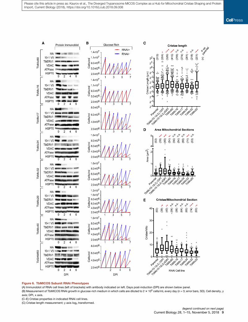
Figure 6. TbMICOS Subunit RNAi Phenotypes
(A) Immunoblot of RNAi cell lines (left of brackets) with antibody indicated on left. Days post-induction (DPI) are shown below panel.
(B) Measurement of TbMICOS RNAi growth in glucose-rich medium in which cells are diluted to 23 106 cells/mL every day (n = 3; error bars, SD). Cell density, y
axis; DPI, x axis.
(C–E) Cristae properties in indicated RNAi cell lines.
(C) Cristae length measurement; y axis log2 transformed.
(legend continued on next page)
Current Biology 28, 1–15, November 5, 2018 9
Please cite this article in press as: Kaurov et al., The Diverged Trypanosome MICOS Complex as a Hub for Mitochondrial Cristae Shaping and ProteinImport, Current Biology (2018), https://doi.org/10.1016/j.cub.2018.09.008

Please cite this article in press as: Kaurov et al., The Diverged Trypanosome MICOS Complex as a Hub for Mitochondrial Cristae Shaping and ProteinImport, Current Biology (2018), https://doi.org/10.1016/j.cub.2018.09.008
other TbMICOS subunits plus TbSAM50. Moreover, TbERV1 de-
creases upon depletion of some TbMICOS subunits, an
intriguing phenomenon we further explored.
Thioredoxin-like TbMic20 Affects the Import of IMSProteinsThe downregulation of the IMS protein TbERV1 upon RNAi
silencing of 3 TbMICOS subunits suggested that the complex
may participate in theMIA pathway. TbERV1 is a key component
of MIA, as evidenced by its knockdown in PS resulting in IMS
protein downregulation [31]. Indeed, 2 of the 3 proteins affecting
TbERV1 in our study, TbMic20 and TbMic32, were among those
in the TbERV1 depletome (Figure 4A). However, an enzyme cata-
lyzing IMS import in trypanosomes, in a way akin to opisthokont
Mia40, remains unidentified [43].
We turned our attention to TbMic20 as a possible functional
analog of Mia40. Along with it being affected by TbERV1 ablation,
this hypothesis is supported by it having elements as a thiore-
doxin-like protein that could facilitate oxidative folding of CX3,9C
IMS proteins. A TbMic20 homology model templated onto thiore-
doxin predicts that the CIPC reaction center occurs on a loop
adjacent to an a helix, resulting in a structural motif similar to
that of the human Mia40 CPC reaction center [22] (Figure S6A).
To address empirically whether TbMic20 is involved inMIA, we
tested whether its depletion would result in reduced IMS protein
abundance and thus phenocopy TbERV1 RNAi. Equal amounts
of mitochondria were isolated from duplicate uninduced and 4
dpi TbMic20 RNAi cells. Proteins were extracted with ionic
detergent, and their trypsin-derived peptides were labeled with
tandem mass tags for sample discrimination after mixing them
in equal parts before LC-MS/MS. In parallel, the same experi-
ment was done for TbMic60.
AR1.6-fold threshold in both biological duplicates was set for
detection of up- and downregulated proteins (Data S2A and S2B
[TbMic20] and S2C and S2D [TbMic60]). Verified and predicted
IMS proteins comprised 51%of the TbMic20 depletome (Figures
7A and S6B). Of these, over half overlap with those from the
TbERV1 depletome (Figures 7A and 7B) [31]. Strangely, a ubiq-
uinol-cytochrome c reductase that was decreased in the
TbERV1 depletome was reproducibly the most upregulated pro-
tein upon TbMic20 RNAi silencing (Figures 7C and S6B). The
other TbMic20 depletome IMS proteins belong to two cate-
gories: those identified by homology or experimentally (IMS)
and those predicted to have a twin CX3,9Cmotifs (CxC). Notably,
TbERV1 was not significantly affected by TbMic20 depletion 4
dpi, indicating that the phenotype is a direct effect (Figure 7C).
Furthermore, such an effect on IMS proteins was not observed
in the 4 dpi TbMic60 depletome analyzed in parallel. Thus, we
provide evidence that TbMic20 participates in MIA.
Many of the affected IMS proteins are annotated as respiratory
chain complex assembly factors (Figures 7CandS6B). Twoof the
most significantly downregulated proteins in the TbMic20 deple-
tomeare the respiratory complex III Rieske iron-sulfur protein and
a complex I subunit (Figures S6B and S7). Given that cristae
(D) Mitochondrial section area measurement.
(E) Cristae per mitochondrial section in RNAi cell lines.
Boxplots show median and interquartile range. Whiskers demark data points in th
cell line and parental control is shown on top with population size (n): ****p < 0.00
10 Current Biology 28, 1–15, November 5, 2018
membranes are enriched in OXPHOS complexes [3], we interro-
gated the TbMic20 depletome for a preferential effect on respira-
tory chain components overmatrix proteins, using the annotation
of Zıkova et al. [25]. Indeed, respiratory chain subunits were
generally decreased in the 4 dpi TbMic20 depletome, and no
such effect occurs in that of TbMic60 (Figure S7). Only the afore-
mentioned pair of complex I and III subunits crossed the 1.6-fold
threshold, suggesting the decline in the other OXPHOS subunits
is a secondary effect of IMS-localizedCX3,9COXPHOSassembly
factor depletion. Furthermore, the TbMic20-depletome more
severely affects OXPHOS complex subunits, with the exception
of complex II, than matrix proteins (Figure 7D). This finding sug-
gests that TbMic20 association with cristae may facilitate popu-
lation of its membrane with OXPHOS components.
We also examined how TbMic20 and TbMic60 RNAi depletion
affected the TbMICOS subunits (Figure 7C). Although not signif-
icant, TbMic34, TbMic32, and TbMic40 were most affected by
TbMic20 ablation. Among the 32 proteins downregulated by
R1.6-fold in the TbMic60 RNAi cells are the target and TbMic16.
TbMic10-1 appeared to be affected as well, confirming afore-
mentionedwestern blot data (Figure 6A).Whether this differential
effect of TbMic20 and TbMic60 depletion reflects a subcomplex
architecture, as has been reported for yeast MICOS [39, 44, 45],
remains to be determined.
DISCUSSION
Defining Conserved Features of MICOSThis study represents the first isolation and characterization of a
MICOS complex outside of opisthokonts. As a highly diverged
excavate unrelated to opisthokonts, T. brucei is well positioned
to help identify highly conserved and relaxed features of MICOS
that mediate CJ biogenesis and cristae shaping. The funda-
mental architecture of MICOS appears to be conserved, with
novel supernumerary subunits attached to a MIM embedded
core represented by Mic10 and Mic60 [11–13], albeit with
diverged qualities that will be discussed later. Furthermore,
some TbMICOS subunits appear to be multimeric, as has been
observed in opisthokonts.
The association of MICOS with ancient Sam50 is conserved.
The yeast Mic60 mitofilin domain mediates interaction with
Sam50 [17–19] to form the even larger mitochondrial IMS
bridging complex [2, 46]. Despite TbMic60 lacking the mitofilin
domain, this interaction between TbMICOS and TbSAM50 was
confirmed by its co-IP with TbMic10-1/2 and TbMic60. Further-
more, TbSAM50 assembles in a >1-MDa complex that was also
observed in TbMICOS. Finally, TbSAM50 RNAi leads to
TbMic10-1 downregulation.
In yeast, Cox17 and Aim24 have been shown to be peripheral
MICOS interaction partners [47, 48]. It is possible that some of
the TbMICOS subunits whose RNAi silencing does not yield the
scored elongated cristae phenotypemay represent such auxiliary
factors. However, a counterargument to this idea is the tight asso-
ciation of the 9 TbMICOS subunits in 7 IPs performed under 2
e 10–90 percentiles. Statistical significance of differences between each RNAi
01; ***p < 0.001; **p < 0.01; *p < 0.05; n.s., not significant. See also Figure S5.

(legend on next page)
Current Biology 28, 1–15, November 5, 2018 11
Please cite this article in press as: Kaurov et al., The Diverged Trypanosome MICOS Complex as a Hub for Mitochondrial Cristae Shaping and ProteinImport, Current Biology (2018), https://doi.org/10.1016/j.cub.2018.09.008
Please cite this article in press as: Kaurov et al., The Diverged Trypanosome MICOS Complex as a Hub for Mitochondrial Cristae Shaping and ProteinImport, Current Biology (2018), https://doi.org/10.1016/j.cub.2018.09.008
conditions. This is similar to the interconnection of yeast MICOS
subunits upon their initial biochemical purification in 3 studies,
none of which contained later identified peripheral proteins
[8–10]. Furthermore,RNAidepletionsof somehumanMICOSsub-
units also donot yield altered cristae [46], a similar result to ours. In
the case of TbMICOS, perhaps the lack of the scored phenotype
upon specific subunit depletion may be due to their functional
redundancy under the examined conditions, as has been
observed in attempts to delete each TbMic10paralog individually.
Nevertheless, we cannot completely rule out the likelihood that
some of the identified TbMICOSs are auxiliary factors, although
we believe this to be a remote possibility.
Novel Features of TbMICOSAlthough lacking the C-terminal mitofilin domain, TbMic60 re-
tains the domain architecture of theN-terminal half of the protein,
comprising of amitochondrial presequence, single TMD, andCC
domain [1, 2], that justifies its designation as a Mic60 ortholog.
The a-proteobacterial ortholog of Mic60 also contains these ele-
ments, minus the presequence. Interestingly, the b- and g-pro-
teobacterial HemX protein, whose gene is syntenic to a-proteo-
bacterial Mic60 [2], bears these elements. Yet HemX lacks a
mitofilin domain, setting a naturally occurring precedent for
its absence between distantly related but still structurally
conserved homologs. However, although bacterial Mic60 has
been shown to bend membranes in vitro [15], such an activity re-
mains to be determined for HemX. Still, the mitofilin domain is
unnecessary for membrane bending, an activity mediated
instead by the CC-domain-trailing a helix interspersed with aro-
matic and charged amino acids [33], a feature found in TbMic60.
Finally, TbMic60 ablation exhibits one of the strongest cristae
morphology phenotypes, analogous to the severity of DMic10
and DMic60 on yeast cristae [8, 10].
It is plausible that another TbMICOS subunit may assume the
missing mitofilin domain’s role, with TbMic34 being a top candi-
date. HA-tagged TbMic60 and TbMic34 have similar expression
levels, which is compatible with their stoichiometric relationship.
Furthermore, they are the only two subunits with CC domains,
which could allow them to form an�60-kDa heterodimer. Finally,
TbMic34 RNAi caused a perceptible cristae-elongation pheno-
type, albeit to a lesser extent than observed upon TbMic60
silencing.
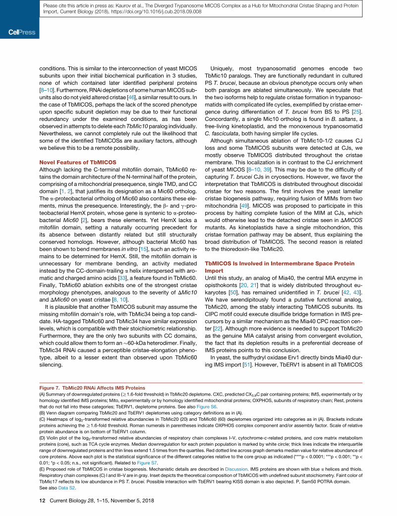
Figure 7. TbMic20 RNAi Affects IMS Proteins
(A) Summary of downregulated proteins (R1.6-fold threshold) in TbMic20 depleto
homology identified IMS proteins; Mito, experimentally or by homology identified
that do not fall into these categories; TbERV1, depletome proteins. See also Figu
(B) Venn diagram comparing TbMic20 and TbERV1 depletomes using category d
(C) Heatmaps of log2-transformed relative abundancies in TbMic20 (20) and Tb
proteins achieving the R1.6-fold threshold. Roman numerals in parentheses ind
protein abundance is on bottom of TbERV1 column.
(D) Violin plot of the log2-transformed relative abundancies of respiratory chain
proteins (core), such as TCA cycle enzymes. Median downregulation for each pr
range of downregulated proteins and thin lines extend 1.5 times from the quartiles
core proteins. Above each plot is the statistical significance of the different categ
0.01; *p < 0.05; n.s., not significant). Related to Figure S7.
(E) Proposed role of TbMICOS in cristae biogenesis. Mechanistic details are de
Respiratory chain complexes (C) I and III–V are in gray. Inset depicts the theoretica
TbMic17 reflects its low abundance in PS T. brucei. Possible interaction with Tb
See also Data S2.
12 Current Biology 28, 1–15, November 5, 2018
Uniquely, most trypanosomatid genomes encode two
TbMic10 paralogs. They are functionally redundant in cultured
PS T. brucei, because an obvious phenotype occurs only when
both paralogs are ablated simultaneously. We speculate that
the two isoforms help to regulate cristae formation in trypanoso-
matids with complicated life cycles, exemplified by cristae emer-
gence during differentiation of T. brucei from BS to PS [25].
Concordantly, a single Mic10 ortholog is found in B. saltans, a
free-living kinetoplastid, and the monoxenous trypanosomatid
C. fasciculata, both having simpler life cycles.
Although simultaneous ablation of TbMic10-1/2 causes CJ
loss and some TbMICOS subunits were detected at CJs, we
mostly observe TbMICOS distributed throughout the cristae
membrane. This localization is in contrast to the CJ enrichment
of yeast MICOS [8–10, 39]. This may be due to the difficulty of
capturing T. brucei CJs in cryosections. However, we favor the
interpretation that TbMICOS is distributed throughout discoidal
cristae for two reasons. The first involves the yeast lamellar
cristae biogenesis pathway, requiring fusion of MIMs from two
mitochondria [49]. MICOS was proposed to participate in this
process by halting complete fusion of the MIM at CJs, which
would otherwise lead to the detached cristae seen in DMICOS
mutants. As kinetoplastids have a single mitochondrion, this
cristae formation pathway may be absent, thus explaining the
broad distribution of TbMICOS. The second reason is related
to the thioredoxin-like TbMic20.
TbMICOS Is Involved in Intermembrane Space ProteinImportUntil this study, an analog of Mia40, the central MIA enzyme in
opisthokonts [20, 21] that is widely distributed throughout eu-
karyotes [50], has remained unidentified in T. brucei [42, 43].
We have serendipitously found a putative functional analog,
TbMic20, among the stably interacting TbMICOS subunits. Its
CIPC motif could execute disulfide bridge formation in IMS pre-
cursors by a similar mechanism as the Mia40 CPC reaction cen-
ter [22]. Although more evidence is needed to support TbMic20
as the genuine MIA catalyst arising from convergent evolution,
the fact that its depletion results in a preferential decrease of
IMS proteins points to this conclusion.
In yeast, the sulfhydryl oxidase Erv1 directly binds Mia40 dur-
ing IMS import [51]. However, TbERV1 is absent in all TbMICOS
me. CXC, predicted CX3,9C pair containing proteins; IMS, experimentally or by
mitochondrial proteins; OXPHOS, subunits of respiratory chain; Rest, proteins
re S6.
efinitions as in (A).
Mic60 (60) depletomes organized into categories as in (A). Brackets indicate
icate OXPHOS complex component and/or assembly factor. Scale of relative
complexes I–V, cytochrome-c-related proteins, and core matrix metabolism
otein population is marked by white circle; thick lines indicate the interquartile
. Red dotted line across graph demarks median value for relative abundance of
ories relative to the core group as indicated (****p < 0.0001; ***p < 0.001; **p <
scribed in Discussion. IMS proteins are shown with blue a helices and thiols.
l composition of TbMICOSwith undefined subunit stoichiometry. Faint color of
ERV1 bearing KISS domain is also depicted. P, Sam50 POTRA domain.
Please cite this article in press as: Kaurov et al., The Diverged Trypanosome MICOS Complex as a Hub for Mitochondrial Cristae Shaping and ProteinImport, Current Biology (2018), https://doi.org/10.1016/j.cub.2018.09.008
preparations and reciprocally TbERV1 IP did not immunocapture
TbMICOS [43]. However, TbERV1 localizes within discoidal
cristae [43], as does TbMic20. Furthermore, TbERV1 has a
‘‘kinetoplastid-specific second’’ (KISS) domain [52], which may
be an adaptation for binding another oxidoreductase besides
Mia40 and/or stabilize the TbERV1 dimer when it is not tran-
siently interacting with TbMic20.
TbMic20 RNAi not only results in a decrease of IMS proteins
but also causes cristae elongation. This observation encapsu-
lates our current model that TbMICOS serves as a hub for cristae
shaping and IMS protein import (Figure 7E). TbMICOS via
TbMic20 facilitates the oxidative folding of CX3,9C-containing
OXPHOS components, including OXPHOS assembly factors.
TbMICOS distribution throughout cristae membranes allows
the concentration of mature assembly factors in proximity to
this membrane. In turn, this mechanism aids the population of
cristae membrane with OXPHOS complex subunits that require
these IMS proteins. TbMICOS also maintains CJs, anchoring
cristae to the MOM via TbSAM50 interaction, bringing the crista
lumen closer to the ATOM protein import pores [53], through
which IMS protein precursors are translocated.
TbMICOS subunit depletion impairs T. brucei growth even un-
der conditions in which energy generation occurs independently
of OXPHOS. Thus, we propose that this is ultimately a conse-
quence of mitochondrial import disruption. Indeed, many MIA
substrates are involved in mitochondrial biogenesis, such as
the small Tim chaperones [54]. Furthermore, the ATOMcomplex,
the entry gate for nearly all proteins into the organelle, is also
essential for T. brucei grown in ample glucose [53].
The Evolutionary Cell Biology of MICOSWe have explored the composition and function of MICOS in
T. brucei to obtain much needed data about how this complex
works outside of opisthokonts. Ultimately, our goal has been to
better understand the MICOS complex by gaining insight into
the evolutionary constraints that shape it. We confirm the hy-
pothesis that the core function of MICOS in diverged eukaryotes
is the maintenance of CJs, cristae shaping, and mediating MOM
and MIM contacts. However, we also uncovered many surpris-
ing novelties that demonstrate MICOS has the potential to follow
diverse evolutionary paths to adapt to different cellular contexts.
Among them are TbMic60’s lacking the conserved mitofilin
domain and TbMic10 duplication into two distinct proteins. Yet
most surprising is the presence of a thioredoxin-like TbMICOS
subunit, which may allow MICOS to participate in IMS protein
import. This activity extends the currently accepted functional
model of MICOS, helping OXPHOS complex insertion into
cristae membranes. It remains to be seen whether these novel
TbMICOS properties are restricted to trypanosomatids or have
a wider distribution in eukaryotes, indicating that opisthokont
MICOS may actually represent the outlier.
STAR+METHODS
Detailed methods are provided in the online version of this paper
and include the following:
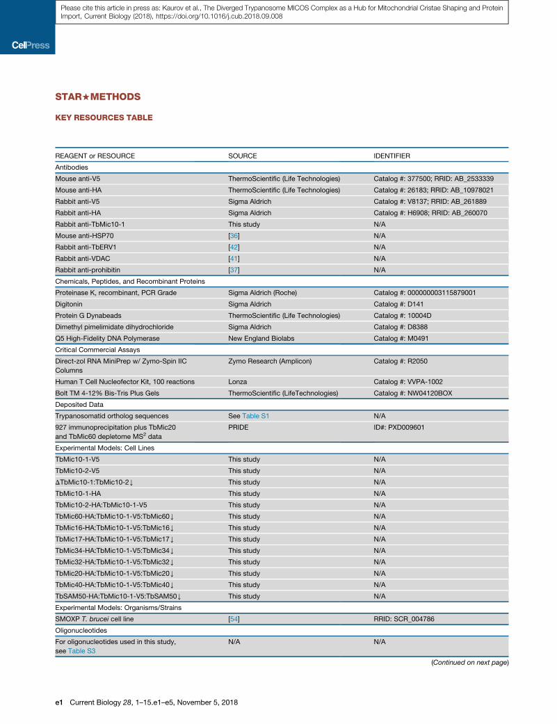
d KEY RESOURCES TABLE
d CONTACT FOR REAGENT AND RESOURCE SHARING
d EXPERIMENTAL MODEL AND SUBJECT DETAILS
d METHOD DETAILS
B Generation of T. brucei transgenic cell lines
B T. brucei growth measurements
B Mitochondria isolation and sub-fractionation
B Antibody crosslinking to protein G Dynabeads
B Immunoprecipitations
B Mass spectrometry
B LC-MS2 analysis of peptides
B SILAC proteomics
B Proteinase K protection assay
B Transmission electron microscopy
B Bioinformatic analysis
d QUANTIFICATION AND STATISTICAL ANALYSIS
B Analysis of immunogold and RNAi transmission elec-
tron microscopy data
B Analysis of TbMic20 and TbMic60 depletome data
d DATA AND SOFTWARE AVAILABILITY
SUPPLEMENTAL INFORMATION
Supplemental Information includes seven figures, three tables, two videos,
and two data files and can be found with this article online at https://doi.org/
10.1016/j.cub.2018.09.008.
ACKNOWLEDGMENTS
We thank Jan Mani and Julia Bruggisser (University of Bern) for help at early
phases of the project, Andreas Reichert (University of Dusseldorf) for initial
consultation, Bettina Knapp for technical assistance with SILAC LC-MS/MS,
and Ale�s Horak for advice on phylogenetics. Support from the Czech Grant
Agency (16-18699S to J.L. and 17-24036S to H.H.), ERC CZ (LL1601) to
J.L., and ERD Funds (project OPVVV 16_019/0000759) to H.H. and J.L. is
acknowledged. A.S. was supported by Swiss National Science Foundation
grant 175563 and NCCR ‘‘RNA & Disease.’’ B.W. was supported by ERC
Consolidator Grant 648235, the Deutsche Forschungsgemeinschaft
(Research Training Group RTG 2202), and Excellence Initiative of the German
Federal and State Governments (EXC 294 BIOSS). Support from MEYS CR is
acknowledged by M.V. and T.B. for the Czech BioImaging grant (LM2015062)
and D.P. and Z.Z. for the CIISB research infrastructure project (LM2015043)
and CEITEC 2020 (LQ1601) for supporting LC-MS/MS measurements at the
Proteomics Core Facility, CEITEC, Masaryk University.
AUTHOR CONTRIBUTIONS
M.V., B.S., and H.H. designed the study. I.K., M.V., B.S., L.R.C., J.H., T.B.,
D.P., C.E., H.B., S.O., and H.H. performed the experiments. I.K., M.V., B.S.,
T.B., D.P., S.O., A.S., and H.H. analyzed the data. B.S., M.V., D.P., B.W.,
Z.Z., A.S., J.L., and H.H. wrote the paper.
DECLARATION OF INTERESTS
The authors declare no competing interests.
Received: June 25, 2018
Revised: August 2, 2018
Accepted: September 4, 2018
Published: October 25, 2018
REFERENCES
1. Munoz-Gomez, S.A., Slamovits, C.H., Dacks, J.B., Baier, K.A., Spencer,
K.D., and Wideman, J.G. (2015). Ancient homology of the mitochondrial
contact site and cristae organizing system points to an endosymbiotic
origin of mitochondrial cristae. Curr. Biol. 25, 1489–1495.
Current Biology 28, 1–15, November 5, 2018 13
Please cite this article in press as: Kaurov et al., The Diverged Trypanosome MICOS Complex as a Hub for Mitochondrial Cristae Shaping and ProteinImport, Current Biology (2018), https://doi.org/10.1016/j.cub.2018.09.008
2. Huynen, M.A., Muhlmeister, M., Gotthardt, K., Guerrero-Castillo, S., and
Brandt, U. (2016). Evolution and structural organization of the mitochon-
drial contact site (MICOS) complex and the mitochondrial intermembrane
space bridging (MIB) complex. Biochim. Biophys. Acta 1863, 91–101.
3. Vogel, F., Bornhovd, C., Neupert, W., and Reichert, A.S. (2006). Dynamic
subcompartmentalization of the mitochondrial inner membrane. J. Cell
Biol. 175, 237–247.
4. Davies, K.M., Anselmi, C., Wittig, I., Faraldo-Gomez, J.D., and Kuhlbrandt,
W. (2012). Structure of the yeast F1Fo-ATP synthase dimer and its role in
shaping the mitochondrial cristae. Proc. Natl. Acad. Sci. USA 109, 13602–
13607.
5. Davies, K.M., Strauss,M., Daum, B., Kief, J.H., Osiewacz, H.D., Rycovska,
A., Zickermann, V., and Kuhlbrandt, W. (2011). Macromolecular organiza-
tion of ATP synthase and complex I in whole mitochondria. Proc. Natl.
Acad. Sci. USA 108, 14121–14126.
6. Cogliati, S., Enriquez, J.A., and Scorrano, L. (2016). Mitochondrial cristae:
where beauty meets functionality. Trends Biochem. Sci. 41, 261–273.
7. Kuhlbrandt, W. (2015). Structure and function of mitochondrial membrane
protein complexes. BMC Biol. 13, 89.
8. Harner, M., Korner, C., Walther, D., Mokranjac, D., Kaesmacher, J.,
Welsch, U., Griffith, J., Mann, M., Reggiori, F., and Neupert, W. (2011).
The mitochondrial contact site complex, a determinant of mitochondrial
architecture. EMBO J. 30, 4356–4370.
9. Hoppins, S., Collins, S.R., Cassidy-Stone, A., Hummel, E., Devay, R.M.,
Lackner, L.L., Westermann, B., Schuldiner, M., Weissman, J.S., and
Nunnari, J. (2011). A mitochondrial-focused genetic interaction map re-
veals a scaffold-like complex required for inner membrane organization
in mitochondria. J. Cell Biol. 195, 323–340.
10. von der Malsburg, K., Muller, J.M., Bohnert, M., Oeljeklaus, S.,
Kwiatkowska, P., Becker, T., Loniewska-Lwowska, A., Wiese, S., Rao,
S., Milenkovic, D., et al. (2011). Dual role of mitofilin in mitochondrial mem-
brane organization and protein biogenesis. Dev. Cell 21, 694–707.
11. Kozjak-Pavlovic, V. (2017). The MICOS complex of human mitochondria.
Cell Tissue Res. 367, 83–93.
12. Wollweber, F., von der Malsburg, K., and van der Laan, M. (2017).
Mitochondrial contact site and cristae organizing system: A central player
in membrane shaping and crosstalk. Biochim. Biophys. Acta 1864, 1481–
1489.
13. Rampelt, H., Zerbes, R.M., van der Laan, M., and Pfanner, N. (2017). Role
of the mitochondrial contact site and cristae organizing system in mem-
brane architecture and dynamics. Biochim. Biophys. Acta 1864, 737–746.
14. Barbot, M., Jans, D.C., Schulz, C., Denkert, N., Kroppen, B., Hoppert, M.,
Jakobs, S., and Meinecke, M. (2015). Mic10 oligomerizes to bend mito-
chondrial inner membranes at cristae junctions. Cell Metab. 21, 756–763.
15. Tarasenko, D., Barbot, M., Jans, D.C., Kroppen, B., Sadowski, B., Heim,
G., Mobius, W., Jakobs, S., and Meinecke, M. (2017). The MICOS compo-
nent Mic60 displays a conserved membrane-bending activity that is
necessary for normal cristae morphology. J. Cell Biol. 216, 889–899.
16. Bohnert, M., Zerbes, R.M., Davies, K.M., Muhleip, A.W., Rampelt, H.,
Horvath, S.E., Boenke, T., Kram, A., Perschil, I., Veenhuis, M., et al.
(2015). Central role of Mic10 in the mitochondrial contact site and cristae
organizing system. Cell Metab. 21, 747–755.
17. Bohnert, M.,Wenz, L.S., Zerbes, R.M., Horvath, S.E., Stroud, D.A., von der
Malsburg, K., Muller, J.M., Oeljeklaus, S., Perschil, I., Warscheid, B., et al.
(2012). Role of mitochondrial inner membrane organizing system in protein
biogenesis of the mitochondrial outer membrane. Mol. Biol. Cell 23, 3948–
3956.
18. Korner, C., Barrera, M., Dukanovic, J., Eydt, K., Harner, M., Rabl, R.,
Vogel, F., Rapaport, D., Neupert, W., and Reichert, A.S. (2012). The
C-terminal domain of Fcj1 is required for formation of crista junctions
and interacts with the TOB/SAM complex in mitochondria. Mol. Biol.
Cell 23, 2143–2155.
19. Zerbes, R.M., Bohnert, M., Stroud, D.A., von der Malsburg, K., Kram, A.,
Oeljeklaus, S., Warscheid, B., Becker, T., Wiedemann, N., Veenhuis, M.,
14 Current Biology 28, 1–15, November 5, 2018
et al. (2012). Role of MINOS in mitochondrial membrane architecture:
cristaemorphology and outermembrane interactions differentially depend
on mitofilin domains. J. Mol. Biol. 422, 183–191.
20. Stojanovski, D., Bragoszewski, P., and Chacinska, A. (2012). The MIA
pathway: a tight bond between protein transport and oxidative folding in
mitochondria. Biochim. Biophys. Acta 1823, 1142–1150.
21. Mordas, A., and Tokatlidis, K. (2015). The MIA pathway: a key regulator of
mitochondrial oxidative protein folding and biogenesis. Acc. Chem. Res.
48, 2191–2199.
22. Banci, L., Bertini, I., Cefaro, C., Ciofi-Baffoni, S., Gallo, A., Martinelli, M.,
Sideris, D.P., Katrakili, N., and Tokatlidis, K. (2009). MIA40 is an oxidore-
ductase that catalyzes oxidative protein folding in mitochondria. Nat.
Struct. Mol. Biol. 16, 198–206.
23. Lynch, M., Field, M.C., Goodson, H.V., Malik, H.S., Pereira-Leal, J.B.,
Roos, D.S., Turkewitz, A.P., and Sazer, S. (2014). Evolutionary cell biology:
two origins, one objective. Proc. Natl. Acad. Sci. USA 111, 16990–16994.
24. Hampl, V., Hug, L., Leigh, J.W., Dacks, J.B., Lang, B.F., Simpson, A.G.,
and Roger, A.J. (2009). Phylogenomic analyses support the monophyly
of Excavata and resolve relationships among eukaryotic ‘‘supergroups’’.
Proc. Natl. Acad. Sci. USA 106, 3859–3864.
25. Zıkova, A., Verner, Z., Nenarokova, A., Michels, P.A.M., and Luke�s, J.
(2017). A paradigm shift: Themitoproteomes of procyclic and bloodstream
Trypanosoma brucei are comparably complex. PLoS Pathog. 13,
e1006679.
26. Ebenezer, T.E., Carrington, M., Lebert, M., Kelly, S., and Field, M.C. (2017).
Euglena gracilis genome and transcriptome: organelles, nuclear genome
assembly strategies and initial features. In Euglena: Biochemistry, Cell
and Molecular Biology, S. Schwartzbach, and S. Shigeoka, eds. (Cham:
Springer), pp. 125–140.
27. Jackson, A.P., Quail, M.A., and Berriman, M. (2008). Insights into the
genome sequence of a free-living Kinetoplastid: Bodo saltans
(Kinetoplastida: Euglenozoa). BMC Genomics 9, 594.
28. Pfanner, N., van der Laan, M., Amati, P., Capaldi, R.A., Caudy, A.A.,
Chacinska, A., Darshi, M., Deckers, M., Hoppins, S., Icho, T., et al.
(2014). Uniform nomenclature for the mitochondrial contact site and
cristae organizing system. J. Cell Biol. 204, 1083–1086.
29. Urbaniak, M.D., Guther, M.L., and Ferguson, M.A. (2012). Comparative
SILAC proteomic analysis of Trypanosoma brucei bloodstream and procy-
clic lifecycle stages. PLoS ONE 7, e36619.
30. Butter, F., Bucerius, F., Michel, M., �Ci�cova, Z., Mann, M., and Janzen, C.J.
(2013). Comparative proteomics of two life cycle stages of stable isotope-
labeled Trypanosoma brucei reveals novel components of the parasite’s
host adaptation machinery. Mol. Cell. Proteomics 12, 172–179.
31. Peikert, C.D., Mani, J., Morgenstern, M., K€aser, S., Knapp, B., Wenger, C.,
Harsman, A., Oeljeklaus, S., Schneider, A., and Warscheid, B. (2017).
Charting organellar importomes by quantitative mass spectrometry. Nat.
Commun. 8, 15272.
32. Mach, J., Poliak, P., Matuskova, A., Zarsky, V., Janata, J., Luke�s, J., and
Tachezy, J. (2013). An advanced system of the mitochondrial processing
peptidase and core protein family in Trypanosoma brucei and multiple or-
igins of the core I subunit in eukaryotes. Genome Biol. Evol. 5, 860–875.
33. Hessenberger, M., Zerbes, R.M., Rampelt, H., Kunz, S., Xavier, A.H.,
Purfurst, B., Lilie, H., Pfanner, N., van der Laan, M., and Daumke, O.
(2017). Regulated membrane remodeling by Mic60 controls formation of
mitochondrial crista junctions. Nat. Commun. 8, 15258.
34. Soding, J., Biegert, A., and Lupas, A.N. (2005). The HHpred interactive
server for protein homology detection and structure prediction. Nucleic
Acids Res. 33, W244–W248.
35. Pusnik, M., Small, I., Read, L.K., Fabbro, T., and Schneider, A. (2007).
Pentatricopeptide repeat proteins in Trypanosoma brucei function in mito-
chondrial ribosomes. Mol. Cell. Biol. 27, 6876–6888.
36. Panigrahi, A.K., Zıkova, A., Dalley, R.A., Acestor, N., Ogata, Y., Anupama,
A., Myler, P.J., and Stuart, K.D. (2008). Mitochondrial complexes in

Please cite this article in press as: Kaurov et al., The Diverged Trypanosome MICOS Complex as a Hub for Mitochondrial Cristae Shaping and ProteinImport, Current Biology (2018), https://doi.org/10.1016/j.cub.2018.09.008
Trypanosoma brucei: a novel complex and a unique oxidoreductase com-
plex. Mol. Cell. Proteomics 7, 534–545.
37. Tyc, J., Faktorova, D., Kriegova, E., Jirk�u, M., Vavrova, Z., Maslov, D.A.,
and Luke�s, J. (2010). Probing for primary functions of prohibitin in
Trypanosoma brucei. Int. J. Parasitol. 40, 73–83.
38. Mayhew, T.M., Lucocq, J.M., and Griffiths, G. (2002). Relative labelling in-
dex: a novel stereological approach to test for non-random immunogold
labelling of organelles and membranes on transmission electron micro-
scopy thin sections. J. Microsc. 205, 153–164.
39. Friedman, J.R., Mourier, A., Yamada, J., McCaffery, J.M., and Nunnari, J.
(2015). MICOS coordinates with respiratory complexes and lipids to
establish mitochondrial inner membrane architecture. eLife 4, e07739.
40. Coustou, V., Biran, M., Breton, M., Guegan, F., Riviere, L., Plazolles, N.,
Nolan, D., Barrett, M.P., Franconi, J.M., and Bringaud, F. (2008).
Glucose-induced remodeling of intermediary and energy metabolism in
procyclic Trypanosoma brucei. J. Biol. Chem. 283, 16342–16354.
41. Pusnik, M., Charriere, F., M€aser, P., Waller, R.F., Dagley, M.J., Lithgow, T.,
and Schneider, A. (2009). The single mitochondrial porin of Trypanosoma
brucei is the main metabolite transporter in the outer mitochondrial mem-
brane. Mol. Biol. Evol. 26, 671–680.
42. Basu, S., Leonard, J.C., Desai, N., Mavridou, D.A., Tang, K.H., Goddard,
A.D., Ginger, M.L., Luke�s, J., and Allen, J.W. (2013). Divergence of Erv1-
associated mitochondrial import and export pathways in trypanosomes
and anaerobic protists. Eukaryot. Cell 12, 343–355.
43. Haindrich, A.C., Boudova, M., Vancova, M., Diaz, P.P., Horakova, E., and
Luke�s, J. (2017). The intermembrane space protein Erv1 of Trypanosoma
brucei is essential for mitochondrial Fe-S cluster assembly and operates
alone. Mol. Biochem. Parasitol. 214, 47–51.
44. Guarani, V., McNeill, E.M., Paulo, J.A., Huttlin, E.L., Frohlich, F., Gygi, S.P.,
Van Vactor, D., and Harper, J.W. (2015). QIL1 is a novel mitochondrial pro-
tein required for MICOS complex stability and cristae morphology. eLife 4,
e06265.
45. Zerbes, R.M., Hoß, P., Pfanner, N., van der Laan, M., and Bohnert, M.
(2016). Distinct roles of Mic12 and Mic27 in the mitochondrial contact
site and cristae organizing system. J. Mol. Biol. 428, 1485–1492.
46. Ott, C., Dorsch, E., Fraunholz, M., Straub, S., and Kozjak-Pavlovic, V.
(2015). Detailed analysis of the human mitochondrial contact site complex
indicate a hierarchy of subunits. PLoS ONE 10, e0120213.
47. Chojnacka, M., Gornicka, A., Oeljeklaus, S., Warscheid, B., and
Chacinska, A. (2015). Cox17 protein is an auxiliary factor involved in the
control of the mitochondrial contact site and cristae organizing system.
J. Biol. Chem. 290, 15304–15312.
48. Harner, M.E., Unger, A.K., Izawa, T., Walther, D.M., Ozbalci, C., Geimer,
S., Reggiori, F., Brugger, B., Mann, M., Westermann, B., and Neupert,
W. (2014). Aim24 and MICOS modulate respiratory function, tafazzin-
related cardiolipin modification and mitochondrial architecture. eLife 3,
e01684.
49. Harner, M.E., Unger, A.K., Geerts, W.J., Mari, M., Izawa, T., Stenger, M.,
Geimer, S., Reggiori, F., Westermann, B., and Neupert, W. (2016). An ev-
idence based hypothesis on the existence of two pathways of mitochon-
drial crista formation. eLife 5, e18853.
50. Munoz-Gomez, S.A., Slamovits, C.H., Dacks, J.B., and Wideman, J.G.
(2015). The evolution of MICOS: Ancestral and derived functions and inter-
actions. Commun. Integr. Biol. 8, e1094593.
51. Mesecke, N., Terziyska, N., Kozany, C., Baumann, F., Neupert, W., Hell,
K., and Herrmann, J.M. (2005). A disulfide relay system in the intermem-
brane space of mitochondria that mediates protein import. Cell 121,
1059–1069.
52. Eckers, E., Petrungaro, C., Gross, D., Riemer, J., Hell, K., and Deponte, M.
(2013). Divergent molecular evolution of the mitochondrial sulfhydryl:cyto-
chrome C oxidoreductase Erv in opisthokonts and parasitic protists.
J. Biol. Chem. 288, 2676–2688.
53. Mani, J., Desy, S., Niemann, M., Chanfon, A., Oeljeklaus, S., Pusnik, M.,
Schmidt, O., Gerbeth, C., Meisinger, C., Warscheid, B., and Schneider,
A. (2015). Mitochondrial protein import receptors in Kinetoplastids reveal
convergent evolution over large phylogenetic distances. Nat. Commun.
6, 6646.
54. Wenger, C., Oeljeklaus, S., Warscheid, B., Schneider, A., and Harsman, A.
(2017). A trypanosomal orthologue of an intermembrane space chaperone
has a non-canonical function in biogenesis of the single mitochondrial in-
ner membrane protein translocase. PLoS Pathog. 13, e1006550.
55. Poon, S.K., Peacock, L., Gibson, W., Gull, K., and Kelly, S. (2012). A
modular and optimized single marker system for generating
Trypanosoma brucei cell lines expressing T7 RNA polymerase and the
tetracycline repressor. Open Biol. 2, 110037.
56. Dean, S., Sunter, J., Wheeler, R.J., Hodkinson, I., Gluenz, E., and Gull, K.
(2015). A toolkit enabling efficient, scalable and reproducible gene tagging
in trypanosomatids. Open Biol. 5, 140197.
57. McAllaster, M.R., Sinclair-Davis, A.N., Hilton, N.A., and de Graffenried,
C.L. (2016). A unified approach towards Trypanosoma brucei functional
genomics using Gibson assembly. Mol. Biochem. Parasitol. 210, 13–21.
58. Merritt, C., and Stuart, K. (2013). Identification of essential and non-essen-
tial protein kinases by a fusion PCR method for efficient production of
transgenic Trypanosoma brucei. Mol. Biochem. Parasitol. 190, 44–49.
59. Schimanski, B., Nguyen, T.N., and Gunzl, A. (2005). Characterization of a
multisubunit transcription factor complex essential for spliced-leader RNA
gene transcription in Trypanosoma brucei. Mol. Cell. Biol. 25, 7303–7313.
60. Wi�sniewski, J.R., Ostasiewicz, P., and Mann, M. (2011). High recovery
FASP applied to the proteomic analysis of microdissected formalin fixed
paraffin embedded cancer tissues retrieves known colon cancer markers.
J. Proteome Res. 10, 3040–3049.
61. Stejskal, K., Pot�e�sil, D., and Zdrahal, Z. (2013). Suppression of peptide
sample losses in autosampler vials. J. Proteome Res. 12, 3057–3062.
62. K€aser, S., Willemin, M., Schnarwiler, F., Schimanski, B., Poveda-Huertes,
D., Oeljeklaus, S., Haenni, B., Zuber, B., Warscheid, B., Meisinger, C., and
Schneider, A. (2017). Biogenesis of themitochondrial DNA inheritancema-
chinery in the mitochondrial outer membrane of Trypanosoma brucei.
PLoS Pathog. 13, e1006808.
63. Katoh, K., Misawa, K., Kuma, K., and Miyata, T. (2002). MAFFT: a novel
method for rapid multiple sequence alignment based on fast Fourier trans-
form. Nucleic Acids Res. 30, 3059–3066.
64. Stamatakis, A. (2014). RAxML version 8: a tool for phylogenetic analysis
and post-analysis of large phylogenies. Bioinformatics 30, 1312–1313.
65. Ronquist, F., Teslenko, M., van der Mark, P., Ayres, D.L., Darling, A.,
Hohna, S., Larget, B., Liu, L., Suchard, M.A., and Huelsenbeck, J.P.
(2012). MrBayes 3.2: efficient Bayesian phylogenetic inference and model
choice across a large model space. Syst. Biol. 61, 539–542.
66. Biasini, M., Bienert, S., Waterhouse, A., Arnold, K., Studer, G., Schmidt, T.,
Kiefer, F., Gallo Cassarino, T., Bertoni, M., Bordoli, L., and Schwede, T.
(2014). SWISS-MODEL: modelling protein tertiary and quaternary struc-
ture using evolutionary information. Nucleic Acids Res. 42, W252–W258.
67. Krogh, A., Larsson, B., von Heijne, G., and Sonnhammer, E.L. (2001).
Predicting transmembrane protein topology with a hidden Markov model:
application to complete genomes. J. Mol. Biol. 305, 567–580.
68. Lupas, A., Van Dyke, M., and Stock, J. (1991). Predicting coiled coils from
protein sequences. Science 252, 1162–1164.
Current Biology 28, 1–15, November 5, 2018 15
Please cite this article in press as: Kaurov et al., The Diverged Trypanosome MICOS Complex as a Hub for Mitochondrial Cristae Shaping and ProteinImport, Current Biology (2018), https://doi.org/10.1016/j.cub.2018.09.008
STAR+METHODS
KEY RESOURCES TABLE
REAGENT or RESOURCE SOURCE IDENTIFIER
Antibodies
Mouse anti-V5 ThermoScientific (Life Technologies) Catalog #: 377500; RRID: AB_2533339
Mouse anti-HA ThermoScientific (Life Technologies) Catalog #: 26183; RRID: AB_10978021
Rabbit anti-V5 Sigma Aldrich Catalog #: V8137; RRID: AB_261889
Rabbit anti-HA Sigma Aldrich Catalog #: H6908; RRID: AB_260070
Rabbit anti-TbMic10-1 This study N/A
Mouse anti-HSP70 [36] N/A
Rabbit anti-TbERV1 [42] N/A
Rabbit anti-VDAC [41] N/A
Rabbit anti-prohibitin [37] N/A
Chemicals, Peptides, and Recombinant Proteins
Proteinase K, recombinant, PCR Grade Sigma Aldrich (Roche) Catalog #: 000000003115879001
Digitonin Sigma Aldrich Catalog #: D141
Protein G Dynabeads ThermoScientific (Life Technologies) Catalog #: 10004D
Dimethyl pimelimidate dihydrochloride Sigma Aldrich Catalog #: D8388
Q5 High-Fidelity DNA Polymerase New England Biolabs Catalog #: M0491
Critical Commercial Assays
Direct-zol RNA MiniPrep w/ Zymo-Spin IIC
Columns
Zymo Research (Amplicon) Catalog #: R2050
Human T Cell Nucleofector Kit, 100 reactions Lonza Catalog #: VVPA-1002
Bolt TM 4-12% Bis-Tris Plus Gels ThermoScientific (LifeTechnologies) Catalog #: NW04120BOX
Deposited Data
Trypanosomatid ortholog sequences See Table S1 N/A
927 immunoprecipitation plus TbMic20
and TbMic60 depletome MS2 data
PRIDE ID#: PXD009601
Experimental Models: Cell Lines
TbMic10-1-V5 This study N/A
TbMic10-2-V5 This study N/A
DTbMic10-1:TbMic10-2Y This study N/A
TbMic10-1-HA This study N/A
TbMic10-2-HA:TbMic10-1-V5 This study N/A
TbMic60-HA:TbMic10-1-V5:TbMic60Y This study N/A
TbMic16-HA:TbMic10-1-V5:TbMic16Y This study N/A
TbMic17-HA:TbMic10-1-V5:TbMic17Y This study N/A
TbMic34-HA:TbMic10-1-V5:TbMic34Y This study N/A
TbMic32-HA:TbMic10-1-V5:TbMic32Y This study N/A
TbMic20-HA:TbMic10-1-V5:TbMic20Y This study N/A
TbMic40-HA:TbMic10-1-V5:TbMic40Y This study N/A
TbSAM50-HA:TbMic10-1-V5:TbSAM50Y This study N/A
Experimental Models: Organisms/Strains
SMOXP T. brucei cell line [54] RRID: SCR_004786
Oligonucleotides
For oligonucleotides used in this study,
see Table S3
N/A N/A
(Continued on next page)
e1 Current Biology 28, 1–15.e1–e5, November 5, 2018

Continued
REAGENT or RESOURCE SOURCE IDENTIFIER
Recombinant DNA
pTrypSon [55] N/A
pPOTv4 [38] N/A
pPOT-V5-HygR This study N/A
pPOT-HA-NeoR This study N/A
Software and Algorithms
GraphPad Prism 7 GraphPad https://www.graphpad.com/
Image Lab Bio-Rad http://www.bio-rad.com/en-de/product/
image-lab-software?ID=KRE6P5E8Z
Geneious 9.1.7 Biomatters https://www.geneious.com/
Please cite this article in press as: Kaurov et al., The Diverged Trypanosome MICOS Complex as a Hub for Mitochondrial Cristae Shaping and ProteinImport, Current Biology (2018), https://doi.org/10.1016/j.cub.2018.09.008
CONTACT FOR REAGENT AND RESOURCE SHARING
Further information and requests for resources and reagents should be directed to and will be fulfilled by the Lead Contact, Hassan
Hashimi ([email protected])
EXPERIMENTAL MODEL AND SUBJECT DETAILS
The procyclic T. brucei (RRID: SCR_004786) 927 strain SmOxP cell line [55] served as the parental cell line for all experiments except
those depicted in Figures 3D–3H, which utilized 427 strain 29-13 cell line. T. brucei were grown at 27�C and ambient atmosphere in
SDM79medium, which was supplemented with 6 mM glucose, or SDM80, a glucose poor medium [40]. Both media contained heat-
inactivated 10% (v/v) fetal bovine serum and 7.5 mg/L hemin. Cells were maintained at exponential growth phase as exemplified by
the growth curves in Figures 6B, S2C, and S5A plus described in Method Details.
METHOD DETAILS
Generation of T. brucei transgenic cell linesThe SmOxP cell lines were transformed with gene tagging constructs derived from the pPOTv4 vector [56] modified to contain the V5
(with original hygromycin resistance cassette) and HA epitope tags, the latter also with a neomycin resistancemarker. Transformants
were selected using the appropriate antibiotic. For generation of long hairpin RNAi cell lines, PCR amplicons derived from a given
TbMICOS subunit gene were cloned into the pTrypSon vector by the already described Gibson assembly protocol [57]. Constructs
for gene knockout were generated by an established fusion PCR approach [58]. Oligonucleotides used for PCR amplification and
destination plasmids are given in Table S3.
For SILAC-IP transgenic cell lines based on T. brucei 29-13 were generated. The coding sequences of TbMic10-1, TbMic10-2 and
TbMic34 were amplified from genomic DNA and cloned into a derivative of pLEW100 modified to enable tetracycline-inducible
expression of C-terminal triple, c-Myc tagged proteins after selection with puromycin. In the case of TbMic20, one allele was tagged
in situ at the N terminus with a single HA epitope via site directed integration of a PCR product amplified from pPURO-HA-TFIIA-1
[59]. Long primers containing homologous regions of the 50 UTR (positions - 89 to �1 relative to the start codon) and the ORF
(positions +4 to + 94) were designed to provide sequences for homologous recombination. Puromycin was used to select for stable
transfectants.
T. brucei growth measurementsCultures were grown in triplicate in the presence and absence of the tetracycline-class antibiotic doxycycline for RNAi-induction. Cell
density was counted using the BeckmanCoulter Z2Cell and Particle Counter every 24H and subsequently diluted to 23 106 cells/ml,
maintaining cells in exponential phase of growth.
Mitochondria isolation and sub-fractionationSubfractionation of mitochondria was performed using an established protocol [35]. A suspension of hypotonically isolated mito-
chondria in 10 mM MgCl2 were initially sub-fractionated into matrix and membrane parts by 10 cycles of snap freezing in liquid
N2 and thawing at RT. The suspension was then centrifuged for 5 min at 10,000 x g at 4�C. The supernatant was collected as the
matrix fraction. Part of the pellet was collected as the membrane fraction while the rest was further processed by resuspension in
0.1 MNa2CO3. After a 10 minute incubation on ice, the suspension was centrifuged for 20 min at 100,000 x g at 4�C. The supernatant
was removed as the peripheral fraction. Fresh 0.1MNa2CO3was applied to the pellet for another centrifugation asmentioned before.
Current Biology 28, 1–15.e1–e5, November 5, 2018 e2

Please cite this article in press as: Kaurov et al., The Diverged Trypanosome MICOS Complex as a Hub for Mitochondrial Cristae Shaping and ProteinImport, Current Biology (2018), https://doi.org/10.1016/j.cub.2018.09.008
The pellet was removed as the integral protein fraction. Proteins were precipitated from all fractions with trichloroacetic acid prior to
their resolution by SDS-PAGE.
Digitonin preparation of crudemitochondria and quantitative LC-MS2 andmitoplasts for proteinase K protection assays were done
according to an established protocol [54]. Briefly, to obtain crude mitochondrial fractions, 108 T. brucei cells were treated with
0.015% digitonin (w/v) on ice for 5 min. Mitochondria were concentrated by centrifugation at 6,800 x g for 3 min at 4�C. This pellet
was further treated with 0.2% digitonin for 15 min on ice and centrifuged as before to obtain mitoplasts.
Antibody crosslinking to protein G DynabeadsFifteen mg of Dynabeads (Thermo Scientific) were washed twice in PBSwith 0.02% (v/v) Tween 20 (PBS-T20) and rotated overnight at
4�Cwith 100mg ofmouse anti-V5 or anti-HA antibodies (ThermoScientific) in 1mLPBS-T20. Afterward, the beadswerewashed twice
in 0.1 M Triethanolamine in PBS (PBS TEA). The antibodies were conjugated to beads by rotation in PBS TEA supplemented with
6 mg/ml Dimethyl pimelimidate for 30min at RT twice. The beads were washed again twice in PBS TEA. The beads were then treated
with quenching buffer (25mMethanolamine in PBS) at RT for 5min, washed once in PBS, and incubated twicewith 1Mglycine pH 3.0
at RT to remove unbound antibody. The beadswerewashed three timeswith PBS-T20 and stored at 4�C in 0.09%NaN3 until needed.
Antibodies used in the study are listed in Key Resources Table.
ImmunoprecipitationsHypotonically isolated frozen mitoplast pellets (0.5 mg) were solubilized in IPP50 (50 mM KCl, 20 mM Tris-HCl pH 7.7, 3 mMMgCl2,
10% glycerol, 0.5 mM DTT, 1 mM phenylmethanesulfonyl fluoride (PMSF), complete EDTA free protease inhibitor cocktail (Rosche))
supplemented with 1% Igepal (v/v) for 20 min on ice. After centrifugation (18 000 X g, 15 min, 4�C) the supernatant was added to
1.5 mg of anti-V5 or anti-HA magnetic beads, previously washed three times in 200ml of IPP50 + 1% Igepal for 5 min at RT. The sol-
ubilized mitochondria were rotated with beads for 90 min at 4�C. After removal of the flow through, the beads were washed three
times in IPP50 + 1% Igepal. Before elution the beads were transferred into a fresh tube. Elution was done in 0.1 M glycine pH 2.0
for 10 min at 70�C and shaking at 1000 rpm. The eluate was neutralized with 1 M Tris pH 8.0. The eluates were further processed
for LC-MS2 analysis or resolved by SDS-PAGE. IPs were performed in triplicate.
Mass spectrometryTriplicate eluates of co-IP proteins from the 927 strain and duplicate TbMic20 and TbMic60 depletomes were processed by filter-
aided sample preparation (FASP) method [60] with some modifications. The samples were mixed with 8 M UA buffer (8 M urea in
100mM Tris-HCl, pH 8.5), loaded onto aMicrocon device with amolecular weight cut-off of 30 kDa (MerckMillipore) and centrifuged
at 7,0003 g (subsequent centrifugation steps done at 14,0003 g) for 30min at 20�C. The retained proteins were washed with 200 mL
UA buffer. The final protein concentrates trapped in the Microcon device were mixed with 100 mL of UA buffer containing 50 mM io-
doacetamide and incubated in the dark for 20 min. After the next centrifugation step, the samples were washed three times with
100 mL UA buffer and three times with 100 mL of 50 mM NaHCO3. Sequencing grade trypsin (Promega) was added onto the filter
and themixture was incubated for 18 h at 37�C (enzyme:protein ratio 1:100). The tryptic peptides were finally eluted by centrifugation
followed by two additional elutions with 50 mL of 50mM NaHCO3. After FASP, peptides of co-IP proteins were directly extracted into
LC-MS vials by 2.5% formic acid (FA) in 50% acetonitrile (ACN) and 100% ACN with addition of polyethylene glycol (20,000; final
concentration 0.001%) [61] and concentrated by SpeedVac (Thermo Fisher Scientific) prior to LC-MS analyses.
Triethylammonium bicarbonate was used instead of NaHCO3 during all FASP steps in case of depletome samples. Duplicate
TbMic20 or TbMic60 sampleswere separately labeled using TandemMass Tags (TMT6plex; Thermo Fisher Scientific) using theman-
ufacturer’s instructions. Labeling efficiency was checked by LC-MS analyses and database searches using variable TMT-related
modifications and was found to be > 96% (portion of the correctly labeled peptide spectrum matches (PSMs) out of all PSMs). In-
dividual samples were mixed in the ratio of 1:1:1:1.
Both pooled samples were offline fractionated using basic pH reverse phase liquid chromatography (mobile phase A: 20 mM
Ammonium Hydroxide in water; mobile phase B: 20 mM Ammonium Hydroxide in 80% acetonitrile (ACN); stationary phase: XBridge
C18 3.5mm, 150 3 3mm; Waters) using the Ultimate3000 UHPLC+ system (SR-3000, LPG-3400 XRS, WPS-3000 TXRS, TCC-3000
RS, VWD-3400 RS). A 30minute nonlinear gradient (0 min: 0.5%B, 5min: 10%B, 30min: 60%B; 35min: 80%B – systemwash; flow
rate 0.5 ml/min) was used to fractionate peptides into 33 fractions (1 fraction/min), which were pooled into 11 fractions (1+11+21,
2+12+22, etc.) that were analyzed on the LC-MS system.
LC-MS2 analysis of peptidesLC-MS2 analyses of all peptide mixtures were done using RSLCnano system (SRD-3400, NCS-3500RS CAP, WPS-3000 TPL RS)
connected to the Orbitrap Elite hybrid spectrometer (Thermo Fisher Scientific). Prior to LC separation, tryptic digests were online
concentrated and desalted using a trapping column (100 mm 3 30 mm) filled with 3.5-mm X-Bridge BEH 130 C18 sorbent (Waters).
After washing of the trapping column with 0.1% FA, the peptides were eluted (flow 300 nl/min) from the trapping column onto an
analytical column (Acclaim Pepmap100 C18, 3 mm particles, 75 mm 3 500 mm; Thermo Fisher Scientific) by 100 min nonlinear
gradient program (1%–56% of mobile phase B; mobile phase A: 0.1% FA in water; mobile phase B: 0.1% FA in 80% ACN). Equili-
bration of the trapping column and the column was done prior to sample injection to sample loop. The analytical column outlet was
e3 Current Biology 28, 1–15.e1–e5, November 5, 2018
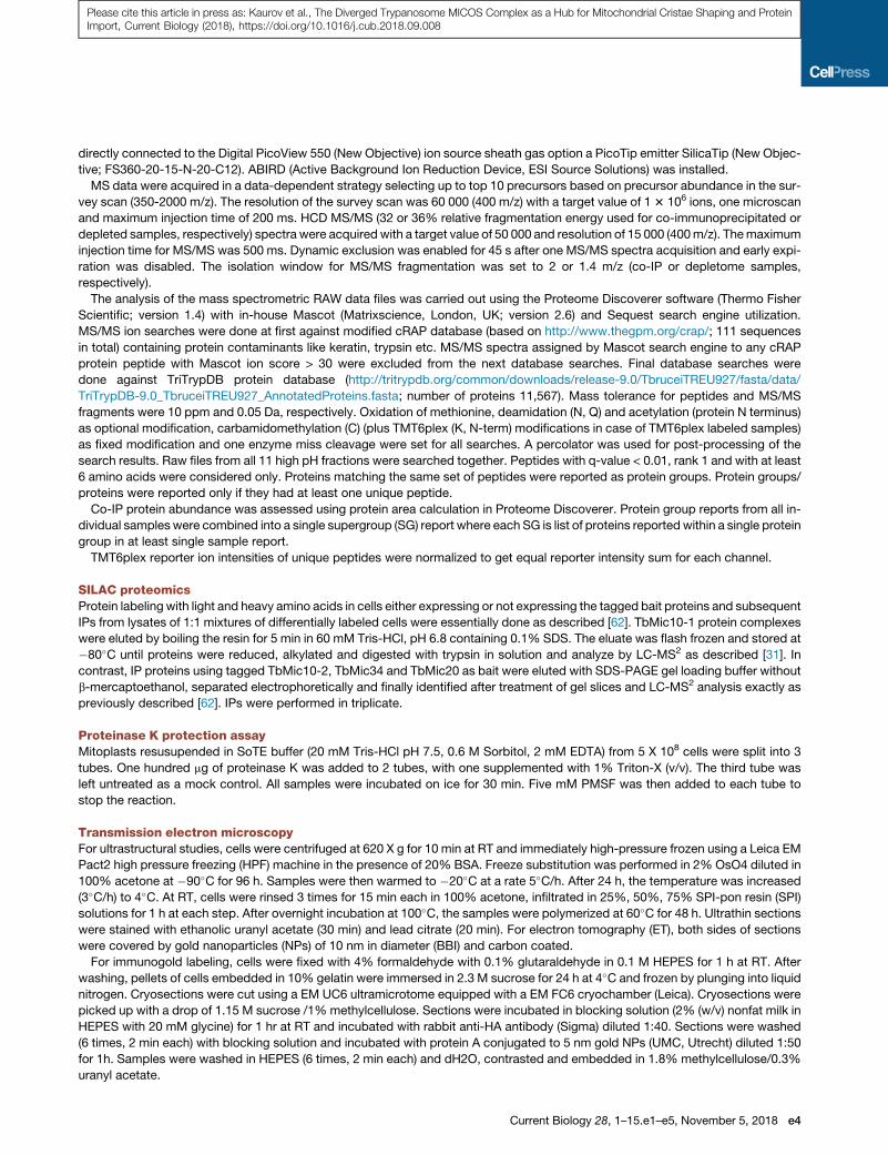
Please cite this article in press as: Kaurov et al., The Diverged Trypanosome MICOS Complex as a Hub for Mitochondrial Cristae Shaping and ProteinImport, Current Biology (2018), https://doi.org/10.1016/j.cub.2018.09.008
directly connected to the Digital PicoView 550 (New Objective) ion source sheath gas option a PicoTip emitter SilicaTip (New Objec-
tive; FS360-20-15-N-20-C12). ABIRD (Active Background Ion Reduction Device, ESI Source Solutions) was installed.
MS data were acquired in a data-dependent strategy selecting up to top 10 precursors based on precursor abundance in the sur-
vey scan (350-2000 m/z). The resolution of the survey scan was 60 000 (400 m/z) with a target value of 13 106 ions, one microscan
and maximum injection time of 200 ms. HCD MS/MS (32 or 36% relative fragmentation energy used for co-immunoprecipitated or
depleted samples, respectively) spectra were acquired with a target value of 50 000 and resolution of 15 000 (400m/z). Themaximum
injection time for MS/MS was 500 ms. Dynamic exclusion was enabled for 45 s after one MS/MS spectra acquisition and early expi-
ration was disabled. The isolation window for MS/MS fragmentation was set to 2 or 1.4 m/z (co-IP or depletome samples,
respectively).
The analysis of the mass spectrometric RAW data files was carried out using the Proteome Discoverer software (Thermo Fisher
Scientific; version 1.4) with in-house Mascot (Matrixscience, London, UK; version 2.6) and Sequest search engine utilization.
MS/MS ion searches were done at first against modified cRAP database (based on http://www.thegpm.org/crap/; 111 sequences
in total) containing protein contaminants like keratin, trypsin etc. MS/MS spectra assigned by Mascot search engine to any cRAP
protein peptide with Mascot ion score > 30 were excluded from the next database searches. Final database searches were
done against TriTrypDB protein database (http://tritrypdb.org/common/downloads/release-9.0/TbruceiTREU927/fasta/data/
TriTrypDB-9.0_TbruceiTREU927_AnnotatedProteins.fasta; number of proteins 11,567). Mass tolerance for peptides and MS/MS
fragments were 10 ppm and 0.05 Da, respectively. Oxidation of methionine, deamidation (N, Q) and acetylation (protein N terminus)
as optional modification, carbamidomethylation (C) (plus TMT6plex (K, N-term) modifications in case of TMT6plex labeled samples)
as fixed modification and one enzyme miss cleavage were set for all searches. A percolator was used for post-processing of the
search results. Raw files from all 11 high pH fractions were searched together. Peptides with q-value < 0.01, rank 1 and with at least
6 amino acids were considered only. Proteins matching the same set of peptides were reported as protein groups. Protein groups/
proteins were reported only if they had at least one unique peptide.
Co-IP protein abundance was assessed using protein area calculation in Proteome Discoverer. Protein group reports from all in-
dividual samples were combined into a single supergroup (SG) report where each SG is list of proteins reported within a single protein
group in at least single sample report.
TMT6plex reporter ion intensities of unique peptides were normalized to get equal reporter intensity sum for each channel.
SILAC proteomicsProtein labeling with light and heavy amino acids in cells either expressing or not expressing the tagged bait proteins and subsequent
IPs from lysates of 1:1 mixtures of differentially labeled cells were essentially done as described [62]. TbMic10-1 protein complexes
were eluted by boiling the resin for 5 min in 60 mM Tris-HCl, pH 6.8 containing 0.1% SDS. The eluate was flash frozen and stored at
�80�C until proteins were reduced, alkylated and digested with trypsin in solution and analyze by LC-MS2 as described [31]. In
contrast, IP proteins using tagged TbMic10-2, TbMic34 and TbMic20 as bait were eluted with SDS-PAGE gel loading buffer without
b-mercaptoethanol, separated electrophoretically and finally identified after treatment of gel slices and LC-MS2 analysis exactly as
previously described [62]. IPs were performed in triplicate.
Proteinase K protection assayMitoplasts resusupended in SoTE buffer (20 mM Tris-HCl pH 7.5, 0.6 M Sorbitol, 2 mM EDTA) from 5 X 10⁸ cells were split into 3
tubes. One hundred mg of proteinase K was added to 2 tubes, with one supplemented with 1% Triton-X (v/v). The third tube was
left untreated as a mock control. All samples were incubated on ice for 30 min. Five mM PMSF was then added to each tube to
stop the reaction.
Transmission electron microscopyFor ultrastructural studies, cells were centrifuged at 620 X g for 10 min at RT and immediately high-pressure frozen using a Leica EM
Pact2 high pressure freezing (HPF) machine in the presence of 20% BSA. Freeze substitution was performed in 2% OsO4 diluted in
100% acetone at �90�C for 96 h. Samples were then warmed to �20�C at a rate 5�C/h. After 24 h, the temperature was increased
(3�C/h) to 4�C. At RT, cells were rinsed 3 times for 15 min each in 100% acetone, infiltrated in 25%, 50%, 75% SPI-pon resin (SPI)
solutions for 1 h at each step. After overnight incubation at 100�C, the samples were polymerized at 60�C for 48 h. Ultrathin sections
were stained with ethanolic uranyl acetate (30 min) and lead citrate (20 min). For electron tomography (ET), both sides of sections
were covered by gold nanoparticles (NPs) of 10 nm in diameter (BBI) and carbon coated.
For immunogold labeling, cells were fixed with 4% formaldehyde with 0.1% glutaraldehyde in 0.1 M HEPES for 1 h at RT. After
washing, pellets of cells embedded in 10% gelatin were immersed in 2.3 M sucrose for 24 h at 4�C and frozen by plunging into liquid
nitrogen. Cryosections were cut using a EM UC6 ultramicrotome equipped with a EM FC6 cryochamber (Leica). Cryosections were
picked up with a drop of 1.15 M sucrose /1% methylcellulose. Sections were incubated in blocking solution (2% (w/v) nonfat milk in
HEPES with 20 mM glycine) for 1 hr at RT and incubated with rabbit anti-HA antibody (Sigma) diluted 1:40. Sections were washed
(6 times, 2 min each) with blocking solution and incubated with protein A conjugated to 5 nm gold NPs (UMC, Utrecht) diluted 1:50
for 1h. Samples were washed in HEPES (6 times, 2 min each) and dH2O, contrasted and embedded in 1.8% methylcellulose/0.3%
uranyl acetate.
Current Biology 28, 1–15.e1–e5, November 5, 2018 e4

Please cite this article in press as: Kaurov et al., The Diverged Trypanosome MICOS Complex as a Hub for Mitochondrial Cristae Shaping and ProteinImport, Current Biology (2018), https://doi.org/10.1016/j.cub.2018.09.008
Samples were observed with a JEOL 1010 transmission electron microscope (TEM) operating at an accelerating voltage 80 kV and
equipped with a MegaView III CCD camera (SIS). Electron tomograms were collected at tilt steps of 0.6-0.8� using the JEOL 2100F
TEMworking at 200 kV, equipped with a high-tilt stage plus a Gatan camera (Orius SC 1000) and controlled with SerialEM automated
acquisition software. Tomograms were reconstructed using the IMOD software package or Amira. A 3Dmedian filter was applied on
reconstructed stacks. Manual masking of the area of interest was employed to generate 3D surface models.
Bioinformatic analysisMic10 sequences from various genomes were aligned using MAFFT using the local pair algorithm [63]. Parts of this initial alignment
that were represented by R 60% gaps were removed via the GAPTREEZ server (https://www.hiv.lanl.gov/content/sequence/
GAPSTREEZE/gap.html). This processed alignment was then used to construct a maximum likelihood tree using RAxML [64] with
the LG+G model. The fast-bootstrap algorithm from 1000 replicates was used to estimate the best scoring tree and bootstrap sup-
port. Additional branching support was then determined by computing Bayesian posterior probabilities usingMr. Bayes 3.2 [65]. Four
independent MCMC chains were run for 3 3 106 generations under the LG+G model and default chain parameters. Topology and
posterior probabilities were reconstructed after omitting the first 5 3 105 generations.
Homology models generated in SWISSPROT [66]. Transmembrane domain and coiled coil prediction performed using TMHMM
v2.0 [67] and COILS [68] servers, respectively.
QUANTIFICATION AND STATISTICAL ANALYSIS
Analysis of immunogold and RNAi transmission electron microscopy dataImmunogold-labeling data were statistically evaluated as follows: NPs were counted in two compartments from randomly acquired
areas of sections: mitochondria (subdivided into the following sub-compartments: inner boundary membrane, cristae and a unde-
fined area) and background. Labeling density (LD) was calculated as the number of gold NPs per area. Random images were
analyzed until at least 100 NPs were counted in the HA-epitope containing samples. Chi-square analysis, with the null hypothesis
of no difference from random distribution, and RLI index were counted according toMayhew and colleagues [38]. The null hypothesis
was rejected in all examined immunodecorated HA-epitope samples. LD data are summarized in Table S2.
Cristae lengths were measured using ImageJ on images of randomly acquired mitochondrial sections. Between 39 and 74 mito-
chondrial sections were counted tomeasure the length of at least 200 cristae. Statistical significance of cristae lengths in comparison
to the parental cell line controls were determined by unpaired Student’s t test using GraphPad Prism 7 software.
Analysis of TbMic20 and TbMic60 depletome dataTbMic20 and TbMic60 RNAi 4 dpi for depletome analysis, which includes their respective non-induced controls, was performed in
duplicate. Proteins with log2 transformed relative valuesR 1.6-fold in each RNAi relative to non-induced controls were considered to
be part of the TbMic20 depletome. For determination of a secondary effect of TbMic20 RNAi on OXPHOS proteins compared to ma-
trix controls, all log2 relative values of proteins fitting into the catagories as defined by Zikova and colleagues [25] from each duplicate
was incorporated into a violin plot. Statistical significance of subunits of each respiratory chain complex compared to core matrix
proteins was determined by unpaired Student’s t test using GraphPad Prism 7 software.
DATA AND SOFTWARE AVAILABILITY
Themass spectrometry proteomics data pertaining to T. brucei 927 IPs plus TbMic20 and TbMic60 depletomes have been deposited
to the ProteomeXchange Consortium via the PRIDE partner repository with the dataset identifier PRIDE: PXD009601.
e5 Current Biology 28, 1–15.e1–e5, November 5, 2018