the diversity of costimulatory and inhibitory receptor pathways and the regulation of antiviral t...
TRANSCRIPT

Available online at www.sciencedirect.com
The diversity of costimulatory and inhibitory receptor pathwaysand the regulation of antiviral T cell responsesAlison Crawford and E John Wherry
T cell responses are regulated by integrating positive and
negative signals from costimulatory and inhibitory receptors.
While the function of specific T cell costimulatory molecules
during infections has been appreciated for some time, recent
observations have now revealed a crucial role for inhibitory
receptors in regulating T cell responses to pathogens,
especially during chronic infections. A key emerging principle is
that there is considerable diversity in the number and type of
inhibitory receptors that can be expressed by T cells during
both acute and chronic infections. These distinct inhibitory
pathways appear to cooperate in regulating T cell function,
could have distinct mechanisms of action, and are likely to
provide novel therapeutic targets during persisting infections
and other diseases.
Address
Immunology Program, The Wistar Institute, 3601 Spruce Street,
Philadelphia, PA 19104, United States
Corresponding author: Wherry, E John ([email protected])
Current Opinion in Immunology 2009, 21:179–186
This review comes from a themed issue on
Lymphocyte Development
Edited by Casey Weaver and Alexander Rudensky
Available online 4th March 2009
0952-7915/$ – see front matter
# 2009 Elsevier Ltd. All rights reserved.
DOI 10.1016/j.coi.2009.01.010
IntroductionUpon infection, naı̈ve antigen-specific T cells become
activated, differentiate, and develop into functional effec-
tor and memory T cells or, during chronic infections,
exhausted T cells. T cells integrate multiple signals
during differentiation, but the precise pathways that
control optimal T cell memory versus exhaustion remain
incompletely understood. This review focuses on our
current understanding of inhibitory receptors during T
cell responses following acute versus chronic viral infec-
tions. In discussing these inhibitory receptors and their
roles in regulating functional and dysfunctional T cell
responses during infection it is also informative to
examine the role of costimulatory pathways.
Initial activation of a naı̈ve T cell occurs upon interaction
of the TCR with specific peptide presented by MHC
www.sciencedirect.com
molecules. This is the major signal for T cell activation
and differentiation (signal 1), but additional signals from
costimulation (signal 2) and inflammation (signal 3) are
required for an effective T cell response. In the absence
of coordinated stimulation from signals 1, 2, and 3, proper
T cell activation and differentiation does not occur [1–3].
For example, TCR signaling in the absence of signal 2
from costimulation induces T cell anergy in vitro [2]. Invivo, a poor costimulatory environment or a preferential
engagement of coinhibitory pathways during T cell prim-
ing can lead to tolerance [4,5]. Several years ago the
model for T cell activation versus anergy or tolerization
was a simple one. Our developing understanding of signal
3 from inflammation and the increased number and
diversity of costimulatory and inhibitory receptors is
revealing the decision between full activation and toler-
ance/dysfunction to be more complex. We are only just
beginning to understand the interplay between different
costimulatory and inhibitory receptor signals and how
they regulate initial T cell activation and division, clonal
deletion, the generation and maintenance of memory T
cells, and T cell exhaustion.
Diversity of costimulatory and inhibitoryreceptors on T cellsRecent work has highlighted the diversity of costimulatory
and inhibitory molecules on T cells (Table 1). Many of
these receptors are part of the Ig superfamily and specifi-
cally the CD28:B7 subfamily [1]. CD28 family members
include the costimulatory receptors CD28 and ICOS and
the inhibitory receptors CTLA-4, PD-1, and BTLA. The
TNF:TNFR family also contains several costimulatory
molecules such as CD27, CD40L, 4-1BB, OX40, and
LIGHT that bind the ligands CD70, CD40, 4-1BBL,
OX40L, and HVEM respectively [6,7]. Interestingly, the
CD28 family member BTLA and another Ig superfamily
molecule CD160 bind the TNF family member HVEM in
unusual examples of cross-family interactions [8�,9].
Inhibitory receptors can bind the same ligand as a
costimulatory molecule, as is the case for CD28 and
CTLA-4 binding B7-1 and B7-2 or LIGHT, BTLA, and
CD160 binding HVEM or unique ligands that are not
shared with a costimulatory receptor (e.g. PD-1 and
PD-L1 and PD-L2) [1]. These interactions can be complex
and PD-L1, for example, has recently been demonstrated
to bind to B7-1 and deliver a bidirectional inhibitory signal
[10].
The receptors and ligands described above are major
players in T cell activation and inhibition. However,
Current Opinion in Immunology 2009, 21:179–186

180 Lymphocyte Development
Table 1
Costimulatory and inhibitory receptors.
Receptor
or ligand
on T cell
Superfamily Cellular expression pattern T cell expression Binds Costimulatory
or Inhibitory
References
CD28 Ig (CD28 family) T cells Constitutive
on Naı̈ve
B7.1, B7.2 Costimulatory [1]
ICOS Ig (CD28 family) T cells, NKT cells, NK cells Induced upon
activation
ICOSL (B7RP1) Costimulatory [1]
CD40L TNF/TNFR T cells Induced upon
activation
CD40 Costimulatory [6]
OX40 TNF/TNFR T cells Induced upon
activation
OX40L Costimulatory [6]
4-1BB TNF/TNFR T cells, DC subset, FDCs,
eosinophils, NK cells
Induced upon
activation
41BBL Costimulatory [6]
CD27 TNF/TNFR T cells, NK cells, B cells Constitutive
on Naı̈ve
CD70 Costimulatory [6]
LIGHT TNF/TNFR T cells, monocytes,
NK cells, immature DC
Induced upon
activation
HVEM Costimulatory [6]
CTLA4 Ig (CD28 family) T cells Induced upon
activation
B7.1/B7.2 Inhibitory [1]
PD-1 Ig (CD28 family) T cells, NKT cells, B cells
monocytes
Induced upon
activation
PD-L1/PD-L2 Inhibitory [1]
PD-L1 Ig (CD28 family) T cells, NKT cells, B cells
monocytes, DC, endothelial
cells, hepatocytes, and so on
Constitutive and
upregulated upon
activation
PD-1, B7.1 Inhibitory [1,10]
BTLA Ig (CD28 family) T cells, B cells, DC (macs and
NK in B6 mice only)
Constitutive
on Naı̈ve
HVEM Inhibitory [61]
CD160 Ig HVEM and MHCI (low affinity) Induced upon
activation
HVEM Inhibitory [8�]
TIM3 Ig T cells, macs, DCs Induced upon
activation
GALECTIN 9,
phosphatidyl serine
Inhibitory [16]
2B4 Ig (CD2 family) T cells, NK cells, dendritic
epidermal T cells, monocytes
Induced upon
activation
CD48 Costimulatory
and Inhibitory
[12]
LAG3 Ig T cell, NK cells, B cells Induced upon
activation
MHC-II Inhibitory [11,58]
CD200R Ig T cells, macrophages, DCs,
neutrophils, basophils,
mast cells
Constitutive
on Naı̈ve
CD200 Inhibitory? [17]
PIR-B Class 1/Class 1-like
molecules?
T cell, B cells, DCs ? MHC-I Inhibitory [57��]
NKG2A C-lectin T cells, NK cells Induced upon
activation
Qa-1B, non-classical
MHC-I, stress induced
molecules (e.g. Rae-1,
MICA, MICB)
Inhibitory [57��]
Ly49 family
(mice)
C-type lectin T cells, NK cells, NK T
cells monocytes/
macrophages
Variable MHC-I Costimulatory
and inhibitory
[14]
KIRs (human) Ig T cells Induced upon
activation
HLA-1 Inhibitory [14]
GP49B Ig T cells, macrophages,
neutrophils, and NK
cells, mast cells
? avb3 Inhibitory [57��]
the number of cell surface molecules that can regulate T
cell responses is ever increasing. Recently, molecules
including LAG-3 [11], 2B4/CD244 [12,13], CD30-
CD30L [6], NK receptors [14], the Tim molecules
(Tim1-4) [15�,16], CD200R [17], Siglecs [18] and others
have been implicated in regulating T cell responses. It is
currently unclear to what degree there is functional
overlap or specificity in these pathways and whether there
are common themes and principles about costimulation
and inhibition by these different molecules.
Current Opinion in Immunology 2009, 21:179–186
Costimulatory and inhibitory receptors duringinfectionsCostimulatory and inhibitory receptors are thought to
help the immune system balance productive immune
responses to foreign antigens versus tolerance to self-
antigens. Indeed, a number of costimulatory pathways
can augment T cell responses to acute infections. During
some viral infections, the absence of costimulatory path-
ways can lead to a substantial quantitative reduction in
the primary antiviral T cell response [1,19] though the
www.sciencedirect.com

The diversity of costimulatory and inhibitory receptor pathways Crawford and Wherry 181
level of costimulation required can depend on the type of
infection. Primary T cell responses to influenza virus are
substantially diminished in mice lacking various costimu-
latory molecules while T cell responses to acute LCMV
infection are only modestly reduced in the same KO mice
[1,6]. Similar examples for other infections also exist
[20,21].
While there is considerable evidence that inhibitory
receptors can influence T cell responses to self-antigens
[5], the role of inhibitory receptors in T cell priming and
memory T cell differentiation during acute infection is
less clear. CTLA-4 KO CD8 T cells respond normally to
acute LCMV infection [22] and CTLA-4 expressing T
cells appear to have normal proliferative responses and
effector function during acute LCMV or vaccinia virus
infection [23,24]. However, inhibitory receptor pathways
can negatively regulate immunity to rabies virus, adeno-
virus, and Sendai virus [11,25,26]. During acute infection
with rabies virus, the absence of PD-L1 led to better
control of virus and a greater accumulation of CD8 T cells
in the CNS [25]. When PD-1 KO mice were infected with
adenovirus, T cells expanded more in peripheral tissues
leading to rapid clearance of the virus [26]. LAG-3 can
also regulate acute T cell responses; LAG-3 KO mice
infected with Sendai virus had increased numbers of
virus-specific CD4 and CD8 T cells 30 days post infection
[11]. While much of the data examining the role of
inhibitory receptors during acute infection are consistent
with a negative role for these pathways, two recent studies
with Listeria monocytogenes (LM) infection suggest a
possible alternative role for the PD-1 pathway in this
setting [27,28�]. In these studies, PD-L1 blockade
delayed the kinetics of CD8+ T cell priming, reduced
the magnitude of T cell expansion, and diminished
secondary T cell responses [27,28�]. It is unclear whether
this role for the PD-1 pathway during LM infection
reflects increased T cell death when PD-1:PD-L1 is
blocked or a true positive signal through this pathway
under some circumstances. Additional studies should
help clarify the precise role of the PD-1 pathway in
different infections. However, the majority of studies
are consistent with a negative role for most inhibitory
pathways during infection.
Costimulatory and inhibitory receptors duringchronic infectionMany of the costimulatory receptor KO mice discussed
above have profound defects in T cell responses and viral
control during chronic infections [29–34] suggesting a
crucial role for costimulation when pathogens persist.
Indeed, enhancing costimulation can in some circum-
stances improve T cell responses during chronic infec-
tions. For example, 4-1BB has a key role during Friend
leukemia virus infection [34] and stimulation of the 4-
1BB pathway led to expansion of HIV-specific CD8+ T
cells in vitro most probably by regulating CD8 T cell
www.sciencedirect.com
survival [35]. It will be interesting to define how aug-
menting other costimulatory signals influences T cell
responses during chronic viral infections.
There is now accumulating evidence that inhibitory
receptors have a prominent role in regulating T cell
responses during chronic viral infections. In contrast to
acute infections where T cells expand, contract, and form
functional, long-lived memory cells, during chronic in-
fection T cells develop functional deficiencies that pre-
vent optimal control of infection [36]. This loss of
function or exhaustion is hierarchical, with IL-2, cytotox-
icity and robust proliferation lost early, TNFa persisting
for longer and IFN-g lost only at more extreme stages of
exhaustion [36]. T cell exhaustion has been described
during a number of chronic infections in mice as well as
human infections with HIV, HBV, and HCV [37,38��,39].
These functional defects are probably a key reason for
failure of immunological control of these persisting patho-
gens and recent insights indicate a central role for inhibi-
tory receptors in regulating T cell exhaustion.
A functional role for inhibitory receptors during chronic
infection was initially demonstrated during chronic
LCMV infection where blockade of the PD-1:PD-L1
pathway rejuvenated CD8 T cell function and enhanced
viral control [40]. The PD-1 pathway also appears to
regulate T cell responses during other mouse models
of chronic infection [41], primate infection with SIV
[42,43], and in humans infected with HIV [38��,44–48],
HBV [37,49,50], HCV [39,51,52] where disrupting this
pathway in vitro improves T cell responses. In vivoblockade of the PD-1 pathway during SIV infection of
macaques not only improves T cell (and B cell) responses,
but also lowers viral load and improves survival of the
primates receiving treatment [53]. Thus, there is clearly a
role for the PD-1:PD-L1 pathway during chronic infec-
tions and manipulation of this pathway has therapeutic
potential.
There is reason to be cautiously optimistic about the
clinical promise of PD-1 pathway blockade. This inter-
vention alone, however, does not completely restore
optimal T cell function and the level of functional recov-
ery and even the precise impact of PD-1 blockade on
different effector functions and/or apoptosis differs
depending on the study [42,46–48]. Some insights into
the nature of partial reversal of exhaustion and the range
of responses to PD-1 pathway blockade might come from
recent studies demonstrating that populations of
exhausted CD8 T cells are heterogeneous [54]. For
example, exhausted LCMV-specific CD8 T cells with
intermediate PD-1 expression were potently re-invigo-
rated by PD-1:PD-L1 blockade, but there was little if any
benefit for the PD-1Hi subset of exhausted CD8 T cells
[54]. A similar situation might also exist during chronic
HCV infection in humans where PD-1Lo HCV-specific
Current Opinion in Immunology 2009, 21:179–186
182 Lymphocyte Development
CD8 T cells from the blood responded to PD-1 pathway
blockade while PD-1Hi HCV-specific CD8 T cells from
the livers of the same individuals did not [55].
Gene expression profiling studies suggested the presence
of a number of other known or potential inhibitory
receptors on exhausted CD8 T cells [56��]. In addition
to PD-1, LCMV-specific exhausted CD8 T cells also
expressed 2B4, LAG-3, CTLA-4, PirB, GP49 and
CD160 [57��]. Many of these inhibitory receptors were
co-expressed by the same exhausted CD8 T cell and as
the severity of the infection increased, both the amount of
each receptor and the number of different inhibitory
receptors expressed per cell increased [57��]. Thus, the
PD-1Hi subset of exhausted CD8 T cells also co-
expressed multiple other inhibitory receptors. Blockade
of the PD-1 pathway together with a second inhibitory
receptor LAG-3 demonstrated cooperation between
these inhibitory pathways and led to a synergistic restor-
ation of CD8 T cell function and viral control [57��].Several recent studies have demonstrated that HIV-
specific T cells can also express more than one inhibitory
receptor. Kaufmann et al. showed that PD-1 and CTLA-4
are co-expressed by HIV-specific CD4+ T cells [38��].Both PD-1 and CTLA-4 negatively regulated the func-
tion of HIV-specific CD4 T cells and co-expression
patterns and combined blockade of these inhibitory
receptors in vitro suggested cooperative inhibition could
occur. One interesting observation was that while CTLA-
4 and PD-1 co-regulated HIV-specific CD4 T cells, HIV-
specific CD8 T cells expressed substantial amounts of
PD-1, but not CTLA-4 suggesting that CD4 and CD8 T
cell responses could be regulated by distinct sets of
inhibitory receptors. A second study recently demon-
strated the expression and inhibitory role of Tim-3 on
HIV-specific T cells [15�]. Interestingly, Tim-3 and PD-1
were expressed by sometimes distinct and sometimes
partially overlapping populations of CD8+ T cells
depending on the subject [15�]. One concept that
emerges from these studies is that there is diversity in
the cell surface inhibitory pathways available to nega-
tively regulate T cell responses to chronic infections.
Specific inhibitory receptors can be expressed by distinct
T cell subsets resulting in differential regulation, while
other inhibitory receptors are co-expressed and can oper-
ate synergistically. Thus, as with costimulatory mol-
ecules, the precise inhibitory receptors of therapeutic
interest might depend on the type and severity of in-
fection, whether CD4 or CD8 T cells are of interest as
well as other factors such as the activation state of the T
cell.
Mechanisms of costimulatory and inhibitoryreceptor pathwaysA mechanistic understanding of how inhibitory receptors
restrain T cell responses would not only aid in our un-
derstanding of memory T cell differentiation following
Current Opinion in Immunology 2009, 21:179–186
chronic viral infections, but could also provide novel
targets for therapeutics. Unfortunately, for many inhibi-
tory receptors there is little information in this area.
Again, examining some aspects of costimulation may
provide a guide. For the best studied costimulatory path-
ways such as CD28, engagement of the costimulatory
receptor compliments TCR signaling both quantitatively
and qualitatively. For example, CD28 signaling lowers
the threshold for activation, enhances IL2 production and
CD25 expression, improves survival by upregulating
Bcl-XL, and promotes progression through the cell cycle
[1]. In addition, the coupling of CD28 costimulation to
glucose metabolism via AKT [58] is probably a key factor
in ensuring robust and sustained T cell responses upon
initial activation. While costimulatory molecules of the
TNF family can also enhance T cell proliferation and
cytokine production and regulate Bcl-2 and Bcl-XL
expression, they do so through different signaling path-
ways. For example, 4-1BB signals via TRAF1, TRAF2,
and ERK [6]. Thus, while the end result of costimulation
by different pathways can be similar the mechanistic
intermediates can be quite distinct.
In contrast to many costimulatory pathways, the coinhibi-
tory molecules CTLA-4 [59], PD-1 [60], BTLA [61], and
CD160 [8�] all inhibit IL-2 production and these four
receptors as well as LAG-3 [11] can negatively regulate
cell cycle progression. CTLA-4 inhibits CD28 augmented
gene transcription, but does not prevent gene regulation by
TCR alone [62]. By contrast, PD-1 appears to block both
TCR signals [63,64] and CD28 signals [65]. Interestingly,
though PD-1 and CTLA-4 both negatively regulate CD28
signaling and AKT activation, they do so by different
mechanisms [65]. While the signaling mechanism for some
inhibitory pathways is beginning to be defined, there is far
less known about the intracellular mechanisms of action of
inhibitory receptors than the costimulatory receptors.
Given the similarities in downstream targets, it is tempting
to speculate that molecules such as CTLA-4 and PD-1
have evolved to specifically antagonize CD28 (and perhaps
ICOS) mediated positive costimulation. It will be inter-
esting to examine if other costimulatory pathways such as
TNF receptor family members are resistant to or can
overcome CTLA-4 or PD-1 mediated inhibition.
It is currently unclear whether inhibitory receptors merely
block the induction of gene expression by TCR/costimula-
tion or whether inhibitory receptor signaling could actually
induce alternative gene transcription. Several models for
how inhibitory receptor signals might alter gene expression
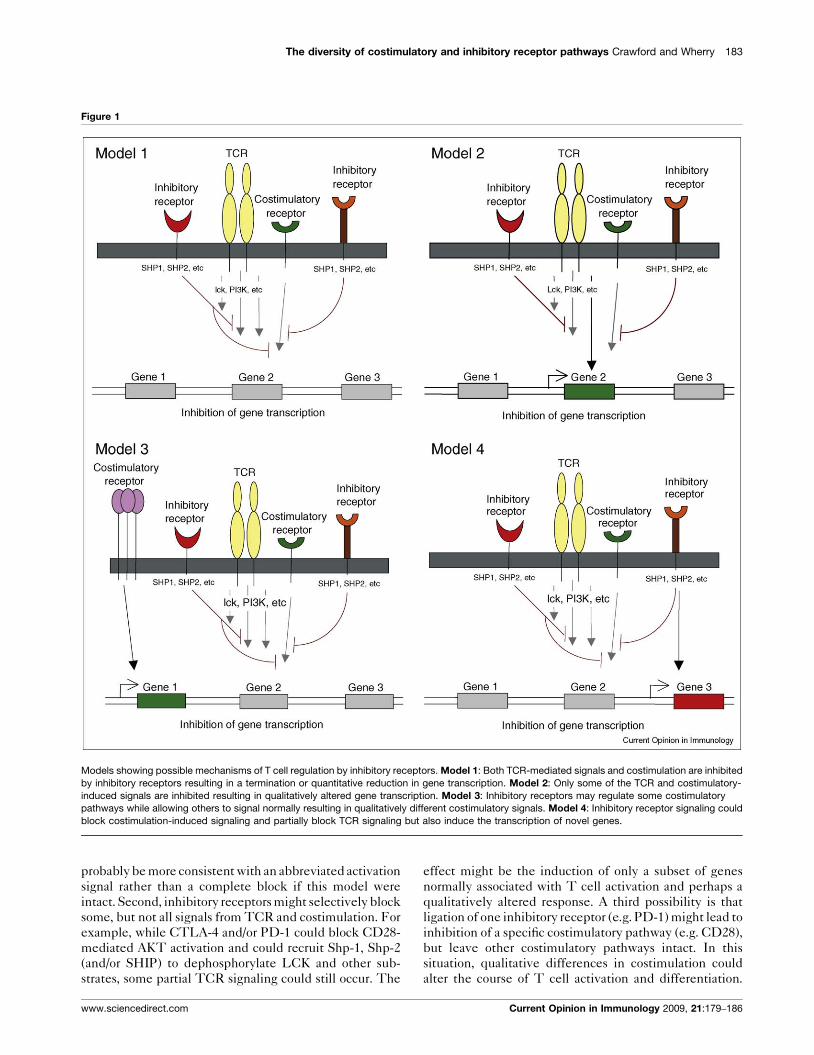
in T cells are possible and are described in Figure 1. First,
inhibitory receptor signaling could simply block all signals
from the TCR and costimulation leading to an attenuation
or elimination of TCR induced gene expression. The
effect of this inhibition would be a broad quantitative
reduction in gene expression. A temporal delay in expres-
sion of inhibitory versus costimulatory receptors would
www.sciencedirect.com
The diversity of costimulatory and inhibitory receptor pathways Crawford and Wherry 183
Figure 1
Models showing possible mechanisms of T cell regulation by inhibitory receptors. Model 1: Both TCR-mediated signals and costimulation are inhibited
by inhibitory receptors resulting in a termination or quantitative reduction in gene transcription. Model 2: Only some of the TCR and costimulatory-
induced signals are inhibited resulting in qualitatively altered gene transcription. Model 3: Inhibitory receptors may regulate some costimulatory
pathways while allowing others to signal normally resulting in qualitatively different costimulatory signals. Model 4: Inhibitory receptor signaling could
block costimulation-induced signaling and partially block TCR signaling but also induce the transcription of novel genes.
probably be more consistent with an abbreviated activation
signal rather than a complete block if this model were
intact. Second, inhibitory receptors might selectively block
some, but not all signals from TCR and costimulation. For
example, while CTLA-4 and/or PD-1 could block CD28-
mediated AKT activation and could recruit Shp-1, Shp-2
(and/or SHIP) to dephosphorylate LCK and other sub-
strates, some partial TCR signaling could still occur. The
www.sciencedirect.com
effect might be the induction of only a subset of genes
normally associated with T cell activation and perhaps a
qualitatively altered response. A third possibility is that
ligation of one inhibitory receptor (e.g. PD-1) might lead to
inhibition of a specific costimulatory pathway (e.g. CD28),
but leave other costimulatory pathways intact. In this
situation, qualitative differences in costimulation could
alter the course of T cell activation and differentiation.
Current Opinion in Immunology 2009, 21:179–186
184 Lymphocyte Development
A fourth possibility is that inhibitory receptors actively
induce or regulate gene expression independent of their
inhibition of TCR or costimulatory signaling. For
example, inhibitory molecules might recruit and/or acti-
vate selective signaling pathways that lead to new gene
transcription (or active repression). This latter model
would probably occur with some partial TCR signaling
since there is little evidence that inhibitory receptor
signaling has an impact without TCR engagement.
These models are not mutually exclusive and these
and other potential mechanisms could occur for different
inhibitory receptor pathways.
Conclusion and future directionsT cells can express a diverse array of both costimulatory
and inhibitory receptors, and these molecules can posi-
tively or negatively regulate T cell function. In some
cases pathogens have exploited negative regulatory path-
ways to facilitate persistence and inhibitory receptors
appear to play a prominent role in T cell dysfunction
during persisting infections. In vitro and in vivo blockade
studies have revealed the therapeutic potential of mod-
ulating costimulatory and inhibitory receptors on T cells.
A better understanding of the complexity of co-expres-
sion of inhibitory and costimulatory receptors on T cells
in different settings is likely to reveal T cell functions and
pathways regulated by specific receptors. It is likely there
will be opportunities to therapeutically ‘tune’ T cell
responses by modulating specific receptor:ligand path-
ways. Since these same costimulatory and inhibitory
pathways are used not only during T cell responses to
pathogens, but also in anti-tumor immunity and also
autoimmunity, a better mechanistic understanding of
the regulation of T cell responses by diverse and co-
expressed positive and negative regulatory molecules
could have broad therapeutic implications.
AcknowledgementsThis work was supported by grants from the National Institutes of Health((HHSN26620050030C, AI071309, and AI077098 to EJW) and theFoundation for NIH through the Grand Challenges in Global HealthInitiative (to EJW)) and the American Heart Association (AC).
References and recommended readingPapers of particular interest, published within the period of review,have been highlighted as:
� of special interest�� of outstanding interest
1. Greenwald RJ, Freeman GJ, Sharpe AH: The B7 family revisited.Annu Rev Immunol 2005, 23:515-548.
2. Schwartz RH: T cell anergy. Annu Rev Immunol 2003, 21:305-334.
3. Mescher MF, Curtsinger JM, Agarwal P, Casey KA, Gerner M,Hammerbeck CD, Popescu F, Xiao Z: Signals required forprogramming effector and memory development by CD8+ Tcells. Immunol Rev 2006, 211:81-92.
4. Goldberg MV, Maris CH, Hipkiss EL, Flies AS, Zhen L, Tuder RM,Grosso JF, Harris TJ, Getnet D, Whartenby KA et al.: Role of PD-1and its ligand, B7-H1, in early fate decisions of CD8 T cells.Blood 2007, 110:186-192.
Current Opinion in Immunology 2009, 21:179–186
5. Redmond WL, Sherman LA: Peripheral tolerance of CD8 Tlymphocytes. Immunity 2005, 22:275-284.
6. Watts TH: TNF/TNFR family members in costimulation of T cellresponses. Annu Rev Immunol 2005, 23:23-68.
7. Croft M: Costimulation of T cells by OX40, 4-1BB, and CD27.Cytokine Growth Factor Rev 2003, 14:265-273.
8.�
Cai G, Anumanthan A, Brown JA, Greenfield EA, Zhu B,Freeman GJ: CD160 inhibits activation of human CD4+ T cellsthrough interaction with herpesvirus entry mediator. NatImmunol 2008, 9:176-185.
This paper demonstrates that CD160 is a receptor for HVEM and that,similar to BTLA, binding of HVEM to CD160 results in negative signaling tothe T cell resulting in inhibition of proliferation and cytokine production.
9. Gonzalez LC, Loyet KM, Calemine-Fenaux J, Chauhan V,Wranik B, Ouyang W, Eaton DL: A coreceptor interactionbetween the CD28 and TNF receptor family members B and Tlymphocyte attenuator and herpesvirus entry mediator.Proc Natl Acad Sci U S A 2005, 102:1116-1121.
10. Butte MJ, Keir ME, Phamduy TB, Sharpe AH, Freeman GJ:Programmed death-1 ligand 1 interacts specifically with theB7-1 costimulatory molecule to inhibit T cell responses.Immunity 2007, 27:111-122.
11. Workman CJ, Cauley LS, Kim IJ, Blackman MA, Woodland DL,Vignali DA: Lymphocyte activation gene-3 (CD223) regulatesthe size of the expanding T cell population following antigenactivation in vivo. J Immunol 2004, 172:5450-5455.
12. Lee KM, McNerney ME, Stepp SE, Mathew PA, Schatzle JD,Bennett M, Kumar V: 2B4 acts as a non-majorhistocompatibility complex binding inhibitory receptor onmouse natural killer cells. J Exp Med 2004, 199:1245-1254.
13. Laouar A, Manocha M, Wan M, Yagita H, van Lier RA, Manjunath N:Cutting Edge: Distinct NK receptor profiles are imprinted onCD8 T cells in the mucosa and periphery during the sameantigen challenge: role of tissue-specific factors. J Immunol2007, 178:652-656.
14. Lanier LL: Evolutionary struggles between NK cells andviruses. Nat Rev Immunol 2008, 8:259-268.
15.�
Jones RB, Ndhlovu LC, Barbour JD, Sheth PM, Jha AR, Long BR,Wong JC, Satkunarajah M, Schweneker M, Chapman JM et al.:Tim-3 expression defines a novel population of dysfunctionalT cells with highly elevated frequencies in progressive HIV-1infection. J Exp Med 2008, 205:2763-2779.
16. Kuchroo VK, Umetsu DT, DeKruyff RH, Freeman GJ: The TIMgene family: emerging roles in immunity and disease. Nat RevImmunol 2003, 3:454-462.
17. Snelgrove RJ, Goulding J, Didierlaurent AM, Lyonga D, Vekaria S,Edwards L, Gwyer E, Sedgwick JD, Barclay AN, Hussell T: Acritical function for CD200 in lung immune homeostasis andthe severity of influenza infection. Nat Immunol 2008,9:1074-1083.
18. Crocker PR, Paulson JC, Varki A: Siglecs and their roles in theimmune system. Nat Rev Immunol 2007, 7:255-266.
19. Bertram EM, Dawicki W, Watts TH: Role of T cell costimulation inanti-viral immunity. Semin Immunol 2004, 16:185-196.
20. Fang M, Sigal LJ: Direct CD28 costimulation is required forCD8+ T cell-mediated resistance to an acute viral disease in anatural host. J Immunol 2006, 177:8027-8036.
21. Shahinian A, Pfeffer K, Lee KP, Kundig TM, Kishihara K,Wakeham A, Kawai K, Ohashi PS, Thompson CB, Mak TW:Differential T cell costimulatory requirements in CD28-deficient mice. Science 1993, 261:609-612.
22. Bachmann MF, Waterhouse P, Speiser DE, McKall-Faienza K,Mak TW, Ohashi PS: Normal responsiveness of CTLA-4-deficient anti-viral cytotoxic T cells. J Immunol 1998,160:95-100.
23. Raue HP, Slifka MK: Pivotal advance: CTLA-4+ T cells exhibitnormal antiviral functions during acute viral infection. J LeukocBiol 2007, 81:1165-1175.
www.sciencedirect.com

The diversity of costimulatory and inhibitory receptor pathways Crawford and Wherry 185
24. Homann D, Dummer W, Wolfe T, Rodrigo E, Theofilopoulos AN,Oldstone MB, von Herrath MG: Lack of intrinsic CTLA-4expression has minimal effect on regulation of antiviral T-cellimmunity. J Virol 2006, 80:270-280.
25. Lafon M, Megret F, Meuth SG, Simon O, Velandia Romero ML,Lafage M, Chen L, Alexopoulou L, Flavell RA, Prehaud C et al.:Detrimental contribution of the immuno-inhibitor B7-H1 torabies virus encephalitis. J Immunol 2008, 180:7506-7515.
26. Iwai Y, Terawaki S, Ikegawa M, Okazaki T, Honjo T: PD-1 inhibitsantiviral immunity at the effector phase in the liver. J Exp Med2003, 198:39-50.
27. Seo SK, Jeong HY, Park SG, Lee SW, Choi IW, Chen L, Choi I:Blockade of endogenous B7-H1 suppresses antibacterialprotection after primary Listeria monocytogenes infection.Immunology 2008, 123:90-99.
28.�
Rowe JH, Johanns TM, Ertelt JM, Way SS: PDL-1 blockadeimpedes T cell expansion and protective immunity primed byattenuated Listeria monocytogenes. J Immunol 2008, 180:7553-7557.
Blockade of PD-1 was examined to determine its role in regulating T cellresponses to acute infection with Listeria monocytogenes. Surprisingly,blockade of this pathway resulted in reduced primary and secondary Tcell responses. This paper is significantly important because it demon-strated not only that inhibitory receptors can regulate T cell responses toacute infection but that the role of the PD-1 pathway may not be negativein all cases.
29. Suresh M, Whitmire JK, Harrington LE, Larsen CP, Pearson TC,Altman JD, Ahmed R: Role of CD28-B7 interactions ingeneration and maintenance of CD8 T cell memory. J Immunol2001, 167:5565-5573.
30. Whitmire JK, Flavell RA, Grewal IS, Larsen CP, Pearson TC,Ahmed R: CD40-CD40 ligand costimulation is required forgenerating antiviral CD4 T cell responses but is dispensablefor CD8 T cell responses. J Immunol 1999, 163:3194-3201.
31. Fuse S, Bellfy S, Yagita H, Usherwood EJ: CD8+ T celldysfunction and increase in murine gammaherpesvirus latentviral burden in the absence of 4-1BB ligand. J Immunol 2007,178:5227-5236.
32. Fuse S, Zhang W, Usherwood EJ: Control of memory CD8+ T celldifferentiation by CD80/CD86-CD28 costimulation andrestoration by IL-2 during the recall response. J Immunol 2008,180:1148-1157.
33. Kemball CC, Lee ED, Szomolanyi-Tsuda E, Pearson TC,Larsen CP, Lukacher AE: Costimulation requirements forantiviral CD8+ T cells differ for acute and persistent phases ofpolyoma virus infection. J Immunol 2006, 176:1814-1824.
34. Robertson SJ, Messer RJ, Carmody AB, Mittler RS, Burlak C,Hasenkrug KJ: CD137 costimulation of CD8+ T cells confersresistance to suppression by virus-induced regulatory T cells.J Immunol 2008, 180:5267-5274.
35. Wang C, Wen T, Routy JP, Bernard NF, Sekaly RP, Watts TH: 4-1BBL induces TNF receptor-associated factor 1-dependentBim modulation in human T cells and is a critical component inthe costimulation-dependent rescue of functionally impairedHIV-specific CD8 T cells. J Immunol 2007, 179:8252-8263.
36. Shin H, Wherry EJ: CD8 T cell dysfunction during chronic viralinfection. Curr Opin Immunol 2007, 19:408-415.
37. Boni C, Fisicaro P, Valdatta C, Amadei B, Di Vincenzo P, Giuberti T,Laccabue D, Zerbini A, Cavalli A, Missale G et al.:Characterization of hepatitis B virus (HBV)-specific T-celldysfunction in chronic HBV infection. J Virol 2007,81:4215-4225.
38.��
Kaufmann DE, Kavanagh DG, Pereyra F, Zaunders JJ,Mackey EW, Miura T, Palmer S, Brockman M, Rathod A,Piechocka-Trocha A et al.: Upregulation of CTLA-4 by HIV-specific CD4+ T cells correlates with disease progression anddefines a reversible immune dysfunction. Nat Immunol 2007,8:1246-1254.
Kaufmann et al. demonstrated that CD4+ T cells from the blood of HIV-infected individuals co-express PD-1 and CTLA-4. CTLA-4 appears to beupregulated specifically by HIV-specific CD4+ T cells but not CD8+ T cellssuggesting that inhibitory receptors can differentially regulate CD4 versus
www.sciencedirect.com
CD8 T cell responses. Both PD-1 and CTLA-4 negatively regulated thefunction of HIV-specific CD4 T cells.
39. Urbani S, Amadei B, Tola D, Massari M, Schivazappa S, Missale G,Ferrari C: PD-1 expression in acute hepatitis C virus (HCV)infection is associated with HCV-specific CD8 exhaustion.J Virol 2006, 80:11398-11403.
40. Barber DL, Wherry EJ, Masopust D, Zhu B, Allison JP, Sharpe AH,Freeman GJ, Ahmed R: Restoring function in exhausted CD8 Tcells during chronic viral infection. Nature 2006, 439:682-687.
41. Isogawa M, Furuichi Y, Chisari FV: Oscillating CD8(+) T celleffector functions after antigen recognition in the liver.Immunity 2005, 23:53-63.
42. Petrovas C, Price DA, Mattapallil J, Ambrozak DR, Geldmacher C,Cecchinato V, Vaccari M, Tryniszewska E, Gostick E, Roederer Met al.: SIV-specific CD8+ T cells express high levels of PD1 andcytokines but have impaired proliferative capacity in acuteand chronic SIVmac251 infection. Blood 2007, 110:928-936.
43. Velu V, Kannanganat S, Ibegbu C, Chennareddi L, Villinger F,Freeman GJ, Ahmed R, Amara RR: Elevated expression levels ofinhibitory receptor programmed death 1 on simianimmunodeficiency virus-specific CD8 T cells during chronicinfection but not after vaccination. J Virol 2007, 81:5819-5828.
44. D’Souza M, Fontenot AP, Mack DG, Lozupone C, Dillon S,Meditz A, Wilson CC, Connick E, Palmer BE: Programmed death1 expression on HIV-specific CD4+ T cells is driven by viralreplication and associated with T cell dysfunction. J Immunol2007, 179:1979-1987.
45. Day CL, Kaufmann DE, Kiepiela P, Brown JA, Moodley ES, Reddy S,Mackey EW, Miller JD, Leslie AJ, DePierres C et al.: PD-1expression on HIV-specific T cells is associated with T-cellexhaustion and disease progression. Nature 2006, 443:350-354.
46. Petrovas C, Casazza JP, Brenchley JM, Price DA, Gostick E,Adams WC, Precopio ML, Schacker T, Roederer M, Douek DCet al.: PD-1 is a regulator of virus-specific CD8+ T cell survivalin HIV infection. J Exp Med 2006, 203:2281-2292.
47. Trautmann L, Janbazian L, Chomont N, Said EA, Gimmig S,Bessette B, Boulassel MR, Delwart E, Sepulveda H, Balderas RSet al.: Upregulation of PD-1 expression on HIV-specific CD8+ Tcells leads to reversible immune dysfunction. Nat Med 2006,12:1198-1202.
48. Zhang JY, Zhang Z, Wang X, Fu JL, Yao J, Jiao Y, Chen L, Zhang H,Wei J, Jin L et al.: PD-1 up-regulation is correlated with HIV-specific memory CD8+ T-cell exhaustion in typicalprogressors but not in long-term nonprogressors. Blood 2007,109:4671-4678.
49. Boettler T, Panther E, Bengsch B, Nazarova N, Spangenberg HC,Blum HE, Thimme R: Expression of the interleukin-7 receptoralpha chain (CD127) on virus-specific CD8+ T cells identifiesfunctionally and phenotypically defined memory T cells duringacute resolving hepatitis B virus infection. J Virol 2006,80:3532-3540.
50. Zhang Z, Zhang JY, Wherry EJ, Jin B, Xu B, Zou ZS, Zhang SY,Li BS, Wang HF, Wu H et al.: Dynamic programmed death 1expression by virus-specific CD8 T cells correlates with theoutcome of acute hepatitis B. Gastroenterology 2008,134:2168-2171.
51. Radziewicz H, Ibegbu CC, Fernandez ML, Workowski KA,Obideen K, Wehbi M, Hanson HL, Steinberg JP, Masopust D,Wherry EJ et al.: Liver-infiltrating lymphocytes in chronichuman hepatitis C virus infection display an exhaustedphenotype with high levels of PD-1 and low levels of CD127expression. J Virol 2007, 81:2545-2553.
52. Golden-Mason L, Palmer B, Klarquist J, Mengshol JA,Castelblanco N, Rosen HR: Upregulation of PD-1 expression oncirculating and intrahepatic hepatitis C virus-specific CD8+ Tcells associated with reversible immune dysfunction.J Virol 2007, 81:9249-9258.
53. Velu V, Titanji K, Zhu B, Husain S, Pladevega A, Lai L,Vanderford TH, Chennareddi L, Silvestri G, Freeman GJ et al.:Enhancing SIV-specific immunity in vivo by PD-1 blockade.Nature 2008 doi: 10.1038/nature07662.
Current Opinion in Immunology 2009, 21:179–186

186 Lymphocyte Development
54. Blackburn SD, Shin H, Freeman GJ, Wherry EJ: Selectiveexpansion of a subset of exhausted CD8 T cells by alphaPD-L1blockade. Proc Natl Acad Sci U S A 2008, 105:15016-15021.
55. Nakamoto N, Kaplan DE, Coleclough J, Li Y, Valiga ME,Kaminski M, Shaked A, Olthoff K, Gostick E, Price DA et al.:Functional restoration of HCV-specific CD8 T cells by PD-1blockade is defined by PD-1 expression andcompartmentalization. Gastroenterology 2008, 134:1927-1937.
56.��
Wherry EJ, Ha SJ, Kaech SM, Haining WN, Sarkar S, Kalia V,Subramaniam S, Blattman JN, Barber DL, Ahmed R: Molecularsignature of CD8+ T cell exhaustion during chronic viralinfection. Immunity 2007, 27:670-684.
Gene expression profiles of LCMV-specific T cells from acute and chronicLCMV-infected mice were examined and compared with naı̈ve T cells. Atotal of 490 genes were upregulated or downregulated by at least twofoldin the exhausted CD8+ T cells identifying a number of inhibitory receptors,transcription factors and metabolic pathways that may be novel targetsfor immunotherapy.
57.��
Blackburn SD, Shin H, Haining WN, Zou T, Workman CJ, Polley A,Betts MR, Freeman GJ, Vignali DA, Wherry EJ: Coregulation ofCD8(+) T cell exhaustion by multiple inhibitory receptorsduring chronic viral infection. Nat Immunol 2008.
This paper demonstrates that exhausted CD8 T cells from chronicallyinfected LCMV mice upregulate a wide range of inhibitory receptors.Furthermore, in vivo blockade of two of the molecules (PD-L1 and LAG3)show that while aLAG3 alone had no effect on the frequency of LCMV-specific CD8 T cells, dual blockade had a greater effect than aPD-L1alone. Moreover, dual blockade also had a greater effect on rescuing ofCD8 T cell function than aPD-L1 alone leading to significantly lower viraltiters. This study shows inhibitory receptors can work synergistically andsuggests that combination therapies have considerable potential.
58. Frauwirth KA, Riley JL, Harris MH, Parry RV, Rathmell JC, Plas DR,Elstrom RL, June CH, Thompson CB: The CD28 signaling
Current Opinion in Immunology 2009, 21:179–186
pathway regulates glucose metabolism. Immunity 2002,16:769-777.
59. Brunner MC, Chambers CA, Chan FK, Hanke J, Winoto A,Allison JP: CTLA-4-mediated inhibition of early events of T cellproliferation. J Immunol 1999, 162:5813-5820.
60. Carter L, Fouser LA, Jussif J, Fitz L, Deng B, Wood CR, Collins M,Honjo T, Freeman GJ, Carreno BM: PD-1:PD-L inhibitorypathway affects both CD4(+) and CD8(+) T cells and isovercome by IL-2. Eur J Immunol 2002, 32:634-643.
61. Krieg C, Han P, Stone R, Goularte OD, Kaye J: Functionalanalysis of B and T lymphocyte attenuator engagement onCD4+ and CD8+ T cells. J Immunol 2005, 175:6420-6427.
62. Riley JL, Mao M, Kobayashi S, Biery M, Burchard J, Cavet G,Gregson BP, June CH, Linsley PS: Modulation of TCR-inducedtranscriptional profiles by ligation of CD28, ICOS, andCTLA-4 receptors. Proc Natl Acad Sci U S A 2002,99:11790-11795.
63. Keir ME, Latchman YE, Freeman GJ, Sharpe AH: Programmeddeath-1 (PD-1):PD-ligand 1 interactions inhibit TCR-mediatedpositive selection of thymocytes. J Immunol 2005,175:7372-7379.
64. Sheppard KA, Fitz LJ, Lee JM, Benander C, George JA, Wooters J,Qiu Y, Jussif JM, Carter LL, Wood CR et al.: PD-1 inhibits T-cellreceptor induced phosphorylation of the ZAP70/CD3zetasignalosome and downstream signaling to PKCtheta. FEBSLett 2004, 574:37-41.
65. Parry RV, Chemnitz JM, Frauwirth KA, Lanfranco AR, Braunstein I,Kobayashi SV, Linsley PS, Thompson CB, Riley JL: CTLA-4 andPD-1 receptors inhibit T-cell activation by distinctmechanisms. Mol Cell Biol 2005, 25:9543-9553.
www.sciencedirect.com