the early embryo c development of · the motive of my studies on the embryogenesis of silkworms...
TRANSCRIPT

Keiichiro Miya Edited by Toshinobu Yaginuma and Koichi Suzuki
The Early Embryo凶cDevelopment of Bomb戸 mod- An ultrastructural point of view -
GENDAITOSHO


Keiichiro Miya
Edited by Toshinobu Yaginuma and Koichi Suzuki
The Early Embηronic Development of Bomb戸 mod
- An ultrastructural point of viewー
GENDAlTOSHO

Copyright (C) 2003 by GENDAITOSHO
百lisbook is in copyright. Subject to statutory exception
組 dto the provisions of relevant collective Iicensing agreements,
no reproduction of any part may take place without
the written pennission of GENDAITOSHO.
ISBN 4-906666-25-6
Published by GENDAITOSHO
Amenity-Tower 5F 11240 Tana, Sagamihara City
Kanagawa 229・1124,Japan

Editors' Preface
Insects are the most flourishing group of organisms on earth. The number of species has reached more出anone million, possibly部 muchas ten millions, and it is estimated that the total insect population is a hundred million times higher than the size of man-kind.百lisis the reason why the earth is called a planet of insects and why the insects are in the focus of many investigations. It is a significant task to learn how insects lead their Iives and what makes them so successful in colonizing nearly every comer ofthe
earth. If we understand insects, we can better grasp our own position in nature and handle environmental problems created by human exp卸 sion.
It goes without saying that some kinds of insects are regarded as nuisances worth extermination because they attack our food resources and transmit diseases. However, 出isis a view ofa“mぉterof nature", a role that we imposed on ourselves. Insects are an indispensable component of most ecosystems. Some became pests,合omour point of view, when man beg叩 togrow some plants in unnaturally high densities and in monoc-ultures and thereby established ideal conditions for these species to feed on such plants. Also, representative medical pest insects, such as mosquitoes, are considered to acci-dentally sting human beings instead of many other animal species in order to obtain the blood as their diets so出創出eythemselves may survive.
Suppose that all the species of insects thoroughly disappeared合omthe earth. Most ofthe flowering plants would not be fertilized, thus failing to bear企uitsand seeds. The decomposition of organic material, such as old leaves, would be hampered and soil fertility would be lost in a few years. Many microorganisms, such as bacteria and fungi, which are normally eaten by the insects, would proliferate and some would probably become pathogenic to man. On the other hand, thousands of animals including many 仕eshwater釘sh,nearly all amphibians, most reptiles and birds, and most small mam-mals would lose their food. It is not difficult to凶lagine白紙 theentire biosphere would collapse and man could not survive.
The contribution of most insects to our Iives is indirect. The usefulness is obvious only in a few species, such as the honeybee or the silkworm. No one also doubts the contribution of Drosophila melanogaster towards understanding various aspects of life. D. melanogωter is one ofthe best examined organisms but is it possible to understand all insects on the

11 Editors' Preface
ished, investigations are coming to the stage of c1ari命ingthe roles of individual genes. To understand the gene interplay in the developmental processes, the formation of cells and their consti旬tingelements must frrst be tackled by ultrastructural studies. The pro-cess of early embryogenesis is a particularly suitable system for the developmental studies. Several novel approaches, which should complement the knowledge obtained in D. me/anogaster. are suggested in this book. A combination ofthe information on the embryogenesis of the fruit fly and the silkworm will facilitate the understanding of
early embryogenesis in all insects. Dr. Keiichiro Miya (Iwate University Emeritus Professor) is the authority on Insect
Embryology and Mo叩hology.He plans to publish“A Pictorial Explanation of Silk-worm Development" consisting of伽 eevolumes entitled Early Embryogenesis, Game-togenesis, and Organogenesis. He is still at work on白ismonumental undertaking. When meeting Dr. Miya on several occasions, we convinced hirn白紙 itis desirable to publish a condensed version of his work as soon as possible. He agreed to prepare this book
that covers Early Embryogenesis. We are honored by the invitation to take on the re-sponsibility as the editors.
Acknowledgments The publication of出isbook was supported by a year 2002 Grant-in-Aid for Scien-
tific Research合omJapan Society for the Promotion of Science (JSPS) (Grant-in-Aid for Publication of Scientific Research Results, no. 145316).
We wish to曲ai1kMrs. Yasuyo Sakai and Dr. Toshiharu Tanaka for their help with the English edition, Miss. Kazuno Koide for her assistance with the word processor, and many colleagues for their suggestions and advice. We would p釘ticularlylike to 血創tkDr. Franti!¥ek Sehnal for his critical reading ofthe entire manuscript.
February 2003
Toshinobu Yaginuma Graduate School ofBioagricultural Sciences, Nagoya University
Koichi Suzuki School of Agricultural Sciences, Iwate University

I
I
I
Contents
Editors' Preface.………….................................................................
VI
'a-E今
3
Preface ………・・
Introduction ..........................…・・・・・・・・・・・・・・・・・・・…・・・・・・・・・・・・・・・一.......
A briefhistory of embryological investigation on the silkworm 2 Considerations on the fixation and staining method …・・
e
J
ζ
J
ζ
J
r
O
司,弓,。。nynツ
Chapter 1 Architecture of Mature Egg .............…・・・・1.1 EggsheII ・H ・H ・..
1.1.1 Chorion ………… 1.1.2 ViteIIine membrane ……...・H ・.....・H ・..
1.2 Ooplasm and yolk system…… 1.2.1 Periplasm at the anterior region 1.2.2 Periplasm outside the anterior region … 1.2.3 Yolk system .....………・・一一一
1.3 Egg nucleus …………………..
Figs. 1-16
今,&吋,&内,‘,
3弓
3弓
3弓
3
a品守
aa守
aayaa守
a且Taaマ
aay
a品y
a晶
yaayaaマ
a品y
a品守
ε
J
ε
J
ε
J
ζ
J
a品yaayaaTaa守
a品yaaTaa守
a且守
aaTaay
Chapter 2 Stages of Embryogenesis . 2.1 Stage of fertilization . 2.2 Cleavage stage ………………………… 2.3 Stage ofblastoderm and germanlage formation 2.4 Stage of germband formation . 2.4.1 Completion of serosa……ー2.4.2 Initiation ofyolk cleavage …・2λ.3 Di貸erentiationof protocephalon and protocorm
(pyriform-shaped stage)・……H ・H ・..
2.4.4 Spoon-shaped stage 2.4.5 Telson di鉦erentiation.. 2.5 Diapause stage 1… 2.6 Diapause stage II .. 2.7 Hibemating stage 1 (stage pre-A).. 2.8 Hibemating stage II (stage A) ・・2.9 Hibemating stage III (stage B-A)…...・ H ・..…...・H ・H ・H ・..……...・H ・...
2.10 Hibemating stage IV (stage B-B)一2.11 Critical stage 1 (stage C-A)

IV Contents
2.12 Critical stage 11 (stage C-B)…....・...…-…...・ H ・-………………...・H ・..…・ 452.13 Stage of appearance ofneural groove (stage D-A) … …・・……・…一……… 45 2.14 Stage of appearance of abdominal appendages (stage D-B)……………… 45 2.15 Stage of appearance of processes of labrum….....・ H ・-…....・ H ・...・ H ・....…・ 462.16 Shortening stage …...・ H ・..………………………...・ H ・..……………………… 462.17 Stage of cephalothoracic segmentation ............................................. 46 2.18 Blastokineis (embryonic revolution) ...・ H ・..…………...・ H ・..……………… 462.19 Stage of completion of blastokinesis .…H ・H ・-……・…...・ H ・-……………… 462.20 Stage of appear組問 oftrichogencells .・・・・・・・・・・・・・・・・・・・・・・・・・・ー・ー・ー・・・・・・・・・・ 46 2.21 Stage of appearance of setae ..・・・・・・・・・・・・・・・・・…・・・・・・・・・ ・・ ・・ ・・・・・・・・・・・ー…・・・・・・ 46 2.22 Stage of appearance of taenidia .………………......… -……・………・…・ 462.23 Stage ofhead pigmentation 1 .............................. ........................... 47 2.24 Stage ofhead pigmentation 11 ...................・一一一.............................. 47
2.25 Stage ofbody pigmentation 1……………...・H ・..………………...・H ・..…… 472.26 Stage ofbody pigmentation 11 ............................….......................... 47 2.27 Stage of hatching .........一一.................................….......................... 47
Figs. 17 -18
Chapter 3 Fertilization ........................................................................ 52
3.1 Ultrastructural changes at the anterior region ............................一…... 52 3.1.1 Structural changes in the periplasm .…..........................................・・・・ 52 3.1.2 Changes ofthe viteIline membrane .…...・ H ・......・H ・...・ H ・-… ……………… 533.2 Ultrastructural changes in other egg regions .....一一.......................…・・・・ 53 3.3 Changes ofthe egg nucIeus .................一一一.............................一一一.. 54 3.4 Sperm nucleus .............................................................................. 55
Figs. 19 -28
Chapter 4 Cleavage.................…・・・・・…・・・・・・・・・・・・・・・・・・・・・・・・・・・・・・・....…・・・・・・・・・・・・ 76 4.1 Changes in the periplasm ...................................・・・・・・・・・・・・・・・・・・・・・…・・・・ 76 4.2 Structure of cleavage nucIei .......…・・・・・・・・・・・・・・・…・・・・・・・・・・・・・・・・・・・..........… 76
Figs. 29-32
Chapter 5 Blastoderm and Germanlage Formation ..・・・・・・・・・…................... 86 5.1 Cell structure in the blastoderm and the germanlage …………・… ・ 865.2 Structure ofyolk nuclei ........................................................…....... 87
Figs. 33 -41
Chapter 6 Germband Formation .....................…・・・・・・・・ ・・・・・ ・・・・・・・…・・・・・・・・ 106 6.1 Completion of serosa ...・・・・・・・・・…ー・・・・・・…・・・・・・・・・・・・・・・・・・一....................... 106 6.1.1 Structure of serosa cells .........…ー・・・・・・…・・・・・・・・・・・・・・・ ・・・・・・・・・・・・・・・・・・・・・・・・・・・ 106 6.1.2 Structure of germband cells ...…................................................…・・ 107
6.1.3 S汀uc如reofyolk nuclei ...・H ・..…… ……………...・H ・-……………………… 107
Figs. 42 -50

Contents V
6.2 Initiation ofyolk cIeavage ………...・ H ・-………・…………………………… 1266.2.1 Structure of gennband and amnion cells…・……・…・…-…… H ・H ・-……・…・ 1266.2.2 S釘uctureof primordial genn cells .....・ H ・-一……………………………ー・ 126 6.2.3 S汀uctureofyolk system .一一一.....一・・・・・・・・・・・・・・…・・・…・・・・・・・・・・・・・・・・・・・・・・・・・・ 126
Figs. 51-56
6.3 Differentiation of protocephalon叩 dprotoconn (pyrifonn寸lapedstage)・……-……… …ー……….....・ H ・.....・H ・-…........140
Figs. 57 -60
6.4 Spoon-shaped stage ..……………...・ H ・..…………………………………… 150
6.4.1 Structure oftwo embryonic envelope cells ………………………………… 150 6.4.2 Structure of gennband cells ............................................................ 150
Figs. 61-68
6.5 Telson differentiation ・・・・・・・・・・・・・・・・・・・・・・・・・・・・・・・・・・・・・・・・・・・・・・・・・・・・・・・・・・・・・…・・… 168 6.5.1 S釘uctureoftwo embryonic envelope cells ....・・・・・・・・・・・・・・・・・・・・…・・・・・・・・・・・・ 168 6.5.2 Struc加reof gennband cells ..............................................…・・・・・・・・・・・ 168 6.5.3 Structure ofyolk cells..........….........一一...................…・・・・・・・・・・・・・・・・・・・・・ 168
Figs. 69-75
Chapter 7 Diapause Stage 1 ...............……・・・・・・・・・・・・ー・・・・・・・・・・・ー・・・・・…・…...... 184
Figs. 76-82
Chapter 8 Summary and Suggestions for Future Researcb ・・・・・・・・・…・・…・・・・200 8.1 Origin and dis汀ibutionof the organelles in the mature egg ..……...・H ・... 200 8.2 Relationship between the spenn and the oolemma …・…・…ー… H ・H ・-一…一 2008.3 Migration of the cIeavage nuclei toward the egg surface ........….......... 201 8.4 Differentiation of the embryonic and extra-embryonic regions ......一....... 201 8.5 Differentiation of protocepbalon and prot田 onnand the excIusion
of cytoplasm .....………… ー…………一……………一一……………… 2018.6 Development ofthe gennband and the excIusion of cytoplasm…………… 202 8.7 Yolk cIeavage and functions ofthe yolk cells …...・ H ・-…………………一 2028.8 Structural distinction between the diapause and non-diapause eggs …一...202 8.9 S汀uc同raldifferences in the gennband cells一一一一一…・ー…………… 203
References ............................................................... ........... ......... ....... 205
Su同町tIndex .……・一一…・……・ー……・……・・…………...・ H ・-… H ・H ・-………… 209

VI
Preface
A Pictorial Explanation of Silkworm Development
Early Embryogenesis
The motive of my studies on the embryogenesis of silkworms comes合oma series ofinvestigations about hereditary characters ofthe colors ofsilkworm eggs, which was led by Prof. Eisaku Kawaguchi in the Laboratory of Sericultural Sciences, Faculty of Agriculture, Hokkaido University in the 16th year of Showa (1941). Participating in the project as a student ofthe bachelor's co町 seat that time, 1 took charge of making sec-tions of eggs企omthe deposition to the hibernating stage to compare and examine the development of serosa pigment granules, one of the major factors determining山 colorof eggs in various silkworm races. After白ediscovery oflarge size cells in the diapaus-ing embryos, the elucidation of their origin and the characterization of their features were regarded as a significant problem to be solved. This prompted me to focus my attention on this theme in my research for graduation.
In those days the major method in cytology and embryology wおおお1I0ws:after a sample is fixed with a variety of fixatives, it is incIuded in some embedding material, such出 paraffin,to make sections that are stained with dyes and examined through a light microscope. The observation was limited to examining 町田知ralfeatures of cells and comparing morphological differences of embryos of various developmental stages. The movements of a cell and the differentiation changes occurring with the lapse of time had to be presumed only合oms住uc旬raldifferences detected in the plural samples. Later, technical progress in ph邸 e-con釘astmicroscopy made it possible to describe the s汀ucωreof a living cell. It was an amazing step that revealed artifacts caused by the fixation procedures.
Because the silkworm egg is enclosed by a thick, tough chorion伽 tprevents rapid penetration of fixatives, the fixation of cell organelles is 0食eninsufficient. Carnoy's f1uid and hematoxylin-eosin staining were commonly used for出efixation and staining. Due to the elution of lipids, however, the cells 0食encontracted and this prevented
detailed investigations of structural processes during differentiation. Several kinds of other fixatives were tested and compared with each other. Finally, near satisfactory distinction ofthe big cells in question was obtained with heated Allen-Bouin's f1uid. It was recognized that the big cells segregate合omthe germband in a particular region during an early stage of germband formation. Those cells were proved to be primordial germ cells by 白
Preface VII
di仔erentiationof the male and female gonads, and so on. A general description of the cascade of these processes was formulated (Miya, 1958, 1959a), however it proved necess釘Yto go back to the study of oogenesis in order to understand embryogenesis, especially of the early stage. Consequently, we were obliged to extend our study to gametogenesis, including bo曲 theoogenesis and the spermatogenesis.
At白attime, some amazing and effective devices, toge出erwith related cytological techniques, were developed for practical use. The si伊 ificanceof electron microscopy (scanning and町ansmission)and novel techniques offixation, embedding, thin section-ing, and electron staining, etc. was immense. Also at Iwate University, advanced elec-tron microscopes and some devices attached to them were made available to our
investigations in the newly opened Laboratory of Electron Microscopy. But it was not easy to leam a developing technique and to make the best use of the functions of the new devices. We met similar difficulties錨 didour predecessors who leamed and de-veloped the classical fixation staining technique at the end of the 19白 C印刷ry.At出ebeginning, we sometimes reached wrong conclusions due to poor techniques, but over time the methods of elec住onmicroscopy were developed to near perfection. Just as白eCamoy's fluid fixation followed by the hematoxylin-eosin staining were reliably used for light microscopy, glutaraldehyde-osmium fixation, ep仰ox巧yテ.刊sinembedding, and町 a-nium-lead staining became trusty standards in the electron microscopy. We applied ultrastructural investigations to various developmental stages, ranging企omgametoge-nesis to embryogenesis [the post-embryogenesis is left out because there is a splendid work by Akai (1976a,b )]. Occasional discrepancies in the results, reflecting the condi-tions of fixation and staining as well as the kinds of samples used in each experiment were inevitable, and they are commented on in the descriptions ofthe respective fig-ures.
In any morphological investigation, the observation reveals only the state at the moment of fixation. It is predictable出ata living cell will react to its environment and itsmo中hwill change all the time. Accordingly, in the case of ultrastructural morphol-ogy, it is difficult to decide whether two adjacent cells di仔eringin their morphology perform different functions or ifthey are ofthe same kind and exh

VIII Preface
ously until his last days. 1 also wish to thank the late Prof. Tetsuo Inukai for his valuable 出 sistance組 dadvice for my degree thesis, the late Prof. Toichi Uchida, the late Prof. Sajiro Makino and lastly Prof. Chihisa Watabane ofHokkaido University.
Needless to say, suggestions and words of encouragement合ommy superiors and colleagues are indispensable to continue and improve my investigations. I am very grateful to the late Seinosuke Omura (the Head ofSericultural Experiment Station), the late Prof. Hisao Ariga and the late Prof. Narumi Yoshitake (Tokyo University), the late Dr. Takeo Takami (Sericultural Experiment Station) and Dr. Hiromu Akai (Tokyo Uni-
versity of Agriculture), Dr. Yataro Tajima (Institute of Sericulture) and Emeritus Prof. Hiroshi Ando (Tsukuba University). Thanks must also be given to Emeritus Prof. Morihisa Kurihara and Prof. Koichi Suzuki (Iwate University) who assisted me as co-operative investigators during the project and the late Mr. Ichiro Tanimura who made a great contribution by operating electron microscopes and preparing a great number of samples (Laboratory ofElectron Microscopy, Iwate University).
June,2002 Keiichiro Miya
Kawagishi, Morioka, Japan

Introduction
The early embryogenesis of insects consists of the establishment of a body ground plan by matemal infonnation that has been stored in the egg during oogenesis and by the spatial and temporal pattem of gene expression in the zygotic nuclei. The relative importance of these two factors and mode of their mutual interaction vary and depend on the species: in one Case, the basal system is controlled by matemal infonnation to a great extent, whereas in another case the matemal infonnation is not so e能 ctive.The embryogenesis of silkwonns is closer to the fonner model, and matemal detenninants appear crucial for establishing the cephalocaudal embryonic axis, the dorsoventral po・larity, the embryonic versus the extra-embryonic regions,部 wellas the areas of pre-sumptive ectodenn and mesodenn. While the differentiation of primordial genn cells is triggered by the matemal detenninants, the di任erentiationof gonads合omthe meso-denn is con釘olledby gene expression in the zygotic nuclei. This is why we must study oogenesis in order to understand the early embryogenesis. The origin of the constitu-tive elements of a mature egg has not been described su釘icientlyand further investiga-tions on their physiological roles and their relation to the matemal infonnation are also needed. The present book is limited to the ultrastructure of the mature egg and the changes occurring in early embryogenesis.
1 A brief history of embryological investigation on the silkworm
Thanks to the technical progress of light microscopy in the late 19th cent町 y,the eggs of insects came to be regarded as a subject of study in embryology. The first investigation on耳ilkwonnembryogenesis was carried out by Tichomiroff (1879). He made a report onthe blastodenn fonnation, the differentiation of endodenn and the development of various organs such as the silk glands, tracheae, setae and so on. The cellularization of blastodenn was viewed as epigenesis within the egg according to Weismann (1863). On the other hand, Tichomiroff objected to Weismann 's inte中reta-tion ofthe fonnation ofthe inner gennband by ectodenn invagination. Further studies led him to the conclusion that mesodenn originates合omcells separated企omthe primi-tive groove that appears at the middle line ofan embryo and in the region oftheωbular invagination that appears at the end of the blastopore.
In Japan, the first research on silkwonn embryogenesis w酪 conductedby Toyama (1896). While he highly esteemed Tichomiroff's investigation, Toyama noticed sev-eral unsatisfactory points. He especially fiωused his attention on the inaccuracies in the
development staging, which were caused by the variability of the examined samples. He pointed out that the resuIts were not practical. Then he carried out a series of investigations with the lapse of time. Eventually he succeeded in defining all m司orstages of embryogenesis合omthe egg architecture, the fonnation of blastodenn and
2 Introduction
gennband, the winter diapause, the period of embryogenesis resumption next spring, up to the fonnation of organs.百 isimportant work, which apperu吋泊“TheBulletin ofthe
College of Agriculture.おかoImperial University" in 1902, is not only the胎st問 port
covering the whole silkwonn embryogenesis, but it is also evaluated as one ofthe major papers in lepidopterology. It is the frrst thorough description of insect embryogenesis worldwide. The fonnation of mesodenn and endodenn, a difficult question in those days, is described in detail. The fonnation of mesodenn proceeds in a variety of pat-
tems according to出egennband region. The mesodenn is fonned by deep invagination
atthe合ontend ofthe blastopore, by an inward grow曲ofthe axial gennband part in the
gnathal region. In the ventral region the presumptive mesodenn is intemalized by being overgrown by the ectodenn. The paper includes many other important and interesting views, for example: the endodenn (mid-intestine rudiment) of silkwonns differentiates 合omcells at the bottom of the stomodeum and出eproctodeum, histolysis and subse-quent changes of an oral cell mass, the differentiation of salivary gland and subtracheal gland (prothoracic gland), the fonnation of an intemal skeleton of a head, and so on. These investigations by Toyama were collected into one chapter in "A Textbook of the Sillcworm Egg" in 1909, which was used as a manual by all who dealt with silkwonn embryogenesis. Later, Ikeda (1910, 1912) examined embryogenesis企omthe blasto・denn to diapause stage 1. He reported these results together with his subsequent obser-vations on the process of organ fonnation in the paper“Embryonic Development" published酪 achapter in“Experiments on Anatomy and P紗'siologyof the Sillcworm" (1913).
When the basis of staging of silkwonn embryogenesis became established, the at-tention of researches tumed to (1) the development of practical technique useful in the incubation of silkwonn eggs; for ex創nple,easy recognition ofthe developmental stages of silkwonn embryos. Another task was (2) the reexamination of each phenomenon during embryogenesis and correction ofthe already described facts; for example, Iw出法i(1931, 1932) made observations of blastodenn cells, yolk cells and blood cells. His report shows白紙themembrane, which sepぽatescleavage nuclei entering the periplasm, originates by inward invagination ofthe non-structural membrane白紙 appearedon the sur
In甘oduction3
cells. Miya出edto trace白efate ofprimordial germ cells and pursued problems ofthe gonad formation wi白 theaid of cauterization (introduced by Takami)邸 well錨 withmorphological observations. These works on silkworm embryogenesis were collected byKuwana組 dTakami in the volume “Embryology olInvertebrotes" edited by Kume and Dan (1957). They represent important contribution to insect embryology on a world scale.
百leultr出回C伽ralstudies of silkworm started in the later half of the 20th cen同町-
Akai (1957, 1958) c1arified the ultras甘uc加reof chorion and Akutsu and Yoshitake (1974) subsequently examined the chorion ofthe“gray eggs". As to the inner features of eggs, Miya (1959b, 1960) observed the ultrastrucωre of serosa and yolk cells, and Takei and Nagashima (1975) compared the embryonic development of diapause and non-diapause eggs. Unfortunately,白eirimmaturity in the microscopy technique to some extent disvalued their efforts.
Afterw釘 ds,with improved fixation procedures, the architecture of silkworm eggs and the ultrastructural features of embryogenesis could be elucidated. Okada (1970) made observations of the embryonic fine structures of the diapause and the non-dia-pause eggs. He found that several characteristic changes take place during the dia-何回e,such出 thevesicle-like mitochondria or the concen甘icarrangement of the rough endoplasmic reticulum (rER). Then Miya et 01. (1972) made investigations on the se-rosa and yolk cells and discovered remarkable changes in the morphological structure ofmitochondria. Takesue et 01. (1976) compared the development and changes ofYOlk granules in the diapause eggs with the non-diapause eggs. The architecture of newly laid eggs, the changes precipitated by the sperm entry, the vitelline membrane, and the periplasm were emph部 izedin Miya's report (1978); Kobayashi and Miya (1987) as-sessed relationships between the embryonic and the ex汀a-embryonicregions from the 叫回S甘uc加raldi能 renceof rER attached to the periplasm of newly laid eggs. Concem-ing the blastoderm and the germband formation, Takesue et 01. (1977, 1980)組 dTakesue and Keino (1980) examined the relation between migration ofthe c1eavage nuclei and the microtubules, and insisted that the cytoplasmic membrane in the blastoderm cell formation is formed not by the invagination of oolemma, but by the protrusion of cIeav-age nucIei toward the surface of peripl部 m.As for the organogenesis, there ar
2 Considerations on the fixation and staining method
Since the silkworm egg is covered with a thick tough chorion like most other insect

4 lntroduction
eggs, which prevents rapid penetration of fixatives, it is necessary to search for the most appropriate kind of fixation. The penetration of osmium tetroxide is particularly poor加 drequires removal of the eggshell and cutting of the embryo. Before fertiliza-
tion, the silkworm egg is extremely viscose between its chorion and the vitelline mem-brane, and removal of the eggshell is impossible. Once the egg is fertilized, however, the removal becomes possible because the change ofthe vitelline membrane into the “fertilization membrane" is associated with a reduction of viscosity. However, a lot of practice and time are required. Another obstacle is that曲efixed yolk prevents pene甘か
tion of other liquids, such as the embedding media. Therefore, mechanical separation of the region to be examined and the most appropriate arrangement of the size of sec-tions and the angle against the vertical pole ofthe egg are required to obtain satisfactory micrographs. In the case of stabbing, penetration is unsatisfactory except around the stabbed spots.
With the progress of development and during the formation of cuticles on白ese-rosa, the eggshell becomes easy to sep訂 ate,and the fixation without chorion interven-tion is readily available. Moreover, it is possible to fix only the embryo separated企'Omthe yolk when necessary.
In regard to the obstacles described above, most examinations of the early embry-onic development were done on specimens fixed with the osmium tetroxide at the frrst stage, and subsequently with the glutaraldehyde-osmium. The egg was typically cut and washed in ice-cold buffer吋 2.5%glutaraldehyde solution (veronal buffer, pH 7.4)釦 d出enfixed with ice-cold 1% osmium (veronal orphosphate buffer, pH 7.4) for 1.5・2hr.In some cases, the egg was fixed in the former fixative for 1.5・2hr and washed in the bu能 rfor 30 min, and then fixed in osmium te甘'Oxidesolution for 2 hr. Subsequently, the egg was c1eaned in the buffer again and dehydrated with ethanol series. The meth-acrylate resin and later the epoxy resin (Epon 812) were used for embedding. The method ofuranyl-Iead double stain was taken up. Uranyl acetate was followed by lead nitrate or lead citrate.
Although we paid much attention to the preparation of the samples, we have to admit a certain discordance among the results because we started with compru郁 ivelyeasy methods to cover a large range of embryogenesis. A note on the methodology was attached to the figu

5
Chapter 1 Architecture of Mature Egg
A marure egg ofthe silkwonn is nonnally a depressed spheroid, approximately 1.3 mm long, 1.0 mm wide and 0.6 mm thick. These features, however, vary according to races and breeding conditions. The micropylar channels open at曲eapical surface on one pole of the egg, where spenns enter into the egg. This pole is called the anterior, being distinguished it合omthe opposite pole that is called the posterior; the side with a somewhat convex shape is called the ven釘ヨ1and its opposite region is called the dorsal. The fonnation of embryonic anlage occurs in the ventral region. The egg cell is en-dowed by a nonnal cell membrane, enveloped by an extracellular vitelline membrane, andon the s町 face,protected by a thick tough chorion. The ooplasm can be divided into a superficial layer (the periplasm) and the central p制 witha reticular struc加re(the reticuloplasm). Most ofthe yolk is contained in the reticuloplasm, and the egg-nucIeus is located at the dorsal side cIose to the micropylar region.
Froman ul釘astructuralpoint ofview,白edefmition of a“ma旬開 egg"has somewhat delicate asp配 ts.In silkwonn eggs, the s戸nnsinjected into曲efemale copulatory pouch (b町 sacopulatrix) after mating migrate into the seminal receptacIe (spennatheca) that serves as a spぽ mstorage organ. During oviposition the spenn is released企omthe receptacIe into the oviduct to fertilize the descending eggs before they are laid from出eovipositor. The moment of laying is commonly regarded as the beginning of embryo-genesis and is taken as a time equal to zero in some situations when the egg laying is delayed, and thus the development may start in the female body. ln exceptional cases, morphological changes can take place already during the egg detachment合omthe fol-IicIe epi出elium,before it begins a descent through the oviduct (ovulation). This hap-pens when parthenogenic development is precociously induced, for example by the treatment with hot water (Astaurov, 1967). Accordingly, the comparison of the egg ultras加 C刷rebefore and after 0刊 lationseems necessaη. In this book, however, the morphology of eggs descending into the oviduct of pre-mating females is taken as a
basis.
1.1 Eggshell
The eggshell consists of chorion and vitel1ine membrane, which are fonned at the fmal stage of oogenesis. Their fonnation will be discussed in the chapter on oogenesis in another book on “Gametogenesis" .
1.1.1 Chorion
The chorion of silkwonns is the outennost egg envelope. It is fonned by elastic and hard proteins and has a complicated structure that is compatible with physiological
6 Architecture ofMature Egg
functions such部 theprotection of an egg cell,出eprovision for spenn en町"gas change, and water,retention. The boundaries of follicIe cells are imprinted on the chorion SUT-
face in a characteristic pattem (egg pattems). Fig. lA represents a typical surface struc-加reof the chorion. The pattems vary according to races and regions: the boundaries sometimes swell to septa-like elevations as shown in the figure. In other chorions the boundaries may look like fine grooves. Aggregates of small callus-like piles of chorion mayappe釘 withinthe field defmed by the imprints of cell boundaries. Several aeropyles open at the imprint encompassing more白anthree follicIe cells in a specific egg region
(Ohtsuki, 1979). 百lechorion is composed ofthree layers, whose cross section is shown in Fig. 18.
The inner layer consists of two白血 granularsublayers about 1μm thick and a甘abecu・lar layer, with numerous spaces between the two (Fig. 2C). The middle layer consists of a pile of30-35血児・fibrous白血 sublayers at about 10-12μm thick (Fig. 28).百leouter layer is composed of25-30白血sublayersabout 4μm thick and very dense (Matsuzaki, 1968).
The surface of the micropylar region is considerably different合omthe remaining egg surface as demonstrated in Fig. 3A. It shows a petal-like pattem (micropylar ro-sette) with a micropylar opening (micropylar orifice) in the center. The number ofmi・cropyl訂 channelsis nonnally 3-4, but in some samples かれhannelsare recognized (Ohtsuki, 1979; Kawaguchi et al., 2002).
Three kinds of cells are involved in the fonnation of chorion in the micropylar region: micropylar channel-fonning cells, micropyl釘 orifice-fonningcells and petal-like pattem (micropylar rosette )-fonning cells (Yamauchi and Yoshitake, 1984)笛 rep-resented in the drawing ofFig. 38. Fig. 4A shows白紙theouter and inner layers are not detected in thin sections of the micropylar region (Akai, 1958; Miya, 1978, 1984a). Fig. 48 reveals that the lamellae ofthe middle layer, which is penetrated by micropylar channels made ofthe micropylar channel-fonning cells, has an irregular arrangement (Akutsu and Yoshitake, 1974).
1.1.2 Vitelline membrane
The vitelline membrane used to be considered as a kind of simple noncellular mem-brane fonned before the chorion fonnation. However, recent electron microscopic obser-vations have revealed that the vitelline membrane structure is complex and differs in various insect species. The vitelline membrane of silkwonns is composed of a thin layer adjoining the chorion and showing partly scarified piles, a subsequent electron dense layer about 0.25μm thick, and a broad inner layer containing abundant electron dense irregularly-shaped granules scattered evenly among finer granules (Akutsu and
Yoshitake, 1977 ; Miya, 1978, 1984a). In some observations, the outer thin layer is vague due to fixation conditions or specific features of the particular silkwonn race (Fig. 58). In the micropylar region, both the outer and the inner layers become thicker and in the immediate vicinity ofthe micropylar channel tubule, the outer layer reaches 2.0-2.5μm in thickness. In this area, a wide space occurs in the outer layer,合omthe end ofwhich a channel tubule (about 0.6μm in diameter), crooked somewhat and en-cIosed by a thin membrane (about 6 nm thick), penetrates the inner layer and gets close to the oolemma but tenninates in the inner layer without making any contact with the oolemma (Fig. 5A and Fig. 7 A). The channel tubule contains many fine vesicIes and

Architecture of Mature Egg 7
granules, which are presumably derived合omthe com-Iike processes of its forming cells. These structural elements flow into the inner layer企omthe opening of the chan-nel tubule (Fig. 7B).
1.2 Ooplasm and yolk system
As in most insect eggs, the ooplぉmof silkworm eggs is divided into the peripl部 m,a superficial layer that is free from the yolk spheres, and the endoplasm or the reticuloplasm, which occupies the central part ofthe egg. The periplasm ofsilkworms is comparatively thick. Particularly, the anterior region specified for山 spermen町possesses a conspicuous ul仕出釘ucturedi宵erent合'Omthe other regions. The determina-
tion of certain blastoderm cells to the primordial germ cells is thought to occur in the pole plasm as in Diptera. However, unlike in Diptera, the plasm in this silkworm egg does not exhibit any special structural features. The ribosomes and mitochondria are the most conspicuous ooplasmic organelles. Polysomes are normally not formed in unfertilized eggs. Instead, some part ofthe ribosomes are bound with佃 endoplasmicreticulum to form a cristema-like or vesicular rough endoplasmic reticulum, which gives a particular arrangement to each region of the ooplasm.
1.2.1 Periplasm at the anterior region
Theul汀astructureat the anterior region is represented diagrammatically in Fig. 6, and its electron micrograph is in Fig. 8. As discussed previously, the vitelline mem-brane increases the thickness at the anterior region, and the oolemma protrudes big cone-shaped processes into the inner layer,合omwhich a great number of tiny pro-cesses extend. Around this area, the rough endoplasmic reticulum arranged in stacks parallel to the oolemma surface develops remarkably. A dense granule zone connects to the periph町 of白estacked s加山reofthe ER (the zone of endoplasmic reticulum in Fig. 9A). There are no other organelles but a small number ofGolgi bodies in the rER zone. No mitochondria are dis甘ibutedthere. In the ooplasmic processes, there are me-dium-dense minute vesicIes, 80μm in diameter, and a minute vesicular zone of fine vacuoles (Fig. 9), which is also one ofthe most characteristic structures limited to the anterior region. Also, a few lipid droplets and round granules with thick cortex (出eysometimes show concentric arrangements and incIudes calcium phosphate; Miya, 1984a)
are detected in these processes. A reticular structure connects to the inside of the ER zone, where yolk spherules
(which are densely stained with toluidine blue and also have many needle-like struc-
佃res),mitochondria, multivesicular bodies, lipid droplets, vacuoles of various size, and glycogen granules are recognized. The yolk spherules are obviously the structures that Takesue et al. (1976) described as Y g-d. Their ultrastructure varies as shown in Fig.13・B.
Between the rER zone of the anterior region and its adjacent periplasm, there is a
zone白紙containselectron-dense granules, vesicIes harboring these granules, and abun-dant mitochondria (the granule zone, Fig. 9B). These remarkable features ofthe gran-ule zone can be presumed to have relation to the sperm en釘y.The significance of the anterior region for the sperm entry and its subsequent role in embryogenesis still re・

8 Architecture ofMature Egg
main to be solved in the fu佃re.
1.2.2 Periplasm outside tbe anterior region
It has been reported that silkwonn egg is not completely ellipsoidal but bulges out on one side more than on the other; the chromophile properties ofthe∞plasm 1 hr after oviposition diversifシaccordingto the region, and the embryo develops at the more convex side ofthe egg (Takahashi and Yagi, 1926). Further investigations revealed白紙
出echromophilic differences depend on the structure and the density of pyronine-posi・tive granules, which attach to the periplasm in the presumptive embryonic region, and that they reflect the range ofthe embryonic and extra-embryonic blastodenn regions (Kobayashi and Miya, 1987, Fig. 10). From the ultrastructural point ofview, the pyro-nine-positive granules correspond to the structures with concentric arrangement of the stacks ofrough endoplasmic reticulum. As to the size and shape ofthe granules, there are regional di任erencesas shown in Figs. 11 and 12. When embryogenesis starts, the pyronine-positive granules seen in the light microscope disappear in parallel with the disappearance ofthe stacked structures observed in the electron microscope. It remains to be cJarified how much these structures participate in the detennination ofthe embry-
onic region and the architecture of embryonic anlage. In some Lepidoptera whose periplasm is comparatively thick, the periplasm some-
times consists of several layers that can be distinguished by the distribution of the or-ganelles. For example, the periplasm ofsome Noctuidae and Tortricidae is divided into the outer layer, which incJudes bordered concavities, vesicJes, multivesicular bodies, Golgi bodies, and cistemae or vesicJes with dense granules. Further, the inner layer contains a great amount of mitochondria and stacks of rER arranged parallel to the egg surface (Ferenbach et 01., 1987). In some Acraeidae, an outer layer with multivesicular bodies and fine vesicJes of ultrastructural granules, a middle layer which consists of yoIk granules and lipid droplets, and an inner layer containing abundant mitochondria and rER are detected (Balinsky, 1986).
In the silkwonn egg, the periplasm outside the anterior egg region is composed of a superficial layer, in which short rod-like mitochondria, lipid droplets, yolk spherules, vacuoles and glycogen granules are distributed in ooplasm rich in ribosomes, and an adjacent subcorticallayer, which contains multivesicular bodies, a little larger vacuoles and C-yolk spheres w

Architecture of Mature Egg 9
1.2.3 Yolk system
The yolk system that occupies most of the egg is composed of the reticuloplasm with numerous yolk spheres, lipid droplets and glycogen granules. Free ribosomes, rough endopl部 micreticulum and mitochondria are scattered in the ooplasmic network, but no other structures are detectable. Two types ofyolk spheres are distinguishable morphologically; the A-yolk spheres contain homogeneous白隠 particles(Fig. 14A) 佃 d曲eB-yolk spheres contain numerous electron-dense granules (Fig. 14B). The A-yolk spheres are abundant in the peripheral region.
1.3 Egg nucleus
The egg nucleus is situated on the dorsal side of the egg at a short distance合om曲emicropylar region. An unfertilized egg stops its c1eavage during the middle stage ofthe frrst maturation division. Fig. 15 represents the s汀uctureat this stage, where rows of electron-dense chromosomes in the equatoria1 plane are bound by some materials of middle electron density. These chromosomes come合omthe s戸laptonemalcomplex (R錨 m凶 sen叩 dHolm, 1982) which make homologous chromosomes bind, and remain at出eequatorial plane d町 ingthe later stage. There are many stacks of rER with irreg1.トlarly concentric arrangements, enclosing spindles白紙consistof microtubules adjacent to chromosomes. Small zones of dense白隠 particles (rER-associated rosettes) attach to 出econ甘actileregions of stacks that are scattered randomly in rER. The same situation occurs with stacks of rER at the micropylar region. Small sizes of Golgi bodies are also scattered, but no mitochondria can be observed. A lot of Iipid droplets enclose the
stacks (Fig. 16).

10 Architecture ofMature Egg
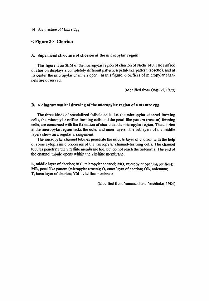
< Figu開 1>Chorion
The superficial structure of chorion
百lesurface of chorion is imprinted by the boundaries of follicle cells白紙producedit and thereby shows a conspicuous pattern (the egg patterns). This figure is a scanning
electron micrograph (SEM) ofthe upper central structure ofan egg (Nichi 140). The imp巾 tsof the follicIe cell boundaries are elevated, clearly demarcating small chorion fields beset with petit callus-like piles. At the imprints of the boundary of more than
three cells, aeropyles open.
Arrow, aperopyle (Modified合omOhtsuki, 1979)
The section of chorion
This is a cross section of chorion in a lateral region of an egg of the commercial hybrid (Shunrei x Shogetsu). The chorion is composed ofthree layers; the inner layer which consists of two thin granular sublayers and a trabecular porous layer, the thick middle layer which displays a pile of abundant thin fine-fibrous sublayers with grain-like arrangement, and the outer layer (thinner白anthe middle layer) which shows a pile of thin layers with slightly high electron density. The thickness of chorion varies ac-cording to races. The European races have the出ickestchorion, while the thickness is reduced in the Japanese, Chinese-univoltine, and maximally in the Chinese-bivoltine races. Even in the same egg, there are regional differences, for example its chorion becomes the thickest at the upper central region (Ohtsuki, 1979).
Cbm, middle layer of chorion; Cho, outer layer of chorion; Tl, inner layer of chorion; Vm, vitelline membrane
(Modified合omMiya, 1978)
Note : The gultaraldehyde-osmium double fixation is adopted unless a note is added to a Figure. The scale indicates 1μm unless a number is added to the bar.


12 Architecture of Mature Egg
< Figure 2> Chorion
Enlarged structure of chorion of the commercial hybrid (Shunrei x Shogetsu).
A. Enlarged micrograph of the outer layer and part of the middle layer
The outer layer consists of a pile of compact sublayers of a moderate electron den-sity, about 0.2μm thick. Sixteen tbin layers are observed in this figure. Some dense material is outside the outer layer. This adbesive material is secreted合'omthe female collaterial (mucous) gland during egg laying. The spaces between the outer sublayers, which are regarded as the imprints of follicle cell boundaries, and tubular structures between the thin sublayers ofthe middle layer, are the aeropyles.
Ap, aeropyles; Cbm, middle layer of chorion; Cbo, outer layer of chorion
B. Enlarged micrograpb of tbe middle layer of cborion
The middle layer displays a pile ofthin sublayers about 0.4μm thick with fine-fibrous grain-Iike arrangements. This sample displays 40 sublayers. Small irregular spaces are scattered among and within the sublayers that are less compact由anthe outer
layer.
C. Enlarged micrograpb of tbe inner layer and pa凶 oftbe middle layer
The s汀uctureof the inner layer is very complex. There are two thin layers and a trabecular layer between them. The innermost thin layer is about 0.2μm in thickness and contains m佃 yunevenly dis甘ibutedgranular porosities. The middle回 becularlayer is about 0.4μm thick, and the outer layer is about 0.4μm, containing a lot of sponge-Iike spaces. In some races, the outer layer is vague in certain chorion regions.
It seems that the inner layer has the cIosest relation wi白 suchphysiological func・tions ofthe egg such as breathing (Hinton, 1969).
Cbm, middle layer of chorion; Tl, inner layer of chorion
14 Architecture of Mature Egg
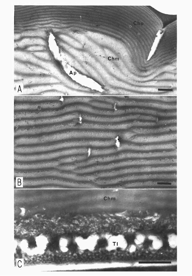
< Figu開 3>Chorion
A. Superficial structure of chorion at the micropylar region
百lisfig'町 eis an SEM ofthe micropylar region of chorion ofNichi 140. The s町白.ce
of chorion displays a completely different pattern, a petal-Iike pattern (rosette), and at its center出emicropylar channels open. In this figure, 6 orifices of micropylar chan-
nels are observed.
(Modified合omOhtsuki, 1979)
B. A diagrammatical drawing of the micropylar region of a mature egg
The three kinds of specialized follicle cells, i.e. the micropylar channel-fornting cells, the micropylar orifice-fornting cells and the petal-Iike pattern (rosette)-fornting cells, are concemed with白eforntation of chorion 鉱山emicropylar region. The chorion at the micropylar region lacks the outer and inner layers. The sublayers ofthe middle
layers show an iπegular arrangement. The micropylar channel tubules penetrate the middle layer of chorion with the help
of some cytoplasmic processes of the micropylar channel-fornting cells. The channel tubules pene汀ate出evitelline membrane too, but do not reach the oolemrna. The end of the channel tubule opens within the vitelline membrane.
L, middle layer of chorion; MC, micropylar channel; MO, micropylar opening (orifice); MR, petal-Iike pattem (micropylar rosette); 0, outer layer of chorion; OL, oolemma; T, inner layer of chorion; VM , vitelline membrane
(Modified from Yamauchi and Yoshitake, 1984)

B

16 Architecture of Mature Egg
< Figu問 4>Chorion
A. Structure of chorion at the micropylar and adjacent regions
Thes住uctureof chorion at the micropyl釘組dadjacent regions is shown in an egg of the Daizo. As the outer layer comes up to白emicropylar region, it becomes由innerand thinner, and it completely disappears at the micropylar region. At frrst the number of sublayers forming the outer layer is 9 in出isfigure. The outer layer becomes thinner 加 dat the same time the number ofthin sublayers reduces.
After由eseptum of the trabecular layer that occupies the central part of the inner layer disappears, a series of porosities are produced, and they become granular vesicles. They will, however, disappear completely at the micropylar region.
While 38 sublayers ofthe middle layer are arranged to make a stack parallel to the surface of the egg, the arrangement goes out of order at the micropylar region, and many spaces are scattered among the sublayers.
Chm, middle layer of chorion; Cho, outer layer of chorion; TI, inner layer of chorion
B. Enlarged figure of chorion at the micropylar region
An enlarged micropylar region of chorion of a Daizo egg is shown. The stacks of sublayers of the middle layer are completely disturbed. There are various sizes of spaces at the central part. The right side of the figure shows a slant section of a micropylar channel, whose inner wall is penetrated wi由 manymicrovilli, and a lot of fme particles are observed within the channel. Such structures will disappear after sperm goes through the channel.
Chm, middle layer of chorion; Mp, micropylar channel

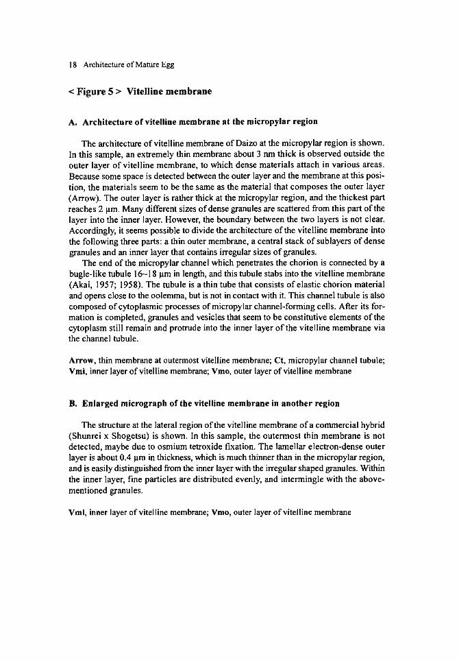
18 Architecture ofMature Egg
< Figure 5 > Vitelline membrane
A. Architecture ofvitelline membrane at the micropylar region
The architecture ofvitelline membrane ofDaizo at出emicropylar region is shown. In this sample, an extremely thin membrane about 3 nm thick is observed outside the outer layer of vite¥line membrane, to which dense materials attach in various areas. Because some space is detected between the outer layer and the membrane at this posi-tion, the materials seem to be the same as the material that composes the outer layer (Aηow). The outer layer is rather thick at the micropylar region, and the thickest part reaches 2μm. Manydi釘erentsizes of dense granules are scattered from this part of the layer into the inner layer. However, the boundary between the two layers is not clear. Accordingly, it seems possible to divide the architecture ofthe vitelline membrane into the following three parts: a thin outer membrane, a central stack of sublayers of dense granules叩 dan inner layer白紙 containsirregular sizes of granules.
The end ofthe micropylar channel which penetrates the chorion is connected by a bugle-like tubule 16-18μm in length, and出istubule stabs into the vitelline membrane (Akai, 1957; 1958). The tubule is a thin tube that consists of elastic chorion material and opens close to the oolemma, but is not in contact with it.百lischannel tubule is also
composed of cytoplasmic processes of micropylar channel-forming cells. A食erits for-mation is comtleted, granules and vesicJes that seem to be constitutive elements ofthe cytoplasm still remain and pro汀udeinto the inner layer ofthe vitelline membrane via the channel tubule.
Arrow, thin membrane at outermost vitelline membrane; Ct, micropylar channel tubule; Vmi, inner layer ofvitelline membrane; Vmo, outer layer ofvitelline membrane
B. Enlarged micrograph of the vitelline membrane in another region
The structure at the lateral region of the vitelline membrane of a commercial hybrid (Shunrei x Shogetsu) is shown. In this sample, the outermost thin membrane is not detected, maybe due to osmium tetroxide fixation. The lame¥lar electron-dense outer layer is about 0.4μm in thickness, which is much thinner than in the micropylar region, and is easily distinguished企omthe inner layer wi白血e廿Tegularshaped granules. Within the inner layer, fine particles are distributed evenly, and intermingle with the above-mentioned granules.
Vmi, inner layer ofvitelline membrane; Vmo, outer layer ofvitelline membrane


20 Architecture ofMature Egg
< Figure 6 > A diagrammatic drawing of ultrastructure at the anterior 問 glOn
The anterior periplasm region ofthe silkworm egg exhibits quite di貸erents加 ctures合omother regions because ofvarious phenomena conceming sperm reception. Relat-ing to出eperiplasm structures that play a significant role in出esperm entry, an expla-nation has been made in this figure. The chorion lacks its inner layer in the micropylar
region, and the micropylar channels that penetrate the middle layer protrude inward. 80th the outer and inner layers ofthe vitelline membrane are thicker in the micropylar region曲anin any other egg region. Especially, at the spot pierced with the micropylar channel tubule, the outer layer gains its thickness of2.0-2.5μm. In this area, a wide space occurs in the outer layer. From the end ofthe space, a channel tubule (about 0.6 μm in diameter) crooked somewhat and enclosed by a thin membrane (about 6 nm in 出ickness),pene甘atesthe inner layer to reach almost the surface of the oolemma.
The periplasm at the micropylar region shows a relatively thick layer of ooplasm 白紙terminatesin a series ofsmall com-like processes,合omwhich short microvilli-like processes pro汀udeinto the inner layer of the vitelline membrane. In the periplasm, stacks of rough endoplasmic reticulum have developed parallel to the oolemma. This architecture of lamellar stacks is one of the most conspicuous features白紙 canbe ob-served only 鉱山eperiplasm ofthe micropylar region. Within the ooplasm, outside of the stack, there are a great number of vesicles of various sizes and also aggregates of minute vesicles, about 80 nm in diameter, which contain less-dense materials. Such a S釘uc伽reis particular to the micropylar region called the “minute vesicle zone". Within the stacks of rough endoplasmic reticulum called the ER zone, there are no mitochon-dria, but some occur under the stacks. ER-associated rosettes made of electron-dense granules in a group stick to the end ofthe stacks. At the outside ofthe ER zone, there is an釘 eawhere a lot of vesicles that contain dense granules and a lot of filiform mito-chondria are also present (出egranule zone).
The reticuloplasm Iies adjacent to the periplasm. This ooplasmic network contains multivesicular bodies, a large size ofvacuoles and a layer (subcorticallayer) enclosing C-yolk spheres出atconsist oftranslucent materials and electron-dense spherules. The subcorticallayer appears thicker at出eanterior region and has less C-yolk spheres than any other region ofthe egg.
Ct, micropylar channel tubule; Ea, rER-associated rosette; ER, zone of endoplasmic reticu-lum; Gl, glycogen granule; Gol, Golgi body; GR, granule zone; Lip, lipid droplet; Mb, multivesicular body-Iike s汀ucture;Mit, mitochondrion; Mv, minute vesicle zone; Rg, thick-cortex round granule; rER, rough endoplasmic reticulum; Vc, vacuole; Vm, vitelline mem-brane; Yc, C-yolk sphere; Ys, yolk spherule

一・f
一一.・¥
• • •
.
y
21訴訟2Q : ~:â 9#~
. .
.
. ・.・ .
ナG.I

22 Architecture ofMature Egg
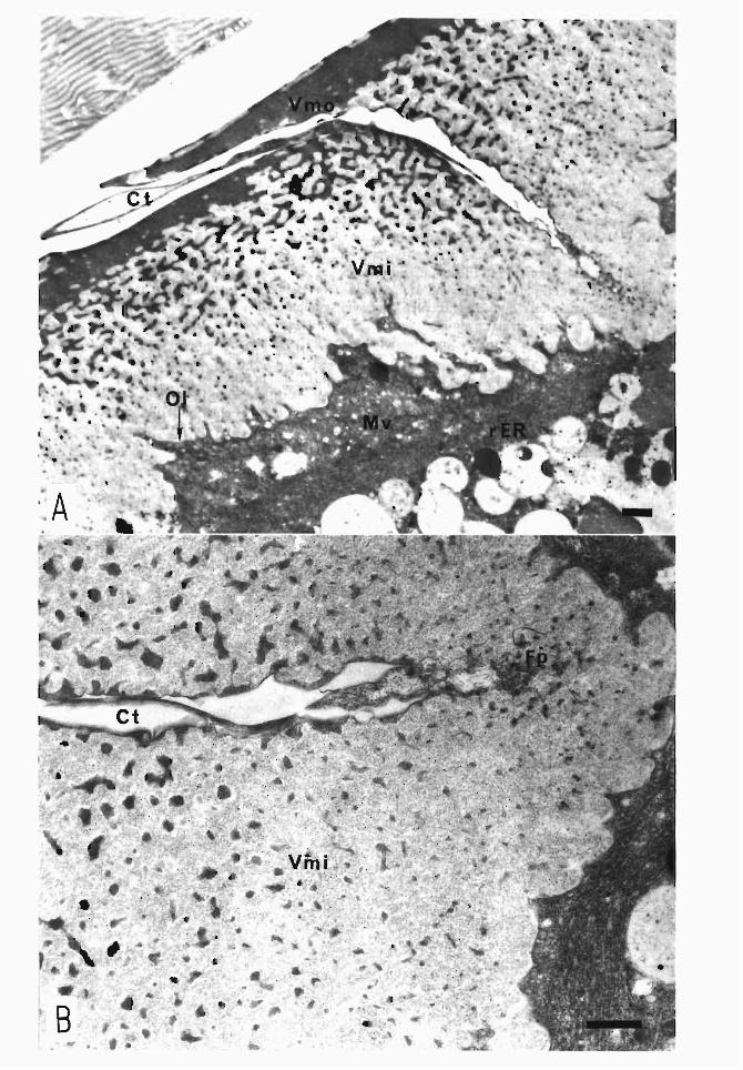
< Figure 7 > Vitelline membrane. at the micropylar region
A. Vitelline membrane at the micropylar region
The structure ofthe vitelline membrane ofDaizo at the micropylar region is shown.
The micropylar channel crooks and pene回 testhe middle layer of chorion and the outer
layer of vitelline membrane. The outer layer is composed of the outermost thin layer
and electron-dense sublayers. At the stabbing spot of micropylar channels, the outer layer remarkably increases the thickness and produces a wide space. Micropylar chan-
nel tubules run through this space. The inner layer is also thick, and it encloses scat-tered irregular-shaped dense granules. The micropylar channel tubule opens within the inner layer near the oolemma. Fine particles and minute vesicles are observed in the
inner layer around the opening.
Ct, micropylar channel tubule; Mv, minute vesicle; 01, oolemma; rER, rough endoplasmic reticulum; Vmi, inner layer ofvitelline membrane; Vmo, outer layer ofvitelline membrane
B. Enlarged micrograph of the micropylar channel tubule
An enlarged micrograph ofthe micropylar channel tubule ofDaizo is shown.百le
inner layer of the vitelline membrane is tightly packed with large electron-dense gran-ules at由earea c10se to the outer layer. Both the size and白edensity gradually decrease
toward the oolemma.
ηle micropylar channel tubule runs obliquely through the inner layer ofthe vitelline membrane but does not reach the oolemma. Instead it opens around the area. The chan-nel tubule contains many minute vesicles and fine particles, which are presumed to be deposited by the com-like processes ofthe forming cells. A part ofthese vesicles and p制 iclesflow白rough恥 channeltubule into the inner layer of恥 vitellinemembrane.
Ct, micropylar channel tubule; Fp, group of fine particles and minute vesicles released 合omthe channel tubule into the inner layer ofVmi; Vmi, inner layer ofvitelline membrane

24 Architecture of Mature Egg
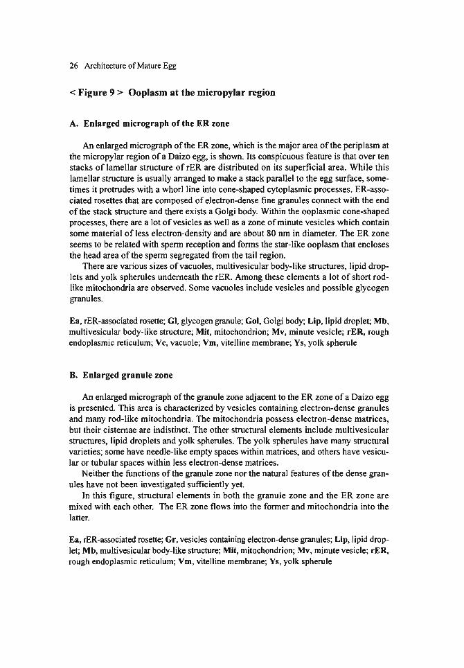
< Figure 8 > Ooplasm at the micropylar region
The figure shows the junction ofthe ER zone and the granu1e zone ofthe perip1asm
at the micropy1ar region of a Daizo egg. The perip1asm at the micropy1ar region dis-
p1ays seria1 rows of cone-1ike oop1asmic processes,合omwhich microvilli protrude into
the inner 1ayer ofthe vitelline membrane. In the ER zone, stacks of rough endop1asmic reticu1um paralle1 to白es町 faceofthe
egg take priority, and ER-associated rosettes consisting of groups of e1ectron-dense fine particles attach to the end of the stacks. Outside the stacks, there are numerous minute vesic1es, a few 1ipid drop1ets, and round granu1es with a thick cortex. Inside the stacks, multivesicu1ar body-1ike structures, various sizes ofvacuo1es, lipid drop1ets and glycogen granules are present. The ooplasm that fills the space contains a 10t of short
rod-like mitochondria.
The granule zone is characterized by a lack ofrER and presence ofmany short rod-like mitochondria and small vesicles wi曲 electron-densegranules. In this訂 eaofthe subcorticallayer, there are also many yolk spheru1es, multivesicu1ar body-like struc-tures, vacuoles of diverse sizes, lipid drop1ets, C-yolk spheres, and glycogen granu1es. The granule zone occupies the boundary between the anterior region of the periplasm and the other regions of an egg.
Ch, chorion; Ea, rER-associated resette; ER, zone of endoplasmic reticulum; GI, glycogen granule; Gol, Golgi body; GR, granule zone; Gr, electron-dense granule; Lip, lipid drop-let; Mb, multivesicular body-like structure; Mit, mitochondrion; Mv, minute vesicle; rER, rough endoplasmic reticulum; Vm, vitelline membrane; Ys, yolk spherule

. -' . . . .
Ch
ー. 『・-.
. .
.、
. !・
-・..
.r
26 Architecture ofMature Egg
< Figure 9 > Ooplasm at the micropylar問 gion
A. Enlarged micrograph of the ER zone
An enlarged micrograph ofthe ER zone, which is the major area ofthe periplasm at the micropylar region of a Daizo egg, is shown. Its conspicuous feature is that over ten
stacks of lamellar structure of rER are distributed on its superficial area. While this
lamellar structure is usually arranged to make a stack parallel to the egg surface, some-times it protrudes with a whorI line into cone-shaped cytoplasmic processes. ER-asso・
ciated rosettes that are composed of electron-dense fme granules connect wi由 theend
ofthe stack structure and there exists a Golgi body. Within the ooplasmic cone-shaped
processes, there are a lot ofvesicIes as well as a zone ofminute vesicIes which contain some material of less electron-density and are about 80 nm in diameter. The ER zone
seems to be related with sperm reception and forms the star-like ooplasm that encIoses the head area of the sperm segregated from the tail region.
There are various sizes ofvacuoles, multivesicular body-like structures, lipid drop-lets and yolk spherules undemeath the rER. Among these elements a lot of short rod-like mitochondria are observed. Some vacuoles incIude vesicIes and possible glycogen
granules.
Ea, rER-associated rosette; GI, glycogen gr叩 ule;Gol, Golgi body; Lip, Iipid droplet; Mb, multivesicular body-like structure; Mit, mitochondrion; Mv, minute vesicIe; rER, rough endoplasmic reticulum; Vc, vacuole; Vm, vitelline membrane; Ys, yolk spherule
B. Enlarged granule zone
An enlarged micrograph of the granule zone adjacent to the ER zone of a Daizo egg is presented. This釘 eais characterized by vesicIes containing electron-dense granules
and many rod-like mitochondria. The mitochondria possess electron-dense matrices, but their cistemae are indistinct. The other structural elements incIude multivesicular structures, Iipid droplets and yolk spherules. The yolk spherules have many s甘ucturalvarieties; some have needle-like empty spaces within matrices, and others have vesicu-lar orωbular spaces within less elec汀on・densematrices.
Neither the functions ofthe granule zone nor the natural feat町田 ofthedense gran-ules have not been investigated sufficiently yet.
In this figure, structural elements in both the granule zone and the ER zone are
mixed with each other. The ER zone flows into the former and mitochondria into the
latter.
Ea, rER-associated rosette; Gr, vesicIes containing electron-dense granules; Lip, Iipid drop-let; Mb, multivesicular body-Iike s汀uc制re;Mit, mitochondrion; Mv, minute vesicIe; rER, rough endoplasmic reticulum; Vm, vitelline membrane; Ys, yolk spherule

4
B r ds

28 Architecture of Mature Egg
< Figure 10> Diagrammatical drawings showing the 判明umptiveembry-
onic and extra-embryonic regions
A. Presumptive embryonic and extra-embryonic regions in the mature egg These diagrams iIIus甘atethe distribution of pyronine-positive granules in a mature
egg; Figure A (Ieft and right) shows a合ontalsection and a cross section, respectively. In the企ontalsection, obvious pyronine-positive granules are observed only at the lat-eral sides ofthe egg, but not in its anterior and posterior regions. The following values
出 tothe ranges without pyronine-positive granules were available: 20.3:t 1.7% on the whole circumference ofthe egg at the anterior region; 16.8 :t 0.8% at the posterior reglOn.
In the cross section, the above-mentioned pyronine-positive granules are seen in the lateral egg regions. The size of the granules is smaller in the ventral region and no
granules can be observed in the dorsal region. The range without the granules at the dorsal region was measured and the gained value is 15.6:t 1.6% on the whole circum-ference.
Ap, anterior pole; Pp, posterior po¥e; Ls, ¥atera¥ side; Vs, ventra¥ side; Ds, dorsa¥ side; Nc, who¥e circumference of an egg in case of a fronta¥ section; mc, range with no granu¥es at the anterior region; nc, range with no granu¥es at the posterior region; Lc, who¥e circumfer-ence of an egg in case of a cross section; Ic, range with no granu¥es atthe dorsa¥ region
B. Embryonic and extra-embryonic regions during the formation of embryonic anlage The embryonic and extra-embryonic regions are depicted during the formation of
the embryonic anlage in the合ontal(Ieft in figure B) and cross sections (right in figure B), respectively. Embryonic anlage is well developed on both sides ofthe lateral region except on the anterior and posterior egg poles. The range of extra-embryonic region with cells destined to form the serosa encompasses 21.8:t5.7 % ofthe egg circumfer-ence in the anterior, and 14.4 :t 5.7 % in the posterior regions. In a cross section, the range of extra引 nbryonicblastoderm occupies 13.7:t5.2 % ofthe egg circumference. These values are consistent with the data on the distribution of the pyronine-positive
granules (see above). The use ofp戸onine-positivegranules出 anindicator ofthe pre-sumptive embryonic region can be suggested. As embryogenesis continues on, how-ever, these granules gradually disappear. Therefore, we have not reached a conclusion
that these granules are defmitely the factor to determine the embryonic region. From an
ultras町田知almorphological point of view, pyronine-positive granules co汀espondto a
large concentric arrangement of rER and its enclosing structural elements. But again, this structure collapses with the progress of development. Investigation on this point should be continued much further.
Ap, anterior po¥e; Pp, posterior po¥e; Ls, ¥atera¥ side; Vs, ventra¥ side; Ds, dorsa¥ side; Nb, who¥e circumference of an egg in case of a fronta¥ section; mb, extra-embryonic range at the anterior region; nb, extra-embryonic range at the posterior region; Lb, who¥e circum-ference of如 eggin case of a cross section; Ib, extra-embryonic range at the dorsa¥ region
(Modified from Kobayashi and Miya, 1987)

里!x 100=20.3土1.7(0/0) 岡c
旦~x100=21.8:t5.7 (~o) Nb
-S
ELE
Vs Ds
Ls ~~ xI00=13.7土5.2(~o) Lb
旦txI00=14.4主1.6(~o) 聞b

30 Architecture ofMature Egg
< Figure 11 > Periplasm
This is the structure of periplasm at the lateroventral region of a Daizo egg. The
vitelline membrane is much thinner than in the micropylar region. In this figure, the outer layer ofthe vitelline membrane is 0.14..:.0.28μm and the inner layer is about 2μm
thick. The oolemrna pro汀udesmicrovilli into the inner layer. on the surface of periplasm, a lot of yolk spherules, smalllipid droplets, short rod-
like mitochondria, multivesicular structures and glycogen granules are distributed. In yolk spherules some varieties in respect to electron-density or its inner s汀uc同町S釘 e
observed. L訂 geC-yolk spheres (which include electron-dense白legranules) and big vacuoles
exist adjacent to白eperiplぉmsurface. In the space between them, there is a subcortical layer filled with the reticular plasm, in which rod-like mitochondria, thick-cortex round granules, Iipid droplets and glycogen granules are scattered. The C-yolk sphere is白eyolk that is formed at恥白lalstage of vitellogenesis, and it exi拘 onlyin a subco同ical
layer. It is characterized by a less electron-dense matrix containing high electron-dense
fine granules. It also shows some variation; for example, it possesses vesicles and gly-cogen granules.
One of the remarkable features of the periplasm at the lateroventral region is the
presence of pyronine-positive granules, as explained in the previous figure. From an ultrastructural point of view, these granules seem to co汀espondto a large concentric arrangement of rER and the structural element that surrounds the arrangement. This structure is distributed aかcentto the subcorticallayer. A detailed explanation will be
made in Fig. 13A.
Cb, chorion; GI, glycogen granule; Lip, lipid droplet; Mb, multivesicular body-Iike struc-ture; Mit, mitochondrion; rER, rough endoplasmic reticulum; Rg, thick-cortex round gran-ule; Vc, vacuole; Vm, vitelline membrane; Yc, C-yolk sphere; Ys, yolk spherule


32 Architecture ofMature Egg
< Figure 12 > Periplasm
This micrograph shows the structure of periplasm in the dorsal region of a Daizo egg. The vitelline membrane is about the same as in the lateroventral region, being much thinner than in the micropylar region. The oolemma protrudes the microvilli into
the inner layer of vitelline membrane. In the apical area of the periplasm, there are a lot of yolk spherules, small Iipid
droplets, short rod-Iike mitochondria and multivesicular structures. Among these ele-
ments, there are a lot of spaces where glycogen granules are dis汀ibuted.In yolk spherules many varieties are recognized just as in the cases at the lateroventral region.
The subcorticallayer adjacent to the apical area of periplasm is occupied with large C-yolk spheres and vacuoles. The C-yolk in this fig町 eappears 白11of ingredients, i.e., electron-dense granules, their enclosing vesicles, fme granules about 0.3μm in diam-
eter, 部 wellas small vacuoles are detected.
Although such a large concentric arrangement ofrER邸 observedat白elateroventral region, can not be observed among the ooplasmic network structure, there is a wide
territory that is occupied with stacks of rER. In other ooplasmic networks, rod-Iike mitochondria, lipid droplets and thick-cortex round granules are observed.
Ch, chorion; GI, glycogen granule; Lip, Iipid droplet; Mb, multivesicular body-Iike struc-ture; Mit, mitochondrion; rER, roUgh endoplasmic reticulum; Rg, thick-cortex round gran-ule; Vc, vacuole; Vm, vitelline membrane; Yc, C-yolk sphere; Ys, yolk spherule


34 Architecture ofMature Egg
< Figu問 13> Periplasm
A. Enlarged micrograph of an concentric arrangement of rER
This is a micrograph of a large concentric arrangement of rER adjacent to the sub-cortical periplasm layer in出elateroventral region of a Daizo egg.百leentire structure represents a shape of a star and contains stacks of rER with concentric arrangements every 0.07μm approximately. There are no other organelles inside, but only rod-Iike mitochondria and lipid droplets are distributed around this 町田知re.
The star-Iike ooplasm is surrounded by large C-yolk spheres, vacuoles, lipid drop-lets and glycogen granules. What is observed as a加gepyronine-positive granule出rougha Iight microscope in the Camoy's fixation is considered to be a stacked group ofthese
organelles.
GI, g1ycogen granule; Lip, lipid droplet; Mit, mitochondria; Vc, vacuole; Yc, C-yolk sphere
B. Enlarged micrograph of yolk spherules
S甘uctureofyolk spherules at the apical area ofthe periplasm in the dorsal region of a Daizo egg is shown. There are abundant and somewhat variable yolk spherules in出isarea. As a b邸 icarchitecture, needle-like structures and Iight vesicular or small佃bularstructures are scattered in a high electron-dense matrix, and they display a variety of pattems. The density ofthe fme particles that build the matrix is low and some ofthem have less electron density as a whole. A thick section of the sample fixed with gultaraldehyde-osmium fixation is stained deep blue with toluidine blue.
GI, glycogen granule; Lip, lipid droplet; Mit, mitochondrion


36 Architecture ofMature Egg
< Figure 14> Yolk system
A. A-yolk sphere
The structure of an A-yolk sphere in the yolk system of a Daizo egg is represented.
The yolk system that occupies most ofthe inner egg consists ofthe reticuloplasm that encIoses proteid yolk spheres, Iipid droplets and glycogen granules.
The structure can be morphologically cIassified in two types. ln the A-yolk spheres, 自negranules are evenly scattered, and its granule density determines the entire elec-tron-density ofthe sphere. A-yolk spheres are present mainly around the yolk system.
Within this reticuloplasm, the ground substance incIuding ribosomes and short rod-like mitochondria are observed.
GI, glycogen granule; Lip, Iipid droplet; Mit, mitochondrion; Rp, reticuloplasm; Ya, A-yolk sphere
B. B-yolk sphere
The structure of a B-yolk sphere in the yolk system of a Daizo egg is shown. It is the same sample as Figure A above. B-yolk spheres mainly occupy the central part ofthe
egg. A conspicuous feature of the B-yolk spheres is that they contain numerous elec-甘on-densegranules within the evenly distributed fme granulous ground substance.
In the reticuloplasm, the ground substance including ribosomes部 wellas short rod-like mitochondria are observed. Sometimes yolk spherules are distributed.
GI, glycogen granule; Lip, lipid droplet; Mit, mitochondria; Rp, reticuloplasm; Yb, B-yolk sphere; Ys, yolk spherule


38 Architecture ofMature Egg
< Figure 15 > Egg nucleus
The nucleus of an unfertilized (no spenn-entry) silkwonn egg stops dividing in the middle ofthe first maturation division and resumes meiosis after it is activated. This figure shows the structure of a nucleus of a Daizo egg dissected企omthe oviduct. Dur-ing the zygotene stage ofthe meiotic prophase, homologous chromosomes are bound by the s戸laptonemalcomplex (Rasmussen and Holm, 1982) and are arranged in the equatorial plane as in the metaphase. The electron-dense s汀uc刷re泊 thefigure repre-sents each homologous chromosome and the less electron-dense material that connects the two structures represents the synaptonemal complex.
Each pair of chromosomes is attached to spindle fibers that are made up of fine tubular structures and abundant fine granules. The spindle is surrounded by the fine vesicular or佃bul釘 roughendoplasmic reticulum, part of which builds stacked struc-tures. No other organelles or inclusions are present.
Chr, chromosome; rER, rough endoplasmic reticulum; Sc, derivative from the synaptone-mal complex; Sp, spindle fiber

40 Architecture of Mature Egg
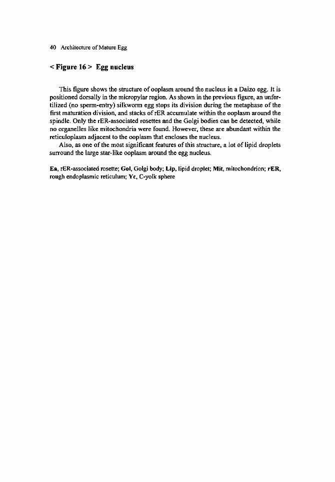
< Figure 16 > Egg nucleus
This figure shows the structure of ooplasm around the nucIeus in a Daizo egg. It is positioned dorsally in曲emicropylar region. As shown in血eprevious figure, an unfer-tilized (no spenn-entry) silkwonn egg stops its division during the metaphase of the frrst maturation division, and stacks ofrER accumulate wi由inthe ooplasm around the spindle. Only the rER-associated rosettes and the Golgi bodies can be detected, while no organelles Iike mitochondria were found. However, these are abundant within the reticuloplasm adjacent to the ooplasm白紙 enclosesthe nucIeus.
Also, as one ofthe most significant fea知resof this structure, a lot of Iipid droplets surround the large star-Iike ooplasm around the egg nucleus.
Ea, rER-associated rosette; Gol, Golgi body; Lip, lipid droplet; Mit, mitochondrion; rER, rough endoplasmic reticulum; Yc, C-yolk sphere


42
Chapter 2 Stages of Embryogenesis
Zygote nucleus (synkaryon) formed by白efusion ofthe male and female pronuclei starts embryogenesis with continuously repeated mitotic divisions. Taking its mo叩ho・logical features into consideration, the whole process is c1assified as several develop-mental stages. In the case of silkworm, with the exception of iωmultivoltine and bivoltine races, the embryogenesis stops obligatorily in a comparatively early stage and the em-bryos enter diapause. Numerous investigations, important for practical sericulture, have been made on the handling of diapausing“eggs" and the methods of artificial diapause termination. Takami (1969) divided the development ofsilkworms into the following six stages: (1) pre-diapause, (2) diapause, (3) hibemation, (4) critical stage, (5) forma-tion of organs, and (6) completion of larva. Subsequently, they were specified into 30 stages. The pre-diapause includes 7 stages: fertilization, cleavage, germanlage forma-tion, yolk cleavage, pyriform-shaped stage, Kokeshi (China-spoon like)-shaped stage and Chemical spatula-shaped stage. The two stages of diapause are diapause 1 and diapause 11. The hibemating period has 4 stages; the pre A, A, B-A and the B-B.百lecritical stage has the C-A叩 dthe C・B.The stages of organ formation釘 edivided into 10; the D-A, D-B, appearance of labral appendages, shortening stage, cephalothoracic segmentation, blastokinesis, completion ofblastokinesis, appearance of甘ichogencells, appearance of setae and the appearance of tracheal taenidia. The completion of I釘 vaincludes 5 stages; the head pigmentation 1, head pigmentation 11, body pigmentation 1, body pigmentation 11 and the hatch泊g.
Later, Ohtsuki (1979) replaced some expressions like A, B and C with others, and added some corrections to reclassi命thedevelopment into 30 stages (Ref. Yamashita and Yaginuma, 1991). 1 follow his c1assification but a few corrections have been added to accentuate the ultrastructural point of view.百ledevelopment a食erdiapause wil¥ be described in Vol. III,“Organogenesis". Each stage ofembryogenesis is characterized in Figs. 17 and 18.
2.1 Stage of fertilization
百lezygote nucleus (synkaryon) is formed by fusion ofthe male and female pronu-clei about two hours a食erperm entry.
2.2 Cleavage stage
Synchronous mitotic divisions produce a lot of c1eavage nuclei that migrate toward the periphery and begin to penetrate出eperiplasm beginning about 10 hr a食eroviposi-tion. From this stage to just before the diapause stage, some corrections are added,

Stages of Embryogenesis 43
based on u1釘astructuralobservations.
2.3 Stage of blastoderm and germanlage formation
As will be mentioned later, cleavage nuclei of the silkworm do not reach and pen-etrate出eperiplasm simultaneously all over the egg but do so frrst only in its anterior-half. The cell membranes are formed soon after the nuclei have reached the peripl部 m,and a distinction between the syncytial and the cellular blastoderm is di伍cult.Further-
more, at the advanced stage when the cleavage nuclei occupy the whole surface ofthe egg,出egermanlage can already be distinguished合omthe ex汀a-embryonicregion by higher cell density. Hence, in the silkworm there is no clear “blastoderm" as in some other insects. Takami named the described s飽ge“germanlageformation", while Ohtsuki called it“blastoderm formation".
2.4 Stage of germband formation
The expressions of“germanlage" and“germband" in silkworm embryogenesis do notcoπespond to those used in recent general descriptions of insect embryology. For example, according to Sander el al. (1985),“germanlage" means a monocellular em-bryo after serosa segregates合omthe blastoderm and "germband" means the subse-quent embryonic stage until gastrulation and segmentation. In my treatise, however, "germanlage" is defmed as an embryo which is separated clearly企om白eex甘a-embry-onic region and is still siωated at出esurface of an egg;‘germband' indicates an embryo from the stage of segregation from the surface and sinking into the egg,出roughg部国・
lation and segmentation, up to just before the diapause stage. Therefore, the stage of germband formation can be divided into a few more stages.
2.4.1 Completion of serosa
The cells in the extra-embryonic region gradually become flattened卸 dmigrate toward the lateral folds of germanlage that has segregated合omthe egg surface and begins to sink into the inner part of the egg. The extra-embryonic cells form a mem-brane (serosa) which encloses the whole egg. The germband at this stage is wide佃 dextends symmetrically in a saddle-like arrangement on both sides ofthe ventral median line.
2.4.2 Initiation of yolk cleavage
The broad germband grows anteriorly and posteriorly, and at the same time its width begins to decrease. From this stage, the yolk constituents start to be rearranged. Lipid droplets gather around the nuclei, which were left within the yolk mass during the blas-toderm formation. This stage precedes the phenomenon ofyolk cleavage when yolk cells are formed around the yolk nuclei. A remarkable phenomenon ofthis stage is the exclusion of a part of cytopl部 mat白e佃 teriorand posterior edges ofthe germband.

44 Stages of Embryogenesis
2.4.3 Differentiation of protocephalon and protocorm (pyriform-shaped stage)
The broad gennband keeps growing anteriorly and posteriorly, and simultaneously becomes narrower in the center part. As a result, it assumes a pyrifonn shape and it
becomes possible to distinguish批 protocephalonfrom the protoconn. Amnion anlage cells differentiate at the periphery ofthe gennband and become flattened as they extend over出eventral region to fonn the second embryonic envelope (amnion).
2.4.4 Spoon-shaped stage
Due to the growth ofthe gennband, the protocephalon and protoconn become dis-tinctly spoon-shaped, and the anterior and posterior ends of the gennband curve into the inner part ofthe egg. The amnion is completed, and the yolk c1eavage produces a
yolk system consisting of numerous yolk cells. Gastrulation, which occurs by primitive groove invagination, starts仕omthe center of the protocephalon.
2.4.5 Telson differentiation
The gennband is long and is composed of ectodenn and mesodenn. The prirnitive segments, which originate合omthe mesodenn segmentation, become distinct. The pos・
terior end is recognized as the telson, and also its inward invagination is advanced. Yolk cells are observed between the serosa and the amnion. ln the diapause egg, it takes about 48 hr to reach this stage after oviposition.
The above-mentioned periods constitute the pre・diapausestage. 1 follow Ohtuski's classification and characterization of subsequent stages ranging合omdiapause to eclo-
sion (the expressions in parentheses show c1assification by Takami).
2.5 Diapause stage 1
The embryo migrates企omthe outennost egg surface to the inner part. Around the
center, spaces without yolk cells can be observed.
2.6 Diapause stage n
The spaces that occupy the central part ofthe egg become larger. Free yolk cells are
scarcely detected.
2.7 Hibernating stage 1 (stage pre-A)
If a diapausing egg is kept at a low-temperature (around 5'C) similar to natural winter conditions, it gradually tenninates the diapause and starts its development when the appropriate temperaωre (about 25'C) is regained.百lestage is called吐lehibemat-
ing stageぺwhichis divided into 4 stages. In this stage (stage pre-A by Takami), the egg has not completely tenninated the diapause and a long tirne elapses to eclosion when it is
Stages ofEmbryogenesis 45
甘沼lSferredto 2S'C. The development of eggs within an egg batch is not simultaneous.
2.8 Hibernating stage 11 (stage A)
The egg almost completely tenninates the diapause.
2.9 Hibernating stage 111 (stage B・A)
The aggregates of yolk cells begin to be separated.
2.10 Hibernating stage IV (stage B・B)
The mesodenn at the由oracicsegment becomes considerably larger, and the aggre-gates ofthe yoIk cells are mostly separated.
2.11 Critical stage 1 (stage C・A)
This indicates the period企omthe end ofhibemation to the beginning ofthe organ fonnation. In an embryo during白isstage, the mass of mesodenn cells泊eachsegment conspicuously extends to both sides.
2.12 Critical stage 11 (stage C・B)
The protocephalon of the embryo sufficiently spreads.百lisstage occurs just before 出eappearance of the neural groove.
No remarkable changes in embryo morphology釘e問 cognized合om曲eabove-men-tioned diapause, but ul汀astructurally,morphological changes ofthe organelles釘 'echar-acteristic during these stages. There are a report on embryonic cells by Okada (1979) and also one on serosa cells and yoIk cells by Miya et 01. (1972).百lesereports will be described in ano由erbook, Vol. III,“Organogenesis" .
2.13 Stage of appearance of neural groove (stage D・A)
The embryo begins to extend, and a neural groove appe釘salong its ven回 Imedian line. The mass of mesodenn cells in each segment is divided into the right創叫 leftsides.
2.14 Stage of appearance of abdominal appendages (stage D・B)
Appendages appe釘鉱山e伊 athaland出oracicsegments, and succeedingly at the

46 Stages of Embryogenesis
abdominal segments. The length of embryo in this stage is longest during all the stages up to the blastokinesis.
2.15 Stage of appearance of pr舵 ess回 oflabrum
A pair of callus-like prωesses oflabrum appears at出e企ontend ofthe protocephalon.
2.16 Shortening stage
The embryo shortens, and spiracular and silk gland invaginations occ町.
2.17 Stage of cephalothoracic segmentation
The shortening ofthe embryo advances more, and伊 athalsegments unite, resulting in clear distinction of the白turehead企omthe thorax.
2.18 Blastokinesis (embryonic問 volution)
The ventral part of embryo faces the ven仕alregion ofthe egg. A sigmoid movement begins企omthe caudal end ofthe embryo白紙 isgradually pushed with its dorsal side to the dorsal side ofthe egg. The blastokinesis is accomplished in a short time, but can be divided into three phases, because its positional changes are clearly detected.
2.19 Stage of completion of blastokinesis
When blastokinesis ends, the embryo starts growing again. The formation of the dorsal integument makes progress, and the alimentary canals are completed.
2.20 Stage of appearance of trichogen cells
On the surface ofthe embryo, small masses oftrichogen cells are formed.
2.21 Stage of appearance of setae
Trichogen cells produce setae, which cover the surface of the embryo. Ocelli begin to be pigmented.
2.22 Stage of appearance of taenidia Taenidia are formed in the trachea. During this stage, mandibles start to be pig-

Stages of Embryogenesis 47
mented, and the formation of larval organs is completed.
2.23 Stage of bead pigmentation 1
After the completion of larval organs, pigmeotation of bodies makes progress to reach hatching. This pigmeotation is divided into 4 stages. A head is pigmented with brown.
2.24 Stage of bead pigmentat如nII
The serosa is swallowed, and the pigmented head looks blue合omthe outside. Ex-cept for the head, 00 other region has been pigmented yet.
2.25 Stage of body pigmentation 1
Other regions start to be pigmented (in addition to the head).
2.26 Stage of body pigmentation 11
百lewhole embryo is pigmented and looks blue.
2.27 Stage of batcbing
When the larval body is completed, the youog larva eats and breaks the chorion around the micropylar region to hatch.
In non-diapause eggs, the diapause stage, hibemating stage and critical stage are absent.

48 Stages of Embryogenesis
< Figure 17> Developmental stages ofembryos (part 1)
1. Fertilization (a longitudinal section): There are one zygote nucleus (synkaryon) and
three polar body nuclei.
2. Cleavage (a longitudinal section): The number of c1eavage nuclei increases by s戸1・
chronous mitotic divisions, and the cleavage nuclei begin to migrate toward the periphery.
3. Blastoderm and germanlage formation (a longitudinal section): The cleavage nu-
c1ei gradually reach the egg surface beginning企omthe anterior region. Rapid for-mation ofthe cell membranes makes it di佃cultto distinguish the s卯 cytialblastoderm
from the cellular blastoderm. The c1eavage nuclei which remain within the egg will
differentiate to the yolk nuclei.
4. Germband formation 1 (completion ofserosa) (a lateral view: all fig町 esbelow are
lateral views.): The germanlage separates from the egg surface and begins to sink
into the inside. The blastoderm cells ofthe ex甘a-embryonicregion flatten and pen-
etrate into the space where the germanlage had been present. The cellular mem-
brane (serosa) which covers the whole egg is completed. 5. Germband formation 11 (di能 rentiationof protocephalon叩 dprotocorm, pyriform・
shaped stage): As a result of elongation and a decrease in width, the germband acquires a“pyriform-shape", and protocephalon and protocorm are recognized.
6. Germband formation III (spoon-shaped stage): By continuous extension ofthe
germband,出eprotocephalon and the protocorm become obvious. The amnion and
the yolk c1eavage are completed. 7. Germband formation IV (telson di能 rentiation):The germband elongates and the
invagination of gastral groove (gぉ汀ulation)leads to the formation of separate ec-
toderm and mesoderm. Primitive segments become distinguishable due to meso-
derm segmentation, and the posterior end ofthe embryo is recognized部 thetelson.
8. Diapause stage 1: The embryo sinks into the inner part of the yolk mass. In the
center of the egg, spaces without yolk cells can be observed. 9. Diapause stage 11: The spaces in the central part reach their largest size.
10. Hibemating stage 1: The embryo gradually terminates the diapause under low-tem-
pera加rein the winter. It resumes its development in the appropriate temperature
(around 25t) only slowly because the diapause has not been completely termi-nated.
11. Hibemating stage 11 (stage A): The embryo almost completely terminates the dia-
pause, but its development is suppressed under natural winter circumstances such as low tempe
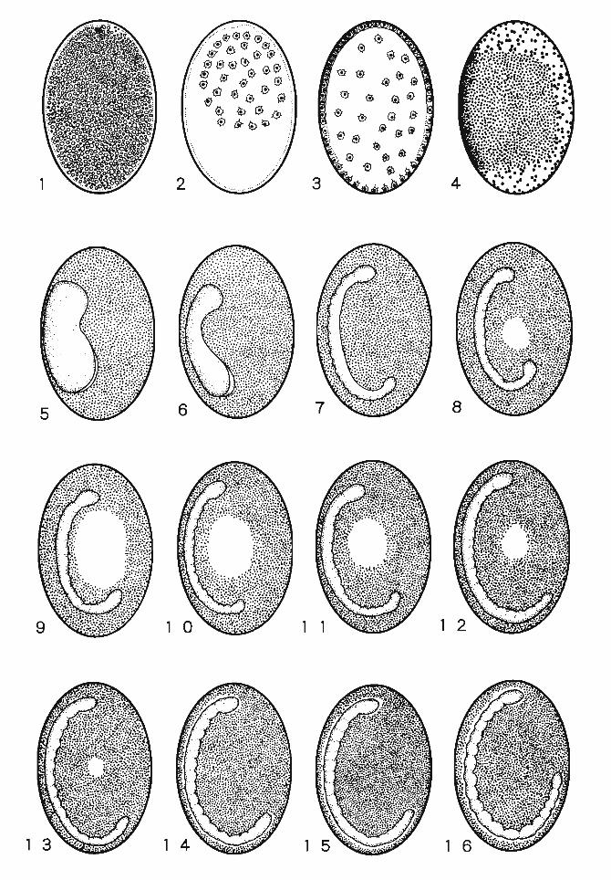

50 Stages of Embryogenesis
< Figure 18 > Developmental stages of embryos (pa同 2)
1. Appearance of abdominal appendages: Appendages appear on the gnathal and tho-racic segments, and subsequently on the abdominal segments.
2. Appearance of processes of labrum: A pair of callus-like processes of labrum ap-pears 鉱山e合ontend of the protocephalon.
3. Shortening stage: The shortening of embryo progresses, and spiracular invagina-tions develop.
4. Cephalo出oracicsegmentation: The shortening ofthe embryo advances. The gnathal segments unite, resuIting in the distinction of the向佃rehead合om白ethorax.
5. Early stage ofblastokinesis: The embryo is initially formed with its ventrum facing the ventral side ofthe egg. Later, with a sigmoid movement企omthe caudal end, the embryo changes its position and moves to the dorsal side. This movement is called blastokinesis (embryonic revolution) and it白lishesin a short time. It is divided into the, three stages.
6. Middle stage ofblastokinesis: The embryo shows a sigmoid position. 7. Final stage ofblastokinesis: The revolution ofthe tail part is completed. 8. Completion of blastokinesis: When blastokinesis ends, the embryo starts growing
again. The dorsal integument formation is completed, except for part ofthe thorax. 9. Appearance oftrichogen cells: Small masses oftrichogen cells that are going to
produce setae are formed on the embryo surface. 10. Appearance of setae: 8unches of setae are formed by the trichogen cells. 11. Appe釘 anceof taenidium: As taenidia are formed within the甘achea,the layout of
the tracheal system is visible合omthe outside. The formation of larval organs is completed.
12. Head pigmentation 1:百lehead ofthe embryo is pigmented to a brown color. 13. Head pigmentation 11: The pigmentation ofthe head is completed. 14. 80dy pigmentation 1: Other body regions start to be pigmented. 15. 80dy pigmentation 11: The whole embryo is pigmented and appears blue. 16. Hatching: When the larval body is completed,白e加vaeats and breaks the chorion
in the micropylar region to hatch. (Modified合omOhtsuki, 1979)

'園
自国
'同匝・
-


205
References
Akai, H. (1957) Studies on the electron microscopic structure of egg shell in Bombyx mori L. J. Sericult. Sci. Jpn., 26: 355・340(Jap飢 ese).
Akai, H. (1958) Observations of the micropylar 陀 gionofthe chorion in the silkwonn, Bomb.戸 moriL. Acta Sericol., 26: 19・21(Japanese).
Akai, H. (1976a) Ultrastructural Morphology oflnsects. 29Op., Tokyo Univ. Press, Tokyo (Japa-nese).
Akai, H. (1976b) SuゆceStructures of Insect. 139p., Tokyo Univ. Press, Tokyo (Japanese). Akutsu, S.佃 dYoshitake, N. (1974) Elec甘onmicroscopic observations on the greyeggshell ofthe
silkworm, Bomb.戸 moriL. J. Sericult. Sci.,み'n.43: 461・466(Japanese) A1a山u,S. and Yoshitake, N. (1977) Electron microscopic observations on the vitelline membrane
formation in the silkworm, Bomb.戸 moriL., J. Sericult. Sci.み'n.,46: 509・514(Japanese). Ando, H. (1988) lnsects. In Invertebrate Embryology (Dan, K., Sek.iguchi, K., Ando, H. and Watanabe,
H., eds.), Vol. 11, pp. 131・248.Baifuh・kan,Tokyo (Japanese). Asta町ov,B. L. (1967) Artificial par曲enogenesis釦 dexprimental polyploidy in silkworm. J. Sericult
Sci. Jpn., 36: 277・285.Balinsky, B. 1. (1986) Early di能 rentiationi目白 eggof恥 butterf1y,Acraea horta under normal
condition and after ultravioloet irradiation. Acta Embryol. Morphol. Experi. N. s., 16: 103・141.Fehrenbach, H., Dittrich, V. and Aissler, D. (1987) Eggshell fine structur官 ofthree lepidopteran
戸山:Cydia. pomonel/a (し)(Tortricidae), Heliothis virescens (Fabr.) and与"KJdopteraIittoralis (Boisd.) (Noctuidae). Int. J. Insect Morphol. Emblヲ101.,16: 201・141.
Frriedlander, M. and Gitay, H. (1972)百efate of the normal-ancleated spermatoz伺 ininseminated females of批 silkworm,Bombyx mori. J. Morphol., 138: 12ト129.
Gilbert, S. F. (2∞0) Developmental Biology (伊 edition),Sinauer Associates Inc., Massachusetts. Hashimoto, S. and Miya, K. (1987) Formation ofbrain ofthe normal and the embryonic lethal,“star-
spot duplication" embryos in the silkworm, Bomb.戸 mori(Bombycidae, Lipidoptera). In Recent Advances的 Embryologyin Japon and Poland (Ando, H. and Cz. Jura, eds.), pp. 237・254.lsebu Co. Ltd., Tsukuba.
Hinton, H. E. (1969) Respiratory systems ofinsect egg shells. Ann. Rev. Entomol., 14: 343・368.Ikeda, E. (1910) Embryonic development and morphological changes of Bombyx mori. Sangyoh・
Shinpoh, 18: 26-42 (Japanese). Ikeda, E. (1912) Morphological change of silkworm eggs until 2 days from oviposition. Sangyolト
Shinpoh, 20: 9・11;43・47(Japanese). Ikeda, E. (1913) Embryonic Development.
206 References
Bombyx mori. 1. Morphol., 158・57・72.Keino, H.釦 dTakesue, S. (1982) Scanning electron microscopic study on the early development of
silkworrn eggs (Bombyx mori L.). Develop. Growth Differ., 24: 287・294.Kobayashi, Y. and Miya, K. (1987) Structure of egg cortex relating to presumptive embryonic and
extraembryonic regions i目白 silkworrn,Bombyx mori (Bombycidae, Lepidoptera). In Recent Advances in lnsect Embryology in Japan and Poland (Ando, H. and Cz. J町a,eds.), pp.18ト194,Isebu Co. Ltd., Tsukuba.
Kuwana, J. and Takami, T. (1957) Insecta. In Embryology ollnvertebrate (Kume, M. and Dan, K., edsふpp.287・343,Baifukan, Tokyo (Japanese).
Matsuzaki, M. (1968) Electron microscopic observations on the chorion forrnation ofthe silkworrn, Bombyx mori L. 1..Sericult. Sci. Jpn. 37: 483・490(Japanese wi白 anEnglish summary).
Miya, K. (1958) Studies on the embryonic development of gonad in the silkworrn, Bombyx mori L. Part 1. DifTerentiation of gerrn cells. J. Fac. Agric. lwate Univ., 3: 436-467.
Miya, K. (1959a) Studies on the embryonic development of the gonad in the silkworrn, Bombyx mori L.P釘t11. Forrnation ofthe gonad. J. Fac. Agric.lwate. Univ., 4: 126・151
Miya, K. (1959b) Ele叫 onmlcro缶copic蜘 dieson血eembryonic development in the silkworrn, Bombyx mori. 1. Observations on白ehibemating egg. J. Seric. Sci. Jpn., 28: 163-164 (Jap叩 ese).
Miya, K. (1960) Electron microscopic studies on the embryonic development in the silkworrn, Bombyx mori. 11. Fine structure ofthe serosa. J. Seric. Sci. Jpn., 29: 273 (Japanese).
Miya, K. (1975) Ultrastructural changes ofembryonic cells during organogenesis in the silkworrn, Bombyx mori. 1. The gonad. 1. Fac. Agric. lwate Univ., 12: 329・363.
Miya, K. (1976) Ultrastructural changes of embryonic cells during organogenesis in the silkworrn, Bombyx mori. 11. The alimentary canal and the Malpighian tubules. 1. Fac. Agric. lwate Univ. 13: 95-122.
Miya, K. (1978) Electron microscope studies on the early embryonic development ofthe silkworrn, Bombyx mori. 1 Architecture ofthe newly laid egg and the changes by sperrn en町.J. Fac. Agric. lwate Univ. 14: 11-35.
Miya, K. (1984a) Early embryogenesis of Bombyx mori. In lnsect Ultrastructure (King, R. C. and Akai, H., eds.), Vol. 2, pp. 49・73,Plenum Press, New York.
Miya, K. (1984b) Early embryogenesis of“Kidney-shaped egg" in the silkmoth, Bomb砂 moriL. J. Fac. Agric.lwate Univ., 17: 131・149.
Miya, K., K町 ihara,M. and Tanimura, 1. (1972) Changes offine structures ofthe serosa cell and the yolk cell duri

References 207
silkwonn, Bombyx mori L. Zool. Mag. 77: 383・387(Japanese with an Englisb swnmary). Okada, M. (1970) Electron microscope studies on diapause embryos ofthe silkwonn, Bomb.砂 mori
L. Sci. Rep.おかoKyoiku Daigaku. Ser. B, 14: 95・111.Rasmussen, S. W. and Holm, P. B. (1982)ηle meiotic prophase in Bombyx mori. In Insect Ultra-
structure (King, R. C.叩 dAkai, H., eds.), Vol. 1, pp.61・85,Plenum Press, New York. Sakaguchi, B., Chikushi, H. and Doira, H. (1973) Observation of eggshell structur官 controlledby
gene action in Bomb.戸 mori.J. Fac. Agric. Kyushu Univ., 18: 53・62.Sander, K., Gutzeit, H. O. and Jackle, H. (1985) Insect embryogenesis: morphology, physiology,
geneticaI and molecularぉpects.In Comprehensive Insect Physiology. Biochemistry and Phar-macology (Kerkut, G. A. and Gilbert, L. 1., eds.), Vol. 1, pp. 319・386,Pergamon Press, Oxfo吋,NewYork.
Suzuki, Y. (1995) Genes山atare involved in morphogenesis and tissue differentiation of Bombyx embryos. In Insect Biochemistry and Molecular Biology (Ohnishi, E., Sonobe, H. and Tak油ashi,S. Y., eds.), pp. 301・315,Nagoya Univ. Pr官ss,Nagoya.
Tak油ashi,S. and Yagi, N. (1926) A defined region where an embryo develops in a Bombyx egg. Sangyoh-Shinpoh, (393): 266・268(Japanese).
T紘ami,T. (1969) A General Textbook ofthe Silkworm Eggs. 37Ip., Zenkoku Sanshu Kyokai, Tokyo (Japanese).
Takei, R. and Nagashima, E. (1975) Electron microscope investigation on the early developmental stages of diapause and non-diapause eggs in the silkwonn, Bomb.戸 moriL. J. Sericult. Sci. Jpn. 44: 118・124(Japanese).
Takesue, S. and Keino, H. (1980) Mechanism of nuclear migration before blastodenn fonnation in the silkwonn egg (Bombyx mori L). Zool. Mag., 89: 408.
Takesue, S., Keino, H. and Endo, K. (1976) Studies on the yolk granules ofthe silkwonn, Bomb.戸mori L.: The mo巾hologyof diapause and non-diapause eggs during early developmental stages. Wilhelm Ro制量Archiv.,180: 93・105.
Takesue, S., Keino, H. and Onitake, K. (1980) Blast叫 ennfonnation in the silkwonn egg (Bomb.戸mori L.) J. Embryol. Exp. Morphol., 60: 117・124.
Tekesue, S., Onitake, K. and Keino, H. (1977) Studies on the blastodenn and gennband fonnation during early embryogenesis in the silkwonn, Bomb.戸 moriL. 2001. Mag. 86: 345 (Japanese).
Tichomiroff, A. (1879) Ueber die entwicklungsgeshichte 抑制denwunns.Zool. Anz., 2: 64-67. Toyama, K. (1896) Structure of出eeggs and development of白eembryos in恥 silkwonn,Bombyx
mori L. Nogakukai・hoh,(28): 28・80(Japanese). Toyama,


Subject Index (except for Figure legends)
Abdominal segment, 46
Acrosome reaction, 52
Aeropyle, 6
Alimentary canal, 3, 46
Amnion, 44, 106, 107, 126, 140, 150, 168,
184,202
Blastoderm, 1,2,3,7,8,43,86,87, 106,
107,201,202
Blastokinesis, 42, 46
Blood cell, 2
Body pigmentation, 42,47
Brain, 3
Bursa copulatrix, 5, 52
Caudal end, 46, 140, 201
Cellular blastoderm, 43, 86, 87
Cephalic region, 201
Cephalocaudal 似 is,1,200
Chemical spatula-shaped, 42, 106
Chorion, 3,4,5,6,47,52,
Chromatin, 54,86,107, 126, 150, 151
Chromosome, 9,54,77
Cleavage, 2, 3, 9, 42, 43, 44, 76, 77, 86, 87,
106, 126, 168, 2Ql, 202
Cleavage nucleus, 77,86,201
Copulatory pouch, 5
Critical stage, 42,45,47
Cuticle, 4, 184
Desmosome, 107, 150, 168
Diapause, 2,3,42,43,44,45,47,54, 106,
126, 140, 150, 168, 184,200,202,203
Diapause egg, 3,44,47, 126, 140, 150, 168,
184,202
Dorsoventral axis, 1, 200
Eclosion, 44
Ectoderm, 1, 2, 44, 168, 200, 202
Eggshell, 4, 5
Eliminated chromatin, 54
209
Embryogenesis, 1,2,3,4,5, 7, 8, 42, 43, 52,
76, 106, 107, 126,200,201,202,203
Embryonic anlage, 5, 8
Embryonic envelope, 44, 150, 168, 202
Embryonic region, 1,3,8,43, 86, 87, 106,
200,201
Endoderm, 1, 2
Endoplasm, 3,7,8,9
Equatorial plane, 9, 54
ER-associated rosette, 9, 54
Exocytosis, 53
Extra-embryonic region, 1,3,8,43,86,87,
106,200,201
Female pronucleus, 54, 76
Fertilization, 4, 42, 52, 53, 54, 76
Fertilization membrane, 4, 53, 54, 76
Follicle cell, 6, 200
Follicle epi白elium,5, 200
Gastrulation, 43, 44
Germanlage, 42,43,86, 106, 107, 150,201,
202
Germband, 1, 2, 3, 43, 44, 106, 107, 126,
140,150,151,168,169,184,201,202,203
Genomes, 76
Glycogen, 7,8,9,53,87, 106, 107, 126, 140,
150, 184, 202
Gnathal segment, 46
Golgi body, 7, 8, 9, 54, 126
Gonad, 1,3
Gonomeη76
Hatching, 42, 47
Head, 2,4,42,43,46,47, 52, 53, 55, 106,
107,201
Head pigmentation, 42,47
Hibernating (Hibernation), 42,44,45,47
210 Subject Index
Integument, 46 Phagosome, 168, 184
Polar body, 54
Karyokinesis, 54 Pole cell, 200
Ko/ceshi (China-spoon Iike)-shaped, 42, 106 Pole plasm, 7,200
Polytrophic ovariole, 200
Labral appendage, 42 Pre・diapause,42, 44, 184
Labrum, 46 Primary yolk nuc1eus, 86, 87, 106
Male pronuc1eus, 54, 76
Malpighian tubule, 2, 3
Maturation division, 9, 54
Mesoderm, 1,2,44,45,150,168,200,202
Micropy1ar channel, 5, 6, 52, 200
Micropylar channel tubule, 6, 52, 200
Micropylar orifice, 6
Micropylar rosette, 6
Midgut, 2
Mitochondria, 3, 7, 8,9, 54, 55, 77, 86, 87,
106, 107, 126, 150, 168, 184,202
Neural groove, 45
Non-diapause egg, 3,47, 126, 140, 150,202
NucIeolus, 87,126,140,151,168,184,202
Nurse cell, 200
Ocellus, 46
Oocyte, 200
Oogenesis, 1, 5, 200
Oolemma, 3, 6, 7, 8, 52, 53, 54, 55, 76, 86,
200,201
Ooplasm, 5, 7, 8, 9, 53, 54, 55, 76, 77, 86, 87,
107, 126,200,201
Organelle, 7,8,45,53,77,86, 106, 107, 140,
150, 151, 168, 169, 184,200,201,202
Oviduct,久 52
Oviposition, 5,8,42,44,54, 150, 168, 184
Ovipositor, 5, 52
Ovulation, 5
Periplasm, 2,3,5,7,8,42,43,52,53,76,86,
87, 106,201
Primitive groove, 1,44, 150, 168,202
Primordial germ cell, 1,2,3,7, 126,200
Proctodeum, 2
Prothoracic gland, 2
Protocephalon, 44, 45,46, 106, 126, 140,
150, 168,201,202
Protocorm, 44, 106, 126, 140, 150, 168,201,
202
Pyriform-shaped stage, 42, 44, 106, 140,
150,202
Reticuloplasm, 5,7,9, 77, 86, 87, 106
Secondary vitelline membrane, 106, 126
Segmentation, 42,43,44,46, 168
Seminal duct, 52
Seminal receptacIe, 5
Serosa, 3,4,43,44,45,47, 106, 107, 140,
150, 168, 184,201,203
Serosal cuticIe, 184
Seta, 1,42,46, 126
Silk gland, 1,46
Silk gland invagination, 46
Spermatheca, 5, 52
Spermatophore, 52
Spiracular invagination, 46
Spiral tube, 52
Spoon-shaped stage, 44, 106, 150, 202
Stomodeum, 2
Subcorticallayer, 8,53,76, 106,200,201
Subesophageal body, 2
Synaptonemal complex, 9, 54
Syncytial blastoderm, 86
Synkaryon, 42, 76

Taenidium, 42, 46
Telson, 44, 106, 150, 168,202
Thoracic segment, 45
η10rax, 46
Trachea, 1, 42, 46
Trichogen田 11,42,46
Vagina, 52
Vestibrum, 52
Vitel1ine membrane, 3,4,5,6, 7, 8, 52, 53,
54,76,106,126,200
Vitellogenesis, 8
Yolk cell, 2, 3, 43, 44, 45, 107, 140, 168,
169, 184,201,202
Yolk cIeavage, 2,42,43,44, 106, 168,202
Yolk granule, 3, 8
YolknucIeus, 43,86,87,106,107,126,168,
202
Yolk sphere, 7,8,9, 77, 87, 106, 126, 184,
202
Yolk spherule, 7,8, 106, 107, 150
Yolk system, 7,8,9,44, 106, 126, 140, 168,
202
Zygote, 1,42,76,77
Subject lndex 211

ISBN4-906666-25-6 C3045 ¥14000E