the early stages of heart development: insights from ... · the early stages of heart development:...
TRANSCRIPT

Journal of
Cardiovascular
Development and Disease
Review
The Early Stages of Heart Development: Insightsfrom Chicken Embryos
Johannes G. Wittig and Andrea Münsterberg *
School of Biological Sciences, University of East Anglia, Norwich Research Park, Norwich NR4 7TJ, UK;[email protected]* Correspondence: [email protected]; Tel.: +44-1603-592232
Academic Editors: Rolf Bodmer and Georg VoglerReceived: 4 March 2016; Accepted: 30 March 2016; Published: 5 April 2016
Abstract: The heart is the first functioning organ in the developing embryo and a detailedunderstanding of the molecular and cellular mechanisms involved in its formation provides insightsinto congenital malformations affecting its function and therefore the survival of the organism.Because many developmental mechanisms are highly conserved, it is possible to extrapolate fromobservations made in invertebrate and vertebrate model organisms to humans. This review willhighlight the contributions made through studying heart development in avian embryos, particularlythe chicken. The major advantage of chick embryos is their accessibility for surgical manipulationand functional interference approaches, both gain- and loss-of-function. In addition to experimentsperformed in ovo, the dissection of tissues for ex vivo culture, genomic, or biochemical approachesis straightforward. Furthermore, embryos can be cultured for time-lapse imaging, which enablestracking of fluorescently labeled cells and detailed analysis of tissue morphogenesis. Owing to thesefeatures, investigations in chick embryos have led to important discoveries, often complementinggenetic studies in mice and zebrafish. As well as including some historical aspects, we cover here someof the crucial advances made in understanding early heart development using the chicken model.
Keywords: chick embryo; fate mapping; heart fields; morphogenesis; in ovo studies
1. Introduction
The detailed mechanistic understanding of developmental processes is a major requirement tobe able to identify the embryonic origin of diseases and to develop future therapeutic interventions.Different model organisms have been established to study patterning and organogenesis in developingembryos. Important metazoan model organisms include the nematode (Caenorhabditis elegans), the fruitfly (Drosophila melanogaster), the tunicate (Ciona intestinalis), a few species of sea urchin, the teleost fish(Danio rerio), the African claw-toed frog (Xenopus laevis), the mouse (Mus musculus), and the chicken(Gallus gallus). All of these have different advantages and have made significant contributions to ourunderstanding of developmental processes. The focus of this review will be the chicken, specifically itsrole in our current understanding of early heart formation.
The chicken is a “classic” model organism and the first meaningful information obtained throughits use arose in the 17th century, when it was shown that embryos are not preformed but developbody parts progressively. Further fundamental discoveries were dependent on the development ofoptical microscopes, which made it possible to discover the three germ layers: ectoderm, mesoderm,and endoderm. A comment on Charles Bonnet’s ideas on “fecundation” and development of thegerm (egg) was published in the late 19th century [1]. Since then developmental biology research haschanged dramatically owing to advances in genetics and in cell and molecular biology, which enabledmuch progress and a “golden age” for the discipline [2]. Analyses have become more sophisticated,focusing on discrete regions in the developing animal.
J. Cardiovasc. Dev. Dis. 2016, 3, 12; doi:10.3390/jcdd3020012 www.mdpi.com/journal/jcdd

J. Cardiovasc. Dev. Dis. 2016, 3, 12 2 of 15
The chick embryo is ideal for studying the early development of the heart, the first functioningorgan in the embryo. A major advantage is that the chick develops ex utero in an egg, which allowseasy accessibility during all stages of development post-laying. This ease of access enables in ovomanipulations and observation of the embryo, such as dissection, grafting, micro-injection, andlabeling, and this has made the chicken popular, even before the molecular age [3–5]. Particularlypowerful have been grafting and ablation experiments. When combined with the use of quail/chickchimeras [6], this approach allowed the tracing of grafted cells before genetic labeling became possible.Establishing methods for ex ovo development and introduction of constructs encoding fluorescentlylabeled proteins by electroporation has facilitated the imaging of cell movement in live embryosusing advanced microscopy [7,8]. Advanced tools for image registration allow for the alignment andcomparison of multiple specimens in the absence of morphological landmarks [9]. By directly labelingthe extracellular matrix, it has also been possible to measure active versus passive motion of cells,including cardiac progenitors, during gastrulation [10,11]. The use of CRISPR/Cas9-mediated genomeediting via targeted electroporation allows the generation of genetic mosaics; combined with imagingthe behavior of mutant cells can then be studied in detail, for example in developing somites [12].Furthermore, improved methods for transgenesis and the availability of lines, both quail and chick,transgenic for fluorescent markers expressed either ubiquitously or restricted to specific cell lineages,has enhanced the utility of avian models [13–15].
Finally, the mature chick heart comprises four chambers with in- and out-flow tracts, and despitesome differences, for example during septation and aortic arch remodeling [16], it resembles the humananatomy more closely than other non-mammalian model organisms. Owing to those features, andthe available tool-kit described above, avian embryos will almost certainly continue to contributesignificant insights into the development of the heart.
2. Cardiac Development and Morphogenesis
2.1. Mapping Studies and Characterization of Cardiogenic Fields
In the chick embryo, systematic observations and comparative analyses were boosted whenHamburger and Hamilton established a classification scheme for developmental stages that wasuniversally adopted [17]. A recent reference guide maps the stages of heart development onto theHH-stage series [18]. In addition, the series has been refined for the stages of gastrulation [19], whichstarts with the formation of the primitive streak in the midline of the embryo.
In the early chick gastrula (Hamburger-Hamilton, HH stage 3), cardiac progenitors are located inthe mid-primitive streak, from which they ingress to enter the mesoderm bilaterally [20–23]. By HH4,the late gastrula/early neurula stage, the contribution of the primitive streak to the heart ceases [21,24].At that stage precardiac areas are organized into bilateral heart fields located in the lateral platemesoderm, which subsequently splits into the somatic and splanchnic layers, the latter comprisingcardiogenic cells. Bilateral heart fields were originally characterized by culturing isolated cells andtesting their potential to generate spontaneously contracting cardiomyocytes [20,25].
Early studies tracing cardiac cells in gastrula stage embryos used isotope labeling andautoradiography, thus defining bilateral heart fields that are initially separate but then fuse togenerate the tubular heart at early somite stages [26]. In mouse embryos, the timing is differentand the heart field mesoderm merges together across the midline at the 1-somite stage (E7.5), forminga “crescent” [27,28].
Additional insights regarding the origin of cells contributing to the heart as well as the aorticarches derived arteries were obtained through interspecies grafts that generate quail–chick chimeras.This approach, developed by Lièvre and Le Douarin [29], was important for studies in avian modelsystems and a reliable and sensitive alternative to methods involving radioactive isotopes [25]. Usingquail–chick chimeras and fluorescent vital dye injections, a more precise fate map was generated [21].This showed that cardiomyocyte and endocardial precursors arise from a rostral portion of the

J. Cardiovasc. Dev. Dis. 2016, 3, 12 3 of 15
HH3 primitive streak, and that the craniocaudal organization of cells within the streak reflects thecraniocaudal arrangement of the linear heart tube [21], extending the earlier cardiogenic “potencymap” of the primitive streak by DeHaan [20]. The linear heart tube becomes extended and refined byadditional cell populations contributing to the mature heart (see Section 2.3).
2.2. Pre-Gastrula and Gastrula Stages
2.2.1. Specification and Migration of Cardiac Progenitor Cells
Cardiogenic potential can be detected in pre-streak, blastula stage embryos prior to gastrulationbefore the heart fields emerge. Pre-streak stage chick embryos are a flat disc composed of two layers,the epiblast (upper layer) and the hypoblast (lower layer). Cardiac progenitors are found withinthe posterior half of the epiblast [30] and these cells have cardiogenic potential in culture [31,32].These authors also showed that the hypoblast is required to induce cardiac myogenesis in the earlyepiblast, and furthermore, that Tgfβ/activin is sufficient to substitute for its cardiogenic-inducingability [31,32]. In contrast, BMP-2 and BMP-4 inhibit cardiogenesis at this stage, consistent with studiesthat show that BMP antagonists, such as chordin, can induce the expression of the early marker,smooth-muscle alpha actin (SMA), in cultured posterior epiblasts at pre-gastrula stages [33]. In mice,transplantation experiments combined with embryo culture showed that epiblast cells can acquirea cardiac fate independent of ingression through the primitive streak [34]. Thus, in both chicks andmice, ingression itself is not necessary for fate specification.
Soon after gastrulation, prospective cardiac cells migrate to the anterior lateral mesoderm and thebilateral heart fields contain prospective endocardial and myocardial cells, indicating that cardiac fatesare allocated in the primitive streak or earlier prior to cell migration. This idea was confirmed usinglineage tracing with low titers of a replication-defective retrovirus expressing LacZ. The labeled cellsgave rise to either myocardial or endocardial derivatives [35].
Using chick embryos and ex vivo tissue recombination experiments it was possible to identifythe origin of signals in the endoderm underlying the bilateral heart field mesoderm in the anteriorlateral plate that trigger the commitment to the cardiac lineage [36]. Pioneering studies identifiedthe crucial role of BMP signaling post-gastrulation. Beads soaked in recombinant BMP-2 couldinduce ectopic expression of early cardiac markers, such as the transcription factors GATA-4 andNkx-2.5. Furthermore, recombinant BMP-2 or BMP-4 protein induced myocardial differentiationand beating in explants of non-cardiogenic mesoderm, while exposure to the secreted proteinNoggin, a BMP-antagonist, completely inhibited differentiation of precardiac mesoderm [37,38].The competency to respond to BMP-2/4 alone was stage dependent [39] and restricted to anteriormesoderm explants. Subsequently it was shown that interactions between BMP-2 and FGF-4 pathwaysare important for the induction of cardiac cell fate in the posterior mesoderm [40] by directly targetingthe transcription factor Nkx2.5 [41].
Additional experiments conducted in both chick and Xenopus gastrula stage embryos revealedthat inhibition of canonical Wnt/β-catenin signaling is critical for heart development [42,43], whereasβ-catenin-dependent Wnt signaling in the posterior lateral mesoderm induced hematopoiesis [42].The Wnt family of secreted proteins initiates several signal transduction pathways, recently reviewedin the context of heart development [44]. Antagonists of β-catenin-dependent Wnt signaling thatpromote cardiogenesis include dickkopf (Dkk1) and crescent. In chicks, crescent is expressed inanterior endoderm during gastrulation and can induce the expression of cardiac genes in posterior,non-cardiogenic tissues in vitro [42]. The conditional genetic ablation of β-catenin in early mouseembryos also led to a proposed cell fate switch and ectopic heart formation [45]. These observationsare consistent with the idea that β-catenin-dependent Wnt signaling represses cardiogenesis; however,this is context dependent. At an early stage of development, prospective cardiac cells are exposedto canonical Wnt-ligands: both Wnt-3a and Wnt-8c (known as Wnt8a in mouse and human) areexpressed in the primitive streak. Indeed, during the differentiation of embryonic stem cell derived
J. Cardiovasc. Dev. Dis. 2016, 3, 12 4 of 15
embryoid bodies, Wnt/β-catenin signaling is initially required for induction of mesoderm and thuscardiomyogenesis. Therefore, this pathway either enhances or inhibits cardiogenic differentiationdepending on the stage of development; it has been proposed that canonical signaling retainscardiac precursors in a proliferative precursor state, whereas non-canonical signaling promotes theirdifferentiation (reviewed in [44,46]).
Taken together, work in avian embryos demonstrated that inhibitors of β-catenin-dependent Wntsignaling act in concert with BMP and FGF signaling molecules to specify cells to cardiac fates duringearly neurula stages. These insights led to efforts to differentiate human pluripotent stem cells intocardiomyocytes [47]. Additional data indicate that FGF and BMP signaling pathway interactions areregulated by negative feedback loops involving microRNAs, particularly miR-130 and miR-133 [48,49].
Furthermore, β-catenin-independent (or non-canonical) signaling is important for cardiogenesis.Wnt binding to frizzled receptors and signaling through Dvl can activate alternative pathways,including the planar cell polarity (PCP) and Wnt/Ca2+ pathways [44]. Known mediators of theWnt/PCP pathway involve the ligand Wnt-11 and the small GTPase RhoA. In chicken embryos, RhoAcontrols tissue polarity and cell movement of cardiogenic progenitors [50,51]. Live-imaging and celltracking of cardiac progenitors have shown that during gastrulation a combination of BMP-2/4- andWnt/GSK3β-mediated signals is involved in controlling the migration of these cells towards thebilateral heart fields [52]. This work also showed that the two pathways are integrated by differentialphosphorylation of Smad-1: (1) at the carboxy-terminus in response to BMP-receptor activation; and(2) in the linker region by GSK3β kinase.
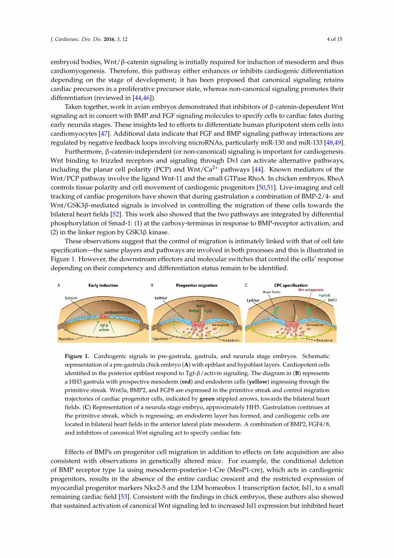
These observations suggest that the control of migration is intimately linked with that of cell fatespecification—the same players and pathways are involved in both processes and this is illustrated inFigure 1. However, the downstream effectors and molecular switches that control the cells’ responsedepending on their competency and differentiation status remain to be identified.
J. Cardiovasc. Dev. Dis. 2016, 3, 12 4 of 15
inhibits cardiogenic differentiation depending on the stage of development; it has been proposed that canonical signaling retains cardiac precursors in a proliferative precursor state, whereas non-canonical signaling promotes their differentiation (reviewed in [44,46]).
Taken together, work in avian embryos demonstrated that inhibitors of β-catenin-dependent Wnt signaling act in concert with BMP and FGF signaling molecules to specify cells to cardiac fates during early neurula stages. These insights led to efforts to differentiate human pluripotent stem cells into cardiomyocytes [47]. Additional data indicate that FGF and BMP signaling pathway interactions are regulated by negative feedback loops involving microRNAs, particularly miR-130 and miR-133 [48,49].
Furthermore, β-catenin-independent (or non-canonical) signaling is important for cardiogenesis. Wnt binding to frizzled receptors and signaling through Dvl can activate alternative pathways, including the planar cell polarity (PCP) and Wnt/Ca2+ pathways [44]. Known mediators of the Wnt/PCP pathway involve the ligand Wnt-11 and the small GTPase RhoA. In chicken embryos, RhoA controls tissue polarity and cell movement of cardiogenic progenitors [50,51]. Live-imaging and cell tracking of cardiac progenitors have shown that during gastrulation a combination of BMP-2/4- and Wnt/GSK3β-mediated signals is involved in controlling the migration of these cells towards the bilateral heart fields [52]. This work also showed that the two pathways are integrated by differential phosphorylation of Smad-1: (1) at the carboxy-terminus in response to BMP-receptor activation; and (2) in the linker region by GSK3β kinase.
These observations suggest that the control of migration is intimately linked with that of cell fate specification—the same players and pathways are involved in both processes and this is illustrated in Figure 1. However, the downstream effectors and molecular switches that control the cells’ response depending on their competency and differentiation status remain to be identified.
Figure 1. Cardiogenic signals in pre-gastrula, gastrula, and neurula stage embryos. Schematic representation of a pre-gastrula chick embryo (A) with epiblast and hypoblast layers. Cardiopotent cells identified in the posterior epiblast respond to Tgf-β/activin signaling. The diagram in (B) represents a HH3 gastrula with prospective mesoderm (red) and endoderm cells (yellow) ingressing through the primitive streak. Wnt3a, BMP2, and FGF8 are expressed in the primitive streak and control migration trajectories of cardiac progenitor cells, indicated by green stippled arrows, towards the bilateral heart fields. (C) Representation of a neurula stage embryo, approximately HH5. Gastrulation continues at the primitive streak, which is regressing; an endoderm layer has formed, and cardiogenic cells are located in bilateral heart fields in the anterior lateral plate mesoderm. A combination of BMP2, FGF4/8, and inhibitors of canonical Wnt signaling act to specify cardiac fate. Effects of BMPs on progenitor cell migration in addition to effects on fate acquisition are also
consistent with observations in genetically altered mice. For example, the conditional deletion of BMP receptor type 1a using mesoderm-posterior-1-Cre (MesP1-cre), which acts in cardiogenic progenitors, results in the absence of the entire cardiac crescent and the restricted expression of myocardial progenitor markers Nkx2-5 and the LIM homeobox 1 transcription factor, Isl1, to a small remaining cardiac field [53]. Consistent with the findings in chick embryos, these authors also showed that sustained activation of canonical Wnt signaling led to increased Isl1 expression but inhibited heart tube formation at the eight-somite stage [50,53]. Thus far it has not been possible to observe cardiac progenitor cell migration in real time using mice; however, advanced imaging approaches will soon be able to address this challenge [54].
Figure 1. Cardiogenic signals in pre-gastrula, gastrula, and neurula stage embryos. Schematicrepresentation of a pre-gastrula chick embryo (A) with epiblast and hypoblast layers. Cardiopotent cellsidentified in the posterior epiblast respond to Tgf-β/activin signaling. The diagram in (B) representsa HH3 gastrula with prospective mesoderm (red) and endoderm cells (yellow) ingressing through theprimitive streak. Wnt3a, BMP2, and FGF8 are expressed in the primitive streak and control migrationtrajectories of cardiac progenitor cells, indicated by green stippled arrows, towards the bilateral heartfields. (C) Representation of a neurula stage embryo, approximately HH5. Gastrulation continues atthe primitive streak, which is regressing; an endoderm layer has formed, and cardiogenic cells arelocated in bilateral heart fields in the anterior lateral plate mesoderm. A combination of BMP2, FGF4/8,and inhibitors of canonical Wnt signaling act to specify cardiac fate.
Effects of BMPs on progenitor cell migration in addition to effects on fate acquisition are alsoconsistent with observations in genetically altered mice. For example, the conditional deletionof BMP receptor type 1a using mesoderm-posterior-1-Cre (MesP1-cre), which acts in cardiogenicprogenitors, results in the absence of the entire cardiac crescent and the restricted expression ofmyocardial progenitor markers Nkx2-5 and the LIM homeobox 1 transcription factor, Isl1, to a smallremaining cardiac field [53]. Consistent with the findings in chick embryos, these authors also showedthat sustained activation of canonical Wnt signaling led to increased Isl1 expression but inhibited heart

J. Cardiovasc. Dev. Dis. 2016, 3, 12 5 of 15
tube formation at the eight-somite stage [50,53]. Thus far it has not been possible to observe cardiacprogenitor cell migration in real time using mice; however, advanced imaging approaches will soon beable to address this challenge [54].
2.2.2. Establishment of Left–Right Asymmetry
Shortly after the emergence of cardiogenic progenitors from the primitive streak and aroundthe time that they arrive in the heart fields, the bilateral symmetry of the early embryo is broken.Ultimately this leads to the striking left–right asymmetry in the placement and differentiation oforgans, which is seen in all vertebrates. Experiments in chick embryos have made major contributionsto our understanding of the mechanisms involved in this process. For a review see [55]. In particular,the gene network that provides left–right information was characterized in chick embryos [56]. Initialbreaking of symmetry starts at Hensen’s node, the organizing center at the anterior end of the fullyextended HH4 primitive streak. Several signaling molecules are asymmetrically expressed, includingactivin receptor IIa, Sonic hedgehog (Shh), and cNR1 (the chick homologue of mouse nodal); theexperimental manipulation of these pathways, through implantation of growth factor soaked beads orcell pellets, affects heart situs [56]. Furthermore, recent work showed that N-cadherin is involved inasymmetric gene expression and the leftward cell movements in Hensen’s node [57].
In mice, the use of a nodal-lacZ reporter allele confirmed its asymmetric expression on the leftside [58]. Although the mechanisms leading to initial breaking of symmetry are different in miceand chicks [59,60], in both species the transcription factor Pitx2 acts downstream of nodal and Shhsignaling. In chick embryos misexpression of Pitx2 is sufficient to produce reversed heart looping [61].The literature on genetic manipulations of Pitx2 is extensive and cannot be covered here; suffice it tosay that cardiac laterality defects are usually observed (for example [62], and references in [55]).
The signaling molecules expressed on the left side interact with a right-sided program, initiatedby BMP-4 at Hensen’s node inducing FGF8, which in turn activates Snai1, a Zn-finger transcriptionalrepressor. Snai1 is necessary for the formation of the proepicardium (PE), which in the chick developsonly on the right side—a vestigial PE on the left undergoes apoptosis. Ectopic expression of FGF8 orSnail on the left led to bilateral PE formation [63]. In the mouse, the PE, which is characterized byexpression of WT1 and TBX18, develops bilaterally. This may reflect differences in FGF8, which isa determinant of the right side in the chick but mediates left side identity in mice [59,64].
2.3. Discovery of Additional Heart Fields
Classic mapping experiments using labeling with iron oxide particles followed by time-lapsephotography indicated that new segments are added to the linear heart tube during looping, inparticular to generate outflow myocardium [65,66]. Cells residing in the ventral region of thesubcephalic fold of HH9´ were shown to be included at the cephalic end of the heart tube by HH12.Similar labeling showed that precursors for the right and left primitive atria are not yet present in theHH8–9 straight heart tube [67] but become incorporated later during loop stages. Building on thisearly work, the origins of secondarily added cell populations were characterized in more detail inboth the chick and mouse, using fluorescent dye or genetic labeling, respectively [68–70]. This showedthat cell populations contributing to the outflow tract (OFT) are located in the pharyngeal mesodermand the splanchnic mesoderm anterior, and immediately adjacent to the straight heart tube. Theseregions have been termed the anterior and secondary heart fields (AHF/SHF), respectively, and theirderivatives are shown in Figure 2. The cells contributing to the OFT express the transcription factorsNkx2.5 and GATA-4. They are also positive for HNK-1 immunostaining as they translocate into theheart [69,70]. Using vital dye injections and tissue grafting it was possible to map the location andingression sites of prospective AHF and SHF cells in the primitive streak of gastrula stage HH3 chickembryos [71]. This work showed that during early somite stages the Isl1-positive AHF progenitorswere located in the cranial paraxial mesoderm and the pharyngeal mesoderm [71], also consistent with

J. Cardiovasc. Dev. Dis. 2016, 3, 12 6 of 15
studies that identified a close relationship between these progenitors and some craniofacial skeletalmuscles, in both the chick and mouse [72,73].J. Cardiovasc. Dev. Dis. 2016, 3, 12 6 of 15
Figure 2. Cardiac morphogenesis in chick embryos. Schematic ventral views of HH8 to HH24 chick hearts. Fate mapping revealed the location of first and second heart fields (FHF, SHF), marked in green and red. Fusion generates a primitive heart tube by HH9; secondarily added cell populations have not yet entered (red dots). In all images, components of the heart derived predominantly from FHF are in green and components derived predominantly from SHF and also AHF are in red. During dextral-looping the straight heart tube transforms into a C-shaped bend by HH13 and SHF/AHF-derived cells contribute to the heart; primitive atria move dorsocranially. Further positional changes are indicated. The proepicardium (PE) is located on the dorsal side (stippled grey arrow); it generates the epicardium. The expansion of the epicardium over the heart by HH24 is indicated by stripes. The cardiac neural crest (CNC), shown as blue spots, contributes to outflow tract septation and remodeling of the great arteries. See text for details. A, atrium; C, conus, CNC, cardiac neural crest; HT heart tube; LA/RA, left/right atrium; LV/RV, left/right ventricle; T, truncus arteriosus
In vivo live imaging in quail embryos was used to determine the origins of the endocardium. This identified an endocardium-forming field located medial to and distinct from the first and second heart fields. These progenitors are restricted in their potential and enter the heart from the arterial pole [74]. Conditional genetic ablations showed that in the mouse the origins of the endocardium are more heterogeneous [74,75] and are specified by a gene network initiated by the early cardiac transcription factor Nkx2.5 [76].
In the mouse, cells that generate in particular the right ventricle and outflow myocardium were characterized through the expression of an FGF-10 lacZ knock-in allele in the pharyngeal mesoderm [68]. The second heart field populations of cells are reviewed in detail in [77,78]. Additional makers have since been identified and genetic studies in mice have helped to explain congenital heart defects that affect the OFT, comprising the aortic and pulmonary trunk [79]. OFT septation and the remodeling of the great arteries also depend on the neural crest (see below), which adds to the complexity of some mutant phenotypes.
Work in chick embryos investigating a signaling mechanism within the AHF niche showed that BMP and FGF crosstalk coordinates the balance between proliferation and differentiation of cardiac progenitors [80]. Close interaction with cardiac neural crest cells was also shown to be required for the regulation of AHF cell differentiation [81]. Furthermore, studies in both the chick and the mouse have revealed the close relationship between head skeletal muscles and AHF/SHF-derived cardiac muscles, which share overlapping expression of a genetic program that is evolutionarily conserved [73,82–84] (reviewed in [85,86]).
More recently the origin of pacemaker cells (PC) of the sinoatrial node (SAN) was identified in a “tertiary” heart field. Using electrophysiological measurements in chick embryos, it was shown that mesoderm cells in a region posterior to the HH8 stage heart fields generate action potentials. By late looping stages these cells contribute PCs of the sinoatrial node. This work also revealed that Wnt8c promotes PC fate [87]. Prior to this, voltage sensitive dyes had been used to monitor
Figure 2. Cardiac morphogenesis in chick embryos. Schematic ventral views of HH8 to HH24 chickhearts. Fate mapping revealed the location of first and second heart fields (FHF, SHF), marked ingreen and red. Fusion generates a primitive heart tube by HH9; secondarily added cell populationshave not yet entered (red dots). In all images, components of the heart derived predominantlyfrom FHF are in green and components derived predominantly from SHF and also AHF are inred. During dextral-looping the straight heart tube transforms into a C-shaped bend by HH13 andSHF/AHF-derived cells contribute to the heart; primitive atria move dorsocranially. Further positionalchanges are indicated. The proepicardium (PE) is located on the dorsal side (stippled grey arrow); itgenerates the epicardium. The expansion of the epicardium over the heart by HH24 is indicated bystripes. The cardiac neural crest (CNC), shown as blue spots, contributes to outflow tract septation andremodeling of the great arteries. See text for details. A, atrium; C, conus, CNC, cardiac neural crest; HTheart tube; LA/RA, left/right atrium; LV/RV, left/right ventricle; T, truncus arteriosus.
In vivo live imaging in quail embryos was used to determine the origins of the endocardium.This identified an endocardium-forming field located medial to and distinct from the first and secondheart fields. These progenitors are restricted in their potential and enter the heart from the arterialpole [74]. Conditional genetic ablations showed that in the mouse the origins of the endocardiumare more heterogeneous [74,75] and are specified by a gene network initiated by the early cardiactranscription factor Nkx2.5 [76].
In the mouse, cells that generate in particular the right ventricle and outflow myocardiumwere characterized through the expression of an FGF-10 lacZ knock-in allele in the pharyngealmesoderm [68]. The second heart field populations of cells are reviewed in detail in [77,78]. Additionalmakers have since been identified and genetic studies in mice have helped to explain congenitalheart defects that affect the OFT, comprising the aortic and pulmonary trunk [79]. OFT septation andthe remodeling of the great arteries also depend on the neural crest (see below), which adds to thecomplexity of some mutant phenotypes.
Work in chick embryos investigating a signaling mechanism within the AHF niche showed thatBMP and FGF crosstalk coordinates the balance between proliferation and differentiation of cardiacprogenitors [80]. Close interaction with cardiac neural crest cells was also shown to be required for theregulation of AHF cell differentiation [81]. Furthermore, studies in both the chick and the mouse haverevealed the close relationship between head skeletal muscles and AHF/SHF-derived cardiac muscles,which share overlapping expression of a genetic program that is evolutionarily conserved [73,82–84](reviewed in [85,86]).
More recently the origin of pacemaker cells (PC) of the sinoatrial node (SAN) was identified ina “tertiary” heart field. Using electrophysiological measurements in chick embryos, it was shown thatmesoderm cells in a region posterior to the HH8 stage heart fields generate action potentials. By late

J. Cardiovasc. Dev. Dis. 2016, 3, 12 7 of 15
looping stages these cells contribute PCs of the sinoatrial node. This work also revealed that Wnt8cpromotes PC fate [87]. Prior to this, voltage sensitive dyes had been used to monitor spontaneousaction potential activity, which was detected at 7–8 somite stages in the pre-beating heart using opticalrecording [88].
2.4. Formation and Transformation of the Straight Heart Tube
Insights regarding the origin of cardiac precursors in pre-gastrula stage embryos and cardiogenicfields at gastrula stages were not among the first investigations into heart formation in the chick.Studies about morphology and how an organ acquires its final form were conducted much earlier.For example, the process of heart looping was first observed in 1758 by Albrecht Haller (cited in [89]),who noticed a transformation of the heart tube into a loop-like shape during heart maturation. Eventhough it was discovered early, a comprehensive summary of this phenomenon did not appear in theliterature until 1922, when the term “cardiac looping” was introduced [90].
Insights into the formation of the heart tube itself included the discovery of the bilateral heartfields, which migrate to the midline and fuse [26]. Initial experiments conducted to analyze theprocess of fusion determined a craniocaudal course of the merging of the endocardial and myocardialheart primordia [25]. However, this observation was revised to show that fusion occurs in a centralregion and progresses in cranial and caudal directions, similar to what had been observed in mouseembryos [66].
Our understanding of the molecular and cellular drivers of the fusion process is still limited, butevidence in the chick supports a mechanical role for the endoderm at the anterior intestinal portal.Tracking experiments combined with the use of the myosin-II inhibitor, Blebbistatin, and computationalmodeling showed that shortening of the endoderm, driven by cytoskeletal contractions, is involved inmotion of the heart fields towards the midline [91]. Disruption of the fusion process leads to cardia bifida,a severe malformation of the heart, which can be experimentally induced. For example, after surgicalincision along the midline of a HH7 chick embryo, two separate contractile tubes form [92]. Cardia bifidawas also observed in MesP1 null mice, most likely because the migration of mesoderm progenitorswas affected [93]. Furthermore, in chick embryos cardia bifida was seen after inhibition of the RhoAGTPase, by siRNA, or by electroporating mutant forms of RhoA into cardiac progenitors in theHH3 primitive streak [50,51]. This implicates RhoA-mediated regulation of cytoskeleton dynamicsin directional movements of cardiogenic progenitors. The effects of RhoA mutants mimicked whatwas seen after overexpression of Wnt3a, which controls cardiac progenitor cell migration (see above),potentially through chemotactic guidance [50]. Interestingly, non-canonical Wnt-signaling via RhoGTPase was shown to be important during midline conversion of organ primordia, including hearttube assembly in zebrafish [94]. Cardia bifida will lead to embryonic death rather than a congenital heartdefect. Nevertheless, mechanistic studies resulting in cardia bifida will provide important informationabout the relative contributions of the primary germ layers and signaling pathways involved in earlyheart morphogenesis.
After formation of the straight heart tube the looping process begins—reviewed and updatedby Männer J. [95]. Major advances made during the late 20th century describe cardiac looping infour phases: (1) the pre-looping phase (HH8–9); (2) the phase of dextral looping, leading to thetransformation of the originally straight heart tube into a C-shaped bend/loop whose convexity isdirected toward the right of the body (HH9+–13); (3) the phase of transformation of the C-shapedheart loop into the S-shaped heart loop (HH14–16); and (4) a phase of late positional changes ofthe primitive outflow tract (conus) with respect to the atria, with the process being completed byHH24 [95]. For more information about heart looping and a series of pictures, see Figure 2 and thefollowing reviews and books [95–97].
Despite the fact that detailed observations and descriptions of heart looping were acquiredsome time ago, our understanding of the relevant mechanical forces is still in its infancy. Importantbiomechanical processes include major morphogenetic events such as cranial flexure, which isintimately linked with the caudal shift of the ventricular bend. Some evidence suggests that the

J. Cardiovasc. Dev. Dis. 2016, 3, 12 8 of 15
bending head and neck regions lead to a compression of the heart loop; however, the conversescenario whereby the caudal shift exerts a pulling force on the head cannot be completely excluded atpresent [95]. Additional mechanical force is exerted by increased blood flow and blood pressure, andit is evident that altered hemodynamics can contribute to laterality and congenital heart defects [96].Modern imaging approaches, including light sheet microscopy, which can image live tissues withoutinducing photo-damage, and computational modeling in combination with studies of cell behavior arekey technologies for advancing this field [8,54]. For a summary of approaches for the heart in chicksand other model organisms, see [98].
2.5. Cardiac Neural Crest
Experiments using avian embryos, particularly quail–chick chimeras, enabled the analysis ofneural crest cell (NCC) migration and differentiation [29,99]. This approach revealed an importantcontribution by NCCs to the heart. Specifically, replacing chick NCCs arising from the posteriorhindbrain adjacent to somites 1–3 with that of quail NCC showed that these cells contribute to theaortico-pulmonary and conotruncal septa; thus they were called “cardiac” NCCs [100,101], althoughthey also contribute to non-cardiac tissues. Cardiac NCCs are crucial for the remodeling of thepharyngeal arteries into an aortic arch, and for septation of the outflow tract into the pulmonary arteryand aorta. In mouse embryos, the use of genetic labels such as Wnt1-cre and ROSA26 reporter linesenabled the tracking of cardiac neural crest cell derived tissues [102].
More recently, it has been shown in chick embryos that the chemokine stromal cell-derived factor-1(SDF1) and its cognate receptor, Cxcr4, are important for the migration of cardiac NCCs towards theheart. This suggested that SDF1 acts as a chemoattractant for cardiac NCCs. Misregulation of SDF1signaling caused cardiac anomalies including incomplete septation of the aorta and pulmonary trunk(also described as Persistent truncus arteriosus or PTA), and ventricular septal defects (VSD) [103].The experiments in chicks were consistent with observations demonstrating that mice deficient forSdf1 or its receptors, Cxcr4 and Cxcr7, exhibit ventricular septal defects [104]. The important role ofcardiac NCCs for the etiology of common congenital birth defects, including outflow tract septationdefects, has been reviewed (for example, [16]).
2.6. Cardiac Chambers
Following heart looping, maturation of the heart into four chambers, two atria and two ventricles,is initiated. The primitive atrium becomes divided by the formation of a septum primum. This septuminitiates from the dorsocranial atrial wall at HH14 and grows towards the developing endocardialcushions in the atrioventricular canal (AVC). It has been shown that reciprocal myocardial–endocardialinteractions coordinate the formation of valves [105] that optimize blood flow. In addition, qPCRanalysis of microRNAs demonstrated distinct expression profiles within the atrial, ventricular, andatrioventricular canal regions of the developing chick heart. In particular miR-23b, miR-199a, andmiR-15a displayed increased expression during early AVC development and characterization of targetgenes suggests that they are involved in regulating epithelial-mesenchymal transition (EMT) signalingpathways [106].
Around the same time, the chamber walls undergo morphological changes. At first, themyocardial layer of the ventricular walls forms protrusions, called trabeculae, which project intothe chamber lumen and are covered by a layer of endocardium. The process of trabeculae formationbegins at HH16 at the outer curvature of the primitive ventricle—later trabeculae contribute toventricular septation. Trabeculae grow in length; when growth ceases their shape and morphologychange. During this phase of remodeling, trabeculae start to thicken at their anchors in the chamberwall. In the chick, a compact myocardium with a mature trabeculae network is formed around halfwaythrough gestation, by approximately HH34. Throughout embryonic stages the increased surface areagenerated by trabeculae supports nutrition and oxygen uptake prior to vascularization. Post-birthtrabeculae prevent suction, specifically the flow of blood back into the atria. For a more detaileddescription readers are referred to reviews [107,108] and the references therein.

J. Cardiovasc. Dev. Dis. 2016, 3, 12 9 of 15
2.7. The Proepicardium
Concomitant with the initiation of trabeculation, cells of the proepicardium migrate to thepost-looped heart to form its outermost layer, the epicardium, which invades the myocardialwall, resulting in establishment of the coronary vasculature and an increased number of cardiacfibroblasts in the myocardial wall [109–111]. Failed fusion of the proepicardium to the heart resultsin severe coronary and heart defects and a better understanding of its precise roles will be needed todevelop new therapies [112]. Loss-of-PE-function can be induced by photoablation and this induceslong-lasting abnormalities in the heart, including a thin myocardium and defects in the coronaryvasculature [113]. Interestingly, the epicardium of the distal OFT has a different embryonic origin andgene expression profile, as shown by transplantation and mapping studies [114]. Quail–chick graftingalso demonstrated that the PE contributes hemangioblasts but not lymphangioblasts [115]. In both thechick and the mouse. RANKL/NFATC1 signaling induces expression of extracellular matrix-degradingenzymes, which is important for the invasion of epicardial cells into the myocardium [116]. Work inchick embryos examined PE origin [117] and showed that myocardium-derived BMP signals inducethe protrusion of Tbx18/WT1-positive proepicardial cells toward the looping heart tube [118]. In bothhumans and chicks, Tbx5 is implicated in the migration of proepicardial cells [119]. Genetic lineagetracing in mice identified a sub-compartment of proepicardial cells positive for Scleraxis (Scx) andSemaphorin3D (Sema3D), which give rise to coronary vascular endothelium and contribute to theearly sinus venosus and cardiac endocardium [120].
3. Conclusions
Compared to mammalian model organisms, the chick has discrete advantages for experimentalembryology. Due to long generation times, genetic approaches are not straightforward in the chicken;however, in ovo accessibility allows transient gain- and loss-of-function approaches, which compensatesfor this shortfall. In this review we have illustrated how approaches in the chick model have facilitatedimportant insights into the origin of cardiogenic cells and the developmental signals involved in theirspecification and migration. The timeline in Figure 3 summarizes some crucial milestones. No doubt,ongoing and future work using avian species will provide more original insights into the molecularand cellular mechanisms that underpin the early development of the vertebrate heart.J. Cardiovasc. Dev. Dis. 2016, 3, 12 10 of 15
Figure 3. Timeline of important discoveries in chick embryos.
Acknowledgments: The authors would like to thank Grant Wheeler for commenting on the manuscript. J.G.W. is funded by a grant from the British Heart Foundation (BHF FS/15/41/31564) to A.M. Research in the laboratory was supported by BHF grant PG/11/118/29292 and BBSRC grant BB/K003437/1 to A.M.
Author Contributions: J.G.W. prepared illustrations; J.G.W. and A.M. wrote the manuscript.
Conflicts of Interest: The authors declare no conflict of interest.
Abbreviations
The following abbreviations are used in this manuscript:
AHF/SHF anterior/secondary heart field HH Hamburger-Hamilton NCC neural crest cells OFT outflow tract PC pacemaker cell PE proepicardium
References
1. B, X. Charles Bonnet's idea of the development of the chick. Science 1894, 23, 71–72. 2. St Johnston, D. The renaissance of developmental biology. PLoS Biol. 2015, 13, e1002149. 3. Stern, C.D. The chick; a great model system becomes even greater. Dev. Cell 2005, 8, 9–17. 4. Kain, K.H.; Miller, J.W.; Jones-Paris, C.R.; Thomason, R.T.; Lewis, J.D.; Bader, D.M.; Barnett, J.V.; Zijlstra,
A. The chick embryo as an expanding experimental model for cancer and cardiovascular research. Dev. Dyn. 2014, 243, 216–228.
5. Stern, C.D. The chick embryo-past, present and future as a model system in developmental biology. Mech. Dev. 2004, 121, 1011–1013.
6. Le Douarin, N. A biological cell labeling technique and its use in experimental embryology. Dev. Biol. 1973, 30, 217–222.
Figure 3. Timeline of important discoveries in chick embryos.

J. Cardiovasc. Dev. Dis. 2016, 3, 12 10 of 15
Acknowledgments: The authors would like to thank Grant Wheeler for commenting on the manuscript. J.G.W. isfunded by a grant from the British Heart Foundation (BHF FS/15/41/31564) to A.M. Research in the laboratorywas supported by BHF grant PG/11/118/29292 and BBSRC grant BB/K003437/1 to A.M.
Author Contributions: J.G.W. prepared illustrations; J.G.W. and A.M. wrote the manuscript.
Conflicts of Interest: The authors declare no conflict of interest.
Abbreviations
The following abbreviations are used in this manuscript:
AHF/SHF anterior/secondary heart fieldHH Hamburger-HamiltonNCC neural crest cellsOFT outflow tractPC pacemaker cellPE proepicardium
References
1. B, X. Charles Bonnet’s idea of the development of the chick. Science 1894, 23, 71–72. [CrossRef] [PubMed]2. St Johnston, D. The renaissance of developmental biology. PLoS Biol. 2015, 13, e1002149. [CrossRef] [PubMed]3. Stern, C.D. The chick; a great model system becomes even greater. Dev. Cell 2005, 8, 9–17. [PubMed]4. Kain, K.H.; Miller, J.W.; Jones-Paris, C.R.; Thomason, R.T.; Lewis, J.D.; Bader, D.M.; Barnett, J.V.; Zijlstra, A.
The chick embryo as an expanding experimental model for cancer and cardiovascular research. Dev. Dyn.2014, 243, 216–228. [CrossRef] [PubMed]
5. Stern, C.D. The chick embryo-past, present and future as a model system in developmental biology. Mech. Dev.2004, 121, 1011–1013. [CrossRef] [PubMed]
6. Le Douarin, N. A biological cell labeling technique and its use in experimental embryology. Dev. Biol.1973, 30, 217–222. [CrossRef]
7. Yang, X.; Dormann, D.; Münsterberg, A.E.; Weijer, C.J. Cell movement patterns during gastrulation in thechick are controlled by positive and negative chemotaxis mediated by fgf4 and fgf8. Dev. Cell 2002, 3,425–437. [CrossRef]
8. Rozbicki, E.; Chuai, M.; Karjalainen, A.I.; Song, F.; Sang, H.M.; Martin, R.; Knolker, H.J.; MacDonald, M.P.;Weijer, C.J. Myosin-ii-mediated cell shape changes and cell intercalation contribute to primitive streakformation. Nat. Cell Biol. 2015, 17, 397–408. [CrossRef] [PubMed]
9. Grocott, T.; Thomas, P.; Münsterberg, A.E. Atlas toolkit: Fast registration of 3D morphological datasets in theabsence of landmarks. Sci. Rep. 2016, 6, 20732. [CrossRef] [PubMed]
10. Zamir, E.A.; Czirok, A.; Cui, C.; Little, C.D.; Rongish, B.J. Mesodermal cell displacements during aviangastrulation are due to both individual cell-autonomous and convective tissue movements. Proc. Natl. Acad.Sci. USA 2006, 103, 19806–19811. [CrossRef] [PubMed]
11. Zamir, E.A.; Rongish, B.J.; Little, C.D. The ECM moves during primitive streak formation–computation ofECM versus cellular motion. PLoS Biol. 2008, 6, e247. [CrossRef] [PubMed]
12. Veron, N.; Qu, Z.; Kipen, P.A.; Hirst, C.E.; Marcelle, C. Crispr mediated somatic cell genome engineering inthe chicken. Dev. Biol. 2015, 407, 68–74. [CrossRef] [PubMed]
13. Cui, C.; Filla, M.B.; Jones, E.A.; Lansford, R.; Cheuvront, T.; Al-Roubaie, S.; Rongish, B.J.; Little, C.D.Embryogenesis of the first circulating endothelial cells. PLoS ONE 2013, 8, e60841. [CrossRef] [PubMed]
14. Balic, A.; Garcia-Morales, C.; Vervelde, L.; Gilhooley, H.; Sherman, A.; Garceau, V.; Gutowska, M.W.;Burt, D.W.; Kaiser, P.; Hume, D.A.; et al. Visualisation of chicken macrophages using transgenic reportergenes: Insights into the development of the avian macrophage lineage. Development 2014, 141, 3255–3265.[CrossRef] [PubMed]
15. Macdonald, J.; Taylor, L.; Sherman, A.; Kawakami, K.; Takahashi, Y.; Sang, H.M.; McGrew, M.J. Efficientgenetic modification and germ-line transmission of primordial germ cells using piggybac and tol2transposons. Proc. Natl. Acad. Sci. USA 2012, 109, E1466–E1472. [CrossRef] [PubMed]

J. Cardiovasc. Dev. Dis. 2016, 3, 12 11 of 15
16. Plein, A.; Fantin, A.; Ruhrberg, C. Neural crest cells in cardiovascular development. Curr. Top. Dev. Biol.2015, 111, 183–200. [PubMed]
17. Hamburger, V.; Hamilton, H.L. A series of normal stages in the development of the chick embryo. J. Morphol.1951, 88, 49–92. [CrossRef] [PubMed]
18. Martinsen, B.J. Reference guide to the stages of chick heart embryology. Dev. Dyn. 2005, 233, 1217–1237.[CrossRef] [PubMed]
19. Lopez-Sanchez, C.; Puelles, L.; Garcia-Martinez, V.; Rodriguez-Gallardo, L. Morphological and molecularanalysis of the early developing chick requires an expanded series of primitive streak stages. J. Morphol.2005, 264, 105–116. [CrossRef] [PubMed]
20. DeHaan, R.L. Organization of the cardiogenic plate in the early chick embryo. Acta Embryol. Morphol. Exp.1963, 6, 26–38.
21. Garcia-Martinez, V.; Schoenwolf, G.C. Primitive-streak origin of the cardiovascular system in avian embryos.Dev. Biol. 1993, 159, 706–719. [CrossRef] [PubMed]
22. Bellairs, R. The primitive streak. Anat. Embryol. (Berl.) 1986, 174, 1–14. [CrossRef] [PubMed]23. Rosenquist, G.C. Location and movements of cardiogenic cells in the chick embryo: The heart-forming
portion of the primitive streak. Dev. Biol. 1970, 22, 461–475. [CrossRef]24. Psychoyos, D.; Stern, C.D. Fates and migratory routes of primitive streak cells in the chick embryo.
Development 1996, 122, 1523–1534. [PubMed]25. Stalsberg, H.; DeHaan, R.L. The precardiac areas and formation of the tubular heart in the chick embryo.
Dev. Biol. 1969, 19, 128–159. [CrossRef]26. Rosenquist, G.C.; DeHaan, R.L. Migration of precardiac cells in the chick embryo: A radioautographic study;
Carnegie Institution of Washington: Washington, DC, USA, 1966.27. DeRuiter, M.C.; Poelmann, R.E.; VanderPlas-de Vries, I.; Mentink, M.M.; Gittenberger-de Groot, A.C.
The development of the myocardium and endocardium in mouse embryos. Fusion of two heart tubes?Anat. Embryol. (Berl.) 1992, 185, 461–473. [CrossRef] [PubMed]
28. Colas, J.F.; Lawson, A.; Schoenwolf, G.C. Evidence that translation of smooth muscle alpha-actin mrna isdelayed in the chick promyocardium until fusion of the bilateral heart-forming regions. Dev. Dyn. 2000, 218,316–330. [CrossRef]
29. Le Lievre, C.S.; Le Douarin, N.M. Mesenchymal derivatives of the neural crest: Analysis of chimaeric quailand chick embryos. J. Embryol. Exp. Morphol. 1975, 34, 125–154. [PubMed]
30. Hatada, Y.; Stern, C.D. A fate map of the epiblast of the early chick embryo. Development 1994, 120, 2879–2889.[PubMed]
31. Ladd, A.N.; Yatskievych, T.A.; Antin, P.B. Regulation of avian cardiac myogenesis by activin/tgfbeta andbone morphogenetic proteins. Dev. Biol. 1998, 204, 407–419. [CrossRef] [PubMed]
32. Yatskievych, T.A.; Ladd, A.N.; Antin, P.B. Induction of cardiac myogenesis in avian pregastrula epiblast:The role of the hypoblast and activin. Development 1997, 124, 2561–2570. [PubMed]
33. Matsui, H.; Ikeda, K.; Nakatani, K.; Sakabe, M.; Yamagishi, T.; Nakanishi, T.; Nakajima, Y. Induction of initialcardiomyocyte alpha-actin–smooth muscle alpha-actin–in cultured avian pregastrula epiblast: A role fornodal and bmp antagonist. Dev. Dyn. 2005, 233, 1419–1429. [CrossRef] [PubMed]
34. Tam, P.P.; Parameswaran, M.; Kinder, S.J.; Weinberger, R.P. The allocation of epiblast cells to the embryonicheart and other mesodermal lineages: The role of ingression and tissue movement during gastrulation.Development 1997, 124, 1631–1642. [PubMed]
35. Wei, Y.; Mikawa, T. Fate diversity of primitive streak cells during heart field formation in ovo. Dev. Dyn.2000, 219, 505–513. [CrossRef]
36. Schultheiss, T.M.; Xydas, S.; Lassar, A.B. Induction of avian cardiac myogenesis by anterior endoderm.Development 1995, 121, 4203–4214. [PubMed]
37. Schultheiss, T.M.; Burch, J.B.; Lassar, A.B. A role for bone morphogenetic proteins in the induction of cardiacmyogenesis. Genes Dev. 1997, 11, 451–462. [CrossRef] [PubMed]
38. Andree, B.; Duprez, D.; Vorbusch, B.; Arnold, H.H.; Brand, T. Bmp-2 induces ectopic expression of cardiaclineage markers and interferes with somite formation in chicken embryos. Mech. Dev. 1998, 70, 119–131.[CrossRef]
39. Schlange, T.; Andree, B.; Arnold, H.H.; Brand, T. Bmp2 is required for early heart development duringa distinct time period. Mech. Dev. 2000, 91, 259–270. [CrossRef]

J. Cardiovasc. Dev. Dis. 2016, 3, 12 12 of 15
40. Alsan, B.H.; Schultheiss, T.M. Regulation of avian cardiogenesis by fgf8 signaling. Development 2002, 129,1935–1943. [PubMed]
41. Lee, K.H.; Evans, S.; Ruan, T.Y.; Lassar, A.B. Smad-mediated modulation of yy1 activity regulates the bmpresponse and cardiac-specific expression of a gata4/5/6-dependent chick nkx2.5 enhancer. Development2004, 131, 4709–4723. [CrossRef] [PubMed]
42. Marvin, M.J.; Di Rocco, G.; Gardiner, A.; Bush, S.M.; Lassar, A.B. Inhibition of wnt activity induces heartformation from posterior mesoderm. Genes Dev. 2001, 15, 316–327. [CrossRef] [PubMed]
43. Schneider, V.A.; Mercola, M. Wnt antagonism initiates cardiogenesis in Xenopus laevis. Genes Dev. 2001, 15,304–315. [CrossRef] [PubMed]
44. Ruiz-Villalba, A.; Hoppler, S.; van den Hoff, M.J. Wnt signaling in the heart fields: Variations on a commontheme. Dev. Dyn. 2016, 245, 294–306. [CrossRef] [PubMed]
45. Lickert, H.; Kutsch, S.; Kanzler, B.; Tamai, Y.; Taketo, M.M.; Kemler, R. Formation of multiple hearts in micefollowing deletion of beta-catenin in the embryonic endoderm. Dev. Cell 2002, 3, 171–181. [CrossRef]
46. Tzahor, E. Wnt/beta-catenin signaling and cardiogenesis: Timing does matter. Dev. Cell 2007, 13, 10–13.[CrossRef] [PubMed]
47. Noseda, M.; Peterkin, T.; Simoes, F.C.; Patient, R.; Schneider, M.D. Cardiopoietic factors: Extracellular signalsfor cardiac lineage commitment. Circ. Res. 2011, 108, 129–152. [CrossRef] [PubMed]
48. Lopez-Sanchez, C.; Franco, D.; Bonet, F.; Garcia-Lopez, V.; Aranega, A.; Garcia-Martinez, V. Negativefgf8-bmp2 feed-back is regulated by mir-130 during early cardiac specification. Dev. Biol. 2015, 406, 63–73.[CrossRef] [PubMed]
49. Lopez-Sanchez, C.; Franco, D.; Bonet, F.; Garcia-Lopez, V.; Aranega, A.; Garcia-Martinez, V. Reciprocalrepression between fgf8 and mir-133 regulates cardiac induction through bmp2 signaling. Data in brief2015, 5, 59–64. [CrossRef] [PubMed]
50. Yue, Q.; Wagstaff, L.; Yang, X.; Weijer, C.; Münsterberg, A. Wnt3a-mediated chemorepulsion controlsmovement patterns of cardiac progenitors and requires rhoa function. Development 2008, 135, 1029–1037.[CrossRef] [PubMed]
51. Kaarbo, M.; Crane, D.I.; Murrell, W.G. Rhoa is highly up-regulated in the process of early heart developmentof the chick and important for normal embryogenesis. Dev. Dyn. 2003, 227, 35–47. [CrossRef] [PubMed]
52. Song, J.; McColl, J.; Camp, E.; Kennerley, N.; Mok, G.F.; McCormick, D.; Grocott, T.; Wheeler, G.N.;Münsterberg, A.E. Smad1 transcription factor integrates bmp2 and wnt3a signals in migrating cardiacprogenitor cells. Proc. Natl. Acad. Sci. USA 2014, 111, 7337–7342. [CrossRef] [PubMed]
53. Klaus, A.; Saga, Y.; Taketo, M.M.; Tzahor, E.; Birchmeier, W. Distinct roles of wnt/beta-catenin and bmpsignaling during early cardiogenesis. Proc. Natl. Acad. Sci. USA 2007, 104, 18531–18536. [CrossRef] [PubMed]
54. Udan, R.S.; Piazza, V.G.; Hsu, C.W.; Hadjantonakis, A.K.; Dickinson, M.E. Quantitative imaging of celldynamics in mouse embryos using light-sheet microscopy. Development 2014, 141, 4406–4414. [CrossRef][PubMed]
55. Ramsdell, A.F. Left-right asymmetry and congenital cardiac defects: Getting to the heart of the matter invertebrate left-right axis determination. Dev. Biol. 2005, 288, 1–20. [CrossRef] [PubMed]
56. Levin, M.; Johnson, R.L.; Stern, C.D.; Kuehn, M.; Tabin, C. A molecular pathway determining left-rightasymmetry in chick embryogenesis. Cell 1995, 82, 803–814. [CrossRef]
57. Mendes, R.V.; Martins, G.G.; Cristovao, A.M.; Saude, L. N-cadherin locks left-right asymmetry by endingthe leftward movement of Hensen’s node cells. Dev. Cell 2014, 30, 353–360. [CrossRef] [PubMed]
58. Collignon, J.; Varlet, I.; Robertson, E.J. Relationship between asymmetric nodal expression and the directionof embryonic turning. Nature 1996, 381, 155–158. [CrossRef] [PubMed]
59. Meyers, E.N.; Martin, G.R. Differences in left-right axis pathways in mouse and chick: Functions of fgf8 andshh. Science 1999, 285, 403–406. [CrossRef] [PubMed]
60. Schlueter, J.; Brand, T. Left-right axis development: Examples of similar and divergent strategies to generateasymmetric morphogenesis in chick and mouse embryos. Cytogenet. Genome Res. 2007, 117, 256–267.[CrossRef] [PubMed]
61. Logan, M.; Pagan-Westphal, S.M.; Smith, D.M.; Paganessi, L.; Tabin, C.J. The transcription factor pitx2mediates situs-specific morphogenesis in response to left-right asymmetric signals. Cell 1998, 94, 307–317.[CrossRef]

J. Cardiovasc. Dev. Dis. 2016, 3, 12 13 of 15
62. Tessari, A.; Pietrobon, M.; Notte, A.; Cifelli, G.; Gage, P.J.; Schneider, M.D.; Lembo, G.; Campione, M.Myocardial pitx2 differentially regulates the left atrial identity and ventricular asymmetric remodelingprograms. Circ. Res. 2008, 102, 813–822. [CrossRef] [PubMed]
63. Schlueter, J.; Brand, T. A right-sided pathway involving fgf8/snai1 controls asymmetric development of theproepicardium in the chick embryo. Proc. Natl. Acad. Sci. USA 2009, 106, 7485–7490. [CrossRef] [PubMed]
64. Schulte, I.; Schlueter, J.; Abu-Issa, R.; Brand, T.; Manner, J. Morphological and molecular left-rightasymmetries in the development of the proepicardium: A comparative analysis on mouse and chickembryos. Dev. Dyn. 2007, 236, 684–695. [CrossRef] [PubMed]
65. de la Cruz, M.V.; Sanchez Gomez, C.; Arteaga, M.M.; Arguello, C. Experimental study of the development ofthe truncus and the conus in the chick embryo. J. Anat. 1977, 123, 661–686. [PubMed]
66. Arguello, C.; de la Cruz, M.V.; Gomez, C.S. Experimental study of the formation of the heart tube in thechick embryo. J. Embryol. Exp. Morphol. 1975, 33, 1–11. [PubMed]
67. de La Cruz, M.V.; Sanchez-Gomez, C.; Palomino, M.A. The primitive cardiac regions in the straight tubeheart (stage 9) and their anatomical expression in the mature heart: An experimental study in the chickembryo. J. Anat. 1989, 165, 121–131. [PubMed]
68. Kelly, R.G.; Brown, N.A.; Buckingham, M.E. The arterial pole of the mouse heart forms from fgf10-expressingcells in pharyngeal mesoderm. Dev. Cell 2001, 1, 435–440. [CrossRef]
69. Mjaatvedt, C.H.; Nakaoka, T.; Moreno-Rodriguez, R.; Norris, R.A.; Kern, M.J.; Eisenberg, C.A.; Turner, D.;Markwald, R.R. The outflow tract of the heart is recruited from a novel heart-forming field. Dev. Biol.2001, 238, 97–109. [CrossRef] [PubMed]
70. Waldo, K.L.; Kumiski, D.H.; Wallis, K.T.; Stadt, H.A.; Hutson, M.R.; Platt, D.H.; Kirby, M.L. Conotruncalmyocardium arises from a secondary heart field. Development 2001, 128, 3179–3188. [PubMed]
71. Camp, E.; Dietrich, S.; Münsterberg, A. Fate mapping identifies the origin of SHF/AHFprogenitors in thechick primitive streak. PLoS ONE 2012, 7, e51948. [CrossRef] [PubMed]
72. Nathan, E.; Monovich, A.; Tirosh-Finkel, L.; Harrelson, Z.; Rousso, T.; Rinon, A.; Harel, I.; Evans, S.M.;Tzahor, E. The contribution of islet1-expressing splanchnic mesoderm cells to distinct branchiomeric musclesreveals significant heterogeneity in head muscle development. Development 2008, 135, 647–657. [CrossRef][PubMed]
73. Lescroart, F.; Kelly, R.G.; Le Garrec, J.F.; Nicolas, J.F.; Meilhac, S.M.; Buckingham, M. Clonal analysis revealscommon lineage relationships between head muscles and second heart field derivatives in the mouse embryo.Development 2010, 137, 3269–3279. [CrossRef] [PubMed]
74. Milgrom-Hoffman, M.; Harrelson, Z.; Ferrara, N.; Zelzer, E.; Evans, S.M.; Tzahor, E. The heart endocardiumis derived from vascular endothelial progenitors. Development 2011, 138, 4777–4787. [CrossRef] [PubMed]
75. Misfeldt, A.M.; Boyle, S.C.; Tompkins, K.L.; Bautch, V.L.; Labosky, P.A.; Baldwin, H.S. Endocardial cells area distinct endothelial lineage derived from flk1+ multipotent cardiovascular progenitors. Dev. Biol. 2009, 333,78–89. [CrossRef] [PubMed]
76. Ferdous, A.; Caprioli, A.; Iacovino, M.; Martin, C.M.; Morris, J.; Richardson, J.A.; Latif, S.; Hammer, R.E.;Harvey, R.P.; Olson, E.N.; et al. Nkx2–5 transactivates the ets-related protein 71 gene and specifiesan endothelial/endocardial fate in the developing embryo. Proc. Natl. Acad. Sci. USA 2009, 106, 814–819.[CrossRef] [PubMed]
77. Dyer, L.A.; Kirby, M.L. The role of secondary heart field in cardiac development. Dev. Biol. 2009, 336, 137–144.[CrossRef] [PubMed]
78. Vincent, S.D.; Buckingham, M.E. How to make a heart: The origin and regulation of cardiac progenitor cells.Curr. Top. Dev. Biol. 2010, 90, 1–41. [PubMed]
79. Buckingham, M.; Meilhac, S.; Zaffran, S. Building the mammalian heart from two sources of myocardialcells. Nature reviews. Genetics 2005, 6, 826–835. [CrossRef] [PubMed]
80. Hutson, M.R.; Zeng, X.L.; Kim, A.J.; Antoon, E.; Harward, S.; Kirby, M.L. Arterial pole progenitors interpretopposing fgf/bmp signals to proliferate or differentiate. Development 2010, 137, 3001–3011. [CrossRef][PubMed]
81. Tirosh-Finkel, L.; Zeisel, A.; Brodt-Ivenshitz, M.; Shamai, A.; Yao, Z.; Seger, R.; Domany, E.; Tzahor, E.Bmp-mediated inhibition of fgf signaling promotes cardiomyocyte differentiation of anterior heart fieldprogenitors. Development 2010, 137, 2989–3000. [CrossRef] [PubMed]

J. Cardiovasc. Dev. Dis. 2016, 3, 12 14 of 15
82. Bothe, I.; Dietrich, S. The molecular setup of the avian head mesoderm and its implication for craniofacialmyogenesis. Dev. Dyn. 2006, 235, 2845–2860. [CrossRef] [PubMed]
83. Tirosh-Finkel, L.; Elhanany, H.; Rinon, A.; Tzahor, E. Mesoderm progenitor cells of common origin contributeto the head musculature and the cardiac outflow tract. Development 2006, 133, 1943–1953. [CrossRef][PubMed]
84. Bothe, I.; Tenin, G.; Oseni, A.; Dietrich, S. Dynamic control of head mesoderm patterning. Development2011, 138, 2807–2821. [CrossRef] [PubMed]
85. Grifone, R.; Kelly, R.G. Heartening news for head muscle development. Trends in genetics : TIG 2007, 23,365–369. [CrossRef] [PubMed]
86. Diogo, R.; Kelly, R.G.; Christiaen, L.; Levine, M.; Ziermann, J.M.; Molnar, J.L.; Noden, D.M.; Tzahor, E.A new heart for a new head in vertebrate cardiopharyngeal evolution. Nature 2015, 520, 466–473. [CrossRef][PubMed]
87. Bressan, M.; Liu, G.; Mikawa, T. Early mesodermal cues assign avian cardiac pacemaker fate potential ina tertiary heart field. Science 2013, 340, 744–748. [CrossRef] [PubMed]
88. Kamino, K.; Hirota, A.; Fujii, S. Localization of pacemaking activity in early embryonic heart monitoredusing voltage-sensitive dye. Nature 1981, 290, 595–597. [CrossRef] [PubMed]
89. Needham, J.; Hughes, A. A history of embryology, 2nd ed.; Cambridge University Press: London, UK, 1959.90. Patten, B.M. The formation of the cardiac loop in the chick. Am. J. Anat. 1922, 30, 373–397. [CrossRef]91. Varner, V.D.; Taber, L.A. Not just inductive: A crucial mechanical role for the endoderm during heart tube
assembly. Development 2012, 139, 1680–1690. [CrossRef] [PubMed]92. DeHaan, R.L. Cardia bifida and the development of pacemaker function in the early chick heart. Dev. Biol.
1959, 1, 586–602. [CrossRef]93. Kitajima, S.; Takagi, A.; Inoue, T.; Saga, Y. Mesp1 and mesp2 are essential for the development of cardiac
mesoderm. Development 2000, 127, 3215–3226. [PubMed]94. Matsui, T.; Raya, A.; Kawakami, Y.; Callol-Massot, C.; Capdevila, J.; Rodriguez-Esteban, C.; Izpisua
Belmonte, J.C. Noncanonical wnt signaling regulates midline convergence of organ primordia duringzebrafish development. Genes Dev. 2005, 19, 164–175. [CrossRef] [PubMed]
95. Manner, J. Cardiac looping in the chick embryo: A morphological review with special reference toterminological and biomechanical aspects of the looping process. Anat. Rec. 2000, 259, 248–262. [CrossRef]
96. Manner, J. The anatomy of cardiac looping: A step towards the understanding of the morphogenesis ofseveral forms of congenital cardiac malformations. Clin. Anat. 2009, 22, 21–35. [CrossRef] [PubMed]
97. de la Cruz, M.; Sanchez-Gomez, C. Straight tube heart. Primitive cardiac cavities vs. Primitive cardiacsegments. In Living Morphogenesis of the Heart; de la Cruz, M., Markwald, R., Eds.; Birkhäuser Boston: Boston,MA, USA, 1998; pp. 85–98.
98. Goenezen, S.; Rennie, M.Y.; Rugonyi, S. Biomechanics of early cardiac development. Biomech. Model. Mechanobiol.2012, 11, 1187–1204. [CrossRef] [PubMed]
99. Le Douarin, N.M. The avian embryo as a model to study the development of the neural crest: A long andstill ongoing story. Mech. Dev. 2004, 121, 1089–1102. [CrossRef] [PubMed]
100. Kirby, M.L.; Gale, T.F.; Stewart, D.E. Neural crest cells contribute to normal aorticopulmonary septation.Science 1983, 220, 1059–1061. [CrossRef] [PubMed]
101. Waldo, K.; Miyagawa-Tomita, S.; Kumiski, D.; Kirby, M.L. Cardiac neural crest cells provide new insight intoseptation of the cardiac outflow tract: Aortic sac to ventricular septal closure. Dev. Biol. 1998, 196, 129–144.[CrossRef] [PubMed]
102. Zhang, Y.; Ruest, L.B. Analysis of neural crest cell fate during cardiovascular development using cre-activatedlacz/beta-galactosidase staining. Methods Mol. Biol. 2012, 843, 125–138. [PubMed]
103. Escot, S.; Blavet, C.; Hartle, S.; Duband, J.L.; Fournier-Thibault, C. Misregulation of sdf1-cxcr4 signalingimpairs early cardiac neural crest cell migration leading to conotruncal defects. Circ. Res. 2013, 113, 505–516.[CrossRef] [PubMed]
104. Sierro, F.; Biben, C.; Martinez-Munoz, L.; Mellado, M.; Ransohoff, R.M.; Li, M.; Woehl, B.; Leung, H.;Groom, J.; Batten, M.; et al. Disrupted cardiac development but normal hematopoiesis in mice deficient in thesecond cxcl12/sdf-1 receptor, cxcr7. Proc. Natl. Acad. Sci. USA 2007, 104, 14759–14764. [CrossRef] [PubMed]

J. Cardiovasc. Dev. Dis. 2016, 3, 12 15 of 15
105. Bressan, M.; Yang, P.B.; Louie, J.D.; Navetta, A.M.; Garriock, R.J.; Mikawa, T. Reciprocal myocardial-endocardial interactions pattern the delay in atrioventricular junction conduction. Development 2014, 141,4149–4157. [CrossRef] [PubMed]
106. Bonet, F.; Duenas, A.; Lopez-Sanchez, C.; Garcia-Martinez, V.; Aranega, A.E.; Franco, D. Mir-23b andmir-199a impair epithelial-to-mesenchymal transition during atrioventricular endocardial cushion formation.Dev. Dyn. 2015, 244, 1259–1275. [CrossRef] [PubMed]
107. Samsa, L.A.; Yang, B.; Liu, J. Embryonic cardiac chamber maturation: Trabeculation, conduction, andcardiomyocyte proliferation. Am. J. Med. Genet. C Semin. Med. Genet. 2013, 163C, 157–168. [CrossRef][PubMed]
108. Moorman, A.F.; Christoffels, V.M. Cardiac chamber formation: Development, genes, and evolution.Physiol. Rev. 2003, 83, 1223–1267. [CrossRef] [PubMed]
109. Mikawa, T.; Gourdie, R.G. Pericardial mesoderm generates a population of coronary smooth muscle cellsmigrating into the heart along with ingrowth of the epicardial organ. Dev. Biol. 1996, 174, 221–232. [CrossRef][PubMed]
110. Reese, D.E.; Mikawa, T.; Bader, D.M. Development of the coronary vessel system. Circ. Res. 2002, 91, 761–768.[CrossRef] [PubMed]
111. Kattan, J.; Dettman, R.W.; Bristow, J. Formation and remodeling of the coronary vascular bed in the embryonicavian heart. Dev. Dyn. 2004, 230, 34–43. [CrossRef] [PubMed]
112. Olivey, H.E.; Compton, L.A.; Barnett, J.V. Coronary vessel development: The epicardium delivers.Trends Cardiovasc. Med. 2004, 14, 247–251. [CrossRef] [PubMed]
113. Manner, J.; Schlueter, J.; Brand, T. Experimental analyses of the function of the proepicardium using a newmicrosurgical procedure to induce loss-of-proepicardial-function in chick embryos. Dev. Dyn. 2005, 233,1454–1463. [CrossRef] [PubMed]
114. Perez-Pomares, J.M.; Phelps, A.; Sedmerova, M.; Wessels, A. Epicardial-like cells on the distal arterialend of the cardiac outflow tract do not derive from the proepicardium but are derivatives of the cephalicpericardium. Dev. Dyn. 2003, 227, 56–68. [CrossRef] [PubMed]
115. Wilting, J.; Buttler, K.; Schulte, I.; Papoutsi, M.; Schweigerer, L.; Manner, J. The proepicardium delivershemangioblasts but not lymphangioblasts to the developing heart. Dev. Biol. 2007, 305, 451–459. [CrossRef][PubMed]
116. Combs, M.D.; Braitsch, C.M.; Lange, A.W.; James, J.F.; Yutzey, K.E. Nfatc1 promotes epicardium-derived cellinvasion into myocardium. Development 2011, 138, 1747–1757. [CrossRef] [PubMed]
117. Schlueter, J.; Brand, T. Subpopulation of proepicardial cells is derived from the somatic mesoderm in thechick embryo. Circ. Res. 2013, 113, 1128–1137. [CrossRef] [PubMed]
118. Ishii, Y.; Garriock, R.J.; Navetta, A.M.; Coughlin, L.E.; Mikawa, T. Bmp signals promote proepicardialprotrusion necessary for recruitment of coronary vessel and epicardial progenitors to the heart. Dev. Cell2010, 19, 307–316. [CrossRef] [PubMed]
119. Hatcher, C.J.; Diman, N.Y.; Kim, M.S.; Pennisi, D.; Song, Y.; Goldstein, M.M.; Mikawa, T.; Basson, C.T. A rolefor tbx5 in proepicardial cell migration during cardiogenesis. Physiol. Genomics 2004, 18, 129–140. [CrossRef][PubMed]
120. Katz, T.C.; Singh, M.K.; Degenhardt, K.; Rivera-Feliciano, J.; Johnson, R.L.; Epstein, J.A.; Tabin, C.J. Distinctcompartments of the proepicardial organ give rise to coronary vascular endothelial cells. Dev. Cell 2012, 22,639–650. [CrossRef] [PubMed]
© 2016 by the authors; licensee MDPI, Basel, Switzerland. This article is an open accessarticle distributed under the terms and conditions of the Creative Commons by Attribution(CC-BY) license (http://creativecommons.org/licenses/by/4.0/).