the effect of amino groups on the stability of dna duplexes and
TRANSCRIPT
2522–2534 Nucleic Acids Research, 2001, Vol. 29, No. 12 © 2001 Oxford University Press
The effect of amino groups on the stability of DNAduplexes and triplexes based on purines derived frominosineElena Cubero, Ramon Güimil-García1, F. Javier Luque2, Ramon Eritja1 and Modesto Orozco*
Departament de Bioquímica i Biologia Molecular, Facultat de Química, Universitat de Barcelona, Martí i Franques 1,Barcelona 08028, Spain, 1Instituto de Biología Molecular de Barcelona, C.S.I.C., Jordi Girona 18-26, Barcelona08034, Spain and 2Departament de Fisicoquímica, Facultat de Farmàcia, Universitat de Barcelona, Avgda Diagonals/n, Barcelona 08028, Spain
Received February 22, 2001; Revised and Accepted May 1, 2001
ABSTRACT
The effect of amino groups attached at positions 2and 8 of the hypoxanthine moiety in the structure,reactivity and stability of DNA duplexes and triplexesis studied by means of quantum mechanical calcula-tions, as well as extended molecular dynamics (MD)and thermodynamic integration (MD/TI) simulations.Theoretical estimates of the change in stabilityrelated to 2′-deoxyguanosine (G) → 2′-deoxyinosine(I) → 8-amino-2′-deoxyinosine (8AI) mutations havebeen experimentally verified, after synthesis of thecorresponding compounds. An amino group placedat position 2 stabilizes the duplex, as expected, andsurprisingly also the triplex. The presence of anamino group at position 8 of the hypoxanthine moietystabilizes the triplex but, surprisingly, destabilizesthe duplex. The subtle electronic redistributionoccurring upon the introduction of an amino groupon the purine seems to be responsible for thissurprising behavior. Interesting ‘universal base’properties are found for 8AI.
INTRODUCTION
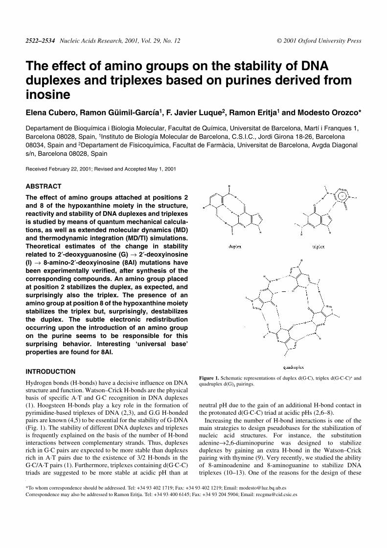
Hydrogen bonds (H-bonds) have a decisive influence on DNAstructure and function. Watson–Crick H-bonds are the physicalbasis of specific A·T and G·C recognition in DNA duplexes(1). Hoogsteen H-bonds play a key role in the formation ofpyrimidine-based triplexes of DNA (2,3), and G.G H-bondedpairs are known (4,5) to be essential for the stability of G-DNA(Fig. 1). The stability of different DNA duplexes and triplexesis frequently explained on the basis of the number of H-bondinteractions between complementary strands. Thus, duplexesrich in G·C pairs are expected to be more stable than duplexesrich in A·T pairs due to the existence of 3/2 H-bonds in theG·C/A·T pairs (1). Furthermore, triplexes containing d(G·C-C)triads are suggested to be more stable at acidic pH than at
neutral pH due to the gain of an additional H-bond contact inthe protonated d(G·C-C) triad at acidic pHs (2,6–8).
Increasing the number of H-bond interactions is one of themain strategies to design pseudobases for the stabilization ofnucleic acid structures. For instance, the substitutionadenine→2,6-diaminopurine was designed to stabilizeduplexes by gaining an extra H-bond in the Watson–Crickpairing with thymine (9). Very recently, we studied the abilityof 8-aminoadenine and 8-aminoguanine to stabilize DNAtriplexes (10–13). One of the reasons for the design of these
*To whom correspondence should be addressed. Tel: +34 93 402 1719; Fax: +34 93 402 1219; Email: [email protected] may also be addressed to Ramon Eritja. Tel: +34 93 400 6145; Fax: +34 93 204 5904; Email: [email protected]
Figure 1. Schematic representations of duplex d(G·C), triplex d(G·C-C)+ andquadruplex d(G)4 pairings.
Nucleic Acids Research, 2001, Vol. 29, No. 12 2523
two molecules was the expected gain of one extra Hoogsteen-like H-bond with the pyrimidine strand in the triplex structure.
Experimental (14–16) and theoretical (17,18) studies on thestructure and stability of apolar bases have raised doubts aboutthe suitability of criteria for predicting the stability of nucleicacid structures based only on counting the number of H-bondcontacts. These pseudobases are found in DNA helices that arestable at room temperature, despite the fact that they cannotform H-bonds, even in apolar environments (14–18). It is clearthat H-bond interactions might contribute to the stability ofDNA helices, but the assumption of a direct relationshipbetween the number of H-bonds and the stability of the helixmight not be correct.
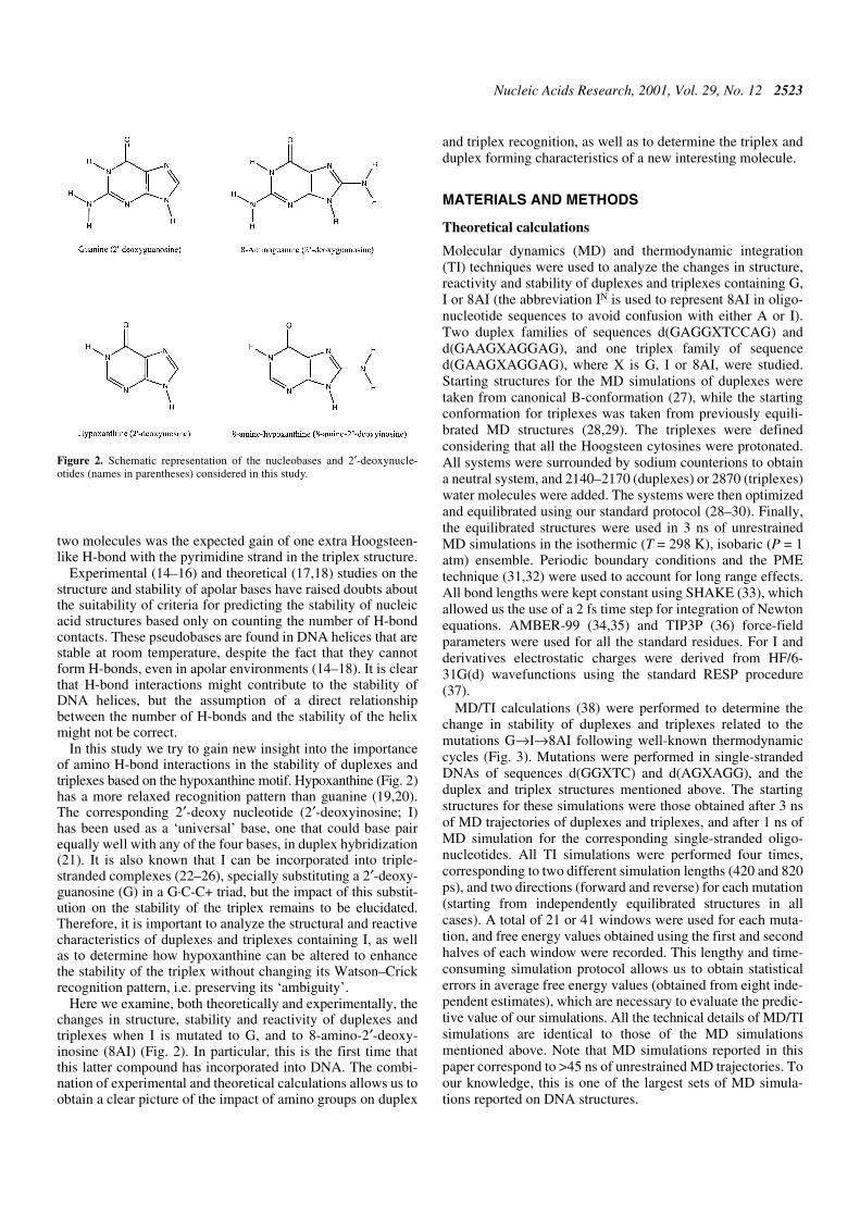
In this study we try to gain new insight into the importanceof amino H-bond interactions in the stability of duplexes andtriplexes based on the hypoxanthine motif. Hypoxanthine (Fig. 2)has a more relaxed recognition pattern than guanine (19,20).The corresponding 2′-deoxy nucleotide (2′-deoxyinosine; I)has been used as a ‘universal’ base, one that could base pairequally well with any of the four bases, in duplex hybridization(21). It is also known that I can be incorporated into triple-stranded complexes (22–26), specially substituting a 2′-deoxy-guanosine (G) in a G·C-C+ triad, but the impact of this substit-ution on the stability of the triplex remains to be elucidated.Therefore, it is important to analyze the structural and reactivecharacteristics of duplexes and triplexes containing I, as wellas to determine how hypoxanthine can be altered to enhancethe stability of the triplex without changing its Watson–Crickrecognition pattern, i.e. preserving its ‘ambiguity’.
Here we examine, both theoretically and experimentally, thechanges in structure, stability and reactivity of duplexes andtriplexes when I is mutated to G, and to 8-amino-2′-deoxy-inosine (8AI) (Fig. 2). In particular, this is the first time thatthis latter compound has incorporated into DNA. The combi-nation of experimental and theoretical calculations allows us toobtain a clear picture of the impact of amino groups on duplex
and triplex recognition, as well as to determine the triplex andduplex forming characteristics of a new interesting molecule.
MATERIALS AND METHODS
Theoretical calculations
Molecular dynamics (MD) and thermodynamic integration(TI) techniques were used to analyze the changes in structure,reactivity and stability of duplexes and triplexes containing G,I or 8AI (the abbreviation IN is used to represent 8AI in oligo-nucleotide sequences to avoid confusion with either A or I).Two duplex families of sequences d(GAGGXTCCAG) andd(GAAGXAGGAG), and one triplex family of sequenced(GAAGXAGGAG), where X is G, I or 8AI, were studied.Starting structures for the MD simulations of duplexes weretaken from canonical B-conformation (27), while the startingconformation for triplexes was taken from previously equili-brated MD structures (28,29). The triplexes were definedconsidering that all the Hoogsteen cytosines were protonated.All systems were surrounded by sodium counterions to obtaina neutral system, and 2140–2170 (duplexes) or 2870 (triplexes)water molecules were added. The systems were then optimizedand equilibrated using our standard protocol (28–30). Finally,the equilibrated structures were used in 3 ns of unrestrainedMD simulations in the isothermic (T = 298 K), isobaric (P = 1atm) ensemble. Periodic boundary conditions and the PMEtechnique (31,32) were used to account for long range effects.All bond lengths were kept constant using SHAKE (33), whichallowed us the use of a 2 fs time step for integration of Newtonequations. AMBER-99 (34,35) and TIP3P (36) force-fieldparameters were used for all the standard residues. For I andderivatives electrostatic charges were derived from HF/6-31G(d) wavefunctions using the standard RESP procedure(37).
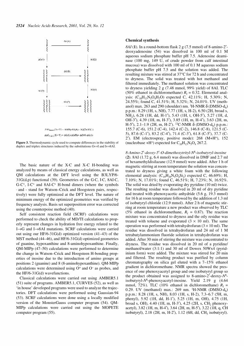
MD/TI calculations (38) were performed to determine thechange in stability of duplexes and triplexes related to themutations G→I→8AI following well-known thermodynamiccycles (Fig. 3). Mutations were performed in single-strandedDNAs of sequences d(GGXTC) and d(AGXAGG), and theduplex and triplex structures mentioned above. The startingstructures for these simulations were those obtained after 3 nsof MD trajectories of duplexes and triplexes, and after 1 ns ofMD simulation for the corresponding single-stranded oligo-nucleotides. All TI simulations were performed four times,corresponding to two different simulation lengths (420 and 820ps), and two directions (forward and reverse) for each mutation(starting from independently equilibrated structures in allcases). A total of 21 or 41 windows were used for each muta-tion, and free energy values obtained using the first and secondhalves of each window were recorded. This lengthy and time-consuming simulation protocol allows us to obtain statisticalerrors in average free energy values (obtained from eight inde-pendent estimates), which are necessary to evaluate the predic-tive value of our simulations. All the technical details of MD/TIsimulations are identical to those of the MD simulationsmentioned above. Note that MD simulations reported in thispaper correspond to >45 ns of unrestrained MD trajectories. Toour knowledge, this is one of the largest sets of MD simula-tions reported on DNA structures.
Figure 2. Schematic representation of the nucleobases and 2′-deoxynucle-otides (names in parentheses) considered in this study.
2524 Nucleic Acids Research, 2001, Vol. 29, No. 12
The basic nature of the X·C and X-C H-bonding wasanalyzed by means of classical energy calculations, as well asQM calculations at the DFT level using the B3LYP/6-31G(d,p) functional (39). Geometries of the G·C, I·C, 8AI·C,G-C+, I-C+ and 8AI-C+ H-bond dimers (where the symbols· and - stand for Watson–Crick and Hoogsteen pairs, respec-tively) were fully optimized at the DFT level. The nature ofminimum energy of the optimized geometries was verified byfrequency analysis. Basis set superposition error was correctedusing the counterpoise method (40).
Self consistent reaction field (SCRF) calculations wereperformed to check the ability of MD/TI calculations to prop-erly represent changes in hydration free energy related to theI→G and I→8AI mutations. SCRF calculations were carriedout using our HF/6-31G(d) optimized version (41–43) of theMST method (44–46), and HF/6-31G(d) optimized geometriesof guanine, hypoxanthine and 8-aminohypoxanthine. Finally,QM-MIPp (47–50) calculations were performed to determinethe change in Watson–Crick and Hoogsteen H-bonding prop-erties of inosine due to the introduction of amino groups atpositions 2 (guanine) and 8 (8-aminohypoxanthine). QM-MIPpcalculations were determined using O+ and O– as probes, andthe HF/6-31G(d) wavefunctions.
Classical calculations were carried out using AMBER5.1(51) suite of programs. AMBER5.1, CURVES (52), as well as‘in house’ developed programs were used to analyze the trajec-tories. DFT calculations were performed using Gaussian-94(53). SCRF calculations were done using a locally modifiedversion of the MonsterGauss computer program (54). QM-MIPp calculations were carried out using the MOPETEcomputer program (55).
Chemical synthesis
8AI (1). In a round-bottom flask 2 g (7.5 mmol) of 8-amino-2′-deoxyadenosine (54) was dissolved in 100 ml of 0.1 Maqueous sodium phosphate buffer pH 7.5. Adenosine deami-nase (100 mg, 149 U, of crude powder from calf intestinalmucosa) was dissolved with 100 ml of 0.1 M aqueous sodiumphosphate buffer pH 7.5 and the solution was added. Theresulting mixture was stirred at 37°C for 72 h and concentratedto dryness. The solid was treated with hot methanol andfiltered immediately. The methanol solution was concentratedto dryness yielding 2 g (7.48 mmol, 99% yield) of 8AI. TLC(50% ethanol in dichloromethane) Rf = 0.32. Elemental anal-ysis: (C10H13N5O4H2O) expected C, 42.11%; H, 5.30%; N,24.55%; found C, 41.51%; H, 5.32%; N, 24.01%. UV (meth-anol) max. 263 and 290 (shoulder) nm. 1H-NMR δ (DMSO-d6)p.p.m.: 8.29 (1H, s, NH), 7.77 (1H, s, H-2), 6.50 (2H, broad s,NH2), 6.28 (1H, dd, H-1′), 5.43 (1H, t, OH-5′), 5.27 (1H, d,OH-3′), 4.39 (1H, m, H-3′), 3.85 (1H, m, H-4′), 3.63 (2H, m,H-5′), 2.1–1.9 (2H, m, H-2′). 13C-NMR δ (DMSO-d6) p.p.m.:155.7 (C-6), 151.2 (C-4), 142.4 (C-2), 146.8 (C-8), 121.5 (C-5), 87.6 (C-1′), 83.2 (C-4′), 71.4 (C-3′), 61.8 (C-5′), 37.7 (C-2′). EM (electrospray, positive mode): 268 (M+H+), 152(nucleobase +H+) expected for C10H13N5O4 267.2.
8-Amino-2′-deoxy-5′-O-dimethoxytrityl-N8-isobutyryl-inosine(2). 8AI (1.72 g, 6.4 mmol) was dissolved in DMF and 2.7 mlof hexamethyldisilazane (12.9 mmol) were added. After 1 h ofmagnetic stirring at room temperature the solution was concen-trated to dryness giving a white foam with the followingelemental analysis: (C19H29N5O4Si2) expected C, 46.69%; H,7.10%; N, 17.01%; found C, 46.51%; H, 7.23%; N, 16.92%.The solid was dried by evaporating dry pyridine (10 ml) twice.The resulting residue was dissolved in 20 ml of dry pyridineand treated with phenoxyacetic anhydride (5.6 g, 19.3 mmol)for 16 h at room temperature followed by the addition of 1.3 mlof isobutyryl chloride (12.9 mmol). After 2 h of magnetic stir-ring at room temperature a new product was observed by TLC(5% ethanol in dichloromethane, Rf = 0.87). The reactionmixture was concentrated to dryness and the oily residue wastreated with toluene and evaporated (3 × 10 ml). The sameoperation was performed with tetrahydrofuran (3 × 10 ml). Theresidue was dissolved in tetrahydrofuran and 24 ml of 1 Mtetrabutylammonium fluoride solution in tetrahydrofuran wasadded. After 30 min of stirring the mixture was concentrated todryness. The residue was dissolved in 20 ml of a pyridine/methanol/water (3:1:1) and 30 ml of Dowex 50Wx4 (pyrid-inium form) were added. The mixture was stirred for 20 minand filtered. The resulting product was purified by columnchromatography on silica gel eluted with a 7–15% ethanolgradient in dichloromethane. NMR spectra showed the pres-ence of one phenoxyacetyl group and one isobutyryl group sothe product obtained was assigned to 8-amino-2′-deoxy-N8-isobutyryl-N8-phenoxyacetyl-inosine. Yield 2.19 g (4.64mmol, 72%). TLC (10% ethanol in dichloromethane) Rf =0.29. UV (methanol) max.: 269 nm. 1H-NMR (DMSO-d6)p.p.m.: 8.28 (1H, s, NH), 8.03 (1H, s, H-2), 7.3–6.7 (5H, m,phenyl), 5.92 (1H, dd, H-1′), 5.25 (1H, m, OH), 4.75 (1H,broad s, OH), 4.40 (1H, m, H-3′), 4.25 (2H, s, CH2 phenoxy-acetyl), 3.82 (1H, m, H-4′), 3.64 (2H, m, H-5′), 3.22 (1H, q, CHisobutyryl), 2.18 (2H, m, H-2′), 1.12 (6H, dd, CH3 isobutyryl).
Figure 3. Thermodynamic cycle used to compute differences in the stability ofduplex and triplex structures induced by the substitutions G↔I and I↔8AI.
Nucleic Acids Research, 2001, Vol. 29, No. 12 2525
13C-NMR (DMSO-d6) p.p.m.: 180.4 (C=O, isobutyryl), 176.6(C=O, phenoxyacetyl), 160.4 (C-1, phenyl), 159.3 (C-6), 149.3(C-4), 147.1 (C-2), 144.1 (C-8), 131.2 (C-3 and C-5 phenyl),124.1 (C-5), 122.8 (C-4 phenyl), 116.4 (C-2 and C-6 phenyl),90.4 (C-1′), 87.4 (C-4′), 73.9 (C-3′), 68.6 (CH2 phenoxy-acetyl), 64.2 (C-5′), 41.2 (C-2′), 37.0 (CH isobutyryl), 20.7 y20.4 (CH3 isobutyryl). EM (electrospray, positive mode):472.2 (M+H), 376.2 (M-phenoxy), 338.2 (M-phenoxyacetyl),222.2 (N-phenoxyacetyl-N-isobutyrylamide) expected forC22H25N5O7 471.4.
8-Amino-2′-deoxy-N8-isobutyryl-N8-phenoxyacetyl-inosine(2.19 g, 4.6 mmol) was dissolved into dry pyridine (20 ml) anddimethoxytrityl chloride (1.73 g, 5.1 mmol) was added to thesolution. After 2 h of magnetic stirring at room temperature,methanol (2 ml) was added and the solution was concentratedto dryness. The residue was dissolved in dichloromethane andthe solution was washed with aqueous sodium bicarbonatesolution and brine. The organic phase was dried over Na2SO4and evaporated. Silica gel chromatography (2–10% ethanolgradient in dichloromethane) yielded 2.1 g (3.3 mmol, 72%yield) of 5′-dimethoxytrityl-N8-isobutyryl-8-amino-2′-deoxy-inosine. Unexpectedly, the phenoxyacetyl group was elimi-nated during the work-up. TLC (10% ethanol indichloromethane) Rf = 0.4. Elemental analysis: (C35H37N5O7)expected C, 65.72%; H, 5.83%; N, 10.95%; found C, 64.62%;H, 5.87%; N, 10.60%. UV (methanol) max.: 236, 274(shoulder) and 283 (shoulder) nm. 1H-NMR (DMSO-d6)p.p.m.: 8.29 (1H, s, NH), 7.63 (1H, s, H-2), 7.4–6.7 (13H, m,DMT), 5.92 (1H, t, H 1′), 4.82 (1H, m, 3′-OH), 4.45 (1H, m,H-3′), 3.95 (1H, m, H-4′), 3.35 (2H, m, H-5′), 3.18 (1H, q, CHisobutyryl), 2.2 (2H, m, H-2′), 1.16 (6H, CH3 isobutyryl). 13C-RMN (Cl3CD): 179.2 (C=O, isobutyryl), 158.4 (C-4 anisoylDMT), 157.7 (C-6), 147.9 (C-4), 144.7 (C-8), 144.7 (C-1phenyl DMT), 143.9 (C-2), 136.1 (C-1 anisoyl DMT), 130.0(C-2 and C-6 anisoyl DMT), 128.2 (C-2 and C-6 phenylDMT), 127.7 (C-3 and C-5 phenyl DMT), 126.8 (C-4 phenylDMT), 121.6 (C-5), 113.0 (C-3 and C-5 anisoyl DMT), 86.3(C-1′), 86.3 (Cquat. DMT), 85.9 (C-4′), 72.6 (C-3′), 64.2(C-5′), 55.1 (CH3O DMT), 38.1 (C-2’), 35.4 (CH isobutyryl), 19.4and 18.9 (CH3 isobutyryl). EM (electrospray, positive mode):640.1 (M+H), 303.3 (DMT+) expected for C35H37N5O7 639.7.
8-Amino-2′-deoxy-5′-O-dimethoxytrityl-N8-isobutyryl-inosine-3′-O-(2-cyanoethyl)-N,N-diisopropylphosphoramidite (3). Theprotected nucleoside (1 g, 1.56 mmol) described above wasdissolved in dry dichloromethane (30 ml) anddiisopropylethylamine was added (0.85 ml, 4.7 mmol). Chloro2-cyanoethoxy diisopropylamino phosphine (0.6 ml, 0.34 mmol)was added to the solution dropwise with a syringe. After 1 h ofmagnetic stirring at room temperature, methanol (2 ml) wasadded and the solution was concentrated to dryness. Theresulting residue was dissolved in dichloromethane andwashed with aqueous sodium bicarbonate solution and brine.The organic phase was dried over Na2SO4 and evaporated.Silica gel chromatography (ethyl acetate/hexane 1:2) yielded1.1 g (1.31 mmol, 84% yield) of the desired phosphoramidite.TLC (ethyl acetate/hexane 1:2) Rf = 0.34. 31P-NMR (Cl3CD):143.07 and 143.37 (two diastereoisomers).
Oligonucleotide synthesis
Oligonucleotides were prepared on an automatic DNA synthe-sizer using standard and the modified phosphoramidite of the8-aminohypoxanthine described above. The phosphoramiditeof protected 8AI (3) was dissolved in dry dichloromethane tomake a 0.1 M solution. The rest of the phosphoramidites weredissolved in dry acetonitrile (0.1 M solution). Sequences ofoligonucleotides containing 8-aminohypoxanthine were: A, 5′-CTAING-3′; B, 5′-GCA ATG GAIN CCT CTA-3′; C, 5′-GAAGINA GGA GAT TTT TCT CCT CCT TC-3′; D, 5′-GAAGINA INGA INAT TTT TCT CCT CCT TC-3′, IN being = 8AI.Complementary oligonucleotides containing the natural basesand I were also prepared using commercially available chemi-cals and following standard protocols. After the assembly ofthe sequences, oligonucleotide supports were treated with 32%aqueous ammonia at 55°C for 16 h. Ammonia solutions wereconcentrated to dryness and the products were purified byreverse-phase HPLC. Oligonucleotides were synthesized on0.2 µmol scale and with the last DMT group at the 5′-end(DMT on protocol) to help reverse-phase purification. All puri-fied products presented a major peak, which was collected.Characterization of pentanucleotide A, 5′-CTAING-3′, wasperfomed by mass spectrometry and by snake venom phos-phodiesterase and alkaline phosphatase digestion followed byHPLC analysis of the nucleosides (HPLC conditions B). 8AIeluted together with dG. EM (MALDI): 1501.5 expected forC49H62N20O28P4 1502.8. Nucleoside composition: dC 1.1 (1),dG + 8-amino-dI 1.8 (2), T 1.1 (1), dA 1.1 (1). Yield (OD unitsat 260 nm after HPLC purification, 0.2 µmol) were 15mer B10.3 OD, 26mer C 18.4 OD, 26mer D 15.4 OD. HPLC condi-tions: HPLC solutions were as follows. Solvent A, 5% ACN in100 mM triethylammonium acetate pH 6.5 and solvent B, 70%ACN in 100 mM triethylammonium acetate pH 6.5. Foranalytical runs the following conditions were used. Column,Nucleosil 120C18, 250 × 4 mm; flow rate: 1 ml/min. (i) Condi-tions, a 40 min linear gradient from 0 to 75% B; (ii) a 20 minlinear gradient from 0 to 20% B. For preparative runs thefollowing conditions were used: columns, PRP-1 (Hamilton),250 × 10 mm; flow rate, 3 ml/min; a 30 min linear gradientfrom 10 to 80% B (DMT on), or a 30 min linear gradient from0 to 50% B (DMT off).
Melting experiments
Solutions of equimolar amounts of the pentadecamer carrying8AI or I at the central position and its complementarysequences carrying each of the four natural bases opposite themodified base were mixed in 0.15 M NaCl, 0.05 M Tris–HClbuffer pH 7.5. Melting experiments with triple helix wereperformed by mixing equimolar amounts of the modifiedoligonucleotide (26mer) and the Hoogsteen pyrimidine strand(11mer) in 1 M NaCl, 0.1 mM sodium phosphate/citric acidbuffer. The solutions were heated to 90°C, allowed to coolslowly to room temperature and then samples were kept in therefrigerator overnight. UV absorption spectra and meltingexperiments (absorbance versus temperature) were recorded in1 cm path length cells using a spectrophotometer, which has atemperature controller with a programmed temperatureincrease of 0.5°C/min. Melting experiments were performedusing a concentration of 4 µM monitoring the absorbance at260 nm.
2526 Nucleic Acids Research, 2001, Vol. 29, No. 12
Thermodynamic analysis
Analysis of the melting curves was carried out as described byLoakes and Brown (56). The enthalpy of the melting transition(in kcal/mol) was obtained using the equation ∆H = –4.38/(1/T1/2 – 1/T3/4), where T1/2 is the temperature at the maximumof the first derivative of the melting curve, and T3/4 is thetemperature at which the differential curve is half of T1/2 (bothin degrees Kelvin). A similar analysis on the triplex was notcarried out due to several factors such as the absence of transi-tions at certain pHs, the lower melting temperature (Tm) ofhypoxanthine triplexes and the proximity of the triplex transi-tion to the duplex transition of the 8AI triplexes.
Analysis of the thermodynamic data of triplex→duplextransition was performed as previously described (13). Meltingcurves were obtained at concentrations ranging from 0.5 to40 µM of triplex. The Tm of the first transition (correspondingto the dissociation of s11 from the triplex) was measured at themaximum of the first derivative of the melting curve. The plotof 1/Tm versus lnC was linear. Linear regression of the datagave a slope, and a y-intercept, from which ∆Ht and ∆St wereobtained (13). The free energy was obtained from the standardequation: ∆Gt = ∆Ht – T∆St.
The samples used in the concentration-dependent experimentswere prepared in a similar way to that described in the previoussection, but melting experiments were obtained using 0.1, 0.5and 1 cm path length cells. The DNA concentration wasdetermined by UV absorbance measurements (260 nm) at90°C, using for the DNA coil state the following extinctioncoefficients: 7500, 8500, 12500, 12500, 12500 and 15000 M–1
cm–1 for C, T, G, 8AI, I and A, respectively.
RESULTS AND DISCUSSION
Synthesis
To our knowledge the synthesis of 8AI has not been described,although the synthesis of the corresponding ribonucleoside,8-aminoinosine, was reported in 1967 (57). First, a syntheticroute similar to the route described for the preparation of8-aminoinosine was tried. This route had the following steps:(i) bromination of position –8 of I, (ii) displacement ofbromine with azide or hydrazine and (iii) catalytic hydrogena-tion. Unfortunately bromination of I under the conditionsdescribed for the bromination of inosine was not successfuland the conditions described by Long et al. (57) for the nextsteps were considered too extreme for a 2′-deoxynucleoside.For these reasons an alternative route was considered (Fig. 4).In this route, 8-amino-2′-deoxyadenosine was first preparedfrom dA as described (57). 8AI was obtained by enzymaticdeamination of 8-amino-2′-deoxyadenosine by adenosinedeaminase. The formation of a more polar compound wasfollowed by TLC and HPLC showing complete reaction after72 h. Spectral data (mass spectrometry, UV, 1H- and 13C-NMR) ofthe new compound was consistent with the desired compound(1).
Next, protection of the amino group was studied (Fig. 4).Reaction with dimethylaminoformamidine dimethylacetal inmethanol and in N,N-dimethylformamide did not yield thedesired dmf-protected nucleoside as described for 8-amino-guanine (12) and 8-aminoadenine (10) derivatives. Instead the
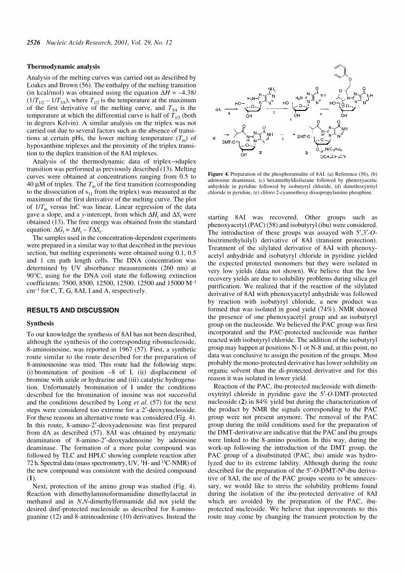
starting 8AI was recovered. Other groups such asphenoxyacetyl (PAC) (58) and isobutyryl (ibu) were considered.The introduction of these groups was assayed with 5′,3′-O-bis(trimethylsilyl) derivative of 8AI (transient protection).Treatment of the silylated derivative of 8AI with phenoxy-acetyl anhydride and isobutyryl chloride in pyridine yieldedthe expected protected monomers but they were isolated invery low yields (data not shown). We believe that the lowrecovery yields are due to solubility problems during silica gelpurification. We realized that if the reaction of the silylatedderivative of 8AI with phenoxyacetyl anhydride was followedby reaction with isobutyryl chloride, a new product wasformed that was isolated in good yield (74%). NMR showedthe presence of one phenoxyacetyl group and an isobutyrylgroup on the nucleoside. We believed the PAC group was firstincorporated and the PAC-protected nucleoside was furtherreacted with isobutyryl chloride. The addition of the isobutyrylgroup may happen at positions N-1 or N-8 and, at this point, nodata was conclusive to assign the position of the groups. Mostprobably the mono-protected derivative has lower solubility onorganic solvent than the di-protected derivative and for thisreason it was isolated in lower yield.
Reaction of the PAC, ibu-protected nucleoside with dimeth-oxytrityl chloride in pyridine gave the 5′-O-DMT-protectednucleoside (2) in 84% yield but during the characterization ofthe product by NMR the signals corresponding to the PACgroup were not present anymore. The removal of the PACgroup during the mild conditions used for the preparation ofthe DMT-derivative are indicative that the PAC and ibu groupswere linked to the 8-amino position. In this way, during thework-up following the introduction of the DMT group, thePAC group of a disubstituted (PAC, ibu) amide was hydro-lyzed due to its extreme lability. Although during the routedescribed for the preparation of the 5′-O-DMT-N8-ibu deriva-tive of 8AI, the use of the PAC groups seems to be unneces-sary, we would like to stress the solubility problems foundduring the isolation of the ibu-protected derivative of 8AIwhich are avoided by the preparation of the PAC, ibu-protected nucleoside. We believe that improvements to thisroute may come by changing the transient protection by the
Figure 4. Preparation of the phosphoramidite of 8AI. (a) Reference (56), (b)adenosine deaminase, (c) hexamethyldisilazane followed by phenoxyaceticanhydride in pyridine followed by isobutyryl chloride, (d) dimethoxytritylchloride in pyridine, (e) chloro 2-cyanoethoxy diisopropylamino phosphine.
Nucleic Acids Research, 2001, Vol. 29, No. 12 2527
classical per-acylation method although this route has not beentried in this work (59).
Finally, the phosphitylation of the DMT, ibu-protected nucleo-side with the corresponding chlorophosphine gave the desiredphosphoramidite (3) in 84% yield.
The stability of the isobutyryl group to ammonia wasanalyzed. First, DMT, ibu-protected nucleoside was dissolvedin dioxane and it was treated with concentrated ammonia at55°C overnight. Complete removal of the ibu group wasobserved in <6 h as seen by TLC (data not shown). Afterwards,the pentanucleotide 5′-CTAING-3′ (IN = 8AI) was prepared and
the support was treated with concentrated ammonia at 55°Covernight. The resulting pentanucleotide was analyzed byanalytical HPLC giving a major product, which had theexpected mass and nucleoside composition.
Theoretical calculations
MD analysis of DNA duplexes. MD simulations of the twofamilies of duplexes studied here [d(GAGGXTCCAG) andd(GAAGXAGGAG)] provide stable trajectories, irrespectiveof the nature of X (G, I or 8AI). This is clearly noted in the rootmean square deviations (r.m.s.d.), which amount to 1.3–1.7
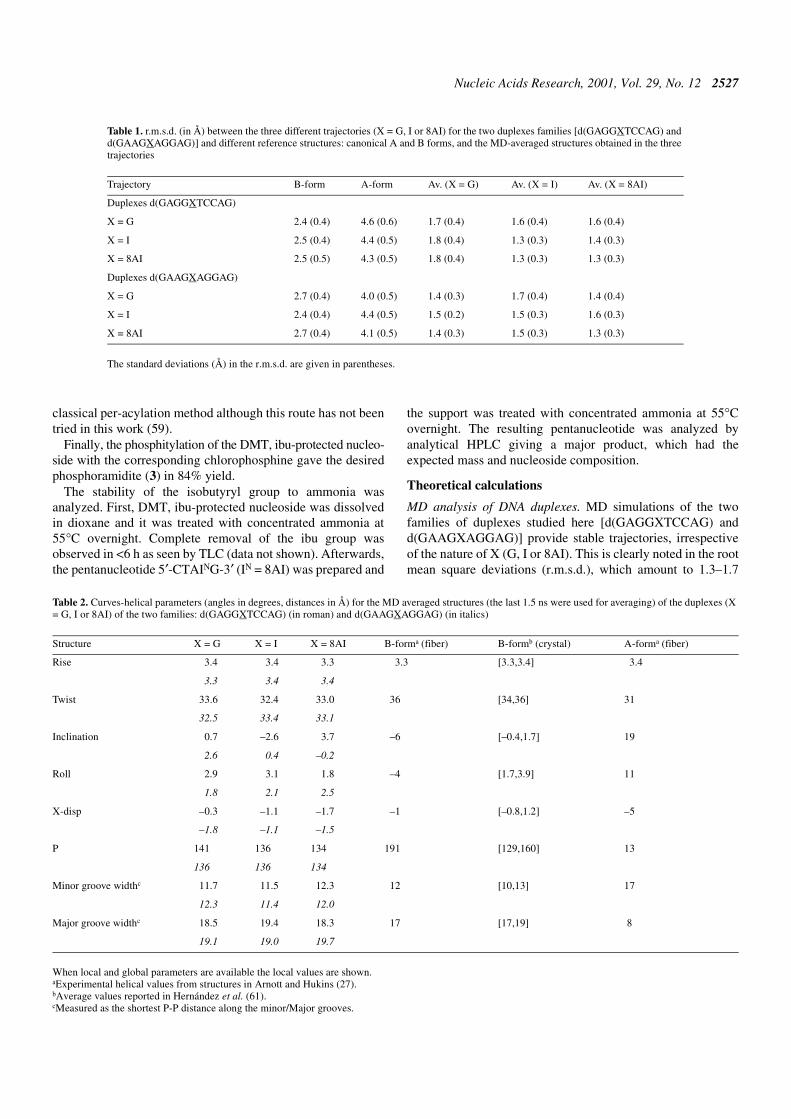
Table 1. r.m.s.d. (in Å) between the three different trajectories (X = G, I or 8AI) for the two duplexes families [d(GAGGXTCCAG) andd(GAAGXAGGAG)] and different reference structures: canonical A and B forms, and the MD-averaged structures obtained in the threetrajectories
The standard deviations (Å) in the r.m.s.d. are given in parentheses.
Trajectory B-form A-form Av. (X = G) Av. (X = I) Av. (X = 8AI)
Duplexes d(GAGGXTCCAG)
X = G 2.4 (0.4) 4.6 (0.6) 1.7 (0.4) 1.6 (0.4) 1.6 (0.4)
X = I 2.5 (0.4) 4.4 (0.5) 1.8 (0.4) 1.3 (0.3) 1.4 (0.3)
X = 8AI 2.5 (0.5) 4.3 (0.5) 1.8 (0.4) 1.3 (0.3) 1.3 (0.3)
Duplexes d(GAAGXAGGAG)
X = G 2.7 (0.4) 4.0 (0.5) 1.4 (0.3) 1.7 (0.4) 1.4 (0.4)
X = I 2.4 (0.4) 4.4 (0.5) 1.5 (0.2) 1.5 (0.3) 1.6 (0.3)
X = 8AI 2.7 (0.4) 4.1 (0.5) 1.4 (0.3) 1.5 (0.3) 1.3 (0.3)
Table 2. Curves-helical parameters (angles in degrees, distances in Å) for the MD averaged structures (the last 1.5 ns were used for averaging) of the duplexes (X= G, I or 8AI) of the two families: d(GAGGXTCCAG) (in roman) and d(GAAGXAGGAG) (in italics)
When local and global parameters are available the local values are shown.aExperimental helical values from structures in Arnott and Hukins (27).bAverage values reported in Hernández et al. (61).cMeasured as the shortest P-P distance along the minor/Major grooves.
Structure X = G X = I X = 8AI B-forma (fiber) B-formb (crystal) A-forma (fiber)
Rise 3.4 3.4 3.3 3.3 [3.3,3.4] 3.4
3.3 3.4 3.4
Twist 33.6 32.4 33.0 36 [34,36] 31
32.5 33.4 33.1
Inclination 0.7 –2.6 3.7 –6 [–0.4,1.7] 19
2.6 0.4 –0.2
Roll 2.9 3.1 1.8 –4 [1.7,3.9] 11
1.8 2.1 2.5
X-disp –0.3 –1.1 –1.7 –1 [–0.8,1.2] –5
–1.8 –1.1 –1.5
P 141 136 134 191 [129,160] 13
136 136 134
Minor groove widthc 11.7 11.5 12.3 12 [10,13] 17
12.3 11.4 12.0
Major groove widthc 18.5 19.4 18.3 17 [17,19] 8
19.1 19.0 19.7
2528 Nucleic Acids Research, 2001, Vol. 29, No. 12
(±0.3) Å when the different trajectories are compared withtheir corresponding MD-averaged structures (Table 1). Theser.m.s.d. values are typical of converged MD simulations ofDNA duplexes (60,61). Disruptions of the helix or breathingmovements are not observed, thus supporting the stability ofthe helical structure along the trajectories. The structures foundfor each family of duplexes are in general identical, irrespec-tive of the nature of X. This is clearly seen in cross-r.m.s.d.values (those obtained when one trajectory is compared withthe averaged structure of another trajectory) of 1.3–1.8 Å. Insummary, our results suggest that the DNA is flexible enoughto accommodate possible local distortions arising from thepresence of the 8-amino group, without any important struc-tural alteration.
The structures sampled for the six trajectories are thoseexpected for a B-type structure, as noted in r.m.s.d. values of2.4–2.5 (±0.4) Å for the entire duplexes. This shows that MD-trajectories sample typical regions of the B-type configura-tional space. This is confirmed by helical analysis of the sixtrajectories (Table 2), which shows helical values in goodagreement to those expected for B-type duplexes, and far fromthose of an A-type duplex (27).
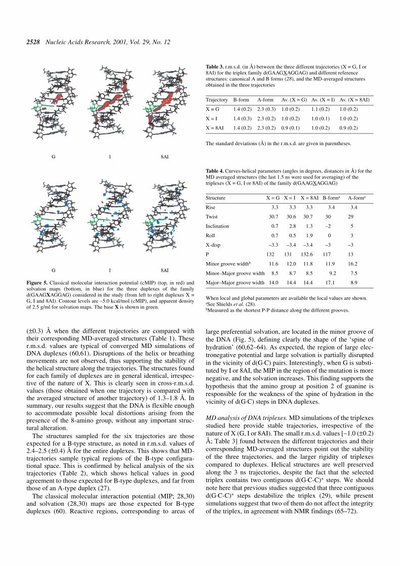
The classical molecular interaction potential (MIP; 28,30)and solvation (28,30) maps are those expected for B-typeduplexes (60). Reactive regions, corresponding to areas of
large preferential solvation, are located in the minor groove ofthe DNA (Fig. 5), defining clearly the shape of the ‘spine ofhydration’ (60,62–64). As expected, the region of large elec-tronegative potential and large solvation is partially disruptedin the vicinity of d(G·C) pairs. Interestingly, when G is substi-tuted by I or 8AI, the MIP in the region of the mutation is morenegative, and the solvation increases. This finding supports thehypothesis that the amino group at position 2 of guanine isresponsible for the weakness of the spine of hydration in thevicinity of d(G·C) steps in DNA duplexes.
MD analysis of DNA triplexes. MD simulations of the triplexesstudied here provide stable trajectories, irrespective of thenature of X (G, I or 8AI). The small r.m.s.d. values [∼1.0 (±0.2)Å; Table 3] found between the different trajectories and theircorresponding MD-averaged structures point out the stabilityof the three trajectories, and the larger rigidity of triplexescompared to duplexes. Helical structures are well preservedalong the 3 ns trajectories, despite the fact that the selectedtriplex contains two contiguous d(G·C-C)+ steps. We shouldnote here that previous studies suggested that three contiguousd(G·C-C)+ steps destabilize the triplex (29), while presentsimulations suggest that two of them do not affect the integrityof the triplex, in agreement with NMR findings (65–72).
Figure 5. Classical molecular interaction potential (cMIP) (top, in red) andsolvation maps (bottom, in blue) for the three duplexes of the familyd(GAAGXAGGAG) considered in the study (from left to right duplexes X =G, I and 8AI). Contour levels are –5.0 kcal/mol (cMIP), and apparent densityof 2.5 g/ml for solvation maps. The base X is shown in green.
Table 3. r.m.s.d. (in Å) between the three different trajectories (X = G, I or8AI) for the triplex family d(GAAGXAGGAG) and different referencestructures: canonical A and B forms (28), and the MD-averaged structuresobtained in the three trajectories
The standard deviations (Å) in the r.m.s.d. are given in parentheses.
Trajectory B-form A-form Av. (X = G) Av. (X = I) Av. (X = 8AI)
X = G 1.4 (0.2) 2.3 (0.3) 1.0 (0.2) 1.1 (0.2) 1.0 (0.2)
X = I 1.4 (0.3) 2.3 (0.2) 1.0 (0.2) 1.0 (0.1) 1.0 (0.2)
X = 8AI 1.4 (0.2) 2.3 (0.2) 0.9 (0.1) 1.0 (0.2) 0.9 (0.2)
Table 4. Curves-helical parameters (angles in degrees, distances in Å) for theMD averaged structures (the last 1.5 ns were used for averaging) of thetriplexes (X = G, I or 8AI) of the family d(GAAGXAGGAG)
When local and global parameters are available the local values are shown.aSee Shields et al. (28).bMeasured as the shortest P-P distance along the different grooves.
Structure X = G X = I X = 8AI B-forma A-forma
Rise 3.3 3.3 3.3 3.4 3.4
Twist 30.7 30.6 30.7 30 29
Inclination 0.7 2.8 1.3 –2 5
Roll 0.7 0.5 1.9 0 3
X-disp –3.3 –3.4 –3.4 –3 –3
P 132 131 132.6 117 13
Minor groove widthb 11.6 12.0 11.8 11.9 16.2
Minor–Major groove width 8.5 8.7 8.5 9.2 7.5
Major–Major groove width 14.0 14.4 14.4 17.1 8.9

Nucleic Acids Research, 2001, Vol. 29, No. 12 2529
The three trajectories sample the same region of the configu-rational space, as noted in the cross r.m.s.d. values (∼1 Å; Table3). As expected (28,29), the structures sampled in the trajecto-ries pertain to the B-family, as noted in the r.m.s.d. of 1.4 Å fromthe canonical B-form, and in the helical parameters shown inTable 4.
MIP and solvation analyses show clearly two regions of thetriplex able to interact with small polar molecules, which arethe minor (-m) groove, and the minor part of the Major groove(-mM; see 28 for nomenclature). The regions of large negativeMIP are mostly located in the plane of the purine for the -mgroove, and between triad planes for the -mM groove (Fig. 6).The presence of an amino group in the -m groove of the triplexslightly decreases the ability of this groove to interact withsmall polar solutes (Fig. 6). However, an amino group in the -mM groove does not lead to detectable alterations in the MIPand solvation maps, suggesting that the water environment inthe -mM groove is not largely disrupted. This can be under-stood considering that the regions of most negative MIP andpreferential solvation in the -mM groove are not located in thesame plane as the purines (Fig. 6).
Free energy calculations. MD calculations suggest that duplexand triplex structures containing I or 8AI are stable and havesimilar structures. Based on the lack of large structural altera-tions upon replacement of G by I or 8AI, MD/TI calculationswere performed to determine the changes in stability inducedby the G→I→8AI substitutions
Free energy profiles for the G→I and 8AI→I mutations weresmooth and without hysteresis (Fig. 7). The excellent conver-gence of the results is clearly noted in the small standard error
Figure 6. cMIP (top, in red) and solvation maps (bottom, in blue) for the threetriplexes considered in the study (from left to right triplexes X = G, I and 8AI).Contour levels are –5.0 kcal/mol (cMIP), and apparent density of 3.0 g/ml forsolvation maps. The base X is shown in green.
Figure 7. Difference (triplex–duplex and duplex–single-stranded) free energy profiles (in kcal/mol) obtained from the different mutations. Black, mutation G→Iin the d(GAAGXAGGAG) duplex and triplex; green, mutation G→I in the d(GAGGXTCCAG) duplex; red, mutation 8AI→I in the d(GAAGXAGGAG) duplexand triplex; blue, mutation 8AI→I in the d(GAGGXTCCAG) duplex. Error bars are displayed.
2530 Nucleic Acids Research, 2001, Vol. 29, No. 12
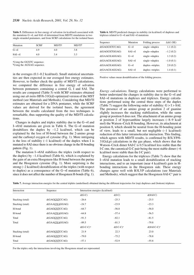
in the averages (0.1–0.2 kcal/mol). Small statistical uncertain-ties are then expected in our averaged free energy estimates.However, to further check the quality of MD/TI calculations,we computed the difference in free energy of solvationbetween pentamers containing a central G, I and 8AI. Theresults are compared (Table 5) with SCRF estimates obtainedusing our ab initio HF/6-31G(d)-optimized version of the MSTmethod (see Materials and Methods). Considering that MD/TIestimates are obtained for a DNA pentamer, while the SCRFvalues are derived for the isolated bases, the agreementbetween the results calculated from the two techniques isremarkable, thus supporting the quality of the MD/TI calcula-tions.
Changes in duplex and triplex stability due to the G→I andI→8AI mutations are given in Table 6. The G→I mutationdestabilizes the duplex by ∼1.2 kcal/mol, which can beexplained by the loss of H-bond between the 2-amino groupand the carbonyl oxygen of cytosine (Fig. 1). More intriguingis the loss of stability (∼1 kcal/mol) of the duplex when I ismutated to 8AI since there is no obvious change in the H-bondingpattern (Fig. 1).
The mutation I→8AI stabilizes the triplex (with respect tothe duplex) by ∼1.4 kcal/mol (Table 6), which is explained bythe gain of an extra Hoogsteen-like H-bond between the purineand the Hoogsteen cytosine (Fig. 1). More surprising is thestrong (∼2 kcal/mol) destabilization of the triplex (with respectto duplex) as a consequence of the G→I mutation (Table 6),since it does not affect the number of Hoogsteen H-bonds (Fig. 1).
Energy calculations. Energy calculations were performed tobetter understand the changes in stability due to the G→I and8AI→I mutations in duplexes and triplexes. Energy calcula-tions performed using the central three steps of the duplex(Table 7) suggest the following order of stability: G > I > 8AI.The presence of an amino group at position 2 of guanineslightly increases the stacking stabilization, while the samegroup at position 8 does not. The attachment of an amino groupat position 2 of hypoxanthine largely increases (∼8–9 kcal/mol) the Watson–Crick H-bonding. However, its attachment atposition 8, which should be neutral from the H-bonding pointof view, leads to a small, but not negligible (∼1 kcal/mol)reduction of this latter intramolecular interaction. This finding,which agrees with MD/TI results, is confirmed by B3LYP/6-31G(d,p) calculations in the gas phase, which shows that theWatson–Crick dimer 8AI·C is 0.5 kcal/mol less stable than theI·C one, the canonical G·C pair being the most stable dimer (∼6kcal/mol more stable than the I·C pair).
Energy calculations for the triplexes (Table 7) show that theI→8AI mutation leads to a small destabilization of stackinginteractions, and to an important (near 4 kcal/mol) gain in H-bonding interactions in the Hoogsteen side. These energychanges agree well with B3LYP calculations (see Materialsand Methods), which suggest that the Hoogsteen 8AI-C+ pair is
Table 5. Differences in free energy of solvation (in kcal/mol) associated withthe mutations G→I, and 8AI→I determined from MD/TI simulations in twosingle-stranded pentamers, and from SCRF calculations for the isolated bases
aUsing the GGXTC sequence.bUsing the AGXAG sequence.
Mutation SCRF MD/TIa MD/TIb
G→I 4.9 4.0 3.8
8AI→I 4.0 3.4 3.3
Table 7. Average interaction energies for the central triplets (underlined) obtained during the different trajectories for (top) duplexes and (bottom) triplexes
For the triplex only the interactions involving the Hoogsteen strand are represented.
Interaction Sequence Interaction energies (kcal/mol)
d(G·C) d(I·C) d(8AI·C)
Stacking (total) d(GAGGXTCCAG) –26.6 –25.3 –25.5
Stacking (total) d(GAAGXAGGAG) –24.7 –23.9 –23.3
H-bond d(GAGGXTCCAG) –64.7 –56.8 –56.0
H-bond d(GAAGXAGGAG) –64.8 –57.4 –56.5
Total d(GAGGXTCCAG) –91.3 –82.1 –81.5
Total d(GAAGXAGGAG) –89.5 –81.3 –79.8
d(G·C-C)+ d(I·C-C)+ d(8AI·C-C)+
Stacking (total) d(GAGGXTCCAG) 21.9 22.3 23.0
H-bond d(GAGGXTCCAG) –79.0 –75.2 –78.8
Total d(GAGGXTCCAG) –57.1 –52.9 –55.8
Table 6. MD/TI predicted changes in stability (in kcal/mol) of duplexes andtriplexes related to G→I and 8AI→I mutations
Positive values mean destabilization of the folding process.
Sequence Mutation Folding process ∆∆G (SE)
d(GAGGXTCCAG) G→I single→duplex 1.1 (0.2)
d(GAGGXTGGAG) 8AI→I single→duplex –1.2 (0.2)
d(GAAGXAGGAG) G→I single→duplex 1.3 (0.2)
d(GAAGXAGGAG) 8AI→I single→duplex –1.0 (0.1)
d(GAAGXAGGAG) G→I duplex→triplex 2.0 (0.2)
d(GAAGXAGGAG) 8AI→I duplex→triplex 1.4 (0.1)
Nucleic Acids Research, 2001, Vol. 29, No. 12 2531
4 kcal/mol more stable than the I-C+ one. It should be notedthat the stabilization obtained by the extra Hoogsteen H-bondis then smaller than that expected for a normal H-bond, whichis easily explained considering that the presence of the 8-amino group is not favored by the positive charge of the Hoog-steen cytosine.
Classical energy calculations (Table 7) suggest that theHoogsteen G-C+ pair is very stable due to the large strength ofthe two Hoogsteen H-bonds. In fact, classical calculationssuggest similar stability for the G-C+ and 8AG-C+ pairs, notfully supported by B3LYP calculations, which suggest that the8AI-C+ dimer is slightly more stable (∼2 kcal/mol) than the G-C+ one. This discrepancy might overestimate the stability oftriplexes containing d(8AI·C-C)+ triads with respect to thosecontaining d(G·C-C)+ triads. However, there is general agree-ment between classical and QM calculations. It is then clearthat (i) the Hoogsteen H-bond involving the 8-amino group isnot very strong and (ii) the presence of the 2-amino groupenhances the stability of Hoogsteen H-bonds.
Both MD/TI and energy calculations show that the presenceof an amino group at position 2 of I strongly stabilizesduplexes relative to single-stranded oligonucleotides, andtriplexes relative to duplexes. On the contrary, the presence ofan amino group at position 8 stabilizes the triplex relative toduplex, but destabilizes the duplex relative to single-strandedoligonucleotides. As noted above, simple counting of H-bondinteractions explains the effect of the 2-amino group in theWatson–Crick pairing, and that of the 8-amino group in theHoogsteen side. However, they do not explain the effects of the2-amino in the Hoogsteen pairing, and of the 8-amino in theWatson–Crick interaction.
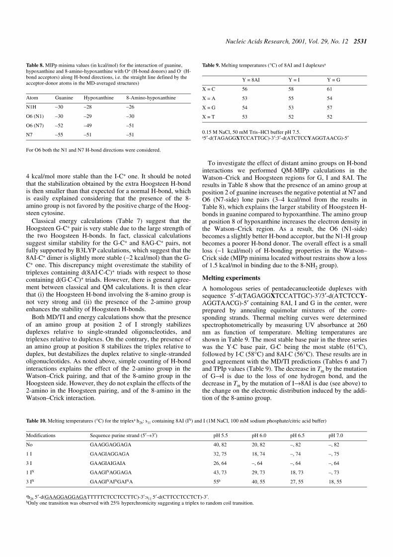
To investigate the effect of distant amino groups on H-bondinteractions we performed QM-MIPp calculations in theWatson–Crick and Hoogsteen regions for G, I and 8AI. Theresults in Table 8 show that the presence of an amino group atposition 2 of guanine increases the negative potential at N7 andO6 (N7-side) lone pairs (3–4 kcal/mol from the results inTable 8), which explains the larger stability of Hoogsteen H-bonds in guanine compared to hypoxanthine. The amino groupat position 8 of hypoxanthine increases the electron density inthe Watson–Crick region. As a result, the O6 (N1-side)becomes a slightly better H-bond acceptor, but the N1-H groupbecomes a poorer H-bond donor. The overall effect is a smallloss (∼1 kcal/mol) of H-bonding properties in the Watson–Crick side (MIPp minima located without restrains show a lossof 1.5 kcal/mol in binding due to the 8-NH2 group).
Melting experiments
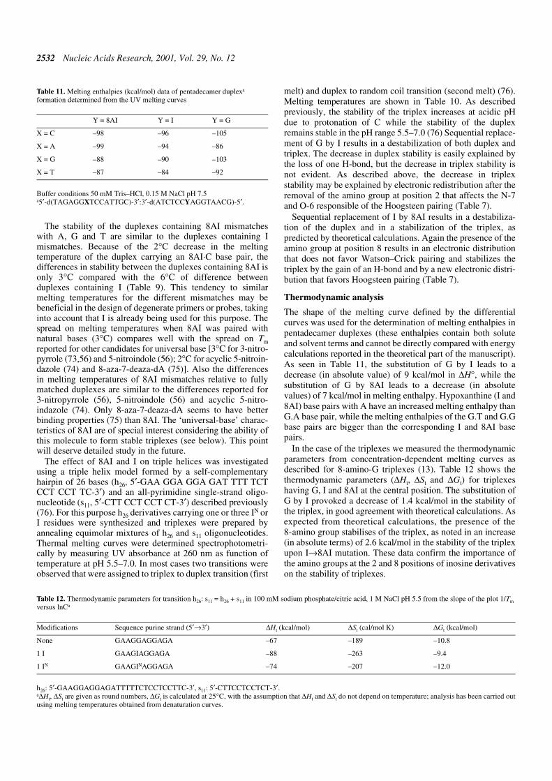
A homologous series of pentadecanucleotide duplexes withsequence 5′-d(TAGAGGXTCCATTGC)-3′/3′-d(ATCTCCY-AGGTAACG)-5′ containing 8AI, I and G in the center, wereprepared by annealing equimolar mixtures of the corre-sponding strands. Thermal melting curves were determinedspectrophotometrically by measuring UV absorbance at 260nm as function of temperature. Melting temperatures areshown in Table 9. The most stable base pair in the three serieswas the Y·C base pair, G·C being the most stable (61°C),followed by I·C (58°C) and 8AI·C (56°C). These results are ingood agreement with the MD/TI predictions (Tables 6 and 7)and TPIp values (Table 9). The decrease in Tm by the mutationof G→I is due to the loss of one hydrogen bond, and thedecrease in Tm by the mutation of I→8AI is due (see above) tothe change on the electronic distribution induced by the addi-tion of the 8-amino group.
Table 8. MIPp minima values (in kcal/mol) for the interaction of guanine,hypoxanthine and 8-amino-hypoxanthine with O+ (H-bond donors) and O– (H-bond acceptors) along H-bond directions, i.e. the straight line defined by theacceptor-donor atoms in the MD-averaged structures)
For O6 both the N1 and N7 H-bond directions were considered.
Atom Guanine Hypoxanthine 8-Amino-hypoxanthine
N1H –30 –28 –26
O6 (N1) –30 –29 –30
O6 (N7) –52 –49 –51
N7 –55 –51 –51
Table 10. Melting temperatures (°C) for the triplexa h26: s11 containing 8AI (IN) and I (1M NaCl, 100 mM sodium phosphate/citric acid buffer)
ah26 5′-d(GAAGGAGGAGATTTTTCTCCTCCTTC)-3′:s11 5′-d(CTTCCTCCTCT)-3′.bOnly one transition was observed with 25% hyperchromicity suggesting a triplex to random coil transition.
Modifications Sequence purine strand (5′→3′) pH 5.5 pH 6.0 pH 6.5 pH 7.0
No GAAGGAGGAGA 40, 82 20, 82 –, 82 –, 82
1 I GAAGIAGGAGA 32, 75 18, 74 –, 74 –, 75
3 I GAAGIAIGAIA 26, 64 –, 64 –, 64 –, 64
1 IN GAAGINAGGAGA 43, 73 29, 73 18, 73 –, 73
3 IN GAAGINAINGAINA 55b 40, 55 27, 55 18, 55
Table 9. Melting temperatures (°C) of 8AI and I duplexesa
0.15 M NaCl, 50 mM Tris–HCl buffer pH 7.5.a5′-d(TAGAGGXTCCATTGC)-3′:3′-d(ATCTCCYAGGTAACG)-5′
Y = 8AI Y = I Y = G
X = C 56 58 61
X = A 53 55 54
X = G 54 53 57
X = T 53 52 52
2532 Nucleic Acids Research, 2001, Vol. 29, No. 12
The stability of the duplexes containing 8AI mismatcheswith A, G and T are similar to the duplexes containing Imismatches. Because of the 2°C decrease in the meltingtemperature of the duplex carrying an 8AI·C base pair, thedifferences in stability between the duplexes containing 8AI isonly 3°C compared with the 6°C of difference betweenduplexes containing I (Table 9). This tendency to similarmelting temperatures for the different mismatches may bebeneficial in the design of degenerate primers or probes, takinginto account that I is already being used for this purpose. Thespread on melting temperatures when 8AI was paired withnatural bases (3°C) compares well with the spread on Tmreported for other candidates for universal base [3°C for 3-nitro-pyrrole (73,56) and 5-nitroindole (56); 2°C for acyclic 5-nitroin-dazole (74) and 8-aza-7-deaza-dA (75)]. Also the differencesin melting temperatures of 8AI mismatches relative to fullymatched duplexes are similar to the differences reported for3-nitropyrrole (56), 5-nitroindole (56) and acyclic 5-nitro-indazole (74). Only 8-aza-7-deaza-dA seems to have betterbinding properties (75) than 8AI. The ‘universal-base’ charac-teristics of 8AI are of special interest considering the ability ofthis molecule to form stable triplexes (see below). This pointwill deserve detailed study in the future.
The effect of 8AI and I on triple helices was investigatedusing a triple helix model formed by a self-complementaryhairpin of 26 bases (h26, 5′-GAA GGA GGA GAT TTT TCTCCT CCT TC-3′) and an all-pyrimidine single-strand oligo-nucleotide (s11, 5′-CTT CCT CCT CT-3′) described previously(76). For this purpose h26 derivatives carrying one or three IN orI residues were synthesized and triplexes were prepared byannealing equimolar mixtures of h26 and s11 oligonucleotides.Thermal melting curves were determined spectrophotometri-cally by measuring UV absorbance at 260 nm as function oftemperature at pH 5.5–7.0. In most cases two transitions wereobserved that were assigned to triplex to duplex transition (first
melt) and duplex to random coil transition (second melt) (76).Melting temperatures are shown in Table 10. As describedpreviously, the stability of the triplex increases at acidic pHdue to protonation of C while the stability of the duplexremains stable in the pH range 5.5–7.0 (76) Sequential replace-ment of G by I results in a destabilization of both duplex andtriplex. The decrease in duplex stability is easily explained bythe loss of one H-bond, but the decrease in triplex stability isnot evident. As described above, the decrease in triplexstability may be explained by electronic redistribution after theremoval of the amino group at position 2 that affects the N-7and O-6 responsible of the Hoogsteen pairing (Table 7).
Sequential replacement of I by 8AI results in a destabiliza-tion of the duplex and in a stabilization of the triplex, aspredicted by theoretical calculations. Again the presence of theamino group at position 8 results in an electronic distributionthat does not favor Watson–Crick pairing and stabilizes thetriplex by the gain of an H-bond and by a new electronic distri-bution that favors Hoogsteen pairing (Table 7).
Thermodynamic analysis
The shape of the melting curve defined by the differentialcurves was used for the determination of melting enthalpies inpentadecamer duplexes (these enthalpies contain both soluteand solvent terms and cannot be directly compared with energycalculations reported in the theoretical part of the manuscript).As seen in Table 11, the substitution of G by I leads to adecrease (in absolute value) of 9 kcal/mol in ∆H°, while thesubstitution of G by 8AI leads to a decrease (in absolutevalues) of 7 kcal/mol in melting enthalpy. Hypoxanthine (I and8AI) base pairs with A have an increased melting enthalpy thanG.A base pair, while the melting enthalpies of the G.T and G.Gbase pairs are bigger than the corresponding I and 8AI basepairs.
In the case of the triplexes we measured the thermodynamicparameters from concentration-dependent melting curves asdescribed for 8-amino-G triplexes (13). Table 12 shows thethermodynamic parameters (∆Ht, ∆St and ∆Gt) for triplexeshaving G, I and 8AI at the central position. The substitution ofG by I provoked a decrease of 1.4 kcal/mol in the stability ofthe triplex, in good agreement with theoretical calculations. Asexpected from theoretical calculations, the presence of the8-amino group stabilises of the triplex, as noted in an increase(in absolute terms) of 2.6 kcal/mol in the stability of the triplexupon I→8AI mutation. These data confirm the importance ofthe amino groups at the 2 and 8 positions of inosine derivativeson the stability of triplexes.
Table 11. Melting enthalpies (kcal/mol) data of pentadecamer duplexa
formation determined from the UV melting curves
Buffer conditions 50 mM Tris–HCl, 0.15 M NaCl pH 7.5a5′-d(TAGAGGXTCCATTGC)-3′:3′-d(ATCTCCYAGGTAACG)-5′.
Y = 8AI Y = I Y = G
X = C –98 –96 –105
X = A –99 –94 –86
X = G –88 –90 –103
X = T –87 –84 –92
Table 12. Thermodynamic parameters for transition h26: s11 = h26 + s11 in 100 mM sodium phosphate/citric acid, 1 M NaCl pH 5.5 from the slope of the plot 1/Tmversus lnCa
h26: 5′-GAAGGAGGAGATTTTTCTCCTCCTTC-3′, s11: 5′-CTTCCTCCTCT-3′.a∆Ht, ∆St are given as round numbers, ∆Gt is calculated at 25°C, with the assumption that ∆Ht and ∆St do not depend on temperature; analysis has been carried outusing melting temperatures obtained from denaturation curves.
Modifications Sequence purine strand (5′→3′) ∆Ht (kcal/mol) ∆St (cal/mol K) ∆Gt (kcal/mol)
None GAAGGAGGAGA –67 –189 –10.8
1 I GAAGIAGGAGA –88 –263 –9.4
1 IN GAAGINAGGAGA –74 –207 –12.0

Nucleic Acids Research, 2001, Vol. 29, No. 12 2533
CONCLUSIONS
A combination of state of the art theoretical and experimentaltechniques have allowed us to investigate the effect that aminogroups added on positions 2 and 8 of the hypoxanthine moietyhas on the structure, flexibility and stability of duplexes andtriplexes. A new pseudobase 2-aminohypoxanthine is theoreti-cally designed, synthesized and experimentally tested,showing interesting properties as a unique ‘universal base’ andtriplex-stabilizing pseudobase. Good agreement is foundbetween theoretical prediction and experimental results,demonstrating the predictive power of high level theoreticalcalculations. Overall, our results demonstrate that the stabilityof nucleic acid structures is a subtle balance of different terms,and that simple counting of elemental interactions can lead toerroneous conclusions.
ACKNOWLEDGEMENTS
We are grateful to Prof. J.Tomasi for providing us with hisoriginal code of the PCM model, which was modified by us tocarry out the MST calculations. We also acknowledge theDirección General de Investigación Científica y Técnica(grants PB98-1222, PM99-0046 and BQU2000-0649) and theGeneralitat de Catalunya (2000-SGR-0018) for financialsupport, and the Centre de Supercomputació de Catalunya forcomputational facilities.
REFERENCES
1. Saenger,W. (1984) Principles of Nucleic Acid Structure. Springer-Verlag.New York, NY.
2. Soyfer,V.N. and Potaman,V.N. (1996) Triple Helical Nucleic Acids.Springer-Verlag. New York, NY.
3. Sun,J.S. and Hélène,C. (1993) Oligonucleotide-directed triple-helixformation. Curr. Opin. Struct. Biol., 6, 327–333.
4. Laughlan,G., Murchie,A.I., Norman,M.H., Moody,P.C., Lilley,D.M. andLuisi,B. (1994) The high-resolution crystal structure of a parallel-strandedguanine tetraplex. Science, 265, 520–524.
5. Williamson,J.R. (1994) G-Quartet structures in telomeric DNA. Annu.Rev. Biophys. Biomol. Struct., 23, 703–730.
6. Lee,J.S., Johnson,D.A. and Moogan,A.R. (1979) Complexes formed by(pyrimidine)n. (purine)n DNAs on lowering the pH are three-stranded.Nucleic Acids Res., 6, 3073–3091.
7. Völker,J. and Klump,H.H. (1994) Electrostatic effects in DNA triplehelices. Biochemistry, 33, 13502–13508.
8. Wittung,P., Nielsen,P. and Norden,B. (1997) Extended DNA-recognitionrepertoire of peptide nucleic acid (PNA): PNA–dsDNA triplex formedwith cytosine-rich homopyrimidine PNA. Biochemistry, 36, 7973–7979.
9. Chollet,A. and Kawashima,E. (1988) DNA containing the base analogue2-aminoadenine: preparation, use as hybridization probes and cleavage byrestriction endonucleases. Nucleic Acids Res., 16, 305–317.
10. Güimil-García,R., Ferrer,E., Macías,M.J., Eritja,R. and Orozco,M. (1999)Theoretical calculations, synthesis and base pairing properties ofoligonucleotides containing 8-amino-2′-deoxyadenosine. Nucleic AcidsRes., 27, 1991–1998.
11. Eritja,R., Ferrer,E., Güimil-García,R. and Orozco,M. (1999) Modifiedoligonucleotides with triple-helix stabilization properties. Nucl. Nucl., 18,1619–1621.
12. Güimil-García,R., Bachi,A., Eritja,R., Luque,F.J. and Orozco,M. (1998)Triple helix stabilization properties of oligonucleotides containing 8-amino-2′-deoxyguanosine. Bioorg. Med. Chem. Lett., 8, 3011–3016.
13. Soliva,R., Güimil-García,R., Blas,J.R., Eritja,R., Asensio,J.L.,González,C., Luque,F.J. and Orozco,M. (2000) DNA-triplex stabilizingproperties of 8-aminoguanine. Nucleic Acids Res., 28, 4531–4539.
14. Schweitzer,B.A. and Kool,E.T. (1995) Hydrophobic, non-hydrogen-bonding bases and base pairs in DNA. J. Am. Chem. Soc., 117, 1863–1972.
15. Moran,S., Ren,R. and Kool,E.T. (1997) A thymidine triphosphate shapeanalog lacking Watson–Crick pairing ability is replicated with highsequence selectivity. Proc. Natl Acad. Sci. USA, 94, 10506–10511.
16. Matray,T.J. and Kool,E.T. (1998) Selective and stable DNA base pairingwithout hydrogen. J. Am. Chem. Soc., 120, 6191–6192.
17. Cubero,E., Sherer,E.C., Luque,F.J., Orozco,M. and Laughton,C.A. (1999)Observation of spontaneous base pair breathing events in the moleculardynamics simulation of a difluorotoluene-containing DNAoligonucleotide. J. Am. Chem. Soc., 121, 8653–8654.
18. Cubero,E., Laughton,C.A., Luque,F.J. and Orozco,M. (2000) Moleculardynamics study of oligonucleotides containing difluorotoluene. J. Am.Chem. Soc., 122, 6891–6899.
19. Ohtsuka,E., Matsuki,S., Ikehara,M., Takahashi,T. and Matsubara,K.J.(1985) An alternative approach to deoxyoligonucleotides as hybridizationprobes by insertion of deoxyinosine at ambiguous codon positions. J. Biol.Chem., 260, 2605–2608.
20. Martin,F.H., Castro,M.M., Aboul-ela,F. and Tinoco,I. (1985) Base pairinginvolving deoxyinosine: implications for probe design. Nucleic AcidsRes., 13, 8927–8938.
21. Loakes,D., Brown,D.M., Linde,S. and Hill,F. (1995) 3-Nitropyrrole and 5-nitroindole as universal bases in primers for DNA sequencing and PCR.Nucleic Acids Res., 23, 2361–2366.
22. Thiele,D. and Guschlbauer,W. (1969) Protonated polynucleotides. VII.Thermal transitions between different complexes of polyinosinic acid andpolycytidylic acid in an acid medium. Biopolymers, 8, 361–378.
23. Gargallo,R., Tauler,R. and Izquierdo-Ridorsa,A. (1997) Acid-base andcopper (II) complexation equilibria of poly(inosinic)-poly(cytidylic).Biopolymers, 42, 271–283.
24. Letai,A.G., Palladino,M.A., Fromm,E., Rizzo,V. and Fresco,J.R. (1988)Specificity in formation of triple-stranded nucleic acid helical complexes:studies with agarose-linked polyribonucleotide affinity columns.Biochemistry, 27, 9108–9112.
25. Mills,M., Völker,J. and Klump,H.H. (1996) Triple helical structuresinvolving inosine: there is a penalty for promiscuity. Biochemistry, 35,13338–13344.
26. Hogeland,J.S. and Weller,D.D. (1993) Investigations ofoligodeoxyinosine for triple helix formation. Antisense Res. Dev., 3, 285–290.
27. Arnott,S. and Hukins,D.W.L. (1972) Optimized parameters for A-DNAand B-DNA. Biochem. Biophys. Res. Commun., 47, 1504–1509.
28. Shields,G., Laughton,C.A. and Orozco,M. (1997) Molecular dynamicssimulations of the d(T·A·T) triple helix. J. Am. Chem. Soc., 119, 7463–7469.
29. Soliva,R., Laughton,C.A., Luque,F.J. and Orozco,M. (1998) Moleculardynamics simulations in aqueous solution of triple helices containingd(G·C·C) trios. J. Am. Chem. Soc., 120, 11226–11233.
30. Shields,G., Laughton,C.A. and Orozco,M. (1998) Molecular dynamicssimulation of a PNA·DNA·PNA triple helix in aqueous solution. J. Am.Chem. Soc., 120, 5895–5904.
31. Essmann,U., Perera,L., Berkowitz,M.L., Darden,T., Lee,H. andPedersen,L.G. (1995) A smooth particle mesh Ewald method. J. Chem.Phys., 103, 8577–8593.
32. Darden,T.A., York,D. and Pedersen,L. (1993) Particle mesh Ewald: anN·log(N) method for Ewald sums in large systems. J. Chem. Phys., 98,10089–10092.
33. Ryckaert,J.P., Ciccote,G. and Berendsen,J.C. (1977) Numericalintegration of the cartesian equations of motion of a system withconstraints: Molecular dynamics of n-alkanes. J. Comput. Phys., 23, 327–341.
34. Cornell,W.D., Cieplak,P., Bayly,C.I., Gould,I.R., Merz,K.M.,Ferguson,D.M., Spellmeyer,D.C., Fox,T., Caldwell,J.W. andKollman,P.A. (1995) A second generation force field for the simulation ofproteins, nucleic acids, and organic molecules. J. Am. Chem. Soc., 117,5179–5197.
35. Cheatham,T.E., Cieplak,P. and Kollman,P.A. (1999) A modified versionof the Cornell et al. force field with improved sugar pucker phases andhelical repeat. J. Biomol. Struct. Dyn., 16, 845–862.
36. Jorgensen,W.L., Chandrasekhar,J., Madura,J.D., Impey,R. andKlein,M.L. (1983) Comparison of simple potential functions forsimulating liquid water. J. Chem. Phys., 79, 926–935.
37. Bayly,C.I., Cieplak,P., Cornell,W.D. and Kollman,P.A. (1993) A well-behaved electrostatic potential based method using charge restraints forderiving atomic charges: the RESP model. J. Phys. Chem., 97, 10269–10280.

2534 Nucleic Acids Research, 2001, Vol. 29, No. 12
38. McQuarri,D.A. (1976) Statistical Mechanics. Harper and Row, NewYork, NY.
39. Lee,C., Yang,W. and Parr,R.G. (1998) Development of the Colle–Salvetticorrelation-energy formula into a functional of the electron density. Phys.Rev. B, 37, 785–789.
40. Boys,S.F. and Bernardi,F. (1970) The calculation of small moleculesinteractions by the differences of separate total energies. Some procedureswith reduced errors. Mol. Phys., 19, 553–559.
41. Luque,F.J., Bachs,M. and Orozco,M. (1994) An optimized AM1/MSTmethod for the MST-SCRF representation of solvated systems. J. Comp.Chem., 15, 847–857.
42. Orozco,M., Bachs,M. and Luque,F.J. (1995) Development of optimizedMST/SCRF methods for semiempirical calculations: the MNDO and PM3Hamiltonians. J. Comp. Chem., 16, 563–575.
43. Bachs,M., Luque,F.J. and Orozco,M. (1994) Optimization of solutecavities and van der Waals parameters in ab initio MST-SCRFcalculations of neutral molecules. J. Comp. Chem., 15, 446–454.
44. Miertus,S., Scrocco,E. and Tomasi,J. (1981) Electrostatic interaction of asolute with a continuum. A direct utilization of ab initio molecularpotentials for the prevision of solvent effects. Chem. Phys., 55, 117–129.
45. Miertus,S. and Tomasi,J. (1982) Approximate evaluations of theelectrostatic free energy and internal energy changes in solutionprocesses. Chem. Phys., 65, 239–245.
46. Tomasi,J. and Persico,M. (1994) Molecular interactions in solution: anoverview of methods based on continuous distribution of the solvent.Chem. Rev., 94, 2027–2094.
47. Orozco,M. and Luque,F.J. (1993) Molecular interaction potential: a newtool for the theoretical study of molecular reactivity. J. Comp. Chem., 14,587–602.
48. Luque,F.J. and Orozco,M. (1998) Polarization effects in generalizedmolecular interaction potential: New Hamiltonian for reactivity studiesand mixed QM/MM calculations. J. Comp. Chem., 19, 866–881.
49. Hernández,B., Luque,F.J. and Orozco,M. (1999) Parametrization of theGMIPp for the study of stacking interactions. J. Comp. Chem., 20, 937–946.
50. Cubero,E., Luque,F.J. and Orozco,M. (1998) Is polarization important incation-pi interactions? Proc. Natl Acad. Sci. USA, 95, 5976–5980.
51. Case,D.A., Pearlman,D.A., Caldwell,J.W., Cheatham,T.E., Ross,W.S.,Simmerling,C.L., Darden,T.A., Merz,K.M., Stanton,R.V., Cheng,A.L. etal. (1997) AMBER 5. University of California, San Francisco, CA.
52. Lavery,R. and Sklena,J. (1988) The definition of generalized helicoidalparameters and of axis curvature for irregular nucleic acids. J. Biomol.Struct. Dyn., 6, 63–91.
53. Frisch,M.J., Trucks,G.W., Schlegel,H.B., Gill,P.M.W., Johnson,B.G.,Robb,M.A., Cheeseman,J.R., Keith,T.A., Petersson,G.A.,Montgomery,J.A. et al. (1995) GAUSSIAN 94 (Rev. A.1). Gaussian Inc.,Pittsburgh, PA.
54. Peterson,M. and Poirier,R. (1980) MonsterGauss. Department ofChemistry. University of Toronto, Toronto, Canada [modified (1987) byCammi,R., Bonaccorsi,R. and Tomasi,J., University of Pisa, Pisa, Italy;further modified (1995) by Luque,F.J. and Orozco,M., University ofBarcelona, Barcelona, Spain].
55. Luque,F.J. and Orozco,M. (2000) MOPETE Computer Program.University of Barcelona, Barcelona, Spain.
56. Loakes,D. and Brown,D.M. (1994) 5-Nitroindole as a universal baseanalogue. Nucleic Acids Res., 22, 4039–4043.
57. Long,R.A., Robins,R.K. and Townsend,L.B. (1967) Purine nucleosidesXV. The synthesis of 8-amino- and 8-substituted amino purinenucleosides. J. Org. Chem., 32, 2751–2756.
58. Schulhof,J.C., Molko,D. and Teoule,R. (1987) The final deprotection stepin oligonucleotide synthesis is reduced to a mild and rapid ammoniatreatment by using labile base-protecting groups. Nucleic Acids Res., 15,397–416.
59. Jones,R.A. (1984) Preparation of protected deoxyribonucleosides. InGait,M.J. (ed.), Oligonucleotide Synthesis, A Practical Approach. IRLPress, Oxford, UK, pp. 23–34.
60. Soliva,R., Luque,F.J., Alhambra,C. and Orozco,M. (1999) Role of sugarre-puckering in the transition of A and B forms of DNA in solution. Amolecular dynamics study. J. Biomol. Struct. Dyn., 17, 89–99.
61. Hernández,B., Soliva,R., Luque,F.J. and Orozco,M. (2000)Misincorporation of 2′-deoxyoxanosine into DNA: a molecular basis forNO-induced mutagenesis derived from theoretical calculations. NucleicAcids Res., 28, 4873–4883.
62. Dickerson,R.E. (1992) DNA structure from A to Z. Methods Enzymol.,211, 67–127.
63. Drew,H.R., Wing,R.M., Takana,S., Broka,C., Tanaka,S., Itakura,K. andDickerson,R.E. (1981) Structure of a B-DNA dodecamer: Conformationand dynamics. Proc. Natl Acad. Sci. USA, 78, 2179–2183.
64. Shui,X., McFail-Isom,L., Hu,G.G. and Williams,L.D. (1998) The B-DNAdodecamer at high resolution reveals a spine of water on sodium.Biochemistry, 37, 8341–8355.
65. Rajagopal,P. and Feigon,J. (1989) NMR studies of triple-strand formationfrom the homopurine–homopyridine deoxyribonucleotides d(GA)4 andd(TC)4. Biochemistry, 28, 7859–7870.
66. Rajagopal,P. and Feigon,J. (1989) Measurement of γ-rays from coldfusion. Nature, 339, 667–669.
67. Radhakrishnan,I. and Patel,D.J. (1994) DNA triplexes: solutionstructures, hydration sites, energetics, interactions, and function.Biochemistry, 33, 11405–11416.
68. Radhakrishnan,I. and Patel,D.J. (1994) Hydration sites inpurine.purine.pyrimidine and pyrimidine.purine.pyrimidine DNAtriplexes in aqueous solution. Structure, 2, 395–405.
69. Wang,E., Koshlap,K.M., Gilllespie,P., Dervan,P.B. and Feigon,J. (1996)Solution structure of a pyrimidine·purine·pyrimidine triplex containingthe sequence-specific intercalating non-natural base D3. J. Mol. Biol.,257, 1052–1069.
70. Asensio,J.L., Dhesai,J., Bergquist,S., Brown,T. and Lane,A.N. (1998) Thecontribution of cytosine protonation to the stability of parallel DNA triplehelices. J. Mol. Biol., 275, 811–822.
71. Asensio,J.L., Brown,T. and Lane,A.N. (1999) Solution conformation of aparallel DNA triple helix with 5‘ and 3‘ triplex-duplex junctions. Struct.Fold Des., 7, 1–11.
72. Asensio,J.L., Brown,T. and Lane,A.N. (1998) Comparison of the solutionstructures of intramolecular DNA triple helices containing adjacent andnon-adjacent CG.C+ triplets. Nucleic Acids Res., 26, 3677–3686.
73. Nichols,R., Andrews,P.C., Zhang,P. and Bergstrom,D.E. (1994) Auniversal nucleoside for use at ambiguous sites in DNA primers. Nature,369, 492–493.
74. Van Aerschot,A., Rozenski,J., Loakes,D., Pillet,N., Schepers,G. andHerdewijn,P. (1995) An acyclic 5-nitroindazole nucleoside analogue asambiguous nucleoside. Nucleic Acids Res., 23, 4363–4370.
75. Seela,F. and Debelak,H. (2000) The N8-(2′-deoxyribofuranoside) of 8-aza-7-deazaadenine: a universal nucleoside forming specific hydrogenbonds with the four canonical DNA constituents. Nucleic Acids Res., 28,3224–3232.
76. Xodo,L.E., Manzini,G., Quadrifolio,F., van der Marel,G.A. and vanBoom,J.H. (1991) Effect of 5-methylcytosine on the stability of triple-stranded DNA—a thermodynamic study. Nucleic Acids Res., 19, 5625–5631.