the effect of choline-stabilized orthosilicic acid in
TRANSCRIPT

Teughels et al. BMC Oral Health (2021) 21:485 https://doi.org/10.1186/s12903-021-01817-4
RESEARCH
The effect of choline-stabilized orthosilicic acid in patients with peri-implantitis: an exploratory randomized, double-blind, placebo controlled studyWim Teughels1†, Gizem Unal Celik2†, Mihai Tarce1, Ine De Cock3, Sara M. Persyn3*† and Mehmet C. Haytac2†
Abstract
Background: Choline-stabilized orthosilicic acid (CS-OSA) was previously found to stimulate bone collagen forma-tion in osteopenia and to improve biomarkers of cartilage degradation in knee osteoarthritis. The aim of the present study was to investigate the effect of oral administration of CS-OSA on clinical symptoms of peri-implantitis and the associated bone loss.
Methods: Twenty-one patients with peri-implantitis were randomized in CS-OSA or placebo groups. After initial clinical and cone beam computed tomography (CBCT) measurements [probing pocket depth (PPD), bleeding on probing (BOP), mucosal recession (REC), distance from implant shoulder to alveolar crest (IS-AC) and distance from implant shoulder to first bone-to-implant contact (IS-BIC)], flap operations were performed at the peri-implantitis sites. All patients were instructed to use either placebo or CS-OSA capsules twice a day for 1 year. Measurements were repeated 6 and 12 months after randomization.
Results: The data of 18 patients (36 implants) were used in the per protocol analysis. PPD and BOP improved signifi-cantly (p < 0.05) compared to baseline for both groups after 6 and 12 months. However, REC significantly increased in the placebo group but not in the CS-OSA group. The change in REC over 6 and 12 months was significantly different between groups (p < 0.01). IS-BIC and IS-AC measurements remained stable in the CS-OSA group whereas in the pla-cebo group, both parameters increased significantly after 6 and 12 months. The change in IS-BIC over 12 months was significantly different between groups (p < 0.05).
Conclusion: The results of this preliminary study suggest that CS-OSA may stabilize and even prevent further bone loss after surgical peri-implantitis treatment and support mucosal tissue healing.
Trial registration
The trial was retrospectively registered at ISRCTN registry, registration number: ISRCTN14348802, registration date: 24/06/2020.
Keywords: Choline-stabilized orthosilicic acid, Peri-implantitis, Bone, Mucosal recession
© The Author(s) 2021. Open Access This article is licensed under a Creative Commons Attribution 4.0 International License, which permits use, sharing, adaptation, distribution and reproduction in any medium or format, as long as you give appropriate credit to the original author(s) and the source, provide a link to the Creative Commons licence, and indicate if changes were made. The images or other third party material in this article are included in the article’s Creative Commons licence, unless indicated otherwise in a credit line to the material. If material is not included in the article’s Creative Commons licence and your intended use is not permitted by statutory regulation or exceeds the permitted use, you will need to obtain permission directly from the copyright holder. To view a copy of this licence, visit http:// creat iveco mmons. org/ licen ses/ by/4. 0/. The Creative Commons Public Domain Dedication waiver (http:// creat iveco mmons. org/ publi cdoma in/ zero/1. 0/) applies to the data made available in this article, unless otherwise stated in a credit line to the data.
BackgroundDental implants have been widely used for the replace-ment of missing teeth since the 1980’s [1]. High long-term survival rates of over 95% have been reported [2]. However, the prevalence of peri-implant diseases
Open Access
*Correspondence: [email protected]†Wim Teughels, Gizem Unal Celik, Sara M. Persyn and Mehmet C. Haytac have contributed equally to the work3 Research and Development, Bio Minerals NV, Zenderstraat 12, 9070 Destelbergen, BelgiumFull list of author information is available at the end of the article

Page 2 of 11Teughels et al. BMC Oral Health (2021) 21:485
affecting both soft and hard tissues that may eventually lead to implant failure (loss) has substantially increased to up to more than 20% in the past decade [1–4]. Appro-priate treatment of implants affected by peri-implant dis-eases is becoming increasingly important as the number of implants placed per year continues to increase [5].
Peri-implantitis is a plaque-associated pathological condition occurring in tissues around dental implants, characterized by inflammation in the peri-implant mucosa and subsequent progressive loss of supporting bone [6]. The diagnosis and follow-up of peri-implanti-tis are made by detecting radiographic bone loss, clini-cal signs of inflammation, bleeding on probing (BOP) with or without suppuration, increased probing pocket depth (PPD) and recession of the mucosal margin (REC) [6, 7]. Based on the consensus report of Workgroup 4 of the 2017 World Workshop on the Classification of Peri-odontal and Peri-Implant Diseases and Conditions, bone levels of ≥ 3 mm apical of the most coronal portion of the intra-osseous part of the implant together with bleed-ing on probing are consistent with the diagnosis of peri-implantitis in studies [6]. Intraoral radiography (IR) is the most commonly used technique for the measurement of peri-implant bone loss [8]. However, IR is a two-dimen-sional (2D) imaging technique offering merely mesiodis-tal and vertical detection of bone defects [9]. Therefore cone beam computed tomography (CBCT) has been widely used for three-dimensional (3D) assessment offer-ing additional spatial information including buccolingual visualization of the peri-implant bone [10].
The main goals of peri-implantitis treatment are resolv-ing inflammation and preventing further bone loss by decontaminating the implant surface. Non-surgical treatment by mechanical debridement with or without antiseptic and/or antibiotic therapy has shown limited success [11, 12]. The surgical treatment options including flap surgery with or without osseous resection, regenera-tion with bone grafts and guided bone regeneration are proven to be more effective [5, 12]. However until now, a gold standard treatment for peri-implantitis is lacking [12].
Choline-stabilized orthosilicic acid (CS-OSA), is a con-centrated and stable complex of choline and orthosilicic acid [13]. Physiological concentrations of orthosilicic acid were found to stimulate the synthesis of collagen type I in human osteoblast-like cells and skin fibroblasts [14]. CS-OSA supplementation of animals resulted in an increased femoral bone density [15, 16]. This effect was confirmed in humans demonstrating a beneficial effect on bone turnover, especially on bone collagen formation and femoral bone mineral density, when treating osteo-penic women with CS-OSA and Ca/Vit D3 for 12 months compared with Ca/Vit D3 alone [17]. A possible effect
of CS-OSA on collagen metabolism was also suggested when photoaged women were found to have improved surface and mechanical properties of the skin when tak-ing CS-OSA compared to women who took a placebo [18]. Recently, 12-week CS-OSA supplementation of men with knee osteoarthritis resulted in symptomatic improvements associated with a significant reduction of cartilage degradation biomarkers, suggesting a possible effect of CS-OSA on collagen metabolism in both carti-lage and subchondral bone [19].
Based on these previous studies [13–19], one can hypothesize that CS-OSA may have a possible effect on bone loss in peri-implantitis. The aim of this explorative study was to evaluate the effect of the oral intake of CS-OSA over a 12-month period on clinical symptoms of peri-implantitis and the associated bone loss.
MethodsPatientsA 12-month, randomized, double-blind placebo-con-trolled parallel-group study was performed in patients with peri-implantitis. Ethical approval was obtained from the local Ethical Committee of the Cukurova Uni-versity, Adana, Turkey (65/4, 04.05.2017). The study was conducted in accordance with the Helsinki Dec-laration as revised in 2013 and was retrospectively reg-istered at ISRCTN registry with registration number: ISRCTN14348802 and registration date: 24/06/2020.
Patients consulting the Department of Periodontol-ogy of the dental school of the Cukurova University were screened for participating in the study between September 2017 and January 2018. The inclusion crite-ria defined eligible patients as men and women between 18 and 75 years of age with single or multiple osseoin-tegrated implants suffering from peri-implantitis which was defined as bone loss of more than 3 mm measured on intra-oral radiographs and a PPD of more than 4 mm with BOP or suppuration on probing. All patients gave written informed consent.
The exclusion criteria were the following: pregnancy or breastfeeding, smoking or history of smoking (less than 6 months prior to the start of the study), recent or cur-rent alcohol and drug abuse, gingival index score > 2 [20], active, untreated periodontitis, mobile implants, poorly controlled diabetes (Glycated hemoglobin (HbA1c) level > 6%), osteonecrosis of the jaw and participation in another clinical trial. Furthermore, patients with renal failure, documented history of stroke, myocardial infarct or cancer and patients belonging to a high risk group for HIV were excluded. Additionally, specified concomi-tant and previous medications (i.e. dietary supplements containing a silicon source, systemic antibiotics, bispho-sphonates, local antiseptics) that could interfere with

Page 3 of 11Teughels et al. BMC Oral Health (2021) 21:485
the outcome of the study were prohibited during the trial. A wash-out period of 3 months and 6 months was required for the use of food supplements containing sili-con sources and systemic antibiotics, respectively, prior to the baseline visit.
Patient visitsDuring a first visit, the screening visit, patients were screened for the inclusion and exclusion criteria. If needed, a wash-out period was required. At the baseline visit (T0), baseline assessments were performed by clini-cal parameters evaluation, OHIP-14 questionnaire com-pletion, radiographic analysis and biochemical analysis. Surgery was performed within a period of 10 days after the baseline visit. The sutures were removed 10 days after surgery. Assessments were repeated 6 and 12 months after the baseline visit (T6, T12). The visits at 2, 4, 8 and 10 months after baseline were performed to check for adverse events, oral hygiene condition and study medica-tion compliance and to motivate the patient to continue with the study. All patients received proper oral hygiene instructions and were given the same tooth brush (Oral B Pro-flex Clinical Line), tooth paste (Oral B Ipana Pro-expert Clinical Line) and interdental brush (Oral B Interdental 228 mm) at the baseline visit. During each follow-up visit (2-monthly), the oral hygiene condition of each patient was evaluated, a professional prophylaxis was performed if needed, oral hygiene instructions were repeated and the tooth brush, tooth paste and interdental brush were replaced to standardize the maintenance pro-cedures for each patient.
Study medication and randomizationTwenty-one patients were randomly assigned to take a capsule of either the active treatment (520 mg beadlets containing 5 mg of silicon and 100 mg of choline in the form of CS-OSA (ch-OSA®, Bio Minerals NV, Belgium [18])) or placebo (520 mg microcrystalline cellulose beadlets; Pharmatrans Sanaq AG, Switzerland) twice daily, one in the morning and another in the evening with a glass of water or juice, for 12 months. The treatment allocation occurred sequentially in a 1:1 ratio using a ran-domization list, which was generated by an independent statistician in R (software version 3.3.3 for Windows; The R Foundation for Statistical Computing, Vienna, Austria). More specifically, block randomization was used in ran-domly selected block sizes of 2 or 4. The individual code was kept in a sealed envelope by the investigator, who was instructed not to open this except in case of medical emergency.
At baseline and after 2, 4, 6, 8 and 10 months the study medication was delivered in bottles labeled with the patient’s randomization number (according to the
allocation sequence). Blinding among patients, investiga-tors and monitors was maintained by providing identical packaging, appearance, taste and odor for CS-OSA and placebo capsules, respectively. Treatment compliance was verified at subsequent visits by counting the number of unused capsules.
Assessments and outcome measuresDuring screening, clinical parameters including PPD, BOP (present = 1, absent = 0) and REC (relative to the implant abutment junction) were measured at six sites per implant with a calibrated probe (North Carolina periodontal probe, Hu-Friedy, Chicago, IL, USA). An IR (i.e. less than 3 months old) was used to determine the bone loss at the peri-implantitis sites. All initial and post-operative clinical measurements were carried out by a single examiner (M.C.H.) who was blinded to the study groups. A prior measurement of PPD in 20 implants with peri-implantitis (not included in the study) was repeated three times for intra-examiner calibration resulting in an intra‐agreement coefficient of κ = 0.86. Evaluation of clinical parameters was repeated at baseline and at 6 and 12 months after randomization by the same clinician that performed the measurements during screening. Addi-tionally a Patient’s Oral Health related Quality of Life questionnaire (OHIP-14) was completed by each patient, blood samples were taken and a CBCT was performed at these visits.
OHIP‑14 questionnaireThe OHIP-14 questionnaire (a Turkish translation of Başol et al. [21], was used in the present study), is a validated questionnaire containing 14 questions spe-cifically designed for patients with dental problems [22]. The questionnaire was self-completed by the patients at baseline and at 6 and 12 months. The frequency of each symptom is scored on a 5-point scale, ranging from never (score 0), hardly ever (score 1), occasionally (score 2), fairly often (score 3) and very often (score 4). The scores are summed to yield a total OHIP score (range 0–56), with higher scores on the OHIP being indicative of more inconvenience in daily life.
Radiographic analysisCBCT scans were taken with the Planmeca ProMax 3D Mid (Helsinki, Finland) at baseline and at 6 and 12 months. The scans were made with the following tech-nical parameters: 90 kV, 10 mA, a field of view area of 45 × 45 × 43,6 mm, a voxel size of 75 µm and a scanning time of 27 s. The CBCT scans were registered using a voxel-based algorithm (Amira 2019.1, Thermofisher Sci-entific) as described previously [23]. Measurements were then performed using the open-source 3D Slicer 4.10
Page 4 of 11Teughels et al. BMC Oral Health (2021) 21:485
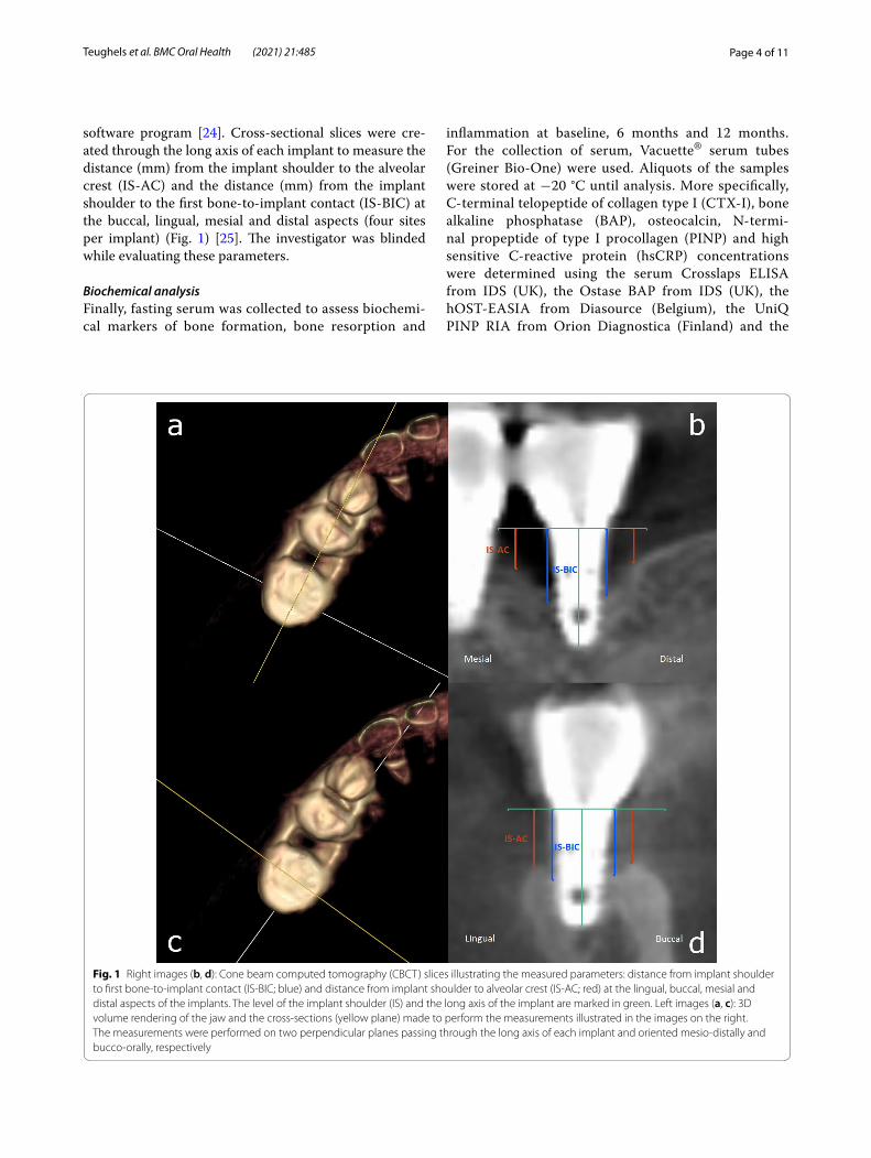
software program [24]. Cross-sectional slices were cre-ated through the long axis of each implant to measure the distance (mm) from the implant shoulder to the alveolar crest (IS-AC) and the distance (mm) from the implant shoulder to the first bone-to-implant contact (IS-BIC) at the buccal, lingual, mesial and distal aspects (four sites per implant) (Fig. 1) [25]. The investigator was blinded while evaluating these parameters.
Biochemical analysisFinally, fasting serum was collected to assess biochemi-cal markers of bone formation, bone resorption and
inflammation at baseline, 6 months and 12 months. For the collection of serum, Vacuette® serum tubes (Greiner Bio-One) were used. Aliquots of the samples were stored at −20 °C until analysis. More specifically, C-terminal telopeptide of collagen type I (CTX-I), bone alkaline phosphatase (BAP), osteocalcin, N-termi-nal propeptide of type I procollagen (PINP) and high sensitive C-reactive protein (hsCRP) concentrations were determined using the serum Crosslaps ELISA from IDS (UK), the Ostase BAP from IDS (UK), the hOST-EASIA from Diasource (Belgium), the UniQ PINP RIA from Orion Diagnostica (Finland) and the
Fig. 1 Right images (b, d): Cone beam computed tomography (CBCT) slices illustrating the measured parameters: distance from implant shoulder to first bone-to-implant contact (IS-BIC; blue) and distance from implant shoulder to alveolar crest (IS-AC; red) at the lingual, buccal, mesial and distal aspects of the implants. The level of the implant shoulder (IS) and the long axis of the implant are marked in green. Left images (a, c): 3D volume rendering of the jaw and the cross-sections (yellow plane) made to perform the measurements illustrated in the images on the right. The measurements were performed on two perpendicular planes passing through the long axis of each implant and oriented mesio-distally and bucco-orally, respectively

Page 5 of 11Teughels et al. BMC Oral Health (2021) 21:485
Liquid Unassayed Multiqual from Bio-Rad Laboratories (USA), respectively.
Surgical procedureAll surgical procedures were performed by the same experienced periodontist (G.U.C.) who was blinded to the study groups. After local anesthesia, sulcular inci-sions were made and full-thickness mucoperiosteal flaps were raised to expose the implant surfaces. Inflamma-tory tissue was removed, and the implant surfaces were mechanically cleaned with titanium-coated curettes and chemically disinfected with EDTA gel. Then the soft tis-sue flaps were sutured with 5–0 sutures. Bone grafts, membranes or other biologics were placed in neither of the two treatment groups (placebo and CS-OSA).
Outcome measuresThe primary outcome measure was the change in PPD at peri-implantitis sites from baseline to 12 months. Sec-ondary outcome measures included the changes in PPD from baseline to 6 months and changes in BOP, REC, IS-AC, IS-BIC, biomarkers and OHIP scores from base-line to 6 and 12 months.
StatisticsAs this study was an exploratory trial, no power calcula-tion was performed. Statistical analyses were performed using the per protocol population, defined as all ran-domized patients meeting the inclusion criteria, who completed the trial and who did not have major proto-col violations (i.e. no valid primary diagnosis, wash-out period not respected, use of prohibited concomitant medication, less than 6 months of treatment with study medication, randomization code was broken, the patient received wrong study medication, study medication com-pliance of less than 75%, in- and exclusion criteria not respected, screening failure and medical reason). Results are presented as mean ± standard deviation (SD) in tables and mean with 95% Confidence Interval (CI) in figures. The CBCT parameters, IS-AC and IS-BIC, were analyzed using a linear mixed model with sites nested in implant and patient taken as a random variable. Within group analyses were Bonferroni corrected. All other variables were analyzed by non-parametric tests since the data were not normally distributed. Clinical parameters (PPD and REC), OHIP-scores and biomarker values were ana-lyzed between and within treatments using Mann-With-ney U and Friedman with post hoc Bonferroni-corrected Wilcoxon signed-ranks, respectively. BOP, a binary vari-able, was evaluated using Chi square tests for between-groups analyses whereas differences within treatment groups were analyzed using Cochran’s Q test and post hoc Bonferroni corrected McNemar tests.
A two-tailed p-value below 0.05 was considered statis-tically significant.
All data were analyzed using SPSS software (version 26.0 for Windows, IBM Corp, Armonk, NY, USA).
ResultsPatientsBetween September 2017 and January 2018, a total of 21 eligible subjects were randomized and allocated to receive CS-OSA (n = 10) or a placebo (n = 11). Of these subjects, 5 were successfully treated for chronic peri-odontitis. No active periodontitis was observed during baseline. There were 4 patients with well-controlled dia-betes and 3 of them received CS-OSA. As demonstrated in Fig. 2, data of 18 patients were used in the per pro-tocol analysis. This includes 20 implants in the placebo group and 16 implants in the active treatment group. All implants were tissue level implants. CBCT images of suf-ficient quality (i.e. without artefacts) to perform analyses were available from 19 implants in the placebo group and 7 in the active treatment group. The study patients’ base-line demographic characteristics are shown in Table 1. No significant differences were found in baseline demo-graphics between the two groups.
Compliance and safetyAll but one patient reached the minimum compliance of 75%. The mean compliance was 93 ± 8% in the placebo group and 93 ± 6% in the CS-OSA group. One serious adverse event was reported in the active treatment group as the patient passed away due to a heart attack, and was reported by the investigator as unrelated to the study medication. In fact, the analysis of baseline serum sam-ples (i.e. prior of taking study medication) showed that the patient had a high cardiovascular risk as both a high cholesterol level and a high hs-CRP level (> 3.0 mg/L) was found. There were no adverse events reported which were related to the study medication.
Outcome measuresClinical parameter outcomes (PPD, REC, BOP) were analyzed at 120 implant sites in the placebo group and 96 sites in the active treatment group. CBCT parameters (IS-AC and IS-BIC) were analyzed at 76 implant sites in the placebo group and 28 in the active treatment group. The baseline outcome measures are summarized in Table 1. Baseline PPD and BOP were significantly lower in the placebo group compared to the active treatment group (PPD: p < 0.01; BOP: p < 0.05).
The results of both the primary and secondary outcome measures are summarized in Table 2. PPD and BOP improved significantly (p < 0.05) compared to baseline for both groups after 6 and 12 months. However, REC

Page 6 of 11Teughels et al. BMC Oral Health (2021) 21:485
significantly increased in the placebo group (p < 0.05) but not in the active treatment group (Fig. 3). The REC value after 6 and 12 months of treatment was significantly higher (p < 0.001) in the placebo group compared to the active treatment group (Fig. 3). Furthermore, the change in REC over 6 and 12 months was significantly different between groups (Table 2). The significant improvement in PPD and BOP after 6 and 12 months was not signifi-cantly different between the two treatment groups.
The CBCT parameters IS-AC and IS-BIC signifi-cantly increased after 6 and 12 months of treatment in the placebo group but not in the active treatment group. The change in IS-BIC over 12 months was significantly lower in the active treatment group compared to the placebo group (Table 2), resulting in a mean difference (95% CI) between placebo and CS-OSA of − 0.72 mm (−1.34 to −0.10) (Fig. 4). The change in IS-BIC over the last 6 months of treatment (between 6 and 12 months of treatment) was also significantly different between groups with a mean difference (95% CI) of − 0.46 (− 0.91 to − 0.01) (Fig. 4). There were no between group differ-ences observed with respect to IS-AC.
Evaluation of the total OHIP scores indicates no sig-nificant improvement in quality of life, neither in the pla-cebo group nor in the active treatment group (Table 2). Biomarker analysis showed no significant differences between the treatment groups (Table 2). With respect to
changes in hsCRP levels after 12 months of treatment it is noteworthy to mention that in 70% of the patients in the placebo group the hsCRP levels increased while in the active treatment group increase was only observed in 50% of the patients.
DiscussionThis is the first preliminary study to explore the effect of CS-OSA in patients with peri-implantitis. The main goals of peri-implantitis treatment are resolving inflam-mation and preventing further bone loss. However, a "gold standard" treatment is still lacking [12]. The clini-cal parameters PPD and BOP, significantly decreased after 6 and 12 months of CS-OSA and placebo treatment. The observed decreases were not significantly different between groups. We may therefore conclude that there was no significant difference in the primary outcome measure, i.e. the change in PPD at peri-implantitis sites from baseline to 12 months of treatment. The observed decreases in PPD and BOP are in both study groups likely to be the result of the peri-implantitis treatment per-formed at the baseline visit, i.e. debridement with open flap surgery, followed by 2-monthly repeated oral hygiene instructions. These are common interventions for treat-ing peri-implantitis, which have been previously dem-onstrated to be effective [12, 26–29]. However, mucosal recession has been reported to be a side-effect of such
Patients screened (n=21)
Patients randomized (n=21)
Patients excluded (n=0)
Placebo (n=11) CS-OSA (n=10)
Discontinued intervention (n=1)Unknown: never started taking medication (n=1)
Discontinued intervention (n=1)Serious Adverse Event unrelated to studymedication (n=1)
Analysed (n=10): 20 implants Analysed (n=8): 16 implantsExcluded from analysis (compliance < 75%) (n=1)
CBCT Analysis (n=9): 19 implants1 implant of 1 subject is excluded from analysis due to radiographic artefacts
CBCT Analysis (n=6): 7 implants9 implants of 2 subjects are excluded fromanalysis due to radiographic artefacts
Fig. 2 Flow diagram of study enrolment, allocation, follow-up and analysis. CBCT: cone beam computed tomography; CS-OSA: choline-stabilized orthosilicic acid

Page 7 of 11Teughels et al. BMC Oral Health (2021) 21:485
surgical treatment [30]. Indeed, an increase in REC was observed after 6 and 12 months of treatment in the pla-cebo group, however not in the active treatment group. In fact, the REC value didn’t change during the study period in patients taking CS-OSA suggesting that this complex may have a positive effect on soft tissue healing.
In the present preliminary study, a statistically signifi-cant difference between the placebo and the CS-OSA group was observed for bone loss measured by the change in IS-BIC after 12 months of treatment. More specifically, the IS-BIC increased in the placebo group and remained stable in the active treatment group. These findings suggest that CS-OSA may prevent further bone loss at implants with peri-implantitis.
To summarize, a significant increase in both REC and IS-BIC is found after 12 weeks of placebo treatment, while both REC and IS-BIC remained stable in the CS-OSA group. The beneficial effect of CS-OSA on recession might be explained by a direct effect on the gingiva or might be secondary to the beneficial effect of CS-OSA on bone loss, i.e. preventing further bone loss and therefore stabilizing both bone level and the level of the gingival margin.
The effectiveness of CS-OSA on bone and connective tissue health has been previously demonstrated [14, 17, 18, 31]. More specifically, both pre-clinical and clinical studies have shown that CS-OSA has a stimulating effect on the collagen synthesis. Reffitt et al. [14] reported that physiological concentrations of orthosilicic acid stimu-late collagen type I synthesis in human osteoblast-like cells and dermal fibroblasts in vitro and promote osteo-blastic differentiation. An earlier study in young animals has shown an increase in collagen concentration in the dermis of animals who were fed with CS-OSA in their diet compared to control animals [31]. These reported increases in skin collagen can explain the improved sur-face and mechanical properties of the skin which were reported after oral intake of CS-OSA in humans [18]. Furthermore, in the study of Spector et al. [17] the use of CS-OSA resulted in an increase of serum PINP and femoral bone density in osteopenic women, indicating improved bone collagen synthesis.
Choline is classified by the Food and Nutrition Board as an essential nutrient and is likely to contribute to the biological activity of CS-OSA [32]. It is a precursor of phospholipids, which are essential components of bio-logical membranes, and is involved in cell signaling and lipid transport/metabolism. One of its metabolites, betaine, participates in the methylation of homocyst-eine to methionine and therefore reduces the plasma total homocysteine levels [33]. This reduction positively affects collagen cross-linking, since homocysteine has been shown to interfere with post-translational modifi-cations of collagen through direct and indirect inhibition of lysyl oxidase as well as through down regulation of other genes involved in collagen cross-linking [34]. Ele-vated levels of plasma homocysteine have been detected in patients with chronic periodontitis [35, 36]. These elevated homocysteine levels reduced after periodontal treatment, indicating an important role of homocysteine in periodontal pathologies [36].
The previously described studies ([14, 17, 18, 31, 32, 34–36]) support the hypothesis of a possible effect of CS-OSA on collagen metabolism improving bone and soft tissue healing in patients with peri-implantitis. A posi-tive effect of CS-OSA was indeed confirmed in the pre-sent study by the effects observed on IS-BIC and REC, however the biomarker analysis failed to support this hypothesis as no significant differences in serum osteoc-alcin, PINP, BAP and CTX-I levels were found. These are biomarkers for bone formation and resorption, respec-tively. This is in contrast with the study of Golub et al. [37] demonstrating reduced serum biomarkers of bone and collagen destruction after periodontal treatment. However, in the latter study, significant effects were observed in the serum of post-menopausal osteopenic
Table 1 Baseline demographic characteristics and outcome measures
Data expressed as mean ± SD. Significant difference between groups: p < 0.05 = significant (bold) #IS-AC and IS-BIC analysis was performed on cone beam computed tomography (CBCT) scans of sufficient quality (i.e. without artefacts) taken from 9 patients (19 implants) in the placebo group and 6 patients (7 implants) in the CS-OSA group. CS-OSA: choline-stabilized orthosilicic acid, PPD: probing pocket depth, REC: mucosal recession, BOP: bleeding on probing, IS-AC: distance from implant shoulder to alveolar crest, IS-BIC: distance from implant shoulder to first bone-to-implant contact, OHIP: patient’s oral health related quality of life questionnaire, hsCRP: high sensitive C-reactive protein, BAP: bone alkaline phosphatase, CTX-I: C-terminal telopeptide of collagen I, PINP: N-terminal propeptide of type I procollagen
Variable Treatment group p value
Placebo CS-OSA
Number of patients 10 8
Number of implants 20 16
Age 51.50 ± 10.19 52.50 ± 7.29 0.818
Number of males 3 5 0.168
PPD (mm) 5.04 ± 2.37 6.01 ± 2.54 0.009REC (mm) 0.78 ± 1.27 0.70 ± 1.22 0.577
BOP (%) 86.67 ± 34.14 94.79 ± 22.34 0.045IS-AC (mm)# 2.57 ± 2.56 2.4 ± 2.19 0.765
IS-BIC (mm)# 3.83 ± 2.39 4.43 ± 1.98 0.467
OHIP Total 15.10 ± 10.81 14.63 ± 8.35 0.515
hsCRP (mg/L) 2.40 ± 2.40 4.12 ± 2.83 0.055
Osteocalcin (ng/mL) 9.04 ± 3.39 9.88 ± 3.67 0.762
BAP (µg/L) 10.22 ± 4.20 10.13 ± 1.58 0.203
CTX-I (ng/mL) 0.48 ± 0.22 0.52 ± 0.23 0.696
PINP (ng/mL) 43.35 ± 12.61 47.03 ± 8.51 0.573

Page 8 of 11Teughels et al. BMC Oral Health (2021) 21:485
Tabl
e 2
Prim
ary
and
seco
ndar
y ou
tcom
e m
easu
res
and
the
abso
lute
out
com
e sc
ores
Sign
ifica
nt d
iffer
ence
in c
hang
es o
ver 6
and
12
mon
ths
betw
een
grou
ps: p
< 0
.05 =
sign
ifica
nt (b
old)
. a Sign
ifica
nt d
iffer
ence
from
bas
elin
e w
ithin
the
grou
p; b si
gnifi
cant
diff
eren
ce fr
om 6
mon
ths
with
in th
e gr
oup;
c si
gnifi
cant
diff
eren
ce b
etw
een
grou
ps (p
lace
bo v
ersu
s ch
olin
e-st
abili
zed
orth
osili
cic
acid
(CS-
OSA
)). # IS
-AC
and
IS-B
IC a
naly
sis
was
per
form
ed o
n co
ne-b
eam
com
pute
d to
mog
raph
y (C
BCT)
sca
ns o
f suffi
cien
t qua
lity
(i.e.
with
out a
rtef
acts
) tak
en fr
om 9
pat
ient
s (1
9 im
plan
ts) i
n th
e pl
aceb
o gr
oup
and
6 pa
tient
s (7
impl
ants
) in
the
CS-O
SA g
roup
. CS-
OSA
: cho
line-
stab
ilize
d or
thos
ilici
c ac
id, P
PD: p
robi
ng p
ocke
t dep
th, B
OP:
ble
edin
g on
pro
bing
, REC
: muc
osal
rece
ssio
n, IS
-AC:
dis
tanc
e fr
om im
plan
t sho
ulde
r to
alve
olar
cre
st, I
S-BI
C: d
ista
nce
from
impl
ant s
houl
der t
o fir
st b
one-
to-im
plan
t con
tact
, OH
IP: p
atie
nt’s
oral
hea
lth re
late
d qu
ality
of l
ife
ques
tionn
aire
, hsC
RP: h
igh
sens
itive
C-r
eact
ive
prot
ein,
BA
P: b
one
alka
line
phos
phat
ase,
CTX
-I: C
-ter
min
al te
lope
ptid
e of
col
lage
n I,
PIN
P: N
-ter
min
al p
rope
ptid
e of
type
I pr
ocol
lage
n
Vari
able
Trea
tmen
t gro
upp
valu
e
Plac
ebo
(n =
10)
CS-O
SA (n
= 8
)p
valu
edi
ffere
nce
in c
hang
e 6
mon
ths
p va
lue
diffe
renc
e in
cha
nge
12 m
onth
sBa
selin
e (m
ean ±
SD
)6
mon
ths
(mea
n ±
SD
)12
mon
ths
(mea
n ±
SD
)Ch
ange
6
mon
ths—
base
line
(mea
n ±
SD
)
Chan
ge
12 m
onth
s—ba
selin
e(m
ean ±
SD
)
Base
line
(mea
n ±
SD
)6
mon
ths
(mea
n ±
SD
)12
mon
ths
(mea
n ±
SD
)Ch
ange
6
mon
ths—
base
line
(mea
n ±
SD
)
Chan
ge
12 m
onth
s—ba
selin
e (m
ean ±
SD
)
PPD
(mm
)5.
0 ±
2.4
c2.
8 ±
1.5
a, c
2.5 ±
1.2
a, c
−
2.2
± 2
.4−
2.5
± 2
.16.
0 ±
2.5
c4.
1 ±
2.4
a, c
3.8 ±
2.2
a, c
− 2
.0 ±
2.4
− 2
.2 ±
2.6
0.32
00.
170
BOP
(%)
86.7
± 3
4.1
c35
.8 ±
48.
2a30
.8 ±
46.
4a, c
− 5
0.8 ±
58.
0−
55.
8 ±
54.
794
.8 ±
22.
3 c
40.6
± 4
9.4a
50.0
± 5
0.3a,
c−
54.
2 ±
54.
1−
44.
8 ±
54.
00.
691
0.21
8
REC
(mm
)0.
8 ±
1.3
1.4 ±
1.2
a, c
1.2 ±
1.2
a, c
0.6 ±
1.0
c0.
4 ±
1.1
c0.
7 ±
1.2
0.9 ±
1.2
c0.
7 ±
1.0
c0.
2 ±
0.9
c−
0.1
± 1
.0 c
0.00
60.
009
IS-A
C (m
m)#
2.6 ±
2.6
3.0 ±
2.7
a3.
3 ±
2.6
a0.
4 ±
1.0
0.7 ±
1.5
2.4 ±
2.2
2.8 ±
2.2
2.7 ±
2.1
0.4 ±
0.9
0.3 ±
0.9
0.85
90.
254
IS-B
IC (m
m) #
3.8 ±
2.4
4.3 ±
2.3
a4.
6 ±
2.4
a0.
5 ±
1.3
0.8 ±
1.6
c4.
4 ±
2.0
4.7 ±
1.9
4.5 ±
2.1
0.2 ±
0.7
0.1 ±
0.8
c0.
283
0.02
3
OH
IP T
otal
15.1
± 1
0.8
14.0
± 1
2.6
13.1
± 7
.6−
1.1
± 6
.8−
2.0
± 9
.114
.6 ±
8.4
12.6
± 8
.610
.1 ±
5.7
− 2
.0 ±
10.
0−
4.5
± 9
.80.
965
0.51
5
hsC
RP (m
g/L)
2.4 ±
2.4
2.6 ±
2.6
2.9 ±
1.6
0.2 ±
3.1
0.5 ±
1.6
4.1 ±
2.8
4.0 ±
2.5
3.6 ±
1.8
− 0
.1 ±
0.8
− 0
.6 ±
3.3
0.82
90.
360
Ost
eoca
lcin
(n
g/m
L)9.
0 ±
3.4
9.1 ±
2.3
9.4 ±
2.0
0.0 ±
1.9
0.4 ±
3.1
9.9 ±
3.7
9.4 ±
3.1
9.8 ±
4.5
− 0
.5 ±
3.4
− 0
.1 ±
2.7
0.17
30.
696
BAP
(µg/
L)10
.2 ±
4.2
8.1 ±
3.0
a10
.3 ±
5.9
− 2
.2 ±
2.0
0.1
± 2
.210
.1 ±
1.6
8.2 ±
1.3
a10
.3 ±
2.4
− 1
.9 ±
1.2
0.2 ±
2.0
0.82
90.
696
CTX
-I (n
g/m
L)0.
5 ±
0.2
0.4 ±
0.2
a0.
5 ±
0.2
b−
0.1
± 0
.10.
0 ±
0.1
0.5 ±
0.2
0.4 ±
0.2
0.5 ±
0.2
− 0
.1 ±
0.2
− 0
.1 ±
0.1
0.51
50.
146
PIN
P (n
g/m
L)43
.4 ±
12.
643
.2 ±
11.
543
.1 ±
13.
2−
0.1
± 8
.8−
0.3
± 6
.647
.0 ±
8.5
48.9
± 2
2.8
44.3
± 1
8.4
1.9 ±
15.
7−
2.8
± 1
2.4
0.89
70.
897

Page 9 of 11Teughels et al. BMC Oral Health (2021) 21:485
women within 5 years of menopause, a time-period asso-ciated with high-turnover bone loss, therefore exhibiting not only local but also systemic bone loss. When also tak-ing into account the lack of significant reductions in the inflammatory marker hsCRP in the present study, while the clinical parameter for inflammation, BOP, improved in both treatment groups, it should be encouraged to
analyze biomarkers of inflammation, collagen destruc-tion, and bone resorption locally in the peri-implant crevicular fluid. Indeed, an interesting cross-sectional study examining the biomarker profile in peri-implant crevicular fluid from healthy implants and implants with peri-implantitis confirmed that local biomarkers might contribute to distinguish peri-implant health from dis-ease [38]. Also it should be noticed that a large variation was observed in biomarker levels in the present study, which makes it difficult to detect significant differences between groups.
In fact, an important limitation of the present study is the limited number of participants. A limited sample size, without a prior power calculation, was chosen because of the exploratory character of the study. This might explain why the present study failed to show significant effects with respect to the parameters evaluated at patient level, including biomarker and OHIP questionnaire analysis. On the other hand, the significant effects that were found in this study cannot be generalized, but give important insights for future research that should be performed in a larger study population. Another limitation of the study is that not all risk factors for peri-implantitis such as genetic traits, soft tissue quality or quantity, pros-thetic design and occlusal overload [7] were standardized between both treatment groups (data unknown). With the linear mixed model, an additional analysis could be performed to investigate the influence of the risk factors “history of periodontitis” and “diabetes” on the outcome results for the bone loss parameters. When history of periodontitis and diabetes were considered as confound-ers, the significant difference that was found between the placebo and the CS-OSA group for change in IS-BIC after 12 months of treatment remained significant in favor of CS-OSA.
In order to fully understand the mechanisms of action of CS-OSA in preventing further bone loss at the peri-implant site level, as suggested in the present study, it might be useful to investigate biomarkers for bone and collagen metabolism locally in the peri-implant crevic-ular fluid. As in the present study bone loss was evalu-ated by linear bone measurements using CBCT, it would be interesting to also look at the effect of CS-OSA on alveolar bone density. Such a study has not yet been performed, since the CBCT technology used in the pre-sent study is not the ideal tool for evaluating bone den-sity [39]. The one-year study period in the present trial is a minimum duration to evaluate changes in bone level and/or bone density. In future research, the study period should be prolonged to evaluate the long-term outcomes of the treatment.
0
0,2
0,4
0,6
0,8
1
1,2
1,4
1,6
1,8
Baseline 6 months 12 months
REC
(mm
)
Placebo (n = 120)
CS-OSA (n = 96)
DID (95% CI)
T0-T6: -0.39 (-0.65, -0.13)T0-T12: -0.47 (-0.74, -0.19)T6-T12: -0.08 (-0.24, 0.08)
#
*
#
*
Fig. 3 Mucosal recession (REC) at baseline, 6 months and 12 months of treatment with choline-stabilized orthosilicic acid (CS-OSA) compared to placebo. Data is presented as mean with 95% confidence interval (CI). The insert box shows the difference in difference (DID) between baseline and 6 months (T0-T6), baseline and 12 months (T0-T12) and 6 months and 12 months (T6-T12) of treatment with placebo and CS-OSA. *p < 0.05 vs baseline (within placebo); #p < 0.05 vs placebo. n = the amount of implant sites
0
1
2
3
4
5
6
7
Baseline 6 months 12 months
IS-B
IC (m
m)
Placebo (n = 76)
CS-OSA (n = 28)
DID (95% CI)
T0-T6: -0.26 (-0.73, 0.22)T0-T12: -0.72 (-1.34, -0.10)T6-T12: -0.46 (-0.91, -0.01)
* *
Fig. 4 Distance from the implant shoulder to first bone-to-implant contact (IS-BIC) at baseline, 6 months and 12 months of treatment with choline-stabilized orthosilicic acid (CS-OSA) compared to placebo. Data is presented as mean with 95% confidence interval (CI). The insert box shows the difference in difference (DID) between baseline and 6 months (T0–T6), baseline and 12 months (T0-T12) and 6 months and 12 months (T6-T12) of placebo and CS-OSA treatment. *p < 0.05 vs. baseline (within placebo). n = the amount of implant sites

Page 10 of 11Teughels et al. BMC Oral Health (2021) 21:485
ConclusionsThe results of this preliminary study suggest that CS-OSA stabilizes and even prevents further bone loss after surgical peri-implantitis treatment in combina-tion with a beneficial effect on mucosal tissue healing. Future research in a larger, more standardized study population and a longer study period is needed to con-firm the results of the present study and to fully under-stand the exact mechanisms of action.
Abbreviations2D: Two-dimensional; 3D: Three-dimensional; BAP: Bone alkaline phosphatase; BOP: Bleeding on probing; CBCT: Cone beam computed tomography; CS-OSA: Choline-stabilized orthosilicic acid; CTX-I: C-terminal telopeptide of collagen type I; hsCRP: High sensitive C-reactive protein; IR: Intraoral radiography; IS-AC: Distance from implant shoulder to alveolar crest; IS-BIC: Distance from implant shoulder to first bone-to-implant contact; OHIP-14: Patient’s Oral Health related Quality of Life questionnaire; PINP: N-terminal propeptide of type I procollagen; PPD: Probing pocket depth; REC: Mucosal recession.
Supplementary InformationThe online version contains supplementary material available at https:// doi. org/ 10. 1186/ s12903- 021- 01817-4.
Additional file 1. Dataset Teughels et al. Peri-implantitis study.
AcknowledgementsThe authors thank Joren Bosmans for the statistical analysis.
Authors’ contributionsWT, MCH and SMP have made substantial contributions to conception and design of the study. GUC and MCH have been involved in data collection. WT, MCH, SMP, IDC and MT contributed to data interpretation and analysis. All authors have been involved in drafting the manuscript and revising it critically and have given final approval of the version to be published. All authors read and approved the final manuscript.
FundingBio Minerals NV provided the study medication (CS-OSA and placebo) and funding.
Availability of data and materialsAll data generated or analysed during this study are included in this published article and its supplementary information files.
Declarations
Ethics approval and consent to participateWritten informed consent was obtained from all patients prior to randomiza-tion. The study was approved by the local Ethical Committee of the Cukurova University, Adana, Turkey (65/4, 04.05.2017).
Consent for publicationsNot applicable.
Competing interestsSMP and IDC are employed as research associates of the study sponsor Bio Minerals NV (Belgium, the manufacturer of choline-stabilized orthosilicic acid). MCH has received financial support from Bio Minerals NV to conduct the study. WT, GUC and MT report no conflicts of interest related to this study.
Author details1 Section of Periodontology, Department of Oral Health Sciences, KU Leuven and Dentistry, University Hospitals, Leuven, Belgium. 2 Department of Perio-dontology, Faculty of Dentistry, Cukurova University, Adana, Turkey. 3 Research and Development, Bio Minerals NV, Zenderstraat 12, 9070 Destelbergen, Belgium.
Received: 30 November 2020 Accepted: 8 September 2021
References 1. Dreyer H, Grischke J, Tiede C, Eberhard J, Schweitzer A, Toikkanen SE,
Glockner S, Krause G, Stiesch M. Epidemiology and risk factors of peri-implantitis: a systematic review. J Periodontal Res. 2018;53(5):657–81.
2. Doornewaard R, Jacquet W, Cosyn J, De Bruyn H. How do peri-implant biologic parameters correspond with implant survival and peri-implanti-tis? A critical review. Clin Oral Implant Res. 2018;29(Suppl 18):100–23.
3. Derks J, Tomasi C. Peri-implant health and disease. A systematic review of current epidemiology. J Clin Periodontol. 2015;42(Suppl 16):S158-171.
4. Cosgarea R, Sculean A, Shibli JA, Salvi GE. Prevalence of peri-implant diseases - a critical review on the current evidence. Braz Oral Res. 2019;33(suppl 1):e063.
5. Robertson K, Shahbazian T, MacLeod S. Treatment of peri-implantitis and the failing implant. Dent Clin North Am. 2015;59(2):329–43.
6. Berglundh T, Armitage G, Araujo MG, Avila-Ortiz G, Blanco J, Camargo PM, Chen S, Cochran D, Derks J, Figuero E, et al. Peri-implant diseases and conditions: consensus report of workgroup 4 of the 2017 World Workshop on the Classification of Periodontal and Peri-Implant Diseases and Conditions. J Clin Periodontol. 2018;45(Suppl 20):S286-s291.
7. Schwarz F, Derks J, Monje A, Wang HL. Peri-implantitis. J Periodontol. 2018;89(Suppl 1):S267-s290.
8. Harris D, Horner K, Grondahl K, Jacobs R, Helmrot E, Benic GI, Bornstein MM, Dawood A, Quirynen M. E.A.O. guidelines for the use of diagnostic imaging in implant dentistry 2011. A consensus workshop organ-ized by the European Association for Osseointegration at the Medical University of Warsaw. Clin Oral Implants Res. 2012;23(11):1243–53.
9. Jacobs R, Vranckx M, Vanderstuyft T, Quirynen M, Salmon B. CBCT vs other imaging modalities to assess peri-implant bone and diagnose complications: a systematic review. Eur J Oral Implantol. 2018;11(Suppl 1):77–92.
10. Golubovic V, Mihatovic I, Becker J, Schwarz F. Accuracy of cone-beam computed tomography to assess the configuration and extent of ligature-induced peri-implantitis defects. A pilot study. Oral Maxillof Surg. 2012;16(4):349–54.
11. Laleman I, Pauwels M, Quirynen M, Teughels W. The usage of a lactobacilli probiotic in the non-surgical therapy of peri-implantitis: a randomized pilot study. Clin Oral Implant Res. 2020;31(1):84–92.
12. Heitz-Mayfield LJ, Mombelli A. The therapy of peri-implantitis: a sys-tematic review. Int J Oral Maxillofac Implants. 2014;29(Suppl):325–45.
13. Calomme M, Cos P, D’Haese P, Vingerhoets R, Lamberts LV, De Broe M, Van Hoorebeke C, Vanden Berghe D. Silicon absorption from stabilized orthosilicic acid and other supplements in healthy subjects. Trace Elem Man Anim. 2000;10:1111–4.
14. Reffitt DM, Ogston N, Jugdaohsingh R, Cheung HF, Evans BA, Thomp-son RP, Powell JJ, Hampson GN. Orthosilicic acid stimulates collagen type 1 synthesis and osteoblastic differentiation in human osteoblast-like cells in vitro. Bone. 2003;32(2):127–35.
15. Calomme M, Wijnen P, Sindambiwe JB, Cos P, Mertens J, Geusens P, Vanden Berghe D. Effect of choline-stabilized orthosilicic acid on bone density in chicks. Calcif Tissue Int. 2002;70:292.
16. Calomme M, Geusens P, Demeester N, Behets GJ, D’Haese P, Sindam-biwe JB, Van Hoof V, Vanden Berghe D. Partial prevention of long-term femoral bone loss in aged ovariectomized rats supplemented with choline-stabilized orthosilicic acid. Calcif Tissue Int. 2006;78(4):227–32.
17. Spector TD, Calomme MR, Anderson SH, Clement G, Bevan L, Demeester N, Swaminathan R, Jugdaohsingh R, Berghe DA, Powell JJ.

Page 11 of 11Teughels et al. BMC Oral Health (2021) 21:485
• fast, convenient online submission
•
thorough peer review by experienced researchers in your field
• rapid publication on acceptance
• support for research data, including large and complex data types
•
gold Open Access which fosters wider collaboration and increased citations
maximum visibility for your research: over 100M website views per year •
At BMC, research is always in progress.
Learn more biomedcentral.com/submissions
Ready to submit your researchReady to submit your research ? Choose BMC and benefit from: ? Choose BMC and benefit from:
Choline-stabilized orthosilicic acid supplementation as an adjunct to calcium/vitamin D3 stimulates markers of bone formation in osteo-penic females: a randomized, placebo-controlled trial. BMC Musculo-skelet Disord. 2008;9:85.
18. Barel A, Calomme M, Timchenko A, De Paepe K, Demeester N, Rogiers V, Clarys P, Vanden Berghe D. Effect of oral intake of choline-stabilized orthosilicic acid on skin, nails and hair in women with photodamaged skin. Arch Dermatol Res. 2005;297(4):147–53.
19. Geusens P, Pavelka K, Rovensky J, Vanhoof J, Demeester N, Calomme M, Vanden Berghe D. A 12-week randomized, double-blind, placebo-con-trolled multicenter study of choline-stabilized orthosilicic acid in patients with symptomatic knee osteoarthritis. BMC Musculoskelet Disord. 2017;18(1):2.
20. Loe H, Silness J. Periodontal disease in pregnancy. I. Prevalence and sever-ity. Acta Odontol Scand. 1963;21:533–51.
21. Başol ME, Karaağaçlioğlu L, Yilmaz B. Developing a Turkish Oral Health Impact Profile-OHIP-14-TR. Turkiye Klinikleri J Dent Sci. 2014;20(2):85–92.
22. Slade GD. Derivation and validation of a short-form oral health impact profile. Commun Dent Oral Epidemiol. 1997;25(4):284–90.
23. Vanderstuyft T, Tarce M, Sanaan B, Jacobs R, de Faria VK, Quirynen M. Inaccuracy of buccal bone thickness estimation on cone-beam CT due to implant blooming: an ex-vivo study. J Clin Periodontol. 2019;46(11):1134–43.
24. Fedorov A, Beichel R, Kalpathy-Cramer J, Finet J, Fillion-Robin JC, Pujol S, Bauer C, Jennings D, Fennessy F, Sonka M, et al. 3D Slicer as an image computing platform for the Quantitative Imaging Network. Magn Reson Imaging. 2012;30(9):1323–41.
25. Benic GI, Mokti M, Chen CJ, Weber HP, Hämmerle CH, Gallucci GO. Dimen-sions of buccal bone and mucosa at immediately placed implants after 7 years: a clinical and cone beam computed tomography study. Clin Oral Implant Res. 2012;23(5):560–6.
26. Carcuac O, Derks J, Abrahamsson I, Wennström JL, Petzold M, Berglundh T. Surgical treatment of peri-implantitis: 3-year results from a randomized controlled clinical trial. J Clin Periodontol. 2017;44(12):1294–303.
27. Hallström H, Persson GR, Lindgren S, Renvert S. Open flap debridement of peri-implantitis with or without adjunctive systemic antibiotics: a randomized clinical trial. J Clin Periodontol. 2017;44(12):1285–93.
28. Renvert S, Polyzois I, Claffey N. Surgical therapy for the control of peri-implantitis. Clin Oral Implant Res. 2012;23(Suppl 6):84–94.
29. Berglundh T, Wennström JL, Lindhe J. Long-term outcome of surgical treatment of peri-implantitis. A 2–11-year retrospective study. Clin Oral Implants Res. 2018;29(4):404–10.
30. Becker W, Becker BE, Caffesse R, Kerry G, Ochsenbein C, Morrison E, Prichard J. A longitudinal study comparing scaling, osseous surgery,
and modified Widman procedures: results after 5 years. J Periodontol. 2001;72(12):1675–84.
31. Calomme MR, Vanden Berghe DA. Supplementation of calves with stabilized orthosilicic acid. Effect on the Si, Ca, Mg, and P concentrations in serum and the collagen concentration in skin and cartilage. Biol Trace Elem Res. 1997;56(2):153–65.
32. Institute of Medicine Standing Committee on the Scientific Evaluation of Dietary Reference I, its Panel on Folate OBV, Choline: The National Academies Collection: Reports funded by National Institutes of Health. In: Dietary Reference Intakes for Thiamin, Riboflavin, Niacin, Vitamin B6, Folate, Vitamin B12, Pantothenic Acid, Biotin, and Choline. edn. Washing-ton (DC): National Academies Press (US), National Academy of Sciences.; 1998.
33. Ueland PM. Choline and betaine in health and disease. J Inherit Metab Dis. 2011;34(1):3–15.
34. Thaler R, Agsten M, Spitzer S, Paschalis EP, Karlic H, Klaushofer K, Varga F. Homocysteine suppresses the expression of the collagen cross-linker lysyl oxidase involving IL-6, Fli1, and epigenetic DNA methylation. J Biol Chem. 2011;286(7):5578–88.
35. Joseph R, Nath SG, Joseraj MG. Elevated plasma homocysteine levels in chronic periodontitis: a hospital-based case-control study. J Periodontol. 2011;82(3):439–44.
36. Bhardwaj S, Prabhuji ML, Karthikeyan BV. Effect of non-surgical periodon-tal therapy on plasma homocysteine levels in Indian population with chronic periodontitis: a pilot study. J Clin Periodontol. 2015;42(3):221–7.
37. Golub LM, Lee HM, Stoner JA, Reinhardt RA, Sorsa T, Goren AD, Payne JB. Doxycycline effects on serum bone biomarkers in post-menopausal women. J Dent Res. 2010;89(6):644–9.
38. Zani SR, Moss K, Shibli JA, Teixeira ER, de Oliveira MR, Onuma T, Feres M, Teles RP. Peri-implant crevicular fluid biomarkers as discriminants of peri-implant health and disease. J Clin Periodontol. 2016;43(10):825–32.
39. Corpas Ldos S, Jacobs R, Quirynen M, Huang Y, Naert I, Duyck J. Peri-implant bone tissue assessment by comparing the outcome of intra-oral radiograph and cone beam computed tomography analyses to the histological standard. Clin Oral Implant Res. 2011;22(5):492–9.
Publisher’s NoteSpringer Nature remains neutral with regard to jurisdictional claims in pub-lished maps and institutional affiliations.