the effect of ddt congeners on reproductive function in ... · cmu. journal (2006) vol. 5(2)219 the...
TRANSCRIPT
CMU. Journal (2006) Vol. 5(2) 219
The Effect of DDT Congeners on Reproductive Functionin Male Guppy (Poecilia reticulata)
Surachai Pikulkaew* and Dilok Wongsathein
Food Animal Clinic, Faculty of Veterinary Medicine, Chiang Mai University, Chiang Mai 50100, Thailand
*Corresponding author. E-mail: [email protected]
ABSTRACT Endocrine disrupters (EDs) such as DDT and its metabolite have been implicated in the impairment of reproductive performance via estrogenic actions in fish. This study was to evaluate the effect of DDT congeners on the reproductive performance of male guppies. Thirty male guppies were separated into 5 groups with 6 fish in each group. Group 1 was a control group; group 2 was fed with 0.10 ppb estradiol benzoate (positive control) and groups 3, 4 and 5 were fed with 0.05, 0.10 and 0.20 ppb of DDT congeners, respectively. Every group was treated for 30 days. Survival rates of groups 1-4 were 100% but for group 5, survival rate dropped to 66%. Male courtship behavior, caudal fin color intensity, gonopo-dial length and offspring sex ratio were not significantly different among groups. However, group 1 had higher rate of male sexual behavior and higher color intensity of caudal fins compared to the other groups. Group 2 fish had lower testis weight and fecundity than group 1 (p<0.05). Histopathological examinations showed that the testis of fish in groups 2, 3, 4 and 5 had more spermatozeumata and spermatids than spermatocyte. In addition, Sertoli cells and the efferent duct cells were hypertrophied and sperms were detached from the inner cells. In conclusion, DDT congeners impair male reproductive performance in guppies.
Key words: DDT congeners, Reproductive function, Male guppy, Testis
INTRODUCTION Xenoestrogen, a synthetic chemical in the environment, has been implicated in disrupting normal endocrine function in wildlife and human, e.g., falling sperm counts and decreased semen quality in human, developmental abnormalities of the gonads and abnormal sex hormone concentrations in juvenile alligators from Lake Apopka, Florida and male fish in rivers which show female characteristics (Sumpter, 1995). A group of these chemicals, known as endocrine disrupters, was defined by the 1996 European Commission as “exogenous substances that cause adverse health effects in an intact organism or its progeny, consequent to changes in endocrine function” (Olsson et al., 1998; Valle, 2001). These chemicals include herbicides, pesticides, fungicides, plasticizers, polychlorinated biphenyls (PCBs) and alkylphenolic com-pounds (Danzo, 1998). The effects of these chemicals are believed to include mimicking or antagonizing endogenous hormones, disruption of the synthesis and metabolism of endogenous hormone and modification of hormone receptors (Sonnenschein and Soto, 1998).

CMU. Journal (2006) Vol. 5(2)220 CMU. Journal (2006) Vol. 5(2) 221
Dichlorodiphenyl-trichloroethane (DDT) is one of xenoestrogen group that has been widely used as a pesticide and insecticide to control pests and vectors. However, in the 1970s, DDT was banned in many countries although it is still used in some developing countries (Donohoe and Curtis, 1996; Bayley et al., 2002). Such compounds are still common in environment due to their high persistence and accumulation in adipose tissues. Moreover, DDT and its metabolites have been associated with several disorders of the endocrine and reproductive systems in human and wildlife. For example, p,’p – DDT has an estrogenic effect on the mammalian uterus and avian oviduct by inhibiting oestradiol binding to the estrogen receptor (Danzo, 1998); p,p’ – DDE has an antiandrogenic activity in rats (Zaroogian et al., 2001; Bayley et al., 2002); p,’p – DDD can bind to estrogen receptors (Noriega and Hayes, 2000); and o,p’ – DDT is thought to have estrogenic activity. Some data have indicated that it may act in an androgenic or antiandrogenic manner (Zaroogian et al., 2001). In vivo studies, o,p’ – DDT has shown estrogenic effect and competitive binding to the estrogen receptor (ER) in mammals, birds and fish by using in vitro competitive binding assays (Leanos-Castaneda et al., 2002). o,’p – DDE has been found to have estrogenic activity (Donohoe and Curtis, 1996). Aquatic ecosystems are at the greatest risk from pollutants since all chemicals, whether on land or in the atmosphere, will eventually enter rivers and oceans. Fish, like other oviparous vertebrates, are sensitive to exogenous estrogen. The guppy (Poecilia reticulata) is an omnivorous freshwater fish teleost that belongs to the Poecillidae family. It was selected as the test organism because it is viviparous, has a short reproductive period, reproductive rate apparently independent of season and is easy to maintain and distinguish secondary sex characteristics (Bayley et al., 2002). The guppy’s testis is a single bilobed organ, located ventrally to the swimbladder (Kinnberg et al., 2003). Numerous tubules radiate from the central cavity towards the periphery of the testis. Tubules contain cysts with different spermatogenetic stages associated with Sertoli cells. These reorganize to form cysts by the time spermatogonia transform into primary spermatocytes. As spermatogenesis proceeds, the cysts migrate along the tubule towards the efferent duct. The secondary spermatocytes in the cysts transform into spermatids which differentiate into spermatozoa. These form spermatozeugmata, bundles of spermatozoa, with head pointing outward and tail towards the center. The Sertoli cells surrounding the mature spermatozeugmata pass into the lumen of the efferent duct. At the time of spermiation, the cysts open and the spermatozeugmata are voided into the spermatozeugmata duct. Then, the cyst Sertoli cells hypertrophy and transform into efferent duct cells (Kinnberg and Toft, 2003). The aim of the present study was to investigate the effect of DDT congeners on reproductive parameters in male guppy, including sexual behavior, caudal fin coloration, gonopodial length and gonad morphology and cytology. An experiment with estradiol benzoate was included to test the hypothesis of an estrogenic effect of DDT congeners.
MATERIALS AND METHODSExposure and maintenance of fish Sexually-mature male guppies were obtained from an ornamental fish farm in Chiang Mai Province. The fish were approximately 4 months old, with a mean weight of 0.095 g and were chosen randomly from stock aquaria. A total of 30 fish were divided into 5 groups

CMU. Journal (2006) Vol. 5(2)220 CMU. Journal (2006) Vol. 5(2) 221
of 6 fish each. They were fed twice daily with a micropellet food (SakuraTM). Group 1 was the control group fed with 30 mg of food, mixed with acetone alone (50μl) and allowed to dry. Group 2 fish were fed estrogen benzoate, dissolved in acetone and mixed with food at a concentration of 0.10 ng/mg (ppb). Groups 3, 4 and 5 were fed the DDT congeners (obtained from Research Institute for Health Sciences, Chiang Mai University) including p,’p – DDT (2.76 ng/mg), p,’p – DDE (1.36 ng/mg), p,’p – DDD (0.53 ng/mg), o,’p – DDT (1.28 ng/mg) and o,’p – DDE (0.07 ng/mg). These were dissolved in acetone and mixed with the food at concentrations of 0.05, 0.10 and 0.20 ng/mg (ppb) respectively. The acetone in the food was evaporated off. The experiment was conducted in glass aquaria, each containing 5 liter of filter water, for 30 days. During the experiment, feces were removed daily and filter water was added to maintain a water volume of 5 liters.
Male sexual behavior After exposure, each male guppy in all groups was transferred to a breeding tank, together with unexposed non-receptive females (approximately 4 months old). Male court-ship behavior was observed in 15-minute test interval. Bozynski and Liley (2003) defined the criteria of male courtship behavior as follow: 1. Attendance: an attending male orients towards the female from a stationary position or follows the female closely within one body length as she moves about the tank. 2. Partial gonopodial swing: any movement of the gonopodium, not beyond 90° from the resting position. 3. Gonopodial swing: a gonopodial movement beyond 90° from the resting position. 4. Gonopodial thrust: occurs during a mating attempt when the male approaches the female from behind and thrust his gonopodium towards the female’s genital pore. 5. Sigmoid display: the male positions himself in front of or to the side of the female and twists his body laterally into a sigmoid shape and quivers. Sigmoid display, followed by a mating attempt, is a frequent occurrence. After the behavioral test, breeding was allowed to occur for one week and the fish were fed with uncontaminated food during this period. Then the female fish were separated to a new aquarium for 2 months. The numbers of the offspring (fecundity) and sex ratio of the offspring were recorded after the gestation period.
Caudal fin color intensity The intensity of the red coloration of the caudal fin was evaluated. After breeding, the male fish were anaesthetized in ethyl-4-aminobenzoate at a concentration of 100 mg/l. Then the fish were placed on a white flat bed to evaluate caudal fin color intensity and the image was cap-tured by SONY cyber-shot, DSC-F505V resolution 640 pixels to adobe Photoshop. After that the freeware program RGB.EXE that was available from http:www.ebd.csic.es/rv/index.html stored the RGB values of each pixel. A criterion which defined pixel as red or not red was (=IF(RED<100; “not red;” IF((GREEN*1.1)>RED; “Not red;” IF((BLUE*1.25)>RED; “not red;” “red”)))) (Larsson et al., 2002). This criterion was transferred to MICROSOFT EXCEL to calculate between the red pixels proportion and the number of all pixels.
Gonopodial index, gonosomatic index and histopathological procedures. The fish were euthanized with an ethyl-4-aminobenzoate overdose. The length of the total body and the length of the gonopodium were measured for each fish. The gonopodial

CMU. Journal (2006) Vol. 5(2)222 CMU. Journal (2006) Vol. 5(2) 223
index (GPI) was calculated in terms of the gonopodial length as a percentage of the total body length. Then the whole body weight and the testis wet weight were measured for each fish. The gonosomatic index (GSI) was calculated in terms of the gonadal weight as a percen-tage of the whole body weight. After that the testes were fixed in neutral-buffered formalin, dehydrated, embedded in paraffin, sectioned at 5 μm and stained with hematoxylin and eosin (H&E) for histopathological examination.
Statistic analysis The data obtained on caudal fin color intensity, gonopodial index and gonosomatic index were analyzed by using a one-way ANOVA to test for significant differences among the treatments.
RESULTSSurvival Over 30 days, 44% of the male guppies exposed to feed contaminated with 0.20 DDT congeners (group 5) died 20 days after exposure. The guppies showed generalized hemor-rhage that might be from the secondary bacterial infection (Leatherland and Woo, 1998). No mortality was observed in the other groups.
Male sexual behavior The male fish in control group performed more partial gonopodial swings, gonopodial swings, gonopodial thrusts and sigmoid displays and spent more time attending the females than the treated groups. In addition, no gonopodial thrusts occurred in the DDT 0.10 and DDT 0.20 groups. Thus, males exposed to various doses of DDT were less sexually-active than the control fish. However, fish in group 0.05 ppb estradiol benzoate did increase the number of sigmoid displays compared with other groups. Male sexual behavior is shown in Table 1.
Table 1. Sexual behavior of the male guppy (Mean + SD).
Number of attendance
Duration of attendance(second)
Number of partial
gonopodial swing
Number of gonopodial
swing
Number of gonopodial
thrust
Number of sigmoid
display
1. Control 3.50 + 0.55 9.50 + 3.14 8.33 + 2.42 6.00 + 1.26 1.67 + 0.52 3.00 + 4.38
2. Estrogen 0.05 ppb
1.40 + 0.55 5.53 + 5.67 9.00 + 6.20 6.20 + 7.26 0.80 + 1.10 6.60 + 6.31
3. DDT 0.05 ppb
3.33 + 1.97 3.48 + 4.53 2.00 + 1.26 3.17 + 6.40 1.83 + 3.25 0.67 + 1.03
4. DDT 0.10 ppb
3.00 + 2.53 2.62 + 3.13 5.33 + 6.12 3.67 + 5.09 0.00 + 0.00 4.00 + 4.52
5. DDT 0.20 ppb
2.11 + 0.75 4.58 + 4.45 7.62 + 8.79 3.82 + 4.98 0.00 + 0.00 3.10 + 2.88

CMU. Journal (2006) Vol. 5(2)222 CMU. Journal (2006) Vol. 5(2) 223
Caudal fin color intensity Exposure to various doses of DDT and estradiol benzoate decreased mean redness (% red area) compared with the control group but not significantly (Figure 1).
Figure 1. Caudal fin color intensity of the male guppy.
Gonopodial index and gonosomatic index There were no significant differences in the gonopodial index between the control group and the treated groups. However, exposure to estradiol benzoate resulted in significant (P<0.05) reductions in the gonosomatic index, compared to the control fish and treated groups. Furthermore, various doses of DDT resulted in lower mean of gonosomatic indices, but not significantly so, compared to control fish. The gonopodial index and gonosomatic index are shown in Table 2.
Table 2. Gonopodial index (GPI) and gonadosomatic index (GSI) of the male guppy (Mean + SD).
GPI (%) GSI (%)1. Controls 21.808 + 1.515 a* 2.510 + 0.456 a
2. Estrogen 0.05 ppb 22.147 + 2.652 a 1.695 + 0.568 b
3. DDT 0.05 ppb 21.996 + 2.178 a 1.977 + 0.378 a
4. DDT 0.10 ppb 22.515 + 2.802 a 2.300 + 0.276 a
5. DDT 0.20 ppb 20.479 + 9.936 a 2.230 + 1.203 a
* Different letters indicate significant differences at 95% confidential level.
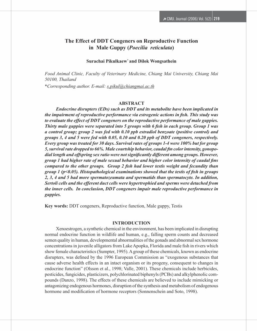
Histopathology of testis The testes of the control group (Figure 2) contained regularly-organized cysts with all stages of spermatogenesis. These included peripheral spermatogonia, primary spermato-cytes, secondary spermatocytes, spermatids and developing spermatozeugmata, closer to the centrally-situated efferent ducts and contained mature spermatozeugmata. Moreover, trans-formed hypertrophied Sertoli cells were incorporated in the efferent duct epithelium. For fish in the other groups, the histopathological examination of the testes revealed moderate to severe effects on structure. With estradiol benzoate (Figure 3A), the testis had
%Rednesslevel(Mean�SD) 100
80
60
40
20
0Control Estrogen 0.05 ppb DDT 0.05 ppb DDT 0.10 ppb DDT 0.20 ppb
CMU. Journal (2006) Vol. 5(2)224 CMU. Journal (2006) Vol. 5(2) 225
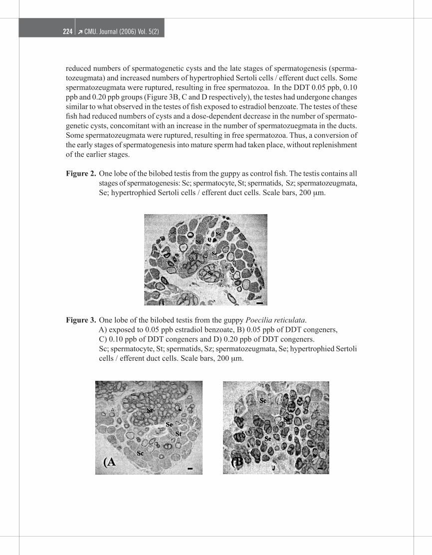
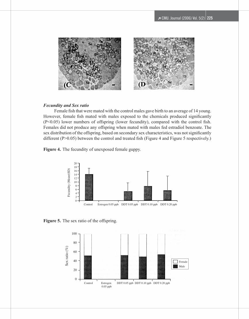
reduced numbers of spermatogenetic cysts and the late stages of spermatogenesis (sperma-tozeugmata) and increased numbers of hypertrophied Sertoli cells / efferent duct cells. Some spermatozeugmata were ruptured, resulting in free spermatozoa. In the DDT 0.05 ppb, 0.10 ppb and 0.20 ppb groups (Figure 3B, C and D respectively), the testes had undergone changes similar to what observed in the testes of fish exposed to estradiol benzoate. The testes of these fish had reduced numbers of cysts and a dose-dependent decrease in the number of spermato-genetic cysts, concomitant with an increase in the number of spermatozuegmata in the ducts. Some spermatozeugmata were ruptured, resulting in free spermatozoa. Thus, a conversion of the early stages of spermatogenesis into mature sperm had taken place, without replenishment of the earlier stages.
Figure 2. One lobe of the bilobed testis from the guppy as control fish. The testis contains all stages of spermatogenesis: Sc; spermatocyte, St; spermatids, Sz; spermatozeugmata, Se; hypertrophied Sertoli cells / efferent duct cells. Scale bars, 200 μm.
Figure 3. One lobe of the bilobed testis from the guppy Poecilia reticulata. A) exposed to 0.05 ppb estradiol benzoate, B) 0.05 ppb of DDT congeners, C) 0.10 ppb of DDT congeners and D) 0.20 ppb of DDT congeners. Sc; spermatocyte, St; spermatids, Sz; spermatozeugmata, Se; hypertrophied Sertoli
cells / efferent duct cells. Scale bars, 200 μm.
CMU. Journal (2006) Vol. 5(2)224 CMU. Journal (2006) Vol. 5(2) 225
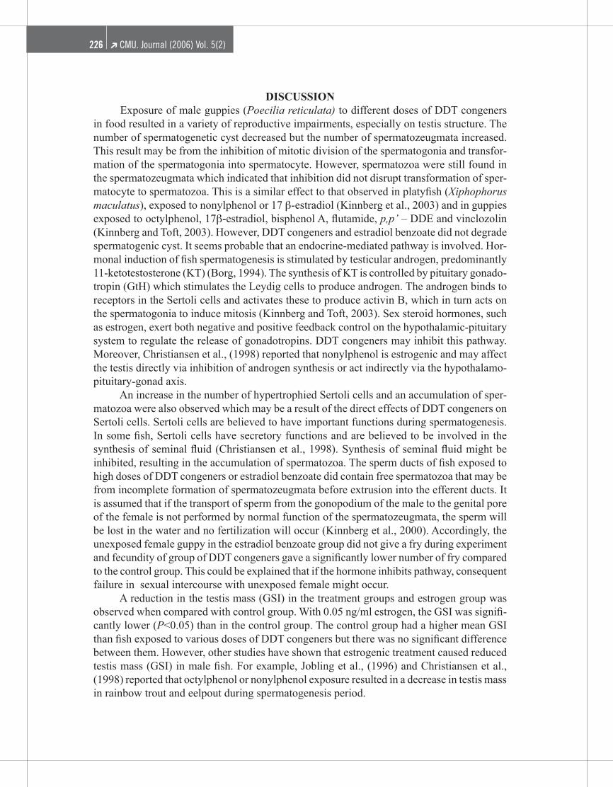
Fecundity and Sex ratio Female fish that were mated with the control males gave birth to an average of 14 young. However, female fish mated with males exposed to the chemicals produced significantly (P<0.05) lower numbers of offspring (lower fecundity), compared with the control fish. Females did not produce any offspring when mated with males fed estradiol benzoate. The sex distribution of the offspring, based on secondary sex characteristics, was not significantly different (P>0.05) between the control and treated fish (Figure 4 and Figure 5 respectively.)
Figure 4. The fecundity of unexposed female guppy.
Figure 5. The sex ratio of the offspring.
20181614121086420
Fecundity(Mean�SD)
Control Estrogen 0.05 ppb DDT 0.05 ppb DDT 0.10 ppb DDT 0.20 ppb
100
80
60
40
20
0
Sexratio(%)
Control Estrogen0.05 ppb
DDT 0.05 ppb DDT 0.10 ppb DDT 0.20 ppb
FemaleMale
CMU. Journal (2006) Vol. 5(2)226 CMU. Journal (2006) Vol. 5(2) 227
DISCUSSION Exposure of male guppies (Poecilia reticulata) to different doses of DDT congeners in food resulted in a variety of reproductive impairments, especially on testis structure. The number of spermatogenetic cyst decreased but the number of spermatozeugmata increased. This result may be from the inhibition of mitotic division of the spermatogonia and transfor-mation of the spermatogonia into spermatocyte. However, spermatozoa were still found in the spermatozeugmata which indicated that inhibition did not disrupt transformation of sper-matocyte to spermatozoa. This is a similar effect to that observed in platyfish (Xiphophorus maculatus), exposed to nonylphenol or 17 β-estradiol (Kinnberg et al., 2003) and in guppies exposed to octylphenol, 17β-estradiol, bisphenol A, flutamide, p,p’ – DDE and vinclozolin (Kinnberg and Toft, 2003). However, DDT congeners and estradiol benzoate did not degrade spermatogenic cyst. It seems probable that an endocrine-mediated pathway is involved. Hor-monal induction of fish spermatogenesis is stimulated by testicular androgen, predominantly 11-ketotestosterone (KT) (Borg, 1994). The synthesis of KT is controlled by pituitary gonado-tropin (GtH) which stimulates the Leydig cells to produce androgen. The androgen binds to receptors in the Sertoli cells and activates these to produce activin B, which in turn acts on the spermatogonia to induce mitosis (Kinnberg and Toft, 2003). Sex steroid hormones, such as estrogen, exert both negative and positive feedback control on the hypothalamic-pituitary system to regulate the release of gonadotropins. DDT congeners may inhibit this pathway. Moreover, Christiansen et al., (1998) reported that nonylphenol is estrogenic and may affect the testis directly via inhibition of androgen synthesis or act indirectly via the hypothalamo-pituitary-gonad axis. An increase in the number of hypertrophied Sertoli cells and an accumulation of sper-matozoa were also observed which may be a result of the direct effects of DDT congeners on Sertoli cells. Sertoli cells are believed to have important functions during spermatogenesis. In some fish, Sertoli cells have secretory functions and are believed to be involved in the synthesis of seminal fluid (Christiansen et al., 1998). Synthesis of seminal fluid might be inhibited, resulting in the accumulation of spermatozoa. The sperm ducts of fish exposed to high doses of DDT congeners or estradiol benzoate did contain free spermatozoa that may be from incomplete formation of spermatozeugmata before extrusion into the efferent ducts. It is assumed that if the transport of sperm from the gonopodium of the male to the genital pore of the female is not performed by normal function of the spermatozeugmata, the sperm will be lost in the water and no fertilization will occur (Kinnberg et al., 2000). Accordingly, the unexposed female guppy in the estradiol benzoate group did not give a fry during experiment and fecundity of group of DDT congeners gave a significantly lower number of fry compared to the control group. This could be explained that if the hormone inhibits pathway, consequent failure in sexual intercourse with unexposed female might occur. A reduction in the testis mass (GSI) in the treatment groups and estrogen group was observed when compared with control group. With 0.05 ng/ml estrogen, the GSI was signifi-cantly lower (P<0.05) than in the control group. The control group had a higher mean GSI than fish exposed to various doses of DDT congeners but there was no significant difference between them. However, other studies have shown that estrogenic treatment caused reduced testis mass (GSI) in male fish. For example, Jobling et al., (1996) and Christiansen et al., (1998) reported that octylphenol or nonylphenol exposure resulted in a decrease in testis mass in rainbow trout and eelpout during spermatogenesis period.
CMU. Journal (2006) Vol. 5(2)226 CMU. Journal (2006) Vol. 5(2) 227
On the other hand, sexual behavior, gonopodial index (GPI) and caudal fin color intensity which are known as secondary sex characteristics of male guppies are under the control of androgen (Christiansen et al., 1998). Both the estradiol benzoate group and the DDT conge-ner groups had less sexual behavior than the control group, but not significant in frequency. Similar with previous experiment, Bayley et al., (1999) observed that the male guppy, when exposed to 4-tert-octylphenol and 17 β-estradiol, decreased in number of sexual behavior. Gonopodial lengths were unaffected by estradiol benzoate and DDT congeners. However, Bayley et al., (2002) showed that juvenile male guppies had smaller gonopodium after exposure to vinclozolin, p,p-DDE and flutamide. This may be from age-relation. The gonopodial length was significantly correlated with female orientation response to the male (Bayley et al., 2002). Exposure to DDT congeners possibly affects this male sexual character since gonopodial development in poeciliids is steroid- dependent. The red color of caudal fin is known to be important for female’s choice of mating, i.e., a decrease in the color intensity is likely to decrease male’s reproductive success (Tolf and Baatrup, 2001). From the experiment, caudal fin color intensity in the estradiol and DDT congener groups was lower than in the control group, although not significant. Moreover, the intensity of male display of the Trinidadian guppy is correlated to sperm quality (Bayley et al., 1999).
CONCLUSION This study reveals interesting effects of DDT congeners on the reproductive function of male guppies. DDT congeners act as xenoestrogen, impairing testis function via hormonal systems. Estrogen has similar effects, indicating that the effects of DDT congeners might be estrogenic. Furthermore, the guppy proved to be a suitable biomarker for the detection of the effects of DDT congeners on reproduction.
ACKNOWLEDGEMENTS The authors are grateful to Junior Researchers Award, Chiang Mai University, 2002 and Dr. Tippawan Prapamontol, Research Institute for Health Sciences, Chiang Mai University.
REFERENCESBayley, M., J.R. Nielsen, and E. Baatrup. 1999. Guppy sexual behavior as an effective
biomarker of estrogen mimics. Ecotoxicology and Environmental Safety 43(1): 68-73.Bayley, M., M. Junge, and E. Baatrup. 2002. Exposure of juvenile guppies to three antian-
drogens causes demasculinization and a reduced sperm count in adult males. Aquatic Toxicology 56: 227-239.
Borg, B. 1994. Mini Review : Androgens in teleost fishes. Comparative Biochemistry and Physiology 109C(3): 219-245.
Bozynski, C.C., and N.R. Liley. 2003. The effect of female presence on spermiation, and of male sexual activity on ‘ready’ sperm in the male guppy. Animal Behaviour 65: 53-8.
Christiansen, T., B. Korsgaard, and A. Jespersen. 1998. Effect of nonylphenol and 17 β-oestradiol on vitellogenin synthesis, testicular structure and cytology in male eelpout Zoarces viviparus. The Journal of Experimental Biology 201: 179-192.
CMU. Journal (2006) Vol. 5(2)228
Danzo, B.J. 1998. Review – The effects of environmental hormones on reproduction. Cellular and Molecular Life Sciences 54: 1249-1264.
Donohoe, R.M., and L.R. Curtis. 1996. Estrogenic activity of chlordecone, o,p’-DDT and o,p’-DDE in juvenile rainbow trout : induction of vitellogenesis and interaction with hepatic estrogen binding sites. Aquatic Toxicology 36: 31-52.
Kinnberg, K., B. Korsgaard, and P. Bjerregaard. 2000. Concentration-dependent effects of nonylphenol on testis structure in adult platyfish Xiphophorus maculatus. Marine Environmental Research 50: 169-173.
Kinnberg, K., and G. Toft. 2003. Effects of estrogenic and antiandrogenic compounds on the testis structure of the adult guppy (Poecilia reticulata). Ecotoxicology and Environ-mental Safety 54: 16-24.
Kinnberg, K., B. Korsgaard, and P. Bjerregaard. 2003. Effects of octylphenol and 17β-oestradiol on the gonads of guppies (Poecilia reticulata) exposed as adults via the water or as embryos via the mother. Comparative Biochemistry and Physiology Part C 134: 45-55.
Larsson, D.G.J., K. Kinnberg, J. Sturve, E. Stephensen, M. SkÖn, and L. Förlin. 2002. Studies of masculinization, detoxification and oxidative stress responses in guppies (Poecilia reticulata) exposed to effluent from a pulp mill. Ecotoxicology and Environmental Safety 52: 13-20.
Leanos-Castaneda, O.L., G.V.D. Kraak, A. Lister, R. Sima-Alvarez. and G. Gold-Bouchot. 2002. o,p’- DDT induction of vitellogenesis and its inhibition by tomoxifen in Nile tilapia (Oreochromis niloticus). Marine Environment Research 54: 703-707.
Leatherland, J.F. and P.K.T. Woo. 1998. Fish diseases and disorders: Volume 2: Non-infec-tious disorders. CAB Publishing, London, UK.
Noriega, N.C. and T.B. Hayes. 2000. DDT congener effects on secondary sex coloration in the reed frog Hyperolius argus: a partial evaluation of the Hyperolius argus endocrine screen. Comparative Biochemistry and Physiology Part B. 126: 231-237.
Olsson, P.E., B. Borg., B. BrunstrÖm., H. Håkansson, and E. Klasson-Wehler. 1998. Endocrine disrupting substances – impairment of reproduction and development. ElandersGotab, Stockholm, Sweden.
Sonnenschein, C. and A.M. Soto. 1998. An update review of environmental estrogen and androgen mimics and antagonists. The Journal of Steroid Biochemistry and Molecular Biology 65(1-6): 143-150.
Sumpter, J.P. 1995. Feminized responses in fish to environmental estrogens. Toxicology Letters 82/83: 737-742.
Toft, G., and E. Baatrup. 2001. Sexual characteristics are altered by 4-tert-octylphenol and 17β-oestradiol in the adult male guppy (Poecilia reticulata). Ecotoxicology and Environmental Safety 48: 76-84.
Valle, M.G.R. 2001. Xenoestrogen exposure and reproductive success in zebrafish (Danio rerio). International Master of Science Programme, Faculty of Veterinary Medicine, Swedish University of Agricultural Sciences, Uppsala.
Zaroogian, G., G. Gardner, D.B. Horowitz, R. Gutjahr-Gobell, R. Haebler, and L. Mills. 2001. Effect of 17β-estradiol, o,p’-DDT, octylphenol and p,p’-DDE on gonadal development and liver and kidney pathology in juvenile male summer flounder (Paralichthys den-tatus). Aquatic Toxicology 54: 101-112.