the effect of fasting on local steroidogenesis in the
TRANSCRIPT

The effect of fasting on Local Steroidogenesis in the Brown
Anole, Anolis sagrei
Rachael Munoz
April 21st, 2016
Advisor: Dr. Bobby Fokidis
In partial fulfillment of the Rollins College Honors Degree Program
as well as the Requirements for
Honors in the Major in Biochemistry/Molecular Biology

The effect of fasting on Local Steroidogenesis in the Brown Anole, Anolis sagrei
Rachael Munoz and Bobby Fokidis
Department of Biology
Abstract
Everyday animals must confront and respond to stressors in their environment. The
activation of the stress response (i.e. the “fight or flight”) enables the individual to survive
stressors through hormone cascades that dictate physiological changes such as energy allocation.
In our model system, the brown anole lizard (Anolis sagrei), this cascade, which initiates in the
brain, ultimately involves the release of glucocorticoids (GCs) such as corticosterone (CORT; a
steroid hormone) from the adrenal glands. Along with GCs, the adrenal cortex also secretes the
androgen steroid, dehydroepiandrosterone (DHEA), which has been shown to possess positive
anti-stress effects. This is traditionally known as the systemic response, but recent research has
focused on local steroid production by organs other than that of the adrenal. While research in
local steroidogenesis has been conducted, there is no clear reason why steroids are being
produced locally. In order to determine if stress could induce local production, anoles were either
fasted or maintained on a normal diet to determine how the lack of food (a stressor) affects local
steroidogenesis. CORT levels were only significantly higher in the intestine samples of fasted
anoles compared to controls. Further, hormone levels were higher in multiple organs when
compared to plasma, providing evidence for local steroidogenesis.
2
Introduction
The saying “stress is a part of life” is true,
and animals must routinely deal with stressful
changes in their environment. In a physiological
sense, stress is clearly defined as the presence of a
potential challenge, or threat, to an animal’s internal
stability that evokes a physiological “stress
response”. In vertebrates, this stress response
involves the endocrine system, and is an adaptation that allows animals to cope with
environmental changes by enabling physiological and behavioral mechanisms that ultimately
promote survival (Charmandari et al. 2005). During stress, heart and breathing rates increase,
there is an elevated awareness and alertness, and a sudden release of energy from stored reserves
(Sapolsky et al. 2000). There are two distinct phases in this stress response. The first phase
involves the secretion of the catecholamine neurotransmitters, adrenaline (or epinephrine) and
noradrenaline (or norepinephrine) from the adrenal medulla. These act quickly to raise heart and
respiratory rates, and promote vasodilation to increase the blood flow to working muscles and
other tissues that require nutrients and oxygen supply (Sapolsky et al. 2000). Within a
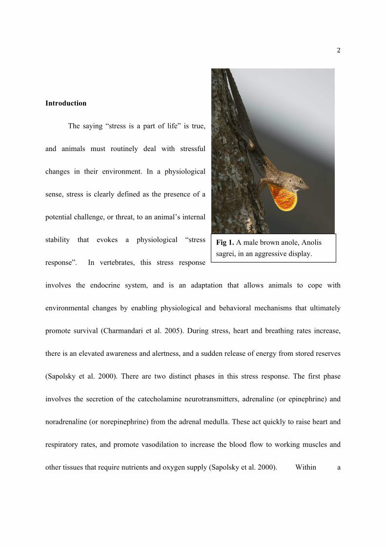
Fig 1. A male brown anole, Anolis
sagrei, in an aggressive display.

3
couple of minutes, the second phase occurs involving the secretion of steroid hormones, namely
glucocorticoids (GCs) into the blood from the adrenal cortex (Charmandari et al. 2005).
Corticosterone (CORT) is the primary GC in reptiles, birds and small mammals, but is
functionally comparable to cortisol, which predominates in fish and most mammals. This
endocrine cascade is known as the hypothalamic-pituitary-adrenal (HPA) axis (Fig. 2; Sapolsky
et al. 2000, Hill et al. 2012). The HPA axis initiates with neuroendocrine cells in the
paraventricular nucleus (PVN) of the hypothalamus, secreting a corticotrophin-releasing
hormone (CRH), which in turn stimulates the release of adrenocorticotrophic hormone (ACTH)
from the anterior pituitary gland,
into the bloodstream. Then, ACTH
will act on its respective receptors
to stimulate the synthesis and
secretion of GCs from cells in the
adrenal cortex. These GCs can then
act globally on glucocorticoid
receptors (GR) or
mineralocorticoidreceptors (MR) depending on the tissue being examined (Luca et al. 2013),
including specific areas of the brain (e.g., the hippocampus, prefrontal cortex, and amygdala)
Fig 2. The hormone cascade of the hypothalamic-pituitary
adrenal (HPA) axis, otherwise known as the stress axis.

4
which ultimately results in a widespread and coordinated physiological response that makes
survival possible during stress (Liu, Yuen 2010). While the “acute stress” response is adaptive
for short-term survival, society has focused on the negative consequences of stress that result
from persistent and prolonged exposure to GCs (otherwise known as “chronic stress”). Chronic
stress produces many negative effects on health including suppressing the immune system,
interfering with reproductive physiology, affecting cardiovascular health and cognitive
impairment (reviewed in McEwen 2008).
Whether an organism is mammalian, avian, or reptilian, their physiological response to
stress is universal. For this reason, a variety of model organisms may be used to study this
response. Although traditionally known as a systemic response with circulating glucocortocoids
produced from the adrenal gland, research has began to focus on local hormone production by
organs other than the adrenal. While evidence has been found for local steroiodogenesis in many
model organisms, this data has not been found in reptiles. Further, the driving force behind local
steroidogenesis has not been determined. For this reason, anole lizards were fasted to evaluate
how a stressor such as fasting alters local production in reptiles.
The adrenal gland
The adrenal gland is the primary endocrine gland associated with stress and is composed of two
main sections, an inner medulla, and outer cortex. The adrenal medulla secretes epinephrine

5
(adrenaline), while the cortex secretes steroid hormones such as GCs (Rosol et al. 2001). The
adrenal cortex is composed of three distinct layers; 1) the outer zone, called the zona
glomerulosa, produces aldosterone which functions in fluid and blood pressure control, 2) The
central, zona fasciculata, is the thickest section, composing about 70% of the adrenal cortex and
functions in GC production, and 3) in some species, the innermost zona reticularis produces
some GCs and androgens (Rosol et al. 2001). It is interesting to note that despite the adrenals’
small size, the gland is one of the most vascularized organs, highlighting its overall importance
in the endocrine system (Vrezas 2012). Although important, the adrenal does not act alone and
requires two negative feedback loops to function properly.
Adrenal Regulation
Adrenal steroidogenesis is regulated by two main endocrine feedback loops, the
hypothalamic-pituitary-adrenal axis (HPA axis), and the renin-angiotensin-aldosterone system
(RAAS). RAAS regulates both blood pressure and fluid homeostasis through a cascade pathway.
The first substrate, angiotensinogen is cleaved by an aspartyl protease, renin, forming a
decapeptide, angiotensin I. Angiotensin I is further cleaved by an angiotensin-converting enzyme
(ACE) to produce the octapeptide, Angiotensin-II (Ang-II). Ang II then causes vasoconstriction
by activating ang-II type 1 receptors within blood vessels and increasing arginine vasopressin
release from the posterior pituitary gland (Lavoie 2003).

6
The HPA axis interacts with the renin-angiotensin system by the activity of Ang-II. More
specifically, Ang-II stimulated mRNA expression of steroid-producing enzymes such as
steroidogenic acute regulatory protein, 3β-hydroxysteroid dehydrogenase, CYP11B1, and
CYP11B2 (Oki 2013). Further, glucocorticoids induce ACE production in rats, both in cultured
cells and in vivo lung tissue (Mendelsohn 1987). This direct relationship illustrates the
importance of both systems and how they interact. Localized activity of ACE and GCs in the
lungs and other tissues also point towards local effects of these hormones (Mendelsohn 1987).
The HPA-Axis
The hypothalamic–pituitary–adrenal axis (HPA axis) is anatomically composed of the
paraventricular nucleus of the hypothalamus (PVN), the anterior pituitary gland, and the adrenal
gland (Smith 2006). When responding to a stressor, dedicated neurons localized in the PVN
synthesize and secrete corticotropin-releasing hormone (CRH). CRH then binds to its receptor in
the anterior pituitary, stimulating the synthesis and release of adrenocorticotropic hormone
(ACTH) into general circulation which then acts to stimulate the adrenal cortex to release GCs
throughout systemic circulation (Wang 2004).
The HPA axis is tightly regulated by negative feedback loops from GCs in circulation (Fig 2). As
GCs bind to their glucocorticoid receptor (GR), they alter transcription levels of HPA
components (Smith 2006). The rise in GC levels decreases mRNA levels of an ACTH precursor

7
molecule, and therefore the amount of ACTH produced by the pituitary (Keller-Wood and
Dallman 1984). As GC concentration increases following stress exposure, the HPA activity is
inhibited. Further, the presence of GCs in an organism alter the expression of GC receptors, or
GRs. A negative feedback loop between circulating GCs and their receptors is present in order to
prevent tissue damage from high GC levels (Bamberger et. al. 1996).
The above description of HPA axis stimulation and inhibition are the current models for
systemic hormone production. Yet, organs may in fact not depend solely on circulating
hormones produced by the adrenals during stressful situations, and instead rely on local
synthesis.
Extra-adrenal steroid production in response to stress
Originally, GC synthesis was only thought to occur in the adrenal glands (Kostadinova et
al. 2012), however more recent research suggests GC synthesis occurs in other tissues, including
the intestine, brain, skin, and heart (reviewed in Taves et al. 2011). This localized (extra-adrenal)
synthesis of GCs may occur independent of adrenal hormone production, and thus is not directly
related to circulating GC levels. These tissues contain the necessary steroidogenic enzymes to
produce these hormones as detected by gene expression (i.e., mRNA) that suggests production of
steroidogenic enzymes. (Taves et al. 2011). Local (tissue) GC production has been documented
in both mammals and birds, and recently these observations raise the possibility of “localized

8
HPA axis in each tissue”, which regulates this GC production, independent of adrenal regulation
(Taves et al. 2011).
Along with GCs, the adrenal cortex also secretes the androgen steroid,
dehydroepiandrosterone or DHEA (Van Voorhees et al. 2014). As with GCs, other tissues can
also synthesize DHEA, although this production is much less studied. In humans, DHEA is most
present in circulation in its sulfated form (DHEAS), though the relationship between DHEA and
DHEAS can vary across vertebrate species (Maninger et al. 2010). In the context of chronic
stress, DHEA is thought to exhibit “anti-stress” properties, specifically by protecting tissues from
excessive GC exposure (Kalimi et al 1994). Unlike other hormones, no specific DHEA receptor
has been identified, and thus DHEA may exert its effects by being converted to more bioactive
steroids, such as testosterone or estrogen (Labrie et al. 2001). As a steroid precursor lacking a
receptor, the presence of DHEA is tested by expression of the steroidogenic enzymes necessary
to produce androgens. If the enzymes are present, then DHEA is being used in that tissue to form
testosterone and/or estrogen.
The dichotomy of having the same tissues
produce both a “stress” and an “anti-stress”
hormone is intriguing, and yet has received
little research attention. Despite the growing Fig 3. Chemical structure of corticosterone.

9
research on extra-adrenal steroid production, little is known about the factors regulating the
ability to synthesize steroids locally in varying organs.
Corticosterone
In lizards, such as anoles, corticosterone (CORT) is the primary GC, comparable to the
commonly known human hormone, cortisol. The structure contains three six membered rings and
one 5 membered ring fused to form one planar molecule (Fig. 3). CORT is a glucocorticoid that
induces glycogenesis, protein catabolism and suppression of digestion and the immune system
(Yang, Wilczynski 2003). These physiological changes occur in order to mobilize energy to
where it is needed and to suppress unnecessary functions. CORT also affects reproduction by
suppressing androgens such as testosterone.
Role of CORT in tissues during stress
Many studies have focused on plasma CORT levels as an overall indicator for stress, with
minimal regard to local organ production of this hormone. However, CORT is released by the
mammalian cardiovascular system under stressful conditions such as myocardial infarction with
the increased gene expression (mRNA levels) of CYP11B2, the aldosterone- producing gene.
(Davies et al. 2003). Through RNase protection and GR steroid binding assays, Sheppard (2002)
determined a high level of CORT present in cardiomyocytes. Thus, not only are glucocorticoids
circulating throughout an organism, but they are being localized to organs such as the heart in

10
times of stress. This finding was further confirmed through the presence of mRNA for
steroidogenic enzymes in heart muscle (Sheppard 2002). mRNA expression and the presence of
both glucocorticoid and mineralocorticoid receptors in heart cells (cardiomyocytes) suggest an
important role of GCs in the heart. Due to elevated GC levels in heart failure patients, it is
hypothesized that changes in GC concentrations may cause the different responses during heart
trauma. According to Young (2001), GCs were only detected in patients with myocardial
infarction and heart failure and CYP11B2 was expressed only in failing hearts. Though other
factors such as drug exposure or a stressful cellular environment may be involved, it does
suggest GCs may also play an important protective role.
The role of extra-adrenal CORT
Although a relatively new area of study, local steroid production has been detected in a
number of organs from human and mouse models. Evidence for extra-adrenal CORT production
is present, but the physiological significance is unclear and it depends on the relative local
concentration, target cell proximity, and organ-specific hormone regulation (Davies et al. 2003).
Depending on the organism, CORT levels vary between organs, making it difficult to compare
among species. If local CORT concentrations are low, their presence may play an important role
in the specific organ from which it is being produced. Interestingly, local steroidogenesis is

11
independent of adrenal GC synthesis, making their comparison difficult. Among the uncertainty,
one theme in local GC synthesis is the location of synthesis within immune-involved organs.
Thymus
One such organ, the thymus, is an immune gland responsible for T-lymphocyte
development, and was the first organ in which extra-adrenal steroid production was detected
(Vacchio et al 1994). It is interesting to
note, the mouse thymus secreted the highest
GC levels during fetal development and
following birth when the adrenal gland is
not fully active. Therefore, the thymus had
to locally produce GCs, and not rely on the
adrenal gland (Ashwell 2000). During this
time of lymphocyte development, GCs
determined the fate of thymocytes before
they developed into mature T-cells. When
compared to the adrenal, thymus CORT levels were high but remained lower than adrenal levels
(Vacchio et. al. 1994), suggesting a lower concentration locally.
Fig 4. HPA negative feedback loop on the
proinflammatory response. Adopted from
Hermoso, Cidlowski (2003).

12
Brain
When produced locally, CORT acts as a neurosteroid in the brain by affecting neuron
formation, development, myelination, and neuronal reactions to stress (Kostadinova et al 2012).
The enzyme, P450 (encoded by the CYP11B1 gene) that catalyzes the final step in CORT
synthesis is expressed in rat hippocampus as both mRNA and protein (Higo et al. 2011).
Following adrenalectomy, significant CORT levels were still detected in the brain suggesting
extra-adrenal CORT production is occurring. This could also suggest CORT is not being
metabolized and instead persists. According to Higo et al. (2011), low CORT levels contribute
to synaptic plasticity and neuroprotection in hippocampal neurons. Hippocampal synaptic
plasticity enhances with low CORT levels by changing how the necessary glutamate receptors
function (Tse et al. 2011). CORT alters the functional properties of glutamate receptors by
changing the motility and function of α-amino-3-hydroxy-5-methylisoxazole-4-propionic acid
subtypes of glutamate receptor (AMPAR) that are responsible for the expression of synaptic
plasticity. Another receptor, N-methyl-D-aspartate receptors (NMDARs), involved in inducing
synaptic plasticity, are also altered (Tse et al. 2011). Although chronic levels of high CORT are
associated with negative health effects, low CORT levels may be beneficial locally.
Skin

13
The skin serves as a physical and chemical barrier from the environment and protects organisms
from chemicals, infections, and pathogens. Following an injury to human skin, keratin-producing
epidermal cells (keratinocytes), induce the release of interleukin-1 (IL1) to activate keratinocytes
in order to begin the proinflammatory phase of wound healing (Vukelic et al 2011).
Keratinocytes expressed CYP11B1, the enzyme that catalyzes the final conversion of
deoxycorticosterone to CORT (Vukelic et al 2011). The interplay of increased CYP11B1,
CORT, and GC receptor expression found in wounds may function to both prevent excessive
inflammation from leading to tissue damage and initiate the proinflammatory response and
(Vukelic et al. 2011). This initial response involves many steps; first toll-like receptors (TLRs)
recognize pathogen-associated molecular patterns (PAMPs) that are specific to each pathogen
(Hermoso, Cidlowski 2003). TLR activation then activates the innate immunity, activating B
cells. Interestingly enough, this initial response is tightly regulated by the HPA axis.
Proinflammatory cytokines activate the HPA, causing GC synthesis, release, and the formation
of a negative feedback loop on proinflammatory molecules (Fig. 4). More specifically, GCs
inhibit cytokine gene expression and of course their activity on target cells. Similar to other
extra-adrenal organs, the skin also produces low CORT levels

14
Lungs
Similar to the skin, lungs are constantly exposed to foreign substances such as antigens,
microbes, and pathogens. For this reason, they contain a high concentration of immune cells
(Kostadinova et al 2012). Mouse lung tissue expressed steroidogenic enzymes necessary for
CORT production (Hosteller et al. 2012). At first glance, the ability for lungs to synthesize an
immunosuppressant, CORT, seems counterintuitive. Yet, excessive immune responses can lead
to tissue damage, therefore, the lung must sense inflammatory responses and induce local CORT
synthesis to prevent extensive lung tissue damage (Hosteller et al. 2012).
Role of extra-adrenal DHEA
Although specific functions have been elusive to discern, DHEA may play a protective
role during stress. Maninger et al (2010) found increased circulating DHEAS concentrations in
rhesus monkeys (Macaca mullata) with repeated exposure to acute stress. Administering DHEA
following immobilization stress reversed the weight gain seen in chronically stressed rats (Hu
2000). Furthermore, DHEA supplementation given to Wistar rats reversed the effects of a high
fat diet, which induced insulin resistance (Veras 2014). Evidence from these studies illustrate the
potential for DHEA to mitigate the harmful effects of stress, however how this varies across
organs has not been investigated. Extra-adrenal DHEA activity is not fully understood and it is
evident that local organ production of such hormones continues to be a new topic of interest.

15
DHEA plays a much greater role as a systemic androgen precursor. Recent studies have focused
on stress responses mediated by DHEA in organs, especially the heart. Excess GCs, possibly
produced by increased HPA activation, have been linked to cardiac dysfunction such as heart
hypertrophy, or thickening of the cardiac muscle (Nakamura 2004). Following DHEA treatment,
hypertrophic responses reduced in cardiomyocytes, even during GC receptor stimulation.
DHEA has protective effects that are exerted on a number of organs (Pelissier et al. 2004). When
the colon, small intestine, and livers of rats were treated with DHEA, it leads to decreased
oxidative damage of proteins and lipids. To date, DHEA synthesis has been detected in the liver,
gastrointestinal tract, and adrenal (Tashiro 2000). Following surgery, DHEA localization in the
intestine was studied by immunohistochemistry to determine that parietal cells actively
synthesized DHEA in gastric mucousa (Tashiro 2000).
Fasting as a stressor
Food availability is one of the most important factors impacting the physiology of an
organism. The lack of food is clearly perceived as a stressor as it elevates circulating GC levels
and imposes drastic effects including; suppressing reproduction and immunity, and mobilizing
stored energy reserves resulting in a further effect of fasting. Field research on the red-legged
kittiwake (Rissa brevirostri) documented that food shortages increased CORT (the predominant
GC in birds and reptiles) levels alongside decreases in body mass (Kitaysky et al. 2001). Similar
16
studies in domestic chickens (Gallus gallus) have shown that increased CORT secretion lowers
the levels of the sex steroids; testosterone and progesterone, thereby completely suppressing
reproduction entirely (Henriksen et al. 2011). These studies have focused on circulating (i.e.,
systemic) CORT levels likely derived from the adrenal gland, but how food availability may
alter local steroid production remains essentially unstudied.
A recent study demonstrated that acute fasting (6 hours) increased aggression and
changed circulating steroid levels in male zebra finches, Taenopygia guttata (Fokidis et al.
2013). Specifically, the lack of food caused both secretion of CORT and DHEA to increase, as
well as a simultaneous decrease in the sex steroids, testosterone and estrogen, in a number of
tissues (Fokidis et al. 2013). This provided the first evidence for an effect of fasting on hormone
distribution across tissues, but was limited in the number of tissues investigated and though
studied in birds, it has not been tested in reptiles.
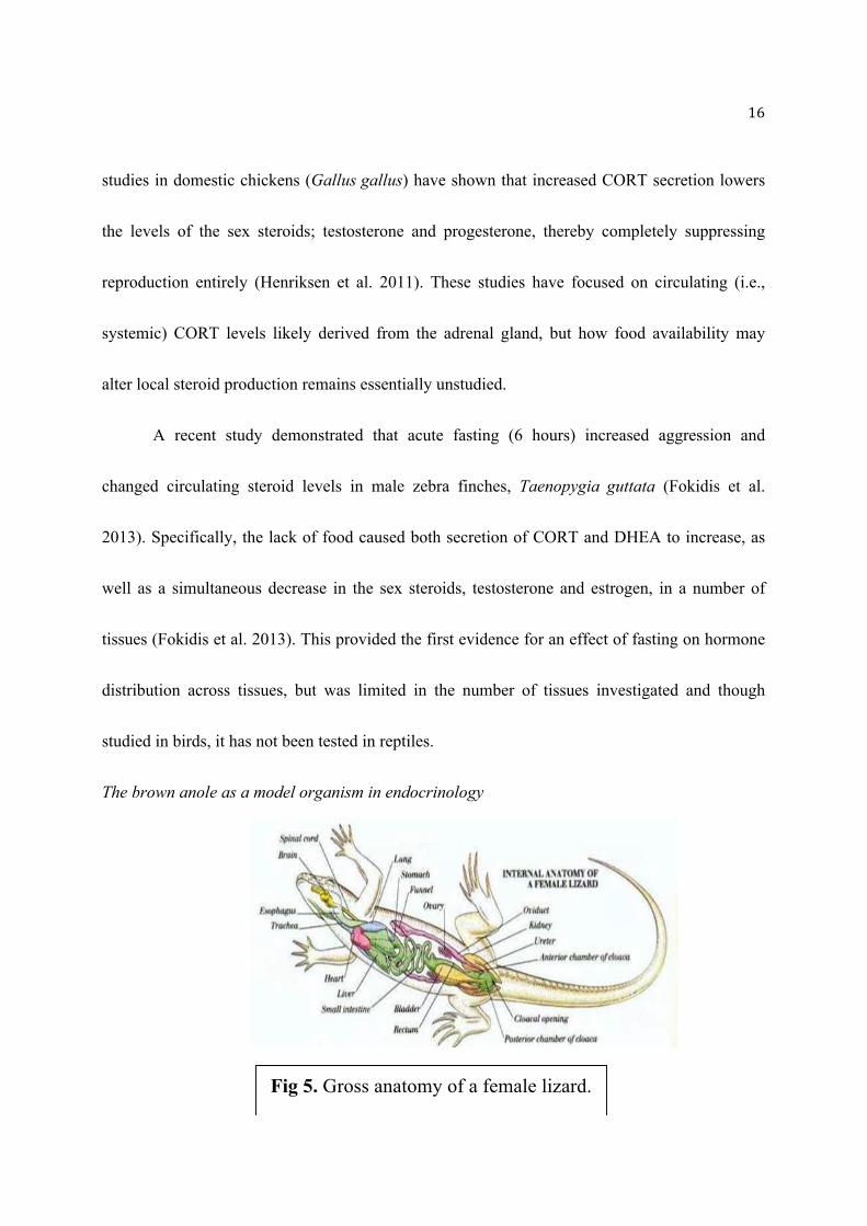
The brown anole as a model organism in endocrinology
Fig 5. Gross anatomy of a female lizard.

17
Brown anoles (Anolis sagrei) are an abundant, but invasive lizard species in the southeastern
region of the U.S, including Florida (Wade and Cohen 2012). Both the brown (A. sagrei) and the
similar native green (A. carolinesis) anoles have been extensively used as models for
neuroendocrinology and for linking steroid hormones and behavior. Anoles have multiple
behaviors, one of which is exploratory behavior, which is associated with mild stress
(Greemberg 2002). These visible behavioral patterns are altered and influenced by stress
hormones, making the anole a simple model organism to study.
A substantial amount is understood concerning the anatomy (Fig. 5), behavior, and
physiology of the Anolis. This study
focuses on the abundant brown anole,
which is more easily available and larger
than the green anoles, making them a more
convenient species for research.
The synthesis of steroids involves a
complex metabolic pathway that involves
many steroid precursors and steroidogenic
enzymes (Fig. 6). Steroid hormones are a
critical regulator of adult anole behavior
Fig 6. Steroidogenesis pathway of CORT, DHEA from
cholesterol. The gene names that express the necessary
enzymes are also included above each arrow. Scheme

18
(Wade 2012), and in particular, anoles exhibit sexual dimorphism in both anatomy and behavior
that is regulated by steroids (Lovern et al. 2004; Husak et al 2009). In several anole species
including A. sagrei, male anoles responded to a gonadotropin-releasing hormone (GnRH)
challenge (the central regulator of reproduction) by decreasing circulating testosterone levels
while simultaneously elevating CORT levels in the field (Husak et al. 2009). This suggests that a
robust stress response is maintained during reproduction even when artificially stimulated by the
predominant regulator of testosterone (i.e., GnRH) (Husak et al. 2009). Similar research
demonstrated that injection with arginine vasotocin (AVT) also increased circulating CORT
suggesting a maintained robust stress response even in captivity (Dunham and Wilczynski 2014).
The influence of food on hormone levels in anoles, has only been investigated in a single study
by Lovern et al. (2008). Here, female anoles on an ad libitum diet (without restraint) were in
better body condition, and had higher plasma testosterone concentrations than a fasting group
(Lovern et al. 2008). A study conducted by Tokraz et al. (1998) on Anolis lizards showed that as
testosterone spiked during the breeding season, CORT levels decreased. This provides a trend
suggesting that the stress response inhibits reproduction in the lizard, as in most species. Despite
the substantial endocrine research on anoles, these studies focused on circulating steroid levels
and not the ability of the organs to produce the steroids themselves. Again, while the above
19
studies have set some foundation for local steroidogenesis, there have been no studies to this
point on reptiles.
While there are some hypotheses on the local stress response, there are few studies that
explain how or why local production occurs. If in fact, chronic fasting could induce a systemic
stress response, there should also be evidence of increased local steroid production when
compared to a control group. If local production occurs, it is hypothesized that hormone levels
within organs should be higher than circulating, or plasma levels.
Materials & Methods
Collection and captivity
Adult male anoles were captured on and around the Rollins College campus using a
fishing pole noosing apparatus. Following capture, each anole was weighted (±0.1g) and their
snout-vent length (SVL; (±0.1mm) and tail length (±0.1mm) were measured. The lizards were
housed in cages (12 x 6.5 x 11.5 inches) on botanical carts with four 40-watt full spectrum light
bulbs per shelf. Each cage contained a carpet bottom; a mesh hammock, a PVC perch, and a live
plant (Pothos sp.). A warm (27° C) and humid (94% relative humidity) environment was
maintained by spraying the cage and plant with water once daily. A photoperiod of 14-hour light
to 10-hour dark was maintained. The health of the anoles was evaluated daily and all

20
experimental procedures were approved by the Institutional Animal Care and Use Committee at
Rollins College.
The fasting experiment
Before initiating the study, anoles were allowed to acclimate to captivity for a week.
Anoles were then randomly assigned (determined by a random number generator) to one of two
treatment groups: 1) a group that was fasted for one week (experimental; N=13) and 2) a control
group given two large crickets every 2 days (N=12). The groups were equally weighted to ensure
equal mass averages between the groups before the experiment began (t= -0.498, df= 23,
p=0.623). To avoid the effects of the time of data collection the experiment was staggered, with
only five lizards being studied per day over the course of a week.
Tissue Sample Preparation
Following the experiment (1.5 weeks), anoles were sacrificed by rapid decapitation and a
trunk blood sample was rapidly collected in heparinized capillary tubes and stored on ice. Anoles
were then dissected and several organs were collected (Table 1). Tissues were immediately snap
frozen in dry ice and stored at -80oC until further processing. Prior to tissue preparation, tissues
were weighed to the nearest + 0.1mg and then homogenized using zirconia beads in a mixture of
84% methanol (by vol.) and placed in an automatic bead ruptor. Samples were left overnight at
21
4oC and then centrifuged with the resulting supernatant being collected for solid phase extraction
(SPE).
Solid phase extraction
A widely used technique, SPE, enables the extraction of nonpolar compounds from a
highly polar solution, such as steroids from aqueous tissue samples, using special carbon-bonded
silica (C18) chromatographic filters (Newman et al. 2008; Fokidis et al. 2013). The process
involves six steps for extracting and purifying steroids from the surrounding sample. Briefly,
these steps are: 1) solvation; which uses an organic solvent to saturated a filtering material; 2)
equilibration: using water to provide a gradient that concentrates the steroids; 3) sample loading;
adding the tissue samples; 4) interference elution; using an organic solvent to remove impurities
and substances that may interfere with further steroid analysis; 5) sample elution; removing the
purified steroid sample; 6) sample drying; using a vacuum concentrator.
Here, SPE was performed with 5 mL C18 column cartridges (Agilent Technologies,
Santa Clara, CA, USA). The columns were first primed with 3 mL of 100% ethanol (solvation)
and then 10 mL of distilled water were added (equilibration). The homogenized tissue samples
were diluted with 10 mL of water and loaded after priming (sample loading). Following addition
of the samples, 10 mL of 40% methanol was loaded and eluted until the columns ran dry at
maximum vacuum pressure for five minutes (interference elution) The final purified solutions

22
were collected with 5 mL of 90% methanol (sample elution) and dried in a speed vacuum
concentrator (Thermo Fisher Scientific Inc., Pittsburgh, Pennsylvania, USA) at 40 °C for 4 hours
(sample drying). This technique has already been validated for use in Dr. Fokidis’ laboratory and
it has demonstrated to yield a high recovery (83-96%) of steroid from various tissues. The dried
samples were then stored at -80 C until reconstituted for the steroid quantification assays.
Enzyme-linked Immunoassay (ELISA)
Competitive ELISA kits were used to measure both CORT (Arbor Assays Inc., Ann
Arbor, Michigan, USA) and DHEA (Eagle Biosciences Inc., Nashua, New Hampshire, USA)
from tissue samples. Dried samples were reconstituted with 5 µL of 100% ethanol and 115 µL of
the kit’s assay buffer with 50 µL each used for both the CORT and DHEA assays. Prior to the
CORT assay, plasma samples were prediluted 10-fold to insure they were within the linear range
of the standard curve. The respective assay kits protocol was followed and the 96 well
microplates were read using a spectrophotometer at 450 nm for both CORT and DHEA. Final
concentrations were determined by interpolating from the assay standard curve of known
concentrations (0-10,000 pg/mL for CORT and 0-60 pg/mL for DHEA) using GraphPad Prism
version 6 (GraphPad Software Inc., La Jolla, California, USA).
Data Analysis

23
All data were first tested for both normality (Kolmogorov-Smirnov test) and equal
variance (Levene’s test). If normally distributed then a parametric two-tailed t-test was
performed, between control and fasted steroid levels for each tissue, or between plasma and
tissue levels. If the data violated assumptions of normality and equal variance then a non-
parametric Mann-Whitney Rank Sum Test (U-test) was used to determine significance. Although
analysis of variance (ANOVA) was considered, not all organs were collected and steroids
successfully quantified for each individual, resulting in missing data that limited the use of this
approach. Statistical analyses were performed using Sigma Plot version 13 (Systat Inc., San Jose,
California, USA) with α set at 0.05. All graphical data here are presented as means ± standard
error.
Results
This study sought to observe how stressors such as fasting could alter steroid hormone
levels. In order to determine if fasting could induce a systemic stress response, CORT and
DHEA levels were compared between control and fasted groups. To determine the presence of
local steroidogeneis, steroid levels within each organ were compared to circulating plasma.

24
Fasting causes significant weight loss in anoles compared to fed controls
The control group had an average change in mass of -0.077g while the fasted
group had an average change of -0.85 g, which was statistically significant (t=3.02, df= 23,
P=0.006; Fig. 7). Thus, the fasting protocol was sufficient to induce a physiological stress in the
treatment group.
Fig 7. Change in body mass between control (white) and fasted anoles
(black).

25
Table 1. Abbreviations for organs collected from brown anoles.
Abbreviation Organ Control (N) Fasted (N)
PL Plasma 12 13
ADR Adrenal 9 7
BLD Blood 6 7
BRN Brain 12 13
HRT Heart 12 13
LNG Lung 11 13
LV Liver 11 13
STO Stomach 12 13
INT Intestine 11 13
PAN Pancreas 5 5
KID Kidney 12 13
TES Testes 12 13
HEP Hemipenes 10 9
MUS Muscle 12 13
26
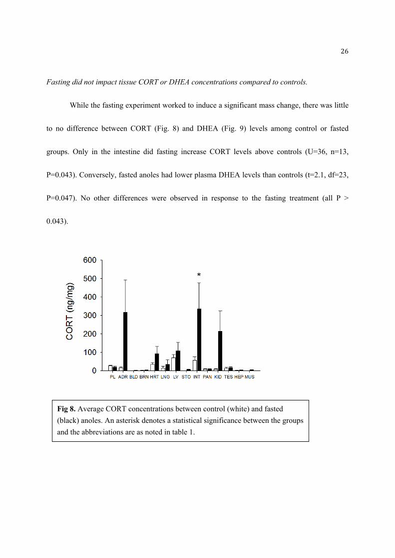
Fasting did not impact tissue CORT or DHEA concentrations compared to controls.
While the fasting experiment worked to induce a significant mass change, there was little
to no difference between CORT (Fig. 8) and DHEA (Fig. 9) levels among control or fasted
groups. Only in the intestine did fasting increase CORT levels above controls (U=36, n=13,
P=0.043). Conversely, fasted anoles had lower plasma DHEA levels than controls (t=2.1, df=23,
P=0.047). No other differences were observed in response to the fasting treatment (all P >
0.043).
Fig 8. Average CORT concentrations between control (white) and fasted
(black) anoles. An asterisk denotes a statistical significance between the groups
and the abbreviations are as noted in table 1.

27
Fig 9. Average DHEA concentrations between control (white) and fasted
(black) anoles. An asterisk denotes a statistical significance between the groups
and the abbreviations are as noted in table 1.

28
Evidence for hepatic, intestinal and renal synthesis of CORT in anoles
Comparing organ steroid levels to those in circulating plasma could provide evidence of
local steroid synthesis (i.e., higher hormone levels in the organ compared to plasma) or evidence
of steroid utilization (i.e., higher levels in plasma compared to organs). Here, both control and
fasted groups were evaluated separately. In controls, several organs had CORT levels that were
statistically lower than that found in circulating plasma (Fig. 10). These tissues including: blood
(t = 4.598, df= 17, P=0.000256); brain (U=12, n=12, P< 0.001); lung (U=21, n=12, P=0.006);
stomach (U-4, n=12, P<0.001); pancreas (t = 2.405, df=15, P=0.03); kidney (t = 3.510, df=22,
P=0.002); testes (U=36, n=12, P=0.04); hemipenes (U=8, n=12, P<0.001) and muscle (U=12,
n=12, P<0.001), In contrast, only the intestine (U=39, n=14, P=0.012) and liver (U=28, n=14,
P=0.008) had levels higher than circulating plasma.
Fig 10. Local CORT production among control organs in comparison to circulating
plasma (represented in white). An asterisk denotes significance from plasma.

29
Plasma CORT levels were significantly higher (Fig. 11) than in blood (U=11, n=13, P=0.006),
brain (U=28, n=11, P=0.035), and stomach (U=41, n=13, P=0.027). In contrast, plasma CORT
levels were lower (Fig. 11) than in liver (U=42, n=13, P=0.031), intestine (U=7, n= 13, P<.001),
and kidneys (U=44, n=13, P=0.04).
Fig 11. Local CORT production among fasted organs in comparison to
circulating plasma (represented in white). An asterisk denotes significance
from plasma.

30
Evidence for widespread tissue DHEA synthesis in anoles
For control anoles, compared to plasma, several organs had statistically higher DHEA
concentrations (Fig. 12) including: adrenal (U=1, n=12, P< 0.001), heart (U=22, n=12, P=0.004),
lung (U=15, n=12, P= 0.002), liver (U=26, n=12, P=0.015), and the hemipenes (U=0, n=12, P <
0.001). In contrast, plasma DHEA levels were lower in pancreas (t = -4.777, df=15, P < 0.001)
and the testes (U=23, n= 12, P=0.005).
Fig 12. Local DHEA production among control organs in comparison to
circulating plasma. An asterisk denotes significance from plasma.

31
In fasted anoles, plasma DHEA was significantly lower (Fig. 13) than in: brain (U=40,
n=13, P=0.024); heart (U=7, n=13, P< 0.001); lung (U=24, n=13, P=0.002); liver (U=40, n=13,
P=0.024); intestine (U=12, n=13, P< 0.001); and hemipenes (U=4, n=13, P < 0.001).
Concentrations of DHEA in the stomach (U=32, n=13, P=0.008) and pancreas (t=2.860, df=16,
P=0.01) were significantly lower than in plasma (Fig. 13).
Fig 13. Local DHEA production among fasted organs in comparison to
circulating plasma. An asterisk denotes significance from plasma.
32
Discussion
The systemic stress response involving widespread circulation of CORT produced by the
adrenal cortex is well understood. However, minimal research has focused on local
steroidogenesis, or the ability of non-steroidogenic organs to produce their own steroids. In this
study, I sought to determine whether fasting (a chronic stressor) could promote local
steroidogeneiss in anole lizards. In response to fasting, I expected organ concentrations of CORT
and DHEA to exceed that of circulating plasma, which could suggest local steroid production.
Several organs demonstrated higher steroid levels than those present in circulation (i.e. plasma),
which provides evidence that local steroid synthesis is occurring, however there was no profound
effect of fasting. As only the second study to evaluate local CORT and DHEA, it is important to
further research this field to determine how and why local stroidiogeensis occurs.
Local CORT production
Digestive organs
The intestine was the only organ where CORT increased with fasting and regardless of
treatment, the highest CORT levels were observed. Lowette et al. (2014) reported that fasting
increased intestinal CORT, as in this study. To confirm this, they reported increased mRNA
expression of 11β-hydroxysteroid dehydrogenase Type 1 (11β-HSD1), a CORT-converting
enzyme in enteric (intestinal) sensory neurons.
33
These data suggest that intestinal CORT synthesis is extensive with local CORT playing an
important role possibly through the enteric nervous system (ENS), the set of neurons embedded
in the intestinal wall. CORT also reduced electrical stimulation induced by calcium, suggesting a
neuromodulatory role in the GI tract during fasting (Lowette et al., 2014). The same study noted
decreased mixing within the intestine with high CORT levels from fasting. Local CORT may be
inducing these physiological changes (reducing the intestinal neuronal response and mixing) in
order to inhibit unnecessary processes during a stressor.
While the intestine remained consistent with higher CORT levels, other digestive organs
did not exhibit a similar pattern. Both the pancreas and stomach had lower hormone levels than
plasma. The low pancreatic levels of CORT could be caused by increased pancreatic uptake in
order to maintain physiological processes such as glucose metabolism. Interestingly, CORT
affects how the body processes insulin and during stress, CORT promotes the release of glucose
into circulation from energy reserves to be used for metabolism (Sapolsky et al. 2000). While
insulin is necessary to regulate glucose levels, CORT does interact with the peptide through a
poorly understood molecular mechanism, by making insulin less efficient at glucose uptake
(Beleen et al. 2014). At the molecular level, somatostatin (SST; a hormone that inhibits growth
hormone), CORT, and ghrelin all influence glucose homeostasis within the pancreas (Beleen et

34
al. 2014). Yet, under severe metabolic conditions, such as possibly fasting, the expression of
these regulators may be compromised. While CORT was found in the pancreas, this deregulation
could explain why such a low concentration was observed in the control and fasted groups.
Therefore, chronic stress not only impairs the HPA axis but also how CORT receptors function
(Beleen et al. 2014). Since CORT receptors may not be fully active under stress, it may be
possible that less CORT is being detected in the pancreas. This could explain why such low
levels of CORT were measured in the pancreas, even if the hormone was being produced.
Elevated CORT levels will shutdown unnecessary processes during a stress response.
Therefore, I predicted that the stomach would produce CORT in response to fasting, to limit
protein digestion, yet, regardless of treatment, gastric CORT levels remained low. As local
CORT levels were lower than in circulating plasma, this may suggest the stomach uses CORT
directly from circulation, as opposed to local production. In response to fasting, Filaretova et al.
(2004) found that GCs had a gastro-protective role. In fact, with CORT supplementation, the
decrease in blood flow velocity and mucous production was further prevented (Filaretova et al.
2004). Another study illustrated how food deprivation decreased the activity of digestive
enzymes such as trypsin and lipase (Bolasina et al. 2005). As, enzyme activity is highly
sensitive to nutritional conditions, CORT could be interacting with digestive enzymes to inhibit
or lessen their activity.

35
In the liver, I reported higher organ CORT levels than in plasma from both fasted and
control lizards, although the difference is greater in the latter. The liver performs metabolic
functions such as glycogen storage, red blood cell decomposition, detoxification, and these
processes are readily affected by stressors (Djordjevic et al., 2010). For example, an increase in
GCs stimulates liver gluconeogenesis for use as an energy source during stressful conditions
(Djordjevic et al., 2010). Furthermore, the liver plays an important role in detoxification and
antioxidant enzyme activity is also regulated by GCs (Djordjevic et al., 2010). According to
Vazquez-Medina et al. (2010), prolonged fasting conditions increases the production of reactive
oxygen species (ROS), which leads to oxidative damage and inflammation. For this reason, an
up regulation of enzymes responsible for breaking down ROS would be beneficial. In a
combined stress study that used isolation and acute immobilization stress on Wistar rats, both
CORT and liver antioxidant enzyme levels increased (Djordjevic et al., 2010). This increase
suggests that GCs may signal the liver to produce more enzymes for detoxification of ROS
during stress, and could therefore explain the elevated CORT levels observed in the liver in this
study.
Kidney
36
Compared to controls, kidney CORT levels were higher in the fasted group, and both
were significant above levels found in plasma. Interestingly the kidney in male anoles also
performs a sexual role in supplying a nutrient medium that acts as an energy source for sperm
(Cuellar et al. 1972). Based on this relationship with reproduction, one could predict higher renal
activity and nutrient production during the breeding season. This study was conducted in summer
at the peak of the anole breeding season in Florida (Wade 2011), thus the high CORT levels
observed in the fasted kidneys may indicate an inhibition of the kidney’s reproductive function in
male anoles. Yet, at this time, little is known about the kidney’s role in reproduction, this is
solely a prediction.
Heart
Although no statistically significant differences were observed, CORT levels in cardiac
muscle tended to be higher in the fasted group and generally higher than in plasma. Interestingly,
the heart is composed of proteins that can be either built up (anabolism) or broken down
(catabolism) depending on hormone balance (Tischler 1981). Hormones, such as the GCs present
during fasting can lead to catabolic effects in cardiac muscle proteins, and fasting can induce
atrophy and protein catabolism in rabbit hearts (Samarel et al. 1987). Fasting inhibits the
production of new cardiac muscle protein and caused an increase in the percentage of degraded
protein (Samarel et al. 1987). The high CORT levels in this study may have interfered with

37
protein synthesis. This hypothesis would then need to be confirmed by protein analysis to
determine the impact of high CORT levels had on cardiac tissue.
Blood cells
Plasma consistently expressed much higher CORT concentrations than the blood,
confirming that GCs circulate in the plasma portion and not within the white and red blood cells.
Nonetheless CORT may be playing a role in red blood cells. Vertebrates cope with physiological
changes (such as fasting) through immune system responses to immune challenges (Graham et
al. 2012). According to Dhabhar et al. (2012), chronic stress induces a quick redistribution of
immune cells throughout the blood. This stress-induced leukocyte redistribution is a fundamental
response that directs leukocytes to their target organs (Dhabhar et al. 2012), with an increase in
the mobilization of neutrophils, lymphoctyes, and helper T cells (Dhabhar et al. 2012). Thus
while CORT levels remain low in the blood, the hormone could be used quickly to induce such
immune changes. It is also important to note that this immune redistribution has been
evolutionarily conserved across the vertebrates, including reptiles, and this suggests a strong
adaptive advantage.
Brain
In response to stress, anoles should have elevated CORT levels in the brain which leads to a
decrease in dopamine within the brain (Greenberg 2002). In these anoles, both orientation and

38
locomotor responses were depressed, which were similar characteristics observed in this
study. Although CORT acting within the brain has been widely accepted, this study
demonstrated low CORT levels regardless of the treatment. A study assessing chronic stress in
rats reported that CORT affected the brain regions associated with motivation and reward, which
are regulated by dopamine (Lucas et al. 2003). Further, Lucas et al. (2003) also reported low
CORT levels that that correlate with reduced behavioral activity and longer latencies in reacting
to mildly threatening stimuli (i.e., less anxious). Although both treatment groups contained low
CORT, the fasted group was much less resistant to handling at the end of the fasting experiment.
This deviation from normal anole behavior to resist handling could obviously well be due caused
by a lack of energy from fasting or due to the physiological brain changes discussed.
Local DHEA production
DHEA, an androgen precursor, may have anti-stress properties against GCs during the
stress response. While most comparisons of DHEA levels between fasted and control groups
were not significantly different, multiple organs did contain significant concentrations above that
observed in plasma. Regardless of treatment group, the adrenal, heart, and intestine continued to
have higher DHEA levels than plasma supporting the hypothesis of local steroid production in
these organs. This is the first study to report the presence of non-plasma DHEA and to document

39
local steroid production in a reptile species. While DHEA levels were measurable in the anole, it
is important to note the lower concentrations when compared to other species. In anoles, DHEA
concentrations ranged from 0 to7 ng/mg, whereas songbirds contained upwards of 15 ng/ng
(Fokidis et al. 2013). Furthermore, DHEA levels increased dramatically in fasted male zebra
finches, while fasting did not have a similar effect in this study. This difference could be due to
the needs of each species; while birds might need the protection DHEA offers, anoles may be
more resistant to such stressors.
Testes and Hemipenes
The testes did not show any variance of DHEA concentration in neither control nor fasted
groups. The testes could either be using DHEA quickly for testosterone production or may not
have a large role in sex hormone production. However the hemipenes, the copulatory organ in
reptiles, contained a higher DHEA content, regardless of treatment, when compared to plasma.
As a reproductive organ, the local production of an androgen precursor such as DHEA would
makes sense however there is little to no research on the hormonal regulation of hemipenes. In
another lizard species, snow skinks (Niveoscincus microlepidotus), researchers found that both
the adrenal gland and gonads produced testosterone and testosterone levels peaked with maximal
hemipene growth (Girling et al. 2006), suggesting a role for sex hormones in hemipenes
development. Again, while evidence for local T production by the hemipenes has not been
40
confirmed, the presence of DHEA in this study may provide evidence for local androgen
production. Lacking a receptor, DHEA must be converted to an active product such as
testosterone or estrogen (Kalimi et al. 1994). It would be beneficial to run testosterone (T) assays
in the future for both reproductive and non-reproductive organs to determine whether DHEA
levels positively correlate with T levels, which could indicate that DHEA is used as a substrate in
the specified organs.
Intestine
The intestine contained significantly elevated DHEA levels while fasting, just below that
of the adrenal. As with CORT in the intestine, DHEA can have antioxidant effects, exerting a
protective role by reducing tissue susceptibility to oxidation of both lipids and protein (Pelissier
et al. 2004). When male rats were treated with DHEA, intestinal tissue responded with enhanced
antioxidant capacity and an increased mucous production (Pelissier et al. 2004). In fact, the
excess production of acidic mucous has been found to be the major protectant of intestinal
epithelia (Pelissier et al. 2004). Here, DHEA could have these protective effects in the intestine,
which would explain the higher level of DHEA produced by the organ.
Brain

41
The brain contained elevated DHEA levels in the fasted group, suggesting a protective
role of the hormone in brain tissue. Again, oxidative stress can cause lipid peroxidation and
degenerative changes in the hippocampus, is the region of the brain most targeted by GCs.
Bastianetto et al. (1999) studied the hippocampus and found that DHEA may play a
neuroprotective role by directly acting as an antioxidant to neutralize free radicals induced by
chronic stressors. Again, it seems that one of the most important anti-stress roles of DHEA is
through its antioxidant properties.
Future Direction
Future research should aim to increase sample size to account for individual differences
among the anoles, and further to measure chronic versus acute stress, the fasting experiment
could be staggered. Observing hormone levels through varying time points would determine the
time at which fasting becomes a chronic stressor. If fasting does exert a stressor, performing the
experiment at this time should result in a significant difference among control and fasted
hormone levels. Future studies may also involve evaluating the expression of steroidogenic
enzymes in the organs produced CORT and DHEA locally in this study. This data, along with
complimentary tests (i.e. testosterone and glucose assays), could provide a more thorough
explanation on local steroidogenesis and how it occurs.
Conclusion
42
Although the one-week fasting experiment was sufficient to decrease body mass
suggesting it induced chronic stress on anoles, neither CORT nor DHEA levels varied between
fasted and control groups. While the fasting experiment did not result in significant differences
between the treatment groups, there was in fact evidence for local steroidogenesis for almost all
organs in male anoles. Moreover, this was the first study to report DHEA production in reptiles.
Overall, the digestive and reproductive systems showed the greatest hormone changes and due to
their sensitivity to stressors, there’s no coincidence for this trend. Although only the second
study to research local steroidogenesis and the first to measure DHEA in a reptile, the results
were significant in showing how a stressor could affect local production among organs. These
findings may be used in future research to determine if steroidogenic enzymes are expressed in
the same tissues with elevated GC levels, in order to prove local steroidogenesis occurs within
organs.
Acknowledgements
I would like to thank my thesis advisor, Dr. Bobby Fokidis, for his continued guidance
with conducting this project. I would also like to thank my thesis committee, consisting of Drs.
Jay Pieczynski, Paul Stephenson, and Carol Lauer for their constructive feedback throughout the
project. Finally, I would like to recognize the Fokidis family, specifically Petra, for assisting with

43
the anole capturing process. This research was funded through the John Hauck Foundation and
the Office of the Dean of Arts and Sciences, and funds provided by the Department of Biology.
44
Literature Cited
Ashwell, J. Lu, F., Vacchio, M. (2000) Glucocorticoids in T cell development and function. Annu. Rev.
Immunol. 18, 309-45.
Bamberger, C., Schulte, H., Chrousos, G. (1996). Molecular determinants of Glucocorticoid Receptor
Function and Tissue Sensitivity to Glucocorticoids. Endocr. Rev. 17, 245-261.
Basitanetto, S., Ramassamy, C., Poirier, J., Quirion, R. (1999) Dehydroepiandrosterone (DHEA) protects
hippocampal cells from oxidative stress-induced damage. Molecular Brain Research 20, 35-41.
Beleen, C., Martinez-Fuentes, A., Gracia-Navarro, F. (2012). Role of SST, CORT and ghrelin and its
receptors at the endocrine pancreas. Front Endocrinol. 3, 1-61.
Bolasina, S., Perez, A., Yamashita, Y. (2005). Digestive enzymes activity during ontogenetic
development and effect of starvation in Japanese flounder, Paralichthys olivaceus. Aquaculture 252, 503-
515.
Charmandari, E., Tsigos, C., Chrousos, G. (2005). Endocrinology of the stress response Annu. Rev.
Physiol. 67, 259-284.
Cohen, R., Wade, J. (2012). Expression of Aromatase and Two Isozymes of 5a-Reductase in the
Developing Green Anole Forebrain. J. Neuroendocrinol. 24,1213-1221.
Cuellar, H., Roth, J., Fawcett, J., Jones, R. (1972). Evidence for Sperm Sustenance by Secretions of the
Renal Sexual Segment of Male Lizards, Anolis carolinensis. Herpetologica 28, 53-57.
Dhabhar, F., Malarkey, W., Neri, E., McEwen, B. (2012). Stress-Induced Redistribution of Immune
Cells- From Barracks to Boulevards to Battlefields: A Tale of Three Homrones.
Psychoneuroendocrinology 37, 1345–1368.
Djordjevic, J., Djordjevic, A., Adzic, M., Niciforovic, A., Radojcic, M. (2010) Chronic Stress
Differentially Affects Antioxidant Enzymes and Modifies the Acute Stress Response in Liver of Wistar
Rats. Physiol. Res. 59, 729-736.
Dunham, L.A., Wilczynski, W. (2014). Arginine vasotocin, steroid hormones, and social behavior in the
green anole lizard, Anolis carolinensis J. Exp. Biol. 217, 3670-3676.
Davies, E., MacKenzie, S. (2003). Extra-adrenal production of corticosteroids. Clin. Exp.
Pharmacol. Physiol. 30, 437-445.

45
Filaretova, L., Podvigina, T., Bagaeva, T., Tanaka, A., Takeuchi, K. (2004). Mechanisms Underlying the
Gastroprotective Action of Glucocorticoids Released in Response to Ulcerogenic Stress Factors. Ann. N.
Y. Acad. Sci. 1018, 288–292.
Fleury, A., Mathieu, A., Ducharme, L., Hales, D., LeHoux, J. (2004). Phosphorylation and function of the
hamster adrenal steroidogenic acute regulatory protein (StAR). J. Steroid Biochem. Mol. Biol. 91, 259-
271.
Fokidis, H.B., Prior, N.H., Soma, K.K. (2013). Fasting Increases Aggression and Differentially
Modulates Local And Systemic Steroid Levels in Male Zebra Finches. Endocrinology 154, 4328-4399.
Girling, J., Jones, S. (2006). In vitro steroid production by adrenals and kidney–gonads from embryonic
southern snow skinks (Niveoscincus microlepidotus): Implications for the control of the timing of
parturition? Gen. Comp. Endocr. 145, 169-176.
Graham, S., Kelehear, C., Brown, G., Shine, R. (2012). Corticosterone–immune interactions during
captive stress in invading Australian cane toads (Rhinella marina). Horm. Behav. 62, 146–153.
Greenberg, N. (2002). Ethological Aspects of Stress in a Model Lizard, Anolis carolinensis. Integ. And
Comp. Biol. 42, 526–540.
Gunin, A., Nikolaev, D. (2000). Two-Month Glucocorticoid Treatment Increases Proliferation in the
Stomach and Large Intestine of Rats. Digestion 61, 151-156.
Henriksen, R., Ton, G.G., Rettenbacher, S. (2011). Elevated Plasma Corticosterone Decreases Yolk
Testosterone and Progesterone in Chickens: Linking Maternal Stress and Hormone-Mediated Maternal
Effects. PLoS One 6, 1-8.
Hermoso, M., Cidlowski, J. (2003). Putting the Brake on Inflammatory Responses: the Role of
Glucocorticoids. IUBMB Life 55, 497–504.
Higo, S., Hojo, Y., Hirotaka, I., Komatsuzaki, Y., Ooishi, Y., Murakami, G., Mukai, H.,
Yamazaki, T., Nakahara, D., Barron, A., Kimoto, T., Kawato, S. (2011). Endogenous Synthesis
of Corticosteroids in the Hippocampus. PLoS One 6, e21631.
46
Hill, R.W., Wyse, G.A., Anderson, M. (2012). Animal Physiology Sunderland (MA): Sinauer.
Hosteller, N., Bianchi, P. Gennari-Moser, C., Kassahn, D., Schoonjans, K., Corazza, N., Brunner, T.
(2012). Local glucocorticoid production in the mouse lung is induced by immune cell stimulation.
Allergy 67, 227-234.
Hu, Y., Cardounel, A., Gursoy, E., Anderson, P., Kalimi, M. (2000). Anti-stress effects of
dehydroepiandrosterone. Biochem. Pharmacol. 59, 753-762.
Husak, J.F., Irschick, D.J., Henningsen, J.P., Kirkbride, K.S., Lailvaux, S.P., Moore, I.T. (2009).
Hormonal Response of Male Green Anole Lizards (Anolis carolinensis) to GnRH Challenge. J. Exp.
Zool. 311A, 105-114.
Kalimi, M., Shafagoj, Y., Loria, R., Padgett, D., Regelson, W. (1994). Anti-glucocorticoid effects of
dehydroepiandrosterone (DHEA). Mol. Cell. Biochem. 131, 99-104.
Keller-Wood, M., Dallman, M. (1984). Corticosteroid inhibition of ACTH secretion. Endocr. Rev. 5, 1-
24.
Kitaysky, A.S., Kitaiskaia, E.V, Wingfield, J.C, Piatt, J.F. (2001). Dietary restriction causes chronic
elevation of corticosterone and enhances stress response in red-legged kittiwake chicks. J. Comp. Physiol.
B. 171, 701-709.
Kostadinova, I., Hostettler, N., Bianchi, P. Brunner, T. (2012). Extra-Adrenal Glucocorticoid Synthesis in
Mucosal Tissues and Its Implication in Mucosal Immune Homeostasis and Tumor Development.
Glucocorticoids 352, 61-84.
Labrie, F., Luu-The, V., Labrie, C., Simard, J. (2001). DHEA and its transformation into androgens and
estrogens in peripheral target tissues: intracrinology. Front. Neuroendocrinol. 22, 185-212.
Lavoie, J., Sigmund, C. (2003). Minireview: overview of the renin-angiotensin system--an endocrine and
paracrine system. Endocrinology 144, 2179-2183.
Liu, W., Yuen, E.. (2010). The Stress Hormone Corticosterone Increases Synaptic α-Amino-3-Hydroxy-
5-Methyl-4-Isoxazolepropionic Acid (AMPA) Receptors via Serum- and Glucocorticoid-Inducible
Kinase (SGK) Regulation of the GDI-Rab4 Complex. J. Biol. Chem. 285.9, 6101–6108.
47
Lovern, M.B, Holmes, M.M., Wade, J. (2004). The Green Anole (Anolis carolinensis): A Reptilian
Model for Laboratory Studies of Reproductive Morphology and Behavior. ILAR J. 45, 54-64.
Lovern, M.B., Adams, A.L. (2008). The effects of diet on plasma and yolk steroids in lizards (Anolis
carolinensis). Integr. Comp. Biol. 48, 428-436.
Lowette, K., Tack, J., Vanden Berghe, P. (2014). Role of corticosterone in the murine enteric nervous
system during fasting. Am J Physiol Gastrointest Liver Physiol. 307, 905-913.
Luca, F. Maranville, J.C., Richards, A.L., Witonsky, D.B., Stephens, M. Di Rienzo, A. (2013). Genetic,
Functional and Molecular Features of Glucocorticoid Receptor Binding. PLoS ONE 8 e61654.
Lucas, L., Celen, Z., Tamashiro, ., Blanchard, R., Blanchard, D., Markham, C., Saki, R., McEwen, B.
(2003). Repeated exposure to social stress has long-term effects on indirect markers of dopaminergic
activity in brain regions associated with motivated behavior
Maninger, N., Capitanio, J., Mason, W., Ruys, J., Mendoza, S. (2010). Acute and chronic stress increase
DHEAS concentrations in rhesus monkeys. Psychoneuroendocrinology 35, 1055-1062.
McEwen, B.S. (2008). Central effects of stress hormones in health and disease: Understanding the
protective and damaging effects of stress and stress mediators. Eur. J. Pharmacol. 583, 174–185.
Mostaghel, E. A. (2013). Steroid hormone synthetic pathways in prostate cancer. Transl. Androl. Urol. 2,
212-227.
Nakamura, S., Yoshimura, M., Nakayama, M., Ito, T., Mizuno, Y., Harada, E., Sakamato, T., Saito, Y.,
Nakao, K., Yasue, H., Ogawa, H. (2004). Possible association of heart failure status with synthetic
balance between aldosterone and dehydroepiandrosterone in human heart. Circulation 110, 1787-11793.
Newman, A.E.M, Chin, E.H, Schmidt, K.C., Bond, L., Wynne-Edwards, K.E., Soma, K.K.
(2008). Analysis of steroids in songbird plasma and brain by coupling solid phase extraction to
radioimmunoassay. Gen. Comp. Endocr. 155, 503–510.

48
Oki, K, Kopf, P., Campbell, W., Luis, M., Yamakazi, T., Gomez-Sanchez, C., Gomez-Sanchez, E. (2013).
Angiotensin II and III Metabolism and Effects on Steroid Production in the HAC15 Human
Adrenocortical Cell Line. Endocrinology 154, 214-221.
Pelissier, M., Trap, C. Malewiak, M.m Morfin, R. (2004). Antioxidant effects of dehydroepiandrosterone
and 7alpha-hydroxy-dehydroepiandrosterone in the rat colon, intestine and liver. Steroids 69, 137-144.
Rosol, T.J., Yarrington, J.T., Latendresse, J., Capen, C.C. (2001). Adrenal gland: structure, function, and
mechanisms of toxicity. Toxicol. Pathol. 29 41-48.
Samarel, A., Parmacek, M., Magid, N., Decker, R., Lesch, M. (1987). Protein synthesis and degradation
during starvation-induced cardiac atrophy in rabbits. Circ. Res. 60, 933-941. ‘
Sapolsky, R.M., Romero, L.M., Munck, A.U. (2000). How Do Glucocorticoids Influence Stress
Responses? Integrating Permissive, Suppressive, Stimulatory, and Preparative Actions. Endocr. Rev. 21,
55-89.
Sheppard, K., Autelitano, D. (2002). 11β-Hydroxysteroid dehydrogenase 1 transforms 11-
dehydrocorticosterone into transcriptionally active glucocorticoid in neonatal rat heart. Endocrinology
143, 198-204.
Smith, S., Vale, W. (2006). The role of the hypothalamic-pituitary-axis in neuroendocrine responses to
stress. Dialogues Clin Neurosci. 2006 8, 383–395.
Tashiro, A., Sasano, H., Nishikawa, T. Yabuki, N., Muramatsu, Y., Coughtrie, M.m Nagura, H. Hongo,
M. (2000). Expression and activity of dehydroepiandrosterone sulfotransferase in human gastric mucosa.
J. Steroid Biochem. Mol. Bio. 72, 149-154.
Taves, M., Gomez-Sanchez, C., Soma, K. (2011). Extra-adrenal glucocorticoids and mineralocorticoids:
evidence for local synthesis, regulation, and function. Am. J. Physiol. Endocrinol. Metab. 301, E11-E24.
Tischler, M. (1981). Hormonal regulation of protein degradation in skeletal and cardiac muscle. Life
Sciences 28, 2569-2576.

49
Tokraz, R., McMann, S., Seitz, L., John-Alder, H. (1998). Plasma Corticosterone and Testosterone Levels
during the Annual Reproductive Cycle of Male Brown Anoles (Anolis sagrei). Physiol. Zool. 71, 139-
146.
Tse, Y., Bagot, R., Hutter, J., Wong, A., Wong, T. (2011). Modulation of Synaptic Plasticity by Stress
Hormone Associates with Plastic Alteration of Synaptic NMDA Receptor in the Adult Hippocampus.
PLoS One 6, e27215.
Vacchio, M., Papadopoulos, V., Ashwell, J. (1994). Steroid Production in the Thymus: Implications for
Thymocyte Selection. J. Exp. Med. 179, 1835-1846.
Van Donkelaar, E., Vaessen, R., Pawluski, J., Sierksma, A., Blokland, A., Canete, R., Steinbusch, H.
(2014). Long-Term Corticosterone Exposure Decreases Insulin Sensitivity and Induces Depressive-Like
Behaviour in the C57BL/6NCrl Mouse. PLoS ONE 9, e106960.
Van Voorhees, E., Dennis, M., Calhoun, P. Beckham, J. (2014). Association of DHEA, DHEAS, and
cortisol with childhood trauma exposure and post-traumatic stress disorder. Int. Clin. Psychopharmacol.
29, 56-62.
Vazquez-Medina, J., Crocker, D., Forman, H., Ortiz, R. (2010). Prolonged fasting does not increase
oxidative damage or inflammation in postweaned northern elephant seal pups. J. Exp. Bio. 213, 2524-
2530.
Veras, K., Almeida, F., Nachbar, R., De Jesus, D., Camporez, J., Carpinelli, A., Goedecke, J., De
Oliveria, C. (2014). DHEA supplementation in ovariectomized rats reduces impaired glucose-stimulated
insulin secretion induced by a high-fat diet. FEBS Open Bio. 18, 141-146.
Vrezas, I., Willenberg, H.S., Bornstein, S.R. (2012). Adrenal Cortex, Development, Anatomy,
Physiology. Endotext.
Wade, J. (2012). Sculpting reproductive circuits: Relationships among hormones, morphology and
behavior in anole lizards. Gen. Comp. Endocr. 176, 456-460.

50
Wang, W., Murphy, B., Dow, K., David Andrew, R., Fraser, D. (2004). Systemic adrenocorticotropic
hormone administration down-regulates the expression of corticotropin-releasing hormone (CRH) and
CRH-binding protein in infant rat hippocampus. Pediatr. Res. 55, 604-610.
Yang, E., Wilczynski, W. (2003). Interaction effects of corticosterone and experience on aggressive
behavior in the green anole lizard. Horm. Behav. 44, 281-292.
Young, M., Clyne, C., Cole, T., Funder, J. (2001). Cardiac Steroidogenesis in the Normal and Failing
Heart. J. Clin. Endocrinol. Metab. 86, 5121–5126.