the effect of high-intensity resistance exercise on lumbar
TRANSCRIPT
RESEARCH ARTICLE Open Access
The effect of high-intensity resistanceexercise on lumbar musculature in patientswith low back pain: a preliminary studyDavid B. Berry1,2†, Jennifer Padwal3†, Seth Johnson4, Erin K. Englund4, Samuel R. Ward1,4,5 and Bahar Shahidi4*
Abstract
Background: Muscle atrophy and fatty infiltration of the lumbar extensors is associated with LBP. Exercise-basedrehabilitation targets strengthening these muscles, but few studies show consistent changes in muscle quality withstandard-of-care rehabilitation. The goal of this study was to assess the effect of high-intensity resistance exercise onlumbar extensor muscle size (cross sectional area) and quality (fat fraction) in individuals with low back pain (LBP).
Methods: Fourteen patients with LBP were recruited from a local rehabilitation clinic. Patients underwent MRIscanning before and after a standardized 10-week high-intensity machine-based, resistance exercise program.Patient pain, disability, anxiety/depression, satisfaction, strength, and range of motion was compared pre- andpost-rehabilitation using analysis of covariance (covariates: age, gender). Exercise-induced changes in MRI, andpatient functional outcome measures were correlated using Pearson’s correlation test.
Results: No significant differences were found in muscle size or fatty infiltration of the lumbar extensors over thecourse of rehabilitation (p > 0.31). However, patients reported reduced pain (p = 0.002) and were stronger (p = 0.03) atthe conclusion of the program. Improvements in muscle size and quality for both multifidus and erector spinaecorrelated with improvements in disability, anxiety/depression, and strength.
Conclusion: While average muscle size and fatty infiltration levels did not change with high-intensity exercise, theresults suggest that a subgroup of patients who demonstrate improvements in muscle health demonstrate the largestfunctional improvements. Future research is needed to identify which patients are most likely to respond to this typeof treatment.
Keywords: Low back pain, MRI, Lumbar muscle, Resistance based exercise, Rehabilitation
BackgroundLow back pain (LBP) is a debilitating condition, and ishighly prevalent in the United States, affecting 65–85%of the population during their lifetime [1, 2]. Althoughacute LBP is thought to be self-limiting, recurrence andprogression to chronic LBP is common, even when earlytreatment is sought [3]. Improving strength and stabilityof the trunk musculature through therapeutic exercise isa common physical rehabilitation goal for patients withLBP [4–7]. Success rates of therapeutic strategies vary,likely due to the high variability in exercise protocols
and dosing prescriptions [7]. As such, there is a signifi-cant need for standardized dosing in studies involvingexercise-based rehabilitation and controlled trials dem-onstrating differences in pain and functional outcomesafter these interventions [8, 9].Lumbar muscular atrophy and fatty infiltration (a
measure of muscle quality) is closely correlated withLBP [10–13]. The lumbar extensors, which are thoughtto provide muscular stability to the vertebral column inorder to prevent injury [14, 15], undergo acceleratedatrophy and fatty infiltration in individuals with LBP ascompared to age-matched healthy counterparts [10, 11].Therefore, physiologic changes in muscle, such as hyper-trophy and reversal of fatty infiltration, should be con-sidered when assessing the effectiveness of physical
© The Author(s). 2019 Open Access This article is distributed under the terms of the Creative Commons Attribution 4.0International License (http://creativecommons.org/licenses/by/4.0/), which permits unrestricted use, distribution, andreproduction in any medium, provided you give appropriate credit to the original author(s) and the source, provide a link tothe Creative Commons license, and indicate if changes were made. The Creative Commons Public Domain Dedication waiver(http://creativecommons.org/publicdomain/zero/1.0/) applies to the data made available in this article, unless otherwise stated.
* Correspondence: [email protected]†David B. Berry and Jennifer Padwal contributed equally to this work.4Departments of Orthopaedic Surgery, University of California San Diego, LaJolla, California, USAFull list of author information is available at the end of the article
Berry et al. BMC Musculoskeletal Disorders (2019) 20:290 https://doi.org/10.1186/s12891-019-2658-1

rehabilitation. However, when comparing therapeuticstrategies to target increasing trunk muscular strength,evidence is conflicting as to whether currently utilizedexercise doses or modalities are sufficient to elicit aphysiologic response of the muscle in the form of hyper-trophy or reduction in fatty infiltration in the presenceof LBP. In fact, changes in muscle size and reversal offatty infiltration in this population are rarely observed inresponse to most exercise programs [3, 16–18]. The onlystudies reporting consistent increases in muscle crosssectional area (mCSA) utilize high intensity strengthen-ing protocols [16, 19–22]. However, these studies didnot assess whether high intensity strengthening proto-cols can reverse lumbar fatty infiltration.Given the potential benefit of high-intensity rehabilita-
tion, which may activate the lumbar extensor musclegroups to a degree required for muscle hypertrophy, suc-cessful implementation could result in better outcomesfor patients with LBP. Therefore, the goal of this prelim-inary study was to evaluate mCSA and fatty infiltrationin patients with LBP before and after undergoing a stan-dardized, high-intensity, resistance-based exercise pro-gram. A secondary goal of this study was to correlatechanges in mCSA or fatty infiltration with psychosocialand functional changes before and after rehabilitation.We hypothesized that mCSA of the lumbar muscleswould increase and fatty infiltration would decrease in pa-tients with LBP over the course of high-intensity,resistance-based physical rehabilitation. Additionally, wehypothesized that improvements in psychosocial andfunctional outcome measures over the course of rehabili-tation would positively correlate with increases in mCSAand decreased fatty infiltration in the lumbar musculature.
MethodsParticipantsThe University of California, San Diego InstitutionalReview Board approved this study. All subjects providedoral and written consent to participate. Patients wererecruited from an outpatient rehabilitation center, forwhich they were undergoing a standardized 10-week,high-intensity, resistance-based physical therapy pro-gram targeting increasing lumbar extensor strength aspart of their prescribed care for a diagnosis of degenera-tive disc disease, stenosis, spondylosis, or nonspecificlow back pain. After completion of the standard rehabili-tation program, patients were recruited for this study ifthey had undergone a pre-treatment magnetic resonanceimaging (MRI) scan as part of their prescribed care, were18 or over, and had completed the entire rehabilitationprotocol (20 visits). All patients were undergoing conser-vative care for their LBP symptoms. Patients did not re-ceive financial compensation for participation in thisstudy. Patients were included regardless of prior exercise
experience or comorbidity. Patients who were not clearedby their physician to initiate an exercise rehabilitationprogram due to fracture or other spine pathology consid-ered to be a contraindication to exercise or range ofmotion were not eligible for screening or recruitment.Patients were excluded if they had previously undergonesurgery for a LBP related injury, or if they did not have apre-rehabilitation MRI for comparison. An a priori poweranalysis to determine sample size was not performed.Rather, a convenience sampling approach was used to en-roll eligible patients within the study timeframe (August2016 – March 2018).
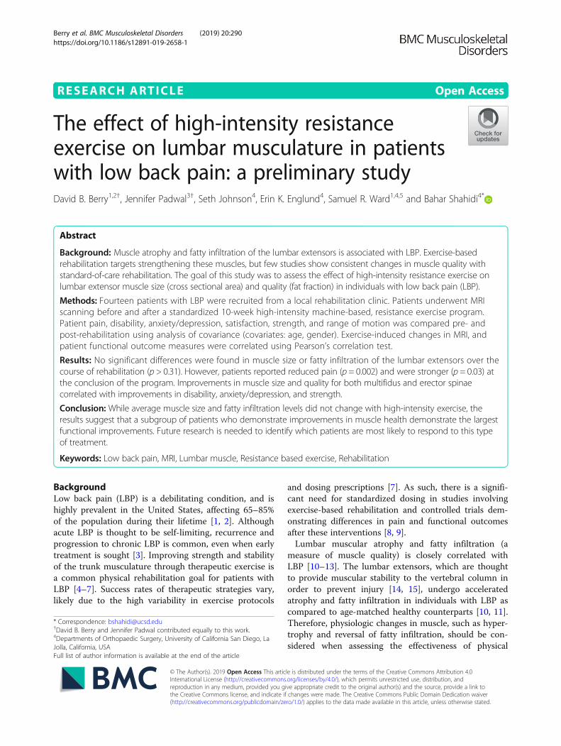
Resistance-based exercise protocolUpon initiating the program, a physical examination wasperformed including measurements of lumbar strengthand range of motion (ROM) using an isokinetic dyna-mometer (MedX Holdings Inc., Cheyenne WI; Fig. 1).This device allows for isolation of the lumbar extensorsthrough pelvic stabilization in conjunction with mea-surements of torque across a monitored patient-tolerated range of motion. The patients were then indi-vidually prescribed lumbar extension resistance exerciseson the MedX machine based on a maximal voluntarycontraction. The exercises were performed throughoutthe maximum available range of motion that a patientwas able to perform under supervision of a trained phys-ical therapist. Each patient was assigned to a single phys-ical therapist who supervised all of their trainingsessions for the duration of the program. Treatment ex-ercise doses were prescribed at 60–80% of that maximaleffort for 15–20 repetitions [24, 25]. Patients wereinstructed by the physical therapist to perform exercisesthroughout their available range of motion unless theirsymptoms increased with the exercise. Exercise wasadvanced in subsequent visits by 5–10% of the exerciseload once they were able to tolerate > 20 repetitionswithout an increase in pain. If they were able to reach >10 repetitions but < 20 repetitions, their exercise loadremained the same at their next visit. If they were unableto reach 10 repetitions, their exercise load was decreased5–10% at their next visit. Strength and ROM were mea-sured from the machine-based torque measurementsduring lumbar extension exercises at each visit. Twentyvisits over ten weeks was considered the standard regi-men to complete the rehabilitation protocol. All patientswere also provided with a copy of the, “Take Back Con-trol” book upon initiation of care, which provides guid-ance on healthy lifestyle modifications such as remainingactive and maintaining a healthy diet [26]. Any adverseevent such as an increase in symptoms in response totreatment was reported to the treating physician forfollow up.
Berry et al. BMC Musculoskeletal Disorders (2019) 20:290 Page 2 of 9
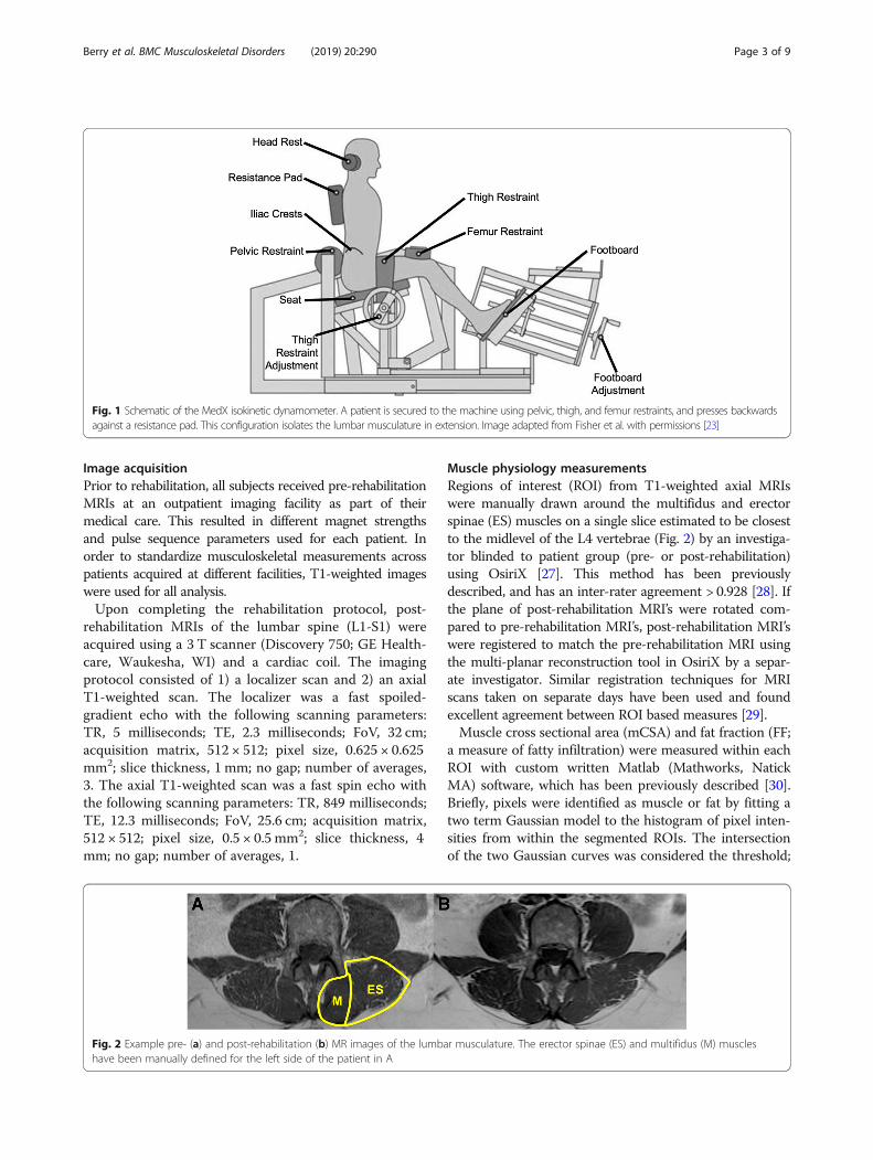
Image acquisitionPrior to rehabilitation, all subjects received pre-rehabilitationMRIs at an outpatient imaging facility as part of theirmedical care. This resulted in different magnet strengthsand pulse sequence parameters used for each patient. Inorder to standardize musculoskeletal measurements acrosspatients acquired at different facilities, T1-weighted imageswere used for all analysis.Upon completing the rehabilitation protocol, post-
rehabilitation MRIs of the lumbar spine (L1-S1) wereacquired using a 3 T scanner (Discovery 750; GE Health-care, Waukesha, WI) and a cardiac coil. The imagingprotocol consisted of 1) a localizer scan and 2) an axialT1-weighted scan. The localizer was a fast spoiled-gradient echo with the following scanning parameters:TR, 5 milliseconds; TE, 2.3 milliseconds; FoV, 32 cm;acquisition matrix, 512 × 512; pixel size, 0.625 × 0.625mm2; slice thickness, 1 mm; no gap; number of averages,3. The axial T1-weighted scan was a fast spin echo withthe following scanning parameters: TR, 849 milliseconds;TE, 12.3 milliseconds; FoV, 25.6 cm; acquisition matrix,512 × 512; pixel size, 0.5 × 0.5 mm2; slice thickness, 4mm; no gap; number of averages, 1.
Muscle physiology measurementsRegions of interest (ROI) from T1-weighted axial MRIswere manually drawn around the multifidus and erectorspinae (ES) muscles on a single slice estimated to be closestto the midlevel of the L4 vertebrae (Fig. 2) by an investiga-tor blinded to patient group (pre- or post-rehabilitation)using OsiriX [27]. This method has been previouslydescribed, and has an inter-rater agreement > 0.928 [28]. Ifthe plane of post-rehabilitation MRI’s were rotated com-pared to pre-rehabilitation MRI’s, post-rehabilitation MRI’swere registered to match the pre-rehabilitation MRI usingthe multi-planar reconstruction tool in OsiriX by a separ-ate investigator. Similar registration techniques for MRIscans taken on separate days have been used and foundexcellent agreement between ROI based measures [29].Muscle cross sectional area (mCSA) and fat fraction (FF;
a measure of fatty infiltration) were measured within eachROI with custom written Matlab (Mathworks, NatickMA) software, which has been previously described [30].Briefly, pixels were identified as muscle or fat by fitting atwo term Gaussian model to the histogram of pixel inten-sities from within the segmented ROIs. The intersectionof the two Gaussian curves was considered the threshold;
Fig. 1 Schematic of the MedX isokinetic dynamometer. A patient is secured to the machine using pelvic, thigh, and femur restraints, and presses backwardsagainst a resistance pad. This configuration isolates the lumbar musculature in extension. Image adapted from Fisher et al. with permissions [23]
Fig. 2 Example pre- (a) and post-rehabilitation (b) MR images of the lumbar musculature. The erector spinae (ES) and multifidus (M) muscleshave been manually defined for the left side of the patient in A
Berry et al. BMC Musculoskeletal Disorders (2019) 20:290 Page 3 of 9

pixel intensities above the threshold were classified as fat,pixel values below the threshold were classified as muscle.FF was calculated from the following equation:
FF ¼ #pixels;fat#pixels;fat þ #pixels;muscle
mCSA was calculated from the total cross-sectionalarea (tCSA) of the ROI [28]:
mCSA ¼ tCSA � 1−FFð ÞFF and mCSA data were averaged across sides for all
subsequent analyses.
Functional outcome measuresA 100 mm visual analog scale (VAS) was used to assessa patient’s perceived LBP at the beginning and end of re-habilitation, with higher values indicating more pain[31–33]. LBP related disability was assessed using theOswestry Disability Index (ODI) questionnaire at the be-ginning and end of the program [34]. The Patient HealthQuestionnaire-4 (PHQ4) was used to assess patient de-pression/anxiety at the beginning and end of the pro-gram [35, 36]. Maximum lumbar extension strength wasmeasured on the MedX isokinetic dynamometer at thebeginning and end of the program, and was monitoredthroughout the program for verification of exercise in-tensity. ROM was measured in degrees as the maximumrange of motion through which the patient was able toperform the resistance exercise.
Statistical analysisDemographic measures collected for each patient in-cluded gender, age, and weight. The primary outcomemeasures of muscle health were mCSA and FF. The pri-mary functional outcome measures from this study wereVAS, ODI, PHQ4, strength, and ROM. Normality of allvariables was checked using a Shapiro-Wilk test. Eachoutcome measure was compared using a 1-way analysisof covariance (covariates: age, gender), to identify theeffect of treatment on each outcome measure (factor:treatment). Pearson’s correlation test for normally dis-tributed data or Spearman rank correlation test for non-normally distributed data was used to determine thestrength of association between changes in musclehealth, and functional (Strength, VAS, ODI) or psycho-social (PHQ4) improvements with the program. Statis-tical analysis was performed using SPSS version 25.0(IBM Corporation, Armonk NY).
ResultsDemographicsFourteen patients volunteered for this study (Table 1).The majority of patients (N = 13) participating in this
study were being seen for a primary diagnosis of degen-erative disc disease, with secondary diagnoses of stenosis(N = 8) or spondylosis (N = 2). One subject was diag-nosed as having nonspecific LBP. As a group, on averagea 1.1 kg loss in weight was observed over the course ofrehabilitation (p = 0.711).
Effect of high-intensity resistance exercise on MRI-basedmeasures of muscle healthSmall changes were observed for mCSA of the erector spi-nae (− 7.9 mm2; F(1,23) = 0.063; p = 0.804) and multifidus(+ 41.6mm2; F(1,23) = 0.026; p = 0.873) when controllingfor age and gender, which were likely to be observed bychance (Fig. 3A, B). Additionally, small changes wereobserved for FF of the erector spinae (− 0.013; F(1,23) =1.079; p = 0.310) and multifidus (− 0.007; F(1,23) = 0.331;p = 0.570) when controlling for age and gender, whichwere also likely to be observed by chance (Fig. 3C, D). Thecovariate of age predicted FF for both muscles and mCSAfor only multifidus (p < 0.01); older patients had smallermCSA and higher FF. Males had a greater multifidusmCSA (p = 0.008).
Effect of high-intensity resistance exercise on functionaloutcomesOn a group level, a 27.2 mm decrease in pain (p = 0.002),a 86.9 Nm increase in strength (p = 0.03) was observed(Table 1; Fig. 4A,B) . Additionally, a 2.3% decrease in ODIassessed disability (p = 0.689), a 1.0 point decrease inPHQ4 assessed anxiety/depression (p = 0.518) and a 5.0 °increase in ROM (p = 0.173) was observed over the courseof treatment (Table 1; Fig. 4C-E). At the conclusion of thestandard rehabilitation period (20 visits), 7 patients de-cided to continue with a high-intensity, resistance basedmaintenance exercise program.
Table 1 Mean ± standard deviation of the demographicmeasures of patients included in this study
N = 14 Pre-rehab Post-Rehab
Age (years) 52.8 ± 14.8
Gender (M:F) 7:7
Weight (kg) 82.4 ± 15.81 81.3 ± 14.3
Visual Analog Scale (mm) 47.9 ± 22.2 20.7 ± 18.2
Strength (Nm) 196.8 ± 78.7 283.7 ± 131.9
ODI (%) 28.7 ± 12.4 26.4 ± 16.5
PHQ4 2.9 ± 5.0 1.9 ± 3.2
Range of Motion (°) 61.0 ± 10.9 66.0 ± 8.2
M male, F female, kg kilogram, ODI Oswestry Disability Index, PHQ4 PatientHealth Questionaire-4
Berry et al. BMC Musculoskeletal Disorders (2019) 20:290 Page 4 of 9

Relationships between functional outcomes and MRI-based measures of muscle healthAll functional outcome and MRI-based measures ofmuscle health were normally distributed except forPHQ4 (p < 0.001) and ROM (p = 0.005). Correlationswere observed between improvements in functionaloutcomes and both FF and mCSA. Reductions in ESFF were associated with decreased depression/anxietywith the program (p = 0.009; r = − 0.666; Fig. 5A) aswell as decreased LBP related disability (p = 0.011; r =− 0.658; Fig. 5B). Larger improvements in multifidusmCSA in response to the program were associatedwith increased isometric lumbar strength (p = 0.003;r = 0.738; Fig. 5C). However, the observed change instrength was not found to correlate with the change in pain(r = − 0.253; p = 0.382), disability (r = 0.345; p = 0.228), ordepression/anxiety (r = − 0.329; p = 0.251) over the courseof rehabilitation.
DiscussionThis was a preliminary study, evaluating changes inmCSA and FF in response to a standardized, high-intensity, machine-based resistance exercise program inpatients with LBP. We hypothesized that increasedmCSA and decreased FF would be observed after thisprogram in conjunction with improvements in patientfunctional outcomes. These data demonstrate that onaverage, patients did not demonstrate improvements inmuscle size or quality as measured by T1-weighted MRI
in response to this program, despite improvements instrength and reduced pain. Correlations were foundbetween functional and MRI measured outcomes, in thatpatients who demonstrated improvements in musclehealth also demonstrated the largest functional improve-ments in LBP related disability, strength, and depres-sion/anxiety. These findings, although limited by a smallsample size, suggest that while resistance based exercisemay not result in improvement in muscle health andfunctional improvements for all patients with LBP, thereare some patients who demonstrate the largest improve-ments in muscle health and are also the most responsiveto treatment and vice versa.The magnitude of change in muscle health and func-
tional outcomes is similar to prior studies on the effectsof exercise on individuals with LBP for pain-specific out-comes, but not for outcomes of disability or depression/anxiety [37–39]. The cohort’s average pain reductionwas 27.1 mm ± 20.9 mm, which exceeds the minimalclinically important difference (MCID) of 20 mm forVAS in patients with LBP [37]. Previous exercise trials inthese populations report improvements in pain rangingfrom 7 to 13 points, and reductions in LBP related dis-ability of between 6.9 to 20 points [38, 39]. The smallertreatment effects for LBP related disability and anxiety/depression may be due to the low initial scores for levelsof LBP related disability and depression/anxiety in thiscohort as compared to other studies [37–39]. Interest-ingly, while the rehabilitation protocol made patients
Fig. 3 Muscle cross sectional area (mCSA) (a, b) and fat fraction (FF) (c, d) measures of the erector spinae (a, c) and multifidus (b, d) muscles. Nosignificant differences in muscle physiology assessed by magnetic resonance imaging were found between pre- and post-rehabilitationmeasurements. Data reported as mean ± standard deviation
Berry et al. BMC Musculoskeletal Disorders (2019) 20:290 Page 5 of 9

stronger, and muscle health was related to treatment-related strength gains, the amount of strength gaineddid not correlate directly with functional improvements.The muscle-specific changes that have been reported
in response to various exercise programs demonstrate
Fig. 4 Functional outcome measures pre- and post-rehabilitation. VAS(a) was measured to assess pain. Strength (b) and ROM (e) weremeasured using a MedX isometric dynamometer. ODI (c) was used toassess low back pain related disability. PHQ4 (d) was used to assessanxiety/depression related to low back pain. * indicates p < 0.05. **indicates p < 0.01. Data reported as mean ± standard deviation
Fig. 5 Significant correlations between patient reported outcomemeasures (x-axis) and muscle health measured with MRI (y-axis)
Berry et al. BMC Musculoskeletal Disorders (2019) 20:290 Page 6 of 9

conflicting results, and have largely been based on mea-sures of muscle size. To our knowledge, only two otherstudies have investigated changes in multifidus fatty in-filtration in response to an exercise program. One studyonly included males in their cohort, and found similarresults using an isolated lumbar extension protocol withvariable frequency [17]. A study by Welch et al. did finddecreases in lumbar paraspinal muscle FF by 2.5% [40].However their patient cohort was young with lowerbaseline FF, so their rehabilitation protocol may not ex-hibit muscle physiological changes in the average patientpopulation with LBP. A systematic review by Shahtah-massebi et al. reported that only one of 7 exercise inter-vention studies reported significant improvements inmultifidus size after a machine-based resistance exerciseprogram [19, 41]. Even when these studies were ex-panded to include other exercise modalities such asmotor control or aerobic exercises, only two of 18 stud-ies reported improvements in multifidus CSA after theprogram [20]. Our study is consistent with the priorliterature in that there is no strong evidence of a meanchange in muscle size or quality. However, our data mayprovide preliminary evidence that the lack of an appre-ciable effect size in response to exercise when averagedacross the whole study population may be because treat-ment outcomes are combined across responder andnon-responder patient subgroups, whose distinguishingcharacteristics have yet to be identified or well-defined.When we further examined patients who demonstratedimprovements in muscle health parameters, we foundpatients who had lower levels of baseline disability andanxiety/depression had larger improvements in musclephysiology for ES only, however no such trend wasobserved for multifidus. This suggests that patents withless anxiety/depression initially are more likely to experi-ence significant physiologic changes from high-intensity,resistance based exercise.There are several limitations to this study. First, as this
was a preliminary study, our sample size was small.However, we observed similar results to a similar studywith a larger sample size by Welch et al., highlightingthe potential importance of identifying patient sub-groups [40]. Additionally, as patients were only enrolledin this study if they had pre-rehabilitation MR imaging,the pre-rehabilitation MRIs were acquired at outsidefacilities. This resulted in different MR field strengths,MR acquisition parameters, and patient positioning inthe scanner. We attempted to minimize error by meas-uring the spine at the same location (L4), in both pre-and post-rehabilitation images. The L4 level was chosenas L4 fat signal fraction correlates highly (R2 > 0.92) withwhole lumbar fat fraction [42]. Furthermore, we regis-tered post-rehabilitation MR images to precisely matchslice orientations across acquisitions in 3D, in order to
ensure that the same area of the muscles was being ana-lyzed in both the pre- and post-rehabilitation images.Standard T1-weighted MRI of the lumbar musculaturemay be too crude a technique to assess early muscleadaptation to resistance based training, as it only has theability to detect changes on a whole muscle level. Chem-ical shift imaging techniques such as Dixon or IDEALMRI allow for intravoxel quantification of fat, and maybe more sensitive to changes in the muscle fat content[43, 44]. Furthermore, diffusion tensor imaging (DTI) - aMRI based technique sensitive to muscle microstructure- may provide more sensitivity to changes in musclemicrostructure associated with exercise such as fiberhypertrophy. Previous studies have shown differences inDTI derived measures before and after strenuous exercisethat were not detected with routine T1- and T2-weightedMRI [45, 46]. Therefore, future studies may considerincorporating chemical shift imaging or DTI based tech-niques to better assess microstructural changes associatedwith resistance-based exercise in patients with LBP.In addition to registering pre- and post-rehabilitation
images, we took several additional steps to minimizebias and error in this study. We attempted to minimizebias introduced with manual thresholding techniques byusing an automated technique to determine the fat/muscle pixel intensity threshold. Additionally, due to thefact that this study employed convenience sampling, wewere unable to control for patient-specific factors suchas LBP duration, method of injury, previous treatments,etc. Without controlling for these characteristics, it is moredifficult to understand which population may benefit mostfrom this resistance based physical rehabilitation program.A future prospective study with a well-characterized pa-tient population could address these issues.A unique feature of this study was the standardization
of the exercise dosing across all patients. While resistancebased exercise is generally associated with good outcomes,dosing and protocol variations make it difficult to com-pare across studies. The rehabilitation exercise protocolused in this study was found to significantly reduce painand increase strength across patients. Most importantly,we observed that patients who demonstrate changes inmuscle physiology respond better to a high-intensityresistance based exercise program, which must be moreclosely investigated in future experiments.This exercise program followed in this study was
developed by the SpineZone Rehabilitation Center (SanDiego, CA). The exercise protocol and progression asbased on prior literature using machine-based resistanceexercise to strengthen the posterior lumbar musculature,as well as modifications made by trained physical therapiststo accommodate individuals with pain and pathology forsafety in concordance with the ACSM guidelines for olderor more fragile populations [24, 47–49]. A single set was
Berry et al. BMC Musculoskeletal Disorders (2019) 20:290 Page 7 of 9

utilized because in prior research on lumbar extension-specific exercise, it was demonstrated to induce the largestchanges at a 2x per week frequency when compared to ahigher number of sets [47]. Although the modificationsused in this study have not been rigorously studied in theliterature, it was found that in order for patients to achievea perceived exertion of greater than 7 without exacerbationof symptoms, a higher number of repetitions at a submaxi-mal intensity was required. We recognize this as a poten-tial limitation, however this program aimed to optimizestrength training with symptom reduction without riskingpatient safety.
ConclusionsIn this study, we assessed mCSA and FF of the lumbarparaspinal muscles over the course of high-intensity resist-ance rehabilitation in patients with LBP. Although onlyslight group wide changes in average muscle size or fattyinfiltration were observed, overall patients experiencedlarge improvements in pain and strength. Improvementsin disability, anxiety/depression, and strength were foundto correlate with increased mCSA and decreased FF,which suggests that high-intensity, resistance exerciseelicits a physiologic response in the lumbar muscles of asubgroup patients with LBP. These preliminary findingsmay suggest that there are some patients who experiencea more robust response to this intervention strategy,although the sample size of this study precludes this con-clusion. Future research is needed to identify whetherthese subgroups exist, and which patients may benefitmost from this rehabilitation approach.
AbbreviationsES: Erector Spinae; FF: Fat fraction; LBP: Low back pain; MCID: Minimalclinically important difference; mCSA: Muscle cross sectional area;MRI: Magnetic resonance imaging; ODI: Oswestry disability index;PHQ4: Patient health questionnaire – 4; ROI: Region of interest; ROM: Rangeof motion; VAS: visual analog score
AcknowledgementsWe would like to thank Dr. Kamshad Raiszadeh, Dr. Kian Raiszadeh, JonathanWu, Jonathan Tapicer, and all of the staff at SpineZone in San Diego, CA forproviding equipment and patients for this study.
Author’s contributionsDB designed the experiment, analyzed the MRI data, and was a majorcontributor in writing the manuscript. JP was a major contributor in writingthe manuscript and analyzed the MRI data. SJ analyzed the MRI data. EKEanalyzed the MRI data and contributed to writing the manuscript. SRWdesigned the experiment and revised the manuscript. BS designed theexperiment, analyzed the MRI data, and revised the manuscript. All authorsread and approved the final manuscript.
FundingThis study was funded by the 2018 Magistro Family Foundation ResearchGrant, from the Foundation for Physical Therapy Research. These fundingbodies provided funding for data acquisition and collection only, with nocontribution to analysis, study design, interpretation, or manuscriptpreparation.
Availability of data and materialsThe datasets used and/or analyzed during the current study are availablefrom the corresponding author on reasonable request.
Ethics approval and consent to participateThis experiment was conducted with approval from the UC San DiegoInstitutional Review Board. All subjects provided oral and written consent toparticipate.
Consent for publicationNot applicable.
Competing interestsThe authors declare that they have no competing interests.
Author details1Departments of Bioengineering, University of California San Diego, La Jolla,California, USA. 2Departments of Nanoengineering, University of CaliforniaSan Diego, La Jolla, California, USA. 3Departments of Medicine, University ofCalifornia San Diego, La Jolla, California, USA. 4Departments of OrthopaedicSurgery, University of California San Diego, La Jolla, California, USA.5Departments of Radiology, University of California San Diego, La Jolla,California, USA.
Received: 12 March 2019 Accepted: 28 May 2019
References1. Murray CJ, Atkinson C, Bhalla K, Birbeck G, Burstein R, Chou D, et al. The
state of US health, 1990-2010: burden of diseases, injuries, and risk factors.Jama. 2013;310:591–608.
2. Andersson GB. Epidemiological features of chronic low-back pain. Lancet.1999;354:581–5.
3. Danneels LA, Cools AM, Vanderstraeten GG, Cambier DC, Witvrouw EE,Bourgois J, et al. The effects of three different training modalities on thecross-sectional area of the paravertebral muscles. Scand J Med Sci Sport.2001;11:335–41 https://doi.org/10.1136/bjsm.35.3.186.
4. Chou R, Qaseem A, Snow V, Casey D, Cross JT Jr, Shekelle P, et al. Diagnosisand treatment of low back pain: a joint clinical practice guideline from theAmerican College of Physicians and the American pain society. Ann InternMed. 2007;147:478–91 https://doi.org/10.7326/0003-4819-147-7-200710020-00006.
5. Chou R, Huffman LH. Nonpharmacologic therapies for acute and chroniclow back pain: a review of the evidence for an American pain society/American College of Physicians clinical practice guideline. Ann Intern Med.2007;147:492–504 https://doi.org/10.7326/0003-4819-147-7-200710020-00007.
6. Rundell SD, Sherman KJ, Heagerty PJ, Mock CN, Jarvik JG. The clinical courseof pain and function in older adults with a new primary care visit for backpain. J Am Geriatr Soc. 2015;63:524–30. https://doi.org/10.1111/jgs.13241.
7. Hayden JA, van Tulder MW, Tomlinson G. Systematic review: strategies forusing exercise therapy to improve outcomes in chronic low back pain. AnnIntern Med. 2005;142:776–85 https://doi.org/10.7326/0003-4819-142-9-200505030-00014.
8. Slade SC, Dionne CE, Underwood M, Buchbinder R, Beck B, Bennell K, et al.Consensus on exercise reporting template (CERT): modified Delphi study.Phys Ther. 2016;96:1514–24.
9. Slade SC, Dionne CE, Underwood M, Buchbinder R. Consensus on exercisereporting template (CERT): explanation and elaboration statement. Br JSports Med. 2016;50:1428–37.
10. Parkkola R, Rytokoski U, Kormano M. Magnetic resonance imaging of the discsand trunk muscles in patients with chronic low back pain and healthy controlsubjects. Spine (Phila pa 1976), vol. 18; 1993. p. 830–6. https://doi.org/10.1097/00007632-199306000-00004.
11. Mattila M, Hurme M, Alaranta H, Paljarvi L, Kalimo H, Falck B, et al. Themultifidus muscle in patients with lumbar disc herniation. A histochemicaland morphometric analysis of intraoperative biopsies. Spine (Phila Pa 1976).1986;11:732–8 https://doi.org/10.1097/00007632-198609000-00013.
12. Alaranta H, Tallroth K, Soukka A, Heliovaara M. Fat content of lumbarextensor muscles and low back disability: a radiographic and clinicalcomparison. J Spinal Disord. 1993;6:137–40.
Berry et al. BMC Musculoskeletal Disorders (2019) 20:290 Page 8 of 9

13. Kjaer P, Bendix T, Sorensen JS, Korsholm L, Leboeuf-Yde C. Are MRI-definedfat infiltrations in the multifidus muscles associated with low back pain.BMC Med. 2007;5(2).
14. Jeon K, Kim T, Lee SH. Effects of muscle extension strength exercise on trunkmuscle strength and stability of patients with lumbar herniated nucleuspulposus. J Phys Ther Sci. 2016;28:1418–21. https://doi.org/10.1589/jpts.28.1418.
15. Yaprak Y. The effects of back extension training on back muscle strengthand spinal range of motion in young females. Biol Sport. 2013;30:201–6.https://doi.org/10.5604/20831862.1047500.
16. Danneels L, Vanderstraeten G, Cambier D, Witvrouw E, Bourgois J, DankaertsW, et al. Effects of three different training modalities on the cross sectionalarea of the lumbar multifidus muscle in patients with chronic low backpain. Br J Sport Med. 2001;35:186–91. https://doi.org/10.1136/bjsm.35.3.186.
17. Willemink MJ, van Es HW, Helmhout PH, Diederik AL, Kelder JC, vanHeesewijk JP. The effects of dynamic isolated lumbar extensor training onlumbar multifidus functional cross-sectional area and functional status ofpatients with chronic nonspecific low back pain. Spine (Phila Pa 1976). 2012;37:E1651–8. https://doi.org/10.1097/BRS.0b013e318274fb2f.
18. Mooney V, Gulick J, Perlman M, Levy D, Pozos R, Leggett S, et al.Relationships between myoelectric activity, strength, and MRI of lumbarextensor muscles in back pain patients and normal subjects. J Spinal Disord.1997;10:348–56.
19. Shahtahmassebi B, Hebert JJ, Stomski NJ, Hecimovich M, Fairchild TJ. Theeffect of exercise training on lower trunk muscle morphology. Sport Med.2014;44:1439–58.
20. Kliziene I, Sipaviciene S, Klizas S, Imbrasiene D. Effects of core stabilityexercises on multifidus muscles in healthy women and women withchronic low-back pain. J Back Musculoskelet Rehabil. 2015;28:841–7. https://doi.org/10.3233/bmr-150596.
21. Woodham M, Woodham AW, Skeate JW, Freeman MD. Long-term lumbarMultifidus muscle atrophy changes documented with magnetic resonanceimaging: a case series. J Radiol Case Rep. 2014;8:27–34.
22. O’leary S, Jull G, Van Wyk L, Pedler A, Elliott J. Morphological changes in thecervical muscles of women with chronic whiplash can be modified withexercise-a pilot study. Muscle Nerve. 2015;52:772–9.
23. Fisher J, Bruce-Low S, Smith D. A randomized trial to consider the effect ofRomanian deadlift exercise on the development of lumbar extensionstrength. Phys Ther Sport. 2013;14(3):139–45. https://doi.org/10.1016/j.ptsp.2012.04.001.
24. Pollock ML, Leggett SH, Graves JE, Jones A, Fulton M, Cirulli J. Effect ofresistance training on lumbar extension strength. Am J Sports Med. 1989;17:624–9.
25. Nelson BW, O’Reilly E, Miller M, Hogan M, Wegner JA, Kelly C. The clinicaleffects of intensive, specific exercise on chronic low back pain: a controlledstudy of 895 consecutive patients with 1-year follow up. Orthopedics. 1995;18:971–81.
26. Raiszadeh K. Take Back control - a Surgeon’s guide to healing your spinewithout medications or surgery: OptiFit Publishers; 2016.
27. Rosset A, Spadola L, Ratib O. OsiriX: an open-source software for navigatingin multidimensional DICOM images. J Digit Imaging. 2004;17:205–16.
28. Berry DB, Padwal J, Johnson S, Parra CL, Ward SR, Shahidi B. Methodologicalconsiderations in region of interest definitions for paraspinal muscles inaxial MRIs of the lumbar spine. BMC Musculoskelet Disord. 2018;19:135.
29. Heemskerk AM, Sinha TK, Wilson KJ, Ding Z, Damon BM. Repeatability ofDTI-based skeletal muscle fiber tracking. NMR Biomed. 2010;23:294–303.
30. Shahidi B, Parra CL, Berry DB, Hubbard JC, Gombatto S, Zlomislic V, et al.Contribution of lumbar spine pathology and age to Paraspinal muscle sizeand fatty infiltration. Spine (Phila Pa 1976). 2017;42:616–23.
31. McCormack HM, Horne DJ, Sheather S. Clinical applications of visualanalogue scales: a critical review. Psychol Med. 1988;18:1007–19.
32. Ogon M, Krismer M, Sollner W, Kantner-Rumplmair W, Lampe A. Chroniclow back pain measurement with visual analogue scales in differentsettings. Pain. 1996;64:425–8.
33. Olaogun MOB, Adedoyin RA, Ikem IC, Anifaloba OR. Reliability of rating lowback pain with a visual analogue scale and a semantic differential scale.Physiother Theory Pract. 2004;20:135–42 https://doi.org/10.1080/09593980490453048.
34. Fairbank JC, Pynsent PB. The Oswestry disability index. Spine (Phila Pa 1976)2000;25:2940–2952; discussion 2952.
35. Kroenke K, Spitzer RL, Williams JB, Lowe B. An ultra-brief screening scale foranxiety and depression: the PHQ4. Psychosomatics. 2009;50:613–21. https://doi.org/10.1176/appi.psy.50.6.613.
36. Lowe B, Wahl I, Rose M, Spitzer C, Glaesmer H, Wingenfeld K, et al. A 4-itemmeasure of depression and anxiety: validation and standardization of thepatient health Questionnaire-4 (PHQ4) in the general population. J AffectDisord. 2010;122:86–95. https://doi.org/10.1016/j.jad.2009.06.019.
37. Bombardier C, Hayden J, Beaton DE. Minimal clinically important difference.Low back pain: outcome measures. J Rheumatol. 2001;28:431–8.
38. Hayden JA, van Tulder MW, Malmivaara A, Koes BW. Exercise therapy fortreatment of non-specific low back pain. Cochrane Database Syst Rev. 2005:Cd000335. https://doi.org/10.1002/14651858.CD000335.pub2.
39. Weinstein JN, Lurie JD, Tosteson TD, Skinner JS, Hanscom B, Tosteson ANA,et al. Surgical vs nonoperative treatment for lumbar disk herniation: thespine patient outcomes research trial (SPORT) observational cohort. Jama.2006;296:2451–9. https://doi.org/10.1001/jama.296.20.2451.
40. Welch N, Moran K, Antony J, Richter C, Marshall B, Coyle J, et al. The effectsof a free-weight-based resistance training intervention on pain, squatbiomechanics and MRI-defined lumbar fat infiltration and functional cross-sectional area in those with chronic low back. BMJ Open Sport Exerc Med.2015;1:e000050. https://doi.org/10.1136/bmjsem-2015-000050.
41. Choi HK, Gwon H, Kim SR, Park CS, Cho BJ. Effects of active rehabilitationtherapy on muscular back strength and subjective pain degree in chroniclower back pain patients. J Phys Ther Sci. 2016:2700–2. https://doi.org/10.1589/jpts.28.2700.
42. Crawford RJ, Cornwall J, Abbott R, Elliott JM. Manually defining regions ofinterest when quantifying paravertebral muscles fatty infiltration from axialmagnetic resonance imaging: a proposed method for the lumbar spinewith anatomical cross-reference. BMC Musculoskelet Disord. 2017;18:25.
43. Dixon WT. Simple proton spectroscopic imaging. Radiology. 1984;153:189–94. https://doi.org/10.1148/radiology.153.1.6089263.
44. Reeder SB, Pineda AR, Wen Z, Shimakawa A, Yu H, Brittain JH, et al. Iterativedecomposition of water and fat with echo asymmetry and least-squaresestimation (IDEAL): application with fast spin-echo imaging. Magn ResonMed. 2005;54:636–44. https://doi.org/10.1002/mrm.20624.
45. Froeling M, Oudeman J, Strijkers GJ, Maas M, Drost MR, Nicolay K, et al.Muscle changes detected with diffusion-tensor imaging after long-distancerunning. Radiology. 2015;274:548–62. https://doi.org/10.1148/radiol.14140702.
46. Cermak NM, Noseworthy MD, Bourgeois JM, Tarnopolsky MA, Gibala MJ.Diffusion tensor MRI to assess skeletal muscle disruption following eccentricexercise. Muscle Nerve. 2012;46:42–50. https://doi.org/10.1002/mus.23276.
47. Graves JE, Pollock ML, Foster D, Leggett SH, Carpenter DM, Vuoso R, et al.Effect of training frequency and specificity on isometric lumbar extensionstrength. Spine (Phila pa 1976), vol. 15; 1990. p. 504–9.
48. Starkey DB, Pollock ML, Ishida Y, Welsch MA, Brechue WF, Graves JE, et al.Effect of resistance training volume on strength and muscle thickness. MedSci Sports Exerc. 1996;28:1311–20.
49. Swain DP, editor. American College of Sports Medicine’s resource manualfor guidelines for exercise testing and prescription. 7th ed. Baltimore:Wolters Kluwer Health/Lippincott Williams & Wilkins; 2014.
Publisher’s NoteSpringer Nature remains neutral with regard to jurisdictional claims inpublished maps and institutional affiliations.
Berry et al. BMC Musculoskeletal Disorders (2019) 20:290 Page 9 of 9