the effect of physiotherapeutic kinesio taping on selected
TRANSCRIPT

The effect of physiotherapeutic
Kinesio taping on selected physical
symptoms associated with
major depressive disorder
Karen Erasmus
Student number 200010575

The effect of physiotherapeutic Kinesio
taping on selected physical symptoms
associated with major depressive disorder
A research report by
Karen Erasmus
Submitted in partial fulfilment of the requirement for the degree in
MSc (PHYSIOTHERAPY)
In the Physiotherapy Department
Faculty of Health Science
University of the Free State
24 January 2014
STUDY LEADER: Mrs K. Bodenstein
CO-STUDY-LEADER: Dr M. du Toit
Declaration by Study Leader
I, Karen Bodenstein, study leader of Karen Erasmus, hereby confirm that the
Department of Physiotherapy, University of the Free State, approves the
submission of this script. I certify that the script submitted as partial fulfillment of
the requirements for the degree MSc (Physiotherapy) at the University of the
Free State is the student‟s independent effort and has not previously been
submitted, either partially or as a whole, to the assessors. This script has also
not previously been submitted for a degree at another university/faculty.
______________________________ ______________________
Karen Bodenstein Date
Lecturer
Department of Physiotherapy
UFS

Declaration by Student
I, Karen Erasmus, certify that the script hereby submitted by me as partial
fulfilment of the requirements for the degree MSc (Physiotherapy) at the
University of the Free State is my independent effort and has not previously been
submitted for a degree at another university/faculty. I furthermore waive
copyright of the script in favour of the University of the Free State.
_____________________________
Karen Erasmus
24 January 2014

Acknowledgments
I want to thank the following people as without them this study and the last
4 years would not have been possible:
Karen Bodenstein as my study leader in physiotherapy – thank you for your
time, advice, patience and support on an academic and personal level. I am
very grateful and blessed to have you as my study leader.
Dr Martie du Toit as co-study leader in psychology..
Gerda Aspeling, my research assistant and friend. Thank you for the support
and companionship during the hours spent at various institutions. To Gerda‟s
family, thank you for giving me and this study your time.
Prof Gina Joubert for the analysis of the data. You were always available
with advice and encouragement.
Karen de Bruin who went above and beyond the call of duty when it came to
language editing.
Tharina Annandale who was willing to check the data forms and was always
there with a word of advice and encouragement.
Jaco Hough for assisting me with the printing.
My husband, family and friends – thank you for believing in me.

i
Table of Contents List of Figures ................................................................................................................................... iv
List of Tables .................................................................................................................................... iv
List of Graphs ................................................................................................................................... iv
List of Abbreviations ......................................................................................................................... v
Abstract vi
Chapter 1 Introduction .................................................................................................................. 1
1.1 Introduction and Background to the Study ....................................................... 1
1.2 Research Question ........................................................................................... 3
1.3 Aim and objectives the Study ........................................................................... 4
1.4 Literature Review .............................................................................................. 5
1.5 Significance of the Study .................................................................................. 6
1.6 Definition of Terms ............................................................................................ 7
1.7 Format of the Research Report ........................................................................ 8
1.8 Conclusion ........................................................................................................ 8
Chapter 2 Literature Review ........................................................................................................ 9
2.1 Introduction ....................................................................................................... 9
2.2 Properties of Kinesio®
Tape .............................................................................. 9
2.3 The Effects of Kinesio® Taping ....................................................................... 11
2.4 The Application of Kinesio® Tape ................................................................... 12
2.5 Physiotherapeutic Kinesio® Taping ................................................................. 15
2.6 Major Depressive Disorder ............................................................................. 16
2.7 Current Role of the Physiotherapist in Mental Health .................................... 19
2.8 Selected Physical Symptoms Associated with MDD ...................................... 19
2.8.1 Muscle Tension and Associated Pain ................................................ 19
2.8.2 Restricted Breathing .......................................................................... 22
2.8.3 Decrease in Flexibility and Centring of Movements .......................... 26
2.9 Conclusion ...................................................................................................... 29
Chapter 3 Methodology .............................................................................................................. 30
3.1 Introduction ..................................................................................................... 30
3.2 Aim and objectives the study .......................................................................... 30
3.3 Orientation with Regards to the Study Process .............................................. 31
3.4 Research Design ............................................................................................ 31
3.5 Sample and/or Study Participants .................................................................. 35
3.5.1 Eligibility Criteria ................................................................................ 36 3.5.1.1 Inclusion Criteria ................................................................ 36 3.5.1.2 Exclusion Criteria ............................................................... 37
3.6 Study Interventions ......................................................................................... 38
3.6.1 Kinesio® Tape Application ................................................................. 38
3.6.2 Placebo Application ........................................................................... 39

ii
3.7 Measurement .................................................................................................. 40
3.7.1 Assessments ..................................................................................... 40
3.7.2 Reliability and Validity ........................................................................ 45
3.8 Pilot Study ....................................................................................................... 45
3.9 Data Collection................................................................................................ 46
3.9.1 Data Collection at the Public Psychiatric Institution .......................... 46
3.9.2 Data collection setting ........................................................................ 47
3.9.3 Data Collection at the Private Psychiatric Institution ......................... 47
3.9.4 Data Collection Procedures for All Institutions .................................. 48
3.10 Ethical Issues .................................................................................................. 49
3.10.1 Protection from Harm ......................................................................... 49
3.10.2 Informed Consent .............................................................................. 50
3.10.3 Right to Privacy .................................................................................. 50
3.10.4 Professional Honesty with Colleagues .............................................. 51
3.11 Coding of Questionnaires ............................................................................... 52
3.12 Data Analysis .................................................................................................. 52
3.13 Measurement and Methodological Errors ....................................................... 53
3.14 Conclusion ...................................................................................................... 55
Chapter 4 Results ...................................................................................................................... 56
4.1 Introduction ..................................................................................................... 56
4.2 Disposition of Study Participants .................................................................... 56
4.3 Results of the Questionnaires ......................................................................... 56
4.3.1 Demographic Information .................................................................. 57
4.3.2 Pain Symptoms .................................................................................. 59
4.3.3 Pain areas .......................................................................................... 60
4.3.4 Manual Assessment of Respiratory Motion ....................................... 65
4.3.5 The Tinetti Mobility Test ..................................................................... 66
4.4 Summary of Study Results ............................................................................. 67
4.5 Conclusion ...................................................................................................... 68
Chapter 5 Discussion ................................................................................................................. 69
5.1 Introduction ..................................................................................................... 69
5.2 Discussion of Results ..................................................................................... 69
5.2.1 Demographic Information .................................................................. 69
5.2.2 Pain Symptoms .................................................................................. 71
5.2.3 Pain Areas ......................................................................................... 71
5.2.4 Post-Application Results .................................................................... 73
5.2.5 Results of the Pain Score .................................................................. 74
5.2.6 Results of the restricted breathing score ........................................... 75
5.2.7 Results of the loss of flexibility and centring of movement score ...... 77
5.3 Conclusion ...................................................................................................... 78
Chapter 6 Conclusion and Recommendations .......................................................................... 79
6.1 Introduction ..................................................................................................... 79
6.2 Conclusions .................................................................................................... 79

iii
6.3 Limitations of this Study .................................................................................. 81
6.4 Recommendations .......................................................................................... 82
6.5 Value of the Study .......................................................................................... 83
6.6 Final Summary ................................................................................................ 83
Chapter 7 References ................................................................................................................ 84
Chapter 8 Personal Communications ........................................................................................ 94
Appendices ................................................................................................................................... 95
Appendix A ................................................................................................................ 96
Appendix B ................................................................................................................ 99
Appendix C .............................................................................................................. 105
Appendix D .............................................................................................................. 112
Appendix E .............................................................................................................. 119
Appendix F .............................................................................................................. 121
Appendix G .............................................................................................................. 123
Appendix H .............................................................................................................. 131
Summary ................................................................................................................................. 134
Opsomming ................................................................................................................................. 137

iv
List of Figures
Figure 2-1 The longitudinal sine wave on the tape .................................................................. 11
Figure 2-2 Kinesio® tape strips ................................................................................................. 13
Figure 2-3 Kinesio® application to skin and effect on underlying structures ............................ 15
Figure 2-4 Main structures of the limbic system ...................................................................... 17
Figure 2-5 M. erector spinae .................................................................................................... 25
Figure 3-1 Flowchart of study process ..................................................................................... 34
Figure 3-2 Kinesio® taping according to standardised treatment guidelines ........................... 39
Figure 3-3 Placebo application – Kinesio® taping not in current guidelines ............................ 40
List of Tables
Table 2-1 Neurotransmitter system associated with MDD ..................................................... 18
Table 4-1 Age distribution across the groups ......................................................................... 58
Table 4-2 Therapies received during study period ................................................................. 59
Table 4-3 Pain complaint areas .............................................................................................. 60
Table 4-4 Pre-application pain scores .................................................................................... 62
Table 4-5 Post-application pain scores ................................................................................... 62
Table 4-6 24 hours post-application pain scores .................................................................... 63
Table 4-7 Differences between pre-, post-, and 24 hours post-application of the combined components of SF-MPQ ........................................................................ 63
Table 4-8 Percentage of participants experiencing pain ........................................................ 63
Table 4-9 Percentage improvement of SF-MPQ .................................................................... 64
Table 4-10 Percentage clinical significant improvement of SF-MPQ ....................................... 64
Table 4-11 The balance of breathing score of the MARM ........................................................ 65
Table 4-12 The percentage of ribcage movement score of the MARM .................................... 66
Table 4-13 Tinetti Mobility Test: Balance and gait scores ........................................................ 67
List of Graphs
Graph 4-1 Gender distribution across groups.......................................................................... 57
Graph 4-2 Classification of MDD severity ................................................................................ 58
Graph 4-3 Participants complaining of multiple sites ............................................................... 60
v
List of Abbreviations
BAS Body Awareness Scale
BAS-H Body Awareness Scale - Health
DASH Disability of Arm, Shoulder and Hand
DSM-5 Diagnostic and Statistical Manual of Mental Disorders – Fifth Edition
EMG Electromyography
HAM-D Hamilton Depression Rating Scale
HPCSA Health Professionals Council of South Africa
KTA Kinesio® Taping Association
MARM Manual Assessment of Respiratory Motion
MDD Major depressive disorder
OSDB Obstructive sleep disorder breathing
RSA Respiratory sinus arrhythmia
SF-MPQ Short Form McGill Pain Questionnaire
VAS Visual analogue scale

vi
Abstract
Kinesio® taping is a relatively new form of therapeutic taping that has a variety of
applications. Physiotherapists can use the tape to treat certain physical
symptoms for example, pain, swelling and dysfunctional muscle activation.
Major depressive disorder (MDD) is a mood disorder and physical symptoms
most associated with MDD, and tested in this study were muscle tension, pain
complaints, restricted breathing, less flexibility and centring of movement. The
aim of this study was to investigate the effect of physiotherapeutic Kinesio®
taping on selected physical symptoms associated with MDD. A double-blind,
randomised controlled design was used, following a quantitative study approach.
The study population consisted of 40 patients with MDD admitted to a private and
public psychiatric institution in Bloemfontein.
The majority (77.5%) of participants complained of pain during the study period
(24 hours) and 58% complained of multiple areas of pain. The sensory and
affective components associated with pain and tested by the Short Form McGill
Pain Questionnaire (SF-MPQ) showed improvement in combined scores for both
the experimental and placebo groups. The results of the Manual Assessment of
Respiratory Motion (MARM) displayed improvement in both the placebo and
experimental groups for balance of breathing and percentage ribcage motion.
The Tinetti Mobility Test which assesses balance and gait showed no distinct
results possibly due to the scale not being sensitive enough for the movement
disorders tested
Limitations of the study could have influenced the outcomes measured and it
should be taken into account that of the 40 participants, 21 received
physiotherapy. Other therapies received by participants during the study were
not standardised and could not be controlled due to the multitude of stakeholders
involved in the care of the patients.

vii
The treatment of physical symptoms associated with MDD with Kinesio® taping
had mixed results, but Kinesio® taping could be a valuable adjuvant treatment
modality. The importance of physiotherapy as part of the treatment regime for
patients suffering from MDD was highlighted.
Key terms: effects, physiotherapeutic, Kinesio® taping, physical
symptoms, major depressive disorder, pain, restricted breathing, loss of
centring of movements

1
Chapter 1 Introduction
1.1 Introduction and Background to the Study
Kinesio® taping is a relatively new form of therapeutic taping, using a novel kind
of elastic therapeutic tape. The tape and technique was developed by Dr Kenso
Kase and differs from classic, non-elastic tape in the sense that Kinesio® tape
has a wider treatment approach than just the stabilising and immobilising of joints
(Kumbrink, 2012:2). The original concept of Kinesio® taping began in 1973, but
the technique is still evolving. The information regarding Kinesio® taping is
mostly from books on the subject (Kase, Wallis and Kase, 2003 and Kumbrink,
2012) and is in this stage merely anecdotal as scientific studies on the subject
are scarce. Studies on the subject use the methods of Kinesio® taping as set
out in the manual written by Kenzo Kase (Kase, Wallis and Kase, 2003:12). This
book is currently the reference point on the Kinesio® taping technique. In his
book, Clinical Therapeutic Applications of the Kinesio Taping Method, Dr Kase,
notes that the development of Kinesio® taping has led not only to theoretical
usability but also to a practical approach to the taping method (Kase, Wallis and
Kase, 2003:12).
The basic functions and effects of Kinesio® taping can be summarised as follows
(Kumbrink, 2012:6; Murray, 2000:1):
Improvement of muscle function
Elimination of circulatory impairments
Pain reduction
Support of muscle function
Increased proprioception through increased stimulation of the cutaneous
mechanoreceptors

2
Influencing proprioception and the physiological activity of ligaments and muscles
by stimulating cutaneous receptors is older than the Kinesio® taping method; this
approach can be traced back to physiotherapy treatments that use manual
therapy, rehabilitation and non-elastic taping methods (Kumbrink, 2012:2).
Kinesio® taping is used in a variety of treatment settings and for a wide range of
conditions (Kumbrink, 2012:6).
Major depressive disorder (MDD) is a mood disorder characterised by a
depressed mood. Those suffering from the disorder experience a loss of energy
and interest, feelings of guilt, difficulty in concentration, change in appetite and
thoughts of death or suicide. Common symptoms of the mood disorder include
changes in activity level, cognitive abilities, speech, sleep, sexual activity and
biological rhythms. The disorder further almost always causes social and
work-related problems (Sadock and Sadock, 2007:527).
Physical symptoms are closely related to MDD and can impede the treatment of
the disorder. Physical symptoms associated with the disorder include joint pain,
limb pain, back pain, gastrointestinal problems, fatigue, psychomotor activity
changes and appetite changes. These symptoms can increase the duration of
the MDD episode and cause relapse (Trivedi, 2004:12-13).
Jacobsen, Lassen, Friss, Videbechand Licht (2006:295-296) investigated the
physical symptoms most often associated with MDD. These are:
Muscle tension
Pain complaints
Restricted breathing
Less flexibility and centring of movements
Negative attitudes towards physical appearance and ability
Physical symptoms associated with MDD are often ignored in the assessment
and treatment of the disorder. Remission of MDD without total relief of the
physical symptoms might lead to a false or incomplete remission (Trivedi,

3
2004:13). The link between physical complaints and MDD emphasises the need
for physiotherapy as part of the holistic treatment regime for the depressed
patient. According to the literature (González-Iglesias, Fernández-De-Las-
Peñas, Cleland, Huijbregts and Gutiérrez-Vega, 2009:516) certain physical
symptoms for example, pain, swelling and dysfunctional muscle activation can be
treated by physiotherapists with the use of Kinesio® taping. These physical
symptoms are not always associated with MDD despite the fact that a high
percentage of patients with MDD in primary care settings only complain of
physical symptoms (Trivedi, 2004:12). Considering the abovementioned effects,
it is clear that Kinesio® taping could play a role in the mental health field and
especially in the treatment of the MDD-associated physical symptoms (Jacobsen
et al., 2006:295-296).
There are several benefits of Kinesio® taping intervention in the field of
physiotherapy:
1. It will expand the field of physiotherapy into mental health and psychiatry, and
contribute to the clinical guidelines of physiotherapy in psychiatry.
The outcome of the study will contribute to evidence-based research into the
treatment of mood disorders through physiotherapeutic intervention.
Kinesio® taping is cost effective as 6 to 10 applications are possible per roll and
the taping can be worn for several days without re-application (Kinesio South
Africa, 2013).
The treatment allows for supportive therapy at home or in the ward – the taping
continues to work as long as it remains on the patient (Kumbrink, 2012:3).
1.2 Research Question
The researcher works in a ward for patients with mood disorders at a public
psychiatric institution in Bloemfontein. The ward programme includes
physiotherapy assessment and intervention only on referral for physical
conditions, secondary to pharmacological and psychotherapy interventions. The
4
physiotherapy provided at this institution includes manual therapy, electrotherapy
and exercises. The researcher has worked in this specific ward since April 2010
and observed that patients‟ physical complaints decreased with physiotherapy
interventions. Kinesio® taping is a new and exciting modality in rehabilitation at
the moment. The researcher attended a workshop on the taping in September
2010 and found the concept very interesting. The question therefore arose
whether Kinesio® taping as new physiotherapeutic intervention method could
have a positive effect on selected physical symptoms associated with MDD.
1.3 Aim and objectives the Study
The main aim of the study was to investigate the effect of physiotherapeutic
Kinesio® taping on selected physical symptoms associated with MDD.
The specific objectives of the randomised controlled study, within patients
between 18 – 65 years, admitted with major depressive disorder to either a public
or private psychiatric institution in Bloemfontein was to:
determine pain complaints before, immediately after and 24 hours after
Kinesio® taping application
determine areas of pain complaints before, immediately after and 24 hours
after Kinesio® taping application
determine breathing restriction before, immediately after and 24 hours
after Kinesio® taping application
determine flexibility and centring of movements before, immediately after
and 24 hours after Kinesio® taping application

5
1.4 Literature Review
Research directly linking Kinesio® taping and MDD-associated physical
symptoms could not be found during the literature review. However, the
literature search did find a number of articles on the physical symptoms of MDD
and the treatment of similar symptoms, not associated with MDD using Kinesio®
taping. Although the literature review could not link Kinesio® taping directly with
the management of the physical symptoms associated with MDD, evidence
exists that Kinesio® taping could treat these physical symptoms in isolation and
this will be explained in the following paragraphs.
Four of the five physical symptoms associated with MDD (refer to 1.1) as
identified by Jacobsen et al. (2006:295-296) were selected to be tested in this
study. The fifth symptom, negative attitudes towards physical appearance and
ability, falls out of the scope of practice of physiotherapy in mental health. The
treatment of these four MDD-associated symptoms with Kinesio® taping is
augmented throughout the literature.
Pain can be managed through the use of Kinesio® taping as demonstrated in a
case study of a 20 year-old female patient. Application of Kinesio® taping not
only improved the range of motion of shoulder abduction and flexion, but on a
10-point visual analogue scale (VAS) pain levels decreased from 10 to 2.7 during
movement (García-Muro, Rodriguez-Fernández and Herrero-de-Lucas,
2010:292,294-295).
Kinesio® taping treatment showed that it could improve muscle activity and
performance in baseball players during a crossover study with a
pre-test/post-test repeated measures design. Measurements were done on
strength, electromyography (EMG) or electrical activity in the muscle and
scapular motion. The results indicated that taping might have an effect on
muscle movement (Hsu, Chen, Lin, Wang and Shih, 2009:2-5,7). Kinesio®
taping can further provide stability for task performance. The use of Kinesio®
taping improved post-measurement scores of 15 children tested in a

6
rehabilitation hospital in Chicago. Upper limb function was assessed by the
Melbourne Assessment of Unilateral Upper Limb Function and the scores
improved over time, with a mean of 60.5 out of 122 before application of the tape
and 70.1 out of 122 at the 3 day follow-up (Yasukawa, Patel and Sisung,
2006:105-109).
The use of Kinesio® taping during a randomised controlled study on shoulder
impingement contributed to significantly lower scores on the Disability of Arm,
Shoulder and Hand (DASH) scale from a median of 57.5 before treatment to a
median of 18 after treatment (Kaya, Zinnuroglu and Tugeu, 2010:203-205).
The use of Kinesio® taping as intervention method is, however, controversial. In
a study by Zubeyir, Nilufer, Burcu, Onur, Bahar, Saadet, Gülden (2012) the
researchers found no significant effect of Kinesio® taping on primary and
accessory respiratory muscle strength. Nevertheless it has to be noted that the
researchers did not test volumetric changes as applicable in this study (Zubeyir
et al., 2012:242-244).
This study will examine whether Kinesio® taping can have an effect on selected
physical symptoms associated with MDD. Therefore the study will contribute to
the current knowledge of Kinesio® taping.
1.5 Significance of the Study
Physiotherapeutic interventions in the treatment of physical symptoms
associated with MDD can form an integral part of the treatment regime. The
physical symptoms are nonetheless commonly overlooked as being a physical
ailment and not part of the psychiatric illness. Insufficient treatment of physical
symptoms as part of MDD can lengthen the path of the disease and cause
recurrence.
The significance of this study lies in the assessment of a new treatment modality
not previously associated with psychiatry. The outcome of the study will also
impact on the physiotherapy profession as physiotherapy is not always seen as

7
part of the treatment regime for MDD. The study will assist the physiotherapist in
treating the patient with MDD in a more effective and holistic manner. It will also
emphasise the importance of the physiotherapists as part of the
multi-professional team in the treatment of affective disorders.
A greater understanding of the treatment of physical symptoms of MDD will
enhance service delivery, improve the quality of care given to patients and
minimise the burden of the disease. The results of this study can be used for
future presentations or publications and contribute to the body of knowledge in
physiotherapy in general and specifically in the treatment of mood disorders.
1.6 Definition of Terms
Kinesio® Tex Gold is a specific elastic therapeutic tape designed for Kinesio®
taping treatment. It is designed and manufactured by Kinesio in Japan and is the
material used in this study.
Kinesio® taping is defined as the application of Kinesio® Tex directly to the skin
to achieve the therapeutic effects (Donec, Varžaitytė and Kriščiūnas, 2012:98).
Major depressive disorder is diagnosed when one or more major depressive
episodes has occurred. A major depressive episode is characterised by the
intensity of sadness that results in “symptoms of reduced pleasure in activities
that used to be pleasurable, weight and sleep disturbance, changes in level of
physical activity, fatigue, feelings of worthlessness, reduced ability to concentrate
and make decisions, or continuing preoccupation with death or thoughts of
suicide. The symptoms must be present most of the day, nearly every day, for a
period of at least two weeks” (World Health Organization/German institute of
medical documentation on information, 1994/2006).
Physical symptoms are defined as those physical symptoms most associated
with MDD (Jacobsen et al., 2006).

8
1.7 Format of the Research Report
The format of the research report for this study is as follows:
Chapter 1 gives an outline of the study. The background, relevant literature and
methodology are briefly explained, and the research question and study objective
are stated.
Chapter 2 is an overview of the relevant literature concerned with Kinesio®
taping, MDD and the physical symptoms associated with the disease. Pertinent
anatomy is mentioned and the treatment with Kinesio® taping discussed in the
light of the physical symptoms associated with MDD.
Chapter 3 is an extensive detailed description of the methodology. Topics
covered are the study design, sample, pilot study, inclusion and exclusion
criteria, and ethical considerations.
Chapter 4 presents the results in the form of graphs and tables.
Chapter 5 discusses the results taking into account the available research.
Chapter 6 draws conclusions and makes recommendations. The value of the
study is emphasised and limitations of the study are highlighted.
1.8 Conclusion
This chapter is an overview of the background pertaining to the study as well as
the research question that inspired the researcher to formulate the research
project. The relevant literature is briefly discussed. In the following chapter a
more detailed account of the literature review is given to discuss Kinesio® taping
as physiotherapy intervention as well as the specific and selected physical
symptoms associated with MDD.

9
Chapter 2 Literature Review
2.1 Introduction
In this chapter, an overview of the concepts of the study is given. Kinesio® taping
and its applications are examined with regards to its supposed effects and
applications. Applicable anatomical structures are discussed and the structures
and physical symptoms associated with MDD are investigated in depth.
Information for the literature review was obtained through various search engines
on the internet (Google and Google Scholar) and the University of the
Free State‟s catalogue (KovsieKat). The following key words were used in the
search: “effectiveness”, “Kinesio tape”, “body awareness therapy”, “major
depressive disorder”, “breathing”, “pain” and “physical symptoms”. These words
were used separately and in different combinations with each other.
2.2 Properties of Kinesio® Tape
Kinesiology tape is an umbrella term used for elastic therapeutic tape. Elastic
tape differs from the classic taping method in which non-elastic tape is used.
Non-elastic tape prevents movement and stabilises joints, whereas elastic tape
follows the muscle or nerve path, allowing freedom of movement and influencing
lymphatic drainage (Kumbrink, 2012:2,4).
Kinesio® Tex Gold is a brand name of an elastic therapeutic tape by the name of
Kinesio® and it was the chosen brand used in this study. It was developed
25 years ago and has become the “platinum” standard for therapeutic
rehabilitative tape (Kinesio South Africa, 2013). The literature on Kinesio® taping
focused on the decrease of pain and inflammation as well as joint and muscle
re-alignment without compromising mobility (García-Muro, Rodriguez-Fernández
and Herrero-de-Lucas, 2010:292). The mechanisms proposed were the constant
proprioceptive feedback from the skin applications and the facilitation of proximal
10
control, space correction, increased lymphatic drainage and pain relief (Kaya,
Zinnuroglu and Tugeu, 2010:202).
The original method of Kinesio® taping was developed by Dr Kenzo Kase in 1973
(Kase, Wallis and Kase, 2003:20). It has been used by athletes and clinicians in
the sport and medical domains, in the treatment of excessive lymph after
mastectomy and surgery (Lipinska, Śliwiński, Kiebzak, Senderek and Kirenko,
2007:256-269; Szczegielniak, Krajczy, Bogacz, Łuniewski and Śliwiński,
2007:299-307; Tsai, Hung, Yang, Huang and Tsauo, 2009:1353-1360) and in the
neurological paediatric setting (Yasukawa, Patel and Sisung, 2006:104-108).
However, it took centre stage at the 2008 Olympic Games in Beijing when
athletes sported the new funky therapeutic tape (Martinez, 2008:1-4).
Kinesio® tape can be used on all body areas and during all the phases of injury
and injury prevention (Kinesio® Taping Association [KTA] 2008). The quality and
tape properties of different brands of the elastic tape could influence the effect
and outcome of the taping (Kumbrink, 2012:3-4).
Acceptable tape qualities are (Kumbrink, 2012:4-5):
1. Cotton fibres woven at right angles to each other. The longitudinal thread
must run parallel to the outer edges of the tape.
Elastic fibre woven into the fabric longitudinally must have very specific stretch
and endurance limits. Lower elasticity can result in alterations of actions of the
tape as well as discomfort during applications.
The acrylic layer allows stretch into a transverse direction by applying it into the
tape longitudinally in the form of a sine wave. Refer to Figure 2-1 with regards to
the sine wave of a 5 cm strip. The tape itself should only stretch into a
longitudinal direction. The forces are distributed horizontally and vertically so
that in combination, it allows for the lifting of the skin or underlying tissue. This is
one of the principle effects of Kinesio® taping.

11
Figure 2-1 The longitudinal sine wave on the tape (Kumbrink, 2012:4)
2.3 The Effects of Kinesio® Taping
Kinesio® taping effects focussed on decreasing pain, correcting mal-alignment,
increasing vascular and lymphatic flow as well as the correct stimulation of
muscle function. The proposed mechanisms exerted by Kinesio® taping were
(González-Iglesias et al., 2009:516):
Increased local blood circulation
Reduced oedema by decreasing exudative substances
Facilitation of the muscle
Sensory stimulation and proprioception to the skin, muscle and fascia
structures
Providing proper afferent input to the central nervous system
Limiting range of motion of the affected tissue
The value of Kinesio® taping according to the studies considered lay in the
immediate improvement of symptoms (González-Iglesias et al., 2009:516; Kaya,
Zinnuroglu and Tugeu, 2010:205). Randomised clinical pre- and post-test
studies showed that Kinesio® taping contributed to immediate improvement and
resolution of patients‟ symptoms, and that Kinesio® tape application could be
used as a preventative or intervention method (García-Muro, Rodriguez-
Fernández and Herrero-de-Lucas, 2010:295; Hsu et al., 2009:7) (refer also

12
to 2.8). Standing balance in patients with multiple sclerosis was immediately
improved after Kinesio® taping application which implied that the improved
results were not due to a learning effect of the patients (Cortesi, Cattanceo and
Jonsdottir, 2011:370). Donec, Varžaitytė and Kriščiūnas (2012) however, found
that maximal grip force was not influenced directly following Kinesio® taping but
that the maximal grip force increased from 11.2 kg at 30 minutes after application
to 11.8 kg at 1 hour after application.
There are currently no known side effects of Kinesio® taping. However, the
following contra-indications should be considered (Kumbrink, 2012:11):
Open wounds
Scars which have not healed
Skin diseases for example neurodermatitis or psoriasis
Sacral connective tissue massage zone (genital zone) in the first trimester of
pregnancy
Known allergies to acrylic material
2.4 The Application of Kinesio® Tape
Kinesio® tape is applied directly to the skin to achieve the therapeutic effects
(Donec, Varžaitytė and Kriščiūnas, 2012:98). Depending on the desired effect
required of the Kinesio® taping, the tape is stretched or unstretched. Prior to
taping, the tape is cut into “I”, “Y” or fan strips. The base strip is placed without
any stretch 2 cm below the area to be treated. Refer to Figure 2-2 for the ways
the Kinesio® tape is cut (Rogers, 2009). The corners of the tape should be
rounded to prevent premature loosening of the tape. The rounded corners allow
the longitudinal forces to be redistributed around the corners (Kumbrink, 2012:9).

13
Kinesio tape Y Strip Kinesio X Strip Fan Strip or Fork Strip I strip
Figure 2-2 Kinesio® tape strips (Rogers, 2009)
The method of application of the tape determined the effect on the conditions
treated. Corrective application techniques included (Kase, Wallis and Kase,
2003:21):
Mechanical correction or recoiling
Fascia correction or holding
Space correction or lifting
Ligament and tendon correction or pressure
Functional correction or spring
Lymphatic correction or channelling
The space correction method used in this study uses light to moderate or 25-50%
available tension. Kinesio® Tex tape was applied to facilitate more space directly
over the treatment area. The technique was aimed at the reduction of pain,
inflammation and swelling. The application method lifted the skin decreasing
pressure in the area. Reduction of pressure in the treatment area decreased the
stimulation of chemical receptors, lessened inflammation and therefore
decreased pain. The taping methods also led to increased peripheral circulation
and activation of the mechanoreceptors and the gate control theory so that pain
perception was decreased (Kase, Wallis and Kase, 2003:29).
Control of pain through the gate theory had been described by Melzack and Wall
in 1965 and was cited by Low and Reed (1994:78). Pain perception is regulated

14
by a gate that can be opened and closed thus increasing or decreasing
perceived pain from the peripheral and central nervous system. The pain gate is
affected by A-delta (fast) and C-fibres (slow) due to stimulation of the
mechanoreceptors in the posterior horn. The morphine effect on the C-fibre
system can also be activated by A-delta stimulation and causes stimulation of
centres in the midbrain and this can result in serotonin secretion in the posterior
horn. The pain gate can be closed via peripheral or inner forces. Inner forces
consist of stimulation of large myelinated cutaneous sensory fibres for example,
stimulation of the mechanoreceptors (rubbing it better) or via inhibitory control
descending from the brain and activated by motivation (Low and Reed 1994:78).
Kinesio® taping is applied directly to the skin and the skin serves as the originator
of all the proposed effects. The functions of the skin include: sensory perception;
immunity; thermoregulation; and homeostasis of water balance (Amirlak,
Shahabi, Campbell, Totonchi, Rowe and Soltanian, 2008). The sensory system
of the skin monitors information from the internal and external environment as
part of homeostatic feedback control of the body (Kibble and Colby, 2009:49).
Taping could have an effect on the muscle tone as well as muscle control. Skin
receptors and proprioceptors are activated by the application of the tape. Tone
regulation is reinforced and information with regards to position in space and
muscle effort is relayed (Kumbrink, 2012:7). Refer to Figure 2-3 for the Kinesio®
tape application and the underlying structures of the skin (About Kinesio Taping –
Concepts & Effects, 2008).

15
Figure 2-3 Kinesio® application to skin and effect on underlying structures
(About Kinesio Taping – Concepts & Effects, 2008)
2.5 Physiotherapeutic Kinesio® Taping
The Health Professionals Council of South Africa (HPCSA) defines the scope of
practice for a physiotherapist in the mental health setting as the treatment of
physical ailments of psychiatric patients including maintenance or restoration of
physical fitness. This includes the use of mechanical aids such as braces,
prostheses and other therapeutic and supportive devices, including taping (South
African Medical and Dental Council, 1976).
According to Kinesio® Taping South Africa, their Kinesio® taping courses are
accredited with the HPCSA and may be attended by chiropractors,
physiotherapists, medical doctors, occupational therapists, biokineticists, speech
therapists and podiatrists. The course allows the techniques only to be used
within the specific scope of practice of the person applying the tape and not
outside the specific professional qualification (Kinesio South Africa, 2013).
Physiotherapeutic Kinesio® taping therefore implies the application of Kinesio®
Tex utilising the special skill set taught at the course and with the unique
approach of a physiotherapist for example, applying the tape to treat the physical
symptoms of MDD.

16
2.6 Major Depressive Disorder
Mood is a persistent, internal feeling that influences a person‟s behaviour and
perception of the world. Mood can be classified as normal (euthymic), increased
(euphoric) or depressed (dysphoric). When a patient suffers from a sustained
depressed mood, the patient can be diagnosed with a MDD (Sadock and
Sadock, 2007:527). The criteria for MDD have been set out in the fifth edition of
the Diagnostic and Statistical Manual of Mental Disorders (DSM-5) (American
Psychiatric Association, 2013:160-161,344-345).
Major depressive disorder is diagnosed when there is a period of at least
2 weeks during which there is either a depressed mood or the loss of interest or
pleasure in nearly all activities. Changes in appetite, weight, sleep and
decreased energy are reported. Feelings of worthlessness and guilt, and
difficulty in thinking, concentration or making decisions are also noted. Suicide
ideation plans or attempts or recurrent thoughts of death may also occur (Sadock
and Sadock, 2007:527). The DSM-5 has identified psychomotor retardation or
agitation as part of the criterion for MDD. Motor disturbances have to be
observable by others and not merely subjective feelings of restlessness or being
slowed down and it has to be present every day (American Psychiatric
Association, 2013:160-161).
Neuro-Anatomy
The limbic system is the collective name for structures in the brain forming the
centre for emotion, behaviour, motivation, long-term memory, motor function and
olfaction. It consists of a series of cortical and sub-cortical structures that have
connections with the reticular formation and hypothalamus (Baily, 2014).
Depression is associated with dysfunction of the limbic system. Refer to
Figure 2-4 for the main structures of the limbic system (Google Images, 2013b).

17
The limbic system includes the following structures (Kibble and Colby, 2009:106):
1. The cingulated cortex is linked to the highest centres of cognition in the
prefrontal and association areas of the cortex, and is the area where emotion
is perceived.
The hippocampus is a curved elevation of grey matter on the medial surface of
the temporal lobe and is involved in learning and memory.
The amygdala is situated lateral to the hippocampus and below the basal
ganglia. The amygdala is accountable for the perception of strong emotions, for
example fear and aggression, and associates emotions with memories.
Figure 2-4 Main structures of the limbic system (Google Images, 2013b)
A relationship appears to exist between the three main monoamine
neurotransmitters in the brain (that is, norepinephrine, dopamine and serotonin)
and specific symptoms of MDD, for example control of movement. Low levels of
these neurotransmitters have been linked with MDD (Maletic, Robinson, Oakes,
Iyengar, Ball and Russel, 2007:2035).

18
A neurotransmitter is defined as “a chemical contained in the synaptic vesicles in
nerve endings that is released into the synaptic cleft, where it causes the
production of inhibitory or excitatory postsynaptic potentials” (Fox, 2008:743).
Refer to Table 2-1 for neurotransmitters associated with MDD (Kibble and Colby,
2009:48).
Table 2-1 Neurotransmitter system associated with MDD
Chemical Synthesis Signal termination General functions in the nervous system
Norepinephrine From dopamine in the catecholamine pathway
Re-uptake or breakdown via the enzymes monoamine oxidase and catechol-O-mythyltrans-ferase
Alertness
General affect
Serotonin From the amino acid tryptophan via the enzyme tryptophan hydroxylase
Reuptake Mood (5-HT reuptake blockers are commonly prescribed as anti-depressants)
General arousal
Dopamine Derived from the amino acid tyrosine via the enzyme tyrosine hydroxylase in the catecholamine pathway
Reuptake Movement control and general affect
Patients with MDD often complained of physical symptoms and these symptoms
decrease in a linear fashion as the MDD decreases (Jacobsen et al.,
2006:295-296).
The most prominent physical symptoms associated with MDD were (Jacobsen
et al., 2006:295-296):
Muscle tension
Pain complaints

19
Restricted breathing
Negative attitudes towards physical appearance and ability
Less flexibility and centring of movements
2.7 Current Role of the Physiotherapist in Mental Health
Physiotherapists working in the field of mental health are uniquely placed to
provide a broad spectrum of physical approaches to treatment aimed at relieving
symptoms, boosting confidence and improving quality of life. Interventions
include physical activity, exercise and sport, improvement of balance, postural
and movement education, management of chronic or acute pain, manual
therapies, acupuncture and complementary therapies (Gray, 2003:xi)
2.8 Selected Physical Symptoms Associated with MDD
Of the five physical symptoms associated with MDD, according to Jacobsen
et al., (2006:295-296), only four can be treated in the scope of practice of the
physiotherapist and with the physical approach of Kinesio® taping. Therefore,
the concept of negative attitudes towards physical appearance and ability was
omitted from this study. The four selected MDD-associated physical symptoms
are augmented throughout the literature, as discussed in the sections below.
2.8.1 Muscle Tension and Associated Pain
Over the last decade several studies have confirmed the relationship between
pain and MDD (Bär, Brehm, Boettger, Boettger, Wagner and Sauer,
2005:101-102; Carroll, Cassidy and Cote, 2004:138; Currie and Wang
2004:57-58; Dersh, Gatchel and Polatin, 2001:92-93; Jacobsen et al., 2006:296).
Patients diagnosed with MDD have a 50% more likely chance of experiencing
chronic neck and low back pain (Carroll, Cassidy and Cote, 2004:137). The
reverse is also true: chronic pain is a risk factor for the development of MDD and
other psychological disorders (Dersh, Gatchel and Polatin, 2001:92). In a study

20
conducted in Canada the incidence of MDD was 19.8% in those patients that
experienced chronic low back pain (Currie and Wang, 2004:57). In another study
in 1996 by Banks and Kerns cited in Bär et al. (2005:97) the incidence of MDD in
patients with chronic low back pain was found to be between 30 and 54%.
Wideman, Scott, Martel and Sullivan (2012:963) found that depressive symptoms
in patients referred for physical therapy for the management of musculoskeletal
pain conditions would resolve over the course of treatment, and that this
resolution was associated with long term recovery. The level of recovery was
greater in patients receiving physical therapy than patients not receiving physical
therapy (Wideman et al., 2012:963). Furthermore, Jacobsen et al. (2006:296)
emphasised that pain was often a result of muscle tension. The combination of
pain and muscle tension caused restricted movements and changes in posture.
Pain, joint motion and function were successfully treated with Kinesio® taping in a
case study of a 20 year-old female patient with acute myofascial shoulder pain.
Shoulder mobility was restricted due to pain. A variety of tests to assess the
symptoms were preformed including range of motion of abduction, flexion and
external rotation as well as the use of a VAS to determine the pain experience.
Abduction increased with 125° and flexion with 111° at post-treatment
assessment 2 days after the tape was removed. Pain levels decreased from 10
to 2.7 on a 10-point scale during movement. The improvement was
hypothesised as being the result of normalisation of muscular function and not
merely an analgesic effect. Lower muscle tone, after inhibition of the myofascial
trigger points, could account for the decrease in pain. The results of the study
might not be widely applicable as the results were only obtained from a single
case study. The exact methodology was not mentioned although photos were
provided. No mention was made about other therapies received during this time
that could have influenced the results (García-Muro, Rodriguez-Fernández and
Herrero-de-Lucas, 2010:292,294-295).
Stronger evidence has been obtained for the treatment of muscle pain during a
randomised controlled clinical study on 41 patients with whiplash. The

21
experimental group obtained a greater improvement in pain and cervical range of
motion than the control counterparts. In the experimental group, pain decreased
from 4.3 to 3.3 at immediate post-treatment on the numeric pain rating scale
(NPRS) where 0 was no pain and 10 was maximum pain. The cervical range of
motion (in degrees) for the experimental group from baseline to immediate
post-treatment were: flexion 55.8 to 60.7, extension 46.7 to 54.9, right lateral
flexion 42.3 to 47.2, left lateral flexion 41.8 to 44.5, right rotation 56.1 to 61.1, left
rotation 55.7 to 59.9. Although the results showed a statistically significant
reduction in neck pain and increase in cervical range of motion the differences
between the groups did not, however, exceed the minimum effect for clinically
importance as defined by the authors. Again the results could not be generalized
due to the small sample size, follow-up being limited to 24 hours and all patients
being treated by the same therapist. A placebo taping cancelled out placebo
effects and its influence on the results. It seemed that Kinesio-taping was the
only intervention during this time, as the participants were instructed not to drink
any medication (González-Iglesias et al., 2009:516-520).
Muscle activity had been improved with Kinesio® tape applications when scapular
kinematics and muscle performance were tested in 17 amateur baseball players
with shoulder impingement. The study used a crossover, pre-test/post-test
repeated measures design. The study compared the effect of elastic and
placebo taping. Measurements were performed on strength, EMG and scapular
motion in both groups during a movement of scaption (elevation and lowering of
the humerus in the scapular plane). Decreased scapular posterior tilt in the
Kinesio® taping group suggested that the tape might assist in correcting affected
scapular movement and help the arm to function from a more balanced and
stable base. The study design was adequate but the sample size was small and
did not support statistical strength. The study tested only the immediate effect of
the Kinesio® taping but the skin-based application system, used as measuring
instrument in the study could have affected the results (Hsu et al., 2009:2-5,7).

22
Kinesio® taping has been compared to other physical therapy modalities in a
randomised controlled study of 55 patients with shoulder impingement (Kaya,
Zinnuroglu and Tugeu, 2010:204-205). The DASH scale and a VAS were used
to determine baseline disability and pain scores. The DASH and VAS scores
decreased significantly in both groups compared to the baseline evaluation but
the DASH scores of the Kinesio® taping group were significantly lower at the
second week (a score of 18 compared to 31 in the physical therapy group). The
first group received the Kinesio® taping intervention with a full physiotherapy
program consisting of a home exercise program and electrotherapy. Group two
received all the therapies except Kinesio® taping. Therefore the groups were
homogeneous in all aspects except the taping intervention.
Data obtained from 30 voluntary subjects participating in a study on the outcome
of Kinesio® taping on lumbar range of motion proved that active range of motion
of lumbar flexion increased when the subjects were taped with Kinesio® tape
(Yoshid and Kahanov, 2007:104-105,108,111). The effects of Kinesio® taping on
the musculoskeletal system had been attributed to a reflex mechanism of the
nervous system that causes an increase in recruitment of motor units as well as
increased bio-electrical activity and muscle strength. The bio-electrical activity
reached its peak 10 minutes after application but the effects lasted for 24 to
48 hours after removal of the tape (Slupik, Dwornik, Bialoszewski and Zych,
2007:650).
2.8.2 Restricted Breathing
Breathing disorders frequently manifest simultaneously with MDD. The
prevalence of MDD in chronic pulmonary disorders is up to 80% (Kunik, Roundy,
Veazey, Souchek, Richardson, Wray and Stanley, 2005:1208). Respiratory
sinus arrhythmia (RSA) have been linked to breathing frequency. High levels of
RSA have been associated with poor prognosis of MDD 6 months after the onset
of the illness as well as individual symptoms of the MDD. Levels of RSA were

23
not linked to MDD severity (Rottenberg, Wilhelm, Gross and Gotlieb,
2002:266,270).
Overlapping similarities between the presenting symptoms and the
neurophysiology of MDD and that of obstructive sleep disorder breathing (OSDB)
have been demonstrated. OSDB can contribute to or aggravate the symptoms of
those predisposed to MDD and the treatment of OSDB can prevent the
presentation of depressive symptoms (Deldin, Phillips and Thomas, 2006:137).
Jacobsen et al. (2006:296) have explained that restricted breathing and muscle
tension are inclined to disrupt the flow of movements, making movement “un-free
and disharmonious”. This caused adjustments in posture.
Inspiration is an active process that enlarges the thoracic cavity in three
dimensions: transverse, anterior-posterior and vertical (Hamilton and Luttgens,
2002:245 and Moore and Dalley, 1999:72). These dimensions were tested
during this study.
An increase of the thoracic cavity in the transverse plane is a result of the
elevation and eversion of the lateral portion of the ribs. The shape and
anterior-posterior attachments of the ribs are responsible for the so-called
“bucket-handle effect”. A lateral movement of the anterior ends of the ribs
accompanies the elevation of the lower ribs. This puts the diaphragm on stretch
and expands the lower thorax (Hamilton and Luttgens, 2002:245 and Moore and
Dalley, 1999:72).
An increase of the thoracic cavity in the anterior-posterior plane is affected by the
elevation of the anterior ends of the obliquely placed ribs and the body of the
sternum. Rib movement moves the sternum. The elevation of the anterior ends
of the ribs causes the ribs to assume a more horizontal position and results in a
straightening of the costal cartilages. The movement of the thorax in the
transverse and anterior-posterior plane is related to each other. It is a direct
result of the shape of the ribs and of the oblique direction of the axes of motion
(Hamilton and Luttgens, 2002:245-246 and Moore and Dalley, 1999:70-72).
24
An increase in the vertical plane is brought on mainly through the contraction of
the diaphragm, but the elevation of the upper two ribs also contributes. During
inspiration, the thoracic spine extends to the end range of motion and thereby
contributes to the vertical diameter (Hamilton and Luttgens, 2002:246).
Muscles of respiration are divided into muscles of the thorax and muscles of the
spine and shoulder girdle. Muscles of the thorax include those associated with
rib movement and have a primary function of respiration. Muscles of the spine
and shoulder girdle have a secondary function contributing to respiration
(Hamilton and Luttgens 2002:246).
Muscles of the thorax and ribs are (Hamilton and Luttgens, 2002:246 and Moore
and Dalley, 1999:80-84): the diaphragm; m. intercostales (externi and interni); m.
levatores costarum; m. serratus posterior interior; m. serratus posterior superior;
and m. transversus thoracis.
Muscles of the spine with a secondary respiratory function include m.
abdominals; m. erector spinae; the extensors of the cervical and thoracic spine;
m. pectoralis major and minor; m. quadratus lumborum; m. scalenes; m.
sternocleidomastoid; and m. trapezius (Hamilton and Luttgens, 2002:246).
M. erector spinae, one of the deep muscles of the back (Snell, 2000: 828-829),
stabilises the spine and pelvis against the pull of the abdominal muscles. This
results in extension of the spine allowing the abdominal muscles to compress
(Hamilton and Luttgens, 2002:250).
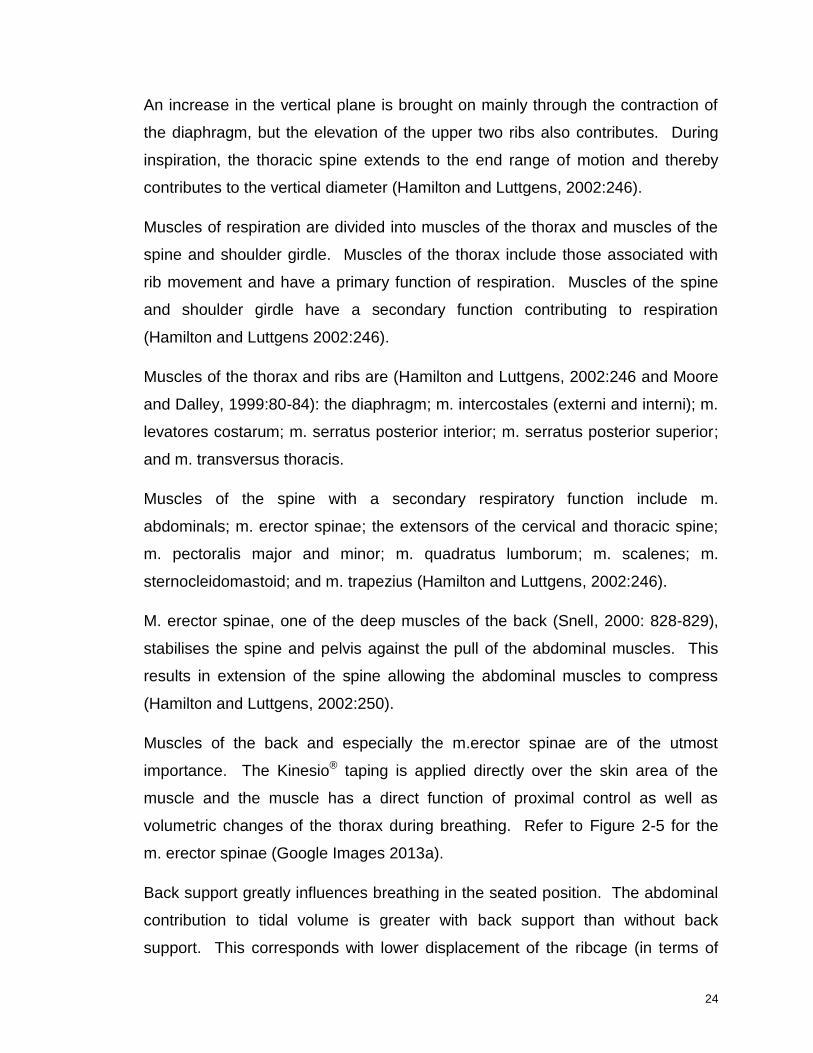
Muscles of the back and especially the m.erector spinae are of the utmost
importance. The Kinesio® taping is applied directly over the skin area of the
muscle and the muscle has a direct function of proximal control as well as
volumetric changes of the thorax during breathing. Refer to Figure 2-5 for the
m. erector spinae (Google Images 2013a).
Back support greatly influences breathing in the seated position. The abdominal
contribution to tidal volume is greater with back support than without back
support. This corresponds with lower displacement of the ribcage (in terms of

25
perimeter, cross-sectional area and volume changes). This is explained by the
effect of tonic contraction of abdominal muscles for postural maintenance and
trunk stabilisation. If back support is removed, abdominal compliance is reduced
and the motion of the abdominal muscles increases. Abdominal muscles as well
as the diaphragm are involved with maintaining posture in the erect position
(Romei, Lo Mauro, D‟Angelo, Turconi, Bresolin, Pedotti and Aliverti,
2010:189-190).
Figure 2-5 M. erector spinae (Google Images, 2013a)
Zubeyir et al. (2012) investigated the effect of elastic taping on primary and
accessory respiratory muscle strength. The researcher compared the inspiratory
muscle strength of 47 subjects. The researchers did not use Kinesio® taping, but
a different brand of elastic taped called Pinotape®. The participants were divided
into two groups: diaphragmatic kinesiology taping and accessory respiratory
muscle kinesiology taping. The researchers found no significant effect on muscle
strength of primary or secondary respiratory muscles in healthy subjects. They
did, however, not test the volumetric measurements (Zubeyir et al.,
2012:242-244).
Kinesio® taping can enhance motor skills, aid stability and improve posture.
Kinesio® Tex can also be used to facilitate movement patterns, align posture and

26
improve function (Kinesio South Africa, 2013) and therefore Kinesio® taping
could have an effect on selected physical symptoms associated with MDD.
2.8.3 Decrease in Flexibility and Centring of Movements
Major depressive disorder has an influence on gait and posture secondary to the
motor retardation that predisposes the patients with MDD to falling. Patients that
suffer from MDD have a lower motor performance than non-depressed patients
and their postural abilities in standing are severely limited compared to their
non-depressed equals (Turcu, Toubin, Mourey, D‟Athis, Manckoundia and
Pfitzenmeyer, 2004:304,306-307). The more severe the MDD episode, the more
impaired the function with regards to motor ability and executive function (Long,
2011).
Decreased flexibility and centring of movements, according to Jacobsen et al.
(2006:296), in patients with MDD are represented clinically as a lack of rotation in
the trunk, a lack of swinging of the arms during gait, reduced coordination of the
arms and a decreased ability to initiate movements from the movement centre
(proximal control).
Back extensors posteriorly and abdominal muscles anteriorly are important to
facilitate trunk and therefore proximal control. Back extensors that surround the
vertebral column provide a flexible support for the trunk (Jaraczewska and Long,
2006:33). The postural tone of the back muscles is important for maintenance of
normal curvature of the vertebral column as the muscles extend from the sacrum
to the skull (Snell, 2000:828). The muscles provide adjustable tension on the
spine allowing the spine to deviate in any direction while maintaining adequate
support. A stable thorax is needed for the abdominal muscles to function
optimally. Excessive kyphotic posture, muscle weakness or muscle imbalance
causes a compression of the ribcage, reducing the volume of the lungs
(Jaraczewska and Long, 2006:33).
Kinesio® taping can be used to improve purposeful movement and provide the
needed stability and alignment (proximal control) to perform a task (Yasukawa,

27
Patel and Sisung, 2006:104). The effect of Kinesio® Tex tape to enhance
stability has been demonstrated during a study at a rehabilitation institute in
Chicago. Fifteen children with diverse neurological damage, admitted as
in-patients, were tested in a pilot study to determine the effects of Kinesio® taping
on upper limb function. The children were pre-tested with the Melbourne
Assessment of Unilateral Upper Limb Function, taped with Kinesio® taping and
re-tested. The results reflected improved post-measurement scores that were
statistically significant and that improved over time with a standard deviation of
23.3 at the follow-up measurement after 3 days. This study had a very small
sample size of 15 participants and the patients were not uniform with either
diagnosis or taping applied, but they presented with the same causes and
functional impairments. The assessment tool was standardized and ideal for
testing upper limb function. No control was used in this study and the
participants received multiple therapies during this time. Although improvement
was seen further research is needed to determine the direct impact the taping
intervention had on the impairments (Yasukawa, Patel and Sisung,
2006:105-109).
This has also been confirmed by a case study at the same institute. A
12-year-old boy‟s right arm function was evaluated with the Melbourne
Assessment of Unilateral Upper Limb Function while he was seated in his
wheelchair with the lateral trunk support removed. During testing he displayed
dystonic movement and overshooting when trying to grasp objects in front of him.
He scored 57 out of the possible 122 (47%) on the scale. The Kinesio® tape was
applied bilaterally to the erector spinae muscle from L5 to T2 as well as the
shoulder and hand to attain the correct alignment and assist with stability and a
functional upright position. Immediately after Kinesio® taping, post-measurement
of the upper limb function improved and he scored 61 out of 122 (50%). Three
days after wearing the Kinesio® Tex he still scored 50% on the Melbourne
Assessment (Yasukawa, Patel and Sisung, 2006:107-108).
28
Kinesio® taping has been used in neurological conditions, for example in the
treatment of subluxation following a stroke. It has an effect on the sensorimotor
system as well as on proprioception (Jaraczewska and Long, 2006:32). The
Berg Balance Scale was used to assess the effect of Kinesio® taping on the
static balance in subjects with multiple sclerosis. The balance of the subjects
improved only in an anterior posterior direction. The reason for this could be that
the taping was applied in such a manner that it primarily worked on the
flexion-extension movement of the ankle joint. Subjects with poorer quality initial
assessments had better outcome from tape application. Kinesio® taping had no
adverse effects but results were specific and axis dependent (Cortesi, Cattanceo
and Jonsdottir, 2011:366,368-369).
Bicici, Karatas and Baltaci (2012) have tested functional ability including dynamic
balance, in basketball players with chronic inversion ankle sprains. Kinesio® Tex
did not limit function as in the case of white rigid athletic tape (Bicici, Karatas and
Baltaci, 2012:164). Kinesio® tape could be used to promote postural alignment
and stability by supporting weak muscles, relaxing overstretched muscles and
reducing pain to promote functional activity (Jaraczewska and Long, 2006:37).
Kinesio® tape applied directly and without any tension could also stimulate
mechanoreceptors and therefore also proprioception (Kase, Wallis and Kase,
2003:36).
The modification of balance and postural control is explained by two theories.
Firstly, it is advocated that due to the mechanical properties of Kinesio® taping a
reflex reaction is exerted on the nervous system. It causes overlapping of
muscle fibres and results in increased muscle activation (sensorimotor effect).
Secondly, the application of tape directly to the skin stimulates the feed forward
mechanism in the body allowing for an increase in proprioception and joint
control (Slupik et al., 2007:650-651).
29
2.9 Conclusion
The association between physical complaints and MDD emphasises the need for
physiotherapy as part of the holistic treatment regime for the depressed patient.
Furthermore Kinesio® taping can facilitate muscles, control joint instability, assist
postural alignment as well as relax overused muscles (Kaya, Zinnuroglu and
Tugeu, 2010:204-205).
Muscle tension and pain, restricted breathing and decreased centring of
movement were successfully treated with Kinesio® taping in the literature as
discussed in this chapter. These symptoms correspond with the physical
symptoms associated with MDD. Therefore Kinesio® taping could have an
influence on selected MDD-associated physical symptoms.
30
Chapter 3 Methodology
3.1 Introduction
This chapter discusses all the elements of the methodology of this study. After a
thorough literature review, the physical symptoms associated with MDD to be
evaluated were selected (refer to 2.8). The aim of the study was refined and
specified. The desired study population, which allowed for data gathering, was
selected. The study intervention was by means of a Kinesio® Tex taping. Data
were collected through standardised questionnaires via closed interviews and
observation. A graphic representation of the study process is depicted in 3.3.
3.2 Aim and objectives the study
The main aim of the study was to investigate the effect of physiotherapeutic
Kinesio® taping on selected physical symptoms associated with MDD.
The specific objectives of the randomised controlled study, within patients
between 18 – 65 years, admitted with major depressive disorder to either a public
or private psychiatric institution in Bloemfontein, was to:
determine pain complaints before, immediately after and 24 hours after
Kinesio® taping application as measured by the Short Form McGill Pain
Questionnaire .
determine pain areas before, immediately after and 24 hours after
Kinesio® taping application as measured by the Short Form McGill Pain
Questionnaire.
determine breathing restricting before, immediately after and 24 hours
after Kinesio® taping application as measured by the Manual Assessment
of Respiratory Motion.
31
determine flexibility and centring of movements before, immediately after
and 24 hours after Kinesio® taping application as measured by the Tinetti
Mobility Test
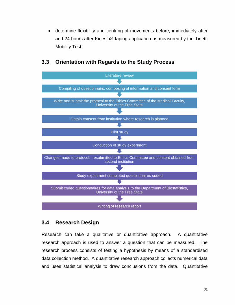
3.3 Orientation with Regards to the Study Process
3.4 Research Design
Research can take a qualitative or quantitative approach. A quantitative
research approach is used to answer a question that can be measured. The
research process consists of testing a hypothesis by means of a standardised
data collection method. A quantitative research approach collects numerical data
and uses statistical analysis to draw conclusions from the data. Quantitative
Writing of research report
Submit coded questionnaires for data analysis to the Department of Biostatistics, University of the Free State
Study experiment completed questionnaires coded
Changes made to protocol, resubmitted to Ethics Committee and consent obtained from second institution
Conduction of study experiment
Pilot study
Obtain consent from institution where research is planned
Write and submit the protocol to the Ethics Committee of the Medical Faculty, University of the Free State
Compiling of questionnairs, composing of information and consent form
Literature review

32
research or an experimental approach confirms or rejects the hypothesis (Leedy
and Ormrod, 2005:94-95).
A qualitative research approach strives to explore and interpret certain research
aspects. The approach is concerned with themes and categories and the
analysis is subjective. The purpose of the study is to describe and understand
the phenomena from the participants‟ point of view (Leedy and Ormrod,
2005:94-96).
According to Baily (1997:43-46) an experimental study design has the following
properties:
Manipulation or intervention. The researchers manipulate one or more
measurable variables.
Control. Control is defined as the elimination of interfering influences that are
not part of the study design.
Randomisation. The process reduces systematic bias by ensuring that the
study participants are representative of the group (random selection) from the
population and ensuring that the placebo control and experimental group
participants are similar (random assignment to the groups).
In a randomised controlled study the participants are allocated to either an
intervention or control group. The control group receives a placebo treatment
which means that the treatment has no known effects but looks similar to the
intervention treatment (Morroni and Myer, 2007:89).
A study is blinded when the participants and researchers do not know to which
group (experimental or placebo) the study participants are allocated to. In
single-blinded studies only the participants are blinded and thus unaware of the
group to which they belong. In double-blinded studies, both the participants and
researchers are blinded. In triple-blinded studies the participants, researchers
and data analysts are blinded (Morroni and Myer, 2007:90).

33
With regards to the abovementioned characteristics, this study followed a
quantitative research approach: numerical data were collected by means of a
standardised procedure (standardised questionnaires) and statistical analysis
was used to draw conclusions from the data. This was a double-blind,
randomised controlled study: the participants were assigned to either an
experimental or control group, and both the researcher administering the
evaluation and the participants were unaware of group assignment throughout
the study. Keeping the taping area covered with a single layer of clothing
ensured further blindness of the said researcher. Refer to Figure 3-1 for the
design of the study (Morroni and Myer, 2007:90).

34
Figure 3-1 Flowchart of study process
Screened for inclusion
Excluded
Included: study participants that fulfil inclusion
critera
Consented to participate
Randomised
Intervention group
Pre-application
Intervention
Post-application
24 hours post-application
Control group
Pre-application
Placebo intervention
Post-application
24 hours post- application
Refusal to participate
35
3.5 Sample and/or Study Participants
Study participants were patients (male and female) with a DSM-5 criterion
diagnosis of MDD admitted to a public psychiatric institution in the affective ward
from January 2012 to December 2013. Sample size was determined by taking
into account that for small populations with less than a 100 elements the whole
population should be sampled (Leedy and Ormrod, 2005:207). The number of
patients admitted to the public psychiatric institution in the Free State from
January 2010 to December 2010, according to the affective ward admissions
register, was 93. Of this 93, only 45 potentially met the proposed inclusion
criteria. Of these, 38 females were enrolled. Therefore it was likely to assume
that if the trend continued a sample size of 40 would be plausible.
To accommodate the inadequate sample size obtained from this institution from
January 2012 to February 2013, three private psychiatric institutions in
Bloemfontein were contacted to be sampled from February 2013 to December
2013. Of the three private institutions only one institution gave permission for
research to be conducted.
The sample was chosen on the availability of appropriate patients admitted to the
ward and integrated in the ward programme. Admission to the ward allows for
the sample to be identical with regards to meal times, sleeping times, distance
from physiotherapy department as well as other interventions part of the hospital
regime. Admission to the ward also ensures minimum fall out of patients from
the study.
At the public psychiatric institution the following interventions were received daily
by participants: Occupational therapy and pharmacological intervention
(medication). Physiotherapy and psychotherapy were on referral basis only. At
the private psychiatric institution all participants in the study received daily
occupational therapy, psychotherapy, physiotherapy and pharmacological
intervention (medication). The therapies received at the two institutions differed
and could not be kept the same. The researcher worked at the public psychiatric

36
institution and could control the physiotherapy received by the participants. The
reason physiotherapy could not be controlled at the private psychiatric institution
was the fact that the service is provided by private physiotherapists and they
could not be expected to discontinue services temporarily on account of the
study . This physiotherapy were compensated for by using only patients referred
from one psychiatrist to one physiotherapy practice where the treatment regime
was kept consistent and included manual therapy, massage, electrotherapy
modalities and dry needling for pain as well as a daily exercise program. The
physiotherapy at the public psychiatric institution included manual therapy,
massage and electrotherapy modalities for pain management.
The Department of Biostatistics at the University of the Free State assisted the
researcher with the sampling method. The process was administered by the
researcher herself. Convenience sampling was used with random assignment of
participants to either an experimental or control group (Polit and Beck,
2006:259). Random assignment to either an experimental or control group was
achieved by using a randomisation list provided by the Department of
Biostatistics, University of the Free State. Each institution sampled had a
separate randomisation list to randomise the participants into either an
experimental or control group. Participants that consented were placed
chronologically and successively on the list.
3.5.1 Eligibility Criteria
3.5.1.1 Inclusion Criteria
Study participants were eligible for inclusion in this study if they met following
criteria:
Male or female
Patients admitted in Bloemfontein to a ward for affective disorders in a public
psychiatric institution from January 2012 to December 2013 or to a private
psychiatric institution from February 2013 to December 2013

37
Diagnosis of MDD
Between the ages of 18 and 65 years (inclusive)
Speak and understand English and Afrikaans
Signed informed consent
3.5.1.2 Exclusion Criteria
Patients presenting any of the following were not included in the study:
Psychosis
Any primary Axis I diagnosis other than MDD
Severe suicidality as determined by ward criteria
Drug addiction
Brain injury or mental retardation
Pregnancy
Medical diagnosis preventing completion of the assessment tool or use of the
intervention method.
Contra-indications for the use of Kinesio® tape. The contra-indications are
(Kinesio® Taping Association (KTA) 2008:56):
malignancy
cellulitis or skin infection
open wounds
deep vein thrombosis
skin irritation after application of a Kinesio® tape test strip
The diagnoses of patients admitted to the institutions were made by the
psychiatry registrar under supervision of a qualified psychiatrist or admitting
psychiatrist. The inclusion and exclusion criteria were applied by the researcher
herself.

38
3.6 Study Interventions
3.6.1 Kinesio® Tape Application
As muscle tension, pain complaints, restricted breathing, and less flexibility and
centring of movements are the physical symptoms most associated with MDD
(Jacobsen et al., 2006:295) these areas were targeted by one general Kinesio®
tape application.
Kinesio® tape was applied from the distal to proximal attachment of the m.
erector spinae from L5 to T2 to facilitate a functional upright position of the trunk,
promote proximal stability and decrease pain and stiffness due to inflammation
(Yasukawa, Patel and Sisung, 2006:108). A bilateral Y-strip application was
used. The patient was placed in a neutral spine position – in standing. The base
application was applied in the sacro-iliac joint region; a minimum of 5 cm below
the area to be taped. During application the patient was asked to move into
lumber flexion with rotation to the opposite side to be taped. The Kinesio® tape
was applied without any tension. The left tail of the Y-strip was laid down first.
The participant was then asked to move into lumber flexion and rotation to the
other side and the right tail of the Y-strip was applied (Kase, Wallis and Kase,
2003:83). Refer to Figure 3-2 for the Kinesio® taping intervention (black coloured
tape used for photographic purposes only).

39
Figure 3-2 Kinesio® taping according to standardised treatment guidelines (Van Vuuren, 2010a)
3.6.2 Placebo Application
The placebo taping consisted of four Kinesio® tape I-strips. The dimensions of
each strip were 5 cm by 2 cm. The patient was placed in a neutral spine
position. The base application consisted of two I-strips and was applied
bilaterally in the sacro-iliac joint region of both sides. During application the
patient was asked to move into lumber flexion with rotation to the right side. One
I-strip was placed on the spina of the left scapula. The participant was then
instructed to move into lumbar flexion and rotation to the left side and the right
I-strip was applied to the spina of the right scapula. This taping method on the
bony areas prevented any interference of the space correction effects of the tape
on pain, breathing and proprioception and no effects were expected (refer to
Figure 3-3, black coloured tape used for photographic purposes only).

40
Figure 3-3 Placebo application – Kinesio® taping not in current guidelines (Van Vuuren, 2010b)
3.7 Measurement
3.7.1 Assessments
Major Depressive Disorder: Physical Symptoms
Physical symptoms most associated with MDD were previously classified by
Jacobsen et al. (2006:295-296) by means of the Body Awareness Scale (BAS)
(Roxendal, 1985:16-22). As described by Ryding, Rudebeck and Roxendal
(2000:176-182), the BAS and Body Awareness Scale – Health (BAS-H) are the
only scales currently available to measure all of the physical symptoms
simultaneously. Unfortunately these standardised scales applicable to psychiatry
are only available in Swedish and still in the process of being translated
(Gyllensten 2011: Personal communication). Therefore to evaluate the selected
physical symptoms of MDD the symptoms were assessed separately using
standardised, valid scales for that specific symptom as used universally in
physiotherapy.

41
The symptoms tested are:
Muscle tension
Pain complaints
Restricted breathing
Decreased flexibility and centring of movements
All questionnaires were completed and filled out by the research assistant
(Appendices A, B and E). No participants completed any questionnaires. All the
questionnaires were verbally translated by the research assistant into Afrikaans if
the study participant requested it. The terms used by the research assistant
when translating from English to Afrikaans during the interviews were agreed
upon beforehand by both the research assistant and researcher. The terms used
were kept consistent throughout the study.
Hamilton Depression Rating Scale
The severity of the current major depressive episode was determined by a score
from the HAM-D (refer to Appendix B for the summary of the total score).
Determining the severity of the current MDD episode was important for two
reasons: Firstly, an increase in the severity of a MDD episode can predispose the
patient to a psychotic episode. Psychosis was part of the exclusion criteria for the
study (Long, 2011). Secondly, severity of MDD is an indication of the functional
impact of the disorder and the extent of the symptoms. The efficacy of treatment
can vary due to symptom severity and therefore the severity of MDD could
influence the results of the outcomes measured (Fourier, Robert, Derubeis,
Hollon, Dimidjian, Amsterdam, Shelton and Fawcett 2010:6).The severity of MDD
episode was tested to give a reference point on interpreting the results.
The scale was developed by Max Hamilton from the University of Leeds and has
endured as the gold standard in clinical practice and research. The complete
scale is included in Appendix F (Carroll, 2005:2395-2396; Hamilton, 1960:56-62).
42
The HAM-D takes 15 to 20 minutes to complete and score. Eight items are
scored on a 5-point scale and nine items are scored from 0 to 2. A mild
depression is characterised by a score ranging from 8 to 13, whilst a score
ranging from 14 to 18 is indicative of moderate depression. Severe depression
can be derived from a score that falls within the range of 19 to 22. A score
greater than 23 indicates very severe depression. A score of 7 and below
indicates the absence of depression or a euthymic mood (Hamilton, 1960:56-52).
Mild MDD is characterized by two or three symptoms but the person can
continue with most activities. During moderate MDD four to five of the tell-tale
signs are present and the individual has a problem continuing with activities of
daily living. During severe MDD several characterizing symptoms are present,
more physical symptoms could be present and severe MDD is linked to psychotic
symptoms and psychomotor retardation (Long, 2011).
Short Form McGill Pain Questionnaire
Muscle tension and pain complaints were assessed using the Short Form McGill
Pain Questionnaire (SF-MPQ) administered through a structured interview. The
complete scale provides five scores: sensory perception of pain, affective
perception of pain, overall scores for the McGill Pain Questionnaire domains,
pain intensity through the visual analogue scale as well as the Present Pain
Intensity. It was developed by Dr Ronald Melzack in 1980 as the McGill Pain
Questionnaire but was too long to use effectively in clinical studies (Melzack,
2005:202). The scale was only used if the participant answers affirmatively to
the question whether he/she has pain. The participant was then asked to fill in a
body chart with regards to pain complaints (refer to Appendix E). Only the
sensory and affective domains of the questionnaire were assessed during this
study (refer to Appendix A).
The SF-MPQ contains 11 sensory and 4 affective dimensions of pain with a
maximum score of 45. The dimensions are assessed by means of an ordinal
scale from 0 (no pain) to 3 (severe pain). There is currently no specific cut-off
value when interpreting the results (Hawker, Mian, Kendzerska and French,
43
2011:S244), but a minimum clinical important change was found to be a
difference in pre- and post-measurement of greater than 5 for an improvement in
symptoms (Strand, Ljunggren, Bogen, Ask and Johnsen, 2008:924).
Manual Assessment of Respiratory Motion
The Manual Assessment of Respiratory Motion (MARM), a manual assessment
technique to evaluate and quantify breathing patterns was used to measure
restricted breathing (Courtney, Van Dixhoorn and Cohen, 2008:93-94). Refer to
Appendix B. The MARM is a graphic representation of the examiner‟s manual
estimations. The MARM procedure used in this study was the following
(Courtney, Van Dixhoorn and Cohen, 2008:93-94):
“Sit behind the subject and place both your hands on the lower lateral rib cage so
that your whole hand rests firmly and comfortably and does not restrict breathing
motion. Your thumbs should be approximately parallel to the spine, pointing
vertically and your hand comfortably open with fingers spread so your hand
comfortably open with fingers that the little finger approaches a horizontal
orientation. Note that the 4th and 5th fingers reach below the lower ribs and can
feel abdominal expansion. You will make and assessment of the extent of
overall vertical motion your hand feels relative to the overall lateral motion. Also
decide if the motion is predominantly upper rib cage, lower rib cage/abdomen or
relatively balanced. Use this information to determine relative distance from the
horizontal line of the upper and lower lines of the MARM diagram. The upper line
will be further from the horizontal and closer to the top if there is more vertical
and upper rib cage motion. The lower line will be further from the horizontal and
closer to the bottom if there is more lateral and lower rib cage/abdomen motion.
Finally get a sense of the overall magnitude and freedom of rib cage motion.
Place lines further apart to represent greater overall motion and closer for less
motion.”
As this is a palpation test performed by the research assistant, the test was
performed over a single layer of clothing without compromising on the integrity of
44
the test. The research assistant was unaware of the randomisation of the study
participants this allowed the study to maintain double blind throughout.
Variables are calculated by measuring the angles between the lines (Courtney,
Van Dixhoorn and Cohen 2008:93).
The MARM measurement variables (Courtney, Van Dixhoorn and Cohen
2008:94):
2. Volume is the angle formed between the upper and lower line.
Balance represents the difference between the upper area and lower area.
Freedom of movement or ribcage movement is the area above the horizontal
line, divided by the total area between the upper and lower line, multiplied by
100.
Normal values of functional breathing determine average ribcage movement to
be around 50 and balance of breathing to be 0. Deviation from the norm can be
interpreted as dysfunctional breathing patterns (Courtney, Van Dixhoorn and
Cohen 2008:97).
Tinetti Mobility Test
Loss of flexibility and centring of movement was evaluated with the Tinetti
Mobility Test (refer to Appendix B). The scale has been developed to evaluate
balance and gait in the adult population (Kegelmeyer, Kloos, Thomas and
Kostyk, 2007:1370). The Tinetti Mobility Test is a functional performance test.
The participants were asked to perform certain functions on command of the
examiner for example standing up from a chair, walking over 3 meters of tiled
flooring and turning. The examiner would observe these actions and then score
them on a scale from 0 to 2. Zero represents the most impairment and 2 the
highest function of the performed activity. Individual scores for gait and balance
are combined to form and overall score out of 28. Scoring below 19 predisposes
a patient for falling and is an indication of a high degree of disturbance in gait.

45
Scores between 19 and 24 indicates a moderate degree of disturbance
(Kegelmeyer et al., 2007:1371,1374).
3.7.2 Reliability and Validity
The HAM-D has an acceptable sensitivity and specificity to determine MDD as an
observer rating scale (Strik, Honig, Lousberg and Denollet, 2001:425). The
HAM-D has a high internal consistency with a Cronbach‟s alpha of 0.83 (Rush,
Trivedi, Ibrahim, Carmody, Arnow, Klein, Markowits, Ninan, Kornstein, Manber,
Thase, Kocsis and Keller, 2003:575).
The SF-MPQ validity and reliability was tested on numerous occasions and its
usefulness proven by translation of the scale into several languages (Melzack,
2005:202). The internal consistency as determined by Cronbach‟s alpha was
0.95 for the sensory, 0.88 for affective, 0.89 for the average and 0.96 for total
pain components (Grafton, Foster and Wright, 2005:77).
The MARM scale is able to distinguish between different breathing patterns and
posture. The MARM has good agreement between examiners and is a valid and
reliable research tool, but future studies in a clinical setting are needed to confirm
the validity and reliability as no internal consistency coefficient is available at
present (Courtney, Van Dixhoorn and Cohen, 2008:98-99; Courtney, Cohen and
Reece, 2009:91).
The Tinetti Mobility Test was tested in 2007 and the study concluded that the
inter-rater and intra-rater reliability, construct validity and criterion validity was
high. The intra-class correlation coefficient (0.87) was greater than 0.80 for all
raters (Kegelmeyer et al., 2007:1373).
3.8 Pilot Study
After approval was obtained from the Ethics Committee of the Faculty of Health
Sciences, University of the Free State, a pilot study was conducted to evaluate
the study procedure and to ensure that all persons involved in the study were

46
aware of their role during the research project. The pilot study also tested the
user-friendliness of the data forms.
The pilot study was conducted during January and February 2012 on three
patients who met the study eligibility criteria and were admitted to the public
psychiatric institution in Bloemfontein. The placebo taping was incorporated in
the pilot study by adding a further four patients during March and April 2012. All
techniques and procedures were tested and the researcher and research
assistant were confident about the execution of the study. Small format changes
were made to the questionnaires to ensure easy usage of the questionnaires.
Coding blocks were checked and changed accordingly. All changes were
communicated to the Ethical Committee (refer to Appendix G). The pilot study
was completed in April 2012. The data of the pilot study were not included in the
main study.
3.9 Data Collection
3.9.1 Data Collection at the Public Psychiatric Institution
The researcher contacted the nursing matron of the ward telephonically
regarding admissions that potentially met the eligibility criteria for the study. All
admitted patients were assessed and diagnosed by their attending psychiatrist
who documented the diagnosis in the patient file. The researcher screened the
patient file to determine eligibility of patients for the study. The researcher
determined the language preference of the potential participant and informed
consent was obtained in either Afrikaans or English. The research participant
then immediately received a test strip of Kinesio® taping on the right anterior
forearm to determine the skin reaction prior to full use (Kase, Wallis and Kase,
2003:12).
The HAM-D was administered by an intern psychologist or ward psychologist
within 48 hours of obtaining consent. After completion of training to administer
the HAM-D in November 2012, the researcher obtained permission from the
Ethics Committee of the Faculty of Health Sciences, University of the Free State,

47
to administer the scale (refer to Appendix G for amendment 4). This helped to
control the logistics of the study. The HAM-D was scored immediately after
consent was obtained. The total score from the HAM-D is summarised in
Appendix B. The HAM-D was not repeated during the study as it was only an
indication of the severity of the major depressive episode during testing.
3.9.2 Data collection setting
Evaluations were performed in the following settings: A well-lit, private area with
enough space to perform the Tinetti Mobility Test (a minimum of 3 metres tiled
floor is needed for the gait evaluation). A chair without armrests was used and
the temperature was regulated by means of an air conditioner to ensure a
comfortable environment. Lighting was sufficient to ensure effective observation.
The sequence and contents of the evaluations were executed without variations.
The research personnel were kept constant. Evaluations were done during the
same time every day for each participant over the 24-hours of the study. During
the 10-minute resting period before tape application, the Kinesio® tape test strip
was removed and the skin examined for any adverse reactions. The pre-, post-
and 24 hours post-application assessments included all three scales as
discussed in 3.7.1.
3.9.3 Data Collection at the Private Psychiatric Institution
The researcher was telephonically contacted on Mondays and Tuesdays
regarding admissions that potentially met the eligibility criteria for the study. . All
admitted patients were assessed and diagnosed by their attending psychiatrist
who documented the diagnosis in the patient file. The researcher screened the
patient file to determine eligibility of patients for the study. A suitable time to visit
the institution, which complied with the ward programme, was scheduled. During
this scheduled time, the researcher visited the institution and obtained informed
consent from participants in either Afrikaans of English (according to their
language preference). The research participants then also immediately received
a test strip with Kinesio® taping on the right anterior forearm to determine the

48
skin reaction prior to full use (Kase, Wallis and Kase, 2003:12). The same
procedure (refer to 3.9.1) was followed for scoring of the HAM-D.
The researcher met the study participants at the institution. The participants
rested for 10 minutes before tape application. During this time the Kinesio® tape
test strip was removed and the skin was examined for any adverse reactions.
3.9.4 Data Collection Procedures for All Institutions
Participants were randomly assigned to either an experimental or control group
according to a randomization list for each institution and instructed not to remove
the test strip until pre-application. The randomization list was provided by the
Department Biostatistics, University of the Free State. Stratified randomisation
was used. The stratification was done per institution to ensure equal distribution
between institutions. The researcher applied the randomisation list to participants
that were eligible for inclusion in the study and that had consented to participate
in the research study. All pre-application was completed within the first 48 hours
after the HAM-D had been administered.
The participants were pre-tested with all three scales before the intervention to
determine a baseline measurement. The assessment was done by the research
assistant, a physiotherapist trained in the assessment tools. The research
assistant remained blinded to the assignment of the participants throughout the
study. The research assistant performing the assessments was unaware of the
group the participant was randomised into as the taped area was covered with a
single layer of clothing throughout the assessments during the study. The data
were captured on data forms (refer to Appendices A, B and E). The experimental
group received an intervention with Kinesio® Tex Gold tape in line with
standardised accepted treatment guidelines. The control group received a
placebo application with Kinesio® tape, which is not in the current treatment
guidelines and of which the effects are not yet known.
Skin preparation took 5 minutes. The skin had to be free of oils and lotions and
was cleaned with an alcohol swab prior to application of the tape. Excess body

49
hair in the treatment area could limit adhesion of the tape and was shaved
beforehand (Kase, Wallis and Kase, 2003:13). The taping method took
approximately 5 minutes and was performed by the researcher. The researcher
was trained in the application method of Kinesio® tape. The participants then
rested an additional 2 minutes to minimise any carry over from the strapping
method before post-application assessments.
The assessment with all three scales was repeated by the research assistant to
obtain a post-application measurement. The participants were instructed not to
remove the tape and the assessments of both groups were repeated 24 hours
after the first assessment to determine the sustained effect of the taping method
(Kase, Wallis and Kase, 2003:16).
All assessments were completed at the same time of the day and scheduled to
accommodate the ward programme. The routine treatment protocol, while
admitted to the institution, continued for both groups during this time.
3.10 Ethical Issues
The following ethical issues as set out by Leedy and Ormrod (2005:101) were
taken into consideration during the execution of this research study.
3.10.1 Protection from Harm
Participants in the study were not exposed to physical or psychological harm.
Risk associated with study participation should not be more than normal risk of
day-to-day living. Kinesio® tape is a well-tolerated, latex free tape that is safe for
paediatric and geriatric populations (Kinesio Taping Association [KTA], 2008:23).
No side effects have been noticed in subjects that participated in previous
Kinesio® taping studies; not during the treatment or follow-up periods (Kaya,
Zinnuroglu and Tugeu, 2010:205). There was no foreseeable discomfort for the
participant. Participants received a test strip of Kinesio® taping on the right
anterior forearm prior to the intervention. The strip was removed and the skin
inspected for any adverse effects before the participants were allowed to

50
continue with the study. The participant‟s skin was also examined after the
intervention for any irritation or irregular effects.
3.10.2 Informed Consent
The informed consent document (Appendix C) contained the most important
information with regards to the study purpose and procedure. This allowed the
study participants to give informed consent for participation in this study. The
required information in the document was determined by the Ethics Committee of
the Faculty of Health Sciences, University of the Free State. The rights of the
study participants were clearly stated and a contact number was provided for the
secretariat of the Ethics Committee.
Study participants that consented to participate in the study were requested to
complete and sign an authorisation document attached to Appendix C. The
researcher obtained written consent from the board of directors of all the
institutions where this research study was conducted (Appendix D). The
researcher‟s contact details were also provided in case of any enquiries.
Participants were informed about the nature of the study, were given a choice
whether or not to participate and were able to withdraw from the study at any
time. In this study the differences in intervention between the experimental and
control groups could not be mentioned as it could influence the results.
Participants were informed that they would receive a physiotherapy assessment
and intervention with Kinesio® tape (refer to Appendix C). Participants were
informed about a possible skin irritation due to the taping and that the skin would
be inspected after the application of a Kinesio® tape test strip and evaluated for
any adverse effects.
3.10.3 Right to Privacy
All participants and results were handled as strictly confidential.
51
3.10.4 Professional Honesty with Colleagues
Results were reported honestly and in a complete fashion. Only completed data
sets were used. Data were not fabricated. Plagiarism and document theft was
avoided at all times – acknowledgement of material belonging to others is
mandatory. Honesty, integrity and respect for the science and the participants
are essential. Strict scientific methods were used during the study. The proposal
was approved by the Ethics Committee of the Faculty of Health Sciences,
University of the Free State. All amendment to the study were approved by the
the Ethics Committee of the Faculty of Health Sciences, University of the Free
State and included, small formatting changes to the questionnaires, approval for
the researcher to perform the HAM-D evaluation and inclusion a new research
area (Refer to Appendix G).
Permission for the study to be performed was obtained from the institutions
where the study was planned. In this research study approval was granted by
one public and one private mental health institution in Bloemfontein. (Refer to
Appendix D).
The treating physician and the physiotherapist treating the participants were
informed of the study and consent was obtained from them. Relevant feedback
was given during the execution of the study. Feedback will be given on request
to the participant after the completion of the study. Written feedback to the
physicians, physiotherapist and institutions concerned with the research project
will be given after compiling the research report.
If the study proves to have a beneficial effect on selected physical symptoms
associated with MDD, the method will be incorporated in the routine
physiotherapy regime at the public psychiatric institution in Bloemfontein.
52
3.11 Coding of Questionnaires
The coding of the questionnaires was the responsibility of the researcher. The
coding was monitored by an objective third party schooled in coding of
questionnaires. In the questionnaires, a numerical value was assigned to the
different options of questions. For example yes = “1” and no = “2”. If “yes” was
marked during the structured interview or observation the value of the answer
was transferred to the coding block.
Coding of open ended questions, for example the body chart, was accomplished
by assigning a number to areas noted on the chart by the different participants in
chronological order. The first area noted was “upper thoracic” and the number 1
was assigned to it. The coding list with regards to different areas is added in
Appendix H.
3.12 Data Analysis
Statistical analyses of the data were performed by the Department of
Biostatistics, University of the Free State. The experimental and control group
consisted of randomised participants from both institutions and were analysed as
such. Results were summarised by frequencies and percentages (categorical
variables) and means and standard deviations or percentiles (numerical
variables). Changes from pre-test to post-test were summarised similarly and
the groups compared using 95% confidence intervals for differences in means,
medians or percentages. Categorical variables were compared using chi-
squared or Fisher exact test (in the case of small numbers) and numerical
variables using t-test (normally distributed variables) and Mann-Whitney test
(skew distribution). Changes within groups were analysed using paired t-tests
(normally distributed variables) or signed rank tests (skew distributions) (Joubert
2011 and 2014: Personal communication).

53
3.13 Measurement and Methodological Errors
The following areas of measurement and methodological errors were identified
and counter actions to prevent the errors are mentioned.
The sample selection method was biased as not all persons in the population
had a chance of being selected (Leedy and Ormrod, 2005:209). Only patients
admitted to psychiatric institutions were selected. This was limited by sampling
both patients from a public psychiatric institution as well as a private psychiatric
institution. If a patient declined participation in the study, the following patient
was considered. The eligibility criteria were diligently applied by the researcher
(refer to 3.5.1). Participants were allocated to treatment using a randomisation
list supplied by the Department of Biostatistics, University of the Free State.
Randomisation was performed separately for the two institutions involved to
ensure identical groups with regards to facility, medication and ward programme.
Physiotherapy intervention could not be controlled at the private mental health
institution, but all patients were referred for physiotherapy to the same practice
as part of the daily programme. All referred patients had contact with a
physiotherapist prior to the assessment and received the same treatment regime.
All other interventions (occupational therapy, psychotherapy and pharmacology)
were part of the treatment regime at the institutions and were recorded by the
researcher. The outcomes measured could have been influenced by the
therapies as differences might have been detected if separate institutions were
compared to each other. The experimental and placebo groups however were
completely balanced with regards to institution and treatments received at these
institutions. This had to be taken into account when interpreting the results.
Severity of the current MDD episode can influence efficacy of the intervention
tested and therefore a baseline score for MDD was assessed before
measurements began to use as reference point on interpreting the results. The
researcher administered the HAM-D from March 2013 as she received training in

54
administering the scale during November 2012. Previously the HAM-D was
administered by a psychologist or intern psychologist.
The assessment of the participants was carried out by a physiotherapist trained
in the assessment procedures. The physiotherapist was unaware of the group
the patient was assigned to, thus ensuring blindness and preventing bias.
Standardised instructions according to the procedure of the scales were given
during the assessment as encouragement as diverse instructions can influence
the results of the test. A qualified and certified Kinesio® tape therapist applied
the Kinesio® tape according to standardised guidelines. Environmental variables
were minimised by assessing and re-testing a participant at the same time of the
day and in the same surroundings.
Reliability was enhanced by the same therapist performing the pre- and post-
application measurements. Observer expectancy was minimised by keeping the
study blinded. The Hawthorne effect was minimised by the use of a placebo
control group.
Kinesio® Tex tape is available in red, blue, black and beige. The red tape is a
darker colour on the light spectrum, thus absorbing more light and can increase
temperature under the Kinesio® tape application. The blue tape is lighter and
thus decreases temperature under the Kinesio® tape application. This plays a
role in colour therapy, but there is no difference in the manufacturing of the tape
except the change in the dye colour required for the different colours. The
Kinesio® Tex brand of Kinesio® tape and the neutral beige colour was used in
this study to prevent colour or brand influencing the results (Kase, Wallis and
Kase, 2003:17).
Skin preparation was done before the application of Kinesio® tape as any
restriction of the adhesive ability of the tape to adhere to the skin could influence
the effectiveness of the application and length of the tape (Kase, Wallis and
Kase, 2003:12).
55
3.14 Conclusion
This chapter describes the research methodology and procedure for data
collection in detail. Measurement and methodological errors that can impede the
validity and reliability of the study is taken into consideration and methods are
suggested to minimise such errors. Ethical considerations with regards to the
study procedure are also explained. The results of this study are presented in
Chapter 4.

56
Chapter 4 Results
4.1 Introduction
In this chapter the results of the study are presented in the form of graphs and
tables. The analysed data are presented in accordance with the research
objectives.
4.2 Disposition of Study Participants
The study was conducted between January 2012 and December 2013 in
Bloemfontein.
Of the 40 participants included this study, 22 (55%) participants were recruited
from a private psychiatric institution and 18 (45%) participants from a public
psychiatric institution.
The participants were randomised into two groups of 20 participants each. In
each group eleven participants were recruited from a private psychiatric
institution and nine participants were recruited from a public psychiatric
institution. The experimental group received Kinesio® taping as described in the
literature (refer to 3.6.1). The placebo group received a placebo taping (refer to
3.6.2).
All enrolled study participants completed the 24-hour study period.
4.3 Results of the Questionnaires
Appendices A and E were concerned with the gathering of demographic
information including gender and age, and the manifestation of the participant‟s
pain. Appendix B collected baseline scores on the HAM-D, MARM and the
Tinetti Mobility Test.
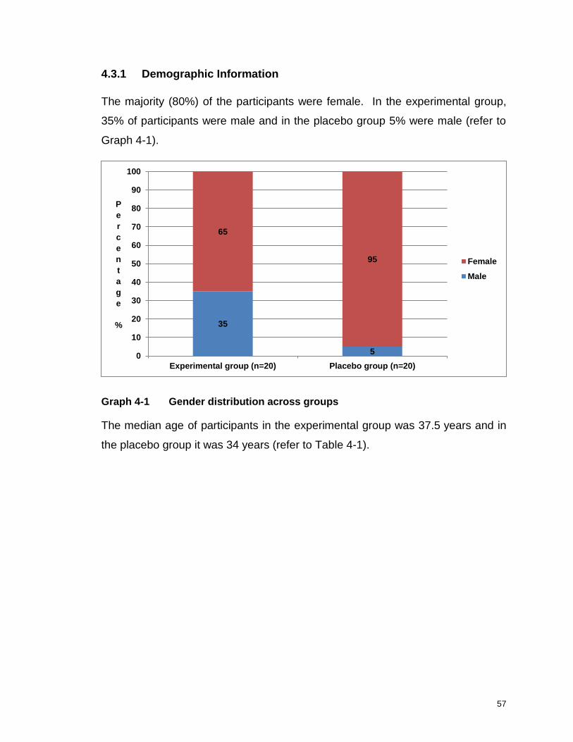
57
4.3.1 Demographic Information
The majority (80%) of the participants were female. In the experimental group,
35% of participants were male and in the placebo group 5% were male (refer to
Graph 4-1).
Graph 4-1 Gender distribution across groups
The median age of participants in the experimental group was 37.5 years and in
the placebo group it was 34 years (refer to Table 4-1).
35
5
65
95
0
10
20
30
40
50
60
70
80
90
100
Experimental group (n=20) Placebo group (n=20)
P
e
r
c
e
n
t
a
g
e
%
Female
Male
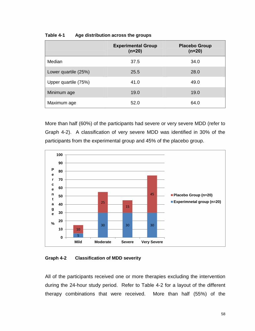
58
Table 4-1 Age distribution across the groups
Experimental Group (n=20)
Placebo Group (n=20)
Median 37.5 34.0
Lower quartile (25%) 25.5 28.0
Upper quartile (75%) 41.0 49.0
Minimum age 19.0 19.0
Maximum age 52.0 64.0
More than half (60%) of the participants had severe or very severe MDD (refer to
Graph 4-2). A classification of very severe MDD was identified in 30% of the
participants from the experimental group and 45% of the placebo group.
Graph 4-2 Classification of MDD severity
All of the participants received one or more therapies excluding the intervention
during the 24-hour study period. Refer to Table 4-2 for a layout of the different
therapy combinations that were received. More than half (55%) of the
5
30 30 30 10
25 15
45
0
10
20
30
40
50
60
70
80
90
100
Mild Moderate Severe Very Severe
P
e
r
c
e
n
t
a
g
e
%
Placebo Group (n=20)
Experimnetal group (n=20)

59
participants received all available therapies: occupational therapy,
psychotherapy, pharmacology and physiotherapy.
Table 4-2 Therapies received during study period
Therapy combination Number and percentage of experimental participants
(n=20)
Number and percentage of
placebo participants
(n=20)
All available therapies:
Occupational therapy
Psychotherapy
Pharmacology
Physiotherapy
11 (55%) 11 (55%)
Occupational therapy
Psychotherapy
Pharmacology
9 (45%) 6 (30%)
Occupational therapy and pharmacology
0 (0%) 2 (10%)
Psychotherapy
Pharmacology
Physiotherapy
0 (0%) 1 (5%)
More than half (57.5%) of the participants received physiotherapy during the
course of the study.
4.3.2 Pain Symptoms
Most (77.5%) of the participants experienced pain. Respectively, 80% of the
participants in the experimental group and 75% of participants in the placebo
group experienced pain during the pre-application phase. In the experimental
group 69% of the participants with pain reported more than one area of pain
during pre-application, compared to the placebo group where 47% of the
participants reported more than one area of pain (refer to Graph 4-3).
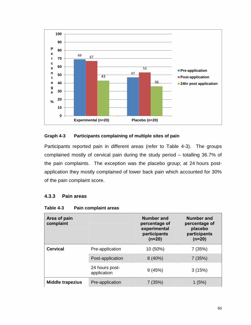
60
Graph 4-3 Participants complaining of multiple sites of pain
Participants reported pain in different areas (refer to Table 4-3). The groups
complained mostly of cervical pain during the study period – totalling 36.7% of
the pain complaints. The exception was the placebo group; at 24 hours post-
application they mostly complained of lower back pain which accounted for 30%
of the pain complaint score.
4.3.3 Pain areas
Table 4-3 Pain complaint areas
Area of pain complaint
Number and percentage of experimental participants
(n=20)
Number and percentage of
placebo participants
(n=20)
Cervical Pre-application 10 (50%) 7 (35%)
Post-application 8 (40%) 7 (35%)
24 hours post-application
9 (45%) 3 (15%)
Middle trapezius Pre-application 7 (35%) 1 (5%)
69
47
67
53
43
36
0
10
20
30
40
50
60
70
80
90
100
Experimental (n=20) Placebo (n=20)
P
e
r
c
e
n
t
a
g
e
%
Pre-application
Post-application
24hr post application
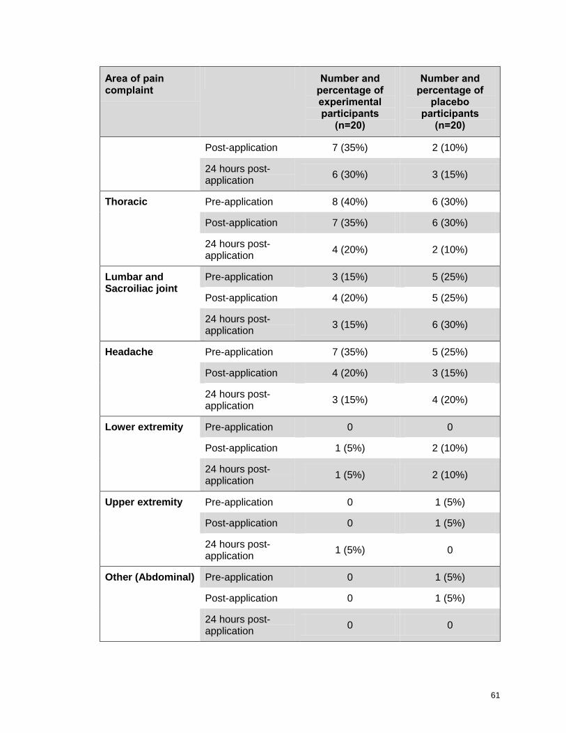
61
Area of pain complaint
Number and percentage of experimental participants
(n=20)
Number and percentage of
placebo participants
(n=20)
Post-application 7 (35%) 2 (10%)
24 hours post-application
6 (30%) 3 (15%)
Thoracic Pre-application 8 (40%) 6 (30%)
Post-application 7 (35%) 6 (30%)
24 hours post-application
4 (20%) 2 (10%)
Lumbar and Sacroiliac joint
Pre-application 3 (15%) 5 (25%)
Post-application 4 (20%) 5 (25%)
24 hours post-application
3 (15%) 6 (30%)
Headache Pre-application 7 (35%) 5 (25%)
Post-application 4 (20%) 3 (15%)
24 hours post-application
3 (15%) 4 (20%)
Lower extremity Pre-application 0 0
Post-application 1 (5%) 2 (10%)
24 hours post-application
1 (5%) 2 (10%)
Upper extremity Pre-application 0 1 (5%)
Post-application 0 1 (5%)
24 hours post-application
1 (5%) 0
Other (Abdominal) Pre-application 0 1 (5%)
Post-application 0 1 (5%)
24 hours post-application
0 0

62
Utilising the SF-MPQ a numerical value was obtained to quantify the participants‟
pain experience.
Table 4-4 Pre-application pain scores
Components of SF-MPQ
Experimental Group (n=20)
Placebo Group (n=20)
Median Lower
Quartile Upper
Quartile Median
Lower Quartile
Upper Quartile
Sensory 11.5 3.0 15.0 10.5 2.0 15.5
Affective 7.5 2.5 8.5 7.5 1.0 12.0
Combined components
19.0 6.5 23.5 21.5 3.0 25.5
The assessment tool evaluated sensory and affective components of pain. The
experimental and placebo groups were closely matched with regards to
pre-application scores (refer to Table 4-4).
The pain scores decreased in a similar pattern for the experimental and placebo
groups at post- and 24 hours post-application (refer to Table 4-5 and Table 4-6).
Table 4-5 Post-application pain scores
Components of SF-MPQ
Experimental Group (n=20)
Placebo Group (n=20)
Median Lower
Quartile Upper
Quartile Median
Lower Quartile
Upper Quartile
Sensory 9.5 2.0 14.5 8.0 0.5 13.5
Affective 5.5 0.5 8.5 4.0 0 11.5
Combined components
15.0 2.5 23.0 12.0 0.5 24.0
There is a change from pre- to post-application within the groups with a p-value
of 0.08 for the combined components of both the experimental and placebo
groups.

63
Table 4-6 24 hours post-application pain scores
Components of SF-MPQ
Experimental Group (n=20)
Placebo Group (n=20)
Median Lower
Quartile Upper
Quartile Median
Lower Quartile
Upper Quartile
Sensory 4.0 0 8.0 4.5 0 8.0
Affective 2.0 0 5.5 1.5 0 6.5
Combined components
5.5 0 14.5 7.0 0 16.0
From pre- to 24 hours post-application, the groups showed significant changes
within the groups with a p-value of <0.01 for the combined components. The
difference between the two groups for change over time was not significant with
p-values of 0.85 and 0.75 for the experimental and placebo groups, respectively.
Table 4-7 Differences between pre-, post-, and 24 hours post-application of the combined components of SF-MPQ
Experimental Group (n=20)
Placebo Group (n=20)
Median Lower
Quartile Upper
Quartile Median
Lower Quartile
Upper Quartile
Pre- vs. post-application
0 -3.5 0 0 -0.5 0
Pre- vs. 24 hours post-application
-5.5 -19.0 0 -8.0 -15.5 -1.5
Table 4-8 Percentage of participants experiencing pain
Time point Experimental Group
(n=20) Placebo Group
(n=20)
Pre-application 80% 75%
Post-application 75% 75%
24 hours post-application 70% 70%

64
In the experimental group, 30% of the participants showed improvement in the
sensory component of pain between pre- and post-application compared to 25%
in the placebo group, with a p-value of 0.87 (refer to Table 4-9). Only 15% of the
participants in the experimental group showed a clinical significant change at
post-application compared to 20% of the placebo group (refer to Table 4-10).
Half (50%) of the participants in the experimental group showed a clinical
significant change of the combined components of the SF-MPQ at the 24 hours
post-application compared to 70% of the placebo group (refer to Table 4-10).
Table 4-9 Percentage improvement of SF-MPQ
Components of SF-MPQ
Post-application 24 hours post-application
Experimental (n=20)
Placebo (n=20)
Experimental (n=20)
Placebo (n=20)
Sensory 30 25 60 75
Affective 10 15 55 75
Combined components 30 25 65 75
Table 4-10 Percentage clinical significant improvement of SF-MPQ
Components of SF-MPQ
Post-application 24 hours post-application
Experimental (n=20)
Placebo (n=20)
Experimental (n=20)
Placebo (n=20)
Sensory 10 20 45 55
Affective 5 15 55 25
Combined components 15 20 50 70
There were no statistically significant differences between the combined
components of pre- and post-application scores with p-values of 0.85 and 0.75
for pre- to post-application and from post- to 24 hours post-application,
respectively.

65
4.3.4 Manual Assessment of Respiratory Motion
The MARM is a visual representation to quantify dysfunctional breathing. Two
variables are presented: percentage ribcage movement and balance of breathing
(refer to Chapter 3.7.1). The maximum balance score obtained by the
participants was 101 and the minimum 0. The maximum percentage of ribcage
motion achieved by the participants was 61.2% and the minimum was 25%. The
experimental and the placebo group displayed slight improvement of balance of
breathing at immediate post-application with a slight decrease in scores at
24 hours post-application (refer to Table 4-11).
Table 4-11 The balance of breathing score of the MARM
Time point
Experimental Group (n=20)
Placebo Group (n=20)
Median Lower
Quartile Upper
Quartile Median
Lower Quartile
Upper Quartile
Pre-application 13.5 9.5 18.0 10.0 4.5 24
Post-application
6.5 4.0 10.0 7.5 4.0 12.5
24 hours post-application
8.5 3.5 14.0 10.0 6.0 18.0
Within the experimental group the changes were significant for all phases of
testing with p-values of <0.01 from pre- to post-application and 0.01 from
pre-application to 24 hours post-application. In the placebo group the changes
from pre- to post-application were significant (p-value of 0.01) but the changes
from pre-application to 24 hours post-application were not statistically significant
(p-value of 0.95).
There was no significant change over time between the two groups with p-values
of 0.38 from pre- to post-application and 0.20 from pre- to 24 hours post-
application.

66
The percentage of ribcage movement improved slightly for both groups at
immediate post-application. The improvement was maintained at 24 hours post-
application (refer to Table 4-12).
Table 4-12 The percentage of ribcage movement score of the MARM
Time point
Experimental Group (n=20)
Placebo Group (n=20)
Median Lower
Quartile Upper
Quartile Median
Lower Quartile
Upper Quartile
Pre-application 39.35 34.74 44.24 42.97 33.33 49.05
Post-application
45.66 41.92 47.44 45.49 39.94 50.51
24 hours post-application
46.38 42.75 49.70 44.68 38.89 51.38
There was no difference in percentage of ribcage movement between the
experimental and placebo groups. The p-value for pre- to post-application was
0.57 and 0.53 for pre-application to 24 hours post-application.
4.3.5 The Tinetti Mobility Test
Throughout the study period, 90% of the study participants scored above 25 on
the Tinetti Mobility Test: Balance and Gait (refer to Table 4-13). A score of
above 25 indicates no dysfunction with regards to balance and gait disturbances
(refer to 3.7).
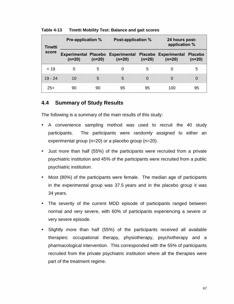
67
Table 4-13 Tinetti Mobility Test: Balance and gait scores
Tinetti score
Pre-application % Post-application % 24 hours post-application %
Experimental (n=20)
Placebo (n=20)
Experimental (n=20)
Placebo (n=20)
Experimental (n=20)
Placebo (n=20)
< 19 0 5 0 5 0 5
19 - 24 10 5 5 0 0 0
25+ 90 90 95 95 100 95
4.4 Summary of Study Results
The following is a summary of the main results of this study:
A convenience sampling method was used to recruit the 40 study
participants. The participants were randomly assigned to either an
experimental group (n=20) or a placebo group (n=20).
Just more than half (55%) of the participants were recruited from a private
psychiatric institution and 45% of the participants were recruited from a public
psychiatric institution.
Most (80%) of the participants were female. The median age of participants
in the experimental group was 37.5 years and in the placebo group it was
34 years.
The severity of the current MDD episode of participants ranged between
normal and very severe, with 60% of participants experiencing a severe or
very severe episode.
Slightly more than half (55%) of the participants received all available
therapies: occupational therapy, physiotherapy, psychotherapy and a
pharmacological intervention. This corresponded with the 55% of participants
recruited from the private psychiatric institution where all the therapies were
part of the treatment regime.
68
The majority (77.5%) of participants complained of pain during the study
period and 58% complained of multiple areas of pain.
Participants complained mostly of cervical pain (36.7%) except for the
placebo group at 24 hours post-application who complained generally of
lower back pain.
The sensory and affective components associated with pain and tested by the
SF-MPQ showed improvement in combined scores for both the experimental
and placebo groups. The affective domain tested the emotional component
of pain and the results indicated a clinically significant effect on this domain.
The results of MARM displayed improvement in both the placebo and
experimental groups for balance of breathing and percentage ribcage motion.
The Tinetti Mobility Test showed no distinct results due to the scale not being
sensitive enough for the movement disorders tested.
4.5 Conclusion
The results from the questionnaires, used as data collection method, are
presented in this chapter. In Chapter 5 the results are discussed in detail and
compared to the available literature.

69
Chapter 5 Discussion
5.1 Introduction
The aim of this study was to determine whether Kinesio® taping had an effect on
selected physical symptoms associated with MDD. This was a randomised
controlled study and followed and experimental study design.
In this chapter the results of this study are discussed. Possible explanations for
the results are given and the results are related to literature and similar studies.
5.2 Discussion of Results
5.2.1 Demographic Information
Just more than half (55%) of the participants were recruited from a private
psychiatric institution and 45% of the participants were recruited from a public
psychiatric institution. Although the ratio was very closely matched, a possible
reason for a higher recruitment from the private institution was that complex
medication-resistant cases were admitted to the public psychiatric institution
which resulted in a high number of participants at this institution not meeting the
study eligibility criteria. Patients diagnosed with a mood disorder have a higher
risk for additional Axis I disorders, such as alcohol abuse or dependence, panic
disorder, obsessive compulsive disorder and social anxiety disorder (Sadock and
Sadock, 2007:529). An additional Axis I diagnosis was part of the exclusion
criteria for participation in this study.
The majority (80%) of the participants were female. This result corresponds with
that of Sadock and Sadock (2007:529), who stated that regardless of country or
culture, a general tendency exists of an almost two-fold greater prevalence of
MDD in woman than in men. The reasons hypothesised were the hormonal
differences between men and women, effects of childbirth, conflicting
psycho-social stressors between the genders and behavioural models of learned

70
helplessness in woman. Another opinion on the gender difference was that it
seems that women were more likely to seek treatment and therefore more
women will be sampled from a psychiatric treatment institution (Scheibe,
Preuschof, Cristi and Bagby, 2003:231).
The unequal ratio between male and female participants admitted to the
institutions for MDD did; however, not influence the treatment, course of the
illness, age of onset, number of episodes or duration of current episode (Scheibe
et al., 2003:231). The gender of the participants therefore had no noteworthy
effect on the results of this study.
The median age of participants in the experimental group was 37.5 years and for
participants in the placebo group it was 34 years. The ages were matched with
those available in literature which stated that the mean age of onset for MDD was
40 years, with 50% of all patients having their first episode between 20 and
50 years of age (Sadock and Sadock, 2007:529).
With regards to the classification of MDD, 60% of participants were diagnosed
with having a severe or very severe episode of MDD, 35% of participants had
mild or moderate classifications of the current episode and 5% of participants‟
classification of MDD was seen as normal. The efficacy of treatment in MDD can
fluctuate according to the MDD severity. The outcomes between experimental
and control can be minimal and clinically insignificant if the participants scores
below 25 on the HAMD during a clinical trial (Fourier, Robert, Derubeis, Hollon,
Dimidjian, Amsterdam, Shelton and Fawcett 2010:6).
The difference in severity of the MDD scores could have been affected by the
length of stay in the hospital. The most appropriate time for the researcher to
recruit the participants was on a Tuesday evening, thus, some of the participants
might already have been admitted during the previous week. The effects of
longer hospitalisation as well as psychotherapy, physiotherapy and occupational
therapy could be more noticeable and have some bearing on the severity of the
current episode of MDD as tested by the researcher.
71
All the participants received one or more therapies, excluding the study
intervention, during the 24-hour study period, with 55% of participants receiving
all available therapies. This corresponded with the percentage of participants
recruited from the private psychiatric institution as all therapies formed part of the
treatment protocol at this institution. The researcher was working at the public
psychiatric institution at the time of the study and could control the physiotherapy
intervention there during the study period. At the public psychiatric institution,
occupational therapy and pharmacology were routinely part of the treatment
protocol whereas psychotherapy and physiotherapy were based on referral, thus,
accounting for the differences in therapies received. The experimental and
placebo groups were completely balanced with regards to institution (in each
group 11 participants were recruited from a private psychiatric institution and 9
participants were recruited from a public psychiatric institution) and treatments
received at these institutions.
5.2.2 Pain Symptoms
Most (77.5%) of the participants experienced pain during the study period. This
correlated with literature, as seen in a review by Bair, Robinson, Katon and
Kroenke (2003:2434), where the prevalence of pain symptoms in patients with
MDD ranged from 15% to 100% with a mean of 65%. Most of these studies were
uncontrolled and conducted in psychiatric settings but according to the authors
the prevalence rates of pain symptoms were independent of the setting (Bair
et al., 2003:2434).
5.2.3 Pain Areas
Patients diagnosed with MDD have a 50% chance of experiencing chronic neck
and lower back pain (Carroll, Cassidy and Cote, 2004:137). This was confirmed
by the results of this study as neck pain was the pain complaint area with the
highest overall prevalence of 36.7%. Lower back pain had an overall prevalence
of 21.6%, ranking third on the overall pain prevalence list. The results of this
study differed slightly from a study conducted by George, Coronado, Beneciuk,

72
Valencia, Werneke and Hart (2011:368). They concluded that the prevalence of
cervical and lower back pain would be exactly the same for patients suffering
from severe MDD. George et al. (2011:361-369) did, however, not take into
account variations between regions.
Their anatomical regions were combined into four groups: cervical, lumbar, upper
extremity and lower extremity. The current study took all pain complaints into
consideration and the following anatomical regions were identified: cervical,
thoracic and lumbar area, shoulder area, upper extremity, lower extremity and
other (abdominal). This difference in grouping of anatomical regions could
account for the variations in data.
The relationship between pain and MDD was confirmed by a number of studies.
Carroll, Cassidy and Cote (2004:138) found that MDD was a strong precursor for
a troublesome episode of neck or back pain. Currie and Wang (2004:57) found
that 19.8% of the Canadian population that complained of lower back pain had
depression. This study confirmed a link between lower back pain (21.6%) and
neck pain (36.7%) and MDD with a co-morbidity of greater than 20%.
At 24 hours post-application, lower back pain in the placebo group accounted for
30% of the pain area complaints compared to 15% in the experimental group.
The percentage pain complaints for lower back pain in the placebo group had
increased with 5%. All the other pain complaint areas had decreased from
post-application to 24 hours post-application. This could possibly have been due
to the fact that the placebo group received a placebo taping and not the full
taping over the lumber area, causing an increase in the percentage of lower back
pain complaints. The fact that the experimental group did not show this increase
in lower back pain at 24 hours post-application might be due to the role that
Kinesio® taping played in pain prevention (Kumbrink, 2012:6).
The Kinesio® taping method consisted of taping over and around a muscle to
effect changes in pain, muscle contraction and lymph drainage (Kinesio South
Africa, 2013). The results of this study showed that neck pain scored
73
consistently higher as a prominent pain area. This result could be attributed to
the fact that only one general taping was applied over the erector spinae muscle
in the lumbar and thoracic area. As no taping was applied over the cervical or
shoulder area this could explain why the neck pain symptoms did not decrease.
5.2.4 Post-Application Results
While interpreting the results of the questionnaires, the results of post-application
of all the evaluation tools showed little improvement from the baseline scores.
This correlated with the study by Slupik et al. (2007:650-651) who found that
Kinesio® taping did not have an immediate effect on muscle activation, as
previously believed, but that the effects of the taping reached its full potential only
24 hours after the application of the tape.
Secondly, the effect of a placebo response could not be ignored in this study.
The placebo effect/response was an improvement resulting from a number of
non-specific factors, for example, patients‟ and clinicians‟ beliefs, expectations,
past experiences and the clinical setting during a study, and was not just a
psychological response to treatment. Previously the placebo effect had been
seen as the improvement that could be measured in subjects in the control
group, but the placebo effect may or may not have been generated by a placebo
treatment (Finniss, Kaptchuk, Miller and Benedeti, 2010:686-687).
In the psychiatric setting, the response to a placebo treatment and therefore the
influence of the placebo effect increased from 20% in the 1980s to the present
rate of 45% and 48% in studies performed on MDD (Pretorius, Van der Merwe
and Westmore, 2010:70). Patients with MDD involved in randomised controlled
studies have been displaying an increased receptiveness to these non-specific
factors. The reason for this is not yet clearly understood but the placebo effect
caused improvement in symptoms of the placebo control group and could
influence the conclusions drawn from these studies. Currently, it is difficult to
distinguish between the outcomes of the experimental and placebo treatment
when researching in the field of MDD (Pretorius, Van der Merwe and Westmore,

74
2010:70). This corresponded with the results of this study where the
experimental and placebo groups had both improved on post-application.
5.2.5 Results of the Pain Score
The SF-MPQ recorded improvement in pain symptoms throughout the study
period. The experimental and placebo group showed the same ratio of
improvement and the results were not statistically significant (p-values of
experimental group were 0.85 and for the placebo group 0.75). The value to
determine whether the results were statistically significant was determined by the
Department of Biostatistics, University of the Free State, and was defined as a
change between groups not due to chance (Leedy and Ormrod, 2005:271).
A clinical significant change is seen as the scale‟s responsiveness to change and
has been found for the Norwegian SF-MPQ to be a mean improvement of 5 or
more on the 45-item scale (Strand et al., 2008:924; Hawker et al., 2011:
S243-S244).
The combined SF-MPQ score was determined by adding the sensory and
affective domains of the scale. The sensory and affective domain improved at
post-application and 24 hours post-application for both the experimental and
placebo groups (refer to Table 4-5 to Table 4-7). A clinical significant change in
the SF-MPQ was observed in both groups and throughout the study.
The results of this study regarding changes in pain symptoms correlated with the
results of Kaya, Zinnuroglu and Tugeu (2010:205) who had found no difference
in pain symptoms or improvement of disability in shoulder impingement when a
physiotherapy intervention group and Kinesio® taping group were compared. In
contrast with this, in a study by González-Iglesias et al. (2009:517-519), the
patients had statistically significant improvements in pain and range of motion,
immediately and after 24 hours, but the improvements were small and not
clinically meaningful. The 51 patients had been randomised into a experimental
and control group to test the short term effects of cervical Kinesio® taping on

75
cervical pain and range of motion after a whiplash injury (González-Iglesias et al.,
2009:515-521).
The results of this study with regards to improvement in pain symptoms could
further be explained by considering the effects of hospitalisation, medication,
other therapies and the fact that pain symptoms decrease with a decrease in
MDD symptoms (Wideman et al., 2012).
In participants complaining of multiple sites of pain, the sites of pain only
decreased in the experimental group from 69% (pre-test) to 43% at 24 hours
post-application compared to the placebo group where pain was reported to
decrease from 47% (pre-test) to 36% at 24 hours post-application. The reason
for this could be that only one standard taping was used in the experimental
group (over the lumbar and thoracic area) while most complaints were cervical
and over the middle fibres of m. trapezius (refer to Table 4-3).
Another reason for the multiple sites of pain could be the biological link between
pain and depression. Limbic structures, involved with emotions, have a role in
interpreting peripheral stimuli. Under normal conditions the limbic system has a
controlling effect and can decrease the intensity of painful signals. In MDD, there
is a shortage of neurotransmitters, especially serotonin and norepinephrine. This
shortage of neurotransmitters can cause the limbic system to lose the ability to
decrease the signal strength. More pain is experienced and multiple sites are
involved (Bair et al., 2003:2441).
5.2.6 Results of the restricted breathing score
The MARM scores for both the experimental and placebo groups improved.
The changes over time for the experimental and placebo group were not
statistically significant with p-values of 0.38 (experimental) and 0.20 (placebo)
from pre-application to 24 hours post-application. The results of this study were
confirmed by the outcomes of Zubeyir et al. (2012:243-244) who had found that
Kinesio® taping has no significant effect on maximum primary and accessory
respiratory muscle strength when the taping was applied over the primary and

76
accessory respiratory muscles of healthy individuals. The study did; however,
not evaluate the effect of Kinesio® taping on spirometric or volumetric
measurements (Zubeyir et al., 2012: 244). In this study it was found that
Kinesio® taping did not have a significant effect on volumetric changes with
regards to balance and percentage ribcage motion between the experimental
and placebo group (refer to Table 4-11).
The experimental and the placebo group displayed slight improvement of
balance of breathing at post-application (refer to Table 4-11) and could be
attributed to the stimulation of the motor units by the taping. The taping resulted
in an increase in muscle tone (Slupik et al., 2007:650) of the erector spinae
muscles resulting in a more erect posture and therefore more ribcage motion and
increase balance of breathing.
At 24 hours post-application, both groups had a slight decline in scores for
balance of breathing. The decrease in scores after 24 hours corresponded with
the results published by Szczegielniak, Krajczy, Bogacz, Łuniewski and Śliwiński
(2007:337-341). The effect of Kinesio® taping on lung volume was investigated
by Szczegielniak et al. in 2007 who had found an increase in spirometric values
24 hours after application. The value of the parameters, however, slightly
decreased the following day and no reason was given for this factor.
Another possible reason for the decrease in balance of breathing scores at
24 hours post-application could be that the effectiveness of the tape might have a
shorter timeframe than previously anticipated (Slupik et al., 2007:651), although it
was proposed that the Kinesio® Tex tape could be worn for 3 to 5 days before the
elastic polymer diminishes and the tape stops working (Kase, Wallis and Kase,
2003:12).
The slight decline in percentage ribcage motion scores at 24 hours
post-application of the participants in the placebo group (refer to Table 4-11)
could be accounted for by the lack of taping over the ribcage with the placebo
taping and thus no continuous activation of the muscles in that area.

77
5.2.7 Results of the loss of flexibility and centring of movement score
The Tinetti Mobility Test scores for balance and gait were consistently above 25
for 90% of the participants overall and for 95% of both groups in the
post-application phase. This translated in only one of the study participants
being pre-disposed to fall and thus no disturbances in balance or gait were
observed. Therefore no loss of flexibility or centring of movement was
documented.
This did not compare with literature which found that more than 30% of patients
diagnosed with MDD had motor disturbances (Caligiuri and Ellwanger, 2000:91).
A possible explanation for the difference in the results of this study could be that
the Tinetti Mobility Test was not sensitive enough to pick up slight changes.
Caligiuri and Ellwanger (200:85-89) have emphasised that instrument sensitivity
was essential to pick up abnormalities in motor performance. In their study there
had been significant differences between the observed score and using a
platform instrument which is sensitive for motor function. Only 22% of patients
presented with motor disturbances, as diagnosed by means of observation,
compared to 60% diagnosed by means of instrumental measurements (Caligiuri
and Ellwanger, 2000:89).
Although the sensitivity of the test used in this study was not optimal, the results
compared with literature where the functional performance of basketball players
was tested (Bicici, Karatas and Baltaci, 2012:162-163). The athletes had been
taped with athletic tape and Kinesio® taping. Results showed no difference
between the two tapings or in the performance of the placebo group when doing
a balance test (hopping and single limb stance). Bicici, Karatas and Baltaci
(2012) had found that Kinesio® taping did not cause any decrease in
performance on any of the functional tests. The taping however was not applied
in a similar manner (ankle taping) as in this study but the proposed mechanism of
Kinesio® taping remains the same regardless of the area taped. This was
confirmed by the current study as no decrease in performance was noted during
the assessment of the Tinetti Mobility Test. Bicici, Karatas and Baltaci (2012)

78
had also found that dynamic balance did not change with either tapings whilst the
static balance scores were significantly better in the Kinesio® Tex group.
This study used the Tinetti Mobility Test as a functional test which tested
dynamic balance and functional activities, for example, gait. It was proposed that
Kinesio® taping can influence proprioception and therefore balance by activating
the mechanoreceptors (Kumbrink 2012:7). It therefore seemed that the Tinetti
Mobility Test was not sensitive enough to pick up the difference between the
taping and placebo application.
5.3 Conclusion
In this chapter the results of this study are discussed in detail relating to the
available research on the subject. The results are interpreted with regard to the
clinical profile of the patients and the mechanisms of Kinesio® taping. The
limitations of the study and recommendations for future studies are discussed in
Chapter 6.

79
Chapter 6 Conclusion and
Recommendations
6.1 Introduction
In this final chapter of the dissertation conclusions are drawn from the results
found in this study regarding the physiotherapeutic effects of Kinesio® taping on
selected physical symptoms associated with MDD. The limitations of this study
are identified and recommendations for future research made. Finally the value
of the study is emphasised.
6.2 Conclusions
Kinesio® taping is used in a variety of clinical and therapeutic settings as well as
in many different kinds of sports, for example, soccer, rugby, gymnastics, skiing,
handball, volleyball and biathlon. Furthermore, it is used as after-care in
orthopaedics, neurology, gynaecology, surgery, oncology, geriatrics and
paediatrics. The concept of Kinesio® taping is also being introduced into
hospitals and rehabilitation centres (Kumbrink, 2012:6). The use of Kinesio®
taping in psychiatry and especially in the treatment of the physical symptoms
associated with MDD has not yet been researched and the parameters of use
are still not clearly defined. The taping methods and current applications are,
however, expanding daily into new areas.
Participants diagnosed with MDD were enrolled from both public (55%) and
private (45%) psychiatric institutions, where they received a variety of therapies
to treat the disorder (refer to 4.2).
The development and progression of the disorder in the participants in this study
were in line with the available literature, with more females (80%) being enrolled
(refer to Graph 4-1) and participants mostly being above 30 years of age (refer to
Table 4-1).
80
Pain is one of the main physical complaints of MDD and in this study 77.5% of
participants experienced pain. The severity and number of pain areas increase
during an episode of MDD (refer to 2.8.1 and 5.2.2). The severity of the current
MDD episode also correlated with physical symptoms experienced by the
participants. In this study, 60% of participants experienced a severe episode of
MDD and 58% reported multiple areas of pain.
Physiotherapists are especially well equipped to treat the physical complaints
associated with MDD and a variety of treatment modalities are available.
Kinesio® taping can be utilised in the psychiatric setting for some of the
MDD-associated physical symptoms. It should; however, be used for the
appropriate condition and the therapist should have proper training to maximise
the outcome of this specialised treatment technique (Kumbrink, 2012:6) (refer to).
Participants in this study complained of multiple pain areas which, in the
experimental group decreased from 69% (pre-test) to 43% (24 hours post-
application) compared to the placebo group where pain deceased from 47%
(pre-test) to 36% (24 hours post-application). Therefore it seems that Kinesio®
taping can only influence underlying tissue and that the taping has to be applied
over or around the affected area to ensure effectiveness (refer to 5.2.2).
In this study, Kinesio® taping did not seem to have an immediate effect on
muscle activation, as previously believed, as greater results were achieved only
24 hours after the intervention (refer to Table 4-11 and Table 4-12 and 5.2.7).
Motor disturbances, a decrease in flexibility and centring of movement is not a
forthright physical symptom in MDD as previously believed and was not easily
observed. The Tinetti Mobility Test could not distinguish any difference between
the two groups during the study period (refer to 4.3.5 and 5.2.8). From the
results of this study it seems that Kinesio® taping could be used as adjuvant
treatment modality to complement physiotherapy modalities currently used in this
field of practice when treating pain, restricted breathing, decrease in flexibility
and loss of centring of movement in patients diagnosed with MDD.

81
6.3 Limitations of this Study
1. The admission date of the participants was not taken into account, and the
time lapse between admission and the first evaluation by the researcher could
have influenced the results. This can also account for lower MDD scores at
baseline.
2. The sample size was small and participants were only recruited from two
psychiatric institutions in the Free State.
3. The sample was obtained only from one referring practice at the private
institution which might not be representative of the bigger population admitted
at the private hospitals.
4. Recruitment bias could have factored in due to the convenience sampling
method used. This might have had an effect on the placebo response.5. It
was not possible for the sample to be blind with regards to depression
(Jacobsen et al., 2006:296), due to the severity of the disease and the fact
that patients diagnosed with MDD formed part of the inclusion criteria.
5. The Tinetti Mobility Test was not sensitive enough to evaluate the decrease in
flexibility and centring of movements.
6. Of the 40 participants, 21 received physiotherapy. This could have influenced
the outcomes measured after 24 hours. The physiotherapy could have had a
positive effect on the outcomes measured accounting for a greater
improvement of the groups due to supersession of Kinesio® Taping and other
physiotherapy modalities. This has to be taken into consideration when
interpreting the results.
7. Other therapies received by participants during the study were not
standarised and could not be controlled due to the multitude of stakeholders
involved in the care of the patients. It is a limitation that the public and private
hospitals could not be analysed separately (with regards to therapies
received) and compared.

82
6.4 Recommendations
1. Patients admitted to psychiatric intuitions should be referred to physiotherapy
for screening and evaluation as 77.5% of participants in this study complained
of pain. MDD can only fully go into remission if all the symptoms (including
physical symptoms) are adequately addressed (refer to 2.8).
2. This study could be repeated with a changed area of taping. The taping could
be applied over the area of pain complaints and this could determine the
effect of Kinesio® taping on pain management in patients with MDD. The use
of a more sensitive balance and gait scale could help draw more accurate
conclusions on the loss of flexibility and centring of movement experienced by
patients diagnosed with MDD.
3. Follow-up studies are needed to determine the effective response time and
most appropriate taping method for Kinesio® taping in the psychiatric setting.
4. Repeating the current study employing the BAS-H (Ryding, Rudebeck and
Roxendal, 2000) once it is translated into English could be of value to
compare the results with that of previous studies with the BAS-H (refer to
3.7.1).
5. Research should be undertaken comparing results between groups where all
other therapies received by the participants are kept constant with only
Kinesio® taping as variable as well as comparing a group where no
physiotherapy is received with a group where Kinesio® taping is part of a
physiotherapy intervention.
6. Further research is needed to determine which physiotherapy interventions
will be most effective in treating the selected physical symptoms associated
with MDD.
83
6.5 Value of the Study
This study gathered valuable information on selected MDD-associated physical
symptoms and the effect of Kinesio® taping on these symptoms. A new avenue
for research was identified in this study as Kinesio® taping is a relatively new
treatment technique not previously associated with psychiatry and specifically
with MDD. The holistic treatment of MDD will ease the burden of the disease by
a decrease in the length of hospital stay and recurrent admissions. This study
also emphasises that physiotherapy is an essential part of the treatment regime
for patients suffering from MDD as the physical symptoms of MDD are often
ignored.
However, Kinesio® taping has to be applied according to the most effective
taping method for the symptoms at hand and given enough time to possibly have
the desired effect. It also seems that the taping should not be used in insolation.
Treatment modalities are seldom used in isolation and Kinesio® taping could be
of value as auxiliary treatment modality applied by physiotherapists as part of the
multi-professional team approach necessary to manage MDD effectively.
Physiotherapists can contribute their extensive knowledge on physical
symptoms, such as pain, to treat these patients and ensure quality care and
effective holistic management of patients suffering from MDD.
6.6 Final Summary
The aim of the study was to determine the effectiveness of physiotherapeutic
Kinesio® taping on selected physical symptoms associated with MDD. The
treatment of these physical symptoms with Kinesio® taping had mixed results, but
Kinesio® taping could be a valuable adjuvant treatment modality. The
importance of physiotherapy as part of the treatment regime for patients suffering
from MDD was highlighted and the importance of future research in this field of
practise emphasised.
84
Chapter 7 References
About Kinesio Taping – Concepts & Effects. 2008. From What is Kinesio
Taping. Available at: <http://www.progress.com.sg/kinesio/about> [Accessed
3 May 2013].
Amirlak, B., Campbell, L., Totonchi, A., Rowe, D. and Soltainian, M. 2013. Skin
Anatomy. From eMedicine Clinical Procedures: Available at: <http://emedicine.
medscape.com/article/1294744> [Accessed 12 August 2010].
American Psychiatric Association. 2013. Diagnostic and Statistical Manual of
Mental Disorders. 5th ed. Arlington, VA: American Psychiatric Publishing.
Baily, D. 1997. Research for the Health Professional. A Practical Guide. 2nd ed.
Philadelphia: F.A. Davies Company.
Baily, R. 2014. From About.com Biology Limbic System. Available at
<http://biology.about.com/od/anatomy//a/aa042205a.htm> [Accessed 12 January
2014]
Bair, M., Robinson, R., Katon, W. and Kroenke, K. 2003. Depression and Pain
Comorbidity: A Literature Review. Archive of Internal Medicine, 163(20): 2433-
2445.
Bär, K., Brehm, S., Boettger, M., Boettger, S., Wagner, G. and Sauer, H. 2005.
Pain perception in major depression depends on pain modality. Pain, 117(1-2):
97-103.
Bicici, S., Karatas, N. and Baltaci, G. 2012. Effect of athletic taping and
kinesiotaping on measurements of functional performance in basketball players
with chronic inversion ankle sprains. International Journal of Sports Physical
Therapy, 7(2): 154-166.
Caligiuri, M. and Ellwanger, J. 2000. Motor and cognitive aspects of motor
retardation in depression. Journal of Affective Disorders, 57(1-3): 83-93.

85
Carroll, B. J. 2005. Letters to the editor. American Journal of Psychiatry,
162(12):2395-2396. Available at <http://ajp.psychiatryonline.org> [Accessed 23
October 2013]
Carroll, L., Cassidy, J. and Cote, P. 2004. Depression as a risk factor for onset
of an episode of troublesome neck and low back pain. Pain, 107(1-2): 134-139.
Cortesi, M., Cattanceo, D. and Jonsdottir, J. 2011. Effect of kinesio taping on
standing balance in subjects with multiple sclerosis: A pilot study.
NeuroRehabilitation, 28(4): 365-372.
Courtney, R., Cohen, M. and Reece, J. 2009. Comparison of the Manual
Assessment of Respiratory Motion (MARM) and the Hi Lo Breathing Assessment
in determining a simulated breathing pattern. International Journal of
Osteopathic Medicine, 12(3): 86-91.
Courtney, R., Van Dixhoorn, J. and Cohen, M. 2008. Evaluation of Breathing
Pattern: Comparison of a Manual Assessment of Respiratory Motion (MARM)
and Respiratory Induction Plethysmography. Applied Psychophysiological
Biofeedback, 33(2): 91-100.
Currie, S. and Wang, J. 2004. Chronic back pain and major depression in the
general Canadian population. Pain, 107(1-2): 54-60.
Deldin, P., Phillips, L. and Thomas, R. 2006. A preliminary study of sleep-
disordered breathing in major depressive disorder. Sleep Medicine, 7(2): 131-
139.
Dersh, J., Gatchel, R. and Polatin, P. 2001. Chronic spinal disorders and
psychopathology: research findings and theoretical considerations. Spine
Journal, 1(2): 88-94.
Donec, V., Varžaitytė, L. and Kriščiūnas, A. 2012. The effect of Kinesio Taping
on Maximal grip force and key pinch force. Polish Annals of Medicine, 19: 98-
105.

86
Finniss, D., Kaptchuk, T., Miller, F. and Benedeti, F. 2010. Biological, clinical,
and ethical advances of placebo effects. Lancet, 375(9715): 686-695.
Fourier, J.C., Robert, M.A., Derubeis, R.J., Hollon, S.D., Dimidjian,S.,
Amsterdam, J.D., Shelton,R.C., and Fawcett, J. 2010. Antidepressant Drug
effects and Depression Severity: A Patient-Level Metha-Analysis. Journal of the
American Medical Association, 303(1):6).
Fox, S.I. 2008. Human Physiology. 10th ed. New York: Mcgraw-Hill Companies .
García-Muro, F., Rodriguez-Fernández, A. and Herrero-de-Lucas, A. 2010.
Treatment of myofascial pain in the shoulder with Kinesio taping. A case report.
Manual Therapy, 15(3): 292-295.
George, S., Coronado, R., Beneciuk, J., Valencia, C., Werneke, M. and Hart, D.
2011. Depressive Symptoms, Anatomical Region, and Clinical Outcomes for
Patients Seeking Outpatient Physical Therapy for Musculoskeletal Pain.
Physical Therapy, 91(3): 358-372.
González-Iglesias, J., Fernández-De-Las-Peñas, C., Cleland, J., Huijbregts, P.
and Gutiérrez-Vega, M. 2009. Short-Term Effects of Cervical Kinesio Taping on
Pain and Cervical Range of Motion in Patients With Acute Whiplash Injury: A
Randomized Clinical Trial. Journal of Orthopeadic and Sports Physical Therapy,
39(7): 515-521.
Google Images. 2013a. Erector Spinae Muscle. Available at:
<http://www.google.co.za/imgres?imgurl=http://www.realbodywork.com/learn/tors
o/erectors.jpg&imgrefurl=http://www.realbodywork.com/learn/torso/erector.htm&h
=319&w=291&sz=20&tbnid=rpoJFUhBVc27AM:&tbnh=90&tbnw=82&zoom=1&us
g=__LkrNPEci6Zx66ffAXEWHR6NoKWs=&doci> [Accessed 5 May 2013].
Google Images. 2013b. The Limbic System. Available at:
<http://www.google.co.za/imgres?imgurl=http://spinwarp.ucsd.edu/neuroweb/Tex
t/br-800epi/br-800epi.gif&imgrefurl=http://spinwarp.ucsd.edu/neuroweb/Text/br-

87
800epi.htm&h=376&w=506&sz=30&tbnid=a2XrfpNx9aTnzM:&tbnh=90&tbnw=12
1&zoom=1&usg=__9CkjjErbNQwHVFEPla_pg> [Accessed 3 May 2013].
Grafton, K., Foster, N. and Wright, C. 2005. Test-Retest Reliability of the Short-
Form McGill Pain Questionnaire. Assessment of Intraclass Correlation
Coefficients and Limits of Agreement in Patients with Osteoarthritis. Clinical
Journal of Pain, 21(1): 73-82.
Gray, P. 2003. Foreword In: Everett, T., Donaghy, M. and Feaver, S, eds.
Interventions for Mental Health: An Evidence-based Approach for
Physiotherapists and Occupational Therapists. China: Butterworth-Heinemann.
Hamilton, M. 1960. A rating scale for depression. Journal of Neurology,
Neurosurgery, and Psychiatry, 23: 56-62.
Hamilton, N. and Luttgens, K. 2002. Kinesiology. Scientific Basis of Human
Motion. 10th ed. New York: McGraw-Hill Companies.
Hawker, G., Mian, S., Kendzerska, T. and French, M. 2011. Measures of Adult
Pain: Visual Analog Scale for Pain (VAS Pain), Numeric Rating Scale for Pain
(NRS Pain), McGill Pain Questionnaire (MPQ), Short-Form McGill Pain
Questionnaire (SF-MPQ), Chronic Pain Grade Scale (CPGS), Short Form-36
Bodily Pain Scale (SF-36 BPS), and Measure of Intermittent and Constant
Osteoarthritis Pain (ICOAP). Arthritis Care and Research, 63(suppl.11): S240-
S252.
Hsu, Y., Chen, W., Lin, H., Wang, W. and Shih, Y. 2009. The effect of taping on
scapular kinematics and muscle performance in baseball players with shoulder
impingement syndrome. Journal of Electromyography and Kinesiology, 19(6):
1092-1099.
Jacobsen, L., Lassen, I., Friss, P., Videbech, P. and Licht, R. 2006. Bodily
symptoms in moderate and severe depression. Nordic Journal of Psychiatry, 60:
294-298.

88
Jaraczewska, E. and Long, C. 2006. Kinesio Taping in Stroke: Improving
Functional Use of the Upper Extremity in Hemiplegia. Topics in Stroke
Rehabilitation, 13(3): 31-42.
Kase, K., Wallis, J. and Kase, T. 2003. Clinical Therapeutic Applications of the
Kinesio Taping Method. 2nd ed. Tokoyo: Kenzo Kase.
Kaya, E., Zinnuroglu, M. and Tugeu, I. 2010. Kinesio taping compared to
physical therapy modalities for the treatment of shoulder impingement syndrome.
Clinical Rheumatology, 30: 201-207.
Kegelmeyer, D., Kloos, A., Thomas, K. and Kostyk, S. K. 2007. Reliability and
Validity of the Tinetti Mobility Test for Individuals with Parkinson Disease.
Physical Therapy, 87(10): 1369-1378.
Kibble, J. and Colby, R. 2009. Medical Physiology: The Big Picture. New York:
The McGraw-Hill Companies, Inc.
Kinesio South Africa. 2013. Courses. From Kinesio South Africa: Available at:
<http://www.kinesiotapingsa.co.za/courses> [Accessed 9 May 2013].
Kinesio South Africa. 2013. From: About Kinesio. Available at:
<http://www.kinesiotapingafrica.co.za> [Accessed 26 February 2013].Kinesio
Taping Association (KTA). 2008. KT 1 Workbook. Kinesio Taping Course, KT1
and KT 2 (pp. 8-51). Bloemfontein: Kinesio Taping Association.
Kumbrink, B. 2012. K Taping An Illustrated Guide. Berlin Heidelberg: Springer
Medizin.
Kunik, M., Roundy, K., Veazey, C., Souchek, J., Richardson, P., Wray, N. and
Stanley, M.A. 2005. Surprisingly High Prevalence of Anxiety and Depression in
Chronic Breathing Disorders. Chest, 127(4): 1205-1211.
Leedy, P. and Ormrod, J. 2005. Practical Research. Planning and Design.
8th ed. New Jersey: Pearson Prentice Hall.

89
Lipinska, A., Śliwiński, Z., Kiebzak, W., Senderek, T. and Kirenko, J. 2007. The
influence of kinesiotaping applications on lymphoedema of an upper limb in
women after mastectomy. Fizjotherapia Polska, 7(3): 258-269.
Long, P.W. 2011. Major Depressive Disorder. From: Internet Mental Health.
Available at http://wwwmentalhealth.com/dis/p20-md01.html [Accessed 8 April
2014].
Low, J. and Reed, A. 1994. Electrotherapy Explained: principles and practice.
2nd ed. Johannesburg: Butterworth-Heinemann.
Maletic, V., Robinson, M., Oakes, T., Iyengar, S., Ball, S.G. and Russell, J.
2007. Neurobiology of Depression: An Integrated View of Key Findings. Internal
Journal of Clinical Practice, 61(12): 2030-2040.
Martinez, J. 2008. What was that on Kerri Walsh‟s Shoulder at the Olympics?
Kinesio Tape at the Summer Olympics. Available at: <http://EzineArticles.com/
?expert=John_M._Martinez-M.D> [Accessed 11 March 2011].
Melzack, R. 2005. The McGill Pain Questionnaire: from description to
measurement. Anesthesiology, 103(1): 199-202.
Morroni, C. and Myer, L. 2007. Study design. In: Joubert, G. and Ehrlich, R,
Epidemiology: A research Manual for South Africa. 2nd ed. Cape Town: Oxford
University Press Southern Africa.
Moore, K.L. and Dalley, A.F. 1999. Clinically Oriented Anatomy. 4th ed.
Baltimore: Lippincott Williams and Wilkins
Murray, H. 2000. Effect of Kinesio taping on muscle strength after ACL-repair.
Available from: <http://www.kinesiotaping.com/images/kinesio-
association/pdf/research/2000-2.pdf> [Accessed 24 July 2013].
Polit, D. and Beck, C. 2006. Essentials of Nursing Research. Methods, Appraisal
and Utilization. Philadelphia: Lippincott Williams & Wilkins.
90
Pretorius, P., Van der Merwe, L. and Westmore, I. 2010. Confounding placebo
response in clinical trials: Analysing the little we know and current strategies to
address the crisis. South African Journal of Psychiatry, 16(3): 70-74.
Rogers, A. 2009. Sportsmed infor.net. from Kinesio Tex Tape Y strip, I strip,
Fan Strip. Available at: <http://sportsmedinfo.net/blog-topics/128-kinesio-strip-
definitions> [Accessed 4 March 2013].
Romei, M., Lo Mauro, A., D‟Angelo, M., Turconi, A., Bresolin, N., Pedotti, A. and
Aliverti, A. 2010. Effects of gender and posture on throraco-abdominal
kinematics during quiet breathing in healthy adults. Respiratory Physiology and
Neurobiology, 172: 184-191.
Rottenberg, J., Wilhelm, F., Gross, J. and Gotlieb, I. 2002. Respiratory Sinus
arrhythmia as a predictor of outcome in major depressive disorder. Journal of
Affective Disorders, 71(1-3): 265-272.
Roxendal, G. 1985. Body awareness therapy and body awareness scale,
treatment and evaluation in psychiatric physiotherapy. Goteborgs Univeritet:
Unpublished (Thesis).
Rush, A., Trivedi, M., Ibrahim, H., Carmody, T., Arnow, B., Klein, D., Markowits,
J.D., Ninan, P.T., Kornstein, S., Manber, R., Thase, M.E., Kocsis, J.H. and Keller,
M.B. 2003. The 16-Item Quick Inventory of Depressive Symptomatology
(QIDS), Clinician Rating (QIDS-C), and Self-Report (QIDS-SR): A Psychometric
Evaluation in Patients with Chronic Major Depression. Biological Psychiatry,
54(5): 573-583.
Ryding, C., Rudebeck, C. and Roxendal, G. 2000. Assessing Body Awareness
in Healthy Subjects - The First Steps Towards the Construction of the BAS-
Health. Advances in Physiotherapy, 2: 176-182.
Sadock, B. and Sadock, V. 2007. Kaplan and Sadock’s Synopsis of Psychiatry:
Behavioral Sciences/Clinical Psychiatry. 10th ed. Philadelphia: Lippnincott
Williams & Wilkens.
91
Scheibe, S., Preuschof, C., Cristi, C., and Bagby, R. 2003. Are there gender
differences in major depression and its response to antidepressants? Journal of
Affective Disorders, 75(3): 223-235.
Slupik, A., Dwornik, M., Bialoszewski, D. and Zych, E. 2007. Effect of Kinesio
Taping on bioelectrical activity of vastus medialis muscle. Preliminary report.
Ortopedia Traumatologia Rehabilitacja, 9: 644-651.
Snell, R. 2000. Clinical Anatomy for Medical Students. 6th ed. Baltimore:
Lippincott Williams & Wilkins.
Strand, L., Ljunggren, A., Bogen, B., Ask, T. and Johnsen, T. 2008. The Short-
Form McGill Questionnaire as and outcome measure: Test-retest reliability and
responsiveness to change. European Journal of Pain, 12: 917-925.
Strik, J., Honig, A., Lousberg, R. and Denollet, J. 2001. Sensitivity and
Specificity of Observer and Self-report Questionnaires in Major and Minor
Depression Following Myocardial Infarction. Psychosomatics, 42(5): 423-428.
Szczegielniak, J., Krajczy, M., Bogacz, K., Łuniewski, J. and Śliwiński, Z. 2007.
Kinesiotaping in physiotherapy after abdominal surgery. Fizjoterapia Polska,
7(3): 299-307.
Szczegielniak, J., Łuniewski, J., Bunio, A., Bogacz, K. and Śliwiński, Z. 2007.
The use of Kinesio taping in patients with acute stages of bronchial asthma.
Medycyna Sportowa, 23(6): 337-341.
The South African Medical and Dental Council. 1976. Health Professional
Council of South Africa. from Scope of profession of physiotherapy. Available at:
<http://www.hpcsa.co.za/downlaods/refualtions/scope of profession of
physiotherapy.pdf> [Accessed 4 March 2013].
Trivedi, M. 2004. The Link Between Depression and Physical Symptoms.
Primary Care Companion Journal of Clinical Psychiatry, 6(suppl1): 12-16.

92
Tsai, H., Hung, H., Yang, J., Huang, C. and Tsauo, J. 2009. Could Kinesio tape
replace the bandage in decongestive lymphatic therapy for breast-cancer-related
lymphedema? A pilot study. from Kinesiotex.com/portal. Available at:
<http://Kinesiotex.com/portal DOI 10.1007/s00520-009-0592-8> [Accessed 8
June 2011].
Turcu, A., Toubin, S., Mourey, F., D‟Athis, P., Manckoundia, P. and
Pfitzenmeyer, P. 2004. Falls and Depression in Older People. Gerontology,
50(5) :303-308.
Van Vuuren, K. 2010a. (Photograph) Kinesio® taping according to standardised
treatment guidelines.
Van Vuuren, K. 2010b. (Photograph) Placebo application – Kinesio® taping not
in current guidelines.
Wideman, T., Scott, W., Martel, M. and Sullivan, M. 2012. Recovery from
depressive symptoms over the course of Physical Therapy: A Prospective Cohort
Study of Individuals with work-related orthopaedic injuries and symptoms of
depression. Journal of Orthopaedic and Sports Physical Therapy, 42(11): 957-
967.
World Health Organization/German institute of medical documentation on
information. 1994/2006. International Statistical Classification of Disease and
Related health problems 10th Revision. From WHO ICD version 2007. Available
at: <http://www.who.int/classifications/apps/icd/icd10online> [Accessed 14 July
2011].
Yasukawa, A., Patel, P. and Sisung, C. 2006. Pilot study: Investigating the
Effects of Kinesio Taping in and Acute Pediatric Rehabilitation Setting. American
Journal of Occupational Therapy, 60: 104-110.
Yoshid, A. and Kahanov, L. 2007. The effect of Kinesio Taping on Lower Trunk
Range of Motion. Research in Sports Medicine, 15(2): 103-112.

93
Zubeyir, S., Nilufer, K., Burcu, C., Onur, A., Bahar, K., Saadet, Y. and Gülden,
P.M. 2012. The effect of Kinesiology Taping on Respiratory Muscle Strength.
Journal of Physical Therapy Science, 24: 241-244.

94
Chapter 8 Personal Communications
Gyllensten, A.L. 2011. Email correspondence with regards to availability of the
Body Awareness Scale-Health on 03 March 2011 at 15H03.
Joubert, G. 2011. Email correspondence with regards to the data analysis of the
study by the Department Biostatistics at the University of the Free State on
22 June 2011 at 08H52.
Joubert, G. 2014. Interview with regards to the data analysis and the specific
test performed of the study by the Department Biostatistics at the University of
the Free State on 22 June 2011 at 08H52.

95
Appendices

96
Appendix A
Demographic information
Pain
Therapies received
SF-MPQ

97

98

99
Appendix B
Summary of total HAM-D Score
MARM
Tinetti Mobility Test

100

101

102

103

104

105
Appendix C
Information document for study participants and
authorisation form (English and Afrikaans)

106

107

108

109

110

111

112
Appendix D
Letter to request approval for research from public
psychiatric institution
Permission letter from public psychiatric institution
Letter to request approval for research from private
psychiatric institution
Permission letter from private psychiatric institution
113

114
115

116

117
118

119
Appendix E
Body chart

120

121
Appendix F
HAM-D

122

123
Appendix G
Approval from Ethical Committee of the Faculty of Health
Science, University of the Free State
Amendments to protocol approved by Ethical Committee of
the Faculty of Health Science, University of the Free State

124

125

126

127
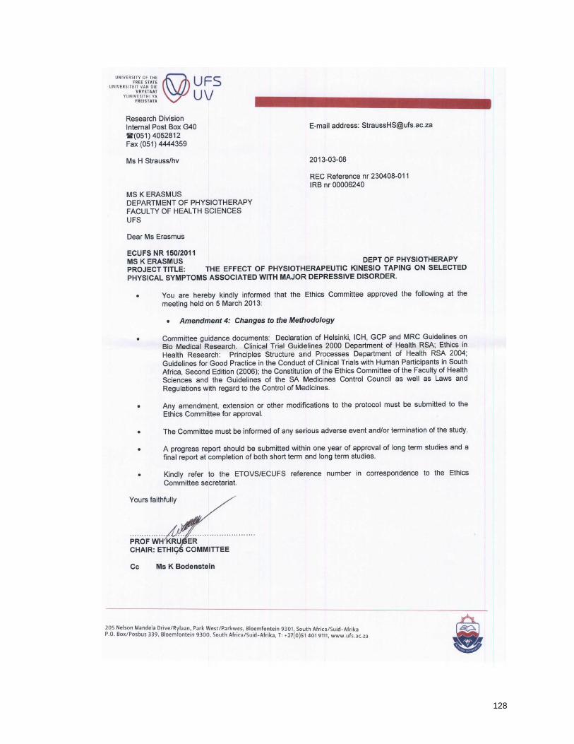
128

129

130

131
Appendix H
Coding list

132
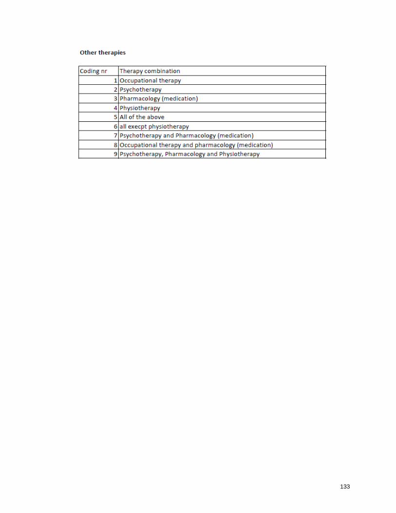
133
134
Summary
Kinesio® taping is a relatively new form of therapeutic taping that has a variety of
applications. Physiotherapists can use the tape to treat certain physical
symptoms for example, pain, swelling and dysfunctional muscle activation.
Major depressive disorder (MDD) is a mood disorder which also causes physical
symptoms. The physical symptoms most associated with MDD are muscle
tension, pain complaints, restricted breathing, less flexibility and centring of
movements, and negative attitudes towards physical appearance and ability.
These physical symptoms are, however, often ignored in the assessment and
treatment of the disorder. Without total relief of the physical symptoms
associated with MDD, remission of the condition could be false or incomplete.
The association between physical complaints and MDD emphasises the need for
physiotherapy as part of the holistic treatment regime for the depressed patient.
The aim of this study was to investigate the effect of physiotherapeutic Kinesio®
taping on selected physical symptoms associated with MDD.
A double-blind, randomised controlled design was used, following a quantitative
study approach. Study participants (40) were male and female patients with a
diagnosis of MDD admitted to a public and a private psychiatric institution,
respectively, in the Free State. Evaluation of the selected physical symptoms
was conducted before, immediately after and 24 hours after the application of the
Kinesio® taping. One researcher performed the evaluations and another
researcher applied the taping and completed a baseline mood questionnaire with
the study participants.
Just more than half (55%) of the participants were recruited from a private
psychiatric institution and 45% of the participants were recruited from a public
psychiatric institution. Most (80%) of the participants were female. The median
age of the participants in the experimental group was 37.5 years and in the
placebo group it was 34 years.
135
The severity of the current MDD episode of participants ranged between normal
and very severe, with 60% of participants experiencing a severe or very severe
episode. Slightly more than half (55%) of the participants received all available
therapies: occupational therapy, physiotherapy, psychotherapy and a
pharmacological intervention. This corresponds with the 55% of participants
recruited from the private psychiatric institution where all the therapies are part of
the daily treatment regime. Pain was assessed using the Short Form McGill Pain
Questionnaire (SF-MPQ) by means of a structured interview. Restriction in
breathing was evaluated by a palpation test (Manual Assessment of Respiratory
Motion) and loss of flexibility and centring of movement was an observation test
of functional activities (Tinetti Mobility Test).
The majority (77.5%) of participants complained of pain during the study period
and 58% complained of multiple areas of pain. Participants complained mostly of
cervical pain (36.7%), except for the placebo group who at 24 hours
post-application reported lower back pain (30%).
The sensory and affective components associated with pain and tested by the
SF-MPQ showed improvement in combined scores for both the experimental and
placebo groups. The affective domain tests the emotional component of pain
and the results indicate a clinically significant effect on this domain. The results
of the Manual Assessment of Respiratory Motion (MARM) displayed
improvement in both the placebo and experimental groups for balance of
breathing and percentage ribcage motion. The Tinetti Mobility Test which
assesses balance and gait showed no distinct results possibly due to the scale
not being sensitive enough for the movement disorders tested.
Limitations of the study could have influenced the outcomes measured and it
should be taken into account that of the 40 participants, 21 received
physiotherapy and the other therapies received by participants during the study
was not standarised and could not be controlled due to the multitude of
stakeholders involved in the care of the patients. The treatment of physical
symptoms associated with MDD with Kinesio® taping had mixed results, but

136
Kinesio® taping could be a valuable adjuvant treatment modality. The
importance of physiotherapy as part of the treatment regime for patients suffering
from MDD was highlighted and the importance of future research in this field of
practise emphasised.
Key terms: effects, physiotherapeutic, Kinesio® taping, physical
symptoms, major depressive disorder, pain, restricted breathing, loss of
centring of movements

137
Opsomming
Kinesio® pleisters is a relatiewe nuwe vorm van terapeutiese verbinding en het „n
groot verskeidenheid toepassings. Fisioterapeute kan die pleisters gebruik om
sekere fisiese simptome byvoorbeeld pyn, swelling en spier aktiverings
disfunksie te behandel.
Major depressiewe versteuring (MDV) is „n gemoedversteuring wat ook tot fisiese
simptome lei. Die fisiese simptome wat meestal met die versteuring geassosieer
word is spierspanning, klagtes van pyn, asemhalingsinkorting, verminderde
soepelheid, verlies aan sentrering van beweging, asook „n negatiewe houding
jeens fisiese voorkoms en vermoeëns. Hierdie fisiese simptome word dikwels
geïgnoreer tydens die evaluering en behandeling van die versteuring. Sonder
die aangehele verligting van die geassosieerde fisiese simptome van MDV kan
remissie vals of onvolledig wees. Die assosiasie tussen fisiese klagtes en MDV
beklemtoon die behoefte aan fisioterapie as deel van die holistiese
behandelingsregime vir die depressiewe pasiëntDie doel van hierdie
navorsingstudie was om die effek van fisioterapeutiese Kinesio® pleister
aanwending op geselekteerde fisiese simptome geassosieer met MDV te
ondersoek.
Hierdie was „n dubbel-blinde, ewekansige gekontroleerde proefneming en „n
kwantitatiewe studiemetode is gevolg. Studiedeelnemers was manlike en
vroulike pasiënte wat met MDV gediagnoseer en opgeneem is in onderskeidelik
„n staats en „n privaat psigiatriese instansie in die Vrystaat. Evaluering van die
fisiese simptome is uitgevoer voor, onmiddelik na en 24 uur na die aanwending
van die Kinesio® pleisters. Een navorser het die evaluasies behartig en „n ander
navorser het die pleisters aangewend en die basislyn gemoedsvraelys met die
deelnemers gedoenNet meer as die helfte (55%) van die deelnemers is vanaf die
privaat psigiatriese instansie gewerf en 45% van die deelnemers vanaf die
publieke instansie. Die meeste (80%) van die deelnemers van was vroulik. Die

138
mediane ouderdom van die deelnemers in die eksperimentele groep was 37.5
jaar en in die plasebo groep 34 jaar.
Die graad van die huidige MDV episode van die deelnemers het gewissel tussen
normaal en baie ernstig, met 60% van die deelnemers wat „n erge of baie erge
episode beleef het. Net meer as die helfte (55%) van die deelnemers het alle
beskikbare behandeling ontvang: arbeidsterapie, fisioterapie, psigoterapie en
farmakologie. Hierdie resultate stem ooreen met 55% van die deelnemers wat
afkomstig was van die privaat psigiatriese instansie waar al die terapieë deel was
van die daaglikse behandeling. Pyn is gevalueer met behulp van die “Short Form
McGill Pain Questionnaire (SF-MPQ)” deur n gestruktureerde onderhoud
Beperking in asemhaling is „n palperings toets („”Manual Assessment of
Respiratory Motion”) en verlies van soepelheid en sentrering van beweging is „n
observasie toets van funksionele aktiwitiete (“Tinetti Mobility Test”).
Die meerderheid (77.5%) van die deelnemers het pyn klagtes gehad gedurende
die studie periode en 58% het gekla van pyn in meer as een area. Deelnemers
het meestal (36.7%) nekpyn rapporteer, behalwe vir die plasebo groep wat teen
24 uur na aanwending hoofsaaklik (30%) van lae rugpyn gekla het.
Die sensoriese en affektiewe komponente geassosieer met pyn en gemeet deur
die “Short Form McGill Pain Questionnaire” het verbetering in die gekombineerde
punte vir beide die eksperimentele en plasebo groepe getoon. Die resultate van
die affektiewe afdeling, wat die emosionele komponent van pyn meet, was klinies
beduidend. Die resultate van die “Manual Assessment of Respiratory Motion”
toon verbetering in beide die eksperimentele en plasebo groepe vir balans van
asemhaling sowel as die persentasie borskas beweging. Die “Tinetti Mobility
Test” wat balans en looppatroon evalueer het geen duidelike resultate gehad nie
as gevolg van die skaal wat moontlik nie sensitief genoeg was om
bewegingsversteurings wat getoets is waar te neem nie.
Tekortkominge kon die uitkomste van die studie beȉnvloed het. Dit moet in ag
geneem word dat van die 40 deelnemers, 21 fisioterapie ontvang. het en dat die
ander terapieȅ wat deur die deelnemers ontvang is nie gestandariseerd was of

139
beheer kon word nie as gevolg van die hoeveelheid belanghebbendes betrokke
by die behandeling van die pasiȅnte.
Die behandeling van die geselekteerde fisiese simptome geassosieer met MDV
met Kinesio® pleisters het gemengde resultate gelewer, maar die
behandelingstegniek kan „n baie waardevolle aanvullende
behandelingsmodaliteit wees. Die belangrikheid van fisioterapie as deel van die
behangelingsregime vir pasiënte wat aan MDV ly is bevestig en uitgelig en die
noodsaaklikheid van toekomstige navorsing in die veld is beklemtoon.