the effective encapsulation of a hydrophobic lipid-insoluble drug in solid lipid nanoparticles using...
TRANSCRIPT

Tse
MMSa
b
c
a
ARRAA
KRSDEL
1
1spTfibsf
o
(
0h
Colloids and Surfaces B: Biointerfaces 112 (2013) 408–414
Contents lists available at ScienceDirect
Colloids and Surfaces B: Biointerfaces
journa l homepage: www.e lsev ier .com/ locate /co lsur fb
he effective encapsulation of a hydrophobic lipid-insoluble drug inolid lipid nanoparticles using a modified double emulsion solventvaporation method
ohsen Nabi-Meibodia,b, Alireza Vatanaraa,∗, Abdolhossein Rouholamini Najafabadia,ohammad Reza Rouinia, Vahid Ramezania,b, Kambiz Gilania,
eyed Mohammad Hossein Etemadzadehc, Kayhan Azadmaneshc
Department of Pharmaceutics, Faculty of Pharmacy, Tehran University of Medical Science, Tehran, IranFaculty of Pharmacy, Shahid Sadoughi University of Medical Science, Yazd, IranDepartment of Virology, Pasteur Institute of Iran, Tehran, Iran
r t i c l e i n f o
rticle history:eceived 27 November 2012eceived in revised form 16 May 2013ccepted 7 June 2013vailable online 29 June 2013
eywords:aloxifene HClolid lipid nanoparticlesouble emulsion solvent evaporation
a b s t r a c t
Raloxifene HCl (RH), a selective estrogen receptor modulator (SERM), is indicated for the prophylaxisor treatment of postmenopausal osteoporosis. RH shows extremely poor bioavailability due to limitedsolubility and an extensive intestinal/hepatic first-pass metabolism. Solid lipid nanoparticles (SLNs) arevaluable carriers that can enhance drug bioavailability. However, in the case of RH, the encapsulation ofthe drug in SLNs remains a challenge because of its poor solubility in both water and lipids. In this study,a series of RH-containing SLNs (RH-SLNs) were generated using a modified double emulsion solventevaporation (DESE) method. Briefly, RH with various drug/lipid ratios was solubilized in the inner coreof a double emulsion using different water/organic solvent mixtures. Our best formulation was achievedwith the formation of negatively charged nanoparticles, 180 nm in diameter, with an encapsulation and
ntrapment efficiencyoading efficiency
loading efficiency of 85% and 4.5%, respectively. It also showed a Fickian mechanism of the drug releasein the basic dissolution media. Thermal analysis revealed a distinct decrease in the crystallinity of lipidsand RH in comparison with the unprocessed materials. The results of a cell viability assay also showed abetter antiproliferative effect of the drug-loaded SLNs versus the free drug solution. Thus, these resultsindicated that the modified DESE method could be proposed for the effective encapsulation of RH in SLNswith appropriate physicochemical and biological properties.
. Introduction
Solid lipid nanoparticles (SLNs) were developed in the early990s as an alternative drug delivery system to emulsions, lipo-omes or polymeric nanoparticles. SLNs are referred to as sphericalarticles, with an average diameter between 10 and 1000 nm.hey consist of a solid lipid core matrix that is stabilized by sur-actants. This system provides unique advantages, including thencorporation of both lipophilic and hydrophilic drugs, higher oralioavailability, protection against chemical degradation, the pos-ibility of controlled drug delivery, good in vivo tolerability and
easibility of scale up [1–4].Several factors determine the loading capacity and the patternf drug release from SLNs, including physicochemical properties
∗ Corresponding author. Tel.: +98 21 66959057; fax: +98 21 66461178.E-mail addresses: [email protected], [email protected]
A. Vatanara).
927-7765/$ – see front matter © 2013 Elsevier B.V. All rights reserved.ttp://dx.doi.org/10.1016/j.colsurfb.2013.06.013
© 2013 Elsevier B.V. All rights reserved.
of the drug, the lipid structure and its crystal habit, the type andconcentration of the emulsifier and the method of preparation [5].
The method of preparation of lipid nanoparticles is gener-ally selected based on the drug solubility in lipids. If the drugis hydrophobic or a lipid derivative, direct incorporation can beachieved by high pressure homogenization, microemulsion for-mation or solvent emulsification evaporation/diffusion [6–8]. Onthe other hand, a more elaborate method should be applied forhydrophilic or hydrophobic drugs that are soluble in acidic or basicmedia as well as for peptides, proteins and genes. Herein, the incor-poration of the drug in SLNs via the double emulsion (W1/O/W2)solvent evaporation technique or adsorption on the surface ofpreformed SLNs is used to produce the desired SLN formulation[9–12].
Raloxifene HCL (RH) is a selective estrogen receptor modula-
tor (SERM) that acts as an estrogen agonist and antagonist in thebone and the breast, respectively. The Food and Drug Administra-tion (FDA) approved the drug for the treatment of osteoporosisand for the prevention of breast cancer in postmenopausal women.
rface
Hb
womiteS
eSaicii
2
2
((w(s(
2
eeiWawat2iphetawrraf
2
(sm2s
into microtubes. The tubes were then incubated in a waterbath shaker (Memmert, Germany) at 37 ◦C and at 50 rpm. At
M. Nabi-Meibodi et al. / Colloids and Su
owever, RH is poorly water-soluble and shows extremely low oralioavailability (2% of the given dose) [13–15].
Our preliminary studies demonstrated that RH does not mergeell into the melted lipid during SLN preparation, as has been
bserved with hydrophilic drugs. In addition, RH could not be for-ulated by the double emulsion technique due to its low solubility
n aqueous media. Hence, the development of new strategies forhe preparation methods can provide an effective solution for thentrapment of RH and other chemically similar substances into theLNs.
In this study, we introduced some modifications into the doublemulsion technique to overcome the loading hindrance of RH intoLNs. For this purpose, RH was solubilized in a mixture of waternd some organic solvents and could thus be incorporated into thenner core of the double emulsion. The SLNs generated were thenharacterized in terms of their physicochemical properties, load-ng and entrapment efficiencies, release behavior, as well as theirn vitro anti-proliferative activity.
. Materials and methods
.1. Materials
Glycerol monostearate (GMS) was kindly donated by GattefosséFrance), while glycerol tristearate (GTS) and glycerol trimyristateGTM) were supplied by Sigma–Aldrich (Germany). Raloxifene HClas provided by Glochem Industries Ltd. (India). Poloxamer 188
P188) was obtained from BASF (Germany). Trehalose dihydrate,oy lecithin (SL), Tween 80, as well as all of the organic solventsanalytical grade) were purchased from Merck (Germany).
.2. Preparation of RH-Loaded SLNs
RH-loaded SLNs were prepared by the modified doublemulsion (W1/O/W2) solvent evaporation (DESE) technique firstmployed by Garcia-Fuentes et al. [10]. Initially, RH was dissolvedn 1 ml of different mixtures of organic solvent/water (1:1) as the
1 Phase. The oily phase was prepared by dissolving 300 mg of GMSnd SL (1:1, w/w) in 5 ml of chloroform. The W1 phase was mixedith the oily phase by stirring on a magnetic stirrer (IKA, Germany)
nd was then sonicated (400 W, 20 cycles with 1 s active–2 s dura-ion) by an ultrasonic probe (Hielscher, Germany) in an ice bath formin at 80% amplitude. The resultant nanoemulsion was injected
nto 35 ml of a P188 aqueous solution (0.5%, w/v) using a syringeump (Kd Scientific, USA) at a rate of 3 ml/min under high shearomogenization (IKA, Germany). The organic solvents were thenvaporated by means of a rotary evaporator (Büchi, Switzerland)o form SLNs. The nanoparticles were harvested by centrifugationt 45,000 × g for 1 h at 10 ◦C (Sigma, Germany) and washed twiceith deionized water to remove unentrapped drug, surfactants and
emaining organic solvents. The retrieved nanoparticles were thenesuspended in 2 ml of 10% (w/v) trehalose aqueous solution, frozent −20 ◦C and were finally freeze dried (Christ, Germany) at −40 ◦Cor 48 h.
.3. Particle size and zeta potential
The average particle size in nanometers, polydispersity indexPDI) and zeta potential were determined by photon correlationpectroscopy equipped with a zetasizer (Nano ZSTM Malvern Instru-ents, UK). Measurements were carried out at an angle of 173◦ at
5 ◦C. All of the measurements were repeated three times for eachample.
s B: Biointerfaces 112 (2013) 408–414 409
2.4. Encapsulation and loading efficiency
Freeze dried SLNs were degraded in hot methanol; after cool-ing, the dissolved drug was separated from the precipitated lipidsby centrifugation (4000 × g for 1 h at 10 ◦C). The drug content inthe supernatant was determined according to the method devel-oped by Trontelj et al. with some modifications [16]. RH wasassayed by a reverse-phase HPLC system (Waters, USA) on a5 �m, 4.6 mm × 250 mm Nucleosil C18 column (Macherey Nagel,Germany) at 40 ◦C. The mobile phase was a mixture of acetonitrileand 0.05 M phosphate buffer (pH 3; 35:65, v/v), with a flow rateof 0.7 ml/min. Detection was carried out on 287 nm with a 100 �linjection volume. A linear regression curve (y = 118.3x − 7.555,R2 = 0.9989) within the range of 1–20 �g/ml was used to calculatethe concentration of RH. The detection limit (DL) and the quantifi-cation limit (QL) were determined according to the ICH’s Guidancefor Industry, Q2B Validation of Analytical Procedures, using thefollowing equations [16].
DL = 3.3�
S
QL = 10�
S
where (�) is the standard deviation of response (in this case, theblank), and (S) is the slope of the calibration curve. The values of DLand QL obtained were 0.25 and 0.76 �g/ml, respectively.
The entrapment efficiency (EE), loading efficiency (LE) and yieldefficiency (YE) were calculated by the following equations:
Entrapment Efficiency
= Obtained mass of drug in freeze dryed SLNInitial mass of drug
× 100%
Loading Efficiency
= Obtained mass of drug in freeze dryed SLNObtained mass of freeze dryed SLN
× 100%
Yield Efficiency
= Obtained mass of drug in freeze dryed SLNInitial theoretical mass of SLN
× 100%
The experiments were performed in triplicate.
2.5. In vitro release study
The in vitro drug release from SLNs was evaluated in 0.1 MHCl (pH 1.2) and 10 mM phosphate buffer (pH 6.8), containing0.1% Tween 80. Briefly, freshly prepared formulations were resus-pended in the two dissolution media listed earlier and divided
predetermined time intervals, the sample microtubes were cen-trifuged at 45,000 × g for 1 h at 10 ◦C and precipitated nanoparticleswere freeze dried. Finally, the amount of drug retained in the pre-cipitated nanoparticles was determined using the HPLC systemunder the same operating conditions described earlier. The per-centage of the cumulative release at each point was calculated usingthe following formula:
4 urface
CRem
in e
2
gwa
2
wtiir
2
7(sAfiRcwontSl(lbapcTm
2
siC
3
3
hptsidwio
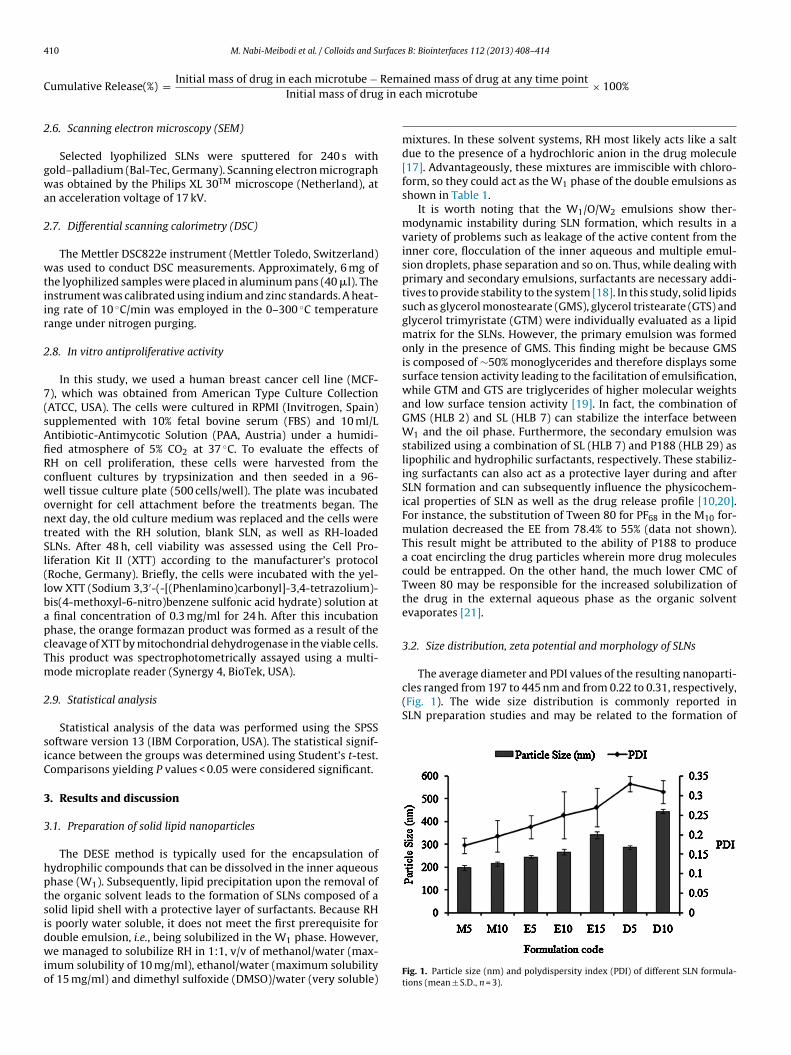
cles ranged from 197 to 445 nm and from 0.22 to 0.31, respectively,(Fig. 1). The wide size distribution is commonly reported inSLN preparation studies and may be related to the formation of
10 M. Nabi-Meibodi et al. / Colloids and S
umulative Release(%) = Initial mass of drug in each microtube −Initial mass of drug
.6. Scanning electron microscopy (SEM)
Selected lyophilized SLNs were sputtered for 240 s withold–palladium (Bal-Tec, Germany). Scanning electron micrographas obtained by the Philips XL 30TM microscope (Netherland), at
n acceleration voltage of 17 kV.
.7. Differential scanning calorimetry (DSC)
The Mettler DSC822e instrument (Mettler Toledo, Switzerland)as used to conduct DSC measurements. Approximately, 6 mg of
he lyophilized samples were placed in aluminum pans (40 �l). Thenstrument was calibrated using indium and zinc standards. A heat-ng rate of 10 ◦C/min was employed in the 0–300 ◦C temperatureange under nitrogen purging.
.8. In vitro antiproliferative activity
In this study, we used a human breast cancer cell line (MCF-), which was obtained from American Type Culture CollectionATCC, USA). The cells were cultured in RPMI (Invitrogen, Spain)upplemented with 10% fetal bovine serum (FBS) and 10 ml/Lntibiotic-Antimycotic Solution (PAA, Austria) under a humidi-ed atmosphere of 5% CO2 at 37 ◦C. To evaluate the effects ofH on cell proliferation, these cells were harvested from theonfluent cultures by trypsinization and then seeded in a 96-ell tissue culture plate (500 cells/well). The plate was incubated
vernight for cell attachment before the treatments began. Theext day, the old culture medium was replaced and the cells werereated with the RH solution, blank SLN, as well as RH-loadedLNs. After 48 h, cell viability was assessed using the Cell Pro-iferation Kit II (XTT) according to the manufacturer’s protocolRoche, Germany). Briefly, the cells were incubated with the yel-ow XTT (Sodium 3,3′-(-[(Phenlamino)carbonyl]-3,4-tetrazolium)-is(4-methoxyl-6-nitro)benzene sulfonic acid hydrate) solution atfinal concentration of 0.3 mg/ml for 24 h. After this incubation
hase, the orange formazan product was formed as a result of theleavage of XTT by mitochondrial dehydrogenase in the viable cells.his product was spectrophotometrically assayed using a multi-ode microplate reader (Synergy 4, BioTek, USA).
.9. Statistical analysis
Statistical analysis of the data was performed using the SPSSoftware version 13 (IBM Corporation, USA). The statistical signif-cance between the groups was determined using Student’s t-test.omparisons yielding P values < 0.05 were considered significant.
. Results and discussion
.1. Preparation of solid lipid nanoparticles
The DESE method is typically used for the encapsulation ofydrophilic compounds that can be dissolved in the inner aqueoushase (W1). Subsequently, lipid precipitation upon the removal ofhe organic solvent leads to the formation of SLNs composed of aolid lipid shell with a protective layer of surfactants. Because RHs poorly water soluble, it does not meet the first prerequisite for
ouble emulsion, i.e., being solubilized in the W1 phase. However,e managed to solubilize RH in 1:1, v/v of methanol/water (max-mum solubility of 10 mg/ml), ethanol/water (maximum solubilityf 15 mg/ml) and dimethyl sulfoxide (DMSO)/water (very soluble)
s B: Biointerfaces 112 (2013) 408–414
ained mass of drug at any time pointach microtube
× 100%
mixtures. In these solvent systems, RH most likely acts like a saltdue to the presence of a hydrochloric anion in the drug molecule[17]. Advantageously, these mixtures are immiscible with chloro-form, so they could act as the W1 phase of the double emulsions asshown in Table 1.
It is worth noting that the W1/O/W2 emulsions show ther-modynamic instability during SLN formation, which results in avariety of problems such as leakage of the active content from theinner core, flocculation of the inner aqueous and multiple emul-sion droplets, phase separation and so on. Thus, while dealing withprimary and secondary emulsions, surfactants are necessary addi-tives to provide stability to the system [18]. In this study, solid lipidssuch as glycerol monostearate (GMS), glycerol tristearate (GTS) andglycerol trimyristate (GTM) were individually evaluated as a lipidmatrix for the SLNs. However, the primary emulsion was formedonly in the presence of GMS. This finding might be because GMSis composed of ∼50% monoglycerides and therefore displays somesurface tension activity leading to the facilitation of emulsification,while GTM and GTS are triglycerides of higher molecular weightsand low surface tension activity [19]. In fact, the combination ofGMS (HLB 2) and SL (HLB 7) can stabilize the interface betweenW1 and the oil phase. Furthermore, the secondary emulsion wasstabilized using a combination of SL (HLB 7) and P188 (HLB 29) aslipophilic and hydrophilic surfactants, respectively. These stabiliz-ing surfactants can also act as a protective layer during and afterSLN formation and can subsequently influence the physicochem-ical properties of SLN as well as the drug release profile [10,20].For instance, the substitution of Tween 80 for PF68 in the M10 for-mulation decreased the EE from 78.4% to 55% (data not shown).This result might be attributed to the ability of P188 to producea coat encircling the drug particles wherein more drug moleculescould be entrapped. On the other hand, the much lower CMC ofTween 80 may be responsible for the increased solubilization ofthe drug in the external aqueous phase as the organic solventevaporates [21].
3.2. Size distribution, zeta potential and morphology of SLNs
The average diameter and PDI values of the resulting nanoparti-
Fig. 1. Particle size (nm) and polydispersity index (PDI) of different SLN formula-tions (mean ± S.D., n = 3).

M. Nabi-Meibodi et al. / Colloids and Surfaces B: Biointerfaces 112 (2013) 408–414 411
Table 1Composition of different formulation of SLNs containing Raloxifene HCl (RH).
Formulation code M5 M10 E5 E10 E15 D5 D10 D15
W1 Composition Raloxifen HCl (mg) 5 10 5 10 15 5 10 15Organic solvents/Water (1:1) Ma Ma Eb Eb Eb Dc Dc Dc
O ingredients dGMS (mg) 150eSL (mg) 150
W2 Composition fP188 (mg) 175Purified water (ml) q.s. to 35
a Methanol.b Ethanol.c Dimethylsulfoxide.d Glycerol monostearate.e Soy lecithin.f Poloxamer 188.
Fig. 2. SEM image (A) and Particle
Table 2Zeta potential of different SLN formulation (mean ± S.D., n = 3).
Formulation code Zeta potential (mV)
M5 −32.9 ± 2.1M10 −35.0 ± 1.2E5 −34.7 ± 2.2E10 −35.3 ± 1.7E −42.7 ± 2.3
ms
tsta
tsz−bumbt[
3
a
phase was changed from a methanol/water mixture to ethanol orDMSO/water, the drug encapsulation efficiency decreased drasti-cally from 74.2% to 57.6% and 45.9%, respectively. The reduction
15
D5 −29.2 ± 0.9D10 −36.6 ± 1.8
ultiple phospholipid layers or the formation of other structuresuch as liposomes, micelles, and drug nanosuspensions [1,10].
SEM imaging was employed to study the morphology as well ashe size of SLNs. It was evident that the particles were spherical inhape and had smooth surfaces. In addition, the size observed fromhe SEM image correlated with the sizes obtained from PCS (Fig. 2and b).
The zeta potential reflects the strength of the particle elec-rical barrier and is used as a key parameter in evaluating thetability of colloidal dispersions [22]. As presented in Table 2, theeta potentials of the SLN formulations varied between −29 and43 mV. Regarding the zeta potential, no considerable differenceetween the nanoparticles was observed. A highly negative resid-al charge was attributed to the chemical nature of the applied lipidatrix and surfactants. Ionization of stearic acid molecules might
e responsible for some of the negative surface charges. Moreover,he anionic fractions of SL can also induce an extra negative charge23].
.3. Encapsulation and loading efficiency
Drug-loading capacity is a parameter used to assess the suit-bility of a particular drug-carrier system [24]. Fig. 3 shows the
size distribution (B) of M10.
results of EE and LE for different formulations that were analyzed.The highest encapsulation efficiency values were obtained when5 and 10 mg RH was solubilized in the W1 phase in M5 and M10formulations (74.2 ± 3.2% and 69.9 ± 4.5%, respectively). Addition-ally, the M10 formulation showed the highest LE value (4.3 ± 0.2%).The extent of drug loading is equal to or even more than thereported values for lipid-soluble drugs such as gambogenic acid,enrofloxacin, doxorubicin and nimodipine [25–28].
Although a similar amount of drug (5, 10 or 15 mg) was initiallyadded to the different W1 phase compositions, the SLN formula-tions that were generated exhibited considerable differences intheir respective EE and LE values. For example, when the W1
Fig. 3. Entrapment efficiency (EE), loading efficiency (LE) and yield efficiency (YE)of different SLN formulations (mean ± S.D., n = 3).

412 M. Nabi-Meibodi et al. / Colloids and Surfaces B: Biointerfaces 112 (2013) 408–414
F poloxf
ibelGtticilit(
3
aaepmfsaiTmdftas
llc
ig. 4. Overlaid DSC thermograms: glycerol monostearate (a), raloxifene HCl (b),ormulation (e) and M10 formulation (f).
n the EE value might be attributed to the partitioning of the drugetween the W1 phase and the oily phase during organic phasevaporation. Consequently, RH and GMS were mixed at differentevels based on the varying ability of the W1 phase in solubilizingMS (DMSO/water > ethanol/water > methanol/water). The higher
he mixing level, the lower EE and LE would be obtained due tohe inability of the lipid matrix to the drug. In this way, a failuren the preparation of D15 or other formulations with an initial drugontent of more than 15 mg could be explained by the instabilitynduced by mixing. Moreover, the mixing of the drug and the lipidseads to a reduction in the stability of SLNs. Therefore, changesn the YE%, a stability-dependent variable, are closely related tohe changes in the EE efficiency among the various formulationsFig. 3).
.4. DSC study
DSC is a conventional technique that has been used to evalu-te drug–lipid interactions as well as the degree of crystallizationnd polymorphism of drugs [29]. To describe the solid state andntrapment mechanism of RH in the SLNs, thermal analysis waserformed on GMS, RH, P188, trehalose dihydrate, the physicalixture with a composition of the M10 formulation, and the M10
ormulation (Fig. 4a–f). The pure RH, bulk GMS and P188 demon-trated a single sharp endothermic peak at approximately 271, 64nd 56 ◦C, respectively. There was no clear change in the melt-ng point of the raw materials when they were physically mixed.he presence of the RH endothermic peak in both the physicalixture and the SLN formulation showed that RH could not be
issolved in the lipid matrix or other parts of the formulation. Inact, there are a few published studies that show the presence ofhe crystalline form of the drug in SLNs. This result may be favor-ble because crystalline materials exhibit better physicochemicaltability in comparison with metastable amorphous materials [30].
On the other hand, comparing the thermograms of SLN formu-ations with raw and bulk materials revealed a broadening of theipid melting peak and a decline in its melting point. These changesould be due to the plasticizing effect of surfactants, in addition to
amer 188 (c), trehalose dehydrate (d), physical mixture with composition of M10
the submicron dimension of the particles that have a vast surfacearea [31,32].
3.5. In vitro release study
Because RH is a labile molecule and is susceptible to degradationin the dissolution media, the methodology described in the Section2 was selected for the release study. The profiles of drug release inthe different media are shown in Fig. 5. In an Overall veiw, four typesof drug release zones can be seen in the tested formulations: (i)initial burst release period in which the surface drug is released intothe dissolution medium; here, it is considered as 1 h; (ii) inductionperiod of release in which the drug is released at a slowly decreasingfast rate; (iii) sustained release period in which the drug is releasedat a regular slow rate; and (iv) final release period in which theparticles disintegrate to release the residual drug at a rapid rate.
The burst release could be induced by the imperfect encapsula-tion of the drug inside the nanoparticles due to the unstable natureof the SLN. In fact, the intrinsic instability of SLNs during formationmay cause a partial relocation of the loaded drug molecules ontothe surface of the nanoparticles [33–35]. Furthermore, the burstrelease of the drug may also be explained by the fact that glycerolmonostearate has surfactant properties and can thus interact withwater molecules. In other words, employing lipids with polar char-acteristics promotes the formation of colloidal structures such asliquid crystalline phases or mixed micelles in addition to SLNs. So,GMS may increase the amount of drug molecules in the externalaqueous phase or may cause a higher mobility in the lipid particlesurface from where the drug substance is readily released [5].
In formulations with the same W1 phase compositions, a higherinitial RH content resulted in a more obvious burst release. As afore-mentioned, during the formation of SLNs, a limited capacity of thecarrier results in the deposition of some drug molecules in the sur-factant layer of the carrier surface. Thus, the higher the amountof drug in the formulation is, the more the deposition of drug
molecules in the surfactant layer of SLNs.The induction and sustained release periods can be mainlyrelated to the drug diffusion from the core of the nanoparticle ontothe surface. Moreover, it has been reported that P188, a swellable,

M. Nabi-Meibodi et al. / Colloids and Surfaces B: Biointerfaces 112 (2013) 408–414 413
Fig. 5. In vitro release profiles of raloxifene HCl from different formulations in two dissolution media: Phosphate buffer (10 mM, pH 6.8) (A) and HCl (0.1 M, pH 1.2) (B)containing 0.1% (w/v) Tween 80 (n = 3). Lines with the styles of solid (—), dash (- - -) and dot (.....) show formulations with W1 phase of methanol/water, ethanol/water andDMSO/water mixture, respectively. Lines with the marker options of square (�), circle (�), and triangle (�) show formulations with the initial drug amount of 5, 10 and15 mg, respectively.
Table 3Kinetic parameters of Korsmeyer–Peppas model for in vitro drug release.
Formulation code Phosphate buffer (pH 6.8) medium HCl (pH 1.2) medium
K N R2a k n R2a
M5 12.19 0.468 0.951 13.53 0.515 0.975M10 13.84 0.495 0.932 13.86 0.528 0.980E5 16.11 0.477 0.871 16.28 0.530 0.956E10 21.27 0.519 0.868 22.06 0.563 0.933E15 24.05 0.529 0.847 34.25 0.558 0.793
hcrblass
ietlfiemmttttm
snta
bmwo
wd
D5 21.73 0.510D10 15.58 0.771
a Coefficient of determination.
ydrophilic macromolecule, forms a hydrogel barrier and can henceontrol drug diffusion along with the lipid layers [36]. The slowelease of RH from SLNs can be favorable when absorption from aiological barrier is intended. Based on the hypothesis that drug-
oaded solid lipid nanoparticles can be protected from degradationnd metabolism, in an ideal formulation, the encapsulated drughould not be released completely prior to the delivery at the targetite [2,37].
The number of release zones varies for different formulationsn the two dissolution media. For example, all of the formulationsxhibit the first three of the above-mentioned zones over a 24-hime period in a phosphate buffered medium, whereas all formu-ations except the E15 and D10 include the burst, induction and thenal release period in an HCl medium. The %RH released at thend of the first hour (initial burst) from E15 and D10 in an acidicedium is 70% and 82%, respectively. This finding indicates theerging of the burst and the final release period in the absence of
he induction and sustained periods. In other words, a majority ofhe drug is released due to particle disintegration. This disintegra-ion can be accelerated owing to the higher instability induced byhe higher initial drug loading, especially in the acidic dissolution
edium.As shown previously, P188 as a non-ionic steric emulsifier is not
uitable for stabilizing the GMS lipid in the artificial gastrointesti-al medium (GI) at pH 1.1 [38]. Thus, if the oral route is intended,he final SLN-containing dosage form should be protected from thecidic environment of the GI using an enteric coating system.
The drug release from SLNs is usually considered as a com-ination of Fickian (diffusion) and non-Fickian transport of drugolecules through the lipid layers [39]. Thus, the obtained dataere fitted into Korsmeyer–Peppas model to verify the mechanism
f diffusion:
Mt
M∞= ˛tn
here Mt is the drug released at time t, M∞ is the quantity ofrug released at infinite time, ˛ is the kinetic constant, and n is an
0.848 20.39 0.560 0.9450.459 37.37 0.560 0.767
exponent to characterize different release mechanisms. The valueof n is 0.43 for pure Fickian and higher values of n, between 0.43 and0.85, or n = 0.85, for mass transfer following a non-Fickian mecha-nism [40]. The values of different parameters related to the best-fitlines are included in Table 3.
Comparing the correlation coefficient (R2) values, all of theformulation except the E15 and D10 provided a good fit to theKorsmeyer–Peppas model. Almost all of the formulations exceptthe D10 showed the value of n ∼ 0.5, suggesting the Fickian mech-anism of the drug release in the phosphate buffered (pH 6.8)medium. For the same formulation, the n values is ∼0.05 more in theHCl (pH 1.2) medium, confirming that the drug release from SLNswould be a combination of diffusion and erosion. As mentioned ear-lier, the SLNs are more susceptible to degradation in acidic media[39].
3.6. In vitro antiproliferative activity
As a carrier system, SLNs have shown many features that arevaluable for nanomedicines. Thus, SLNs have been investigated asdelivery systems for drugs and recently for genes. In both cases, thisdelivery system is reported to act more efficiently compared to thedrug or gene alone [12].
As presented in Fig. 6, following the treatment of MCF-7 cells with RH, blank SLN and drug-loaded SLNs, the blankSLN did not have any effect, as cell viability remained con-stant. Correspondingly, the RH solution did not influence cellgrowth, but these cells were more sensitive to RH-loadedSLNs in equimolar concentrations (p < 0.001), as evidenced byan approximately 25% decrease in cell viability 48 h aftertreatment.
SLNs have proven to be good carriers to prevent drug degra-dation. Thus, the improved cytotoxicity of RH can be attributed to
the increased stability of the drug in the carrier. Additionally, thisenhanced efficacy may be related to the cell attachment or inter-nalization of RH-loaded SLNs, followed by drug release inside ornear the cells [2,37].
414 M. Nabi-Meibodi et al. / Colloids and Surface
FcS
4
msWatm(pttsdittbc
mw
A
o
R
[
[
[
[
[[[
[[
[[[[
[
[
[
[
[[
[[
[[
[[
[
[
ig. 6. Antiprolifration assay of blank SLN, RH solution, RH loaded SLN, at 10 �Moncentration, in MCF-7 cell line (mean ± S.D., n = 3). **p < 0.001 (RH vs. RH loadedLN).
. Conclusions
RH-loaded SLNs were prepared by employing a modified DESEethod in which a combination of organic solvents and water
erved as the W1 or internal aqueous phase. The type of the1 phase, lipid matrix, surfactant as well as the initial drug
mount significantly influenced the physicochemical characteris-ics of the SLNs. Thus, the M10 formulation containing 10 mg RH in a
ethanol/water mixture (1:1, v/v) showed the highest EE% and LE%∼69% and 3.4%, w/w, respectively). In vitro release studies wereerformed by measuring the remaining entrapped drug instead ofhe dissolved drug. Therefore, the drug instability in the dissolu-ion medium could not affect dissolution studies. The drug releasetudies showed a Fickian drug release at pH 6.8 while the particleisintegration and erosion can accelerate the release of the drug
n the acidic dissolution medium. Hence, further modifications inhe formulations will be necessary especially for oral administra-ion. Additionally, complementary antiproliferative studies in thereast cancer cell line revealed that the RH-loaded SLNs were moreytotoxic in comparison with the free drug.
In conclusion, the results of the present study indicate that theodified DESE method can be potentially used for loading drugsith limited solubility in both water and lipids into SLNs.
cknowledgment
This study has been funded and supported by Tehran Universityf Medical Sciences (TUMS).
eferences
[1] W. Mehnert, K. Mader, Adv. Drug Deliv. Rev. 47 (2001) 165–196.
[[[
[
s B: Biointerfaces 112 (2013) 408–414
[2] L.G. Souza, E.J. Silva, A.L.L. Martins, M.F. Mota, R.C. Braga, E.M. Lima, M.C.Valadares, S.F. Taveira, R.N. Marreto, Eur. J. Pharm. Biopharm. 79 (2011)189–196.
[3] R.H. Muller, S. Runge, V. Ravelli, W. Mehnert, A.F. Thunemann, E.B. Souto, Int. J.Pharm. 317 (2006) 82–89.
[4] M. Muchow, P. Maincent, R.H. Müller, Drug Dev. Ind. Pharm. 34 (2008)1394–1405.
[5] L.B. Jensen, E. Magnussson, L. Gunnarsson, C. Vermehren, H.M. Nielsen, K.Petersson, Int. J. Pharm. 390 (2010) 53–60.
[6] A. Kovacevic, S. Savic, G. Vuleta, R.H. Muller, C.M. Keck, Int. J. Pharm. 406 (2011)163–172.
[7] M. Ghadiri, S. Fatemi, A. Vatanara, D. Doroud, A.R. Najafabadi, M. Darabi, A.A.Rahimi, Int. J. Pharm. 424 (2012) 128–137.
[8] B. Sjostrom, B. Bergenstahl, Int. J. Pharm. 88 (1992) 53–62.[9] Q. Lv, A. Yu, Y. Xi, H. Li, Z. Song, J. Cui, F. Cao, G. Zhai, Int. J. Pharm. 372 (2009)
191–198.10] M. Garcia-Fuentes, D. Torres, M.J. Alonso, Colloids Surf. B: Biointerfaces 27
(2003) 159–168.11] M. Garcia-Fuentes, C. Prego, D. Torres, M.J. Alonso, Eur. J. Pharm. Sci. 25 (2005)
133–143.12] D. Doroud, F. Zahedifard, A. Vatanara, A.R. Najafabadi, Y. Taslimi, R.
Vahabpour, F. Torkashvand, B. Vaziri, S. Rafati, J. Control. Release 153 (2011)154–162.
13] K. Visvanathan, R.T. Chlebowski, P. Hurley, N.F. Col, M. Ropka, D. Collyar, M.Morrow, C. Runowicz, K.I. Pritchard, K. Hagerty, B. Arun, J. Garber, V.G. Vogel,J.L. Wade, P. Brown, J. Cuzick, B.S. Kramer, S.M. Lippman, J. Clin. Oncol. 27 (2009)3235–3258.
14] J.S. Teeter, R.D. Meyerhoff, Environ. Toxicol. Chem. 21 (2002) 729–736.15] H.-C. Drorith, Eur. J. Obstet. Gyn. R. B. 85 (1999) 23–29.16] J. Trontelj, T. Vovk, M. Bogataj, A. Mrhar, Pharmacol. Res. 52 (2005)
334–339.17] D. Bikiaris, V. Karavelidis, E. Karavas, Molecules 14 (2009) 2410–2430.18] T. Hino, Y. Kawashima, S. Shimabayashi, Adv. Drug Deliv. Rev. 45 (2000)
27–45.19] V. Jenning, A.F. Thunemann, S.H. Gohla, Int. J. Pharm. 199 (2000) 167–177.20] H. Heiati, N.C. Phillips, R. Tawashi, Pharm. Res. 13 (1996) 1406–1410.21] L. Hu, X. Tang, F. Cui, J. Pharm. Pharmacol. 56 (2004) 1527–1535.22] I. Montasser, H. Fessi, A.W. Coleman, Eur. J. Pharm. Biopharm. 54 (2002)
281–284.23] H.L. Wong, R. Bendayan, A.M. Rauth, X.Y. Wu, J. Pharm. Sci. 93 (2004)
1993–2008.24] K. Ruckmani, M. Sivakumar, P.A. Ganeshkumar, J. Nanosci. Nanotechnol. 6
(2006) 2991–2995.25] X. Huang, Y.-J. Chen, D.-Y. Peng, Q.-L. Li, X.-S. Wang, D.-L. Wang, W.D. Chen,
Colloids Surf. B: Biointerfaces 102 (2013) 391–397.26] S. Xie, L. Zhu, Z. Dong, X. Wang, Y. Wang, X. Li, W. Zhou, Colloids Surf. B:
Biointerfaces 83 (2011) 382–387.27] R.K. Subedi, K.W. Kang, H.-K. Choi, Eur. J. Pharm. Sci. 37 (2009) 508–513.28] F.-Q. Hu, S.-P. Jiang, Y.-Z. Du, H. Yuan, Y.-Q. Ye, S. Zeng, Int. J. Pharm. 314 (2006)
83–89.29] H. Bunjes, T. Unruh, Adv. Drug Deliv. Rev. 59 (2007) 379–402.30] J.F. Hao, F.G. Wang, X.D. Wang, D.R. Zhang, Y.P. Bi, Y.S. Gao, X.M. Zhao, Q. Zhang,
Eur. J. Pharm. Sci. 47 (2012) 497–505.31] B. Siekmann, K. Westesen, Colloids Surf. B: Biointerfaces 3 (1994) 159–175.32] T. Delmas, A.C. Couffin, P.A. Bayle, F. de Crecy, E. Neumann, F. Vinet, M. Bardet,
J. Bibette, I. Texier, J. Colloid Interface Sci. 360 (2011) 471–481.33] R. Jalil, J.R. Nixon, J. Microencapsul. 6 (1989) 473–484.34] N. Ubrich, P. Bouillot, C. Pellerin, M. Hoffman, P. Maincent, J. Control. Release
97 (2004) 291–300.35] Z. Rahman, A.S. Zidan, M.A. Khan, Eur. J. Pharm. Biopharm. 76 (2010)
127–137.36] J. Ye, Q. Wang, X. Zhou, N. Zhang, Int. J. Pharm. 352 (2008) 273–279.
37] P. Sharma, S. Ganta, W.A. Denny, S. Garg, Int. J. Pharm. 367 (2009) 187–194.38] E. Zimmermann, R.H. Müller, Eur. J. Pharm. Biopharm. 52 (2001) 203–210.39] A.C. Silva, M.H. Amaral, E. Gonzalez-Mira, D. Santos, D. Ferreira, Colloids Surf.B: Biointerfaces 93 (2012) 241–248.40] P. Costa, J. Manuel, S. Lobo, Eur. J. Pharm. Sci. 13 (2001) 123–133.