the effectiveness of electroacupuncture in...
TRANSCRIPT

THE EFFECTIVENESS OF
ELECTROACUPUNCTURE IN REDUCING
POSTOPERATIVE PAIN, ANALGESIC
REQUIREMENT AND PREVENTION OF
POSTOPERATIVE NAUSEA AND VOMITING
POST TOTAL ABDOMINAL HYSTERECTOMY
SURGERY
By
DR S PRAVEENA D/O SEEVAUNNAMTUM
DISSERTATION SUBMITTED IN PARTIAL FULFILLMENT OF THE REQUIREMENT FOR THE
DEGREE OF MASTER OF MEDICINE (ANAESTHESIOLOGY)
UNIVERSITI SAINS MALAYSIA
2015

ii
ACKNOWLEDGEMENTS
Firstly, I would like to express my gratitude to Lord Ganesha for blessing me
with the wisdom and dedication to accomplish this dissertation.
I would also like to extend my deepest appreciation to both my supervisors, Dr.
Kavita Bhojwani and Professor Dr. Nik Abdullah Nik Mohamad. Dr. Kavita has helped
in many ways with the support needed to conduct this study in Hospital Raja
Permaisuri Bainun, Ipoh. She inspired me to do a study based on acupuncture in
clinical practise as well as sorted the logistics of it. Professor Dr. Nik Abdullah Nik
Mohamad has also given constant support, encouragement and guidance in completion
of this dissertation. With his invaluable advice, I have managed to successfully
accomplish this task with confidence.
My sincere appreciation also goes to Sister Choo Wai Ling for helping with
recruitment as well as the collection of data. I am also grateful to the support team
consisting of medical officers of Anaesthesia Department and Intensive Care of
Hospital Raja Permaisuri Bainun Ipoh and nurses from the Acute Pain Service (APS)
for helping in conducting this study as well.
Finally, I would like to thank my dearest family for their unconditional love
and patience. I dedicate this dissertation to my pillar of support, my beloved husband,
Dr. Allan Ravi.

iii
TABLE OF CONTENTS
TITLE Page
ACKNOWLEDGEMENTS ii
TABLE OF CONTENTS iii
LIST OF TABLES vii
LIST OF FIGURES viii
LIST OF ABBREVIATIONS xi
ABSTRACTS
ABSTRAK xiii
ABSTRACT xv
CHAPTER 1: INTRODUCTION
1.1 Postoperative pain and its challenges 1
1.2 Acupuncture 4
1.3 Electroacupuncture 9
1.4 Use of Electroacupuncture in this study 12
CHAPTER 2: LITERATURE REVIEW
2.1 Pain 14
2.1.1 Pain perception 14
2.1.2 Pain pathway 16
2.1.3 Gate Control Theory 18
2.1.4 Neurotransmitters and Pain 20
iv
2.1.5 Acute Pain post Hysterectomy 22
2.1.6 Validation of Numerical Rating Scale 23
2.2 Acupuncture 26
2.2.1 History of Acupuncture 26
2.2.2 Types of Acupuncture 27
2.2.3 Electroacupuncture and how it works 29
2.3 Postoperative nausea and vomiting 33
2.3.1 Definition of postoperative nausea and vomiting 33
2.3.2 Physiology of vomiting 34
2.3.3 Risk factors for PONV 37
2.3.4 Strategies to reduce PONV 40
2.4 Morphine 42
2.4.1 History of opioids 42
2.4.2 Opioid Receptors 44
2.4.3 Pharmacokinetics of morphine 48
2.4.4 Pharmacodynamics of morphine 50
2.5 Patient controlled analgesia 52
2.6 Objectives 56
2.7 Research Hypotheses 57
CHAPTER 3: METHODOLOGY
3.1 Study design 58

v
3.2 Inclusion Criteria 59
3.3 Exclusion Criteria 59
3.4 Sample Size Calculation 60
3.5 Research Methodology 64
3.5.1 Preoperative 64
3.5.2 Intraoperative 66
3.5.3 Postoperative 71
3.6 Measurement Tools 73
3.7 Methodology Flow Chart 75
3.8 Data Collection and Statistical Analyses 76
3.8.1 Specific Objective 1 76
3.8.2 Specific Objective 2 77
3.8.3 Specific Objective 3 77
3.9 Ethical Consideration 78
CHAPTER 4: RESULTS
4.1 Profile of Sample 79
4.2 Comparison of mean pain score via Numerical Rating Scale (NRS) 84
4.3 Comparison of mean total PCA Morphine demand and dose within 24 hours 87
4.4 Incidence of nausea 89
4.5 Incidence of antiemetic usage 92
vi
CHAPTER 5: DISCUSSION
5.1 Subject Recruitment 95
5.2 Analgesic effect of electroacupuncture as adjuvant therapy 96
5.3 Electroacupuncture as adjuvant therapy 99
5.4 Electroacupuncture to reduce incidence of PONV 102
5.5 Electroacupuncture to reduce usage of pharmacological antiemetics. 104
5.6 Clinical Practice Implications 106
5.7 Limitations 107
CHAPTER 6 : SUMMARY AND CONCLUSION
6.1 Summary 109
6.2 Conclusion 110
CHAPTER 7: RECOMMENDATIONS FOR FUTURE STUDY 111
REFERENCES 112
APPENDICES
Appendix A: Letter of Ethical Approval from NMRR 116
Appendix B: Letter of Ethical Approval from USM 120
Appendix C: Patient Information Sheet and Consent Form 122
Appendix D: Maklumat Kajian dan Borang Keizinan Pesakit 127
Appendix E: Data Collection Sheet 132

vii
LIST OF TABLES
Page
Table 2.1 Endogenous opioid peptides and their sensitivty for the
different subtypes of opioid receptor.
46
Table 4.1 Demographic data between electroacupuncture and control
group.
79
Table 4.2 Demographic data between electroacupuncture and control
group
82
Table 4.3 Comparison of mean pain score via Numerical Rating Scale
(NRS) between electroacupuncture and control group
84
Table 4.4 Comparison of mean total PCA Morphine Demand and Dose
within 24 hours between electroacupuncture and control group
87
Table 4.5 Comparison of incidence of nausea at various intervals
between electroacupuncture and control group.
89
Table 4.6 Comparison of incidence of antiemetic usage at various
intervals between electroacupuncture and control group
92

viii
LIST OF FIGURES
Page
Figure 1.1 Mappings of body structures and functions by points along
the outer ears, on the nose, in the scalp, on the hands, on the
feet, at the wrists and ankles
5
Figure 1.2 Pericardium Meridian point 6 (p6)
6
Figure 1.3 Large Intestine Meridian point 4 (p4)
7
Figure 1.4 Acupuncture needles attached to small clips to deliver the
electric pulses
10
Figure 1.5 Hwato electronic acupuncture treatment instrument (model
No SDZ-V, Suzhou Medical Appliances Co., Ltd, Suzhou,
China)
10
Figure 1.6 An electroacupuncture procedure
11
Figure 2.1 Pain pathway
17
Figure 2.2 Descending pathways from the brain close the gate by
inhibiting the projector neurons and diminishing pain
perception
19
Figure 2.3
Visual Analog Scale 23
LIST OF FIGURES, continued
ix
Page
Figure 2.4
Numerical Rating Scale 24
Figure 2.5 Verbal Rating Scale
25
Figure 2.6 Afferent and Efferent pathways affecting the vomiting centre 36
Figure 2.7 G protein receptors and its mechanism of action 44
Figure 2.8 Structure of Morphine 48
Figure 2.9 The difference between PCA doses and conventional
intramuscular (IM) doses of opioid in relation to opioid
concentration and effect versus time
53
Figure 3.1 Location of Pericardium Meridian p6 point (Neiguan) 67
Figure 3.2 Location of The Large Intestine p4 point (Hegu)
68
Figure 3.3 Example of setting up of the electroacupuncture needles
69
Figure 3.4 Numerical Rating Scale tool
73
LIST OF FIGURES, continued
Page
x
Figure 4.1 Comparison of mean Numerical Rating Score at various time
intervals between electroacupuncture and control group
86
Figure 4.2 Comparison of incidence of nausea at various time intervals
between electroacupuncture and control group
90
Figure 4.3 Comparison of incidence of antiemetic usage at various time
intervals between electroacupuncture and control group
93
LIST OF ABBREVIATIONS
BMA British Medical Association
CGRP Calcitonin gene related peptide
CNS Central nervous system
CSF Cerebrospinal Fluid
CTZ Chemoreceptor trigger zone
cAMP Cyclic adenosine monophosphate

xi
DOP Delta orphanin peptide
D2 Dopamine 2
EA Electroacupuncture
ERAS Enhanced Recovery After Surgery
fMRI Functional magnetic resonance imaging
Hz Hertz
H1 Histamine 1
IASP International Association of Pain
IUPHAR International Union of Pharmacology
I.V Intravenous
KOP Kappa orphanin peptide
p4 Large Intestine Meridian point 4
MEAC Minimum effective analgesic concentration
MTC Minimum toxic concentration
MOP Mu orphanin peptide
mGCA Multivariate Granger causality analysis
LIST OF ABBREVIATIONS, continued
NIH National Institute of Health
NOP Nociceptin orphanin peptide
NRM Nucleus raphe magnus
NRS Numerical Rating Scale
PCA Patient controlled analgesia
PAG Periaqueductal grey

xii
p6 Pericardium Meridian point 6
PACU Post Anaesthesia Care Unit
5HT3 Serotonin
STG Superior temporal gyrus
TCM Traditional Chinese Medicine
VAS Visual Analog Score
VRS Verbal Rating Scale
WHO World Health Organization
ABSTRAK
KEBERKESANAN ELEKTROAKUPUNKTUR UNTUK MENGURANGKAN
RASA SAKIT DAN MENGELAKAN RASA LOYA DAN MUNTAH SELEPAS
PEMBEDAHAN HISTEREKTOMI
Pengenalan: Kami menyelidik kaedah alternatif mengunakan elektroakupunktur untuk
mengurangkan sakit, loya dan muntah terhadap pesakit selepas pembedahan
histerektomi.

xiii
Objektif: Ini adalah kajian prospektif secara rawak di mana kami meletakkan pesakit
dalam salah satu kumpulan yang menerima rangsangan elektroakupunktur atau tidak.
Pesakit tidak mengetahui samada mereka menerima rangsangan elektroakupunktur atau
tidak. Kami menyelidik samada penggunaan rangsangan elektoakupunktur semasa
pembedahan mengurangkan sakit, loya atau muntah selepas pembedahan histerektomi.
Kajian ini dijalankan di Hospital Raja Permaisuri Bainun, Ipoh.
Kaedah kajian: Seramai 64 wanita dibahagikan secara rawak samada menerima
rangsangan elektroakupunktur atau tidak. Rangsangan elektroakupunktur diberikan
semasa pembedahan sehingga tamat pembedahan. Semua pesakit menerima bius am
dan ubat tahan sakit dikawal pesakit iaitu morfin. Kami mengumpul maklumat selepas
pembedahan mengenai Numerical Rating Scale, kejadian loya dan penggunaan ubat
tahan muntah pada 30 minit, 2 jam, 4 jam dan 24 jam selepas pembedahan. Kami juga
mencatatkan permintaan untuk morfin dan jumlah penggunaan morfin dalam 24 jam
pertama. Penilaian data ini dibuat oleh kakitangan jururawat yang tidak mengetahui jika
pesakit menerima rangsangan elektroakupunktur atau tidak.
Keputusan: Kami mendapati bahawa skor kesakitan lebih rendah secara signifikan
buat pesakit yang menerima rangsangan elektroakupunktur pada 30 minit (p=0.004).
Min skor kesakitan pada kumpulan elektroakupunktur adalah 2.75 ± 2.34 berbanding
min skor kesakitan kumpulan kawalan iaitu 4.50 ± 2.37. Pada 2 jam, skor kesakitan
lebih rendah secara signifikan buat pesakit yang menerima rangsangan
elektroakupunktur (p=0.002). Min skor kesakitan pada kumpulan elektroakupunktur
adalah of 2.25 ± 1.80 berbanding min skor kesakitan kumpulan kawalan iaitu 3.88 ±
2.21. Min permintaan morfin dalam 24 jam adalah jauh lebih rendah secara signifikan
dalam kumpulan yang menerima elektroakupunktur (p=0.003). Min permintaan morfin
adalah 27.28 ± 21.61 kali diminta berbanding 55.25 ± 46.85 kali diminta dalam

xiv
kumpulan kawalan. Min penggunaan morfin dalam masa 24 jam dalam kumpulan
elektroakupunktur adalan lebih rendah secara signifikan (p=0.006). Min penggunaan
morfin dalam 24 jam dalam kumpulan elektroakupunktur adalah 21.38 ± 14.38 mg
berbanding 33.94 ± 20.34 mg dalam kumpulan kawalan. Kadar loya selepas
pembedahan pada 30 minit adalah lebih rendah iaitu 15.63 % berbanding 46.88 %
dalam kumpulan kawalan. Nilai ini adalah signifikan (p=0.007).
Kesimpulan: Kajian ini mendapati bahawa dengan penggunaan elektroakupunktur
semasa pembedahan terdapat pengurangan ketara dalam skor kesakitan sehingga 2 jam
selepas pembedahan, permintaan morfin dan penggunaan morfin dalam 24 jam pertama
dan kadar loya dalam 30 minit selepas pembedahan.
Kata kunci: Electroakupunktur, sakit selepas pembedahan, mual selepas
pembedahan, muntah selepas pembedahan, penggunaan opioid, pembedahan
ginekologi
ABSTRACT
THE EFFECTIVENESS OF ELECTROACUPUNCTURE IN REDUCING
POSTOPERATIVE PAIN, ANALGESIC REQUIREMENT AND PREVENTION
OF POSTOPERATIVE NAUSEA AND VOMITING POST TOTAL
ABDOMINAL HYSTERECTOMY SURGERY
Introduction: We explored the use of intraoperative single session low frequency ( 2
Hertz) electroacupuncture stimulation to show an opioid sparing effect whilst having
better analgesic profile and reduction of postoperative nausea and vomiting (PONV) in
xv
patients recovering from hysterectomies. The tiny focused electrical current is
postulated to modulate the pain pathway via release of endogenous opioid substances
and stimulation of descending pain inhibitory pathways.
Objectives: This was a prospective, double blinded randomized study of the effect of
intraoperative electroacupuncture on postoperative pain, analgesic requirement and
prevention of postoperative nausea and vomiting in patients scheduled for total
abdominal hysterectomy with or without bilateral salphingo oophorectomy. The study
was conducted in Hospital Raja Permaisuri Bainun, Ipoh.
Methodology: Sixty four (64) women were randomly allocated to receive or not
electroacupuncture. Electroacupuncture was started as a single continuous session
started intraoperatively till the end of surgery. All patients received similar general
anaesthesia and postoperative patient controlled analgesia morphine. Postoperative
Numerical Rating Scale, incidence of nausea, vomiting and usage of rescue antiemetics
at 30 minutes, 2 hours, 4 hours and 24 hours were recorded. Total morphine demand
and usage were also recorded in first 24 hours. Assessment was by a blinded nursing
staff. Data entry and analysis was conducted with PASW Statistics Data Editor
(Statistical Package for Social Science SPSS Version 21)
Results: Postoperative pain score was numerically lower in the electroacupuncture
group with a significant reduction in the mean pain score at 30 minutes and 2 hours
postoperation, showing a mean pain score of 2.75 ± (2.34) and 2.25 ± (1.80) (p value
<0.05) respectively. In comparison, the mean pain score for the control group were
4.50 ± (2.37) and 3.88 ± (2.21) (p value < 0.05) respectively. The mean PCA morphine
demand within 24 hours was significantly lower in electroacupuncture group showing
27.28 ± (21.61) times pressed as compared to the control group which recorded 55.25 ±

xvi
(46.85) times pressed (p value < 0.05). The mean morphine dosage requirement within
24 hours showed a significant reduction in usage in electroacupuncture group showing
a mean of 21.38 ± (14.38) mg as compared to the control group which recorded a usage
of 33.94 ± (20.34) mg (p value < 0.05). Incidence of postoperative nausea was
significantly reduced in electroacupuncture group at 30 minutes with a rate of 15.6 %
versus 46.9 % in control group (p value < 0.05).
Conclusion: This study concludes that in subjects receiving electroacupuncture
intraoperatively, there was a significant reduction in postoperative pain score up to the
first 2 hours, decrease in morphine demand and requirement in the first 24 hours and
reduction in incidence of postoperative nausea at 30 minutes postoperatively.
Keywords: Electroacupuncture, postoperative pain, postoperative nausea,
postoperative vomiting, opioid usage, gynaecological surgery
1
CHAPTER 1
INTRODUCTION
1.1 Postoperative pain and its challenges
Pain is defined by the International Association of Pain (IASP) as an unpleasant
sensory and emotional experience associated with actual or potential tissue damage, or
described in terms of such damage. Pain in the postoperative period is one of the major
factors that impede recovery from anaesthesia and surgery.
It is understood that perception of postoperative pain is affected by many factors
including the patient’s age, sex, personality, knowledge of and confidence in the
procedure and the individual's physiological condition. The same procedure can produce
different degrees of postoperative pain in different patients.
Poorly managed pain can result in increased sympathetic response like
hypertension, overactive metabolism, tachycardia, diaphoresis, mydriasis, pallor,
insomnia, and increased myocardial oxygen consumption. In patients with coronary
artery disease, postoperative pain may even result in myocardial ischaemia and possible
infarction that in turn may delay postoperative recovery (Stone and Wheatley, 2002).
Unfortunately, in our National Audit on Postoperative Pain (2007), 74 % of
patients experience moderate to severe pain in the first 24 hours postoperatively. This
dismal figures has alerted us to comply with “ Pain As the 5th Vital Sign” guidelines

2
and have Acute Pain Services (APS) team that review patients 1 hour postoperatively
with regards to their pain control.
To add to our challanges, our surgical fraternity are fast embracing the principles
of fast track surgery (KK et al., 2010). Fast track is a concept that systematically uses
multimodal strategies perioperatively to reduce postoperative organ dysfunction and
complications following elective surgery. It has four strands that mainly involves
improving preoperative care, reducing the physical stress of the operation, decreasing
postoperative discomfort, thereby leading to improved postoperative mobility and
earlied supported discharge (Place and Scott, 2014). The perioperative factors include
pain, postoperative nausea and vomiting (PONV), paralytic ileus and fatigue. Today, the
concept “fast track” is used synonymously with “enhanced recovery after surgery”
(ERAS) which consists of a team of surgeons, anaesthetists, nurses and physiotherapists
(KK et al., 2010).
In our local settings, most post laparotomy patients are put on a patient
controlled analgesia (PCA) that administers intravenous opioids like morphine. The
patient receives immediate delivery of pain medication without the need for a nurse to
administer it. The patient controls when the medication is given. Among adverse effects
from opioid use are respiratory depression, constipation, vomiting, gastroparesis, and
central nervous system depression including somnolence and consciousness
disturbance. Opioid-related side effects often occur post-operatively, and are related to
the total dosage of opioid medication (Stone and Wheatley, 2002).

3
Hence, we are exploring non pharmaceutical methods of reducing opioids use
while having better pain management and reduction of postoperative nausea and
vomiting (PONV). We hope to study the use of electroacupuncture intraoperatively to
reduce total opioid dosage postoperatively.

4
1.2 Acupuncture
Acupuncture is a form of complementary medicine that is finding its place in
modern medicine. It consist of inserting fine needles (32 to 36 gauge size) into body
locations referred to as acupoints. In acupuncture, there are 12 major meridian lines, 8
secondary meridian lines with 356 acupoints on these meridian lines (Chapple, 2013).
These are complete mappings of body structures and functions by points along the outer
ears, on the nose, in the scalp, on the hands, on the feet, and at the wrists and ankles
(Figure 1.1).

5
Figure 1.1: Mappings of body structures and functions by points along the
outer ears, on the nose, in the scalp, on the hands, on the feet, at the wrists and
ankles (Chapple, 2013)

6
In Traditional Chinese Medicine (TCM) system, the body is seen as a delicate
system of two opposing and inseparable forces known as yin and yang. It is believed
that the body is maintained in a healthy state by having good flow of energy which is
the yin and yang. A major assumption is that anything that blocks the flow of energy,
known as Qi along its pathways or meridians will leave the body in a state of imbalance
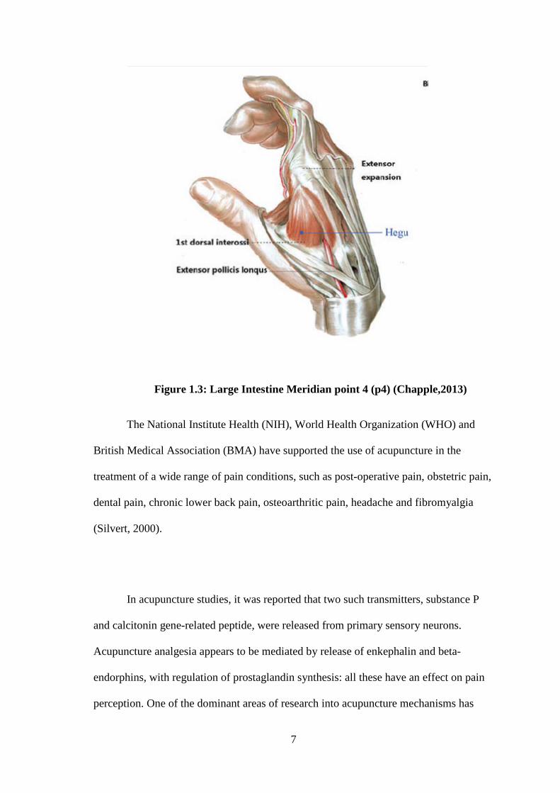
or poor health. In this study, both the Pericardium Meridian point 6 (p6) (Figure1.2) and
Large Intestine Meridian point 4 (p4) (Figure 1.3) are stimulated.
Figure 1.2: Pericardium Meridian point 6 (p6) (Chapple,2013)
7
Figure 1.3: Large Intestine Meridian point 4 (p4) (Chapple,2013)
The National Institute Health (NIH), World Health Organization (WHO) and
British Medical Association (BMA) have supported the use of acupuncture in the
treatment of a wide range of pain conditions, such as post-operative pain, obstetric pain,
dental pain, chronic lower back pain, osteoarthritic pain, headache and fibromyalgia
(Silvert, 2000).
In acupuncture studies, it was reported that two such transmitters, substance P
and calcitonin gene-related peptide, were released from primary sensory neurons.
Acupuncture analgesia appears to be mediated by release of enkephalin and beta-
endorphins, with regulation of prostaglandin synthesis: all these have an effect on pain
perception. One of the dominant areas of research into acupuncture mechanisms has

8
been its effect on endorphins. Endorphins are one of several neuropeptides that have
been shown to alleviate pain, and have been described as the body's own "opiates"
(Tobaldini et al., 2014).

9
1.3 Electroacupuncture
Electroacupuncture (EA) is different from acupuncture in that it applies needling
stimulation and electric pulses to acupuncture meridians and points in order to
strengthen the stimulating effect of treatment. However, as with traditional acupuncture,
needles are inserted on specific acupuncture points along the body. The needles are then
attached to a device that generates continuous electric pulses using small clips. These
devices are used to adjust the frequency and intensity of the impulse being delivered,
depending on the condition being treated. Electroacupuncture uses two needles at time
so that the impulses can pass from one needle to the other (Chen and Wang, 2013).
Using different frequencies via electroacupuncture specific endogenous opioid
responses have been reported (Wilkinson and Faleiro, 2007). In low frequency
stimulation (1 to 2 Hertz) there are release of endorphins and enkephalins (Aδ fibers). In
mid range frequencies (12 to 15 Hertz) stimulation results in the production of all three
opioid classes. In high freqencies (100 Hertz) results in dynorphins release and has no
effect of endorphins or enkephalins (Aβ fibers). There is no further gain in opioid
peptide release beyond 200 Hertz (Wilkinson and Faleiro, 2007).
As such, this study utilises electroacupuncture 2 Hertz (Hz) on bilateral
Pericardium Meridian point 6 (p6) and Large Intestine Meridian point 4 (p4)
intraoperatively in addition to standard opioid analgesia to capture the release of
endorphins and enkephalins to improve pain relief postoperatively.

10
Figure 1.4: Acupuncture needles attached to small clips to deliver the
electric pulses (model No SDZ-V, Suzhou Medical Appliances Co., Ltd, Suzhou,
China)
Figure 1.5: Hwato electronic acupuncture treatment instrument (model No
SDZ-V, Suzhou Medical Appliances Co., Ltd, Suzhou, China)

11
Figure 1.6: An electroacupuncture procedure (model No SDZ-IV, Suzhou
Medical Appliances Co., Ltd, Suzhou, China)

12
1.4 Use of Electroacupuncture in this study
In this study, we enrolled 64 consented elective abdominal hysterectomy patients
to study the effects of addition of electroacupuncture to standard opioid analgesia with
relation to postoperative pain and opioid related side effects. This is a randomised single
blinded study and subjects are randomised into 2 groups. This research analysed the
analgesic and antiemetic effect patients post hysterectomy within the first 24 hours.
Consented subjects were randomised into 2 groups.
Group A (electroacupuncture group): Received electroacupuncture of 2 Hertz at
continuous wave in addition to standard care.
Group B (control group): Received standard care and we did not attach any
electroacupuncture needles on the patient. Standard care was defined as general
anaesthesia for surgery like all other patients undergoing the same surgery and also
receiving standard pain relief medications as well.
We analysed postoperative analagesia via Numerical Rating Scale that is already
in use in all Ministry of Health hospitals at 30 minutes, 2 hours, 4 hours and 24 hours.
Data on the total Patient Controlled Analagesia (PCA) demand and total PCA doses in
the first 24 hours were collected. We also analysed incidence of nausea and

13
postoperative antiemesis use by looking at incidences of need of rescue pharmacologic
antiemetic in the first 30 minutes and 24 hours.
The aim of this study is to examine the effects of electroacupuncture (EA) on
reducing postoperative pain, analgesic requirement & nausea and vomiting on subjects
recovering from elective hysterectomies. The hypothesis is that patients receiving
addition of electroacupuncture would demonstrate increased analgesic and antiemetic
relief over a duration of time and reduced opioid related side effects compared to
control group.

14
CHAPTER 2
LITERATURE REVIEW
2.1 Pain
2.1.1 Pain perception
Pain is subjective and difficult to quantify. It is recognised by its international
definition to have an affective and sensory component. Although neuroanatomical basis
for pain reception develops before birth, individual pain responses are learned in early
childhood. Again, these responses are modulated based on the individuals, social,
cultural, psychological, cognitive and genetic factors (KK et al., 2010).
As such, it is not surprising that in ancient times, humankind had even viewed
pain as a form of punishment inflicted by gods or demons on humans. Early medical
beliefs of perception had often been conflicting. The great ancient Greek physician
Hippocrates believed that pain is associated with too much of blood, phlegm, yellow
bile or black bile. Some reports also say that the ancient Muslims physician Avicenna
associated pain with a change in physical condition of the body (2014).
In modern medicine, we have more understanding of pain pathways and are able
to classify it (Jessel, 1982). Pain may be classified in many ways, that is:
1. Duration: acute, chronic

15
2. Pathophysiology: nociceptive, neuropathic
3. Aetiology: arthritic, cancer pain
4. Affected area: headache, low back pain
16
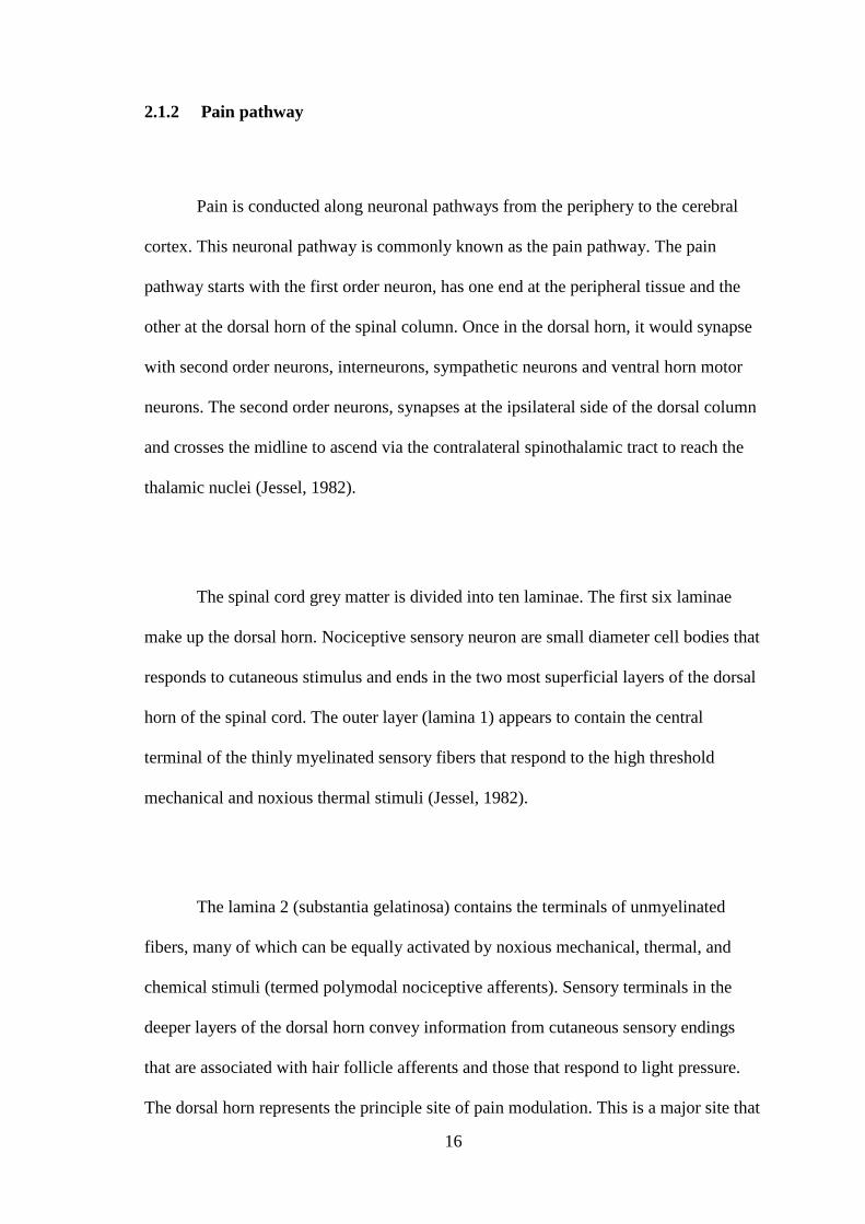
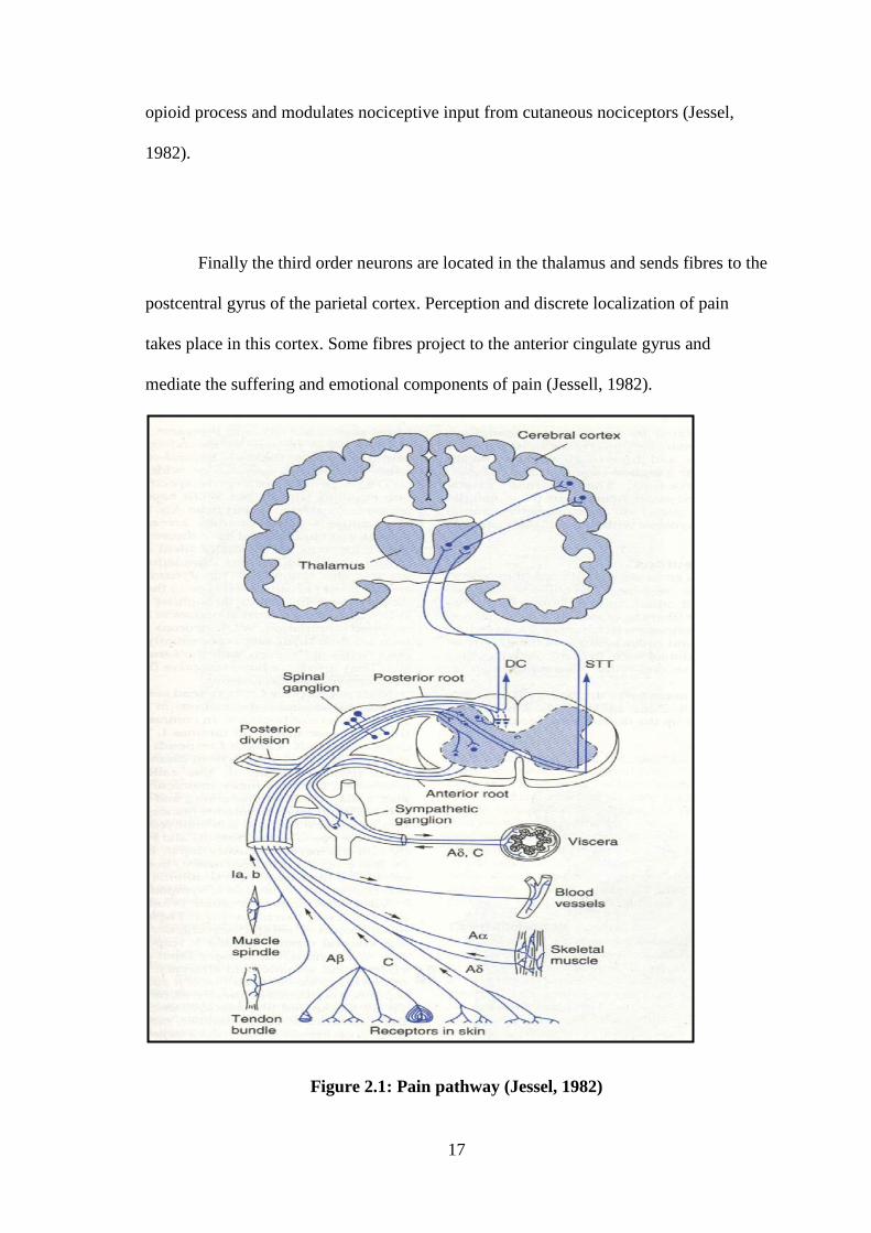
2.1.2 Pain pathway
Pain is conducted along neuronal pathways from the periphery to the cerebral
cortex. This neuronal pathway is commonly known as the pain pathway. The pain
pathway starts with the first order neuron, has one end at the peripheral tissue and the
other at the dorsal horn of the spinal column. Once in the dorsal horn, it would synapse
with second order neurons, interneurons, sympathetic neurons and ventral horn motor
neurons. The second order neurons, synapses at the ipsilateral side of the dorsal column
and crosses the midline to ascend via the contralateral spinothalamic tract to reach the
thalamic nuclei (Jessel, 1982).
The spinal cord grey matter is divided into ten laminae. The first six laminae
make up the dorsal horn. Nociceptive sensory neuron are small diameter cell bodies that
responds to cutaneous stimulus and ends in the two most superficial layers of the dorsal
horn of the spinal cord. The outer layer (lamina 1) appears to contain the central
terminal of the thinly myelinated sensory fibers that respond to the high threshold
mechanical and noxious thermal stimuli (Jessel, 1982).
The lamina 2 (substantia gelatinosa) contains the terminals of unmyelinated
fibers, many of which can be equally activated by noxious mechanical, thermal, and
chemical stimuli (termed polymodal nociceptive afferents). Sensory terminals in the
deeper layers of the dorsal horn convey information from cutaneous sensory endings
that are associated with hair follicle afferents and those that respond to light pressure.
The dorsal horn represents the principle site of pain modulation. This is a major site that
17
opioid process and modulates nociceptive input from cutaneous nociceptors (Jessel,
1982).
Finally the third order neurons are located in the thalamus and sends fibres to the
postcentral gyrus of the parietal cortex. Perception and discrete localization of pain
takes place in this cortex. Some fibres project to the anterior cingulate gyrus and
mediate the suffering and emotional components of pain (Jessell, 1982).
Figure 2.1: Pain pathway (Jessel, 1982)
18
2.1.3 Gate Control Theory
In 1965, Ronald Melzack and Patrick Wall published their paper in Science
proposing that a gating mechanism exists within the dorsal horn of the spinal cord. It
became famously known as “New Gate Control Theory of Pain”. It was succinctly
stated that the transmission of pain from the peripheral nerve through the spinal cord
was subject to modulation by both intrinsic neurons and controls emanating from the
brain.
The Gate Theory proposed that small (C) fibers activated excitatory system that
excited output cells. These latter cells have their activity controlled by the balance of
large fibers (A beta) mediated inhibitions and are under the control of descending
systems (Dickenson, 2002).
The interplay among these connections determines when painful stimuli go to the brain:
1. Without any input, the inhibitory neuron prevents the projection neuron from
sending signals to the brain (gate is closed).
2. Normal somatosensory input happens when there is more large-fiber (A beta)
stimulation (or only large-fiber stimulation). Both the inhibitory neuron and the

19
projection neuron are stimulated, but the inhibitory neuron prevents the
projection neuron from sending signals to the brain (gate is closed).
3. Nociception (pain reception) happens when there is more small (C) fiber
stimulation or only small (C) fiber stimulation. This inactivates the inhibitory
neuron, and the projection neuron sends signals to the brain informing it of pain
(gate is open).
Figure 2.2: Descending pathways from the brain close the gate by inhibiting the
projector neurons and diminishing pain perception (Dickenson, 2002)

20
2.1.4 Neurotransmitters and Pain
Nociceptors are free nerve endings that responds to multiple stimulus like heat,
mechanical, chemical, pressure. In contrast to the well-established cholinergic and
adrenergic properties of the principal parasympathetic and sympathetic neurons there is
still considerable uncertainty over the nature of the transmitters used by primary sensory
neurons (Jessel, 1982).
Alogens or pain producing transmitters identified includes bradykinin,
histamine, serotonin, prostaglandin, capsaicin, adenosine triphosphate. Several
neuropeptides and excitatory amino acids function as neurotransmitters for afferent
neuron subserving pain. Most important peptides are Substance P and calcitonin gene
related peptide (CGRP). Most important excitatory amino acid is Glutamate (Jessel,
1982).
Substance P is a transmitter synthesized and released by the first order neuron
both peripherally and in the dorsal horn. Substance P is present in 10 % to 20 % of
spinal sensory neurons in all mammalian species examined, including man, and is found
within synaptic vesicles in the central terminals of sensory neurons located in laminae I
and II of the dorsal horn (Jessell, 1982).
As for neurotransmitters in the dorsal horn involved in pain processing, they are
opioid peptides. And this is the major site for opiate-induced analgesia. Leucine and

21
methionine enkephalins, the first opioid peptides to be discovered, represent only a
small subset of the family of opioid peptides. Although there is still no direct proof that
opiate receptors located on sensory neurons contain substance P, biochemical evidence
for an interaction between opiates and substance P has now emerged in several different
systems. The specificity of these opiate effects is demonstrated by the resumption of
substance P release after administration of the opiate antagonist naloxone (Jessell,
1982).
Pain perception is also modulated by neurotransmitters from the supraspinal
level in the descending pathway. The periaqueductal grey (PAG) region and the nucleus
raphe magnus (NRM), which lies more caudally in the medulla, are among the most
effective sites in eliciting analgesia. These nuclei are also sites at which focal
microinjection of opiates has been shown to elicit analgesia, raising the possibility of
release of endogenous opioids. This association is strengthened by the finding that
opiate antagonists are often effective in abolishing analgesia elicited by brainstem
stimulation (Yu et al., 2013).

22
2.1.5 Acute Pain post Hysterectomy
Hysterectomy is the surgery to remove a woman’s uterus. Depending on the
reason of the hysterectomy, their surgeon may opt to remove all or only part of the
uterus. For example, in subtotal hysterectomy, the surgeon removes only the upper part
of the uterus, keeping the cervix in place. A total hysterectomy removes the whole
uterus and cervix. In this study we have chosen to include total abdominal
hysterectomy. In this type of surgery the surgeon opts for a midline or Pfannansteil
incision to remove the uterus (Tay and Bromwich, 1998).
There are many causes that lead the patient to opt for a surgical intervention.
Among those are uterine fibroids, endometriosis, uterine leiomyoma, uterine
adenomyosis, uterine prolapse, uterine cancer, cervical cancer or ovarian cancer. In
some of the aetiologies, the patient may have been subjected to years of some form of
abdominal pain (Tay and Bromwich, 1998).
Chronic postoperative pain can generally occur after any surgery. Interestingly,
the incidence of chronic pain post hysterectomy is up by 30 %, although the relative
role of the different pathogenic factors has not been defined. It was found in one study
involving 90 women undergoing elective hysterectomy, preoperative increased
superficial and vaginal mechanosensitivity was related to the intensity of early pain
after hysterectomy. It was correlated that poor control of acute pain in this patient
population reflected in chronic pain later during their follow up (Brandsborg et al.,
2011).

23
2.1.6 Validitation of Numerical Rating Scale (NRS)
Current guidelines on good management of pain practice includes the ability of
pain to be assesed regularly and documented. There are multiple unidimensional
measurements tools that may be used for the assessment of pain. Amongst those
available are Visual Analogue Score (VAS), Numerial Rating Scale (NRS) and Verbal
Rating Scale (VRS). All these tools are essentially used to measure the pain intensity
experienced by the patient (Huskisson, 1974).
The VAS is a 10-cm line, anchored by verbal descriptors, usually ‘no pain’ and
‘worst imaginable pain’. The patient is asked to mark a 100 mm line to indicate pain
intensity. The score is measured from the zero anchor to the patient’s mark (Figure 2.3)
.
Figure 2.3: Visual Analog Score (Huskisson, 1974)
24
One of the limitations of the VAS is that it must be administered on paper or
electronically (Carlsson 1983). Interestingly, the graphic orientation of the VAS
whether horizontally or vertically can make a difference to the statistical distribution of
the data obtained using it. A study by Huskisson, 1974 explored the use of the VAS by
English language speakers found that there was a 7% failure rate for the VAS when it
was presented vertically but less when presented horizontally. They postulated that the
graphic orientation of the VAS should be decided according to the normal reading
tradition of the population on which it is being used.
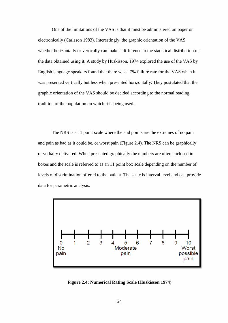
The NRS is a 11 point scale where the end points are the extremes of no pain
and pain as bad as it could be, or worst pain (Figure 2.4). The NRS can be graphically
or verbally delivered. When presented graphically the numbers are often enclosed in
boxes and the scale is referred to as an 11 point box scale depending on the number of
levels of discrimination offered to the patient. The scale is interval level and can provide
data for parametric analysis.
Figure 2.4: Numerical Rating Scale (Huskisson 1974)