the effects of estradiol benzoate on protein synthesis by
TRANSCRIPT

University of RichmondUR Scholarship Repository
Master's Theses Student Research
Summer 1968
The effects of estradiol benzoate on proteinsynthesis by the isolated perfused snake liverWilliam Ralph Boone
Follow this and additional works at: http://scholarship.richmond.edu/masters-theses
This Thesis is brought to you for free and open access by the Student Research at UR Scholarship Repository. It has been accepted for inclusion inMaster's Theses by an authorized administrator of UR Scholarship Repository. For more information, please [email protected].
Recommended CitationBoone, William Ralph, "The effects of estradiol benzoate on protein synthesis by the isolated perfused snake liver" (1968). Master'sTheses. Paper 281.

THE EFFECTS OF ESTRADIOL BENZOATE
ON PROTEIN SYNTHESIS
BY THE ISOLATED PERFUSED SNAKE LIVER
Approved:
& ~ _cj__-4.L~-Dean of the Grad7aieSchool
'Examining Committee:

THE EFFECTS OF ESTRADIOL BENZOATE
ON PROTEIN SYNTHESIS
BY THE ISOLATED PERFUSED SNAKE LIVER
by
William Ralph Boone
A thesis submitted to the faculty of the Graduate School
of the University of Richmond in partial fulfillment of
the requirements _for the Degree of Master of Science.·
August, 1968
LIBRARY UNIVERSITY OF RICHMOND
ViRGiNIA

TABLE OF CONTENTS
I. Abstract. •o•o•••••••o••••••ooo•oooooooo 1
II. Acknowledgements~ ············ 2
III. Introduction, ......................... • •••• 0 • 3
iV. Materials and Metho-ds · ..... o ••••• o •• ·• ••••••••• ~ .. • • • • • • • • 5
V. Results .. ··-· . ............................................ 11
VI. Discussion ................ . ....•.....•............... 13
VII. Literature Cited . ....................................... 1 7
VIII. List of Tables .................................. . • ••. 0 19
1. Effect of Estradiol Benzoate on Incorporation of Carbon-14 Leucine into Plasma Protein .•••••• , •
2. Effect of Estradiol Benzoate on Plasma
• • • 1 9
Protein Levels ........... o ••••••• °' ••••••••••••••••• 20
3. Effect of Estradiol Benzoate on Protein Concentration in Plasma Fraction IV •••
4. Effect of Estradiol Benzoate on Protein
0 0 0 G 0 0 0 o 0 0 0 0 0 o .21
Concentration in Plasma Frc:ction II •••••••••••••••••• 22
IX,. List of Figures ..... .......................... o •••••• · ••• 23
1. Loss of Radioactivity From Whole Perfusate in Control and Estradiol Treated Livers •••••.••••••••••• 23
2. Effect of Estradiol Benzoate on Incorporation of Carbon-14 Leucine into Plasma Proteins •••.•••.•••.•. 24
3. Effect of Estradiol Benzoate on Plasma Protein Levels o •••• o •••••••••••••• • .- •· •••••••••••••••••••••• 2 5
UBRt\RV UNiVERSlTY o;=· RICHMOND
ViRGlNlA

4. Electrophoretogram of the Plasma Proteins of Natrix fasciata fasciata .. o. o •••••••••• •••••••••••••• • 26
5. Effect of Estradiol Benzoate on Protein Concentration in Fraction IV •••••••••••••••••••.••••• 27
6. Effect of Estradiol Benzoate on Protein Concentration in Fraction II •••••••••••••••••••••••••• 28
X. Vita . .................... " ........... ~ ................. 29

1.
ABSTRACT
The effects of estradiol benzoate on protein synthesis by the
isolated perfused liver of Natrix fasciata fasciata (Linnaeus, 1766),
the southern banded water snake, were investigated. The incorpora
tion of carbon-14 labeled leucine into plasma protein of the liver per
fusate was used as an index of protein synthesis by the perfused liver.
Both control and experimental livers demonstrated incorpora
tion of the labeled leucine into plasma protein. However, estrogen
tr·eated livers expressed significantly greater incorporation of the
amino acid into plasma protein giving an almost linear response.
Total plasma protein levels were also significantly higher in the
perfusions involving estrogen treated livers.
Electrophoresis of perfusate plasma revealed five distinct
protein fractions. Two of these fractions increased significantly
in the perfused livers treated with estradiol.

2.
ACKNOWLEDGEMENTS
I would like to express my sincere gratitude to Dr. Francis B.
Leftwich for his timely advice and close guidance throughout the con
ception and formulation of this thesis.
I would also like to thank the following; Drs. W. S. Woolcott and
W. M. Reams, Jr. for their constructive evaluations of this thesis;
Dr. W. H. Leftwich of the Department of Psychology, University of
Richmond, for his help in conducting the statistical analyses; and Mrs.
Jane Atkinson of the Medical College of Virginia for her help in· the
clarification of electrophoretic data. Appreciation is also expressed to
my wife, Mary Lou; for many hours spent in the typing of this thesis.

3.
INTRODUCTION
During the estrus cycle of Thamnophis sauritus, the viviparous
ribbon snake, increases in plasma vitellin (phosphoprotein) levels and
liver weight ha'.ve been shown to occur in a cycle corresponding to de
velopment of the ovarian follicle. Shortly before ovulationll liver w-eight
begins to decline and plasma vitellin levels fall to ane strus concentra
tions (Dessauer and Fox 1956 and 1959). Based on these findings
and the fact that yolk vitellin has been found to be irn.munologically
similar to plasma vitellin, these workers proposed the liver to ·be
the primary site of yolk protein ~ynthe sis.
Other. investigations have demonstrated that injections of
estracliol, into both males and anestru_s females of oviparous and
viviparous reptilian species, elicit changes characteristic of estrus
females. Dessauer and Fox (1959) found that injections of estradiol
into male ribbon snakes induced liver enlargement and de~
synthesis of plasma vit~llin. Similar responses to estradiol were
observed by Clark (1967) in male Chrysemys picta, the painted
turtle. Significant increases in total serum protein were observed
by one week post injection.
On the premise that estradiol induced vitellinogenesis would
first be preceded by synthesis of RNA, Hahn and Gorbman (1967)
compared liver RNA concentrations in estrogen injected and control
lizards (Uta stansburiana). It was found that liver RNA synthesis
was increased up to 200% in animals receiving injections of estradiol.

The present study was devised" to examine the effects of
estradiol benzoate on P!Otein synthesis by the perfused liver of
N. fasciata fasciata. Carbon-14 leucine served as a tracer com
poundin observing the effect of the hormone on the rate of protein
syn the sis by the liver. Sin:ce Miller "(1951} clearly ~ernonstrated
4.
that the mammalian liver functions in the synthesis of plasma proteins,
it was assumed that the reptilian liver also functions in this capacity,
It was therefore expected that an increase in protein synthesis would
be rna.nifest by marked incorporation of the labeled leucine. Thus,
data concerning the presence of this radioactive amino acid in plasma
protein, coupled with other parameters such as total plasma protein
levels and quantitation of various plasma protein fractions would
serve in part in determining if the liver, isolated from the animal
and hence from other tissue influences, does indeed function in the
synthesis of plasma protein in response to estradiol.
MATERIALS AND METHODS
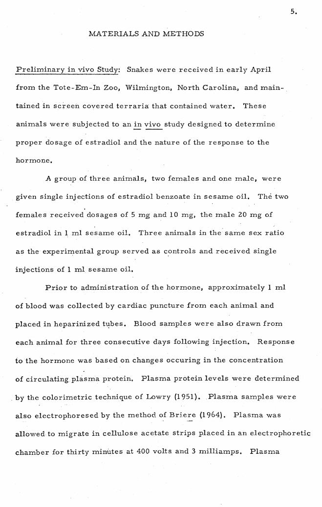
Preliminary in vivo Study: Snakes were received in early April
from the Tote-Em-In Zoo, Wilmington, North Carolina, and main
tained in sc~ee:b. covered terraria that contained water. These
animals were subjected to an in vivo study designed to determine
proper dosage of estradiol and the nature of the response to the
hormone.
A group of three animals, two females and one male, were
given single injections of estradiol benzoate in sesame oil. The two
females received dosages of 5 mg and 10 mg, the male 20 mg of
estradiol in 1 ml sesame oil. Three animals in the same sex ratio
as the experimental group served as controls and received single
injections of 1 ml sesame oil.
5.
Prior to administration of the hormone, approximately 1 ml
of blood was collected by cardiac puncture from each animal and
placed in heparinized t~bes. Blood samples were also drawn from
each animal for three consecutive days following injection. Response
to the hormone was based on changes occuring in the concentration
of circulating plasma protein. Plasma protein levels were determined
. by the colorimetric technique of Lowry (1951). Plasma samples were
also electrophoresed by the method of Briere (1964). Plasma was
allowed to migrate in cellulose acetate strips placed in an electrophoretic
chamber for thirty minutes at 400 volts and 3 milliamps. Plasma

6.
protein fractions were then quantitated with a recording densitometer.
Plasma protein levels in the animal receiving 5 mg estradiol
·were found to exhibit the greatest increase. This animal had an initial
plasma protein level of 6. 2 gm %. By the third day after injection of the
hormone the total protein level had increased to 8, 1 gm %. The two
animals receiving 10 mg and 20 mg of estradiol showed increases ·in
total protein from 5.4 gm % to 6.8 gm %, and from 4. 7 gm % to 5. 7 gm %
respectively over a three day period. Control animals injected with
sesame oil showed slight variations ranging from . 20 - • 50 gm % over
the same period, but did not show definite increases. These results demon
strate that a response to estradi?l could be induced in~ !.:_ fasciata,
therefore establishing the feasibility of a perfusion study with this species,
Clark (1967) demonstrated that_ dose levels as low as 20 micrograms
were capable of inducing protein synthesis in painted turtles. This is
in contrast to dose levels as high as 1 mg administered by Dessauer and
Fox {1956) to the ribbon snake. The much larger concentrations of
estradiol administered _in the present study suggested that dose levels of
a pharmacological as well as a physiological nature were capable of in
ducing protein synthesis. However, since large doses of estradiol are·
known to inhibit protein synthesis in mammals, it is possible that the
. 10 mg and 20 mg doses administered would have produced this effect if
observed over a longer period of time. From this study and previous
work it appeared that a dosage range of 20-5000 micrograms of estradiol
would elicit protein synthesis without inhibitory influences.

Because estradiol would be rnore concentrated when administered
directly to the isolated liver, a working dose of 200 micrograms
was selected for the perfusion studies.
Plasma samples were found to resolve electrophoretically
into five protein fractions. These fractions were identified with
respect to their rate of migration (Tondo 1958). Although the re
lative percentage of each fraction varied throughout the three days
of this .study, no changes were observed in the quantitative distribu
tion of the five fractions in control or estradiol injected animals.
Liver Perfusions: A second group of snakes obtained in June from
the same source served as blood and liver donors for the perfusion
studies.
7.
Appr6ximately 24 hours. prior to perfusion two or three blood
donor snakes (anestrus females) were exsanquinated by heart puncture,
A total volume of 50-60 ml of whole blood was obtained. The blood
was heparinized and diluted with Krebs-Ringers bicarbonate solution
(Umbreit et al. 1959) to a final volume of 80-90 ml, making a 60 %
whole blood perfusate. The perfusate W(:lS stored for approximately
20 hours at 7° C.
One-half four before the start of each perfusion, the perfusate
was supplemented with approximately ten microcu_r_~es of carbon-14
labeled leucine (in 0.1 N.HCl, New England Nuclear Corporation) and
75 mg glucose, added to inhibit gluconeogenesis. The perfusate was

8.
then placed in the perfusate reservoir of the perfusion apparatus and
circulated until the beginning of perfusion to insure proper oxygenation
and temperature equilibrium. The perfusion apparatus was that
described by Turner (1967).
Snakes utilized as liver donors were pithed and secured to an
· · operating platform. The livers were exposed and the portal and post
caval veins cannulated as described by Turner (1967}. After cannulation,
each liver was removed by severing the animal approximately one-
half inch in front and back of the liver, thus removing a portion of
. the body with the cannulated liver still resting in situ. The encased . -.--
liver was then connected in the p.erfusion apparatus with the portal
vein cannula receiving the flow of perfusate.
A total of eight livers were pe:r:fused. Four of these (one
male and three anestrus femaies) received O. 2 mg estradiol
benzoate (Nutritional Biochemicals Corporation) in O. 1 mi 95% ethanol.
One hour after the start of each perfusion, the hormone was injected
into the open arm of the "lung" of the perfusion apparatus and rapidly
mixed with the perfusate. In the four remaining perfusions (three males
~nd and one anestrus female} the livers were not treated with exogenous
estradiol and thus served as controls.
In each perfusion, 1 ml samples of perfusate were withdrawn
at hourly intervals for six hours after the ~~-art of perfusion. Each
perfusate sample was analyzed for total radioactivity, plasma protein
radioactivity, and plasma protein concentration. In addition, an

9.
aliquot of each perfusate sample was electrophoresed and the plasma
protein fractions analyzed as described in the preliminary study.
Total perfusate radioactivity was measured by placing
O. 1 ml of each perfusate sample onto a planchet (1. inch in diameter),
air drying and reading in a thin window (100 micrograms/ cm2) gas-
flow Geiger-Muller detector operated at 1450 volts. A counting
time of five minutes, which gave a minimum of 200 counts above
backgr~mnd, was employed.
In order to analyze the radioactivity of the protein in perfusate
samples it was first necessary to_ isolate the plasma proteins. This
was accomplished by precipitating the proteins from a O. 1 ml aliquot
of perfusate with 5% trichloroacetic acid (TCA). The protein precipitate
was washed several times with 5% TCA. The final precipitates were
solubilized in 10% KOH and placed on planchets. After air drying, the
radioactivity was determined as described previously. Plasma protein
concentration of each perfusate sample was measured by the Lowry
technique.
Previous in vivo studies by Dessauer and Fox (1959) and Clark
(i 967) demonstrated that estradiol would_ induce the same plasma changes
characteristic of estrus in both male and female snakes and fresh water
turtles. Therefore, data obtained from males and females in both
the control and experimental group in the present study were pooled
and expressed as mean values.
A two factorial statistical design with analysis of variance was

utilized in the treatment of all data in this study (Winer. 1962).
Significant values were expressed at the • 05 level of confidence.
1 o.

11.
RESULTS
Uptake of carbon-14 leucine by the liver was measured by
loss of radioactivity from the circulating perfusate. Decrease in
radioactivity from the whole perfusate, expressed as a per cent of
the initial activity, was found to be enhanced in the experimental
group as early as two hours after administration of estradiol (fig. 1).
During .the sixth hour of perfusion, the loss of radioactivity in the
control group surpassed that in the estrogen treated group. This
is most likely due to the fact t.hat protein synthesis by experimental
livers was higher and therefore a higher percentage of radioactivity
in the form of protein would be leaving the experimental liver and
returning to the perfusate.
Incorporation of carbon-:14 leucine into plasma protein was
expressed as cpm/mg protein. Values from two through six hours
·were expressed relative to the value obtained at one hour of perfusion.
·There was a significant difference between means of the experimental
and control groups at all time intervals (table 1 ). Incorporation of
the carbori-14 leucine was slight for both. groups during the first hour
of perfusion. However, from the second through the sixth hour, livers
treated with estradiol exhibited an almost linear relationship with
time, increasing from • 01 to . 58 cpm/mg protein._._ Control livers
remained relatively constant over the same time period (fig. 2).
LIBRARY UNIVERSITY OF RICHMOND.
VIRGiNIA

12.
Plasma protein levels remained relatively constant in control
livers throughout the five hours of perfusion. After administration
of the hormone, however, there was an initial increase followed
by a drop in plasma protein concentration in the third hour of per
fusion. This drop in concentration was followed by an increase in·
the fourth and fifth hours (fig. 3, table 2). Although fluctuations
existed, a significant increase with time was found in the estrogen
treated. group when compared with controls.
Plasma proteins resolved electrophoretically into five fractions
(fig. 4). Although each of the fractions in both experimental and
control groups was found to vary slightly in per cent composition,
only fraction II and Nin the experimental group increased significantly.
Fraction IV exhibited a significant cummulative increase over controls
(fig. 5, table 3), while fraction. II increased significantly from the
second through the fifth hour of perfusion (fig. 6, table 4).

13.
DISCUSSION:
Both uptake and incorporation of carbon-14 leucine into plasma
protein by estradiol treated livers were found to be enhanced in this
study. These findings are in agreement with other studies (Noall,
et al. 1957; Daniels and Kalman 1961} which showed that uptake of
the model amino acid, alpha-aminoisobutyric acid (AIB} into the
uterus .and liver of immature rats was enhanced by injections of
estradiol. Results such as these have led to suggestions that the
mode of action of estradiol is_ related to alteration in permeability
of the cell membrane, or at least in part, may be concerned with
the regulation of the biosynthesis of enzymes involved in protein
synthesis (Mueller 1958}.
Studies by Frieden (195_2) and Mueller (i 952) have shown that
injection of estradiol into rats and pigs increases the rate of incorpora
tion of radioactive amino acids into uterine proteins (in vitro}. Mueller
(1958) demonstrated that intravenous injections of estradiol into rats
elicited significant increases (within four hours) in the incorporation
of labeled glycine into uterine protein (in vivo). Although these studies
do not serve in deciphering the mode of action of estradiol, it is
apparent that uptake and incorporation of labeled amino acids into
protein can be induced in liver and uterine tissue !?Y this hormone.
In the present study, perfusions involving livers treated with
estradiol showed a significant increase in plasma protein with time
14.
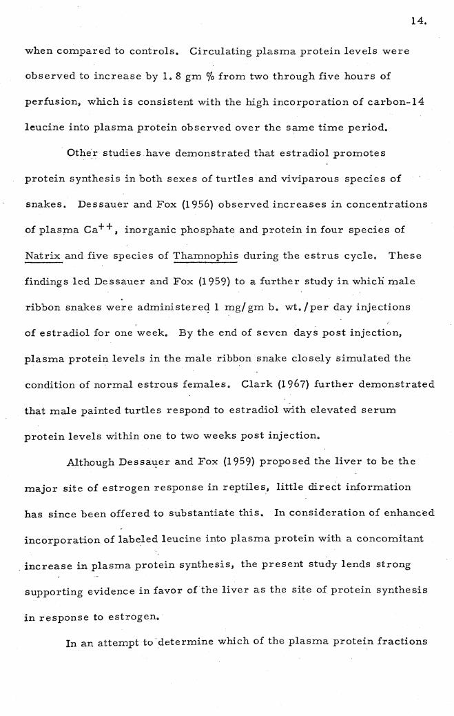
when compared to controls. Circulating plasma protein levels were
observed to increase by 1. 8 gm % from two through five hours of
perfusioni which is consistent with the high incorporation of carbon-14
leucine into plasma protein observed over the same time period •
. Othe:r studies have demonstrated that estradiol promotes
protein synthesis in both sexes of turtles and viviparous species of
snakes. Dessauer and Fox (1956} observed increases in concentrations
of plasma ca++' inorganic phosphate and protein in four species of
Natrix and five species of Thamnophis during the estrus cycle. These
findings led Dessauer and Fox (1959) to a further study in which male
ribbon snakes were administereq 1 mg/ gm b. wt. /per day injections
of estradiol for one week. By the end of seven days post injection,
plasma protein levels in the male ribb.on snake closely simulated the
condition of normal estrous females. Clark (1967} further demonstrated
that male painted turtles respond to estradiol with elevated serum
protein levels within one to two weeks post injection.
Although Dessa~er and Fox (1959) proposed the liver to be the
major site of estrogen response in reptiles, little direct information
has since been offered to substantiate this. In consideration of enhanced
incorporation of labeled leucine into plasma protein with a concomitant
. increase in plasma protein synthesis, the present study lends strong
supporting evidence in favor of the liver as the site of protein synthesis
in response to estrogen.·
In an attempt to· determine which of the plasma protein fractions

15 •.
were involved in the estrogen response, quantitation of the five plasma
fractions resolved by electrophoresis was performed. Significant
differences between fractions in control and estradiol treated perfusions
were found only in fractions II and IV •
. Dessauer and Fox (1958) demonstrated the appearance of a "new"
band in the electrophoretic patterns of female snakes (~ sauritus} in
estrus. This fraction was postulated to be the phospholipoprotein
involve.cl in vitellinogenesis, for it disappeared soon after the onset of
ovulation when circulating levels of estrogen were once again low in
the serum. Although these workers utilized paper electrophore"sis,
fraction IV in the present study ~sing cellulose acetate closely
approximated the migratory position of the phospholipoprotein band
described for T. sauritus. Based on this observation and the fact
that specific staining technigues reveal fraction IV to contain lipo
protein (Turner 1967), it appears that the protein increase in this
fraction may be similar to the response observed in the phopholipoprotein
band in T. sauritus.
The nature of fraction II is more elusive. Rao and David (1967),
demonstrated in a study of Uromastix hardwickii, the spiny tail lizard ·
that the alpha-one fraction (Tiselius 1 s nomenclature) responded in
. instances where protein balance in the serum was disturbed. Since
fraction II of the present study has approximately the same migratory
position as alpha-one it might be suggested that the mild state of
"shock" under which the perfused liver functions is responsible for the
16.
increase in protein in that fraction. Another possibility is based on the
study of Seal and Doe (1962) who found an increase of an alpha-one
binding globulin in diethylstilbestrol - treated humans. This steroid
binding protein was further found to increase _in concentration during
pregnancy. It is conceivable then, that by four hours after administration
of estradiol the snake liver in the present study had begun to synthesize
a binding protein which perhaps bands with fraction II.

Literature Cited
Briere, R. · O. and J. D. Mull. 1964. Electrophoresis of serum protein with cellulose acetate. Am. J. Clin. Path. 42: 54 7.
Clark, N. B. 1967. Influence of estrogens upon serum calcium, phosphate and protein concentrations of fresh-water turtles. Comp. Biochem. Physiol. 20:823-834.
Daniels, J. R. and S. M. Kalman. 1961. Time course of aminoisobutyric acid 1-c 14 uptake by estrogen stimulated rat uterus. Fed. Proc. 11: 262.
Des sauer, H. C., W. Fox, and N. C. Gilbert. 1956. Plasma calcium, magnesium and protein of viviparous colubrid snakes during estrous cycle. Proc. Soc. 92:299-301.
, and W. Fox. 1958. Geographic variations in plasma -----protein patterns in snakes. Proc. Soc. 98:101-105.
1 7.
1959. Changes in ovarian follicle composition with plasma ----..,...-levels of snakes during estrus •. Am. J. Physiol. 197:360-366.
Frieden, E. H., A. C. Steele, and M. A. Telfer. 1952. · Hormonal effects on the incorporatibn of labeled amino acids in tissue of the reproductive tract. Fed. Proc. 11: 215 •.
Hahna W. E. and A. Gorbman. 1967. Liver RNA synthesis associated with estradiol-induced vitellinogenesis. Proc. N. W. Reg. Endocrinol Soc.
Lowry,. O. H. 1951. Protein measurement with the Falin phenol reagent. J. ·Biol. Chem. 193:267-276. ·
Miller, L. L., C. G. Bly, M. L. Watso_n and W. F. Bale. 1951. The dominant role of the liver in plasma protein synthesis. J. Exp. Med~ 94:431.
Mueller, G. C. and G. N. Yanagi. 1952. Activating influence of estradiol on the in vitro incorporation of glycine-2 C 14 uptake by estrogen stimulated rat uterus. Fed. Proc. 11: 262.
, G. C., A. M. · Herranen, and K. F. Jervell. 1958. -----Studies on the mechanism of action of estro.gens. Recent Progr. Hormone Res. 14:95.

18.
Neall, M. W., T. R. Riggs, L. M. Walker, and H. N. Christensen. 1957. Endocrine control of amino acid transfer. Science. 126:1002.
Rao, C. A. and G. F. David. 1967. The effect of certain steroids on the serum protein concentrations of the lizard, Uromastix hardwickii Gray. Gen. and Comp. Endocrinol. 9:227-233.
Seal, U. S •.. and R. P. Doe. 1963~ Corticosteriod-binding globulin: species distribution and small scale purification. Endocrinol. 73:371.
Turner, J. E. 1967. A metabolic study of the isolated perfused snake liver. (Masters Thesis), Univ. of Richmond, Dept. of Biology.
Tondo, C. V. 1958. Paper electrophoresis differences between turtle and human serum. Comp. Biochem, Physiol. 9:137-149.
Umbreit, W.W., R.H. Burris, andJ. F. Stauffer. 1959. Ma"nometric Technique.s. Burgress Pub. Co., Minneapolis, 305 p.
Winer, B. J. 1962 .. Stastical Principles in Experimental Design. McGr.aw Hill N. Y.

TABLE 1
Effect of Estradiol Benzoate on Incorporation of Carbon-14 Leucine into Plasma Proteins
(cpm/mg protein)
Perfus.ion Time (hours')
2 3 4 5 6
Experimental • 01+. 03 • 22+. 02 .36+.06 .45+.06 • 58+. 09 - - - - -
Control -.zz+.os - • 18+. 11 -.13+.09 -.17+.08 -.15+.08 - - . - - -
* Each number represents the mean of the algebraic differences
from one hour value + standard error of the mean.

TABLE 2
Effect of Estradiol Benzoate on Plasma Protein Levels
{gm/ 100 ml perfusate)
Perfusion Time (hours)
0 1 ·2 3 4 5 I '
Experimental *4.1+1. l 4. 9+1. 0,. 5.3+1.0 4.4+.95 5.l+.52 6.0+.97 - - - - -..
Control *4.5+1.0 4. 3+1. 1 4. 3+1. 2 4.5+1.0 4. 4+1. 1 4. 4+1. 1 - - - - - -
~< Each value represents the mean + standard error of the mean of four Perfusions .•
N 0 .

Fraction N
Experimental
Control
TABLE 3·
Effect of Estradiol Benzoate on Protein Concentrations
(gms/ 100 ml perfusate) in Plasma Fraction IV
Perfusion Time (hours)
0 l 2 3 4 5
>!<l. 3+. 21 l. 46+. 24 1.55+.28 1.53+.22 1. 44+. 38 2.30+.35 - - - - - -
*l. 5+. 24 l.40+.23 1.50+.22 2.15+.28 1.33+.22 1.42+.26 - - - - - -
* Each value represents the mean + standard error of the mean of four perfusions.
N ......

Fraction II
Experimental
Control
TABLE 4
Effect of Estradiol Benzoate on Protein Concentrations
(gms/ 100 ml perfusate) in Plc:sma Fraction: II
Perfusion Time (hours) ·
0 1 2 3 4 5
>."<. 28+. 09 • ~;8+. 09 . 23+. 09 • 71+. 02 • 74+. 07 .84+.08 - - - - - -
):c. 25+. 08 • 28+. 07 • 14+. 07 • 36 +. 05 .19+.04 • 24+. 08 - - - - - -
Each value represents the mean+ standard _error of the mean of four perfusions.
N N .

23.
FIGUI_tE I
Loss of Radioactivity From Whole Pei:fusate in Control and Estradiol
Benzoate Treated Livers
Administration of estradiol occured 1. 0 hour after perfusion in the ex
perimental group. Values represent means.

-~ Q)
0..
> ........ u <U 0
-0 <U
0:::
<U
·-c
E 0 ~
l.J... (/)
(/)
0 _J
....., c o..; u
50
401
30
10.
1 3
/
Experimental
- -·Control
4 5 6
Per f us i on Ti me I Hours J

24.
FIGURE 2
·Effect of Estradiol Benzoate on Incorporation of Carbon-14 Leucine
into Plasma Proteins
Each point represents the mean of the algebraic differences from the
one hour value + standard error of the mean.

.60 .
. so·
.40i
.30~
x -Experimental
c:: Cl> A-Control ·- :J
I Cl> ro .20' ~ > 0 ~
a.. ~· I: .
bD
E 0 .io: ~_: E _: 0. Q)
u a::
o.
·-.20:
Pe r f LI s i on Tim e [ H o LI r·s l

25.
FIGURE 3
Effect of Estradiol Benzoate on Plasma Protein Levels
(gm/ 100 .ml perfusate)
Each value represents the mean + standard error of the mean.
· Administration of estradiol occured 1. 0 hour after perfusion in the ex
perimental group.

7 '.
0 2
ui Experimental
::::i Control
3 4
Perfusion Time [Hours]
5

26.
FIGURE 4 .
Electrophoretogram of the Plasma Proteins of Natrix fasciata fasciata

0

27.
FIGURE 5
Effect of Estradiol Benzoate on Protein Concentration in Fraction JV
(gm/ 100 ml perfusate)
Each value represents the mean+ standard error of the mean of four
perfusions.

2.J
22
2.0'
Gm%
1.8
14· . ,
1.2
to~ ... 0
-
~ .-+-lH;Hl
-
1
....... Experimental
CJ Control
·-r-
~ r-t-- 11fili1l
-
2 3
-
~ r-t-m im
mtim -
.4
Perfusion Time !Hours!
n ~;:::::::
111111111
:::::::: .-+--'-fimm
5

28.
FIGURE 6
Effect of Estradiol Benzoate on Protei.n Concentration in Fraction II
'gm/ 100 ml perfusate)
Each value represents the mean + standard error of the mean of four
perfusions.

.90.
so: .
. SO
Gm% .50
.40
.30
.20 .
. 10 ··.
0
rm Experimental
::::::: Cont ro I
Pe r f LI s i o n T i m e [ H-o LI r s J

29.
VITA
William Ralph Boone was born on October 6, 1942 in Hopewell,
Virginia. He attended Hopewell High School and completed his second
ary education in June, 1962. He began college study at Richard Bland
College in Petersburg, Virginia, and in June 1966 received his
Bachelor of Science Degree in Biology from the College of William and
Mary •. He was married June, 1966 to the former Miss Mary Lou
Chipley. They have one child, a daughter. He began graduate study
. at the University of Richmond in September, 1966. While at the
University he was initiated into the Beta Beta Beta Honorary Biological
Society. He completed his requirements for the Master of Science
Degree in August,. 1968. He has accepted an appointment as Instructor
of Biology at· University College of the University of Rich1nond for the
1968-69 term.