the effects of physical activity on functional mri...
TRANSCRIPT
ORIGINAL RESEARCH ARTICLEpublished: 12 March 2013
doi: 10.3389/fnhum.2013.00072
The effects of physical activity on functional MRI activationassociated with cognitive control in children: a randomizedcontrolled interventionLaura Chaddock-Heyman1*, Kirk I. Erickson2, Michelle W. Voss3, Anya M. Knecht1,Matthew B. Pontifex4, Darla M. Castelli 5, Charles H. Hillman6 and Arthur F. Kramer 1
1 Department of Psychology, The Beckman Institute for Advanced Science and Technology, University of Illinois at Urbana-Champaign, Urbana, IL, USA2 Department of Psychology, University of Pittsburgh, Pittsburgh, PA, USA3 Department of Psychology, The University of Iowa, Iowa City, IA, USA4 Department of Kinesiology, Michigan State University, East Lansing, MI, USA5 Department of Kinesiology and Health Education, The University of Texas at Austin, Austin, TX, USA6 Department of Kinesiology and Community Health, University of Illinois at Urbana-Champaign, Urbana, IL, USA
Edited by:
Burkhard Pleger, Max PlanckInstitute for Human Cognitive andBrain Sciences, Germany
Reviewed by:
Marco Taubert, Max-Planck-Institutefor Human Cognitive and BrainSciences, GermanyHubert R. Dinse, Ruhr-UniversitätBochum, Germany
*Correspondence:
Laura Chaddock-Heyman,Department of Psychology, TheBeckman Institute for AdvancedScience and Technology, Universityof Illinois at Urbana-Champaign,405 North Mathews Avenue,Urbana, IL 61801, USA.e-mail: [email protected]
This study used functional magnetic resonance imaging (fMRI) to examine the influence ofa 9-month physical activity program on task-evoked brain activation during childhood. Theresults demonstrated that 8- to 9-year-old children who participated in 60+ min of physicalactivity, 5 days per week, for 9 months, showed decreases in fMRI brain activation in theright anterior prefrontal cortex coupled with within-group improvements in performance ona task of attentional and interference control. Children assigned to a wait-list control groupdid not show changes in brain function. Furthermore, at post-test, children in the physicalactivity group showed similar anterior frontal brain patterns and incongruent accuracyrates to a group of college-aged young adults. Children in the wait-list control group stilldiffered from the young adults in terms of anterior prefrontal activation and performanceat post-test. There were no significant changes in fMRI activation in the anterior cingulatecortex (ACC) for either group. These results suggest that physical activity during childhoodmay enhance specific elements of prefrontal cortex function involved in cognitive control.
Keywords: activation, brain, children, fitness, fMRI, physical activity
INTRODUCTIONPhysical activity and higher aerobic fitness are associated withimproved brain function across the lifespan (Kramer et al., 1999;Hillman et al., 2005, 2009; Davis et al., 2011; Kamijo et al., 2011;Pontifex et al., 2011; Voss et al., 2011; Chaddock et al., 2012a).Prior studies have reported that substantial effects of physicalactivity occur on tasks that measure cognitive control (Hawkinset al., 1992; Colcombe and Kramer, 2003; Tomporowski et al.,2008), which refers to aspects of cognition that describe the abil-ity to flexibly adapt behavior toward specific goals, maintenanceof these goals, monitoring of errors, and formulation of decisions(Botvinick et al., 2001; Braver and Barch, 2006). To achieve highlevels of cognitive control, individuals must be able to selectivelyattend to relevant information, filter distractions, and inhibitinappropriate response tendencies (Bunge and Crone, 2009). Ofparticular interest to this investigation, higher fit and physicallyactive children have been found to outperform their lower fitpeers on tasks of cognitive control [e.g., flanker tasks (Hillmanet al., 2009; Pontifex et al., 2011; Voss et al., 2011; Chaddocket al., 2012a), Stroop tasks (Buck et al., 2008), n-back tasks(Kamijo et al., 2011), and real world street crossing multitaskingparadigms (Chaddock et al., 2012b)].
Here, functional magnetic resonance imaging (fMRI) wasused to examine the influence of a 9-month physical activity
program on brain activation patterns associated with cognitivecontrol during childhood. Only a few studies with children haveused fMRI to examine how physical activity and aerobic fitnessrelate to brain function during tasks engaging cognitive control(Chaddock et al., 2011; Davis et al., 2011; Voss et al., 2011). In onestudy by Davis et al. (2011), overweight children (age 7–13 years)involved in 13 weeks of aerobic games showed improvements incognitive control (i.e., a “planning” score said to measure strat-egy and self-regulation) and increases in frontal fMRI activationduring an antisaccade task, which provides a measure of responseinhibition. However, the interpretation of the fMRI results wasconstrained because performance on the antisaccade task was notreported. Whereas Davis et al. (2011) suggested that increasedfrontal activation with physical activity may be associated withimprovements in cognitive control, two other studies showedthat decreased frontal activation in higher fit children was asso-ciated with better cognitive control (Voss et al., 2011; Chaddocket al., 2012a). In a study by Chaddock et al. (2012a), childrenwith higher aerobic fitness levels showed reduced activation inthe frontal cortex from early to late stages of a flanker task, cou-pled with maintenance of attentional and interference control.It is noteworthy that these fitness differences in activation wereonly apparent for incongruent flanker trials that required sub-stantial cognitive control. During congruent trials, both higher fit
Frontiers in Human Neuroscience www.frontiersin.org March 2013 | Volume 7 | Article 72 | 1
HUMAN NEUROSCIENCE

Chaddock-Heyman et al. Physical activity and fMRI activation
and lower fit children showed decreases in activation and main-tenance of task performance. In conjunction with these findings,Voss et al. (2011) showed that higher fit children exhibited lessactivation than lower fit children in a network of brain regionsincluding anterior frontal areas involved in task maintenanceand cognitive control, coupled with higher accuracy rates duringincongruent flanker task trials that required increased cognitivecontrol. Together, previous studies suggest that physical activityand aerobic fitness influence brain function in regions such as thefrontal cortex as well as the ability to adapt neural processes tomeet and maintain task goals (Davis et al., 2011; Voss et al., 2011;Chaddock et al., 2012a).
The present study examined brain function, in terms of acti-vation and task performance, during a task of cognitive controlin children participating in an after school physical activity inter-vention compared to children in a wait-list control group. Such alongitudinal design significantly strengthens and extends correla-tional research on aerobic fitness and childhood brain function(Voss et al., 2011; Chaddock et al., 2012a). Although higherfit and lower fit child groups in previous cross-sectional stud-ies did not differ in variables known to influence cognitive andbrain health [e.g., IQ, age, socioeconomic status (SES), puber-tal timing, Attention Deficit Hyperactivity Disorder (ADHD)],cross-sectional designs raise the possibility that the observed dif-ferences were caused by other unmeasured factors (e.g., genes,personality characteristics, nutrition, intellectual stimulation,etc.). Randomized, controlled trials like the present study arenecessary to account for potential selection bias and to establishdirect and causal associations among physical activity, aerobic fit-ness and brain activation patterns in children. Here, children wererandomly assigned to either an after school physical activity groupor wait-list control group in order to examine how participationin a physical activity program aimed at improving aerobic fitnessinfluences performance on a task of cognitive control and brainfunction associated with cognitive control.
In addition, in the present study, to further strengthen andextend previous research, the brain function of the physicalactivity intervention children and wait-list control children wascompared to the activation of college-aged young adults. Adulttask performance and activation patterns are often characterizedas the “mature” or “optimal” model of brain function to whichchildren can be compared (Luna et al., 2010). Although fMRIstudies of age-related differences in cognitive control report avariety of results (see Luna et al., 2010 for a review), the majorityof studies demonstrate increased frontal activity in children rela-tive to adults (e.g., Casey et al., 1997; Durston et al., 2002; Boothet al., 2003; Scherf et al., 2006; Velanova et al., 2008), coupledwith poorer task performance during cognitive tasks of inhibition(e.g., Go/NoGo, flanker, antisaccade tasks; Diamond, 2006) andworking memory (e.g., n-back, visual spatial working memory;Baddeley, 1986; Bunge and Crone, 2009). Nevertheless, through-out childhood, there are continued improvements in cognitivecontrol, and the frontal cortex plays a primary role in perfor-mance changes (Luna et al., 2010). Thus, this study exploredhow participation in physical activity during childhood influencesbrain function in frontal brain regions, as well as how the changesmirror patterns of adult activation and cognitive abilities.
The first goal of this study was to determine the brain areas,especially frontal brain regions, in children that were associatedwith an fMRI task of cognitive control. For example, cognitivecontrol has been associated with frontal regions including (1) theanterior prefrontal cortex, hypothesized to maintain task goals,(2) the lateral prefrontal cortex, hypothesized to initiate flexibleadjustments in cognitive control, and play a role in working mem-ory, and (3) the anterior cingulate cortex (ACC), hypothesizedto evaluate and monitor conflict and thereby signal the need toadjust control (Hazeltine et al., 2000; Botvinick et al., 2001; Bungeet al., 2002; Braver and Barch, 2006; Dosenbach et al., 2007). Thesecond goal was to examine whether brain function in the regionsassociated with the task changed from pre-test to post-test inthe physical activity intervention group compared to any changesoccurring in the wait-list control group. It was hypothesized thatchildren involved in a 9-month physical activity program wouldshow improvements in performance on the task, coupled withdecreased activation in frontal brain regions from pre-test to post-test, relative to a wait-list control group. The third goal was tocompare the activation patterns of both groups of children tothe activation of young adults. It was predicted that the frontalactivation patterns and performance of physically active childrenat post-test would show greater similarity to the brain functionof young adults, relative to the post-test patterns of the wait-listcontrol children.
METHODSPARTICIPANTSEight- to nine-year-old children were recruited from the Urbana,Illinois School District 116. All children completed demographicassessments, a VO2 max test to assess aerobic fitness, and an MRIsession (which included a structural and functional MRI scan) atpre-test (i.e., before randomization into a physical activity inter-vention group or a wait-list control group) and post-test (i.e.,after the completion of the intervention, approximately 9 monthslater).
Thirty-two children were eligible for the study. Seven children(three physical activity intervention children four wait-list controlchildren) were excluded from the analyses for excessive motionduring the fMRI task. Two children were excluded from the anal-yses (two physical activity intervention children) for less thanchance task performance. Accordingly, 23 children, with pre-testand post-test fMRI data, were included in the final analyses, with14 children (seven female, seven male) assigned to the physicalactivity intervention group and nine children (six female, threemale) assigned to the wait-list control group. Twenty-four youngadults (10 female, 14 male) (mean age of 22.5 years) were alsorecruited from the University of Illinois to compare children’sbrain and performance patterns to a young adult group.
DEMOGRAPHIC ASSESSMENTS AND FITNESS TESTINGTo be eligible for the study, children had to have a KaufmanBrief Intelligence Test (KBIT) composite score greater than 85(Kaufman and Kaufman, 1990) and qualify as prepubescent(Tanner puberty score ≤ 2; Taylor et al., 2001). Children werealso screened for the presence of attentional disorders using theADHD Rating Scale IV (DuPaul et al., 1998), and were excluded
Frontiers in Human Neuroscience www.frontiersin.org March 2013 | Volume 7 | Article 72 | 2

Chaddock-Heyman et al. Physical activity and fMRI activation
if they scored above the 85th percentile. Body mass index (BMI)was calculated as weight (kg)/height(cm)2, and SES was deter-mined by creating a trichotomous index: participation in a free orreduced-price meal program at school, the highest level of educa-tion obtained by the child’s mother and father, and the numberof parents who worked full-time (Birnbaum et al., 2002; Hillmanet al., 2012).
Eligible children were further required to (1) report an absenceof school-related learning disabilities (i.e., individual educationplan related to learning), adverse health conditions, physicalincapacities, or neurological disorders, (2) report no use ofmedications that influence central nervous system function, (3)demonstrate right handedness (as measured by the EdinburghHandedness Questionnaire; Oldfield, 1971), (4) complete a mockMRI session successfully to screen for claustrophobia in anMRI machine, (5) be capable of performing physical activity,and (6) sign an informed assent approved by the University ofIllinois at Urbana-Champaign. A legal guardian also providedwritten informed consent in accordance with the InstitutionalReview Board of the University of Illinois at Urbana-Champaign.Children were paid for their time ($10/h for demographic assess-ments and fitness testing and $15/h for MRI testing).
AEROBIC FITNESS TESTINGChildren completed a VO2 max test at pre-test and post-testto assess aerobic fitness. The aerobic fitness of each child wasmeasured as maximal oxygen consumption (VO2 max) duringa graded exercise test (GXT). The GXT employed a modifiedBalke Protocol and was administered on a LifeFitness 92T motor-driven treadmill (LifeFitness, Schiller Park, IL). Children walkedand/or ran on a treadmill at a constant speed with increasinggrade increments of 2.5% every 2 min until volitional exhaustionoccurred.
Oxygen consumption was measured using a computerizedindirect calorimetry system (ParvoMedics True Max 2400) withaverages for VO2 and respiratory exchange ratio (RER) assessedevery 20 s. A polar heart rate (HR) monitor (Polar WearLink+31; Polar Electro, Finland) was used to measure HR throughoutthe test, and ratings of perceived exertion (RPE) were assessedevery 2 min using the children’s OMNI scale (Utter et al., 2002).Maximal oxygen consumption was expressed in ml/Kg/min andVO2 max was based upon maximal effort as evidenced by (1)a plateau in oxygen consumption corresponding to an increaseof less than 2 ml/Kg/min despite an increase in workload; (2) apeak HR ≥ 185 beats per minute (American College of SportsMedicine, 2006) and an HR plateau (Freedson and Goodman,1993); (3) RER ≥ 1.0 (Bar-Or, 1983); and/or (4) a score on thechildren’s OMNI RPE scale ≥ 8 (Utter et al., 2002).
PHYSICAL ACTIVITY TRAINING INTERVENTION AND WAIT-LISTCONTROL GROUPThe physical activity intervention occurred for 2 h aftereach school day, from September until May, for 150 daysout of the 170-day school year. The program, FitnessImproves Thinking in Kids (FIT Kids) (http://clinicaltrials.gov/ct2/show/NCT01334359?term=FITKids&rank=1) is basedon the Child and Adolescent Trial for Cardiovascular Health
(CATCH) curriculum (McKenzie et al., 1994). CATCH is one ofthe only evidenced-based physical activity programs to incor-porate educational, behavioral, and environmental components(Luepker et al., 1996; Nader et al., 1999), resulting in moderateto vigorous physical activity engagement. Although the primaryaim of the program targeted improving aerobic fitness throughengagement in a variety of age-appropriate physical activities,it was also designed to meet a child’s daily need for physicalactivity by providing 3 or more days per week of aerobic activityas well as muscle and bone strengthening activities (UnitedStates Department of Health and Human Services, 2008). Theenvironment was non-competitive and integrated activities suchas fitness activities and low organized games (Castelli et al., 2011).
Within a daily lesson, the children participated in an averageof 76.8 min of moderate to vigorous physical activity (recordedby E600 Polar HR monitors; Polar Electro, Finland, and AccusplitEagle 170 pedometers, San Jose CA), thus exceeding the nationalphysical activity guideline of 60 min of moderate to vigorousphysical activity per day (United States Department of Healthand Human Services, 2008). Children completed stations thatfocused on a specific health-related fitness component (e.g., car-diorespiratory endurance, muscular strength, motor skills) andparticipated in game play. The activities were aerobically demand-ing, but simultaneously provided opportunities to refine motorskills. The program also included consumption of a healthy snackand the introduction of a themed educational component relatedto health promotion (i.e., goal setting, self-management). On theweekends, the children were encouraged to continue their partic-ipation in physical activity with their family, and physical activityworksheets were utilized during school holidays to log continuedengagement. Average attendance across the 9-month interventionwas 82% (SD = 13.3%).
The wait-list control group was not contacted following ran-domization. They completed all facets of the pre-test and post-test, similar to those children who were randomized into theafter school physical activity intervention. As incentive to stay inthe study, children in the wait-list control group were affordedthe opportunity to participate in the intervention during thefollowing school year.
IMAGING METHODChildren and young adults completed structural and functionalMRI scans. Prior to scanning, all participants were tested forvisual acuity, and corrective lenses were added to MRI safe plasticframes to ensure a corrected vision of at least 20/40 while in thescanner. The lenses and frames did not obstruct a mirror aboveparticipants’ eyes that enabled them to view images on a backprojection.
Structural MRI protocolHigh resolution T1-weighted brain images were acquired usinga 3D MPRAGE (Magnetization Prepared Rapid Gradient EchoImaging) protocol with 192 contiguous axial slices, collected inascending fashion parallel to the anterior and posterior com-missures, echo time (TE) = 2.32 ms, repetition time (TR) =1900 ms, field of view (FOV) = 230 mm, acquisition matrix256 × 256 mm, slice thickness = 0.90 mm, and flip angle = 9◦.
Frontiers in Human Neuroscience www.frontiersin.org March 2013 | Volume 7 | Article 72 | 3

Chaddock-Heyman et al. Physical activity and fMRI activation
All images were collected on a Siemens Magnetom Trio 3Twhole-body MRI scanner.
Functional MRI protocolFunctional MRI scans were acquired during an event-related cog-nitive control task. Five shapes were presented, and participantswere instructed to look at the middle shape. Three task conditionswere included: neutral (-->--, --<--), incongruent (<<><<,>><>>), and NoGo (<<X<<, >>X>>) trials (see Figure 1).When the middle arrow pointed to the left, participants wereinstructed to press a button with their left index finger. When themiddle arrow pointed to the right, participants were instructedto press a button with their right index finger. When the mid-dle shape was an X, participants were told not to press a button.Participants were asked to respond as quickly and accuratelyas possible. The neutral condition was designed to require lessattentional, interference and inhibitory control. The incongruentcondition required attentional and interference control to filterpotentially misleading flankers that were mapped to incorrectbehavioral responses. The NoGo condition required subjects toinhibit a prepotent tendency to respond, given that the majority oftrials (i.e., incongruent, neutral) required an active “go” response.
During the task, 40 trials of each of the three possible con-ditions (-->--, --<--, >><>>, <<><<, >>X>>, <<X<<)were presented in a random order. The response window includedthe presentation of the array of shapes for 500 ms, followedby a blank screen for 1000 ms. Each stimulus array was sepa-rated by a fixation cross (+) presented for 1500 ms. Forty addi-tional fixation crosses that jittered between 1500 and 6000 mswere also randomly presented after the constant 1500 ms fixa-tion cross throughout the task. The jitter prevented participantsfrom expecting a specific frequency of responding. White shapesand white fixation crosses were presented on a black back-ground. The participant was engaged in the task for about 6 min.Stimulus presentation, timing, and task performance measureswere controlled by E-Prime software (Psychology Software Tools,Sharpsburg, Pennsylvania).
For the fMRI protocol during the flanker task, a fast echo-planar imaging (EPI) sequence with Blood Oxygenation LevelDependent (BOLD) contrast was employed. A total of 328
FIGURE 1 | Sample stimuli for the fMRI task of cognitive control.
volumes (TR = 1500 ms; TE = 25 ms; flip angle = 80◦) werecollected for each participant.
Image analysisNeuroimaging data analysis was conducted using FSL 4.1.9(FMRIB’s Software Library, www.fmrib.ox.ac.uk/fsl). All childdata and young adult data followed the same pre-processing,registration, and first level analysis stream. Preprocessing of thefunctional data included motion correction via a rigid body algo-rithm in MCFLIRT (Jenkinson et al., 2002), removal of non-brainstructures using BET (Brain Extraction Technique; Smith et al.,2002), spatial smoothing using a 5.0 mm FWHM (full width athalf maximum) three-dimensional Gaussian kernel, and tempo-ral filtering with a high pass frequency cut-off of 40 s. In addition,the high-resolution T1 structural images of each participant wereskull stripped using BET (Smith et al., 2002). The functionalimages of each participant were spatially registered to his/herindividual skull-stripped high-resolution anatomical image, andthen to an MNI template in stereotaxic space. Registrations wereconducted using a 12-parameter affine transformation [FMRIB’sLinear Image Registration Tool (FLIRT); (Jenkinson and Smith,2001; Jenkinson et al., 2002)].
Regression-based analysis of each participant’s fMRI data wascarried out using FSL’s FEAT Version 5.98 (Beckmann et al.,2003). The time series at each voxel was modeled against theexpected time series model derived by convolving the onset ofeach event type (incongruent, neutral, NoGo) with a double-gamma function, representing the expected time course of thehemodynamic response function. Only correct task trials wereincluded in the model, and error trials were entered as covariatesof no interest. The same high pass temporal filtering applied tothe data was applied to the general linear model for the best possi-ble match between the data and model. In addition, the temporalderivative was entered into the model (i.e., shifting the waveformslightly in time) to achieve a better model fit to the data andto reduce unexplained noise. The first level analysis calculated aparameter estimate for the fMRI model at each voxel to estimatehow strongly the model waveform fits the data, and this analy-sis resulted in voxel-wise statistical parametric maps for the entirebrain of each participant for each task condition.
Next, the brain maps of all children at pre-test and post-testwere forwarded to a higher-level mixed-effects group analysis tolocalize areas of cortex in all child participants at pre-test andpost-test that were activated during the task of cognitive con-trol. Higher-level group analyses were carried out using FLAME(FMRIB’s Local Analysis of Mixed Effects). To ensure that individ-ual and group differences in gray matter volume did not confoundthe results, estimated total mean gray matter volume for eachchild at pre-test and post-test, smoothed with 3 mm HWHM ker-nel, was used as a voxel-wise covariate in the higher level FLAMEanalyses.
A Z statistic map that showed average activation during all taskconditions relative to fixation baseline was created for all children,across pre-test and post-test. This conjunction map was usedto locate and extract ROIs so that the regions would be chosenindependently of effects associated with group (physical activ-ity intervention, wait-list control), task condition (incongruent,
Frontiers in Human Neuroscience www.frontiersin.org March 2013 | Volume 7 | Article 72 | 4

Chaddock-Heyman et al. Physical activity and fMRI activation
neutral, NoGo), and time (pre-test, post-test). This techniquehelped ensure that the localization of the ROIs was unbiased inrelation to the predictor variables.
Because of the widespread activation for the task, a more con-servative statistical threshold to identify clusters for the regions-of-interest analysis was employed (Kriegeskorte et al., 2009).Accordingly, the Z statistic maps were thresholded at Z > 6.00,with a (corrected) cluster significance threshold of p < 0.05(Worsley, 2001). Of particular interest, two clusters in the frontalcortex were observed: (1) the right anterior prefrontal cortex(right frontal pole) (with x, y, z voxel coordinates of 27, 94, 40,Z = 6.2) (see Figure 2), and (2) the ACC (with x, y, z voxel coor-dinates of 44, 69, 55, Z = 7.1) (see Figure 2). Eight millimeter(diameter) masks (which contained 125 voxels, 1000 mm3) werecreated around each peak to use as functionally defined ROIs
FIGURE 2 | The 8 mm (diameter) box ROIs (1000 mm3) in the frontal
cortex, derived from an average activation map during incongruent,
neutral and NoGo conditions of the task of cognitive control,
across both physical activity and control child groups at pre-test
and post-test (thresholded at Z > 6). Right anterior prefrontalcortex = yellow; ACC = red; average activation map = blue.
(see Figure 2). Mean percent signal change (versus fixation) wasextracted for incongruent, neutral, and NoGo conditions. Notethat some activation was also seen in the insula and occipital lobe,but these areas are not a focus of the paper given lack of effects andlack of hypotheses in these areas.
STATISTICAL ANALYSISMultivariate repeated measures ANOVAs were first conducted toexplore changes in aerobic fitness (VO2 max) and task perfor-mance in the physical activity and wait-list control groups frompre-test to post-test. Given a priori hypotheses, paired t-tests werealso conducted to compare within-group changes in fitness andtask performance. In addition, task performance of the physicalactivity intervention group and wait-list control group at pre-testand post-test were compared to the young adult group.
Next, the peaks of activation in the brain during the fMRItask of cognitive control, in all children, across task conditions,at pre-test and post-test were determined. Within these regionsof interest (ROIs), repeated measures 2 (group: physical activityintervention, wait-list control) × 3 (task condition: incongru-ent, neutral, NoGo) × 2 (time: pre-test, post-test) ANOVAs wereconducted to explore changes in mean percent signal change inthe ROIs. If the omnibus ANOVA reached significance, post-hoccomparisons were performed (with Bonferroni-corrected t-tests)to examine how activation patterns within each task conditionchanged with participation in physical activity or assignmentto a wait-list control group. Further, independent t-tests wereconducted between the physical activity intervention group, wait-list control group, and young adults at pre-test and post-test toexplore how changes in activation over time in children comparedto activation in a young adult sample. The family-wise alpha levelwas set at p = 0.05.
RESULTSPARTICIPANT DEMOGRAPHICS AND AEROBIC FITNESSDemographic and fitness data at pre-test and post-test are pro-vided in Table 1. Demographic and fitness variables of age, gen-der, race, KBIT (IQ), SES, pubertal timing, and VO2 max, didnot differ between the physical activity intervention group andthe wait-list control group (all p > 0.05).
The physical activity intervention group showed a 6% increasein VO2 max percentile from pre-test to post-test [t(13) = 2.0,p = 0.06], and the wait-list control group showed a 2% increasein VO2 max percentile [t(8) = 1.0, p = 0.3]. There was also amarginal effect of time for VO2 max percentile [F(1, 21) = 4.1,p = 0.057], but no group × time interaction [F(1, 29) = 0.9, p =0.3]. Together, the data suggest an increase in VO2 max in allchildren with age and development (Janz and Mahoney, 1997).However, the physical activity intervention group showed addi-tional within-group gains in VO2 max as a function of their dailyexposure to physical activity.
TASK PERFORMANCEAll task performance data for the physical activity interventiongroup, the wait-list control group, and the young adults are inTable 2. To confirm the efficacy of the task, performance dif-ferences in all children during the three task conditions were
Frontiers in Human Neuroscience www.frontiersin.org March 2013 | Volume 7 | Article 72 | 5

Chaddock-Heyman et al. Physical activity and fMRI activation
Table 1 | Mean (SD) for physical activity and control groups at pre-test and post-test.
Physical activity Control
Pre-test Post-test Pre-test Post-test
Age (years) 8.9 (0.7) 9.6 (0.7) 8.9 (0.4) 9.5 (0.5)
Gender 7 girls, 7 boys 6 girls, 3 boys
IQ 122.3 (14.9) 122.6 (11.8) 114.4 (16.2) 116.4 (18.7)
Pubertal timing 1.3 (0.4) 1.5 (0.4) 1.1 (0.2) 1.5 (0.8)
SES 2.2 (0.9) 2.3 (0.9) 1.8 (0.9) 1.7 (0.9)
VO2 max (ml/Kg/min) 38.3 (4.0) 40.6 (4.1) 37.3 (6.2) 39.0 (4.5)
VO2 max percentile 14.0 (14.9)a 20.0 (18.6)a 14.8 (12.5) 16.9 (13.5)
BMI (kg/cm2) 18.4 (3.6) 18.7 (4.4) 19.3 (3.7) 20.0 (3.4)
Note: IQ, composite standardized score of intelligence quotient from the Kaufman Brief Intelligence Test (Kaufman and Kaufman, 1990), SES, socioeconomic status.
Values that share a common superscript are significantly different at p < 0.05.
Table 2 | Mean task performance (SD) (range) for the physical activity (PA) and control (C) groups at pre-test and post-test, as well as the
young adult group.
PA pre-test PA post-test C pre-test C post-test Young
Incongruent RT (ms) 919.4 (177.3)(658.3–1246.7)a**
801.5 (173.7)(602.2–1163.0)a**
937.3 (108.6)(793.4–1166.7)
841.8 (132.6)(663.6–1141.3)
606.1 (86.9)(464.6–760.1)
Neutral RT (ms) 826.6 (139.6)(652.3–1072.6)a**
755.6 (157.7)(585.8–1108.3)a**
850.1 (84.1)(693.7–975.4)
789.6 (132.3)(649.6–1098.5)
551.8 (83.1)(413.5–708.7)
Incongruent accuracy(% correct)
85.9 (9.8)(70.0–98.0)a*
91.2 (4.9) (85–100)a* 83.9 (14.8)(60.0–100.0)
86.9 (9.9) (70.0–98.0) 92.6 (4.6) (78.0–100)
Neutral accuracy(% correct)
91.6 (6.2)(78.0–100)a**
95.7 (3.5)(88.0–100)a**
89.2 (12.6) (63.0–100) 91.1 (5.7) (83.0–100) 96.5 (1.9) (93.0–100)
NoGo accuracy(% correct)
98.8 (1.6) (95.0–100) 98.9 (2.1) (93.0–100) 98.6 (1.8) (95.0–100) 98.6 (1.8) (95.0–100) 98.9 (2.2) (93.0–100)
Note: Values that share a common superscript are significantly different at ∗∗p < 0.05. ∗p < 0.1. Effect sizes (Cohen’s d): PA pre-test to post-test changes: incongru-
ent RT-ES = 0.67, neutral RT-ES = 0.48, incongruent accuracy-ES = 0.68, neutral accuracy-ES = 0.81, NoGo accuracy-ES = 0.05. C pre-test to post-test changes:
incongruent RT-ES = 0.78, neutral RT-ES = 0.54, incongruent accuracy-ES = 0.23, neutral accuracy-ES = 0.19, NoGo accuracy-ES = 0.
explored. In general, the performance data suggested that theincongruent flanker task provided the greatest challenge to theparticipants’ ability to pay attention, suppress distraction, andmaintain a task set. That is, shorter reaction time (RT) for neutraltrials (M = 805.5 ms, SD = 130.4 ms) compared to incongruenttrials (M = 875.0 ms, SD = 152.0 ms) was found at both pre-test and post-test [main effect of task condition, F(1, 21) = 44.1,p < 0.001]. Higher accuracy for neutral trials (M = 94.7%, SD =0.05) compared to incongruent trials (M = 90.3%, SD = 0.07%)was also found at both pre-test and post-test [t(22) = 6.1, p <
0.001] [main effect of task condition, F(2, 21) = 42.2, p < 0.001].However, inconsistent with predictions, children performed at
near ceiling accuracy rates during the NoGo task condition. NoGoaccuracy (M = 98%, SD = 1.5%) was significantly higher thanincongruent accuracy (M = 87%, SE = 1.6%) [t(22) = 7.3, p <
0.001] and neutral accuracy (M = 92%, SD = 7.7%) [t(22) = 5.0,p < 0.001] [main effect of task condition, F(2, 21) = 42.2, p <
0.001]. These results suggest that the NoGo task condition wasnot sufficiently difficult to yield group differences in responseinhibition, or that 8- and 9-year-old children may have more“mature” response inhibition abilities than interference controlskills (Bunge et al., 2002; van den Wildenberg and van der Molen,
2004; Liston et al., 2006; Bunge and Crone, 2009). The taskdesign may also have affected performance outcomes. In mostGo/NoGo paradigms, participants press a button on the go tri-als and must override this prepotent response when a NoGo trialappears. However, in this modified task presented herein, partic-ipants had to analyze each stimulus array to determine whetherthey should press a left button, a right button, or withhold theirresponse. Thus, children in this study were unlikely to have devel-oped a prepotent response tendency because they were unable toplan a motor response until the stimulus appeared. This may haveled to the high performance across all children on the NoGo tri-als. Given these limitations, the present study focuses on resultsregarding incongruent and neutral flanker task conditions thatrequired different amounts of cognitive control.
Improvements in task performance after 9 months were foundfor all children, which were predicted with development andpractice. Shorter post-test RT across incongruent and neutraltrials (M = 797.1 ms, SD = 155.9 ms) was found relative to pre-test RT (M = 883.4 ms, SD = 135.2 ms) [main effect of time,F(1, 21) = 18.8, p < 0.001], with larger changes from pre-test topost-test for incongruent RT [M = 109.1 ms, SD = 101.6 ms;effect size (ES) = 3.18] compared to neutral RT (M = 66.7 ms,
Frontiers in Human Neuroscience www.frontiersin.org March 2013 | Volume 7 | Article 72 | 6

Chaddock-Heyman et al. Physical activity and fMRI activation
SD =92.5 ms; ES =2.27) [condition× time interaction, F(1, 21) =8.2, p = 0.009]. The main effect of time was only marginally signif-icant for accuracy [F(1, 42) = 2.9, p = 0.1], which suggested onlymodest increases in task accuracy for all children from pre-test(M = 91.3%, SE = 7.2%) to post-test (M = 93.8%, SE = 0.8%).
Physical activity and wait-list control groupsThe group × condition × time interaction did not reach signif-icance for RT [F(1, 21) = 0.17, p = 0.68] or accuracy [F(1, 42) =0.29, p = 0.59]. Change scores (difference between post-test per-formance and pre-test performance) were also calculated for thephysical activity intervention group (PA) and wait-list controlgroup (C) (Incongruent RT—PA: M = 117.8 ms, SD = 71.4 ms,C: M = 95.5 ms, SD = 140.5 ms; Neutral RT—PA: M = 71.0 ms,SD = 80 ms, C: 60.4 ms, SE = 113.3 ms; Incongruent accuracy—PA: M = 5.3%, SD = 10.6%, C: M = 3.1%, SD = 16.7%; Neutralaccuracy—PA: M = 4.3%, SD = 5.8%, C: M = 2.0%, SD =9.4%; NoGo accuracy—PA: M = 0.1%; SD = 2.3%, C: M =0.0%, SD = 1.8%), but independent t-tests did not yield signif-icant differences between the groups (all t < 0.7, p > 0.4).
Because of a priori hypotheses about greater changes in taskperformance for the physical activity intervention group relativeto the wait-list control group, paired t-tests were conducted tofurther explore the data. Consistent with hypotheses, the physicalactivity intervention group showed shorter RT for both incon-gruent trials [t(13) = 6.2, p < 0.001] and neutral trials [t(13) =3.3, p = 0.006] at post-test relative to pre-test (see Table 2 andFigure 3A). The physical activity intervention group also showeda trend for increased accuracy for incongruent trials [t(13) = 1.9,p = 0.08] (see Figure 3B) and a significant increase in accuracyfor neutral trials [t(13) = 2.5, p = 0.03] at post-test relative to pre-test, but no changes in accuracy for NoGo trials [t(13) = 0.3, 0.8](see Table 2). Alternatively, the wait-list control group showeda trend for shorter RT from pre-test to post-test [incongruent:t(8) = 2.1, p = 0.08; neutral: t(8) = 1.6, p = 0.15], but no signif-icant changes in accuracy from pre-test to post-test (all t < 0.7,all p > 0.6) (see Table 2). The data raise the possibility that thephysical activity intervention group was responsible for some ofthe general performance improvements across all children frompre-test to post-test.
Children and young adultsTo gain more insight into changes in task performance within thephysical activity intervention group and wait-list control group,the pre-test and post-test task performance for the groups of chil-dren were compared to a group of young adults (see Table 2). Itwas predicted that children and young adults would differ in taskperformance at pre-test, due to age effects, but participation inphysical activity may reduce the age effects at post-test.
As predicted, at pre-test, the physical activity interventiongroup and wait-list control group showed longer RT than youngadults during incongruent [PA: t(36) = 7.3, p < 0.001; C: t(31) =9.1, p < 0.001] and neutral [PA: t(36) = 7.7, p < 0.001; C: t(31) =5.6, p < 0.001] trials. Both groups of children also showed loweraccuracy rates during incongruent [PA: t(36) = 2.8, p = 0.03;C: t(31) = 2.6, p = 0.01] and neutral [PA: t(36) = 3.6, p = 0.01;C: t(31) = 2.8, p = 0.008] trials.
However, at post-test, the physical activity intervention groupdid not differ from young adults in terms of incongruent accu-racy [t(36) = 0.7, p = 0.5] (see Figure 3B) or neutral accuracy[t(36) = 0.9, p = 0.4]. Alternatively, the wait-list control groupstill showed lower accuracy rates than young adults duringincongruent [t(31) = 2.2, p = 0.04] and neutral [t(31) = 4.1, p <
0.001] task trials. In terms of RT, both the physical activity inter-vention and wait-list control groups showed longer RT thanyoung adults during incongruent [PA: t(36) = 4.7, p < 0.001; C:t(31) = 9.2, p < 0.001] (see Figure 3A) and neutral [PA: t(36) =5.2, p < 0.001; C: t(31) = 6.2, p < 0.001] trials at post-test. Noage-related performance differences between the physical activ-ity intervention group, wait-list control group and young adultswere found for NoGo trials at pre-test [PA: t(36) = 0.2, p = 0.9;C: t(31) = 0.3, p = 0.7] or post-test [PA: t(36) = 0.1, p = 0.9; C:t(31) = 0.3, p = 0.7].
In summary, the performance comparisons by age suggest thatall children showed significantly slower response speed and loweraccuracy rates during incongruent and neutral trials than youngadults at pre-test. Nine months later at post-test, all children stillperformed the task more slowly than the young adults, but chil-dren who participated in the physical activity intervention did notdiffer from the adult group in terms of task accuracy. On the otherhand, the wait-list control group remained less accurate than theyoung adults at post-test.
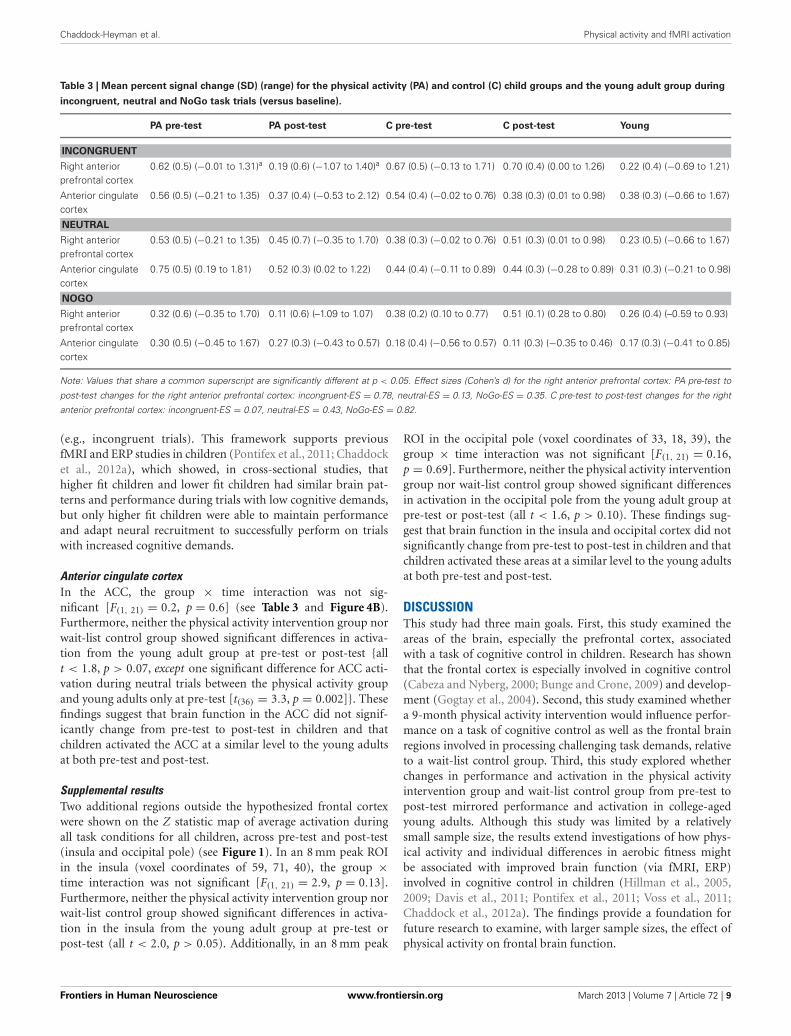
FUNCTIONAL ROIsTable 3 contains mean percent signal change values of each ROI(see Figure 2) at pre-test and post-test in the physical activityintervention and wait-list control groups of children. Table 3 alsocontains mean percent signal change values in each ROI for theyoung adults.
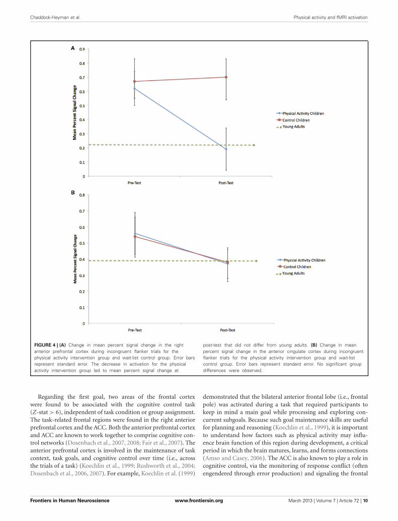
Right anterior prefrontal cortexConsistent with predictions, a significant group × time interac-tion [F(1, 21) = 5.4, p = 0.03] demonstrated that children in thephysical activity intervention group and wait-list control groupshowed differential changes in fMRI activation in the right ante-rior prefrontal cortex, from pre-test to post-test. The physicalactivity intervention group showed a significant decrease in rightanterior prefrontal activation across all task conditions from pre-test (M = 0.49, SD = 0.34) to post-test (M = 0.25, SD = 0.41).No change in activation from pre-test (M = 0.47, SD = 0.33)to post-test (M = 0.58, SD = 0.45) was found for the wait-list control group in the right anterior prefrontal cortex. In anexploratory planned comparison, this change in activation thatwas found for the physical activity intervention group was drivenby activation decreases during the incongruent condition [t(13) =3.5, p = 0.004] (see Figure 4A), and no significant within-groupchanges in neutral or NoGo activation (see Table 3).
Similar to the performance comparisons above, pre-test andpost-test brain activation in the right anterior prefrontal cortexof the physical activity intervention group and wait-list con-trol group of children were compared to the activation of theyoung adults in this ROI. Consistent with predictions, duringincongruent trials, which necessitated increased cognitive con-trol, both the physical activity intervention group [t(36) = 2.6,
Frontiers in Human Neuroscience www.frontiersin.org March 2013 | Volume 7 | Article 72 | 7

Chaddock-Heyman et al. Physical activity and fMRI activation
FIGURE 3 | (A) Change in incongruent RT for the physical activityintervention group and wait-list control group. Error bars representstandard error. The child groups showed longer RT at pre-test andpost-test compared to young adults. (B) Change in incongruent accuracy
for the physical activity intervention group and wait-list control group. Thewithin-group increase in accuracy for the physical activity interventiongroup led to accuracy rates at post-test that did not differ from youngadults. Error bars represent standard error.
p = 0.01] and the wait-list control group [t(31) = 2.5, p = 0.02]had more activation in the right anterior prefrontal cortex com-pared to the young adults at pre-test (see Table 3). At post-test,activation of the right anterior prefrontal cortex in the physicalactivity intervention group became statistically equivalent to theyoung adults [t(36) = 0.2, p = 0.8] (see Figure 4A), whereas, thewait-list control group still showed activation differences at post-test [t(31) = 2.9, p = 0.008] (see Table 3). During neutral trials(and NoGo trials), which required less cognitive control thanincongruent trials, neither group of children was statistically dif-ferent in right anterior prefrontal cortex activation at pre-test orpost-test (all p > 0.05). In sum, children in the physical activity
intervention group showed significant decreases in activation inthe right anterior prefrontal cortex from pre-test to post-test dur-ing incongruent flanker trials, which led to activation patternsthat mirrored the patterns of young adults at post-test. Wait-listcontrol children did not show changes in right anterior prefrontalcortex activation from pre-test to post-test and showed significantactivation differences from young adults at pre-test and post-test.
It is noteworthy that all children showed adult-like activationduring task trials that required less cognitive control (e.g., neu-tral and NoGo trials). Furthermore, the data suggest that thewait-list control children were unable to upregulate these pro-cesses to support task conditions requiring additional control
Frontiers in Human Neuroscience www.frontiersin.org March 2013 | Volume 7 | Article 72 | 8
Chaddock-Heyman et al. Physical activity and fMRI activation
Table 3 | Mean percent signal change (SD) (range) for the physical activity (PA) and control (C) child groups and the young adult group during
incongruent, neutral and NoGo task trials (versus baseline).
PA pre-test PA post-test C pre-test C post-test Young
INCONGRUENT
Right anteriorprefrontal cortex
0.62 (0.5) (−0.01 to 1.31)a 0.19 (0.6) (−1.07 to 1.40)a 0.67 (0.5) (−0.13 to 1.71) 0.70 (0.4) (0.00 to 1.26) 0.22 (0.4) (−0.69 to 1.21)
Anterior cingulatecortex
0.56 (0.5) (−0.21 to 1.35) 0.37 (0.4) (−0.53 to 2.12) 0.54 (0.4) (−0.02 to 0.76) 0.38 (0.3) (0.01 to 0.98) 0.38 (0.3) (−0.66 to 1.67)
NEUTRAL
Right anteriorprefrontal cortex
0.53 (0.5) (−0.21 to 1.35) 0.45 (0.7) (−0.35 to 1.70) 0.38 (0.3) (−0.02 to 0.76) 0.51 (0.3) (0.01 to 0.98) 0.23 (0.5) (−0.66 to 1.67)
Anterior cingulatecortex
0.75 (0.5) (0.19 to 1.81) 0.52 (0.3) (0.02 to 1.22) 0.44 (0.4) (−0.11 to 0.89) 0.44 (0.3) (−0.28 to 0.89) 0.31 (0.3) (−0.21 to 0.98)
NOGO
Right anteriorprefrontal cortex
0.32 (0.6) (−0.35 to 1.70) 0.11 (0.6) (–1.09 to 1.07) 0.38 (0.2) (0.10 to 0.77) 0.51 (0.1) (0.28 to 0.80) 0.26 (0.4) (–0.59 to 0.93)
Anterior cingulatecortex
0.30 (0.5) (−0.45 to 1.67) 0.27 (0.3) (−0.43 to 0.57) 0.18 (0.4) (−0.56 to 0.57) 0.11 (0.3) (−0.35 to 0.46) 0.17 (0.3) (−0.41 to 0.85)
Note: Values that share a common superscript are significantly different at p < 0.05. Effect sizes (Cohen’s d) for the right anterior prefrontal cortex: PA pre-test to
post-test changes for the right anterior prefrontal cortex: incongruent-ES = 0.78, neutral-ES = 0.13, NoGo-ES = 0.35. C pre-test to post-test changes for the right
anterior prefrontal cortex: incongruent-ES = 0.07, neutral-ES = 0.43, NoGo-ES = 0.82.
(e.g., incongruent trials). This framework supports previousfMRI and ERP studies in children (Pontifex et al., 2011; Chaddocket al., 2012a), which showed, in cross-sectional studies, thathigher fit children and lower fit children had similar brain pat-terns and performance during trials with low cognitive demands,but only higher fit children were able to maintain performanceand adapt neural recruitment to successfully perform on trialswith increased cognitive demands.
Anterior cingulate cortexIn the ACC, the group × time interaction was not sig-nificant [F(1, 21) = 0.2, p = 0.6] (see Table 3 and Figure 4B).Furthermore, neither the physical activity intervention group norwait-list control group showed significant differences in activa-tion from the young adult group at pre-test or post-test {allt < 1.8, p > 0.07, except one significant difference for ACC acti-vation during neutral trials between the physical activity groupand young adults only at pre-test [t(36) = 3.3, p = 0.002]}. Thesefindings suggest that brain function in the ACC did not signif-icantly change from pre-test to post-test in children and thatchildren activated the ACC at a similar level to the young adultsat both pre-test and post-test.
Supplemental resultsTwo additional regions outside the hypothesized frontal cortexwere shown on the Z statistic map of average activation duringall task conditions for all children, across pre-test and post-test(insula and occipital pole) (see Figure 1). In an 8 mm peak ROIin the insula (voxel coordinates of 59, 71, 40), the group ×time interaction was not significant [F(1, 21) = 2.9, p = 0.13].Furthermore, neither the physical activity intervention group norwait-list control group showed significant differences in activa-tion in the insula from the young adult group at pre-test orpost-test (all t < 2.0, p > 0.05). Additionally, in an 8 mm peak
ROI in the occipital pole (voxel coordinates of 33, 18, 39), thegroup × time interaction was not significant [F(1, 21) = 0.16,p = 0.69]. Furthermore, neither the physical activity interventiongroup nor wait-list control group showed significant differencesin activation in the occipital pole from the young adult group atpre-test or post-test (all t < 1.6, p > 0.10). These findings sug-gest that brain function in the insula and occipital cortex did notsignificantly change from pre-test to post-test in children and thatchildren activated these areas at a similar level to the young adultsat both pre-test and post-test.
DISCUSSIONThis study had three main goals. First, this study examined theareas of the brain, especially the prefrontal cortex, associatedwith a task of cognitive control in children. Research has shownthat the frontal cortex is especially involved in cognitive control(Cabeza and Nyberg, 2000; Bunge and Crone, 2009) and develop-ment (Gogtay et al., 2004). Second, this study examined whethera 9-month physical activity intervention would influence perfor-mance on a task of cognitive control as well as the frontal brainregions involved in processing challenging task demands, relativeto a wait-list control group. Third, this study explored whetherchanges in performance and activation in the physical activityintervention group and wait-list control group from pre-test topost-test mirrored performance and activation in college-agedyoung adults. Although this study was limited by a relativelysmall sample size, the results extend investigations of how phys-ical activity and individual differences in aerobic fitness mightbe associated with improved brain function (via fMRI, ERP)involved in cognitive control in children (Hillman et al., 2005,2009; Davis et al., 2011; Pontifex et al., 2011; Voss et al., 2011;Chaddock et al., 2012a). The findings provide a foundation forfuture research to examine, with larger sample sizes, the effect ofphysical activity on frontal brain function.
Frontiers in Human Neuroscience www.frontiersin.org March 2013 | Volume 7 | Article 72 | 9
Chaddock-Heyman et al. Physical activity and fMRI activation
FIGURE 4 | (A) Change in mean percent signal change in the rightanterior prefrontal cortex during incongruent flanker trials for thephysical activity intervention group and wait-list control group. Error barsrepresent standard error. The decrease in activation for the physicalactivity intervention group led to mean percent signal change at
post-test that did not differ from young adults. (B) Change in meanpercent signal change in the anterior cingulate cortex during incongruentflanker trials for the physical activity intervention group and wait-listcontrol group. Error bars represent standard error. No significant groupdifferences were observed.
Regarding the first goal, two areas of the frontal cortexwere found to be associated with the cognitive control task(Z-stat > 6), independent of task condition or group assignment.The task-related frontal regions were found in the right anteriorprefrontal cortex and the ACC. Both the anterior prefrontal cortexand ACC are known to work together to comprise cognitive con-trol networks (Dosenbach et al., 2007, 2008; Fair et al., 2007). Theanterior prefrontal cortex is involved in the maintenance of taskcontext, task goals, and cognitive control over time (i.e., acrossthe trials of a task) (Koechlin et al., 1999; Rushworth et al., 2004;Dosenbach et al., 2006, 2007). For example, Koechlin et al. (1999)
demonstrated that the bilateral anterior frontal lobe (i.e., frontalpole) was activated during a task that required participants tokeep in mind a main goal while processing and exploring con-current subgoals. Because such goal maintenance skills are usefulfor planning and reasoning (Koechlin et al., 1999), it is importantto understand how factors such as physical activity may influ-ence brain function of this region during development, a criticalperiod in which the brain matures, learns, and forms connections(Amso and Casey, 2006). The ACC is also known to play a role incognitive control, via the monitoring of response conflict (oftenengendered through error production) and signaling the frontal
Frontiers in Human Neuroscience www.frontiersin.org March 2013 | Volume 7 | Article 72 | 10
Chaddock-Heyman et al. Physical activity and fMRI activation
cortex to regulate top-down cognitive control (Botvinick et al.,2001; Dosenbach et al., 2007, 2008). Both of these areas have beenfound to relate to physical activity and aerobic fitness across thelifespan (Colcombe et al., 2004; Voss et al., 2011; Chaddock et al.,2012a). The present study used a randomized controlled interven-tion design in children to explore the effects of physical activity onthe fMRI brain function of both of these regions.
In regards to the second goal, a significant group × timeinteraction demonstrated that children in the physical activityintervention group showed significant decreases in fMRI acti-vation in the right anterior prefrontal cortex from pre-test topost-test, whereas the activation patterns in this frontal region inthe wait-list control group remained unchanged. It is noteworthythat exploratory planned comparisons revealed that these acti-vation changes in the physical activity intervention group weredriven by decreases in activation during incongruent flanker tri-als that required the greatest challenge to the participants’ abilityto pay attention and suppress distraction. In fact, relevant to thethird goal, the activation decreases in the physical activity inter-vention group during the incongruent flanker condition led topost-test fMRI patterns in the right anterior prefrontal cortexthat did not differ in magnitude from young adult activation. Onthe other hand, children in the wait-list control group differedfrom young adults in right anterior prefrontal activation duringincongruent flanker trials at both pre-test to post-test.
Together, these group-related and age-related activation pat-terns raise the possibility that participation in physical activityduring childhood can lead to more adult-like recruitment ofanterior prefrontal brain areas important for maintenance andgoal-oriented cognitive control. Here, improved brain function isassociated with decreases in anterior prefrontal cortex activationfrom pre-test to post-test, which is consistent with the frameworkthat less brain activation reflects more mature brain function, as anumber of studies show decreased activation and superior perfor-mance on cognitive tasks in adults compared to children (Caseyet al., 1997; Durston et al., 2002; Booth et al., 2003; Scherf et al.,2006; Velanova et al., 2008). Behaviorally, exploratory plannedcomparisons demonstrated that the physical activity interven-tion group showed within-group performance improvements interms of both speed and accuracy during incongruent and neu-tral flanker trials. The incongruent accuracy rates of the physicalactivity intervention children at post-test also mirrored thoseof the young adults. In contrast, wait-list control children didnot show changes in task performance from pre-test to post-test. These performance differences could be driven by changesin maintenance of task context and task goals with the physicalactivity intervention, which are functions linked to the anteriorprefrontal cortex (Koechlin et al., 1999; Rushworth et al., 2004;Dosenbach et al., 2006, 2007).
In fact, previous studies have demonstrated an associationbetween physical activity, aerobic fitness, and anterior prefrontalbrain function involved in goal maintenance across the lifespan(Voss et al., 2010, 2011; Kamijo et al., 2011). This longitudinalintervention study in children extends and strengthens these find-ings. In children, a cross-sectional fMRI study by Voss et al. (2011)demonstrated that higher fit children showed less activation in anetwork of brain regions including the anterior prefrontal cortex,
coupled with better flanker task performance, relative to lower fitchildren. An ERP study by Kamijo et al. (2011) also demonstratedthat children involved in a physical activity intervention showedlarger amplitudes over the frontal scalp regions in the contingentnegative variation (CNV), an ERP component known to play arole in cognitive preparation and task maintenance, as well asbetter working memory performance. In older adults, a physi-cal activity intervention that involved walking 3 days per week,for 1 year, led to changes in functional connectivity in a frontal-executive network (Voss et al., 2010), a network that includesthe right and left anterior prefrontal cortex (Dosenbach et al.,2006). The results of the present study contribute to this litera-ture and suggest plasticity of the right anterior prefrontal cortexwith prolonged physical activity participation.
No changes in activation for the physical activity interventiongroup or wait-list control group were found in the ACC. In addi-tion, no differences were observed in the comparison of childand adult ACC activation at pre-test or post-test. Consistent withthese findings, a cross-sectional study of the association betweenaerobic fitness and cognitive control in children did not demon-strate fitness differences in the ACC during incongruent flankertrials (Voss et al., 2011). Further, Chaddock et al. (2012a) alsoreported few fitness-related activation differences in this area.However, higher fit children (Pontifex et al., 2011), higher fityounger adults (Themanson et al., 2008), and higher fit olderadults (Colcombe et al., 2004), as well as older adults involved in aphysical activity intervention (Colcombe et al., 2004), have shownsmaller ERN amplitudes [an ERP component said to originate inthe dorsal portion of the ACC (Dehaene et al., 1994; Carter et al.,1998; van Veen and Carter, 2002; Miltner et al., 2003)], and lessACC activation, respectively, which are associated with perfor-mance improvements on a flanker task. Such activation patternsin the ACC are usually interpreted as a reduction in conflict or alower threshold for the detection and signaling of conflict to theprefrontal cortex, which leads to better error detection. To addressthis divergent evidence, additional research is needed to betterunderstand different responses to physical activity in children andolder adults, how effects in extreme fitness groups (higher fit,lower fit) in cross-sectional studies differ from effects of an inter-vention with lower fit individuals, as well as how ERP componentsmap onto fMRI activity.
The data also raise the possibility that the two groups of chil-dren differed in their cognitive strategies at post-test. Cognitivecontrol strategies are theorized to develop from one that is morerapid and reactive (i.e., reactive control) to one that can flexi-bly sustain goal-oriented control (i.e., proactive control) (Braveret al., 2007, 2009; Fair et al., 2007). Participation in physicalactivity during childhood may influence fMRI brain patternsunderlying control strategies, specifically the anterior prefrontalcortex (Fair et al., 2007; Paxton et al., 2008). That is, physicallyactive children may learn to maintain a sustained task set duringcognitive demands that require selective attention and distractionsuppression, which may lead to a more proactive control strategyas well as more accurate and adult-like task performance. Thiswould parallel research that suggests that higher fit children andolder adults use a more proactive control neural strategy thanlower fit individuals, especially during incongruent flanker task
Frontiers in Human Neuroscience www.frontiersin.org March 2013 | Volume 7 | Article 72 | 11

Chaddock-Heyman et al. Physical activity and fMRI activation
conditions (Colcombe et al., 2004; Pontifex et al., 2011; Voss et al.,2011). Alternatively, children in a wait-list control group may beless able to adapt their task strategy and task set at post-test, andmay continue to use a more reactive strategy, given that anteriorprefrontal activation and performance on incongruent task trialswere unchanged.
These results have important implications for public healthand the educational environment. Physical activity opportuni-ties are being reduced or eliminated during the school day aswell as decreasing outside the school environment (Troiano et al.,2008). Children are becoming increasingly sedentary and unfit,which leads to an increased risk for disease and obesity (UnitedStates Department of Health and Human Services, 2008; Centersfor Disease Control and Prevention, 2009), as well as cognitive
impairment (Chaddock et al., 2012c). The present study suggeststhat physical activity is important to the development of the brainand cognition during childhood. These results should raise publicawareness of the cognitive benefits of being active and encourageparticipation in a multicomponent physical activity program suchas physical education, classroom activity breaks, and active trans-port to school (United States Department of Health and HumanServices, 2013).
ACKNOWLEDGMENTSThe National Institute of Child Health and Human Development(NICHD) provided the principal source of funding (RO1HD055352 and RO1 HD069381). Thank you to Holly Tracy,Nancy Dodge, and John Powers for their help with data collection.
REFERENCESAmerican College of Sports Medicine.
(2006). ACSM’s Guidelines forExercise Testing and Prescription 7thEdn., New York, NY: LippincottWilliams and Wilkins, 366.
Amso, D., and Casey, B. J. (2006).Beyond what develops when: neu-roimaging may inform how cog-nition changes with development.Curr. Dir. Psychol. Sci. 15, 24–29.
Baddeley, A. D. (1986). WorkingMemory. New York, NY: ClarendonPress.
Bar-Or, O. (1983). Pediatric SportsMedicine for the Practitioner: FromPhysiologic Principles to ClinicalApplications. New York, NY:Springer, 398.
Beckmann, C. F., Jenkinson, M., andSmith, S. M. (2003). General multi-level linear modeling for groupanalysis in FMRI. Neuroimage 20,1052–1063.
Birnbaum, A. S., Lytle, L. A., Murray,D. M., Story, M., Perry, C. L., andBoutelle, K. N. (2002). Survey devel-opment for assessing correlates ofyoung adolescents’ eating. Am. J.Health Behav. 26, 284–295.
Booth, J. R., Burman, D. B., Meyer, J. R.,Lei, Z., Trommer, B. L., Davenport,N. D., et al. (2003). Neural devel-opment of selective attention andresponse inhibition. Neuroimage 20,737–751.
Botvinick, M. M., Braver, T. S., Barch,D. M., Carter, C. S., and Cohen, J.D. (2001). Conflict monitoring andcognitive control. Psychol. Rev. 108,624–652.
Braver, T. S., and Barch, D. M. (2006).Extracting core components of cog-nitive control. Trends Cogn. Sci. 10,529–532.
Braver, T. S., Gray, J. R., and Burgess,G. C. (2007). “Explaining the manyvarieties of working memory varia-tion: dual mechanisms of cognitivecontrol,” in Variation in Working
Memory, eds A. R. A. Conway, C.Jarrold, M. J. Kane, A. Miyake, andJ. N. Towse (New York, NY: OxfordUniversity Press), 76–106.
Braver, T. S., Paxton, J. L., Locke,H. S., and Barch, D. M. (2009).Flexible neural mechanisms of cog-nitive control within human pre-frontal cortex. Proc. Natl. Acad. Sci.U.S.A. 106, 7351–7356.
Buck, S. M., Hillman, C. H., andCastelli, D. M. (2008). The rela-tion of aerobic fitness to stroop taskperformance in preadolescent chil-dren. Med. Sci. Sports Exerc. 40,166–172.
Bunge, S. A., and Crone, E. A. (2009).“Neural correlates of the devel-opment of cognitive control,” inNeuroimaging in DevelopmentalClinical Neuroscience, eds J. Rumseyand M. Ernst (Cambridge, UK:Cambridge University Press),22–37.
Bunge, S. A., Dudukovic, N. M.,Thomason, M. E., Vaidya, C. J., andGabrieli, J. D. E. (2002). Immaturefrontal lobe contributions to cog-nitive control in children: evidencefrom fMRI. Neuron 33, 301–311.
Cabeza, R., and Nyberg, L. (2000).Imaging cognition II: an empiricalreview of 275 PET and fMRI studies.J. Cogn. Neurosci. 12, 1–47.
Carter, C. S., Braver, T. S., Barch, D.M., Botvinick, M. M., Noll, D., andCohen, J. D. (1998). Anterior cingu-lated cortex, error detection, and theonline monitoring of performance.Science 280, 747–749.
Casey, B. J., Trainor, R. J., Orendi, J.L., Schubert, A. B., Nystrom, L. E.,Giedd, J. N., et al. (1997). A devel-opmental functional MRI study ofprefrontal activation during perfor-mance of a go-no-go task. J. Cogn.Neurosci. 9, 835–847.
Castelli, D. M., Hillman, C. H., Hirsch,J., Hirsch, A., and Drollette, E.(2011). FIT Kids: Time in target
heart zone and cognitive perfor-mance. Prev. Med. 52, S55–S59.
Centers for Disease Control andPrevention. (2009). Chronic Diseasesand Health Promotion. Availableonline at: http://www.cdc.gov/chronicdisease/overview/ [Accessed4 January 2013].
Chaddock, L., Erickson, K. I., Prakash,R. S., Voss, M. V., VanPatter, M.,Pontifex, M. B., et al. (2012a). Afunctional MRI investigation of theassociation between childhood aer-obic fitness and neurocognitive con-trol. Biol. Psychol. 89, 260–268.
Chaddock, L., Neider, M. B., Lutz,A., Hillman, C. H., and Kramer,A. F. (2012b). Role of childhoodaerobic fitness in successful streetcrossing. Med. Sci. Sports Exerc. 44,749–753.
Chaddock, L., Voss, M. W., and Kramer,A. F. (2012c). Physical activity andfitness effects on cognition andbrain health in children and olderadults. Kinesiol. Rev. 1, 37–45.
Chaddock, L., Hillman, C. H., Buck,S. M., and Cohen, N. J. (2011).Aerobic fitness and executive con-trol of relational memory in pread-olescent children. Med. Sci. SportsExerc. 43, 344–349.
Colcombe, S., and Kramer, A. F. (2003).Fitness effects on the cognitive func-tion of older adults: a meta-analyticstudy. Psychol. Sci. 14, 125–130.
Colcombe, S. J., Kramer, A. F., Erickson,K. I., Scalf, P., McAuley, E., Cohen,N. J., et al. (2004). Cardiovascularfitness, cortical plasticity, and aging.Proc. Natl. Acad. Sci. U.S.A. 101,3316–3321.
Davis, C. L., Tomporowski, P. D.,McDowell, J. E., Austin, B. P., Miller,P. H., Yanasak, N. E., et al. (2011).Exercise improves executive func-tion and achievement and altersbrain activation in overweight chil-dren: a randomized, controlled trial.Health Psychol. 30, 91–98.
Dehaene, S., Posner, M. I., and Tucker,D. M. (1994). Localization of aneural system for error detectionand compensation. Psychol. Sci. 5,303–305.
Diamond, A. (2006). “The early devel-opment of executive functions,” inLifespan Cognition: Mechanisms ofChange, eds E. Bialystok and F. I.M. Craik (New York, NY: OxfordUniversity Press), 70–95.
Dosenbach, N. U., Fair, D. A., Cohen,A. L., Schlaggar, B. L., and Petersen,S. E. (2008). A dual-networks archi-tecture of top-down control. TrendsCogn. Sci. 12, 99–105.
Dosenbach, N. U., Fair, D. A., Miezin,F. M., Cohen, A. L., Wenger, K. K.,Dosenbach, R. A. T., et al. (2007).Distinct brain networks for adaptiveand stable task control in humans.Proc. Natl. Acad. Sci. U.S.A. 104,11073–11078.
Dosenbach, N. U., Visscher, K. M.,Palmer, E. D., Miezin, F. M., Wenger,K. K., Kang, H. C., et al. (2006). Acore system for the implementationof task sets. Neuron 50, 799–812.
DuPaul, G. J., Power, T. J.,Anastopoulos, A., and Reid, R.(1998). ADHD Rating Scale–IV:Checklists, Norms, and ClinicalInterpretation. New York, NY:Guilford Press, 79.
Durston, S., Thomas, K. M., Yang,Y., Ulug, A. M., Zimmerman, R.D., and Casey, B. J. (2002). Aneural basis for the developmentof inhibitory control. Dev. Sci. 5,F9–F16.
Fair, D. A., Dosenbach, N. U., Church,J. A., Cohen, A. L., Brahmbhatt,S., Miezin, F. M., et al. (2007).Development of distinct controlnetworks through segregation andintegration. Proc. Natl. Acad. Sci.U.S.A. 104, 13507–13512.
Freedson, P. S., and Goodman, T.L. (1993). “Measurement of oxy-gen consumption,” in Pediatric
Frontiers in Human Neuroscience www.frontiersin.org March 2013 | Volume 7 | Article 72 | 12

Chaddock-Heyman et al. Physical activity and fMRI activation
Laboratory Exercise Testing: ClinicalGuidelines, ed T. W. Rowland(Champaign, IL: Human Kinetics),91–113.
Gogtay, N., Giedd, J. N., Lusk, L.,Hayashi, K. M., Greenstein, D.,Vaituzid, A. C., et al. (2004).Dynamic mapping of human corti-cal development during childhoodthrough early adulthood. Proc. Natl.Acad. Sci. U.S.A. 101, 8174–8179.
Hawkins, H., Kramer, A., and Capaldi,D. (1992). Aging, exercise and atten-tion. Psychol. Aging 7, 643–653.
Hazeltine, E., Poldrack, R., andGabrieli, J. D. E. (2000). Neuralactivation during response com-petition. J. Cogn. Neurosci. 12,118–129.
Hillman, C. H., Buck, S. M.,Themanson, J. R., Pontifex, M.B., and Castelli, D. M. (2009).Aerobic fitness and cognitivedevelopment: event-related brainpotential and task performance ofexecutive control in preadolescentchildren. Dev. Psychol. 45, 114–129.
Hillman, C. H., Castelli, D. M., andBuck, S. M. (2005). Aerobic fit-ness and neurocognitive functionin healthy preadolescent chil-dren. Med. Sci. Sports Exerc. 37,1967–1974.
Hillman, C. H., Pontifex, M. B., Motl,R. W., O’Leary, K. C., Johnson, C.R., Scudder, M. R., et al. (2012).From ERP’s to academics. Dev.Cogn. Neurosci. 2, S90–S98.
Janz, K. F., and Mahoney, L. T. (1997).Three-year follow-up of changesin aerobic fitness during puberty:the Muscatine Study. Res. Q. Exerc.Sport 68, 1–9.
Jenkinson, M., Bannister, P. R., Brady,J. M., and Smith, S. M. (2002).Improved optimisation for therobust and accurate linear registra-tion and motion correction of brainimages. Neuroimage 17, 825–841.
Jenkinson, M., and Smith, S. M.(2001). A global optimisationmethod for robust affine registra-tion of brain images. Med. ImageAnal. 5, 143–156.
Kamijo, K., Pontifex, M. B., O’Leary, K.C., Scudder, M. R., Wu, C., Castelli,D. M., et al. (2011). The effectsof an afterschool physical activityprogram on working memory inpreadolescent children. Dev. Sci. 14,1046–1058.
Kaufman, A. S., and Kaufman, N. L.(1990). Kaufman Brief IntelligenceTest. Circle Pines, MN: AGS.
Koechlin, E., Basso, G., Pietrini, P.,Panzer, S., and Grafman, J. (1999).The role of the anterior prefrontalcortex in human cognition. Nature399, 148–151.
Kramer, A. F., Hahn, S., Cohen, N.J., Banich, M. T., McAuley, E.,Harrison, C., et al. (1999). Aging,fitness, and neurocognitive func-tion. Nature 400, 418–419.
Kriegeskorte, N., Simmons, W. K.,Bellgowan, P. S. F., and Baker, C. I.(2009). Circular analysis in systemsneuroscience: the dangers of doubledipping. Nat. Neurosci. 12, 535–540.
Liston, C., Watts, R., Tottenham, N.,Davidson, M. C., Niogi, S., Ulug,A. M., et al. (2006). Frontostriatalmicrostructure modulates efficientrecruitment of cognitive control.Cereb. Cortex 16, 553–560.
Luepker, R. V., Perry, C. L., McKinlay,S. M., Nader, P. R., Parcel, G. S.,Stone, E. J., et al. (1996). Outcomesof a field trial to improve chil-dren’s dietary patterns and physicalactivity: the Child and AdolescentTrial for Cardiovascular Health(CATCH). J. Am. Med. Assoc. 275,768–776.
Luna, B., Padmanabhan, A., andO’Hearn, K. (2010). What hasfMRI told us about the develop-ment of cognitive control throughadolescence? Brain Cogn. 72,101–113.
McKenzie, T. L., Strikmiller, P. K.,Stone, E. J., Woods, S. E., Ehlinger,S. S., Romero, K. A., et al. (1994).CATCH: physical activity processevaluation in a multicenter trial.Health Educ. Q. 2, S73–S89.
Miltner, W. H. R., Lemke, U., Weiss,T., Holroyd, C., Scheffers, M.K., and Coles, M. G. H. (2003).Implementation of error-processingin the human anterior cingulatedcortex: a source analysis of themagnetic equivalent of the error-related negativity. Biol. Psychol. 64,157–166.
Nader, P. R., Stone, E. J., Lytle, L. A.,Perry, C. L., Osganian, S. K., Kelder,S., et al. (1999). Three-year mainte-nance of improved diet and physicalactivity. The CATCH cohort. Arch.Pediatr. Adolesc. Med. 153, 695–704.
Oldfield, R. C. (1971). The assessmentand analysis of handedness:the Edinburgh inventory.Neuropsychologia 9, 97–113.
Paxton, J. L., Barch, D. M., Racine,C. A., and Braver, T. S. (2008).Cognitive control, goal mainte-nance, and prefrontal function inhealthy aging. Cereb. Cortex 18,1010–1028.
Pontifex, M. B., Raine, L. B., Johnson,C. R., Chaddock, L., Voss, M.W., Cohen, N. J., et al. (2011).Cardiorespiratory fitness and theflexible modulation of cognitivecontrol in preadolescent children.J. Cogn. Neurosci. 23, 1332–1345.
Rushworth, M. F. S., Walton, M. E.,Kennerley, S. W., and Bannerman,D. M. (2004). Action Actionsets and decisions in the medialfrontal cortex. Trends Cogn. Sci. 8,410–417.
Scherf, K. S., Sweeney, J. A., and Luna,B. (2006). Brain basis of develop-mental change in visuospatial work-ing memory. J. Cogn. Neurosci. 18,1045–1058.
Smith, S. M., Zhang, Y., Jenkinson, M.,Chen, J., Matthews, P. M., Federico,A., et al. (2002). Accurate, robustand automated longitudinal andcross-sectional brain change analy-sis. Neuroimage 17, 479–489.
Taylor, S. J. C., Whincup, P. H.,Hindmarsh, P. C., Lampe, F.,Odoki, K., and Cook, D. G. (2001).Performance of a new pubertalself-assessment questionnaire: apreliminary study. Paediatr. Perinat.Epidemiol. 15, 88–94.
Themanson, J. R., Pontifex, M. B.,and Hillman, C. H. (2008). Fitnessand action monitoring: evidencefor improved cognitive flexibilityin young adults. Neuroscience 157,319–328.
Tomporowski, P. D., Davis, C. L.,Miller, P. H., and Naglieri, J. A.(2008). Exercise and children’s intel-ligence, cognition, and academicachievement. Educ. Psychol. Rev. 20,111–131.
Troiano, R. P., Berrigan, D., Dodd,K. W., Mâsse, L. C., Tilert, T.,and McDowell, M. (2008). Physicalactivity in the United States mea-sured by accelerometer. Med. Sci.Sports Exerc. 40, 181–188.
United States Department of Healthand Human Services (USDHHS).(2008). 2008 Physical ActivityGuidelines for Americans: Be Active,Healthy, and Happy! Availableonline at: http://www.health.gov/paguidelines/guidelines/default.aspx [Accessed 4 January 2013].
United States Department of Healthand Human Services (USDHHS).(2013). Physical Activity Guidelinesfor Americans Mid-course Report:Strategies to Increase PhysicalActivity Among Youth. Availableonline at: http://www.health.gov/paguidelines/midcourse/PAG_Mid-course_Report.pdf [Accessed 5January 2013].
Utter, A. C., Roberson, R. J., Nieman,D. C., and Kang, J. (2002).Children’s OMNI scale of perceivedexertion: walking/running evalu-ation. Med. Sci. Sports Exerc. 34,139–144.
van den Wildenberg, W. P. M., andvan der Molen, M. W. (2004).Developmental trends in simple and
selective inhibition of compatibleand incompatible responses. J. Exp.Child Psychol. 87, 201–220.
van Veen, V., and Carter, C. S. (2002).The timing of action-monitoringprocesses in the anterior cingu-lated cortex. J. Cogn. Neurosci. 14,593–602.
Velanova, K., Wheeler, M. E., and Luna,B. (2008). Maturational changes inanterior cingulate and frontopari-etal recruitment support the devel-opment of error processing andinhibitory control. Cereb. Cortex 18,2505–2522.
Voss, M. W., Chaddock, L., Kim, J.S., VanPatter, M., Pontifex, M. B.,Raine, L. B., et al. (2011). Aerobicfitness is associated with greaterefficiency of the network under-lying cognitive control in preado-lescent children. Neuroscience 199,166–176.
Voss, M. W., Prakash, R. W., Erickson,K. I., Basak, C., Chaddock, L., Kim,J. S., et al. (2010). Plasticity of brainnetworks in a randomized interven-tion trial of exercise training in olderadults. Front. Aging Neurosci. 2:pii:32. doi: 10.3389/fnagi.2010.00032
Worsley, K. J. (2001). “Statisticalanalysis of activation images,” inFunctional MRI: An Introductionto Methods, eds P. Jezzard, P. M.Matthews, and S. M. Smith (NewYork, NY: Oxford University Press),250–271.
Conflict of Interest Statement: Theauthors declare that the researchwas conducted in the absence of anycommercial or financial relationshipsthat could be construed as a potentialconflict of interest.
Received: 16 January 2013; accepted:25 February 2013; published online: 12March 2013.Citation: Chaddock-Heyman L, EricksonKI, Voss MW, Knecht AM, PontifexMB, Castelli DM, Hillman CH andKramer AF (2013) The effects of phys-ical activity on functional MRI activa-tion associated with cognitive control inchildren: a randomized controlled inter-vention. Front. Hum. Neurosci. 7:72. doi:10.3389/fnhum.2013.00072Copyright © 2013 Chaddock-Heyman,Erickson, Voss, Knecht, Pontifex, Castelli,Hillman and Kramer. This is an open-access article distributed under the termsof the Creative Commons AttributionLicense, which permits use, distributionand reproduction in other forums, pro-vided the original authors and sourceare credited and subject to any copy-right notices concerning any third-partygraphics etc.
Frontiers in Human Neuroscience www.frontiersin.org March 2013 | Volume 7 | Article 72 | 13