the effects of various processing conditions on the properties...
TRANSCRIPT

THE EFFECTS OF VARIOUS PROCESSING CONDITIONS ON THE PROPERTIES OF
HYDROXYAPATITE
TEH YEE CHING
FACULTY OF ENGINEERING UNIVERSITY OF MALAYA
KUALA LUMPUR
2017

THE EFFECTS OF VARIOUS PROCESSING
CONDITIONS ON THE PROPERTIES OF
HYDROXYAPATITE
TEH YEE CHING
THESIS SUBMITTED IN FULFILMENT OF THE
REQUIREMENTS FOR THE DEGREE OF DOCTOR OF
PHILOSOPHY
FACULTY OF ENGINEERING
UNIVERSITY OF MALAYA
KUALA LUMPUR
2017

ii
UNIVERSITY OF MALAYA
ORIGINAL LITERARY WORK DECLARATION
Name of Candidate: TEH YEE CHING (I.C/Passport No: )
Registration/Matric No: KHA 120096
Name of Degree: DOCTOR OF PHILOSOPHY
Title of Project Paper/Research Report/Dissertation/Thesis (―this Work‖):
THE EFFECTS OF VARIOUS PROCESSING CONDITIONS ON THE
PROPERTIES OF HYDROXYAPATITE
Field of Study: MECHANICAL ENGINEERING
I do solemnly and sincerely declare that:
(1) I am the sole author/writer of this Work;
(2) This Work is original;
(3) Any use of any work in which copyright exists was done by way of fair
dealing and for permitted purposes and any excerpt or extract from, or
reference to or reproduction of any copyright work has been disclosed
expressly and sufficiently and the title of the Work and its authorship have
been acknowledged in this Work;
(4) I do not have any actual knowledge nor do I ought reasonably to know that
the making of this work constitutes an infringement of any copyright work;
(5) I hereby assign all and every rights in the copyright to this Work to the
University of Malaya (―UM‖), who henceforth shall be owner of the
copyright in this Work and that any reproduction or use in any form or by any
means whatsoever is prohibited without the written consent of UM having
been first had and obtained;
(6) I am fully aware that if in the course of making this Work I have infringed
any copyright whether intentionally or otherwise, I may be subject to legal
action or any other action as may be determined by UM.
Candidate‘s Signature Date:
Subscribed and solemnly declared before,
Witness‘s Signature Date:
Name:
Designation:

iii
ABSTRACT
The sintering behavior of hydroxyapatite (HA) powder produced by wet chemical
precipitation method via three different drying methods, i.e. freeze drying (FD-HA),
microwave drying (MD-HA) and oven drying (OD-HA) were investigated over the
temperature range of 1050°C to 1350°C. The characterization of HA was assessed in
terms of powder morphology, powder element analysis, phase stability, bulk density,
microstructure, grain size, Vickers hardness and fracture toughness. Based on these
results, the HA powder that demonstrated the optimum properties was chosen for
further studies to investigate the effects of microwave sintering on the sinterability of
the chosen HA. The microwave sintering carried out in temperature ranging from 950°C
to 1250°C. The sinterability of microwave sintered HA was compared to that of
conventional pressureless sintered HA. Subsequently, the effects of adding zinc oxide
(ZnO) ranging from 0.1 wt% to 1.0 wt% on the sinterability of HA when sintered
between 1100°C to 1300°C via conventional pressureless sintering were also evaluated.
In the present study, the use of microwave drying accelerates the manufacturing of
HA powder as only 15 minutes were required to dry the HA powder (MD-HA) while at
least 16 hours and 36 hours drying time were required for conventional oven drying
(OD-HA) and freeze drying (FD-HA), respectively. It has been revealed that MD-HA
possess overall better sinterability and mechanical properties than OD-HA and FD-HA.
The optimum sintering temperature for the synthesized MD-HA was 1200°C with the
following properties being recorded: relative density of 97.5%, Vickers hardness of 5.04
GPa and fracture toughness of 1.15 MPam1/2
. Besides, decomposition of MD-HA phase
upon sintering was not observed in the present work but small amount of tricalcium
phosphate was observed in OD-HA when sintered at 1350°C.

iv
The current study also revealed that microwave sintering played an important role in
enhancing the mechanical properties of HA matrix particularly at low sintering
temperature. HA with high fracture toughness value of ~1.85 MPam1/2
was produced at
1050°C via microwave sintering. In addition, the addition of 0.5 wt% of ZnO was found
to be beneficial in improving the fracture toughness of HA powder. The results
indicated that that the resulting 0.5 wt% ZnO-doped HA sintered body exhibited an
increased toughness of to 1.37 MPam1/2
and hardness value to 5.63 GPa when
compared to the undoped body (1.16 MPam1/2
for fracture toughness and 4.75 GPa for
hardness) at 1150°C via conventional sintering.
The main advantageous of this research is the economical and rapid production of
HA that exhibited enhanced sinterability at low temperatures that is suitable for the
production of biomedical devices without compromising the phase stability and
biocompatibility nature of the HA.

v
Abstrak
Sifat persinteran HA dihasilkan daripada pemendakan kimia basah melalui tiga
kaedah yang berbeza iaitu pegeringan melalui pembekuan (FD-HA), pergeringan
melalui gelombang microwave (MD-HA) dan pegeringan melalui ketuhar (OD-HA)
telah disiasat dalam lingkugan suhu persinteran 1050°C hingga 1350°C. Pencirian HA
dinilai dari segi morfologi serbuk, analisis unsur serbuk, kestabilan fasa, ketumpatan
pukal, mikrostruktur, saiz butiran, kekerasan Vickers dan keliatan patah.
Berdasarkan keputusan ini, serbuk HA yang menunjukkan ciri-ciri yang terbaik akan
dipilih untuk melanjutkan pelajaran untuk menyiasatkan kesan persinteran gelombang
mikrowave atas sifat persinteran HA. Suhu ketuhar gelombang mikro persinteran adalah
dari 950°C hingga 1250°C. Sifat persinteran HA melalui gelombang microwave
dibandingkan dengan HA yang disinterkan melalui pensinteran konvensional tanpa
tekanan. Selepas itu, kesan penambahan ZnO antara 0.1 % berat hingga 1 % berat atas
sifat persinteran HA melalui persinteran konvensional dan ketuhar gelombang mikro
dalam lingkungan suhu 1100°C hingga 1300°C telah dikaji.
Dalam kajian ini, penggunaan pengeringan ketuhar gelombang mikro
mempercepatkan pembuatan serbuk HA kerana hanya 15 minit dikehendaki untuk
mengeringkan serbuk HA manakala sekurang-kurangnya 16 jam masa pengeringan
dikehendaki untuk pengeringan ketuhar konvensional dan 36 jam untuk pengeringan
pembekuan. Kajian ini membuktikan MD-HA telah didapati mempunyai sifat
persinteran dan sifat-sifat mekanikal yang lebih baik daripada OD-HA dan FD-HA.
Suhu pensinteran optimum untuk MD-HA disinter adalah 1200°C dengan sifat-sifat
berikut direkodkan: ketumpatan pukal relatif 97.5% , Vickers kekerasan 5.04 GPa dan
patah keliatan 1.15 MPam1/2
. Selain itu, tiada kesan decomposition fasa MD-HA bila
disinterkan tetapi sedikit trikalsium fosfat dikesan dalam OD-HA apabila disinter pada

vi
1350°C. Kajian ini juga mendedahkan bahawa pensinteran microwave memainkan
peranan penting dalam meningkatkan sifat-sifat mekanik matriks HA pada suhu
pensinteran rendah. HA dengan nilai keliatan patah yang tinggi ~ 1.85 MPam1/2
telah
dihasilkan pada suhu 1050°C melalui persinteran microwave. Di samping
itu,penambahan 0.5 wt% ZnO membawa kebaikan dalam meningkatkan keliatan
pepatahan HA. Keputusan menunjukkan bahawa 0.5 wt% ZnO dop HA menunjukkan
perningkatan dalam keliatan pepatahan ke 1.37 MPam1/2
dan kekerasan ke 5.63 GPa
apabila berbanding kepada HA tanpa ZnO (1.16 MPam1/2
keliatan pepatahan dan 4.75
GPa kekerasan) pada 1150°C melalui persinteran konvensional.
Faedah utama kajian ini adalah menghasilkan HA yang dengan cara yang murah dan
cepat selain mempamerkan sifat persinteran yang tinggi pada suhu persinteran yang
rendah dan sesuai sebagai peranti bioperubatan tanpa mengorbankan kestabilan fasa dan
bioserasi HA.
vii
ACKNOWLEDGEMENTS
I would like to express my sincere appreciation to the following individuals that have
constantly guided and supported me throughout the course of my PhD research:
My project supervisor, Prof. Ir. Dr. Ramesh Singh for his vital
encouragement, support, knowledge transfer and constructive suggestions
throughout the research and the thesis writing.
My Co-supervisor, Assoc. Prof. Dr. Tan Chou Yong for his assistance,
continuous guidance, advices and correction throughout the course of this
research and the thesis writing.
The Surface Engineering, Advanced Material Processing Laboratories and
High Impact Research (HIR) of University of Malaya for providing facilities
and instrument support.
My colleagues and lab instructors at University of Malaya for their
immeasurable guidance and advices.
Geran Penyelidikan Universiti Malaya (UMRG) Grant No. RP011B-13AET,
Fundamental Research and Grant Scheme (FRGS) Grant No. FP029-2013A
and Postgraduate Research Grant (PPP) Grant No. PG080-2013A for the
financial support.
Lastly, I would like to thank my family members and friends for their unconditional
support, understanding, patience and encouragement.

viii
TABLE OF CONTENTS
Abstract ............................................................................................................................ iii
Acknowledgements ......................................................................................................... vii
Table of Contents ........................................................................................................... viii
List of Figures ................................................................................................................. xii
List of Tables................................................................................................................ xviii
List of Symbols and Abbreviations ................................................................................. xx
List of Appendices ........................................................................................................ xxii
CHAPTER 1: INTRODUCTION .................................................................................. 1
1.1 Background of the Study ......................................................................................... 1
1.2 Scope of Research.................................................................................................... 6
1.3 Objectives of the Research ...................................................................................... 7
1.4 Structure of the Thesis ............................................................................................. 8
CHAPTER 2: SYNTHESIS METHODS OF HYDROXYAPATITE ...................... 10
2.1 Introduction to Hydroxyapatite.............................................................................. 10
2.2 Synthesis Method of Hydroxyapatite (HA) Powders ............................................ 16
2.2.1 Wet Chemical Method .............................................................................. 16
2.2.1.1 Starting Precursors .................................................................... 18
2.2.1.2 Synthesis Temperature .............................................................. 20
2.2.1.3 Reaction pH ............................................................................... 21
2.2.1.4 Ca/P Ratio ................................................................................. 23
2.2.1.5 Drying Methods ......................................................................... 24
2.2.1.6 Other Synthesis Parameters ....................................................... 34
2.2.2 Hydrothermal ............................................................................................ 36

ix
2.2.3 Sol-Gel Method ........................................................................................ 37
2.2.4 Mechanochemical (Solid State Reaction) ................................................ 38
2.2.5 Other Processing Techniques ................................................................... 40
CHAPTER 3: THE SINTERING AND SINTERABILITY OF
HYDROXYAPATITE .................................................................................................. 41
3.1 Powders Consolidation (Sintering) Techniques .................................................... 41
3.1.1 Conventional Pressureless Sintering (CPS) ............................................. 41
3.1.2 Microwave Sintering (MS) ....................................................................... 42
3.1.3 Spark Plasma Sintering (SPS) .................................................................. 49
3.1.4 Hot Pressing Sintering (HPS) ................................................................... 50
3.1.5 Two Steps Sintering (TSS) ....................................................................... 52
3.2 Sintering Temperature ........................................................................................... 53
3.3 Sintering Time ....................................................................................................... 59
3.4 Sintering Ramp Rate .............................................................................................. 60
3.5 Sintering Additives ................................................................................................ 61
3.5.1 Zinc Oxide as Sintering Additives ........................................................... 65
CHAPTER 4: EXPERIMENTAL TECHNIQUES ................................................... 69
4.1 Synthesis of HA Powder via Wet Chemical Method ............................................ 69
4.1.1 HA Powder Prepared via Oven Drying .................................................... 70
4.1.2 HA Powder Prepared via Microwave Drying .......................................... 71
4.1.3 HA Powder Prepared via Freeze Drying .................................................. 72
4.2 ZnO-doped HA Powder Preparation ..................................................................... 73
4.3 Green Samples Preparation.................................................................................... 75
4.4 Sintering ................................................................................................................. 75
4.4.1 Conventional Sintering ............................................................................. 75

x
4.4.2 Microwave Sintering ................................................................................ 76
4.4.2.1 Samples Arrangement ............................................................... 77
4.5 Grinding and Polishing .......................................................................................... 78
4.6 Characterization ..................................................................................................... 79
4.6.1 Specific Surface Area and Crystallite Size ............................................... 79
4.6.2 Transmission Electron Microscopy (TEM) Analysis ............................... 80
4.6.3 Fourier Transformation Infrared (FTIR) .................................................. 80
4.6.4 Scanning Electron Microscopy (SEM) and Energy Dispersive X-Ray
Analysis (EDX) ........................................................................................ 80
4.6.5 X-Ray Diffraction (XRD)......................................................................... 81
4.6.6 Microstructural Examination .................................................................... 81
4.6.7 Grain Size Measurement .......................................................................... 82
4.6.8 Bulk Density Measurement ...................................................................... 83
4.6.9 Vickers Hardness and Fracture Toughness Evaluation ............................ 84
CHAPTER 5: RESULTS AND DISCUSSIONS (PART 1) ....................................... 86
5.1 HA Powder Characteristic ..................................................................................... 86
5.1.1 XRD Analysis and Crystallite Size .......................................................... 86
5.1.2 FTIR Analysis of the Synthesized HA Powder ........................................ 88
5.1.3 EDX Analysis of the Synthesized HA Powder ........................................ 91
5.1.4 FE-SEM Analysis of the Synthesized HA Powder .................................. 93
5.1.5 TEM Analysis of the Synthesized HA Powder ........................................ 96
5.1.6 Specific Surface Area of the Synthesized HA Powder ............................ 98
5.2 Sinterability of the HA Powder ............................................................................. 99
5.2.1 HA Phase Stability ................................................................................... 99
5.2.2 FTIR Analysis of Sintered HA Samples ................................................ 104
5.2.3 Bulk Density ........................................................................................... 105

xi
5.2.4 Microstructure Evolution and Grain Size ............................................... 107
5.2.5 Vickers Hardness and Fracture Toughness ............................................ 114
CHAPTER 6: RESULTS AND DISCUSSIONS (PART 2) ..................................... 121
6.1 Effect of Microwave Sintering on the Sinterability of MD-HA .......................... 121
6.1.1 XRD Analysis of CPS and MWS Sintered HA ...................................... 121
6.1.2 Bulk Density of CPS and MWS Sintered HA ........................................ 123
6.1.3 Microstructural Evolution and Grain Size .............................................. 125
6.1.4 Vickers Hardness and Fracture Toughness of CPS and MWS Sintered HA
132
6.2 Effect of Zinc Oxide (ZnO) addition on the Sinterability of HA ........................ 139
6.2.1 XRD Analysis of Undoped and ZnO-doped HA Powder ...................... 139
6.2.2 XRD Analysis of Undoped and ZnO-doped Sintered HA ..................... 140
6.2.3 Bulk Density of Undoped and ZnO-doped Sintered HA ....................... 141
6.2.4 Microstructure Analysis of Undoped and ZnO-doped Sintered HA ...... 142
6.2.5 Vickers Hardness and Fracture Toughness of Undoped and ZnO-doped
Sintered HA ............................................................................................ 148
6.2.6 Toughening Mechanism ......................................................................... 152
CHAPTER 7: CONCLUSIONS AND FURTHER WORK .................................... 157
7.1 Conclusions ......................................................................................................... 157
7.2 Further Work ....................................................................................................... 163
References ..................................................................................................................... 164
List of Publications and Papers Presented .................................................................... 192
Appendix A ................................................................................................................... 193
Appendix B ................................................................................................................... 200
Appendix C ................................................................................................................... 201

xii
LIST OF FIGURES
Figure 1.1: Flow chart of the research scope. ................................................................... 7
Figure 2.1: Calcium phosphate phase equilibrium diagram at 66 kPa (DeGroot et al.,
1990). .............................................................................................................................. 14
Figure 2.2: General procedures involved in wet chemical method. ................................ 17
Figure 2.3: TEM picures of as-synthesized HA nanocrystal at different synthesis
temperature: (a) 35°C; (b) 85°C (Bouyer et al., 2000). .................................................. 20
Figure 2.4: XRD spectra of HA powder synthesized at different temperatures (Pham et
al., 2013). ........................................................................................................................ 21
Figure 2.5: XRD patterns of HA samples. ...................................................................... 22
Figure 2.6: SEM micrographs of HA nanoparticles synthesized at (a) pH 5 (acidic), (b)
pH 7, and pH 11 (alkaline) (Inthong et al., 2013). .......................................................... 22
Figure 2.7: Relation between drying period and the particle size of HA powder dried at
60ºC (Zhang & Yogokawa, 2008). ................................................................................. 25
Figure 2.8: TEM photos of HA powders dried at 60ºC for (a) 3, (b) 10 and (c) 18 days
(Zhang & Yogokawa, 2008). .......................................................................................... 25
Figure 2.9: Schematic diagram for the triple phases of water (Yu et al., 2011). ............ 27
Figure 2.10: Transmission electron micrographs of HA powders after calcining at 800ºC
for 3 hours: (a) freeze dried HA, (b) oven dried HA (Lu et al., 1998). .......................... 28
Figure 2.11: SEM micrographs of etched fracture surface of (a) freeze dried HA, (b)
oven dried HA, sintered for 3 hours at 1350ºC (Lu et al., 1998). ................................... 28
Figure 2.12: Temperature profile inside sample of (a) conventional drying (b)
microwave drying (Hui, 2008). ....................................................................................... 31
Figure 2.13: SEM micrographs of sintered foam from (a) conventional drying and (b)
microwave drying (Abd Rahman et al., 2009). ............................................................... 33
Figure 2.14: FTIR spectra of HA powder synthesized at different acid addition rate: 1, 2
and 5 ml min-1
(Pham et al., 2013).................................................................................. 35
Figure 2.15: XRD spectra of HA powder synthesized at different acid addition rate: 1, 2
and 5 ml min-1
(Pham et al., 2013).................................................................................. 35

xiii
Figure 3.1: Comparison of heating procedure between (a) Conventional sintering and (b)
Microwave sintering (Agrawal, 2006). ........................................................................... 42
Figure 3.2: Schematic diagram of the main ways that microwaves can interact with
materials (Sutton, 1989). ................................................................................................. 44
Figure 3.3: Comparative sintering curves for HA sintered by microwave and
conventional heating (Ehsani et al., 2013). ..................................................................... 45
Figure 3.4: Microstructural evolution of CPS-HA (a-c) and MS-HA (d-f) when sintered
at 1150ºC (a,d), 1250ºC (b,e) and 1300ºC (c,d) (Ramesh et al., 2008). ......................... 47
Figure 3.5: XRD of HA sintered at different temperatures for 15 minutes, using MS
(Harabi et al., 2010). ....................................................................................................... 48
Figure 3.6: Comparison of XRD patterns before and after consolidation by SPS at
75ºC/min (Cuccu et al., 2015). ........................................................................................ 50
Figure 3.7: Comparison between FTIR spectra of (a) hot pressed and (b) pressureless
sintered HA samples at various sintering temperatures (Rapacz-Kmita et al., 2005). ... 52
Figure 3.8: SEM micrographs of HA compacts sintered under (a) CPS at 1100ºC and (b)
TSS at T1 = 900ºC and T2 = 800ºC (Mazaheri et al., 2009)............................................ 53
Figure 3.9: Temperature dependence of (a) open porosity and (b) average grain size of
hydroxyapatite (Petrakova et al., 2013). ......................................................................... 54
Figure 3.10: Temperature dependence of (a) relative density and (b) microhardness
(Petrakova et al. 2013). ................................................................................................... 55
Figure 3.11: Variation of Vickers hardness and relative density as a function of average
grain size (Muralithran & Ramesh, 2000)....................................................................... 56
Figure 3.12: XRD patterns of HA derived from eggshells sintered at temperatures
ranging from 1050ºC to 1350ºC (Ramesh et al., 2016). ................................................. 57
Figure 3.13: The variation of fracture toughness of conventional pressureless sintered
EHA as a function of sintering temperature (Kamalanathan et al., 2014). ..................... 58
Figure 3.14: SEM micrograph of sample sintered at 1050ºC for (a) 45 minutes and (b) 2
hours (Veljović et al., 2014). .......................................................................................... 59
Figure 3.15: XRD patterns of HA sintered at 1250ºC with ramp rate of (a) 2ºC/min, (b)
5ºC/min and (c) 10ºC/min (Keys: = HA, = α-TCP, ▼= -TCP ♦ = TTCP and ■ =
CaO) (Samuel et al., 2012). ........................................................................................... 61
xiv
Figure 3.16: Fracture toughness and Vickers hardness of MgO-doped and undoped HA
(Tan et al., 2013). ............................................................................................................ 65
Figure 3.17: Effect of ZnO contents on the (a) stiffness, (b) densification, (c)
microhardness and (d) fracture toughness of the strut (Feng et al., 2014)...................... 68
Figure 4.1: OHA wet chemical method flow sheet. ........................................................ 70
Figure 4.2: HA wet chemical method with microwave drying flow sheet. .................... 72
Figure 4.3: HA wet chemical method with freeze drying flow sheet. ............................ 73
Figure 4.4: A flow chart showing the powders prepared in the present work. ............... 74
Figure 4.5: Sintering profile of conventional sintering. .................................................. 76
Figure 4.6: Sintering profile of microwave sintering. ..................................................... 77
Figure 4.7: Samples arrangement in the microwave furnace: (a) plan view; (b) side view.
......................................................................................................................................... 78
Figure 4.8: Diagram showing the score given for the type of intersections. .................. 83
Figure 4.9: Schematic indentation fracture pattern of an idealized Vickers median (or
half-penny) crack system (Niihara et al., 1982). ............................................................. 85
Figure 5.1: The XRD profiles of HA powder synthesized through wet precipitation
method via three different drying methods. .................................................................... 86
Figure 5.2: The FTIR spectrum of the synthesized HA powders: (a) FD-HA, (b) MD-
HA and (c) OD-HA. ........................................................................................................ 89
Figure 5.3: The EDX spectrum and elemental composition of FD-HA. ........................ 91
Figure 5.4: The EDX spectrum and elemental composition of MD-HA. ....................... 91
Figure 5.5: The EDX spectrum and elemental composition of OD-HA. ........................ 92
Figure 5.6: FE-SEM micrograph of synthesized FD-HA powder. ................................. 94
Figure 5.7: FE-SEM micrograph of synthesized MD-HA powder. ................................ 94
Figure 5.8: FE-SEM micrograph of synthesized OD-HA powder. ................................. 95
Figure 5.9: TEM micrographs of synthesized HA powder: (a) FD-HA, (b) MD-HA and
(c) OD-HA. ..................................................................................................................... 97

xv
Figure 5.10: The XRD profiles of FD-HA sintered samples (a) 1050°C, (b) 1150°C, (c)
1250°C and (d) 1350°C. All peaks belong to the HA phase. ........................................ 100
Figure 5.11: The XRD profiles of MD-HA sintered samples (a) 1050°C, (b) 1150°C, (c)
1250°C and (d) 1350°C. All peaks belong to the HA phase. ........................................ 101
Figure 5.12: The XRD profiles of OD-HA sintered samples (a) 1050°C, (b) 1150°C, (c)
1250°C and (d) 1350°C. The unmarked peaks belong to the HA phase. ...................... 101
Figure 5.13: FTIR profiles of HA sintered at 1350°C (left) with their respective as-
synthesized powder (right): (a) FD-HA, (b) MD-HA and (c) OD-HA. ........................ 104
Figure 5.14: The effect of sintering temperature on the relative density of HA. .......... 105
Figure 5.15: SEM images of (a) FD-HA, (b) MD-HA and (c) OD-HA sintered at
1050°C. ......................................................................................................................... 108
Figure 5.16: SEM images of (a) FD-HA, (b) MD-HA and (c) OD-HA sintered at
1150°C. ......................................................................................................................... 110
Figure 5.17: SEM images of (a) FD-HA, (b) MD-HA and (c) OD-HA sintered at
1200°C. ......................................................................................................................... 111
Figure 5.18: SEM images of (a) FD-HA, (b) MD-HA and (c) OD-HA sintered at
1350°C. ......................................................................................................................... 113
Figure 5.19: The effect of sintering temperature on the average grain size of sintered
HA. ................................................................................................................................ 114
Figure 5.20: The effect of sintering temperature on the hardness of HA. .................... 116
Figure 5.21: The variation of the relative density and hardness of sintered FD-HA as a
function of average grain size. ...................................................................................... 116
Figure 5.22: The effect of sintering temperature on the fracture toughness of HA. ..... 118
Figure 5.23: The effects of grain size on the fracture toughness of sintered HA. ........ 119
Figure 6.6.1: The XRD profiles of HA sintered by CPS at different temperatures. All
peaks belong to HA phase. ............................................................................................ 121
Figure 6.2: The XRD profiles of HA sintered by MWS at different temperatures. All
peaks belong to HA phase. ............................................................................................ 122
Figure 6.3: The effect of sintering temperature on the density of HA sintered by
conventional sintering and microwave sintering technique. ......................................... 123

xvi
Figure 6.4: SEM images of HA sintered by (a) conventional sintering and (b)
microwave sintering at 950°C. ...................................................................................... 126
Figure 6.5: SEM images of HA sintered by (a) conventional sintering and (b)
microwave sintering at 1000°C. .................................................................................... 127
Figure 6.6: SEM images of HA sintered by (a) conventional sintering and (b)
microwave sintering at 1050°C. .................................................................................... 128
Figure 6.7: SEM images of HA sintered by (a) conventional sintering and (b)
microwave sintering at 1100°C. .................................................................................... 129
Figure 6.8: SEM images of HA sintered by (a) conventional sintering and (b)
microwave sintering at 1250°C. .................................................................................... 130
Figure 6.9: The effect of sintering temperatures on the average grain size of HA samples
sintered by (a) conventional sintering and (b) microwave sintering. ............................ 131
Figure 6.10: The effect of sintering temperature on the hardness of HA sintered by
conventional sintering and microwave sintering technique. ......................................... 133
Figure 6.11: Vickers hardness dependence on the relative density of HA sintered by
conventional sintering and microwave sintering technique. ......................................... 134
Figure 6.12: The effect of sintering temperature on the fracture toughness of HA
sintered by conventional sintering and microwave sintering technique. ...................... 135
Figure 6.13: The variation of fracture toughness and relative density of HA sintered by
microwave sintering technique. .................................................................................... 137
Figure 6.14: The schematic diagram of the dislocation pile up in (a) large grains and (b)
small grains. .................................................................................................................. 138
Figure 6.15: The XRD profiles of undoped HA powders and HA powders containing
0.1 wt%, 0.3 wt%, 0.5 wt% and 1 wt% ZnO, respectively. .......................................... 139
Figure 6.16: XRD patterns of HA samples sintered at 1300°C for undoped HA and HA
containing 0.1 wt%, 0.3 wt%, 0.5 wt% ZnO and 1 wt% ZnO, respectively. ................ 140
Figure 6.17: Relative density variation as a function of sintering temperatures for HA
with different amount of ZnO addition. ........................................................................ 142
Figure 6.18: SEM analysis of HA samples sintered at 1150°C for (a) undoped HA (b)
0.1 wt% ZnO-doped HA and (c) 0.3 wt% ZnO-doped HA. ......................................... 143
Figure 6.19: SEM analysis of 0.5 wt% ZnO-doped HA samples sintered at 1150°C
(inset as EDX spectrum). .............................................................................................. 144

xvii
Figure 6.20: SEM analysis of 1.0 wt% ZnO-doped HA samples sintered at 1150°C
(inset as EDX spectrum). .............................................................................................. 145
Figure 6.21: SEM analysis of HA samples sintered at 1300°C for (a) undoped HA (b)
0.1 wt% ZnO-doped HA, (c) 0.3 wt% ZnO-doped HA, (d) 0.5 wt% ZnO-doped HA and
(e) 1 wt% ZnO-doped HA. ............................................................................................ 147
Figure 6.22: Effect of sintering temperature and ZnO addition on the average grain size
of HA. ............................................................................................................................ 148
Figure 6.23: Effect of sintering temperature and ZnO addition on the Vickers hardness
of HA. ............................................................................................................................ 149
Figure 6.24: The dependence of the hardness of undoped and ZnO-doped HA on the
inverse square root of grain size.................................................................................... 150
Figure 6.25: Effect of sintering temperature and ZnO addition on the fracture toughness
of HA. ............................................................................................................................ 151
Figure 6.26: SEM micrograph of the indentation crack paths of pure HA sintered at
1150ºC. .......................................................................................................................... 154
Figure 6.27: SEM micrograph of indentation crack paths of 0.5 wt% ZnO-doped HA
sintered at 1150ºC. ........................................................................................................ 154
Figure 6.28: A close up view of the crack paths of Figure 6.27 indicated the crack
deflection. ...................................................................................................................... 155
Figure 6.29: A close up view of the crack paths of Figure 6.27 indicated the crack
bridging. ........................................................................................................................ 155
Figure 6.30: A schematic diagram of the proposed toughening mechanism: (a) crack
bridging and (b) crack deflection. ................................................................................. 156

xviii
LIST OF TABLES
Table 2.1: Comparison between composition and physical properties of human enamel,
bone and HA ceramic (Hench, 1998; LeGeros & LeGeros, 1993). ................................ 10
Table 2.2: Ionic concentration (mmol/dm3) of SBF and human blood plasma (Oréfice et
al., 2000). ........................................................................................................................ 11
Table 2.3: Comparison of mechanical properties of sintered HA with human hard tissue
(LeGeros & LeGeros, 1993). .......................................................................................... 16
Table 2.4: Mechanical properties of HA prepared with different pH values (Inthong et
al., 2013). ........................................................................................................................ 23
Table 2.5: Phase composition of HA prepared with different Ca/P ratio (Raynaud et al.,
2002). .............................................................................................................................. 23
Table 3.1: Total sintering time taken to achieve the respective sintered density based on
the two different sintering techniques (Ramesh et al., 2008). ........................................ 46
Table 3.2: Mechanical properties of HA sintered via CPS and TSS (Mazaheri et al.,
2009). .............................................................................................................................. 53
Table 3.3: Grain size at which maximum hardness were measured for undoped and
MnO2-doped HA (Ramesh et al., 2007b)........................................................................ 63
Table 3.4: Properties of HA containing varying amounts of ZnO (Bandyopadhyay et al.,
2007). .............................................................................................................................. 67
Table 4.1: Specifications of Sharp R-898M Microwave Oven. ...................................... 71
Table 5.5.1: Estimate crystal size of HA particles based on the Scherrer‘s equation. .... 87
Table 5.2: Wave number for the functional groups of FD-HA, MD-HA, OD-HA and the
comparison to the result obtained from previous study (Brundavanam et al., 2015). .... 90
Table 5.3: Summary of the average size of HA powder synthesized via different drying
methods. .......................................................................................................................... 97
Table 5.4: Summary of the average size of HA powder synthesized via different drying
methods. .......................................................................................................................... 99
Table 5.5: Critical grain size for the sintered HA with their corresponding maximum
hardness and fracture toughness.................................................................................... 120
Table 6.1: A comparison of optimum fracture toughness values of current study to the
available literatures (Ramesh et al., 2008; Kutty et al., 2015; Thuault et al., 2014). ... 136

xix
Table 6.2: Grain size of undoped and ZnO-doped HA sintered at 1150ºC. .................. 146
Table 6.3: A comparison of optimum Vickers hardness between undoped and ZnO-
doped HA. ..................................................................................................................... 150
xx
LIST OF SYMBOLS AND ABBREVIATIONS
: Bulk Density
-TCP : Alpha-Tricalcium Phosphate
Al2O3 : Alumina
BET : Brunauer-Emmett-Teller
C4P : Tetracalcium Phosphate
Ca(OH)2 : Calcium Hydroxide
Ca/P : Calcium to Phosphorous Ratio
Ca10(PO4)6(OH)2 : Calcium Phosphate Tribasic / Hydroxyapatite
Ca3(PO4)2 : β-tricalcium Phosphate
CaCO3 : Calcium Carbonate
CaO : Calcium Oxide
CIP : Cold Isostatic Press
EDX : Energy Dispersive X-Ray
FE-SEM : Field Emission Scanning Electron Microscope
FD-HA : Freeze Dried Hydroxyapatite
FTIR : Fourier Transform Infrared
H3PO4 : Orthophosphoric Acid
HA : Hydroxyapatite
HDPE : High Density Polyethylene
Hv : Vickers Hardness
ICDD : International Center for Diffraction Data
JCPDS : Joint Committee of Powder Diffraction Standard
KIc : Fracture Toughness
MD-HA : Microwave Dried Hydroxyapatite
xxi
MTT : 3-(4,5-Dimethylthiazol-2-yl)-2,5-Diphenyltetrazolium Bromide
MWS : Microwave Sintering
NH3 : Ammonia
NH4OH : Ammonium Hydroxide
OH : Hydroxyl
OD-HA : Oven Dried Hydroxyapatite
SBF : Simulated Body Fluid
SEM : Scanning Electron Microscope
SiC : Silicon Carbide
TCP : Tricalcium Phosphate
TEM : Transmission Electron Microscope
TTCP : Tetracalcium Phosphate
XRD : X-Ray Diffraction
ZnO : Zinc Oxide
β-TCP : Beta-Tricalcium Phosphate

xxii
LIST OF APPENDICES
Appendix A: Instrumentation…………………………………………………... 193
Appendix B: Water Density Table……………………………………………... 200
Appendix C: JCPDS Files ……………………………………………………… 201

1
CHAPTER 1: INTRODUCTION
1.1 Background of the Study
The bones in the human body play important roles such as providing structural
support, protecting bodily organs and serving calcium phosphorus for the blood cell
formation (Karin, 2005). Unfortunately, bone is susceptible to fractures due to injuries,
degenerative diseases and aging where the hard tissue loss gradually in their biological
system (Best et al., 2008). An estimated 1.7 million hip fractures occur annually around
the world, with one third happened in Asia. This problem is rapidly emerging in Asia as
statistics show that more than half of all osteoporotic hip fractures expected in Asia by
the year of 2050 (Lau et al., 1997). Therefore, medical treatment is eagerly needed to
heal or replace the damaged hard tissue or bones.
In early stage, transplantation such as autograft or allograft was a promising method
to replace damaged hard tissue or bones. However, it was later found to be unsuitable
for medical purpose due to the scarcity of suitable donor tissues, risk of disease
transmission, risk of tissue rejection and low success rate (Karin, 2005). This fact leads
to the exigency research and development of advanced synthetic materials for the
fabrication of replacement implants. Metallic biomaterials used in orthopaedic have
drawbacks due to corrosion, wear and negative tissue reaction which lead to loosening
of the implant (Hench, 1991). Therefore, an ideal implant material must show
biocompatibility. An interpretation of the word ―biocompatibility‖ has been based upon
the interaction(s) between synthetic substances to the local tissues. In this respect,
interactions have been correlated with conditions of minimal harm or change, either to
the host or to the implants (Doremus, 1992; Lemons, 1996). A material that exhibits
excellent biocompatibility is non-immunogenic, non-toxic, non-irritant, has no
mutagenic effects on the biological system and stable under extreme physical and
chemical conditions in the living body (Suchanek & Yoshimura, 1998; Williams, 2008).

2
Therefore, great demands have been placed on the use of ceramics as implant
materials as they possess favourable properties such as ease of processing and cause no
toxic response in human body (Katti, 2004; Fathi, 2008) in addition to their good
biocompatibility; the ability to stay in body without giving adverse effects (Hench,
1998). These ceramics are subsequently termed as ‗Bioceramics‘. Besides, bioceramics
are non-toxic, have thermal and chemical stability; high wear resistance and have
wonderful durability. These excellent properties all contribute to make them as good
candidate material for surgical implants (Jayaswal et al., 2010).
Amongst all the bioceramics, calcium phosphate family ceramics, particularly
hydroxyapatite [Ca10(PO4)6(OH)2, HA] (Liu et al., 1997) has been widely employed as
medical implant and hard tissue replacement because it is chemically similar with the
inorganic component of hard tissue of human bones and teeth (Best et al., 2008; Irma et
al., 2006; Hench, 1998). Furthermore, HA is a bioactive material, having excellent
biocompatibility which denotes that it does not exhibit any rejection by the human body
(Suchanek and Yoshimura, 1998). Therefore, a great deal of different synthesising
methods to produce HA powder has been established such as wet chemical precipitation
(Loo et al., 2008; Sung et al., 2004), mechanochemical (Mochales et al., 2004; Nasiri-
Tabrizi et al., 2009) and sol-gel (Han et al., 2004; Rajabi-Zamani et al., 2008).
The wet chemical precipitation method is one of many novel methods found to be
simple and cost effective (Kong et al., 2007; Verwilghen et al., 2007). The earlier work
showed that powder synthesised through the wet chemical precipitation method is
homogenous, with good crystallinity, physiologically stable, morphologically similar to
hard tissue and has high relative density (Kothapalli et al., 2004; Donadel et al., 2005;
Tolouei et al., 2012). In wet chemical method, drying of the precipitate is one of the
crucial steps. Drying is divided into heating and non-heating method. Conventional
3
oven drying is categorized under heating method as it involves convective, conductive
and radiation drying by external heat sources. It is the most commonly used drying
method in wet chemical due to its simplicity and low cost. However, oven drying
usually takes a very long drying hour for the precipitate to dry thoroughly which is not
practical for mass production and may lead to serious agglomeration of the synthesized
powder (Yu et al., 2010). Freeze drying on the other hand is a widely used non-heating
drying in wet chemical method (Lu et al., 1998; Stanley & Nesaraj, 2014; Yoruc &
Koca, 2009). In freeze drying, the initial liquid suspension is frozen and the pressure
above the frozen states is reduced and the water is removed by sublimation. This drying
method produces homogenous and uniform fine-grained powders (Lu et al., 1998;
Wang & Lloyd, 1991). Nonetheless, there are a few drawbacks of freeze drying method
such as high equipment cost, complex operations and procedures as well as long drying
time. Therefore, there is a need to search for an alternative drying method that would
significantly reduce the drying hours of HA precipitate while eliminating the serious
powder agglomeration problem.
Microwave drying has been identified as one of the alternative drying method of
HA precipitate as this drying method offers several advantages including shorten the
drying times, provides reduction of energy requirements in synthesizing, improve the
quality of products and lower operating cost (Atong et al., 2006; Feng at al., 2012; Yu et
al., 2010; Abd Rahman et al., 2009; Tonanon et al., 2006; Hart et al., 2007).
Microwaves are electromagnetic waves with wavelengths range 1 mm to 1 m; having
frequencies lies between 300 MHz to 300 GHz (Sun et al., 1994). The range of
wavelengths and frequencies allows microwaves penetrate into the wet product
effectively that the heat is generated uniformly within the material. Microwaves are
very specific to small polar molecules such as water which makes it suitable to be used
in the drying of wet HA precipitate as water is the only by product of the wet chemical

4
method. Due to the dipolar nature, water molecules randomly oriented in the materials if
there is no microwave field exists (Das et al., 2009). With the presence of the
alternating microwave fields, water molecules tend to follow the electric field associated
with the electromagnetic radiation by oscillating at high frequencies (many millions
times per second). This high frequency oscillations produce molecular friction results in
the generation of the instantaneous heat within the material. The repeated movement of
water molecules due to the flip flopping electrical field causes the material to gain more
energy and heat up. This heat generation creates a temperature gradient between the
core and the surface of the material. The high temperature in the core drives the
evaporating liquid to the surface of the material (lower temperature) which enables the
water movement and its subsequent removal from the material. The heating of water
occurs selectively due to the greater dielectric loss of water as compared to the material
to be dried. Therefore, further drying or the danger of overheating could be avoided
once the water is removed. Hence, microwave drying has a great potential in producing
high quality HA powders coupled with enhanced mechanical properties of the sintered
samples with shorter processing time.
To be an ideal implant, the simultaneous achievement of bioactivity and a match
of the mechanical properties of the implant with the bone are required to guarantee
clinical success. Therefore, numerous research has been done to produce dense HA
ceramics through powder compaction followed by time consuming conventional
sintering to improve the mechanical properties of HA. However, the use of HA is still
limited in load bearing applications because of its low fracture toughness, thus, is prone
to mechanical failure (Rodríguez-Lorenzo et al., 2002). The low mechanical properties
of HA could be attributed to the conventional sintering method as it regularly calls for
high sintering temperature and lengthier sintering schedule (approximately 18–24 hours)
which propagate rough grained microstructure, resulting poor mechanical properties

5
(Ramesh et al., 2007). Microwave energy has been very popular and reported to
produce ceramics such as alumina (Fang et al., 2004; Cheng et al., 2000), zirconia
(Binner et al., 2008), zinc oxide (Gunnewiek & Kiminami, 2014; Savary et al., 2011)
and etc. (Oghbaei & Mirzaee, 2010) that possessed improved mechanical properties.
Microwave sintering is fundamentally different from conventional sintering as it is fast
and rapid. The heat is generated volumetrically by the electromagnetic energy (Das et
al., 2009) within the material instead of being transferred from outer are of the material
(Ramesh et al., 2007; Agrawal, 1998). As the microwave sintering is rapid, it could
improve the mechanical properties of HA by suppressing the grain coarsening that
occurred due to long sintering time. In short, microwave sintering offers shorter time of
processing, uniform heating, enhanced material properties and suppressed grain
coarsening that normally occurred in conventional sintering. Hence, it has great
potential to produce HA ceramics with high mechanical properties.
Another economical technique to improve the mechanical properties of HA
while maintaining its bioactivity is by incorporating appropriate low temperature
sintering additives (Suchanek et al., 1997) Zinc oxide (ZnO) is of interest as sintering
additives as zinc has proven to play an important role in proliferative effects on
osteoblastic cells and the beneficial effects of zinc oxide (ZnO) on the bioactive
properties of HA have been extensively studied (Ishikawa et al., 2002; Jallot et al.,
2005). Besides, there are numerous works investigated the microwave sintering effects
on the sinterability of pure HA (Ramesh et al., 2007; Yang et al., 2002; Vijayan &
Varma, 2002; Nath et al., 2006; Bose et al., 2010). However, the sinterability of zinc
oxide doped HA has not been systematically studied.
Hence, the effects of microwave drying, microwave sintering and the addition of
ZnO on the sintering behavior of hydroxyapatite were investigated.

6
1.2 Scope of Research
The research is divided into three phases where the initial phase of the research was
to prepare pure HA powder using novel wet chemical precipitation method (Ramesh,
2004) via freeze drying, microwave drying and conventional oven drying. Freeze drying
of HA precipitate was carried out in a freeze dryer at temperature below - 45°C and the
vacuum below 0.049 mBar for 36 hours. On the other hand, microwave drying of the
HA slurry was carried out in a household microwave oven at 900 watts for 15 minutes
while oven drying was carried out in a conventional oven for 16 hours at 60°C before
sieving. The morphology, specific surface area, phase stability and elemental
composition of the synthesized powders were examined and compared. Subsequently,
the sintering behaviour of the three synthesized HA was compared in terms of HA
phases stability, bulk density, hardness, fracture toughness and grain size. Optimisation
studies were carried out at temperature ranging from 1050ºC to 1350ºC using a standard
heating and cooling rate of 2ºC/min and a holding time of 2 hours. The sintering was
conducted in conventional electrical furnace.
Based on these results, the HA powder that demonstrated the optimum properties
was chosen for further studies to investigate the effect of microwave sintering and
sintering additives on the sinterability of HA. In the first part of second phase, the HA
pellets were microwave sintered (MWS) in a microwave furnace at a constant power
output of 2000 watts. The sintering regime employed was at temperature range of
950°C - 1250°C. Then, the sinterability of microwave sintered HA was compared to that
of conventional sintered HA samples.
The subsequent stage of the research was to reinforce zinc oxide (ZnO) into HA
according to the different amount of weight percentage: 0.1 wt%, 0.3 wt%, 0.5 wt% and
1.0 wt% respectively. All the undoped and ZnO-doped HA samples were subjected to

7
conventional sintering at temperatures ranging from 1100°C to 1300°C. The effects of
sintering temperature and the influence of ZnO as dopants on the densification,
microstructure, hardness, fracture toughness and phase stability of the sintered HA were
evaluated. The flowchart of the research scope is shown in Figure 1.1.
Figure 1.1: Flow chart of the research scope.
Hence, the ultimate goal of the present study is to fabricate and study the effects of
drying methods, sintering methods and the ZnO addition on pure HA and to produce
HA with high relative density (~97% theoretical) and enhanced fracture toughness via
rapid and effective method.
1.3 Objectives of the Research
The objectives of the present research are as follows:
To develop a simple, repeatable, and relatively rapid drying process to
synthesize pure hydroxyapatite (HA) powder using a household microwave
oven via wet chemical precipitation method.
To produce nanostructured and submicron HA compacts those are suitable
for clinical applications.
Wet Chemical Synthesis of HA
Oven Drying Microwave Drying Freeze Drying Phas
e 1
Optimization
Improve the Mechanical Properties of HA
Sintering Method
Microwave Sintering
Sintering Additives
Zinc Oxide (ZnO)
Phas
e 2

8
To study and compare the phase stability, densification, microstructural
differences and mechanical properties of the microwave and conventionally
sintered HA.
To enhance the fracture toughness of HA through the addition of sintering
additives
To evaluate the mechanical properties of the engineered HA.
1.4 Structure of the Thesis
In chapter 2, literature review on bioceramics and their classification are addressed.
Subsequently, a general literature on hydroxyapatite (HA) and its importance are
presented. Types of the synthesis methods of HA and the synthesis parameters involved
in wet chemical method are extensively discussed. Besides that, the microwave theories
and introduction to the potential usage of microwave drying on HA have been reviewed.
Chapter 3 presents the significant parameters that affect the sinterability of HA such
as sintering temperature, sintering time, sintering ramp rate, powder consolidation
techniques and sintering additives that have been reported by various researchers. At the
end of the chapter, the potential of ZnO as sintering additives on ceramics are presented.
This chapter provides a framework for a better perceptive of factors controlling the
physical and mechanical properties of HA ceramics.
A detailed description of the experimental techniques such as drying process,
synthesis process, ultrasonic process, ball milling process, uniaxial pressing, cold
isostatic pressing, sintering process, polishing and the usage of several apparatus used in
this research are documented in Chapter 4. In addition, powders and sintered samples
characterization such as X-Ray Diffraction (XRD), Scanning Electron Microscope
(SEM), Energy Dispersive X-Ray (EDX), Field Emission Scanning Electron
Microscope (FE-SEM), Brunauer-Emmett-Teller (BET) method, Transmission Electron
9
Microscope (TEM), Fourier Transform Infrared (FTIR), density measurement, hardness
and fracture toughness measurement are further discussed in this chapter.
The experimental findings and discussion are explored and comprehended in Chapter
5 and 6. In chapter 5, discussion mainly focused on the comparison between HA
powders synthesized via three different drying methods, i.e. microwave drying, freeze
drying and oven drying in terms of powder characteristics and sintering behaviour.
Based on the result of this study, the powder that exhibited the overall best sintering
properties is selected for further studies. In Chapter 6, the effect of microwave sintering
on the sinterability of the selected pure HA powder is deliberated. Additionally, the
sinterability of the ZnO-doped HA and pure HA in conventional sintering are compared
and discussed with regards to phase stability, bulk density, hardness and fracture
toughness.
Lastly, Chapter 7 emphasizes on the suggestion for future work and conclusion
drawn from the current research findings. The appendices, documented the pictures of
equipment used, water density table and the JCPDS files.

10
CHAPTER 2: SYNTHESIS METHODS OF HYDROXYAPATITE
2.1 Introduction to Hydroxyapatite
Hydroxyapatite (HA) is a hydrated calcium phosphate mineral and is the hydroxyl
end member of the complex apatite group (Myoui et al, 2003).The chemical formula of
HA is Ca10(PO4)6(OH)2 and it is a natural occurring phosphate on earth. HA is also
known as hydroxylapatite, apatite and calcium hydroxyapatite (Chou et al., 1999;
DeGroot et al., 1987). Pure HA has the theoretical composition of 39.68 wt% Ca, 18.45
wt% P and a set of crystallographic properties which have close resemblance of that
hard tissue. Due to its similarity with the inorganic component of human bone and teeth
as shown in Table 2.1 (Hench, 1998; LeGeros & LeGerous, 1993), hydroxyapatite (HA)
has drawn great interest from researchers to be used clinically in different applications.
Table 2.1: Comparison between composition and physical properties of human
enamel, bone and HA ceramic (Hench, 1998; LeGeros & LeGeros, 1993).
Composition (wt%) Enamel Bone HA
Calcium, Ca2+
36 24.5 39.68
Phosphorus, P 17.7 11.5 18.45
Ca/P molar ratio 1.62 1.65 1.667
Sodium, Na+ 0.5 0.7 -
Potassium, K+ 0.08 0.03 -
Magnesium, Mg2+
0.44 0.55 -
Carbonate as CO32-
3.5 7.4 -
Fluoride, F- 0.01 0.02 -
Chloride, Cl- 0.30 0.10 -
Total inorganic (mineral) 97 65 100
Total organic 1 25 -
Absorbed H2O 1.5 9.7 -
Crystallographic Properties
Lattice Parameters ( 0.003 Å)
a-xis 9.441 9.419 9.422
c-axis 6.882 6.880 6.880
Crystallinity index 70 – 75 33 – 37 100
Average crystallite size 1300 300 250 30 -
Ignition products @
800°C - 950°C β-TCP + HA HA + CaO HA

11
The similarities of HA to the hard tissues‘ mineral phase promote osseointegration
process and integrate well with the surrounding host bone and promote new bone
formation without showing any adverse effects like toxicity, inflammatory and
immunogenic (Wang et al., 2007; Murugan & Ramakrishna, 2005). Hence it has been
widely used for hard tissue repairs include bone and tooth defect fillers, alveolar ridge
augmentations and reconstruction, small and unloaded ear implants, repair of
periodontal bony defects, dental implant, biocompatible and bioactive coatings on
metallic implants for dental implants and hip joint prosthesis (Dorozhkin, 2009; Xia et
al., 2013; Valletregi, 2004; Yang & Chang, 2005; Saiz et al., 2007; Dorozhkin, 2010).
Its biocompatibility and ability to bond with surrounding tissues/bone has been
experimentally proven to be superior by in vitro and in vivo methods (Akao et al., 1993;
Cao & Hench, 1996; Sinha et al., 2001). In vitro test is known as cell culture test where
the bioactivity of biomaterials is estimated by a simulation environment (Sun et al.,
2006; Banerjee et al., 2007) such as the simulated body fluid (SBF). SBF has ions and
ion concentration close to human blood plasma as shown in Table 2.2 (Oréfice et al.,
2000).
Table 2.2: Ionic concentration (mmol/dm3) of SBF and human blood plasma
(Oréfice et al., 2000).
Ion SBF Blood Plasma
Na+ 142.0 142.0
K+ 5.0 5.0
Mg2+
1.5 1.5
Ca2+
2.5 2.5
Cl- 147.8 103.0
HCO3-
4.2 27.0
HPO42-
1.0 1.0
SO42-
0.5 0.5

12
The growth rate of the apatite layer on the surfaces of the material immersed in SBF
can be used to estimate the bioactivity of the material. In the experiment conducted by
Sun et al. (2006), HA powder particles sintered at 900°C was soaked in SBF and was
found to possess high bioactivity as apatite layer formed in short period on the HA
particles surface. Kim et al. (2005) reported that apatite with sharp needle-like
morphology grew on the surface of dense HA sample after its immersion in SBF for a
short period of time. On the other hand, study indicated that HA is non-cytotoxic as
human osteoblast derived from human bone tissue and cells attached well on dense HA
surfaces (Banerjee et al., 2007). In 2010, Catros et al. (2010) did an in vitro
characterization of HA powder by observing cell proliferation using MTT assay. The
result showed that HA was biocompatible with osteoblastic MG63 cells and the
formation of mature bone tissue was observed. The biological performance of HA is
important in the field of tissue engineering.
In vivo test on the other hand, involves the implantation of the material in body. The
samples were implanted in a living organism to access the bioactivity, biocompatibility
and cytotoxicity of the material. Early in 1993, Tatsuo et al. (1993) implanted HA into
the tibias of male rats for a month. The authors observed a 100% contact between HA
and the natural bone of the young rats. Another in vivo test has been done on eight mice
on the calvarial bone (Catros et al., 2010). HA demonstrated osteoconductive properties
after 1 month healing as there is no foreign body reaction detected around the implanted
HA crystallite. This finding proved that HA composite has extraordinary
biocompatibility and could integrate with bone without forming fibrous tissue. There are
many more in vivo studies of HA implanted in other animals like sheep (Liu et al., 2000;
Gatti et al., 1990), dogs (Xue et al., 2004), rats (Okamoto et al., 2006) and rabbits (Chu
et al., 2006; Darimont et al., 2002). All these studies draw the same conclusion that the
HA bond chemically with the bone after a certain period of implantation.

13
Aside from implantation in animals, there are researchers conducted to access the
biocompatibility of HA in human hard tissue. Van Blitterswijk et al. (1985) have shown
great biocompatibility of HA ceramics when implanted in human middle ear for a
studied duration of 4 – 40 months. Further to that, Oguchi et al. (1995) reported that HA
appeared to bond directly to human bones without causing damages to the fibrous tissue
after an implantation period of 3.5 to 9 years. Besides, Sires and Benda (2000) have
carried out the histological findings of HA orbital implant after 5.5 years of
implantation in a 17 years old female patient in which the authors concluded that bone
may integrate throughout the pores of HA orbital implants.
The practical potential applications of HA stem primarily from the nature of the HA
structure. HA is a compound of a definite composition, Ca10(PO4)6(OH)2 and a definite
crystallographic structure. Stoichiometric HA has a Ca/P ratio of 1.67 and a crystal
structure of hexagonal system with space group P63/m with lattice parameters of a=b=
9.42, c= 6.88 Å and γ = 120º (LeGeros & LeGeros, 1993). Besides that, HA can be
easily obtained from solid solutions via chemical reactions with various kinds of metal
oxides, halides and carbonates. Ca2+
can be substituted to some extent with monovalent
(Na+, K
+), divalent (Sr
2+, Ba
2+, Pb
2+) and trivalent (Y
3+) cations, while the OH
- can be
substituted by fluoride, chloride or carbonate ions (Barralet et al., 1995; Jha et al., 1997).
The substitutions in the apatite structure for (Ca), (PO4) or (OH) group result in changes
in properties such as lattice parameter, morphology and solubility without significantly
changing the hexagonal symmetry as described in great detail by Elliott et al. (1973).
In terms of phase stability, HA is the most stable calcium phosphate at normal
temperature and pH between 4 to 12 (Koutsopoulous, 2002). However, at higher
temperature, phases such as Ca3(PO4)2 (β-tricalcium phosphate, C3P, TCP) and Ca4P2O9
(tetracalcium phosphate, C4P) are present. As shown in the phase diagram (Figure 2.1),

14
the phase equilibrium of HA depends on both the temperature and the partial pressure of
water (pH2O) in the sintering atmosphere. When the water is present, HA can be formed
and is stable up to 1360°C for CaO and P2O5. Without water, C4P and C3P are the stable
phases (DeGroot et al., 1990). It is also noteworthy that OH- ions remain stable in the
HA structure even at high temperatures up to 1350°C (Jha et al., 1997).
Figure 2.1: Calcium phosphate phase equilibrium diagram at 66 kPa (DeGroot
et al., 1990).
In general, HA exists in various forms and has found numerous uses in biomedical
application including fully dense sintered implant (Banerjee et al., 2007), coatings of
orthopedic and dental implants (Yang & Chang, 2005), porous form for alveolar ridge
augmentation and scaffolds for bone growth (Saiz et al., 2007) and as powders in total
hip and knee surgery (Hench, 1991). Different phases of calcium phosphate ceramics
are used in biomedical application depending upon whether a resorbable/biodegradable
or bioactive material is desired.
1700°C
1600°C
1500°C
1400°C
1300°C
1200°C
70 65 60 HA C3P 50
CaO (%wt)
Tem
per
atu
re (
°C)

15
Besides hard tissue repair, HA is considered as potential material as a temporary
scaffold for bone tissue engineering applications, allowing subsequent bone tissue
regeneration after implantation in vivo (Oh et al., 2006; Zhou & Lee, 2011). HA can
also be served as drug carrier for controlled drug/protein delivery to the site of infection
in body. It is worth mentioning that it suppresses inflammation process in the infection
part, has low toxicity, has inertia to microbial degradation and excellent storage ability
(Li et al., 2010; Rodriguez-Ruiz et al., 2013; Lin et al., 2013; Wu et al., 2011).
The other applications of HA include soft tissue repairs where HA can activate the
fibroblasts to support the skin wounds healing (Okabaysahi et al., 2009), applications in
cell targeting, bioimaging and diagnosis (Kozlova et al., 2012; Chen et al., 2012;
Ashokan et al., 2010) where mono-dispersed nano-sized HA enhanced the simultaneous
contrast of magnetic resonance imaging (MRI) and near-infrared (NIR) fluorescence
imaging and also as purification agent in chromatography for the separation of nuclei
acid, proteins and antibodies (Akkaya, 2013; Morrison et al., 2011).
As reported in the previous section, HA is known to be bioactive where bone growth
is supported directly on the surface of the material when implanted next to bone. This
bioactive response leads HA to be used in clinical applications in both powder and bulk
form as mentioned above. However, there is concern with regards to its mechanical
properties. It has relatively low fracture toughness, i.e. 0.7 – 1.2 MPam1/2
as compared
to 2.2 – 4.6 MPam1/2
for natural bone (Table 2.3). Consequently, the usage of HA is
limited to non-load bearing applications (Ruys et al., 1995; Muralithran & Ramesh,
2000; Ramesh et al, 2007) such as artificial hip joint, knee joint, etc. (Suchanek &
Yoshimura, 1998). Table 2.3 compares the mechanical properties of sintered HA with
human hard tissue (LeGeros & LeGeros, 1993). In view to this limitation, parameters
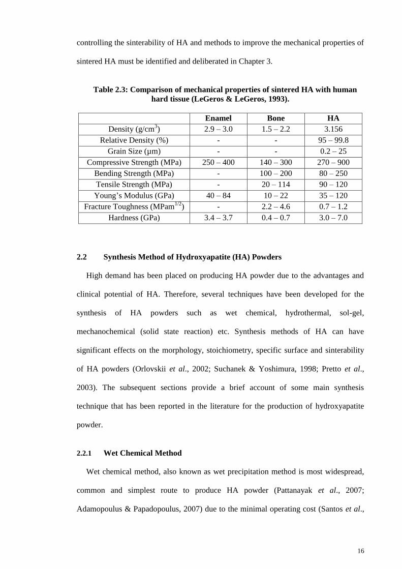
16
controlling the sinterability of HA and methods to improve the mechanical properties of
sintered HA must be identified and deliberated in Chapter 3.
Table 2.3: Comparison of mechanical properties of sintered HA with human
hard tissue (LeGeros & LeGeros, 1993).
Enamel Bone HA
Density (g/cm3) 2.9 – 3.0 1.5 – 2.2 3.156
Relative Density (%) - - 95 – 99.8
Grain Size (µm) - - 0.2 – 25
Compressive Strength (MPa) 250 – 400 140 – 300 270 – 900
Bending Strength (MPa) - 100 – 200 80 – 250
Tensile Strength (MPa) - 20 – 114 90 – 120
Young‘s Modulus (GPa) 40 – 84 10 – 22 35 – 120
Fracture Toughness (MPam1/2
) - 2.2 – 4.6 0.7 – 1.2
Hardness (GPa) 3.4 – 3.7 0.4 – 0.7 3.0 – 7.0
2.2 Synthesis Method of Hydroxyapatite (HA) Powders
High demand has been placed on producing HA powder due to the advantages and
clinical potential of HA. Therefore, several techniques have been developed for the
synthesis of HA powders such as wet chemical, hydrothermal, sol-gel,
mechanochemical (solid state reaction) etc. Synthesis methods of HA can have
significant effects on the morphology, stoichiometry, specific surface and sinterability
of HA powders (Orlovskii et al., 2002; Suchanek & Yoshimura, 1998; Pretto et al.,
2003). The subsequent sections provide a brief account of some main synthesis
technique that has been reported in the literature for the production of hydroxyapatite
powder.
2.2.1 Wet Chemical Method
Wet chemical method, also known as wet precipitation method is most widespread,
common and simplest route to produce HA powder (Pattanayak et al., 2007;
Adamopoulus & Papadopoulus, 2007) due to the minimal operating cost (Santos et al.,

17
Step 1: Titration process by reacting calcium
(Ca2+
) ion with phosphate (PO43-
) ion at
pH above 7 and room temperature
Step 2: Stirring
Step 3: Aging
Step 4: Washing
Step 5: Filtering
Step 6: Drying
Step 7: Crushing and Sieving
2004), inexpensive raw materials, low probability of contamination (Afshar et al., 2003)
and it can be easily carried out at low temperature ranging from room temperature to
100°C (Kumar et al., 2004). Generally, steps involved in the wet chemical method of
HA include:
Figure 2.2: General procedures involved in wet chemical method.
HA powder synthesized through this method is homogenous, has high purity and is
morphologically similar to hard tissue (Donadel et al., 2005; Kothapalli et al., 2004).
However, the shortcomings of this method are the resulting powder is poorly
crystallized without regular shape and the powder quality is greatly affected even by a
slight difference in the reaction/process variables (Kumta et al., 2005). Therefore, it is
indispensible to study the effects of the process variable associated with the wet
chemical method; in relation to the impacts they imposed on the HA powder properties
and sinterability.

18
2.2.1.1 Starting Precursors
A variety of starting precursors can be selected such as calcium nitrate (Ca(NO3)2),
calcium chloride (CaCl2), calcium carbonate (CaCO3), calcium hydroxide (Ca(OH)2),
calcium sulphate (CaSO4) as calcium ions and ammonium phosphate (NH4H2PO4),
phosphoric acid (H3PO4), potassium phosphate (K3PO4), and diammonium phosphate
((NH4)2HPO4) as phosphate ions source (Sadat-Shojai et al., 2013).
Initially, calcium nitrate (Ca(NO3)2) and diammonium phosphate ((NH4)2HPO4) was
used as starting precursors by Hayek and Stadlman (1955) via the following equation:
10Ca(NO3)2 + 6(NH4)2HPO4 + 8NH4OH
Ca10(PO4)6(OH)2 + 6H2O + 20NH4NO3 (2.1)
Later, various researchers (Sung et al., 2004; Bianco et al., 2007; Mobasherpour,
2007; Pattanayak et al., 2007; Monmaturapoj; 2008) used calcium nitrate tetrahydrate
(Ca(NO3)2·4H2O) to produce HA based on the following equation:
10 Ca(NO3)2·4H2O + 6(NH4)2HPO4 + 8NH4OH
Ca10(PO4)6(OH)2 + 20H2O + 20NH4NO3 (2.2)
These authors reported that the synthesized HA powders have particle size ranging
from nanometer to micrometer and have Ca/P ratio in the range of 1.25 to 1.70.
Akao et al. (1981) on the other hand, proposed that calcium hydroxide (Ca(OH)2)
and orthophosphoric acid (H3PO4) to be used as starting materials. HA powders with a
Ca/P ratio of 1.69 were successfully synthesized. The reaction follows the formula:
10Ca(OH)2 + 6H3PO4 => Ca10(PO4)6(OH)2 + 18H2O (2.3)

19
The byproduct contains only water which makes this reaction favorable to the
researchers. Osaka et al. (1991), Tampieri et al. (2000), Afshar et al. (2003), Ramesh et
al. (2008) and Teh et al. (2014) have also adopted this method. These authors attained
HA powders in the nanometer to submicron range with Ca/P ratio ranging from 1.5 to
1.9.
There are studies on deriving calcium precursors from natural resources and bio-
waste such as eggshells, seashells and snail shells and they appear to be a promising
source of calcium for the preparation of hydroxyapatite. Adak et al. (2011) extracted
calcium source from dead snail shells. The dead snail shells were washed and heated at
1000°C to decompose the organic matters and converted to calcium oxide which in turn
on exposure to atmosphere forms calcium hydroxide. Pure HA powder with high
thermal stability (no secondary phases were detected at 1200°C) was then produced via
chemical route. The HA particle exhibited spherical shape with average particle size of
60 – 80 nm.
Using the similar extraction method, Kamalanathan et al. (2014) obtained calcium
hydroxide from raw eggshells as calcium precursor. HA was then synthesized using the
Ca(OH)2 and H3PO4 via wet chemical method. XRD results indicated the synthesis of
single phase and nanocrystalline (35.3 nm) HA was produced in the work. Furthermore,
phase pure HA was obtained and remained stable after albeit sintered at 1250°C.
Similar to the abovementioned methods, CaO was derived from sea shells as starting
material which was converted to calcium hydroxide (Ca(OH)2) and subsequently
reacted with phosphoric acid by wet chemical reaction (Santhosh et al., 2013). The
synthesized powder was pure HA in rod-shape, having crystallite size about 101 nm
with Ca/P ratio of 1.8.

20
2.2.1.2 Synthesis Temperature
In 2007, Bianco et al. (2007) compared wet chemical synthesized HA powder
produced at 40°C and 70°C. Both synthesis temperatures produce needle-like HA
particle which is not in agreement with the study conducted by Bouyer et al. (2000). As
shown in Figure 2.3, low synthesis temperature gives needle shape nanoparticle while
increasing the reaction temperature leads to particle of a more regular shape close to
circular (Figure 2.3). Thus, it can be observed that the shape factor (particle length
divided by particle width) of the HA particle decrease with an increase of the synthesis
temperature.
Figure 2.3: TEM picures of as-synthesized HA nanocrystal at different synthesis
temperature: (a) 35°C; (b) 85°C (Bouyer et al., 2000).
Besides affecting the morphology of the HA particles, reaction temperature was
found to affect the crystallinity but not the phase purity of the synthesized HA as shown
in Figure 2.4 (Pham et al., 2013). The XRD spectra (Figure 2.4) shows that all the HA
has characteristics peaks of pure HA while but the crystallinity increased with
increasing synthesis temperature as the peaks get sharper at higher temperature.
a)
b)
50 nm 50 nm

21
Figure 2.4: XRD spectra of HA powder synthesized at different temperatures
(Pham et al., 2013).
2.2.1.3 Reaction pH
Proper control of the reaction pH is indispensible in wet chemical process as
improper control of the pH can affect the phase purity, microstructural configuration,
crystallite size, mechanical properties etc (Sadat-Shojai et al., 2013). The pH of the
solution is usually adjusted by using ammonium hydroxide (NH4OH) or sodium
hydroxide (NaOH) (Orlovskii et al., 2002) to provide an alkaline medium.
Wang et al. (2010) pointed out that pH values equal or higher than 10 is required to
produce pure HA powder as shown in Figure 2.5. HA synthesized at pH 11 and 10
revealed pure HA peaks with slight difference in degree of crystallinity as shown in
Figure 2.5. As observed from Figure 2.5, the HA prepared at pH 9 was proved to be the
mixture of β-TCP and HA where the diffraction of β-TCP was very weak while the
phase of HA prepared at pH 8 was most of β-TCP.
HAp
Inte
nsi
ty (
a.u.)
20 30 40 50 60 70 80
2θ (degrees)
80°C
60°C
40°C
25°C

22
Figure 2.5: XRD patterns of HA samples.
Besides that, Inthong et al., (2013) claimed that the reaction pH has effects on HA
particle as increasing the pH value would increase the formation of particle
agglomeration but reduce the particle size of the HA powder as shown in Figure 2.6.
Figure 2.6: SEM micrographs of HA nanoparticles synthesized at (a) pH 5
(acidic), (b) pH 7, and pH 11 (alkaline) (Inthong et al., 2013).
In the same study, reaction pH value has direct effect on the grain size and
mechanical properties of HA. It is perceived that HA synthesized at pH value of 9
shows the highest density and hardness value as shown in Table 2.4. Low reaction pH
value (5 – 7) results in lower density and hardness value compared to those synthesized
at high pH value (8 – 11).
Inte
nsi
ty (
a.u.)
β-TCP
pH 8
20 25 30 35 40 45 50 55
2θ (degrees)
a) b) c)
100 nm 100 nm 100 nm
pH 9
pH 10 pH 11

23
Table 2.4: Mechanical properties of HA prepared with different pH values
(Inthong et al., 2013).
pH value Density (g/cm3) Grain Size (µm) Hardness (GPa)
5 2.75 2.19 3.12
6 2.82 1.64 3.39
7 2.83 1.72 3.50
8 3.00 5.97 4.63
9 3.04 6.81 6.06
10 2.98 4.34 4.68
11 3.02 2.38 4.67
2.2.1.4 Ca/P Ratio
Pham et al. (2013) studied the effect of various Ca/P ratio (3/2, 10/6 and 10/3) on the
properties of HA powder and Ca/P ratio did not affect the purity of HA as all the HA
powder revealed XRD peaks corresponding to pure HA without any presence of
secondary phases. This result is different from those reported by Raynaud et al. (2002).
According to Raynaud et al. (2002), powders having a Ca/P molar ratio of 1.667 and
composed of single HA phase could only be synthesized with a high reproducibility
provided that the Ca/P ratio of the initial reagents were fixed at 1.667. Any Ca/P ratio
lower or higher than 1.667 would lead to phase decomposition as shown in Table 2.5.
Furthermore, Adak et al. (2011) stated that HA of Ca/P = 1.67 has better densification
and finer particle size.
Table 2.5: Phase composition of HA prepared with different Ca/P ratio
(Raynaud et al., 2002).
Ca/P (Molar Ratio) Phase Composition (wt%)
< 1.50 -TCP + -Ca2P2O7
(minor phase ~ 10 wt %)
1.511 ± 0.001 93% -TCP + 7% HA
1.535 ± 0.004 78% -TCP + 22% HA
1.631 ± 0.004 80% HA + 20% -TCP
1.643 ± 0.003 87% HA + 13% -TCP
1.655 ± 0.002 93.5% HA + 6.5% -TCP

24
Table 2.5, continued.
1.663 ± 0.001 98% HA + 2% -TCP
1.667 ± 0.005 > 99.5% HA
1.71 ± 0.01 98.5% HA + 1.5% CaO
2.2.1.5 Drying Methods
Drying can be defined as a process of removal of water as vapor from the material
surface (Abd Rahman et al., 2009). It is one of the crucial steps in the wet chemical
synthesis of HA powder. A few drying methods such as oven drying, freeze drying,
spray drying and microwave drying have been utilized in wet chemical synthesis of HA
powders.
Oven Drying (a)
Majority of the research employed oven drying on the HA slurry at drying
temperature ranging between 65 – 105ºC to obtain HA powder (Lazic et al., 2001;
Saeri et al., 2003; Smiciklas et al., 2005; Markovic et al., 2004; Ramesh et al.,
2007). Oven drying involves heating of the surface and the heat transferred into the
material by the means of convective, conductive and radiation (Singh et al., 2015)
and it usually takes a very long drying hours (16 – 48 hours). Zhang and Yokogawa
(2008) studied the effect of drying conditions during HA powder synthesis on the
properties of the HA powders. The HA slurry was dried at temperature ranging from
60 – 150°C for 3 – 21 days, respectively. In this study, the author noticed that the
drying period had no effects on the phase stability, Ca/P molar ratio and particle
shape of the HA powders. However, they found that HA powder particle sizes
decreased with the drying time (Figure 2.7) but independent of the drying
temperature. They also found that the longer the drying period, the larger the powder
agglomeration as evidence from the TEM micrographs (Figure 2.8) of HA powder.

25
Figure 2.7: Relation between drying period and the particle size of HA powder
dried at 60ºC (Zhang & Yogokawa, 2008).
Figure 2.8: TEM photos of HA powders dried at 60ºC for (a) 3, (b) 10 and (c) 18
days (Zhang & Yogokawa, 2008).
The severe powder agglomeration of HA powders produced via wet chemical
methods is in agreement with various studies (Lu et al., 1998; Hsu & Chiou, 2011;
2
1.8
1.6
1.4
1.2
1
0.8
0.6
0.4
Part
icle
Siz
e (µ
m)
3 7 10 14 18 21
Drying Period (days)
a) b)
c)
2 nm 2 nm
2 nm

26
Yu et al., 2010). Although oven drying is economic and simple but it is also well
known that wet chemical synthesized HA powders dried by the means of
conventional oven drying are invariably agglomerated often with several levels of
porosity Hence, drying methods such as freeze drying and spray drying were
introduced to dry the HA precipitate while microwave drying was found to be
potential alternative to those drying methods. The comparison studies between oven
dried HA powders with the HA dried by the alternative drying methods are
discussed in the next sections.
Freeze Drying (b)
Freeze drying has been reported as an efficient way to eliminate the powder
agglomerations problem (Girija et al., 2012) and preparing uniform fine-grained
powders which can be sintered to very high density (Lu et al., 1998; Chesnaud et al.,
2007; Shlyakhtin et al., 2000; Wang et al., 1992). Hence, it has become a popular
drying method in the wet chemical synthesis of hydroxyapatite (Lu et al., 1998;
Girija et al., 2012, Wang et al., 2010).
Firstly, the HA slurry is frozen to solid state (Chen & Wang, 2007) which can be
carried out by immersing the slurry in liquid nitrogen (fast freezing) or in an
industrial deep freezer (slow freezing). Fast freezing rate is desirable to prevent
aggregation and recombination of the particles but it usually requires special
equipment and there are risks in handling liquid nitrogen. After freezing, the frozen
HA slurry, mostly in ice form is sublimated by reducing the pressure below the triple
point (Figure 2.9) of the water through vacuum pump. Therefore, in freeze drying,
the frozen HA precipitate directly transformed into a gaseous form without passing
through a liquid phase and high temperature is not required.

27
Figure 2.9: Schematic diagram for the triple phases of water (Yu et al., 2011).
A systematic study on the effects of freeze drying on the powder properties and
sinterability of hydroxyapatite was reported by Lu et al. (1998). In their work, HA
precipitate mixture was rapidly frozen with liquid nitrogen and subsequently brought
into freezing drying apparatus for about 36 – 70 hours to get HA powder form,
hereafter known as FD-HA. The authors confirmed that as-synthesized FD-HA
powder is pure and has ideal stoichiometry (Ca/P = 1.67). However, it was found
that the FD-HA powder decompose to β-TCP at sintering temperature of 1300ºC. A
similar decomposition temperature was found on the conventional oven dried HA
powder indicated that freeze drying has no negative impact on the thermal stability
of the HA powder.
TEM images show that the powder particle size of FD-HA (33 nm) was smaller
than oven dried HA powder (40 nm) and the FD-HA powders are homogenous while
oven dried HA is in the form of large agglomerates (Figure 2.10). This could be the
reason that the density of sintered FD-HA is always higher than the oven dried HA
throughout the sintering regime as Lin et al. (2007) reported that smaller particle
Pre
ssu
re
Temperature

28
size with less agglomerates provides higher driving force for the densification of
ceramics during sintering (Lin et al., 2007).
Figure 2.10: Transmission electron micrographs of HA powders after calcining
at 800ºC for 3 hours: (a) freeze dried HA, (b) oven dried HA (Lu et al., 1998).
A maximum density of ~99% was obtained from FD-HA at 1350ºC and the
maximum hardness for the FD-HA is 820 MPa, compared to 670 MPa for the oven
dried HA. The high hardness of FD-HA is due to the simultaneous effects of the
high density coupled with small and uniform grain size of the sintered FD-HA as
shown in Figure 2.11.
Figure 2.11: SEM micrographs of etched fracture surface of (a) freeze dried HA,
(b) oven dried HA, sintered for 3 hours at 1350ºC (Lu et al., 1998).
The beneficial effects of freeze drying were supported by Girija et al. (2012). In
their work, HA precipitate was subjected to pre-freezing at –5°C. This pre-frozen
a) b)200 nm 200 nm
a) b) 5 µm 5 µm

29
product was dried in a vacuum freeze dryer at –50°C. The freeze dried HA has
higher thermal stability compared to oven dried HA where decomposition to β-TCP
took place at sintering temperature of 900ºC and subsequently transform to α-TCP at
1100°C for oven dried HA while pure HA phase remained for HA synthesized by
freeze drying at all sintering temperature. Moreover, the sintered freeze dried HA
was found to have homogenous grain and highly densified compared to sintered
oven dried HA. The author proposed that the formation of tri-calcium phosphate and
pores in oven dried HA leads to less shrinkage and hence less densification and
higher porosity of the sample.
Spray Drying (c)
The spray drying is a continuous process of drying liquid or slurry into a free
flowing powder material (Murtaza et al., 2012). The slurry of the material is sprayed
as a fine spray or mist into a chamber with heated air to produce spherical powder
agglomerates of relative homogeneity. It offers several advantages such as the
ability to control particle size, particle size distribution, bulk and particle density
with careful choice of spray dryer operating parameters (Bertrand et al., 2005), can
be scaled up to ton quantities (Iskandar et al., 2003), low heat and short processing
time. Hence, spray drying is introduced in the synthesis of HA to produce HA with
homogenous morphology (Luo et al., 1999; Murtaza, 2012; Wang et al., 2009).
Wang et al. (2009) synthesized hydroxyapatite slurry via wet method and spray
drying. The spray dried HA powder has low crystallinity and possessed XRD peaks
that match perfectly to the HA standard peaks without impurities such as TCP and
CaO. Under the examination of SEM, the shape of the spray dried HA powder
particle is smooth and uniform; having microspheres morphology with broad size
distribution.

30
On the other hand, Murtaza et al. (2012) successfully produced uniform HA
particles with solid spherical shape via spray drying which is consistent with the
reported study from Wang et al. (2009). However, Murtaza et al. (2012) obtained
powders with narrow particle size distribution which is opposed to the results
published by Wang et al. (2009). This could be due to different starting precursors
were used in their experiments. Both studies emphasized that by manipulating
process parameters like liquid feed rate, slurry viscosity, inlet air temperature and
compressed air flow rate can affect the HA properties such as specific surface area,
particle size, particle size distribution and powder morphology.
Microwave Drying (d)
In the past 30 years, the research on microwaves has been popular in the food
related field, treatment of biomass and biosolids, mineral processing, materials
processing etc. (Decareau, 1985; Dorantes -Alvarez et al., 2000; Rodríguez et al.,
2003; Yongsawatdigul & Gunasekaran, 1996; Lefeuvre, 1981; Moller & Linn, 2001;
Shyju, 2010) due to its numerous advantages over the conventional method such as
fast and rapid for extreme time and energy savings, material can be dried completely
due to volumetric heating, it provides uniform energy distribution, it is specific only
to certain substance (ie. water), it is environmental friendly as no harmful
greenhouse gas emissions from the heat source (Srogi, 2006; Moller & Linn, 2001;
Abd Rahman et al., 2009). Due to the abovementioned benefits, microwave is
termed as potential alternative drying method to the conventional oven drying.
In conventional drying methods, the heat required to evaporate the moisture has
to be transmitted inward through the moist material from the surface where this
process is very time consuming (Venkatesh & Raghavan, 2004). This hurdle is
eliminated in microwave drying, where internal heat generation phenomenon is the

31
source of drying. Microwave is an electromagnetic waves falls in the frequency
range of 300 MHz to 300 GHz (Sun et al., 1994) that acts specifically on polar
molecules like water. The charged portions of the water molecules move with
respect to the rapid oscillating electric field of the electromagnetic waves. This rapid
movement is realized as molecular vibrations that result in local kinetic heating in
the core of the material in a very short period of time. The high temperature/heat in
the core drives the evaporating liquid to the surface of the material, enabling the
water movement and its subsequent removal from the material rapidly. This
phenomenon is known as volumetric heating (Strumillo & Kudra,1986). The
difference of microwave drying and conventional drying in terms of temperature
profile is shown in Figure 2.12.
Figure 2.12: Temperature profile inside sample of (a) conventional drying (b)
microwave drying (Hui, 2008).
Due to the advantages offered by microwave drying, it has been utilized in the
drying of ceramics. Thevi et al. (2009) reported that milled mixture of β-TCP and
CaCO3 was rapidly dried using microwave oven for 60 – 70 minutes and followed
by conventional sintering to form porous HA scaffolds. The sintered HA is highly
crystalline and the elements exist are mainly calcium phosphorus coupled with a
Ca/P ratio ranging from 1.62 – 1.69 which was confirmed by ICP-AES analysis. On
External Heating Internal Heating
Temp. Temp.
Distance Distance
a) b)

32
the other hand, Sundaram et al. (2008) showed that gelatin-starch with nano HA
composite has been successfully fabricated via microwave drying for 40 – 45
minutes.
Most of the studies were focused on the use of microwave drying in fabricating
porous HA scaffolds composite as stated above. Literature reports on the use of
microwave in drying pure HA and the effects of microwave drying on the properties
of the HA powder are limited. The only available study was done by Santhosh and
Prabu (2012) who compared HA powders that dried in electric furnace at 80°C for 8
hours with HA powder dried in domestic microwave oven at 480 W for 25 minutes.
There was evident from their XRD analysis that no significant change in phase
formation had occurred due to microwave drying. However, the size of HA
crystallites produced by microwave drying were bigger than those produced by
furnace drying. Microwave dried HA had crystallite size in the range of 34 – 102 nm
as compared to 7 – 34 nm of the furnace dried HA. This was due to fact that ion-
covalent materials such as HA is microwave sensitive which absorb the microwave
power rapidly, causing the grain coarsening.
Besides bioceramics, microwave drying has been applied on engineering
ceramics such as alumina and zinc oxide. Abd Rahman et al. (2009) have discussed
the effects of microwave drying on the properties of alumina-zeolite foam. The
ceramic slurry was obtained by mixing commercial alumina powder and zeolite with
water and then underwent microwave drying in a common domestic microwave
oven at 800 watts. It is worth mentioning that 7 minutes is sufficient to remove all
the free and bound water from the material. The dried green bodies were
subsequently sintered at 1350°C. In comparison to conventional oven drying, the
linear shrinkage of the microwave dried foam is lower. In terms of flexural strength,

33
microwave drying shows significant improvement (2.11 MPa) compared to oven
dried foam (0.76 MPa). The higher flexural strength of the microwave dried foam is
attributed to its finer and smaller grains as evident in Figure 2.13.
Figure 2.13: SEM micrographs of sintered foam from (a) conventional drying
and (b) microwave drying (Abd Rahman et al., 2009).
On the other hand, Shirai et al. (2006) applied a rapid microwave drying
technique on ZnO ceramic and the characteristics of the ceramic bodies were
compared with those dried by conventional drying techniques. In their study,
commercially available ZnO powder was dispersed in 60 vol% distilled water with
0.32 mass% of ammonium poly acrylilate dispersant and subjected to ball milling
for 12 hours to obtain the ZnO slurry. After that, a batch of green bodies was dried at
oven at 80°C for at least 24 hours while another batch was dried in a microwave
oven for 30 minutes. Upon drying, the green bodies were sintered in a conventional
furnace at 1000ºC. The authors noticed that only 30 minutes are required to
eliminate all the water contents from the green bodies via microwave drying while in
oven drying, 5 hours is necessary to remove all free and bound waters. Moreover,
Shirai et al. (2006) reported that microwave drying produce sintered ceramic bodies
with relative density that significantly higher than that of oven dried ceramic bodies.
a) b)

34
In conclusion, microwave drying has shown promising results in enhancing the
powder properties, densification and mechanical properties of ceramics within a
short period of time. Hence, it has a great potential to be incorporated in the wet
chemical synthesis of hydroxyapatite powder.
2.2.1.6 Other Synthesis Parameters
Besides the staring precursors, synthesis temperature, reaction pH, Ca/P ratio and
drying methods, the effects of other synthesis parameters such as solvent used for the
precipitation, the acid addition rate, the stirring and aging time were investigated by
researchers too. The solvent/medium used for the precipitation process affects the
morphology and particle size of the synthesized HA (Alobeedallah et al., 2011).
Alobeedallah et al. (2011) found that HA particles formed from ethanol solution were
rod like shape crystals while those synthesized in water had irregular to spherical shape
particle. Besides, the HA crystal formed in ethanol was smaller than those prepare in
water system.
The acid addition rate was found to have effects on the size of the formed HA crystal.
Pham et al. (2013) conducted a study to investigate the effects of acid addition rate on
the crystal size and phase of HA. The results concluded that the increase of acid
addition rate could lead to the increasing HA crystal length. According to Pham et al.
(2013), the faster the acid adding rate, the greater the frequency of reactant collisions, so
larger HA crystals were created. In general, all the FTIR spectra with different dropping
rates have similar shape and specific peaks corresponding to functional groups of HA
(Figure 2.14). XRD results in Figure 2.15 confirmed that pure HA was synthesized
regardless of acid addition rates.

35
Figure 2.14: FTIR spectra of HA powder synthesized at different acid addition
rate: 1, 2 and 5 ml min-1
(Pham et al., 2013).
Figure 2.15: XRD spectra of HA powder synthesized at different acid addition
rate: 1, 2 and 5 ml min-1
(Pham et al., 2013).
Stirring during the precipitation has found to have effects on the HA powder
properties. Jarcho et al. (1976) reported that stirring of 24 hours or more gives the
solution a Ca/P ratio close to the stoichiometric hydroxyapatite 1.67. In the study
conducted by Santos et al. (2004), they claimed pure HA produced under vigorous
stirring while biphasic HA/TCP was obtained under slow stirring. Moreover, a powerful
2 ml/min
5 ml/min
1 ml/min
4000 3500 3000 2500 2000 1500 1000 500
Tra
nsm
itta
nce
In
tensi
ty (
a.u.)
20 30 40 50 60 70 80
2θ (degrees)
° HAp

36
and high speed stirring would yield a good homogenous medium for the precipitation of
HA (Afshar et al., 2003).
In terms of aging or settling time, Pham et al. (2013) investigated the effects of
various settling time (0, 2, 15, 21 and 24 hours) on the HA powder properties. It was
found that all the synthesized HA are pure HA with cylinder shape morphology
regardless of the settling time. However, it does have effects on the particle size. The
particle size reduced when the settling time increased from 0 to 15 hours and increased
continuously up to 21 and 24 hours. Thus, the authors concluded that 15 hours is the
optimal settling time to get the smallest HA particles powder.
2.2.2 Hydrothermal
The synthesis of HA powder through hydrothermal process involves the reaction of
chemicals in an aqueous solution at elevated temperature (typically above water‘s
boiling point) in a sealed vessel (pressure exceeds the ambient pressure) (Zhang et al.,
2011; Manafi & Rahimipour, 2011; Yoshimura & Byrappa, 2008). Besides producing
high crystalline and high purity HA powder, it has been reported that hydrothermal
offers advantages such as low cost reagents, short reaction time and post heat treatment
on HA powder is not required (Guo and Xiao, 2006; Guo et al., 2005; Zhang & Vecchio,
2007). In addition, hydrothermal synthesis can produce powders with different
morphologies such as nanowire, sphere, nanobelt and prism-like particles (Zhu et al.,
2008).
Liu et al. (1997) reported a simplified hydrothermal process of synthesizing high
quality HA powders by utilizing a pressurized cooking utensil. According to this
method, reagent grade Ca(H2PO4)2.H2O and Ca(OH)2 powders were ball-milled and
dried under an infrared lamp and subsequently mixed with 2 litres distilled water in a
commercially available pressurized pot at pressure of ~ 0.125 MPa and ~ 109º for 1 – 3

37
hours. The reaction product was filtered, washed and dried at 90ºC to obtain HA powder.
Some researchers used different combination of starting precursors such as CaCO3 and
NH4H2PO4 (Orlovskii et al., 2002), Ca2P2O7 and CaO (Li et al., 1994) and etc (Andres-
Verges et al., 1998; Guo & Xiao, 2006) to produce HA powder.
Although, the hydrothermal process is able to produce single phase HA, this process
is very time consuming and requires equipment that must be corrosion resistant, able to
with stand high pressures and high temperatures and hence expensive compared to other
methods.
2.2.3 Sol-Gel Method
Sol-gel method is one of the preferred method to produce HA powder due to the low
synthesis temperature, homogenous molecular mixing of starting materials and effective
in producing nanophasic HA. It needs strict control process parameters (Balamurugan et
al., 2006) as it involves the molecular level mixing of the calcium and phosphorus. It
has been reported that sol gel synthesized HA has enhanced stability at the bone
interfaces in vitro and in vivo (Li et al., 1994). Moreover, the HA powder obtained via
sol-gel method has high purity and large surface area with small particle size (Fathi et
al., 2008; Hsieh et al., 2001, Wang et al., 2007). In general, sol gel process involves the
creation of a three-dimensional, interconnected network, termed a ―gel‖, from a
suspension of very small, colloidal particles, called a ―sol‖ (Chen et al., 2011; Ioitescu
et al., 2009). One of the major disadvantages of this method is the complicated and
lengthy process includes the mixing of alkoxides in either aqueous or an organic phase,
aging at room temperature, gelation, drying, stabilization and post heat treatment
(calcination).
The aging of the solution necessitate more than 24 hours to stabilize the solution
system to enable the formation of monophasic HA (Liu et al., 2001). The need of long
38
aging time is in agreement with the studies carried out by Chai et al. (1998) and Liu et
al. (2002). Apart from that, the high cost of the raw materials especially alkoxide-based
precursors is also a major discouragement to researchers (Kim & Kumta, 2004).
Eshtiagh-Hosseini et al. (2007) and Jillavenkatesa and Condrate Sr. (1998) reported that
the generation of secondary phase like calcium oxide (CaO) is always found in sol-gel
derived HA. The formation of CaO is unfavorable as CaO adversely affects the
biocompatibility of HA. Besides CaO, insufficient aging or uncontrolled gelation and
heat treatment may cause the generation of various impurities such as Ca2P2O7,
Ca3(PO4)2 and CaCO3( Eshtiagh-Hosseini et al., 2007; Hsieh et al., 2001).
2.2.4 Mechanochemical (Solid State Reaction)
Mechanochemical powder synthesis takes advantage of the perturbation of surface-
bonded species by pressure to enhance thermodynamic and kinetic reactions between
solids (Coreno et al., 2005; Mochales et al., 2004; Chen et al., 2004). This
transformation is induced by mechanical milling of the precursors. During the process,
the precursors subjected to the deep squeezing and crushing between the milling media
or the milling media and the container wall while the molar ratio between the precursors
is kept at the stoichiometric ratio. Phase transformations occur at room temperature as a
high local heat is generated in the milling container during the milling process. The
main processing variables include the following: (a) the type of milling equipment such
as low energy ball mills, attrition mill, planetary and vibratory mills; (b) the type of
milling medium; (c) the type and diameter of the milling balls; (d) the duration of the
milling process; (e) the powder to ball mass ratio and (f) the type of starting precursors
(Suchanek et al., 2002; Tian et al., 2008; Silva et al., 2004; Fathi & Mohammadi
Zahrani, 2009; Honarmandi et al., 2010). Besides, mechanochemical method involves
long heat-treatment times (post synthesis calcination) (Orlovskii et al., 2002; Suchanek
et al., 1997; Naruporn Monmaturapoj & Chokchai, 2010). It offers advantages such as

39
simple, high productive, suitable for mass production and is cost effective (Pramanik et
al., 2007). However, it has a few shortcomings such as long milling hours to ensure
homogeneity of mixing materials, produces HA with low specific surface area and low
sinterability (Suchanek &Yoshimura, 1998).
The commonly used starting precursors selected to prepare HA powder via this
method includes dicalcium phosphate anhydrous (CaHPO4), dicalcium phosphate
dihydrate (CaHPO4.2H2O), monocalcium phosphate monohydrate (Ca(H2PO4)2.H2O),
calcium pyrophosphate (Ca2P2O7), calcium carbonate (CaCO3), calcium oxide (CaO)
and calcium hydroxide (Ca(OH)2), etc. (Otsuka et al., 1994; Chaikina, 1997; Rhee,
2002). Silva et al. (2003) carried out mechanochemical synthesis in planetary mill with
different types of precursors combinations and they found out that the following
reactions successfully produced single phase nano-sized HA powder.
3Ca3(PO4)2.xH2O +Ca(OH)2 => Ca10(PO4)6(OH)2 + xH2O (2.4)
10Ca(OH)2 + 3P2O5 => Ca10(PO4)6(OH)2 + 9H2O (2.5)
4CaCO3 + gCaHPO4 => Ca10(PO4)6(OH)2 + 2H2O + 4CO2 (2.6)
In order to find a suitable medium for mechanochemical, Rhee (2002) compared the
results when either water or acetone was used as the medium. Rhee concluded that pure
HA powder could be obtained after milling for 8 hours using water as milling medium
while powders attained through acetone as milling medium revealed the presence of β-
TCP. In another work, Yeong et al. (2001) used ethanol as the milling medium and the
milling process has to be extended to 20 hours in order to produce pure HA powder.
Thus, water is favourable in producing pure HA powder via mechanochemical reaction.

40
2.2.5 Other Processing Techniques
There are also alternative techniques for preparation of HA powder, such as spray-
pyrolysis (An et al., 2007; Cho et al., 2008; Itatani et al., 2010), electrocrystallization
technique (Manso et al., 2000; Shirkhanzadeh, 1994), emulsion route (Zhang & Cooper,
2005; Yang et al., 2011; Jarudilokkul et al., 2007), self propagating combustion
synthesis (Tas, 2000; Sasikumar et al., 2010), hydrolysis route (Shih et al., 2006;
Monma & Kamiya, 1987) and microwave hydrothermal route (Pushpakanth et al., 2008;
Han et al., 2006).
These methods have the ability to synthesize HA powder with varying degree of
purity, crystallinity, morphology and stoichiometry. However, there are some major
drawbacks of these methods such as time consuming and complex methodology,
requires special and expensive equipment and high cost of starting precursors which
make them unsuitable for mass production (Pushpakanth et al., 2008; Cao et al., 2005;
Wang et al. 2007).
In summary, it is well established that the processing of ceramics is complicated by
the number of steps typically required for manufacture and the need to optimize the
processing steps. The processing of ceramics is extremely important because ceramics
are basically flaw intolerant materials. As a result, minor chemical and physical defects
can severely degrade properties. Proper selection of raw materials and processing
conditions is especially important in ceramics because mistakes generally cannot be
corrected during the firing and post-firing processes.
Thus, there is a great need for a simple, reliable and reproducible synthesis technique
to manufacture a high purity and crystalline hydroxyapatite powder which has a good
sinterability at low firing temperatures coupled with superior mechanical properties
suitable for biomedical applications.

41
CHAPTER 3: THE SINTERING AND SINTERABILITY OF
HYDROXYAPATITE
3.1 Powders Consolidation (Sintering) Techniques
One of the main methods to produce bulk hydroxyapatite (HA) is sintering process
(Prokopiev & Sevostianov, 2006). Sintering process is a consolidation process that
eliminates the intra- or inters- agglomerate pore of material and brings the individual
particles together to form a solid body (Uhlmann et al., 1976). It has been reported that
sintered HA has high density (Ramesh et al., 2007c) than non-sintered HA and can form
tight bonds with bone tissue (Stea et al., 1995). To date, various consolidation
techniques have been developed to sinter bulk HA such as conventional pressureless
sintering (CPS), microwave sintering (MS), hot pressing sintering (HPS), spark plasma
sintering (SPS) and two steps sintering (TSS).
3.1.1 Conventional Pressureless Sintering (CPS)
Conventional pressureless sintering (CPS) is performed in a conventional
programmable furnace and it is a widely used consolidation method on hydroxyapatite
(HA) owing to its practicability and simplicity (Wang & Chaki, 1993). However, the
main problem associated with this method is the generation of thermal stress which
leads to the formation of microcracks in the HA specimen (Prokopiev & Sevostianov,
2006). To avoid this problem, the employed ramp rate must be less than 5°C/min, and
thus, makes CPS a very time consuming process. Furthermore, high sintering
temperature and long holding time required in CPS not only lead to the production of
large grain HA (Mazaheri et al., 2008), but also promote the decomposition of HA into
secondary phases. The presence of secondary phases can hinder the mechanical
properties of HA (Wang & Chaki, 1993). Due to these drawbacks, other alternatives
such as MS, HPS, SPS and TSS are introduced.

42
3.1.2 Microwave Sintering (MS)
Microwave energy provides advantages in various industries such as fast and rapid,
time and energy saving, complete drying of material and environmental friendly. In
terms of sintering, microwave sintering (MS) serves as alternative to the common
conventional pressureless sintering (CPS) and has been applied successfully to sinter
various types of ceramics (Clark & Sutton, 1996; Agrawal, 1998; Leparoux et al., 2003).
In MS, the density of the material can be enhanced to higher level than would be
achieved in CPS at the same temperature (Agrawal, 2006; Wang et al., 2006). MS also
requires less sintering time and less energy consumption. In addition, MS enhanced the
microstructure and mechanical properties of ceramics by inhibiting the excessive grain
growth (Ramesh et al., 2007a). Both CPS and MS provide heat to the material in order
for them to densify. However, they work differently in terms of the heat transfer profile.
Heat is transferred to the ceramics via conduction, convection and radiation from the
surface into the interior of the ceramics in CPS (Figure 3.1 (a)). In contrast, MS
demonstrates inverse temperature flow as electromagnetic radiations are transformed
into heat inside the core of the ceramic and subsequently transported to the surface from
the core as shown in Figure 3.1 (b).
Figure 3.1: Comparison of heating procedure between (a) Conventional
sintering and (b) Microwave sintering (Agrawal, 2006).
a) b) COVENTIONAL
Heating Element
MICROWAVE
Microwave Port
INSULATION FURNACE
INSULATION
MICROWAVE CAVITY
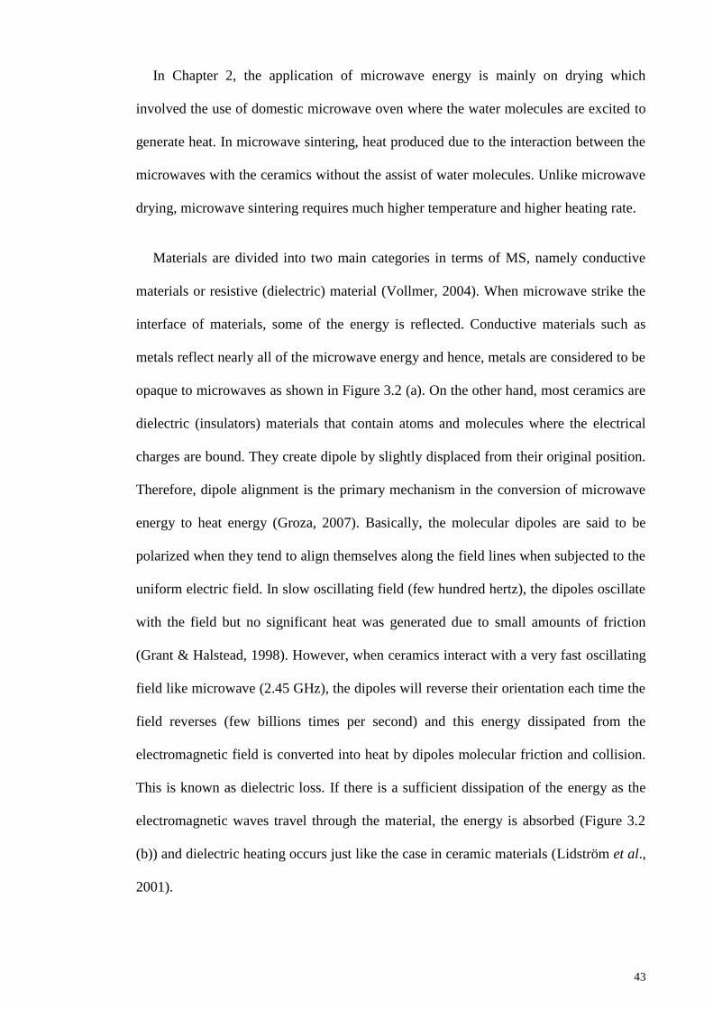
43
In Chapter 2, the application of microwave energy is mainly on drying which
involved the use of domestic microwave oven where the water molecules are excited to
generate heat. In microwave sintering, heat produced due to the interaction between the
microwaves with the ceramics without the assist of water molecules. Unlike microwave
drying, microwave sintering requires much higher temperature and higher heating rate.
Materials are divided into two main categories in terms of MS, namely conductive
materials or resistive (dielectric) material (Vollmer, 2004). When microwave strike the
interface of materials, some of the energy is reflected. Conductive materials such as
metals reflect nearly all of the microwave energy and hence, metals are considered to be
opaque to microwaves as shown in Figure 3.2 (a). On the other hand, most ceramics are
dielectric (insulators) materials that contain atoms and molecules where the electrical
charges are bound. They create dipole by slightly displaced from their original position.
Therefore, dipole alignment is the primary mechanism in the conversion of microwave
energy to heat energy (Groza, 2007). Basically, the molecular dipoles are said to be
polarized when they tend to align themselves along the field lines when subjected to the
uniform electric field. In slow oscillating field (few hundred hertz), the dipoles oscillate
with the field but no significant heat was generated due to small amounts of friction
(Grant & Halstead, 1998). However, when ceramics interact with a very fast oscillating
field like microwave (2.45 GHz), the dipoles will reverse their orientation each time the
field reverses (few billions times per second) and this energy dissipated from the
electromagnetic field is converted into heat by dipoles molecular friction and collision.
This is known as dielectric loss. If there is a sufficient dissipation of the energy as the
electromagnetic waves travel through the material, the energy is absorbed (Figure 3.2
(b)) and dielectric heating occurs just like the case in ceramic materials (Lidström et al.,
2001).

44
Figure 3.2: Schematic diagram of the main ways that microwaves can interact
with materials (Sutton, 1989).
Although MS is advantageous in various aspects, a major problem has been
encountered in the MS of hydroxyapatite (HA) as HA lacked of microwave
susceptibility at ambient temperature due to their loss factor and low thermal
conductivity (Del Regno, 2006; Lasri et al., 2000). Below critical temperature, HA is
poor absorber and it needs to be heated up to a critical temperature to become
microwave absorbent (Edabzadeh & Valefi, 2008; Haung et al., 2009). This problem
can be overcome by using an external susceptor to initiate the heating. Susceptor is an
excellent microwave absorber and it supplies heat to HA by absorbing microwave up to
a temperature high enough that HA starts to absorb the microwave and heat by itself
(independent of suspector).
The most common susceptor materials used in the microwave sintering of HA are
silicon carbide (SiC) and zirconia (ZrO2) (Nath et al., 2006; Fang et al., 1994; Vijayan
& Varma, 2002; Bose et al., 2010; Yang et al., 2002; Ramesh et al., 2008, Ramesh et
al., 2007a). All these authors used either zirconia or SiC as susceptor to sinter the HA in
microwave furnace. They found that MW has substantial effects on the densification,
Opaque
(Conductor)
Reflection
Absorber
(lossy insulator)
absorption

45
microstructure, phase stability and mechanical properties on the sintered HA compared
to CPS.
For instance, Ehsani et al. (2013) experimented sintering on HA using both MS
and CPS technique. The authors reported that with MS, densification began at 900°C
which was 100°C lower than for CPS. MS-HA obtained higher density than CPS-HA
throughout the sintering regime and met plateau at temperature above 1300°C as shown
in Figure 3.3.
Figure 3.3: Comparative sintering curves for HA sintered by microwave and
conventional heating (Ehsani et al., 2013).
The beneficial effect of MS in densification of HA is in agreement with the
experiment carried out by Nath et al. (2006). In their study, the density of MS-HA was
higher than that of CPS and maximum relative density of 99% was obtained by MS-HA
at 1000°C sintered for 30 minutes. At the similar temperature, CPS-HA could attain
only 96.99% albeit being sintered for 3 hours. Although the density of MS-HA is higher,
MS-HA sintered at 1100°C has grain size of about ~20 – 30 µm, which significantly
bigger than that of CPS-HA (5 – 10 µm) at the same temperature.
10
90
80
70
60
50
Den
sity
(%
Th
eor.
)
900 1000 1100 1200 1300 1400
Temperature (°C)
Microwave
Sintering
Conventional
Sintering

46
However, Ramesh et al. (2008) reported that the densification of CPS-HA was
higher than MS-HA although the grain size was finer for the latter as shown in Table
3.1 and Figure 3.4, respectively.
Table 3.1: Total sintering time taken to achieve the respective sintered density
based on the two different sintering techniques (Ramesh et al., 2008).
Sintering
Temperature
(°C)
CPS MS
Relative
density (%)
Time
taken
(min)
Relative
density (%)
Time
taken
(min)
1000 96.8 1090 90.8 29.4
1050 98.4 1140 95.2 30.4
1100 99.0 1190 96.5 31.4
1150 99.0 1240 96.7 32.4
1200 99.0 1290 96.6 33.4
1250 99.1 1340 97.0 34.4
1300 99.0 1390 98.0 35.4

47
Figure 3.4: Microstructural evolution of CPS-HA (a-c) and MS-HA (d-f) when
sintered at 1150ºC (a,d), 1250ºC (b,e) and 1300ºC (c,d) (Ramesh et al., 2008).
The fine and smaller grain size of MS-HA was the main factor contributed to the
high fracture toughness of MS-HA. Overall, throughout the sintering regime, MS-HA
obtained fracture toughness higher than CPS-HA, ranging from 1.24 MPam1/2
to 1.45
MPam1/2
while CPS-HA attained value from 0.91 to 1.22 MPam1/2
. The author
concluded that MS not only improved the toughness and microstructure of HA, it
significantly reduced the time taken to sinter the samples as shown in Table 3.1.
In terms of phase stability, Harabi et al. (2010) found that HA starts to decompose
into TCP when microwave sintered at 1050°C and the percentage of this phase was
increased from 46% to 61% when the sintering temperature increases from 1050°C to
1100°C, respectively. For samples sintered at 1150°C and above, the TCP slowly
2.5µm
10 µm
5 µm
1 µm
1 µm
1 µm

48
transformed into α-TCP. The XRD pattern of the microwave sintered HA at various
sintering temperature is presented in Figure 3.5. The author claimed that this
decomposition is due to the higher heating efficiency of microwave heating. This work
is in contrary to research carried out by previous workers (Yang et al., 2002; Ramesh et
al., 2008; Ramesh et al., 2007a) as they did not found a significant HA decomposition.
Figure 3.5: XRD of HA sintered at different temperatures for 15 minutes, using
MS (Harabi et al., 2010).
In summary, MS has beneficial effects on the sinterability of HA (Vijayan & Varma,
2002; Bose et al., 2010; Ramesh et al., 2007a; Dasgupta et al., 2013; Mangkonsu et al.,
2016). It can be concluded that HA compacts of certain green density can be sintered by
microwave irradiation to high densities (> 96% of theoretical density), high hardness (>
4.19 GPa), high fracture toughness (> 1.3 MPam1/2
) and fine grain size (nano to micron
size) within a short period of time (5 – 30 minutes). Besides that, in most of the studies,
MS also did not alter the phase stability of the sintered HA throughout the employed
sintering profile. In all cases, pure HA is recorded and no sign of the presence of TCP or
any other phases is observed.
20 25 30 35 40 45 50
2θ (degrees)
Inte
nsi
ty (
a.u.)

49
3.1.3 Spark Plasma Sintering (SPS)
Spark plasma sintering (SPS) is a rapid sintering method that uses self-heating
phenomena of the ceramic powder where green compacts are subjected to arc
discharges produced by a high energized electric current. Xu et al. (2007) studied the
sintering behaviour of HA prepared by CPS and SPS. With CPS at 1200°C for 5 hours
and 10°C/min heating rate, relative density of 90.6% was obtained whereas a relative
density as high as ~96.3% was successfully attained by employing SPS at 1100°C for
only 3 minutes with 100°C/min heating rate. Gu et al. (2002) on the other hand
managed to obtain HA with high relative density (~99.5%) and high fracture toughness
(1.25 MPam1/2
) at 950°C in 5 minutes using SPS technique. However, HA decomposed
into -TCP at temperature beyond 1000°C. The low thermal stability of SPS-HA is in
agreement with the research by Cuccu et al. (2015). They reported that SPS accelerated
the decomposition of HA at 800°C where the phase transformed drastically from
biphasic HA-TCP into -TCP phase and at 1200°C, the main phase was -TCP rather
than HA as shown in Figure 3.6.

50
Figure 3.6: Comparison of XRD patterns before and after consolidation by SPS
at 75ºC/min (Cuccu et al., 2015).
It has been well known that SPS successfully sintered HA with higher density in a
significantly shorter sintering time and lower sintering temperature compared to CPS. In
spite of these advantages, sophisticated and expensive equipment is required for SPS
which cast a shadow on the mass production of this method (Mazaheri et al., 2009).
3.1.4 Hot Pressing Sintering (HPS)
Hot pressing is one of the pressure assisted sintering method that can enhance the
densification of HA while limiting grain growth. Veljović et al. (2008) made
comparison study between HPS and CPS of HA. Dense HA ceramics with fine grain
size (ranging from 50 nm to 380 nm) were produced by hot pressing HA green
compacts at 20 MPa and temperature of 950°C and 1000°C for 2 hours.
1200°C
800°C
700°C
Before SPS
Inte
nsi
ty (
a.u.)
20 25 30 35 40 45 50 55 60
2θ (degrees)
Hap (74-0565)
CaHPO4 (71-1760)
β-TCP (70-2065)

51
On the other hand, CPS of HA at 1000°C, 1050°C, and 1100°C resulted in compacts
having porous microstructure. Moreover, the fracture toughness and microhardness of
HA attained through CPS was only 0.28 MPam1/2
and 2.39 GPa, respectively. For the
HPS-HA, both fracture toughness and microhardness were greatly higher, marked at
1.52 MPam1/2
and 4.30 GPa. The authors also found that HPS did not affect the phase
stability of HA as no decomposition was observed.
This result was in agreement with the study carried out by Rapacz-Kmita et al. (2005)
who noticed that no decomposition into secondary phases occurred for HA samples
sintered through neither HPS nor CPS. Nonetheless, dehydroxylation was observed
from CPS-HA sintered at 1300°C where OH- groups in the HA structure, correspond to
the band at 635 cm-1
is missing as shown in Figure 3.7 (b) as compared to the hot
pressed HA (Figure 3.7 (a))
(a)
HAp 1300°C
HAp 1250°C
HAp 1200°C
HAp 1150°C
Wave number [cm]-1
Abso
rban
ce

52
(b)
Figure 3.7: Comparison between FTIR spectra of (a) hot pressed and (b)
pressureless sintered HA samples at various sintering temperatures (Rapacz-
Kmita et al., 2005).
3.1.5 Two Steps Sintering (TSS)
Another novel method to control the grain growth of HA especially at the final stage
of sintering is called two steps sintering (TSS) which was first proposed by Chen and
Wang (2000). This is a modified pressureless sintering technique which suppresses the
grain boundary migration that responsible for grain growth while keeping grain
boundary diffusion for densification. This method modifies sintering regimes as
followed: high temperature (T1) sintering that could sinter the sample to a critical
relative density of ~70% and followed by rapid cooling at lower temperature (T2) until
the densification is completed. This method was also applied by Mazaheri et al. (2009)
to control the exaggerated grain growth of HA nanopowder compacts. In order to
achieve dense HA specimen, CPS requires sintering temperature higher than 1100°C
while TSS requires only 900°C for T1 and 800°C for T2 to obtain full dense HA samples
(>98%). The densification, grain growth and mechanical properties of CPS-HA and
TSS-HA are compared and tabulated in Table 3.2. The high fracture toughness of TSS-
HAp 1300°C
HAp 1250°C
HAp 1200°C
HAp 1150°C
Wave number [cm]-1
Abso
rban
ce
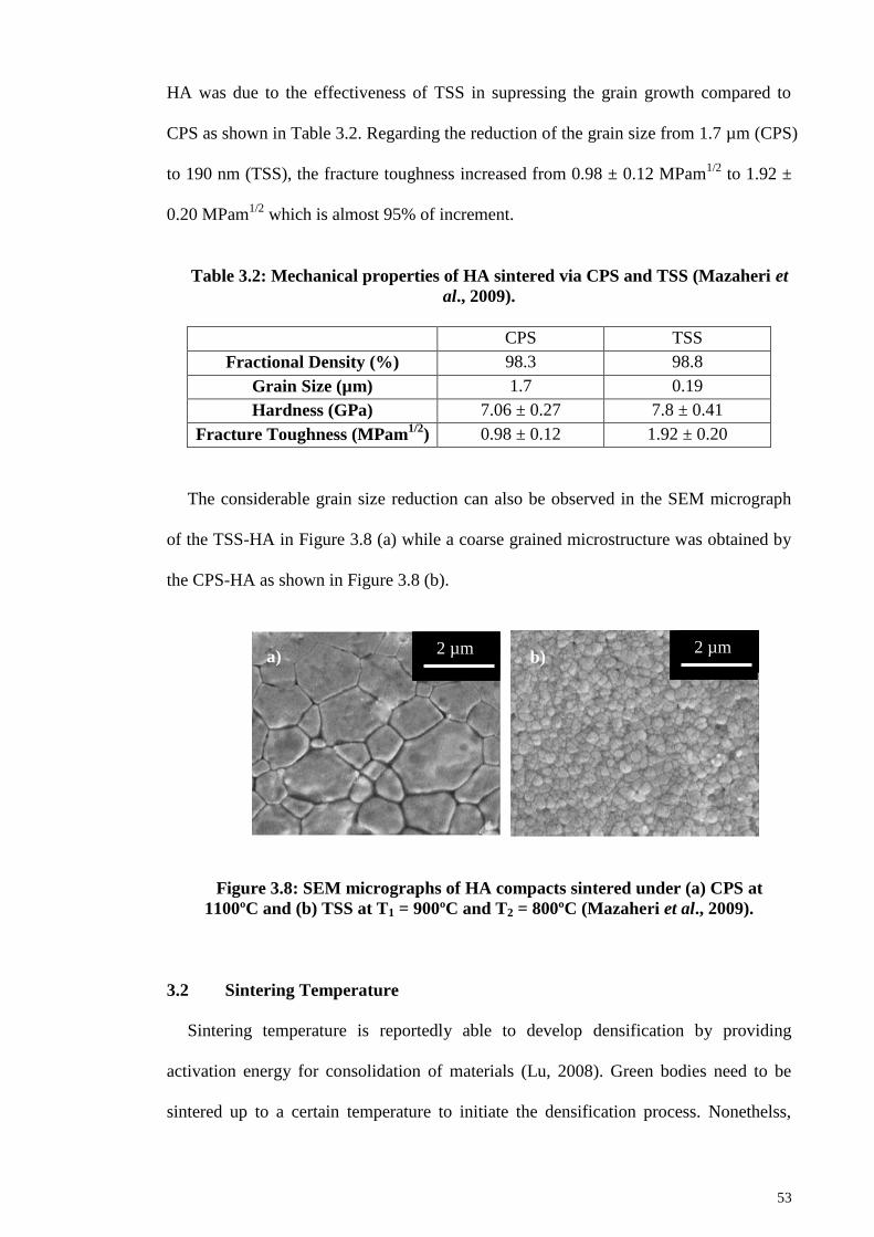
53
HA was due to the effectiveness of TSS in supressing the grain growth compared to
CPS as shown in Table 3.2. Regarding the reduction of the grain size from 1.7 µm (CPS)
to 190 nm (TSS), the fracture toughness increased from 0.98 ± 0.12 MPam1/2
to 1.92 ±
0.20 MPam1/2
which is almost 95% of increment.
Table 3.2: Mechanical properties of HA sintered via CPS and TSS (Mazaheri et
al., 2009).
CPS TSS
Fractional Density (%) 98.3 98.8
Grain Size (µm) 1.7 0.19
Hardness (GPa) 7.06 ± 0.27 7.8 ± 0.41
Fracture Toughness (MPam1/2
) 0.98 ± 0.12 1.92 ± 0.20
The considerable grain size reduction can also be observed in the SEM micrograph
of the TSS-HA in Figure 3.8 (a) while a coarse grained microstructure was obtained by
the CPS-HA as shown in Figure 3.8 (b).
Figure 3.8: SEM micrographs of HA compacts sintered under (a) CPS at
1100ºC and (b) TSS at T1 = 900ºC and T2 = 800ºC (Mazaheri et al., 2009).
3.2 Sintering Temperature
Sintering temperature is reportedly able to develop densification by providing
activation energy for consolidation of materials (Lu, 2008). Green bodies need to be
sintered up to a certain temperature to initiate the densification process. Nonethelss,
a) b) 2 µm 2 µm

54
when surplus activation (due to elevated sintering temperature) supplied to material,
severe grain enlargement or decomposition of material began (Muralithran & Ramesh,
2000).
Petrakova et al. (2013) investigated the sinterability of HA at temperature range from
900°C to 1400°C. It was observed that the porosity of the samples decreased with
increasing temperature but to the detriment of grain growth as perceived in Figure 3.9.
However, the relative density and microhardness increased proportionally to the
sintering temperature as shown in Figure 3.10. The authors hence concluded that the
strength of HA was dependent on the porosity and density of the samples rather than the
grain size.
Figure 3.9: Temperature dependence of (a) open porosity and (b) average grain
size of hydroxyapatite (Petrakova et al., 2013).
50
40
30
20
10
0
Op
en P
oro
sity
, %
5
4
3
2
1
0
Aver
age
Gra
in S
ize
(µm
)
800 1000 1200 1400
Temperature, °C
(a) (b)
800 1000 1200 1400
Temperature, °C

55
Figure 3.10: Temperature dependence of (a) relative density and (b)
microhardness (Petrakova et al. 2013).
However, Muralithran and Ramesh (2000) had different findings in terms of the
relationship between the hardness, density and grain size. The author reported that
below ~ 2µm, the hardness trend correlated well with the change in relative density i.e.
hardness increases steadily with relative density (Figure 3.11). However, above 2 µm,
the variation in hardness seem not be in agreement with the density curve as the relative
density was found to remain constant at ~98% while the hardness gradually decreased
with increasing grain size. Hence, it is hypothesised that below some critical grain size,
the hardness is governed by density and grain size is the controlling parameter when the
grain size is above the critical grain size.
100
90
80
70
60
50
Rel
ati
ve
Den
sity
, %
7
6
5
3
2
1
0
4
Mic
roh
ard
nes
s, G
Pa
(a) (b)
800 1000 1200 1400
Temperature, °C
800 1000 1200 1400
Temperature, °C

56
Figure 3.11: Variation of Vickers hardness and relative density as a function of
average grain size (Muralithran & Ramesh, 2000).
The author concluded that the optimum sintering temperature was at 1250°C; having
relative density of > 99% and microhardness of ~ 6 GPa. SEM examination of HA
samples sintered below 1200°C showed the presence of large pores which explained the
low bulk density of these samples at low temperature. Besides, the author found that
high sintering temperature can disrupt the phase stability of HA as decomposition of
HA into TCP was detected at 1400°C. Similarly, Zhou et al. (1993) reported that
sintering HA at elevated temperature has the tendency to results in the decomposition of
HA phase. They reported that the dehydroxylation of HA starts at sintering temperature
between 1200°C to 1400°C as OH- were depleted gradually. The thermal
dehydroxylation and decomposition of HA results correlated well with the findings
reported by Ramesh et al. (2016) as shown in Figure 3.12 where α-TCP and TTCP was
detected at 1300°C.
7
6
5
3
2
1
4
0
Vic
ker
s h
ard
nes
s (G
Pa)
0 2 4 6 8 10 12 14 16 18 20 22 24 26 28
Average Grain Size (µm)
90
▲Vickers Hardness
□ Relative Density
100
80
70
Rel
ati
ve
den
sity
(%
)

57
Figure 3.12: XRD patterns of HA derived from eggshells sintered at temperatures
ranging from 1050ºC to 1350ºC (Ramesh et al., 2016).
Besides densification and hardness, sintering temperature has great influence on the
toughness of HA. Kamalanathan et al. (2014) found that sintering temperature has
effects on the fracture toughness of HA as shown in Figure 3.13.
Inte
nsi
ty (
a.u.)
20 25 30 35 40 45 50
2θ (°)

58
Figure 3.13: The variation of fracture toughness of conventional pressureless
sintered EHA as a function of sintering temperature (Kamalanathan et al., 2014).
The authors noted a trend where the fracture toughness increased with sintering
temperature and peaked at 1.14 MPam1/2
at 1000°C and then decreased with further
increase in temperature as shown in Figure 3.13. At 800°C and 900°C, the fracture
toughness values were low at 0.38 MPam1/2
and 0.56 MPam1/2
respectively. This low
value at such low temperatures could be attributed to the weak grain boundaries of the
HA matrix. The author inferred that the variation of the fracture toughness with
sintering temperature was due to the grain size as toughness increased with an increase
in grain size as temperature rises. Small grain size reduces the inherent flaw size of the
sintered microstructure which then improves the mechanical property of HA (Wang &
Shaw, 2009). In a view of the above study, it can be concluded that further rising in the
sintering temperature gave adverse effects on the mechanical properties of HA by the
virtue of serious grain growth.
1.30
1.20
1.10
1.00
0.90
0.80
0.70
0.60
0.50
0.40
0.30
0.20
0.10
0.00
Fra
ctu
re t
ou
gh
nes
s (
MP
am1/2)
800 850 900 950 1000 1050 1100 1150 1200 1250 1300 1350 1400
Sintering Temperature (°C)

59
3.3 Sintering Time
The nominal sintering time chosen is critical to ensure favourable densification of
ceramic body is achieved. Tolouei et al. (2011) carried out sintering of HA using dwell
times of 1 minute and 120 minutes. They found that at lower sintering temperature like
1000°C, long holding time (120 minutes) produced HA with higher density than short
holding time (1 minutes). However, at sintering temperature above 1000°C, both 120
minutes and 1 minute holding time produced HA samples with almost equivalent
densities. Besides, the fracture toughness of HA sintered with short holding time was
higher than those sintered at long holding time. This is because shorter holding time
reduced the jeopardy of extreme grain growth which tends to deteriorate the toughness
of HA. The use of short holding time is contradict to experiment conducted by Liu et al.
(1997) who stated that the HA samples could attained relative density of more than 98%
at 1200°C for 2 hours. The need for 2 hours of holding time is supported by Veljović et
al. (2009) as they claimed that samples sintered for 45 minutes exhibited porous
microstructure (Figure 3.14 (a)). By prolonging the dwell time to 2 hours, porosity
decreased as shown in Figure 3.14 (b). However, HA samples showed sign of fracture
when sintering time prolonged to 8 hours.
Figure 3.14: SEM micrograph of sample sintered at 1050ºC for (a) 45 minutes
and (b) 2 hours (Veljović et al., 2014).
a) b)

60
3.4 Sintering Ramp Rate
Sintering ramp rate is another intrinsic part of sintering that could affect the
microstructural condition of the sintered HA as heat transferred to ceramics body due to
thermal gradient (Chu et al., 1991). Some researchers reported that higher ramp rate
have positive effects in retaining relatively fine microstructure at high sintered densities
(Brook, 1982; Johnson, 1984). Controversial discussion performed by Ramesh et al.
(2007c) and Prokopiev & Sevostianov (2006). These authors recommended that the
heating rate mustn‘t be too high above as excessive heating rate leads to thermal stress.
This thermal residual stress will propagate microcracks and slowly to macro-cracks in
sintered bodies (Pramanik et al., 2007). This is in agreement with the study carried out
by Samuel et al. (2012) who reported that HA decomposition in their work is due to the
thermal stresses generated by accelerated ramp rates. They found that at high
temperature of 1300°C, HA sample sintered with a low ramp rate (2°C/min) maintain
pure HA phase as shown in Figure 3.15 (a) while HA decomposition to β-TCP was
confirmed in the HA sample sintered with ramp rate of 5°C/min as presented in Figure
3.15 (b). A further increase in ramp rate (10°C/min) resulted in the transition of β-TCP
into α-TCP as shown in Figure 3.15 (c). Gibson et al. (2001) supported this finding by
highlighting the decomposition of HA was associated with partial dehydroxylation due
to the rapid heating rate, resulting in partial decomposition of the HA to TCP.

61
Figure 3.15: XRD patterns of HA sintered at 1250ºC with ramp rate of (a)
2ºC/min, (b) 5ºC/min and (c) 10ºC/min (Keys: = HA, = α-TCP, ▼= -TCP ♦ =
TTCP and ■ = CaO) (Samuel et al., 2012).
3.5 Sintering Additives
As discussed in the previous sections, the sinterability of HA can be enhanced by
unconventional sintering methods. Besides these, adding sintering additives (also
known as dopants) was reported to be cost effective and have beneficial effects on the
sinterability of HA (Suchanek et al., 1997; Oktar et al., 2007; Ramesh et al., 2007b).
The sintering additives should significantly enhance the densification without
decomposition of HA, prohibit abnormal grain growth of HA grains and hence improve
the toughness of HA ceramics.
600
500
400
300
200
100
0
Inte
nsi
ty
20 25 30 35 40 45 50
2 θ (°)
a)
b)
c)
600
500
400
300
200
100
0
Inte
nsi
ty
600
500
400
300
200
100
0
Inte
nsi
ty

62
The decomposition of HA into secondary phase such as CaO is unfavourable as it
would affect the biological performance of HA. Hence, various studies have been
carried out to study the effects of different sintering additives on the phase stability of
HA especially upon sintering. Suchanek et al. (1997) have doped HA with the 5 wt% of
the selected sintering additives such as K-, Na-, Li-, Mg-, Ca-, Al-, Si- Li- oxides, K-
chlorides and silicon. All the doped HA samples were subsequently sintered at 1000°C
– 1100°C. Exaggerated grain growth was not observed in the samples studied. The
addition of K2CO3, Na2CO3, KF and all sodium phosphates was found to improve the
sinterability of HA. The authors further revealed that other than the undoped HA, KCL
and -NaCaPO4, all other additives investigated caused either decomposition of HA into
-TCP or formation of CaO throughout the sintering regime employed. These secondary
phases should be avoided as it increases the biodegradability of the HA ceramics. The
authors concluded that -NaCaPO4 would be an effective sintering agent as it
significantly enhances densification without affecting the HA phase stability.
It was found that the amount of the dopant addition has effects on the phase stability
of HA. Tolouei et al. (2011) noticed that the doping HA by nano-silica of 0.8 wt% or
higher resulted in the formation of secondary phases such as β-TCP when sintered at
1250°C and the percentage of β-TCP formation increased when the amount of nano-
silica increased. The decomposition of HA into β-TCP was found to hinder the
densification and mechanical properties of HA as the density and hardness achieved for
doped-HA was greatly lower than the pure HA. This is attributed to the differential
shrinkage between HA and the secondary phases formed at the sintering temperature
(Rao & Kannan; 1999). The findings correlate well with the findings carried out by Xu
and Khor (2007) who reported that the apatite structure became less stable and a
secondary phases of β-TCP was detected in all the samples doped with silica. Similarly,
Kutty and Singh (2001) found that the HA phase decreases with increasing Y-TZP

63
content in the system regardless of sintering temperature employed. The presence of α-
TCP, β-TCP and CaO peaks were detected, indicating that HA phase was disrupted by
the addition of Y-TZP.
The addition of dopants not only affects the thermal stability of HA but it influences
the mechanical properties of the ceramic. The influences of additives on the
microstructure and sinterability of HA using 0.05 wt% to 1 wt% manganese oxide
(MnO2) had been studied by Ramesh et al. (2007b). According to Ramesh et al. (2007b),
the doping of 0.05 wt% MnO2 was proven to obtain a hardness of 7.58 GPa and fracture
toughness of 1.65 MPam1/2
as compared to 5.72 GPa and 1.22 MPam1/2
for undoped HA.
It was found that the hardness of HA was closely related to the grain size. The hardness
of the HA increase with grain size and reached a maximum at a certain grain size limit
before decreasing as shown in Table 3.3. It was seen that 0.05 wt% doped HA sample
had the smallest grain size and hence highest hardness and fracture toughness. Besides,
the authors found that the addition of 1 wt% MnO2 was found to be detrimental to
hardness of the sample as the hardness marked the lowest value at 5.32 GPa.
Table 3.3: Grain size at which maximum hardness were measured for undoped
and MnO2-doped HA (Ramesh et al., 2007b).
MnO2 Content (wt%) Grain Size (µm) Maximum Vickers
Hardness (GPa)
0 (undoped) 0.50 7.21
0.05 0.47 7.58
0.10 0.58 7.00
0.30 0.52 7.30
0.50 1.03 7.04
1.00 1.56 5.32
On the other hand, Oktar et al. (2007) did a study on the sinterability of HA when
doped with a variety of dopants. In this experiment, with the objective of improving the
sinterability and mechanical properties of HA, it was doped with 5 wt% and 10 wt% of

64
SiO2, MgO, Al2O3 and ZrO2. The presences of these oxides in HA are encouraging as
they can act as crack arresters which will directly reduce the brittleness of HA. From
their work, doping HA with 5 wt% MgO gave the best sintering result because it
possessed denser HA compared to other dopants while the addition of SiO2 caused a
collapse in the hardness and density of HA. The additions of Al2O3 and ZrO2 did not
exhibit significant improvement in mechanical strength of HA.
The beneficial effects of the addition of MgO was supported by Tan et al. (2013).
As presented in Figure 3.16 (a), doping of pure HA with different amount of MgO
noticeably enhanced the fracture toughness with the highest value recorded at 1.48
MPam1/2
for 1.0 wt% MgO-doped HA as compared to 1.08 0.05 MPam1/2
measured
for the undoped HA when sintered at 1150°C. This value is encouraging as other
researchers who doped HA with different additives such as zirconia (Kim et al, 2003),
alumina (Champion et al., 1996) and titania (Manjubala and Kumar, 2000) obtained
fracture toughness ranging between 1.0-1.4 MPam1/2
. Moreover, there are reports
indicating that decomposition would occur when HA is doped with these dopants.
However, in their study, the phase stability of HA was not disrupted by the addition of
MgO as pure HA phase remained for all the doped and undoped samples throughout the
sintering regime. Furthermore, hardness and relative density have greatly increased in
HA with the reinforcement of MgO in the HA matrix. With the addition of MgO, the
hardness of HA maintained in a high range of ~6 GPa to 7.52 GPa even at considerably
low sintering temperatures as shown in Figure 3.16 (b).

65
Figure 3.16: Fracture toughness and Vickers hardness of MgO-doped and
undoped HA (Tan et al., 2013).
3.5.1 Zinc Oxide as Sintering Additives
In short, it is clear that the mechanical properties of HA depend on the type and the
amount of sintering additives. It should be aware that the dopants added into HA matrix
should enhance the sinterability of HA without compromising the biocompatibility of
HA to ensure the reliability of HA as an effective implant material. Therefore, it is
0.8
0.9
1.0
1.1
1.2
1.3
1.4
1.5
1.6
950 1000 1050 1100 1150 1200 1250 1300 1350 1400
Fra
ctu
re T
ou
gh
nes
s (M
Pa
m1/2
)
Sintering Temperature (ºC)
Undoped
0.05 wt% Mg
0.1 wt% Mg
0.3 wt% Mg
0.5 wt% Mg
1 wt% Mg
a)
4.0
4.5
5.0
5.5
6.0
6.5
7.0
7.5
8.0
950 1000 1050 1100 1150 1200 1250 1300 1350 1400
Vic
ker
s H
ard
nes
s (G
Pa
)
Sintering Temperature (ºC)
Undoped
0.05 wt% Mg
0.1 wt% Mg
0.3 wt% Mg
0.5 wt% Mg
1 wt% Mg
b)

66
important to choose a suitable dopant with adequate quantity of addition to ensure the
stability of the dopant within the HA matrix.
Zinc oxide (ZnO) was found to be a potential sintering additive to be doped into HA
matrix as zinc is an essential trace element in human bone. It presents in a small amount
in the enamel of human teeth and bone (Bandyopadhyay et al., 2006). Moreover, zinc
promotes bone growth (Yamaguchi et al., 1987), inhibits bone resorption (Moonga &
Dempster, 1995) and offers antimicrobial resistance (Hernández-Sierra et al., 2008)
which is indispensible for bone implant materials. In the study carried out by Yu et al.
(2014), HAP/ZnO biocomposite has significant antibacterial efficacy against various
microorganisms, indicate enormous potential for its use in anti-inflammatory
application as maxillofacial bone repair biomaterial. Additionally, zinc oxide has been
doped into beta-tricalcium phosphate (β-TCP) to develop zinc-releasing biomaterials
and it has been proved to promote bone formation (Ito et al., 2000). The positive effect
of ZnO in biological properties of calcium phosphate is supported by Kawamura et al.
(2000) as zinc promotes excellent bioactivity than the undoped calcium phosphate.
Stimulated bone formation was observed around ZnO containing calcium phosphate
ceramics implanted in rabbit femora. The bioactive properties of ZnO-doped HA have
also been extensively studied (Ishikawa et al., 2002; Jallot et al., 2005). Although small
addition of ZnO is advantageous to the host material, but a high zinc concentration can
have serious toxic side effects on human cells (Bettger & O‘dell, 1993). In addition, Ito
et al. (2000) have also reported that the addition of ZnO higher than 1.2 wt% in TCP
could release zinc and subsequently cause cytotoxicity effects in human body. This is in
agreement with the cell culture study by Bandyopadhyaya et al. (2007) where the cell
material interaction shows that ZnO addition on calcium phosphate ceramic was non-
toxic at the initial stage. However, as the ZnO content increased, the cell spreading and
cell-cell interactions in both TCP and HA decreased. It can be concluded that good cell

67
attachment is up to 1.5 wt% ZnO doping and addition of ZnO more than 1.5 wt%
limited the cell spreading and growth on the HA surface. Therefore, the ZnO content in
the doped ceramic has to be less than 1.2 – 1.5 wt%.
To be a reliable human implant, the ZnO-doped HA ceramics should have a
harmonious balance between biocompatibility and mechanical properties. Hence, the
ZnO addition should also enhance the mechanical properties of HA ceramics.
Bandyopadhyay et al. (2007) have proved that the addition of ZnO improved the
sintered density and microhardness of HA as shown in Table 3.4.
Table 3.4: Properties of HA containing varying amounts of ZnO
(Bandyopadhyay et al., 2007).
HA
(wt %)
ZnO
(wt %)
Linear
Shrinkage
(%)
Sintered
Relative
Density (%)
Microhardness
(MPa)
100 0.0 47.3 81 3696
98.5 1.5 51.7 88 4781
97.5 2.5 51.1 89 4797
96.5 3.5 51.4 92 4540
Further to that, interconnected porous β-TCP scaffolds improved by ZnO were
successfully prepared by Feng et al. (2014). It was found that the addition of ZnO
improved the densification of the strut of β-TCP scaffolds. Moreover, the addition of
ZnO could improve the mechanical properties such as fracture toughness, stiffness and
hardness of the scaffolds as presented in Figure 3.17. In vitro cell tests showed that the
scaffold with ZnO doping exhibited a good cellular viability as well as cellular
proliferation. The formation of a new bone like apatite layer indicated that the scaffold
demonstrated better bioactivity.

68
Figure 3.17: Effect of ZnO contents on the (a) stiffness, (b) densification, (c)
microhardness and (d) fracture toughness of the strut (Feng et al., 2014).
Researchers have also done investigation on the effects of ZnO addition on the
sinterability of other ceramics such as ceria based material, SnO2 based ceramic and
Tin-doped indium oxide. With the addition of ZnO, the relative density, grain size and
microstructure of these host materials have improved without affecting the phase
stability (Liu et al., 2014; He et al., 2012; Chen et al., 2014). Hence, ZnO is a potential
sintering additives to improve the mechanical properties of HA without compromising
the phase stability and biological performance of HA.
In summary, this chapter has provided a brief overview of the various parameters
that governs the sinterability of hydroxyapatite ceramics such as powder consolidation
technique, sintering temperature, sintering time and heating rates and sintering additives.
Therefore, particular attention has to be paid to optimize these parameters in the quest to
improve the sinterability of the hydroxyapatite powders.

69
CHAPTER 4: EXPERIMENTAL TECHNIQUES
4.1 Synthesis of HA Powder via Wet Chemical Method
The hydroxyapatite (HA) powder used in the present work was prepared according to
a novel wet chemical method comprising precipitation from aqueous medium via a
titration process by reacting calcium ion with phosphate ion based on a molar ratio of
Ca/P = 1.67 and maintained at a pH of about 10-12 by the addition of small amounts of
ammonium (NH3) solution (25% concentration) (Ramesh, 2004). The starting
precursors used were commercially available calcium hydroxide Ca(OH)2 (98% purity,
RNM) and orthophosphoric acid H3PO4 (85% purity, Merck) following the chemical
reaction:
10Ca(OH)2 + 6H3PO4 Ca10(PO4)6(OH)2 + 18 H2O (4.1)
In general, the synthesis process involved the preparation of H3PO4 and Ca(OH)2
solution. To prepare the H3PO4 solution, the required amount of H3PO4 was diluted in
distilled water, stirred for 10 minutes and subsequently contained in a titration funnel.
The pH of the prepared acid solution was about 1. A similar procedure was employed to
prepare the Ca(OH)2 solution under stirring condition. The pH of the prepared Ca(OH)2
solution was about 12. The beaker containing the Ca(OH)2 solution was covered with a
cling film to minimize contamination as well as any reaction between carbon dioxide in
air with the mixture.
The prepared H3PO4 solution was then added dropwise into the stirring suspension of
Ca(OH)2 in distilled water. The titration process normally takes about 3 hours.
Throughout the titration process, the pH of the mixture was monitored and kept above
10.5 by the addition of NH3 solution. Stirring process was continued after the titration
process for another 6 hours and the precipitate was allowed to settle down overnight
before filtration. For the filtration process, the aged HA solution was poured into the

70
prepared funnel connected to the pump and layered with 2 filter papers. The filtered
precipitate (HA slurry) was rinsed three times using distilled water and placed in
ceramic bowls to be dried. In this experiment, three different drying methods were
applied, namely conventional oven drying, microwave drying and freeze drying.
4.1.1 HA Powder Prepared via Oven Drying
The rinsed precipitate (HA slurry) was placed in a ceramic bowl and dried in an oven
(Memmert UF30, Germany) at about 60C. The slurry was weighed at hourly intervals
throughout the drying process until the water has been completely removed (constant
weight was obtained). It was found that 16 hours of oven drying period is sufficient to
remove all the water as there was no change in the slurry‘s weight after 16 hours of
drying. The dried HA slurry was crushed using mortar and pestle and then hand sieved
(212 µm) to obtain HA powder, hereafter named OHA. The flow sheet of this method is
shown in Figure 4.1.
Figure 4.1: OHA wet chemical method flow sheet.
Distilled water Distilled water
Stirring Stirring
Precipitation
Aging
Filtration
Oven Drying (16 hours)
Crush and Sieve
HA powder (OHA)
Ca(OH)2 H3PO4
Add NH3

71
4.1.2 HA Powder Prepared via Microwave Drying
In terms of microwave drying, the rinsed HA precipitate was placed in a ceramic
bowl and then subjected to microwave irradiation in a household microwave oven
(Sharp, R-898M, 900 W power).The specifications of the microwave oven are shown in
Table 4.1.
Table 4.1: Specifications of Sharp R-898M Microwave Oven.
Specifications Details
Microwave Power Consumption 1400 watt
Heater Power Consumption 1700 watt
Microwave Output 900 watt
Heater Output 1600 watt
Outside Dimension 520 mm (W) x 309 mm (H) x 486
mm (D)
Cavity Dimension 343 mm (W) x 209 mm (H) x 357
mm (D)
Microwave Frequency 2450 MHz
The precipitate was dried in the microwave oven at 100% power (900 watt) to
remove the water content completely. The temperature and rate of heating were not
controlled as those parameters were fixed for this microwave oven. Under microwave
drying, the precipitate was removed periodically from the microwave oven for every 1
minute. This procedure was repeated to reach the case of no change in precipitate
weight. The time taken for the samples to reach constant weight was 15 minutes. The
microwave dried HA cake was crushed and sieved to obtain HA powder, hereafter
named MD-HA. The flow sheet of this method is shown in Figure 4.2.

72
Figure 4.2: HA wet chemical method with microwave drying flow sheet.
4.1.3 HA Powder Prepared via Freeze Drying
As for freeze drying, the rinsed HA precipitate was transferred into a specific freeze
dry container. The amount of precipitate was about 1/3 of the container‘s capacity to
ensure the high evaporation efficiency throughout the freeze drying process. The freeze
dry container was immediately transferred into a freezer (Telstar, Igloo, Netherlands);
deep freeze at –80C for 24 hours to solidify the water and induce solid liquid phase
separation. On the other hand, the freeze dryer (FDU-1200, Japan) was pre-set at –45C
and was evacuated by means of vacuum pump (60L/min, 0.049 mBar). After 24 hours,
the frozen sample was removed from freezer and quickly taken to the freeze dryer. By
maintaining the temperature of the HA sample below –45C and the vacuum below
0.049 mBar, water was removed by sublimation in the freeze dryer. The weight of the
HA precipitate was not recorded periodically as removing the sample from the freeze
dryer could affect the temperature and pressure of the system. After 36 hours of freeze
Distilled water Distilled water
Stirring Stirring
Precipitation
Aging
Filtration
Microwave Drying (15 mintues)
Crush and Sieve
HA powder (MHA)
Ca(OH)2 H3PO4
Add NH3

73
drying, the HA sample was completely dried as no frost was observed on the surface of
the freeze dry container. Crushing of HA is not required as a fluffy HA powder was
obtained. The HA powder was sieved to obtain HA powder, hereafter named FD-HA.
The flow sheet of this method is shown in Figure 4.3.
Figure 4.3: HA wet chemical method with freeze drying flow sheet.
4.2 ZnO-doped HA Powder Preparation
The starting zinc oxide (ZnO) powder used in the present research was of 99% purity
(Systerm). The powder compositions, i.e. 0.1, 0.3, 0.5, 1.0 wt% ZnO-doped HA were
prepared through a process involving ultrasonic and ball milling.
In a typical procedure, both powders (MD-HA powder and ZnO powder) were
measured according to the respective ratio (composition) and put into a beaker
Distilled water Distilled water
Stirring Stirring
Precipitation
Aging
Filtration
Freeze at –80C for 24 hours
Freeze dried for 36 hours
Sieve
Ca(OH)2 H3PO4
Add NH3
HA powder (FHA)
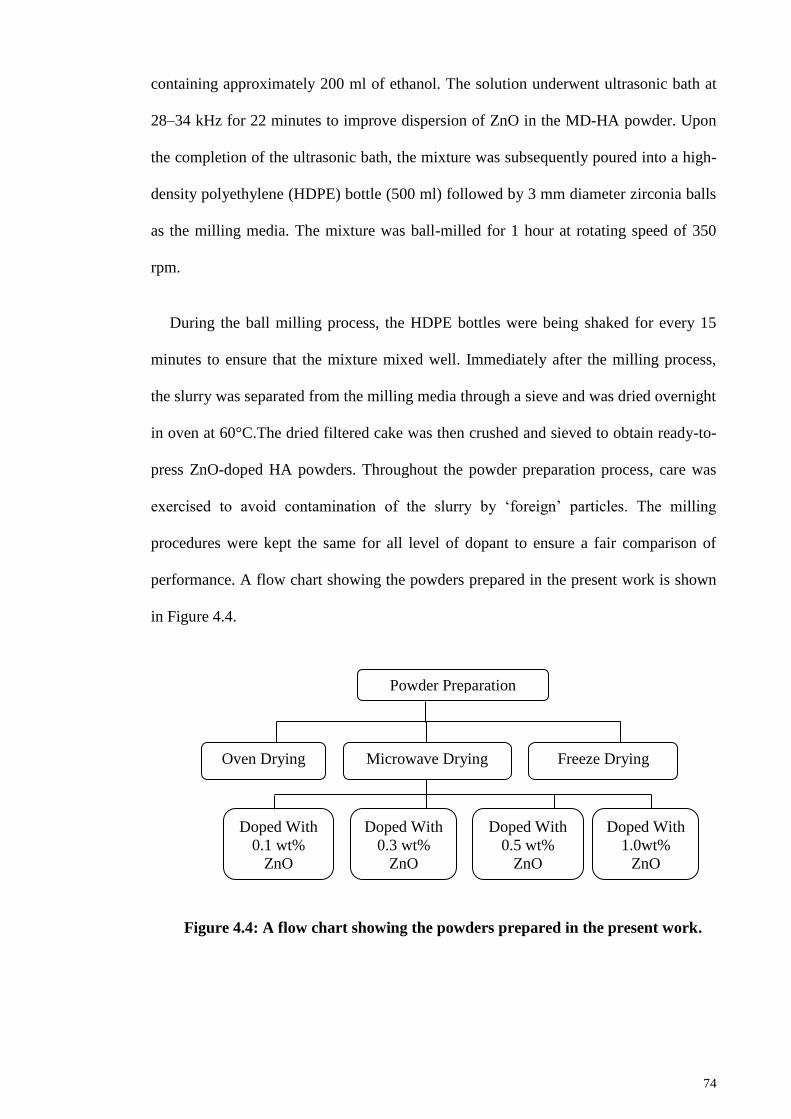
74
containing approximately 200 ml of ethanol. The solution underwent ultrasonic bath at
28–34 kHz for 22 minutes to improve dispersion of ZnO in the MD-HA powder. Upon
the completion of the ultrasonic bath, the mixture was subsequently poured into a high-
density polyethylene (HDPE) bottle (500 ml) followed by 3 mm diameter zirconia balls
as the milling media. The mixture was ball-milled for 1 hour at rotating speed of 350
rpm.
During the ball milling process, the HDPE bottles were being shaked for every 15
minutes to ensure that the mixture mixed well. Immediately after the milling process,
the slurry was separated from the milling media through a sieve and was dried overnight
in oven at 60°C.The dried filtered cake was then crushed and sieved to obtain ready-to-
press ZnO-doped HA powders. Throughout the powder preparation process, care was
exercised to avoid contamination of the slurry by ‗foreign‘ particles. The milling
procedures were kept the same for all level of dopant to ensure a fair comparison of
performance. A flow chart showing the powders prepared in the present work is shown
in Figure 4.4.
Figure 4.4: A flow chart showing the powders prepared in the present work.
Powder Preparation
Microwave Drying Freeze Drying Oven Drying
Doped With
1.0wt%
ZnO
Doped With
0.1 wt%
ZnO
Doped With
0.3 wt%
ZnO
Doped With
0.5 wt%
ZnO
75
4.3 Green Samples Preparation
The as prepared HA powders (OD-HA, MD-HA and FD-HA) as well as the ZnO-
doped HA powder were uniaxial compacted at about 1.3 MPa to 2.5 MPa into discs (20
hardened steel mould and die set. The cleanliness of the die was essential, so as not to
contaminate the samples. The die used in conjunction with the sample preparation was
cleaned with an oil-based liquid such as WD-40, in order to prevent powder lamination.
It is well established that the density variation in the green product will cause
differential shrinkage and crack initiation upon sintering (Bortzmeyer, 1995; Satapathy,
1997). Thus, in order to avoid this, the green compacts were subsequently cold
isostatically pressed (CIP) at a pressure of 200 MPa (Riken, Seiki, Japan) and hold at
this pressure for about 1 minute. In CIP, pressure was applied on the as-pressed samples
in all directions with the aid of hydraulic oil inside the cold isostatic pressing machine.
Therefore, it produces more uniform density green samples. The green bodies with
uniform density enable more uniform shrinkage during the sintering process and this
prevents the samples from warping and cracking.
4.4 Sintering
4.4.1 Conventional Sintering
In the present research, sintering was achieved by by conventional pressureless
sintering where the green compacts were sintered in air, using a standard rapid heating
furnace (LT Furnace, Malaysia) at different temperatures; 1050oC–1350
oC (50
oC
interval) for the OD-HA, MD-HA and FD-HA powder while temperature range of
1100oC–1300
oC for ZnO-doped HA powder. A furnace ramp rate of 2
oC/min (heating
and cooling) and soaking time of 2 hours was used for each firing. In this method, the

76
Time (min)
Desired Temperature
Temperature (°C)
duration of the sintering process ranges from between 15 to 24 hours depending on the
sintering temperature. The sintering profile is shown in Figure 4.5.
Figure 4.5: Sintering profile of conventional sintering.
4.4.2 Microwave Sintering
All the pressed HA pellets were microwave sintered (MS) in a microwave furnace
(Delta MF, China) with an alumina fibre insulation box. A constant power output of
2000 watt was employed to sinter the HA samples at temperature of 950oC–1250
oC.
Figure 4.6 plots the sintering profile of the microwave sintered HA. A constant
microwave power of 2000 watt at 2.45 GHz was maintained throughout the sintering
process. The sintering rate for the first 250oC was not controlled as this parameter was
fixed for this microwave furnace; depending on the absorbance of the material. From
250oC to 500
oC, the sintering rate was set as 15
oC/min to avoid thermal shock. Then,
the samples were sintered at 30oC/min to the desired temperatures (950
oC–1200
oC) with
a holding time of 15 minutes to avoid excessive grain growth. Finally, the furnace was
switched off and natural cooling of the sample was allowed. In this method, the duration
of the sintering process ranges from between 5 to 6 hours depending on the sintering
temperature.
2 hours holding time

77
Figure 4.6: Sintering profile of microwave sintering.
4.4.2.1 Samples Arrangement
A variety of sample arrangements was tried, and it was found that disc shaped
samples arranged as shown in Figure 4.7 gave the optimum results.
To initiate the microwave heating and minimize the thermal gradients, both silicon
carbide (SiC) cylindrical and irregular shaped susceptor were used as auxiliary heating
elements (hybrid heating). They were arranged as shown in Figure 4.7, where
cylindrical SiC was placed right in front of the source of magnetron and the irregular
shaped SiC was scattered around the insulation box. The insulation box is made from
alumina fiber (not microwave susceptible) and was required to retain heat. At the same
time, the samples were placed on top of pre-sintered zirconia plate. It was necessary to
include zirconia plate in the arrangement in order to have sufficient load to achieve
good coupling of the electromagnetic energy. The loss factor of the zirconia plate
became sufficiently large to permit heating by microwave absorption, permitting more
efficient microwave heating of the sample.
Desired Temperature
Temperature (oC)
Time (min)
Heating rate depends of
material absorbance
Heating rate of 15°C/min to
avoid thermal shock
Heating rate of 30°C/min
250°C
500°C
Hold for 15 minutes

78
Figure 4.7: Samples arrangement in the microwave furnace: (a) plan view; (b)
side view.
4.5 Grinding and Polishing
All the conventional and microwave sintered HA disc samples were firstly ground
and then polished on one face prior to density measurement, phase analysis, Vickers
hardness testing, fracture toughness evaluation and SEM investigation. Surface grinding
and polishing was done manually using an Imtech Grinder-Polisher. The samples were
Alumina fiber
insulation box
Cylindrical shape
SiC as susceptors
Irregular shaped SiC
susceptor surrounding
the samples
HA pellets
Pre-sintered zirconia
plates underneath
the samples
a)
Alumina fiber
insulation box cover
Alumina fiber
insulation box
Source of
magnetron power
coming from this
direction
b)
Source of magnetron power
coming from this direction

79
ground on SiC paper (600 grit-1200 grit) before being diamond polished to a 3 µm
finish and subsequently to a 1 µm finish so as to obtain an optical reflective surface.
4.6 Characterization
The characterization process includes the specific surface area, crystallite size,
Transmission Electron Microscopy (TEM) for HA powders; Fourier Transform Infrared
(FTIR), X-Ray diffraction (XRD), Scanning Electron Microscopy (SEM) with Energy
Dispersive X-Ray (EDX) analysis for both HA powders and HA sintered samples
followed by grain size evaluation (SEM and Field Emission Scanning Electron
Microscopy (FE-SEM)), bulk density measurement, Vickers hardness and fracture
toughness examination for all the HA sintered samples.
4.6.1 Specific Surface Area and Crystallite Size
The specific surface area of the powder was measured by the Brunauer-Emmett-
Teller (BET) method. Nitrogen gas adsorption analysis was performed on a
physisorptionanalyser (ASAP 2020, Micromeritics, USA). Samples were outgassed at
150°C for 30 minutes under vacuum.
The average crystallite size (d) can be estimated by measuring the broadening of a
particular peak in a XRD diffraction pattern associated with a particular planar
reflection from within the crystal unit cell based on Scherrer‘s formula (Cullity and
Stock, 2001):
d = 0.9/B cos (4.2)
where is the wavelength of the X-ray which is 1.54056 Å for CuK radiation, is
the diffraction angle at 2 = ~31.7° and ~25.7° and B (in radians) is the measured full
width at the half maximum (FWHM) of a diffracting reflection.

80
4.6.2 Transmission Electron Microscopy (TEM) Analysis
The morphology of the as synthesized oven dried (OD-HA), microwave dried (MD-
HA) and freeze dried (FD-HA) HA powder was analysed using TEM (JEOL, JEM-
2100F, Japan) operated at an accelerating voltage of 120 kV. The particle size of the
powder was measured from TEM micrographs. Prior to TEM observation, the as
synthesized HA powder was dispersed in 1 – 2 ml of spectroscopic grade ethanol
solution followed by ultrasonication for 1 hour to break up agglomerates.
Approximately 50 µl of the OD-HA and MD-HA suspensions produced were deposited
on a 200 mesh copper grid which was subsequently dried to remove the ethanol solvent.
The deposited HA powder on the 200 mesh copper grid was then used in the TEM
analysis.
4.6.3 Fourier Transformation Infrared (FTIR)
A Fourier transform infrared spectrometer (FTIR – Brukers IFS-66-VS, Germany)
with a reflectance mode was used for qualitative analysis of the molecular radicals.
Prior to testing, 40 mg of KBr is mixed with 0.4 mg of the tested powder. Subsequently,
the powder is pressed into pellet of 13 mm in diameter. Advanced preparation is not
required for the sintered samples. The possible structural variation and reactions in
those samples were examined. The infrared spectrum with a resolution of 4 cm-1
and a
scan number of 32 was adopted with a scan range 400 – 4000 cm-1
.
4.6.4 Scanning Electron Microscopy (SEM) and Energy Dispersive X-Ray
Analysis (EDX)
The morphology, the Ca/P ratio and the distribution of elemental within the HA
powder and sintered samples were examined on a phenom Pro-X microscope equipped
with an energy dispersive X-Ray (Pro Suite & Elemental Analysis, Phenom).

81
Measurements were made in terms of atomic %, weight % and elemental spectra
acquisition.
4.6.5 X-Ray Diffraction (XRD)
X-ray diffraction (XRD) provides information that relates to the crystal lattice of the
material and can characterise the crystalline phases present. In the present work, the
phases present in the powders as well as the sintered samples were determined at room
temperature using X-Ray diffraction (Empyrean, PANalytical, Netherlands) operated at
35 kV and 15 mA with Cu-K as the radiation source. The X-ray scan speed and step
scan were 0.5°/min. and 0.02°, respectively. The peaks obtained were compared to
standard reference JCPDS-ICCD (Joint Committee of Powder Diffraction Standard –
International Center for Diffraction Data) files for HA (PDF No. 74-566 for
Ca10(PO4)6(OH)2, -TCP (PDF No. 9-348), -TCP (PDF No. 9-169), TTCP (PDF No.
25-1137) and CaO (PDF No. 37-1497). The files are attached in Appendix C.
4.6.6 Microstructural Examination
The microstructural evolution of the HA under various sintering temperature was
examined by using the SEM (Pro-X, Phenom) and FE-SEM (JEOL, JSM7600F, Japan)
both at an accelerating voltage of 10 kV and 15 kV. The sintered samples were firstly
polished to a mirror like surface finished and then thermally etched to delineate the
grain boundaries. The etching temperature employed was 50C below the sintering
temperature of the sample at a heating and cooling rate of 10C/min, with a holding
time of 30 minutes prior to cooling. In order to remove any contamination, the surface
to be examined was cleaned in acetone and subsequently stuck on to an aluminium stub.
The samples for SEM and FE-SEM examination were coated with platinum, which
provides a conducting layer, to prevent charging from occurring in the microscope.

82
4.6.7 Grain Size Measurement
The grain size of sintered HA was determined on thermally etched specimens from
scanning electron micrographs using the line intercept method. This technique requires
measurements taken from polished sections. In a typical analysis, a known test line is
drawn on a A4 size SEM micrograph of the selected polished section and the number of
intercept between the test line and grain boundaries are counted. The test line should
cover at least 50 grains and several lines are drawn and measured before average value
is taken.
The average grain size is then calculated according to the equation proposed by
Mendelson (1969):
LD 56.1 (4.3)
where D is the average grain size and L is the measured average interception length
over a large number of grains which can be represented by:
L = MN
C
(4.4)
where C, M and N are the total length of the test line, the magnification of the SEM
micrograph and the number of intercepts respectively.
The technique used to count the number of intersections was according to an
international standard test method for intercept counting (ASTM E112-96, 2004).
Essentially, the end points of a test line are not intersections and not counted unless the
end appeared to exactly touch a grain boundary, when a ‗0.5‘ intersection is scored. A
tangential intersection with a grain boundary is scored as a ‗1‘ intersection while for

83
intersection coinciding with the junction of 3 grains is scored as a ‗1.5‘ intersection as
shown in Figure 4.8.
score : 0
score : 1
grain
score : 1.5
score : 0.5
test line
SEM micrograph
Figure 4.8: Diagram showing the score given for the type of intersections.
4.6.8 Bulk Density Measurement
The bulk densities of dense compacts (above 90% of theoretical density) were
determined by the water immersion technique based on the Archimedes principle using
a standard Mettler Toledo Balance AG204 Densi-meter. In the present research,
distilled water was used as the immersion medium. The procedure to measure the bulk
density can be summarized as follows:
(a) The dry weight of the sample is first measured.
(b) The sample is placed on a dish immersed in the distilled water after the
electronic balance is zeroed. The weight of the sample in water is subsequently recorded.
Extra care has to be taken, as any minor disturbance will incur vibration causing the
readings to fluctuate.
The bulk density was calculated using equation (4.5) as followed:

84
w
wa
a
WW
W
(4.5)
Where ρ is the bulk density of sample (g/cm3), Wa is the weight of the sample in air
(g), Ww is the weight of the sample in water (g) and ρw is the density of distilled water.
The temperature effect on the density of distilled water was taken into consideration
using the table provided in Appendix B. The relative density was calculated by taking
the theoretical density of HA as 3.156 gcm-3
. However, for low-density samples (below
90% of theoretical density) the bulk density was obtained from the measurement of
geometric dimensions and sample mass. This is because the former method allows
water to penetrate the pores, resulting in an overestimate value of the sample‘s density.
4.6.9 Vickers Hardness and Fracture Toughness Evaluation
The Vickers hardness testing method was used to ascertain the hardness of the
sintered HA. The indentations were made using a pyramidal diamond indenter (HMV
series Shimadzu, Japan) with an applied load varying between 50 g to 200 g. In the
Vickers test, the load is applied smoothly, without impact, and held in place for 10
seconds. The physical quality of the indenter and the accuracy of the applied load as
defined clearly in ASTM E384-99 (1999) must be controlled to get the correct results.
In general, the Vickers impression (Figure 4.9) appears to be square, and the two
diagonals have almost similar lengths. After the load is removed, the impression
diagonals as shown in Figure 4.9 are measured usually with a filarmicrometer built in
the attached microscope on the Vickers machine, to the nearest 0.1 µm and the average
value, 2a, is obtained. The Vickers hardness (Hv) is calculated based on the surface area
of the indent using equation (4.6):
2v
2a
1.854PH
(4.6)

85
Where P is the applied load and 2a is the average diagonals. In the present work, five
indentations were made for each sample and the average values were taken.
Figure 4.9: Schematic indentation fracture pattern of an idealized Vickers
median (or half-penny) crack system (Niihara et al., 1982).
Using the same indentation image from Vickers hardness tester, fracture toughness
(KIc) was determined from the equation derived by Niihara (1985):
KIc= 5.05.1
203.0 aHa
cv
(4.7)
Where Hv is the Vickers hardness, c is the characteristics crack length (L+a), L is the
average crack length and a is the half diagonal of indent.
L a a L
Sub-surface
Median crack Sub-surface
Lateral crack
Surface radial crack
Sub-surface
Lateral crack
Plan view
Cross-sectional view

86
CHAPTER 5: RESULTS AND DISCUSSIONS (PART 1)
5.1 HA Powder Characteristic
5.1.1 XRD Analysis and Crystallite Size
The wet chemical precipitated HA powders synthesized via three different drying
methods, namely freeze drying (FD-HA), microwave drying (MD-HA) and oven drying
(OD-HA) were subjected to X-ray diffraction (XRD) to examine the phase purity. The
resulting XRD spectra for the synthesized HA are shown in Figure 5.1.
Figure 5.1: The XRD profiles of HA powder synthesized through wet
precipitation method via three different drying methods.
All powders produced peaks correspond to the standard JCPDS card no: 74-0566 for
stoichiometric HA with no significant evidence of secondary phases such as CaO,
TTCO, β-TCP or α-TCP being detected. As illustrated in Figure 5.1, FD-HA, MD-HA
and OD-HA produced nearly identical peaks with minimal difference in terms of the
degree of crystallinity as MD-HA displayed slightly narrower diffraction at 2θ = 31.9°.
The results proved that microwave drying has no adverse effects on the phase purity of
Inte
nsi
ty (
counts
)
2θ (°)
FD-HA
MD-HA
OD-HA

87
HA powders as supported by Santhosh & Prabu (2012) and Chanda et al. (2009). In
general, broad diffraction peaks were observed for all the HA powders indicated that
HA powders produced by the wet chemical method are poorly cystallined with a large
amount of amorphous phase regardless of drying methods. The broad peaks of wet
chemical produced HA powders could be resulted from the slow addition rate of the
acid solution that gave the nanocrsytalline nature of the synthesized powders. This is in
agreement with the study carried out by Santos et al. (2004), Angelescu et al. (2011)
and Alobeedallah et al. (2011). Moreover, the broad diffraction peaks indicated that the
HA crystal size were very small and the crystal size could be estimated from XRD data
using Scherrer‘s equation (Pratihar et al., 2006; Tian et al., 2008) at prominent peak of
HA such as (211) and (002) reflection as shown in Table 5.1.
Table 5.5.1: Estimate crystal size of HA particles based on the Scherrer’s
equation.
Sample
Crystal size (nm) based on XRD
(2 1 1) reflection
@ 2 = ~ 31.9º
(0 0 2) reflection
FD-HA 21.83 41.61
MD-HA 46.09 45.55
OD-HA 48.64 47.54
The results showed that all the HA powders has crystal size lie within nanometer
range which is favorable for osseointegration due to large interfaces (Murugan &
Ramakrishna, 2005) and HA with small crystal size exhibits higher bioactivity
compared to coarse HA crystal (Mazaheri et al. 2008). From Table 5.1, FD-HA has
smaller crystal size (21.83 nm) than MD-HA and OD-HA at (2 1 1) reflection while
MD-HA and OD-HA recorded similar crystal size at 46.09 nm and 48.64 nm,
respectively. OD-HA recorded the biggest crystal size at both (2 1 1) and (0 0 2)
plane could be due to the aggregation of small particles as powder particles coalesce

88
during the long drying process where heat was constantly supplied to the HA slurry for
16 hours. The long drying hours allows the motion of molecules which increase the
chance of molecules collision, hence, HA particle concentrated to form larger particles.
This could explain the smaller crystal size of FD-HA as no heat was involved in the
freeze drying process, eliminated the chance of particles to collide with each other and
form large agglomerates. A similar finding was reported by Girija et al. (2012) who
found that HA produced by freeze drying has smaller crystal size than that of oven dried
HA powder. Besides, the OD-HA produced slightly bigger crystallite size than MD-HA
is in disagreement to the study carried out by Santhosh & Prabu (2012). In their research,
furnace dried HA had crystal size smaller than those produced by microwave drying.
This contradict finding could be due to the shorter oven drying hour (80°C for 8 hours)
compared to the current research (60°C for 16 hours), reduces the chance of particle
coalescence.
5.1.2 FTIR Analysis of the Synthesized HA Powder
The phase purity of HA produced by three different drying methods was further
examined by FTIR and the FTIR spectrum of the HA powders are shown in Figure 5.2.
A comparison of the wave numbers for the functional groups of the HA produced by
three different drying method and HA powder produced by Brundavanam et al. (2015)
is tabulated in Table 5.2. From Figure 5.2, it can be deduced that all the HA powders
displayed typical FTIR spectrum of pure HA powder. The characteristic bands
corresponding to the (ν3 and ν1) PO43-
were clearly observed at ~1035cm-1
- 1038cm-1
and 962 cm-1
, respectively for all the HA powders. In addition, the characteristic bands
attributed to the apatitic OH- were observed at ~3567 cm
-1 (stretching) for all the
powders, proved that freeze drying and microwave drying did not alter the basic apatitic
structure of HA powder.

89
Broad peaks assigned to chemically absorbed H2O was observed at frequencies
around 3341 cm-1
and 1632 cm-1
for FD-HA, 3360 cm-1
and 1643 cm-1
for MD-HA and
3345 cm-1
and 1640 cm-1
for OD-HA. It is worth mentioning that the chemically
absorbed water peak was more obvious for MD-HA especially at 3345 cm-1
(stretching)
compared to FD-HA and OD-HA. This indicated that OD-HA and FD-HA have lower
water content than the MD-HA powder. The low water content of OD-HA could be due
to the long oven drying hours (16 hours) which eliminated most of the water content
from OD-HA. For FD-HA, liquid was not involved in the entire freeze drying process
which lead to the narrow water peak in the FTIR as indicated in Figure 5.2.
Figure 5.2: The FTIR spectrum of the synthesized HA powders: (a) FD-HA, (b)
MD-HA and (c) OD-HA.
The characteristic peak corresponding to free CO2 was detected from MD-HA
(~2340 cm-1
) and FD-HA (2333 cm-1
) could be attributed to the atmospheric CO2
Tra
nsm
itta
nce
(%
T)
Wavenumber (cm-1
)
a)
b)
c)
OH-
(3568)
OH-
(3567)
OH-
(3566) H2O
(3345)
H2O
(3360)
H2O
(3341) CO2
(2333)
CO2
(2340)
H2O
(1632)
H2O
(1643)
H2O
(1640)
CO32-
(1415)
CO32-
(1421)
CO32-
(1419)
PO43-
(1038)
PO43-
(1035)
PO43-
(1036)
PO43-
(962)
PO43-
(962)
PO43-
(962)
HPO42-
(874)
HPO42-
(874)
HPO42-
(873)
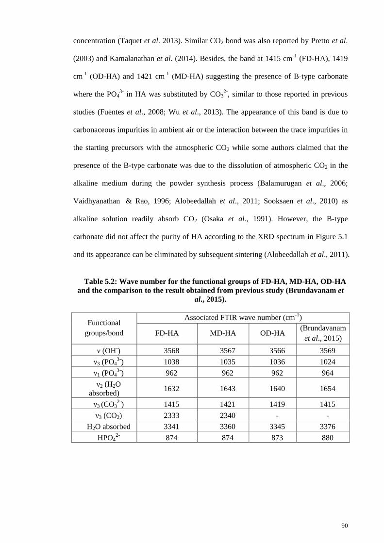
90
concentration (Taquet et al. 2013). Similar CO2 bond was also reported by Pretto et al.
(2003) and Kamalanathan et al. (2014). Besides, the band at 1415 cm-1
(FD-HA), 1419
cm-1
(OD-HA) and 1421 cm-1
(MD-HA) suggesting the presence of B-type carbonate
where the PO43-
in HA was substituted by CO32-
, similar to those reported in previous
studies (Fuentes et al., 2008; Wu et al., 2013). The appearance of this band is due to
carbonaceous impurities in ambient air or the interaction between the trace impurities in
the starting precursors with the atmospheric CO2 while some authors claimed that the
presence of the B-type carbonate was due to the dissolution of atmospheric CO2 in the
alkaline medium during the powder synthesis process (Balamurugan et al., 2006;
Vaidhyanathan & Rao, 1996; Alobeedallah et al., 2011; Sooksaen et al., 2010) as
alkaline solution readily absorb CO2 (Osaka et al., 1991). However, the B-type
carbonate did not affect the purity of HA according to the XRD spectrum in Figure 5.1
and its appearance can be eliminated by subsequent sintering (Alobeedallah et al., 2011).
Table 5.2: Wave number for the functional groups of FD-HA, MD-HA, OD-HA
and the comparison to the result obtained from previous study (Brundavanam et
al., 2015).
Functional
groups/bond
Associated FTIR wave number (cm-1
)
FD-HA MD-HA OD-HA (Brundavanam
et al., 2015)
ν (OH-) 3568 3567 3566 3569
ν3 (PO43-
) 1038 1035 1036 1024
ν1 (PO43-
) 962 962 962 964
ν2 (H2O
absorbed) 1632 1643 1640 1654
ν3 (CO32-
) 1415 1421 1419 1415
ν3 (CO2) 2333 2340 - -
H2O absorbed 3341 3360 3345 3376
HPO42-
874 874 873 880

91
5.1.3 EDX Analysis of the Synthesized HA Powder
Figure 5.3 - 5.5 show the energy-dispersive X-ray (EDX) spectrum and the mass
percentage of all the detected elements of the FD-HA, MD-HA and OD-HA powders,
respectively.
Figure 5.3: The EDX spectrum and elemental composition of FD-HA.
Figure 5.4: The EDX spectrum and elemental composition of MD-HA.
Inte
nsi
ty (
a.u.)
Energy-keV
Inte
nsi
ty (
a.u.)
Energy-keV

92
Figure 5.5: The EDX spectrum and elemental composition of OD-HA.
According to the EDX spectrum (Figure 5.3 - 5.5), all the synthesized HA powders
consist of main constituents of HA, where calcium, phosphorus and oxygen are present.
From the results, it can be seen that all the HA powders, namely FD-HA, MD-HA and
OD-HA have Ca/P ratio greater than the stoichiometric value of HA (1.667); recorded
values of 1.77, 1.85 and 1.90, respectively. This is not in agreement with the findings
from previous studies where Ca/P ratio of pure HA was close to 1.60 - 1.667 (Fanovich
& Porto Lopez, 1998; Ramesh et al., 2012; Pattanayak et al., 2007; Lu et al., 1998).
Apart from that, OD-HA recorded the highest Ca/P ratio of 1.90 compared to MD-HA
and FD-HA which is in agreement with the study carried out by Santhosh et al. (2012)
and Lu et al. (1998).
The high Ca/P ratio of the current study could be associated with the presence of B-
type carbonate substitution as described in Figure 5.2 and Table 5.2. The substitution
was believed to reduce the phosphorus content of HA, thus cause the higher value of
Ca/P ratio (Boutinguiza et al., 2012). Moreover, it was reported that EDX results
Inte
nsi
ty (
a.u.)
Energy-keV

93
represent the sample only at a microscopic level and hence the discrepancy between
EDX data and the expected values have been frequently reported with some deviations
(Kieswetter et al. 1994). Although the Ca/P ratio of all the synthesized powders are
deviated from 1.667, it can be identified as HA because Ca/P ratio for HA could vary
from 1.2 – 2.0 (Larsen & Widdowson, 1971). Liu et al. (2008) and Landi et al. (2000)
stated that deviation from stoichiometric Ca/P ratio would affect the reactivity and
phase stability of HA, however phase disruption was not observed in current study as
evidence from XRD analysis (Figure 5.1).
5.1.4 FE-SEM Analysis of the Synthesized HA Powder
The FE-SEM micrographs of FD-HA, MD-HA and OD-HA are presented in Figure
5.6, Figure 5.7 and Figure 5.8, respectively. Generally, FD-HA (Figure 5.6) and MD-
HA (Figure 5.7) consist of small to large size agglomerates made of small particles
while OD-HA (Figure 5.8) consists of extremely large agglomerates. From Figure 5.6,
the FD-HA powder was a loose agglomerates mixture of fine particle size. The
distribution of HA particles was uniform and homogeneous. The agglomerates sizes are
in the range from extremely small of 0.5 µm to a large size of 3 µm. Therefore, it can be
deduced that the individual HA particle size is extremely small in nanometer range
which is in agreement with the HA crystal size calculated from Scherrer‘s equation
(Table 5.1). As for MD-HA, FE-SEM analysis revealed that the powder consists of a
mixture of small and large agglomerates in size ranging from 0.4 µm to 4 µm. The
larger particle appears to be large agglomerate of loosely packed smaller particles in
nano regime. Conversely, the OD-HA powder as shown in Figure 5.8, consist of large
and hard agglomerates, resulting in rougher surface. The smallest agglomerate of OD-
HA is about 2 µm to as large as 10 µm. The powder seemed to be more compacted
compared to FD-HA and MD-HA. It is well known that wet chemical synthesized HA
powder dried by the means of conventional oven drying are accompanied by severe

94
agglomeration (Lu et al., 1998; Hsu & Chiou, 2011; Yu et al., 2010; Zhang &
Yogokawa, 2008).
Figure 5.6: FE-SEM micrograph of synthesized FD-HA powder.
Figure 5.7: FE-SEM micrograph of synthesized MD-HA powder.
5 µm
5 µm

95
Figure 5.8: FE-SEM micrograph of synthesized OD-HA powder.
The small particle size of FD-HA is due to the fact that no heat was involved in the
freeze drying process and hence crystal structure does not grow as there was no supply
of heat energy. Microwave drying was found to produce HA powders with small
particle size. During microwave drying, the internal and surface of the HA slurry was
heated simultaneously. The water molecules within the HA slurry, being specific to
microwave irradiation was heated instantly and triggered the quick expansion of vapor
during the drying process. This rapid expansion of vapor caused the breakage of large
aggregates into several tiny lumps, hence, small particle size (Qi et al., 2006). Oven
drying removed water from the HA slurry via evaporation at an elevated temperature
which promotes particles in close contact with each other. As the heat was supplied to
the HA slurry constantly, solid particle bridging was formed between particles and lead
to the formation of hard agglomerates (Yu et al., 2005). In conclusion, the drying
method has significant effects on the powder particle and agglomerates size of the
synthesized HA powders. It should be noted that the individual particles of all the
powders could not be recognized due to their extremely small size and thus TEM
analysis has to be carried out to further examine the particle sizes of all the powders.
5 µm

96
5.1.5 TEM Analysis of the Synthesized HA Powder
The effects of different drying route on the morphology of the synthesized HA
powder was determined via transmission electron microscopy (TEM) and the
micrographs are shown in Figure 5.9. FD-HA (Figure 5.9 (a)) and OD-HA (Figure 5.9
(c)) resembled needle-like structure. The needle-like morphology was said to be similar
to the bio-crystal found in human hard tissue (Nejati et al., 2009; Ferraz et al., 2004;
Patel et al., 2001). Nevertheless, MD-HA was found to exhibited nano-rod structure and
similar HA particle shape was reported by Santhosh & Prabu (2012) who synthesized
HA via microwave drying. Siddharthan et al. (2006) claimed that the difference is HA
particle shape was affected by the microwave power. They found that the shapes of HA
changed from needle shape to rod/platelet shape as the microwave power increased
from 175 W to 600 W. The resulting nano-rod shape could be explained as the
consequence of high microwave power (800 W) applied in current research. Although
MD-HA displayed different particle shape from FD-HA and OD-HA, it was found that
all the powders are in nanometric range as tabulated in Table 5.3. Nano-size particles
are desirable as they give better resorption and biological activity which promotes the
adhesion and proliferation of bone cells (Le Geros, 1991; Pham et al., 2013). Moreover,
nano-sized powders produced dense and nanocrystalline sintered ceramic that possesses
superb mechanical properties by having higher driving force for the densification
(Wang & Shaw, 2007). On the contrary, micro-sized powders caused exaggerated grain
growth during sintering due to the agglomerated and irregular morphology (Groza &
Dowding, 1996; Lin et al., 2007).

97
Figure 5.9: TEM micrographs of synthesized HA powder: (a) FD-HA, (b) MD-
HA and (c) OD-HA.
Table 5.3: Summary of the average size of HA powder synthesized via different
drying methods.
Sample Particle size
distribution
Average Particle
Size (Length)
Average Particle
Size (Width)
FD-HA 40 – 180 nm (Length)
10 – 50 nm (Width) 82.0 nm 24.8 nm
MD-HA 20 – 140 nm (Length)
10 – 40 nm (Width) 66.7 nm 23.6 nm
OD-HA 30 – 160 nm (Length)
10 – 30 nm (Width) 75.0 nm 21.4 nm
From Table 5.3, it can be seen that the particle size of FD-HA was slightly larger
compared to MD-HA and OD-HA, having average particle size of 24.8 nm in width and
82.0 nm in length. Despite bigger particle size, the crystal particles show good
a) b)
c)
200 nm 200 nm
200 nm

98
dispersibility with minimal agglomeration (Figure 5.9 (a)) compared to MD-HA and
OD-HA which is consistent with the FE-SEM micrograph shown in Figure 5.6.
Similarly, Wang et al. (2010), Bildstein et al. (2009) and Saluja et al. (2010) also
reported that HA particle produced by freeze drying method displayed bigger particle
size than other drying methods. However, this contradicts to the results reported by Lu
et al. (1998) as they found that the particle size of FD-HA is smaller than that of normal
heat dried HA powder. The dissimilarities in results might be due to the fact that the HA
slurry was froze rapidly with liquid nitrogen in their work which fast cooling prevent ice
nucleation which hinder the growing of large dendritic ice crystal and hence smaller
particle size was produced (Kurapova et al., 2012). Similar to the previous study
(Santhosh & Prabu, 2012), microwave drying manage to produce HA particles in
nanometric range. However, the average particle sizes of MD-HA in the current study,
recorded at 66.7 nm (length) and 23.6 nm (width) was bigger than that reported by
Siddhartahn et al. (2006) at ~32 nm (length) and ~12 nm (width). The bigger HA
particle size obtained from current study could be associated with the use of higher
microwave power which favored the maturation of HA crystal growth. Although OD-
HA has smaller particle size than FD-HA, the agglomeration of powders are rather
serious compared to FD-HA. From Figure 5.9 (c), OD-HA shows agglomerated clusters
with particles in needle shape. Lin et al. (2007) stated that the agglomerations in powder
are detrimental to the densification of ceramics during sintering and should be avoided.
5.1.6 Specific Surface Area of the Synthesized HA Powder
The specific surface areas (SSA) of all the synthesized HA powder were determined
via BET analysis and the finding is tabulated in Table 5.4.
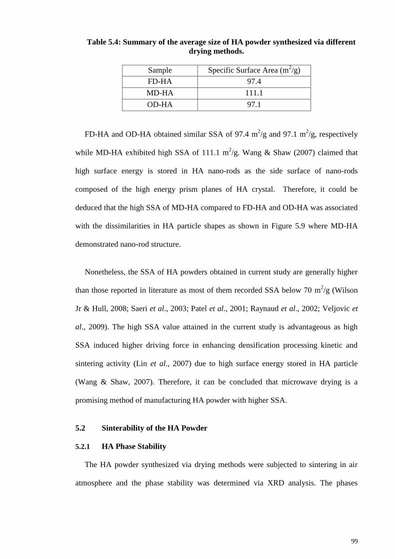
99
Table 5.4: Summary of the average size of HA powder synthesized via different
drying methods.
Sample Specific Surface Area (m2/g)
FD-HA 97.4
MD-HA 111.1
OD-HA 97.1
FD-HA and OD-HA obtained similar SSA of 97.4 m2/g and 97.1 m
2/g, respectively
while MD-HA exhibited high SSA of 111.1 m2/g. Wang & Shaw (2007) claimed that
high surface energy is stored in HA nano-rods as the side surface of nano-rods
composed of the high energy prism planes of HA crystal. Therefore, it could be
deduced that the high SSA of MD-HA compared to FD-HA and OD-HA was associated
with the dissimilarities in HA particle shapes as shown in Figure 5.9 where MD-HA
demonstrated nano-rod structure.
Nonetheless, the SSA of HA powders obtained in current study are generally higher
than those reported in literature as most of them recorded SSA below 70 m2/g (Wilson
Jr & Hull, 2008; Saeri et al., 2003; Patel et al., 2001; Raynaud et al., 2002; Veljovic et
al., 2009). The high SSA value attained in the current study is advantageous as high
SSA induced higher driving force in enhancing densification processing kinetic and
sintering activity (Lin et al., 2007) due to high surface energy stored in HA particle
(Wang & Shaw, 2007). Therefore, it can be concluded that microwave drying is a
promising method of manufacturing HA powder with higher SSA.
5.2 Sinterability of the HA Powder
5.2.1 HA Phase Stability
The HA powder synthesized via drying methods were subjected to sintering in air
atmosphere and the phase stability was determined via XRD analysis. The phases

100
analysis of the sintered FD-HA, MD-HA and OD-HA are shown in Figure 5.10, Figure
5.11 and Figure 5.12, respectively.
The formation or secondary phases were not detected in FD-HA and MD-HA
throughout the sintering regime as all the XRD peaks showed good correlation with the
stoichiometric HA as shown in Figure 5.10 and Figure 5.11. However, β-TCP was
observed in the OD-HA sintered at 1350°C as shown in Figure 5.12. The results
indicated that freeze drying and microwave drying can preserve the thermal stability of
HA better than conventional oven drying method.
Figure 5.10: The XRD profiles of FD-HA sintered samples (a) 1050°C, (b)
1150°C, (c) 1250°C and (d) 1350°C. All peaks belong to the HA phase.
Inte
nsi
ty (
counts
)
2θ (°)
(d)
(b)
(a)
(c)

101
Figure 5.11: The XRD profiles of MD-HA sintered samples (a) 1050°C, (b)
1150°C, (c) 1250°C and (d) 1350°C. All peaks belong to the HA phase.
Figure 5.12: The XRD profiles of OD-HA sintered samples (a) 1050°C, (b)
1150°C, (c) 1250°C and (d) 1350°C. The unmarked peaks belong to the HA phase.
Inte
nsi
ty (
counts
)
2θ (°)
(d)
(b)
(a)
(c) In
tensi
ty (
counts
)
2θ (°)
(d)
(b)
(a)
(c)
▼ β-TCP ▼ ▼

102
Although the phase stability of FD-HA was not disrupted throughout the sintering
regime, a small peak shift to lower diffraction angle was observed for sample sintered at
1350°C. This variation could be the consequence of dehydroxylation phenomenon due
to water loss at high temperature (Lin et al., 2001; Wang & Chaki, 1993) or the loss of
OH- radial from HA lattice (Gu et al., 2002; Bianco et al., 2007). It was reported that
thermal decomposition of HA occur in sequence of dehydroxylation (Slosarczyk et al.,
1996; Wang et al., 2004). During dehydroxylation, HA will lose OH- gradually and
form oxyapatite according to the following equation (Zhou et al., 1993; Kijima &
Tsutsumi, 1979; Trombe & Montel; 1978):
2OH O + H2O (5.1)
Ca10(OH)2(PO4)6 Ca10(PO4)6.O· + H2O (5.2)
where is a vacancy and oxyapatite ,Ca10(PO4)6.O· is also known as hydroxy-ion-
deficient product. Accordingly, one of the lattice site was occupied by the two OH-
group in HA unit cell is now replaced by an oxygen atom while the other was left
vacant. In addition to that, Zhou et al. (1993) reported that the oxyapatite phase is stable
and will not cause phase transformation without further heating.
However, if sintering carried out at higher temperature, HA would decompose into
tricalcium phosphate and tetracalcium phosphate in accordance to the following process:
Ca10(OH)2(PO4)6 2Ca3(PO4)2 + Ca4P2O9 + H2O (5.3)
This phenomenon was observed from OD-HA sintered samples as illustrated in
Figure 5.12. From the XRD profiles, HA phase was the main constituent phases present
in these compacts at sintering temperature ranging from 1050°C to 1250°C. However,
the decomposition of HA into β-TCP was clearly observed from OD-HA sintered at
103
1350°C. The present results obtained for OD-HA samples contradicted the findings of
Muralithran & Ramesh, 2000, Bianco et al., 2007 and Ramesh et al., 2008. In general,
these authors reported that their HA powder which was synthesized using drying
method as the present OD-HA showed that the transformation of HA into secondary
phases only at sintering temperature beyond 1350°C. According to Muralitran &
Ramesh (2000) and Arias et al. (1998), both dehydroxylation and decomposition
reaction could be slow down by providing a high local humid furnace atmosphere
during sintering as the dehydration of the OH- from HA matrix could be prevent. Hence,
it could be deduced that the differences in the findings could be attributed to the
humidity, drying temperature and drying hours of the samples in the conventional oven.
It is worth mentioning that HA produced via microwave drying shows high thermal
stability as no peak shifting nor decomposition were observed on MD-HA throughout
the sintering profile. Since all of the HA sintered under the same sintering atmosphere,
it could be confirmed that the high thermal stability of MD-HA compared to FD-HA
and OD-HA was not due to the humidity of sintering atmosphere. The ability of MD-
HA to preserve the phase stability at high temperature could be attributed to the
significant amount of absorbed water remained in the structure of MD-HA compared to
FD-HA and OD-HA as evidence from FTIR analysis of the HA powders (Figure 5.2).
In conclusion, the thermal stability of all the HA sintered samples (regardless of
drying methods) are generally higher than most of the available literatures (Ruys et al.,
1995; Finoli et al., 2010; Sanosh et al., 2010; Kothapalli et al., 2004; Kong et al., 2002).
They produced HA with low thermal stability where the decomposition of HA started at
sintering temperature lower than 1300°C. As such, the results obtained in the present
work are encouraging and microwave drying was found to produce single phase HA
stable up to 1350°C.

104
5.2.2 FTIR Analysis of Sintered HA Samples
Figure 5.13 shows the FTIR analysis of the FD-HA, MD-HA and OD-HA sintered at
1350°C and the comparison with their respective as-synthesized powder. It was
observed that the CO32-
band and the chemically absorbed H2O bands are no longer
visible in all the HA sintered at 1350°C as indicated in Figure 5.13.
Figure 5.13: FTIR profiles of HA sintered at 1350°C (left) with their respective
as-synthesized powder (right): (a) FD-HA, (b) MD-HA and (c) OD-HA.
The intensity of peaks associated to the OH- band of the sintered FD-HA and MD-
HA are remarkably decreased as shown in Figure 5.13 (a) and (b). The weak OH- band
and the missing H2O bands could be the primary factor that caused the dehydroxylation
of FD-HA at 1350°C as revealed by the aforementioned peak shifting in the XRD
profile shown in Figure 5.10. Besides, the free CO2 was previously appeared in the
synthesized FD-HA and MD-HA are removed from the HA structure as the
Tra
nsm
itta
nce
(%
T)
Wavenumber (cm-1
) Wavenumber (cm-1
)
a)
b)
c)
1350°C As-synthesized
CO32-
H2O
CO2
CO2
H2O
OH-
OH-
OH-

105
corresponding peaks was not seen. The results showed that the sintering of FD-HA and
MD-HA induced the elimination of combined water and carbonates without causing
further decomposition of HA.
On the contrary, the FTIR peaks of OD-HA (Figure 5.13(c)) shows the appearance of
CO2 peak and the absence of the peaks corresponding to OH- after sintering. The CO2
could interrupt the HA phase and cause further decomposition while the absence of OH-
peak confirms its transformation into oxyapatite (Wang et al., 2009). Hence, the
decomposition of OD-HA at 1350°C was due to the combined effects of the appearance
of the CO2 and absence of the OH- peak.
5.2.3 Bulk Density
The variation of sintered density with sintering temperature for all HA samples
synthesized via different drying methods are shown in Figure 5.14.
Figure 5.14: The effect of sintering temperature on the relative density of HA.
70
75
80
85
90
95
100
1000 1050 1100 1150 1200 1250 1300 1350 1400
Rel
ati
ve
Den
sity
(%
)
Sintering Temperature (°C)
FD-HA
MD-HA
OD-HA

106
In general, the results proved that the relative density of the sintered HA increases
when the sintering temperature increase and reach at plateau at 1300°C, irrespective of
the different drying methods of the HA powders. A small increase in density is observed
at 1050°C to 1100°C for FD-HA and OD-HA which corresponds to the first stage of
sintering where necks forming into grains (Gibson et al., 2001). It could be seen that
MD-HA has higher starting density than FD-HA and OD-HA. For example, MD-HA
obtained high relative density of ~91% at 1050°C compared to FD-HA and OD-HA
which attained similar relative density of ~85% at 1050°C. The high starting density of
MD-HA is due to the high specific surface area reported in earlier section which
provides high surface energy for the densification process (Göller & Oktar, 2002). The
second stage of sintering involved the onset of densification and the removal of porosity
and this stage is indicated by a sharp increase in the sintered density. For both FD-HA
and OD-HA, the sharp increment of relative density was between 1100°C to 1150°C.
Conversely, the onset of densification of MD-HA started at lowtemperature of 1050°C.
The MD-HA sintered samples exhibited densification of 93.2% at 1100°C and
achieved densification of >97.5% when sintered above 1200°C. Similar observation
was reported by Gibson et al. (2001) and Rodriguez-Lorenzo (2001) that HA attained
~97% relative density at 1200°C. On the contrary, Mostafa (2005), Prokopiev &
Sevostianov (2006), and Aminzarea et al. (2013) reported otherwise whereby their
sintered HA samples achieved relative density less than 90% at 1200°C. At similar
temperature of 1100°C, FD-HA and OD-HA attained only ~87.6% and 88.2% of
theoretical density and both of them recorded relative density of ~98% at 1200°C which
is slightly higher than that of MD-HA. This is not in agreement with the findings of Van
Landuyt et al. (1995), Thangamani et al. (2002) and Sanosh et al. (2010) as they found
that sintering above 1300°C was required to achieve ~98% of theoretical density. This
difference in observation could be related to the different powder characteristics used.

107
Generally, all the HA samples achieved the maximum density at the same
temperature of 1300°C. FD-HA attained the maximum density of 98.74% at 1300°C
and drop slightly to 98.5% at 1350°C. MD-HA recorded maximum density of 98.4% at
1300°C and drop to 87.74% at 1350°C. On the other hand, OD-HA achieved the
maximum density of 98.42% at 1300°C and drop drastically to 80.3% at 1350°C. The
sharp drop in relative density of OD-HA at 1350°C is ascribed to the presence of
secondary phase (β-TCP) in the sintered OD-HA as indicated in XRD data in Figure
5.12. Overall, MD-HA samples showed better densification than FD-HA and OD-HA as
lower sintering temperature was required to achieve dense MD-HA samples of >90%.
Moreover, as evident from the results in Figure 5.7 and Figure 5.9 (b), MD-HA powder
was in nano-sized, homogenously distributed with small agglomerates and has high
specific surface area. All these factors contribute to the MD-HA achieving higher
densification than FD-HA and OD-HA at low temperatures (Patel et al., 2001; Xu et al.,
2007; Göller & Oktar, 2002).
5.2.4 Microstructure Evolution and Grain Size
SEM analysis was carried out on FD-HA, MD-HA and OD-HA sintered samples to
determine the microstructure and grain size of the samples. As shown in Figure 5.15 (b),
MD-HA sintered at 1050°C shows discrete grains with no visible formation of necking;
corresponds to the second stage of sintering where densification has started and most of
the porosities were removed. The dense microstructure with a few pores (0.1 µm to 0.4
µm) of MD-HA sintered at this temperature correlates well to the relative density results
of ~91%. At similar temperature, large numbers of pores are remaining in the FD-HA
and OD-HA samples as shown in Figure 5.15 (a) and 5.15 (c) respectively. The results
showed that at 1050°C, the sintering was at the first stage for FD-HA where some necks
formation were detected and large pores were observed. As for OD-HA, the sintering
was at the end of first stage sintering as the interconnected pores started to from grains

108
and grain outline can be seen. This could explain the lower density (~85%) of both FD-
HA and OD-HA at 1050°C.
Figure 5.15: SEM images of (a) FD-HA, (b) MD-HA and (c) OD-HA sintered at
1050°C.
1 µm
1 µm
1 µm
a)
b)
c)

109
Subsequently, SEM images of samples sintered at 1150°C Increase the sintering
temperature could improve the densification and homogeneity of samples. At 1150°C,
most of the pores were removed and denser microstructures were observed for all the
samples; indicating that complete sintering has been achieved. These observations are in
agreement with the relative density of the sintered HA samples whereby the FD-HA,
MD-HA and OD-HA attained relative density of ~96.5%, ~96.52% and ~97.6%,
respectively. In addition, the grain size of the FD-HA (Figure 5.16 (a)) and MD-HA
(Figure 5.16 (b)) samples sintered at 1150°C were observed to have a uniform
distribution, having average grain size of 1.03 µm and 0.79 µm, respectively.
Conversely, OD-HA attained an average grain size of 0.97 µm; accompanied by an
uneven microstructure at the similar temperature as shown in Figure 5.16 (c). The
recorded HA grain sizes are in agreement with most of the literatures as researchers
reported that the grain size of HA sintered at 1150°C is often less than 2 µm (Chaki &
Wang, 1994; Veljovic et al., 2008; Mazaheri et al., 2009; Ramesh et al., 2007).
When sintering was carried out at 1200°C, all the HA samples exhibited dense
microstructure where the grains are closely packed as shown in Figure 5.17. OD-HA
samples sintered at 1200°C shows better uniformity in terms of grain size distribution
compared to those sintered 1150°C. Additionally, FD-HA, MD-HA and OD-HA
attained average grain size of 1.73 µm, 1.34 µm and 1.67 µm, respectively at 1200°C.
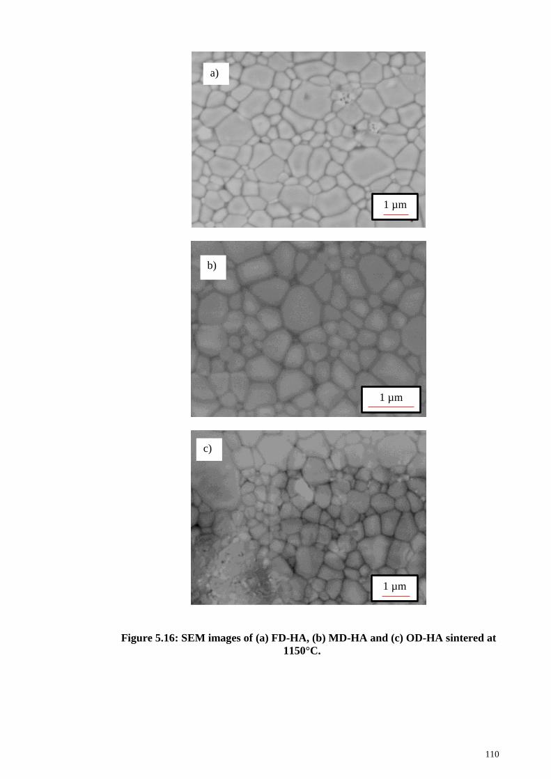
110
Figure 5.16: SEM images of (a) FD-HA, (b) MD-HA and (c) OD-HA sintered at
1150°C.
1 µm
1 µm
1 µm
b)
c)
a)
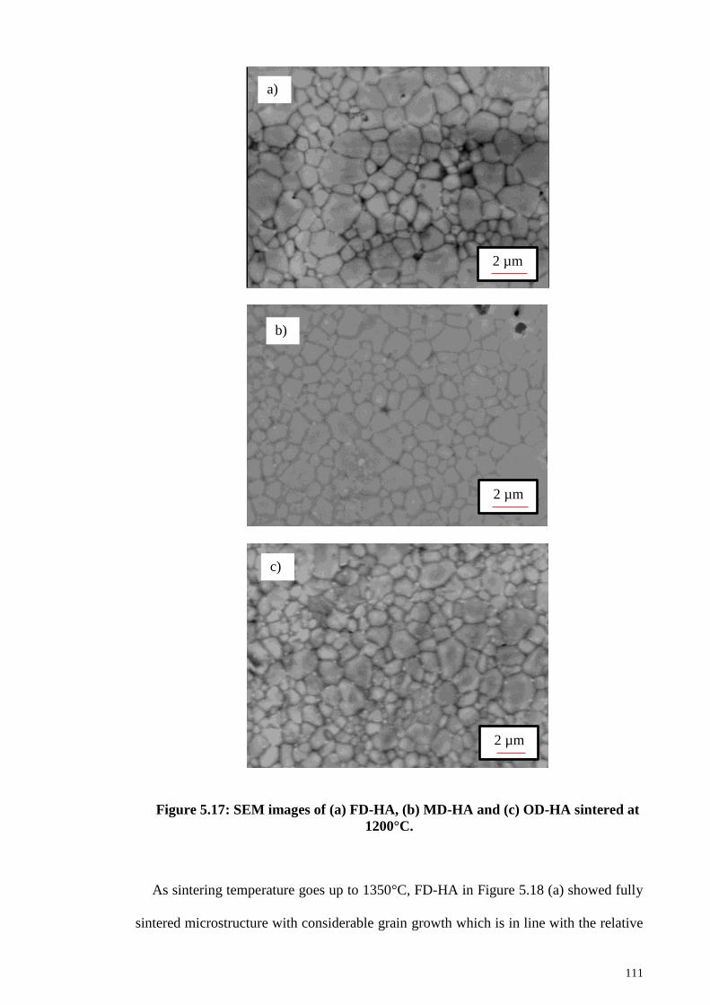
111
Figure 5.17: SEM images of (a) FD-HA, (b) MD-HA and (c) OD-HA sintered at
1200°C.
As sintering temperature goes up to 1350°C, FD-HA in Figure 5.18 (a) showed fully
sintered microstructure with considerable grain growth which is in line with the relative
2 µm
2 µm
2 µm
a)
b)
c)

112
density of ~98.5%. Similarly, OD-HA in Figure 5.18 (c) was observed to display fully
dense microstructure with minimal pores. Therefore, it could be deduced that the low
density (80.34%) of OD-HA at 1350°C was solely triggered by the presence of β-TCP
in the OD-HA matrix. Unlike FD-HA and OD-HA, MD-HA presented fully sintered
microstructure accompanied by the existence of several intergranular pores of ~2 µm at
1350°C as shown in Figure 5.18 (b). The pores were believed to have contributed to the
sudden drop in the relative density (to 87.74%) of MD-HA. These open pores were
formed when the high internal pressure caused by the dehydration of HA at elevated
temperature exceeds the strength of the closed pores (Ramesh et al., 2007).
Although the HA samples displayed different microstructure, the grain growth
occurred for all the HA samples is significant with grains grew up to 8.9 µm - 10.9 µm
as shown in Figure 5.18. This growth in the grain size due to the increase in sintering
temperature was also observed by Kong et al. (2002), Mobasherpour et al. (2007),
Kamalanathan et al. (2014), Ramesh et al. (2013) and Ramesh et al. (2016). This grain
growth was due to the enhancement of homogeneity of the HA powder with increasing
sintering temperature.

113
Figure 5.18: SEM images of (a) FD-HA, (b) MD-HA and (c) OD-HA sintered at
1350°C.
10 µm
5 µm
5 µm
a)
b)
c)

114
Under current study, the average grain sizes for all the synthesized HA with
increasing sintering temperature are shown in Figure 5.19. All three HA samples
demonstrated increasing grain size trend with increasing sintering temperature and MD-
HA samples showed slower grain growth rate at 1050°C - 1200°C compared to FD-HA
and OD-HA. However at an elevated sintering temperature of 1350°C, MD-HA
recorded the highest grain size of 10.85 µm compared to FD-HA (8.9 µm) and OD-HA
(9.3 µm).
Figure 5.19: The effect of sintering temperature on the average grain size of
sintered HA.
5.2.5 Vickers Hardness and Fracture Toughness
The dependence of hardness on the sintering temperature for all the sintered HA is
shown in Figure 5.20. The results showed that hardness of FD-HA was at ~ 3 GPa when
sintered at 1050°C and obtained a maximum hardness of 5.3 GPa at 1150°C followed
by a decrease in hardness thereafter. The observed maximum hardness of FD-HA is
close to 5.83 GPa obtained by Uysul et al. (2014) using similar freeze drying method.
The increasing trend of the hardness of the FD-HA at temperature below 1150°C was
0
1
2
3
4
5
6
7
8
9
10
11
12
13
1050 1150 1200 1350
Aver
age
Gra
in S
ize
(µm
)
Sintering Temperature (°C)
FD-HA
MD-HA
OD-HA

115
attributed to the increase in density (Wang & Shaw, 2009; Niakan et al., 2015).
However the hardness was found to decline as the sintering temperature increased from
1200°C to 1350°C despite the relative density was increasing from ~98% to ~98.5%.
The decline of hardness at sintering temperature beyond 1150°C was due to the
increased of grain size (Hoepfner & Case, 2003; He at al., 2008; Veljovic et al., 2008;
Ramesh et al., 2016). The effects of average grain size and relative density on the
hardness of FD-HA are illustrated in Figure 5.21.
From Figure 5.20, the hardness results show that MD-HA exhibited similar
increasing trend with increasing sintering temperature up to 1200°C. At temperature of
1050°C, the hardness value of MD-HA was at 3.5 GPa, slighter higher than that of FD-
HA at the similar temperature. Subsequently, the hardness value of MD-HA reached a
maximum value of 5.04 GPa at 1200°C and then drop gradually to 4 GPa at 1350°C.
Similar increasing trend was observed for OD-HA sintered sample where hardness
value increased from 3.73 GPa at 1050°C to a maximum of 5.12 GPa at 1150°C and
then declined almost linearly with the increasing of temperature. A sharp decrease to a
hardness value of 3.02 GPa was observed for OD-HA sintered at 1350°C. It is believed
that this decline in the hardness is associated to the simultaneous effects of low density
and the decomposition of HA phase (Koutsopoulos, 2002; Aminzare et al., 2013) as
confirmed by density curve and XRD analysis as shown in Figure 5.14 and Figure 5.12,
respectively. The presence of secondary phases at high temperature creates volume
changes and hence strains in the ceramic matrix which lowers the mechanical integrity
of sintered body (Ruys et al., 1995).
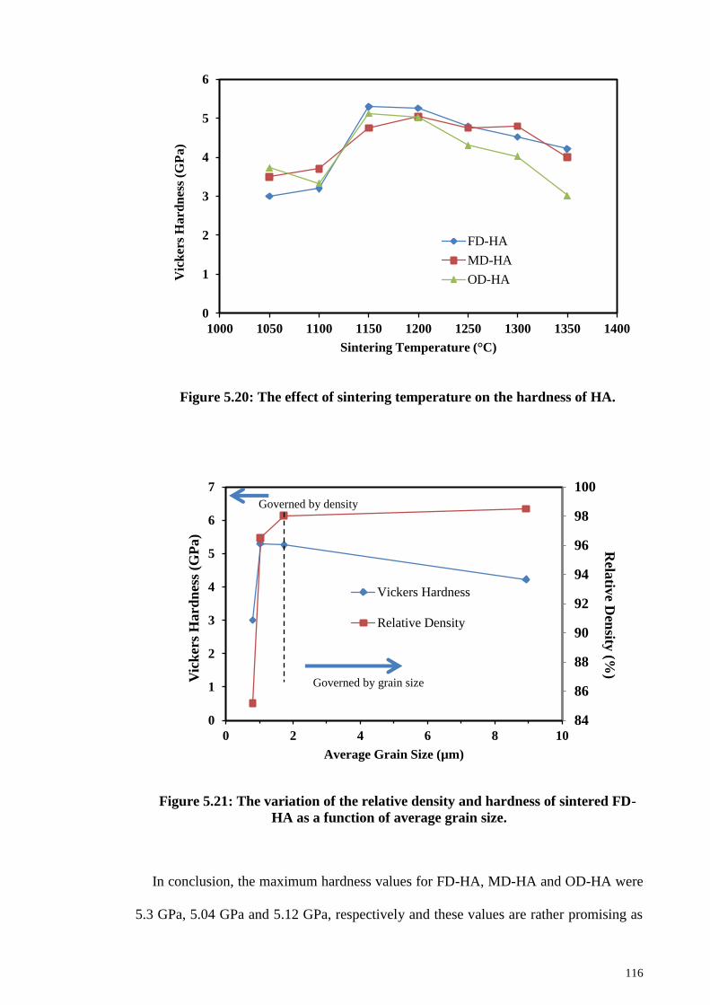
116
Figure 5.20: The effect of sintering temperature on the hardness of HA.
Figure 5.21: The variation of the relative density and hardness of sintered FD-
HA as a function of average grain size.
In conclusion, the maximum hardness values for FD-HA, MD-HA and OD-HA were
5.3 GPa, 5.04 GPa and 5.12 GPa, respectively and these values are rather promising as
0
1
2
3
4
5
6
1000 1050 1100 1150 1200 1250 1300 1350 1400
Vic
ker
s H
ard
nes
s (G
Pa
)
Sintering Temperature (°C)
FD-HA
MD-HA
OD-HA
84
86
88
90
92
94
96
98
100
0
1
2
3
4
5
6
7
0 2 4 6 8 10
Rela
tive D
ensity
(%) V
ick
ers
Hard
nes
s (G
Pa)
Average Grain Size (µm)
Vickers Hardness
Relative Density
Governed by grain size
Governed by density

117
most of the previous studies recorded hardness lower than 5 GPa for conventional
sintered HA (Aminzare et al., 2013; Bhattacharjee et al., 2011; Chen et al., 2008; Lin et
al., 2012). Moreover, the hardness of sintered HA obtained from current study make
them as a potential candidate for bone implant as the hardness is nearly 12 times higher
than cortical bone (0.396 GPa) and cancellous bone (0.345 GPa) (Pramanik et al., 2005).
The effects of sintering temperature on the fracture toughness of the three different
HA samples are shown in Figure 5.22. All the sintered HA exhibited similar trend
where maximum fracture toughness obtained at certain temperature and sintering
beyond that temperature caused the decline of fracture toughness. For example, the
fracture toughness of MD-HA increased from 1.07 MPam1/2
at 1050°C to a maximum
of 1.16 MPam1/2
at 1150°C. Further heating to 1200°C showed no improvement in
fracture toughness and the value remained at 1.15 MPam1/2
. However, sintering MD-HA
at temperature beyond 1200°C showed signed of deterioration and lowest hardness
value of 0.68 MPam1/2
was obtained at 1350°C. Similarly, the FD-HA shows a
maximum fracture toughness of 1.13 MPam1/2
at the sintering temperature of 1150°C
and then decreased to 0.69 MPam1/2
at 1350°C. This maximum value is slightly lower
than that of MD-HA. On the other hand, OD-HA could only attain maximum fracture
toughness value of 1.01 MPam1/2
at 1200°C. Furthermore, any further increased in the
sintering temperature caused severe deterioration of fracture toughness of the sintered
OD-HA as fracture toughness was as low as 0.54 MPam1/2
at 1350°C.

118
Figure 5.22: The effect of sintering temperature on the fracture toughness of
HA.
Correspondingly, the fracture toughness of sintered HA samples is dependent on the
grain size where fracture toughness increase with a decrease in grain size. The
relationship of fracture toughness and grain size of the sintered HA is plotted in Figure
5.23. All the three HA demonstrated similar trend where critical grain size detected at
1150°C whereby beyond this point, the fracture toughness is inversely proportional to
the average grain size, i.e fracture toughness decrease with the increasing of grain size.
A similar trend was observed in the previous literature by Tolouei et al. (2011), Wang
& Shaw (2009), Kamalanathan et al. (2014), Ramesh et al. (2007). From Figure 5.23, it
is clearly observed that smaller average grain size possess higher fracture toughness.
The small grain size is favorable as small grain size lead to more grain boundaries per
unit of volume that exist in the HA matrix, hence giving more limitation to the
dislocations movement as grain boundary can stop dislocations. When the dislocations
were effectively hindered, the permanent distortion of the sample can be prevented and
hence giving the sintered sample higher fracture toughness.
0
0.2
0.4
0.6
0.8
1
1.2
1000 1050 1100 1150 1200 1250 1300 1350 1400
Fra
ctu
re T
ou
gh
nes
s (M
Pa
.m1/2
)
Sintering Temperature (°C)
FD-HA
MD-HA
OD-HA

119
Figure 5.23: The effects of grain size on the fracture toughness of sintered HA.
Overall, the fracture toughness of MD-HA was noted to be superior when compared
to FD-HA and OD-HA throughout the sintering regime. The highest fracture toughness
value of 1.16 MPam1/2
obtained from MD-HA was greater than pure sintered HA
observed in the previous studies (Thangamani et al., 2002; Ramesh et al., 2011;
Banerjee et al., 2007; Wang & Shaw, 2009; Descamps et al., 2013).
Although the mechanical properties of HA powders prepared by wet chemical
method via microwave drying was found to be similar to those prepared by freeze
drying and oven drying, the advantage of microwave drying can be viewed in the
context of time consumed to obtain the respective hardness value and the high fracture
toughness value. This advantage is clearly shown in Table 5.5 which compares the time
taken to produce the MD-HA powders. The time taken to produce HA powders via
microwave drying is significantly lower than the FD-HA and OD-HA. Hence, it can be
concluded that microwave drying is a promising method to produce dense HA with high
fracture toughness in a remarkably shorter time.
0.5
0.6
0.7
0.8
0.9
1
1.1
1.2
1.3
0 2 4 6 8 10 12
Fra
ctu
re T
ou
gh
nes
s (M
Pam
1/2
)
Average Grain Size (µm)
FD-HA
MD-HA
OD-HA
1150°C
1150°C
1150°C

120
Table 5.5: Critical grain size for the sintered HA with their corresponding
maximum hardness and fracture toughness.
Sample Drying
duration
Sintering
Temperature
(°C)
Critical
Grain Size
(µm)
Maximum
Hardness
(GPa)
Maximum
Fracture
Toughness
(MPam1/2
)
FD-HA 36 hours 1150 1.023 5.3 1.13
MD-HA 15 minutes 1150 0.691 4.75 1.16
OD-HA 16 hours 1150 0.974 5.12 1.01
In summary, the current work proved that MD-HA exhibited slighter better
sinterability compared to FD-HA and OD-HA and MD-HA requires significantly
shorter synthesis time. Hence, MD-HA was chosen for further studies to investigate the
effects of microwave sintering and sintering additives on the properties of MD-HA
which will be discussed in the subsequent chapter.

121
CHAPTER 6: RESULTS AND DISCUSSIONS (PART 2)
6.1 Effect of Microwave Sintering on the Sinterability of MD-HA
In Chapter 5, the sinterability of the wet chemical synthesized HA produced by three
different drying methods were investigated. From the results obtained, HA powder
prepared by microwave drying demonstrated the overall optimum properties when
sintered under conventional pressureless sintering (CPS). Microwave sintering has been
widely applied in the sintering of HA compacts by Nath et al. (2006); Bose et al. (2010),
Ramesh et al. (2008); Hassan et al. (2016). In this chapter, the influences of microwave
sintering (MS) on the sintering behavior of the microwave dried HA will be discussed.
6.1.1 XRD Analysis of CPS and MWS Sintered HA
The XRD profiles of HA samples sintered at different temperatures by CPS and
MWS are represented in Figure 6.1 and Figure 6.2, respectively.
20 25 30 35 40 45 50
0
9500
19000
28500
0
7300
14600
21900
20 25 30 35 40 45 50
Figure 6.6.1: The XRD profiles of HA sintered by CPS at different temperatures.
All peaks belong to HA phase.
Inte
nsi
ty (
counts
)
2θ (°)
1250°C
1050°C

122
20 25 30 35 40 45 50
0
110
220
330
0
69
138
207
20 25 30 35 40 45 50
Figure 6.2: The XRD profiles of HA sintered by MWS at different temperatures.
All peaks belong to HA phase.
From Figure 6.1 and Figure 6.2, it was found that HA is stable up to 1250°C for both
sintering methods as the only existing phase is HA. Secondary phases such as TTCP,
TCP and CaO were not presented in the compacts suggesting that the phase stability of
HA was not disrupted by microwave sintering. Further to that, no peak shifting was
observed from XRD profiles regardless of sintering method indicated that
dehydroxylation of HA did not occurred. These results were in good agreement with
those reported by Nath et al. (2006), Ramesh et al. (2007) and Thuault et al. (2014) who
reported that the phase stability of HA was preserved up to 1250°C. However, these
results are in contradiction with the findings obtained by Harabi et al. (2010). The
authors compared the phase stability of CPS and MWS sintered HA. They found that
HA phase of CPS-HA was stable up to 1150°C and then decomposed to β-TCP at
1200°C while MWS-HA started to decompose into TCP when subjected to microwave
sintering at 1050°C and the percentage of TCP phase increased from 46% to 61% when
Inte
nsi
ty (
counts
)
2θ (°)
1250°C
1050°C

123
sintering temperature increased to 1100°C. Finally, the MWS-HA was found to
transform into 85% TTCP and 10% α-TCP with remaining 5% as HA phase. The author
suggested that this phenomenon was attributed to the higher heating efficiency of
microwave heating compared to the conventional sintering (Harabi et al., 2010) which
speed up the decomposition rate of HA into secondary phases.
6.1.2 Bulk Density of CPS and MWS Sintered HA
The effects of CPS and MWS on the densification of HA are illustrated in Figure 6.3.
Figure 6.3: The effect of sintering temperature on the density of HA sintered by
conventional sintering and microwave sintering technique.
Generally, both HA samples sintered by conventional and microwave sintering
exhibited similar trend where relative density increase with increasing temperature but
at different densification rates. At low temperature range (below 1150°C), the density of
MWS-HA was significantly higher than the CPS-HA. The densification curves were
shifted downward nearly 100°C at the lower sintering temperature (< 1150°C) with
difference decrease as higher temperature were reached as shown in Figure 6.3. At low
temperature of 950°C, ~89% densified MWS-HA was obtained and the relative density
75
80
85
90
95
100
900 950 1000 1050 1100 1150 1200 1250 1300
Rel
ati
ve
Den
sity
(%
)
Sintering Temperature (°C)
Coventional Sintering
Microwave Sintering
100°C

124
increased to ~96.8% at 1050°C. Further sintering lead to highly densified MWS-HA;
exhibited >97% of theoretical density when sintering temperature increased to
temperature above 1100°C. The maximum relative density of ~98% was achieved for
MWS-HA sintered at 1250°C. In contrast, the CPS-HA displayed a relatively lower
relative density value of 77.5% at 950°C compared to MWS-HA (~89.3%). CPS-HA
could only achieve relative density above 97% at high sintering temperature of 1200°C.
Apart from that, at sintering temperatures above 1200°C, the densities of both HA
compacts appeared to be similar.
From the results, the densification rate of MWS-HA was remarkably higher than
CPS-HA especially at low temperature range. This high sintering rates was caused by
the high diffusion rate induced by the microwave field that interact directly to the
crystals in the ceramics without the need of heat transfer from surface to the core of the
ceramics. This direct interaction between microwave and the material cause volumetric
heating and hence the sintering and densification can occur at much lower temperatures
with shorter sintering time and lower energy consumption (Demirskyi et al., 2011). The
enhanced densification of MWS-HA at low sintering temperature compared to CPS
have been widely reported (Ehsani et al., 2013; Thuault et al., 2014; Bose et al., 2010;
Mangkonsu et al., 2016). Nonetheless, Ramesh et al. (2008) reported that the
densification of CPS-HA was higher than MWS-HA throughout the sintering regime of
1000°C - 1300°C. This contradiction could be due to the different microwave power
setting of the microwave furnace.
Overall, the high densification rate of MWS-HA is favorable as highly dense HA
could be obtained at low temperature which reduces the risk of decomposition and the
time consumed for the sintering process was greatly reduced. For example, in order to
achieve high relative density of >97%, the CPS-HA samples need to be sintered to

125
1200°C in a conventional pressureless furnace for a total sintering time (inclusive of
sintering, soaking and cooling to the room temperature) of 1290 minutes. On the
contrary, highly dense MWS-HA (~97%) could be achieved at 1050°C albeit the total
microwave sintering time consumed was only 185 minutes which is about 7 times
shorter than that of conventional sintering. Hence, the beneficial effect of microwave
sintering in enhancing densification of HA can be realized.
6.1.3 Microstructural Evolution and Grain Size
The SEM micrographs of the MWS-HA and CPS-HA sintered at 950°C, 1000°C,
1050°C, 1100°C and 1250°C are presented in Figure 6.4 – 6.8.
It can be noted that densification of HA pellets had not really started when subjected
to conventional sintering at 950°C and 1000°C as shown in Figure 6.4 (a) and Figure
6.5 (a) respectively. The samples were hardly densified as no proper grains were formed
and many pores were observed. This corresponds to a relative density of ~77.5% and
~83.2%. On the other hand, it could be observed that final stage of sintering has started
when MWS-HA sintered at 950°C as interconnected pores started to form grain and
grain outline can be seen from Figure 6.4 (b). From Figure 6.5 (b), as temperature goes
up to 1000°C, MWS-HA sample which had remnant porosity (~8.8%) exhibited
microstructure where some areas are fully densified while some nano-pores remained. It
is worth mentioning that MWS-HA sintered at 1000°C showed uniform fine grained
microstructure and the grain size was found to be in nanometer range.
At 1050°C, MWS-HA (Figure 6.6 (b)) displayed almost dense microstructure that
corresponds to the relative density of ~97% with a few extremely small pores (7 nm –
10nm) existed as indicated by the arrows. In contrast, CPS-HA demonstrated larger
pores that varied in size (20 nm – 35 nm) as shown in Figure 6.6 (a), correlate well with
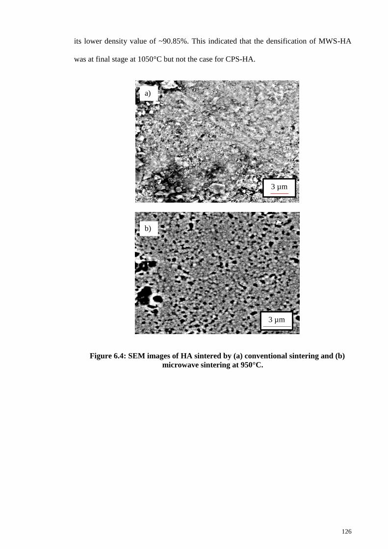
126
its lower density value of ~90.85%. This indicated that the densification of MWS-HA
was at final stage at 1050°C but not the case for CPS-HA.
Figure 6.4: SEM images of HA sintered by (a) conventional sintering and (b)
microwave sintering at 950°C.
3 µm
a)
b)
3 µm

127
Figure 6.5: SEM images of HA sintered by (a) conventional sintering and (b)
microwave sintering at 1000°C.
1 µm
a)
b)
3 µm

128
Figure 6.6: SEM images of HA sintered by (a) conventional sintering and (b)
microwave sintering at 1050°C.
However, as the sintering temperature was increased beyond 1100°C, all the CPS-
HA exhibited very little porosity as shown in Figure 6.7 (a) and Figure 6.8 (a). The
SEM micrographs correlated well with the measured density (Figure 6.3) for all the
samples sintered at high temperature regardless of sintering method as almost full
densification was accomplished when compacts sintered ≥ 1100°C.
1 µm
a)
b)
1 µm

129
Figure 6.7: SEM images of HA sintered by (a) conventional sintering and (b)
microwave sintering at 1100°C.
At high temperature of 1250ºC, both MWS-HA and CPS-HA exhibited fully dense
microstructures which consisted of varying grain sizes and low level of porosity as
shown in Figure 6.8. Significant grain growth was observed for all the HA samples
sintered at 1250ºC with some individual grains grown up to 4 – 6 µm in size. This
growth in grain size due to the increase of sintering temperature was commonly
observed in HA as reported by previous literatures (Layrolle et al., 1998; Mostafa, 2005;
Mobaspherpour et al., 2007).
3 µm
a)
3 µm
b)

130
Figure 6.8: SEM images of HA sintered by (a) conventional sintering and (b)
microwave sintering at 1250°C.
In general, the mean grain size of MWS-HA increased slowly from nanolevel (300
nm) to microlevel (~4.33 µm) with increasing temperature from 950°C to 1250°C. As
for CPS-HA, the grain size couldn‘t be measured at low 950°C and 1000°C as grains
were hardly seen. The average grain size of the sample conventionally sintered at
1050°C was 500 nm and increasing temperature to 1250°C, the grain size increased to
4.45 µm. Figure 6.9 represents the average grain size of HA sintered by microwave
sintering and conventional sintering as a function of the sintering temperature. In both
MWS-HA and CPS-HA, the grain size increase as the sintering temperature increases.
5 µm
a)
5 µm
b)

131
Figure 6.9: The effect of sintering temperatures on the average grain size of HA
samples sintered by (a) conventional sintering and (b) microwave sintering.
It is interesting to discover that the rate of grain growth of microwave sintered HA is
very slow at temperatures ranging from 950°C - 1100°C. However, as temperature
increased further, the grain size for MWS-HA increased rapidly. The grain size of the
MWS-HA increased slowly from ~300 nm (950°C) to ~688 nm (1100°C) and then
increased rapidly by a factor of ~2.7 from ~1.60 µm to ~4.33 µm as temperature
increased from 1150°C to 1250°C. In comparison, the grain growth rate of CPS-HA was
higher than MWS-HA as evidence of the steep slope shown in Figure 6.9. The grain
size of CPS-HA increased by a factor of about 6.4 from ~691 nm to ~4.45 µm at
sintering temperature of 1150°C and 1250°C, respectively. The increasing trend of grain
size against sintering temperature was in good agreement with the trend reported by
(Akao et al., 1981; Ramesh et al., 2007; Muralithran & Ramesh, 2000; Ramesh et al.,
2008). The drastic grain growth of HA was observed at 1250°C regardless of sintering
methods. In conventional sintering, the grain enlargement of HA was associated with
the surplus activation energy supplied to the HA due to elevated sintering temperature.
0
0.4
0.8
1.2
1.6
2
2.4
2.8
3.2
3.6
4
4.4
4.8
900 950 1000 1050 1100 1150 1200 1250 1300
Av
era
ge
Gra
in S
ize
(µm
)
Sintering Temperature (°C)
Conventional Sintering
Microwave Sintering
High grain
growth rate

132
Current research shows that the efficacy of microwave sintering in preventing grain
coarsening is profound at low temperature but not at high sintering temperature.
Excessive grain growth with non-uniform grain size distribution was observed from
MWS-HA sintered at 1250°C as shown in Figure 6.8 (b). At high microwave sintering
temperature, a large amount of microwave was absorbed by HA and leads to localized
heating which resulted in excessive grain growth (Vijayan & Varma, 2002; Bykov et al.,
1999). As the temperature increase, there is risk of getting non-uniform heating profile
in the samples which contribute to the localized heating in different spots of the sample.
Hence, there are some ―hot spots‖ where heating is much more rapid than the other
spots that caused the heterogeneous heating. Therefore, some of the MWS-HA grains
are large and some of the grain retained in ultrafine size as shown in Figure 6.8 (b).
In short, the results showed that microwave sintering is effective in preventing grain
growth while maintaining high density throughout the sintering regime and nano grain
size was maintained up to 1100°C. In microwave sintering, the high heating rate of
MWS-HA lower the sintering time exposure and leads to the suppression of the surface
diffusion. Hence, the exaggerated grain growth was restricted. The smaller grain size of
MWS-HA is vital because smaller grain size would generally yield HA with enhanced
mechanical properties compared to HA containing large grain.
6.1.4 Vickers Hardness and Fracture Toughness of CPS and MWS Sintered HA
The variation of the Vickers hardness of both conventional and microwave sintered
HA at various temperatures are shown in presented in Figure 6.10. In general, the
hardness of HA samples regardless of sintering method revealed similar trends with
increasing sintering temperature up to certain value at particular temperature and then
decrease with further heating. The results showed that the hardness values of MWS-HA
are higher than CPS-HA at lower temperature range (950°C - 1100°C). However,

133
similar hardness values were recorded for both MWS-HA and CPS-HA sintered at
1150°C. Upon 1150°C, the hardness values of CPS-HA are higher than MWS-HA.
At 950°C, both MWS-HA and CPS-HA recorded lowest hardness values of 4.09 GPa
and 2.89 GPa, respectively. As sintering temperature increased, the hardness value of
MWS-HA increased and marked the maximum value of ~4.8 GPa at 1150°C. However,
when microwave sintering was carried out >1150°C, the hardness started to decline
linearly to ~4.4 GPa at 1200°C and ~3.87 GPa at 1250°C. In contrast, the hardness of
CPS-HA start to rise sharply from 3.01 GPa at 1000°C to a maximum of ~5.04 GPa at
1200°C. Similar to MWS-HA, the CPS-HA showed a decline in hardness value as
sintering proceed to 1250°C.
Figure 6.10: The effect of sintering temperature on the hardness of HA sintered
by conventional sintering and microwave sintering technique.
In addition, the hardness of sintered HA was found to depend on the bulk density and
average grain size and these relationships are plotted in Figure 6.11. Figure 6.11 clearly
reflected that increase in relative density of CPS-HA from 77.5% at 950°C to 97.5% at
1200°C enhance the hardness of samples from 2.8 GPa to 5.04 GPa. Similarly,
0
1
2
3
4
5
900 950 1000 1050 1100 1150 1200 1250 1300
Vic
ker
s H
ard
nes
s (G
Pa)
Sintering Temperature (°C)
Conventional Sintering
Microwave Sintering

134
improved hardness value was found when the sintering temperature increased from
950°C to 1150°C, in accordance with the increment in relative density from 89.3% to
97.6%. However, when the temperature exceeds certain point (>1200°C for CPS-HA
and >1150°C for MWS-HA), it was found that the change in hardness was not in
accordance to the change in bulk density as the hardness value decline even the density
increased. This deterioration in hardness value was the result of the large grain size
obtained. For example, the hardness of CPS-HA declines from 5.04 GPa (1200°C) to
4.75 GPa (1250°C) when the grain size increase from ~1.34 µm to ~4.45 µm while big
grain size of ~4.33 µm correspond to the low hardness of 3.87 GPa of MWS-HA
sintered at 1250°C whereas small grain size of ~1.6 µm correspond to 4.8 GPa for
MWS-HA sintered at 1150°C.
Figure 6.11: Vickers hardness dependence on the relative density of HA sintered
by conventional sintering and microwave sintering technique.
The effects of sintering temperature on the fracture toughness of MWS-HA and CPS-
HA are shown in Figure 6.11. The fracture toughness of CPS-HA sintered at 950°C and
1000°C was not measure due to the porous surface of those HA compacts. The graph
0
1
2
3
4
5
6
76 78 80 82 84 86 88 90 92 94 96 98 100
Vic
ker
s H
ard
nes
s (G
Pa)
Relative Density (%)
Conventional Sintering
Microwave Sintering
950°C
1200°C 1150°C
950°C

135
(Figure 6.12) showed that HA compact sintered by microwave sintering exhibited
similar trend to the Vickers hardness trend, i.e. toughness value increase with increasing
temperature and met a plateau at certain temperature and decrease thereafter. The
fracture toughness value of MWS-HA increased greatly from 1.1 MPa.m1/2
at 950°C to
1.85 MPa.m1/2
at 1050°C. Further sintering above 1050°C showed sign of deterioration
as the toughness values decrease almost linearly with increasing sintering temperature
from 1.85 MPa.m1/2
to 0.88 MPa.m1/2
at 1250°C. In comparison, the fracture toughness
of HA samples sintered via conventional sintering perked at 1150°C, recording a value
of 1.16 MPa.m1/2
and decline to a minimum value of 0.81 MPa.m1/2
when sintered at
1250°C.
Figure 6.12: The effect of sintering temperature on the fracture toughness of
HA sintered by conventional sintering and microwave sintering technique.
The fracture toughness value of the MWS-HA sintered at 1050°C was significantly
higher than the values reported in the literature for synthetic HA samples sintered via
microwave sintering as shown in Table 6.1. Sintered dense HA was usually reported to
0
0.2
0.4
0.6
0.8
1
1.2
1.4
1.6
1.8
2
900 950 1000 1050 1100 1150 1200 1250 1300
Fra
ctu
re T
ou
gh
nes
s (M
Pa.m
1/2
)
Sintering Temperature (°C)
Conventional Sintering
Microwave Sintering

136
have low fracture toughness values less than 1 MPa.m1/2
(Tan et al., 2011) which limit
its use in load bearing applications. Hence, the high fracture toughness HA obtained in
current research is encouraging. In conventional sintering, grain growth mechanism
dominant in the early stage of sintering and followed by the densification mechanism
and hence the risk of getting large grain HA is high. Nevertheless, sample was rapidly
sintered to temperature region where densification predominate the grain coarsening
mechanism in microwave sintering. Therefore, the grain growth can be minimized while
retaining high densification during the microwave sintering of HA as evidence from
current study, i.e. MWS-HA recorded relative density ~97% while maintaining nano
grain size of 356 nm lead to a great enhancement of fracture toughness (1.85 MPa.m1/2
)
at 1050°C. Therefore, it is inferred that the high fracture toughness value could be
attributed to the combine effects of improved sinterability and restricted grain growth of
HA during microwave sintering.
Table 6.1: A comparison of optimum fracture toughness values of current study
to the available literatures (Ramesh et al., 2008; Kutty et al., 2015; Thuault et al.,
2014).
Sintering
Temperature (°C) Fracture Toughness
(MPa.m1/2
)
Current Study 1050 1.85
Ramesh et al. (2008) 1050 1.45
Kutty et al. (2015) 1000 1.75
Thuault et al. (2014) 900 1.36
The dependence of fracture toughness on both relative density and grain size is
plotted in Figure 6.13. From Figure 6.13, it is postulated that the fracture toughness of
HA increase as relative density increases below a critical grain size (~356 nm in current

137
study). Beyond the critical grain size, the fracture toughness appeared to be governed by
grain size in the trend that fracture toughness decreases with increasing grain size.
Figure 6.13: The variation of fracture toughness and relative density of HA
sintered by microwave sintering technique.
Small grain size was desirable to produce high toughness HA (Kamalanathan et al.,
2014) as suggested by Hall-Petch equation, f = 0 + kd-1/2
, where f is the fracture
strength, d is the grain size, k and 0 are constants, the strength decreases with
increasing grain size. The strength of the materials depends on the ability to restrict the
motion of dislocations and the grain boundaries serve as barriers to limit the dislocation
movement. At the grain boundary, the dislocations are blocked. As shown in Figure
6.14, the pile up created driving force to push the dislocation towards the nearby grain.
The bigger the grain, the bigger the pile up and therefore the bigger the driving force
and hence, a small load is sufficiently to push the dislocation to the adjacent grain. By
reducing the grain size, there are less pile ups creating less driving force, requiring a
larger stress to move the dislocation to next grain.
70
80
90
100
0
0.2
0.4
0.6
0.8
1
1.2
1.4
1.6
1.8
2
0 0.5 1 1.5 2 2.5 3 3.5 4 4.5 5
Rela
tive D
ensity
(%)
Fra
ctu
re T
ou
gh
nes
s (M
Pa
.m1/2
)
Average Grain Size (µm)
Fracture Toughness
Relative Density
Critical grain size

138
Figure 6.14: The schematic diagram of the dislocation pile up in (a) large grains
and (b) small grains.
In short, small grain size microstructure provided large numbers of grain boundaries
per unit volume which resulting in a smaller number of pile ups in grain (boundaries
limit the dislocation movement) and in turn increase the strength of sample. This
correlates well with the current study where the optimum fracture toughness of 1.85
MPa.m1/2
obtained from HA microwave sintered at 1050°C corresponds to the
extremely small grain size of ~356 nm.
In summary, the results obtained suggest that microwave sintering is a promising
sintering technique to produce highly dense HA ceramics with fine grained
microstructure, high phase stability and enhanced mechanical properties.
Dislocations
Dislocation
s b) a)

139
6.2 Effect of Zinc Oxide (ZnO) addition on the Sinterability of HA
In this section, the effects ZnO addition on the mechanical properties of
hydroxyapatite will be presented. The hydroxyapatite powder used in this work was
prepared by the wet chemical precipitation method via microwave drying. Different
amount of ZnO powder was added into the HA powder based on ball milling method as
described in Chapter 4. The green compacts were then cold isostatic pressed at 200 MPa
followed by pressureless sintering at temperatures range between 1100°C to 1300°C.
6.2.1 XRD Analysis of Undoped and ZnO-doped HA Powder
The XRD signatures of the ZnO-doped as well as the undoped HA powders were
present in Figure 6.15. The analysis shows that all the powders produced only peaks
corresponded to the stoichiometric HA as shown in Figure 6.15. This result proved that
the presence of ZnO had negligible effect on the HA phase stability which is consistent
with the findings of other researchers (Gross et al., 2013; Deepa et al., 2013). However,
it was found that ZnO-doped HA powders have higher intensity than the pure HA as
indicated by the sharper and narrower peaks.
Figure 6.15: The XRD profiles of undoped HA powders and HA powders
containing 0.1 wt%, 0.3 wt%, 0.5 wt% and 1 wt% ZnO, respectively.
Inte
nsi
ty (
counts
)
2θ (°)
1.0 wt% ZnO
0.5 wt% ZnO
0.3 wt% ZnO
0.1 wt% ZnO
Undoped

140
6.2.2 XRD Analysis of Undoped and ZnO-doped Sintered HA
XRD spectrum (Figure 6.16) proved that all the samples sintered at high temperature
of 1300°C were pure HA regardless of the amount of ZnO addition.
Figure 6.16: XRD patterns of HA samples sintered at 1300°C for undoped HA
and HA containing 0.1 wt%, 0.3 wt%, 0.5 wt% ZnO and 1 wt% ZnO, respectively.
Inte
nsi
ty (
counts
)
2θ (°)

141
This result revealed that the presence of ZnO and high sintering temperature had
negligible effects on the sintered HA phase stability. This is inconsistent with the study
reported by Bodhak et al. (2011), Buys et al. (1995) and Finoli et al. (2010) who
observed that high sintering temperature of 1200°C and above could affect the HA
phase stability. Bodhak et al. (2011) reported that the inclusion of ZnO destabilize the
HA phase and the decomposition of ZnO-doped HA started at about 1200°C where
strong β-TCP peaks were detected. The decomposition could be due to the nature of the
starting HA powders but not the inclusion of ZnO as the authors used commercially
available HA powders in their experiment. Buys et al. (1995) and Finoli et al. (2010) on
the other hand reported that thermal decomposition of pure HA into TCP and CaO
started at about 1200°C and the presence of TCP would deteriorate the mechanical
properties of HA (Royer et al., 1993; Wang & Chaki, 1993) while CaO would hinder
the biological performance of HA (Ślósarczyk & Piekarczyk, 1999).
6.2.3 Bulk Density of Undoped and ZnO-doped Sintered HA
The densification curves of undoped and ZnO-doped HA as a function of sintering
temperatures are shown in Figure 6.17. The relative density increased with increasing
sintering temperature for pure HA throughout the sintering regime. However, for ZnO-
doped HA regardless of dopants amount, the bulk density increase with increasing
temperature and met a plateau at about 1250°C. From Figure 6.17, it is noticeable that
ZnO-doped HA samples have demonstrated higher densification that pure HA as all the
doped samples attained high relative density > 98% at 1200°C while pure HA could
obtain only ~97.5% of the theoretical density at the similar temperature. On the other
hand, sintering study showed that 1250°C is optimum sintering temperature in
densifying Zn-doped HA as all the doped samples attained the highest relative density at
1250°C except for pure HA. Pure HA attained highest density of ~98.43% at 1300°C.

142
Figure 6.17: Relative density variation as a function of sintering temperatures
for HA with different amount of ZnO addition.
Besides, it also appeared that higher ZnO amount is beneficial for the densification
of HA especially at high temperature of 1250ºC as shown in Figure 6.17. Higher content
of ZnO appeared to be advantageous to the densification of HA since the 1.0 wt% ZnO-
doped HA attained the highest relative density of ~99.13%; followed by 0.5 wt% ZnO-
doped HA (99.12%), 0.3 wt% (98.64%) ,0.1 wt% ZnO-doped HA (98.3 %) and the
undoped HA achieved only 98.08% of relative density at 1250°C. The inclusion of ZnO
improved the mass transport along the grain boundaries and thus lead to higher final
densities. Moreover, zinc oxide was found to be beneficial in enhancing the
densification of HA when compared to other sintering additives such as silver, iron and
titania (Bhattacharjee et al., 2011; Kramer et al., 2013; Wang & Chaki, 1994).
6.2.4 Microstructure Analysis of Undoped and ZnO-doped Sintered HA
Figure 6.18 shows the representative microstructures of the ZnO-doped HA and
undoped HA sintered at 1150ºC. SEM observation in Figure 6.18 (a) showed that small
pores were detected (indicated by arrows) on undoped HA and this is in agreement with
92
93
94
95
96
97
98
99
100
1050 1100 1150 1200 1250 1300 1350
Rel
ati
ve
Den
sity
(%
)
Sintering Temperature (°C)
Undoped
0.1 wt% ZnO
0.3 wt% ZnO
0.5 wt% ZnO
1.0 wt% ZnO
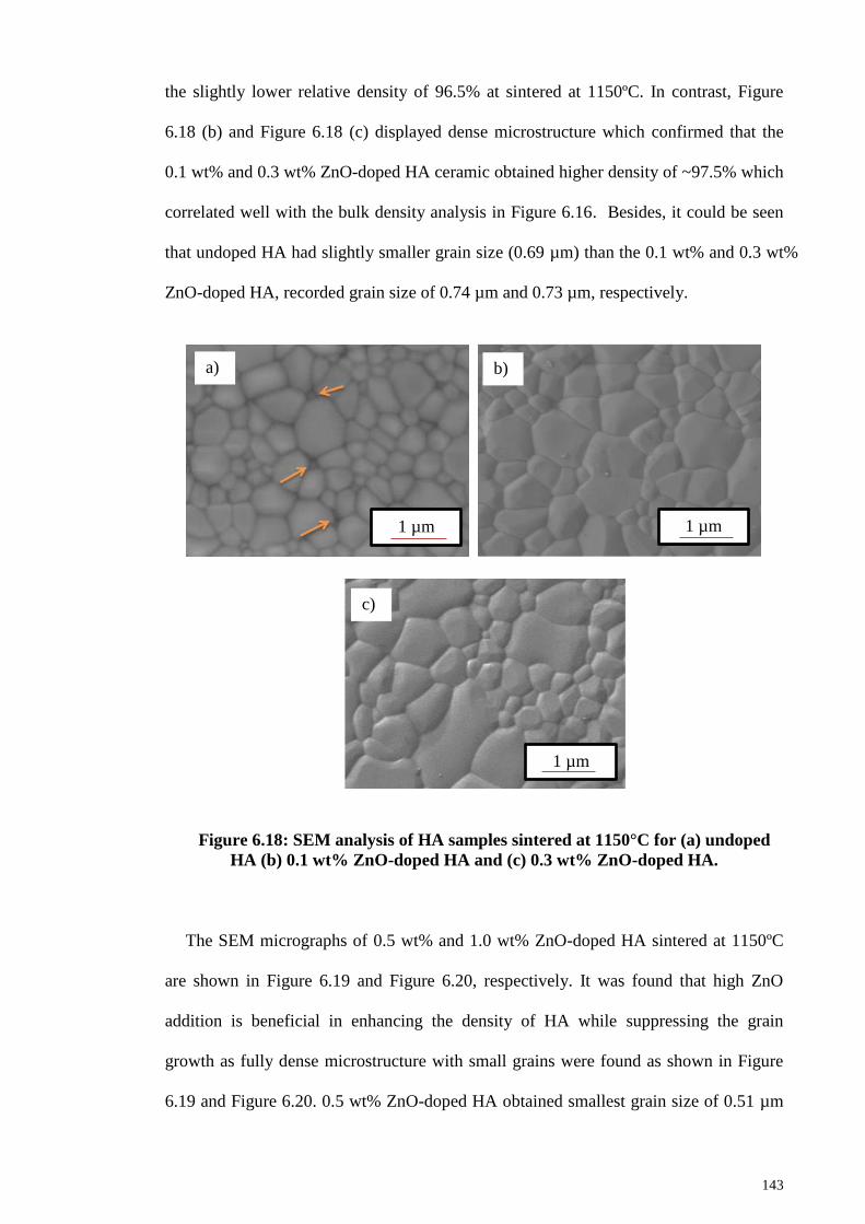
143
the slightly lower relative density of 96.5% at sintered at 1150ºC. In contrast, Figure
6.18 (b) and Figure 6.18 (c) displayed dense microstructure which confirmed that the
0.1 wt% and 0.3 wt% ZnO-doped HA ceramic obtained higher density of ~97.5% which
correlated well with the bulk density analysis in Figure 6.16. Besides, it could be seen
that undoped HA had slightly smaller grain size (0.69 µm) than the 0.1 wt% and 0.3 wt%
ZnO-doped HA, recorded grain size of 0.74 µm and 0.73 µm, respectively.
Figure 6.18: SEM analysis of HA samples sintered at 1150°C for (a) undoped
HA (b) 0.1 wt% ZnO-doped HA and (c) 0.3 wt% ZnO-doped HA.
The SEM micrographs of 0.5 wt% and 1.0 wt% ZnO-doped HA sintered at 1150ºC
are shown in Figure 6.19 and Figure 6.20, respectively. It was found that high ZnO
addition is beneficial in enhancing the density of HA while suppressing the grain
growth as fully dense microstructure with small grains were found as shown in Figure
6.19 and Figure 6.20. 0.5 wt% ZnO-doped HA obtained smallest grain size of 0.51 µm
1 µm
a) b)
c)
1 µm
1 µm
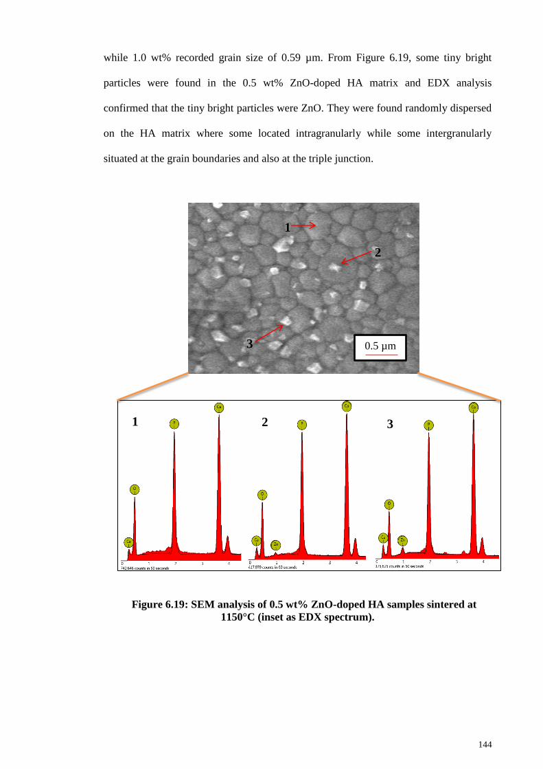
144
while 1.0 wt% recorded grain size of 0.59 µm. From Figure 6.19, some tiny bright
particles were found in the 0.5 wt% ZnO-doped HA matrix and EDX analysis
confirmed that the tiny bright particles were ZnO. They were found randomly dispersed
on the HA matrix where some located intragranularly while some intergranularly
situated at the grain boundaries and also at the triple junction.
Figure 6.19: SEM analysis of 0.5 wt% ZnO-doped HA samples sintered at
1150°C (inset as EDX spectrum).
0.5 µm
1
2
3
1 2 3

145
Besides, Figure 6.20 proved that 1.0 wt % ZnO-doped HA contained ZnO particles
in the matrix as confirmed by EDX analysis. However, the ZnO was found to be in
cluster as shown in Figure 6.20. This could be due to the fact that higher amount of ZnO
addition are more difficult to be dispersed homogenously, caused some of the ZnO
remain as cluster within the HA matrix. In contrast, the tiny bright particles (ZnO) were
not detected on the 0.1 wt% and 0.3 wt% ZnO-doped HA as indicated in Figure 6.18 (b)
and (c), respectively.
Figure 6.20: SEM analysis of 1.0 wt% ZnO-doped HA samples sintered at
1150°C (inset as EDX spectrum).
0.5 µm 1
2
1 2

146
The comparison of the grain size of all the HA sintered at 1150ºC is summarized in
Table 6.2. From Table 6.2, the grain size of 0.1 wt% and 0.3 wt% ZnO-doped HA was
found to be slightly bigger than undoped HA, proved that 0.1 wt% and 0.3 wt% of ZnO
addition are not sufficient to inhibit the grain growth of HA. This was evidence from the
SEM in Figure 6.18 (b) and (c) where no ZnO particles were detected in the samples.
0.5 wt% ZnO-doped HA was found to obtained smallest grain size, proved that 0.5 wt%
ZnO addition can hinder the grain growth of HA. The intragranularly and
intergranularly seated ZnO slow down the grain boundary diffusion/migration by
preventing the HA grains to contact with each other freely. Hence, the diffusions of
atoms along the grain boundaries between HA grains and also between the HA and ZnO
grains were slow down, and the grain growth could be restricted. As ZnO content
increased to 1.0 wt%, the grain growth inhibition was found to be less effective
compared to 0.5 wt% ZnO addition. This could be attributed to inhomogenous
dispersion of ZnO on the HA matrix as shown in Figure 6.20.
Table 6.2: Grain size of undoped and ZnO-doped HA sintered at 1150ºC.
ZnO content (wt%) Grain Size (µm)
Undoped 0.6911
0.1 0.7429
0.3 0.7323
0.5 0.5090
1.0 0.5946
When sintering was carried out at 1300ºC, the grain size increased for all the samples
regardless of dopant addition as shown in Figure 6.21. Pure HA showed the most
significant grain growth as the grain size increased to 6.9 µm. 0.1 wt%, 0.3 wt%, 0.5 wt%
and 1.0 wt% ZnO-doped HA recorded grain size of 4.4 µm, 2.74 µm, 2.15 µm and 3.23
µm, respectively. The results indicated that 0.5 wt% can inhibit grain growth effectively

147
even at high sintering temperature of 1300ºC as it exhibited smallest grain size amongst
all the HA samples. It is worth mentioning that the 1.0 wt% ZnO-doped HA exhibited
microstructure of extremely large grain that accompanied by small grains as shown in
Figure 6.21 (e). This is due to the agglomeration of ZnO cluster on the HA matrix as the
agglomerates can act as seeds for abnormal grain growth due to different densification
rate (Mouzon et al., 2008).
Figure 6.21: SEM analysis of HA samples sintered at 1300°C for (a) undoped
HA (b) 0.1 wt% ZnO-doped HA, (c) 0.3 wt% ZnO-doped HA, (d) 0.5 wt% ZnO-
doped HA and (e) 1 wt% ZnO-doped HA.
5 µm
a) b)
c) d)
e)
2.5 µm
1 µm 1 µm
1 µm
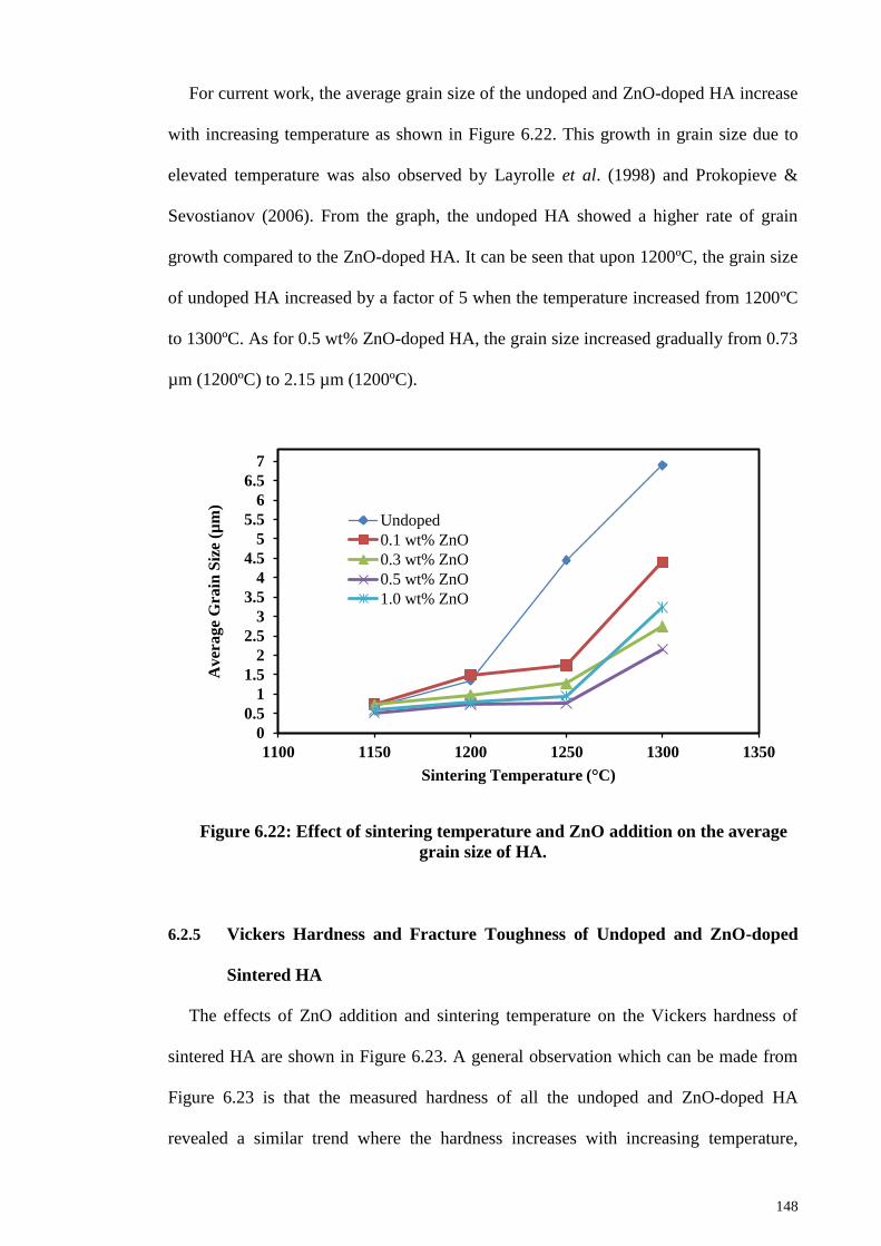
148
For current work, the average grain size of the undoped and ZnO-doped HA increase
with increasing temperature as shown in Figure 6.22. This growth in grain size due to
elevated temperature was also observed by Layrolle et al. (1998) and Prokopieve &
Sevostianov (2006). From the graph, the undoped HA showed a higher rate of grain
growth compared to the ZnO-doped HA. It can be seen that upon 1200ºC, the grain size
of undoped HA increased by a factor of 5 when the temperature increased from 1200ºC
to 1300ºC. As for 0.5 wt% ZnO-doped HA, the grain size increased gradually from 0.73
µm (1200ºC) to 2.15 µm (1200ºC).
Figure 6.22: Effect of sintering temperature and ZnO addition on the average
grain size of HA.
6.2.5 Vickers Hardness and Fracture Toughness of Undoped and ZnO-doped
Sintered HA
The effects of ZnO addition and sintering temperature on the Vickers hardness of
sintered HA are shown in Figure 6.23. A general observation which can be made from
Figure 6.23 is that the measured hardness of all the undoped and ZnO-doped HA
revealed a similar trend where the hardness increases with increasing temperature,
0
0.5
1
1.5
2
2.5
3
3.5
4
4.5
5
5.5
6
6.5
7
1100 1150 1200 1250 1300 1350
Aver
age
Gra
in S
ize
(µm
)
Sintering Temperature (°C)
Undoped
0.1 wt% ZnO
0.3 wt% ZnO
0.5 wt% ZnO
1.0 wt% ZnO

149
consequently reaching the maximum hardness value at certain temperature; followed by
a decrease with further increase of sintering temperature. For instance, the hardness of
0.5 wt% ZnO-doped HA perked at 1150°C (5.63 GPa) and decrease drastically to 4.28
GPa at sintering temperature of 1300°C. On the other hand, the maximum hardness was
obtained at higher sintering temperature (1200°C) by undoped, 0.1 wt%, 0.3 wt% and
1.0 wt% ZnO-doped HA; recording value of 5.04 GPa, 5.12 GPa, 5.3 GPa and 5.37 GPa,
respectively. Subsequent sintering caused deterioration or constant hardness values
ranging from 4.22 GPa to 4. 8 GPa as shown in Figure 6.23.
Figure 6.23: Effect of sintering temperature and ZnO addition on the Vickers
hardness of HA.
The beneficial effect of ZnO in improving the hardness of HA has been revealed. It
could be perceived that at temperature from 1100°C to 1200°C, all the ZnO-doped HA
samples demonstrated hardness superior to that of undoped HA. The maximum
hardness achieved for all undoped and ZnO-doped HA is summarized in Table 6.3. In
short, the addition of 0.5 wt% was found to be most effective as the sample obtained
highest hardness values among all the samples as shown in Table 6.3.
3
3.5
4
4.5
5
5.5
6
1050 1100 1150 1200 1250 1300 1350
Vic
ker
s H
ard
nes
s (G
Pa)
Sintering Temperature (°C)
Undoped
0.1 wt% ZnO
0.3 wt% ZnO
0.5 wt% ZnO
1.0 wt% ZnO

150
The enhancement of hardness obtained from the 0.5 wt% ZnO-doped HA was due to
the smaller grain size. Figure 6.24 shows the hardness of undoped and ZnO-doped HA
ceramics as a function of the inverse square root of grain size. The values in the graph
was reasonably fitted with a line (R2 = 0.9264), which means that this dependence
followed the Hall-Petch relationship (Wang et al., 2009) where hardness increased with
decreasing grain size. The enhancement in the hardness of HA due to the smaller grain
size was also observed by Veljovic et al. (2009), Ramesh et al., (2008) and Tekeli et al.
(2006) as they stated that smaller grain size give more grain boundaries as per unit
volume which acts as obstacle for plastic deformation.
Figure 6.24: The dependence of the hardness of undoped and ZnO-doped HA
on the inverse square root of grain size.
Table 6.3: A comparison of optimum Vickers hardness between undoped and
ZnO-doped HA.
ZnO content (wt%) Maximum Vickers
Hardness (GPa)
Undoped 5.04
0.1 5.12
0.3 5.37
0.5 5.63
1.0 5.37
R² = 0.9264
4
4.2
4.4
4.6
4.8
5
5.2
5.4
5.6
5.8
6
1 1.1 1.2 1.3 1.4 1.5
Hard
nes
s (G
Pa)
d-1/2 (µm-1/2)

151
The fracture toughness variation with respect to the sintering temperature for
undoped and ZnO-doped HA is shown in Figure 6.25. At low temperature of 1100°C,
low fracture toughness values (0.97 – 1.06 MPam1/2
) were recorded for all the sintered
samples which could be attributed to the weak grain boundaries of the HA matrix at low
temperature (Champion, 2013). For undoped HA, no obvious trend or relationship
could be observed between fracture toughness and sintering temperature (Figure 6.25)
as fracture toughness fluctuated with temperatures.
Figure 6.25: Effect of sintering temperature and ZnO addition on the fracture
toughness of HA.
Further analysis indicated that the fracture toughness variation of all the ZnO-doped
HA samples exhibited similar trend to the variation of Vickers hardness (Figure 6.23) as
fracture toughness met a plateau at certain temperature and thereby decrease with
increasing temperature. For example, the fracture toughness of 0.5 wt% ZnO-doped HA
peaked at a maximum value (1.37 MPam1/2
) when sintered at 1150°C and decreased
almost linearly with increasing temperature (>1150°C). Similarly, 0.1 wt% and 1.0 wt%
ZnO-doped HA obtained maximum fracture toughness value of 1.06 MPam1/2
and 1.19
0.6
0.7
0.8
0.9
1
1.1
1.2
1.3
1.4
1.5
1.6
1050 1100 1150 1200 1250 1300 1350
Fra
ctu
re T
ou
gh
nes
s (M
Pa
m1
/2)
Sintering Temperature (°C)
Undoped
0.1 wt% ZnO
0.3 wt% ZnO
0.5 wt% ZnO
1.0 wt% ZnO

152
MPam1/2
respectively at 1150°C and then decrease. On the other hand, 0.3 wt% ZnO-
doped HA obtain its maximum toughness value of 1.09 MPam1/2
at 1200°C and
decreased with further sintering.
From Figure 6.25, low fracture toughness values were recorded for all the samples
sintered at 1300°C. According to Buys et al. (1995), the decline in mechanical
properties of HA are expected at higher temperatures above 1200°C mainly due to HA
decomposition to TCP and CaO. The volume changes associated with the formation of
these new secondary phases creates internal strain within the HA matrix. This was not
the case in the present work since HA decomposition was not detected for all the ZnO-
doped HA sintered at 1300°C and the drastic drop of fracture toughness observed could
be associated to the increment in average grain size as shown in Figure 6.21. These
large grains are believed to provide an easy path for crack propagation and hence lower
fracture toughness (Ramesh et al., 2011).
6.2.6 Toughening Mechanism
The previous section has shown that 0.5 wt% ZnO-doped HA demonstrated the most
enhanced hardness and fracture toughness as compared to other HA samples. Hence, the
cracking behavior introduced by the Vickers indentation of the 1150ºC sintered 0.5 wt%
ZnO-doped HA (Figure 6.27) was compared to the pure HA (Figure 6.26). As shown in
Figure 6.26, pure HA tends to propagate cracks in straight line where the deflection
angle is barely observed due to the absence ZnO particles as obstacles. The overall path
of crack propagation was completely linear, predominantly transgranular fracture mode.
In contrast, the crack path of the 0.5 wt% ZnO-doped HA has significant crack
deflection with a mixture of transgranular and intergranular fracture mode as shown in
Figure 6.27. The toughening mechanism of 0.5 wt% ZnO-doped HA can be summarized
as the combine effects of crack deflection and crack bridging as shown in the magnified

153
image in Figure 6.28 and Figure 6.29, respectively. As for crack deflection (Figure 6.28),
the crack cross the HA grains transgranularly and hit the intragranularly seated ZnO.
This caused the crack to be deflected to the grain boundaries and continue to propagate
intergranularly within the HA and ZnO grain boundaries. The high deflection angle of
crack resulted in torturous crack path where higher fracture energy is needed for the
crack to propagate. According to the following equation stated by Jang et al. (1996):
KIC α γf ½
(6.1)
The fracture toughness (KIC) was directly proportional to the square root of fracture
energy (γf), the higher fracture energy resulted from the crack deflection contributed to
the enhanced toughening in the 0.5 wt % ZnO-doped HA. This toughening via crack
deflection has been reported elsewhere (Faber & Evans, 1983; Wang & Shaw, 2009).
The magnified image in Figure 6.29 showed the presence of crack bridging. The ZnO
particulate bridges the two surface of crack, providing stress to counteract the applied
stress and hence delay the crack propagation. The crack bridging and crack deflections
as a result of ZnO addition were schematically depicted in Figure 6.30 (a) and Figure
6.30 (b).

154
Figure 6.26: SEM micrograph of the indentation crack paths of pure HA
sintered at 1150ºC.
Figure 6.27: SEM micrograph of indentation crack paths of 0.5 wt% ZnO-
doped HA sintered at 1150ºC.
Refer Figure 6.28
Refer Figure 6.29
3 µm
3 µm

155
Figure 6.28: A close up view of the crack paths of Figure 6.27 indicated the
crack deflection.
Figure 6.29: A close up view of the crack paths of Figure 6.27 indicated the
crack bridging.
Crack Bridging
1 µm
1 µm

156
Figure 6.30: A schematic diagram of the proposed toughening mechanism: (a)
crack bridging and (b) crack deflection.
In summary, the inclusion of ZnO into HA matrix is beneficial in terms of
sinterability and mechanical properties without disrupting the phase stability of HA. 0.5
wt% ZnO-doped HA sintered at 1150°C was found to exhibit the highest hardness of
5.63 GPa and fracture toughness of 1.37 MPam1/2
.The higher mechanical properties of
0.5 wt% ZnO-doped HA compared to other samples were attributed to the effectiveness
of 0.5 wt% ZnO in suppressing the grain coarsening. Moreover, the high fracture
toughness value recorded in the present work is very promising as other researchers
who doped HA with different additives such as zirconia, titanium oxide, alumina and
bismuth oxide (Bhattacharjee et al., 2011; Kim et al., 2003; Champion et al., 1996;
Ramesh et al., 2011) obtained fracture toughness less than 1.24 MPam1/2
. Further to that,
zinc oxide was proved to improve the biological properties of calcium phosphate
(Bandyopadhyay et al., 2006; Yu et al., 2014; Ishikawa et al., 2002; Ito et al., 2000),
making it a potential candidate for clinical applications.
ZnO
HA grains
Crack
Crack
a) b)

157
CHAPTER 7: CONCLUSIONS AND FURTHER WORK
7.1 Conclusions
This study was carried out to evaluate the sinterability of HA powders synthesized
via three different drying methods, i.e. oven drying (OD-HA), microwave drying (MD-
HA) and freeze drying (FD-HA). Then, the effect of microwave sintering on the HA
powder were evaluated and explained. The beneficial effect of adding ZnO on the
sintering behaviour of HA particularly on the fracture toughness has also been evaluated.
The following conclusions can be drawn from this present work.
1. Single phase pure HA powder were successfully synthesized by wet chemical
method via oven drying, microwave drying and freeze drying as proved by XRD
analysis and FTIR analysis.
2. EDX analysis showed that FD-HA had Ca/P ratio of 1.77, closer to the
stoichiometry HA (Ca/P = 1.67) compared to MD-HA (Ca/P =1.85) and OD-HA
(Ca/P = 1.90). However, it was found that the Ca/P ratio did not affect the phase
stability of the HA powder as all the three drying methods produced pure HA.
3. FE-SEM micrographs proved that MD-HA and FD-HA consisted of small to
large size agglomerates that made of HA particles in nanosize while OD-HA
consisted of extremely large agglomerates.
4. It was found that both FD-HA and OD-HA demonstrated needle like
morphology with specific surface area of ~ 97 m2/g. In comparison, MD-HA
was in nanorod morphology accompanied with higher specific surface area of ~
111 m2/g.

158
5. Microwave drying significantly reduced the time of HA synthesis and it was
found that MD-HA exhibited higher thermal stability that OD-HA. MD-HA
showed no sign of decomposition into tri-calcium phosphate, tetra tri-calcium
phosphate and calcium oxide despite sintering at a high temperature of 1350ºC.
However, β-TCP peaks were detected from the OD-HA sintered at 1350ºC. On
the other hand, peak shifting was detected from the XRD profiles of FD-HA
indicating the possibility of dehydroxylation.
6. MD-HA was found to be slightly more sinteractive than FD-HA and OD-HA
particularly at low temperature range below 1150ºC. The enhancement in the
sinterability of synthesized HA at low temperature could be attributed to the
nanorod morphology, homogenous distribution and the high specific surface
area of MD-HA particles.
7. All the synthesized HA achieved a final sintered density of 96.5% to 98% of
theoretical density at 1150ºC - 1300ºC regardless of drying methods.
8. All the HA recorded increasing grain size value as the sintering temperature
increased. MD-HA sample demonstrated slower grain growth rate typically at
1050°C - 1200°C compared to FD-HA and OD-HA. However, as the
temperature increased to 1350°C, MD-HA recorded the highest grain size of
10.85 µm compared to FD-HA (8.9 µm) and OD-HA (9.3 µm).
9. Throughout the sintering regime, it was found that the drying methods had
minimal effects on the hardness of HA. All the HA samples exhibited similar
trend whereby hardness increased to a maximum hardness value at certain
temperature (i.e. 1150ºC for FD-HA and OD-HA and 1200ºC for MD-HA) and
thereafter declined with further sintering. FD-HA and OD-HA recorded

159
maximum hardness values of 5.3 GPa and 5.12 GPa, respectively, at 1150ºC
while MD-HA recorded the maximum hardness of 5.04 GPa at 1200ºC.
10. MD-HA possessed better fracture toughness when compared to both FD-HA and
MD-HA throughout the sintering regime. The MD-HA achieved maximum
fracture toughness of 1.16 MPam1/2
at 1150ºC. In comparison, both FD-HA and
OD-HA could only achieved toughness values of 1.13 MPam1/2
and 1 MPam1/2
,
respectively at the similar temperature. The low fracture toughness value of OD-
HA could be attributed to the inhomogenous grain distributions as indicated by
the SEM analysis.
11. Further investigation indicated that the fracture toughness of sintered HA
samples is dependent on the grain size of sintered HA where fracture toughness
increase with a decrease in grain size. Hence, the MD-HA that recorded the
smallest grain size of 0.691 µm at 1150ºC recorded higher fracture toughness
compared to FD-HA (1.023 µm) and OD-HA (0.974 µm) at the similar
temperature.
12. The effects of microwave sintering (MWS) on the sinterability of MD-HA were
investigated in terms of phase stability, sintered bulk densities, microstructure
and grain size, hardness and fracture toughness. The study revealed that
microwave sintering plays an important role in enhancing the densification and
mechanical properties of HA and dense microwave sintered HA was produced at
low temperature of 1050ºC.
13. The study revealed that microwave sintering did not disrupt the phase stability of
HA. Secondary phases such as TTCP, TCP and CaO were not presented in the
compacts throughout the sintering range. Further to that, no peak shifting was

160
observed from XRD profiles regardless of sintering method indicated that
dehydroxylation of HA did not occurred.
14. Both HA samples sintered by conventional pressureless sintering (CPS) and
microwave sintering (MWS) exhibited similar trend where relative density
increase with increasing temperature but at different densification rates. At low
temperature range (below 1150°C), the density of MWS-HA was significantly
higher than the CPS-HA. At low temperature of 950°C, ~89% densified MWS-
HA was obtained and further sintering lead to highly densified MWS-HA;
exhibited >97% of theoretical density when sintering temperature increased to
temperature above 1100°C. In contrast, the CPS-HA displayed a relatively lower
relative density value of 77.5% at 950°C and CPS-HA could only achieve
relative density above 97% at high sintering temperature of 1200°C.
15. In both MWS-HA and CPS-HA, the grain size increase as the sintering
temperature increases. The mean grain size of MWS-HA increased slowly from
nanolevel (300 nm) to microlevel (~4.33 µm) with increasing temperature from
950°C to 1250°C. As for CPS-HA, the grain size couldn‘t be measured at low
950°C and 1000°C as grains were hardly seen. Then, the grain size of CPS-HA
increased by a factor of about 6.4 from ~691 nm to ~4.45 µm at sintering
temperature of 1150°C and 1250°C, respectively.
16. The results showed that microwave sintering is effective in preventing grain
growth while maintaining high density throughout the sintering regime and nano
grain size was maintained up to 1100°C.
17. The results showed that the hardness values of MWS-HA are higher than CPS-
HA at lower temperature range (950°C - 1100°C). However, similar hardness

161
values were recorded for both MWS-HA and CPS-HA sintered at 1150°C. Upon
1150°C, the hardness values of CPS-HA are higher than MWS-HA.
18. The results proved that microwave sintering has significantly enhance the
fracture toughness of the HA. Fracture toughness value as high as 1.85 MPa.m1/2
was obtained from MWS-HA sintered at low temperature of 1050ºC as
compared to 1.16 MPa.m1/2
of CPS-HA sintered 1150ºC.
19. The enhancement in the fracture toughness of microwave sintered HA was due
to the combine effects of improved densification and restricted grain growth of
HA during microwave sintering.
20. ZnO doping carried out in the current work had negligible effects on the phase
stability of HA powders. The XRD traces indicated that the sintering of ZnO-
doped HA (up to 1.0 wt% ZnO) did not result in the formation of secondary
phases throughout the sintering range employed, thus indicating that the addition
of ZnO did not interrupt the thermal stability of HA phase.
21. The bulk density variation of all the composition (undoped, 0.1 wt%, 0.3 wt%,
0.5 wt%, 1.0 wt% of ZnO) possessed similar increasing trend with increasing
sintering temperature. Higher content of ZnO appeared to be advantageous to the
densification of HA since the 1.0 wt% ZnO-doped HA attained the highest
relative density of ~99.13%; followed by 0.5 wt% ZnO-doped HA (99.12%), 0.3
wt% (98.64%) ,0.1 wt% ZnO-doped HA (98.3 %) and the undoped HA achieved
only 98.08% of relative density at 1250°C.
22. The beneficial effect of ZnO especially for the 0.5 wt% addition in enhancing
the mechanical properties (hardness and fracture toughness) of HA has been
revealed. The highest hardness value of 5.63 GPa was obtained for HA doped
162
with 0.5 wt% ZnO sintered at 1150ºC. In addition, at the similar temperature, the
0.5 wt% ZnO-doped HA sample achieved the highest fracture toughness of 1.37
MPam1/2
. In contrast, undoped HA could only attained hardness of 4.75 GPa and
1.16 MPam1/2
at the similar sintering temperature of 1150ºC.
23. The improvement in the hardness of 0.5 wt% ZnO-doped HA compared to
undoped HA is associated with a decreased in the grain size of the HA sintered
body; correlated well with the Hall-Petch relationship. The smaller grain size
gives more grain boundaries as per unit volume which acts as obstacle for plastic
deformation.
24. The introduction of ZnO as dopants into the HA matrix is believed to change the
transgranular fracture mode of the pure HA into the mix mode of transgranular
and intergranular fracture. The ZnO seated intragranularly and intergranularly in
the HA matrix deflected the crack, resulted in torturous crack path where higher
fracture energy is needed for further crack propagation. As fracture toughness is
directly proportional to the fracture energy, the fracture toughness of ZnO-doped
HA was increased. Besides, the ZnO particles were found to bridge the two
surfaces of cracks, delaying the crack propagation which in turn increased the
fracture toughness of the sintered HA.
In conclusion, the objectives of the research were achieved as a rapid microwave
drying method was incorporated to accelerate the synthesis process of HA without
compromising the properties of HA. On the other hand, microwave sintering and the
addition of ZnO were beneficial in enhancing the mechanical properties of HA,
particularly on fracture toughness. A high fracture toughness of 1.85 MPam1/2
was
successfully obtained by sintering microwave dried HA pellets in microwave furnace at
1050ºC.

163
7.2 Further Work
Following are some suggestions for further work to be done:
1. Raman spectroscopy studies would provide a more insightful detail on the
dehydroxylation phenomenon and the relation to the XRD peak shifting of the
current work.
2. The biocompatibility of the HA sintered samples can be examined using the
simulated body fluid (SBF). The surface morphological changes of the HA
samples upon immersion in the SBF could be determined by the scanning
electron microscopy.
3. The use of more sophisticated equipment such as transmission electron
microscope (TEM) could be used to investigate the microstructure of the
sintered HA compacts as the resulting microstructure of sintered HA could
affect the mechanical properties of HA.
4. Other powder consolidation techniques such as two step sintering, spark plasma
sintering, hot pressing and hot isostatic pressing could be applied to produce HA
with high density and high mechanical properties. Besides, a comparison of the
sinterability of ZnO-doped HA obtained from these advanced sintering
techniques could be interesting.
5. The influences of other sintering additives such as magnesium oxide, titanium
oxide, zirconia, alumina etc on the mechanical properties of sintered HA should
also be investigated.

164
REFERENCES
Abd Rahman, H., Rahmat, N., and Shariff, M. (2009). Microwave drying effects on the
properties of alumina-zeolite foam.
Afshar, A., Ghorbani, M., Ehsani, N., Saeri, M., and Sorrell, C. (2003). Some important
factors in the wet precipitation process of hydroxyapatite. Materials & Design,
24(3): 197-202.
Agrawal, D. (2006). Microwave sintering of ceramics, composites and metallic
materials, and melting of glasses. Transactions of the Indian ceramic society,
65(3): 129-144.
Agrawal, D.K. (1998). Microwave processing of ceramics. Current Opinion in Solid
State and Materials Science, 3(5): 480-485.
Akao, M., Aoki, H., and Kato, K. (1981). Mechanical properties of sintered
hydroxyapatite for prosthetic applications. Journal of Materials Science, 16(3):
809-812.
Akao, M., Sakatsume, M., Aoki, H., Takagi, T., and Sasaki, S. (1993). In vitro
mineralization in bovine tooth germ cell cultured with sintered hydroxyapatite.
Journal of Materials Science: Materials in Medicine, 4(6): 569-574.
Akkaya, B. (2013). Porous and spherical hydroxyapatite microcomposites for
immunoglobulin g adsorption. Journal of Applied Polymer Science, 130(4):
2764-2771.
Alobeedallah, H., Ellis, J.L., Rohanizadeh, R., Coster, H., and Dehghani, F. (2011).
Preparation of nanostructured hydroxyapatite in organic solvents for clinical
applications. Trends Biomater. Artif. Organs, 25: 12-19.
Aminzare, M., Eskandari, A., Baroonian, M., Berenov, A., Hesabi, Z.R., Taheri, M.,
and Sadrnezhaad, S. (2013). Hydroxyapatite nanocomposites: Synthesis,
sintering and mechanical properties. Ceramics International, 39(3): 2197-2206.
Andres-Verges, M., Fernandez-Gonzalez, C., and Martinez-Gallego, M. (1998).
Hydrothermal synthesis of calcium deficient hydroxyapatites with controlled
size and homogeneous morphology. Journal of the European Ceramic Society,
18(9): 1245-1250.
Angelescu, N., Ungureanu, D.N., Catangiu, A., Bratu, V., and Anghelina, F.V. (2011).
Synthesis and characterization of hydroxyapatite powders obtained by wet
chemical method. Metalurgia International, 16(4): 125-128.
Arias, J., Garcıa-Sanz, F., Mayor, M., Chiussi, S., Pou, J., Leon, B., and Pérez-Amor,
M. (1998). Physicochemical properties of calcium phosphate coatings produced
by pulsed laser deposition at different water vapour pressures. Biomaterials,
19(10): 883-888.
165
Ashokan, A., Menon, D., Nair, S., and Koyakutty, M. (2010). A molecular receptor
targeted, hydroxyapatite nanocrystal based multi-modal contrast agent.
Biomaterials, 31(9): 2606-2616.
ASTM, E. (1999). 384-99: Standard test method for microindentation hardness of
materials. West Conshohocken, PA: American Standard for Testing Materials
International.
ASTM, E. (2004). 112-96: Standard test methods for determining average grain size.
West Conshocken, Annual Book of ASTM Standards. West Conshohocken, PA,
USA.
Atong, D., Ratanadecho, P., and Vongpradubchai, S. (2006). Drying of a slip casting for
tableware product using microwave continuous belt dryer. Drying technology,
24(5): 589-594.
Balamurugan, A., Michel, J., Faure, J., Benhayoune, H., Wortham, L., Sockalingum, G.,
Banchet, V., Bouthors, S., Laurent-Maquin, D., and Balossier, G. (2006).
Synthesis and structural analysis of sol gel derived stoichiometric monophasic
hydroxyapatite. Ceramics- Silikaty, 50(1): 27-31.
Bandyopadhyay, A., Bernard, S., Xue, W., and Bose, S. (2006). Calcium phosphate‐based resorbable ceramics: Influence of mgo, zno, and sio2 dopants. Journal of
the American Ceramic Society, 89(9): 2675-2688.
Bandyopadhyay, A., Withey, E.A., Moore, J., and Bose, S. (2007). Influence of zno
doping in calcium phosphate ceramics. Materials Science and Engineering: C,
27(1): 14-17.
Banerjee, A., Bandyopadhyay, A., and Bose, S. (2007). Hydroxyapatite nanopowders:
Synthesis, densification and cell–materials interaction. Materials Science and
Engineering: C, 27(4): 729-735.
Barralet, J., Best, S., and Bonfield, W. (1995). Preparation and sintering of carbonate
hydroxyapatite. Paper presented at the Br. Ceram. Proc.
Bertrand, G., Roy, P., Filiatre, C., and Coddet, C. (2005). Spray-dried ceramic powders:
A quantitative correlation between slurry characteristics and shapes of the
granules. Chemical Engineering Science, 60(1): 95-102.
Best, S., Porter, A., Thian, E., and Huang, J. (2008). Bioceramics: Past, present and for
the future. Journal of the European Ceramic Society, 28(7): 1319-1327.
Bettger, W.J., and O'Dell, B.L. (1993). Physiological roles of zinc in the plasma
membrane of mammalian cells. The Journal of Nutritional Biochemistry, 4(4):
194-207.
Bhattacharjee, P., Begam, H., and Chanda, A. (2011). Development and physical,
chemical and mechanical characterization of doped hydroxyapatite.
International Journal of Scientific & Engineering Research, 2(4): 1-8.

166
Bianco, A., Cacciotti, I., Lombardi, M., Montanaro, L., and Gusmano, G. (2007).
Thermal stability and sintering behaviour of hydroxyapatite nanopowders.
Journal of thermal analysis and calorimetry, 88(1): 237-243.
Bildstein, L., Hillaireau, H., Desmaele, D., Lepêtre-Mouelhi, S., Dubernet, C., and
Couvreur, P. (2009). Freeze-drying of squalenoylated nucleoside analogue
nanoparticles. International journal of pharmaceutics, 381(2): 140-145.
Binner, J., Annapoorani, K., Paul, A., Santacruz, I., and Vaidhyanathan, B. (2008).
Dense nanostructured zirconia by two stage conventional/hybrid microwave
sintering. Journal of the European Ceramic Society, 28(5): 973-977.
Bodhak, S., Bose, S., and Bandyopadhyay, A. (2011). Influence of mgo, sro, and zno
dopants on electro‐thermal polarization behavior and in vitro biological
properties of hydroxyapatite ceramics. Journal of the American Ceramic
Society, 94(4): 1281-1288.
Bortzmeyer, D. (1995). Dry pressing of ceramic powders Ceramic processing (pp. 102-
146): Springer.
Bose, S., Dasgupta, S., Tarafder, S., and Bandyopadhyay, A. (2010). Microwave-
processed nanocrystalline hydroxyapatite: Simultaneous enhancement of
mechanical and biological properties. Acta biomaterialia, 6(9): 3782-3790.
Boutinguiza, M., Pou, J., Comesaña, R., Lusquiños, F., De Carlos, A., and León, B.
(2012). Biological hydroxyapatite obtained from fish bones. Materials Science
and Engineering: C, 32(3): 478-486.
Bouyer, E., Gitzhofer, F., and Boulos, M. (2000). Morphological study of
hydroxyapatite nanocrystal suspension. Journal of Materials Science: Materials
in Medicine, 11(8): 523-531.
Brundavanam, S., Poinern, G.E.J., and Fawcett, D. (2015). Synthesis of a
hydroxyapatite nanopowder via ultrasound irradiation from calcium hydroxide
powders for potential biomedical applications. Nanoscience and
Nanoengineering, 3(1): 1-7.
Buys, A., Sorrell, C., Brandwood, A., and Milthorpe, B. (1995). Hydroxyapatite
sintering characteristics: Correlation with powder morphology bv high-
resolution microscopy. Journal of materials science letters, 14(10): 744-747.
Bykov, Y., Eremeev, A., Egorov, S., Ivanov, V., Kotov, Y., Khrustov, V., and Sorokin,
A. (1999). Sintering of nanostructural titanium oxide using millimeter-wave
radiation. NanoStructured Materials, 12(1): 115-118.
Cao, J.M., Feng, J., Deng, S.G., Chang, X., Wang, J., Liu, J.S., Lu, P., Lu, H.X., Zheng,
M.B., and Zhang, F. (2005). Microwave-assisted solid-state synthesis of
hydroxyapatite nanorods at room temperature. Journal of materials science,
40(23): 6311-6313.
Cao, W., and Hench, L.L. (1996). Bioactive materials. Ceramics international, 22(6):
493-507.

167
Catros, S., Guillemot, F., Lebraud, E., Chanseau, C., Perez, S., Bareille, R., Amédée, J.,
and Fricain, J.-C. (2010). Physico-chemical and biological properties of a nano-
hydroxyapatite powder synthesized at room temperature. Irbm, 31(4): 226-233.
Chai, C.S., Gross, K.A., and Ben-Nissan, B. (1998). Critical ageing of hydroxyapatite
sol–gel solutions. Biomaterials, 19(24): 2291-2296.
CHAIKINA, M. (1997). Mechanochemical synthesis of phosphates and apatites-new
way of preparation of complex materials. Phosphorus Research Bulletin, 7(0):
35-38.
Chaki, T., and Wang, P. (1994). Densification and strengthening of silver-reinforced
hydroxyapatite-matrix composite prepared by sintering. Journal of Materials
Science: Materials in Medicine, 5(8): 533-542.
Champion, E. (2013). Sintering of calcium phosphate bioceramics. Acta biomaterialia,
9(4): 5855-5875.
Champion, E., Gautier, S., and Bernache-Assollant, D. (1996). Characterization of hot
pressed al2o3-platelet reinforced hydroxyapatite composites. Journal of
Materials Science: Materials in Medicine, 7(2): 125-130.
Chanda, A., Dasgupta, S., Bose, S., and Bandyopadhyay, A. (2009). Microwave
sintering of calcium phosphate ceramics. Materials Science and Engineering: C,
29(4): 1144-1149.
Chen, B., Zhang, T., Zhang, J., Lin, Q., and Jiang, D. (2008). Microstructure and
mechanical properties of hydroxyapatite obtained by gel-casting process.
Ceramics International, 34(2): 359-364.
Chen, C.-W., Riman, R.E., TenHuisen, K.S., and Brown, K. (2004). Mechanochemical–
hydrothermal synthesis of hydroxyapatite from nonionic surfactant emulsion
precursors. Journal of crystal growth, 270(3): 615-623.
Chen, D., Jiang, C., Sun, H., Feng, B., Lu, X., Weng, J., and Wang, J. (2014). Sintering
study of ito using a zno-doped and microwave hybrid sintering approach.
Journal of Asian Ceramic Societies, 2(1): 57-63.
Chen, F., Huang, P., Zhu, Y.-J., Wu, J., and Cui, D.-X. (2012). Multifunctional eu 3+/gd
3+ dual-doped calcium phosphate vesicle-like nanospheres for sustained drug
release and imaging. Biomaterials, 33(27): 6447-6455.
Chen, G., and Wang, W. (2007). Role of freeze drying in nanotechnology. Drying
Technology, 25(1): 29-35.
Chen, I.-W., and Wang, X.-H. (2000). Sintering dense nanocrystalline ceramics without
final-stage grain growth. Nature, 404(6774): 168-171.
Chen, J., Wang, Y., Chen, X., Ren, L., Lai, C., He, W., and Zhang, Q. (2011). A simple
sol-gel technique for synthesis of nanostructured hydroxyapatite, tricalcium
phosphate and biphasic powders. Materials Letters, 65(12): 1923-1926.

168
Chen, Q., Zhu, C., and Thouas, G. (2012). Progress and challenges in biomaterials used
for bone tissue engineering: Bioactive glasses and elastomeric composites.
Progress in Biomaterials, 1(1): 1-22. doi: 10.1186/2194-0517-1-2
Cheng, J., Agrawal, D., Roy, R., and Jayan, P. (2000). Continuous microwave sintering
of alumina abrasive grits. Journal of materials processing technology, 108(1):
26-29.
Chesnaud, A., Bogicevic, C., Karolak, F., Estournès, C., and Dezanneau, G. (2007).
Preparation of transparent oxyapatite ceramics by combined use of freeze-drying
and spark-plasma sintering. Chemical Communications(15): 1550-1552.
Chesnaud, A., Bogicevic, C., Karolak, F., Estournès, C., and Dezanneau, G. (2007).
Preparation of transparent oxyapatite ceramics by combined use of freeze-drying
and spark-plasma sintering. Chemical Communications(15): 1550-1552.
Cho, J.S., and Kang, Y.C. (2008). Nano-sized hydroxyapatite powders prepared by
flame spray pyrolysis. Journal of Alloys and Compounds, 464(1): 282-287.
Chou, L., Marek, B., and Wagner, W. (1999). Effects of hydroxylapatite coating
crystallinity on biosolubility, cell attachment efficiency and proliferation in
vitro. Biomaterials, 20(10): 977-985.
Chu, C., Xue, X., Zhu, J., and Yin, Z. (2006). In vivo study on biocompatibility and
bonding strength of ti/ti–20vol.% ha/ti–40vol.% ha functionally graded
biomaterial with bone tissues in the rabbit. Materials Science and Engineering:
A, 429(1): 18-24.
Chu, M.Y., Rahaman, M.N., Jonghe, L.C., and Brook, R.J. (1991). Effect of heating rate
on sintering and coarsening. Journal of the American Ceramic Society, 74(6):
1217-1225.
Clark, D.E., and Sutton, W.H. (1996). Microwave processing of materials. Annual
Review of Materials Science, 26(1): 299-331.
Coreño A, J., Coreño A, O., Cruz R, J., and Rodríguez C, C. (2005). Mechanochemical
synthesis of nanocrystalline carbonate-substituted hydroxyapatite. Optical
Materials, 27(7): 1281-1285.
Cuccu, A., Montinaro, S., Orrù, R., Cao, G., Bellucci, D., Sola, A., and Cannillo, V.
(2015). Consolidation of different hydroxyapatite powders by sps: Optimization
of the sintering conditions and characterization of the obtained bulk products.
Ceramics International, 41(1): 725-736.
Darimont, G., Cloots, R., Heinen, E., Seidel, L., and Legrand, R. (2002). In vivo
behaviour of hydroxyapatite coatings on titanium implants: A quantitative study
in the rabbit. Biomaterials, 23(12): 2569-2575.
Das, S., Mukhopadhyay, A., Datta, S., and Basu, D. (2009). Prospects of microwave
processing: An overview. Bulletin of Materials Science, 32(1): 1-13.

169
De Groot, K., Geesink, R., Klein, C., and Serekian, P. (1987). Plasma sprayed coatings
of hydroxylapatite. Journal of biomedical materials research, 21(12): 1375-
1381.
De Groot, K., Klein, C., Wolke, J.d., and de Blieck-Hogervorst, J. (1990). Chemistry of
calcium phosphate bioceramics. Handbook of bioactive ceramics, 2: 3-16.
Decareau, R.V. (1985). Microwaves in the food processing industry. Food science and
technology (USA).
Deepa, C., BEGUM, N., and Aravindan, S. (2013). Preparation and antimicrobial
observations of zinc doped nanohydroxyapatite. Наносистемы: физика,
химия, математика, 4(3).
Del Regno, G.E. (2006). Susceptor for hybrid microwave sintering system, hybrid
microwave sintering system including same and method for sintering ceramic
members using the hybrid microwave sintering system.
Demirskyi, D., Agrawal, D., and Ragulya, A. (2011). Neck growth kinetics during
microwave sintering of nickel powder. Journal of Alloys and Compounds,
509(5): 1790-1795.
Descamps, M., Boilet, L., Moreau, G., Tricoteaux, A., Lu, J., Leriche, A., Lardot, V.,
and Cambier, F. (2013). Processing and properties of biphasic calcium
phosphates bioceramics obtained by pressureless sintering and hot isostatic
pressing. Journal of the European Ceramic Society, 33(7): 1263-1270.
Donadel, K., Laranjeira, M., Gonçalves, V.L., Fávere, V.T., De Lima, J.C., and Prates,
L.H. (2005). Hydroxyapatites produced by wet‐chemical methods. Journal of
the American Ceramic Society, 88(8): 2230-2235.
Dorantes-Alvarez, L., Barbosa-Cánovas, G.V., and Gutiérrez-López, G. (2000).
Blanching of fruits and vegetables using microwaves. Innovations in food
processing: 149-162.
Doremus, R. (1992). Bioceramics. Journal of Materials Science, 27(2): 285-297.
Dorozhkin, S.V. (2009). Calcium orthophosphate cements and concretes. Materials,
2(1): 221-291.
Dorozhkin, S.V. (2010). Calcium orthophosphates as bioceramics: State of the art.
Journal of functional biomaterials, 1(1): 22-107.
Ehsani, N., Sorrell, C., and Ruys, A. (2013). Microwave hybrid processing of
hydroxyapatite. J Biomim Biomater Tissue Eng, 18(108): 2.
Elliott, J.C., Mackie, P., and Young, R. (1973). Monoclinic hydroxyapatite. Science,
180(4090): 1055-1057.
Eshtiagh-Hosseini, H., Housaindokht, M.R., and Chahkandi, M. (2007). Effects of
parameters of sol–gel process on the phase evolution of sol–gel-derived
hydroxyapatite. Materials Chemistry and Physics, 106(2): 310-316.

170
Faber, K., and Evans, A. (1983). Crack deflection processes—i. Theory. Acta
Metallurgica, 31(4): 565-576.
Fang, Y., Agrawal, D.K., Roy, D.M., and Roy, R. (1994). Microwave sintering of
hydroxyapatite ceramics. Journal of materials research, 9(01): 180-187.
Fang, Y., Cheng, J., and Agrawal, D.K. (2004). Effect of powder reactivity on
microwave sintering of alumina. Materials Letters, 58(3): 498-501.
Fanovich, M., and Lopez, J.P. (1998). Influence of temperature and additives on the
microstructure and sintering behaviour of hydroxyapatites with different ca/p
ratios. Journal of Materials Science: Materials in Medicine, 9(1): 53-60.
Fathi, M., Hanifi, A., and Mortazavi, V. (2008). Preparation and bioactivity evaluation
of bone-like hydroxyapatite nanopowder. Journal of materials processing
technology, 202(1): 536-542.
Fathi, M., and Zahrani, E.M. (2009). Mechanical alloying synthesis and bioactivity
evaluation of nanocrystalline fluoridated hydroxyapatite. Journal of Crystal
Growth, 311(5): 1392-1403.
Feng, P., Wei, P., Shuai, C., and Peng, S. (2014). Characterization of mechanical and
biological properties of 3-d scaffolds reinforced with zinc oxide for bone tissue
engineering. PloS one, 9(1): e87755.
Ferraz, M., Monteiro, F., and Manuel, C. (2004). Hydroxyapatite nanoparticles: A
review of preparation methodologies. Journal of Applied Biomaterials and
Biomechanics, 2(2): 74-80.
Finoli, A., McKeel, D., Gerlach, J., and Nettleship, I. (2010). Phase transformation
behaviour of hydroxyapatite foams subject to heat treatmentthis work was
presented at the materials science and technology (ms&t'09) conference
symposium: Next generation biomaterials, pittsburgh, pa, 25–29 october 2009.
Biomedical Materials, 5(1): 015004.
Fuentes, G., Hernández, Y., Campos, Y., López, N., Rojas, M., Peón, E., Almirall, A.,
and Delgado, J. (2008). Composition influence on properties of acrylic
composites loaded with synthetic hydroxyapatite. Latin American applied
research, 38(2): 105.
Gatti, A., Zaffe, D., and Poli, G. (1990). Behaviour of tricalcium phosphate and
hydroxyapatite granules in sheep bone defects. Biomaterials, 11(7): 513-517.
Gibson, I., Ke, S., Best, S., and Bonfield, W. (2001). Effect of powder characteristics on
the sinterability of hydroxyapatite powders. Journal of Materials Science:
Materials in Medicine, 12(2): 163-171.
Girija, E., Kumar, G.S., Thamizhavel, A., Yokogawa, Y., and Kalkura, S.N. (2012).
Role of material processing on the thermal stability and sinterability of
nanocrystalline hydroxyapatite. Powder Technology, 225: 190-195.

171
Göller, G., and Oktar, F.N. (2002). Sintering effects on mechanical properties of
biologically derived dentine hydroxyapatite. Materials Letters, 56(3): 142-147.
Gomes, J.F., Granadeiro, C.C., Silva, M.A., Hoyos, M., Silva, R., and Vieira, T. (2008).
An investigation of the synthesis parameters of the reaction of hydroxyapatite
precipitation in aqueous media. International Journal of Chemical Reactor
Engineering, 6(1).
Grant, E., and Halstead, B.J. (1998). Dielectric parameters relevant to microwave
dielectric heating. Chemical Society Reviews, 27(3): 213-224.
Gross, K., Komarovska, L., and Viksna, A. (2013). Efficient zinc incorporation into
hydroxyapatite through crystallization of an amorphous phase could extend the
properties of zinc apatites. Journal of the Australian Ceramic Society Volume,
49(2): 129-135.
Groza, J., and Dowding, R. (1996). Nanoparticulate materials densification.
Nanostructured materials, 7(7): 749-768.
Groza, J.R. (2007). Nanocrystalline powder consolidation methods. Nanostructured
Materials: Processing, Properties and Applications: 173-217.
Gu, Y., Loh, N., Khor, K., Tor, S., and Cheang, P. (2002). Spark plasma sintering of
hydroxyapatite powders. Biomaterials, 23(1): 37-43.
Gunnewiek, R.F., and Kiminami, R.H. (2014). Effect of heating rate on microwave
sintering of nanocrystalline zinc oxide. Ceramics International, 40(7): 10667-
10675.
Guo, X., and Xiao, P. (2006). Effects of solvents on properties of nanocrystalline
hydroxyapatite produced from hydrothermal process. Journal of the European
Ceramic Society, 26(15): 3383-3391.
Guo, X., Xiao, P., Liu, J., and Shen, Z. (2005). Fabrication of nanostructured
hydroxyapatite via hydrothermal synthesis and spark plasma sintering. Journal
of the American Ceramic Society, 88(4): 1026-1029.
Han, J.-K., Song, H.-Y., Saito, F., and Lee, B.-T. (2006). Synthesis of high purity nano-
sized hydroxyapatite powder by microwave-hydrothermal method. Materials
chemistry and physics, 99(2): 235-239.
Han, Y., Li, S., Wang, X., and Chen, X. (2004). Synthesis and sintering of
nanocrystalline hydroxyapatite powders by citric acid sol–gel combustion
method. Materials Research Bulletin, 39(1): 25-32.
Harabi, A., Belamri, D., Karboua, N., and Mezahi, F.-Z. (2010). Sintering of
bioceramics using a modified domestic microwave oven: Natural hydroxyapatite
sintering. Journal of thermal analysis and calorimetry, 104(1): 383-388.
Hart, J.N., Menzies, D., Cheng, Y.-B., Simon, G.P., and Spiccia, L. (2007). A
comparison of microwave and conventional heat treatments of nanocrystalline
tio 2. Solar energy materials and solar cells, 91(1): 6-16.

172
Hassan, M.N., Mahmoud, M.M., Link, G., El-Fattah, A.A., and Kandil, S. (2016).
Sintering of naturally derived hydroxyapatite using high frequency microwave
processing. Journal of Alloys and Compounds, 682: 107-114.
Hayek, E., and Stadlmann, W. (1955). Preparation of pure hydroxyapatite for adsorption
uses. Angew. Chem, 67: 327.
He, J., Peng, Z., Fu, Z., Wang, C., and Fu, X. (2012). Effect of zno doping on
microstructural and electrical properties of sno 2–ta 2 o 5 based varistors.
Journal of Alloys and Compounds, 528: 79-83.
Hench, L.L. (1991). Bioceramics: From concept to clinic. Journal of the American
Ceramic Society, 74(7): 1487-1510.
Hench, L.L. (1998). Bioceramics. Journal of the American Ceramic Society, 81(7):
1705-1728. doi: 10.1111/j.1151-2916.1998.tb02540.x
Hernández-Sierra, J.F., Ruiz, F., Pena, D.C.C., Martínez-Gutiérrez, F., Martínez, A.E.,
Guillén, A.d.J.P., Tapia-Pérez, H., and Castañón, G.M. (2008). The
antimicrobial sensitivity of streptococcus mutans to nanoparticles of silver, zinc
oxide, and gold. Nanomedicine: Nanotechnology, Biology and Medicine, 4(3):
237-240.
Hing, K.A. (2005). Bioceramic bone graft substitutes: Influence of porosity and
chemistry. International journal of applied ceramic technology, 2(3): 184-199.
Hino, T., Shimabayashi, S., Ohnishi, N., Fujisaki, M., Mori, H., Watanabe, O.,
Kawashima, K., and Nagao, K. (2000). Development of a new type nozzle and
spray-drier for industrial production of fine powders. European journal of
pharmaceutics and biopharmaceutics, 49(1): 79-85.
Hoepfner, T.P., and Case, E. (2003). The influence of the microstructure on the
hardness of sintered hydroxyapatite. Ceramics International, 29(6): 699-706.
Honarmandi, P., Honarmandi, P., Shokuhfar, A., Nasiri-Tabrizi, B., and Ebrahimi-
Kahrizsangi, R. (2010). Milling media effects on synthesis, morphology and
structural characteristics of single crystal hydroxyapatite nanoparticles.
Advances in Applied Ceramics, 109(2): 117-122.
Hsieh, M.-F., Perng, L.-H., Chin, T.-S., and Perng, H.-G. (2001). Phase purity of sol–
gel-derived hydroxyapatite ceramic. Biomaterials, 22(19): 2601-2607.
Hsu, T.-C., and Chiou, J.-S. (2011). Vacuum drying for ultrafine alumina powders.
Drying Technology, 29(5): 566-572.
Huang, Z., Gotoh, M., and Hirose, Y. (2009). Improving sinterability of ceramics using
hybrid microwave heating. journal of materials processing technology, 209(5):
2446-2452.
Inthong, S., Tunkasiri, T., Eitssayeam, S., Pengpat, K., and Rujijanagul, G. (2013).
Physical properties and bioactivity of nanocrystalline hydroxyapatite
synthesized by a co-precipitation route. Ceramics International, 39: S533-S536.

173
Ioiţescu, A., Vlase, G., Vlase, T., Ilia, G., and Doca, N. (2009). Synthesis and
characterization of hydroxyapatite obtained from different organic precursors by
sol–gel method. Journal of thermal analysis and calorimetry, 96(3): 937-942.
Ishikawa, K., Miyamoto, Y., Yuasa, T., Ito, A., Nagayama, M., and Suzuki, K. (2002).
Fabrication of zn containing apatite cement and its initial evaluation using
human osteoblastic cells. Biomaterials, 23(2): 423-428.
Iskandar, F., Gradon, L., and Okuyama, K. (2003). Control of the morphology of
nanostructured particles prepared by the spray drying of a nanoparticle sol.
Journal of Colloid and Interface Science, 265(2): 296-303.
Itatani, K., Tsugawa, T., Umeda, T., Musha, Y., Davies, I.J., and Koda, S. (2010).
Preparation of submicrometer-sized porous spherical hydroxyapatite
agglomerates by ultrasonic spray pyrolysis technique. Journal of the Ceramic
Society of Japan, 118(1378): 462-466.
Ito, A., Ojima, K., Naito, H., Ichinose, N., and Tateishi, T. (2000). Preparation,
solubility, and cytocompatibility of zinc‐releasing calcium phosphate ceramics.
Journal of biomedical materials research, 50(2): 178-183.
Jallot, E., Nedelec, J.-M., Grimault, A.S., Chassot, E., Grandjean-Laquerriere, A.,
Laquerriere, P., and Laurent-Maquin, D. (2005). Stem and edxs characterisation
of physico-chemical reactions at the periphery of sol–gel derived zn-substituted
hydroxyapatites during interactions with biological fluids. Colloids and surfaces
B: Biointerfaces, 42(3): 205-210.
Jamuna-Thevi, K., Zakaria, F., Othman, R., and Muhamad, S. (2009). Development of
macroporous calcium phosphate scaffold processed via microwave rapid drying.
Materials Science and Engineering: C, 29(5): 1732-1740.
Jang, B.-K., Enoki, M., Kishi, T., Lee, S.-H., and Oh, H.-K. (1996). Fracture behaviour
and toughening of alumina-based composites fabricated by microstructural
control Fracture mechanics of ceramics (pp. 371-382): Springer.
Jarcho, M., Bolen, C., Thomas, M., Bobick, J., Kay, J., and Doremus, R.H. (1976).
Hydroxylapatite synthesis and characterization in dense polycrystalline form.
Journal of Materials Science, 11(11): 2027-2035.
Jarudilokkul, S., Tanthapanichakoon, W., and Boonamnuayvittaya, V. (2007). Synthesis
of hydroxyapatite nanoparticles using an emulsion liquid membrane system.
Colloids and Surfaces A: Physicochemical and Engineering Aspects, 296(1):
149-153.
Jayaswal, G.P., Dange, S., and Khalikar, A. (2010). Bioceramic in dental implants: A
review. The Journal of Indian Prosthodontic Society, 10(1): 8-12.
Jha, L., Best, S., Knowles, J., Rehman, I., D SANTOS, J., and Bonfield, W. (1997).
Preparation and characterization of fluoride-substituted apatites. Journal of
materials science: materials in Medicine, 8(4): 185-191.

174
Jha, L., Best, S., Knowles, J., Rehman, I., D SANTOS, J., and Bonfield, W. (1997).
Preparation and characterization of fluoride-substituted apatites. Journal of
materials science: materials in Medicine, 8(4): 185-191.
Jillavenkatesa, A., and Condrate Sr, R. (1998). Sol–gel processing of hydroxyapatite.
Journal of materials science, 33(16): 4111-4119.
Kamalanathan, P., Ramesh, S., Bang, L., Niakan, A., Tan, C., Purbolaksono, J.,
Chandran, H., and Teng, W. (2014). Synthesis and sintering of hydroxyapatite
derived from eggshells as a calcium precursor. Ceramics International, 40(10):
16349-16359.
Katti, K.S. (2004). Biomaterials in total joint replacement. Colloids and Surfaces B:
Biointerfaces, 39(3): 133-142.
Kawamura, H., Ito, A., Miyakawa, S., Layrolle, P., Ojima, K., Ichinose, N., and
Tateishi, T. (2000). Stimulatory effect of zinc‐releasing calcium phosphate
implant on bone formation in rabbit femora. Journal of biomedical materials
research, 50(2): 184-190.
Kieswetter, K., Bauer, T., Brown, S., Van Lente, F., and Merritt, K. (1994).
Characterization of calcium phosphate powders by esca and edxa. Biomaterials,
15(3): 183-188.
Kijima, T., and Tsutsumi, M. (1979). Preparation and thermal properties of dense
polycrystalline oxyhydroxyapatite. Journal of the American Ceramic Society,
62(9‐10): 455-460.
Kim, H.-M., Himeno, T., Kokubo, T., and Nakamura, T. (2005). Process and kinetics of
bonelike apatite formation on sintered hydroxyapatite in a simulated body fluid.
Biomaterials, 26(21): 4366-4373.
Kim, H.W., Kong, Y.M., Koh, Y.H., Kim, H.E., Kim, H.M., and Ko, J.S. (2003).
Pressureless sintering and mechanical and biological properties of fluor‐hydroxyapatite composites with zirconia. Journal of the American Ceramic
Society, 86(12): 2019-2026.
Kim, I.-S., and Kumta, P.N. (2004). Sol–gel synthesis and characterization of
nanostructured hydroxyapatite powder. Materials Science and Engineering: B,
111(2): 232-236.
Kong, L., Ma, J., and Boey, F. (2002). Nanosized hydroxyapatite powders derived from
coprecipitation process. Journal of materials science, 37(6): 1131-1134.
Kothapalli, C., Wei, M., Vasiliev, A., and Shaw, M. (2004). Influence of temperature
and concentration on the sintering behavior and mechanical properties of
hydroxyapatite. Acta Materialia, 52(19): 5655-5663.
Koutsopoulos, S. (2002). Synthesis and characterization of hydroxyapatite crystals: A
review study on the analytical methods. Journal of biomedical materials
research, 62(4): 600-612.
175
Kozlova, D., Chernousova, S., Knuschke, T., Buer, J., Westendorf, A.M., and Epple, M.
(2012). Cell targeting by antibody-functionalized calcium phosphate
nanoparticles. Journal of Materials Chemistry, 22(2): 396-404.
Kramer, E., Zilm, M., and Wei, M. (2013). A comparative study of the sintering
behavior of pure and iron-substituted hydroxyapatite. Bioceramics Development
and Applications, 2013.
Kumar, R., Prakash, K., Cheang, P., and Khor, K. (2004). Temperature driven
morphological changes of chemically precipitated hydroxyapatite nanoparticles.
Langmuir, 20(13): 5196-5200.
Kumta, P.N., Sfeir, C., Lee, D.-H., Olton, D., and Choi, D. (2005). Nanostructured
calcium phosphates for biomedical applications: Novel synthesis and
characterization. Acta Biomaterialia, 1(1): 65-83.
Kurapova, O.Y., Konakov, V., Golubev, S., Ushakov, V., and Archakov, I.Y. (2012).
Cryochemical methods for manufacturing nanosized ceramics and ceramic
precursor powders with low agglomeration degree: A review. Rev. Adv. Mater.
Sci, 32: 112-132.
Kutty, M.G., Bhaduri, S.B., Zhou, H., and Yaghoubi, A. (2015). In situ measurement of
shrinkage and temperature profile in microwave-and conventionally-sintered
hydroxyapatite bioceramic. Materials Letters, 161: 375-378.
Kutty, M.G., and Singh, R. (2001). The effects of zirconia additions on the sintering
behaviour and phase stability of hydroxyapatite ceramics. Pertanika Journal of
Science & Technology, 9(2): 229-238.
Kutz, M. (2002). Handbook of materials selection.
Landi, E., Tampieri, A., Celotti, G., and Sprio, S. (2000). Densification behaviour and
mechanisms of synthetic hydroxyapatites. Journal of the European Ceramic
Society, 20(14): 2377-2387.
Lasri, J., Ramesh, P.D., and Schächter, L. (2000). Energy conversion during microwave
sintering of a multiphase ceramic surrounded by a susceptor. Journal of the
American Ceramic Society, 83(6): 1465-1468.
Lau, E. (1997). Osteoporosis in asia: Crossing the frontiers: World Scientific.
Layrolle, P., Ito, A., and Tateishi, T. (1998). Sol‐gel synthesis of amorphous calcium
phosphate and sintering into microporous hydroxyapatite bioceramics. Journal
of the American Ceramic Society, 81(6): 1421-1428.
Lazić, S., Zec, S., Miljević, N., and Milonjić, S. (2001). The effect of temperature on
the properties of hydroxyapatite precipitated from calcium hydroxide and
phosphoric acid. Thermochimica Acta, 374(1): 13-22.
Lefeuvre, S. (1981). Microwave drying of porous materials. Physics in Technology,
12(4): 155.

176
LeGeros, R.Z., and LeGeros, J.P. (1993). Dense hydroxyapatite. Advanced series in
ceramics, 1: 139-180.
Lemons, J.E. (1996). Ceramics: Past, present, and future. Bone, 19(1): S121-S128.
Leparoux, S., Vaucher, S., and Beffort, O. (2003). Assessment of microwave heating for
sintering of al/sic and for in‐situ synthesis of tic. Advanced Engineering
Materials, 5(6): 449-453.
Li, J., Chen, Y.-C., Tseng, Y.-C., Mozumdar, S., and Huang, L. (2010). Biodegradable
calcium phosphate nanoparticle with lipid coating for systemic sirna delivery.
Journal of controlled release, 142(3): 416-421.
Li, P., and de Groot, K. (1994). Better bioactive ceramics through sol-gel process.
Journal of Sol-Gel Science and Technology, 2(1-3): 797-801.
Li, Z., Wang, P., and Wu, Z. (2005). Preparation of nanosized hydroxyapatite particles
at low temperatures. Journal of materials science, 40(24): 6589-6591.
Lidström, P., Tierney, J., Wathey, B., and Westman, J. (2001). Microwave assisted
organic synthesis—a review. Tetrahedron, 57(45): 9225-9283.
Lin, K., Chang, J., Cheng, R., and Ruan, M. (2007). Hydrothermal microemulsion
synthesis of stoichiometric single crystal hydroxyapatite nanorods with mono-
dispersion and narrow-size distribution. Materials Letters, 61(8): 1683-1687.
Lin, K., Chen, L., and Chang, J. (2012). Fabrication of dense hydroxyapatite
nanobioceramics with enhanced mechanical properties via two-step sintering
process. International Journal of Applied Ceramic Technology, 9(3): 479-485.
doi: 10.1111/j.1744-7402.2011.02654.x
Lin, K., Liu, P., Wei, L., Zou, Z., Zhang, W., Qian, Y., Shen, Y., and Chang, J. (2013).
Strontium substituted hydroxyapatite porous microspheres: Surfactant-free
hydrothermal synthesis, enhanced biological response and sustained drug
release. Chemical engineering journal, 222: 49-59.
Liu, D.-M., Troczynski, T., and Tseng, W.J. (2001). Water-based sol–gel synthesis of
hydroxyapatite: Process development. Biomaterials, 22(13): 1721-1730.
Liu, D.-M., Yang, Q., Troczynski, T., and Tseng, W.J. (2002). Structural evolution of
sol–gel-derived hydroxyapatite. Biomaterials, 23(7): 1679-1687.
Liu, H., Chin, T., Lai, L., Chiu, S., Chung, K., Chang, C., and Lui, M. (1997).
Hydroxyapatite synthesized by a simplified hydrothermal method. Ceramics
International, 23(1): 19-25.
Liu, H., Yazici, H., Ergun, C., Webster, T.J., and Bermek, H. (2008). An in vitro
evaluation of the ca/p ratio for the cytocompatibility of nano-to-micron
particulate calcium phosphates for bone regeneration. Acta Biomaterialia, 4(5):
1472-1479.

177
Liu, H.-S., Chin, T.S., Lai, L., Chiu, S., Chung, K., Chang, C., and Lui, M. (1997).
Hydroxyapatite synthesized by a simplified hydrothermal method. Ceramics
International, 23(1): 19-25.
Liu, Y., Schoenaers, J., De Groot, K., De Wijn, J., and Schepers, E. (2000). Bone
healing in porous implants: A histological and histometrical comparative study
on sheep. Journal of Materials Science: Materials in Medicine, 11(11): 711-717.
Liu, Y., Wang, X.-P., Zhou, D.-F., Ning, D.-Z., Zhang, G.-M., and Meng, J. (2014).
Effect of zno addition on the sintering and electrical properties of ceria‐based
electrolyte materials. Advances in Energy Engineering, 2: 61-66.
Loo, S.C.J., Siew, Y.E., Ho, S., Boey, F.Y.C., and Ma, J. (2008). Synthesis and
hydrothermal treatment of nanostructured hydroxyapatite of controllable sizes.
Journal of Materials Science: Materials in Medicine, 19(3): 1389-1397.
Loty, C., Sautier, J.M., Boulekbache, H., Kokubo, T., Kim, H.M., and Forest, N. (2000).
In vitro bone formation on a bone‐like apatite layer prepared by a biomimetic
process on a bioactive glass–ceramic. Journal of biomedical materials research,
49(4): 423-434.
Lu, H., Qu, Z., and Zhou, Y. (1998). Preparation and mechanical properties of dense
polycrystalline hydroxyapatite through freeze-drying. Journal of Materials
Science: Materials in Medicine, 9(10): 583-587.
Lu, K. (2008). Sintering of nanoceramics. International Materials Reviews, 53(1): 21-
38.
Luo, Z., Cui, F., and Li, W. (1999). Low‐temperature crystallization of calcium
phosphate coatings synthesized by ion‐beam‐assisted deposition. Journal of
biomedical materials research, 46(1): 80-86.
Manafi, S., and Rahimipour, M.R. (2011). Synthesis of nanocrystalline hydroxyapatite
nanorods via hydrothermal conditions. Chemical Engineering & Technology,
34(6): 972-976.
Mandal, B.B., and Kundu, S.C. (2009). Osteogenic and adipogenic differentiation of rat
bone marrow cells on non-mulberry and mulberry silk gland fibroin 3d
scaffolds. Biomaterials, 30(28): 5019-5030.
Mangkonsu, C., Kunio, I., Bunhan, L., Otman, R., and Noor, A.-F.M. (2016). The effect
of microwave sintering on the microstructure and properties of calcium
phosphate ceramic. Procedia Chemistry, 19: 498-504.
Manso, M., Jimenez, C., Morant, C., Herrero, P., and Martınez-Duart, J. (2000).
Electrodeposition of hydroxyapatite coatings in basic conditions. Biomaterials,
21(17): 1755-1761.
Markovic, M., Fowler, B.O., and Tung, M.S. (2004). Preparation and comprehensive
characterization of a calcium hydroxyapatite reference material. Journal of
Research of the National Institute of Standards and Technology, 109(6): 553-
568.
178
Mazaheri, M., Haghighatzadeh, M., Zahedi, A., and Sadrnezhaad, S. (2009). Effect of a
novel sintering process on mechanical properties of hydroxyapatite ceramics.
Journal of alloys and compounds, 471(1): 180-184.
Mazaheri, M., Simchi, A., and Golestani-Fard, F. (2008). Densification and grain
growth of nanocrystalline 3y-tzp during two-step sintering. Journal of the
European Ceramic Society, 28(15): 2933-2939.
Mendelson, M.I. (1969). Average grain size in polycrystalline ceramics. Journal of the
American Ceramic Society, 52(8): 443-446.
Mobasherpour, I., Heshajin, M.S., Kazemzadeh, A., and Zakeri, M. (2007). Synthesis of
nanocrystalline hydroxyapatite by using precipitation method. Journal of Alloys
and Compounds, 430(1): 330-333.
Mochales, C., El Briak-BenAbdeslam, H., Ginebra, M.P., Terol, A., Planell, J.A., and
Boudeville, P. (2004). Dry mechanochemical synthesis of hydroxyapatites from
dcpd and cao: Influence of instrumental parameters on the reaction kinetics.
Biomaterials, 25(7): 1151-1158.
Moller, M., and Linn, H. (2001). Microwave drying of refractory materials. CN
Refractories, 5: 79-80.
Monma, H., and Kamiya, T. (1987). Preparation of hydroxyapatite by the hydrolysis of
brushite. Journal of materials science, 22(12): 4247-4250.
Monmaturapoj, N. (2008). Nano-size hydroxyapatite powders preparation by wet-
chemical precipitation route. Journal of Metals, Materials and Minerals, 18(1):
15-20.
Monmaturapoj, N., and Yatongchai, C. (2010). Effect of sintering on microstructure and
properties of hydroxyapatite produced by different synthesizing methods.
Journal of Metals, Materials and Minerals, 20(2): 53-61.
Moonga, B.S., and Dempster, D.W. (1995). Zinc is a potent inhibitor of osteoclastic
bone resorption in vitro. Journal of Bone and Mineral Research, 10(3): 453-457.
Morrison, C.J., Gagnon, P., and Cramer, S.M. (2011). Purification of monomeric mab
from associated aggregates using selective desorption chromatography in
hydroxyapatite systems. Biotechnology and bioengineering, 108(4): 813-821.
Morrissey, R., Rodríguez-Lorenzo, L.M., and Gross, K.A. (2005). Influence of ferrous
iron incorporation on the structure of hydroxyapatite. Journal of Materials
Science: Materials in Medicine, 16(5): 387-392. doi: 10.1007/s10856-005-6976-
5
Mostafa, N.Y. (2005). Characterization, thermal stability and sintering of
hydroxyapatite powders prepared by different routes. Materials chemistry and
physics, 94(2): 333-341.

179
Mouzon, J., Lindbäck, T., and Odén, M. (2008). Influence of agglomeration on the
transparency of yttria ceramics. Journal of the American Ceramic Society,
91(10): 3380-3387.
Munguía, N., Vázquez, C.G., and Barba, C.P. (2005). Stoichiometric hydroxyapatite
obtained by precipitation and sol gel processes. Revista mexicana de fisica,
51(3): 284-293.
Muralithran, G., and Ramesh, S. (2000). The effects of sintering temperature on the
properties of hydroxyapatite. Ceramics International, 26(2): 221-230.
Murtaza, Q., Stokes, J., and Ardhaoui, M. (2012). Experimental analysis of spray dryer
used in hydroxyapatite thermal spray powder. Journal of thermal spray
technology, 21(5): 963-974.
Murugan, R., and Ramakrishna, S. (2005). Aqueous mediated synthesis of
bioresorbable nanocrystalline hydroxyapatite. Journal of Crystal Growth,
274(1): 209-213.
Myoui, A., Tamai, N., Nishikawa, M., Yoshikawa, H., Araki, N., Nakase, T., and Akita,
A. (2003). Three-dimensionally engineered hydroxyapatite ceramics with
interconnected pores as a bone substitute and tissue engineering scaffold.
Biomater Orthop, 13: 287-300.
Nasiri-Tabrizi, B., Honarmandi, P., Ebrahimi-Kahrizsangi, R., and Honarmandi, P.
(2009). Synthesis of nanosize single-crystal hydroxyapatite via
mechanochemical method. Materials Letters, 63(5): 543-546.
Nath, S., Basu, B., and Sinha, A. (2006). A comparative study of conventional sintering
with microwave sintering of hydroxyapatite synthesized by chemical route.
Trends in Biomaterials & Artificial Organs, 19(2): 93-98.
Nejati, E., Firouzdor, V., Eslaminejad, M., and Bagheri, F. (2009). Needle-like nano
hydroxyapatite/poly (l-lactide acid) composite scaffold for bone tissue
engineering application. Materials Science and Engineering: C, 29(3): 942-949.
Niakan, A., Ramesh, S., Ganesan, P., Tan, C., Purbolaksono, J., Chandran, H., and
Teng, W. (2015). Sintering behaviour of natural porous hydroxyapatite derived
from bovine bone. Ceramics International, 41(2): 3024-3029.
Niihara, K., Morena, R., and Hasselman, D. (1982). Evaluation ofk ic of brittle solids by
the indentation method with low crack-to-indent ratios. Journal of Materials
Science Letters, 1(1): 13-16.
Nihara, K. (1985). Indentation microfracture of ceramics- its application and problems.
CERAM. JAP. Ceram. Jap., 20(1): 12.
Nilsson, M., Wang, J.-S., Wielanek, L., Tanner, K., and Lidgren, L. (2004).
Biodegradation and biocompatability of a calcium sulphate-hydroxyapatite bone
substitute. Bone & Joint Journal, 86(1): 120-125.

180
Norhidayu, D., Sopyan, I., and Ramesh, S. (2008). Development of zinc doped
hydroxyapatite for bone implant applications. Paper presented at the
International Conference on Construction and Building Technology.
Oghbaei, M., and Mirzaee, O. (2010). Microwave versus conventional sintering: A
review of fundamentals, advantages and applications. Journal of Alloys and
Compounds, 494(1): 175-189.
Oguchi, H., Ishikawa, K., Mizoue, K., Seto, K., and Eguchi, G. (1995). Long-term
histological evaluation of hydroxyapatite ceramics in humans. Biomaterials,
16(1): 33-38.
Oh, S., Oh, N., Appleford, M., and Ong, J.L. (2006). Bioceramics for tissue engineering
applications—a review. Am J Biochem Biotechnol, 2(2): 49-56.
Okabayashi, R., Nakamura, M., Okabayashi, T., Tanaka, Y., Nagai, A., and Yamashita,
K. (2009). Efficacy of polarized hydroxyapatite and silk fibroin composite
dressing gel on epidermal recovery from full‐thickness skin wounds. Journal of
Biomedical Materials Research Part B: Applied Biomaterials, 90(2): 641-646.
Okamoto, M., Dohi, Y., Ohgushi, H., Shimaoka, H., Ikeuchi, M., Matsushima, A.,
Yonemasu, K., and Hosoi, H. (2006). Influence of the porosity of hydroxyapatite
ceramics on in vitro and in vivo bone formation by cultured rat bone marrow
stromal cells. Journal of materials science: Materials in medicine, 17(4): 327-
336.
Oktar, F.N., Agathopoulos, S., Ozyegin, L.S., Gunduz, O., Demirkol, N., Bozkurt, Y.,
and Salman, S. (2007). Mechanical properties of bovine hydroxyapatite (bha)
composites doped with sio2, mgo, al2o3, and zro2. Journal of Materials
Science: Materials in Medicine, 18(11): 2137-2143.
Okumura, M., Ohgushi, H., and Tamai, S. (1991). Bonding osteogenesis in coralline
hydroxyapatite combined with bone marrow cells. Biomaterials, 12(4): 411-416.
Oréfice, R.L., Hench, L.L., and Brennan, A.B. (2000). In vitro bioactivity of polymer
matrices reinforced with a bioactive glass phase. Journal of the Brazilian
Chemical Society, 11(1): 78-85.
Orlovskii, V., Komlev, V., and Barinov, S. (2002). Hydroxyapatite and hydroxyapatite-
based ceramics. Inorganic Materials, 38(10): 973-984.
Osaka, A., Miura, Y., Takeuchi, K., Asada, M., and Takahashi, K. (1991). Calcium
apatite prepared from calcium hydroxide and orthophosphoric acid. Journal of
Materials Science: Materials in Medicine, 2(1): 51-55.
Otsuka, M., Matsuda, Y., Hsu, J., Fox, J.L., and Higuchi, W.I. (1994).
Mechanochemical synthesis of bioactive material: Effect of environmental
conditions on the phase transformation of calcium phosphates during grinding.
Bio-medical materials and engineering, 4(5): 357-362.

181
Pang, Y., and Bao, X. (2003). Influence of temperature, ripening time and calcination
on the morphology and crystallinity of hydroxyapatite nanoparticles. Journal of
the European Ceramic Society, 23(10): 1697-1704.
Patel, N., Gibson, I., Ke, S., Best, S., and Bonfield, W. (2001). Calcining influence on
the powder properties of hydroxyapatite. Journal of Materials Science:
Materials in Medicine, 12(2): 181-188.
Pattanayak, D.K., Dash, R., Prasad, R., Rao, B., and Mohan, T.R. (2007). Synthesis and
sintered properties evaluation of calcium phosphate ceramics. Materials Science
and Engineering: C, 27(4): 684-690.
Pham, T.T.T., Nguyen, T.P., Pham, T.N., Vu, T.P., Thai, H., and Dinh, T.M.T. (2013).
Impact of physical and chemical parameters on the hydroxyapatite nanopowder
synthesized by chemical precipitation method. Advances in Natural Sciences:
Nanoscience and Nanotechnology, 4(3): 035014.
Pramanik, S., Agarwal, A.K., and Rai, K. (2005). Development of high strength
hydroxyapatite for hard tissue replacement. Trends in Biomaterials and
Artificial Organs, 19(1): 46-51.
Pramanik, S., Agarwal, A.K., Rai, K., and Garg, A. (2007). Development of high
strength hydroxyapatite by solid-state-sintering process. Ceramics International,
33(3): 419-426.
Prasad, S.E. (1990). A review of: ―Introduction to the principles of ceramic processing‖
james s. Reed john wiley & sons, new york, ny 485 pages, hardcover, 1988.
Materials and Manufacturing Processes, 5(1): 133-134. doi:
10.1080/10426919008953237
Pratihar, S.K., Garg, M., Mehra, S., and Bhattacharyya, S. (2006). Phase evolution and
sintering kinetics of hydroxyapatite synthesized by solution combustion
technique. Journal of Materials Science: Materials in Medicine, 17(6): 501-507.
Pretto, M., Costa, A.L., Landi, E., Tampieri, A., and Galassi, C. (2003). Dispersing
behavior of hydroxyapatite powders produced by wet‐chemical synthesis.
Journal of the American Ceramic Society, 86(9): 1534-1539.
Prokopiev, O., and Sevostianov, I. (2006). Dependence of the mechanical properties of
sintered hydroxyapatite on the sintering temperature. Materials Science and
Engineering: A, 431(1): 218-227.
Pushpakanth, S., Srinivasan, B., Sreedhar, B., and Sastry, T. (2008). An in situ approach
to prepare nanorods of titania–hydroxyapatite (tio 2–hap) nanocomposite by
microwave hydrothermal technique. Materials Chemistry and Physics, 107(2):
492-498.
Qi, Y., Anlage, S.M., Zheng, H., and Ramesh, R. (2007). Local dielectric measurements
of batio 3–cofe 2 o 4 nanocomposites through microwave microscopy. Journal
of materials research, 22(05): 1193-1199.

182
Rajabi-Zamani, A., Behnamghader, A., and Kazemzadeh, A. (2008). Synthesis of
nanocrystalline carbonated hydroxyapatite powder via nonalkoxide sol–gel
method. Materials Science and Engineering: C, 28(8): 1326-1329.
Ramesh, S. (2004). A method for manufacturing hydroxyapatite bioceramic. Malaysia
Patent.
Ramesh, S., Aw, K., Tolouei, R., Amiriyan, M., Tan, C., Hamdi, M., Purbolaksono, J.,
Hassan, M., and Teng, W. (2013). Sintering properties of hydroxyapatite
powders prepared using different methods. Ceramics International, 39(1): 111-
119.
Ramesh, S., Natasha, A., Tan, C., Bang, L., Ching, C., and Chandran, H. (2016). Direct
conversion of eggshell to hydroxyapatite ceramic by a sintering method.
Ceramics International, 42(6): 7824-7829.
Ramesh, S., Tan, C., Bhaduri, S., Teng, W., and Sopyan, I. (2008). Densification
behaviour of nanocrystalline hydroxyapatite bioceramics. Journal of materials
processing technology, 206(1): 221-230.
Ramesh, S., Tan, C., Bhaduri, S., and Teng, W. (2007a). Rapid densification of
nanocrystalline hydroxyapatite for biomedical applications. Ceramics
international, 33(7): 1363-1367.
Ramesh, S., Tan, C., Peralta, C., and Teng, W. (2007b). The effect of manganese oxide
on the sinterability of hydroxyapatite. Science and Technology of Advanced
materials, 8(4): 257-263.
Ramesh, S., Tan, C.Y., Yeo, W., Tolouei, R., Amiriyan, M., Sopyan, I., and Teng, W.D.
(2011). Effects of bismuth oxide on the sinterability of hydroxyapatite. Ceramics
International, 37(2): 599-606.
Rao, R.R., and Kannan, T. (1999). Synthesis and sintering of hydroxyapatite in presence
of oxide additives. Transactions of the Indian Ceramic Society, 58(3): 64-68.
Rapacz-Kmita, A., Paluszkiewicz, C., Ślósarczyk, A., and Paszkiewicz, Z. (2005). Ftir
and xrd investigations on the thermal stability of hydroxyapatite during hot
pressing and pressureless sintering processes. Journal of molecular structure,
744: 653-656.
Raynaud, S., Champion, E., Bernache-Assollant, D., and Thomas, P. (2002). Calcium
phosphate apatites with variable ca/p atomic ratio i. Synthesis, characterisation
and thermal stability of powders. Biomaterials, 23(4): 1065-1072.
Reeds, J. (1988). Introduction of the principles of ceramic processing.
Rhee, S.-H. (2002). Synthesis of hydroxyapatite via mechanochemical treatment.
Biomaterials, 23(4): 1147-1152.
Rodríguez, J.J., Barbosa-Cánovas, G.V., Gutiérrez-López, G.F., Dorantes-Álvarez, L.,
Yeom, H.W., and Zhang, Q.H. (2003). An update on some key alternative food
processing technologies: Microwave, pulsed electric field, high hydrostatic
183
pressure, irradiation and ultrasound. Food science and food biotechnology: 279-
312.
Rodríguez-Lorenzo, L.M., Vallet-Regí, M., Ferreira, J.M.F., Ginebra, M.P., Aparicio,
C., and Planell, J.A. (2002). Hydroxyapatite ceramic bodies with tailored
mechanical properties for different applications. Journal of Biomedical
Materials Research, 60(1): 159-166. doi: 10.1002/jbm.1286
Rodr guez-Ruiz, I., Delgado-López, J.M., Dur n-Olivencia, M.A., Iafisco, M.,
Tampieri, A., Colangelo, D., Prat, M., and Gómez-Morales, J. (2013). Ph-
responsive delivery of doxorubicin from citrate–apatite nanocrystals with
tailored carbonate content. Langmuir, 29(26): 8213-8221.
Royer, A., Viguie, J.C., Heughebaert, M., and Heughebaert, J.C. (1993). Stoichiometry
of hydroxyapatite: Influence on the flexural strength. Journal of Materials
Science: Materials in Medicine, 4(1): 76-82. doi: 10.1007/BF00122982
Ruys, A., Brandwood, A., Milthorpe, B., Dickson, M., Zeigler, K., and Sorrell, C.
(1995). The effects of sintering atmosphere on the chemical compatibility of
hydroxyapatite and particulate additives at 1200 c. Journal of materials science:
materials in medicine, 6(5): 297-301.
Ruys, A., Wei, M., Sorrell, C., Dickson, M., Brandwood, A., and Milthorpe, B. (1995).
Sintering effects on the strength of hydroxyapatite. Biomaterials, 16(5): 409-
415.
Sadat-Shojai, M., Khorasani, M.-T., Dinpanah-Khoshdargi, E., and Jamshidi, A. (2013).
Synthesis methods for nanosized hydroxyapatite with diverse structures. Acta
biomaterialia, 9(8): 7591-7621.
Saeri, M., Afshar, A., Ghorbani, M., Ehsani, N., and Sorrell, C. (2003). The wet
precipitation process of hydroxyapatite. Materials Letters, 57(24): 4064-4069.
Saiz, E., Gremillard, L., Menendez, G., Miranda, P., Gryn, K., and Tomsia, A.P. (2007).
Preparation of porous hydroxyapatite scaffolds. Materials Science and
Engineering: C, 27(3): 546-550.
Salma, K., Berzina-Cimdina, L., and Borodajenko, N. (2010). Calcium phosphate
bioceramics prepared from wet chemically precipitated powders. Processing and
Application of Ceramics, 4(1): 45-51.
Saluja, V., Amorij, J., Kapteyn, J., de Boer, A., Frijlink, H., and Hinrichs, W. (2010). A
comparison between spray drying and spray freeze drying to produce an
influenza subunit vaccine powder for inhalation. Journal of Controlled Release,
144(2): 127-133.
Samuel, L.K.L., Tan, C.Y., Singh, R., Yap, B., Tolouei, R., and Amiriyan, M. (2012).
Effects of ramp rates with short holding time on the sinterability of
hydroxyapatite. Advanced Materials Research, 545: 229-234.

184
Sanosh, K., Chu, M.-C., Balakrishnan, A., Kim, T., and Cho, S.-J. (2010). Pressureless
sintering of nanocrystalline hydroxyapatite at different temperatures. Metals and
Materials International, 16(4): 605-611.
Santhosh, S., and Prabu, S.B. (2012). Characterization of nano-hydroxyapatite
synthesized from sea shells through wet chemical method. International Journal
of Nanoscience, 11(05): 1250031.
Santhosh, S., and Prabu, S.B. (2013). Thermal stability of nano hydroxyapatite
synthesized from sea shells through wet chemical synthesis. Materials Letters,
97: 121-124.
Santos, M.H., Oliveira, M.d., Souza, L.P.d.F., Mansur, H.S., and Vasconcelos, W.L.
(2004). Synthesis control and characterization of hydroxyapatite prepared by wet
precipitation process. Materials Research, 7(4): 625-630.
Sasikumar, S., and Vijayaraghavan, R. (2010). Synthesis and characterization of
bioceramic calcium phosphates by rapid combustion synthesis. Journal of
Materials Science & Technology, 26(12): 1114-1118.
Satapathy, L. (1997). Cold isostatic pressing of advanced ceramics. Interceram, 46(1):
37-40.
Savary, E., Marinel, S., Colder, H., Harnois, C., Lefevre, F., and Retoux, R. (2011).
Microwave sintering of nano-sized zno synthesized by a liquid route. Powder
Technology, 208(2): 521-525.
Savary, E., Marinel, S., Colder, H., Harnois, C., Lefevre, F., and Retoux, R. (2011).
Microwave sintering of nano-sized zno synthesized by a liquid route. Powder
technology, 208(2): 521-525.
Shih, W.-J., Chen, Y.-F., Wang, M.-C., and Hon, M.-H. (2004). Crystal growth and
morphology of the nano-sized hydroxyapatite powders synthesized from cahpo
4· 2h 2 o and caco 3 by hydrolysis method. Journal of crystal growth, 270(1):
211-218.
Shih, W.-J., Wang, J.-W., Wang, M.-C., and Hon, M.-H. (2006). A study on the phase
transformation of the nanosized hydroxyapatite synthesized by hydrolysis using
in situ high temperature x-ray diffraction. Materials Science and Engineering:
C, 26(8): 1434-1438.
Shirai, T., Yasuoka, M., Hotta, Y., and Watari, K. (2006). Rapid microwave drying for
slip cast bodies. Nippon seramikkusu kyokai gakujutsu ronbunshi, 114(2): 217-
219.
Shirkhanzadeh, M. (1994). X-ray diffraction and fourier transform infrared analysis of
nanophase apatite coatings prepared by electrocrystallization. Nanostructured
materials, 4(6): 677-684.
Shlyakhtin, O., Oh, Y.-J., and Tretyakov, Y.D. (2000). Preparation of dense la 0.7 ca
0.3 mno 3 ceramics from freeze-dried precursors. Journal of the European
Ceramic Society, 20(12): 2047-2054.

185
Shyju, D. (2010). Effect of microwave drying in improving granule characteristics in
tablets.
Siddharthan, A., Seshadri, S., and Kumar, T.S. (2006). Influence of microwave power
on nanosized hydroxyapatite particles. Scripta materialia, 55(2): 175-178.
Silva, C., Pinheiro, A., De Oliveira, R., Goes, J., Aranha, N., De Oliveira, L., and
Sombra, A. (2004). Properties and in vivo investigation of nanocrystalline
hydroxyapatite obtained by mechanical alloying. Materials Science and
Engineering: C, 24(4): 549-554.
Silva, C., Pinheiro, A., Miranda, M., Góes, J., and Sombra, A. (2003). Structural
properties of hydroxyapatite obtained by mechanosynthesis. Solid State
Sciences, 5(4): 553-558.
Singh, S., Gupta, D., Jain, V., and Sharma, A.K. (2015). Microwave processing of
materials and applications in manufacturing industries: A review. Materials and
Manufacturing Processes, 30(1): 1-29.
Sinha, A., Ingle, A., Munim, K., Vaidya, S., Sharma, B., and Bhisey, A. (2001).
Development of calcium phosphate based bioceramics. Bulletin of Materials
Science, 24(6): 653-657.
Sires, B.S., and Benda, P.M. (2000). Osteogenesis in a human hydroxyapatite orbital
implant 5.5 years after implantation. American journal of ophthalmology,
130(3): 368-369.
Ślósarczyk, A., and Piekarczyk, J. (1999). Ceramic materials on the basis of
hydroxyapatite and tricalcium phosphate. Ceramics International, 25(6): 561-
565. doi: http://dx.doi.org/10.1016/S0272-8842(98)00019-4
Smičiklas, I., Onjia, A., and Raičević, S. (2005). Experimental design approach in the
synthesis of hydroxyapatite by neutralization method. Separation and
purification technology, 44(2): 97-102.
Song, J., Liu, Y., Zhang, Y., and Jiao, L. (2011). Mechanical properties of
hydroxyapatite ceramics sintered from powders with different morphologies.
Materials Science and Engineering: A, 528(16–17): 5421-5427. doi:
http://dx.doi.org/10.1016/j.msea.2011.03.078
Sooksaen, P., Jumpanoi, N., Suttiphan, P., and Kimchaiyong, E. (2010). Crystallization
of nano-sized hydroxyapatite via wet chemical process under strong alkaline
conditions. Sci J UBU, 1: 20-27.
Srogi, K. (2006). A review: Application of microwave techniques for environmental
analytical chemistry. Analytical Letters, 39(7): 1261-1288.
Stanley, R., and Nesaraj, A.S. (2014). Effect of surfactants on the wet chemical
synthesis of silica nanoparticles. International Journal of Applied Science and
Engineering 12 (1): 9-21.

186
Stea, S., Visentin, M., Savarino, L., Ciapetti, G., Donati, M., Moroni, A., Caja, V., and
Pizzoferrato, A. (1995). Microhardness of bone at the interface with ceramic‐coated metal implants. Journal of biomedical materials research, 29(6): 695-
699.
Strumillo, C., and Kudra, T. (1986). Drying: Principles, science and design. Gordon and
Breach Science Publishers, New York, 42.
Suchanek, W., Yashima, M., Kakihana, M., and Yoshimura, M. (1997). Hydroxyapatite
ceramics with selected sintering additives. Biomaterials, 18(13): 923-933.
Suchanek, W., and Yoshimura, M. (1998). Processing and properties of hydroxyapatite-
based biomaterials for use as hard tissue replacement implants. Journal of
Materials Research, 13(01): 94-117.
Suchanek, W.L., Shuk, P., Byrappa, K., Riman, R.E., TenHuisen, K.S., and Janas, V.F.
(2002). Mechanochemical–hydrothermal synthesis of carbonated apatite
powders at room temperature. Biomaterials, 23(3): 699-710.
Sun, E., Datta, A., and Lobo, S. (1994). Composition-based prediction of dielectric
properties of foods. The Journal of microwave power and electromagnetic
energy: a publication of the International Microwave Power Institute, 30(4):
205-212.
Sun, R., Li, M., Lu, Y., and Wang, A. (2006). Immersion behavior of hydroxyapatite
(ha) powders before and after sintering. Materials characterization, 56(3): 250-
254.
Sundaram, J., Durance, T.D., and Wang, R. (2008). Porous scaffold of gelatin–starch
with nanohydroxyapatite composite processed via novel microwave vacuum
drying. Acta biomaterialia, 4(4): 932-942.
Sung, Y.-M., Lee, J.-C., and Yang, J.-W. (2004). Crystallization and sintering
characteristics of chemically precipitated hydroxyapatite nanopowder. Journal
of Crystal Growth, 262(1): 467-472.
Sutton, W.H. (1989). Microwave processing of ceramic materials. American Ceramic
Society Bulletin, 68(2): 376-386.
Tampieri, A., Celotti, G., Sprio, S., and Mingazzini, C. (2000). Characteristics of
synthetic hydroxyapatites and attempts to improve their thermal stability.
Materials chemistry and physics, 64(1): 54-61.
Tan, C., Tolouei, R., Ramesh, S., Yap, B., and Amiriyan, M. (2011). Calcination effects
on the sinterability of hydroxyapatite bioceramics. Paper presented at the 5th
Kuala Lumpur International Conference on Biomedical Engineering 2011.
Taquet, N., Pironon, J., De Donato, P., Lucas, H., and Barres, O. (2013). Efficiency of
combined ftir and raman spectrometry for online quantification of soil gases:
Application to the monitoring of carbon dioxide storage sites. International
Journal of Greenhouse Gas Control, 12: 359-371.

187
Tas, A.C. (2000). Combustion synthesis of calcium phosphate bioceramic powders.
Journal of the European ceramic society, 20(14): 2389-2394.
Tatsuo, S., Kohsuke, O., Kanako, S., and Ken-Ichi, M. (1993). The effect of aging on
the healing of hydroxylapatite implants. Journal of oral and maxillofacial
surgery, 51(1): 51-56.
Teh, Y., Tan, C., Ramesh, S., Purbolaksono, J., Tan, Y., Chandran, H., Teng, W., and
Yap, B. (2014). Effect of calcination on the sintering behaviour of
hydroxyapatite. Ceramics-Silikáty, 58(4): 320-325.
Tekeli, S. (2006). The flexural strength, fracture toughness, hardness and densification
behaviour of various amount of al 2 o 3-doped 8yscz/al 2 o 3 composites used as
an electrolyte for solid oxide fuel cell. Materials & design, 27(3): 230-235.
Thamaraiselvi, T., Prabakaran, K., and Rajeswari, S. (2006). Synthesis of
hydroxyapatite that mimic bone minerology. Trends Biomater. Artif. Organs,
19(2): 81-83.
Thangamani, N., Chinnakali, K., and Gnanam, F. (2002). The effect of powder
processing on densification, microstructure and mechanical properties of
hydroxyapatite. Ceramics international, 28(4): 355-362.
Thuault, A., Savary, E., Hornez, J.-C., Moreau, G., Descamps, M., Marinel, S., and
Leriche, A. (2014). Improvement of the hydroxyapatite mechanical properties by
direct microwave sintering in single mode cavity. Journal of the European
Ceramic Society, 34(7): 1865-1871.
Tian, T., Jiang, D., Zhang, J., and Lin, Q. (2008). Synthesis of si-substituted
hydroxyapatite by a wet mechanochemical method. Materials Science and
Engineering: C, 28(1): 57-63.
Tolouei, R., Ramesh, S., Tan, C.Y., Satgunam, M., and Amiriyan, M. (2012).
Manufacturing of high toughness hydroxyapatite produced by wet chemical
method. Paper presented at the Applied Mechanics and Materials.
Tolouei, R., Tan, C., Amiriyan, M., and Teng, W. (2011). Sintering effects on the
densification of nanocrystalline hydroxyapatite. Int J Automot Mech Eng
(IJAME), 3: 249-255.
Tonanon, N., Wareenin, Y., Siyasukh, A., Tanthapanichakoon, W., Nishihara, H.,
Mukai, S.R., and Tamon, H. (2006). Preparation of resorcinol formaldehyde (rf)
carbon gels: Use of ultrasonic irradiation followed by microwave drying.
Journal of non-crystalline solids, 352(52): 5683-5686.
Uhlmann, D.R., Bowen, H.K., and Kingery, W. (1976). Introduction to ceramics.
Uysal, I., Severcan, F., Tezcaner, A., and Evis, Z. (2014). Co-doping of hydroxyapatite
with zinc and fluoride improves mechanical and biological properties of
hydroxyapatite. Progress in Natural Science: Materials International, 24(4):
340-349.

188
Vaidhyanathan, B., and Rao, K. (1996). Rapid microwave assisted synthesis of
hydroxyapatite. Bulletin of Materials Science, 19(6): 1163-1165.
Vallet-Regi, M., and González-Calbet, J.M. (2004). Calcium phosphates as substitution
of bone tissues. Progress in Solid State Chemistry, 32(1): 1-31.
Van Blitterswijk, C., Grote, J., Kuypers, W., Blok-van Hoek, C., and Daems, W.T.
(1985). Bioreactions at the tissue/hydroxyapatite interface. Biomaterials, 6(4):
243-251.
Van Dijk, H., Hattu, N., and Prijs, K. (1981). Preparation, microstructure and
mechanical properties of dense polycrystalline hydroxy apatite. Journal of
materials science, 16(6): 1592-1598.
Van Landuyt, P., Li, F., Keustermans, J., Streydio, J., Delannay, F., and Munting, E.
(1995). The influence of high sintering temperatures on the mechanical
properties of hydroxylapatite. Journal of Materials Science: Materials in
Medicine, 6(1): 8-13.
Veljović, D., Jokić, B., Petrović, R., Palcevskis, E., Dindune, A., Mihailescu, I., and
Janaćković, D. (2009). Processing of dense nanostructured hap ceramics by
sintering and hot pressing. Ceramics International, 35(4): 1407-1413.
Venkatesh, M., and Raghavan, G. (2004). An overview of microwave processing and
dielectric properties of agri-food materials. Biosystems Engineering, 88(1): 1-18.
Verwilghen, C., Rio, S., Nzihou, A., Gauthier, D., Flamant, G., and Sharrock, P.J.
(2007). Preparation of high specific surface area hydroxyapatite for
environmental applications. Journal of materials science, 42(15): 6062-6066.
Vijayan, S., and Varma, H. (2002). Microwave sintering of nanosized hydroxyapatite
powder compacts. Materials letters, 56(5): 827-831.
Vollmer, M. (2004). Physics of the microwave oven. Physics Education, 39(1): 74.
Wang, A., Liu, D., Yin, H., Wu, H., Wada, Y., Ren, M., Jiang, T., Cheng, X., and Xu,
Y. (2007). Size-controlled synthesis of hydroxyapatite nanorods by chemical
precipitation in the presence of organic modifiers. Materials Science and
Engineering: C, 27(4): 865-869.
Wang, A.-j., Lu, Y.-p., Zhu, R.-f., Li, S.-t., and Ma, X.-l. (2009). Effect of process
parameters on the performance of spray dried hydroxyapatite microspheres.
Powder Technology, 191(1): 1-6.
Wang, C.T., Lin, L.S., and Yang, S.J. (1992). Preparation of mgal2o4 spinel powders
via freeze‐drying of alkoxide precursors. Journal of the American Ceramic
Society, 75(8): 2240-2243.
Wang, D., Chen, C., Liu, X., and Lei, T. (2007). Effects of sol–gel processing
parameters on the phases and microstructures of ha films. Colloids and Surfaces
B: Biointerfaces, 57(2): 237-242.

189
Wang, J., and Shaw, L.L. (2007). Morphology‐enhanced low‐temperature sintering of
nanocrystalline hydroxyapatite. Advanced Materials, 19(17): 2364-2369.
Wang, J., and Shaw, L.L. (2009). Nanocrystalline hydroxyapatite with simultaneous
enhancements in hardness and toughness. Biomaterials, 30(34): 6565-6572. doi:
http://dx.doi.org/10.1016/j.biomaterials.2009.08.048
Wang, L., and Lloyd, I.K. (1991). Sinterability of calcined freeze‐dried alumina
powders. Journal of the American Ceramic Society, 74(11): 2934-2936.
Wang, P., Li, C., Gong, H., Jiang, X., Wang, H., and Li, K. (2010). Effects of synthesis
conditions on the morphology of hydroxyapatite nanoparticles produced by wet
chemical process. Powder Technology, 203(2): 315-321.
Wang, P.E., and Chaki, T. (1993). Sintering behaviour and mechanical properties of
hydroxyapatite and dicalcium phosphate. Journal of Materials Science:
Materials in Medicine, 4(2): 150-158.
Wang, T., Dorner-Reisel, A., and Müller, E. (2004). Thermogravimetric and
thermokinetic investigation of the dehydroxylation of a hydroxyapatite powder.
Journal of the European Ceramic Society, 24(4): 693-698.
Wang, X., Fan, H., Xiao, Y., and Zhang, X. (2006). Fabrication and characterization of
porous hydroxyapatite/β-tricalcium phosphate ceramics by microwave sintering.
Materials Letters, 60(4): 455-458.
Webster, T.J., Ergun, C., Doremus, R.H., Siegel, R.W., and Bizios, R. (2000). Enhanced
functions of osteoblasts on nanophase ceramics. Biomaterials, 21(17): 1803-
1810.
Williams, D.F. (2008). On the mechanisms of biocompatibility. Biomaterials, 29(20):
2941-2953.
Wilson, O.C., and Hull, J.R. (2008). Surface modification of nanophase hydroxyapatite
with chitosan. Materials Science and Engineering: C, 28(3): 434-437.
Wong, M., Eulenberger, J., Schenk, R., and Hunziker, E. (1995). Effect of surface
topology on the osseointegration of implant materials in trabecular bone.
Journal of biomedical materials research, 29(12): 1567-1575.
Wu, L., Dou, Y., Lin, K., Zhai, W., Cui, W., and Chang, J. (2011). Hierarchically
structured nanocrystalline hydroxyapatite assembled hollow fibers as a
promising protein delivery system. Chemical Communications, 47(42): 11674-
11676.
Wu, S.-C., Tsou, H.-K., Hsu, H.-C., Hsu, S.-K., Liou, S.-P., and Ho, W.-F. (2013). A
hydrothermal synthesis of eggshell and fruit waste extract to produce nanosized
hydroxyapatite. Ceramics International, 39(7): 8183-8188.
Xia, L., Lin, K., Jiang, X., Xu, Y., Zhang, M., Chang, J., and Zhang, Z. (2013).
Enhanced osteogenesis through nano-structured surface design of macroporous

190
hydroxyapatite bioceramic scaffolds via activation of erk and p38 mapk
signaling pathways. Journal of Materials Chemistry B, 1(40): 5403-5416.
Xu, J., Khor, K.A., and Kumar, R. (2007). Physicochemical differences after densifying
radio frequency plasma sprayed hydroxyapatite powders using spark plasma and
conventional sintering techniques. Materials Science and Engineering: A,
457(1): 24-32.
Xue, W., Tao, S., Liu, X., Zheng, X., and Ding, C. (2004). In vivo evaluation of plasma
sprayed hydroxyapatite coatings having different crystallinity. Biomaterials,
25(3): 415-421.
Yamaguchi, M., Oishi, H., and Suketa, Y. (1987). Stimulatory effect of zinc on bone
formation in tissue culture. Biochemical pharmacology, 36(22): 4007-4012.
Yang, J.H., Kim, K.H., You, C.K., Rautray, T.R., and Kwon, T.Y. (2011). Synthesis of
spherical hydroxyapatite granules with interconnected pore channels using
camphene emulsion. Journal of Biomedical Materials Research Part B: Applied
Biomaterials, 99(1): 150-157.
Yang, Y., Ong, J.L., and Tian, J. (2002). Rapid sintering of hydroxyapatite by
microwave processing. Journal of materials science letters, 21(1): 67-69.
Yang, Y.-C., and Chang, E. (2005). Measurements of residual stresses in plasma-
sprayed hydroxyapatite coatings on titanium alloy. Surface and Coatings
Technology, 190(1): 122-131.
Yeong, K., Wang, J., and Ng, S. (2001). Mechanochemical synthesis of nanocrystalline
hydroxyapatite from cao and cahpo 4. Biomaterials, 22(20): 2705-2712.
Yongsawatdigul, J., and Gunasekaran, S. (1996). Microwave‐vacuum drying of
cranberries: Part i. Energy use and efficiency. Journal of Food Processing and
Preservation, 20(2): 121-143.
Yoruc, A.B.H., and Koca, Y. (2009). Double step stirring: A novel method for
precipitation of nano-sized hydroxyapatite powder. Digest J. Nanomater.
Biostructures, 4: 73-81.
Yoshimura, M., and Byrappa, K. (2008). Hydrothermal processing of materials: Past,
present and future. Journal of Materials Science, 43(7): 2085-2103.
Yu, J., Wang, G., and Cheng, B. (2010). Effects of microwave drying on the
microstructure and photocatalytic activity of bimodal mesoporous tio 2 powders.
Journal of Physics and Chemistry of Solids, 71(4): 523-526.
Yu, J., Zhang, W., Li, Y., Wang, G., Yang, L., Jin, J., Chen, Q., and Huang, M. (2014).
Synthesis, characterization, antimicrobial activity and mechanism of a novel
hydroxyapatite whisker/nano zinc oxide biomaterial. Biomedical Materials,
10(1): 015001.

191
Yu, K.-C., Chen, C.-C., and Wu, P.-C. (2011). Research on application and rehydration
rate of vacuum freeze drying of rice. Journal of Applied Sciences, 11(3): 535-
541.
Yu, Q., Dave, R.N., Zhu, C., Quevedo, J.A., and Pfeffer, R. (2005). Enhanced
fluidization of nanoparticles in an oscillating magnetic field. AIChE Journal,
51(7): 1971-1979.
Yubao, L., De Groot, K., De Wijn, J., Klein, C., and Meer, S. (1994). Morphology and
composition of nanograde calcium phosphate needle-like crystals formed by
simple hydrothermal treatment. Journal of materials science: Materials in
Medicine, 5(6-7): 326-331.
Zhang, G., Chen, J., Yang, S., Yu, Q., Wang, Z., and Zhang, Q. (2011). Preparation of
amino-acid-regulated hydroxyapatite particles by hydrothermal method.
Materials Letters, 65(3): 572-574.
Zhang, H., and Cooper, A.I. (2005). Synthesis and applications of emulsion-templated
porous materials. Soft Matter, 1(2): 107-113.
Zhang, X., and Vecchio, K.S. (2007). Hydrothermal synthesis of hydroxyapatite rods.
Journal of Crystal Growth, 308(1): 133-140.
Zhang, Y., and Yokogawa, Y. (2008). Effect of drying conditions during synthesis on
the properties of hydroxyapatite powders. Journal of Materials Science:
Materials in Medicine, 19(2): 623-628.
Zhao, H., Dong, W., Zheng, Y., Liu, A., Yao, J., Li, C., Tang, W., Chen, B., Wang, G.,
and Shi, Z. (2011). The structural and biological properties of hydroxyapatite-
modified titanate nanowire scaffolds. Biomaterials, 32(25): 5837-5846.
Zhou, H., and Lee, J. (2011). Nanoscale hydroxyapatite particles for bone tissue
engineering. Acta Biomaterialia, 7(7): 2769-2781.
Zhou, J., Zhang, X., Chen, J., Zeng, S., and De Groot, K. (1993). High temperature
characteristics of synthetic hydroxyapatite. Journal of materials science:
materials in medicine, 4(1): 83-85.
Zhu, R., Yu, R., Yao, J., Wang, D., and Ke, J. (2008). Morphology control of
hydroxyapatite through hydrothermal process. Journal of alloys and
Compounds, 457(1): 555-559.
Ziegler, A., McNaney, J., Hoffmann, M., and Ritchie, R. (2005). On the effect of local
grain‐boundary chemistry on the macroscopic mechanical properties of a high‐purity y2o3‐al2o3‐containing silicon nitride ceramic: Role of oxygen. Journal of
the American Ceramic Society, 88(7): 1900-1908.
Zlotorzynski, A. (1995). The application of microwave radiation to analytical and
environmental chemistry. Critical Reviews in Analytical Chemistry, 25(1): 43-
76.

192
LIST OF PUBLICATIONS AND PAPERS PRESENTED
Journals
1. The Effects of Calcium-to-Phosphorus Ratio on the Densification and Mechanical
Properties of Hydroxyapatite Ceramic; (2015) International Journal of Applied
Ceramic Technology, Vol. 12 (1), 223-227
2. Effect of Calcination on the Sintering Behaviour of Hydroxyapatite (2014) Ceramics
Silikaty, Vol. 58 (4), 320-325
3. The Effect of Sintering Ramp Rate on the Sinterability of Forsterite Ceramics (2014) Materials Research Innovations, Vol. 18, S6-61-64
4. Sinterability of Forsterite Prepared via Solid State Reaction (2015) International
Journal of Applied Ceramic Technology, Vol. 12(2), 437-442
5. Effect of Attritor Milling on Synthesis and Sintering of Forsterite Ceramics (2016)
International Journal of Applied Ceramic Technology, Vol. 13(4), 726-735
6. Study on the Effects of Milling Time and Sintering Temperature on the Sinterability
of Forsterite (Mg2SiO4) (2015) Journal of Ceramic Society of Japan, Vol. 123, 1032-
1037
Proceedings
1. The Effect of Zinc Oxide on the Sinterability of Hydroxyapatite, Proceedings of the
International Conference on Applied System Innovation (ICASI 2016)
2. The Effect of Microwave Drying on the Sinterability of Hydroxyapatite, Proceedings
of the World Congress on Mechanical, Chemical, and Material Engineering (MCM 2015)
3. Wet Chemical Synthesis of Hydroxyapatite Powder via Microwave Drying.
Proceedings of the AUD/SEED-NET Regional Conference on Materials Engineering
(2014)
4. Synthesis and Characterization of Nano-Hydroxyapatite via Rapid Microwave
Drying, Proceedings of the 4th Nano Today Conference (2015)
5. The Effect of Heat Treatment Time on the Formation of Forsterite . Proceedings of
the 7th International Conference on Bioinformatics and Biomedical Technology (ICBBT
2015)
Awards
1. First Paper Award in the International Conference on Applied System Innovation
(ICASI 2016)). The Effect of Zinc Oxide on the Sinterability of Hydroxyapatite

193
APPENDIX A
INSTRUMENTATION
Figure A-1: Drying Oven
Figure A-2: Microwave Oven

194
Figure A-3: Freeze Dryer
Figure A-4: Table Top Ball Milling

195
Figure A-5: Ultrasonic Bath Machine
Figure A-6: Uniaxial Press

196
Figure A-7: Cold Isostatic Press
Figure A-8: Convetional Sintering Furnace

197
Figure A-9: Microwave Sintering Furnace
Figure A-10: Grinding and Polishing Machine

198
Figure A-11: Vickers Hardness Indenter
Figure A-12: XRD Machine
199
Figure A-13: Densi Meter

200
APPENDIX B
WATER DENSITY TABLE
Figure B-1: Water Density Table

201
APPENDIX C
JCPDS FILES

202

203

204
Figure C-1: JCPDS No. 74-566 for HA

205

206

207
Figure C-2: JCPDS No. 9-345 for α-TCP
208

209

210
Figure C-3: JCPDS No. 9-169 for β-TCP
211

212
Figure C-4: JCPDS No. 11-232 for TTCP

213

214
Figure C-5: JCPDS No. 48-1467 for CaO