the elbow and the cubital fossa - oluwadiya.com limbs/6 the elbow diya.pdfthe elbow and the cubital...
TRANSCRIPT
The Elbow and the cubital fossa
Prof Oluwadiya Kehinde
www.oluwadiya.com

Elbow and Forearm Anatomy
• The elbow joint is formed by the humerus, radius, and the ulna
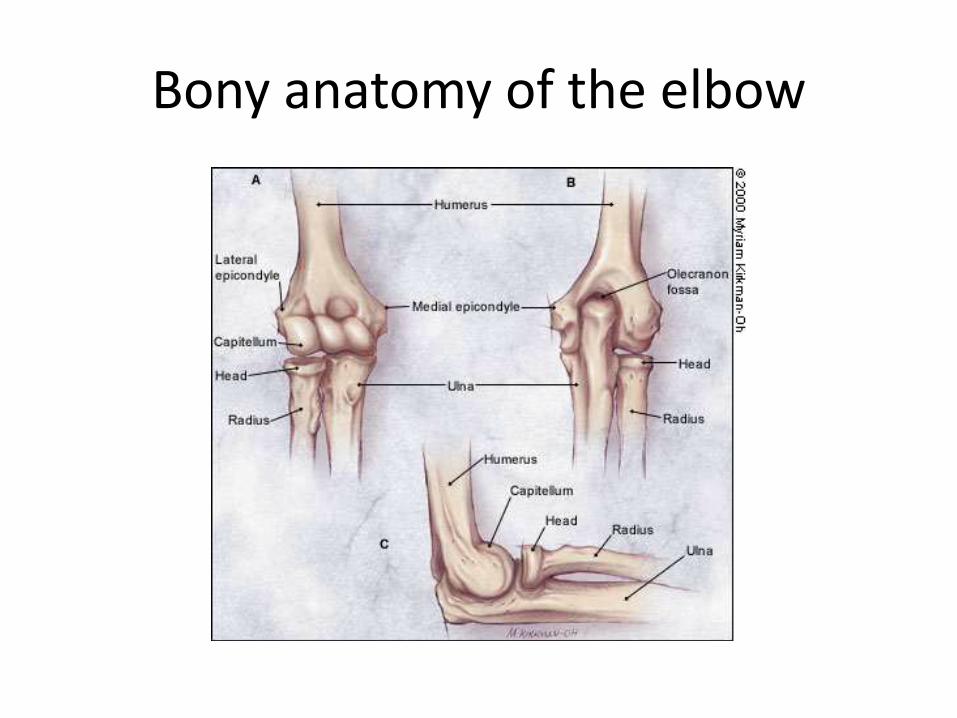
Bony anatomy of the elbow
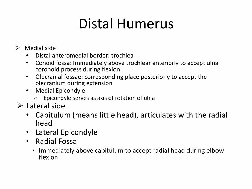
Distal Humerus
Medial side• Distal anteromedial border: trochlea• Conoid fossa: Immediately above trochlear anteriorly to accept ulna
coronoid process during flexion• Olecranial fossae: corresponding place posteriorly to accept the
olecranium during extension• Medial Epicondyle
o Epicondyle serves as axis of rotation of ulna
Lateral side• Capitulum (means little head), articulates with the radial
head• Lateral Epicondyle• Radial Fossa Immediately above capitulum to accept radial head during elbow
flexion

The Distal Humerus
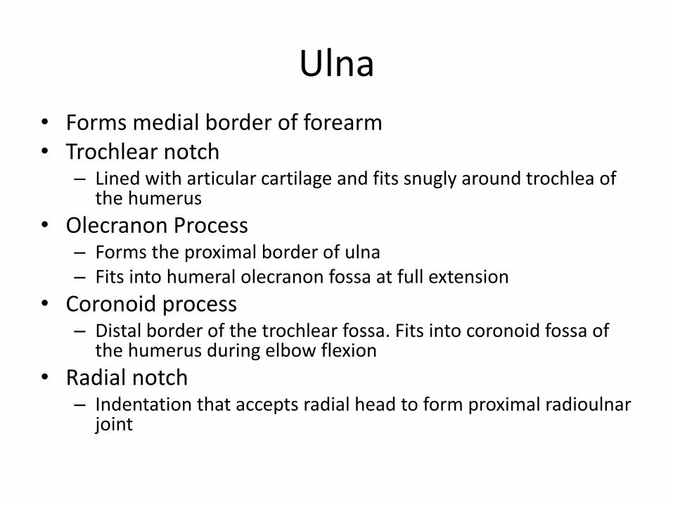
Ulna
• Forms medial border of forearm• Trochlear notch
– Lined with articular cartilage and fits snugly around trochlea of the humerus
• Olecranon Process– Forms the proximal border of ulna– Fits into humeral olecranon fossa at full extension
• Coronoid process– Distal border of the trochlear fossa. Fits into coronoid fossa of
the humerus during elbow flexion
• Radial notch– Indentation that accepts radial head to form proximal radioulnar
joint

The Proximal Ulna

Radius
• Lateral aspect of elbow when in anatomical position
• Bicipital tuberosity (radial tuberosity)
– Insertion site for bicep brachii

Joints of the Elbow
Hinge joint
• Composed of 3 articulations:
1. Humeroulnar joint
2. Humeroradial joint
3. Radioulnar joint

Humeroulnar
• Modified Hinge joint
• Allows for axis of motion:
– Flexion
– Extension

Proximal Radioulnar
• Formed by convex head of the radius and concave radial notch of the ulna
• Allows for axis of movement also:
– Pronation
– Supination

The Joint Capsule• Thin anteriorly and posteriorly
Proximal attachment
• Above coronoid and radial fossa anteriorly
• Above olecranial fossa posteriorly
Distal attachment
• Superior margin of olecranium process posteriorly
• Blends with annular ligament laterally
• Edge of the conoid process anteriorly
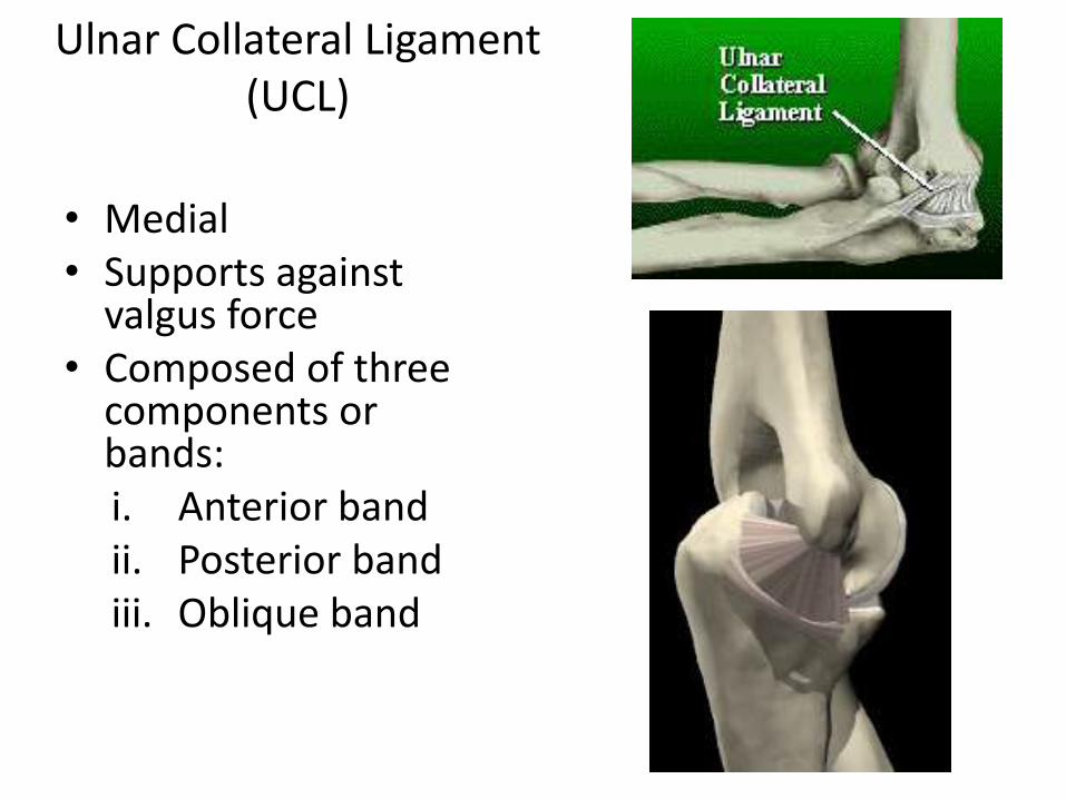
Ulnar Collateral Ligament (UCL)
• Medial• Supports against
valgus force• Composed of three
components or bands: i. Anterior bandii. Posterior bandiii. Oblique band

Radial Collateral Ligament (RCL)
• Thickened area in lateral joint capsule between the lateral epicondyle and annular ligament
• Resists varus stress
• Helps to maintain the relationship between humeral and radial head

Annular Ligament
• Permits rotation of radial head within the radioulnar articulation
• Attaches to anterior & posterior rims of the radial notch of the ulna
• Serves as attachment to radial collateral ligament
• Proximally blends with the elbow capsules

Muscles acting across the elbow
Anterior groupi. Biceps brachiiii. Brachialisiii. Brachioradialisiv. Pronator teresv. Pronator quadratusvi. Flexor carpi radialisvii. Palmaris longusviii. Flexor carpi ulnaris
Posterior groupi. Tricepsii. Anconeusiii. Supinatoriv. Extensor carpi radialisv. Extensor carpi ulnaris

Anterior Group: Biceps Brachii
• O: Long head: superior glenoid
– Short head: coracoid
• I: Radial tuberosity of the ulna
• A: Elbow flexion & supination, shoulder flexion
• N: Musculocutaneous

Anterior Group: Brachialis
• O: Anterior surface: Distal humerus
• I: Tuberosity of the ulna
• A: Elbow flexion
• N: Musculocutaneous

Anterior Group: Brachioradialis
• O: Lateral supracondylar ridge of the distal humerus
• I: Styloid process of the radius
• A: Elbow flexion
• N: Radial Nerve
Note: i. In the forearm, the brachioradialis overlies
the radial nerve and artery
ii. Developmentally, brachioradialis belongs to the extensor (Posterior) group of muscles
Brachioradialis
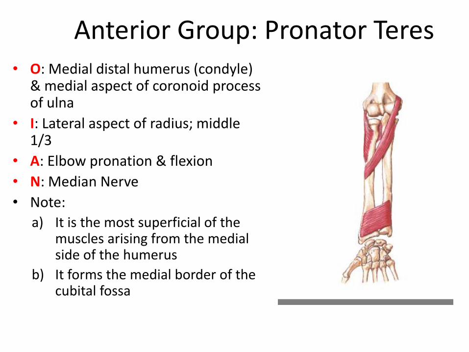
Anterior Group: Pronator Teres• O: Medial distal humerus (condyle)
& medial aspect of coronoid process of ulna
• I: Lateral aspect of radius; middle 1/3
• A: Elbow pronation & flexion
• N: Median Nerve
• Note:
a) It is the most superficial of the muscles arising from the medial side of the humerus
b) It forms the medial border of the cubital fossa
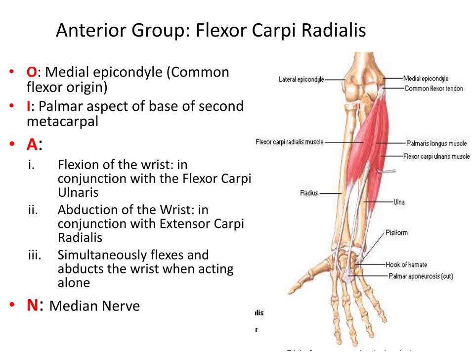
Anterior Group: Flexor Carpi Radialis
• O: Medial epicondyle (Common flexor origin)
• I: Palmar aspect of base of second metacarpal
• A: i. Flexion of the wrist: in
conjunction with the Flexor Carpi Ulnaris
ii. Abduction of the Wrist: in conjunction with Extensor Carpi Radialis
iii. Simultaneously flexes and abducts the wrist when acting alone
• N: Median Nerve

Anterior Group: Palmaris Longus
• O: Medial epicondyle (Common flexor origin)
• I: palmar aponeurosis and part of the flexor retinaculum
• A: Flexion of the wrist
• N: Median
• Note:
i. It is absent in about 14-15% of the population
ii. At the wrist, it is medial to the Median nerve

Anterior Group: Flexor Carpi Ulnaris• O: Humeral head: Medial epicondyle (Common flexor origin)
Ulna head: Olecranium
• I: Pisiform, hook of hamate and base of 5th metacarpal
• A:
i. Flexion of the wrist: in conjunction with the Flexor Carpi Radialis
ii. Adduction of the Wrist: in conjunction with Extensor Carpi Ulnaris
iii. Simultaneously flexes and adducts the wrist when acting alone
• N: Ulnar
• Note:
i. The most medial of the superficial flexor muscles
ii. The ulnar nerve enters the forearm by passing between the humeral and the ulnar heads of its proximal attachment
iii. It is the only muscle of the anterior compartment that is FULLY innervated by the ulna nerve
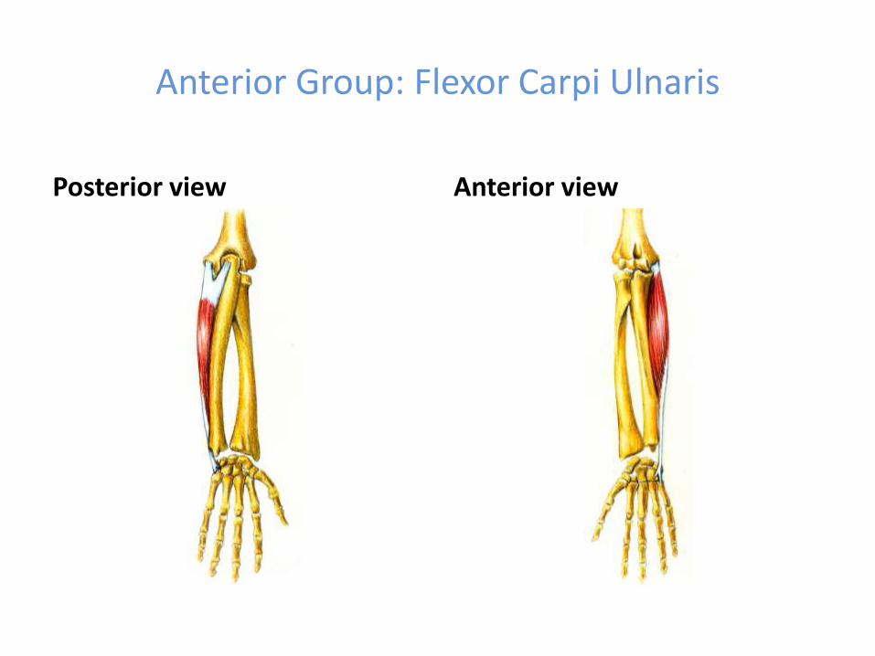
Anterior Group: Flexor Carpi Ulnaris
Posterior view Anterior view

Posterior Group: Triceps Brachii O:
i. Long head: infraglenoid tubercle of scapula
ii. Lateral head: posterior humerus, proximal to the radial groove
iii. Medial head: posterior humerus, distal to the radial groove
I: Olecranial process
A: i. Elbow extension,
ii. weak shoulder extension
iii. Supports the humeral head in shoulder abduction
N: Radial Nerve

Posterior Group: Anconeus
• O: Lateral epicondyle of the humerus
• I: Lateral surface of the olecranon
• A: Assists the Triceps in elbow extension
• N: Radial Nerve
Anconeus

Posterior Group: Supinator
• O: Superficial Head from lateral epicondyle of humerusDeep head from supinator crest of ulna
• I: Wraps round the proximal radius to be inserted on its anterior surface
• A: Supination of the forearm
• N: Radial Nerve
• NOTE:i. Deep branch of radial nerve enters the
posterior compartment by passing through the space between the two heads
ii. Forms part of the floor of the cubital fossa

Posterior Group: Extensor Carpi Radialis
O: Lateral supra epicondylar ridge
I: Dorsum of the base of 2nd
metacapal
A:
o Extension and abduction of the hand at the wrist (When acting alone)
o Pure extension of the Wrist: in conjunction with Extensor Carpi Ulnaris
o Pure abduction of the Wrist: in conjunction with Flexor Carpi Radialis
N: radial

Posterior group: Extensor Carpi Ulnaris
O: Humeral Head: Lateral epicondyle
(Common extensor origin) Ulna head: Posterior border of the ulna
through aponeurotic attachment
I: Dorsal aspect of base of 5th
metacarpal A:
Extension and adduction of the wrist(When acting alone)
Pure extension of the Wrist: in conjunction with Extensor Carpi Radialis
Pure adduction of the Wrist: in conjunction with Flexor Carpi Ulnaris
N: Radial nerve
Extensor Carpi Ulnaris

Cubital Fossa• Triangular area anterior to the elbow
and between:
o Brachioradialis muscle originating from the lateral supracondylar ridge of the humerus
o Pronator teres muscle originating from the medial epicondyle of the humerus
o Base of the triangle is an imaginary horizontal line between the medial and lateral epicondyles
• Floor is the brachialis muscle.
• Roof is formed by superficial fascia and skin

Cubital Fossa: Contents
From lateral to medial:
1. Tendon of the biceps brachii muscle
2. Brachial artery
3. Median nerve
• Crossed on the lower part by the bicipital aponeurosis
• Within the roof are:
1. Median cubital vein
2. Medial cutaneous nerve of the forearm
3. Lateral cutaneous nerves of the forearm

Cubital Fossa: Superficial contents

Elbow: Blood supply
• The elbow anastomosis is made up of 8 arteries: i. 2 Branches of Brachial artery:
Superior and Inferior ulna collateral arteries
ii. 2 branches of Profunda brachii: Radial and Middle Collateral arteries
iii. 2 Branches of Ulna Artery: Anterior and Posterior Ulna Recurrent Arteries
iv. 1 from Radial Artery: Radial Recurrent artery
v. 1 from Common Interosseus Artery: Interosseous Recurrent Artery
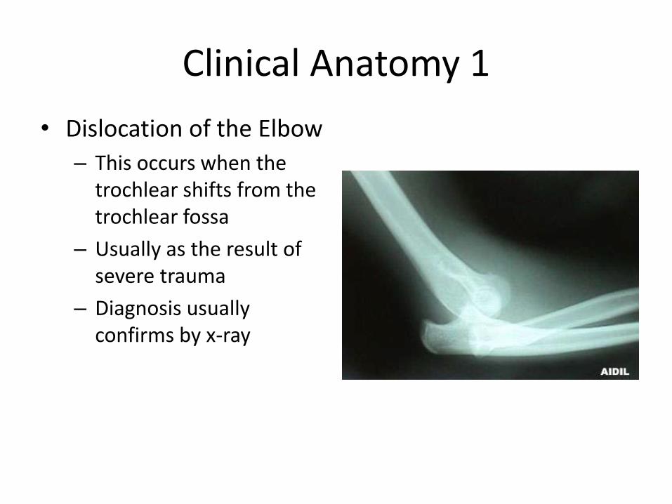
Clinical Anatomy 1
• Dislocation of the Elbow
– This occurs when the trochlear shifts from the trochlear fossa
– Usually as the result of severe trauma
– Diagnosis usually confirms by x-ray
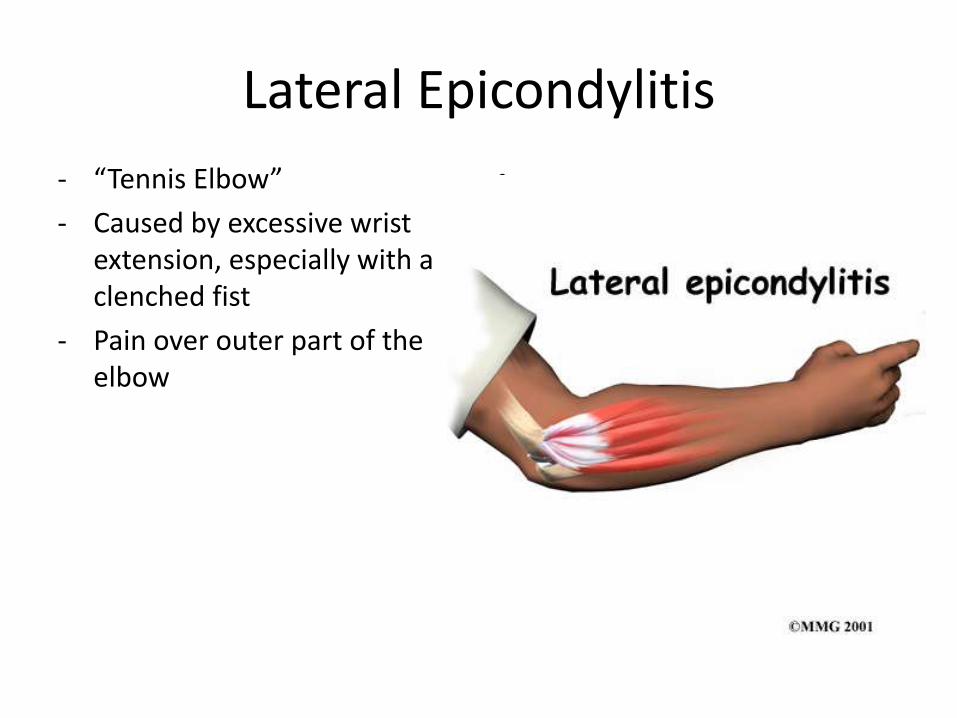
Lateral Epicondylitis
- “Tennis Elbow”
- Caused by excessive wrist extension, especially with a clenched fist
- Pain over outer part of the elbow
•

Olecranon Bursitis
• A collection of fluid in the olecranon bursa that covers the posterior tip of the elbow.
• It is the result of direct trauma to the elbow

Radial Head Dislocation (Pulled Elbow)
Radial Head Dislocation (Pulled Elbow)
• The radial head may be displaced forward, backward or outward
• Children under 5 are prone to subluxation of the radial head due to a “pulling” on the forearm
• Commonly called “pulled elbow” or “Nursemaid’s arm”

MercÍ