the emergence of cerebral asymmetries in early human · pdf file · 2010-05-11gnage...
TRANSCRIPT
THE EMERGENCE OF CEREBRAL ASYMMETRIES IN EARLYHUMAN DEVELOPMENT: A LITERATURE REVIEW AND ANEUROEMBRYOLOGICAL MODEL *
Catherine T. Bestt
Ever since Broca's century-old discovery of cerebral asymmetries in language functions, therehas been speculation about the developmental emergence of human perceptual-cognitive asymmetries. The basic question has been: Do the asymmetries first appear only at some point after birth,starting from an initial state of bilateral equivalence or symmetry at birth, or are the hemispheresinstead functionally asymmetrical from the start? A related question is whet.her functional asymmet.ries can be traced to some lat.eral bias in the structural development of the hemispheres, sucht.hat. one hemisphere matures in advance of t.he other. Thus, the term "emergence" in t.he t.itle ofthis chapt.er might. refer either to functional asymmetries in infant.s or t.o the embryological development. of t.he hemispheres. An underlying assumpt.ion of this chapter is that t.he ontogeny offunctional asymmetries is influenced by an asymmetry in the formation and physical maturat.ionof t.he cerebral hemispheres. Bot.h issues will be addressed in the following discussion, beginningwit.h a review of behavioral evidence for percept.ual-cognitive asymmetries in early infancy, andending wit.h a proposed model for a lateralizing gradient. in the neuroembryologic emergence ofthe cerebral hemispheres during prenatal development.
As for the development of functional asymmetries, Broca (1865) himself speculated that. langnage becomes lateralized to the left hemisphere during language development (see Bever, 1978).Lat.er, Samuel Ort.on (1937) expanded on this concept of "developmental lateralization" in hisinfluential theory that dyslexic children suffer from a failure to establish cerebral dominance forlanguage developmentally. More recently, Lenneberg (1967) furt.her detailed the model of development.al change in lateralization of functions, proposing a critical period for language development.,and hence for t.he progressive est.ablishment. of left-hemisphere language dominance, during t.heperiod between 2 years and puberty. More important for the present discussion, however, hismodel explicitly assumed that the child's cerebral hemispheres are equal in their capabilit.y t.oacquire language, a trait referred to as "equipotentiality," until at least 2 years of age. In fact,t.he focus on language in theoretical discussions of those times, to the exclusion of other cognitivefunctions, led to the reasoning that since infants have not yet acquired language, they should not
* To appear in S. Segalowitz & D. Molfese (Eds) Developmental implications of brain lateralization.New York: Guilford Press.
t Also Department of Psychology, Wesleyan Universit.y, Middlet.owlJ, CT 064~,7
Af.mowledgment. This work has profit.ed from discussions wit.h numerous colleagues and st.udent.s.I am particularly grateful for discussions with, and careful critiques by, Marcel Kinsbourne,Donald Shankweiler, Ursula Kirk, and Marshall Gladstone. This work was supported in part. bya grant. from NICHD t.o Haskins Laborat.ories (NIH:HD-01994) and by a Biomedical ResearchSupport Grant to t.he author t.hrough Wesleyan Universit.y.
= HASKINS LABORATORIES: Status Report all Speech Research SR-88 (1986) =
43
44 Catherine T. Best
show any hemisphere specialization. Implicit in the equipotentiality concept has been the notionthat the infant's hemispheres show functional symmetry, or lack of behavioral differentiation.
Since the mid-1970's, however, evidence of functional cerebral asymmetries in young infantshas indicated that the assumption of functional symmetry between the hemispheres in earlydevelopment cannot be correct. Generally, this literature suggests a pattern of functional cerebralasymmetries by at least 2-3 months of age, and possibly even before full-term birth, that isanalogous to the adult pattern of left hemisphere superiority for language-related functions andright hemisphere superiority for music and holistic perception of patterns and faces. The nextpart of the chapter will focus on perceptual-cognitive asymmetries in infants (for discussion ofmotoric asymmetries in infants, see the chapter by Turkewitz, this volume*), and particularlyon behavioral evidence (electrophysiological data are presented in the chapter by Molfese, thisvolume*).
Functional Asymmetries in Infants
Before the specific findings are reviewed here, some preliminary qualifications are necessary.Up to this point, many questions about infant hemispheric specialization remain unanswered. Itis not yet known, for example, whether infant asymmetries are fundamental responses to certainstimulus properties or classes, such as the physical characteristics of speech vs. nonspeech, orinstead whether they reflect different processing styles, such as feature-analysis vs. holistic processing, as has been proposed for adults. In addition, the behavioral studies are actually quite fewin number. Moreover, they have focused overwhelmingly on auditory asymmetries, particularlyfor human speech. This is due, in part, to a strong theoretical bias toward assessing languagerelated functions, but is also due to pragmatic constraints. The dichotic listening procedure ismore obviously amenable to infant research than are the lateralized behavioral measures of asymmetries in other modalities (e.g., the requirements of the visual split-field tachistoscopic techniqueare obviously not suited to infants!).
Non-auditory Asymmetries.
Thus far, only two behavioral studies of infant cognitive-perceptual asymmetries in other,non-auditory modalities have been conducted, both of which assessed right hemisphere advantagesfor pattern recognition. One of these was inconclusive regarding functional asymmetries duringinfancy; the other is not yet published. In the first, Susan Rose (1984) tested 1-, 2-, and 3year-oIds for a left-hand advantage (right hemisphere superiority) in haptic perception of shapes.After blind, unimanual palpation of a 3-dimensional nonsense shape, children were tested for crossmodal shape recognition on a visual preference task in which they saw a picture of the palpatedobject presented alongside a picture of a differently-shaped object. Although all children showedpreferences for the novel figure, and hence recognition memory for the palpated object, only the2- and 3-year-olds showed a left-hand/right-hemisphere superiority. The 1-year-olds-the onlyinfants in the study-failed to show a right-hemisphere advantage. However, as Rose argues,this cannot be taken as evidence for a lack of infant right-hemisphere specialization, because thevisual test phase of the task involved bihemispheric, or non-Iateralized, visual input. In fact, theleft-hand effects even for the older children were rather small. Perhaps some other, more sensitiveand completely lateralized test measure would detect tactile asymmetries in infants.

Emergence of Cerebral Asymmetries 45
In the other non-auditory behavioral study, Witelson and Barrera (Witelson, personal COlll
munication) tested visual asymmetries in 3-month-olds. They presented the infants with sideby-side slides of two identical photographs, both of which were either of the infant's mother,or of a female stranger, or of a standard black-and-white checkerboard pattern. The infantsshowed a fixation-time preference for the left-side photo of the mother, as well as for the left-sidecheckerboard, suggesting greater activation of their right hemispheres. However, they did notshow any side preference for the stranger. The authors' interpretation was that both mother andcheckerboard constituted "gestalt" patterns to the infants, which they processed holistically, thusshowing a right-hemisphere bias in activation. In contrast, the female stranger was not processedas a gestalt, and thus not handled preferentially by the right hemisphere. This argument, atleast with respect to the infants' responses to mother vs. stranger, is consistent with developmental research on face recognition in children (Levine, 1985). Young children perceive unfamiliarfaces in terms of salient features rather than holistically and show no hemispheric asymmetryfor recognition of those faces. However, the same children do perceive familiar faces holistically,as well as showing a right hemisphere advantage for the familiar faces. Thus, the Witelson andBarrera results offer some suggestion of a right-hemisphere bias in holistic perception of faces(and patterns) by 3 months, which is compatible with the literature on visual asymmetries inadults. However, this suggestion must be viewed as still tentative, given that is is based only ona single, unpublished finding.
Auditory aSylllluetries
By comparison, the behavioral studies of auditory asymmetries have been more numerous,and have included assessments of both left- and right-hemisphere specialization in infants. Thetechnique used is some modification of the dichotic listening procedure. In the first such study,reported at a conference held at Brock University in 1975, Anne Entus (1977) used a non-nutritivesucking measure with a dichotic habituation-dishabituation procedure. Two groups of infants,who averaged 2kmonths in age, were tested for ear differences in discrimination of either musicalnotes played by different instruments, or of consonant differences in speech syllables. In the firstphase of each test, the infants heard a rapidly repeated presentation of a dichotic pair of stimuliuntil they reached a criterion of habituation. At that point, the element in either the right orleft ear was changed to, respectively, a new music note or syllable, while the other ear continuedto receive its original habituation stimulus. The infants in the speech condition showed a greaterrecovery of the sucking response when the syllable changed in the right ear (REA) than when itchanged in the left, indicating left-hemisphere superiority. The music group showed the oppositepattern, a left-ear (LEA) or right-hemisphere advantage. However, Vargha-Khadem and Corballis(1979) subsequently failed to replicate with 2-month-olds the speech REA that Entus found, apoint to which we will return later in the chapter. In this later study, the infants discriminatedthe speech syllable change equally well with both ears.
In a similar dichotic habituation study with 3-month-olds, Glanville, Best, and Levenson(1977) used the heart rate measure of a deceleratory orienting response, reflecting interestedattention to a stimulus, in order to introduce a memory component to the task. Friedes (1977)has presented evidence that, in adults, memory retrieval is more strongly associated with dichoticear asymmetries than is a simple input processing dominance. Therefore, the intervals betweenpresentations of the dichotic pairs in the Glanville et al. test were long enough (M = 2.5sec)
that the infants had to rely on short term memory in order to learn the habituation pair andto recognize the stimulus change on the test trial. In each test block, the habituation pair was

46 Catherine T. Best
8.0
7.0
po-
0 8.0
...I
'"0.0
c
~4.0
;; -.. 3.0...c:0
'"~
2.0
0U
~
a: 1.0
a::x:(;
no ch.~... c: :: :;:" gjl .: ,"c i0 -1.0., :... r: ~ ,.
E " r....."..
Ear tested: LEFT RIGHT LEFT RIGHT
DiscriminationCondition: MUSIC SPEECH
Figure 1. Ear differences in 3-month-olds' discrimination of speech syllables differing in initial consonant, and
of music notes differing in instrument timbre. The task was a dichotic habituation-dishabituation task using
heart rate deceleration as the response measure; represented here is the magnitude of dishabituation (cardiac
deceleration) on the test trial (stimulus change in either right or left ear), relative to the cardiac response on the
last habituation trial (trial 9). Redrawn from data reported in Glanville, Best, and Levenson (1977).
presented nine times, and the stimulus change was then presented on the tenth and final trial. Allinfant.s received separate left and right-ear discrimination test blocks each for speech syllables andfor music notes. The results provided converging evidence with Entus' findings for an adultlikepattern among 3-month-olds of REA in response to speech syllable changes, and LEA in responseto music changes (see Figure 1).
These first dichotic studies still left several important questions, two of which were addressedby subsequent research with infants. First, there have been two attempts to obtain a better specification of the speech properties to which the infant's left hemisphere is preferentially responsive.Second, age changes in behavioral evidence of auditory cerebral asymmetries during infancy havebeen assessed.
The basis of left hemisphere speech specialization. For a more detailed understandingof the infant's left-hemisphere response to speech, Best (1978) used the dichotic heart rate habituation procedure to determine whether 3~-month-olclsshow different patterns of ear asymmetriesfor vowel vs. consonant discriminations. Several studies with adults had suggested that the REAfor speech perception is greatest for consonant perception, while there is often a weaker or absentear advantage for vowel perception (e.g., Darwin, 1971; Studdert-Kennedy & Shankweiler, 1970;Weiss & House, 1973). This pattern may be related to the fad that consonants involve rapidly

Emergence of Cerebral A~ymmetrie~ 47
8.0
7.0
0 8.0..I
'"0.0
!:
~4.0
;;.. 3.0..r:0
~ 2.0..>0
"e0:
1.0
0:J:
"0.. c:." g=c :!C> -1.0 !!.. ..E ""..
Ear tested:
DiscriminationCondition:
LEFT RIGHT
VOWELS
LEFT RIGHT
CONSONANTS
Figure 2. Ear differences in 3 }-month-olds' discrimination of vowels and of consonants in computer-synthesized
syllables. The method and response measure are the same as described for Figure 1. Based on data presented in
Best (1978).
cha.nging acoustic properties, whereas vowels are associat.ed wit.h much more slowly-changing, oreven st.eady-st.at.e, acoustic propert.ies (see Cutting, 1974; Schwartz & Tallal, 1980). Therefore,a set of computer-synthesized syllables were developed, which exaggerated the rapidly changingacoust.ic properties vs. steady-state characteristics associated with consonants vs. vowels. Theresults revealed a REA for discrimination among the exaggerated consonants, consist.ent. bot.hwit.h t.he earlier infant st.udies and wit.h adult findings. However, t.he infant.s showed an LEA forst.eady-st.at.e vowel discrimination (see Figure 2), unlike adults.
These findings suggest.ed t.hat. the infant. 's left. hemisphere may be particularly responsive t.orapidly-changing acoust.ic information, while t.he right hemisphere is more responsive t.o steadyst.at.e spectral informat.ion. The vowel LEA is compat.ible wit.h John Sidt.is' (1980) findings ofa right.-hemisphere advant.age in adults' percept.ion of st.eady-st.ate harmonic information. Thelack of an adult ear advant.age for vowels suggest.s t.hat t.his st.eady-st.at.e information may beeasily t.ransferred across t.he corpus callosum; t.he left.-ear advantage in infants may be due t.ot.he immat.urit.y of t.heir corpus callosa (see also Molfese, Freeman, & Palermo, 1975; Molfese &Molfese, 198.5; St.uddert.-Kennedy & Shankweiler, 1980).
MacKain, St.uddert.-Kennedy, Spieker, and Stern (1983) further explored the nat.ure of t.heinfant.'s left. hemisphere specialization for speech percept.ion, in a bimodal-mat.ching st.udy wit.h

48 Cathel'ine T. Best
5-6-mont.h-olds. The infant.s viewed t.wo side- by-side synchronous video films of a woman repeat.ing two different 2-syllable nonsense words, while they simultaneously heard a synchronousaudio recording (over a centrally-located loudspeaker) that. corresponded to one of the two videodisplays. Infant.s det.ect.ed t.he cross-modal equivalence, as indicated by a looking preferencefor the film that. mat.ched the audio presentation, but. only when the correct video was in t.heright.-side video monit.or. This finding implies selective left-hemisphere activation, and suggests aleft-hemisphere specializat.ion for perception of the common underlying art.iculatory pattern t.hatproduced the disparate informat.ion in the two sensory modalities.
Toget.her, t.hese t.wo st.udies on cerebral asymmetries for t.he propert.ies of speech suggest. that.the infant. 's left hemisphere may be specialized for recognizing articulatory patterns in speech, andpart.icularly t.he rapid acoustic changes resulting from the dynamic art.iculatory gest.ures that. produce consonant. sounds. However, t.his still leaves open the quest.ion "Why the left hemisphere?"One possibilit.y is a lat.eralized gradient in the maturation of the two hemispheres.
Lateral differences in hemisphere maturation? If t.here is a lat.eralized developmentalgradient, uncert.ainty st.ill remains as to whet.her the asymmetry in speech perception wouldresult from earlier or lat.er development of t.he left hemisphere relative to the right. Broca (1865)proposed a left-to-right gradient to explain language lateralization (see Bever, 1978); recently,Corballis and Morgan (1978) seconded the notion of a left-right gradient. However, Taylor (1969),Crowell, Jones, Kapuniai, and Nakagawa (1973), and Brown and Jaffe (1975) have argued for aright-to-left gradient, which is also suggested by recent embryologic evidence that cortical fissuresappear consistently earlier in the right than the left fetal hemisphere (Dooling, Chi, & Gilles,1983).
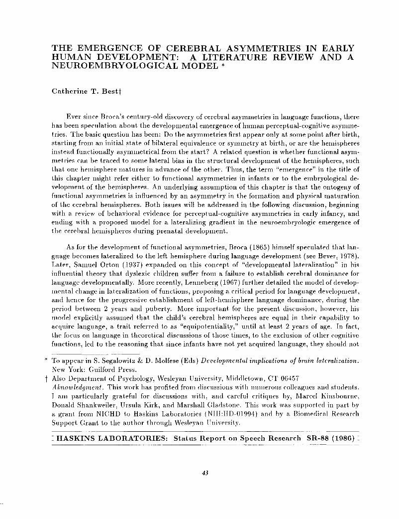
To test. the possibility of early age changes in asymmetrical function, Best, Hoffman, andGlanville (1982) tested for ear asymmetries in memory-based discriminations of speech syllablesvs. music notes by 2-, 3-, and 4-month-old infants. The 3- and 4-month-olds replicated the earlierfindings of a REA/left-hemisphere advantage for speech and LEA/right.-hemisphere advantagefor music. However, t.he 2-month-olds showed only the LEA for music; they did not detectthe speech syllable change in either ear (see Figure 3). These results suggest an increase infunctional maturity of the left hemisphere sometime between 2 and 3 months of age, at least forauditory discriminations that depend on short-term memory capacities. Such a change in corticalmaturity around 2-3 months of age is consistent with report.s of widespread biobehavioral changesand maturation of cortical influences over behavior around that time (Emde & Robinson, 1979).
This finding may also help explain the negative report by Vargha-Khadem and Corballis(1979), which had failed t.o replicate findings by Ent.us (1977) of a speech REA in infants. AIt.hough t.heir infant subjects discriminated t.he syllable change in bot.h ears, discriminat.ion underthe rapid stimulus presentat.ion conditions used in t.he sucking habit.uation procedure clearly doesnot. depend solely on cort.ical involvement, since Frances Graham and her colleagues (Graham,Leavitt, Strock, & Brown, 1978) have found similar speech discrimination in a 6-week-old anencephalic infant. The conclusion of Best. et. 801. (1982) was t.hat t.he speech-specialized functionof t.he left hemisphere may be insufficiently mat.ure at. 2 mont.hs to control behavioral responsesin a memory-dependent. discrimination task. In contrast., the analogous right hemisphere funct.ion appears sufficiently mat.ure at that age to effect a LEA for memory-based music timbrediscrimination, suggest.ing a right-to-left gradient in the mat.urat.ion of asymmetrical perceptualmemory functions. This does not necessarily imply that cerebral lateralization itself develops
Emergence of Cerebral Asymmetries 49
5.0 TWO-MONTH-OLDS THREE -MONTH OLDS FOUR MONTH-OLOS
(- -.I II II II II II II II II II I
I_~~ l
t~:J
Ij
a:°OO~-~"""""u!~~ -10
~-3$2~
c02....•>8 10~
L R L R L R L R L R L R
MUSIC SPEECH MUSIC SPEECH MUSIC SPE ECH
Figure 3. Age changes in ear differences for infants' discrimination of speech syllables and of musical timbre.
Method and response measure are identical to the description of Figure 1. Reprinted with publisher's permission,
from Best, Hoffman, and Glanville (1982), Perception f3 P~ychophy~ic~, 31, 75-85. Copyright (01982 by the
Psychonomic Society.
out. of an unlat.eralized substrate. Alternat.ively, cognit.ive and percept.ual functions may mat.uredevelopment.ally at. different. rates, but within t.he context of a neural substrat.e that is alreadylaterally-specialized from the start (see Witelson, 1977, 198.5; see also Kinsbourne, 197.5).
If the latter view is correct, the question becomes: "What is the source of this lateralizedgradient in functional maturation?" According to developmental biologists, morphologists, andparticularly neuroembryologists, the patterns of embryologic development are ultimately responsible for the structure and form of the adult. organism, including the brain, both at the grossmorphological level and at the histological level. Given the basic neuropsychological assumptionthat variations in neuronal organization and development affect behavior and its development,then, fetal brain development should provide evidence of a lat.eralized developmental gradient. Inthe remainder of t.his chapt.er it. will be argued that a right-to-Ieft. gradient in postnatal fundionalmat uration parallels a similar gradient in the prenatal, embryologic development of the cerebralhemispheres.
50 Catherine T. Best
Proposal for a Lateralized Gradient in N euroembryologic Development
Gross Morphological Asymmetries
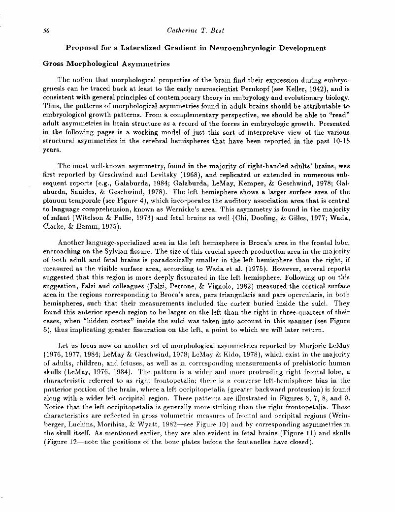
The notion that morphological properties of the brain find their expression during embryogenesis can be traced back at least to the early neuroscientist Pernkopf (see Keller, 1942), and isconsistent with general principles of contemporary theory in embryology and evolutionary biology.Thus, the patterns of morphological asymmetries found in adult brains should be attributable toembryological growth patterns. From a complementary perspective, we should be able to "read"adult asymmetries in brain structure as a record of the forces in embryologic growth. Presentedin the following pages is a working model of just this sort of interpretive view of the variousstructural asymmetries in the cerebral hemispheres that have been reported in the past 10-15years.
The most well-known asymmetry, found in the majority of right-handed adults' brains, wasfirst reported by Geschwind and Levitsky (1968), and replicated or extended in numerous subsequent reports (e.g., Galaburda, 1984; Galaburda, LeMay, Kemper, & Geschwind, 1978; Galaburda, Sanides, & Geschwind, 1978). The left hemisphere shows a larger surface area of theplanum temporale (see Figure 4), which incorporates the auditory association area that is centralto language comprehension, known as Wernicke's area. This asymmetry is found in the majorityof infant (Witelson & Pallie, 1973) and fetal brains as well (Chi, Dooling, & Gilles, 1977; Wada,Clarke, & Hamm, 1975).
Another language-specialized area in the left hemisphere is Broca's area in the frontal lobe,encroaching on the Sylvian fissure. The size of this crucial speech production area in the majorityof both adult and fetal brains is paradoxically smaller in the left hemisphere than the right, ifmeasured as the visible surface area, according to Wada et a1. (1975). However, several reportssuggested that this region is more deeply fissurated in the left hemisphere. Following up on thissuggestion, Falzi and colleagues (Falzi, Perrone, & Vignolo, 1982) measured the cortical surfacearea in the regions corresponding to Broca's area, pars triangularis and pars opercularis, in bothhemispheres, such that their measurements included the cortex buried inside the sulci. Theyfound this anterior speech region to be larger on the left than the right in three-quarters of theircases, when "hidden cortex" inside the sulci was taken into account in this manner (see Figure5), thus implicating greater fissuration on the left, a point to which we will later return.
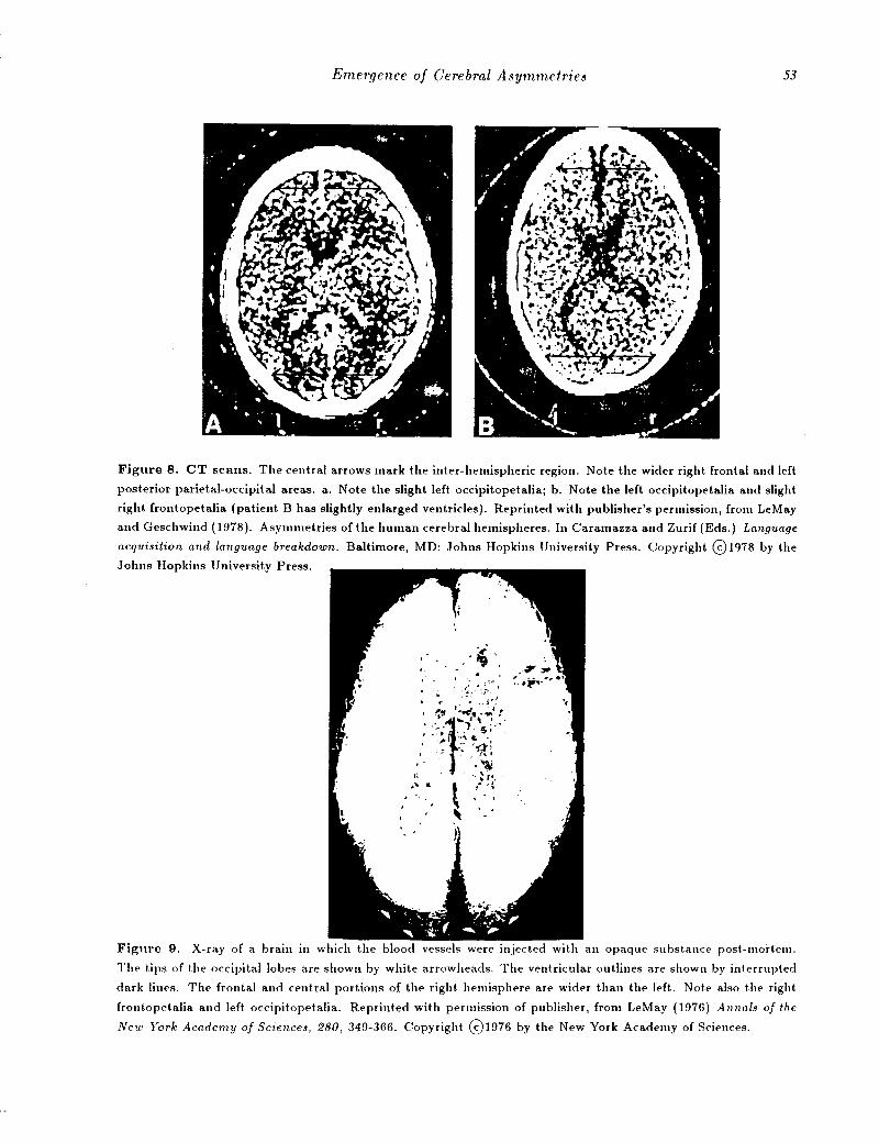
Let us focus now on another set of morphological asymmetries reported by Marjorie LeMay(1976, 1977, 1984; LeMay & Geschwind, 1978; LeMay & Kido, 1978), which exist in the majorityof adults, children, and fetuses, as well as in corresponding measurements of prehistoric humanskulls (LeMay, 1976, 1984). The pattern is a wider and more protruding right frontal lobe, acharacteristic referred to as right frontopetalia; there is a converse left-hemisphere bias in theposterior portion of the brain, where a left occipitopetalia (greater backward protrusion) is foundalong with a wider left occipital region. These patterns are illustrated in Figures 6, 7, 8, and 9.Notice that the left occipitopet.alia is generally more striking than the right frontopetalia. Thesecharacteristics are reflected in gross volumetric measures of frontal and occipital regions (Weinberger, Luchins, Morihisa, & Wyatt, 1982-see Figure 10) and by corresponding asymmetries inthe skull itself. As mentioned earlier, they are also evident in fetal brains (Figure 11) and skulls(Figure 12-note the positions of the bone plates before the fontanelles have closed).

Emergence of Cerebral Asymmetries 51
A A LEF T RIGHT
Figure 4. Anatomical analyses of two brains are shown here, illustrating the left hemisphere bias III size of
planum temporale. Coronal sections are shown in columns A and Bj superior temporal planes in C and D showing
planum temporal (P) and Heschl's gyrus (H); and lateral surfaces of brain showing area Tpt (shaded) in E and F.
Arrowheads point to Sylvian fissures. Note large asymmetry in Tpt and P. Also note buried temporal cortex on
coronal sections (arrows). Reprinted with permission of senior author and publisher, from Galaburda, Sanides, and
Geschwind (1978), Archives of Neurology, 35, 812-817. Copyright 01978 by the American Medical Association.
p~1 >-<--:.'! <), /
Figure 5. "Entire" anterior speech region (defined as pars opercularis and pars triangularis of the third frontal
convolution), and its right hemisphere homologue, shown here superimposed within the polygons on the surface
of both hemispheres. The figure illustrates one of the twelve right-handed cases reported in Falzi, Perrone, and
Vignolo (1982). The authors measured both the extrasulcal and intrasulcal cortex of these regions (the polygons
superimposed on the hemispheres show the sections made in order to measure intrasulcal cortex). There was more
intrasulcal cortex found in the left hemisphere in 3/4 of their cases, indicating greater fissuration on the left than
the right hemisphere. Reprinted with permission of the authors and publisher, from Falzi, Perrone & Vignolo
(1982), A I'chives of Neurology, 39, 239-240. Copyright 01982 by the American Medical Association.

52 Catherine T. Best
Figure 6. Horizontal section of an adult brain, exposing the plana temporale (P) and Heschl's gyri (H). Note
both the larger planum on the left, and the right frontopetalia and left occipitopetalia. Reprinted with publisher's
permission, from Witelson (1977), Annal! of the New York Academy of Science!, 299, 328-354. Copyright 01977
by the New York Academy of Sciences.
Figure 1. a. Diagram of sections taken through the brain during routine examination by x-ray computerized
axial tomography (CT)j b. CT scan though section Aj c. CT scan though section B. Note the left occipitopetalia
and wider right frontal region. Reprinted with publisher's permission from LeMay (1976), Annal! of the New York
Academy of Science!, 280, 349-366. Copyright 01976 by the New York Academy of Sciences.
Emergence of Cerebral Asymmetries 53
Figure 8. CT scans. The central arrows mark the inter-hemispheric region. Note the wider right frontal and left
posterior parietal-occipital areas. a. Note the slight left occipitopetaliaj b. Note the left occipitopetalia and slight
right frontopetalia (patient B has slightly enlarged ventricles). Reprinted with publisher's permission, from LeMay
and Geschwilld (1978). Asymmetries of the human cerebral hemispheres. In Caramazza and Zurif (Eds.) Language
acquisition and language breakdown. Baltimore, MD: Johns Hopkins University Press. Copyright 01978 by the
Johns Hopkins University Press.
Figure 9. X-ray of a brain in which the blood vessels were injected with an opaque substance post-mortem.
The tips of the occipital lobes are shown by whHe arrowheads. The ventricular outlines are shown by interrupted
dark lines. The frontal and central portions of the right hemisphere are wider than the left. Note also the right
frontopet.alia and left occipitopetalia. Reprinted wHh permission of publisher, from LeMay (1976) Annals of the
New York Academy of Sciences, 280, 349-366. Copyright 01976 by the New York Academy of Sciences.

54 Catherine T. Best
Figure 10. Sagittal section of the ad'ult brain. Stippled areas were measured volumetically. The anterior region
was larger on the right, and the posterior region was larger on the left, in the majority of brains studied (taken
from the Yakovlev collection). Reprinted with permission of the publisher, from Weinberger, Luchins, Morihisa,
and Wyatt (1982) Annal~ of Neurology, 11,97-100. Copyright @1982 by Little, Brown & Co.
Figure 11. Photograph of superior surface of a 32-week-old fet.al brain showing a slight. right. front.opet.alia
and a more st.riking left occipitopet.alia. Reprinted wit.h permission of publisher, from LeMay and Geschwind
(1978). Asymmetries of the human cerebral hemispheres. In Caramazza and Zurif (Eds.) Language acquisi.tion
and language breakdown. Baltimore, MD: Johns Hopkins University Press. Copyright @1978 by Johns Hopkins
University Press.

Emergence of Cerebral Asymmetries 55
Figure 12. a. Fetal skull. The bone over the right frontal region, and the coronal suture, forehead, and lower
rim of the orbit are farther forward than on the left side. The vault extends slightly more posteriorly on the
left. b. Upper surface of the skull of a young fetus. The fetus probably had hydrocephalus, but again note the
forward position of the right frontal region, the posterior extension of the left hemisphere beyond the right, and the
positions of the bony islands of the developing vault. Reprinted with permission of publisher, from LeMay (1984).
Radiological, developmental, and fossil asymmetries. In Geschwind and Galaburda (Eds.), Cerebral dominance:
The biological foundations. Cambridge, MA: Harvard University Press.
Morphologic asymmetries as evidence of a lateralized neuroembryologic gradient.The argument put forth in this chapter is that these morphological asymmetries can be read asa record of a right-to-left gradient in the embryologic emergence of the cerebral hemispheres.This proposal depends on several considerations: First, the brain develops in a general anteriorto-posterior direction. Second, this general gradient is complicated by interaction with otherdevelopmental gradients, in the ventro-dorsal and primary ----+ secondary----+ tertiary dimensions. 1
Third, it assumes (based on reasoning and some indirect evidence to be present.ed a bit later) thatearlier onset in the formation and growth of a given region of telencephalon will strongly tend t.oresult. in a larger volume of that region (e.g., greater hemispheric width, but not necessarily a larger
1 There is also a general mediolateral gradient., but. Jacobson (1978) states that it is lessconsistent. than the other gradients, i.e., in nUlllerous regions it reverses to a lat.eromedial gradientor else there is no obvious gradient in either direction along this dimension. Furthermore, it isdifficult. to conceptualize a vector of greater than 3 dimensions cutting through time without somevisualization aid such as computer animation modeling. Therefore, the mediolateral dimensionwill not be considered furt.her in this discussion of t.he proposed model for hemisphere growth.

56 Catherine T. Best
measurement. of cortical surface area or gray matter), relat.ive t.o the homologous contralateralregion. This hypothesis of a right-to-left growth gradient is consistent with recent evidence thatin fetal development, the major (primary region) fissures appear 1-2 weeks earlier on the righthemisphere than on the left (Dooling, et aI., 1983).
The current proposal differs in one crucial respect from earlier proposals of a right-to-left orleft-to-right gradient in brain growth. The earlier models assumed or implied that t.he earliermat.uring hemisphere would show advanced development. over the ot.her hemisphere th1'Oughoutits extent. If we combined that assumption wit.h the assumption stated in the previous paragraphthat. there should be greater volume for earlier-emerging regions, the resulting prediction wouldbe that the earlier-emerging hemisphere should end up larger overall, which is simply not thecase. In fact, t.he simple-minded lateral gradients of earlier models would have to posit some sortof post.-hoc explanation (like Ptolemy's planetary epicycles) for the fact of larger right-hemispherevolume in the frontal regions but larger left-hemisphere volume in the posterior regions.
The present proposal refers t.o a dynamic, developmental gradient in the lateral right.-t.oleft axis of the embryo; that is, it refers to a shift over time from right to left. The proposalalso takes int.o account the fact that there are growth gradients along the other main axes ofembryologic development, i.e., that emergence of the hemispheres takes place over time in 3dimensional space. The right-to-left gradient is only one of several axial growth gradients, andits influence on morphological asymmetries can only be understood in the context of the othergradients. In other words, we need to conceptualize a growth vector cutting through at least threedimensions over time; this vector represents a wave of leading g1'Owth activity. There are actuallythree other developmental gradients that. should be accounted for in this hypothesized growthvector (see Figure 13 for general reference on human fetal brain growth). The anteroposteriorgradient refers to the general direction of growth from the front.al region toward the occipitalregion (e.g., Gilles, Leviton, & Dooling, 1983). This gradient, however, is complicated by agrowth gradient that moves in t.he following direction: from primary motor and sensory zones tosecondary association areas, and finally t.o tert.iary association zones. This is important to keepin mind, because although the motor and premot.or zones of the frontal lobe are early-emerging,the forward extension of the p1'efrontal area is one of the last. developments of the hemispheres,and is a tertiary association area (e.g., Rabinowicz, 1979; Yakovlev & Lecours, 1967). The thirddevelopmental gradient to consider is the ventrodorsal gradient (Jacobson, 1978), from basalregions toward upper or superior regions. The ventrodorsal gradient is distorted, however, inhemispheric development by the fact that the hemispheres develop radially around the core ofthe basal ganglia and the insula (considered t.o be the basal or floor region of t.he hemisphere),moving in an inverted C-shaped direction, folding down and under around the back of the headand then turning forward t.o form, respectively, the occipit.al and temporal poles.
The resulting prediction of a 3-D growth vector starts with an earlier emergence of rightprimary motor (and premotor) and sensory regions t.hat. lie more frontal and ventral (initially, atleast). With concurrent developmental shift.s along all gradient.s, t.he advancing wave ofthe growthvector would then proceed t.oward earlier emergence of t.he left side for tert.iary association regions(including Wernicke's area) that lie more dorsal and posterior (e.g., superior parietal), again atleast initially (but recall the late and presumed left.ward bias in protrusion of prefront.al regions).At some point midway between these extremes, growth should reach equilibrium between the twosides (possibly in secondary association areas).

Emergence of Cerebral Asymmetries 57
Figure 13. Developing human brain, seen from the left side in a succession of embryonic and fetal stages. The
illustrations for the bottom two rows are approximately 4/5 life size, and drawn to scale. Those in the top row
are enlarged to show structural details; the insets show ~ life size scale. Reprinted with permission of publisher,
from Cowan (1979), Scientific American, 241, 112-133. Copyright 01979 by Scientific American, Inc. All rights
reserved.
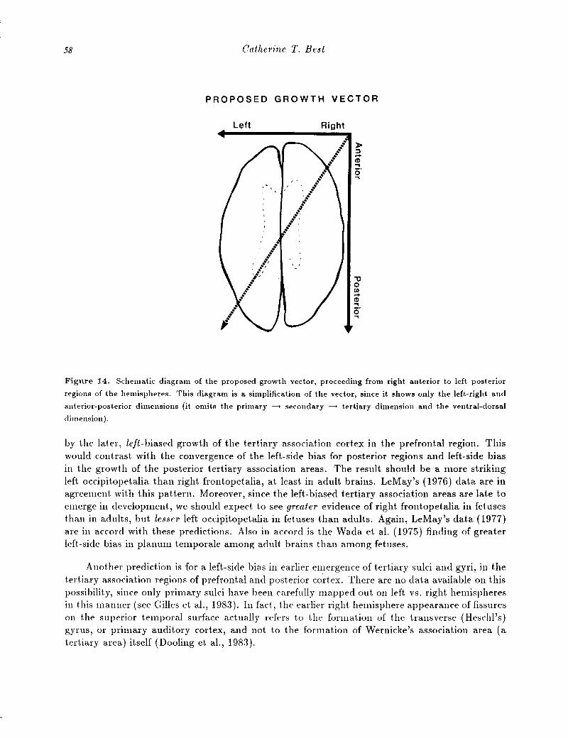
The effect of this growth vector is a counterclockwise torque evident in the shape of thedeveloping as well as the adult brain. This can only be illustrated here in two dimensions at atime. Figure 14 shows a brain viewed from above (from LeMay, 1976), to illustrate the combinedinfluences of the anteroposterior and right-left gradients, producing a counterclockwise torque, asthough some force had molded the brain with a fore-to-aft twist on the left, concurrent with anopposing twist on the right. The counterclockwise torque resulting from the combined effects ofthe ventrodorsal and right-left gradients is seen in coronal views of fetal brains (from Dooling,Chi, & Gilles, 1983), as seen in Figure 15 (easiest to view in the 34-week brain at top).
The overall effect on the hemispheres is as though some force had twisted the left hemisphererearward and dorsal, while twisting the right hemisphere forward and ventral. LeMay's (1984)observations of asymmetries in the positions and angles of the central (Rolandic) fissure and theSylvian fissure are consistent with this image-the Rolandic fissure appears farther forward (andtilted more vertically) on the right hemisphere, even in fetal brains, whereas the Sylvian fissureslants more horizontally (i.e., lower) on the left, with a lower and more posterior endpoint (Sylvianpoint). The right Sylvian fissure angles more sharply upward, and has a more anterior endpoint.
This model of embryologic hemisphere development leads to several predictions. First, thegross morphologic effect of earlier-emerging right frontal-motor regions may become attenuated
58 Catherine T. Best
PROPOSED GROWTH VECTOR
Left Right
."oCIl...CD~.o..,
Fignre 14. Schematic diagram of the proposed growth vector, proceeding from right anterior to left posterior
regions of the hemispheres. This diagram is a simplification of the vector, since it shows only the left-right and
anterior-posterior dimensions (it omits the primary ----+ secondary ----+ tertiary dimension and the ventral-dorsal
dimension) .
by t.he lat.er, left-biased growt.h of the t.ertiary associat.ion cort.ex in t.he prefront.al region. Thiswould contrast. wit.h t.he convergence of the left-side bias for posterior regions and left-side biasin t.he growt.h of t.he posterior tertiary associat.ion areas. The result should be a more strikingleft occipit.opetalia than right. frontopetalia, at least. in adult brains. LeMay's (1976) data are inagreement. wit.h t.his pattern. Moreover, since t.he left-biased t.ert.iary association areas are late t.oemerge in development., we should expect t.o see greater evidence of right. front.opet.alia in fetusest.han in adults, but. lesser left occipit.opetalia in fetuses than adults. Again, LeMay's dat.a (1977)are in accord wit.h these predictions. Also in accord is t.he Wada et a1. (1975) finding of greaterleft.-side bias in planum t.emporale among adult brains than among fetuses.
Anot.her prediction is for a left-side bias in earlier emergence of t.ert.iary sulci and gyri, in thet.ert.iary associat.ion regions of prefront.al and post.erior co·rt.ex. There are no data available on t.hispossibilit.y, since only primary sulci have been carefully mapped out. on left vs. right hemispheresin t.his manner (see Gilles et aI., 1983). In fact, t.he earlier right. hemisphere appearance of fissureson the superior t.emporal surface actually refers to the formation of t.he transverse (Heschl's)gyrus, or primary auditory cortex, and not t.o the formation of Wernicke's association area (atertiary area) it.self (Dooling et aI., 1983).

Emergence of Cerebral Asymmetries
36wh
39 wko
59
Figure 15. Tracings of photographs of sections in frontal plane, of representative fetal brains of different ges
tat.ional ages. Reprinted with permission of publisher, from Gilles, Leviton, and Dooling (1983), The developing
human brain. Boston: John Wright PSG Inc.
Effect of the Growth Vector on Neuronal Organization
The model also carries implicat.ions for asymmetries in neuronal organization of cortical areas,and hence for functional development and plasticity of the various regions on the two sides. Theimpact. of the growth vector upon neuronal organization must be understood in the context ofthe sequent.ial development of the six layers of neocortex and their differing contributions to thedevelopment of cortical fissuration and gyration, that is, the development. of cortical folding. Thefive cort.ical layers that. contain actual cell bodies of neurons develop in an inside-out sequence(Figure 16), with the cells of layer 6, t.he deepest inner layer, reaching their target positions anddeveloping dendritic and axonal connections earliest (e.g., Rakic, 1980). The earliest-developinglayers, .5 and 6, contain primarily efferent cells projecting to regions outside of cortex per se, andso give rise to long, myelinated axons, that is, subcortical white matter. Layer 4 is also directlyassociated with subcortical white matter, because in most. regions of cortex it is the layer thatreceives initial, primary input from afferents to cortex. That is, its cells synapse with incominglong, myelina.ted axons that arise largely from subcortical areas or from other, relatively distantcort.ical regions. Thus, the lower layers 4-6 of cortex contribute disproportionately to the "whitematter" side of regional measurements of ratios of gray matter (aggregated neural cell bodiesand short unmyelinated axons within the cortical mantle itself) to white matter (subcorticalmyelinated axon bundles) (e.g., Gur et al., 1980; Meyer et al., 1978; McHenry et al., 1978). Fort.hose areas with relatively higher proportions of subcortical white matter (low gray/white ratio),there should be a tendency toward greater width and/or volume than in areas with a higher

60 Catherine T. Best
[40 E~O EW [10 (eo £90 [00 EN:) EIlO (,JoO [><00 EM [160 !lRTH
Figure 16. Diagrammat.ic represent.at.ion of heavily labeled neurons in t.he visual cort.ex of juvenile monkeys t.hat.
had been injected with [3 H]-TdR at. selected embryonic days. The t.op picture shows Brodman's area 17, t.he
boHom shows area 18. On t.he left. of each diagram is a drawing of cort.ex sections with cresyl-violet staining, with
Brodman's layers indicated in Roman numerals. WM: white matter, LV: lateral ventricles. Embryonic days (E)
are shown on the X axis, start.ing with the end of the first fetal month (E27) and ending at t.erm (E165). The
vertical lines represent the embryonic days on which subsets of t.he animals received a pulse of [3 H]-TdR. On each
vertical line short horizontal markers indicate t.he positions of all heavily labeled neurons. Since [3 H]-TdR labels
the cells undergoing mitosis at the time of injection, these diagrams indicate that cortex is built in inside-out order,
with layer VI neurons generated earliest and layer II neurons latest. Reprinted with permission from Rakic (1981).
Development.al events leading to laminar and areal organization of the neocortex. In Schmitt, Worden, Adelman
and Dennis (eds.) The organization of the cerebral cortex, Cambridge, MA: MIT Press. Copyright @1981 by the
MassachuseHs Institute of Technology.
gray/white ratio, given that white matter makes up more of the bulk of the width and volume ofthe hemispheres than does cortical gray matter.
Conversely, the cells of the most superficial cellular layer, layer 2, form latest. This latestdeveloping layer contains neurons that make predominantly local connections with other cells lyingwithin the nearby cortical layers. That is, it does not contribute substantially to subcortical whitematter, and thereby contributes relatively more to cortical gray matter. Layer 3, which is nextto-last in development, also contributes more to gray matter than to subcortical white matter, inthat its cells make mostly intracortical connections, including connections with the contralateralhemisphere via the corpus callosum. Thus, layers 2 and 3 contribute disproportionately to the"gray matter" side of the gray/white ratio. Furthermore, the late-developing, superficial layer2 is primarily responsible for the process of cortical fissuration and gyration, during the period

Emergence of Co'ebml Asymmeh'ies 61
of fetal ontogeny when that layer greatly expands in thickness relative to the lower layers, asa result of its developing dendritic processes and proliferation of glial support cells (Jacobson,1978). Therefore, later development and higher gray/white ratios should be associated withdeeper, denser fissuration but also with lesser width and volume in that area of the hemisphere,given the argument made in the previous paragraph.
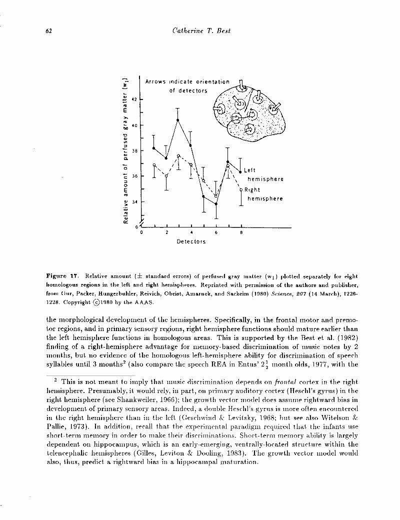
The growth vector would be expected to have the following influence on the development ofcortical gray and white matter: for earlier-developing cortical regions relatively greater growthemphasis would be seen in the earlier-emerging deeper cell layers, which should result in smallerrat.ios of gray matter to white matter and greater regional width/volume. Figure 17, taken fromGur and colleagues (1980), illustrates that indeed the gray-white ratio is smaller in the right hemisphere for at least the primary-motor and -sensory cortical regions. In contrast, later-maturingregions should reflect greater growth emphasis of the later-emerging, more superficial cortical layers, 2 and perhaps 3. This should yield smaller width/volume but a more deeply fissurated regionwith higher gray/white ratios, which may imply a somewhat higher degree of local intracorticalorganization of neuronal connections. In accordance, there is a higher gray /white ratio in the lefthemisphere for motor, premotor, and primary sensory areas (Gur et al., 1980), consistent withSemmes' (1968) claim that the left hemisphere is focally organized, whereas the right is diffuselyorganized (based on her studies of somatosensory deficits in unilateral brain-damaged patients).Correspondingly, it is those same areas in which LeMay (1976) found smaller hemispheric widthon the left, and Weinberger et al. (1982) measured a smaller volume on the left. Also, at leastfor the anterior speech area in the premotor region (Broca's area), there is deeper fissuration onthe left (Falzi et al., 1982).
Interestingly, and consistent with the predictions of the growth vector model, there is ahigher gray / white ratio in the right hemisphere for tertiary association areas in posterior cortex(temporal and parietal association areas, numbered .5 and 6 in Figure 17). Recall the model'sprediction that tertiary association areas in posterior cortex would emerge later in the right. thanin the left hemisphere. Also, in posterior regions, LeMay (1976) found lesser hemispheric width,and Weinberger et al. (1982) found smaller volume, on the right side.
As for development of dendritic processes and of neuronal connections, the influence of thegrowth vector should be a bias toward later-emerging characteristics in the later-maturing regionsof the cortex. Arnold Scheibel (1984) has thus far provided the only data relevant to this issue,and it appears to corroborate one part of the hypothesis. In layer 3 of the anterior speech regionin the left. hemisphere (LOP in Figure 18), the dendritic trees of the neurons show relativelygreater elaboration of the later-emerging dendritic features, such as proportionally more higherorder branching points, than is true of the homologous region in the right hemisphere (Rap inFigure 18).
Implications for Functional Development
This morphological growth vector should also have implications for the development of perceptual and cognitive functions, and for asymmetrical patterns in development.al plast.icity. Grossmorphological asymmetries of the adult brain, both in vivo (Ratcliff, Dila, Taylor, & Milner,1980) and postmortem (Witelson, 1983), are associated, in fact, with at least some measures offunctional asymmet.ries, not.ably speech lateralizat.ion anel handedness. Development. of regionalfunctional maturit.y should proceed according to the same growth vector as already outlined for
62 Catherine T. Best
~Arrows
"-of detectors
Cll42:::
ftl
E>.ftl... 40llIl
'0CllIII::J
"- 38Clla.
-0 Lef t\c: 36 \ hemisphere::J \0 \
E 1Rightftl
Cll 34hemisphere
>-ftlCll
0::o?
0 2 4 6 8
Detectors
Figure 17. Relative amount (± standard errors) of perfused gray matter (wI) plotted separately for eight
homologous regions in the left and right hemispheres. Reprinted with permission of the authors and publisher,
from Our, Packer, Hungerbuhler, Reivich, Obrist, Amarnek, and Sackeim (1980) Science, 207 (14 March), 1226
1228. Copyright 01980 by the AAAS.
the morphological development of the hemispheres. Specifically, in the frontal motor and premotor regions, and in primary sensory regions, right hemisphere functions should mature earlier thanthe left hemisphere functions in homologous areas. This is supported by the Best et al. (1982)finding of a right-hemisphere advantage for memory-based discrimination of music notes by 2months, but no evidence of the homologous left-hemisphere ability for discriminat.ion of speechsyllables unt.il 3 months2 (also compare t.he speech REA in Entus' 2t mont.h olds, 1977, wit.h the
2 This is not meant t.o imply t.hat. music discriminat.ion depends on frontal cortex in the right.hemisphere. Presumably, it would rely, in part, on primary auditory cortex (Heschl's gyrus) in t.heright. hemisphere (see Shankweiler, 1966); the growth vector model does assume rightward bias indevelopment of primary sensory areas. Indeed, a double Heschl's gyrus is more often encounteredin t.he right hemisphere than in the left. (Geschwind & Levitsky, 1968; but see also Wit.elson &Pallie, 1973). In addit.ion, recall t.hat. t.he experimental paradigm required t.hat t.he infants useshort.- t.erm memory in order to make their discriminations. Short.- term memory ability is largelydependent Oll hippocampus, which is an early-emerging, vent.rally-located struct.ure wit.hin t.het.elencephalic hemispheres (Gilles, Levit.on & Dooling, 1983). The growt.h vector model wouldalso, thus, predict a right.ward bias in a hippocampal mat.uration.

Emergence of Gerebml Asymmefl'ies
DENDRITE SEG'-4ENTS I· 2 <~ 4·5·6 4·5·6
TDL
Lop 15537 11608 397 %
Rap 17096 831 2 30.8""
i. /I
Lpe 14339 7744 316 "4I / I
Rpe 18222 6777 25.1 "I.I I .~
f,
2000 1800 1600 1400 600 800 1000 1200
LENGTH IN MICRONS
63
Figure 18. The t.op of t.he figure shows a somewhat schematicized drawing of typical dendritic ensembles from
cells of the left. and right frontal operculum and precent.ral regions in human cort.ex. Not.e the increased number of
higher-order segment.s in left. operculum (Lop) compared to the other three areas, and the relatively greater length
of second- and third-order branches in the right operculum (Rop) and right precentral (Rpc) areas. The bott.om
of the figure shows dendritic length and proportion of the dendritic ensemble made up of lower-order (1,2,3) and
higher-order (4,5,6) dendritic segments in left opercular (Lop), left precentral (Lpc), right opercular (Rop), and
right precentral (Rpc) areas. The column of figures on the extreme right shows the percentage of total dendritic
length (Tdl) occupied by higher-order dendrites in each region. R<;,printed with permission of the publisher from
Scheibel (1984). A dendritic correlate of human speech. In Geschwind and Galaburda (Eds.), Cerebral dominance:
The biological !olJ.ndations. Cambridge, MA: Harvard University Press.

64 Catherine T. Best
lack t.hereof in Vargha-Khadem & Corballis' 2 mont.h olds, 1979). Moreover, it. is consist.ent. wit.hnumerous report.s in t.he language acquisit.ionlit.erat.ure t.hat. children comprehend and produce t.heemot.ional int.onat.ional propert.ies of language earlier t.han t.hey comprehend and produce wordsand word combinat.ions (e.g., Lewis, 1936). Research wit.h bot.h brain-damaged and neurologicallyint.ad adults indicat.es t.hat. t.he right. hemisphere is specialized for affedive, or emot.ional, prosodicaspects of spoken utterances bot.h in percept.ion (e.g., Haggard & Parkinson, 1971; Heilman, Scholes & Wat.son, 197.5; Ley & Bryden, 1982; Papanicolaou, Levin, Eisenberg, & Moore, 1983;Safer & Levent.hal, 1977; Tucker, Wat.son & Heilman, 1976; Wechsler, 1973) and in production(e.g., Benowitz, Bear, Rosent.hal, Mesulam, Zaidel, & Sperry, 1983; Kent. & Rosenbeck, 1982;Ross, 1981, 1984; Ross & Mesulam, 1979; Tucker et. 801., 1976). In addit.ion, according t.o t.he neuroembryological principle t.hat. lat.er-mat.uring brain st.rudures generally show great.er funct.ionalplast.icit.y t.han earlier-mat.uring st.ructures (Jacobson, 1978), t.he relat.ive degree of plast.icit.y ofvarious cort.ical regions should be inversely correlat.ed wit.h t.heir rat.e of mat.urat.ion.
The proposed pat.t.ern of asymmet.ry in functional development. should correspond t.o a great.erplast.icit.y of t.he lat.er-mat.uring, left. front.al-mot.or, -premot.or and primary sensory regions, relat.ive t.o t.he plast.icit.y of t.he homologous right.-hemisphere regions. Conversely, t.here shouldbe lat.er development., and great.er plast.icit.y, on t.he right. relat.ive t.o t.he left. side for post.eriort.ertiary associat.ion area fundions. Unfort.unat.ely, t.here are no published dat.a relevant. t.o t.hisissue. Moreover, it. will be difficult, t.o say t.he least., t.o mat.ch right.- and left.-hemisphere skillsin t.erms of cort.ical areas involved, as well as for level of cognit.ive complexit.y and/or difficulty,in order t.o compare t.heir development. in normal children and t.heir plast.icit.y in brain-damagedchildren. For example, one might. wish t.o compare t.he development. and plast.icit.y of reading abilit.y, which depends on a t.ert.iary associat.ion region of left. hemisphere in adults (angular gyrus),versus that. of complex spat.ial and face-recognit.ion abilit.ies, which depend in adult's upon right.t.ert.iary associat.ion areas (parietal and inferot.emporal). In our societ.y, normal children beginreading around 6 years of age, but. do not. develop t.he abilit.y t.o solve complex mazes, or a right.hemisphere configurat.ional-processing superiorit.y for recognit.ion of unfamiliar faces, unt.il about.10 years of age (e.g., Kohn & Dennis, 1974; Levine, 198.5). If we could assume t.hat. t.he crit.eriaof comparabilit.y were met. by t.hese findings, t.he model would t.hen predict great.er abilit.y of t.heright hemisphere t.o acquire reading skills, relat.ive t.o t.he left. hemisphere's abilit.y t.o acquire spat.ial/facial skills, following early unilat.eral damage. However, t.here are several inherent. problems.The comparabilit.y of cognit.ive levels for t.hese skills is uncert.ain: t.he onset. ages may be, at. least.in part., artifacts of our educat.ional syst.em. Finally, acquisit.ion of a skill may call upon different.cort.ical regions in t.he child t.han t.hose t.hat. underly t.he execut.ion of t.he already-acquired skill int.he adult (see Kirk, 198.5).
Individual differences Il1 functional asymmetries
The growt.h vector is very likely influenced by hormonal and genet.ic factors, given t.he current.underst.anding in neuroembryology t.hat. development.al gradient.s involve some sort. of gradient( s)in biochemical influences on t.he prenat.al guidance of neuronal migrat.ion and development. ofneuronal connedions (J acobson, 1978; Sperry, 1963). The possibilit.y of hormonal influences ongrowt.h gradient.s may aid our underst.anding of t.he development. of sex diH'erellces in brain 01'
ganizat.ion and cognit.ive functions 3 (see also t.he chapt.er by NeHey, t.his volume*), which may
3 The effect. of sex differentiation upon t.he growt.h vector may also be related t.o sex differencesin morphological asymmet.ries for other body part.s, e.g., the hands and feet (Levy & Levy, 1978;

Emergence of CC1'ebral Asymmetries 65
be mediated by sex differences in maturation rates (see Newcombe & Bandura, 1983; Waber,1976,1977, 1979a; but see also Rovet, 1983; Waber, Mann, Merola, & Moylan, 1985). These sexdifferences are most apparent in the extreme cases of sexual anomalies such as Turner's syndrome(XO). Turner's syndrome is associated with large deficits in spatial abilities (e.g., Waber, 1979h),an increase in the rate of prenatal development (Netley & Rovet, 1981), and maldevelopment oftertiary association areas in the right hemisphere (Christensen & Nielsen, 1981). Sex differencesin brain organization and function are also apparent in anomalies such as supernumerary-X syndrome (XXX and XXY), which is linked with low verbal relative to spatial abilities, and slowprenatal growth rates (Netley, this volume*; Netley & Rovet, 1981, 1982, 1983; Rovet & Netley,1983).
Hormonal and genetic influences on the growth vector may also be involved in heritablelearning disorders. For example, Galaburda and colleagues (Galaburda & Eidelberg, 1982; Galaburda & Kemper, 1979; Galaburda, Rosen, Sherman, & AssaI, 1986; Galaburda, Sherman,Rosen, Aboitiz, & Geschwind, 1985) found symmetrical plana temporale in the brains of all fourmale dyslexics and the one female dyslexic that they have studied post-mortem. The same brainsshowed abnormalities in cortical neuronal organization, which predominated in the left hemisphereperisylvian regions. The nature of the latter abnormalities led them to posit a neuroembryologicaldisturbance in the prenatal migration of neurons during mid-gestation. Hormonal/genetic effectson the growth vector may be particularly relevant to understanding observed sex and handednessbiases in the incidence of learning disabilities, for example, the suggestion that testosterone'sefl'ect on brain development forms the basis for the higher incidence of learning disabilities amongmales and left-handel'S (Geschwind & Behan, 1982, 1984; Marx, 1982).
Conclusion
So now, what of the original question: Is the pattern of cerebral asymmetry developmentallyinvariant, or do functional asymmetries develop'? Whether we refer to evidence of functionaland/or structural asymmetries, even in very early development the extant data support "developmental invariance" (see also Kinsbourne, 197.5; Witelson, 1985). Yet this does not necessarilyimply that nothing is changing or developing. The timeless constancy of cerebral asymmetriescoexists with continuous developmental change at many levels. Lateralized perceptual and cognitive functions do undergo developmental change (e.g., the child's language and spatial skillschange both qualitatively and quantitatively), plasticity of function also undergoes developmental change (see Witelson, 1985), and the cerebral hemispheres supporting these abilities undergochange themselves (e.g., increases in dendritic arborization, neuronal connections, neurotransmitter functions, glial support cells, and myelinization).
According to developmental biology, change and constancy are co-determinants of developmental growth in a biological system. The structural and fundional properties of the two cerebralhemispheres do change developmentally, but always in different manners because they developwithin the context of an ever-present lateralization of functions, which is continuous with a lateralized gradient of neuronal differentiation and maturation. The argument presented in thischapter is that the normal direction of this lateralizing gradient is from right to left, and thatit interacts with gradients in the other main axes of embryologic development to result. in a 3D
Means & Walters, 1982); the latter asymmetries in turn appear to be traceable to prenataldevelopment (Mittwoch, 1977).

66 Catherine T. Best
diagonal growth vector from right frontal-motor and primary sensory areas to left-posterior andtertiary association areas. This growth pattern has implications for hemispheric and regionaldifferences in gross morphology and neuronal organization, as well as for differences in plasticityand in maturation of perceptual and cognitive functions.
References
Best, C. T. (1978). The role of consonant and vowel acoustic features in infant cerebral asymmet1,ies for speech perception. Unpublished doctoral dissertation, Michigan State University.
Best, C. T., Hoffman, H., & Glanville, B. B. (1982). Development of infant ear asymmetries forspeech and music. Perception £4 Psychophysics, 31, 75-85.
Benowitz, L. I., Bear, D. M., Rosenthal, R., Mesulam, M. M., Zaidel, E., & Sperry, R. W. (1983).Hemispheric specialization and nonverbal communication. Cortex, 19, 5-12.
Bever, T. G. (1978). Broca and Lashley were right: Cerebral dominance is an accident of growth.In D. Caplan (Ed.), Biological studies of mental processes (pp. 186-230). Cambridge, MA:MIT Press.
Broca, P. (1865). Sur la faculte du langage articule. Bulletin of Social Anthropology. 6, 493-494.Brown, J. W., & Jaffe, J. (1975). Note: Hypothesis on cerebral dominance. Neuropsychologia, 13,
107-110.Chi, F. G., Dooling, E. C., & Gilles, F. H. (1977). Left-right asymmetries of the temporal speech
areas of the human fetus. Archives of Neurology, 34, 346-348.Christensen, A-L., & Nielsen, J. (1981). A neuropsychological investigation of 17 women with
Turner's syndrome. In W. Schmid & J. Nielsen (Eds.), Human behavior and genetics(pp. 151-166). Amsterdam: Elsevier/North-Holland.
Corballis, M. C., & Morgan, M. J. (1978). On the biological basis of human laterality: I. Evidencefor a maturational left-right gradient. The Behavioral and Brain Sciences, 2, 261-267.
Cowan, M. W. (1979). The development of the brain. Scientific American, 241, 112-133.Crowell, D. H., Jones, R. H., Kapuniai, 1. E., & Nakagawa, J. K. (1973). Unilateral cortical
activity in newborn humans: An early index of cerebral dominance? Science, 180, 20.5208.
Cutting, J. E. (1974). Two left hemisphere mechanisms in speech perception. Perception f3 Psychophysics, 16, 601-612.
Darwin, C. J. (1971). Ear differences in the recall of fricatives and vowels. Quarterly Journal ofExperimental Psychology, 23, 46-62.
Dooling, E. C., Chi, J. G., & Gilles, F. H. (1983). Telencephalic development: Changing gyralpatterns. In F. H. Gilles, A. Leviton, & E. C. Dooling (Eds.), The developing humanbrain: Growth and epidemiologic neuropathy (pp. 94-104). John Wright, PSG Inc.
Emde, R. N., & Robinson, J. (1979). The first two months: Recent research in developmentalpsychobiology and the changing view of the newborn. In J. Noshpitz & J. Call (Eds.),Handbook of child psychiatry. Vol. 1: Development. New York: Basic Books.
Entus, A. K. (1977). Hemispheric asymmetry in processing of dichotically presented speech andnonspeech sounds by infants. In S. Segalowitz & F. A. Gruber (Eds.) Language development and neurological theory (pp. 63-(3). New York: Academic Press.
Falzi, G., Perrone, P., & Vignolo, L. A. (1982). Right-left asymmetry in ant.erior speech region.Al'chives of Neurology, 39, 239-240.
Friedes, D. (1977). Do dichot.ic list.ening procedures measure lateralization of informat.ion processing or retrieval strategy? Perception £4 Psychophysics, 21, 259-263.

Emergence of Cerebral Asymmetries 67
Galaburda, A. M. (1984). Anatomical asymmetries. In N. Geschwind & A. M. Galaburda (Eds.),Cerebral dominance: The biological foundations (pp. 11-25). Cambridge, MA: HarvardUniversity Press.
Galaburda, A. M., & Eidelberg, D. (1982). Symmetry and asymmetry and asymmetry in the llUman posterior thalamus: II. Thalamic lesions in a case of developmental dyslexia. A r'chivesof Neurology, 39, 333-336.
Galaburda, A. M., & Kemper, T. L. (1979). Cytoarchitectonic abnormalities in developmentaldyslexia: A case study. Annals of Neurology, 6, 94-100.
Galaburda, A. M., LeMay, M., Kemper, T. L., & Geschwind, N. (1978). Right-left asymmetriesin the brain. Science, 199, 852-856.
Galaburda, A. M., Rosen, G. D., Sherman, G. F., & AssaI, F. (1986). Neuropathological findingsin a woman with developmental dyslexia. A nnals of Neurology, 20, 170.
Galaburda, A. M., Sanides, F., & Geschwind, N. (1978). Cytoarchitectonic left-right asymmetriesin the temporal speech region. Archives of Neurology, 35, 812-817.
Galaburda, A. M., Sherman, G. F., Rosen, G. D., Aboitiz, F., & Geschwind, N. (198.5). Develop-mental dyslexia: Four consecutive patients with corticalanomalies. Annals of Neurology, 18,222-233.
Geschwind, N., & Behan, P. O. (1982). Left-handedness: Association with immune disease, migraine, and developmental learning disorder. Proceedings of the National Academy ofScience, 79, 5079-.5100.·
Geschwind, N., & Behan, P. O. (1984). Laterality, hormones, and immunity. In N. Geschwind &A. M. Galaburda (Eds.), Cerebral dominance: The biological foundations (pp. 211-224).Cambridge, MA: Harvard University Press.
Geschwind, N., & Levitsky, W. (1968). Human brain: Left-right asymmetries in temporal speechregion. Science, 161, 186-189.
Gilles, F. H., Leviton, A., & Dooling, E. C. (1983). The developing human brain: Growth and
epidemiologic neuropathology. Boston: John Wright, PSG, Inc.Glanville, B. B., Best, C. T., & Levenson, R. (1977). A cardiac measure of cerebral asymmetries
in infant auditory perception. Developmental Psychology, 13, .54-59.Graham, F. K., Leavitt, L. A., Strock, B. D., & Brown, J. W. (1978). Precocious cardiac orienting
in a human anencephalic infant. Science. 199, 322-324.Gur, R. C., Packer, I. K., Hungerbuhler, J. P., Reivich, M., Obrist, W. D., Amarnek, W. S., &
Sackeim, H. A. (1980). Differences in the distribution of gray and white matter in humancerebral hemispheres. Science, 207, 1226-1228.
Haggard, M. P., & Parkinson, A. M. (1971). Stimulus and task factors as determinants of earadvantages. Quarte7'iy Journal of Experimental Psychology, 23, 168-177.
Heilman, J. M., Scholes, R., & Watson, R. T. (197.5). Auditory affective agnosia: Disturbedcomprehension of affective speech. Journal of Neurology, Neurosurgery and Psychiatry.38, 69-72.
Jacobson, M. (1978). Developmental neurobiology. New York: Plenum Press.Keller, R. (1942). The asymmetry of the human body. ClBA Symposium. :J, 1126-1127.Kemper, T. L. (1984). Asymmetrical lesions in dyslexia. In N. Geschwilld & A. M. Galaburda
(Eds.), Cer'ebral dominance: The biological foundations (pp. 7.5-89). Cambridge, MA:Harvard University Press.
Kent, R. D., & Rosenbeck, J. C. (1982). Prosodic disturbance and neurologic lesion. Brain and
Language, 15,2.59-291.
68 Catherine T. Best
Kinsbourne, M. (1975). The ontogeny of cerebral dominance. Annals of the New York Academyof Sciences, 263, 244-250.
Kirk, V. (198.5). Hemispheric contributions to the development of graphic skill. In C. T. Best (Ed.),Hemisphe1'ic function and collaboration in the child (pp. 193-228). New York: AcademicPress.
Kohn, B., & Dennis, M. (1974). Selective impairments of visuospatial abilities in infantile hemiplegics after right cerebral hemi-decortication. Neuropsychologia, 12, 505-512.
LeMay, M. (1976). Morphological cerebral asymmetries of modern man, fossil man, and nonhumanprimate. Annals of the New York Academy of Science, 280,349-366.
LeMay, M. (1977). Asymmetries of the skull and handedness. Journal of Neurological Science,32, 243-253.
LeMay, M. (1984). Radiological, developmental, and fossil asymmetries. In N. Geschwind &A. M. Galaburda (Eds.), Cerebral dominance: The biological foundations (pp. 26-42).Cambridge, MA: Harvard University Press.
LeMay, M., & Geschwind, N. (1978). Asymmetries of the human cerebral hemispheres. In A. Caramazza & E. B. Zurif (Eds.), Language acquisition and language b,'eakdown (pp. 311-328).Baltimore, MD: Johns Hopkins Press.
LeMay, M., & Kido, D. K. (1978). Asymmetries of the cerebral hemispheres on computed tomograms. Journal of Computer-Assisted Tomography, 2,471-476.
Lenneberg, E. H. (1967). Biological foundations of language. New York: Wiley.Levine, S. C. (198.5). Developmental changes in right hemisphere involvement in face perception.
In C. T. Best (Ed.), Hemispheric function and collaboration in the child (pp. 157-191).New York: Academic Press.
Levy, J., & Levy, J. M. (1978). Human lateralization from head to foot: Sex-related factors.Science, 200, 1291-1292.
Lewis, M. M. (1936). Infant speech: A study of the beginnings of language. New York: HarcourtBrace.
Ley, R. G., & Bryden, M. P. (1982). A dissociation of right and left hemispheric effects forrecognizing emotional tone and verbal content. Brain and Cognition, 1, 3-9.
MacKain, K. S., Studdert-Kennedy, M., Spieker, S., & Stern, D. (1983). Infant intermodal speechperception is a left heinisphere function. Science, 219, 1347-1349.
Marx, J. L. (1982). Autoimmunity in left-handel's. Science, 217,141-144.McHenry, L. C., Merory, J., Bass, E., Stump, D. A., Williams, R., Witcofski, R., Howard, G.
& Toole, J. F. (1978). Xenon-133 inhalation method for regional cerebral blood flowmeasurements: Normal values and test-results. Stroke, 9, 396-399.
Means, L. W., & Walters, R. E. (1982). Sex, handedness and asymmetry of hand and foot length.Neuropsychologia, 20, 715-719.
Meyer, J. S., Ishihara, N., Deshmukh, V. D., Naritomi, H., Sakai, F., Hsu, M-C., & Pollack, P.(1978). Improved method for noninvasive measurement of regional cerebral blood flowby 133Xenon inhalation. Part I: Description of method and normal values obtained inhealthy volunteers. Strol~e, 9, 19.5-205.
Mittwoch, U. (1977). To be born right is to be born male. New Scientist. 73,74-76.Molfese, D. L., Freeman, R. B., & Palermo, D. S. (197.5). The ontogeny of brain lateralization for
speech and nonspeech stimuli. Brain and Language, 2, 3.56-368.Molfese, D., & Molfese, V. J. (1985). Electrophysiological indices of auditory discrimination in
newborn infants: The basis for predicting later language development? Infant Behaviorand Development, 8, 197-211.

Emergence of Cerebral Asymmetries 69
Netley, C., & Rovet, J. (1981). Prenatal growth rate and hemispheric organization. Paper presented at meeting of the International Neuropsychology Society. Atlanta, GA, February.
Netley, C., & Rovet, J. (1982). Verbal deficits in children with 47,XXY and 47,XXX karyotypes:A descriptive and experimental study. Brain and Language, 17, .58-72.
Netley, C., & Rovet, J. (1983). Relationships among brain organization, maturation rate, and thedevelopment of verbal and nonverbal ability. In S. Segalowitz (Ed.), Language functionsand brain organization (pp. 245-26.5). New York: Academic Press.
Newcombe, N., & Bandura, M. (1983). Effect of age at puberty on spatial ability in girls. Developmental Psychology, 19, 215-224.
Orton, S. T. (1937). Reading, writing, and speech problems in children. New York: W. W. Norton& Co.
Papanicolaou, A. C., Levin, H. S., Eisenberg, H. M., & Moore, B. D. (1983). Evoked potentialindices of selective hemispheric engagement in affective and phonetic tasks. N europsychologia, 21, 401-40.5.
Rabinowicz, T. (1979). The differentiate maturation of the human cerebral cortex. In F. Falkner& J. M. Tanner (Eds), Human Growth. Vol. 3: Neurobiology and nutrition (pp. 97-123).New York: Plenum Press.
Rakic, P. (1981). Developmental events leading to laminar and areal organization ofthe neocortex.In F. O. Schmitt, F. G. Worden, G. Adelman, & S. G. Dennis (Eds.), The organization
of cerebral cortex (pp. 7-28). Cambridge, MA: MIT Press.Ratcliff, G., Dila, C., Taylor, L., & Milner, B. (1980). The morphological asymmetry of the hemi
spheres and cerebral dominance for speech: A possible relationship. Brain and Language,11, 87-88.
Rose, S. A. (1984). Developmental changes in hemispheric specialization for tactual processing invery young children: Evidence from cross-modal transfer. Developmental Psychology, 20,568-.574.
Ross, E. D. (1981). The aprosodias: Functional-anatomic organization of the affective componentsof language in the right hemisphere. Al'chives of Neurology, 38, 561-569.
Ross, E. D. (1984). Right hemisphere role in language, affective behavior and emotion. Trends inNeuroscience, 342-346.
Ross, E. D., & Mesulam, M. M. (1979). Dominant language functions of the right hemisphere:Prosody and emotional gesturing. Archives of Neurology, 36, 144-146.
Rovet, J. (1983). Cognitive and neuropsychological test performance of persons with abnormalitiesof adolescent development: A test of Waber's hypothesis. Child Development, 54,941-9.50.
Rovet, J., & Netley, C. (1983). The triple-X chromosome syndrome in childhood: Recent empiricalfindings. Child Development, 54, 831-845.
Safer, M. A., & Leventhal, H. (1977). Ear differences in evaluating emotional tone of voice and ofverbal content. Journal of Experimental Psychology: Human Perception and PC1'formance,
3, 75-82.Scheibel, A. B. (1984). A dendritic correlate of human speech. In N. Geschwind & A. M. Gal
aburda (Eds.), Cerebral dominance: The biological foundations. Cambridge, MA: HarvardUniversity Press.
Schwartz, J., & Tallal, P. (1980). Rate of acoustic change may underlie hemispheric specializationfor speech perception. Science. 207. 1380-138l.
Semmes, J. (1968). Hemispheric specialization: A possible clue to mechanism. N e1t1'opsychologia,
6, 11-26.
70 Cathel,ine T. Best
Shankweiler, D. (1966). Effects of temporal-lobe damage on percept.ion of dichotically presentedmelodies. Joumal of Compamtive and Physiological Psychology, 62, 115-119.
Sidtis, J. J. (1980). On the nature of the cortical function underlying right hemisphere auditorypercept.ion. Neuropsychologia. 18, 321-330.
Sperry, R. (1963). Chemoaffinity in the orderly growth of nerve fiber patterns and connections.Proceedings of the National Academy of Science, 50, 703-710.
Studdert-Kennedy, M., & Shankweiler, D. (1970). Hemispheric specializat.ion for speech perception. Joumal of the Acoustical Society of America, 48, .579-594.
Studdert-Kennedy, M., & Shankweiler, D. (1980). Hemispheric specialization for language processes. Science, 211, 960-961.
Taylor, D. C. (1969). Differential rates of cerebral maturation between sexes and between hemispheres. Lancet, 2, 140-142.
Tucker, D. M., Watson, R. G., & Heilamn, K. M. (1976). Affective discrimination and evocationin patients wit.h parietal disease. Neurology, 26, 354.
Vargha-Khadem, F., & Corballis, M. C. (1979). Cerebral asymmetry in infants. Bmin and Language, 8, 1-9.
Waber, D. P. (1976). Sex differences in cognition: A function of maturation rate. Science, 192,.572-574.
Waber, D. P. (1977). Sex differences and rate of physical growth. Developmental Psychology, 13,29-38.
Waber, D. P. (1979a). Cognitive abilities and sex-related variations in t.he mat.uration of cerebralcort.ical functions. In M. Wittig & A. Peterson (Eds.) Sex-related differences in cognitivefunctioning. New York: Academic Press.
Waber, D. P. (1979b). Neuropsychological aspects of Turner syndrome. Developmental Medicineand Child Neurology, 231, 58-70.
Waber, D. P., Mann, M. B., Merola, J., & Moylan, P. M. (198.5). Physical maturat.ion rat.e andcognitive performance in early adolescence: A longitudinal examination. DevelopmentalPsychology, 21, 666-681.
Wada, J. A., Clarke, R., & Hamm, A. (1975). Cerebral hemispheric asymmetry in humans.Al'chives of Neurology, 32, 239-246.
Wechsler, A. F. (1973). The effect of organic brain disease on recall of emotionally charged versusneutralnarrat.ive text. Neurology, 23, 130-135.
Weinberger, D. R., Luchins, D. J., Morihisa, J., & Wyatt, R. J. (1982). Asymmetrical volumes ofthe right and left frontal and occipital regions of the human brain. Annals of Neurology,11, 97-100.
Weiss, M. S., & House, A. S. (1973). Perception of dichotically presented vowels. Joumal of theAcoustical Society of America, 53, 51-.53.
Witelson, S. F. (1977). Early hemispheric specialization and interhemispheric plasticity: An empirical and theoretical review. In S. Segalowit.z & F. A. Gruber (Eds.), Language developmentand neurological theory (pp. 213-287). New York: Academic Press.
Witelson, S. F. (1983). Bumps on the brain: Neuroanat.omical asymmetries as a basis for functionalasymmetries. In S. Segalowitz (Ed.), Language functions and bmin organization (pp. 117144). New York: Academic Press.
Witelson, S. F. (1985). Hemisphere specialization from birth: Mark II. In C. T. Best (Eel.),Hemispheric function and collabomtion in the child (pp. 33-85). New York: AcademicPress.

Emergence of Cerebral Asymmetries 71
Witelson, S. F., & Pallie, W. (1973). Left. hemisphere specialization for language in the newborn:Neuroanatomical evidence of asymmetry. Brain, 96, 641-646.
Yakovlev, P. I., & Lecours, A. R. (1967). The myelogenetic cycles of regional maturation of thebrian. In A. Minkowski (Ed.), Regional development of the brain in early life (pp. 3-70).Oxford: Blackwell.